Structural Basis of Lysine-Acetylated HIV1 Tat Recognition by PCAF Bromodomain
12
Molecular Cell, Vol. 9, 575–586, March, 2002, Copyright 2002 by Cell Press Structural Basis of Lysine-Acetylated HIV-1 Tat Recognition by PCAF Bromodomain cation in infected cells (Adams et al., 1994; Garber and Jones, 1999). While the detailed molecular mechanisms underlying Shiraz Mujtaba, 1 Yan He, 1 Lei Zeng, 1 Amjad Farooq, 1 Justin E. Carlson, 1 Melanie Ott, 2 Eric Verdin, 3 and Ming-Ming Zhou 1,4 Tat dissociation from TAR RNA and its transactivation 1 Structural Biology Program of transcription of the integrated HIV-1 genome remain Department of Physiology and Biophysics elusive, increasing lines of evidence suggest that Tat Mount Sinai School of Medicine activity requires its association with several multiprotein New York University complexes, which include the cyclinT1/cyclin-depen- New York, New York 10029 dent kinase 9 (CDK9) complex (Jones, 1997; Wei et al., 2 Applied Tumor Virology 1998) and histone acetyltransferase (HAT) transcrip- Deutsches Krebsforschungszentrum tional coactivators, p300/CBP (CREB binding protein), Im Neuenheimer Feld, 242 and p300/CBP-associated factor (PCAF) (Benkirane et Heidelberg, D-69120 al., 1998; Deng et al., 2000; Hottiger and Nabel, 1998). Germany Recruitment of CDK9 through the N-terminal cysteine- 3 Gladstone Institute of Virology and Immunology rich region of Tat results in hyperphosphorylation of the University of California, San Francisco C-terminal domain of RNAPII that increases elongation 365 Vermont Street efficiency of HIV-1 transcription (Wei et al., 1998). Re- San Francisco, California 94103 cently, it has been shown that Tat activity is dependent on acetylation by p300/CBP at K50 located in the C-terminal arginine-rich motif (ARM) (Kiernan et al., Summary 1999; Ott et al., 1999), a region that is also important for TAR RNA binding and nuclear localization. Mutation The human immunodeficiency virus type 1 (HIV-1) of K50 to arginine, a conserved amino acid substitution trans-activator protein Tat stimulates transcription of that retains the positive charge but prevents acetylation the integrated HIV-1 genome and promotes viral repli- by p300, markedly decreases the synergistic activation cation in infected cells. Tat transactivation activity is of the HIV-1 promoter by Tat and p300 (Kiernan et al., dependent on lysine acetylation and its association 1999; Ott et al., 1999). Tat acetylation at K50 results in with nuclear histone acetyltransferases p300/CBP its dissociation from TAR RNA and promotes formation (CREB binding protein) and p300/CBP-associated fac- of a multiprotein complex comprised of Tat, p300/CBP, tor (PCAF). Here, we show that the bromodomain of and PCAF (Benkirane et al., 1998; Deng et al., 2000). PCAF binds specifically to HIV-1 Tat acetylated at ly- Furthermore, it has been shown that the HAT activity of sine 50 and that this interaction competes effectively PCAF is preferentially required for Tat transactivation of against HIV-1 TAR RNA binding to the lysine-acet- transcription of the integrated but not the unintegrated ylated Tat. The three-dimensional solution structure HIV-1 LTRs (Benkirane et al., 1998). of the PCAF bromodomain in complex with a lysine Protein lysine acetylation is emerging as a central 50-acetylated Tat peptide together with biochemical mechanism in regulation of chromatin remodeling and analyses provides the structural basis for the specific- transcriptional activation (Jenuwein and Allis, 2001; ity of this molecular recognition and reveals insights Kouzarides, 2000; Strahl and Allis, 2000). Bromodo- into the differences in ligand selectivity of bromodo- mains, an extensive family of conserved protein mod- mains. ules found in many chromatin-associated proteins and in nearly all nuclear histone acetyltransferases (Brownell and Allis, 1996; Haynes et al., 1992; Jeanmougin et al., Introduction 1997; Tamkun et al., 1992), have been recently discov- ered to function as acetyl-lysine binding domains (Dhal- The human immunodeficiency virus type 1 (HIV-1) pro- luin et al., 1999; Hudson et al., 2000; Jacobson et al., tein Tat is an atypical trans-activator of transcription 2000; Owen et al., 2000). This new finding suggests a which functions through binding to an RNA element novel mechanism for regulating protein-protein interac- known as the transactivation responsive region (TAR), tions via lysine acetylation (Dyson et al., 2001; Jenuwein located in the retroviral long-terminal repeat (LTR) (Cul- and Allis, 2001; Strahl and Allis, 2000; Winston and Allis, len, 1998; Jeang et al., 1999; Karn, 1999). Tat binds to 1999). This new mechanism supports the hypothesis TAR RNA with high affinity but transiently (Keen et al., that bromodomains could contribute to highly specific 1997; Rana and Jeang, 1999). Dissociation of Tat from histone acetylation by tethering transcriptional HATs to TAR RNA facilitates Tat association with the assembled specific chromosomal sites (Brownell and Allis, 1996; RNA polymerase II (RNAPII) complex (Deng et al., 2000; Manning et al., 2001; Travers, 1999), and to the assembly Kiernan et al., 1999). The latter process enables the and activity of multiprotein complexes of chromatin re- transcriptional machinery complex to elongate effi- modeling such as SAGA and NuA4 (Brown et al., 2001; ciently on the viral DNA template in order to produce Sterner et al., 1999). However, because no specific, bio- full-length HIV transcripts during viral productive repli- logically relevant binding sites had been reported for any particular bromodomain, the major question of ligand specificity of bromodomains still remains unanswered. 4 Correspondence: [email protected]
Transcript of Structural Basis of Lysine-Acetylated HIV1 Tat Recognition by PCAF Bromodomain
Molecular Cell, Vol. 9, 575–586, March, 2002, Copyright 2002 by Cell Press
Structural Basis of Lysine-Acetylated HIV-1Tat Recognition by PCAF Bromodomain
cation in infected cells (Adams et al., 1994; Garber andJones, 1999).
While the detailed molecular mechanisms underlying
Shiraz Mujtaba,1 Yan He,1 Lei Zeng,1
Amjad Farooq,1 Justin E. Carlson,1 Melanie Ott,2
Eric Verdin,3 and Ming-Ming Zhou1,4
Tat dissociation from TAR RNA and its transactivation1Structural Biology Programof transcription of the integrated HIV-1 genome remainDepartment of Physiology and Biophysicselusive, increasing lines of evidence suggest that TatMount Sinai School of Medicineactivity requires its association with several multiproteinNew York Universitycomplexes, which include the cyclinT1/cyclin-depen-New York, New York 10029dent kinase 9 (CDK9) complex (Jones, 1997; Wei et al.,2 Applied Tumor Virology1998) and histone acetyltransferase (HAT) transcrip-Deutsches Krebsforschungszentrumtional coactivators, p300/CBP (CREB binding protein),Im Neuenheimer Feld, 242and p300/CBP-associated factor (PCAF) (Benkirane etHeidelberg, D-69120al., 1998; Deng et al., 2000; Hottiger and Nabel, 1998).GermanyRecruitment of CDK9 through the N-terminal cysteine-3 Gladstone Institute of Virology and Immunologyrich region of Tat results in hyperphosphorylation of theUniversity of California, San FranciscoC-terminal domain of RNAPII that increases elongation365 Vermont Streetefficiency of HIV-1 transcription (Wei et al., 1998). Re-San Francisco, California 94103cently, it has been shown that Tat activity is dependenton acetylation by p300/CBP at K50 located in theC-terminal arginine-rich motif (ARM) (Kiernan et al.,
Summary 1999; Ott et al., 1999), a region that is also importantfor TAR RNA binding and nuclear localization. Mutation
The human immunodeficiency virus type 1 (HIV-1) of K50 to arginine, a conserved amino acid substitutiontrans-activator protein Tat stimulates transcription of that retains the positive charge but prevents acetylationthe integrated HIV-1 genome and promotes viral repli- by p300, markedly decreases the synergistic activationcation in infected cells. Tat transactivation activity is of the HIV-1 promoter by Tat and p300 (Kiernan et al.,dependent on lysine acetylation and its association 1999; Ott et al., 1999). Tat acetylation at K50 results inwith nuclear histone acetyltransferases p300/CBP its dissociation from TAR RNA and promotes formation(CREB binding protein) and p300/CBP-associated fac- of a multiprotein complex comprised of Tat, p300/CBP,tor (PCAF). Here, we show that the bromodomain of and PCAF (Benkirane et al., 1998; Deng et al., 2000).PCAF binds specifically to HIV-1 Tat acetylated at ly- Furthermore, it has been shown that the HAT activity ofsine 50 and that this interaction competes effectively PCAF is preferentially required for Tat transactivation ofagainst HIV-1 TAR RNA binding to the lysine-acet- transcription of the integrated but not the unintegratedylated Tat. The three-dimensional solution structure HIV-1 LTRs (Benkirane et al., 1998).of the PCAF bromodomain in complex with a lysine Protein lysine acetylation is emerging as a central50-acetylated Tat peptide together with biochemical mechanism in regulation of chromatin remodeling andanalyses provides the structural basis for the specific- transcriptional activation (Jenuwein and Allis, 2001;ity of this molecular recognition and reveals insights Kouzarides, 2000; Strahl and Allis, 2000). Bromodo-into the differences in ligand selectivity of bromodo- mains, an extensive family of conserved protein mod-mains. ules found in many chromatin-associated proteins and
in nearly all nuclear histone acetyltransferases (Brownelland Allis, 1996; Haynes et al., 1992; Jeanmougin et al.,Introduction1997; Tamkun et al., 1992), have been recently discov-ered to function as acetyl-lysine binding domains (Dhal-The human immunodeficiency virus type 1 (HIV-1) pro-luin et al., 1999; Hudson et al., 2000; Jacobson et al.,tein Tat is an atypical trans-activator of transcription2000; Owen et al., 2000). This new finding suggests awhich functions through binding to an RNA elementnovel mechanism for regulating protein-protein interac-known as the transactivation responsive region (TAR),tions via lysine acetylation (Dyson et al., 2001; Jenuwein
located in the retroviral long-terminal repeat (LTR) (Cul-and Allis, 2001; Strahl and Allis, 2000; Winston and Allis,
len, 1998; Jeang et al., 1999; Karn, 1999). Tat binds to1999). This new mechanism supports the hypothesis
TAR RNA with high affinity but transiently (Keen et al., that bromodomains could contribute to highly specific1997; Rana and Jeang, 1999). Dissociation of Tat from histone acetylation by tethering transcriptional HATs toTAR RNA facilitates Tat association with the assembled specific chromosomal sites (Brownell and Allis, 1996;RNA polymerase II (RNAPII) complex (Deng et al., 2000; Manning et al., 2001; Travers, 1999), and to the assemblyKiernan et al., 1999). The latter process enables the and activity of multiprotein complexes of chromatin re-transcriptional machinery complex to elongate effi- modeling such as SAGA and NuA4 (Brown et al., 2001;ciently on the viral DNA template in order to produce Sterner et al., 1999). However, because no specific, bio-full-length HIV transcripts during viral productive repli- logically relevant binding sites had been reported for any
particular bromodomain, the major question of ligandspecificity of bromodomains still remains unanswered.4 Correspondence: [email protected]
Molecular Cell576
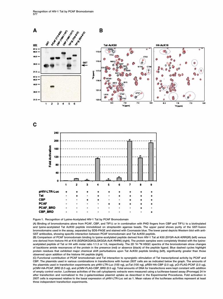
In efforts to determine the mechanisms of action of Tat To assess the biological relevance of the PCAF bro-modomain and Tat interaction to the activation of Tatin transactivation of HIV-1 transcription, we investigated
whether the interaction of the activated, lysine-acet- transcriptional activity by PCAF and p300/CBP (Benkir-ane et al., 1998; Kiernan et al., 1999), we performed cellylated Tat with the nuclear HAT transcriptional cofactors
p300/CBP and PCAF involves any of the bromodomains transfection experiments and measured their combinedeffect on the activity of the HIV-1 promoter using anof the latter proteins. Here, we report that the bromodo-
main of PCAF but not CBP can specifically recognize HIV-1 LTR-luciferase reporter gene assay (Bieniasz etal., 1998; Madore and Cullen, 1993). As shown in Figurethe lysine-acetylated Tat at K50 (not K51 or K28), and
this interaction competes effectively against TAR RNA 1C, synergistic activation of Tat-mediated transcriptionof the HIV-1 promoter in human 293T cells is dependentbinding to the acetylated Tat. We have also determined
the three-dimensional structure of the PCAF bromodo- upon both PCAF and CBP. The latter HAT transcriptionalcoactivator has been recently shown to be responsiblemain in complex with a lysine-acetylated peptide de-
rived from Tat at K50 by using nuclear magnetic reso- for lysine acetylation of Tat at K50 that is required forTat activation (Kiernan et al., 1999; Ott et al., 1999). Morenance (NMR) spectroscopy. NMR structural and
biochemical analyses were further used to gain struc- importantly, our data show that cotransfection of thePCAF bromodomain but not the CBP bromodomain re-tural insights into this important molecular recognition
as well as the differences in ligand selectivity of different sulted in a significant reduction of the synergistic activa-tion of Tat by PCAF and CBP, likely due to an effectivebromodomains.competition of the PCAF bromodomain against the full-length PCAF binding to Tat. Collectively, our in vivoResults and Discussiontransfection study further confirms the highly specificnature of PCAF bromodomain/Tat recognition and high-PCAF Bromodomain Recognition of Lysine-lights the functional importance of this bromodomainAcetylated HIV-1 Tat at K50interaction in the synergistic PCAF- and CBP-inducedTo test whether Tat-p300/CBP-PCAF association in-activation of Tat transcriptional activity in HIV-1 genevolves bromodomains of the histone acetyltransferaseexpression.transcriptional coactivators, we performed an in vitro
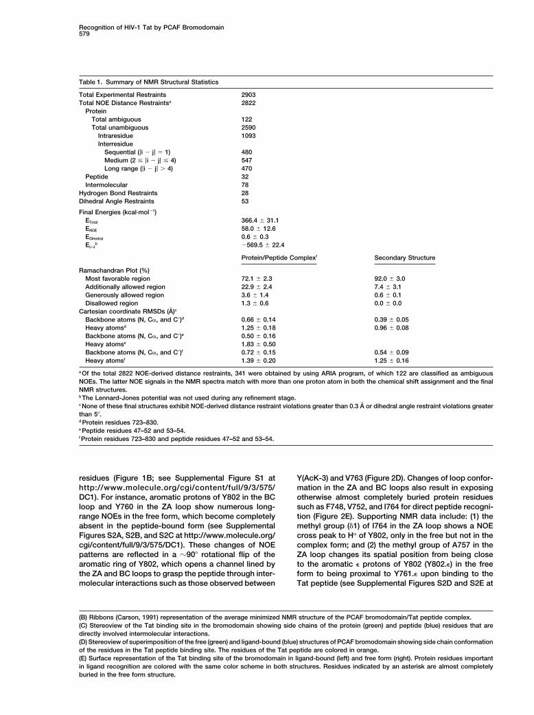
binding assay by using recombinant and purified GST-fusion bromodomains and lysine-acetylated peptides Structure of the PCAF Bromodomain/Tat
Peptide Complexderived from known acetylation sites in Tat at K28 andK50. A lysine-acetylated Tat peptide containing AcK50 To understand the structural basis of this molecular rec-
ognition, we determined the three-dimensional structure(SYGR-AcK-KRRQR, where AcK is an N�-acetyl-lysine)showed no detectable interactions with either bromodo- of the PCAF bromodomain in complex with Tat AcK50
peptide from a total of 2903 NMR-derived restraints.mains or bromodomain and PHD finger (Aasland et al.,1995) tandem modules from CBP or TIF1� (transcrip- The structure for the protein (residues 723–830) and the
peptide (residues 47–54) complex was well defined bytional intermediary factor 1�, also named KAP-1 andKRIP-1) (Friedman et al., 1996) (Figure 1A). Strikingly, the NMR data (Figure 2A, Table 1). The structure of
the bromodomain when complexed to the Tat peptidethe same Tat peptide bound tightly to the bromodomainof PCAF, which shares high sequence homology to CBP consists of a left-handed four-helix bundle (helices �Z,
�A, �B, and �C) and is similar to its free form structurebromodomain (Jeanmougin et al., 1997). The binding isdependent on acetylation of K50 on Tat. Neither of these (Dhalluin et al., 1999), except for the ZA and BC loops
that comprise the acetyl-lysine binding site and undergobromodomains interacted with an acetylated Tat pep-tide derived from K28 (TNCYCK-AcK-CCFH) (data not local conformational changes to accommodate peptide
binding (see below). The Tat peptide adopts an extendedshown), highlighting the selective nature of PCAF bro-modomain recognition of the Tat AcK50 sequence. conformation and lies across a pocket formed between
the ZA and BC loops (Figure 2B). The side chain of theWe performed an NMR study in order to determinethe specificity of PCAF bromodomain binding to lysine- acetyl-lysine intercalates into the protein hydrophobic
cavity and interacts extensively with residues F748,acetylated Tat. As anticipated, PCAF bromodomain didnot bind to Tat AcK28 peptide, nor did CBP bromodomain V752, Y760, I764, Y802, and Y809 (Figure 2C). Peptide
residues flanking the acetyl-lysine contact the protein.to either Tat AcK28 or AcK50 peptide. In contrast, PCAFbromodomain bound to Tat AcK50 peptide with high Particularly, G(AcK�2), R(AcK�1), and R(AcK�3) showed
intermolecular NOEs to the protein, and extensive inter-affinity and caused extensive chemical shift perturba-tions of protein amide resonances, significantly greater actions were observed between the side chains of
Y(AcK�3) and V763 and between Q(AcK�4) and E756.than those seen with an acetylated peptide derived fromhistone H4 at K16, as shown in 2D 1H-15N heteronuclear These specific interactions confer a highly selective as-
sociation between PCAF bromodomain and Tat.single quantum coherence (HSQC) spectra (Figure 1B).This observation agrees with the differences in dissocia- Structural comparison of PCAF bromomdomain in the
free and ligand-bound forms reveals that structuraltion constants (KD), determined by NMR titration to be�10 �M and �300 �M for the former and latter com- changes of the protein, largely localized in the ZA and
BC loops, are directly coupled with the peptide bindingplexes, respectively. These results argue that PCAF bro-modomain binding to H4 peptide is largely limited to (Figure 2D). These structural changes are supported by
extensive NMR data, which include changes of chemicalthe acetyl-lysine, whereas its recognition of Tat mostlikely involves additional interactions with residues shifts and NOE patterns for the backbone amides, side
chain methyl groups, and aromatic rings of many proteinflanking AcK50.
Recognition of HIV-1 Tat by PCAF Bromodomain577
Figure 1. Recognition of Lysine-Acetylated HIV-1 Tat by PCAF Bromodomain
(A) Binding of bromodomains alone from PCAF, CBP, and TIF1� or in combination with PHD fingers from CBP and TIF1� to a biotinylatedand lysine-acetylated Tat AcK50 peptide immobilized on streptavidin agarose beads. The upper panel shows purity of the GST-fusionbromodomains used in the assay, separated by SDS-PAGE and stained with Coomassie blue. The lower panel depicts Western blot with anti-GST antibodies, showing specific interaction between PCAF bromodomain and Tat AcK50 peptide.(B) Comparison of PCAF bromodomain binding to lysine-acetylated peptide derived from HIV-1 Tat at K50 (SYGR-AcK-KRRQR) (left) versusone derived from histone H4 at K16 (SGRGKGGKGLGKGGA-AcK-RHRK) (right). The protein samples were completely titrated with the lysine-acetylated peptide of Tat or H4 with molar ratio 1:1.5 or 1:6, respectively. The 2D 1H-15N HSQC spectra of the bromodomain show changesof backbone amide resonances of the protein in the presence (red) or absence (black) of the peptide ligand. Blue dashed cycles highlightprotein residues that exhibited major chemical shift perturbations upon Tat AcK50 peptide binding (left), significantly greater than thoseobserved upon addition of the histone H4 peptide (right).(C) Functional contribution of PCAF bromodomain and Tat interaction to synergistic stimulation of Tat transcriptional activity by PCAF andCBP. The plasmids used in various combinations in transfections with human 293T cells are as indicated below the graph. The amounts ofthe plasmids used in transfection experiments are pHIV-LTR-Luc (100 ng), pcTat (100 ng), pRSV-HA-CBP (2.0 ug), pCI-FLAG-PCAF (2.0 ug),pCMV-HA-PCAF_BRD (0.5 ug), and pCMV-FLAG-CBP_BRD (0.5 ug). Total amounts of DNA for transfections were kept constant with additionof empty control vector. Luciferase activities of the cell cytoplasmic extracts were measured using a luciferase-based assay (Promega) 24 hrafter transfection and normalized to the �-galactosidase plasmid uptake as described in the Experimental Procedures. Fold activation in293T cells is expressed relative to the basal expression of pHIV-LTR-Luc set as 1. Mean values of the luciferase activities represent at leastthree independent transfection experiments.
Molecular Cell578
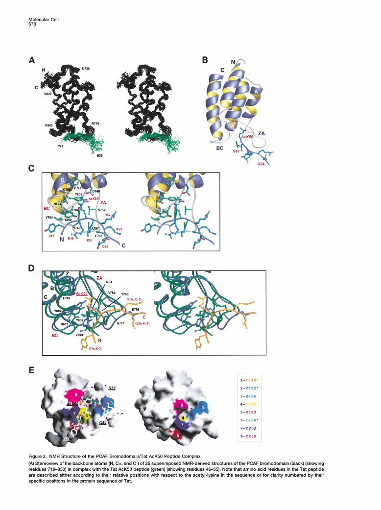
Figure 2. NMR Structure of the PCAF Bromodomain/Tat AcK50 Peptide Complex
(A) Stereoview of the backbone atoms (N, C�, and C�) of 25 superimposed NMR-derived structures of the PCAF bromodomain (black) (showingresidues 719–830) in complex with the Tat AcK50 peptide (green) (showing residues 46–55). Note that amino acid residues in the Tat peptideare described either according to their relative positions with respect to the acetyl-lysine in the sequence or for clarity numbered by theirspecific positions in the protein sequence of Tat.
Recognition of HIV-1 Tat by PCAF Bromodomain579
Table 1. Summary of NMR Structural Statistics
Total Experimental Restraints 2903Total NOE Distance Restraintsa 2822
ProteinTotal ambiguous 122Total unambiguous 2590
Intraresidue 1093Interresidue
Sequential (|i � j| 1) 480Medium (2 |i � j| 4) 547Long range (|i � j| � 4) 470
Peptide 32Intermolecular 78
Hydrogen Bond Restraints 28Dihedral Angle Restraints 53
Final Energies (kcal·mol�1)ETotal 366.4 � 31.1ENOE 58.0 � 12.6EDihedral 0.6 � 0.3EL–J
b �569.5 � 22.4
Protein/Peptide Complexf Secondary Structure
Ramachandran Plot (%)Most favorable region 72.1 � 2.3 92.0 � 3.0Additionally allowed region 22.9 � 2.4 7.4 � 3.1Generously allowed region 3.6 � 1.4 0.6 � 0.1Disallowed region 1.3 � 0.6 0.0 � 0.0
Cartesian coordinate RMSDs (A)c
Backbone atoms (N, C�, and C�)d 0.66 � 0.14 0.39 � 0.05Heavy atomsd 1.25 � 0.18 0.96 � 0.08Backbone atoms (N, C�, and C�)e 0.50 � 0.16Heavy atomse 1.83 � 0.50Backbone atoms (N, C�, and C�)f 0.72 � 0.15 0.54 � 0.09Heavy atomsf 1.39 � 0.20 1.25 � 0.16
a Of the total 2822 NOE-derived distance restraints, 341 were obtained by using ARIA program, of which 122 are classified as ambiguousNOEs. The latter NOE signals in the NMR spectra match with more than one proton atom in both the chemical shift assignment and the finalNMR structures.b The Lennard-Jones potential was not used during any refinement stage.c None of these final structures exhibit NOE-derived distance restraint violations greater than 0.3 A or dihedral angle restraint violations greaterthan 5�.d Protein residues 723–830.e Peptide residues 47–52 and 53–54.f Protein residues 723–830 and peptide residues 47–52 and 53–54.
residues (Figure 1B; see Supplemental Figure S1 at Y(AcK-3) and V763 (Figure 2D). Changes of loop confor-mation in the ZA and BC loops also result in exposinghttp://www.molecule.org/cgi/content/full/9/3/575/
DC1). For instance, aromatic protons of Y802 in the BC otherwise almost completely buried protein residuessuch as F748, V752, and I764 for direct peptide recogni-loop and Y760 in the ZA loop show numerous long-
range NOEs in the free form, which become completely tion (Figure 2E). Supporting NMR data include: (1) themethyl group ( 1) of I764 in the ZA loop shows a NOEabsent in the peptide-bound form (see Supplemental
Figures S2A, S2B, and S2C at http://www.molecule.org/ cross peak to H� of Y802, only in the free but not in thecomplex form; and (2) the methyl group of A757 in thecgi/content/full/9/3/575/DC1). These changes of NOE
patterns are reflected in a �90� rotational flip of the ZA loop changes its spatial position from being closeto the aromatic � protons of Y802 (Y802.�) in the freearomatic ring of Y802, which opens a channel lined by
the ZA and BC loops to grasp the peptide through inter- form to being proximal to Y761.� upon binding to theTat peptide (see Supplemental Figures S2D and S2E atmolecular interactions such as those observed between
(B) Ribbons (Carson, 1991) representation of the average minimized NMR structure of the PCAF bromodomain/Tat peptide complex.(C) Stereoview of the Tat binding site in the bromodomain showing side chains of the protein (green) and peptide (blue) residues that aredirectly involved intermolecular interactions.(D) Stereoview of superimposition of the free (green) and ligand-bound (blue) structures of PCAF bromodomain showing side chain conformationof the residues in the Tat peptide binding site. The residues of the Tat peptide are colored in orange.(E) Surface representation of the Tat binding site of the bromodomain in ligand-bound (left) and free form (right). Protein residues importantin ligand recognition are colored with the same color scheme in both structures. Residues indicated by an asterisk are almost completelyburied in the free form structure.
Molecular Cell580
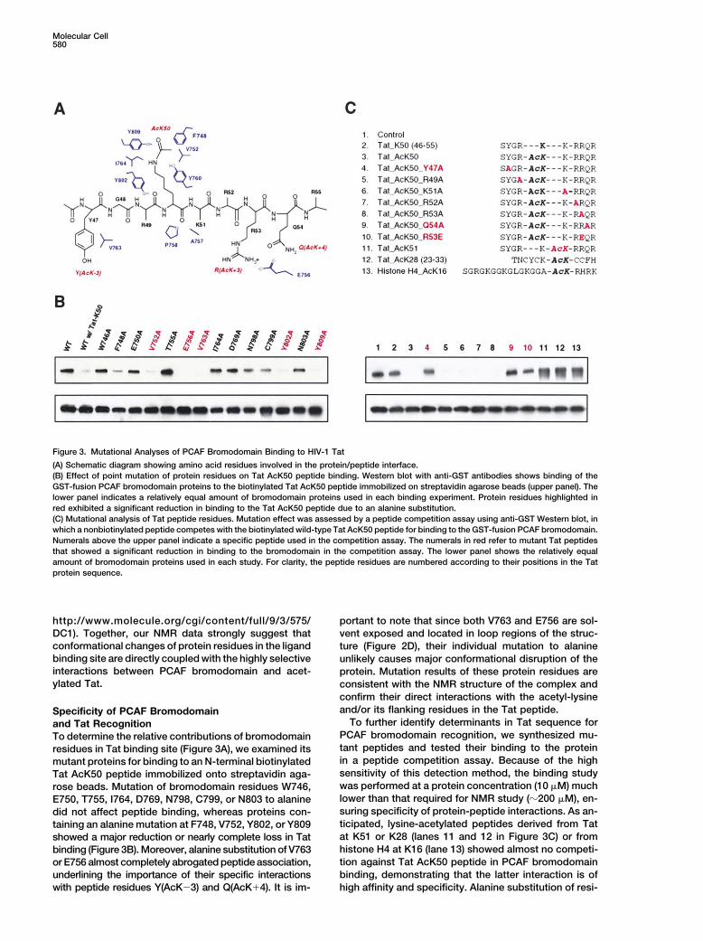
Figure 3. Mutational Analyses of PCAF Bromodomain Binding to HIV-1 Tat
(A) Schematic diagram showing amino acid residues involved in the protein/peptide interface.(B) Effect of point mutation of protein residues on Tat AcK50 peptide binding. Western blot with anti-GST antibodies shows binding of theGST-fusion PCAF bromodomain proteins to the biotinylated Tat AcK50 peptide immobilized on streptavidin agarose beads (upper panel). Thelower panel indicates a relatively equal amount of bromodomain proteins used in each binding experiment. Protein residues highlighted inred exhibited a significant reduction in binding to the Tat AcK50 peptide due to an alanine substitution.(C) Mutational analysis of Tat peptide residues. Mutation effect was assessed by a peptide competition assay using anti-GST Western blot, inwhich a nonbiotinylated peptide competes with the biotinylated wild-type Tat AcK50 peptide for binding to the GST-fusion PCAF bromodomain.Numerals above the upper panel indicate a specific peptide used in the competition assay. The numerals in red refer to mutant Tat peptidesthat showed a significant reduction in binding to the bromodomain in the competition assay. The lower panel shows the relatively equalamount of bromodomain proteins used in each study. For clarity, the peptide residues are numbered according to their positions in the Tatprotein sequence.
http://www.molecule.org/cgi/content/full/9/3/575/ portant to note that since both V763 and E756 are sol-vent exposed and located in loop regions of the struc-DC1). Together, our NMR data strongly suggest that
conformational changes of protein residues in the ligand ture (Figure 2D), their individual mutation to alaninebinding site are directly coupled with the highly selective unlikely causes major conformational disruption of theinteractions between PCAF bromodomain and acet- protein. Mutation results of these protein residues areylated Tat. consistent with the NMR structure of the complex and
confirm their direct interactions with the acetyl-lysineand/or its flanking residues in the Tat peptide.Specificity of PCAF Bromodomain
To further identify determinants in Tat sequence forand Tat RecognitionPCAF bromodomain recognition, we synthesized mu-To determine the relative contributions of bromodomaintant peptides and tested their binding to the proteinresidues in Tat binding site (Figure 3A), we examined itsin a peptide competition assay. Because of the highmutant proteins for binding to an N-terminal biotinylatedsensitivity of this detection method, the binding studyTat AcK50 peptide immobilized onto streptavidin aga-was performed at a protein concentration (10 �M) muchrose beads. Mutation of bromodomain residues W746,lower than that required for NMR study (�200 �M), en-E750, T755, I764, D769, N798, C799, or N803 to alaninesuring specificity of protein-peptide interactions. As an-did not affect peptide binding, whereas proteins con-ticipated, lysine-acetylated peptides derived from Tattaining an alanine mutation at F748, V752, Y802, or Y809at K51 or K28 (lanes 11 and 12 in Figure 3C) or fromshowed a major reduction or nearly complete loss in Tathistone H4 at K16 (lane 13) showed almost no competi-binding (Figure 3B). Moreover, alanine substitution of V763tion against Tat AcK50 peptide in PCAF bromodomainor E756 almost completely abrogated peptide association,binding, demonstrating that the latter interaction is ofunderlining the importance of their specific interactions
with peptide residues Y(AcK�3) and Q(AcK�4). It is im- high affinity and specificity. Alanine substitution of resi-
Recognition of HIV-1 Tat by PCAF Bromodomain581
dues R(AcK�1), K(AcK�1), R(AcK�2), or R(AcK�3) in for the backbone C� atoms. The majority of structuraldeviations are localized in the loop regions, particularlyTat AcK50 peptide slightly weakened its binding to thein the ZA and BC loops (see Supplemental Figure S3 atbromodomain. Conversely, change of Y(AcK�3) (lane 4)http: //www.molecule.org/cgi /content/ full /9/3 /575/or Q(AcK�4) (lane 9) to alanine caused a nearly completeDC1).loss of bromodomain binding, confirming the impor-
The crystal structure of scGCN5p bromodomaintance of their pairwise interactions with V763 and E756solved in complex with an acetylated peptide derivedfor Tat-PCAF association. Finally, while mutation offrom histone H4 at K16 (A-AcK-RHRKILRNSIQGI) re-R(AcK�3) to alanine (lane 8) did not significantly alter Tatveals that the mechanism of acetyl-lysine recognition isbinding to the bromodomain, its substitution to glutamichighly conserved in bromodomains—it involves a nearlyacid (lane 10) exhibited a marked reduction in the pro-identical set of corresponding conserved residues in thetein/peptide interaction. The effect of the latter mutationPCAF and scGCN5p bromodomains (Figures 5A andis likely due to an electrostatic repulsion between the5B) (Owen et al., 2000). In addition to the acetyl-lysine,glutamate and E756 of the protein. Together, these re-scGCN5p bromodomain has a limited number of con-sults explain the structural basis for the highly selectivetacts with two residues at (AcK�2) and (AcK�3) in thenature of PCAF and lysine-acetylated Tat association,H4 peptide. Binding of H(AcK�2) to aromatic rings ofwhich requires specific interactions of the bromodomainY406 and F367 in scGCN5p is reminiscent of PCAF bro-with AcK50 and its flanking residues, including Y(AcK�3),modomain recognition of Tat Y(AcK�3) through interac-R(AcK�3), and Q(AcK�4).tions with Y802 and V763, which are equivalent to thetwo scGCN5p residues. Because of this similar modePCAF Bromodomain Competing against TAR RNAof molecular interaction, the two aromatic residues in thefor Binding to Lysine-Acetylated TatTat and H4 peptides, which are located in very differentThe arginine-rich motif containing R52 and R53 in Tat ispositions with respect to the acetyl-lysine, are bound inalso known to interact with the HIV-1 TAR RNA elementa nearly identical position in the corresponding bromo-(Aboul-ela et al., 1995; Long and Crothers, 1999; Ranadomain structures (Figure 5A). High conservation ofand Jeang, 1999). Tat acetylation at K50 by p300/CBPthese residues in bromodomains (Figure 5B) suggestspromotes Tat dissociation from TAR RNA in early tran-that selection of an aromatic or hydrophobic residuescriptional elongation (Deng et al., 2000; Kiernan et al.,neighboring the acetyl-lysine is possibly conserved for1999). To determine whether lysine acetylation directlymany members of the bromodomain family.affects Tat association with TAR RNA, we performed an
It is important to note that while the major bindingNMR study of a 27 nucleotide HIV-1 TAR RNA bindingdeterminant in scGCN5p bromodomain-H4 complex isto Tat peptides containing either K50 or AcK50. Ourthe acetyl-lysine (Owen et al., 2000), the highly specificresults showed that TAR RNA bound to the nonacet-association of PCAF bromodomain and Tat peptide isylated Tat peptide with a subnanomolar affinity (KD), independent on its interactions not only with the acetyl-agreement with results reported previously (Aboul-elalysine and Y(AcK�3) but also with residues on the otheret al., 1995; Long and Crothers, 1999), and that K50side of the acetyl-lysine at (AcK�3) and (AcK�4) (Fig-acetylation of Tat resulted in a significant reduction ofures 3B and 3C). These differences in the extent of ligandits affinity to TAR RNA (data not shown). More strikingly,interactions explain why the Tat AcK50 peptide com-we found that PCAF bromodomain competes effectivelypetes effectively against a similar histone H4 AcK16against TAR RNA for binding to Tat AcK50 peptide (Fig-peptide for binding to the PCAF bromodomain (Figureures 4A and 4B), suggesting that the binding affinity (KD)3C, lane 13). Moreover, these differences in ligand selec-
of the latter interaction is on the order of low micromolar.tivity provide an explanation for the striking differences
This observation may be explained by possible confor-in location and orientation of the bound peptides in the
mational change of the peptide residues due to acetyla- two bromodomains—the backbones of the Tat and H4tion at K50 or involvement of R53 of Tat in both interac- peptides lie in the two corresponding structures nearlytions. These results strongly imply that the PCAF antiparallel to each other (Figure 5A). Binding of A757bromodomain interaction with AcK50 on Tat not only and E756 in the ZA loop to R(AcK�3) and Q(AcK�4) ofcontributes to Tat-PCAF association but also to the re- the Tat peptide, which are completely lacking in thelease of lysine-acetylated Tat from TAR RNA associa- scGCN5 bromodomain-H4 complex, further explainstion, leading to Tat-mediated HIV-1 transcriptional acti- why the PCAF bromodomain undergoes more extensivevation. conformational changes in the ligand site than those
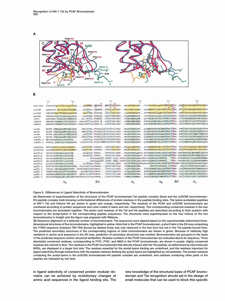
seen in the GCN5 bromodomains (see SupplementalDifferences of Ligand Selectivity of Bromodomains Figure S3 at http://www.molecule.org/cgi/content/full/Structural comparison of bromodomains from PCAF and 9/3/575/DC1). While the biological relevance of theother proteins extends our understanding of differences scGCN5 and histone H4 AcK16 interaction remains toin ligand selectivity. Recent structures of bromodomains be determined, a growing body of evidence, includingfrom human GCN5 (Hudson et al., 2000) and Saccharo- previous reports (Benkirane et al., 1998; Deng et al.,myces cerevisiae GCN5p (Owen et al., 2000), and the 2000), our present study of NMR structure and in vitrodouble bromodomain module of human TAFII250 (Ja- mutagenesis, and results from in vivo functional studiescobson et al., 2000), reinforce the notion that the left- of Tat-mediated HIV-1 transcriptional activation (Figurehanded four-helix bundle fold of the PCAF bromdomain 1C and M.O. and E.V., unpublished data), strongly sup-is conserved in the bromodomain family (Dhalluin et al., port the biological relevance and importance for the1999). Structural similarity is high for the four helices highly selective association of PCAF bromodomain and
acetylated Tat.with pairwise root-mean-square deviations of 0.7–1.8 A
Molecular Cell582
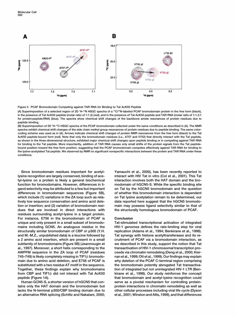
Figure 4. PCAF Bromodomain Competing against TAR RNA for Binding to Tat AcK50 Peptide
(A) Superimposition of a selected region of 2D 1H-15N HSQC spectra of a 13C/15N-labeled PCAF bromodomain protein in the free form (black),in the presence of Tat AcK50 peptide (molar ratio of 1:1.2) (red), and in the presence of Tat AcK50 peptide and TAR RNA (molar ratio of 1:1.2:1for protein:peptide:RNA) (blue). The spectra show chemical shift changes of the backbone amide resonances of protein residues due topeptide binding.(B) Superimposition of 2D 1H-13C HSQC spectra of the PCAF bromodomain collected under the same conditions as described in (A). The NMRspectra exhibit chemical shift changes of the side chain methyl group resonances of protein residues due to peptide binding. The same color-coding scheme was used as in (A). Arrows indicate chemical shift changes of protein NMR resonances from the free form (black) to the TatAcK50 peptide-bound form (red). Note that only the bromodomain residues (i.e., A757 and V752) that directly interact with the Tat peptide,as shown in the three-dimensional structure, exhibited major chemical shift changes upon peptide binding or in competing against TAR RNAfor binding to the Tat peptide. More importantly, addition of TAR RNA causes only small shifts of the protein signals from the Tat peptide-bound position toward the free form position, suggesting that the PCAF bromodomain competes effectively against TAR RNA for binding tothe lysine-acetylated Tat peptide. We observed by NMR no significant nonspecific interactions between the protein and TAR RNA under theseconditions.
Since bromodomain residues important for acetyl- Yamauchi et al., 2000), has been recently reported tointeract with HIV Tat in vitro (Col et al., 2001). This Tatlysine recognition are largely conserved, binding of ace-
tyl-lysine on a protein is likely a general biochemical interaction involves both the HAT domain and the bro-modomain of hGCN5-S. While the specific binding sitefunction for bromodomains. However, differences in li-
gand selectivity may be attributed to a few but important on Tat by the hGCN5 bromodomain and the questionof whether this bromodomain interaction is dependentdifferences in bromodomain sequences (Figure 5B),
which include (1) variations in the ZA loop such as rela- on Tat lysine acetylation remain to be determined, ourdata reported here suggest that the hGCN5 bromodo-tively low sequence conservation and amino acid dele-
tion or insertion; and (2) variation of bromodomain resi- main may possess ligand selectivity similar to that ofthe structurally homologous bromodomain of PCAF.dues that are involved in direct interactions with
residues surrounding acetyl-lysine in a target protein.For instance, E756 in the bromodomain of PCAF is Conclusion
Tat-stimulated transcriptional activation of integratedunique and only present in a small subset of bromodo-mains including GCN5. An analogous residue in the HIV-1 genomes defines the rate-limiting step for viral
replication (Adams et al., 1994; Benkirane et al., 1998).structurally similar bromodomain of CBP or p300 (Y.H.and M.-M.Z., unpublished data) is a leucine followed by Tat synergy with histone acetyltransferases and its re-
cruitment of PCAF via a bromodomain interaction, asa 2 amino acid insertion, which are present in a smallsubfamily of bromodomains (Figure 5B) (Jeanmougin et we described in this study, support the notion that Tat
transactivation of HIV-1 chromosomal transcription pro-al., 1997). Moreover, a short helix corresponding to theAWPFM sequence in the ZA loop of PCAF (residues ceeds via chromatin remodeling (Deng et al., 2000; Kier-
nan et al., 1999; Ott et al., 1999). Our findings may explain745–749) is likely completely missing in TIF1� bromodo-main due to amino acid deletion, and E756 of PCAF is why deletion of the PCAF C-terminal region comprising
the bromodomain potently abrogated Tat transactiva-substituted with a two residue AT motif in the sequence.Together, these findings explain why bromodomains tion of integrated but not unintegrated HIV-1 LTR (Ben-
kirane et al., 1998). Our study reinforces the conceptfrom CBP and TIF1� did not interact with Tat AcK50peptide (Figure 1A). that bromodomain and acetyl-lysine recognition could
serve as a pivotal mechanism for controlling protein-Human GCN5-S, a shorter version of hGCN5 that con-tains only the HAT domain and the bromodomain but protein interactions in chromatin remodeling as well as
other cellular processes including viral life cycle (Dysonlacks the N-terminal p300/CBP binding domain due toan alternative RNA splicing (Schiltz and Nakatani, 2000; et al., 2001; Winston and Allis, 1999), and that differences
Recognition of HIV-1 Tat by PCAF Bromodomain583
Figure 5. Differences in Ligand Selectivity of Bromodomains
(A) Stereoview of superimposition of the structures of the PCAF bromodomain-Tat peptide complex (blue) and the scGCN5 bromodomain-H4 peptide complex (red) showing conformational differences of protein residues in the peptide binding sites. The lysine-acetylated peptidesof HIV-1 Tat and histone H4 are shown in green and orange, respectively. The residues of the PCAF and scGCN5 bromodomains arenumbered according to protein sequences and color-coded in black and red, respectively. The corresponding conserved residues in the twobromodomains are annotated together. The amino acid residues of the Tat and H4 peptides are described according to their position withrespect to the acetyl-lysine in the corresponding peptide sequences. The structures were superimposed on the four helices of the twobromodomains in Insight and the figure was prepared with Ribbons.(B) Sequence alignment of a selected number of bromodomains. The sequences were aligned based on the experimentally determined three-dimensional structures of five bromodomains, highlighted in yellow. Note that in the PCAF bromodomain, a short helix in the ZA loop comprisingthe YYEVI sequence (residues 760–764) (boxed by dashed lines) was only observed in the free form but not in the Tat peptide-bound form.The predicted secondary structures in the corresponding regions of other bromodomains are shown in green. Because of relatively highvariations in amino acid sequence in the ZA loop, prediction of secondary structures was omitted. Bromodomains are grouped on the basisof the predicted sequence and/or structural similarities. Residue numbers of the PCAF bromodomain are indicated above its sequence. Threeabsolutely conserved residues, corresponding to P751, P767, and N803 in the PCAF bromodomain, are shown in purple. Highly conservedresidues are colored in blue. The residues in the PCAF bromodomain that directly interact with the Tat peptide, as determined by intermolecularNOEs, are displayed in a larger font size. The residues essential for the acetyl-lysine binding are underlined, and the residues important forligand selectivity through interactions with the peptide residues flanking the acetyl-lysine are highlighted by red asterisks. The protein residuescontacting the acetyl-lysine in the scGCN5 bromodomain-H4 peptide complex are underlined, and residues contacting other parts of thepeptide are indicated by red dots.
in ligand selectivity of conserved protein modular do- new knowledge of the structural basis of PCAF bromo-domain and Tat recognition should aid in the design ofmains can be achieved by evolutionary changes of
amino acid sequences in the ligand binding site. The small molecules that can be used to block this specific
Molecular Cell584
(Laskowski et al., 1996) analysis indicated that over 98% of theinteraction in order to disrupt HIV-1 transcriptional acti-protein and peptide residues are in allowed regions of the Rama-vation and replication.chandran map.
Experimental ProceduresMutational AnalysisSite-directed mutant proteins of PCAF bromodomain were preparedSample Preparationwith the QuickChange kit (Stratagene). DNA sequencing confirmedThe PCAF bromodomain (residues 719–832) was expressed in Esch-the desired mutations. The GST-fusion bromodomains (10 �M) oferichia coli BL21(DE3) cells using the pET14b vector (Novagen)PCAF, CBP, or TIF1� were incubated with an N-terminal biotinylated(Dhalluin et al., 1999). Isotope-labeled bromodomain proteins wereand lysine-acetylated Tat peptide (50 �M) in 50 mM Tris buffer (pHprepared from cells grown on a minimal medium containing 15NH4Cl7.5), containing 50 mM NaCl, 0.1% BSA, and 1 mM DTT at 22�C forwith or without 13C6-glucose in either H2O or 75% 2 H2O. The protein2 hr. Streptavidin agarose (10 �L) was added to the mixture, andwas purified by affinity chromatography on a nickel-IDA columnthe beads were washed in the Tris buffer containing 500 mM NaCl(Invitrogen), followed by the removal of poly-His tag by thrombinand 0.1% NP-40. Proteins eluted from the agarose beads werecleavage. GST-fusion bromodomains from PCAF, CBP, and TIF1�separated by SDS-PAGE and visualized by Western blotting usingwere expressed in E. coli BL21 (DE3) codon plus cells using theanti-GST antibody (Sigma) and horseradish-peroxidase-conjugatedpGEX4T-3 vector (Pharmacia) and purified with a glutathione sepha-goat anti-rabbit IgG. Peptide competition assay was performed byrose column. NMR spectra of the recombinant CBP and TIF1� pro-incubating a nonbiotinylated peptide with the PCAF bromodomainteins were acquired to ensure that they were properly folded andand the biotinylated Tat AcK50 peptide. The molar ratio of the formerfunctional (see Supplemental Figure S4 at http://www.molecule.org/and latter peptides in the mixture was kept at 1:2.cgi/content/full/9/3/575/DC1). The acetyl-lysine-containing pep-
tides were prepared on a MilliGen 9050 peptide synthesizer (PerkinPlasmid ConstructsElmer) using Fmoc/HBTU chemistry. Acetyl-lysine was incorporatedThe mammalian expression vectors for the PCAF and CBP bromo-using the reagent Fmoc-Ac-Lys with HBTU/DIPEA activation. Thedomains were constructed as follows. Coding sequence for theHIV-1 TAR RNA was obtained from Dharmacon Research, Inc. (Lafa-PCAF bromodomain (residues 719–832) was subcloned into EcoRI-yette, CO).XhoI sites of pCMV-HA vector (Clontech). The CBP bromodomain(residues 1082–1197) was subcloned into BamHI-XhoI sites of
NMR SpectroscopypCMV-FLAG vector (Stratagene). The expression vectors for the full-
NMR samples contained a protein/peptide complex of 0.5 mM inlength PCAF (pCI-FLAG-PCAF) (Li et al., 2000), the full-length CBP
100 mM phosphate buffer (pH 6.5) containing 5 mM perdeuterated(pRSV-HA-CBP) (Kwok et al., 1996), HIV-1 Tat (pcTat), and the HIV-1
DTT and 0.5 mM EDTA in H2O/2 H2O (9/1) or 2 H2O. All NMR spectraLTR-luciferase reporter construct (pHIV-LTR-Luc) (Bieniasz et al.,
were acquired at 30�C on a Bruker 500 or 600 MHz NMR spectrome-1998; Madore and Cullen, 1993) have been previously described.
ter. The 1H, 13C, and 15N resonances of the protein backbone andside chain atoms were assigned by using a standard set of triple-
Cell Culture and Transfectionsresonance experiments (Sattler et al., 1999) with a uniformly 13C/Human 293T cells were propagated in Dulbecco’s modified Eagle’s15N-labeled and 75% deuterated protein in complex with an unla-medium with 10% fetal calf serum and transfected using the calciumbeled peptide. The distance restraints were obtained from 13C- orphosphate coprecipitation method. Amounts of plasmid DNA used15N-edited three-dimensional nuclear Overhauser enhancementin cell transfections are as described in the legend to Figure 1C.spectroscopy (NOESY) spectra (Clore and Gronenborn, 1994).The transfected 293T cells were lysed 24 hr after transfection and
φ-angle restraints were determined based on the 3JHN,H� couplingassayed for luciferase activity of the cell extracts using a luciferase-constants measured in a 3D HNHA spectrum (Clore and Gronenborn,based assay system (Promega) (Bieniasz et al., 1998; Madore and1994). Slowly exchanging amide protons were identified from a se-Cullen, 1993). Luciferase activities derived from HIV-1 LTR wereries of 2D 15N-HSQC spectra recorded after the H2O buffer wasnormalized to a cotransfected vector expressing �-galactosidase.changed to a 2 H2O buffer, which were used together with the initialThe expression level of the transfected proteins was examined bystructures calculated with only NOE-derived distance restraints toWestern blotting using monoclonal antibodies to HA (Roche Diag-generate hydrogen-bond distance restraints in final structure calcu-nostics), FLAG (Stratagene), or �-actin (Sigma), and rabbit polyclonallations. The intermolecular NOEs were detected in 13C-edited (F1), antibodies to the PCAF bromodomain or the CBP bromodomain13C/15N-filtered (F3) 3D NOESY spectrum (Clore and Gronenborn,(see Supplemental Figure S5 at http://www.molecule.org/cgi/con-1994). All NMR spectra were processed with the NMRPipe programtent/full/9/3/575/DC1).(Delaglio et al., 1995) and analyzed using NMRView (Johnson and
Blevins, 1994). NMR binding studies of Tat peptides and TAR RNAAcknowledgmentsinteractions were performed in the phosphate buffer (pH 6.5) con-
taining 200 mM NaCl to minimize any nonspecific interactions.We thank P.D. Bieniasz for providing the pcTat and HIV-1 LTR-lucconstructs, M.J. Walsh and R.L. Schiltz for the PCAF expression
Structure Calculationsplasmids, and N. Zeleznik-Le for the full-length CBP construct. We
Structures of the protein/peptide complex were calculated with aalso thank I. Wolf for peptide synthesis, C. Dhalluin, O. Plotnikova,
distance geometry-simulated annealing protocol using the X-PLORand S. Yan for technical advice and support, A. Koch, K. Manzur,
program (Brunger, 1993). A total of 2359 manually assigned NOE-and K.S. Yan for critical reading of the manuscript, and A.K. Aggar-
derived distance restraints were used in initial structure calculations.wal, D.E. Logothetis, and H. Weinstein for helpful suggestions to
The ARIA (Nilges and O’Donoghue, 1998)-assigned distance re-the study. This work was supported by a National Institutes of Health
straints agree with the structures calculated using only the manuallygrant to M.-M. Z.
determined NOE distance restraints, 28 hydrogen-bond distancerestraints, and 53 φ angle restraints. The final structure calculations
Received September 18, 2001; revised January 9, 2002.employed a total of 2903 NMR experimental restraints from themanual and the ARIA-assisted assignments, including 2700 unam-
Referencesbiguous intramolecular and 78 intermolecular NOE distance re-straints. The distance restraint force constant was 50 kcal mol�1 A�2,
Aasland, R., Gibson, T.J., and Stewart, A.F. (1995). The PHD finger:and no NOE was violated by more than 0.3 A. The torsion restraint
implications for chromatin-mediated transcriptional regulation.force constant was 200 kcal mol�1 rad�2, and no dihedral angle
Trends Biochem. Sci. 20, 56–59.restraint was violated by more than 5�. While only the covalent
Aboul-ela, F., Karn, J., and Varani, G. (1995). The structure of thegeometry terms, NOE, torsion, and repulsive van der Waals termshuman immunodeficiency virus type-1 TAR RNA reveals principleswere used in the structure refinement, a large, negative Lennard-of RNA recognition by Tat protein. J. Mol. Biol. 253, 313–332.Jones potential energy was observed (�569.5 � 22.4 kcal mol�1),
indicating good nonbonded geometry of the structure. Procheck Adams, M., Sharmeen, L., Kimpton, J., Romeo, J.M., Garcia, J.V.,
Recognition of HIV-1 Tat by PCAF Bromodomain585
Peterlin, B.M., Groudine, M., and Emerman, M. (1994). Cellular la- son, R. (1997). The bromodomain revisited. Trends Biochem. Sci.22, 151–153.tency in human immunodeficiency virus-infected individuals with
high CD4 levels can be detected by the presence of promoter- Jenuwein, T., and Allis, C.D. (2001). Translating the histone code.proximal transcripts. Proc. Natl. Acad. Sci. USA 91, 3862–3866. Science 293, 1074–1080.Benkirane, M., Chun, R.F., Xiao, H., Ogryzko, V.V., Howard, B.H., Johnson, B.A., and Blevins, R.A. (1994). NMRView: a computer pro-Nakatani, Y., and Jeang, K.-T. (1998). Activation of integrated provi- gram for the visualization and analysis of NMR data. J. Biomol. NMRrus requires histone acetyltransferase: p300 and P/CAF are co-acti- 4, 603–614.vators for HIV-1 Tat. J. Biol. Chem. 273, 24898–24905. Jones, K.A. (1997). Taking a new TAK on Tat transactivation. GenesBieniasz, P.D., Grdina, T.A., Bogerd, H.P., and Cullen, B.R. (1998). Dev. 11, 2593–2599.Recruitment of a protein complex containing Tat and cyclin T1 to Karn, J. (1999). Tackling Tat. J. Mol. Biol. 293, 235–254.TAR governs the species specificity of HIV-1 Tat. EMBO J. 17, 7056–
Keen, N.J., Churcher, M.J., and Karn, J. (1997). Transfer of Tat and7065.release of TAR RNA during the activation of the human immunodefi-
Brown, C.E., Howe, L., Sousa, K., Alley, S.C., Carozza, M.J., Tan, ciency virus type-1 transcription elongation complex. EMBO J. 16,S., and Workman, J.L. (2001). Recruitment of HAT complexes by 5260–5272.direct activator interactions with the ATM-related Tra1 subunit. Sci-
Kiernan, R.E., Vanhulle, C., Schiltz, L., Adam, E., Xiao, H., Maudoux,ence 292, 2333–2337.F., Calomme, C., Burny, A., Nakatani, Y., Jeang, K.-T., et al. (1999).
Brownell, J.E., and Allis, C.D. (1996). Special HATs for special occa- HIV-1 Tat transcriptional activity is regulated by acetylation. EMBOsions: Linking histone acetylation to chromatin assembly and gene J. 18, 6106–6118.activation. Curr. Opin. Genet. Dev. 6, 176–184.
Kouzarides, T. (2000). Acetylation: a regulatory modification to rivalBrunger, A.T. (1993). X-PLOR Version 3.1: A System for X-Ray Crys- phosphorylation? EMBO J. 19, 1176–1179.tallography and NMR, Version 3.1 edn (New Haven, CT: Yale Univer-
Kwok, R.P., Laurance, M.E., Lundblad, J.R., Goldman, P.S., Shih,sity Press).H., Connor, L.M., Marriott, S.J., and Goodman, R.H. (1996). Control
Carson, M. (1991). Ribbons 2.0. J. Appl. Crystallogr. 24, 958–961. of cAMP-regulated enhancers by the viral transactivator Tax throughClore, G.M., and Gronenborn, A.M. (1994). Multidimensional hetero- CREB and the co-activator CBP. Nature 380, 642–646.nuclear nuclear magnetic resonance of proteins. Methods Enzymol. Laskowski, R.A., Rullmannn, J.A., MacArthur, M.W., Kaptein, R., and239, 249–363. Thornton, J.M. (1996). AQUA and PROCHECK-NMR: programs forCol, E., Caron, C., Seigneurin-Berny, D., Gracia, J., Favier, A., and checking the quality of protein structures solved by NMR. J. Biomol.Khochbin, S. (2001). The histone acetyltransferase, hGCN5, interacts NMR 8, 477–486.with and acetylates the HIV transactivator. Tat. J. Biol. Chem. 276, Li, S.D., Aufiero, B., Schiltz, R.L., and Walsh, M.J. (2000). Regulation28179–28184. of the homeodomain CCAAT displacement/cut protein function byCullen, B.R. (1998). HIV-1 auxiliary proteins: making connections in histone acetyltransferase p300/CREB-binding protein (CBP)-asso-a dying cell. Cell 93, 685–692. ciated factor and CBP. Proc. Natl. Acad. Sci. USA 97, 7166–7171.Delaglio, F., Grzesiek, S., Vuister, G.W., Zhu, G., Pfeifer, J., and Bax, Long, K.S., and Crothers, D.M. (1999). Characterization of the solu-A. (1995). NMRPipe: a multidimensional spectral processing system tion conformations of unbound and Tat peptide-bound forms ofbased on UNIX pipes. J. Biomol. NMR 6, 277–293. HIV-1 TAR RNA. Biochemistry 38, 10059–10069.Deng, L., de la Fuente, C., Fu, P., Wang, L., Donnelly, R., Wade, Madore, S.J., and Cullen, B.R. (1993). Genetic analysis of the cofac-J.D., Lambert, P., Li, H., Lee, C.-G., and Kashanchi, F. (2000). Acet- tor requirement for human immunodeficiency virus type 1 Tat func-ylation of HIV-1 Tat by CBP/P300 increases transcription of inte- tion. J. Virol. 67, 3703–3711.grated HIV-1 genome and enhances binding to core histones. Virol- Manning, E.T., Ikehara, T., Ito, T., Kadonaga, J.T., and Kraus, W.L.ogy 277, 278–295. (2001). p300 forms a stable, template-committed complex with chro-Dhalluin, C., Carlson, J.E., Zeng, L., He, C., Aggarwal, A.K., and Zhou, matin: role for the bromodomain. Mol. Cell Biol. 21, 3876–3887.M.-M. (1999). Structure and ligand of a histone acetyltransferase Nilges, M., and O’Donoghue, S. (1998). Ambiguous NOEs and auto-bromodomain. Nature 399, 491–496. mated NOE assignment. Prog. NMR Spectroscopy 32, 107–139.Dyson, M.H., Rose, S., and Mahadevan, L.C. (2001). Acetylation- Ott, M., Schnolzer, M., Garnica, J., Fischle, W., Emiliani, S., Rackwitz,binding and function of bromodomain-containing proteins in chro- H.-R., and Verdin, E. (1999). Acetylation of the HIV-1 Tat proteinmatin. Front. Biosci. 6, 853–865. by p300 is important for its transcriptional activity. Curr. Biol. 9,Friedman, J.R., Fredericks, W.J., Jensen, D.E., Speicher, D.W., Hu- 1489–1492.ang, X.P., Neilson, E.G., and Rauscher, F.J., III. (1996). KAP-1, a Owen, D.J., Ornaghi, P., Yang, J.C., Lowe, N., Evans, P.R., Ballario,novel corepressor for the highly conserved KRAB repression do- P., Neuhaus, D., Eiletici, P., and Travers, A.A. (2000). The structuralmain. Genes Dev. 10, 2067–2078. basis for the recognition of acetylated histone H4 by the bromodo-Garber, M.E., and Jones, K.A. (1999). HIV-1 Tat: coping with negative main of histone acetyltransferase gcn5p. EMBO J. 19, 6141–6149.elongation factors. Curr. Opin. Immunol. 11, 460–465. Rana, T.M., and Jeang, K.-T. (1999). Biochemical and functionalHaynes, S.R., Dollard, C., Winston, F., Beck, S., Trowsdale, J., and interactions between HIV-1 Tat protein and TAR RNA. Arch. Bio-Dawid, I.B. (1992). The bromodomain: a conserved sequence found chem. Biophys. 365, 175–185.in human, Drosophila and yeast proteins. Nucleic Acids Res. 20, Sattler, M., Schleucher, J., and Griesinger, C. (1999). Heteronuclear2603. multidimensional NMR experiments for the structure determinationHottiger, M.O., and Nabel, G.J. (1998). Interaction of human immuno- of proteins in solution employing pulsed field gradients. Prog. NMRdeficiency virus type 1 Tat with the transcriptional coactivators p300 Spectroscopy 34, 93–158.and CREB binding protein. J. Virol. 72, 8252–8256. Schiltz, R.L., and Nakatani, Y. (2000). The PCAF acetylase complexHudson, B.P., Martinez-Yamout, M.A., Dyson, H.J., and Wright, P.E. as a potential tumor suppressor. Biochim. Biophys. Acta 1470,(2000). Solution structure and acetyl-lysine binding activity of the M37–M53.GCN5 bromodomain. J. Mol. Biol. 304, 355–370. Sterner, D.E., Grant, P.A., Roberts, S.M., Duggan, L.J., Belotserkov-Jacobson, R.H., Ladurner, A.G., King, D.S., and Tjian, R. (2000). skaya, R., Pacella, L.A., Winston, F., Workman, J.L., and Berger,Structure and function of a human TAFII250 double bromodomain S.L. (1999). Functional organization of the yeast SAGA complex:module. Science 288, 1422–1425. distinct components involved in structural integrity, nucleosome
acetylation, and TATA-binding protein interaction. Mol. Cell. Biol.Jeang, K.-T., Xiao, H., and Rich, E.A. (1999). Multifaceted activitiesof the HIV-1 transactivator of transcription. Tat. J. Biol. Chem. 274, 19, 86–98.28837–28840. Strahl, B.D., and Allis, C.D. (2000). The language of covalent histone
modifications. Nature 403, 41–45.Jeanmougin, F., Wurtz, J.M., Douarin, B.L., Chambon, P., and Los-
Molecular Cell586
Tamkun, J.W., Deuring, R., Scott, M.P., Kissinger, M., Pattatucci,A.M., Kaufman, T.C., and Kennison, J.A. (1992). brahma: a regulatorof Drosophila homeotic genes structurally related to the yeast tran-scriptional activator SNF2/SWI2. Cell 68, 561–572.
Travers, A. (1999). Chromatin modification: how to put a HAT on thehistones. Curr. Biol. 9, 23–25.
Wei, P., Garber, M.E., Fang, S.M., Fischer, W.H., and Jones, K.A.(1998). A novel CDK9-associated C-type cyclin interacts with HIV-1Tat and mediates its high-affinity, loop-specific binding to TAR RNA.Cell 92, 451–462.
Winston, F., and Allis, C.D. (1999). The bromodomain: a chromatin-targeting module? Nat. Struct. Biol. 6, 601–604.
Yamauchi, T., Yamauchi, J., Kuwata, T., Tamura, T., Yamashita, T.,Bae, N., Westphal, H., Ozato, K., and Nakatani, Y. (2000). Distinctbut overlapping roles of histone acetylase PCAF and of the closelyrelated PCAF-B/GCN5 in mouse embryogenesis. Proc. Natl. Acad.Sci. USA 97, 11303–11306.
Accession Numbers
Coordinates for the NMR three-dimensional structure of the PCAFbromodomain/HIV-1 Tat peptide complex have been deposited inthe Brookhaven Protein Data Bank under the accession code 1JM4.


![Regulation of Lysine Catabolism through Lysine[mdash]Ketoglutarate Reductase and Saccharopine Dehydrogenase in Arabidopsis](https://static.fdokumen.com/doc/165x107/631cc83693f371de19019c93/regulation-of-lysine-catabolism-through-lysinemdashketoglutarate-reductase-and.jpg)