Inhibition of HIV1 virus replication using small soluble Tat peptides
17
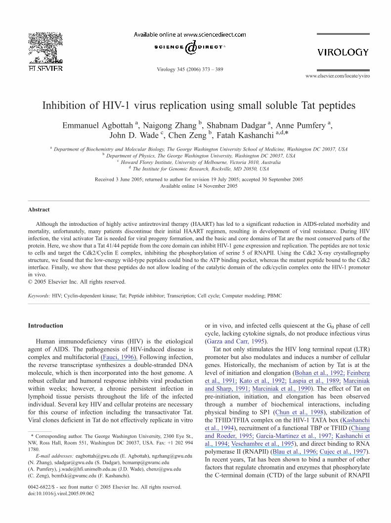
Inhibition of HIV-1 virus replication using small soluble Tat peptides Emmanuel Agbottah a , Naigong Zhang b , Shabnam Dadgar a , Anne Pumfery a , John D. Wade c , Chen Zeng b , Fatah Kashanchi a,d, * a Department of Biochemistry and Molecular Biology, The George Washington University School of Medicine, Washington DC 20037, USA b Department of Physics, The George Washington University, Washington DC 20037, USA c Howard Florey Institute, University of Melbourne, Victoria 3010, Australia d The Institute for Genomic Research, Rockville, MD 20850, USA Received 3 June 2005; returned to author for revision 19 July 2005; accepted 30 September 2005 Available online 14 November 2005 Abstract Although the introduction of highly active antiretroviral therapy (HAART) has led to a significant reduction in AIDS-related morbidity and mortality, unfortunately, many patients discontinue their initial HAART regimen, resulting in development of viral resistance. During HIV infection, the viral activator Tat is needed for viral progeny formation, and the basic and core domains of Tat are the most conserved parts of the protein. Here, we show that a Tat 41/44 peptide from the core domain can inhibit HIV-1 gene expression and replication. The peptides are not toxic to cells and target the Cdk2/Cyclin E complex, inhibiting the phosphorylation of serine 5 of RNAPII. Using the Cdk2 X-ray crystallography structure, we found that the low-energy wild-type peptides could bind to the ATP binding pocket, whereas the mutant peptide bound to the Cdk2 interface. Finally, we show that these peptides do not allow loading of the catalytic domain of the cdk/cyclin complex onto the HIV-1 promoter in vivo. D 2005 Elsevier Inc. All rights reserved. Keywords: HIV; Cyclin-dependent kinase; Tat; Peptide inhibitor; Transcription; Cell cycle; Computer modeling; PBMC Introduction Human immunodeficiency virus (HIV) is the etiological agent of AIDS. The pathogenesis of HIV-induced disease is complex and multifactorial (Fauci, 1996). Following infection, the reverse transcriptase synthesizes a double-stranded DNA molecule, which is then incorporated into the host genome. A robust cellular and humoral response inhibits viral production within weeks; however, a chronic persistent infection in lymphoid tissue persists throughout the life of the infected individual. Several key HIV and cellular proteins are necessary for this course of infection including the transactivator Tat. Viral clones deficient in Tat do not effectively replicate in vitro or in vivo, and infected cells quiescent at the G 0 phase of cell cycle, lacking cytokine signals, do not produce infectious virus (Garza and Carr, 1995). Tat not only stimulates the HIV long terminal repeat (LTR) promoter but also modulates and induces a number of cellular genes. Historically, the mechanism of action by Tat is at the level of initiation and elongation (Bohan et al., 1992; Feinberg et al., 1991; Kato et al., 1992; Laspia et al., 1989; Marciniak and Sharp, 1991; Marciniak et al., 1990). The effect of Tat on pre-initiation, initiation, and elongation has been observed through a number of biochemical interactions, including physical binding to SP1 (Chun et al., 1998), stabilization of the TFIID/TFIIA complex on the HIV-1 TATA box (Kashanchi et al., 1994), recruitment of a functional TBP or TFIID (Chiang and Roeder, 1995; Garcia-Martinez et al., 1997; Kashanchi et al., 1994; Veschambre et al., 1995), and direct binding to RNA polymerase II (RNAPII) (Blau et al., 1996; Cujec et al., 1997). In recent years, Tat has been shown to bind a number of other factors that regulate chromatin and enzymes that phosphorylate the C-terminal domain (CTD) of the large subunit of RNAPII 0042-6822/$ - see front matter D 2005 Elsevier Inc. All rights reserved. doi:10.1016/j.virol.2005.09.062 * Corresponding author. The George Washington University, 2300 Eye St., NW, Ross Hall, Room 551, Washington DC 20037, USA. Fax: +1 202 994 1780. E-mail addresses: [email protected] (E. Agbottah), [email protected] (N. Zhang), [email protected] (S. Dadgar), [email protected] (A. Pumfery), [email protected] (J.D. Wade), [email protected] (C. Zeng), [email protected] (F. Kashanchi). Virology 345 (2006) 373 – 389 www.elsevier.com/locate/yviro
-
Upload
independent -
Category
Documents
-
view
3 -
download
0
Transcript of Inhibition of HIV1 virus replication using small soluble Tat peptides
lsevier.com/locate/yviro
Virology 345 (200
Inhibition of HIV-1 virus replication using small soluble Tat peptides
Emmanuel Agbottah a, Naigong Zhang b, Shabnam Dadgar a, Anne Pumfery a,
John D. Wade c, Chen Zeng b, Fatah Kashanchi a,d,*
a Department of Biochemistry and Molecular Biology, The George Washington University School of Medicine, Washington DC 20037, USAb Department of Physics, The George Washington University, Washington DC 20037, USA
c Howard Florey Institute, University of Melbourne, Victoria 3010, Australiad The Institute for Genomic Research, Rockville, MD 20850, USA
Received 3 June 2005; returned to author for revision 19 July 2005; accepted 30 September 2005
Available online 14 November 2005
Abstract
Although the introduction of highly active antiretroviral therapy (HAART) has led to a significant reduction in AIDS-related morbidity and
mortality, unfortunately, many patients discontinue their initial HAART regimen, resulting in development of viral resistance. During HIV
infection, the viral activator Tat is needed for viral progeny formation, and the basic and core domains of Tat are the most conserved parts of the
protein. Here, we show that a Tat 41/44 peptide from the core domain can inhibit HIV-1 gene expression and replication. The peptides are not toxic
to cells and target the Cdk2/Cyclin E complex, inhibiting the phosphorylation of serine 5 of RNAPII. Using the Cdk2 X-ray crystallography
structure, we found that the low-energy wild-type peptides could bind to the ATP binding pocket, whereas the mutant peptide bound to the Cdk2
interface. Finally, we show that these peptides do not allow loading of the catalytic domain of the cdk/cyclin complex onto the HIV-1 promoter
in vivo.
D 2005 Elsevier Inc. All rights reserved.
Keywords: HIV; Cyclin-dependent kinase; Tat; Peptide inhibitor; Transcription; Cell cycle; Computer modeling; PBMC
Introduction
Human immunodeficiency virus (HIV) is the etiological
agent of AIDS. The pathogenesis of HIV-induced disease is
complex and multifactorial (Fauci, 1996). Following infection,
the reverse transcriptase synthesizes a double-stranded DNA
molecule, which is then incorporated into the host genome. A
robust cellular and humoral response inhibits viral production
within weeks; however, a chronic persistent infection in
lymphoid tissue persists throughout the life of the infected
individual. Several key HIV and cellular proteins are necessary
for this course of infection including the transactivator Tat.
Viral clones deficient in Tat do not effectively replicate in vitro
0042-6822/$ - see front matter D 2005 Elsevier Inc. All rights reserved.
doi:10.1016/j.virol.2005.09.062
* Corresponding author. The George Washington University, 2300 Eye St.,
NW, Ross Hall, Room 551, Washington DC 20037, USA. Fax: +1 202 994
1780.
E-mail addresses: [email protected] (E. Agbottah), [email protected]
(N. Zhang), [email protected] (S. Dadgar), [email protected]
(A. Pumfery), [email protected] (J.D. Wade), [email protected]
(C. Zeng), [email protected] (F. Kashanchi).
or in vivo, and infected cells quiescent at the G0 phase of cell
cycle, lacking cytokine signals, do not produce infectious virus
(Garza and Carr, 1995).
Tat not only stimulates the HIV long terminal repeat (LTR)
promoter but also modulates and induces a number of cellular
genes. Historically, the mechanism of action by Tat is at the
level of initiation and elongation (Bohan et al., 1992; Feinberg
et al., 1991; Kato et al., 1992; Laspia et al., 1989; Marciniak
and Sharp, 1991; Marciniak et al., 1990). The effect of Tat on
pre-initiation, initiation, and elongation has been observed
through a number of biochemical interactions, including
physical binding to SP1 (Chun et al., 1998), stabilization of
the TFIID/TFIIA complex on the HIV-1 TATA box (Kashanchi
et al., 1994), recruitment of a functional TBP or TFIID (Chiang
and Roeder, 1995; Garcia-Martinez et al., 1997; Kashanchi et
al., 1994; Veschambre et al., 1995), and direct binding to RNA
polymerase II (RNAPII) (Blau et al., 1996; Cujec et al., 1997).
In recent years, Tat has been shown to bind a number of other
factors that regulate chromatin and enzymes that phosphorylate
the C-terminal domain (CTD) of the large subunit of RNAPII
6) 373 – 389
www.e

E. Agbottah et al. / Virology 345 (2006) 373–389374
(Herrmann and Rice, 1995; Parada and Roeder, 1996; Pumfery
et al., 2003; Roebuck et al., 1997; Zhou et al., 2004), resulting
in efficient transcription elongation. Finally, when using in
vivo chromatin immunoprecipitation assays, Tat, like typical
activators, stimulates the transcription complex formed on the
HIV-1 LTR which contains TBP but not TBP-associated factors
(Raha et al., 2005).
Activation of HIV-1 LTR transcriptional elongation occurs
following the recruitment of Tat to the transcription machinery
via a specific interaction with an RNA regulatory element
(TAR), a 59-nucleotide RNA leader sequence that folds into a
specific stem-loop structure. After binding to TAR RNA, Tat
stimulates a specific protein kinase called TAK (Tat-associated
kinase), resulting in hyperphosphorylation of the CTD of the
large subunit of the RNAPII. The kinase subunit of TAK,
Cdk9, is analogous to a component of a positive acting
elongation factor isolated from Drosophila called pTEFb (Wu-
Baer et al., 1995). pTEFb acts to stimulate promoter-paused
RNAPII to enter into productive elongation (Chen et al., 1999;
de Falco and Giordano, 1998; Karn, 1999; Majello et al., 1999;
Wei et al., 1998). pTEFb is composed of Cdk9 and the C-type
cyclins T1 (CycT1), CycT2a, CycT2b, or CycK (Herrmann and
Mancini, 2001; Zhou et al., 1998). Cdk9 is a Cdc2-related
kinase protein, previously named PITALRE, and is a serine–
threonine kinase involved in many physiological processes.
Cdk9 and Cyclin T1 are ubiquitous factors that affect many
cellular processes, including cell differentiation and apoptosis
(O’Keeffe et al., 2000; Romano et al., 1999; Yang et al., 2001).
TAR and Tat become entirely dispensable for activation of the
HIV-1 LTR promoter when the CycT1/pTEFb complex is
artificially recruited to a heterologous promoter proximal RNA
target (Fujinaga et al., 1998). Finally, over-expression of a
dominant negative mutant form of Cdk9 specifically inhibits
Tat transactivation and HIV-1 replication (Bieniasz et al.,
1999). pTEFb is also critical in regulating transcriptional
elongation by SPT4 and SPT5. SPT5 domains that bind SPT4
and RNAPII are critical for in vitro transcriptional repression
by the CDK9 pharmacological inhibitor, 5,6-dichloro-1-beta-
d-ribofuranosylbenzimidazole (DRB) and activation by the Tat
protein. Furthermore, SPT5 is a substrate for pTEFb phos-
phorylation, which suggests that the C-terminal repeats in
SPT5, like those in the RNAPII CTD, are sites for pTEFb
phosphorylation and function in modulating its transcription
elongation properties (Fong and Zhou, 2000; Foskett et al.,
2001; Garber et al., 2000; Ivanov et al., 2000; Kim et al., 1999;
Ramanathan et al., 1999; Zhou et al., 2000).
The Tat core domain is the most conserved part of the
protein and contributes to the binding of Tat to the TAR
element (Bannwarth and Gatignol, 2005; Seelamgari et al.,
2004). In the absence of a crystal structure, biophysical studies
have provided little detail on the Tat structure. However, the
structure of two regions was identified by NMR: a hydrophobic
core (residues 32–47) and the C-terminal glutamine-rich
domain (residues 60–76), surrounded by the highly flexible
cysteine-rich and basic domains (Athanassiou et al., 2004;
Davis et al., 2004; Montembault et al., 2004). NMR/CD studies
on Tat peptides suggest an a-helical conformation for the
stretch of basic residues constituting the nuclear localization/
RNA-binding domain and an amphipatic a-helix for the core
region (Calabro et al., 2005; Hakansson and Caffrey, 2003).
A number of laboratories have attempted to reduce viral
transcription with various chemical and/or genetic inhibitors
that restrain Tat-activated transcription (Bourgeois et al., 2002;
Maniatis and Reed, 2002; Turpin et al., 1998). Other methods
of inhibition also include RNA decoys, peptides, and RNAi
inhibitory molecules (Filikov et al., 2000; Hamy et al., 1998;
Jackson et al., 1998). Finally, synthetic small interfering RNAs
(siRNAs) have been shown to induce degradation of specific
mRNA targets in human cells by inducing RNA interference.
Lately, very promising results have been shown with RNAi
against HIV-1 accessory genes and cellular genes that control
HIV-1 gene expression (Hwang et al., 1999; Jacque et al.,
2002; Lawrence, 2002; Lee et al., 2002; Marozzi et al., 1998).
We previously have shown that soluble peptide analogs of
the Tat core domain (amino acids 36–50) were able to
effectively block HIV-1 LTR transactivation (Kashanchi et
al., 1997). Using various Tat peptide analogs, with different
amino acid substitutions in the core domain, we found that
several minimal 15 aa Tat peptide analogs with double amino
acid substitutions (41/44, 41/46, 41/47) exhibited varying
degrees of inhibition of HIV transactivation. Most notably,
the peptide analog 41/44 (Tat 41/44) exhibited an 87-fold
suppression of Tat transactivation. These Tat peptide analogs
had no effect on other promoters including HTLV-I, CMV,
PTHrP, IgH, RAS, RSV, and SIV CAT, indicating that the Tat
peptide analog 41/44 had a preferential effect on the HIV-1
promoter. Furthermore, the Tat peptide 36–72 (41/44) inhib-
ited virus replication by approximately 30%, whereas analog
36–50 (41/44) inhibited replication by approximately 75%.
Finally, we demonstrated that the 36–50 (41/44) peptide
decreased virus production 85%, with no sign of cellular
toxicity (Kashanchi et al., 1997).
In this study, we extended these findings and demonstrated
that a smaller Tat 41/44 peptide from the HIV-1 Tat core
domain can inhibit HIV-1 gene expression and replication.
The advantage of this system is that peptides derived from Tat
are presumably more specific in inhibiting Tat-mediated
activation in vivo. Our studies in cell lines and peripheral
blood mononuclear cells (PBMCs) indicate that these peptides
are not toxic to cells at concentrations that normally inhibit
HIV-1 replication, indicating that inhibition of HIV-1 activa-
ted transcription and viral progeny formation is possible with
these peptides. The inhibition by Tat 41/44 is specific to the
Tat/TAR complex and therefore specific for HIV-1. Our data
further suggest that Tat 41/44 inhibits the kinase activity of
Cdk2/Cyclin E in vitro and serine 5 phosphorylation of
RNAPII that is in the context of the promoter proximal region
of the HIV-1 LTR, and not cellular genes such as GAPDH.
Furthermore, we show that these peptides do not allow
loading of the catalytic domain of the Cdk/Cyclin complex
onto the HIV-1 promoter in vivo. Finally, our results
demonstrate that the Tat 41/44 peptide can inhibit HIV-1
infection of PBMCs in culture, as well as in PBMCs isolated
from patients.
E. Agbottah et al. / Virology 345 (2006) 373–389 375
Results
Shorter Tat peptides inhibit transactivation
In recent years, the use of peptidomimetics has emerged as a
powerful means for overcoming the limitations inherent in the
physical characteristics of peptides, thus improving their
therapeutic potential. In search of new molecular entities for
discovering new drugs and materials, organic chemists have
long been looking for innovative approaches that try to imitate
nature in assembling quickly large numbers of distinct and
diverse molecular structures from Fnature-like_ and yet unna-
tural designer building blocks using a combinatorial approach
overcoming barriers such as peptide bioavailability after oral
administration, intestinal membrane permeability, size limita-
tions, intestinal and hepatic metabolism, and, in some cases,
solubility limitations. A number of successful attempts to date
have included Saquinavir, which is a peptidomimetic inhibitor
of HIV protease, reversible inhibitors of cysteine proteases and
renin, interleukin-1 beta-converting enzyme, neutral endopep-
tidase, herpes simplex virus protease, thrombin, Ras farnesyl-
transferase, the RGD motif, Factor Xa, and various aspartic
proteases (Kravcik, 2001; Martin et al., 1998; Ripka and Rich,
1998; Tossi et al., 2000).
In order to use HIV-1 Tat peptide inhibitors for possible
peptidomimetic studies, we attempted to shorten the 15 aa Tat
41/44 peptide. Here, the goal was to obtain a fully functional
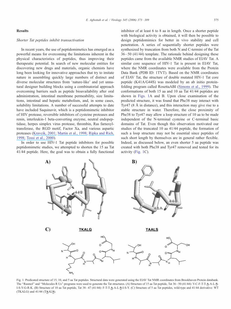
Fig. 1. Predicated structure of 15, 10, and 5 aa Tat peptides. Structural data were gene
The ‘‘Rasmol’’ and ‘‘Molecules R Us’’ programs were used to generate the Tat structu
I-S-Y-G-R-K. (B) Structure of 10 aa Tat peptide, Tat 38–47 (41/44): F-T-T-A-A-L-S
(TKALG) and 41/44 (TAALS).
inhibitor of at least 6 to 8 aa in length. Once a shorter peptide
with biological activity is obtained, it will then be possible to
design peptidomimics for better in vivo stability and cell
penetration. A series of sequentially shorter peptides were
synthesized by truncation from both N and C-termini of the Tat
36–50 (41/44) template. The rationale behind designing these
peptides came from the available NMR studies of EIAV Tat. A
similar core sequence of HIV-1 Tat is present in EIAV Tat,
where the NMR coordinates were available from the Protein
Data Bank (PDB ID: 1TVT). Based on the NMR coordinates
of EIAV Tat, the structure of double mutated HIV-1 Tat core
peptide (K41A/G44S) was modeled by an ab initio protein-
folding program called RosettaABI (Simons et al., 1999). The
conformations of both 15 aa and 10 aa Tat 41/44 peptides are
shown in Figs. 1A and B. Upon close examination of the
predicted structure, it was found that Phe38 may interact with
Tyr47 (8 A in distance), and this interaction may give rise to a
stable structure in water. Therefore, the close proximity of
Phe38 to Tyr47 may allow a loop structure of 10 aa to be made
independent of the N-terminal cysteine or C-terminal basic
domains of Tat. Even though this observation motivated our
studies of the truncated 10 aa 41/44 peptide, the formation of
such a loop structure may not be essential since peptides of
such short length by themselves are in general rather flexible.
Indeed, as discussed below, an even shorter 5 aa peptide was
created with both Phe38 and Tyr47 removed and tested for its
activity (Fig. 1C).
rated using the EIAV Tat NMR coordinates from Brookhaven Protein databank.
res. (A) Structure of 15 aa Tat peptide, Tat 36–50 (41/44): V-C-F-T-T-A-A-L-S-
-I-S-Y. (C) Structure of 5 aa Tat peptides, wild-type and 41/44 derivative: WT
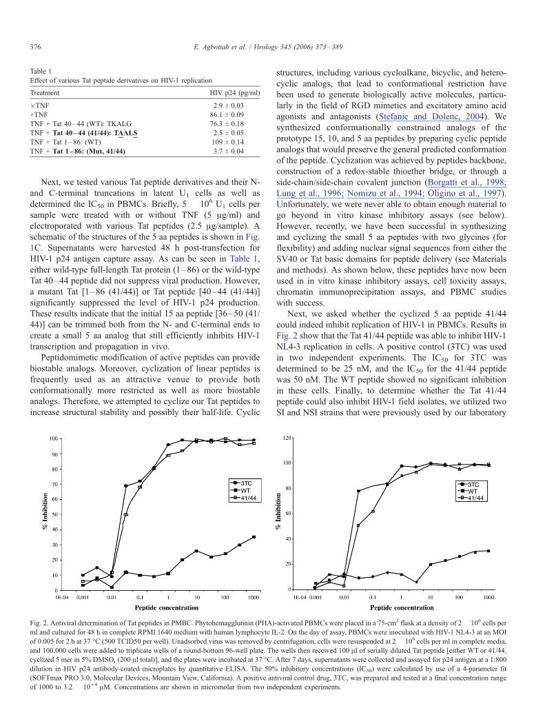
Table 1
Effect of various Tat peptide derivatives on HIV-1 replication
Treatment HIV p24 (pg/ml)
�TNF 2.9 T 0.03
+TNF 86.1 T 0.09
TNF + Tat 40–44 (WT): TKALG 76.3 T 0.18
TNF + Tat 40–44 (41/44): TAALS 2.5 T 0.05
TNF + Tat 1–86: (WT) 109 T 0.14
TNF + Tat 1–86: (Mut, 41/44) 3.7 T 0.04
E. Agbottah et al. / Virology 345 (2006) 373–389376
Next, we tested various Tat peptide derivatives and their N-
and C-terminal truncations in latent U1 cells as well as
determined the IC50 in PBMCs. Briefly, 5 � 106 U1 cells per
sample were treated with or without TNF (5 Ag/ml) and
electroporated with various Tat peptides (2.5 Ag/sample). A
schematic of the structures of the 5 aa peptides is shown in Fig.
1C. Supernatants were harvested 48 h post-transfection for
HIV-1 p24 antigen capture assay. As can be seen in Table 1,
either wild-type full-length Tat protein (1–86) or the wild-type
Tat 40–44 peptide did not suppress viral production. However,
a mutant Tat [1–86 (41/44)] or Tat peptide [40–44 (41/44)]
significantly suppressed the level of HIV-1 p24 production.
These results indicate that the initial 15 aa peptide [36–50 (41/
44)] can be trimmed both from the N- and C-terminal ends to
create a small 5 aa analog that still efficiently inhibits HIV-1
transcription and propagation in vivo.
Peptidomimetic modification of active peptides can provide
biostable analogs. Moreover, cyclization of linear peptides is
frequently used as an attractive venue to provide both
conformationally more restricted as well as more biostable
analogs. Therefore, we attempted to cyclize our Tat peptides to
increase structural stability and possibly their half-life. Cyclic
Fig. 2. Antiviral determination of Tat peptides in PMBC. Phytohemagglutinin (PHA)
ml and cultured for 48 h in complete RPMI 1640 medium with human lymphocyte IL
of 0.005 for 2 h at 37 -C (500 TCID50 per well). Unadsorbed virus was removed by c
and 100,000 cells were added to triplicate wells of a round-bottom 96-well plate. Th
cyclized 5 mer in 5% DMSO, (200 Al total)], and the plates were incubated at 37 -C.dilution in HIV p24 antibody-coated microplates by quantitative ELISA. The 50%
(SOFTmax PRO 3.0, Molecular Devices, Mountain View, California). A positive an
of 1000 to 3.2 � 10�4 AM. Concentrations are shown in micromolar from two ind
structures, including various cycloalkane, bicyclic, and hetero-
cyclic analogs, that lead to conformational restriction have
been used to generate biologically active molecules, particu-
larly in the field of RGD mimetics and excitatory amino acid
agonists and antagonists (Stefanic and Dolenc, 2004). We
synthesized conformationally constrained analogs of the
prototype 15, 10, and 5 aa peptides by preparing cyclic peptide
analogs that would preserve the general predicted conformation
of the peptide. Cyclization was achieved by peptides backbone,
construction of a redox-stable thioether bridge, or through a
side-chain/side-chain covalent junction (Borgatti et al., 1998;
Lung et al., 1996; Nomizu et al., 1994; Oligino et al., 1997).
Unfortunately, we were never able to obtain enough material to
go beyond in vitro kinase inhibitory assays (see below).
However, recently, we have been successful in synthesizing
and cyclizing the small 5 aa peptides with two glycines (for
flexibility) and adding nuclear signal sequences from either the
SV40 or Tat basic domains for peptide delivery (see Materials
and methods). As shown below, these peptides have now been
used in in vitro kinase inhibitory assays, cell toxicity assays,
chromatin immunoprecipitation assays, and PBMC studies
with success.
Next, we asked whether the cyclized 5 aa peptide 41/44
could indeed inhibit replication of HIV-1 in PBMCs. Results in
Fig. 2 show that the Tat 41/44 peptide was able to inhibit HIV-1
NL4-3 replication in cells. A positive control (3TC) was used
in two independent experiments. The IC50 for 3TC was
determined to be 25 nM, and the IC50 for the 41/44 peptide
was 50 nM. The WT peptide showed no significant inhibition
in these cells. Finally, to determine whether the Tat 41/44
peptide could also inhibit HIV-1 field isolates, we utilized two
SI and NSI strains that were previously used by our laboratory
-activated PBMCs were placed in a 75-cm2 flask at a density of 2 � 106 cells per
-2. On the day of assay, PBMCs were inoculated with HIV-1 NL4-3 at an MOI
entrifugation, cells were resuspended at 2 � 106 cells per ml in complete media,
e wells then received 100 Al of serially diluted Tat peptide [either WT or 41/44,
After 7 days, supernatants were collected and assayed for p24 antigen at a 1:800
inhibitory concentrations (IC50) were calculated by use of a 4-parameter fit
tiviral control drug, 3TC, was prepared and tested at a final concentration range
ependent experiments.
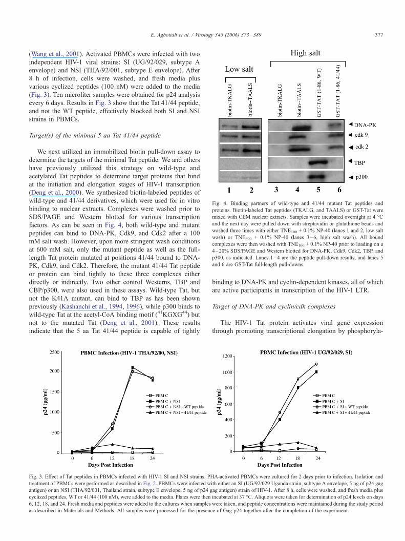
Fig. 4. Binding partners of wild-type and 41/44 mutant Tat peptides and
proteins. Biotin-labeled Tat peptides (TKALG, and TAALS) or GST-Tat were
mixed with CEM nuclear extracts. Samples were incubated overnight at 4 -C
and the next day were pulled down with streptavidin or glutathione beads and
washed three times with either TNE100 + 0.1% NP-40 (lanes 1 and 2, low salt
wash) or TNE600 + 0.1% NP-40 (lanes 3–6, high salt wash). All bound
complexes were then washed with TNE100 + 0.1% NP-40 prior to loading on a
4–20% SDS/PAGE and Western blotted for DNA-PK, Cdk9, Cdk2, TBP, and
p300, as indicated. Lanes 1–4 are the peptide pull-down results, and lanes 5
and 6 are GST-Tat full-length pull-downs.
E. Agbottah et al. / Virology 345 (2006) 373–389 377
(Wang et al., 2001). Activated PBMCs were infected with two
independent HIV-1 viral strains: SI (UG/92/029, subtype A
envelope) and NSI (THA/92/001, subtype E envelope). After
8 h of infection, cells were washed, and fresh media plus
various cyclized peptides (100 nM) were added to the media
(Fig. 3). Ten microliter samples were obtained for p24 analysis
every 6 days. Results in Fig. 3 show that the Tat 41/44 peptide,
and not the WT peptide, effectively blocked both SI and NSI
strains in PBMCs.
Target(s) of the minimal 5 aa Tat 41/44 peptide
We next utilized an immobilized biotin pull-down assay to
determine the targets of the minimal Tat peptide. We and others
have previously utilized this strategy on wild-type and
acetylated Tat peptides to determine target proteins that bind
at the initiation and elongation stages of HIV-1 transcription
(Deng et al., 2000). We synthesized biotin-labeled peptides of
wild-type and 41/44 derivatives, which were used for in vitro
binding to nuclear extracts. Complexes were washed prior to
SDS/PAGE and Western blotted for various transcription
factors. As can be seen in Fig. 4, both wild-type and mutant
peptides can bind to DNA-PK, Cdk9, and Cdk2 after a 100
mM salt wash. However, upon more stringent wash conditions
at 600 mM salt, only the mutant peptide as well as the full-
length Tat protein mutated at positions 41/44 bound to DNA-
PK, Cdk9, and Cdk2. Therefore, the mutant 41/44 Tat peptide
or protein can bind tightly to these three complexes either
directly or indirectly. Two other control Westerns, TBP and
CBP/p300, were also used in these assays. Wild-type Tat, but
not the K41A mutant, can bind to TBP as has been shown
previously (Kashanchi et al., 1994, 1996), while p300 binds to
wild-type Tat at the acetyl-CoA binding motif (41KGXG44) but
not to the mutated Tat (Deng et al., 2001). These results
indicate that the 5 aa Tat 41/44 peptide is capable of tightly
Fig. 3. Effect of Tat peptides in PBMCs infected with HIV-1 SI and NSI strains. PH
treatment of PBMCs were performed as described in Fig. 2. PBMCs were infected w
antigen) or an NSI (THA/92/001, Thailand strain, subtype E envelope, 5 ng of p24 g
cyclized peptides, WT or 41/44 (100 nM), were added to the media. Plates were then
6, 12, 18, and 24. Fresh media and peptides were added to the cultures when samples
as described in Materials and Methods. All samples were processed for the presenc
binding to DNA-PK and cyclin-dependent kinases, all of which
are active participants in transcription of the HIV-1 LTR.
Target of DNA-PK and cyclin/cdk complexes
The HIV-1 Tat protein activates viral gene expression
through promoting transcriptional elongation by phosphoryla-
A-activated PBMCs were cultured for 2 days prior to infection. Isolation and
ith either an SI (UG/92/029 Uganda strain, subtype A envelope, 5 ng of p24 gag
ag antigen) strain of HIV-1. After 8 h, cells were washed, and fresh media plus
incubated at 37 -C. Aliquots were taken for determination of p24 levels on days
were taken, and peptide concentrations were maintained during the study period
e of Gag p24 together after the completion of the experiment.
E. Agbottah et al. / Virology 345 (2006) 373–389378
ting RNAPII. In this process, Tat enhances phosphorylation of
the CTD of RNAPII by activating cyclin-dependent kinases
(Cdks) associated with general transcription factors. The
conserved CTD repeats of RNAPII are important sites of
transcription regulation. To date, four cyclin/cdk complexes
have been shown to phosphorylate the CTD, Cyclin C/Cdk8,
Cyclin H/Cdk7 (subunits of transcription factors TFIIH),
Cyclin T/Cdk9, and Cyclin K/Cdk9 complexes. Other protein
kinases have also been described as able to phosphorylate the
CTD. DNA-PKcs also acts as a CTD kinase only when
stimulated by linear double-stranded DNA and by several
transcriptional activators. Furthermore, we have recently
reported that Cyclin E/Cdk2 could phosphorylate the CTD.
Recombinant Cyclin E/Cdk2 stimulated Tat-dependent HIV-1
transcription in a reconstituted transcription assay, and immu-
nodepletion of Cyclin E/Cdk2 from HeLa nuclear extracts
blocked Tat-dependent transcription (Deng et al., 2002).
In recent years, a more complex picture of transcription has
emerged where the RNAPII CTD functions both as an
assembly platform for and a regulator of transcription and
pre-mRNA processing machineries, including 5V capping,
elongation, splicing, and polyadenylation (Cheng and Sharp,
2003; Chiu et al., 2001; 2002; Cho et al., 1998; Fong and
Bentley, 2001; Ho and Shuman, 1999; Moteki and Price, 2002;
Pei and Shuman, 2002; Proudfoot et al., 2002; Schroeder et al.,
2000; Wen and Shatkin, 1999). Almost all of these events are
triggered during transcription initiation by phosphorylation of
CTD serine 5. In general, phosphorylation of CTD serine 5 is
concentrated near the transcription initiation site (+1 area),
while CTD serine 2 phosphorylation is observed in the
transcription elongation complex. Therefore, we asked whether
the Tat 41/44 peptide was able to inhibit phosphorylation of
serine 2 or 5 of the RNAPII CTD present on the HIV-1
promoter. We first designed experiments to detect any possible
inhibition of Cdk2/Cyclin E kinase activity in vitro, in host
cells, and finally on RNAPII CTD serine 2 and 5 phospho-
rylation on the HIV-1 promoter in vivo using a Chromatin
Immunoprecipitation (ChIP) assay. ChIP is a powerful
approach that allows one to define the interaction of factors
with specific chromosomal sites in living cells, thereby
providing a snapshot of the native chromatin structure and
factors bound to genes in different functional states. ChIP
involves treating cells briefly with formaldehyde to cross-link
proteins to DNA. An antibody against a protein suspected of
binding a given cis-element is then used to immunoprecipitate
chromatin fragments. Polymerase chain reaction analysis of the
immunoprecipitate with primers flanking the cis-element
reveals whether a specific DNA sequence is recovered in an
immune-specific manner and, therefore, whether the protein
contacted the site in living cells (Das et al., 2004; Weinmann
and Farnham, 2002). Results are shown in Figs. 5 and 6. As
can be seen in Fig. 5A, the Tat 41/44 peptide, but not the wild-
type peptide (Lanes 8–10), can efficiently inhibit Cyclin E/
Cdk2 kinase activity in vitro. The Tat 41/44 peptide inhibited
the kinase reaction at an IC50 of 20 nM. We next asked whether
an over-expressed and purified Cyclin E/Cdk2 complex had a
similar inhibition profile as compared to the complex isolated
from HIV-1-infected cells. Fig. 5B shows that recombinant
baculavirus HA-tagged Cdk2/Cyclin E can also be inhibited by
the Tat 41/44 peptides (both linear or cyclized).
To determine whether these peptides were toxic to cells, we
utilized DNA labeling experiments after peptide treatment in
CEM, U937, CaCo-2, PBMC, and ACH2 cells. As can be seen
in Fig. 5C, the Tat peptides, either wild-type or the 41/44
derivative, had no apparent effect on uninfected cells up to 24 h.
Similar experiments were performed at a later date and carried
up to 7 days without any apparent toxicity (Fig. 5D). As
expected, the Tat 41/44 peptides were inhibitory in HIV-1-
infected cells, as evident by the p24 results (Fig. 5C, right hand
insert). These results clearly indicate that, even though Tat 41/
44 targets DNA-PK, Cdk2, and Cdk9, these interactions are not
functionally significant in uninfected cells. Similar results with
knockout of these genes have also been obtained by others in
cell lines and KO mouse systems (Berthet et al., 2003; Geng et
al., 2003; Mendez, 2003; Ortega et al., 2003). However, DNA-
PK, Cdk2, and Cdk9 are critical in HIV-1-infected cells,
especially when these proteins are concentrated on the HIV-1
promoter.
Inhibition of CTD phosphorylation on the HIV-1 promoter
To address whether Tat peptides in ACH2 cells were
inhibiting HIV-1 LTR transcription, we devised a series of in
vivo ChIP assays followed by PCR with specific primers to
HIV (experimental) and GAPDH (control) genes. We used
ACH2 cells after treatment with Tat peptides (described in Fig.
5C) and used the total DNA for a ChIP assay using two
different antibodies. The antibodies were specific for phos-
phorylated RNAPII serine 5 (H14) and RNAPII serine 2 (H5).
Following ChIP, samples were amplified with either HIV-1
LTR or GAPDH primers. As seen in Fig. 6, both linear and
cyclized Tat 41/44 peptides efficiently inhibited serine 5
phosphorylation but not the serine 2 phosphorylation of the
RNAPII CTD. Consistent with inhibition of serine 5, both
levels of HIV RNA capping and elongation by SPT-5 were
reduced with the Tat 41/44 peptide (data not shown). This is
consistent with the notion that serine 5 phosphorylation recruits
subsequent enzymes for capping, elongation, and splicing
machineries. These peptides did not affect the RNAPII,
capping, or elongation of the cellular gene GAPDH. These
results imply that the mechanism of inhibition of the HIV-1
LTR by Tat 41/44 is via inhibition of serine 5 phosphorylation
of an RNA polymerase that is specifically associated with the
HIV-1 promoter and not a cellular promoter.
The experiments described above (Fig. 6) clearly show that
the Tat 41/44 peptide was able to inhibit RNAPII CTD
phosphorylation on HIV-1 DNA but does not address whether
any of the three kinases was the target in the observed
inhibition. We therefore determined whether we could observe
these kinases on the HIV-1 promoter and then subsequently
asked whether the Tat 41/44 peptide could inhibit any of these
enzymes on the HIV-1 genome in vivo. Similar experiments as
described in Fig. 5C were performed in ACH2 cells, and
various antibodies were used for the initial ChIP assays.
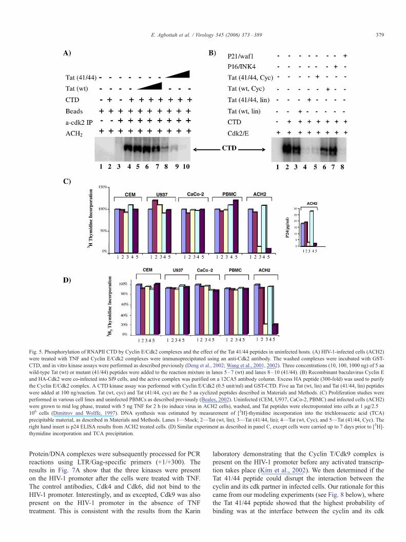
Fig. 5. Phosphorylation of RNAPII CTD by Cyclin E/Cdk2 complexes and the effect of the Tat 41/44 peptides in uninfected hosts. (A) HIV-1-infected cells (ACH2)
were treated with TNF and Cyclin E/Cdk2 complexes were immunoprecipitated using an anti-Cdk2 antibody. The washed complexes were incubated with GST-
CTD, and in vitro kinase assays were performed as described previously (Deng et al., 2002; Wang et al., 2001, 2002). Three concentrations (10, 100, 1000 ng) of 5 aa
wild-type Tat (wt) or mutant (41/44) peptides were added to the reaction mixture in lanes 5–7 (wt) and lanes 8–10 (41/44). (B) Recombinant baculavirus Cyclin E
and HA-Cdk2 were co-infected into Sf9 cells, and the active complex was purified on a 12CA5 antibody column. Excess HA peptide (300-fold) was used to purify
the Cyclin E/Cdk2 complex. A CTD kinase assay was performed with Cyclin E/Cdk2 (0.5 unit/ml) and GST-CTD. Five aa Tat (wt, lin) and Tat (41/44, lin) peptides
were added at 100 ng/reaction. Tat (wt, cyc) and Tat (41/44, cyc) are the 5 aa cyclized peptides described in Materials and Methods. (C) Proliferation studies were
performed in various cell lines and uninfected PBMCs as described previously (Beales, 2002). Uninfected (CEM, U937, CaCo-2, PBMC) and infected cells (ACH2)
were grown to mid log phase, treated with 5 ng TNF for 2 h (to induce virus in ACH2 cells), washed, and Tat peptides were electroporated into cells at 1 Ag/2.5 �106 cells (Dimitrov and Wolffe, 1997). DNA synthesis was estimated by measurement of [3H]-thymidine incorporation into the trichloroacetic acid (TCA)
precipitable material, as described in Materials and Methods. Lanes 1—Mock; 2—Tat (wt, lin); 3—Tat (41/44, lin); 4—Tat (wt, Cyc), and 5—Tat (41/44, Cyc). The
right hand insert is p24 ELISA results from ACH2 treated cells. (D) Similar experiment as described in panel C, except cells were carried up to 7 days prior to [3H]-
thymidine incorporation and TCA precipitation.
E. Agbottah et al. / Virology 345 (2006) 373–389 379
Protein/DNA complexes were subsequently processed for PCR
reactions using LTR/Gag-specific primers (+1/+300). The
results in Fig. 7A show that the three kinases were present
on the HIV-1 promoter after the cells were treated with TNF.
The control antibodies, Cdk4 and Cdk6, did not bind to the
HIV-1 promoter. Interestingly, and as excepted, Cdk9 was also
present on the HIV-1 promoter in the absence of TNF
treatment. This is consistent with the results from the Karin
laboratory demonstrating that the Cyclin T/Cdk9 complex is
present on the HIV-1 promoter before any activated transcrip-
tion takes place (Kim et al., 2002). We then determined if the
Tat 41/44 peptide could disrupt the interaction between the
cyclin and its cdk partner in infected cells. Our rationale for this
came from our modeling experiments (see Fig. 8 below), where
the Tat 41/44 peptide showed that the highest probability of
binding was at the interface between the cyclin and its cdk
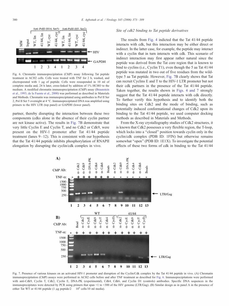
Fig. 6. Chromatin immunoprecipitation (ChIP) assay following Tat peptide
treatment in ACH2 cells. Cells were treated with TNF for 2 h, washed, and
electroporated with 1 Ag of peptide. Cells were resuspended in 10 ml of
complete media and, 24 h later, cross-linked by addition of 1% HCHO to the
medium. A modified chromatin immunoprecipitation (ChIP) assay (Braunstein
et al., 1993; de la Fuente et al., 2000) was performed as described in Materials
and Methods. Chromatin was immunoprecipitated using antibodies to Pol II Ser
2, Pol II Ser 5 overnight at 4 -C. Immunoprecipitated DNAwas amplified using
primers to the HIV LTR (top panel) or GAPDH (lower panel).
E. Agbottah et al. / Virology 345 (2006) 373–389380
partner, thereby disrupting the interaction between these two
components (cdks alone in the absence of their cyclin partner
are not kinase active). The results in Fig. 7B demonstrate that
very little Cyclin E and Cyclin T, and no Cdk2 or Cdk9, were
present on the HIV-1 promoter after Tat 41/44 peptide
treatment (lanes 9–12). This is consistent with our hypothesis
that the Tat 41/44 peptide inhibits phosphorylation of RNAPII
elongation by disrupting the cyclin/cdk complex in vivo.
Fig. 7. Presence of various kinases on an activated HIV-1 promoter and disruption
immunoprecipitation (ChIP) assays were performed in ACH2 cells before and after
with anti-Cdk9, Cyclin T, Cdk2, Cyclin E, DNA-PK (experimental), Cdk4, C
immunoprecipitates were detected by PCR using primers that span +1 to +300 of th
either Tat WT or 41/44 peptide (1 Ag peptide/2 � 106 cells/10 ml media).
Site of cdk2 binding to Tat peptide derivatives
The results from Fig. 4 indicted that the Tat 41/44 peptide
interacts with cdk, but this interaction may be either direct or
indirect. In the latter case, for example, the peptide may interact
with a cyclin that in turn interacts with cdk. This scenario of
indirect interaction may first appear rather natural since the
peptide was derived from the Tat core region that is known to
bind to cyclins (i.e., Cyclin T1), even though the 5 aa Tat 41/44
peptide was mutated in two out of five residues from the wild-
type 5 aa Tat peptide. However, Fig. 7B clearly shows that Tat
can recruit Cyclins E and T to the HIV-1 LTR promoter but not
their cdk partners in the presence of the Tat 41/44 peptide.
Taken together, the results shown in Figs. 4 and 7 strongly
suggest that the Tat 41/44 peptide interacts with cdk directly.
To further verify this hypothesis and to identify both the
binding sites on Cdk2 and the mode of binding, such as
potentially induced conformational changes of Cdk2 upon its
binding to the Tat 41/44 peptide, we used computer docking
methods as described in Materials and Methods.
From the X-ray crystallography studies of Cdk2 structures, it
is known that Cdk2 possesses a very flexible region, the T-loop,
which locks into a ‘‘closed’’ position towards cyclin only in the
cyclin/cdk complex (PDB ID: 1FIN) but otherwise remains
somewhat ‘‘open’’ (PDB ID: 1E1X). To investigate the potential
effects of these two forms of cdk in binding to the Tat 41/44
of the Cyclin/Cdk complex by the Tat 41/44 peptide in vivo. (A) Chromatin
TNF treatment as described for Fig. 6. Immunoprecipitations were performed
dk6, and Cyclin D1 (controls) antibodies. Specific DNA sequences in the
e HIV genome (LTR/Gag). (B) Similar design as in panel A in the presence of
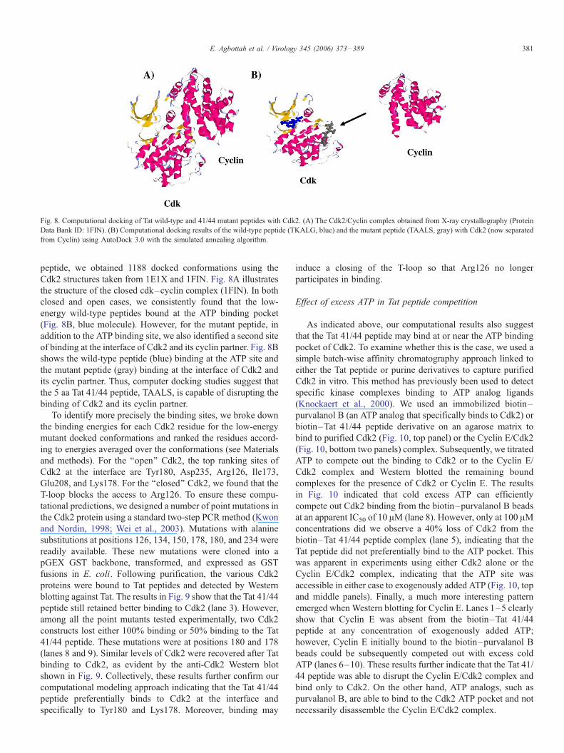
Fig. 8. Computational docking of Tat wild-type and 41/44 mutant peptides with Cdk2. (A) The Cdk2/Cyclin complex obtained from X-ray crystallography (Protein
Data Bank ID: 1FIN). (B) Computational docking results of the wild-type peptide (TKALG, blue) and the mutant peptide (TAALS, gray) with Cdk2 (now separated
from Cyclin) using AutoDock 3.0 with the simulated annealing algorithm.
E. Agbottah et al. / Virology 345 (2006) 373–389 381
peptide, we obtained 1188 docked conformations using the
Cdk2 structures taken from 1E1X and 1FIN. Fig. 8A illustrates
the structure of the closed cdk–cyclin complex (1FIN). In both
closed and open cases, we consistently found that the low-
energy wild-type peptides bound at the ATP binding pocket
(Fig. 8B, blue molecule). However, for the mutant peptide, in
addition to the ATP binding site, we also identified a second site
of binding at the interface of Cdk2 and its cyclin partner. Fig. 8B
shows the wild-type peptide (blue) binding at the ATP site and
the mutant peptide (gray) binding at the interface of Cdk2 and
its cyclin partner. Thus, computer docking studies suggest that
the 5 aa Tat 41/44 peptide, TAALS, is capable of disrupting the
binding of Cdk2 and its cyclin partner.
To identify more precisely the binding sites, we broke down
the binding energies for each Cdk2 residue for the low-energy
mutant docked conformations and ranked the residues accord-
ing to energies averaged over the conformations (see Materials
and methods). For the ‘‘open’’ Cdk2, the top ranking sites of
Cdk2 at the interface are Tyr180, Asp235, Arg126, Ile173,
Glu208, and Lys178. For the ‘‘closed’’ Cdk2, we found that the
T-loop blocks the access to Arg126. To ensure these compu-
tational predictions, we designed a number of point mutations in
the Cdk2 protein using a standard two-step PCR method (Kwon
and Nordin, 1998; Wei et al., 2003). Mutations with alanine
substitutions at positions 126, 134, 150, 178, 180, and 234 were
readily available. These new mutations were cloned into a
pGEX GST backbone, transformed, and expressed as GST
fusions in E. coli. Following purification, the various Cdk2
proteins were bound to Tat peptides and detected by Western
blotting against Tat. The results in Fig. 9 show that the Tat 41/44
peptide still retained better binding to Cdk2 (lane 3). However,
among all the point mutants tested experimentally, two Cdk2
constructs lost either 100% binding or 50% binding to the Tat
41/44 peptide. These mutations were at positions 180 and 178
(lanes 8 and 9). Similar levels of Cdk2 were recovered after Tat
binding to Cdk2, as evident by the anti-Cdk2 Western blot
shown in Fig. 9. Collectively, these results further confirm our
computational modeling approach indicating that the Tat 41/44
peptide preferentially binds to Cdk2 at the interface and
specifically to Tyr180 and Lys178. Moreover, binding may
induce a closing of the T-loop so that Arg126 no longer
participates in binding.
Effect of excess ATP in Tat peptide competition
As indicated above, our computational results also suggest
that the Tat 41/44 peptide may bind at or near the ATP binding
pocket of Cdk2. To examine whether this is the case, we used a
simple batch-wise affinity chromatography approach linked to
either the Tat peptide or purine derivatives to capture purified
Cdk2 in vitro. This method has previously been used to detect
specific kinase complexes binding to ATP analog ligands
(Knockaert et al., 2000). We used an immobilized biotin–
purvalanol B (an ATP analog that specifically binds to Cdk2) or
biotin–Tat 41/44 peptide derivative on an agarose matrix to
bind to purified Cdk2 (Fig. 10, top panel) or the Cyclin E/Cdk2
(Fig. 10, bottom two panels) complex. Subsequently, we titrated
ATP to compete out the binding to Cdk2 or to the Cyclin E/
Cdk2 complex and Western blotted the remaining bound
complexes for the presence of Cdk2 or Cyclin E. The results
in Fig. 10 indicated that cold excess ATP can efficiently
compete out Cdk2 binding from the biotin–purvalanol B beads
at an apparent IC50 of 10 AM (lane 8). However, only at 100 AMconcentrations did we observe a 40% loss of Cdk2 from the
biotin–Tat 41/44 peptide complex (lane 5), indicating that the
Tat peptide did not preferentially bind to the ATP pocket. This
was apparent in experiments using either Cdk2 alone or the
Cyclin E/Cdk2 complex, indicating that the ATP site was
accessible in either case to exogenously added ATP (Fig. 10, top
and middle panels). Finally, a much more interesting pattern
emerged when Western blotting for Cyclin E. Lanes 1–5 clearly
show that Cyclin E was absent from the biotin–Tat 41/44
peptide at any concentration of exogenously added ATP;
however, Cyclin E initially bound to the biotin–purvalanol B
beads could be subsequently competed out with excess cold
ATP (lanes 6–10). These results further indicate that the Tat 41/
44 peptide was able to disrupt the Cyclin E/Cdk2 complex and
bind only to Cdk2. On the other hand, ATP analogs, such as
purvalanol B, are able to bind to the Cdk2 ATP pocket and not
necessarily disassemble the Cyclin E/Cdk2 complex.
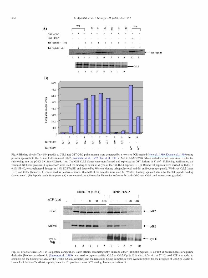
Fig. 10. Effect of excess ATP in Tat peptide competition. Batch affinity chromatography linked to either Tat biotin peptide (10 Ag/100 Al packed beads) or a purine
derivative [biotin–purvalanol A, (Simons et al., 1999)] was used to capture purified Cdk2 or Cdk2/Cyclin E in vitro. After 4 h at 37 -C, cold ATP was added to
compete out the binding to Cdk2 or the Cyclin E/Cdk2 complex, and the remaining bound complexes were Western blotted for the presence of Cdk2 or Cyclin E.
Lanes 1–5: biotin–Tat 41/44 peptide; lanes 6–10: positive control ATP analog, biotin–purvalanol A.
Fig. 9. Binding site for Tat 41/44 peptide to Cdk2. (A) GST-Cdk2 point mutants were generated by a two-step PCR method (Ho et al., 1989; Kwon et al., 1996) using
primers against both the N- and C-terminus of Cdk2 (Rosenblatt et al., 1992; Tsai et al., 1991) (Acc #: AAX32258), which included EcoRI and BamHI sites for
subcloning into the pGEX-3X BamHI/EcoRI site. The GST-Cdk2 clones were transformed and expressed as GST fusions in E. coli. Following purification, the
various GST-Cdk2 proteins (3 Ag/reaction) were used for binding to either wild-type or the Tat 41/44 peptide (10 Ag). Bound Tat peptides were washed in TNE50 +
0.1% NP-40, electrophoresed through an 18% SDS/PAGE, and detected by Western blotting using polyclonal anti-Tat antibody (upper panel). Wild-type Cdk2 (lanes
1–3) and Cdk9 (lanes 10, 11) were used as positive controls. One-half of the samples were used for Western blotting against Cdk2 after the Tat peptide binding
(lower panel). (B) Peptide bands from panel (A) were counted on a Molecular Dynamics software for both Cdk2 and Cdk9, and values were graphed.
E. Agbottah et al. / Virology 345 (2006) 373–389382
E. Agbottah et al. / Virology 345 (2006) 373–389 383
Discussion
Highly active antiretroviral therapy (HAART) dramatically
changed the course of HIV treatment. Currently, this therapy
involves the use of agents from at least two distinct classes of
antivirals: a protease inhibitor (PI) in combination with two
nucleoside/nucleotide reverse transcriptase inhibitors (NRTIs)
or a non-nucleoside reverse transcriptase inhibitor (NNRTI) in
combination with NRTIs (Barbaro et al., 2005). Recently, a
third family of antivirals is now used clinically, with the advent
of Enfuvirtide, the first fusion inhibitor (FI). Several pharma-
cological agents from these classes of antivirals include NRTIs,
NNRTIs, PIs, and FIs. Compounds inhibiting HIV integrase,
the third enzyme of HIV, and inhibitors of the gp120–CD4
interaction also hold great promise. Finally, compounds
interacting with Tat/TAR have also been studied which inhibit
HIV replication in low micromolar range (EM2487, tamacra-
zine, CGP 64222, CGA 137053, or siRNA against Tat among
others) (Barbaro et al., 2005). However, in most cases, use of
such treatment gives rise to resistant viruses, the mechanisms
of inhibition are poorly defined, and only a few of these studies
have explored a wide range of inhibition for various viral
strains.
In the current study, we extend our initial findings that the
Tat 41/44 transdominant peptide from the HIV-1 Tat core
domain can inhibit HIV-1 gene expression. The advantage of
this system is that peptides derived from Tat are presumably
more specific in inhibiting Tat-mediated activation in vivo. We
previously have shown that soluble peptide analogs of the Tat
core domain (amino acid 36–50) were able to effectively block
HIV-1 LTR transactivation, and the activity was specific to
activated transcription of HIV-1 and not other promoters,
including HTLV-I, CMV, PTHrP, IgH, RAS, RSV, and SIV
CAT (Kashanchi et al., 1997). Sequentially shorter peptides
were synthesized by truncation from both N- and C-termini of
the 36–50 (41/44) template. We consistently observed a
significant drop in viral titers in U1 cells with one of the
peptides, Tat 41/44. The effect was apparent not only when
using the mutant peptide, but also when using a full-length Tat
mutated at positions 41/44. Furthermore, our studies in cell
lines and PBMCs indicated that these peptides were not toxic
to cells at concentrations that normally inhibit HIV-1 replica-
tion, indicating that inhibition of HIV-1 activated transcription
and viral progeny formation is possible with these peptides.
The inhibition by Tat 41/44 was specific to the Tat/TAR
complex and, therefore, specific for HIV-1.
When performing Tat peptide biotin pull-down experiments,
we observed binding of a number of kinases that target RNA
Pol II CTD. We concentrated on Cdk2/Cyclin E mainly
because KO animals for either Cdk2 or Cyclin E are viable,
making this cdk/cyclin complex an attractive target for HIV
therapy. In 2003, the Barbacid laboratory first reported that
embryonic fibroblasts lacking Cdk2 proliferate normally and
become immortal after continuous passage in culture. Addi-
tionally, Cdk2�/� mice were viable and survived for up to 2
years, indicating that Cdk2 was also dispensable for the
proliferation and survival of most cell types (Ortega et al.,
2003). At the same time, others reported similar findings
demonstrating that the E type cyclins were also largely
dispensable for development. Cyclin-E-deficient cells prolife-
rated actively under conditions of continuous cell cycling
(Berthet et al., 2003; Geng et al., 2003; Mendez, 2003).
Therefore, Cdk2 and Cyclin E are not essential for the growth
of normal, non-cancerous cells, and use of inhibitors against
the Cdk2/Cyclin E complex may pose a viable option to inhibit
HIV-1 in infected cells.
We have recently reported that Cyclin E/Cdk2 could also
phosphorylate the RNA Pol II CTD, that recombinant Cyclin
E/Cdk2 stimulated Tat-dependent HIV-1 transcription in a
reconstituted transcription assay, and that immunodepletion of
Cyclin E/Cdk2 from HeLa nuclear extracts blocked Tat-
dependent transcription (Deng et al., 2002). These are
significant findings since RNA Pol II CTD functions both as
an assembly platform for and a regulator of the transcription
and pre-mRNA processing machineries. During transcription
initiation, serine 5 of the CTD heptad repeat is phosphorylated.
This phosphorylation triggers a cascade of events beginning
with the dissociation of transcription initiation factors from the
CTD followed by the recruitment of the capping machinery and
the allosteric activation of the capping enzyme. HIV-1 capping
takes place during the transition from transcription initiation to
elongation when the nascent pre-mRNA is only 20–40
nucleotides long (Zhou et al., 2004). Serine 5 is subsequently
dephosphorylated, resulting in release of the capping machin-
ery. Serine 2 of the heptad repeat is then phosphorylated,
leading to the recruitment of factors (hSPT5 and TAT-SF1)
involved in subsequent steps of HIV-1 RNA processing. Our
current data suggest that Tat 41/44 inhibits the kinase activity
of Cdk2/Cyclin E in vitro and serine 5 phosphorylation of
RNAPII in the context of the promoter proximal region of the
HIV-1 LTR and not cellular genes such as GAPDH. Therefore,
down-regulation of serine 5 phosphorylation by the Tat 41/44
peptide may result in control of all pre-mRNA processing of
HIV-1 RNA, including RNA processing, mRNA export,
nonsense-mediated decay (NMD), and RNA degradation.
It is also important to note that we currently have no
evidence suggesting which of the serine 5 phosphorylation
sites in the 52 CTD repeats (conserved or variable regions) are
phosphorylated by the Cdk2/Cyclin E complex. Therefore, it is
possible that there is a partial loss of phosphorylation of some
of the serine 5 sites when treating with the Tat 41/44 peptide.
This would give rise to fewer HIV transcripts, which may be
sufficient to halt the next round of infection (only 10–100
particles out of ¨100,000 are infectious). Along these lines,
Zhou et al. (2001) have previously correlated phosphorylation
of serine 5 in HIV transcription initiation and elongation
complexes with TFIIH. Therefore, an important question
would be why does HIV need so many cyclin/cdk complexes
for its transcriptional activity. Although we do not have a
complete answer at this point, we do speculate that various
cyclin/cdk complexes may be phosphorylating various CTD
(conserved or variable) substrates, different stages of the cell
cycle may determine which cyclin/cdk is used (Agbottah et al.,
2005), and the status of the chromatin structure may determine

E. Agbottah et al. / Virology 345 (2006) 373–389384
which cyclin/cdk is used efficiently, that is, Cdk2/Cyclin E has
a better activity on the native integrated virus as compared to
the naked or disorganized LTR DNA (Ammosova et al., 2005).
We also tested the mode of action for the Tat 41/44 peptide
using computer simulation models. As in all molecular
simulation programs, the energy function is a very important
aspect of these analysis. For our analysis, we included
electrostatic, van der Waals, and hydrogen bond terms. Because
of the large number of energy calculations required for the
thousands of the Metropolis steps for each temperature, we
used the grid method that pre-stores the interaction energies.
For our binding site search problem, we used a large grid that
covers the entire protein and the multiple starting positions of
the ligand peptide. A probe atom is placed at each point on the
grid, and its energy with the receptor protein is calculated and
stored. Such energy tables for all types of atoms in the ligand
(carbon, oxygen, nitrogen, and hydrogen) served as our energy
look-up tables. When the ligand moves in the grid during a
simulated annealing run, the energy of a ligand atom is
obtained by interpolation of the pre-stored table values at the
eight surrounding grid points. We also modified the widely
used AutoDock 3.0 program by writing an automated computer
package to use a parallel computer cluster to carry out the
multiple starting points docking calculations (Fig. 8). We
observed that the wild-type peptide consistently preferred
binding near the ATP binding site, while the mutant peptide
interacted with a cavity of Cdk2 interfacing the cyclin, thus
appearing to be capable of blocking the binding of Cdk2 with
cyclin. Furthermore, in our calculation, the mutant peptide
consistently had lower binding energy than the wild-type
peptide. Therefore, based on experiments performed with the
ChIP assay and modeling on cdk, we propose the following
model for the mechanism of inhibition when using the Tat 41/
44 peptide derivative (Fig. 11). The model predicts that Cdk2/
Cyclin E is present on the HIV-1 promoter, is regulated by Tat
(in the presence of TAR), and ultimately phosphorylates
important substrates in the transcription machinery including
the RNAPII CTD. In the presence of the Tat 41/44 peptide, cdk
dissociates away from the cyclin component making the kinase
inactive. The remaining cyclin complex on the HIV-1 promoter
may ultimately be ubiquitinated and degraded, although
Fig. 11. Proposed model of the mechanism of Tat 41/44 peptide inhibition of the HIV
Tat 41/44 peptide, and not the wild-type peptide, was able to disrupt the interaction
interact. The model is supported by computational docking results and by the ChIP
currently we have no formal proof of this event. However,
there is high likelihood of cyclin degradation since this is one
of the normal patterns of oscillating cyclins including Cyclins
D, E, A, and B. Finally, in the current study, we have not
addressed whether the free cyclin can bind to other cdks, a
phenomenon that is common in many cyclin over-expressing
cells, including breast cancer and HTLV-1 infection. Future
experiments will address these critical issues, along with
peptide feasibility studies (SCID-hu implants) and in vivo
availability of peptides in animals.
Materials and methods
PBMC infection and peptide treatment
Antiviral determination of Tat peptides in PMBC
Phytohemagglutinin (PHA)-activated peripheral blood
mononuclear cells (PBMCs) were thawed from liquid nitro-
gen, placed in a 75-cm2 flask at a density of 2 � 106 cells per
ml, and cultured for 48 h in RPMI 1640 supplemented with
10% heat-inactivated fetal bovine serum, 2 mM l-glutamine,
and 5 U/ml human lymphocyte IL-2 (Boehringer-Mannheim)
(complete cell culture medium). On the day of assay, PBMCs
(7 � 106) were inoculated with HIV-1 NL4-3 at an MOI of
0.005 in bulk in a 15-ml conical tube for 2 h at 37 -C.Unadsorbed virus was removed by centrifugation at 400 � g
for 5 min, cells were resuspended in medium at 2 � 106 cells
per ml in complete cell culture medium, and 100 Al (100,000cells) was added to triplicate wells of a round-bottom 96-well
plate. The wells then received 100 Al of serially diluted Tat
peptide (either WT or 41/44, cyclized 5 aa in 5% DMSO; 200
Al total), and the plates were incubated at 37 -C in a
humidified 5% CO2 atmosphere for 7 days. Plates were then
centrifuged at 400 � g for 5 min, and supernatants were
collected and assayed for p24 antigen at a 1:800 dilution in
HIV p24 antibody-coated microplates (NEN Life Science
Products, Inc.) by quantitative ELISA using the p24 standard
supplied by the manufacturer. The 50% inhibitory concentra-
tion (IC50) was calculated by use of a 4-parameter fit
(SOFTmax PRO 3.0, Molecular Devices, Mountain View,
California). A positive antiviral control drug, 3TC (NIH AIDS
-1 promoter. The model relies on experiments presented in Figs. 5–7, where the
between Cdk and its Cyclin partner at the interface where these two molecules
assays shown in Fig. 7B.
E. Agbottah et al. / Virology 345 (2006) 373–389 385
Research and Reference Program), was prepared and tested at
a final concentration range of 1000 to 0.00032 AM.
Concentrations are shown as micromolar from two indepen-
dent experiments.
Effect of Tat peptides in PBMC infected with HIV-1 SI and NSI
strains
Phytohemagglutinin-activated PBMCs were kept in culture
for 2 days prior to each infection. Isolation and treatment of
PBMCs were performed as described above. PBMCs were
infected with either a syncytium-inducing (SI) (UG/92/029
Uganda strain, subtype A envelope, 5 ng of p24 gag antigen) or
a non-syncytium-inducing (NSI) (THA/92/001, Thailand
strain, subtype E envelope, 5 ng of p24 gag antigen) strain of
HIV-1. Both viral isolates were obtained from the NIH AIDS
Research and Reference Reagent Program. After 8 h of
infection, cells were washed, and fresh media plus two cyclized
peptides WT and 41/44 (100 nM) were added to the media.
Plates were then incubated at 37 -C (5% CO2, incubator). On
day six post-infection, 10 Al supernatant samples were taken
from each well, frozen, and stored (�20 -C) for determination
of p24 levels. Cells were resuspended in complete media, and
50 Al/well of the cell suspension was transferred to new 96-
well plates containing 150 Al/well fresh medium with peptides.
The final peptide concentrations were the same as on day zero.
Plates were incubated at 37 -C (5% CO2, incubator). On day
12, again, 10 Al p24 samples were taken from each well,
frozen, and stored at �20 -C. This time, 150 Al of the culture
supernatants was removed and replaced by 150 Al of fresh
medium with peptides. Again, the final concentrations of the
peptides were the same as on day zero. Plates were incubated at
37 -C (5% CO2, incubator). On day 18, 10 Al samples were
taken from each well, frozen, and stored at �20 -C, and the
experiment was terminated on day 24. All samples were
processed for the presence of Gag p24 at the completion of the
experiment. Tat peptides used throughout these studies were:
Tat 36–50 (WT): V-C-F-T-T-K-A-L-G-I-S-Y-G-R-K
Tat 36–50 (41/44): V-C-F-T-T-A-A-L-S-I-S-Y-G-R-K
Tat 38–47 (WT): F-T-T-K-A-L-G-I-S-Y
Tat 38–47 (41/44): F-T-T-A-A-L-S-I-S-Y
Tat 40–44 (WT): T-K-A-L-G
Tat 40–44 (41/44): T-A-A-L-S
Cyclized WT-Tat: CTKALGC-GG-YGRKKRRQRRR
Cyclized 41/44-Tat: CTAALSC-GG-YGRKKRRQRRR
Cyclized WT-Tat: CTKALGC-GG-PKKKRKV
Peptide synthesis
All peptides including those that were biotin-labeled were
prepared on a PAL-PEG-polystyrene resin by continuous flow
solid phase synthesis on a PerSeptive Biosystems Pioneer
synthesizer (Framingham, MA) using HBTU-activated Fmoc
(N-(9-fluorenyl)methoxycarbonyl) amino acids. Peptides were
purified using conventional reversed phase HPLC on Vydac
C18 (Hesperia, CA) with an overall yield of 25–30%, based on
starting resins. The purity of the peptides was confirmed further
by analytical reversed phase HPLC, capillary zone electropho-
resis, and matrix-assisted laser absorption ionization time-of-
flight (MALDI-TOF) mass spectrometry. The quantity of each
peptide was determined by Bio-Rad protein assay as well as
running small aliquots on 4–20% or 15% SDS/PAGE followed
by silver staining (Silver Stain Plus, Bio-Rad). Small 5 aa WT
and 41/44 Tat peptides were synthesized using 9-fluorenyl-
methoxy carbonyl chemistry with N,N-dicyclohexylcarbodii-
mide-N-methylpyrrolidone/1 hydroxybenzotriazole esters on
an automated ABI synthesizer, model 433. A set of Cys 2
compounds was also synthesized with two glycines for
flexibility and peptide domains for delivery. These peptides
were purified using C18, acetonitrile-in-water gradient chro-
matography, with a Waters high-performance liquid chromato-
graph prior to use. Sequences of the purified peptides were
verified using an ABI automated sequencer. The peptides were
lyophilized and stored at 4 -C prior to use.
Phosphorylation of RNAPII CTD and the effect of Tat peptides
in cells
CTD phosphorylation was performed using anti-Cyclin E
immunoprecipitates from HIV-1-infected cells (ACH2) treated
with TNF or with a recombinant purified Cyclin E/Cdk2
complex. Anti-Cdk2 antibody (10 Ag) and ACH2 protein
extract (3 mg) were used in immunoprecipitations. The
washed complex was incubated with 1 Ag of GST-CTD,
which was expressed in E. coli and purified as previously
described (Deng et al., 2002). In vitro kinase assays with
Cyclin E/Cdk2 have previously been described (Deng et al.,
2002; Wang et al., 2001, 2002). Three concentrations (10, 100,
1000 ng) of wild-type Tat 5 aa (wt) or the mutant (41/44)
peptides were added to the reaction mixture. Recombinant
baculavirus Cyclin E and HA-Cdk2 were co-infected into Sf9
cells, and the active complex was purified on a 12CA5
antibody column. Excess HA peptide (300 fold) was used to
purify the Cyclin E/Cdk2 complex (Deng et al., 2002; Wang et
al., 2001, 2002). A CTD kinase assay was performed with
Cyclin E/Cdk2 (0.5 unit/ml) and 1 Ag of GST-CTD, and
subsequently Tat (wt, lin) and Tat (41/44, lin) 5 aa peptides
were added at 100 ng per reaction. The total kinase reaction
volume was 20 Al. Toxicity/Proliferation studies of Tat
peptides were performed in various cell lines and uninfected
PBMC as described previously (Beales, 2002). Uninfected
cells (CEM, U937, CaCo-2, PBMCs) and infected cells
(ACH2) were grown to mid log phase. Cells were first treated
with 5 ng of TNF for 2 h (to induce virus in ACH2 cells)
and washed. Tat peptides were electroporated into cells at
1 Ag/2.5 � 106 cells (Kashanchi et al., 1992). DNA synthesis
was estimated by measurement of [3H]-thymidine incorpora-
tion into the trichloroacetic acid (TCA) precipitable material.
[3H]-thymidine (0.1 ACi/ml, 10 Ci/mmol) was added 2 h before
the end of a 24 h treatment period, the cells were washed, and
DNA was TCA precipitated and counted as previously
described (Beales, 2002). Some of the experiments were
carried up to 7 days prior to [3H]-thymidine incorporation and
TCA precipitation.
E. Agbottah et al. / Virology 345 (2006) 373–389386
Chromatin immunoprecipitation (ChIP) assay following Tat
peptide treatment in ACH2 cells
Cells (2 � 106) were treated with TNF for 2 h, washed, and
electroporated with 1 Ag of peptide. Cells were put in 10 ml of
complete media, and, 24 h later, they were cross-linked by
addition of 1% formaldehyde to the medium for 10 min. The
chromatin immunoprecipitation (ChIP) method used was a
modification from Braunstein et al. (1993) and de la Fuente et
al. (2000). Crude nuclei prepared by hypotonic lysis were
resuspended in 100 Al SDS lysis buffer (1% SDS, 10 mM
EDTA, 50 mM Tris–HCl (pH 8.1), sonicated under conditions
that reduced DNA length to between 200 and 1000 base pairs,
and debris removed by centrifugation. The chromatin solution
was diluted 10-fold in IP buffer and pre-cleared for 45 min at
4 -C on protein A beads pre-adsorbed with sonicated salmon
sperm DNA. The chromatin solution was then incubated with
10 Ag of various antibodies including Pol II Ser 2, Pol II Ser 5,
SPT-5, cap (monoclonal anti-2,2,7-trimethylguanosine), Cdk9,
Tat-SF1, Tat, Pol II large subunit, and phospho-histone H3
overnight at 4 -C. Immune complexes were collected with
protein A beads pre-adsorbed with sonicated salmon sperm
DNA. Following washes and elution, cross-links were reversed
by heating at 65 -C for 4–5 h, and DNA was recovered by
phenol extraction and ethanol precipitation. Specific DNA
sequences in the immunoprecipitates were detected by PCR
under conditions in which product yield was dependent on
input DNA dose, using specific primers. Primers used were:
LTR: 5VACTTTTCCGGGGAGGCGCGATC3V (Forward),
5VGCCACTGCTAGAGATTTCCACACTG3V (Reverse),
GAPDH: 5VTACTAGCGGTTTTACGGGCG3V (Forward),
5VTCGAACAGGAGGAGCAGAGA3V (Reverse).
Computer modeling of the Tat (41/44) 5 aa peptide binding site
on Cdk2
Among the three experimentally determined binding part-
ners of the Tat 41/44 5 aa peptide (DNA-PK, Cdk9, and Cdk2),
only the structure of Cdk2 is known. It is also important to note
that we have attempted to crystallize both DNA-PK and Cdk9
enzymes in the presence of ATP analogs and have not had
much success in obtaining uniform crystals. Therefore, we took
advantage of the existing crystal structure and carried out
computational docking studies of the wild-type 5 aa Tat
peptide, TKALG, and the mutant peptide, TAALS, with
Cdk2. The Cdk2 structure used was obtained from the Protein
Data Bank ID 1E1X (resolution 1.85 A). The structure is Cdk2
with an inhibitor bound to the ATP binding pocket, and the
inhibitor was removed prior to docking. Docking was
performed using the software AutoDock 3.0 (130) with the
simulated annealing search algorithm. The receptor protein
(Cdk2) was taken as rigid and the ligands (the wild-type and
mutant peptides) as flexible. All torsion angles were allowed to
change (except those about the peptide bond), giving 19
rotatable bonds for the wild-type peptide and 16 for the mutant.
The annealing temperature was reduced with a geometric
scheme, and each docking run took about 2 h on a 2.8 GHz
processor. Because we do not know the binding site, our
docking runs were carried out from multiple starting points
covering the surface of the receptor protein. A Linux computer
cluster with 64 nodes was employed for our simulation.
For each docked ligand conformation, computer scripts
were written to breakdown the total ligand–receptor interaction
energy by receptor protein residues (this calculation determines
binding site on the receptor protein) or by ligand atoms (this
helps in ligand optimization). To determine the binding site of
the wild-type and mutant peptides on Cdk2, binding energy per
residue was calculated for the low-energy docked conforma-
tions, and each Cdk2 residue was ranked by average energy for
these conformations.
It is well known from X-ray crystallography that the
structure of Cdk2 changes when it binds with Cyclin A and
when the Cyclin A/Cdk2 complex is phosphorylated at Thr160
(and thus activated) (Pavletich, 1999). In particular, the binding
of Cyclin A causes the T loop of Cdk2 to move rather
significantly. It has been suggested that Cdk2 possesses ‘‘an
intrinsic conformational flexibility’’ (Pavletich, 1999), and
there is a question on which structure of Cdk2 should be used
as a design template (Davies et al., 2002; Johnson et al., 2002).
Accordingly, both the wild-type and the mutant peptides were
docked with the Cdk2 structure taken from a Cyclin A/Cdk2
complex (PDB id: 1FIN) using the methodology and the
parameters described above.
Acknowledgments
This work was supported by grants from the George
Washington University REF funds to FK and AV, NIH
grants AI44357, AI43894, and 13969 to FK, and grant
1U24NS043571-01 for the NINDS/NIMH Microarray Con-
sortium. CZ gratefully acknowledges the support from the
NSF through grants DMR0094175 and DMR0313129.
References
Agbottah, E., de La Fuente, C., Nekhai, S., Barnett, A., Gianella-Borradori, A.,
Pumfery, A., Kashanchi, F., 2005. Antiviral activity of CYC202 in HIV-1-
infected cells. J. Biol. Chem. 280, 3029–3042.
Ammosova, T., Berro, R., Kashanchi, F., Nekhai, S., 2005. RNA interference
directed to CDK2 inhibits HIV-1 transcription. Virology 341 (2), 171–178
(Oct. 25).
Athanassiou, Z., Dias, R.L., Moehle, K., Dobson, N., Varani, G., Robinson, J.A.,
2004. Structural mimicry of retroviral tat proteins by constrained beta-
hairpin peptidomimetics: ligands with high affinity and selectivity for viral
TAR RNA regulatory elements. J. Am. Chem. Soc. 126, 6906–6913.
Bannwarth, S., Gatignol, A., 2005. HIV-1 TAR RNA: the target of molecular
interactions between the virus and its host. Curr. HIV Res. 3, 61–71.
Barbaro, G., Scozzafava, A., Mastrolorenzo, A., Supuran, C.T., 2005. Highly
active antiretroviral therapy: current state of the art, new agents and their
pharmacological interactions useful for improving therapeutic outcome.
Curr. Pharm. Des. 11, 1805–1843.
Beales, I.L., 2002. Effect of interleukin-1beta on proliferation of gastric
epithelial cells in culture. BMC Gastroenterol. 2, 7.
Berthet, C., Aleem, E., Coppola, V., Tessarollo, L., Kaldis, P., 2003. Cdk2
knockout mice are viable. Curr. Biol. 13, 1775–1785.
Bieniasz, P.D., Grdina, T.A., Bogerd, H.P., Cullen, B.R., 1999. Recruitment of
cyclin T1/P-TEFb to an HIV type 1 long terminal repeat promoter proximal
E. Agbottah et al. / Virology 345 (2006) 373–389 387
RNA target is both necessary and sufficient for full activation of
transcription. Proc. Natl. Acad. Sci. U.S.A. 96, 7791–7796.
Blau, J., Xiao, H., McCracken, S., O’Hare, P., Greenblatt, J., Bentley, D., 1996.
Three functional classes of transcriptional activation domain. Mol. Cell.
Biol. 16, 2044–2055.
Bohan, C.A., Kashanchi, F., Ensoli, B., Buonaguro, L., Boris-Lawrie, K.A.,
Brady, J.N., 1992. Analysis of Tat transactivation of human immunodefi-
ciency virus transcription in vitro. Gene Expression 2, 391–407.
Borgatti, P., Zauli, G., Cantley, L.C., Capitani, S., 1998. Extracellular HIV-1 Tat
protein induces a rapid and selective activation of protein kinase C (PKC)-
alpha, and -epsilon and -zeta isoforms in PC12 cells. Biochem. Biophys.
Res. Commun. 242, 332–337.
Bourgeois, C.F., Kim, Y.K., Churcher, M.J., West, M.J., Karn, J., 2002. Spt5
cooperates with human immunodeficiency virus type 1 Tat by preventing
premature RNA release at terminator sequences. Mol. Cell. Biol. 22,
1079–1093.
Braunstein, M., Rose, A.B., Holmes, S.G., Allis, C.D., Broach, J.R., 1993.
Transcriptional silencing in yeast is associated with reduced nucleosome
acetylation. Genes Dev. 7, 592–604.
Calabro, V., Daugherty, M.D., Frankel, A.D., 2005. A single intermolecular
contact mediates intramolecular stabilization of both RNA and protein.
Proc. Natl. Acad. Sci. U.S.A. 102, 6849–6854.
Chen, D., Fong, Y., Zhou, Q., 1999. Specific interaction of Tat with the human
but not rodent P-TEFb complex mediates the species-specific Tat activation
of HIV-1 transcription. Proc. Natl. Acad. Sci. U.S.A. 96, 2728–2733.
Cheng, C., Sharp, P.A., 2003. RNA polymerase II accumulation in the
promoter-proximal region of the dihydrofolate reductase and gamma-actin
genes. Mol. Cell. Biol. 23, 1961–1967.
Chiang, C.M., Roeder, R.G., 1995. Cloning of an intrinsic human TFIID
subunit that interacts with multiple transcriptional activators. Science 267,
531–536.
Chiu, Y.L., Coronel, E., Ho, C.K., Shuman, S., Rana, T.M., 2001. HIV-1 Tat
protein interacts with mammalian capping enzyme and stimulates capping
of TAR RNA. J. Biol. Chem. 276, 12959–12966.
Chiu, Y.L., Ho, C.K., Saha, N., Schwer, B., Shuman, S., Rana, T.M., 2002.
Tat stimulates cotranscriptional capping of HIV mRNA. Mol. Cell 10,
585–597.
Cho, E.J., Rodriguez, C.R., Takagi, T., Buratowski, S., 1998. Allosteric
interactions between capping enzyme subunits and the RNA polymerase II
carboxy-terminal domain. Genes Dev. 12, 3482–3487.
Chun, R.F., Semmes, O.J., Neuveut, C., Jeang, K.T., 1998. Modulation of Sp1
phosphorylation by human immunodeficiency virus type 1 Tat. J. Virol. 72,
2615–2629.
Cujec, T.P., Cho, H., Maldonado, E., Meyer, J., Reinberg, D., Peterlin, B.M.,
1997. The human immunodeficiency virus transactivator Tat interacts with
the RNA polymerase II holoenzyme. Mol. Cell. Biol. 17, 1817–1823.
Das, P.M., Ramachandran, K., vanWert, J., Singal, R., 2004. Chromatin
immunoprecipitation assay. Biotechniques 37, 961–969.
Davies, T.G., Pratt, D.J., Endicott, J.A., Johnson, L.N., Noble, M.E., 2002.
Structure-based design of cyclin-dependent kinase inhibitors. Pharmacol.
Ther. 93, 125–133.
Davis, B., Afshar, M., Varani, G., Murchie, A.I., Karn, J., Lentzen, G.,
Drysdale, M., Bower, J., Potter, A.J., Starkey, I.D., Swarbrick, T., Aboul-
ela, F., 2004. Rational design of inhibitors of HIV-1 TAR RNA through the
stabilisation of electrostatic ‘‘hot spots’’. J. Mol. Biol. 336, 343–356.
de Falco, G., Giordano, A., 1998. CDK9 (PITALRE): a multifunctional cdc2-
related kinase. J. Cell. Physiol. 177, 501–506.
de la Fuente, C., Deng, L., Santiago, F., Arce, L., Wang, L., Kashanchi, F.,
2000. Gene expression array of HTLV type 1-infected T cells: up-regulation
of transcription factors and cell cycle genes. AIDS Res. Hum. Retrovir. 16,
1695–1700.
Deng, L., de la Fuente, C., Fu, P., Wang, L., Donnelly, R., Wade, J.D., Lambert,
P., Li, H., Lee, C.G., Kashanchi, F., 2000. Acetylation of HIV-1 Tat by
CBP/P300 increases transcription of integrated HIV-1 genome and
enhances binding to core histones. Virology 277, 278–295.
Deng, L., Wang, D., de la Fuente, C., Wang, L., Li, H., Lee, C.G., Donnelly, R.,
Wade, J.D., Lambert, P., Kashanchi, F., 2001. Enhancement of the p300
HAT activity by HIV-1 Tat on chromatin DNA. Virology 289, 312–326.
Deng, L., Ammosova, T., Pumfery, A., Kashanchi, F., Nekhai, S., 2002. HIV-1
Tat interaction with RNA polymerase II C-terminal domain (CTD) and a
dynamic association with CDK2 induce CTD phosphorylation and
transcription from HIV-1 promoter. J. Biol. Chem. 277, 33922–33929.
Dimitrov, S.I., Wolffe, A.P., 1997. Fine resolution of histones by two-
dimensional polyacrylamide gel electrophoresis: developmental implica-
tions. Methods 12, 57–61.
Fauci, A.S., 1996. Host factors in the pathogenesis of HIV disease. Antibiot.
Chemother. 48, 4–12.
Feinberg, M.B., Baltimore, D., Frankel, A.D., 1991. The role of Tat in the
human immunodeficiency virus life cycle indicates a primary effect on
transcriptional elongation. Proc. Natl. Acad. Sci. U.S.A. 88, 4045–4049.
Filikov, A.V., Mohan, V., Vickers, T.A., Griffey, R.H., Cook, P.D., Abagyan,
R.A., James, T.L., 2000. Identification of ligands for RNA targets via
structure-based virtual screening: HIV-1 TAR. J. Comput. Aided Mol. Des.
14, 593–610.
Fong, N., Bentley, D.L., 2001. Capping, splicing, and 3V processing are
independently stimulated by RNA polymerase II: different functions for
different segments of the CTD. Genes Dev. 15, 1783–1795.
Fong, Y.W., Zhou, Q., 2000. Relief of two built-in autoinhibitory mechanisms
in P-TEFb is required for assembly of a multicomponent transcription
elongation complex at the human immunodeficiency virus type 1 promoter.
Mol. Cell. Biol. 20, 5897–5907.
Foskett, S.M., Ghose, R., Tang, D.N., Lewis, D.E., Rice, A.P., 2001.
Antiapoptotic function of Cdk9 (TAK/P-TEFb) in U937 promonocytic
cells. J. Virol. 75, 1220–1228.
Fujinaga, K., Cujec, T.P., Peng, J., Garriga, J., Price, D.H., Grana, X.,
Peterlin, B.M., 1998. The ability of positive transcription elongation
factor B to transactivate human immunodeficiency virus transcription
depends on a functional kinase domain, cyclin T1, and Tat. J. Virol. 72,
7154–7159.
Garber, M.E., Mayall, T.P., Suess, E.M., Meisenhelder, J., Thompson, N.E.,
Jones, K.A., 2000. CDK9 autophosphorylation regulates high-affinity
binding of the human immunodeficiency virus type 1 tat-P-TEFb complex
to TAR RNA. Mol. Cell. Biol. 20, 6958–6969.
Garcia-Martinez, L.F., Ivanov, D., Gaynor, R.B., 1997. Association of Tat with
purified HIV-1 and HIV-2 transcription preinitiation complexes. J. Biol.
Chem. 272, 6951–6958.
Garza Jr., H.H., Carr, D.J., 1995. Interactions of human immunodeficiency
virus type 1 transactivator of transcription protein with signal transduction
pathways. Adv. Neuroimmunol. 5, 321–325.
Geng, Y., Yu, Q., Sicinska, E., Das, M., Schneider, J.E., Bhattacharya, S.,
Rideout, W.M., Bronson, R.T., Gardner, H., Sicinski, P., 2003. Cyclin E
ablation in the mouse. Cell 114, 431–443.
Hakansson, S., Caffrey, M., 2003. Structural and dynamic properties of the
HIV-1 tat transduction domain in the free and heparin-bound states.
Biochemistry 42, 8999–9006.
Hamy, F., Brondani, V., Florsheimer, A., Stark, W., Blommers, M.J., Klimkait,
T., 1998. A new class of HIV-1 Tat antagonist acting through Tat–TAR
inhibition. Biochemistry 37, 5086–5095.
Herrmann, C.H., Mancini, M.A., 2001. The Cdk9 and cyclin T subunits of
TAK/P-TEFb localize to splicing factor-rich nuclear speckle regions. J. Cell
Sci. 114, 1491–1503.
Herrmann, C.H., Rice, A.P., 1995. Lentivirus Tat proteins specifically associate
with a cellular protein kinase, TAK, that hyperphosphorylates the carboxyl-
terminal domain of the large subunit of RNA polymerase II: candidate for a
Tat cofactor. J. Virol. 69, 1612–1620.
Ho, C.K., Shuman, S., 1999. Distinct roles for CTD Ser-2 and Ser-5
phosphorylation in the recruitment and allosteric activation of mammalian
mRNA capping enzyme. Mol. Cell 3, 405–411.
Ho, S.N., Hunt, H.D., Horton, R.M., Pullen, J.K., Pease, L.R., 1989. Site-
directed mutagenesis by overlap extension using the polymerase chain
reaction. Gene 77, 51–59.
Hwang, S., Tamilarasu, N., Ryan, K., Huq, I., Richter, S., Still, W.C.,
Rana, T.M., 1999. Inhibition of gene expression in human cells
through small molecule–RNA interactions. Proc. Natl. Acad. Sci.
U.S.A. 96, 12997–13002.
Ivanov, D., Kwak, Y.T., Guo, J., Gaynor, R.B., 2000. Domains in the SPT5
E. Agbottah et al. / Virology 345 (2006) 373–389388
protein that modulate its transcriptional regulatory properties. Mol. Cell.
Biol. 20, 2970–2983.
Jackson Jr., W.H., Moscoso, H., Nechtman, J.F., Galileo, D.S., Garver, F.A.,
Lanclos, K.D., 1998. Inhibition of HIV-1 replication by an anti-tat
hammerhead ribozyme. Biochem. Biophys. Res. Commun. 245, 81–84.
Jacque, J.M., Triques, K., Stevenson, M., 2002. Modulation of HIV-1
replication by RNA interference. Nature 418, 435–438.
Johnson, L.N., De Moliner, E., Brown, N.R., Song, H., Barford, D., Endicott,
J.A., Noble, M.E., 2002. Structural studies with inhibitors of the cell cycle
regulatory kinase cyclin-dependent protein kinase 2. Pharmacol. Ther. 93,
113–124.
Karn, J., 1999. Tackling Tat. J. Mol. Biol. 293, 235–254.
Kashanchi, F., Duvall, J.F., Brady, J.N., 1992. Electroporation of viral
transactivator proteins into lymphocyte suspension cells. Nucleic Acids
Res. 20, 4673–4674.
Kashanchi, F., Shibata, R., Ross, E.K., Brady, J.N., Martin, M.A., 1994.
Second-site long terminal repeat (LTR) revertants of replication-defective
human immunodeficiency virus: effects of revertant TATA box motifs on
virus infectivity, LTR-directed expression, in vitro RNA synthesis, and
binding of basal transcription factors TFIID and TFIIA. J. Virol. 68,
3298–3307.
Kashanchi, F., Khleif, S.N., Duvall, J.F., Sadaie, M.R., Radonovich, M.F., Cho,
M., Martin, M.A., Chen, S.Y., Weinmann, R., Brady, J.N., 1996. Interaction
of human immunodeficiency virus type 1 Tat with a unique site of TFIID
inhibits negative cofactor Dr1 and stabilizes the TFIID–TFIIA complex.
J. Virol. 70, 5503–5510.
Kashanchi, F., Sadaie, M.R., Brady, J.N., 1997. Inhibition of HIV-1
transcription and virus replication using soluble Tat peptide analogs.
Virology 227, 431–438.
Kato, H., Sumimoto, H., Pognonec, P., Chen, C.H., Rosen, C.A., Roeder, R.G.,
1992. HIV-1 Tat acts as a processivity factor in vitro in conjunction with
cellular elongation factors. Genes Dev. 6, 655–666.
Kim, J.B., Yamaguchi, Y., Wada, T., Handa, H., Sharp, P.A., 1999. Tat-SF1
protein associates with RAP30 and human SPT5 proteins. Mol. Cell. Biol.
19, 5960–5968.
Kim, Y.K., Bourgeois, C.F., Isel, C., Churcher, M.J., Karn, J., 2002.
Phosphorylation of the RNA polymerase II carboxyl-terminal domain
by CDK9 is directly responsible for human immunodeficiency virus
type 1 Tat-activated transcriptional elongation. Mol. Cell. Biol. 22,
4622–4637.
Knockaert, M., Gray, N., Damiens, E., Chang, Y.T., Grellier, P., Grant, K.,
Fergusson, D., Mottram, J., Soete, M., Dubremetz, J.F., Le Roch, K.,
Doerig, C., Schultz, P., Meijer, L., 2000. Intracellular targets of cyclin-
dependent kinase inhibitors: identification by affinity chromatography
using immobilised inhibitors. Chem. Biol. 7, 411–422.
Kravcik, S., 2001. Pharmacology and clinical experience with saquinavir.
Expert. Opin. Pharmacother. 2, 303–315.
Kwon, T.K., Nordin, A.A., 1998. Identification of cdk2 binding sites on the
p27Kip1 cyclin-dependent kinase inhibitor. Oncogene 16, 755–762.
Kwon, T.K., Buchholz, M.A., Nordin, A.A., 1996. The cdk2 binding domain of
p27Kip correlates with the inhibition of the kinase activity of cdk2/cyclin
complexes. Biochem. Biophys. Res. Commun. 220, 703–709.
Laspia, M.F., Rice, A.P., Mathews, M.B., 1989. HIV-1 Tat protein increases
transcriptional initiation and stabilizes elongation. Cell 59, 283–292.
Lawrence, D., 2002. RNAi could hold promise in the treatment of HIV. Lancet
359, 2007.
Lee, N.S., Dohjima, T., Bauer, G., Li, H., Li, M.J., Ehsani, A., Salvaterra, P.,
Rossi, J., 2002. Expression of small interfering RNAs targeted against HIV-
1 rev transcripts in human cells. Nat. Biotechnol. 20, 500–505.
Lung, F.D., Collins, N., Stropova, D., Davis, P., Yamamura, H.I., Porreca, F.,
Hruby, V.J., 1996. Design, synthesis, and biological activities of cyclic
lactam peptide analogues of dynorphine A(1–11)-NH2. J. Med. Chem. 39,
1136–1141.
Majello, B., Napolitano, G., Giordano, A., Lania, L., 1999. Transcriptional
regulation by targeted recruitment of cyclin-dependent CDK9 kinase in
vivo. Oncogene 18, 4598–4605.
Maniatis, T., Reed, R., 2002. An extensive network of coupling among gene
expression machines. Nature 416, 499–506.
Marciniak, R.A., Sharp, P.A., 1991. HIV-1 Tat protein promotes formation of
more-processive elongation complexes. EMBO J. 10, 4189–4196.
Marciniak, R.A., Calnan, B.J., Frankel, A.D., Sharp, P.A., 1990. HIV-1 Tat
protein trans-activates transcription in vitro. Cell 63, 791–802.
Marozzi, A., Meneveri, R., Giacca, M., Gutierrez, M.I., Siccardi, A.G., Ginelli,
E., 1998. In vitro selection of HIV-1 TAR variants by the Tat protein.
J. Biotechnol. 61, 117–128.
Martin, S.F., Dorsey, G.O., Gane, T., Hillier, M.C., Kessler, H., Baur, M.,
Matha, B., Erickson, J.W., Bhat, T.N., Munshi, S., Gulnik, S.V., Topol, I.A.,
1998. Cyclopropane-derived peptidomimetics. Design, synthesis, evalua-
tion, and structure of novel HIV-1 protease inhibitors. J. Med. Chem. 41,
1581–1597.
Mendez, J., 2003. Cell proliferation without cyclin E-CDK2. Cell 114,
398–399.
Montembault, M., Vo-Thanh, G., Deyine, A., Fargeas, V., Villieras, M., Adjou,
A., Dubreuil, D., Esquieu, D., Gregoire, C., Opi, S., Peloponese, J.M.,
Campbell, G., Watkins, J., de Mareuil, J., Aubertin, A.M., Bailly, C., Loret,
E., Lebreton, J., 2004. A possible improvement for structure-based drug
design illustrated by the discovery of a Tat HIV-1 inhibitor. Bioorg. Med.
Chem. Lett. 14, 1543–1546.
Moteki, S., Price, D., 2002. Functional coupling of capping and transcription of
mRNA. Mol. Cell 10, 599–609.
Nomizu, M., Otaka, A., Burke, T.R., Roller, P.P., 1994. Synthesis of
phosphonomethyl-phenylalanine and phosphotyrosine containing cyclic
peptides as inhibitors of protein tyrosine kinase/SH2 interactions. Tetrahe-
dron 50, 2691–2702.
O’Keeffe, B., Fong, Y., Chen, D., Zhou, S., Zhou, Q., 2000. Requirement for a
kinase-specific chaperone pathway in the production of a Cdk9/cyclin T1
heterodimer responsible for P-TEFb-mediated tat stimulation of HIV-1
transcription. J. Biol. Chem. 275, 279–287.
Oligino, L., Lung, F.D., Sastry, L., Bigelow, J., Cao, T., Curran, M., Burke Jr.,
T.R., Wang, S., Krag, D., Roller, P.P., King, C.R, 1997. Nonphosphorylated
peptide ligands for the Grb2 Src homology 2 domain. J. Biol. Chem. 272,
29046–29052.
Ortega, S., Prieto, I., Odajima, J., Martin, A., Dubus, P., Sotillo, R., Barbero,
J.L., Malumbres, M., Barbacid, M., 2003. Cyclin-dependent kinase 2 is
essential for meiosis but not for mitotic cell division in mice. Nat. Genet.
35, 25–31.
Parada, C.A., Roeder, R.G., 1996. Enhanced processivity of RNA polymerase
II triggered by Tat-induced phosphorylation of its carboxy-terminal domain.
Nature 384, 375–378.
Pavletich, N.P., 1999. Mechanisms of cyclin-dependent kinase regulation:
structures of Cdks, their cyclin activators, and Cip and INK4 inhibitors.
J. Mol. Biol. 287, 821–828.
Pei, Y., Shuman, S., 2002. Interactions between fission yeast mRNA capping
enzymes and elongation factor Spt5. J. Biol. Chem. 277, 19639–19648.
Proudfoot, N.J., Furger, A., Dye, M.J., 2002. Integrating mRNA processing
with transcription. Cell 108, 501–512.
Pumfery, A., Deng, L., Maddukuri, A., de la Fuente, C., Li, H., Wade, J.D.,
Lambert, P., Kumar, A., Kashanchi, F., 2003. Chromatin remodeling and
modification during HIV-1 Tat-activated transcription. Current HIV
Research 1, 343–362.
Raha, T., Cheng, S.W., Green, M.R., 2005. HIV-1 Tat stimulates transcription
complex assembly through recruitment of TBP in the absence of TAFs.
PLoS Biol. 3, e44.
Ramanathan, Y., Reza, S.M., Young, T.M., Mathews, M.B., Pe’ery, T., 1999.
Human and rodent transcription elongation factor P-TEFb: interactions with
human immunodeficiency virus type 1 tat and carboxy-terminal domain
substrate. J. Virol. 73, 5448–5458.
Ripka, A.S., Rich, D.H., 1998. Peptidomimetic design. Curr. Opin. Chem. Biol.
2, 441–452.
Roebuck, K.A., Rabbi, M.F., Kagnoff, M.F., 1997. HIV-1 Tat protein can
transactivate a heterologous TATAA element independent of viral promoter
sequences and the trans-activation response element. AIDS 11, 139–146.
Romano, G., Kasten, M., De Falco, G., Micheli, P., Khalili, K., Giordano, A.,
1999. Regulatory functions of Cdk9 and of cyclin T1 in HIV tat
transactivation pathway gene expression. J. Cell. Biochem. 75, 357–368.
Rosenblatt, J., Gu, Y., Morgan, D.O., 1992. Human cyclin-dependent kinase 2
E. Agbottah et al. / Virology 345 (2006) 373–389 389
is activated during the S and G2 phases of the cell cycle and associates with
cyclin A. Proc. Natl. Acad. Sci. U.S.A. 89, 2824–2828.
Schroeder, S.C., Schwer, B., Shuman, S., Bentley, D., 2000. Dynamic
association of capping enzymes with transcribing RNA polymerase II.
Genes Dev. 14, 2435–2440.
Seelamgari, A., Maddukuri, A., Berro, R., De La Fuente, C., Kehn, K., Deng,
L., Dadgar, S., Bottazzi, M.E., Ghedin, E., Pumfery, A., Kashanchi, F.,
2004. Role of viral regulatory and accessory proteins in HIV-1 replication.
Front. Biosci. 9, 2388–2413.
Simons, K.T., Bonneau, R., Ruczinski, I., Baker, D., 1999. Ab initio protein
structure prediction of CASP III targets using ROSETTA. Proteins, Suppl. 3,
171–176.
Stefanic, P., Dolenc, M.S., 2004. Aspartate and glutamate mimetic structures in
biologically active compounds. Curr. Med. Chem. 11, 945–968.
Tossi, A., Bonin, I., Antcheva, N., Norbedo, S., Benedetti, F., Miertus, S., Nair,
A.C., Maliar, T., Dal Bello, F., Palu, G., Romeo, D., 2000. Aspartic protease
inhibitors. An integrated approach for the design and synthesis of
diaminodiol-based peptidomimetics. Eur. J. Biochem. 267, 1715–1722.
Tsai, L.H., Harlow, E., Meyerson, M., 1991. Isolation of the human cdk2 gene
that encodes the cyclin A- and adenovirus E1A-associated p33 kinase.
Nature 353, 174–177.
Turpin, J.A., Buckheit Jr., R.W., Derse, D., Hollingshead, M., Williamson, K.,
Palamone, C., Osterling, M.C., Hill, S.A., Graham, L., Schaeffer, C.A., Bu,
M., Huang, M., Cholody, W.M., Michejda, C.J., Rice, W.G., 1998.
Inhibition of acute-, latent-, and chronic-phase human immunodeficiency
virus type 1 (HIV-1) replication by a bistriazoloacridone analog that
selectively inhibits HIV-1 transcription. Antimicrob. Agents Chemother. 42,
487–494.
Veschambre, P., Simard, P., Jalinot, P., 1995. Evidence for functional interaction
between the HIV-1 Tat transactivator and the TATA box binding protein in
vivo. J. Mol. Biol. 250, 169–180.
Wang, D., de la Fuente, C., Deng, L., Wang, L., Zilberman, I., Eadie, C.,
Healey, M., Stein, D., Denny, T., Harrison, L.E., Meijer, L., Kashanchi, F.,
2001. Inhibition of human immunodeficiency virus type 1 transcription by
chemical cyclin-dependent kinase inhibitors. J. Virol. 75, 7266–7279.
Wang, L., Deng, L., Wu, K., de la Fuente, C., Wang, D., Kehn, K., Maddukuri,
A., Baylor, S., Santiago, F., Agbottah, E., Trigon, S., Morange, M.,
Mahieux, R., Kashanchi, F., 2002. Inhibition of HTLV-1 transcription by
cyclin dependent kinase inhibitors. Mol. Cell. Biochem. 237, 137–153.
Wei, P., Garber, M.E., Fang, S.M., Fischer, W.H., Jones, K.A., 1998. A novel
CDK9-associated C-type cyclin interacts directly with HIV-1 Tat and
mediates its high-affinity, loop-specific binding to TAR RNA. Cell 92,
451–462.
Wei, Y., Jin, J., Harper, J.W., 2003. The cyclin E/Cdk2 substrate and Cajal body
component p220 (NPAT) activates histone transcription through a novel
LisH-like domain. Mol. Cell. Biol. 23, 3669–3680.
Weinmann, A.S., Farnham, P.J., 2002. Identification of unknown target genes
of human transcription factors using chromatin immunoprecipitation.
Methods 26, 37–47.
Wen, Y., Shatkin, A.J., 1999. Transcription elongation factor hSPT5 stimulates
mRNA capping. Genes Dev. 13, 1774–1779.
Wu-Baer, F., Sigman, D., Gaynor, R.B., 1995. Specific binding of RNA
polymerase II to the human immunodeficiency virus trans-activating region
RNA is regulated by cellular cofactors and Tat. Proc. Natl. Acad. Sci.
U.S.A. 92, 7153–7157.
Yang, Z., Zhu, Q., Luo, K., Zhou, Q., 2001. The 7SK small nuclear RNA
inhibits the CDK9/cyclin T1 kinase to control transcription. Nature 414,
317–322.
Zhou, Q., Chen, D., Pierstorff, E., Luo, K., 1998. Transcription elongation
factor P-TEFb mediates Tat activation of HIV-1 transcription at multiple
stages. EMBO J. 17, 3681–3691.
Zhou, M., Halanski, M.A., Radonovich, M.F., Kashanchi, F., Peng, J., Price,
D.H., Brady, J.N., 2000. Tat modifies the activity of CDK9 to phospho-
rylate serine 5 of the RNA polymerase II carboxyl-terminal domain during
human immunodeficiency virus type 1 transcription. Mol. Cell. Biol. 20,
5077–5086.
Zhou, M., Nekhai, S., Bharucha, D.C., Kumar, A., Ge, H., Price, D.H., Egly,
J.M., Brady, J.N., 2001. TFIIH inhibits CDK9 phosphorylation during
human immunodeficiency virus type 1 transcription. J. Biol. Chem. 276,
44633–44640.
Zhou, M., Deng, L., Lacoste, V., Park, H.U., Pumfery, A., Kashanchi, F., Brady,
J.N., Kumar, A., 2004. Coordination of transcription factor phosphorylation
and histone methylation by the P-TEFb kinase during human immunode-
ficiency virus type 1 transcription. J. Virol. 78, 13522–13533.