Binding Properties of Water-Soluble Carbosilane Dendrimers
15
Binding properties of water-soluble carbosilane dendrimers Elzbieta Pedziwiatr 1 , Dzmitry Shcharbin 1 , Louis Chonco 2,4 , Paula Ortega 3,4 , F. Javier de la Mata 3,4 , Rafael Gómez 3,4 , Barbara Klajnert 1 , Maria Bryszewska 1* , Mª Angeles Muñoz-Fernandez 2,4 1 Department of General Biophysics, University of Lodz, Lodz , Poland 2 Laboratorio de Inmunobiología Molecular, Hospital General Universitario Gregorio Marañón; Madrid, Spain 3 Departamento de Química Inorgánica, Universidad de Alcalá, Campus Universitario; Alcalá de Henares, Spain 4 Networking Research Center on Bioengineering, Biomaterials and Nanomedicine (CIBER-BBN), Spain CORRESPONDING AUTHOR: * Maria Bryszewska, Department of General Biophysics, University of Lodz, 12/16 Banacha St. 90-237 Lodz, Poland. tel. + 48 42 635 44 74 fax + 48 42 635 44 74, e-mail: [email protected] Abstract Dendrimers have been proposed as new carriers for drug delivery. They have distinctive characteristics, such as uniform and controlled size, monodispersity and modifiable surface group functionality, which make them extremely useful for biomedical applications. In this study, the binding capacity of water-soluble carbosilane dendrimers was examined. A double fluorimetric titration method with 1-anilinonaphthalene-8-sulphonic acid (ANS) was used to estimate the binding constant and the number of binding centers per dendrimer molecule. The data obtained suggest that ANS interacts non-covalently with the dendrimers. Second generation dendrimers have an open, asymmetric structure that allows them to encapsulate ANS. The ability of the polymers to interact with DNA was assessed by an ethidium bromide (EB) displacement assay. All the dendrimers studied bound to DNA in competition with EB, though the strength of binding varied. Dendrimer interactions with a protein (BSA) were tested using fluorescence quenchers. The dendrimers caused no conformation change in the protein, indicating that interactions between carbosilane dendrimers and BSA are weak and occur preferentially at the protein surface. Keywords: carbosilane dendrimer, drug delivery, fluorimetric titration, EB intercalation, fluorescence quenching INTRODUCTION There is a continual search for novel therapeutic strategies to improve the treatments of different diseases. In recent decades, studies of polymer chemistry in relation to biomedical sciences have led to the birth of nano-sized (5–100 nm) polymer-based pharmaceuticals. This family of constructs, termed polymer therapeutics, nanospheres, nanocontainers or nanodevices, is very promising in biomedical applications such as drug delivery, gene transfection and imaging [1]. Constructs used as carriers for drug delivery should generally be in the nanometer range and uniform in size so that their ability to cross cell membranes is enhanced and the risk of undesired clearance from the body through the liver or spleen is reduced [2]. The synthesis of dendrimers offers the opportunity to generate monodisperse, structure-controlled macromolecular architectures similar to those observed in biological systems [3-5]. Dendrimers may be visualized as consisting of three critical architectural domains: (a) a multivalent surface, containing a large number of potentially reactive/passive sites (nano-scaffolding); (b) an interior shell surrounding the core; and (c) a core to which the dendrons are attached [2]. The conformation of a dendrimer depends on the solvent. Polar
-
Upload
independent -
Category
Documents
-
view
7 -
download
0
Transcript of Binding Properties of Water-Soluble Carbosilane Dendrimers

Binding properties of water-soluble carbosilane dendrimers
Elzbieta Pedziwiatr1, Dzmitry Shcharbin
1, Louis Chonco
2,4, Paula Ortega
3,4,
F. Javier de la Mata3,4
, Rafael Gómez3,4
, Barbara Klajnert1, Maria Bryszewska
1*,
Mª Angeles Muñoz-Fernandez2,4
1Department of General Biophysics, University of Lodz, Lodz , Poland
2Laboratorio de Inmunobiología Molecular, Hospital General Universitario Gregorio Marañón;
Madrid, Spain 3Departamento de Química Inorgánica, Universidad de Alcalá, Campus Universitario; Alcalá de
Henares, Spain 4Networking Research Center on Bioengineering, Biomaterials and Nanomedicine (CIBER-BBN),
Spain
CORRESPONDING AUTHOR: *Maria Bryszewska, Department of General Biophysics,
University of Lodz, 12/16 Banacha St. 90-237 Lodz, Poland. tel. + 48 42 635 44 74 fax + 48 42
635 44 74, e-mail: [email protected]
Abstract
Dendrimers have been proposed as new carriers for drug delivery. They have distinctive
characteristics, such as uniform and controlled size, monodispersity and modifiable surface group
functionality, which make them extremely useful for biomedical applications. In this study, the
binding capacity of water-soluble carbosilane dendrimers was examined. A double fluorimetric
titration method with 1-anilinonaphthalene-8-sulphonic acid (ANS) was used to estimate the
binding constant and the number of binding centers per dendrimer molecule. The data obtained
suggest that ANS interacts non-covalently with the dendrimers. Second generation dendrimers have
an open, asymmetric structure that allows them to encapsulate ANS. The ability of the polymers to
interact with DNA was assessed by an ethidium bromide (EB) displacement assay. All the
dendrimers studied bound to DNA in competition with EB, though the strength of binding varied.
Dendrimer interactions with a protein (BSA) were tested using fluorescence quenchers. The
dendrimers caused no conformation change in the protein, indicating that interactions between
carbosilane dendrimers and BSA are weak and occur preferentially at the protein surface.
Keywords: carbosilane dendrimer, drug delivery, fluorimetric titration, EB intercalation,
fluorescence quenching
INTRODUCTION
There is a continual search for novel therapeutic strategies to improve the treatments of different
diseases. In recent decades, studies of polymer chemistry in relation to biomedical sciences have led
to the birth of nano-sized (5–100 nm) polymer-based pharmaceuticals. This family of constructs,
termed polymer therapeutics, nanospheres, nanocontainers or nanodevices, is very promising in
biomedical applications such as drug delivery, gene transfection and imaging [1]. Constructs used
as carriers for drug delivery should generally be in the nanometer range and uniform in size so that
their ability to cross cell membranes is enhanced and the risk of undesired clearance from the body
through the liver or spleen is reduced [2]. The synthesis of dendrimers offers the opportunity to
generate monodisperse, structure-controlled macromolecular architectures similar to those observed
in biological systems [3-5]. Dendrimers may be visualized as consisting of three critical
architectural domains: (a) a multivalent surface, containing a large number of potentially
reactive/passive sites (nano-scaffolding); (b) an interior shell surrounding the core; and (c) a core to
which the dendrons are attached [2]. The conformation of a dendrimer depends on the solvent. Polar

dendrimers have higher core densities in apolar solvents because the dendrimer arms are folded
back into the interior, but have higher surface densities in polar solvents [6]. Less polar dendrimers
(containing aryl groups or other hydrophobic units) show the opposite dependence of conformation
on solvent and behave as inverse micelles [7].
There have been several thousand publications on the characterization of dendrimers,
deploying a range of techniques including NMR, IR, Raman, UV–Visible and fluorescence
spectrometry, circular dichroism, X-ray diffraction, mass spectrometry, SAXS, SANS, laser light
scattering, microscopy, SEC, EPR, electrochemistry, electrophoresis, intrinsic viscosity, DSC and
dielectric spectroscopy [8]. There are several different kinds of dendrimers, for example
polyamidoamine (PAMAM), polypropyleneimine (PPI), polylysine (Ply), polybenzylether (PBzE),
polyphenylene (PHEN), thiophosphoryl phenoxymethyl (methylhydrazono) (PMMH). In recent
years, much effort has been devoted to the preparation of dendrimers that are designed to be highly
biocompatible and water-soluble. In addition, some dendrimers have been designed to be
biodegradable, with monomer units that are intermediates or products of metabolic pathways [9].
Some dendrimers have different biofunctional moieties, for example folic acid [10] and
polyethylene oxide chains (PEO) [11].
Comparison of the features of dendrimers with those of linear polymers shows that the
dendritic architecture provides several advantages for drug delivery applications. For example, the
controlled multivalency allows several drug molecules, targeting groups and solubilizing groups to
be attached in a well-defined manner to the periphery of a dendrimer. In addition, the low
polydispersity should provide more reproducible pharmacokinetic behavior than is obtained using
linear polymers that contain fractions with vastly different molecular weights. Furthermore, the
relatively globular shapes of dendrimers, as opposed to the random coil structure of most linear
polymers, could affect their biological properties, leading to interesting effects related to
macromolecular architecture [9].
Early studies of dendrimers as potential delivery systems focused on their use as unimolecular
micelles and boxes for the noncovalent encapsulation of drug molecules [12-15].
An alternative approach to the development of dendrimers as drug carriers is to exploit their
well-defined multivalency by attaching drug molecules covalently to the periphery. Drug loading
can be tuned by varying the generation number of the dendrimer, and drug release can be controlled
by incorporating degradable drug-dendrimer linkages. Yang and Lopina have conjugated penicillin
V with G2.5 and G3 PAMAM [16] and the antidepressant venlafaxine with G2.5 PAMAM [17].
Several workers have developed dendrimer conjugates with potential application as vehicles for
delivering anticancer agents such as cisplatin [18], doxorubicin [19], methotrexate [20] and 5-
fluorouracil [21].
The large numbers of ionizable groups on dendrimer surfaces present an interesting
opportunity to attach numerous ionizable drugs electrostatically, enhancing water solubility. Also,
the interiors of several dendrimer classes are available for complexing with ions [22]. Cationic
dendrimers with primary amine end groups (-NH2) on the surface and tertiary amine groups (>N–)
at branching points in the core can interact with anionic groups of polymer chains, i.e. they can be
fully penetrated by linear polyanions [23]. In contrast, linear polycations can only interact with
carboxylated dendrimers via the dendrimer surface groups, possibly because the tertiary amine
groups in the dendrimer core restrict penetration by the polycation [24]. Cationic dendrimers have
been complexed with ibuprofen [25], piroxicam [26] and indomethacin [27], and also with DNA or
oligonucleotides. Electrostatic interactions between dendrimers and anionic genetic material have
been widely studied during recent years and open the possibility of using dendrimers for gene
transfection.
The aim of this study was to characterize the binding properties of water-soluble carbosilane
dendrimers as candidates for drug targeting. For this purpose we have checked the ability of
carbosilane dendrimers to bind ANS and DNA and their interactions with bovine serum albumin.
Water-soluble carbosilane dendrimers containing peripheral ammonium or amine groups have
recently been described by our groups as biocompatible molecules with potential as non-viral
carriers (low toxicity profiles, no antigenicity) [28, 29]. We previously showed that such
dendrimers form complexes (dendriplexes) with oligonucleotides. The efficiency of formation and
the stability of the dendriplexes depend on electrostatic interactions with the oligonucleotides.
Dendriplex formation significantly decreases the interactions between oligonucleotides and albumin
[30, 31]. To screen the dendrimers, however, more simple tests are needed in order to determine
their binding capacity and their ability to interact with proteins. A double fluorometric titration
technique involving intercalation of the fluorescent probe ANS and ethidium bromide (EB) allows
us to study the binding properties of carbosilane dendrimers. Interactions with proteins were tested
using fluorescence quenchers. The accessibility of the protein-dendrimer complex to quenchers
allows such interactions to be estimated.
MATERIALS AND METHODS
Calf thymus DNA (ctDNA), ethidium bromide (EB), 1-anilinonaphthalene-8-sulphonic acid (ANS),
bovine serum albumin (BSA), acrylamide, cesium chloride and potassium iodide were obtained
from Sigma-Aldrich (USA). Other chemicals were of analytical grade. Double-distilled water was
used to prepare all solutions; 0.15 mol/l Na-phosphate buffer (pH 7.4) was used.
All fluorescence measurements were taken with a Perkin-Elmer LS-50B spectrofluorimeter at
37oC.
Dendrimer synthesis
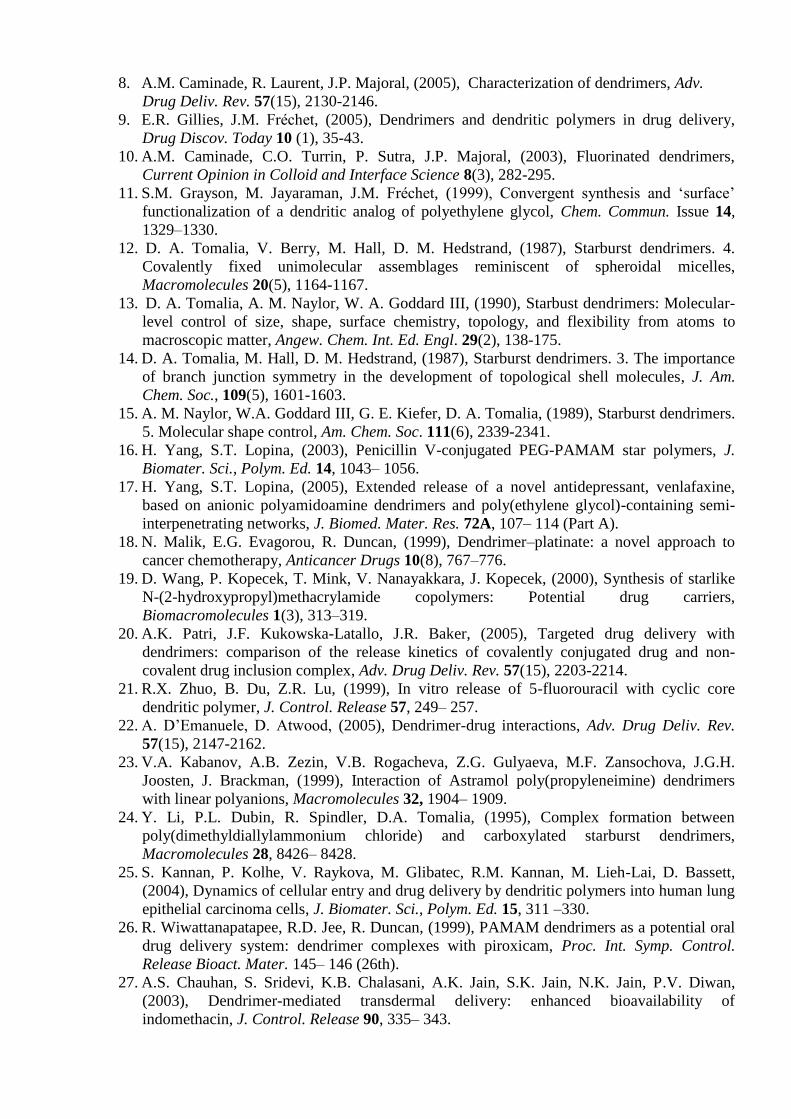
The dendrimers used in this study were cationic carbosilane dendrimers with peripheral ammonium
groups. They were prepared according by reported methods [28, 29]. These dendrimers were 2G-
[Si(OCH2-CH2NMe3+I-)]8 (1), 2G-[Si(OCH2CH2NMe3
+I-)2]8 (2), 2G-
[Si{O(CH2)2N(Me)(CH2)2NMe3+I-}]8 (3) and 2G-[Si{O(CH2)2N(Me)2
+(CH2)2NMe3
+(I
-)2}]8 (4).
The abbreviation 2G means second generation. We have adopted the convention that each
generation is formed on the basis of shell of silicon atoms. Then, starting from the core, this will be
generation zero. The following four silicon atoms form the generation 1, and finally the eight
terminal silicon atoms form the generation 2. This is independently of the number of terminal
groups. We can have a second generation with 8 or 16 groups. They contain one nitrogen atom (N)
per branch (dendrimers 1, 2, called N group dendrimers) or two nitrogens (NN) per branch
(dendrimers 3, 4, called NN group dendrimers). See Figure 1 for molecular structures.
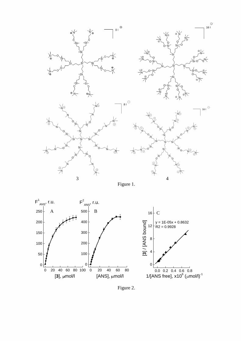
Double fluorimetric titration assay by ANS
ANS was dissolved in phosphate buffer. The excitation and emission wavelengths were set at 370
nm and between 400 and 600 nm respectively. It was established that the dendrimers alone were not
excited by 370 nm irradiation and did not emit fluorescence. The excitation and emission slit widths
were 5 and 2.5 nm, respectively. During measurements the samples were continuously stirred in 1-
cm path length quartz cuvettes.
The binding constant (Kb) and the number of binding sites per dendrimer molecule (n) were
estimated using double fluorimetric titration [32]. In the first fluorimetric titration, increasing
concentrations of dendrimer were added to ANS solution at constant concentration and the
maximum intensity (Fmax) of ANS fluorescence was recorded. This corresponded to the binding of
all the ANS molecules by the dendrimer. The maximum fluorescence intensity of ANS divided by
its concentration gave the specific fluorescence intensity for the bound probe (Fsp):
1
max
ANS
spC
FF , (1)
where (1
ANSC ) is the ANS concentration during the first fluorimetric titration.

In the second titration, the dendrimer concentration (CD) was constant while the ANS
concentration was increased ( 2
ANSC ). The fluorescence intensity (F) was then measured. The
concentration of ANS bound by dendrimers was calculated using the equation:
sp
bound
ANSF
FC , (2)
The concentration of free ANS molecules was determined as follows: bound
ANSANS
free
ANS CCC 2 . (3)
The binding constants for the fluorescent probe molecules (Kb) and the number of ANS binding
centers in the solution (N) could be can be calculated using the formula:
NCNKC free
ANSb
bound
ANS
111, (4)
Equation (4) was modified by replacing the number of binding centers in solution (N) with the
number of binding centers per dendrimer molecule (n), yielding the final equation:
nCnKC
Cfree
ANSb
bound
ANS
D 11, (5)
where
DC
Nn (6)
and the constants were determined from the initial (linear) portions of a graph of bound
ANS
D
C
C versus
free
ANSC
1 (Fig. 1C).
Ethidium bromide intercalation assay
Ethidium bromide and ctDNA were used at final concentrations of 1 μg/ml and 3 μg/ml,
respectively, in 0.15 mol/l Na-phosphate buffer, pH 7.4. The fluorescence spectra of pure EB and of
EB in the presence of DNA were recorded before and after addition of dendrimers. An excitation
wavelength of 477 nm was used. The emission spectra were recorded from 500 to 800 nm. Under
these conditions the fluorescence intensity of pure EB at the emission maximum (618 nm) was
~100 relative units of the device. The fluorescence intensity of EB in the presence of DNA before
and after the dendrimers were added was measured at 604 nm (the emission maximum). The
excitation and emission slit widths were set to 14.0 and 8.0 nm, respectively. The samples were
contained in 1 cm path length quartz cuvettes and were continuously stirred. Before the fluorescent
properties of the DNA–EB–dendrimer complex were examined, it was established that the
dendrimers do not interact with EB.
The data were used to calculate the apparent dendrimer-ctDNA binding (association)
constants (DEN
assK ) using two different equations. The first equation is
50
0
][
][
D
EBKK EB
ass
DEN
ass (7)
where EB
assK is the association constant of EB with ctDNA, [EB]0 is the total concentration of EB in
solution and [D]50 is the concentration that generates a 50% decrease in the initial fluorescence
intensity of the EB-DNA complex [33]. The second equation is
L
assT
I
ass KL
IC
K 1
1 50 (8)
where I
assK is the inhibitor association constant (= DEN
assK ), IC50 is the concentration of inhibitor (I =
dendrimer) necessary to displace 50% of the labeled ligand, LT is the total concentration of the
labeled ligand (EB) and L
assK is the association constant for the labeled ligand (= EB
assK ) [34, 35].
It follows from these equations that
[D]50 = IC50 (9)
In 0.2 mol/l Na-phosphate buffer (pH 7.4), EB
assK was (1.70 0.04)*105 (mol/l)
-1 [36].
To calculate the constants, the data graphs were modified so that the changes in fluorescence
intensity of the EB-ctDNA complex when dendrimers were added were presented as
pureEBcomplex
pureEBcomplexrev
FF
FFF
0
(10),
where Fcomplex
is the fluorescence of EB-ctDNA in the absence and presence of dendrimer, FpureEB
is
the fluorescence of pure (free) EB, and F0complex
is the fluorescence of the EB-ctDNA complex in
the absence of dendrimer when EB is fully bound by the ctDNA.
Fluorescence quenching
BSA was dissolved in 0.15 mol/l Na-phosphate buffer (pH 7.4) at 5 μmol/l. A neutral fluorescence
quencher, acrylamide, and two ionic quenchers, potassium iodide (quenching ion I-) and cesium
chloride (quenching ion Cs+), were used for these studies. Increasing aliquots of the quencher were
added to 5 μmol/l BSA from a stock solution in water. The stock solutions of acrylamide, KI and
CsCl were 1, 5 and 10 mol/l, respectively. The stock KI solution contained 0.1 mmol/l Na2S2O3 to
prevent oxidation of I- to I3
-. The fluorescence intensity at the emission maximum (350 nm) was
measured after excitation at 295 nm. The emission slit width was kept at 10 nm and the excitation
slit width was 3.4 nm. Quenching data were collected for native BSA dissolved in buffer and for
BSA supplemented with 0.1 mmol/l dendrimers.
The quenching results for acrylamide were analyzed by the Stern–Volmer equation:
][10 QKF
FSV (11)
where F0 and F are, respectively, fluorescence intensities in the absence and presence of quencher,
KSV is the Stern–Volmer dynamic quenching constant and [Q] is the concentration of the quencher.
The equation assumes a linear relationship between F0/F and [Q] with a slope of KSV. The Stern–
Volmer constants express the accessibility of the chromophore to the quencher [37].
Statistical analysis
All data are expressed as mean ± S.E.M. of 6 independent experiments. Statistical calculations were
done by Origin 7.0 (OriginLab Corp., USA). The Shapiro-Wilk test was used to ensure normal
distributions. Statistical analysis of the results was done with Student-Fischer test. Also, the
statistical significance of curves was assessed using statistical Box plots including mean, 5% and
95% Wisker lines, 25 and 75 percentiles, and lower and upper confidence intervals (at =0.05).
RESULTS AND DISCUSSION
Double fluorimetric titration using ANS
An aqueous solution of pure ANS fluoresced weakly in the range 400-600 nm with a maximum at
520 nm; its fluorescence yield in a polar environment is low [38]. Figure 2 shows the results of
double fluorimetric titration of ANS and dendrimer 3. Figures 2A and 2B show that the
fluorescence intensity increases non-linearly and reaches a plateau at high dendrimer and ANS
concentrations. The Scatchard curve is approximately linear. Table 1 presents the binding constants,
the number of binding centers, the approximate ANS:dendrimer molar ratio and the spectral shift in
ANS for all the dendrimers studied. Adding carbosilane dendrimers led to both a sharp increase in
fluorescence intensity (Figure 2) and a blue shift of the emission maximum ( max) (Table 1). These

changes indicate that ANS interacts with the dendrimers. When the ANS probe is bound to
hydrophobic sites in membranes or proteins it fluoresces strongly [39], so the results may suggest
that the bound ANS is located in a more hydrophobic environment in the dendrimer. On the other
hand, some authors claim that ANS binds to cationic groups in proteins and that its binding depends
on ion-pair formation [40]. The binding data show that one molecule of an NN group dendrimer
binds one molecule of ANS with a Kb of 105. Second generation dendrimers have open, asymmetric
structures capable of encapsulating ANS. In contrast, the parameter n ranged from 0.1 to 0.3 for the
N group dendrimers, suggesting that micelle formation rather then true molecular binding takes
place. In this case, three or ten dendrimer molecules surround one ANS molecule, changing its
fluorescence parameters. These data show that the NN group dendrimers bind more effectively than
the less reactive N group dendrimers.
Ethidium bromide intercalation assay
EB, a fluorescent phenanthridine dye, is widely used for visualizing nucleic acids [41]. When
the dye interacts with nucleic acids its max is blue-shifted and its fluorescence intensity is
enhanced. It has been confirmed that EB binds to DNA by intercalation [42]. Analytical techniques
including circular dichroism, laser flash photolysis, fluorescence and molecular absorption have
been used to study the intercalation of EB in DNA [43-45]. The extent of fluorescence quenching of
DNA-bound EB has been used to determine the extent of binding between DNA and other ligands
such as metal complexes [46], tannic and ellagic acids [47] and cationic polymers [48, 49].
The emission spectra of DNA-bound EB in the absence and presence of dendrimer 4 are
shown in Figure 3. To exclude interactions between EB and dendrimers, it was shown that the
dendrimers did not affect the shape or intensity of the EB spectrum even at the highest
concentrations used.
The addition of dendrimer to the DNA-EB complex lowered the EB fluorescence emission
intensity, indicating that dendrimer 4 competes with EB for DNA binding. The fluorescence
emission maximum of free EB was 618 nm and that of the EB-DNA complex was 604 nm.
Increasing amounts of 4 caused a concentration-dependent red shift of this maximum from 604 to
618 nm.
Similar results were obtained for all the other dendrimers. In all cases (except for 1) the EB
fluorescence emission intensity was decreased and the EB fluorescence emission maximum was
red-shifted. The results show that all the dendrimers bound to DNA and displaced EB from its sites
of intercalation (Figure 4).
On the basis of the results in Figure 4, the apparent association (binding) constants of three
carbosilane dendrimers were calculated using two similar approaches. These constants are given in
Table 2.
The data show that 3 had an association constant comparable with that of EB while the interaction
of 1 with ctDNA was so weak that it was impossible to estimate its association constant using EB.
In contrast, dendrimers with 16 end groups had greater association constants than EB; 4 showed the
greatest constant among all the dendrimers studied. This indirect means of determining binding
affinity may be misleading, however, because multiple mechanisms for the displacement have been
suggested, including conformational changes that modify base stacking and charge repulsion
between the dye and polyamine polymers.
Quenching of BSA fluorescence emission in presence of dendrimers
BSA has two tryptophan residues. One is located at the bottom of the hydrophobic pocket in
subdomain IIA (Trp213
), and the other is on the surface of the molecule in subdomain IB (Trp134
)
[50]. Three different quenchers, acrylamide, caesium chloride and potassium iodide, were used for
experiments on BSA fluorescence quenching in the absence and presence of dendrimers.
Acrylamide is a neutral polar quencher that can penetrate the interior of proteins by diffusion
because of fluctuations in polypeptide conformation. Both tryptophanyls, Trp134
(at the BSA
surface) and Trp213
(in a hydrophobic pocket), are accessible to acrylamide. Ionic quenchers
suppress tryptophan fluorescence by a heavy ion effect, requiring a direct collision between the ions
(I- or Cs
+) and the excited indole ring. Ionic quenchers are not expected to penetrate into the protein
matrix, so they can only quench tryptophan residues are located on the protein surface [51].
Because both Trps are accessible to acrylamide the quenching results were analyzed using the
Stern–Volmer equation (11).
Figure 5A shows the quenching of BSA fluorescence by acrylamide in the absence and presence of
dendrimers. The quenching curves are linear. Quenching was maximal in dendrimer-free media. In
the presence of N group dendrimers, the quenching process was close to that in controls. In contrast,
the presence of NN group dendrimers decreased the effect of acrylamide. The quenching constants
are presented in Table 3. A possible reason is the attachment of NN group dendrimers to the protein
surface, generating a layer that restricts the accessibility of the protein to the quencher. N group
dendrimers apparently cannot interact effectively with the protein. Figure 5B shows the quenching
of BSA fluorescence by the anionic quencher KI in the absence and presence of dendrimer. The
data show that the quenching process is complex and quasi-linear; it is non-linear in the initial step
(0-0.025 mol/l) but linear over the concentration range 0.025-0.15 mol/l, showing that quenching
has both a static and dynamic nature. Collisional quenching is dynamic while the attachment of the
quencher to the protein may be static. As for acrylamide, N and NN type dendrimers differ. N group
dendrimers affect the quenching process only slightly. In contrast, the addition of NN group
dendrimers at initial concentrations sharply decreases the quenching induced by KI. The
explanation may be the same as for acrylamide: NN group dendrimers attached to the protein
surface prevent any interaction between protein and quencher. Figure 5C shows the quenching of
BSA fluorescence emission by CsCl in the absence and presence of dendrimers. As in the case of
KI, the quenching process is non-linear. CsCl quenches BSA fluorescence only slightly. Addition of
N group dendrimers prevented quenching while addition of NN group dendrimers led to negative
quenching, i.e. the fluorescence intensity increased when CsCl was added. The most likely reason
for this effect is a direct interaction between the CsCl and the NN group dendrimers.
Thus, the results show first that carbosilane dendrimers can bind small molecules. Secondly,
carbosilane dendrimers can interact with DNA and displace EB from it. Thirdly, carbosilane
dendrimers can bind to a protein and decrease its accessibility to other molecules. Taking into
account that ODN-carbosilane dendrimer complexes do not interact with BSA [30], it can be
concluded that NN group carbosilane dendrimers are good candidates for ODN and DNA delivery.
Acknowledgements
This work was supported by grant ERA-NET MNT 2007.
References:
1. R. Duncan, L. Izzo, (2005), Dendrimer biocompatibility and toxicity, Adv. Drug Deliv. Rev.
57(15), 2215-2237.
2. S. Svenson, D.A. Tomalia, (2005), Dendrimers in biomedical applications--reflections on
the field, Adv. Drug Deliv. Rev. 57(15), 2106-2129.
3. D.A. Tomalia, (2004), Birth of a new macromolecular architecture: Dendrimers as quantized
building blocks for nanoscale synthetic organic chemistry, Aldrichimica Acta 37(2), 39-57.
4. D.A. Tomalia, H. Baker, J. Dewald, M. Hall, G. Kallos, S. Martin, J. Roeck, J. Ryder, P.
Smith, (1986), Dendritic macromolecules: Synthesis of starburst dendrimers,
Macromolecules 19(9), 2466-2468.
5. D.A. Tomalia, (1995), Dendrimer molecules, Sci. Am. 272(5), 42-48.
6. M. Ballauff, (2000), Dendrimers III – architecture, nanostructure and supramolecular
chemistry, Topics Curr. Chem. 210, 177-194.
7. Y. Sayed-Sweet, D.M. Hedstrand, R. Spinder, D.A. Tomalia, (1997), Hydrophobically
modified poly(amidoamine) (PAMAM) dendrimers: Their properties at the air-water
interface and use as nanoscopic container molecules, J. Mater. Chem. 7(7), 1199-1205.
8. A.M. Caminade, R. Laurent, J.P. Majoral, (2005), Characterization of dendrimers, Adv.
Drug Deliv. Rev. 57(15), 2130-2146.
9. E.R. Gillies, J.M. Fréchet, (2005), Dendrimers and dendritic polymers in drug delivery,
Drug Discov. Today 10 (1), 35-43.
10. A.M. Caminade, C.O. Turrin, P. Sutra, J.P. Majoral, (2003), Fluorinated dendrimers,
Current Opinion in Colloid and Interface Science 8(3), 282-295.
11. S.M. Grayson, M. Jayaraman, J.M. Fréchet, (1999), Convergent synthesis and ‘surface’
functionalization of a dendritic analog of polyethylene glycol, Chem. Commun. Issue 14,
1329–1330.
12. D. A. Tomalia, V. Berry, M. Hall, D. M. Hedstrand, (1987), Starburst dendrimers. 4.
Covalently fixed unimolecular assemblages reminiscent of spheroidal micelles,
Macromolecules 20(5), 1164-1167.
13. D. A. Tomalia, A. M. Naylor, W. A. Goddard III, (1990), Starbust dendrimers: Molecular-
level control of size, shape, surface chemistry, topology, and flexibility from atoms to
macroscopic matter, Angew. Chem. Int. Ed. Engl. 29(2), 138-175.
14. D. A. Tomalia, M. Hall, D. M. Hedstrand, (1987), Starburst dendrimers. 3. The importance
of branch junction symmetry in the development of topological shell molecules, J. Am.
Chem. Soc., 109(5), 1601-1603.
15. A. M. Naylor, W.A. Goddard III, G. E. Kiefer, D. A. Tomalia, (1989), Starburst dendrimers.
5. Molecular shape control, Am. Chem. Soc. 111(6), 2339-2341.
16. H. Yang, S.T. Lopina, (2003), Penicillin V-conjugated PEG-PAMAM star polymers, J.
Biomater. Sci., Polym. Ed. 14, 1043– 1056.
17. H. Yang, S.T. Lopina, (2005), Extended release of a novel antidepressant, venlafaxine,
based on anionic polyamidoamine dendrimers and poly(ethylene glycol)-containing semi-
interpenetrating networks, J. Biomed. Mater. Res. 72A, 107– 114 (Part A).
18. N. Malik, E.G. Evagorou, R. Duncan, (1999), Dendrimer–platinate: a novel approach to
cancer chemotherapy, Anticancer Drugs 10(8), 767–776.
19. D. Wang, P. Kopecek, T. Mink, V. Nanayakkara, J. Kopecek, (2000), Synthesis of starlike
N-(2-hydroxypropyl)methacrylamide copolymers: Potential drug carriers,
Biomacromolecules 1(3), 313–319.
20. A.K. Patri, J.F. Kukowska-Latallo, J.R. Baker, (2005), Targeted drug delivery with
dendrimers: comparison of the release kinetics of covalently conjugated drug and non-
covalent drug inclusion complex, Adv. Drug Deliv. Rev. 57(15), 2203-2214.
21. R.X. Zhuo, B. Du, Z.R. Lu, (1999), In vitro release of 5-fluorouracil with cyclic core
dendritic polymer, J. Control. Release 57, 249– 257.
22. A. D’Emanuele, D. Atwood, (2005), Dendrimer-drug interactions, Adv. Drug Deliv. Rev.
57(15), 2147-2162.
23. V.A. Kabanov, A.B. Zezin, V.B. Rogacheva, Z.G. Gulyaeva, M.F. Zansochova, J.G.H.
Joosten, J. Brackman, (1999), Interaction of Astramol poly(propyleneimine) dendrimers
with linear polyanions, Macromolecules 32, 1904– 1909.
24. Y. Li, P.L. Dubin, R. Spindler, D.A. Tomalia, (1995), Complex formation between
poly(dimethyldiallylammonium chloride) and carboxylated starburst dendrimers,
Macromolecules 28, 8426– 8428.
25. S. Kannan, P. Kolhe, V. Raykova, M. Glibatec, R.M. Kannan, M. Lieh-Lai, D. Bassett,
(2004), Dynamics of cellular entry and drug delivery by dendritic polymers into human lung
epithelial carcinoma cells, J. Biomater. Sci., Polym. Ed. 15, 311 –330.
26. R. Wiwattanapatapee, R.D. Jee, R. Duncan, (1999), PAMAM dendrimers as a potential oral
drug delivery system: dendrimer complexes with piroxicam, Proc. Int. Symp. Control.
Release Bioact. Mater. 145– 146 (26th).
27. A.S. Chauhan, S. Sridevi, K.B. Chalasani, A.K. Jain, S.K. Jain, N.K. Jain, P.V. Diwan,
(2003), Dendrimer-mediated transdermal delivery: enhanced bioavailability of
indomethacin, J. Control. Release 90, 335– 343.

28. P. Ortega, J.F. Bermejo, L. Chonco, E. de Jesus, F. Javier de la Mata, G. Fernández, J.C.
Flores, R. Gómez, M.J. Serramía, M. Angeles Muñoz-Fernandez, (2006), Novel water-
soluble carbosilane dendrimers: synthesis and biocompatibility, Eur. J. Inorg. Chem. 7,
1388-1396.
29. J.F. Bermejo, P. Ortega, L. Chonco, R. Eritja, R. Samaniego, M. Müllner, E. de Jesus, F.
Javier de la Mata, J.C. Flores, R. Gomez, M. Angeles Muñoz-Fernandez, (2007), Water-
soluble carbosilane dendrimers: synthesis, biocompatibility and complexation with
oligonucleotides; evaluation for medical applications, Chem. Eur. J. 13, 483-495.
30. L. Chonco, J.F. Bermejo-Martin, P. Ortega, D. Shcharbin, E. Pedziwiatr, B. Klajnert, F.
Javier de la Mata, R. Eritja, R. Gomez, M. Bryszewska M. Angeles Muñoz-Fernandez,
(2007), Water-soluble carbosilane dendrimers protect phosphorothioate oligonucleotides
from binding to serum proteins, Org. Biomol. Chem. 5, 1886-1893.
31. D. Shcharbin, E. Pedziwiatr, L. Chonco, J.F. Bermejo-Martin, P. Ortega, F. Javier de la
Mata, R. Eritja,| R. Gomez, B. Klajnert, M. Bryszewska, M. Angeles Muñoz-Fernandez,
(2007), Analysis of interaction between dendriplexes and bovine serum albumin,
Biomacromolecules 8, 2059-2062.
32. D. Shcharbin, B. Klajnert, V. Mazhul, M. Bryszewska, (2003), Estimation of PAMAM
dendrimers’ binding capacity by fluorescent probe ANS, J Fluoresc 13(6), 519–524.
33. Ji-Yan Pang, Yu-Hua Long, Wen-Hua Chen, Zhi-Hong Jiang, (2007) Amplification of
DNA-binding affinities of protoberberine alkaloids by appended polyamines, Bioorganic &
Medicinal Chemistry Letters, 17(4), 1018-1021.
34. Y.C. Cheng, W.H. Prusoff, (1973), Relationship between the inhibition constant (KI) and the
concentration of inhibitor which causes 50 per cent inhibition (I50) of an enzymatic reaction,
Biochem. Pharm. 22, 3099–3108.
35. I.M. Klotz, (1985), Ligand--receptor interactions: facts and fantasies. Q. Rev. Biophys. 18,
227-259.
36. Tzyh-Chyang Tang and Hsuan-Jung Huang, (1999), Electrochemical Studies of the
Intercalation of Ethidium Bromide to DNA, Electroanalysis 11(16), 1185-1190.
37. J.R. Lakowicz (1999) Principles of Fluorescence Spectroscopy 2nd
ed, Kluwer
Academic/Plenum Press, New York.
38. J. Slavik, (1982), Anilinonaphthalene sulfonate as a probe of membrane composition and
function, Biochim. Biophys. Acta 694(1), 1-25.
39. Radda GK. (1972). Fluorescent probes in membrane studies. Biomembranes, 3, 247-266.
40. D. Matulis, Ch. Baumann, V. Bloomfield, R. Lovrien, (1999), 1-anilino-8-naphthalene
sulfonate as a protein conformational tightening agent, Biopolymers 49(6), 451–458.
41. P. Quillardet, M. Hofnung (1988), Ethidium Bromide and Safety- Readers Suggest
Alternative Solutions, Trends Genet. 4, 89-93.
42. C.C. Tsai, S.C. Jain, H.M. Sobel, (1977), Visualization of drug-nucleic acid interactions at
atomic resolution. I. Structure of an ethidium/dinucleoside monophosphate crystalline
complex, ethidium:5-iodouridylyl (3'-5') adenosine, J. Mol. Biol. 114(3), 301-315.
43. W. Chen, N.J. Turro, D.A. Tomalia, (2000), Using ethidium bromide to probe the
interactions between DNA and dendrimers, Langmuir 16(1), 15-19.
44. L.H. Pope, M.C. Davies, C.A. Laughton, C.J. Roberts, S.J.B. Tendler, P.M. Williams
(2000), Atomic force microscopy studies of intercalation-induced changes in plasmid DNA
tertiary structure, Journal of Microscopy, 199 (1) , 68–78.
45. C.Y. Lee, H.-W. Ryu, T.-S. Ko (2001), Binding features of ethidium bromide and their
effects on nuclease susceptibility, Bull. Korean Chem. Soc. 22, 87-89.
46. G.H. Zhao, H.K. Lin, S.R. Zhu, H.W. Sun, Y.T. Chen (1998), Dinuclear palladium(II)
complexes containing two monofunctional [Pd(en)(pyridine)Cl]+ units bridged by Se or S.
Synthesis, characterization, cytotoxicity and kinetic studies of DNA-binding, J. Inorg.
Biochem. 70, 219-226.

47. M. Labieniec, T. Gabryelak, (2006), Interactions of tannic acid and its derivatives (ellagic
and gallic acid) with calf thymus DNA and bovine serum albumin using spectroscopic
method, J. of Photochemistry and Photobiology B: Biology 82, 72-78.
48. Y. Zhou, Y. Li , (2004), Studies of the interactions between poly(diallyldimeythyl
ammonium chloride) and DNA spectroscopic methods, Colloids and Surfaces A:
Physicochem. Eng. Aspects 233, 129 -135.
49. B. Klajnert, W. Walach, M. Bryszewska, A. Dworak, D. Shcharbin, (2006), Cytotoxicity,
haematotoxicity and genotoxicity of high molecular mass arborescent polyoxyethylene
polymers with polyglycidol-block-containing shells, Cell Biology International 30, 248-252.
50. D.C. Carter, X.J. Ho, (1994), Structure of serum albumins, Adv. Protein Chem. 45, 153–203.
51. B. Klajnert, L. Stanislawska, M. Bryszewska, B. Palecz, (2003), Interactions between
PAMAM dendrimers and bovine serum albumin, BBA 1648, 115-126.
Titles of figures
Figure 1. The molecular structure of the dendrimers.
Figure 2. Double fluorimetric titration of ANS vs dendrimer 3: A. The dependence of ANS
fluorescence intensity on the concentration of dendrimer 3 at constant ANS concentration (5
μmol/l). B. The dependence of ANS fluorescence intensity on ANS concentration at constant
concentration of dendrimer 3 (10 μmol/l), C. Scatchard-Klotz plot of [3]/[ANS bound] vs 1/[ANS
free]. exc.= 370 nm, em.= 480 nm.
Figure 3. Fluorescence emission spectra of pure EB (6), EB complexed with ctDNA (1) and EB-
ctDNA complex in the presence of dendrimer 4 at 0.4 mol/l (2), 1 mol/l (3), 2 mol/l (4) and 5
mol/l (5). The spectra at 0.2, 0.6, 3 and 4 mol/l are not presented. INSET shows the dependence
of the fluorescence emission maximum of the EB-ctDNA complex on the concentration of
dendrimer 4 (triangles). The fluorescence emission maximum of pure EB is shown as a circle. [EB]
= 1 g/ml, [DNA] = 3 g/ml, ex. = 477 nm, 0.15 mol/l Na-phosphate buffer, pH 7.4, 37oC.
Figure 4. . The displacement of EB from DNA by carbosilane dendrimers. For details see Materials
and Methods. [EB] = 1 g/ml (2.54 mol/l), [DNA] = 3 g/ml, 0.15 mol/l Na-phosphate buffer, pH
7.4, 37oC. ex. = 477 nm, em. = 604 nm.
Figure 5. Curves of BSA fluorescence quenching by acrylamide (A), KI (B) and CsCl (C) in the
absence and presence of carbosilane dendrimers. [BSA] = 5 μmol/l, [Dendrimer] = 100 μmol/l.
exc. = 295 nm, em. = 350 nm.
1 2
Si
Si
SiSi O
N
O
N
O
N
O
N
Si
SiSi O
N
O
N
O
N
O
N
Si
Si
Si
O
N
ON
O
N
ON
Si
Si
Si
O
N
ON
O
N
ON
16 I
Si Si Si
Si
Si
SiO
Si
O
N
N
NN
SiO
SiO
N
N
NN
SiO
SiO
N
N
N
N
SiO
SiO
N
N
N
N
8 I
Si Si Si
Si
Si
SiO
Si
O
N
N
NN
SiO
SiO
N
N
NN
SiO
SiO
N
N
N
N
16 I
SiO
Si
O
N
N
N
N 3 4
Figure 1.
0 20 40 60 80 100
0
50
100
150
200
250
0 20 40 60 80
0
100
200
300
400
500 B
[ANS], mol/l
F1
ANS, r.u.
[3], mol/l
F2
ANS, r.u.
A
0.0 0.2 0.4 0.6 0.8
0
4
8
12
16
y = 1E-05x + 0.8632
R2 = 0.9928
[3] / [A
NS
bo
un
d]
1/[ANS free], x106 ( mol/l)
-1
C
Figure 2.

550 600 650 700 750 8000
50
100
150
200
250
0 1 2 3 4 5600
604
608
612
616
620
max, nm
[4], M
INSET
3
2
F, r.u.
, nm
1
6
5
4
Figure 3.
2.4 2.8 3.2 3.6 4.00 5000 100000.0
0.5
1.0
[Dendrimer], nmol/l
Log[Dendrimer]
Log IC50
1
2
3
4
pure EB
Frel
Figure 4.

0.00 0.01 0.02 0.03 0.04 0.05 0.06
1.0
1.1
1.2
1.3
1.4
1.5
1.6
Fo/F
, a
.u.
[Acrylamide], mol/l
No dendrimer
1
2
3
4
A
0.00 0.05 0.10 0.150.9
1.0
1.1
1.2
1.3
1.4
1.5
1.6
1.7
1.8
Fo/F
, a
.u.
[KI], mol/l
No dendrimer
1
2
3
4
B
0.0 0.1 0.2 0.3 0.4 0.5 0.6 0.70.5
0.6
0.7
0.8
0.9
1.0
1.1
1.2
Fo/F
, a
.u.
[CsCl], mol/l
No dendrimer
1
2
3
4
C
Figure 5.
Titles of tables.
Table 1. Binding constants, number of binding centers per molecule and blue shift of ANS
fluorescence emission maximum for binding between ANS and water-soluble carbosilane
dendrimers.
Table 2. The association constants of carbosilane dendrimers 2, 3, 4 with ctDNA.
Table 3. The constants of BSA fluorescence quenching by acrylamide in the absence and presence
of carbosilane dendrimers.

Table 1.
Kb * 10-5
, (mol/l)
-1
N,
mol/l
n Dendrimer:ANS (from 520 nm to …)
1 0.7 - 2.0 1 - 3 0.1 - 0.3 8:1 473 1.9 nm
2 0.6 - 2.1 3 - 4 0.3 - 0.4 3:1 470 1.7 nm
3 1.0 - 1.5 8 - 10 0.8 - 1.2 1:1 481 1.4 nm
4 1.0 - 1.4 7 - 13 0.7 - 1.3 1:1 481 1.2 nm
Table 2.
Dendrimer Log IC50
(see Fig.4)
IC50 (= [D]50),
mol/l
DEN
assK *10
-5,
(mol/l)-1
I
assK *10-5
,
(mol/l)-1
2 3.16
(3.06-3.26)*
1.45
(1.15-1.82)
2.98
(2.38-3.76)
3.67
(2.92-4.62)
3 3.76
(3.6-3.93)*
5.75
(3.98-8.51)
0.75
(0.51-1.09)
0.92
(0.62-1.34)
4 2.95
(2.83-3.08)*
0.89
(0.68-1.2)
4.85
(3.6-6.36)
5.98
(4.43-7.82)
* - The upper and lower intervals were obtained from B-splines of two curves (F S.D.) vs Log[Dendrimer] in the same
manner as for F vs Log[Dendrimer] (see Mat&Met).
Table 3.
KSV (Ac), (mol/l)-1
No dendrimer 9.92 1.01
1 9.19 0.94
2 8.89 1.1

3 7.07 0.61
4 6.05 0.71