Water-Soluble Carbosilane Dendrimers: Synthesis Biocompatibility and Complexation with...
13
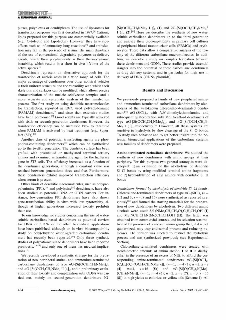
DOI: 10.1002/chem.200600594 Water-Soluble Carbosilane Dendrimers: Synthesis Biocompatibility and Complexation with Oligonucleotides; Evaluation for Medical Applications Jesus F. Bermejo, [a, e] Paula Ortega, [b, e] Louis Chonco, [a] Ramon Eritja, [c] Rafael Samaniego, [d] Matthias Mɒllner, [a] Ernesto de Jesus, [b] F. Javier de la Mata,* [b] Juan Carlos Flores, [b] Rafael Gomez,* [b] and Angeles MuÇoz-Fernandez* [a] Introduction There is currently significant interest in dendrimers as a result of their potential applications, including light harvest- ing and energy transfer, nanoscale catalysis, chemical sen- sors, unimolecular micelles, enzyme mimics, encapsulation of guest molecules, molecular recognition, diagnostic agents, and gene and drug delivery. [1] In particular, the structural precision of dendrimers has motivated numerous studies aimed at biomedical applications. One of the most active re- search areas in dendrimer-based therapeutics is DNA trans- fection. This application is currently based on viral vectors as the most efficient gene-transfer agents. However, the use of viral vectors suffers from adverse effects, such as immune reaction against the viral vector or lymphoproliferative syn- dromes associated with oncogene dysregulation. [2] To over- come these drawbacks, nonviral vehicles such as cationic li- ACHTUNGTRENNUNGpoACHTUNGTRENNUNGsomes, polymers, and dendrimers have been developed. Once in contact with negatively charged DNA oligodeoxy- nucleotides (ODNs) or plasmids, cationic systems form elec- trostatic complexes with the nucleic acids, known as lipo- Abstract: Novel amine- or ammonium- terminated carbosilane dendrimers of type nG-[SiACHTUNGTRENNUNG{OCH 2 ACHTUNGTRENNUNG(C 6 H 3 )-3,5-(OCH 2 - ACHTUNGTRENNUNGCH 2 ACHTUNGTRENNUNGNMe 2 ) 2 }] x , nG-[Si{OACHTUNGTRENNUNG(CH 2 ) 2 N(Me)- ACHTUNGTRENNUNG(CH 2 ) 2 ACHTUNGTRENNUNGNMe 2 }] x and nG-[SiACHTUNGTRENNUNG{(CH 2 ) 3 - ACHTUNGTRENNUNGNH 2 }] x or nG-[SiACHTUNGTRENNUNG{OCH 2 ACHTUNGTRENNUNG(C 6 H 3 )ACHTUNGTRENNUNG-3,5- (OCH 2 ACHTUNGTRENNUNGCH 2 NMe 3 + I) 2 }] x , nG-[Si{O- ACHTUNGTRENNUNG(CH 2 ) 2 N(Me)ACHTUNGTRENNUNG(CH 2 ) 2 NMe 3 + I }] x , and nG-[SiACHTUNGTRENNUNG{(CH 2 ) 3 NH 3 + Cl }] x have been synthesized and characterized up to the third generation by two strategies : 1) alcoholysis of Si Cl bonds with amino alcohols and subsequent quater- nization with MeI, and 2) hydrosilyla- tion of allylamine with Si H bonds of the dendritic systems and subsequent quaternization with HCl. Quaternized carbosilane dendrimers are soluble in water, although degradation is appa- rent due to hydrolysis of Si O bonds. However, dendrimers containing Si C bonds are water-stable. The biocompat- ibility of the second-generation den- drimers in primary cell cultures of pe- ripheral blood mononuclear cells (PBMCs) and erythrocytes have been analyzed, and they show good toxicity profiles over extended periods. In addi- tion, we describe a study on the inter- actions between the different carbosi- lane dendrimers and DNA oligodeoxy- nucleotides (ODNs) and plasmids along with a comparative analysis of their toxicity. They can form complexes with DNA ODNs and plasmids at bio- compatible doses via electrostatic inter- action. Also a preliminary transfection assay has been accomplished. These re- sults demonstrate that the new ammo- nium-terminated carbosilane dendri- ACHTUNGTRENNUNGmers are good base molecules to be considered for biomedical applications. Keywords: carbosilanes · dendri- ACHTUNGTRENNUNGmers · DNA · drug delivery · oligo- ACHTUNGTRENNUNGnucleotides [a] Dr. J.F. Bermejo, L. Chonco, M. Mɒllner, Dr. A. MuÇoz-Fernandez Laboratorio de Inmunobiologȷa Molecular Hospital General Universitario Gregorio MaraÇɃn, Madrid (Spain) Fax: (+ 34) 91-586-8018 E-mail : [email protected] [b] P. Ortega, Dr. E. deJesus, Dr. F. J. dela Mata, Dr. J. C. Flores, Dr. R. Gomez Departamento de Quȷmica InorgƁnica Universidad de AlcalƁ, Campus Universitario 28871 AlcalƁ de Henares (Spain) Fax: (+ 34) 91-885-4683 E-mail : [email protected] [email protected] [c] Dr. R. Eritja Instituto de Biologȷa Molecular de Barcelona, CSIC Jordi Girona, Barcelona (Spain) [d] Dr. R. Samaniego Unidad de Microscopȷa Confocal Hospital General Universitario Gregorio MaraÇɃn, Madrid (Spain) [e] Dr. J.F. Bermejo, P. Ortega Paula Ortega and Jesus F. Bermejo contributed equally to the devel- opment of this work. Supporting Information for this article is available on the WWW under http://www.chemeurj.org/ or from the author. Chem. Eur. J. 2007, 13, 483 – 495 # 2007 Wiley-VCH Verlag GmbH & Co. KGaA, Weinheim 483 FULL PAPER
-
Upload
independent -
Category
Documents
-
view
0 -
download
0
Transcript of Water-Soluble Carbosilane Dendrimers: Synthesis Biocompatibility and Complexation with...

DOI: 10.1002/chem.200600594
Water-Soluble Carbosilane Dendrimers: Synthesis Biocompatibility andComplexation with Oligonucleotides; Evaluation for Medical Applications
Jesus F. Bermejo,[a, e] Paula Ortega,[b, e] Louis Chonco,[a] Ramon Eritja,[c]
Rafael Samaniego,[d] Matthias M6llner,[a] Ernesto de Jesus,[b] F. Javier de la Mata,*[b]
Juan Carlos Flores,[b] Rafael Gomez,*[b] and Angeles MuÇoz-Fernandez*[a]
Introduction
There is currently significant interest in dendrimers as aresult of their potential applications, including light harvest-ing and energy transfer, nanoscale catalysis, chemical sen-sors, unimolecular micelles, enzyme mimics, encapsulationof guest molecules, molecular recognition, diagnostic agents,and gene and drug delivery.[1] In particular, the structuralprecision of dendrimers has motivated numerous studiesaimed at biomedical applications. One of the most active re-search areas in dendrimer-based therapeutics is DNA trans-fection. This application is currently based on viral vectorsas the most efficient gene-transfer agents. However, the useof viral vectors suffers from adverse effects, such as immunereaction against the viral vector or lymphoproliferative syn-dromes associated with oncogene dysregulation.[2] To over-come these drawbacks, nonviral vehicles such as cationic li-ACHTUNGTRENNUNGpo ACHTUNGTRENNUNGsomes, polymers, and dendrimers have been developed.Once in contact with negatively charged DNA oligodeoxy-nucleotides (ODNs) or plasmids, cationic systems form elec-trostatic complexes with the nucleic acids, known as lipo-
Abstract: Novel amine- or ammonium-terminated carbosilane dendrimers oftype nG-[Si ACHTUNGTRENNUNG{OCH2 ACHTUNGTRENNUNG(C6H3)-3,5-(OCH2-ACHTUNGTRENNUNGCH2 ACHTUNGTRENNUNGNMe2)2}]x, nG-[Si{O ACHTUNGTRENNUNG(CH2)2N(Me)-ACHTUNGTRENNUNG(CH2)2ACHTUNGTRENNUNGNMe2}]x and nG-[SiACHTUNGTRENNUNG{(CH2)3-ACHTUNGTRENNUNGNH2}]x or nG-[Si ACHTUNGTRENNUNG{OCH2 ACHTUNGTRENNUNG(C6H3) ACHTUNGTRENNUNG-3,5-(OCH2ACHTUNGTRENNUNGCH2NMe3
+I�)2}]x, nG-[Si{O-ACHTUNGTRENNUNG(CH2)2N(Me)ACHTUNGTRENNUNG(CH2)2NMe3
+I�}]x, andnG-[Si ACHTUNGTRENNUNG{(CH2)3NH3
+Cl�}]x have beensynthesized and characterized up to thethird generation by two strategies:1) alcoholysis of Si�Cl bonds withamino alcohols and subsequent quater-nization with MeI, and 2) hydrosilyla-tion of allylamine with Si�H bonds ofthe dendritic systems and subsequent
quaternization with HCl. Quaternizedcarbosilane dendrimers are soluble inwater, although degradation is appa-rent due to hydrolysis of Si�O bonds.However, dendrimers containing Si�Cbonds are water-stable. The biocompat-ibility of the second-generation den-drimers in primary cell cultures of pe-ripheral blood mononuclear cells(PBMCs) and erythrocytes have beenanalyzed, and they show good toxicity
profiles over extended periods. In addi-tion, we describe a study on the inter-actions between the different carbosi-lane dendrimers and DNA oligodeoxy-nucleotides (ODNs) and plasmidsalong with a comparative analysis oftheir toxicity. They can form complexeswith DNA ODNs and plasmids at bio-compatible doses via electrostatic inter-action. Also a preliminary transfectionassay has been accomplished. These re-sults demonstrate that the new ammo-nium-terminated carbosilane dendri-ACHTUNGTRENNUNGmers are good base molecules to beconsidered for biomedical applications.
Keywords: carbosilanes · dendri-ACHTUNGTRENNUNGmers · DNA · drug delivery · oligo-ACHTUNGTRENNUNGnucleotides
[a] Dr. J. F. Bermejo, L. Chonco, M. M;llner, Dr. A. MuÇoz-FernandezLaboratorio de Inmunobiolog=a MolecularHospital General Universitario Gregorio MaraÇ?n, Madrid (Spain)Fax: (+34)91-586-8018E-mail : [email protected]
[b] P. Ortega, Dr. E. de Jesus, Dr. F. J. de la Mata, Dr. J. C. Flores,Dr. R. GomezDepartamento de Qu=mica InorgGnicaUniversidad de AlcalG, Campus Universitario28871 AlcalG de Henares (Spain)Fax: (+34)91-885-4683E-mail : [email protected]
[c] Dr. R. EritjaInstituto de Biolog=a Molecular de Barcelona, CSICJordi Girona, Barcelona (Spain)
[d] Dr. R. SamaniegoUnidad de Microscop=a ConfocalHospital General Universitario Gregorio MaraÇ?n, Madrid (Spain)
[e] Dr. J. F. Bermejo, P. OrtegaPaula Ortega and Jesus F. Bermejo contributed equally to the devel-opment of this work.
Supporting Information for this article is available on the WWWunder http://www.chemeurj.org/ or from the author.
Chem. Eur. J. 2007, 13, 483 – 495 L 2007 Wiley-VCH Verlag GmbH&Co. KGaA, Weinheim 483
FULL PAPER
plexes, polyplexes or dendriplexes. The use of liposomes fortransfection purposes was first described in 1987.[3] Cationiclipids prepared for this purpose are commercially available(e.g., Cytofectin and Lipofectin). However, they have side-effects such as inflammatory lung reactions,[4] and transfec-tion may fail in the presence of serum. The main drawbackof the use of conventional degradable polymers as deliveryagents, beside their polydispersity, is their thermodynamicinstability, which results in a short in vivo lifetime of theactive species.[5]
Dendrimers represent an alternative approach for thetransfection of nucleic acids in a wide range of cells. Themajor advantage of dendrimers over other nonviral vehiclesis their uniform structure and the versatility with which theirskeletons and surfaces can be modified, which allows precisecharacterization of the nucleic acid/vector complex and amore accurate and systematic analysis of the transfectionprocess. The first study on using dendritic macromoleculesfor transfection, reported in 1993, used polyamidoamine(PAMAM) dendrimers,[6] and since then extensive studieshave been performed.[7] Good results are typically achievedwith sixth- or seventh-generation dendrimers. However, thetransfection efficiency can be increased two- to threefoldwhen PAMAM is activated by heat treatment (e.g., Super-fect (SF)).[8]
Another class of potential transfecting agents are phos-phorus-containing dendrimers,[9] which can be synthesizedup to the twelfth generation. The dendritic surface has beengrafted with protonated or methylated terminal tertiaryamines and examined as transfecting agent for the luciferasegene in 3T3 cells. The efficiency increased as a function ofthe dendrimer generation, although a constant value wasreached between generations three and five. Furthermore,these dendrimers exhibit improved transfection efficiencywhen serum is present.Other kinds of dendritic macromolecules, such as polypro-
pylenimine (PPI),[10] and polylysine[11] dendrimers, have alsobeen studied as potential DNA or ODN carriers. For in-stance, low-generation PPI dendrimers have also showngene-transfection ability in vitro with low cytotoxicity, al-though at higher generations increased toxicity prohibitstheir use.To our knowledge, no studies concerning the use of water-
soluble carbosilane-based dendrimers as potential carriersfor DNA or ODNs or for other biomedical applicationshave been published, although an in vitro biocompatibilitystudy on poly(ethylene oxide)-grafted carbosilane dendri-ACHTUNGTRENNUNGmers has recently been reported.[12] Only three syntheticstudies of polycationic silane dendrimers have been reportedpreviously,[13–15] and only one of them has medical implica-tions.[15]
We recently developed a synthetic strategy for the prepa-ration of new peripheral amine- and ammonium-terminatedcarbosilane dendrimers of type nG-[Si(OCH2CH2NMe2)y]xand nG-[Si(OCH2CH2NMe3
+I�)y]x, and a preliminary evalu-ation of their toxicity and complexation with ODNs was car-ried out, mainly on second-generation dendrimers 2G-
[Si(OCH2CH2NMe3+I�)]8 (1) and 2G-[Si(OCH2CH2NMe3
+
I�)2]8 (2).[16] Here we describe the synthesis of new water-
soluble carbosilane dendrimers up to the third generationand analyze their biocompatibility in primary cell culturesof peripheral blood mononuclear cells (PBMCs) and eryth-rocytes. These data allow a comparative analysis of the tox-icity of the different carbosilane macromolecules. In addi-tion, we describe a study on complex formation betweenthese dendrimers and ODNs. These studies provide essentialinsights into the potential of these carbosilane dendrimersas drug delivery systems, and in particular for their use indelivery of DNA (ODNs, plasmids).
Results and Discussion
We previously prepared a family of new peripheral amine-and ammonium-terminated carbosilane dendrimers by alco-holysis of the well-known chlorosilane-terminated dendri-ACHTUNGTRENNUNGmers[17] nG-(SiCly)x, with N,N-dimethylethanolamine andsubsequent quaternization with MeI to afford dendrimers oftype nG-[Si(OCH2CH2NMe2)y]x and nG-[Si(OCH2CH2N-ACHTUNGTRENNUNGMe3
+I�)y]x, respectively.[16] However, all these systems are
sensitive to hydrolysis by slow cleavage of the Si�O bonds.To study such behavior and to get better insight into the po-tential biomedical applications of the carbosilane systems,new families of dendrimers were prepared.
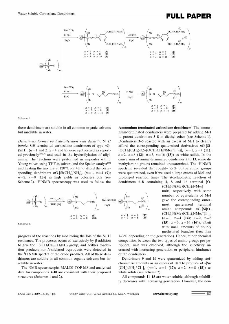
Amine-terminated carbosilane dendrimers : We studied thesynthesis of new dendrimers with amino groups at theirperiphery. For this purpose two general strategies were de-veloped: 1) an extension of the alcoholysis of dendriticSi�Cl bonds by using modified terminal amine fragments,and 2) hydrosilylation of allyl amines with dendritic Si�Hbonds.
Dendrimers formed by alcoholysis of dendritic Si�Cl bonds :Chlorosilane-terminated dendrimers of type nG-(SiCl)x (n=1, 2 and 3; x=4, 8 and 16) were synthesized as reported pre-viously[17] and formed the starting materials for the prepara-tion of new dendrimers by alcoholysis. Two different aminoalcohols were used: 3,5-(NMe2CH2CH2O)2C6H3CH2OH (I)and Me2NCH2CH2N(Me)CH2CH2OH (II). The latter wasobtained from commercial sources, and its selection was mo-tivated by presence of a second amino group that, if it is notquaternized, may trap endosomal protons and reducing nu-cleases. The former was elected to restrict the hydrolysisprocess and was synthesized previously (see ExperimentalSection).Chlorosilane-terminated dendrimers were treated with
stoichiometric amounts of amino alcohol I or II in diethylether in the presence of an excess of NEt3 to afford the cor-responding amine-terminated dendrimers nG-[Si ACHTUNGTRENNUNG{OCH2-ACHTUNGTRENNUNG(C6H3)-3,5-(OCH2CH2NMe2)2}]x (n=1, x=4 (3); n=2, x=8(4); n=3, x=16 (5)) and nG-[Si{O ACHTUNGTRENNUNG(CH2)2N(Me)-ACHTUNGTRENNUNG(CH2)2NMe2}]x (n=1, x=4 (6); n=2, x=8 (7); n=3, x=16(8)) in high yields as colorless or yellow oils (Scheme 1). All
www.chemeurj.org L 2007 Wiley-VCH Verlag GmbH&Co. KGaA, Weinheim Chem. Eur. J. 2007, 13, 483 – 495484
these dendrimers are soluble in all common organic solventsbut insoluble in water.
Dendrimers formed by hydrosilylation with dendritic Si�Hbonds : SiH-terminated carbosilane dendrimers of type nG-(SiH)x (n=1 and 2; x=4 and 8) were synthesized as report-ed previously[17d,e] and used in the hydrosilylation of allyl-ACHTUNGTRENNUNGamine. The reactions were performed in ampoules with JYoung valves using THF as solvent and the Speier catalyst[18]
and heating the mixture at 120 8C for 4 h to afford the corre-sponding dendrimers nG-[Si ACHTUNGTRENNUNG(CH2)3NH2]x (n=1, x=4 (9);n=2, x=8 (10)) in high yields as colorless oils (seeScheme 2). 1H NMR spectroscopy was used to follow the
progress of the reactions by monitoring the loss of the Si�Hresonance. The processes occurred exclusively by b-additionto give the �SiCH2CH2CH2NH2 group, and neither a-addi-tion products nor N-silylated byproducts were detected inthe 1H NMR spectra of the crude products. All of these den-drimers are soluble in all common organic solvents but in-soluble in water.The NMR spectroscopic, MALDI-TOF MS and analytical
data for compounds 3–10 are consistent with their proposedstructures (Schemes 1 and 2).
Ammonium-terminated carbosilane dendrimers : The ammo-nium-terminated dendrimers were prepared by adding MeIto parent dendrimers 3–8 in diethyl ether (see Scheme 1).Dendrimers 3–5 reacted with an excess of MeI to cleanlyafford the corresponding quaternized derivatives nG-[Si-ACHTUNGTRENNUNG{OCH2ACHTUNGTRENNUNG(C6H3)-3,5-(OCH2CH2NMe3
+I�)2}]x (n=1, x=4 (11);n=2, x=8 (12); n=3, x=16 (13)) as white solids. In theconversion of amine-terminated dendrimer 5 to 13, some di-methylamino groups remained unquaternized. The 1H NMRspectrum revealed that roughly 85% of the amino groupswere quaternized, even if we used a large excess of MeI andprolonged reaction times. The stoichiometric reaction ofdendrimers 6–8 containing 4, 8 and 16 terminal [O-
ACHTUNGTRENNUNG(CH2)2N(Me)ACHTUNGTRENNUNG(CH2)2NMe2]units, respectively, with samenumber of equivalents of MeIgave the corresponding outer-most quaternized terminalamine compounds nG-[Si{O-ACHTUNGTRENNUNG(CH2)2N(Me)ACHTUNGTRENNUNG(CH2)2NMe3
+}I�]x(n=1, x=4 (14); n=2, x=8(15); n=3, x=16 (16)), albeitwith small amounts of doublymethylated branches (less than
1–3% depending on the generation). Hence, minor chemicalcompetition between the two types of amino groups per pe-ripheral unit was observed, although the selectivity in-creased with increasing generation or peripheral hindranceof the dendrimers.Dendrimers 9 and 10 were quaternized by adding stoi-
chiometric amounts or an excess of HCl to produce nG-[Si-ACHTUNGTRENNUNG(CH2)3NH3
+Cl�]x (n=1, x=4 (17); n=2, x=8 (18)) aswhite solids (see Scheme 2).All compounds 11–18 are water-soluble, although solubili-
ty decreases with increasing generation. However, the den-
Scheme 1.
Scheme 2.
Chem. Eur. J. 2007, 13, 483 – 495 L 2007 Wiley-VCH Verlag GmbH&Co. KGaA, Weinheim www.chemeurj.org 485
FULL PAPERWater-Soluble Carbosilane Dendrimers
drimers with Si�O bonds decomposed slowly by hydrolysisof these bonds. This behavior was observed in dendrimers 1and 2 with �OCH2CH2NMe2 terminal units.
[16] However, thehydrolysis rates of dendrimers 11–13 containing quaternizedgroups derived from amino alcohol I are considerably atte-nuated with respect to dendrimers with fragment II as pe-ripheral groups, which are similar to those in 1 and 2. Incontrast, dendrimers 17 and 18 based on Si�C bonds arecompletely stable towards hydrolysis.The NMR spectroscopic and analytical data of 11–18 are
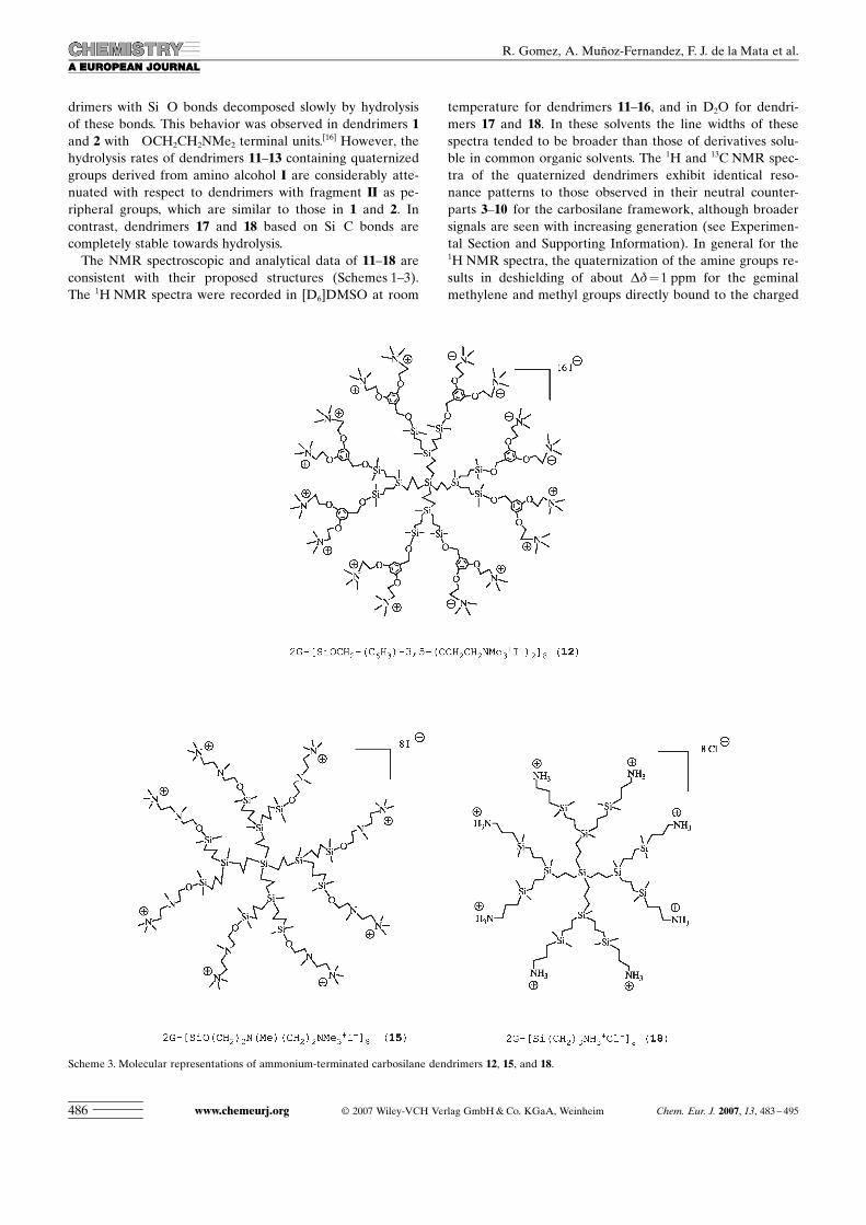
consistent with their proposed structures (Schemes 1–3).The 1H NMR spectra were recorded in [D6]DMSO at room
temperature for dendriACHTUNGTRENNUNGmers 11–16, and in D2O for dendri-mers 17 and 18. In these solvents the line widths of thesespectra tended to be broader than those of derivatives solu-ble in common organic solvents. The 1H and 13C NMR spec-tra of the quaternized dendrimers exhibit identical reso-nance patterns to those observed in their neutral counter-parts 3–10 for the carbosilane framework, although broadersignals are seen with increasing generation (see Experimen-tal Section and Supporting Information). In general for the1H NMR spectra, the quaternization of the amine groups re-sults in deshielding of about Dd=1 ppm for the geminalmethylene and methyl groups directly bound to the charged
Scheme 3. Molecular representations of ammonium-terminated carbosilane dendrimers 12, 15, and 18.
www.chemeurj.org L 2007 Wiley-VCH Verlag GmbH&Co. KGaA, Weinheim Chem. Eur. J. 2007, 13, 483 – 495486
R. Gomez, A. MuÇoz-Fernandez, F. J. de la Mata et al.
nitrogen atoms, whereas small downfield shifts of aroundDd=0.3–0.4 ppm are found for the vicinal methylenegroups. Beyond these positions, no displacement is observedfor the chemical shift due to the positive charge on the ni-trogen atoms. However, this effect is more evident in thecompounds quaternized by MeI than those prepared withHCl. Analogous behavior is observed for the carbon atomsin the 13C NMR spectra. Dendrimers 14–16 with outermostquaternized amines exhibit different 1H and 13C NMR pat-terns for singly and doubly methylated branches that facili-tate their discrimination (see Experimental Section and Sup-porting Information). In any case, signals attributed to themonomethylated forms of the innermost quaternized aminewere observed, even when a deficit of reagent was added.This strongly suggests that the quaternization process startsat the outermost amine and subsequently proceeds to the in-nermost when a slight excess of MeI is added. Attempts tocarry out MALDI-TOF MS of these dendrimers failed prob-ably due, inter alia, to solubility problems.
Toxicity evaluation of dendrimers : Quaternized second-gen-eration carbosilane dendrimers 12, 15, and 18 were testedon a primary cell culture of peripheral blood mononuclearcells (PBMCs) from healthy donors as an initial screeningfor biocompatibility, and the values compared with prelimi-nary data shown by dendrimers 1 and 2.[16] First-generationdendrimer 14 was too water sensitive for toxicity evaluation,while third-generation dendrimers were not tested due tosolubility problems.Toxicity was evaluated by challenging PBMCs with in-
creasing concentrations of the free quaternized carbosilanedendrimers, in order to obtain a range of biocompatibility.Toxicity was initially evaluated by 1) visual examinationunder a phase-contrast light microscope and 2) MTT toxicityassay.According to microscopy studies (see Supporting Infor-
mation), the carbosilane dendrimers best tolerated byPBMCs were 12 and 15, whereas the dendrimer 18 showedthe highest toxicity and dendrimers 1 and 2 had intermedi-ate toxicity profiles. Superfect (SF) and a 4G-PAMAMshowed higher toxicity than all second-generation carbosi-lane dendrimers. However, this is not surprising because ofit is known that dendrimer toxicity increases with increasinggeneration.[1i, 19]
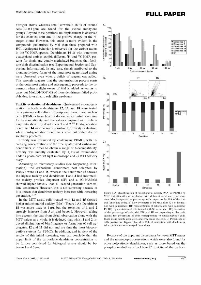
In the MTT assay, cells treated with 12 and 15 showedhigher mitochondrial activity (MA) (Figure 1A). Dendrimer18 was more toxic at 1 mm, but the toxicities of 1 and 2strongly increase from 5 mm and beyond. However, takinginto account the data from visual observation along with theMTT values as a whole, it is deduced that whilst 1 and 2 in-duced diminution of birefringence or formation of cell ag-gregates, 12 and 15 did not and are thus the most biocom-patible systems for PBMCs. In addition, and in view of theresults of this initial screening, one can conclude that theupper limit of the carbosilane dendrimer concentration tobe further considered for biological assays should be be-tween 1 and 5 mm.
Because of the apparent discrepancy between MTT assaysand the microscopic observations, which were also found forother polycationic dendrimers, such as those based on thephosphoramidothioate backbone,[20] toxicity of the carbosi-
Figure 1. A) Quantification of mitochondrial activity (MA) of PBMCs byMTT test after 48 h of incubation with different dendrimer concentra-tions. MA is expressed as percentage with respect to the MA of the con-trol (untreated cells). B) Flow cytometry of PBMCs after 72 h of incuba-tion with dendrimers: B1) representation of cells treated with dendrimer15 ; B2) representation of cells treated with SF dendrimer; B3) evaluationof the percentage of cells with FW and SD corresponding to live cellsagainst the percentage of cells corresponding to dead/apoptotic cells.Black areas denote dead cells, and grey areas live cells. C) Percentage ofcells positive for Trypan Blue after 72 h of incubation with dendrimers.All experiments were assayed three times.
Chem. Eur. J. 2007, 13, 483 – 495 L 2007 Wiley-VCH Verlag GmbH&Co. KGaA, Weinheim www.chemeurj.org 487
FULL PAPERWater-Soluble Carbosilane Dendrimers
lane dendrimers was studied by additional methods: flow cy-tometry, Trypan Blue (TB) uptake, DAPI staining and invivo microscopy. In these studies, the incubation time wasincreased to 72 h, and the doses assayed for carbosilane den-drimers were in the range of 2–4 mm, while less than 1 mmwas used for the reference SF PAMAM dendrimer.
Flow cytometry (FC): PBMCs treated with carbosilane den-drimers at these doses did not show significant changes intheir forward (FW) and side (SD) light-scattering character-istics after 72 h of incubation. Moreover, the percentage ofunviable cells was similar in all cases assayed to that of un-treated cells (around 20%, see Figure 1B). On the otherhand, PBMCs treated with SF dramatically increased theirmortality.
Trypan Blue (TB) uptake : TB is excluded by viable cells butcan penetrate cell membranes of dying or dead cells. WhenTB staining is negative, membrane integrity is present. Thepercentage of cells positive for TB staining for the wellstreated with dendrimers 1, 2, 12, 15, and 18 was similar tothat found in untreated cells (Figure 1C). However, cellstreated with SF displayed significant mortality comparedwith control cells.
DAPI staining : An additional test of cell viability was pro-vided by the staining of cell nuclei with the vital dye DAPI.Cell nuclei undergoing apoptosis or necrosis show reducednuclear size, chromatin condensation and nuclear fragmen-tation, processes that can be easily detected by DAPI stain-ing. Cells treated with all carbosilane dendrimers had similarappearances to untreated cells, showing rounded nuclei withhomogeneously distributed chromatin. In contrast, cellstreated with SF decreased dramatically in number. Further-more, the cell pellet obtained for DAPI staining after centri-fugation of PBMCs treated with SF was notably small. Thisreduction in cell number was confirmed by microscopy (seeSupporting Information).
Time-lapse video microscopy : We monitored cell behaviorby time-lapse and in vivo microscopy for cells treated withcarbosilane dendrimers. In all cases, cells showed similarpatterns of movement and migration to untreated controlcells (for an example, see Supporting Information).Therefore, from these four additional methods, all carbo-
silane dendrimers 1, 2, 12, 15, and 18 assayed under theseconditions showed good biocompatibility on PBMC cells.Toxicity was also evaluated on erythrocytes challenged
with increased dendrimer concentrations by means of induc-tion of hemagglutination along with hemoglobin releasefrom the red blood cells. Dendrimers 1, 2, 12, and 15 wereused for this assay, and dendrimer 18 was not included be-cause it was shown to be the most toxic for lymphocytes insome of the methods and concentrations employed before.A fourth-generation PAMAM dendrimer was also used forcomparison. From visual examination, dendrimer 15 showedthe lowest agglutination, while for dendrimer 12 morpholog-
ic changes were observed even at 1 mm concentration. Anal-ogous results were detected for 4G PAMAM dendrimer(see Supporting Information).The interaction of cationic dendrimers with negatively
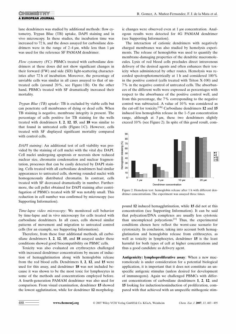
charged membranes was also studied by hemolysis experi-ments. The release of hemoglobin was used to quantify themembrane-damaging properties of the dendritic macromole-cules. Lysis of red blood cells precludes direct intravenousdelivery of the desired agents and often enhances their tox-icity when administered by other routes. Hemolysis was re-corded spectrophotometrically at 1 h and considered 100%in the positive control (cells treated with Triton X-100) and7% in the negative control of untreated cells. The absorban-ces of the different wells were expressed as percentages withrespect to the absorbance of the positive control well, andfrom this percentage, the 7% corresponding to the negativecontrol was subtracted. A value of 10% was considered asthe cut-off for toxicity.[19b] Carbosilane dendrimers 12 and 15induced less hemoglobin release in the 1–5 mm concentrationrange, although at 5 mm, these two dendrimers slightlyexceed 10% (see Figure 2). In spite of this good result, com-
pound 12 induced hemagglutination, while 15 did not at thisconcentration (see Supporting Information). It can be saidthat polycation/DNA complexes are usually less cytotoxicthan uncomplexed polycations.[21] Thus, the experimentalconditions chosen here reflect the worst-case scenario forcytotoxicity. In conclusion, taking into account both hemag-glutination and hemoglobin release from erithrocytes, aswell as toxicity in lymphocytes, dendrimer 15 is the leastharmful for both types of cell at higher concentrations andthus a good candidate as delivery agent.
Antigenicity: lymphoproliferative assay : When a new mac-romolecule is under consideration for a potential biologicalapplication, it is important that it does not constitute an un-specific antigenic stimulus (unless desired for developmentof immunogens). Again we challenged PBMCs with differ-ent concentrations of carbosilane dendrimers 1, 2, 12, and15 looking for induction/noninduction of proliferation, com-pared with that achieved with an unspecific mithogenic stim-
Figure 2. Hemolysis test: hemoglobin release after 1 h with different den-drimer concentrations. The experiment was assayed three times.
www.chemeurj.org L 2007 Wiley-VCH Verlag GmbH&Co. KGaA, Weinheim Chem. Eur. J. 2007, 13, 483 – 495488
R. Gomez, A. MuÇoz-Fernandez, F. J. de la Mata et al.
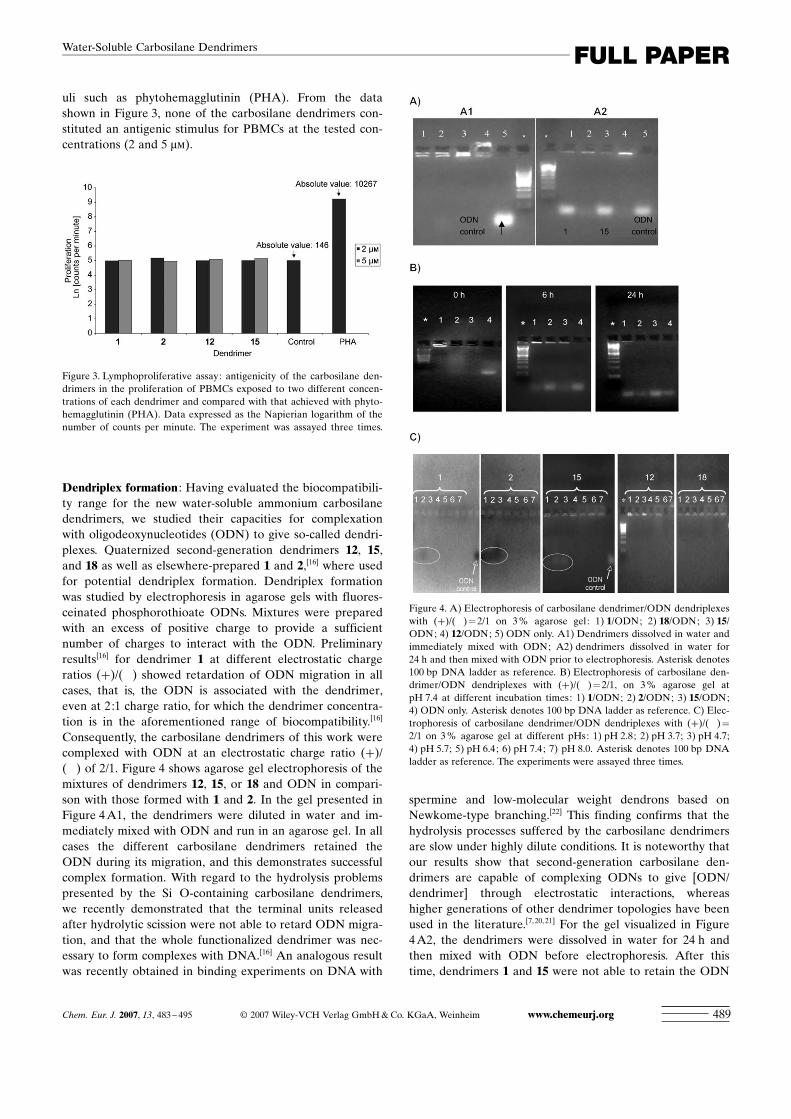
uli such as phytohemagglutinin (PHA). From the datashown in Figure 3, none of the carbosilane dendrimers con-stituted an antigenic stimulus for PBMCs at the tested con-centrations (2 and 5 mm).
Dendriplex formation : Having evaluated the biocompatibili-ty range for the new water-soluble ammonium carbosilanedendrimers, we studied their capacities for complexationwith oligodeoxynucleotides (ODN) to give so-called dendri-plexes. Quaternized second-generation dendrimers 12, 15,and 18 as well as elsewhere-prepared 1 and 2,[16] where usedfor potential dendriplex formation. Dendriplex formationwas studied by electrophoresis in agarose gels with fluores-ceinated phosphorothioate ODNs. Mixtures were preparedwith an excess of positive charge to provide a sufficientnumber of charges to interact with the ODN. Preliminaryresults[16] for dendrimer 1 at different electrostatic chargeratios (+)/(�) showed retardation of ODN migration in allcases, that is, the ODN is associated with the dendrimer,even at 2:1 charge ratio, for which the dendrimer concentra-tion is in the aforementioned range of biocompatibility.[16]
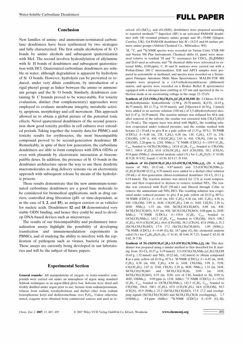
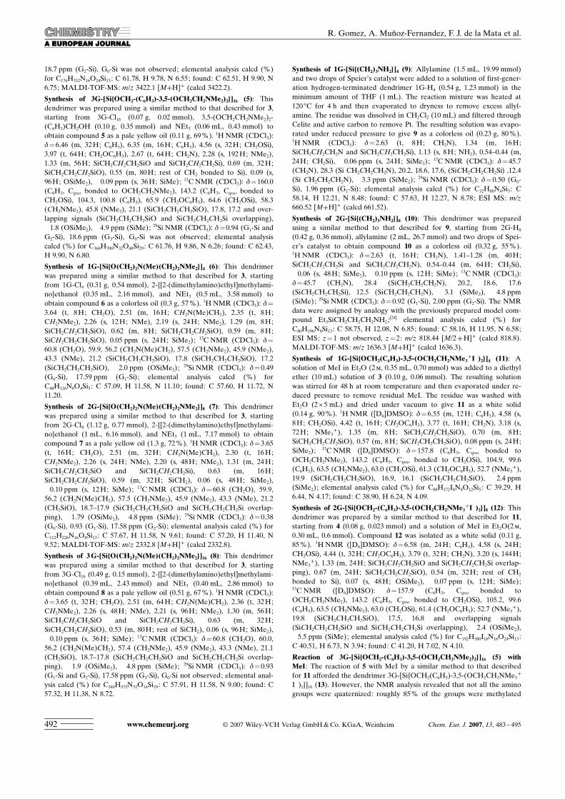
Consequently, the carbosilane dendrimers of this work werecomplexed with ODN at an electrostatic charge ratio (+)/(�) of 2/1. Figure 4 shows agarose gel electrophoresis of themixtures of dendrimers 12, 15, or 18 and ODN in compari-son with those formed with 1 and 2. In the gel presented inFigure 4A1, the dendrimers were diluted in water and im-mediately mixed with ODN and run in an agarose gel. In allcases the different carbosilane dendrimers retained theODN during its migration, and this demonstrates successfulcomplex formation. With regard to the hydrolysis problemspresented by the Si�O-containing carbosilane dendrimers,we recently demonstrated that the terminal units releasedafter hydrolytic scission were not able to retard ODN migra-tion, and that the whole functionalized dendrimer was nec-essary to form complexes with DNA.[16] An analogous resultwas recently obtained in binding experiments on DNA with
spermine and low-molecular weight dendrons based onNewkome-type branching.[22] This finding confirms that thehydrolysis processes suffered by the carbosilane dendrimersare slow under highly dilute conditions. It is noteworthy thatour results show that second-generation carbosilane den-drimers are capable of complexing ODNs to give [ODN/dendrimer] through electrostatic interactions, whereashigher generations of other dendrimer topologies have beenused in the literature.[7,20, 21] For the gel visualized in Figure4A2, the dendrimers were dissolved in water for 24 h andthen mixed with ODN before electrophoresis. After thistime, dendrimers 1 and 15 were not able to retain the ODN
Figure 3. Lymphoproliferative assay: antigenicity of the carbosilane den-drimers in the proliferation of PBMCs exposed to two different concen-trations of each dendrimer and compared with that achieved with phyto-hemagglutinin (PHA). Data expressed as the Napierian logarithm of thenumber of counts per minute. The experiment was assayed three times.
Figure 4. A) Electrophoresis of carbosilane dendrimer/ODN dendriplexeswith (+)/(�)=2/1 on 3% agarose gel: 1) 1/ODN; 2) 18/ODN; 3) 15/ODN; 4) 12/ODN; 5) ODN only. A1) Dendrimers dissolved in water andimmediately mixed with ODN; A2) dendrimers dissolved in water for24 h and then mixed with ODN prior to electrophoresis. Asterisk denotes100 bp DNA ladder as reference. B) Electrophoresis of carbosilane den-drimer/ODN dendriplexes with (+)/(�)=2/1, on 3% agarose gel atpH 7.4 at different incubation times: 1) 1/ODN; 2) 2/ODN; 3) 15/ODN;4) ODN only. Asterisk denotes 100 bp DNA ladder as reference. C) Elec-trophoresis of carbosilane dendrimer/ODN dendriplexes with (+)/(�)=2/1 on 3% agarose gel at different pHs: 1) pH 2.8; 2) pH 3.7; 3) pH 4.7;4) pH 5.7; 5) pH 6.4; 6) pH 7.4; 7) pH 8.0. Asterisk denotes 100 bp DNAladder as reference. The experiments were assayed three times.
Chem. Eur. J. 2007, 13, 483 – 495 L 2007 Wiley-VCH Verlag GmbH&Co. KGaA, Weinheim www.chemeurj.org 489
FULL PAPERWater-Soluble Carbosilane Dendrimers

during its migration, that is, the time dissolved in water af-fects the ability of the dendrimer to form complexes withthe ODN. On the other hand, 12 and 18 were not affectedto the same degree and preserved the capacity to retain themajority of the ODN during its migration. Thus, under theseconditions dendrimers containing Si�O bonds lost their abil-ity to bind ODNs after 24 h in water in dilute concentra-tions, except for dendrimer 12. Likewise, dendrimer 18 isalso water-stable because of the presence of robust Si�Cbonds.For water-sensitive dendrimers 1, 2, and 15, a second type
of electrophoretic migration assay was developed consistingof running dendriplexes in a gel after 0, 6, and 24 h of incu-bation in an atmosphere of 5% of CO2 at 37 8C. As can seefrom Figure 4B, the three dendrimers released the ODNsprogressively. It is possible to conclude that 1, 2, and 15have the ability to release the ODN in a time-dependantway when they are dissolved in water. This feature suggestspotential use in controlled release of ODNs and perhaps ofother polyanionic drugs. The controlled release of activesubstances based on the chemical stability of the linker to-wards hydrolysis has been described, for example, in thecase of phosphorus dendrimers on the basis of slow degrada-tion of imine bonds.[23]
Dendriplex stability was also tested at different pHvalues. Blood physiological pH is 7.4, but anatomical or cel-lular locations with more acidic (stomach, endosome/lyso-some) or basic (duodeni) pH exist. Dendriplexes wereformed as usual and exposed to different solutions fromacid to basic pH (2.8, 3.7, 4.7, 5.7, 6.4, 7.4 and 8.0) priorelectrophoresis (see Figure 4C). Dendriplexes formed be-tween 12 or 18 and ODN were stable at all tested pHvalues, whereas dendriplexes formed between 15 and ODNreleased the ODN at acid pH (<5.7). For dendrimers 1 and2 the results were similar: dendriplexes 1/ODN dissociatedat pH<4.7, and dendriplexes 2/ODN at pH<5.7. All den-driplexes were stable at basic pH (up to pH 8.0). Thus, in anacidic environment dendriplexes 1/ODN, 2/ODN and 15/ODN would release the ODN, whereas under basic condi-tions they would remain stable. This opens new perspectivesfor applications that need pH-controlled ODN release.Toxicity profiles of dendriplexes formed by carbosilane
dendrimers 1, 2, 12, 15, and 18 were studied by some of themethods used for dendrimers alone: flow cytometry andTrypan Blue (TB) uptake (see Supporting Information). Thedata showed very similar values to those obtained for den-drimers without complexation to ODN, as has been reportedelsewhere.[19]
In addition, the ammonium-terminated second-generationcarbosilane dendrimers were able to complex with plasmids.As a demonstrative example, dendrimer 1 formed dendri-plexes with the NfkB plasmid (which codifies Nf-kappaBprotein involved in regulation of immune or inflammationresponses). This was again true even at 2:1 charge ratio (seeFigure 5). This plasmid has an approximate length of 5000base pairs, determined on the basis of its comparative migra-tion with the DNA ladder as reference. This result shows
that, regardless of the low generation, the carbosilane den-drimers have the capacity to bind large DNA molecules.Finally, a preliminary study of the capacity of the dendri-
plex formed by 15 and the fluoresceinated ODN to pene-trate into PBMCs was performed by confocal microscopy.Figure 6 shows the internalization and intracellular distribu-tion of the nucleic material, with which the dendrimer doesnot seem to interfere. Research is in progress to elucidatethe transfection process.
Figure 5. Electrophoresis of dendrimer 1/NfkB plasmid dendriplexes on3% agarose gel: 1) (+)/(�)=2/1; 2) (+)/(�)=6/1; 3) (+)/(�)=10/1;4) (+)/(�)=100/1; 5) and 6) plasmid only. Asterisk denotes 5000 bpDNA ladder as reference. The experiment was assayed three times.
Figure 6. A) Confocal micrograph of internalization of 15/ODN dendri-plex after 48 h. B) Image of an isolated cell; white line denotes a sectionthrough the median plane XY. C) Plots of fluorescence emission throughthe section: green (fluoresceinated ODN), blue (cell nucleus) and red(cell membrane).
www.chemeurj.org L 2007 Wiley-VCH Verlag GmbH&Co. KGaA, Weinheim Chem. Eur. J. 2007, 13, 483 – 495490
R. Gomez, A. MuÇoz-Fernandez, F. J. de la Mata et al.
Conclusion
New families of amine- and ammonium-terminated carbosi-lane dendrimers have been synthesized by two strategiesand fully characterized. The first entails alcoholysis of Si�Clbonds by amino alcohols and subsequent quaternizationwith MeI. The second involves hydrosilylation of allylaminewith Si�H bonds of dendrimers and subsequent quaterniza-tion with HCl. Quaternized carbosilane dendrimers are solu-ble in water, although degradation is apparent by hydrolysisof Si�O bonds. However, hydrolysis can be prevented or re-duced, under very dilute conditions, by introduction of arigid phenyl group as linker between the amino or ammoni-um groups and the Si�O bonds. Similarly, dendrimers con-taining Si�C bounds proved to be water-stable. For toxicityevaluation, distinct (but complementary) approaches wereemployed to evaluate membrane integrity, metabolic activi-ty, apoptosis, morphology and cell movement. This approachallowed us to obtain a global picture of the potential toxiceffects. Novel quaternized dendrimers of the second genera-tion show good toxicity profiles in cell cultures over extend-ed periods. Taking together the toxicity data for PBMCs andtoxicity results for erythrocytes, the most biocompatiblecompound proved to be second-generation dendrimer 15.Remarkably, in spite of their low generation, the carbosilanedendrimers are able to form complexes with DNA ODNs oreven with plasmids by electrostatic interaction at biocom-patible doses. In addition, the presence of Si�O bonds in thedendrimer architecture opens the way to use these dendriticmacromolecules as drug delivery systems via an electrostaticapproach with subsequent release by means of the hydrolyt-ic process.These results demonstrate that the new ammonium-termi-
nated carbosilane dendrimers are a good base molecule tobe considered for biomedical applications, such as drug car-riers, controlled drug liberation (pH- or time-dependant, asin the case of 1, 2, and 15), as antigen carriers or as vehiclesfor nucleic acids. Dendrimers 12 and 18 demonstrated verystable ODN binding, and hence they could be used to devel-op DNA-based devices such as microarrays.The results of our biocompatibility and preliminary inter-
nalization assays highlight the possibility of developingtransfection and immunomodulation experiments inPBMCs, and of studying the ability to interfere with the rep-lication of pathogens such as viruses, bacteria or prions.These assays are currently being developed in our laborato-ries and will be the subject of future papers.
Experimental Section
General remarks : All manipulations of oxygen- or water-sensitive com-pounds were carried out under an atmosphere of argon using standardSchlenk techniques or an argon-filled glove box. Solvents were dried andfreshly distilled under argon prior to use: hexane from sodium/potassium,toluene from sodium, tetrahydrofuran and diethyl ether from sodiumbenzophenone ketyl and dichloromethane over P4O10. Unless otherwisestated, reagents were obtained from commercial sources and used as re-
ceived. nG-(SiClx)y and nG-(SiH)x dendrimers were prepared accordingto reported methods.[17] Superfect (SF) is an activated PAMAM dendri-mer with 140 terminal primary amino groups and M=35000 (Qiagen,Crawley, UK). G4 PAMAM dendri ACHTUNGTRENNUNGmer has M=14215 and 64 surface pri-mary amino groups (Aldrich Chemical Co., Milwaukee, WI).1H, 13C, and 29Si NMR spectra were recorded on Varian Unity VXR-300and Varian 500 Plus Instruments. Chemical shifts (d, ppm) were meas-ured relative to residual 1H and 13C resonances for CDCl3, [D6]DMSOand D2O used as solvents, and
29Si chemical shifts were referenced to ex-ternal SiMe4 (0.00 ppm). C, H and N analyses were carried out with aPerkin-Elmer 240 C microanalyzer. ESI and APCI samples were pre-pared in acetonitrile or methanol, and spectra were recorded on a Termo-quest Finnigan Automass Multi Mass Spectrometer. MALDI-TOF MSsamples were prepared in a 1,8,9-trihydroxyanthracene (dithranol)matrix, and spectra were recorded on a Bruker Reflex II spectrometerequipped with a nitrogen laser emitting at 337 nm and operated in the re-flection mode at an accelerating voltage in the range 23–25 kV.
Synthesis of [3,5-(NMe2CH2CH2O)2] ACHTUNGTRENNUNG(C6H3)CH2OH (I): 2-Chloro-N,N-di-methylethylamine hydrochloride (2.98 g, 20.78 mmol), K2CO3 (6.43 g,46.75 mmol), KI (1.72 g, 10.39 mmol), and [18]crown-6 (0.54 g, 2 mmol)were added to an acetone solution (100 mL) of 3,5-dihydroxybenzyl alco-hol (1.47 g, 10.39 mmol). The reaction mixture was refluxed for 48 h and,after removal of the solvent, the residue was extracted with CH2Cl2/H2O(2T50 mL). The organic layer was dried with MgSO4, the resulting solu-tion evaporated under reduced pressure, and the residue washed withhexane (2T10 mL) to give I as a pale yellow oil (1.53 g, 50%). 1H NMR(CDCl3): d=6.48 (m, 2H; C6H3), 6.36 (m, 1H; C6H3), 4.57 (s, 2H;CH2OH), 3.99 (t, 4H; CH2OC6H3), 2.65 (t, 4H; CH2N), 2.60 (s, 1H;CH2OH), 2.28 ppm (s, 12H; NMe2);
13C NMR (CDCl3): d=159.9 (C6H3,Cipso bonded to OCH2CH2NMe2), 143.8 (C6H3, Cipso bonded to CH2OSi),105.1, 100.6 (C6H3), 65.8 (CH2OC6H3), 64.9 (CH2OH), 58.2 (CH2N),45.8 ppm (NMe2); elemental analysis calcd (%) for C15H26N2O3: C 63.80,H 9.28, N 9.92; found: C 63.50, H 9.17, N 9.83.
Synthesis of 1G-[Si ACHTUNGTRENNUNG{OCH2 ACHTUNGTRENNUNG(C6H3)-3,5-(OCH2CH2NMe2)2}]4 (3): A slightexcess of NEt3 (0.12 mL, 0.87 mmol) and 3,5-(OCH2CH2NMe2)2-ACHTUNGTRENNUNG(C6H3)CH2OH (0.22 g, 0.76 mmol) were added to a diethyl ether solution(50 mL) of first-generation chloro-terminated dendrimer 1G-Cl4 (0.11 g,0.19 mmol). The reaction mixture was stirred for 12 h at room tempera-ture and then evaporated to dryness to remove residual NEt3. The resi-due was extracted with Et2O (50 mL) and filtered through Celite toremove the ammonium salt NEt3·HCl. The resulting solution was evapo-rated under reduced pressure to give 3 as pale yellow oil (0.23 g, 80%).1H NMR (CDCl3): d=6.45 (m, 8H; C6H3), 6.36 (m, 4H; C6H3), 4.56 (s,8H; CH2OSi), 3.99 (t, 16H; CH2OC6H3), 2.66 (t, 16H; CH2N), 2.28 (s,48H; NMe2), 1.33 (m, 8H; SiCH2CH2CH2SiO), 0.68 (m, 8H;SiCH2CH2CH2SiO), 0.55 (m, 8H; SiCH2CH2CH2SiO), 0.08 ppm (s, 24H;SiMe2);
13C NMR (CDCl3): d=159.8 (C6H3, Cipso bonded toOCH2CH2NMe2), 143.2 (C6H3, Cipso bonded to CH2OSi), 104.9, 100.2(C6H3), 65.9 (CH2OC6H3), 64.6 (CH2OSi), 58.2 (CH2N), 45.8 (NMe2), 21.2(SiCH2CH2CH2SiO), 17.9, 17.2 (SiCH2CH2CH2SiO), �1.89 (SiMe2);29Si NMR (CDCl3): d=0.49 (G0-Si), 18.7 ppm (G1-Si); elemental analysiscalcd (%) for C80H148N8O12Si5: C 61.81, H 9.60, N 7.21; found C 62.10, H9.82, N 7.30.
Synthesis of 2G-[Si ACHTUNGTRENNUNG{OCH2 ACHTUNGTRENNUNG(C6H3)-3,5-(OCH2CH2NMe2)2}]8 (4): This den-drimer was prepared using a similar method to that described for 3, start-ing from 2G-Cl8 (0.27 g, 0.19 mmol), 3,5-(OCH2CH2NMe2)2C6H3CH2OH(0.43 g, 1.52 mmol) and NEt3 (0.22 mL, 1.62 mmol) to obtain compound4 as a pale yellow oil (0.54 g, 82%). 1H NMR (CDCl3): d=6.45 (m, 16H;C6H3), 6.36 (m, 8H; C6H3), 4.56 (s, 16H; CH2OSi), 3.99 (t, 32H;CH2OC6H3), 2.67 (t, 32H; CH2N), 2.29 (s, 96H; NMe2), 1.33 (m, 24H;SiCH2CH2CH2SiO and SiCH2CH2CH2Si), 0.69 (m, 16H;SiCH2CH2CH2SiO), 0.55 (m, 32H; rest of CH2 bonded to Si), 0.09 (s,48H; OSiMe2), �0.09 ppm (s, 12H; SiMe); 13C NMR (CDCl3): d=159.0(C6H3, Cipso bonded to OCH2CH2NMe2), 143.3 (C6H3, Cipso bonded toCH2OSi), 104.8, 100.1 (C6H3), 65.9 (CH2OC6H3), 64.6 (CH2OSi), 58.3(CH2N), 45.9 (NMe2), 21.2 (SiCH2CH2CH2SiO), 17.9, 17.2 and overlap-ping signals (SiCH2CH2CH2SiO and SiCH2CH2CH2Si overlapping), �1.7(OSiMe2), �4.9 ppm (SiMe); 29Si NMR (CDCl3): d=0.93 (G1-Si),
Chem. Eur. J. 2007, 13, 483 – 495 L 2007 Wiley-VCH Verlag GmbH&Co. KGaA, Weinheim www.chemeurj.org 491
FULL PAPERWater-Soluble Carbosilane Dendrimers
18.7 ppm (G2-Si), G0-Si was not observed; elemental analysis calcd (%)for C176H332N16O24Si13 : C 61.78, H 9.78, N 6.55; found: C 62.51, H 9.90, N6.75; MALDI-TOF-MS: m/z 3422.1 [M+H]+ (calcd 3422.2).
Synthesis of 3G-[Si ACHTUNGTRENNUNG{OCH2- ACHTUNGTRENNUNG(C6H3)-3,5-(OCH2CH2NMe2)2}]16 (5): Thisdendrimer was prepared using a similar method to that described for 3,starting from 3G-Cl16 (0.07 g, 0.02 mmol), 3,5-(OCH2CH2NMe2)2-ACHTUNGTRENNUNG(C6H3)CH2OH (0.10 g, 0.35 mmol) and NEt3 (0.06 mL, 0.43 mmol) toobtain compound 5 as a pale yellow oil (0.11 g, 69%). 1H NMR (CDCl3):d=6.46 (m, 32H; C6H3), 6.35 (m, 16H; C6H3), 4.56 (s, 32H; CH2OSi),3.97 (t, 64H; CH2OC6H3), 2.67 (t, 64H; CH2N), 2.28 (s, 192H; NMe2),1.33 (m, 56H; SiCH2CH2CH2SiO and SiCH2CH2CH2Si), 0.69 (m, 32H;SiCH2CH2CH2SiO), 0.55 (m, 80H; rest of CH2 bonded to Si), 0.09 (s,96H; OSiMe2), �0.09 ppm (s, 36H; SiMe); 13C NMR (CDCl3): d=160.0(C6H3, Cipso bonded to OCH2CH2NMe2), 143.2 (C6H3, Cipso bonded toCH2OSi), 104.3, 100.8 (C6H3), 65.9 (CH2OC6H3), 64.6 (CH2OSi), 58.3(CH2NMe2), 45.8 (NMe2), 21.1 (SiCH2CH2CH2SiO), 17.8, 17.2 and over-lapping signals (SiCH2CH2CH2SiO and SiCH2CH2CH2Si overlapping),�1.8 (OSiMe2), �4.9 ppm (SiMe); 29Si NMR (CDCl3): d=0.94 (G1-Si andG2-Si), 18.6 ppm (G3-Si), G0-Si was not observed; elemental analysiscalcd (%) for C368H700N32O48Si29: C 61.76, H 9.86, N 6.26; found: C 62.43,H 9.90, N 6.80.
Synthesis of 1G-[Si{O ACHTUNGTRENNUNG(CH2)2N(Me) ACHTUNGTRENNUNG(CH2)2NMe2}]4 (6): This dendrimerwas prepared using a similar method to that described for 3, startingfrom 1G-Cl4 (0.31 g, 0.54 mmol), 2-{[2-(dimethylamino)ethyl]methylami-no}ethanol (0.35 mL, 2.16 mmol), and NEt3 (0.5 mL, 3.58 mmol) toobtain compound 6 as a colorless oil (0.3 g, 57%). 1H NMR (CDCl3): d=3.64 (t, 8H; CH2O), 2.51 (m, 16H; CH2N(Me)CH2), 2.35 (t, 8H;CH2NMe2), 2.26 (s, 12H; NMe), 2.19 (s, 24H; NMe2), 1.29 (m, 8H;SiCH2CH2CH2SiO), 0.62 (m, 8H; SiCH2CH2CH2SiO), 0.59 (m, 8H;SiCH2CH2CH2SiO), 0.05 ppm (s, 24H; SiMe2);
13C NMR (CDCl3): d=60.8 (CH2O), 59.9, 56.2 (CH2N(Me)CH2), 57.5 (CH2NMe2), 45.9 (NMe2),43.3 (NMe), 21.2 (SiCH2CH2CH2SiO), 17.8 (SiCH2CH2CH2SiO), 17.2(SiCH2CH2CH2SiO), �2.0 ppm (OSiMe2);
29Si NMR (CDCl3): d=0.49(G0-Si), 17.59 ppm (G1-Si); elemental analysis calcd (%) forC48H116N8O4Si5: C 57.09, H 11.58, N 11.10; found: C 57.60, H 11.72, N11.20.
Synthesis of 2G-[Si{O ACHTUNGTRENNUNG(CH2)2N(Me) ACHTUNGTRENNUNG(CH2)2NMe2}]8 (7): This dendrimerwas prepared using a similar method to that described for 3, startingfrom 2G-Cl8 (1.12 g, 0.77 mmol), 2-{[2-(dimethylamino)ethyl]methylami-no}ethanol (1 mL, 6.16 mmol), and NEt3 (1 mL, 7.17 mmol) to obtaincompound 7 as a pale yellow oil (1.3 g, 72%). 1H NMR (CDCl3): d=3.65(t, 16H; CH2O), 2.51 (m, 32H; CH2N(Me)CH2), 2.30 (t, 16H;CH2NMe2), 2.26 (s, 24H; NMe), 2.20 (s, 48H; NMe2), 1.31 (m, 24H;SiCH2CH2CH2SiO and SiCH2CH2CH2Si), 0.63 (m, 16H;SiCH2CH2CH2SiO), 0.59 (m, 32H; SiCH2), 0.06 (s, 48H; SiMe2),�0.10 ppm (s, 12H; SiMe); 13C NMR (CDCl3): d=60.8 (CH2O), 59.9,56.2 (CH2N(Me)CH2), 57.5 (CH2NMe2), 45.9 (NMe2), 43.3 (NMe), 21.2(CH2SiO), 18.7–17.9 (SiCH2CH2CH2SiO and SiCH2CH2CH2Si overlap-ping), �1.79 (OSiMe2), �4.8 ppm (SiMe); 29Si NMR (CDCl3): d=0.38(G0-Si), 0.93 (G1-Si), 17.58 ppm (G2-Si); elemental analysis calcd (%) forC112H226N16O8Si13 : C 57.67, H 11.58, N 9.61; found: C 57.20, H 11.40, N9.52; MALDI-TOF-MS: m/z 2332.8 [M+H]+ (calcd 2332.8).
Synthesis of 3G-[Si{O ACHTUNGTRENNUNG(CH2)2N(Me) ACHTUNGTRENNUNG(CH2)2NMe2}]16 (8): This dendrimerwas prepared using a similar method to that described for 3, startingfrom 3G-Cl16 (0.49 g, 0.15 mmol), 2-{[2-(dimethylamino)ethyl]methylami-no}ethanol (0.39 mL, 2.43 mmol) and NEt3 (0.40 mL, 2.86 mmol) toobtain compound 8 as a pale yellow oil (0.51 g, 67%). 1H NMR (CDCl3):d=3.65 (t, 32H; CH2O), 2.51 (m, 64H; CH2N(Me)CH2), 2.36 (t, 32H;CH2NMe2), 2.26 (s, 48H; NMe), 2.21 (s, 96H; NMe2), 1.30 (m, 56H;SiCH2CH2CH2SiO and SiCH2CH2CH2Si), 0.63 (m, 32H;SiCH2CH2CH2SiO), 0.53 (m, 80H; rest of SiCH2), 0.06 (s, 96H; SiMe2),�0.10 ppm (s, 36H; SiMe); 13C NMR (CDCl3): d=60.8 (CH2O), 60.0,56.2 (CH2N(Me)CH2), 57.4 (CH2NMe2), 45.9 (NMe2), 43.3 (NMe), 21.1(CH2SiO), 18.7–17.8 (SiCH2CH2CH2SiO and SiCH2CH2CH2Si overlap-ping), �1.9 (OSiMe2), �4.8 ppm (SiMe); 29Si NMR (CDCl3): d=0.93(G1-Si and G2-Si), 17.58 ppm (G3-Si), G0-Si not observed; elemental anal-ysis calcd (%) for C240H572N32O16Si29 : C 57.91, H 11.58, N 9.00; found: C57.32, H 11.38, N 8.72.
Synthesis of 1G-[Si ACHTUNGTRENNUNG{(CH2)3NH2}]4 (9): Allylamine (1.5 mL, 19.99 mmol)and two drops of SpeierVs catalyst were added to a solution of first-gener-ation hydrogen-terminated dendrimer 1G-H4 (0.54 g, 1.23 mmol) in theminimum amount of THF (1 mL). The reaction mixture was heated at120 8C for 4 h and then evaporated to dryness to remove excess allyl-ACHTUNGTRENNUNGamine. The residue was dissolved in CH2Cl2 (10 mL) and filtered throughCelite and active carbon to remove Pt. The resulting solution was evapo-rated under reduced pressure to give 9 as a colorless oil (0.23 g, 80%).1H NMR (CDCl3): d=2.63 (t, 8H; CH2N), 1.34 (m, 16H;SiCH2CH2CH2N and SiCH2CH2CH2Si), 1.13 (s, 8H; NH2), 0.54–0.44 (m,24H; CH2Si), �0.06 ppm (s, 24H; SiMe2);
13C NMR (CDCl3): d=45.7(CH2N), 28.3 (Si CH2CH2CH2N), 20.2, 18.6, 17.6, (SiCH2CH2CH2Si) ,12.4(Si CH2CH2CH2N), �3.3 ppm (SiMe2);
29Si NMR (CDCl3): d=0.50 (G0-Si), 1.96 ppm (G1-Si); elemental analysis calcd (%) for C32H80N4Si5: C58.14, H 12.21, N 8.48; found: C 57.63, H 12.27, N 8.78; ESI MS: m/z660.52 [M+H]+ (calcd 661.52).
Synthesis of 2G-[Si ACHTUNGTRENNUNG{(CH2)3NH2}]8 (10): This dendrimer was preparedusing a similar method to that described for 9, starting from 2G-H8
(0.42 g, 0.36 mmol), allylamine (2 mL, 26.7 mmol) and two drops of Spei-erVs catalyst to obtain compound 10 as a colorless oil (0.32 g, 55%).1H NMR (CDCl3): d=2.63 (t, 16H; CH2N), 1.41–1.28 (m, 40H;SiCH2CH2CH2Si and SiCH2CH2CH2N), 0.54–0.44 (m, 64H; CH2Si),�0.06 (s, 48H; SiMe2), �0.10 ppm (s, 12H; SiMe); 13C NMR (CDCl3):d=45.7 (CH2N), 28.4 (SiCH2CH2CH2N), 20.2, 18.6, 17.6(SiCH2CH2CH2Si), 12.5 (SiCH2CH2CH2N), �3.1 (SiMe2), �4.8 ppm(SiMe); 29Si NMR (CDCl3): d=0.92 (G1-Si), 2.00 ppm (G2-Si). The NMRdata were assigned by analogy with the previously prepared model com-pound Et3SiCH2CH2CH2NH2;
[24] elemental analysis calcd (%) forC80H196N8Si13 : C 58.75, H 12.08, N 6.85; found: C 58.16, H 11.95, N 6.58;ESI MS: z=1 not observed, z=2: m/z 818.44 [M/2+H]+ (calcd 818.8).MALDI-TOF-MS: m/z 1636.3 [M+H]+ (calcd 1636.3).
Synthesis of 1G-[Si ACHTUNGTRENNUNG{OCH2 ACHTUNGTRENNUNG(C6H3)-3,5-(OCH2CH2NMe3+I�)2}]4 (11): A
solution of MeI in Et2O (2m, 0.35 mL, 0.70 mmol) was added to a diethylether (10 mL) solution of 3 (0.10 g, 0.06 mmol). The resulting solutionwas stirred for 48 h at room temperature and then evaporated under re-duced pressure to remove residual MeI. The residue was washed withEt2O (2T5 mL) and dried under vacuum to give 11 as a white solid(0.14 g, 90%). 1H NMR ([D6]DMSO): d=6.55 (m, 12H; C6H3), 4.58 (s,8H; CH2OSi), 4.42 (t, 16H; CH2OC6H3), 3.77 (t, 16H; CH2N), 3.18 (s,72H; NMe3
+), 1.35 (m, 8H; SiCH2CH2CH2SiO), 0.70 (m, 8H;SiCH2CH2CH2SiO), 0.57 (m, 8H; SiCH2CH2CH2SiO), 0.08 ppm (s, 24H;SiMe2);
13C NMR ([D6]DMSO): d=157.8 (C6H3, Cipso bonded toOCH2CH2NMe2), 143.2 (C6H3, Cipso bonded to CH2OSi), 104.9, 99.6(C6H3), 63.5 (CH2NMe2), 63.0 (CH2OSi), 61.3 (CH2OC6H3), 52.7 (NMe3
+),19.9 (SiCH2CH2CH2SiO), 16.9, 16.1 (SiCH2CH2CH2SiO), �2.4 ppm(SiMe2); elemental analysis calcd (%) for C88H172I8N8O12Si5: C 39.29, H6.44, N 4.17; found: C 38.90, H 6.24, N 4.09.
Synthesis of 2G-[Si ACHTUNGTRENNUNG{OCH2- ACHTUNGTRENNUNG(C6H3)-3,5-(OCH2CH2NMe3+I�)2}]8 (12): This
dendrimer was prepared by a similar method to that described for 11,starting from 4 (0.08 g, 0.023 mmol) and a solution of MeI in Et2O(2m,0.30 mL, 0.6 mmol). Compound 12 was isolated as a white solid (0.11 g,85%). 1H NMR ([D6]DMSO): d=6.58 (m, 24H; C6H3), 4.58 (s, 24H;CH2OSi), 4.44 (t, 32H; CH2OC6H3), 3.79 (t, 32H; CH2N), 3.20 (s, 144H;NMe3
+), 1.33 (m, 24H; SiCH2CH2CH2SiO and SiCH2CH2CH2Si overlap-ping), 0.67 (m, 24H; SiCH2CH2CH2SiO), 0.54 (m, 32H; rest of CH2
bonded to Si), 0.07 (s, 48H; OSiMe2), �0.07 ppm (s, 12H; SiMe);13C NMR ([D6]DMSO): d=157.9 (C6H3, Cipso bonded toOCH2CH2NMe2), 143.2 (C6H3, Cipso bonded to CH2OSi), 105.2, 99.6(C6H3), 63.5 (CH2NMe2), 63.0 (CH2OSi), 61.4 (CH2OC6H3); 52.7 (NMe3
+),19.8 (SiCH2CH2CH2SiO), 17.5, 16.8 and overlapping signals(SiCH2CH2CH2SiO and SiCH2CH2CH2Si overlapping), �2.4 (OSiMe2),�5.5 ppm (SiMe); elemental analysis calcd (%) for C192H380I16N16O24Si13 :C 40.51, H 6.73, N 3.94; found: C 41.20, H 7.02, N 4.10.
Reaction of 3G-[Si ACHTUNGTRENNUNG{OCH2- ACHTUNGTRENNUNG(C6H3)-3,5-(OCH2CH2NMe2)2}]16 (5) withMeI : The reaction of 5 with MeI by a similar method to that describedfor 11 afforded the dendrimer 3G-[Si ACHTUNGTRENNUNG{OCH2ACHTUNGTRENNUNG(C6H3)-3,5-(OCH2CH2NMe3
+
I�)2}]16 (13). However, the NMR analysis revealed that not all the aminogroups were quaternized: roughly 85% of the groups were methylated
www.chemeurj.org L 2007 Wiley-VCH Verlag GmbH&Co. KGaA, Weinheim Chem. Eur. J. 2007, 13, 483 – 495492
R. Gomez, A. MuÇoz-Fernandez, F. J. de la Mata et al.
based on integration of the corresponding signal of the outer�OCH2CH2N� branch of the amino or ammonium groups. The NMRdata of the quaternized branches are analogous to those given for den-drimer 12.
Synthesis of 1G-[Si{O ACHTUNGTRENNUNG(CH2)2N(Me) ACHTUNGTRENNUNG(CH2)2NMe3+I�}]4 (14): This den-
drimer was prepared using a similar method to that described for 11,starting from 6 (0.043 g, 0.047 mmol) and 0.095 mL of a 2m solution inEt2O of MeI (0.19 mmol). Compound 14 was isolated as a white solid(0.49 g, 95%). 1H NMR ([D6]DMSO): d=3.61 (t, 8H; OCH2), 3.47 (t,8H; CH2NMe3
+), 3.11 (s, 36H; NMe3+), 2.76 (t, 8H;
N(Me)CH2CH2NMe3+I�), 2.49 (t, 8H; OCH2CH2N), 2.22 (s, 12H; NMe),
1.30 (m, 8H; SiCH2CH2CH2SiO), 0.61 (m, 8H; SiCH2CH2CH2SiO), 0.53(m, 8H; SiCH2CH2CH2SiO), 0.04 ppm (s, 24H; OSiMe2);
13C NMR([D6]DMSO): d=61.1, 59.5, 58.3, 50.9 (methylene groups ofOCH2CH2N(Me)CH2CH2NMe3
+), 52.2 (NMe3+), 41.3 (NMe), 20.0
(SiCH2CH2CH2SiO), 16.9, 16.1 (SiCH2CH2CH2SiO), �2.5 ppm (OSiMe2).Synthesis of 2G-[Si{O ACHTUNGTRENNUNG(CH2)2N(Me) ACHTUNGTRENNUNG(CH2)2NMe3
+I�}]8 (15): This den-drimer was prepared by a similar method to that described for 11, start-ing from 7 (0.19 g, 0.08 mmol) and a solution of MeI in Et2O (2m,0.34 mL, 0.64 mmol). Compound 15 was isolated as a white solid (0.49 g,95%). 1H NMR ([D6]DMSO): d=3.60 (t, 16H; OCH2), 3.42 (t, 16H;CH2NMe3
+), 3.11 (s, 72H; NMe3+), 2.76 (t, 16H; N(Me)CH2CH2NMe3
+),2.49 (t, 16H; OCH2CH2N), 2.22 (s, 24H; NMe) 1.30 (m, 24H;SiCH2CH2CH2SiO and SiCH2CH2CH2Si), 0.59 (m, 16H;SiCH2CH2CH2SiO), 0.51 (m, 32H; rest of CH2Si groups), 0.03 (s, 48H;OSiMe2), �0.11 ppm (s, 12H; SiMe); 13C NMR ([D6]DMSO): d=61.0(CH2NMe3
+), 59.6 (OCH2), 58.3 (NCH2CH2O), 52.3 (NMe3+), 50.9
(NCH2CH2NMe3+), 41.3 (NMe), 20.0–16.8 (CH2 groups of the carbosi-
lane skeleton), �2.5 (OSiMe2), �5.5 ppm (SiMe).Synthesis of 3G-[Si{O ACHTUNGTRENNUNG(CH2)2N(Me) ACHTUNGTRENNUNG(CH2)2NMe3
+I�}]16 (16): This den-drimer was prepared by a similar method to that described for 11, start-ing from 8 (0.084 g, 0.017 mmol) and a solution of MeI in Et2O (2m,0.13 mL, 0.27 mmol). Compound 16 was isolated as a white solid (0.49 g,95%). 1H NMR ([D6]DMSO): d=3.60 (t, 32H; OCH2), 3.44 (t, 32H;CH2NMe3
+), 3.12 (s, 144H; NMe3+), 2.76 (t, 32H; N(Me)CH2CH2NMe3
+),2.49 (t, 32H; OCH2CH2N), 2.22 (s, 48H; NMe), 1.28 (m, 56H;SiCH2CH2CH2SiO and SiCH2CH2CH2Si), 0.51 (m, 112H;SiCH2CH2CH2SiO and SiCH2CH2CH2Si), 0.03 (s, 96H; OSiMe2),�0.11 ppm (s, 36H; SiMe); 13C NMR ([D6]DMSO): d=61.0 (CH2NMe3
+),59.6 (OCH2), 58.3 (NCH2CH2O), 52.3 (NMe3
+), 50.8 (NCH2CH2NMe3+),
41.3 (NMe), 20.0–16.8 (CH2 groups of the carbosilane skeleton), �2.5(OSiMe2), �5.5 ppm (SiMe).Synthesis of 1G-[Si ACHTUNGTRENNUNG{(CH2)3NH3
+Cl�}]4 (17): A solution of HCl in Et2O(1m, 1.2 mL, 1.2 mmol) was added to a diethyl ether (40 mL) solution of9 (0.17 g, 0.26 mmol). The resulting solution was stirred for 2 h at roomtemperature and then evaporated under reduced pressure to give 17 as awhite solid in a quantitative yield. 1H NMR (D2O): d=2.74 (t, 8H;CH2N), 1.45 (m, 8H; SiCH2CH2CH2N), 1.19 (m, 8H; SiCH2CH2CH2Si),0.38 (m, 24H; CH2Si), �0.19 ppm (s, 24H; SiMe2);
13C NMR (D2O): d=42.0 (CH2N), 21.3 (SiCH2CH2CH2N), 18.8, 18.0, 16.6 (SiCH2CH2CH2Si),11.2 (SiCH2CH2CH2N), �4.4 ppm (SiMe2); elemental analysis calcd (%)for C32H84N4Cl4Si5: C 47.61, H 10.49, N 6.94; found: C 48.57, H 10.46, N6.82.
Synthesis of 2G-[Si ACHTUNGTRENNUNG{(CH2)3NH3+Cl�}]8 (18): This dendrimer was prepared
by a similar method to that described for 17, starting from 10 (0.09 g,0.05 mmol) and a solution of HCl in Et2O (1m, 0.6 mL, 0.6 mmol). Com-pound 18 was isolated as a white solid (0.06 g, 55%). 1H NMR (D2O):d=2.74 (t, 16H; CH2N), 1.47 (m, 16H; SiCH2CH2CH2N), 1.16 (m, 24H;SiCH2CH2CH2Si), 0.39 (m, 64H; CH2Si), �0.16 (s, 48H; SiMe2),�0.25 ppm (s, 12H; SiMe); 13C NMR (D2O): d=42.6 (CH2N), 21.8(SiCH2CH2CH2N), 20.5–17.0 (SiCH2CH2CH2Si), 12.1 (SiCH2CH2CH2N),�3.23 (SiMe2), �4.17 ppm (SiMe); elemental analysis calcd (%) forC80H204N8Cl8Si13: C 49.86, H 10.67, N 5.81; found: C 49.79, H 10.18, N5.76.
PBMCs and oligonucleotide : Peripheral blood mononuclear cells(PBMCs) were derived from healthy voluntary donors, and obtainedfrom leukophoresed blood by Ficoll gradient and elutriation centrifuga-tion. The ODN sequence was 18 bases long and corresponded to an anti-
sense (complementary) sequence of the HIV polypurine tract (PPT) ele-ment mRNA: 5’-fluoresceine-AAT TTT CTT TTC CCC CCT-3’. Fortreatment of PBMCs and oligonucleotide synthesis, see Supporting Infor-mation.
Formation of ODN/dendrimer complexes : Complex formation betweendendrimers and ODNs was performed by an electrostatic approach.Ratios of ODN to dendrimer were based on the calculation of the elec-trostatic charge present on each component, for example, the number ofphosphate groups in the ODN versus the number of terminal ammoniumgroups on the dendrimer. Dendrimers were diluted in sterile distilledwater at 2 mgmL�1. The ODN concentration for complexes with carbosi-lane dendrimers was 0.88 mm (2.57 mg), and for complexes with SF0.34 mm (1 mg); the concentrations of the dendrimers were 3.93 mm(2.96 mg) for 1, 1.99 mm (2.35 mg) for 2, 1.96 mm (2.80 mg) for 12, 3.94 mm(3.42 mg) for 15, 3.98 mm (1.92 mg) for 18 and 0.68 mm for SF (final concen-tration in well). All complexes were formed in 60 mL of serum-freeRPMI medium, with an incubation time of 20 min at room temperature.The concentration of DNA and SF in the complex was chosen accordingto the manufacturers instructions. In the same way, complexes betweenODN and SF were formed according to these instructions.
Evaluation of ODN/dendrimer complex formation : Complex formationwas assessed by evaluation of migration retardation of fluoresceinatedODNs or alternatively NFkappa-B plasmid during electrophoresis on3% agarose gels. A 100 or 5000 bp DNA ladder was used respectively asreference (Gibco BRL).
pH gradient : To check stability of dendriplexes at different pH values,we employed different phosphate and acetate buffer solutions. For acidextreme of pH 2.8 an acidic solution (0.05m glycine/HCl/0.1m NaCl) wasused.
Phase-contrast light microscopy : After incubation with dendrimers,changes in morphology and characteristics of PBMCs, such as cell mem-brane birefringence, were observed through a phase-contrast inverted mi-croscope (Nikon TMS, Nikon, Japan) equipped with a 100X objective(Plan 10/0.30DL/Ph1, Nikon, Japan). Live PBMCs are bright, with a de-fined spherical shape, and float in the culture medium. Dead cells have adarker appearance and are mostly present in the bottom of the well. Inaddition, we assessed the presence or absence of cell aggregation.
3-(4,5-Dimethylthiazol-2-yl)-2,5-diphenyltetrazolium bromide (MTT)assay: This method was selected to analyze detrimental intracellular ef-fects on mitochondria and metabolic activity. The colorimetric MTT test,based on the selective ability of viable cells to reduce MTT to purple for-mazan, relies on intact metabolic activity and is frequently used for cyto-toxicity screening. After 48 h of incubation of PBMCs with different con-centrations of dendrimers in a 96-well plate, culture medium containingthe dendrimers was replaced with 200 mL of serum-free Optimem. 20 mLof sterile, filtered MTT (Sigma) stock solution in PBS (pH 7.4,5 mgmL�1) was added to each well to achieve a final concentration of0.5 mg MTT per millilitre. After 4 h, unconverted dye was removed byaspiration and the formazan crystals were dissolved in dimethyl sulfoxide(200 mL per well; Merck, Darmstadt, Germany). The concentration offormazan was then determined spectrophotometrically in a plate readerat a wavelength of 570 nm (test) and 690 nm (reference). The spectropho-tometer was calibrated to zero absorbance by using Optimem mediumwithout cells. The percentage cell viability relative to control wells (cellswith no dendrimer) was calculated by ([A]test/[A]control)T100. Each den-drimer concentration was tested in triplicate, according to AmericanType Culture Collection (ATCC) directives.
Hemolysis test : The hemolytic and hemagglutinating activity of the car-bosilane dendrimers was evaluated according to Parnham and Wetzig[25]
and compared with that induced by a 4G PAMAM dendrimer. Erythro-cytes were obtained from the bottom of the tube after PBMC extractionfollowing blood centrifugation in Ficoll gradient. Erythrocytes were dilut-ed with cold PBS (pH 7.4) to a convenient volume to make feasible theirvisualisation. This suspension of red blood cells was always freshly pre-pared and used within 24 h after collection. Carbosilane dendrimer solu-tions of different concentrations, also prepared in PBS buffer, wereadded to erythrocytes and were incubated for 60 min at 37 8C in a shak-ing water bath. The presence/absence of hemagglutination was observed
Chem. Eur. J. 2007, 13, 483 – 495 L 2007 Wiley-VCH Verlag GmbH&Co. KGaA, Weinheim www.chemeurj.org 493
FULL PAPERWater-Soluble Carbosilane Dendrimers

under a phase-contrast inverted microscope. In a second step, the releaseof hemoglobin was determined after centrifugation (1500 rpm) by photo-metric analysis of the supernatant at 540 nm. Complete hemo ACHTUNGTRENNUNGlysis wasachieved with 0.2% Triton X-100 to give the 100% control value. Lessthan 10% hemolysis was regarded as no-toxic-effect level in our experi-ments. The experiments were run in triplicate and were repeated twice.
Lymphoproliferative assay: PBMCs were incubated for one week withtwo different concentrations of each dendrimer in a 96-well plate(100000 cells per well seeded in 200 mL of complete RPMI medium withantibiotics, glutamine, and 10% of human AB serum). A well with un-treated cells was included along with a positive control for proliferation(cells treated with 1 mgmL�1 of phytohemagglutinin). PBMC prolifera-tion was evaluated by incorporation of [3H]thymidine into DNA duringthe last 16 h of culture. The cells were pulsed with 1 mCi of [3H]thymidineand harvested in glass-fiber filters by using an automatic cell harvester,and radioactivity incorporation was measured in a liquid scintillationspectrometer. The assay was carried out for triplicate cultures.
Flow cytometry (FC): Dead or dying cells were identified by their typicaldiminished forward (FW) and increased side (SD) light-scattering charac-teristics. In the FC analysis of PBMCs treated with the different dendri-ACHTUNGTRENNUNGmers, we drew a gate around cells presenting FW and SD correspondingto live cells and another one around cells showing FW and SD of dead ordying cells. We then compared the percentage of cells included in thetwo gates. Cells with FW and SD corresponding to monocytes (less than5%) were not included in the analysis. FC was performed in a Beck-mann–Coulter flow cytometer.
Trypan Blue (TB) uptake : TB dye is excluded by viable cells but canpenetrate cell membranes of dying or dead cells. When TB staining isnegative, membrane integrity is present. Cells were treated with 0.6%TB (Sigma) for 5 min and then washed twice with PBS. At least 200 cellswere counted under the microscope for each condition.
Microscopy
DAPI labeling : Cells were seeded on glass slides coated with Poly-l-lysine, fixed with 4% paraformaldehyde and treated with DAPI for10 min. They were then washed three times with PBS and observedunder a Leica TCS SP2 confocal microscope with excitation at 405 nm.
Time-lapse in vivo imaging : After 72 h of incubation with dendrimers ordendriplexes, cells were seeded in special chambers for in vivo microsco-py at 37 8C and 5% CO2. One image was captured every 30 s over aperiod of 10 min. A time-lapse video was prepared from the images, andcell viability and motility were examined.
Acknowledgements
We thank the Ministerio de Ciencia y Tecnolog=a (Project CTQ2005-00795/BQU), the DGI-Comunidad de Madrid (Project GR/MAT/0733/2004), the Plan Nacional de Salud (grant SAF-2004-06778, SAF-2003-09209), the Red TemGtica Cooperativa de investigaci?n en sida y genWtica(grant RIS G03/173 and grant RIG C03/07, respectively) of Fondos deInvestigaci?n Sanitaria (FIS), FIPSE (36514/05), the Direcci?n Generalde Investigaci?n Cient=fica y TWcnica (grant BQU2004-02048), and Fun-daci? LA CAIXA (BM04-52-0) for financial support. J.F.B. is supportedby a grant of Fondos de Investigaci?n Sanitaria Madrid (CM04/00136).The Leica TCS SP2 confocal microscope was acquired with the grantsdonated by the “Fondo de Investigaciones Sanitarias” to the “Fundaci?npara la Investigaci?n del Hospital Gregorio MaraÇ?n”, Madrid. R.S. issupported by “Fondo de Investigaciones Sanitarias” (FIS-CA05/0043).
[1] a) M. Fischer, F. Vçgtle, Angew. Chem. 1999, 111, 934–955; Angew.Chem. Int. Ed. 1999, 38, 884–905, and references therein. b) Den-drimers and other Dendritic Polymers (Eds.: J. M. FrWchet, D. A.Tomalia), Wiley Series in Polymer Science, Wiley, New York, 2001;c) Dendrimers and Dendrons: Concepts, Syntheses, Applications(Eds.: G. R. Newkome, C. N. Moorefield, F. Vçgtle), Wiley-VCH,Weinheim, 2001; d) G. E. Ossterom, J. N. H. Reek, P. C. J. Kamer,
P. W. N. M. van Leeuwen, Angew. Chem. 2001, 113, 1878–1901;Angew. Chem. Int. Ed. 2001, 40, 1828–1849; e) D. Astruc, F. Char-dac, Chem. Rev. 2001, 101, 2991–3023; f) S. M. Grayson, J. M. J. FrW-chet, Chem. Rev. 2001, 101, 3819–3867; g) S. E. Stiriba, H. Frey, R.Haag, Angew. Chem. 2002, 114, 1385–1390; Angew. Chem. Int. Ed.2002, 41, 1329–1334; h) F. Aulenta, W. Hayes, S. Rannard, Eur.Polym. J. 2003, 39, 1741–1771; i) U. Boas, P. M. H. Heegaard, Chem.Soc. Rev. 2004, 33, 43–63; j) S. Svenson, D. A. Tomalia, Adv. DrugDelivery Rev. 2005, 57, 2106–2129.
[2] S. Hacein-Bey-Abina, C. von Kalle, M. Schmidt, F. Le Deist, N.Wulffraat, E. McIntyre, I. Radford, J. L. Villeval, C. C. Fraser, M.Cavazzana-Calvo, A. Fischer, N. Engl. J. Med. 2003, 348, 255–256.
[3] P. L. Felgner, T. R. Gadek, M. Holm, R. Roman, H. W. Chan, M.Wenz, J. P. Northrop, G. M. Ringold, M. Danielsen, Proc. Natl.Acad. Sci. USA 1987, 84, 7413–7417.
[4] G McLachlan, B. J. Stevenson, D. J. Davidson, D. J. Porteous, GeneTher. 2000, 7, 384–392.
[5] T. V. Chirila, P. E. Rakoczy, K. L. Garrett, X. Lou, I. J. Constable,Biomaterials 2002, 23, 321–342.
[6] J. Haensler, F. C. Szoka, Jr., Bioconjugate Chem. 1993, 4, 372–379.[7] For some examples, see: a) A. U. Bielinska, C. Chen, J. Johnson,
J. R. Baker, Jr., Bioconjugate Chem. 1999, 10, 843–850; b) J.Dennig, E. Duncan, Rev. Mol. Biotechnol. 2002, 90, 339–347; c) J.Dennig, Top. Curr. Chem. 2003, 228, 227–236.
[8] M. X. Tang, C. T. Redemann, F. C. Szoka, Jr., Bioconjugate Chem.1996, 7, 703–714.
[9] a) C. Loup, M. A. Zanta, A. M. Caminade, J. P. Majoral, B. Meunier,Chem. Eur. J. 1999, 5, 3644–3650; b) A. M. Caminade, J. P. Majoral,Progr. Polym. Sci. 2005, 30, 491–505.
[10] B. H. Zinselmeyer, S. P. Mackay, A. G. Schatzlein, I. F. Uchegbu,Pharm. Res. 2002, 19, 960–967.
[11] a) T. Niidome, M. Wakamatsu, A. Wada, T. Hirayama, H. Aoyagi, J.Pept. Sci. 2000, 6, 271–279; b) M. Ohsaki, T. Okuda, A. Wada, T.Hirayama, T. Nidome, H. Aoyagi, Bioconjugate Chem. 2002, 13,510–517.
[12] N. Malik, R. Wiwattanapatapee, K. Klopsch, K. Lorenz, H. Frey,J. W. Weener, E. W. Meijer, W. Paulus, R. Duncan, J. Controlled Re-lease 2000, 65, 133–148.
[13] S. W. Krska, D. Seyferth, J. Am. Chem. Soc. 1998, 120, 3604–3612.[14] B. L;hmann, H. Lang, K. Br;ning, Phosphorus Sulfur Silicon Relat.
Elem. 2001, 168, 481–484.[15] A. W. Kleij, R. van de Coevering, R. J. M. Klein Gebbink, A. M.
Noordman, A. L. Spek, G. van Koten, Chem. Eur. J. 2001, 7, 181–192.
[16] P. Ortega, J. F. Bermejo, L. Chonco, E. de Jesffls, F. J. de la Mata, G.FernGndez, J. C. Flores, R. G?mez, M. J. Serram=a, M. A. MuÇoz-FernGndez, Eur. J. Inorg. Chem. 2005, 1388–1396.
[17] a) A. W. van der Made, P. W. N. M. van Leeuwen, J. Chem. Soc.Chem. Commun. 1992, 1400–1401; b) A. W. van der Made,P. W. N. M. van Leeuwen, J. C. de Wilde, R. A. C. Brandes, Adv.Mater. 1993, 5, 466–468; c) L. L. Zhou, J. Roovers, Macromolecules1993, 26, 963–968; d) D. Seyferth, D. Y. Son, A. L. Rheingold, R. L.Ostrander, Organometallics 1994, 13, 2682–2690; e) I. Cuadrado, M.MorGn, J. Losada, C. M. Casado, C. Pascual, B. Alonso, F. Lobete inAdvances in Dendritic Macromolecules, Vol. 3 (Ed.: G. R. New-kome), JAI press Inc., Greenwich, CT, 1996, pp. 151–195.
[18] a) J. L. Speier, J. A. Webster, G. H. Barnes, J. Am. Chem. Soc. 1957,79, 974–979; b) R. A. Benkeser, J. Kang, J. Organomet. Chem. 1980,185, C9-C12.
[19] a) R. Jevprasesphant, J. Penny, R. Jalal, D. Attwood, N. B.McKeown, A. D’Emanuele, Int. J. Pharm. 2003, 252, 263–266; b) D.Fischer, Y. Li, B. Ahlemeyer, J. Krieglstein, T. Kissel, Biomaterials2003, 24, 1121–1131.
[20] M. Maszewska, J. Leclaire, M. Cieslak, B. Nawrot, A. Okruszek,A. M. Caminade, J. P. Majoral, Oligonucleotides 2003, 13, 193–205.
[21] a) A. Bielinska, J. F. Kukowaska-Lantallo, J. Johnson, D. A. Tomalia,J. R. Baker, Jr., Nucleic Acids Res. 1996, 24, 2176–2182; b) H. Yoo,R. L. Juliano, Nucleic Acids Res. 2000, 28, 4225–4231.
www.chemeurj.org L 2007 Wiley-VCH Verlag GmbH&Co. KGaA, Weinheim Chem. Eur. J. 2007, 13, 483 – 495494
R. Gomez, A. MuÇoz-Fernandez, F. J. de la Mata et al.
[22] M. A. Kostiainen, J. G. Hardy, D. K. Smith, Angew. Chem. 2005,117, 2612–2615; Angew. Chem. Int. Ed. 2005, 44, 2556–2559.
[23] R. Gçller, J. P. Vors, A. M. Caminade, J.-P. Mayoral, TetrahedronLett. 2001, 42, 3587–3590.
[24] Synthesis of Et3Si ACHTUNGTRENNUNG(CH2)3NH2: Et3SiH was added to neat allylaminein the presence of Speier catalyst. Selected data for Et3Si-ACHTUNGTRENNUNG(CH2)3NH2:
1H NMR (CDCl3): d=2.62 (t, 2H; CH2N), 1.59, (s, 2H;NH2), 1.35 (m, 2H; SiCH2CH2CH2N), 0.87 (t, 9H; CH2CH3),
0.46 ppm (m, 8H; CH2Si and CH2CH3);13C NMR (CDCl3): d=45.6
(CH2N), 27.9 (SiCH2CH2CH2N), 8.3 (CH2Si), 7.4 (CH2CH3), 3.2 ppm(CH2CH3).
[25] M. J. Parnham, H. Wetzig, Chem. Phys. Lipids 1993, 64, 263–274.
Received: April 28, 2006Published online: September 27, 2006
Chem. Eur. J. 2007, 13, 483 – 495 L 2007 Wiley-VCH Verlag GmbH&Co. KGaA, Weinheim www.chemeurj.org 495
FULL PAPERWater-Soluble Carbosilane Dendrimers