Spray-dried polyelectrolyte microparticles in oral antigen delivery: stability, biocompatibility,...
9
Spray-Dried Polyelectrolyte Microparticles in Oral Antigen Delivery: Stability, Biocompatibility, and Cellular Uptake Rebecca De Smet,* ,† Stephanie Verschuere, † Liesbeth Allais, † Georges Leclercq, ‡ Marijke Dierendonck, § Bruno G. De Geest, § Isabel Van Driessche, ∥ Tine Demoor, † and Claude A. Cuvelier † † Department of Pathology, Ghent University, 5 Blok A, De Pintelaan 185, 9000 Ghent, Belgium ‡ Department of Clinical Chemistry, Microbiology and Immunology, Ghent University, 4 blok A, De Pintelaan 185, 9000 Ghent, Belgium § Laboratory of Pharmaceutical Technology, Department of Pharmaceutics, Ghent University, Harelbekestraat 72, 9000 Ghent, Belgium ∥ Department of Inorganic and Physical Chemistry, Ghent University, Krijgslaan 281, S3, 9000 Ghent, Belgium * S Supporting Information ABSTRACT: During the past decade, extensive research has undeniably improved the formulation and delivery of oral vaccines. Nevertheless, several factors, such as the harsh gastrointestinal environment together with tolerance induction to exogenous antigens, have thus far impeded the optimal effectiveness and clinical application of oral delivery systems. The current study encompasses an initial evaluation of the stability, biocompatibility, and cellular uptake of two promising candidate systems for oral antigen delivery, that is, calcium carbonate- (CP) and mannitol-templated (MP) porous microspheres. Both spray-dried formulations were efficiently internalized by human intestinal epithelial cells (Caco-2 and HT-29) and degraded into phagolysosomal intracellular compartments. In addition, cellular particle uptake and processing significantly up-regulated the expression of (HLA) class-II and costimulatory molecules on intestinal epithelial cells. Even though the high surface-area-to- volume ratio of the microspheres was expected to favor protease access, antigen release was remarkably limited in simulated intestinal fluid and was even absent under gastric conditions. Finally, neither CP nor MP exerted cytotoxicity upon prolonged in vitro incubation with high antigen concentration. Altogether, these data support the potential of CP and MP for oral antigen delivery and motivate the further development of these promising carrier systems in in vivo studies. 1. INTRODUCTION Gastrointestinal infections are still the main cause of enteric diseases and mortality among humans and animals. Diarrheal disease is the second leading cause of death in children under five years of age and is responsible for killing 760 000 children every year. 1 Oral vaccination is the only manner to acquire preventive local adaptive immune responses, required for the protection against intestinal pathogens. However, the hostile environment of the intestinal tract and oral tolerance are huge obstacles that currently challenge the successful development of new mucosal vaccines. 2−5 Many socioeconomic advantages, such as the elimination of contaminated needles and adequate staffing as well as the improved patient compliance, are associated with oral vaccination, especially in developing countries. 6,7 Therefore, there has been much interest in the development of more efficient mucosal vaccines, for example, by the use of microparticles as antigen delivery systems. 8−12 In particular, the production of spray-dried microparticles has gained interest. The spray-drying technique is widely used in pharmaceutical, food, and chemical industries for different purposes. 13 Spray- dried formulations are less subject to antigen damage by chemical and physical processes (such as degradation, Received: April 11, 2014 Published: May 7, 2014 Article pubs.acs.org/Biomac © 2014 American Chemical Society 2301 dx.doi.org/10.1021/bm5005367 | Biomacromolecules 2014, 15, 2301−2309
-
Upload
egyptfishportal -
Category
Documents
-
view
0 -
download
0
Transcript of Spray-dried polyelectrolyte microparticles in oral antigen delivery: stability, biocompatibility,...

Spray-Dried Polyelectrolyte Microparticles in Oral Antigen Delivery:Stability, Biocompatibility, and Cellular UptakeRebecca De Smet,*,† Stephanie Verschuere,† Liesbeth Allais,† Georges Leclercq,‡ Marijke Dierendonck,§
Bruno G. De Geest,§ Isabel Van Driessche,∥ Tine Demoor,† and Claude A. Cuvelier†
†Department of Pathology, Ghent University, 5 Blok A, De Pintelaan 185, 9000 Ghent, Belgium‡Department of Clinical Chemistry, Microbiology and Immunology, Ghent University, 4 blok A, De Pintelaan 185, 9000 Ghent,Belgium§Laboratory of Pharmaceutical Technology, Department of Pharmaceutics, Ghent University, Harelbekestraat 72, 9000 Ghent,Belgium∥Department of Inorganic and Physical Chemistry, Ghent University, Krijgslaan 281, S3, 9000 Ghent, Belgium
*S Supporting Information
ABSTRACT: During the past decade, extensive research has undeniably improved the formulation and delivery of oral vaccines.Nevertheless, several factors, such as the harsh gastrointestinal environment together with tolerance induction to exogenousantigens, have thus far impeded the optimal effectiveness and clinical application of oral delivery systems. The current studyencompasses an initial evaluation of the stability, biocompatibility, and cellular uptake of two promising candidate systems fororal antigen delivery, that is, calcium carbonate- (CP) and mannitol-templated (MP) porous microspheres. Both spray-driedformulations were efficiently internalized by human intestinal epithelial cells (Caco-2 and HT-29) and degraded intophagolysosomal intracellular compartments. In addition, cellular particle uptake and processing significantly up-regulated theexpression of (HLA) class-II and costimulatory molecules on intestinal epithelial cells. Even though the high surface-area-to-volume ratio of the microspheres was expected to favor protease access, antigen release was remarkably limited in simulatedintestinal fluid and was even absent under gastric conditions. Finally, neither CP nor MP exerted cytotoxicity upon prolonged invitro incubation with high antigen concentration. Altogether, these data support the potential of CP and MP for oral antigendelivery and motivate the further development of these promising carrier systems in in vivo studies.
1. INTRODUCTION
Gastrointestinal infections are still the main cause of entericdiseases and mortality among humans and animals. Diarrhealdisease is the second leading cause of death in children underfive years of age and is responsible for killing 760 000 childrenevery year.1 Oral vaccination is the only manner to acquirepreventive local adaptive immune responses, required for theprotection against intestinal pathogens. However, the hostileenvironment of the intestinal tract and oral tolerance are hugeobstacles that currently challenge the successful development ofnew mucosal vaccines.2−5
Many socioeconomic advantages, such as the elimination ofcontaminated needles and adequate staffing as well as the
improved patient compliance, are associated with oralvaccination, especially in developing countries.6,7 Therefore,there has been much interest in the development of moreefficient mucosal vaccines, for example, by the use ofmicroparticles as antigen delivery systems.8−12 In particular,the production of spray-dried microparticles has gained interest.The spray-drying technique is widely used in pharmaceutical,food, and chemical industries for different purposes.13 Spray-dried formulations are less subject to antigen damage bychemical and physical processes (such as degradation,
Received: April 11, 2014Published: May 7, 2014
Article
pubs.acs.org/Biomac
© 2014 American Chemical Society 2301 dx.doi.org/10.1021/bm5005367 | Biomacromolecules 2014, 15, 2301−2309

denaturation) and avoid the cold-chain-constrained settings(for instance, the use of refrigeration for storage and transportof vaccines), a very important aspect for vaccination campaignsin third-world countries. Furthermore, the spray-dryingprocedure allows a good production yield, reproducibility,and scalability and offers the adjustment of specific parametersof the dry formulation such as enhanced stability and controlledparticle size ranges.14−16
A novel encapsulation method yielding spray-dried porouspolyelectrolyte microspheres could be of great interest. Theresulting calcium carbonate- (CP)17 or mannitol-templated(MP) porous microspheres consist of a polyelectrolyteframework that can encapsulate a cospray-dried protein (forinstance ovalbumin (OVA) or bovine serum albumin (BSA)) incombination with CaCO3 nanoparticles or mannitol. The latterserves as a sacrificial template that is extracted upon EDTA orPBS treatment, respectively (Figure 1A). In comparison withlayer-by-layer synthesized polyelectrolyte microcapsules, pre-viously shown to induce cellular uptake and processing ofantigen,18−23 the spray-dried microspheres allow more stableand higher antigen encapsulation. Furthermore, spray-drying ismore efficient, inexpensive, and less time-consuming. Finally,both spray-dried calcium carbonate- and mannitol-templatedporous polyelectrolyte particles have been shown to promotesuccessful uptake, processing , and cross-presentation ofencapsulated OVA by bone-marrow-derived dendritic cells(BMDCs).24−26 Altogether, these features motivate additionalresearch on the use of spray-dried microspheres for thevaccination of animals and humans.The current study thus aimed to evaluate spray-dried CP and
MP as candidate antigen carriers for oral vaccination. First, wecharacterized the antigen-loaded CP and MP microparticlesand studied their stability in simulated gastric and intestinal
media. Subsequently, human Caco-2 and HT-29 cells wereused to model the interaction between the microparticles andthe intestinal epithelium and to monitor the effects on cellviability and maturation status.
2. MATERIAL AND METHODS2.1. Synthesis of Spray-Dried Microspheres and Protein
Encapsulation. The spray-dried CP and MP polyelectrolyte particleswere synthesized, as previously described.24,25 In brief, CaCO3nanoparticles (Plasmachem, Berlin, Germany) or mannitol (Cargill,Minneapolis, MN) were mixed with dextran sulfate (DEXS; 10 kDa),OVA or BSA as model antigen, and poly-L-arginine (PLARG; Mw > 70kDa) (all from Sigma-Aldrich, Diegem, Belgium) in a 40/4/1/5 ratioin water, with a resulting solids concentration of 1% w/w and anantigen load of 2% w/w. PLARG was added dropwise from a 0.5% w/w solution in water to the stirring CaCO3 nanoparticles or mannitol/DEXS/antigen dispersion. The mixture was spray-dried on a lab-scalespray-dryer, type B-290, equipped with a B-296 dehumidifier (BuchiLabortechnik AG, Flawil, Switzerland). The mixture was fed to a two-fluid nozzle (orifice diameter: 0.7 mm) at the top of the spray-dryer,which operated in a cocurrent nitrogen flow mode. Applied processparameters are summarized in Table 1.
2.2. Characterization of Spray-Dried Microspheres. Particlesize distribution was determined by laser diffraction, using aMastersizer 2000 apparatus (Malvern, Worcestershire, U.K.). Image
Figure 1. Schematic representation of the synthesis of antigen-loaded porous polyelectrolyte microparticles via spray-drying technology. A mixturecomposed of polyelectrolytes (dextran sulfate and poly-L-arginine), sacrifial component (CaCO3 nanoparticles or mannitol), and antigen (OVA orBSA) is administered to a spray dryer, resulting in solid microspheres. Decomposition of the CaCO3 nanoparticles or mannitol following treatmentwith EDTA or PBS, respectively, results in porous antigen-loaded polyelectrolyte microspheres. Scanning electron microscopy images (B1 and C1)and size distribution graphs (B2 and C2) of OVA-loaded CP and MP, respectively, obtained with the cospray-drying process.
Table 1. Spray-Drying Process Parameters
process parameters actual range
inlet drying nitrogen temperature (°C) 117−130outlet drying nitrogen temperature (°C) 54−63drying nitrogen aspirator (%) 80−90atomising nitrogen pressure (mm) 45feed spray rate (g/min) 2.8−2.9
Biomacromolecules Article
dx.doi.org/10.1021/bm5005367 | Biomacromolecules 2014, 15, 2301−23092302

analysis of surface-area-to-volume ratio of the spray-dried polyelec-trolyte microspheres was performed on TEM micrographs by ImageJ(NIH, Bethesda, MD). The specific surface areas of microparticles, thepore size, and volume were determined by the Brunauer−Emmett−Teller (BET) multipoint technique and the adsorption/desorptionisotherms using a Belsorp-Minill (Osaka, Japan). Particle sizedistribution was determined by laser diffraction, using a Mastersizer2000 apparatus (Malvern).2.3. Antigen Release Studies of Spray-Dried Microspheres
under Simulated Gastric and Intestinal Conditions. The stabilityand antigen degradation of BSA-Alexa Fluor 488 (AF488)-loadedspray-dried polyelectrolyte microparticles in gastrointestinal fluidswere assessed. The spray-dried CP and MP porous polyelectrolytemicroparticles were incubated with simulated gastric fluid (SGF) andsimulated intestinal fluid (SIF) prepared according to the proceduredescribed in US Pharmacopeia 29.27 In brief, SGF was prepared bydissolving 3.2 g of pepsin (Sigma-Aldrich) in 7 mL of 12 M HCl. Thismixture was diluted to 1 L with Milli-Q water, and the final pH was1.2. SIF consisted of adding 77 mL of 0.2 M NaOH solution and 500mL of Milli-Q water to 6.8 g KH2PO4 in 250 mL of Milli-Q water.Finally, 10 g of pancreatin (Sigma-Aldrich) was added, and the mixturewas made up to 1 L, with a pH of 7.5. At certain time points (0 to 4, 6,12, 24 h), the particle solution was centrifuged (1000g, 5 min, 37 °C)and the supernatant was withdrawn. The release of antigen from thespray-dried microparticles was determined by encapsulating BSA-AF488 and subsequently measuring the BSA-AF488 concentration inthe supernatant using an EnVision multilabel plate reader(PerkinElmer, Waltham, MA).2.4. In Vitro Experiments with Caco-2 and HT-29 Cell
Cultures. The human colorectal adenocarcinoma cell lines Caco-2and HT-29 were purchased from the American Type CultureCollection (Rockville, MD). Cell cultures were maintained inDMEM containing 10% FCS, 100 U/mL penicillin, 100 μg/mLstreptomycin, 2 mM L-glutamine, and 0.1 mM nonessential aminoacids at 37 °C and 5% CO2. Caco-2 cells were seeded on Thincertinserts (Greiner Bio-One, Wemmel, Belgium) at a cell density of 1 ×10⁵ cells/well and cultured for 21 days. The integrity of the cellmonolayers was verified by transepithelial electrical resistance (TEER)measurement with a Millicel-ERS volt-ohmmeter. Fully differentiatedCaco-2 cells reached a TEER above 300−400 Ω·cm2. HT-29 cells wereseeded in a 24-well plate at a cell density of 3 × 10⁵ cells/well andcultured for 5 days until a confluent monolayer was achieved. A BSA-FITC-loaded CP or MP solution (2.5 and 1.25 mg/mL) in DMEMculture medium was applied to the cell monolayer and incubated for 3,6, or 24 h.2.5. Cell Viability Assays. The cytotoxicity of the spray-dried
polyelectrolyte microparticles was evaluated using a colorimetric 3-(4,5-dimethylthiazol-2-yl)-2,5-diphenyltetrazolium bromide (MTT)assay. In brief, Caco-2 cells and HT-29 cells were seeded in a flat-bottomed 96-well plate (Greiner Bio-One) at 2 × 10⁴ and 1 × 10⁴
cells/well, respectively, in DMEM culture medium. Next, the cultureswere incubated with 1.25 and 2.5 mg/mL OVA-encapsulated CP orMP for 6, 12, or 24 h. The microparticles were removed by carefulwashing with PBS. Then, 200 μL of MTT solution (0.5 mg/mL inPBS) was added and incubated for another 4 h at 37 °C. The reactionproduct was solubilized in 200 μL of DMSO, and absorbance (Abs)was measured at 570 nm. DMEM culture medium and 2% Triton-X100 were used as negative and positive controls, respectively. Thecell viability was calculated using the following formula: cell viability =(Abssample/Absnegative control) × 100. The viability of untreated controlswas normalized to 100%. Cytotoxicity = 100 − % cell viability.
2.6. Intestinal Epithelial Cell Uptake Assessment. Followingcell detachment by Accutase (eBioscience, Vienna, Austria) and FcRblocking with human FcR Blocking Reagent (Miltenyi Biotec, BergischGladbach, Germany), single-cell suspensions of Caco-2 and HT-29cells were stained with CD324-APC (clone 67A4) (E-Cadherin)(Miltenyi Biotec), and dead cells were excluded by propidium iodide(0.1 mg/mL) (Invitrogen, Ghent, Belgium). Fluorescent microparticleuptake was quantified using a LSR II cytometer equipped withFACSDiva software version 6.1.2 (both BD Biosciences, Erembode-gem, Belgium). Furthermore, the expression of costimulatorymolecules was analyzed on intestinal epithelial cells (live CD324+
cells) using anti-CD86 (clone IT2.2) and anti-CD40 (Clone 5C3) (alleBioscience). MHC class-II molecule expression was analyzed withanti-HLA-DR (Clone LN3) (eBioscience).
For confocal microscopy analysis, Caco-2 cells and HT-29 cellswere cultured in a Lab Tek II eight-well glass chamber slide (Nunc,Roskilde, Denmark). Upon reaching confluence, the cell monolayerswere incubated with BSA-FITC-loaded microparticle solutions for 24h. Subsequently, cells were washed with PBS and fixed in an aqueous4% formaldehyde solution overnight, followed by nuclear staining with4′,6-diamidino-2-phenylindole (DAPI) and cell membrane stainingwith wheat germ agglutinin (WGA)-Texas Red-X conjugate (20 μg/mL) (Invitrogen). Confocal images of 3D cultures were made with aLeica TCS SP5 AOBS confocal laser scanning microscope. For TEM,cell culture samples are embedded in Epon medium. Semithin sectionsof 1 μm were cut and stained with toluidine blue to select the mostappropriate area. Ultrathin sections of 60 nm were cut and contrastedwith uranyl acetate and lead citrate, followed by imaging with a ZeissTEM900 transmission electron microscope (TEM, Carl Zeiss,Oberkochen, Germany) at 50 kV.
2.7. Statistical Analyses. Reported values are expressed as mean± SEM. Statistical analysis was performed by SPSS 20 Software (SPSS20, Chicago, IL) using nonparametric tests (Mann−Whitney U). Forcomparison of more than two groups, use was made of one-wayanalysis of variance (ANOVA), followed by post hoc Bonferroni testsor nonparametric Kruskall−Wallis tests if conditions for ANOVA werenot met. A P value <0.05 was considered significant.
3. RESULTS AND DISCUSSION
3.1. Characterization of Spray-Dried Microspheres.The morphological differences for CP and MP are apparent onSEM images, as illustrated by Figure 1. CP consists of sphericalparticles with a rough and porous surface (Figure 1B1),whereas MP exhibits a more smooth surface (Figure 1C1).Table 2 summarizes the characteristics of CP and MP spray-dried polyelectrolyte microspheres. Both particle types consistof a polyelectrolyte framework encapsulating the cospray-driedprotein OVA, resulting in stable microparticles with a meandiameter of 5.1 ± 0.9 (Figure 1B2) and 3.2 ± 0.3 μm (Figure1C2), respectively. The surface area and pore size weredetermined by adsorption/desorption isotherms and BETmethods. The CP presented a surface area of 16.8 m2 g−1,while MP showed a value of 2.3 m2 g−1. CPs have much largernanopores (25.8 nm) as compared with MP (13.8 nm).Additionally, the CP and MP have a high surface-area-to-volume ratio (S/V) of 2.8 ± 1.2 and 4.2 ± 2.1 μm−1,respectively. This high surface-area-to-volume ratio may favorantigen accessibility and degradation by proteases.28−30 CP and
Table 2. Analytical Results of Microparticle Characteristics
concept size (μm) surface area (m2/g) mean pore diameter (nm) pore volume (cm3/g) S/V (μm−1)a EEb
CP-OVA 5.1 ± 0.9 16.8 25.8 0.108 2.8 ± 1.2 86 ± 1%24
MP-OVA 3.2 ± 0.3 2.3 13.8 0.008 4.2 ± 2.1 99 ± 1%25
aS/V: surface area to volume ratio. bEE: encapsulation efficiency of OVA.
Biomacromolecules Article
dx.doi.org/10.1021/bm5005367 | Biomacromolecules 2014, 15, 2301−23092303

MP exhibit a high OVA encapsulation efficiency of 86 ± 1 and99 ± 1%, respectively.3.2. Stability Testing for Antigen Release in Simulated
Gastric and Intestinal Conditions. In view of oraladministration of spray-dried formulations, we next performedantigen release studies with CP (Figure 2A) and MP (Figure2B) under simulated gastric and intestinal conditions for timeperiods up to 24 h.Under PBS (control) and SGF conditions, both CP and MP
carriers released negligible quantities of antigen, emphasizingtheir gastroresistant properties. Under SIF conditions, the invitro stability of the particles is mildly compromised for bothCP (9.9 ± 1.4% antigen release) and MP (5.3 ± 0.2% antigenrelease) after 6 h of incubation. However, no additional antigenloss is seen beyond this time point for up to 24 h of incubation.Because SIF and PBS have similar pH values, the differences
observed in antigen release may be ascribed to the presence ofpancreatin in SIF versus pepsin in SGF. Pancreatin likely causespolyelectrolyte disintegration and subsequent antigen release, aphenomenon previously described for chitosan-carrageenanpolyelectrolyte complexes loaded with BSA.31 Similarly,biodegradable poly(D,L-lactic acid) (PLA50) nanoparticlesremained unaffected in the gastric environment yet weredegraded to lactate in simulated intestinal fluid.32 The two-foldhigher antigen-release from CP compared with MP may beexplained by the presence of larger pores in CP (Figure 1B1;Table 2), likely enhancing antigen accessibility for intestinalenzymes.These findings demonstrate that both CP and MP are robust
particle formulations, exhibiting no premature loss of antigenunder gastric conditions and thus fully resisting the harshgastric environment. Because of the limited antigen release inSIF, the risk of oral tolerance induction by soluble antigen isexpected to be minimal in an in vivo situation. The majority ofthe antigen will remain encapsulated by the microparticles,available for uptake by the intestinal epithelium or mucosaldendritic cells. It is furthermore expected for the microparticlesto induce an adjuvant-like activation of the intestinal epitheliumand thus prevent oral tolerance because they have been shownto induce costimulatory molecules on BMDCs.33,34
3.3. In Vitro Biocompatibility Testing. In addition tostability, biocompatibility is an important feature of anycandidate vehicle for antigen delivery, which needs to beinvestigated early on. We therefore performed an MTT assay,the golden standard to monitor cell viability, to detect anypotential cytotoxic effects of CP and MP upon interaction with
the intestinal epithelium. Concretely, mitochondrial dehydro-genase activity (MDA) was evaluated for two human intestinalepithelial cell lines, Caco-2 cells and HT-29 cells, at differenttime points after incubation with CP or MP (Figure 3). On the
basis of previous dilution series, we could verify thatcytotoxicity was induced upon prolonged incubation withboth particle types starting from a concentration of 1.25 mg/mL (data not shown). We thus opted for viability monitoring atearlier time points with 1.25 mg/mL and a two-foldconcentration of 2.5 mg/mL.Considering the 1.25 mg/mL, both CP and MP did not
affect the viability of the Caco-2 cells and HT-29 cells upon 12h exposure. However, extended incubation periods with CP andMP resulted in decreased cell viability for both the Caco-2(91.6 and 82.4%) and HT-29 cells (84.6 and 81.8%),respectively. Likewise, the highest concentration (2.5 mg/mL;Supplemental Figure 1A in the Supporting Information) of CPand MP reduced the viability of Caco-2 (74.6 and 87.5%) andHT-29 cells (87.8 and 75.3%), respectively, to a slightly higherextent than the corresponding 1.25 mg/mL concentration.Notably, it is unlikely that a concentration of 2.5 mg/mL wouldbe achieved for these time periods in an in vivo situation, giventhe dilution, enzymatic degradation, mucoadhesion, and theintestinal transit. In general, these data show that CP and MP
Figure 2. Release profiles of BSA-Alexa Fluor 488 antigen from (A) CP and (B) MP in PBS, simulated gastric fluid (SGF), and simulated intestinalfluid (SIF). Representative data of three independent experiments in triplicate are shown (expressed as relative antigen release (%), mean ± SEM).
Figure 3. Biocompatibility of CP and MP with Caco-2 and HT-29cells was monitored in vitro by measuring the mitochondrialdehydrogenase activity at a particle concentration of 1.25 mg/mL atdifferent time points (6, 12, and 24 h). Results are expressed as apercentage of the mitochondrial activity measured in untreatedcontrols at all three time points. Representative data of threeindependent experiments in triplicate are shown.
Biomacromolecules Article
dx.doi.org/10.1021/bm5005367 | Biomacromolecules 2014, 15, 2301−23092304
antigen delivery vehicles can be considered as safe for oraladministration.3.4. Uptake of Spray-Dried Microparticles by Human
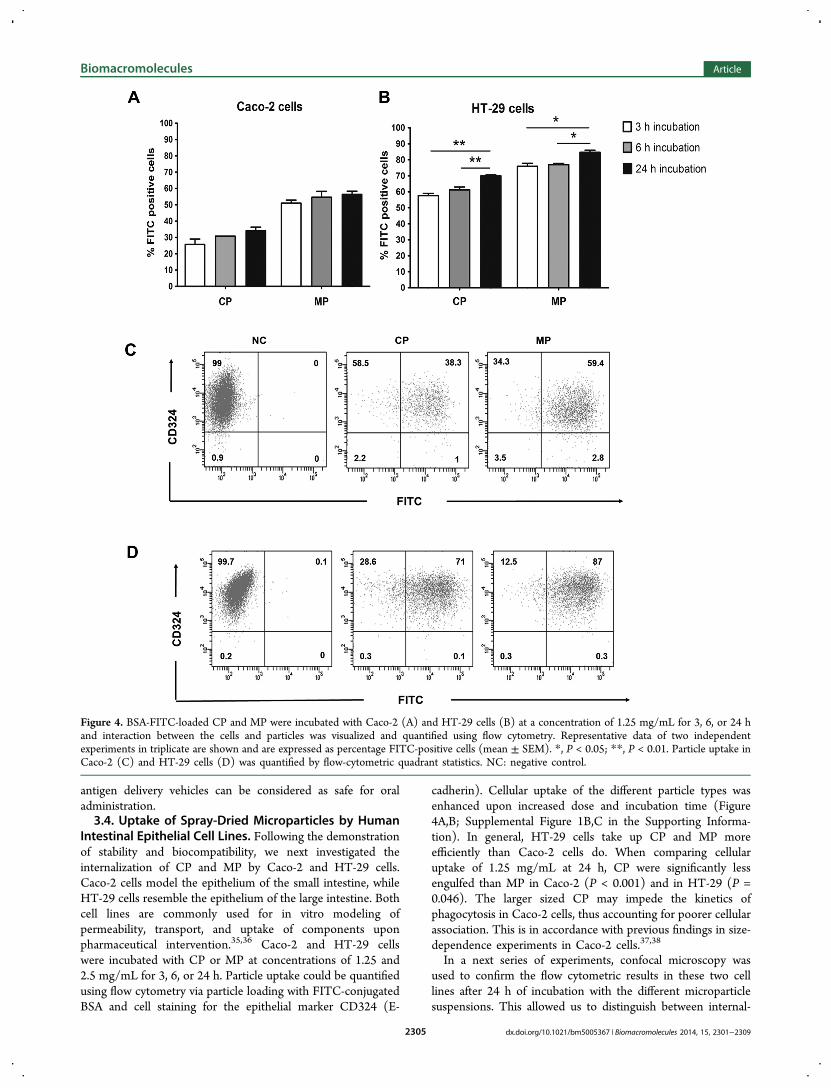
Intestinal Epithelial Cell Lines. Following the demonstrationof stability and biocompatibility, we next investigated theinternalization of CP and MP by Caco-2 and HT-29 cells.Caco-2 cells model the epithelium of the small intestine, whileHT-29 cells resemble the epithelium of the large intestine. Bothcell lines are commonly used for in vitro modeling ofpermeability, transport, and uptake of components uponpharmaceutical intervention.35,36 Caco-2 and HT-29 cellswere incubated with CP or MP at concentrations of 1.25 and2.5 mg/mL for 3, 6, or 24 h. Particle uptake could be quantifiedusing flow cytometry via particle loading with FITC-conjugatedBSA and cell staining for the epithelial marker CD324 (E-
cadherin). Cellular uptake of the different particle types wasenhanced upon increased dose and incubation time (Figure4A,B; Supplemental Figure 1B,C in the Supporting Informa-tion). In general, HT-29 cells take up CP and MP moreefficiently than Caco-2 cells do. When comparing cellularuptake of 1.25 mg/mL at 24 h, CP were significantly lessengulfed than MP in Caco-2 (P < 0.001) and in HT-29 (P =0.046). The larger sized CP may impede the kinetics ofphagocytosis in Caco-2 cells, thus accounting for poorer cellularassociation. This is in accordance with previous findings in size-dependence experiments in Caco-2 cells.37,38
In a next series of experiments, confocal microscopy wasused to confirm the flow cytometric results in these two celllines after 24 h of incubation with the different microparticlesuspensions. This allowed us to distinguish between internal-
Figure 4. BSA-FITC-loaded CP and MP were incubated with Caco-2 (A) and HT-29 cells (B) at a concentration of 1.25 mg/mL for 3, 6, or 24 hand interaction between the cells and particles was visualized and quantified using flow cytometry. Representative data of two independentexperiments in triplicate are shown and are expressed as percentage FITC-positive cells (mean ± SEM). *, P < 0.05; **, P < 0.01. Particle uptake inCaco-2 (C) and HT-29 cells (D) was quantified by flow-cytometric quadrant statistics. NC: negative control.
Biomacromolecules Article
dx.doi.org/10.1021/bm5005367 | Biomacromolecules 2014, 15, 2301−23092305

ization of the particles and extracellular binding to the cellsurface and to determine the exact localization of the particleswithin the cell. As shown in Figure 5, the cellular plasmamembrane was stained red fluorescent with wheat-germagglutinin (WGA)-Texas Red-X, and the nuclei were counter-
stained with DAPI (blue). Figures 5A,C and 5B,D, respectively,clearly show that small clusters of CP and MP are engulfed byCaco-2 and HT-29 cells and that these antigen delivery systemshave retained their characteristic spherical shape. These datashow that both spray-dried microsphere formulations were
Figure 5. Confocal photomicrographs of Caco-2 cells (A,B) and HT-29 cells (C,D) following a 24 h incubation with CP (A,C) or MP (B,D). BSA-FITC-loaded particles (green); plasma-membrane marker wheat-germ agglutinin (WGA)-Texas Red-X (red); and DAPI staining (blue). Full arrowspoint to microparticles; dashed arrows furthermore indicate engulfed particles surrounded by plasma membrane, indicative of endocytosis.
Figure 6. Transmission electron microscopy. HT-29 cells were either (A) left untreated or incubated for 24 h with (B) MP and (C,D) CP. Insetsshow OVA-loaded spray-dried polyelectrolyte microspheres at higher magnification.
Biomacromolecules Article
dx.doi.org/10.1021/bm5005367 | Biomacromolecules 2014, 15, 2301−23092306

efficiently internalized by intestinal epithelial cells and that themajority of the internalized microparticles were clustered in theapical and perinuclear regions of the cells.To provide a detailed picture of the physicochemical
alterations and intracellular fate of the spray-dried CP andMP, we performed TEM. For TEM analysis, the HT-29 cellswere selected because flow cytometry experiments demon-strated that these cells were the most potent in internalizing CPand MP. As depicted in Figure 6, the TEM micrographsconfirm the intracellular presence of CP and MP in HT-29 cellsafter 24 h of incubation. Nonloaded HT-29 cells served asnegative controls (Figure 6A). In Figure 6B, both an externaland internalized MP can be observed. Figure 6C demonstratesHT-29 cells actively engulfing two intact CP surrounded bycellular endocytotic vesicles (dashed arrow). Upon 24 h ofincubation, both MP and CP preserved their porous structure.Moreover, the inset of Figure 6D shows that degraded particlefragments (full arrows) end up in the phagolysosomal vesicles.Overall, the microparticles are confined into phagolysosomalintracellular compartments of intestinal epithelial cells byanalogy to previous reports investigating the intracellular fateof delivery systems.39−41
3.5. Induction of Class-II and Costimulatory Moleculeson Intestinal Epithelial Cells. Intestinal epithelial cells areimportant portals for antigen delivery to specialized antigenpresenting cells (e.g., dendritic cells) in the gut-associatedlymphoid tissue (e.g., the Peyer’s patches). Alternatively,intestinal epithelial cells may directly present antigen to T-
cells in the underlying lymphoid tissue. To verify a potentialrole for intestinal epithelial cells as antigen presenting cellsupon particle internalization, we studied the expression of class-II (HLA-DR) and costimulatory molecules (CD40 and CD86)on Caco-2 and HT-29 cells following incubation with CP orMP. To that end, Caco-2 cells and HT-29 cells were culturedfor 3 h at a particle concentration of 1.25 mg/mL CP or MP.As shown in Figure 7A,D, levels of HLA-DR protein
expression are significantly up-regulated in Caco-2 cells andmoderately increased in HT-29 cells incubated with CP or MP.Indeed, according to several groups, intestinal epithelial cellsuse HLA class-II pathways for antigen processing.42−44
Whereas both CP and MP exposure only slightly increasedCD40 protein expression (Figure 7B,E), an abundant presenceof CD86 was observed on both cell lines compared with theuntreated controls (Figure 7C,F). We previously demonstratedthat β-glucan particles likewise can enhance the activationstatus (CD86 and CD40 expression) of Caco-2 cells; however,β-glucan particles failed to up-regulate HLA-DR expression.45
Together, these data show that both CP and MP have theability to induce maturation of Caco-2 cells and HT-29 cells,characterized by an up-regulated expression of MHC class-IImolecules and the costimulatory molecules CD40 and CD86.This suggests that intestinal epithelial cells may be able toefficiently present antigen when it is delivered by micro-particulate formulations such as CP and MP, exerting adjuvant-like activity. This phenomenon would be of particular
Figure 7. Representative histograms of the HLA-DR expression (A,D) and the phenotypical maturation namely CD40 (B,E) and CD86 (C,F) inCaco-2 cells (upper row) and HT-29 cells (lower row). Human intestinal epithelial cells were left untreated (blue curve) or incubated with CP(green curve) or MP (red curve) for 3 h at a particle concentration of 1.25 mg/mL. Flow-cytometric histograms are a representative of fourreplicates per group.
Biomacromolecules Article
dx.doi.org/10.1021/bm5005367 | Biomacromolecules 2014, 15, 2301−23092307

significance for epithelial cells located near mucosal inductivesites such as the Peyer’s patches.
4. CONCLUSIONS
In conclusion, this study demonstrates that both calciumcarbonate- and mannitol-based microspheres exhibit the desiredstability, biocompatibility, and cellular uptake profile tomotivate further in vivo investigation of these promisingcandidate antigen carriers. First, CP and MP can fully resist theharsh gastric environment and retain most of their antigenloading capacity upon simulation of intestinal conditions.Second, CP and MP do not demonstrate in vitro cytotoxicity atconcentrations and incubation times relevant for in vivoconditions. Third, successful cellular uptake and subsequentinduction of class-II molecules and costimulatory moleculeswere demonstrated for both CP and MP in two differentintestinal epithelial cells. These important initial findingsmotivate a full characterization of the in vivo response.Mapping the antigen release pattern and monitoring theinduction of mucosal adhesion and immune responses will bothbe instrumental in our future research.
■ ASSOCIATED CONTENT
*S Supporting InformationBiocompatibility of CP and MP with Caco-2 and HT-29 cellswas monitored in vitro by measuring the mitochondrialdehydrogenase activity at a particle concentration of 2.5 mg/mL at different time points. BSA-FITC-loaded CP and MPwere incubated with Caco-2 and HT-29 cells at a concentrationof 2.5 mg/mL for 3, 6, or 24 h and the interaction between thecells and particles was visualized and quantified using flowcytometry. The particle uptake in Caco-2 and HT-29 cells wasquantified by flow-cytometric quadrant statistics. This materialis available free of charge via the Internet at http://pubs.acs.org.
■ AUTHOR INFORMATION
Corresponding Author*E-mail: [email protected]. Tel: +32 9 332 36 86. Fax:+ 32 9 332 49 65.
NotesThe authors declare no competing financial interest.
■ ACKNOWLEDGMENTS
This work was supported by the Concerted Research Action ofthe University of Ghent (BOF10/GOA/21, project number:01GC2110W; Ghent, Belgium). We are grateful to EefParthoens for providing help with the confocal laser scanningmicroscope and Tom Planckaert for microparticle BETanalysis. We thank Ran Rumes for the processing of thesamples for transmission electron microscopy, Sophie Vermautfor help with flow cytometry experiments, and Lynn Supply forthe excellent technical support.
■ REFERENCES(1) World Health Organization. Diarrhoeal Disease; Fact SheetNumber 330; World Health Organization: Geneva, Switzerland, 2013.(2) Holmgren, J.; Svennerholm, A. M. Vaccines against mucosalinfections. Curr. Opin. Immunol. 2012, 24, 343−353.(3) Wilkhu, J.; McNeil, S. E.; Kirby, D. J.; Perrie, Y. Formulationdesign considerations for oral vaccines. Ther. Delivery 2011, 2, 1141−1164.
(4) Malik, B.; Goyal, A. K.; Mangal, S.; Zakir, F.; Vyas, S. P.Implication of Gut Immunology in the Design of Oral Vaccines. Curr.Mol. Med. 2010, 10, 47−70.(5) Lycke, N. Recent progress in mucosal vaccine development:potential and limitations. Nat. Rev. Immunol. 2012, 12, 592−605.(6) Borges, O.; Lebre, F.; Bento, D.; Borchard, G.; Junginger, H. E.Mucosal Vaccines: Recent Progress in Understanding the NaturalBarriers. Pharm. Res. 2010, 27, 211−223.(7) Owen, J. L.; Sahay, B.; Mohamadzadeh, M. New generation oforal mucosal vaccines targeting dendritic cells. Curr. Opin. Chem. Biol.2013, 17, 918−824.(8) De Koker, S.; Lambrecht, B. N.; Willart, M. A.; van Kooyk, Y.;Grooten, J.; Vervaet, C.; Remon, J. P.; De Geest, B. G. Designingpolymeric particles for antigen delivery. Chem. Soc. Rev. 2011, 40,320−339.(9) McNeela, E. A.; Lavelle, E. C. Recent Advances in Microparticleand Nanoparticle Delivery Vehicles for Mucosal Vaccination. Curr.Top. Microbiol. 2012, 354, 75−99.(10) Vyas, S. P.; Gupta, P. N. Implication of nanoparticles/microparticles in mucosal vaccine delivery. Expert Rev. Vaccines2007, 6, 401−418.(11) Bramwell, V. W.; Perrie, Y. Particulate delivery systems forvaccines: what can we expect? J. Pharm. Pharmacol. 2006, 58, 717−728.(12) Mishra, N.; Goyal, A. K.; Tiwari, S.; Paliwal, R.; Paliwal, S. R.;Vaidya, B.; Mangal, S.; Gupta, M.; Dube, D.; Mehta, A.; Vyas, S. P.Recent advances in mucosal delivery of vaccines: role ofmucoadhesive/biodegradable polymeric carriers. Expert Opin. Ther.Pat. 2010, 20, 661−679.(13) Esposito, E.; Roncarati, R.; Cortesi, R.; Cervellati, F.; Nastruzzi,C. Production of Eudragit microparticles by spray-drying technique:influence of experimental parameters on morphological and dimen-sional characteristics. Pharm. Dev. Technol. 2000, 5, 267−78.(14) McAdams, D.; Chen, D. X.; Kristensen, D. Spray drying andvaccine stabilization. Expert Rev. Vaccines 2012, 11, 1211−1219.(15) Sollohub, K.; Cal, K. Spray drying technique: II. Currentapplications in pharmaceutical technology. J. Pharm. Sci. 2010, 99,587−597.(16) Vehring, R. Pharmaceutical particle engineering via spray drying.Pharm. Res. 2008, 25, 999−1022.(17) Volodkin, D. V.; Larionova, N. I.; Sukhorukov, G. B. Proteinencapsulation via porous CaCO3 microparticles templating. Bio-macromolecules 2004, 5, 1962−1972.(18) Rivera-Gil, P.; De Koker, S.; De Geest, B. G.; Parak, W. J.Intracellular processing of proteins mediated by biodegradablepolyelectrolyte capsules. Nano Lett. 2009, 9, 4398−4402.(19) Kastl, L.; Sasse, D.; Wulf, V.; Hartmann, R.; Mircheski, J.;Ranke, C.; Carregal-Romero, S.; Martinez-Lopez, J. A.; Fernandez-Chacon, R.; Parak, W. J.; Elsasser, H. P.; Rivera Gil, P. Multipleinternalization pathways of polyelectrolyte multilayer capsules intomammalian cells. ACS Nano 2013, 7, 6605−6618.(20) Reibetanz, U.; Halozan, D.; Brumen, M.; Donath, E. Flowcytometry of HEK 293T cells interacting with polyelectrolytemultilayer capsules containing fluorescein-labeled poly(acrylic acid)as a pH sensor. Biomacromolecules 2007, 8, 1927−1933.(21) Becker, A. L.; Johnston, A. P.; Caruso, F. Layer-by-layer-assembled capsules and films for therapeutic delivery. Small 2010, 6,1836−1852.(22) De Koker, S.; De Geest, B. G.; Singh, S. K.; De Rycke, R.;Naessens, T.; Van Kooyk, Y.; Demeester, J.; De Smedt, S. C.; Grooten,J. Polyelectrolyte Microcapsules as Antigen Delivery Vehicles ToDendritic Cells: Uptake, Processing, and Cross-Presentation ofEncapsulated Antigens. Angew. Chem., Int. Ed. 2009, 48, 8485−8489.(23) De Koker, S.; Naessens, T.; De Geest, B. G.; Bogaert, P.;Demeester, J.; De Smedt, S.; Grooten, J. Biodegradable PolyelectrolyteMicrocapsules: Antigen Delivery Tools with Th17 Skewing Activityafter Pulmonary Delivery. J. Immunol. 2010, 184, 203−211.(24) Dierendonck, M.; De Koker, S.; Cuvelier, C.; Grooten, J.;Vervaet, C.; Remon, J. P.; De Geest, B. G. Facile Two-Step Synthesis
Biomacromolecules Article
dx.doi.org/10.1021/bm5005367 | Biomacromolecules 2014, 15, 2301−23092308

of Porous Antigen-Loaded Degradable Polyelectrolyte Microspheres.Angew. Chem., Int. Ed. 2010, 49, 8620−8624.(25) Dierendonck, M.; De Koker, S.; De Rycke, R.; Bogaert, P.;Grooten, J.; Vervaet, C.; Remon, J. P.; De Geest, B. G. Single-StepFormation of Degradable Intracellular Biomolecule Microreactors.ACS Nano 2011, 5, 6886−6893.(26) Dierendonck, M.; De Koker, S.; Vervaet, C.; Remon, J. P.; DeGeest, B. G. Interaction between polymeric multilayer capsules andimmune cells. J. Controlled Release 2012, 161, 592−599.(27) U.S. Pharmacopeia XXIV and National Formulary 19; TheUnited States Pharmacopeial Convention: Rockville, MD, 2000; pp2235−2236.(28) Song, J. W.; Ren, L. M.; Yin, C. Y.; Ji, Y. Y.; Wu, Z. F.; Li, J. X.;Xiao, F. S. Stable, porous, and bulky particles with high external surfaceand large pore volume from self-assembly of zeolite nanocrystals withcationic polymer. J. Phys. Chem. C 2008, 112, 8609−8613.(29) Finnie, K. S.; Waller, D. J.; Perret, F. L.; Krause-Heuer, A. M.;Lin, H. Q.; Hanna, J. V.; Barbe, C. J. Biodegradability of sol-gel silicamicroparticles for drug delivery. J. Sol-Gel Sci. Technol. 2009, 49, 12−18.(30) Tao, Z. M.; Toms, B.; Goodisman, J.; Asefa, T. MesoporousSilica Microparticles Enhance the Cytotoxicity of Anticancer PlatinumDrugs. ACS Nano 2010, 4, 789−794.(31) Cunben, L.; Hein, S.; Wang, K. Chitosan-Carrageenanpolyelectrolyte complex for the delivery of protein drugs. ISRNBiomater. 2013, 2013, 629807-1−629807-6.(32) Landry, F. B.; Bazile, D. V.; Spenlehauer, G.; Veillard, M.;Kreuter, J. Degradation of poly(D,L-lactic acid) nanoparticles coatedwith albumin in model digestive fluids (USP XXII). Biomaterials 1996,17, 715−723.(33) De Geest, B. G.; Willart, M. A.; Lambrecht, B. N.; Pollard, C.;Vervaet, C.; Remon, J. P.; Grooten, J.; De Koker, S. Surface-engineeredpolyelectrolyte multilayer capsules: synthetic vaccines mimickingmicrobial structure and function. Angew. Chem. 2012, 51, 3862−3866.(34) De Geest, B. G.; Willart, M. A.; Hammad, H.; Lambrecht, B. N.;Pollard, C.; Bogaert, P.; De Filette, M.; Saelens, X.; Vervaet, C.;Remon, J. P.; Grooten, J.; De Koker, S. Polymeric multilayer capsule-mediated vaccination induces protective immunity against cancer andviral infection. ACS Nano 2012, 6, 2136−2149.(35) Volpe, D. A. Drug-permeability and transporter assays in Caco-2and MDCK cell lines. Future Med. Chem. 2011, 3, 2063−2077.(36) Scaldaferri, F.; Pizzoferrato, M.; Gerardi, V.; Lopetuso, L.;Gasbarrini, A. The gut barrier: new acquisitions and therapeuticapproaches. J. Clin. Gastroenterol. 2012, 46 (Suppl), S12−S17.(37) Desai, M. P.; Labhasetwar, V.; Walter, E.; Levy, R. J.; Amidon,G. L. The mechanism of uptake of biodegradable microparticles inCaco-2 cells is size dependent. Pharm. Res. 1997, 14, 1568−1573.(38) He, C.; Yin, L.; Tang, C.; Yin, C. Size-dependent absorptionmechanism of polymeric nanoparticles for oral delivery of proteindrugs. Biomaterials 2012, 33, 8569−8578.(39) Cartiera, M. S.; Johnson, K. M.; Rajendran, V.; Caplan, M. J.;Saltzman, W. M. The uptake and intracellular fate of PLGAnanoparticles in epithelial cells. Biomaterials 2009, 30, 2790−2798.(40) Javier, A. M.; Kreft, O.; Semmling, M.; Kempter, S.; Skirtach, A.G.; Bruns, O. T.; del Pino, P.; Bedard, M. F.; Raedler, J.; Kaes, J.;Plank, C.; Sukhorukov, G. B.; Parak, W. J. Uptake of ColloidalPolyelectrolyte-Coated Particles and Polyelectrolyte Multilayer Cap-sules by Living Cells. Adv. Mater. 2008, 20, 4281−4287.(41) Cohen, S.; Coue, G.; Beno, D.; Korenstein, R.; Engbersen, J. F.J. Bioreducible poly(amidoamine)s as carriers for intracellular proteindelivery to intestinal cells. Biomaterials 2012, 33, 614−623.(42) Hershberg, R. M.; Framson, P. E.; Cho, D. H.; Lee, L. Y.;Kovats, S.; Beitz, J.; Blum, J. S.; Nepom, G. T. Intestinal epithelial cellsuse two distinct pathways for HLA class II antigen processing. J. Clin.Invest. 1997, 100, 204−215.(43) Byrne, B.; Madrigal-Estebas, L.; McEvoy, A.; Carton, J.;Doherty, D. G.; Whelan, A.; Feighery, C.; O’Donoghue, D. P.;O’Farrelly, C. Human duodenal epithelial cells constitutively express
molecular components of antigen presentation but not costimulatorymolecules. Hum. Immunol. 2002, 63, 977−986.(44) Mayer, L.; Eisenhardt, D.; Salomon, P.; Bauer, W.; Plous, R.;Piccinini, L. Expression of class II molecules on intestinal epithelialcells in humans. Differences between normal and inflammatory boweldisease. Gastroenterology 1991, 100, 3−12.(45) De Smet, R.; Demoor, T.; Verschuere, S.; Dullaers, M.; Ostroff,G. R.; Leclercq, G.; Allais, L.; Pilette, C.; Dierendonck, M.; De Geest,B. G.; Cuvelier, C. A. β-Glucan microparticles are good candidates formucosal antigen delivery in oral vaccination. J. Controlled Release 2013,172, 671−678.
Biomacromolecules Article
dx.doi.org/10.1021/bm5005367 | Biomacromolecules 2014, 15, 2301−23092309