Preparation and characterization of chitosan extracted from ...

Journal of Microencapsulation, August 2005; 22(5): 459–470
Preparation and in vitro evaluation of thiolatedchitosan microparticles
K. MACULOTTI1, I. GENTA1, P. PERUGINI1, M. IMAM2,
A. BERNKOP-SCHNURCH3, & F. PAVANETTO1
1Department of Pharmaceutical Chemistry, University of Pavia, Pavia, Italy, 2Institute of
Pharmaceutical Technology and Biopharmaceutics, Center of Pharmacy University of Vienna,
Vienna, Austria, and 3Department of Pharmaceutical Technology, Institute of Pharmacy,
Leopold-Franzens-University Innsbruck, Innsbruck, Austria
(Received 29 June 2004; accepted 15 December 2004)
AbstractThe objective of this study was to prepare a microparticulate drug delivery system being based on anew thiomer, namely a chitosan 2-iminothiolane conjugate (chitosan-TBA conjugate). Due tothiol groups being immobilized on chitosan, chitosan-TBA conjugate exhibits improved mucoadhesiveand permeation enhancing properties. Because of these features microparticulate drug delivery systemsbased on chitosan-TBA conjugate might be a promising tool for the non-invasive administrationof hydrophilic macromolecular drugs. Chitosan-TBA conjugate microspheres were preparedby the emulsification/solvent evaporation method. Fluorescein-isothiocyanate labelled dextran(FITC-dextran) was chosen as a model hydrophilic drug. Microspheres have been characterized bymorphological analysis, thiol group content, swelling behaviour, polymer degradation drug loaddetermination, dissolution test and mucoadhesion studies. Results reported in this work demonstratedthe possibility to obtain stable microspheres without cross-linking agents. Thiolated chitosanmicrospheres seem to be more stable in aqueous media with respect to unmodified chitosan.The degradability by lysozyme appears quite similar for both polymers, showing that chemicalmodification does not influence the biodegradable properties of chitosan. Microspheres were able tocontrol the drug release for at least 1 h, exhibiting comparatively strong mucoadhesive properties.The chitosan-TBA conjugate microparticles remain on the mucosa in a 2.5-fold higher concentrationwith respect to unmodified chitosan microparticles. These data suggest that chitosan-TBA conjugatemicrospheres have the potential to be used as a mucoadhesive drug delivery system.
Keywords: Microparticles, thiomers, mucoadhesion, biodegradability, modified drug release
Introduction
Oral ingestion has historically been the most convenient and commonly employed method
for drug delivery. However, most drugs are not administered orally due to their instability
Correspondence: Professor F. Pavanetto, Department of Pharmaceutical Chemistry, V.le Taramelli 12, 27100 Pavia, Italy.
Tel: 0039 0382 987377. Fax: 0039 0382 422975. E-mail: [email protected]
ISSN 0265-2048 print/ISSN 1464-5246 online # 2005 Taylor & Francis
DOI: 10.1080/02652040500162220
Jour
nal o
f M
icro
enca
psul
atio
n D
ownl
oade
d fr
om in
form
ahea
lthca
re.c
om b
y U
nive
rsita
' di P
avia
on
11/1
4/12
For
pers
onal
use
onl
y.

in the gastrointestinal tract or because of their poor uptake, for example in the case
of hydrophilic drugs. As a result, new formulations are being developed to attempt
to overcome these difficulties. Among these, particulate drug delivery systems such
as biodegradable microspheres have been proposed. Unfortunately, microparticulate
systems generally display low oral absorption efficiencies. To improve particle uptake,
mucoadhesive polymers have been proposed (Lehr 1994; Chen and Langer 1998).
Thiolated polymers, designated thiomers, represent a new promise in the field of muco-
adhesive polymers. In particular, chitosan-2-iminothiolate conjugates provide much
higher adhesive properties than other polymers generally considered to be mucoadhesive
(Bernkop-Schnurch et al. 1999, 2004a, 2004b; Kast and Bernkop-Schurch 2001).
This is probably due to two different mechanisms of mucoadhesion: (a) improved ionic
interaction between the cationic group of thiolated chitosan and the anionic moieties present
in the mucus layer; and (b) formation of disulphide bonds due to the introduction of thiol
groups into the chitosan structure (Kast and Bernkop-Schurch 2001).
The higher mucoadhesive properties of thiomers can intensify the contact with the
mucosa, providing an increased epithelial permeability for many drugs mediated by
polymeric carrier systems (Bernkop-Schnurch et al. 2000).
This work employed the chitosan-2-iminothiolane conjugate as thiolated polymer. This
thiomer can be easily obtained by reaction of chitosan with 2-iminothiolane
(Traut’s reagent), exhibiting promising features for oral drug delivery and very interesting
characteristics. In fact, with respect to other thiomers, chitosan-TBA conjugate displays
a cationic amidine group by which it can strongly interact with the anionic sub-structures
such as sialic acid and sulphonic acid of the mucus layer and with the cell membrane result-
ing in a structural reorganization of tight junction-associated proteins leading to an
enhanced paracellular uptake of hydrophilic molecules (Bernkop-Schnurch et al. 2003).
A previously in vivo study confirmed this hypothesis, showing the possibility to deliver
insulin by an oral route (Krauland et al. 2004).
Chitosan microspheres can be easily prepared according to several methods (Genta et al.
1997b, 2002). They are promising candidates for oral administration of antigens such as
diphtheria or tetanus toxoid and promote an enhancement of both mucosal and systemic
immune responses (Van der Lubben et al. 2001). Microsystems based on chitosan complex
have already been studied for oral protein delivery (Coppi et al. 2001). Moreover, they were
successfully used for oral administration of proteins such as insulin (Radamus et al. 2000).
The aim of this study was the preparation and in vitro evaluation of a chitosan-
2-iminothiolane conjugate microparticulate delivery system. Fluorescein isothiocyanate-
dextran (FITC-Dextran) was chosen as the hydrophilic model drug (Morishita et al. 2002).
In this preliminary work, microsphere characterization, polymer erosion and in vitro
drug release were investigated. In order to verify the improved mucoadhesive properties
of chitosan-TBA conjugate microspheres, an in vitro test was performed employing
unmodified chitosan as control.
Materials and methods
Materials
Chitosan was purchased from Fluka Chemie (Fluka Chemie, Buchs, Switzerland) and
its properties included Mr� 50 kDa; (degree of deacetylation 83–85%), viscosity,
�100 mPa s (1% in 1% acetic acid, 20�C). The 2-iminothiolane HCl (Traut’s reagent)
(Mr 137.63 Da) was supplied from Pierce (Pierce, Oud Beijerland, NL). The fluorescein
460 K. Maculotti et al.
Jour
nal o
f M
icro
enca
psul
atio
n D
ownl
oade
d fr
om in
form
ahea
lthca
re.c
om b
y U
nive
rsita
' di P
avia
on
11/1
4/12
For
pers
onal
use
onl
y.
isothiocyanate-dextran (FITC-Dextran) (Mr 4.3 kDa) was obtained from Sigma
(St Louis, MO). All solvents were of analytical grade.
Synthesis of chitosan 2-iminothiolane conjugates
The thiolated chitosan was prepared as follows: 1 g of chitosan was dissolved in a 1% v/v
acetic acid solution, then the pH was adjusted to pH 5 using 5 M NaOH. 200 mg of
2-iminothiolane HCl were added to the chitosan solution and the reaction mixture was
incubated for 6 h at room temperature under permanent stirring. In order to eliminate the
residual 2-iminothiolane HCl and to isolate the polymer conjugate, the reaction mixture was
dialysed in tubings (molecular mass cut-off 12 kDa cellulose membrane, Sigma, St Louis,
MO) for 3 days at 10�C in the dark against 5 mM HCl, then twice against the same
medium but containing 1% NaCl. Therefore, the samples were exhaustively dialysed last
time against 1 mM HCl to adjust the pH of the polymer to 4.
The polymer conjugates were lyophilized at –30�C and 0.01 mbar (Christ Beta 1-8K,
Germany) and stored at 4�C until further use (Bernkop-Schnurch et al. 2003).
Determination of the thiol group content in the polymer conjugates
The amount of thiol groups on the thiolated chitosan was spectrophotometrically
determined using Ellman’s reagent (DTNB, 5,50-dithiobis(2-nitrobenzoic acid)) (Sigma,
St Louis, MO) (Badyal et al. 2001; Kast et al. 2002).
First, 0.5 mg of polymer were swollen for 2 h in 0.25 ml of demineralized water; then
0.25 ml of 0.5 M phosphate buffer pH 8.0 were added; 0.5 ml of DTNB 0.03% w/v dissolved
in 0.5 M phosphate buffer pH 8.0 were added to the polymer suspension and samples were
incubated for 2 h at room temperature. The precipitated polymer was removed by centrifu-
gation (24 000g, 5 min). Samples (0.3 ml) were transferred to a 96 well microtitration-plate
and the absorbance was measured at 450 nm with a 96 well microtitration-plate
reader (Anthos Reader 2001, Salzburg, Austria). The amount of thiol moieties on the
polymer was calculated from a standard curve of L-cysteine hydrochloride anhydrous
(Sigma-Aldrich, Steinhein, Germany) in 0.5 M phosphate buffer pH 8.0 in concentrations
of 0.8–28mg ml�1.
Placebo microsphere preparation
Chitosan-TBA conjugate microparticulate delivery systems have been prepared according
to the emulsification/solvent evaporation method suitably modified (Genta et al. 1997a).
A 1% w/w thiolated chitosan aqueous solution was dropped into paraffin oil containing
0.5% Span 20 and emulsified by a homogenizer Ultraturrax (Ika Labortechnik, Staufen,
Germany) at 9500 rev min�1 (continuos phase/dispersed phase ratio 15:1 v/v). The W/O
emulsion was transferred in a water bath thermostated at 30�C, stirred by a rotating paddle
system at 350 rev min�1 and then methanol was dropped into the emulsion (methanol/
chitosan solution ratio 1:4 v/w). The emulsion temperature was raised to 40�C and nitrogen
gas was blown at the pressure of 1 bar. The emulsion was kept as described for 8 h, then the
formed microspheres were washed three times with a petroleum ether, centrifuged, filtered
through 0.65 mm filter and kept in a dessicator for 24 h before characterization.
Chitosan microspheres were prepared in the same way as control.
Thiolated chitosan microparticles 461
Jour
nal o
f M
icro
enca
psul
atio
n D
ownl
oade
d fr
om in
form
ahea
lthca
re.c
om b
y U
nive
rsita
' di P
avia
on
11/1
4/12
For
pers
onal
use
onl
y.

Determination of the thiol group content in the placebo chitosan-TBA microspheres
The determination of thiol group content in the chitosan-TBA conjugate microspheres
was performed as previously described for the thiolated polymer (Badyal et al. 2001; Kast
et al. 2002).
Morphological analysis
Microsphere shape was evaluated by scanning electron microscopy (SEM) using a Stereoscan
S90B (Cambridge Instruments, Cambridge, UK). Samples of microparticles were sputtered
with a Au/Pd film in a argon atmosphere. Granulometric analyses were carried out by
a conductive method using a Coulter Counter (Coulter� Multisizer II, Coulter Electonics,
Luton, UK). The orifice of apparatus tube used was 140 mm. Microspheres were
suspended in a solution of isopropyl alcohol containing 5% of NH4SCN. The analyses
were performed in a size range between 2 and 90 mm. Three analyses were performed for
each sample.
FITC-dextran loaded chitosan-TBA microsphere preparation
Drug loading was performed by a slightly modified method reported by Morishita et al.
(2002). Briefly, 20 mg of placebo microparticles were suspended in 1 ml of 0.5% w/v
FITC-dextran aqueous solution. After incubation for 1 h under continuous shaking
(Eppendorf thermomixer) at room temperature, 0.5 ml of ethanol was added and then the
samples were centrifuged for 5 min at 24 000g. After removing the supernatant, the deposit
was washed twice with 1 ml of ethanol and then it was dried by Gyrovap GT system.
Unfortunately, it was not possible to apply this method to unmodified chitosan microspheres
because they are not stable in aqueous solution, in fact they dissolve in the drug solution
used to load the FITC-dextran into the microspheres.
Total drug content determination
FITC-dextran content was determined by an indirect method, determining the total amount
of unloaded FITC-dextran. The residual drug solution, after microspheres removing, were
dried by a Girovap GT system. Then, the residuals were dissolved in demineralized water
and 0.3 ml of solution were transferred in a 96 well microtitration-plate. Drug content was
determined reading the fluorescence of FITC-dextran at 485/535 nm with a fluorimeter
(SLT, Spectra Fluor, Tecan, Austria).
The amount of FITC-dextran was calculated from a standard curve of FITC-dextran
in water in a concentration of 40 mg ml�1 and 500 mg ml�1.
Evaluation of swelling behaviour
The swelling capacity was determined by a microscopic method. The diameter of dried
chitosan-TBA conjugate microspheres was measured by a microscope using a calibrated
ocular. The analyses were carried out using water or 0.1 N HCl or phosphate buffer pH 6.8
or acetate buffer pH 5.5. At pre-determined time points (from 5 to 120 min) at least 400
particles were measured. Results were reported as volume increase calculated from the
462 K. Maculotti et al.
Jour
nal o
f M
icro
enca
psul
atio
n D
ownl
oade
d fr
om in
form
ahea
lthca
re.c
om b
y U
nive
rsita
' di P
avia
on
11/1
4/12
For
pers
onal
use
onl
y.
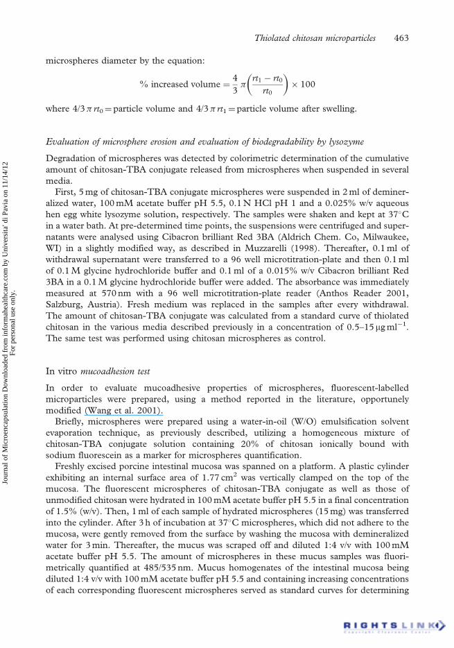
microspheres diameter by the equation:
% increased volume ¼4
3�
rt1 � rt0
rt0
� �� 100
where 4/3� rt0¼ particle volume and 4/3� rt1¼ particle volume after swelling.
Evaluation of microsphere erosion and evaluation of biodegradability by lysozyme
Degradation of microspheres was detected by colorimetric determination of the cumulative
amount of chitosan-TBA conjugate released from microspheres when suspended in several
media.
First, 5 mg of chitosan-TBA conjugate microspheres were suspended in 2 ml of deminer-
alized water, 100 mM acetate buffer pH 5.5, 0.1 N HCl pH 1 and a 0.025% w/v aqueous
hen egg white lysozyme solution, respectively. The samples were shaken and kept at 37�C
in a water bath. At pre-determined time points, the suspensions were centrifuged and super-
natants were analysed using Cibacron brilliant Red 3BA (Aldrich Chem. Co, Milwaukee,
WI) in a slightly modified way, as described in Muzzarelli (1998). Thereafter, 0.1 ml of
withdrawal supernatant were transferred to a 96 well microtitration-plate and then 0.1 ml
of 0.1 M glycine hydrochloride buffer and 0.1 ml of a 0.015% w/v Cibacron brilliant Red
3BA in a 0.1 M glycine hydrochloride buffer were added. The absorbance was immediately
measured at 570 nm with a 96 well microtitration-plate reader (Anthos Reader 2001,
Salzburg, Austria). Fresh medium was replaced in the samples after every withdrawal.
The amount of chitosan-TBA conjugate was calculated from a standard curve of thiolated
chitosan in the various media described previously in a concentration of 0.5–15 mg ml�1.
The same test was performed using chitosan microspheres as control.
In vitro mucoadhesion test
In order to evaluate mucoadhesive properties of microspheres, fluorescent-labelled
microparticles were prepared, using a method reported in the literature, opportunely
modified (Wang et al. 2001).
Briefly, microspheres were prepared using a water-in-oil (W/O) emulsification solvent
evaporation technique, as previously described, utilizing a homogeneous mixture of
chitosan-TBA conjugate solution containing 20% of chitosan ionically bound with
sodium fluorescein as a marker for microspheres quantification.
Freshly excised porcine intestinal mucosa was spanned on a platform. A plastic cylinder
exhibiting an internal surface area of 1.77 cm2 was vertically clamped on the top of the
mucosa. The fluorescent microspheres of chitosan-TBA conjugate as well as those of
unmodified chitosan were hydrated in 100 mM acetate buffer pH 5.5 in a final concentration
of 1.5% (w/v). Then, 1 ml of each sample of hydrated microspheres (15 mg) was transferred
into the cylinder. After 3 h of incubation at 37�C microspheres, which did not adhere to the
mucosa, were gently removed from the surface by washing the mucosa with demineralized
water for 3 min. Thereafter, the mucus was scraped off and diluted 1:4 v/v with 100 mM
acetate buffer pH 5.5. The amount of microspheres in these mucus samples was fluori-
metrically quantified at 485/535 nm. Mucus homogenates of the intestinal mucosa being
diluted 1:4 v/v with 100 mM acetate buffer pH 5.5 and containing increasing concentrations
of each corresponding fluorescent microspheres served as standard curves for determining
Thiolated chitosan microparticles 463
Jour
nal o
f M
icro
enca
psul
atio
n D
ownl
oade
d fr
om in
form
ahea
lthca
re.c
om b
y U
nive
rsita
' di P
avia
on
11/1
4/12
For
pers
onal
use
onl
y.

the percentage of microparticles remaining on the mucosa. Unmodified chitosan fluorescent
labelled microspheres were used as control.
In vitro dissolution test
In vitro drug release tests were performed on FITC-dextran loaded chitosan-TBA conjugate
microspheres in an Eppendorf test tube by suspending 5 mg of drug loaded microspheres in
2 ml of 100 mM phosphate buffer pH 6.8. At pre-determined time points, the samples
were centrifuged at 24 000g for 5 min, 0.3 ml of supernatant were transferred in a 96 well
microtitration-plate and replaced with a fresh medium. The amount of FITC-dextran
released was determined using a fluorimeter, as described previously, for the determination
of total drug content.
Results and discussion
Synthesis of chitosan 2-iminothiolane conjugates
2-Iminothiolane was attached covalently to the amino groups of chitosan, obtaining the
product shown in Figure 1. The lyophilized chitosan-TBA conjugate appeared as white,
odourless powder of fibrous structure. It is completely soluble in water or acidic media
(pH below 6). The total amount of thiol groups bound to the polymer was
46.59� 15.04mmol g�1 of polymer. In this condition it is possible to obtain a slightly
viscous aqueous solution. Otherwise, an increase of thiol groups determined the insolubility
of the polymer in aqueous media (Bernkop-Schnurch et al. 2003).
Microspheres preparation
Good results in terms of yield were achieved both for chitosan-TBA conjugate and for
chitosan microspheres; yields of production being in the range of 75% for chitosan-
TBA conjugate microspheres and almost 95% for unmodified chitosan microspheres.
Figure 1. Presumptive structure of chitosan 2-iminothiolane conjugate.
464 K. Maculotti et al.
Jour
nal o
f M
icro
enca
psul
atio
n D
ownl
oade
d fr
om in
form
ahea
lthca
re.c
om b
y U
nive
rsita
' di P
avia
on
11/1
4/12
For
pers
onal
use
onl
y.

The amount of thiol groups in the chitosan-TBA conjugate microspheres was
4.18� 1.11 mmol g�1 of microspheres. This diminution of thiol moieties on the polymer
took place during the production process of microspheres. During this process, the thiol
groups can be oxidized, forming intra- as well as inter-molecular disulphide bonds, even
if the nitrogen presence reduces this process a lot.
The residual amount of free thiol groups improves the mucoadhesive properties as
described below, although a direct correlation has not been found. In fact, the colorimetric
method used to determine the thiol group moieties cannot distinguish between the free thiol
groups present on the microspheres surface being responsible for improved mucoadhesive
properties and the thiol groups present inside the microspheres.
Morphological analysis
Chitosan-TBA conjugate microspheres appeared quite homogeneous with spherical shape
and smooth non-porous surface, as shown in the photomicrograph (Figure 2). The
unmodified chitosan microspheres have a wrinkled and quite porous surface (data not shown).
Figure 3 shows particle size distribution of chitosan and chitosan-TBA conjugate
microspheres. Particle size distribution was quite homogeneous in both cases.
Microspheres mean size was similar and independent from the polymer. It was �18 mm
for chitosan microspheres, while for chitosan-TBA conjugate microparticles it was 15 mm.
FITC-dextran loaded chitosan-TBA microspheres preparation
The drug encapsulation yields of chitosan-TBA conjugate microspheres were determined to
be 96.57� 1.34%, corresponding to the 241.40� 0.05 mg of drug per mg of microspheres.
The method seems, therefore, to be useful to encapsulate drugs with high water solubility.
Figure 2. Photomicrograph of chitosan-TBA conjugate microspheres.
Thiolated chitosan microparticles 465
Jour
nal o
f M
icro
enca
psul
atio
n D
ownl
oade
d fr
om in
form
ahea
lthca
re.c
om b
y U
nive
rsita
' di P
avia
on
11/1
4/12
For
pers
onal
use
onl
y.

Evaluation of swelling behaviour
Microparticles swell rapidly upon the addition of aqueous media. After this instantaneous
volume increment the swollen microspheres maintained their spherical shape and size.
Microspheres swelling did not seem to be affected by the pH, as shown in Figure 4.
A comparatively more extensive swelling could be observed in acetate buffer.
Evaluation of microspheres erosion and evaluation of degradability by lysozyme
The erosion studies on chitosan and chitosan-TBA conjugate microspheres showed
significant differences in terms of stability. As shown in Figure 5, the chemical degradation
in different media indicated a more pronounced displacement of chitosan microspheres
(in 8 h, respectively, 31.8% chitosan eroded in acetate buffer and 20.5% chitosan eroded
in water) in comparison to chitosan-TBA conjugate microspheres (in 8 h, respectively,
5.7% chitosan-TBA conjugate eroded in acetate buffer and 7.7% chitosan-TBA conjugate
eroded in water). In fact, the presence of disulphide bonds in the chitosan-TBA conjugate
microsphere matrix stabilized the structure. Hence, it is possible to obtain stable
microspheres with this thiolated polymer without the use of chemical or physical
cross-linking agents. On the other hand, the degradability by the use of lysozyme does not
seem to be affected by the different polymers (Figure 6). Consequently, chitosan-TBA
conjugate does not seem to affect the hydrolytic action of lysozyme on the � (1!4) glycosides
bounds present in the chitosan structure (Kurita et al. 1996). The result is particularly
interesting because it evidences that the high stability of chitosan-TBA conjugate micro-
spheres does not affect their degradability by enzymes such as lysozyme.
In vitro mucoadhesion test
The in vitro mucoadhesive properties were evaluated by using fluorescent-labelled
microspheres with a simple and reproducible method.
0
10
20
30
40
50
60
70
80
90
100
100500
Diameter (mm)
Vo
lum
e %
chitosan-TBA conjugate microspheres
chitosan microspheres
Figure 3. Cumulative particle size distribution of (g) chitosan-TBA conjugate microspheres and(œ) chitosan microspheres.
466 K. Maculotti et al.
Jour
nal o
f M
icro
enca
psul
atio
n D
ownl
oade
d fr
om in
form
ahea
lthca
re.c
om b
y U
nive
rsita
' di P
avia
on
11/1
4/12
For
pers
onal
use
onl
y.

Mucoadhesion was expressed as the amount of microparticles remaining on the mucosa
during the test. After 3 h, �20% of chitosan-TBA conjugate microspheres remained on the
mucosa. This result is significantly different in comparison to the chitosan microspheres,
where the percentage of microparticles remaining on the mucosa was less then 10%
(Figure 7).
0
10
20
30
40
50
60
HCl 0.1M Phosphatebuffer
Acetatebuffer
Water
% in
crea
sed
vo
lum
e
Figure 4. Swelling behaviour of chitosan-TBA microspheres in different media (after 30 min fromtest beginning); indicated values are means (�SD) of at least three experiments.
0
5
10
15
20
25
30
35
40
129630 15 18 21 24
Time (h)
% D
isso
lved
ch
ito
san
Figure 5. Comparison of the degradation of chitosan-TBA conjugate microspheres and chitosanmicrospheres in different media expressed as percentage of eroded polymer: (m) chitosan-TBAconjugate microspheres in acetate buffer pH 5, (g) chitosan-TBA conjugate microspheres in H2O,(œ) chitosan microspheres in H2O and (i) chitosan microspheres in acetate buffer pH 5; indicatedvalues are means (�SD) of at least three experiments.
Thiolated chitosan microparticles 467
Jour
nal o
f M
icro
enca
psul
atio
n D
ownl
oade
d fr
om in
form
ahea
lthca
re.c
om b
y U
nive
rsita
' di P
avia
on
11/1
4/12
For
pers
onal
use
onl
y.

These results confirmed the previous tests regarding the mucoadhesive properties of
thiolated polymers (Bernko-Schnurch et al. 2003). In fact, the presence of free thiol
groups seems to increase the mucoadhesive properties of chitosan. Moreover, the partial
oxidation of sulphide groups taking place during the microsphere preparation does
not seems to influence in an evident way the increased mucoadhesive properties of
microparticles.
00 4 8 12 16 20 24
5
10
15
20
25
30
Time (h)
% D
isso
lved
ch
ito
san
chitosan-TBA conjugate microspheres
chitosan microspheres
Figure 6. Comparison of the degradation of chitosan-TBA and chitosan microspheres by lysozyme;indicated values are means (�SD) of at least three experiments.
0
5
10
15
20
25
30
chitosan-TBAmicrospheres
chitosan microspheres
% o
f m
icro
par
ticl
es r
emai
nin
go
n t
he
mu
cosa
Figure 7. Comparison of chitosan-TBA and chitosan microspheres remaining on the mucosa duringthe mucoadhesion test. Indicated values represent the percentage of microparticles present on themucosa after 3 h from the test beginning. Error bars indicate standard deviation (n¼ 3).
468 K. Maculotti et al.
Jour
nal o
f M
icro
enca
psul
atio
n D
ownl
oade
d fr
om in
form
ahea
lthca
re.c
om b
y U
nive
rsita
' di P
avia
on
11/1
4/12
For
pers
onal
use
onl
y.

In vitro dissolution test
Figure 8 shows the release profile of the FITC-dextran from the chitosan-TBA conjugate
microspheres. Result shows that, after a first burst release of �50%, microspheres were able
to control the drug release for at least 1 h.
This behaviour can be correlated with the microspheres swelling properties. In fact, the
instantaneous swelling of microspheres determines the instantaneous diffusion of drug.
After 30 min, the polymer network is able to withhold the drug, which as a water soluble
drug can diffuse through the hydrophilic polymeric network and reach the total release
after at least 2 h.
From another point of view, the FITC-dextran loading method used in this work can
determine a superficial distribution of compound, responsible for the fast release.
Within this study, the possibility to obtain a mucoadhesive microparticulate drug delivery
system based on chitosan-TBA conjugate was detected. Results evidenced the possibility to
obtain a stable, biodegradable microparticulate drug delivery system with highly beneficial
properties in terms of mucoadhesivity with respect to unmodified chitosan. A hydrophilic
model drug, FITC-dextran, was successfully encapsulated into the microspheres and
microspheres were able to control the drug release for at least 1 h.
References
Badyal JP, Cameron AM, Cameron NR, Coe DM, Cox R, Davis BG. 2001. A simple method for the quantitative
analysis of resin bound thiol groups. Tetrahedron Letters 42:8531–8533.
Bernkop-Schnurch A, Giovanelli R, Valenta C. 2000. Peroral administration of enzymes: Strategies to improve the
galenic of dosage for trypsin and bromelain. Drug Development and Industrial Pharmacy 26:115–121.
Bernkop-Schnurch A, Guggi D, Printer Y. 2004a. Thiolated chitosans: Development and in vitro evaluation of a
mucoadhesive, permeation enhancing oral drug delivery system. Journal of Controlled Release 94:177–186.
Bernkop-Schnurch A, Hornof M, Guggi D. 2004b. Thiolated chitosan. European Journal of Pharmaceutics and
Biopharmaceutics 57:9–17.
Bernkop-Schnurch A, Hornof M, Zoidl T. 2003. Thiolated polymers-thiomers: Synthesis and in vitro evaluation of
chitosan-2-iminothiolane conjugates. International Journal of Pharmaceutics 260:229–237.
0
20
40
60
80
100
120
32.51.510.5 20
Time (h)
% D
rug
rel
ease
d
FITC-dextran loaded microspheres FITC-dextran
Figure 8. In vitro release profile of FITC-dextran from chitosan-TBA conjugate microspheresin phosphate buffer pH 6.8; indicated values are means (�SD) of at least three experiments.
Thiolated chitosan microparticles 469
Jour
nal o
f M
icro
enca
psul
atio
n D
ownl
oade
d fr
om in
form
ahea
lthca
re.c
om b
y U
nive
rsita
' di P
avia
on
11/1
4/12
For
pers
onal
use
onl
y.

Bernkop-Schnurch A, Schwarz V, Steininger S. 1999. Polymers with thiol groups: A new generation of
mucoadhesive polymers? Pharmaceutical Research 16:876–881.
Chen H, Langer R. 1998. Oral particulate delivery: Status and future trends. Advances Drug Delivery Reviews
34:339–350.
Coppi G, Iannucelli V, Leo E, Bernabei MT, Cameroni R. 2001. Chitosan-alginate microparticles as a protein
carrier. Drug Development and Industrial Pharmacy 27:393–400.
Genta I, Conti B, Perugini P, Pavanetto F, Spadaro A, Puglisi G. 1997a. Bioadhesive microspheres for ophtalmic
administration of Acyclovir. Journal of Pharmacy and Pharmacology 49:737–742.
Genta I, Giunchedi P, Pavanetto F, Conti B, Perugini P, Conte U. 1997b. Preparation of chitosan microparticulate
drug delivery systems. In: Muzzarelli RAA, Peters MG, editors. Chitin handbook. European Chitin Society.
pp 391–396.
Genta I, Perugini P, Pavanetto F, Modena T, Muzzarelli RAA, Conti B. 2002. Chitosan crosslinking for
pharmaceutical purpose: An overview. In: Muzzarelli RAA, Muzzarelli C, editors. Chitosan in pharmacy
and chemistry. Italy: Atec. pp 11–19.
Kast CE, Bernkop-Schurch A. 2001. Thiolated polymers—thiomers: Development and in vitro evaluation of
chitosan-thioglycolic acid conjugates. Biomaterials 22:2345–2352.
Kast CE, Valenta C, Leopold M, Bernkop-Schnurch A. 2002. Design and in vitro evaluation of a novel bioadhesive
vaginal drug delivery system for clotrimazole. Journal of Controlled Release 81:347–354.
Krauland AH, Guggi D, Bernkop-Schnurch A. 2004. Oral insulin delivery: The potential of thiolated
chitosan-insulin tablets on non-diabetic rats. Journal of Controlled Release 95:547–555.
Kurita K, Akao H, Kaji Y, Kajiama T, Hirakawa M, Kato M, Ishii S, Mori T. 1996. Relationship between lysozyme
susceptibility and the structure of chitin and its derivates. Proceeding of International Symposium in Chitin
Enzimology, Senigallia (AN), Italy, pp 84–85, 155–157.
Lehr CM. 1994. Bioadhesion technologies for the delivery of peptide and protein drugs to the gastrointestinal tract.
Critical Reviews in Therapeutic Drug Carrier Systems 11:119–160.
Morishita M, Lowman AM, Takayama K, Nagai T, Peppas NA. 2002. Elucidation of the mechanism of
incorporation of insulin in controlled release systems based on complexation polymers. Journal of Controlled
Release 81:25–32.
Muzzarelli RAA. 1998. The colorimetric determination of chitosan. Analytical Biochemistry 260:255–257.
Radamas M, Paul W, Dileep KJ, Anitha Y, Sharma CP. 2000. Lipoinsulin encapsulated alginate-chitosan capsule:
Intestinal delivery in diabetic rats. Journal of Microencapsulation 17:405–411.
Van der Lubben IM, Verhoef JC, Borchard G, Junginger HE. 2001. Chitosan for mucosal vaccination. Advanced
Drug Delivery Reviews 52:139–144.
Wang J, Tabata Y, Bi D, Morimoto K. 2001. Evaluation of gastric mucoadhesive properties of aminated gelatin
microspheres. Journal of Controlled Release 73:223–231.
470 K. Maculotti et al.
Jour
nal o
f M
icro
enca
psul
atio
n D
ownl
oade
d fr
om in
form
ahea
lthca
re.c
om b
y U
nive
rsita
' di P
avia
on
11/1
4/12
For
pers
onal
use
onl
y.
Copyright © 2022 FDOKUMEN