Thermal tuning of laser pulse parameters in passively Q-switched Nd:YAG lasers
Biolistic injection of microparticleswith high-power Nd:YAG laser
Tae-hee Han, Ardian B. Gojani, and Jack J. Yoh*School of Mechanical and Aerospace Engineering, Seoul National University, Seoul, South Korea
*Corresponding author: [email protected]
Received 9 September 2009; revised 6 April 2010; accepted 30 April 2010;posted 3 May 2010 (Doc. ID 116887); published 25 May 2010
Irradiation of a high-power laser pulse (above 109 W=cm2) on thin metal foil causes ablation, which ischaracterized by a strong plasma-shock formation followed by a rapid expulsion of surface matter. Theshock propagates through the foil and reverberates on the rear side causing instant deformation of themetal foil, whose surface is treated with microparticles prior to ablation. Based on this principle ofmicroparticle ejection, we develop a laser-based injector that features controllability and stability.We also perform characterization of the penetration depths at varying confinements and energy levels.The confinement media include glass (BK7), water, and ultrasound gel. Biological tissue was replicatedby a gelatin–water solution at a 3% weight ratio. Present data show that the confinement effect resultsin a significant enhancement of penetration depth reached by 5 μm cobalt microparticles. Also, thereexists an optimal thickness at each energy level when using liquid confinement for enhanced particledelivery. © 2010 Optical Society of AmericaOCIS codes: 140.0140, 140.3440, 140.3538, 170.0170, 170.3890.
1. Introduction
Drug delivery systems aim at administering pharma-cological agents into a diseased part of a human bodyfor medical treatment. Various existing drug deliverymethods, such as needle injection and drug patch at-tachment, have been developed and are considereduseful clinical devices. Although they have providedreliable means to introduce drugs into a human body,identified difficulties associated with painless injec-tion, precise dose control, and quickness of remedialeffect require an alternative delivery method.
The biolistic (bio-ballistic) process has been studiedas anovel drug delivery technique that accelerates so-lid drug microparticles to penetrate human tissue.For example, Klein et al. [1] designed a detonation-driven particle gun and Kendall et al. [2,3] proposeda device that uses pressurized gases in a shock tube toaccelerate microparticles. Although these systemsare needle-free and cause less pain than other sys-
tems, it is doubtful that they could be successfullyapplied to clinical procedures because they useexplosive charges or gases as energy sources.
Alternatively, we have developed a biolistic gunbased on the laser ablation of metal by convertingbeamenergy into the kinetic energy ofmicroparticles.As the biolistic gun accelerates drug microparticles,enoughmomentum is generated to breach human softtissue for penetration. In a previous study, it was ver-ified that the biolistic gun could transfer enough mo-mentum tomicroparticles to penetrate into soft tissueand that the confinement effect ofBK7glass enhancespenetration efficiency [4,5]. For the purpose of assur-ing controllability and repeatability of a biolisticinjection for general clinical operation, significantchanges have beenmade to the experimental settingsand the procedure.
In this paper, performance parameter identifica-tion is carried out, with a focus on the particle pene-tration shape with varied confinements and laserenergies. The optimal operation conditions of thebiolistic gun are given.
0003-6935/10/163035-07$15.00/0© 2010 Optical Society of America
1 June 2010 / Vol. 49, No. 16 / APPLIED OPTICS 3035
2. Principle of Biolistic Injection andConfinement Effect
The main idea of biolistic injection is the transfer ofbeamed energy into particle motion, achievedthrough laser ablation. When a high-irradiance laserbeam (above 109 W=cm2) is focused through a convexlens on the surface of a thin metal foil, a small regionof metal ablates with instant vaporization and ioni-zation to reach a state of plasma at high pressure andtemperature [4,5]. A simple estimate of pressure ob-tained by laser ablation is described by the equationPabl ∼ 40ðI=λÞ2=3. Here,Pabl is the ablation pressure, Iis the laser irradiance, and λ is the laser light wave-length. If 1 J of laser energy is focused in a spot witha diameter comparable to the diffraction limit, e.g., afew tens of micrometers, a pressure of the order of102 GPa can be achieved.
When considering light–matter interactions, atten-tion is paid to the coupling of the wavelength and theshock wave strength. Although metals effectively re-flectbothvisibleand infrared lights,while theyabsorbultraviolet light, their dielectric breakdown thresholdis lower at the ultraviolet wavelengths, which resultsin lower plasma peaks. A study from Berthe et al. [6]shows that, in the case of irradiation on water-confined aluminum foil with less than 6 GW=cm2,the plasma pressure produced by a 355 nm light isseveral gigapascals higher than that of a 1064 nm la-ser beam.However, the high pressure achieved by theshort wavelength quickly saturates, while the infra-red can sustain its pressure for an extended period atirradiances over 1010 W=cm2.
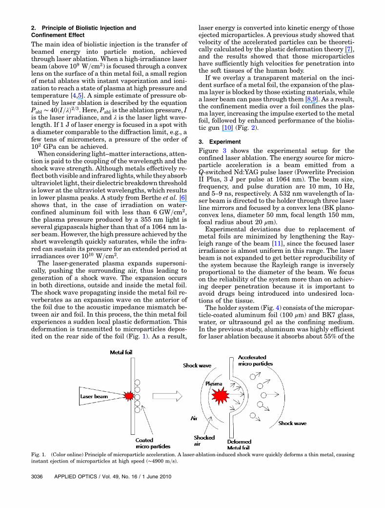
The laser-generated plasma expands supersoni-cally, pushing the surrounding air, thus leading togeneration of a shock wave. The expansion occursin both directions, outside and inside the metal foil.The shock wave propagating inside the metal foil re-verberates as an expansion wave on the anterior ofthe foil due to the acoustic impedance mismatch be-tween air and foil. In this process, the thin metal foilexperiences a sudden local plastic deformation. Thisdeformation is transmitted to microparticles depos-ited on the rear side of the foil (Fig. 1). As a result,
laser energy is converted into kinetic energy of thoseejected microparticles. A previous study showed thatvelocity of the accelerated particles can be theoreti-cally calculated by the plastic deformation theory [7],and the results showed that those microparticleshave sufficiently high velocities for penetration intothe soft tissues of the human body.
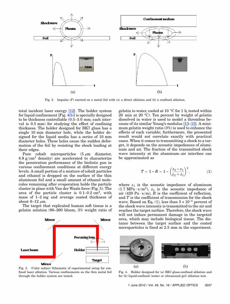
If we overlay a transparent material on the inci-dent surface of a metal foil, the expansion of the plas-ma layer is blocked by those existing materials, whilea laser beam can pass through them [8,9]. As a result,the confinement media over a foil confines the plas-ma layer, increasing the impulse exerted to the metalfoil, followed by enhanced performance of the biolis-tic gun [10] (Fig. 2).
3. Experiment
Figure 3 shows the experimental setup for theconfined laser ablation. The energy source for micro-particle acceleration is a beam emitted from aQ-switched Nd:YAG pulse laser (Powerlite PrecisionII Plus, 3 J per pulse at 1064 nm). The beam size,frequency, and pulse duration are 10 mm, 10 Hz,and 5–9 ns, respectively. A 532 nm wavelength of la-ser beam is directed to the holder through three laserline mirrors and focused by a convex lens (BK plano-convex lens, diameter 50 mm, focal length 150 mm,focal radius about 20 μm).
Experimental deviations due to replacement ofmetal foils are minimized by lengthening the Ray-leigh range of the beam [11], since the focused laserirradiance is almost uniform in this range. The laserbeam is not expanded to get better reproducibility ofthe system because the Rayleigh range is inverselyproportional to the diameter of the beam. We focuson the reliability of the system more than on achiev-ing deeper penetration because it is important toavoid drugs being introduced into undesired loca-tions of the tissue.
The holder system (Fig. 4) consists of the micropar-ticle-coated aluminum foil (100 μm) and BK7 glass,water, or ultrasound gel as the confining medium.In the previous study, aluminum was highly efficientfor laser ablation because it absorbs about 55% of the
Fig. 1. (Color online) Principle of microparticle acceleration. A laser-ablation-induced shock wave quickly deforms a thin metal, causinginstant ejection of microparticles at high speed (∼4900 m=s).
3036 APPLIED OPTICS / Vol. 49, No. 16 / 1 June 2010
total incident laser energy [12]. The holder systemfor liquid confinement [Fig. 4(b)] is specially designedto be thickness controllable (0:5–3:0 mm; each inter-val is 0:5 mm) for studying the effect of confiningthickness. The holder designed for BK7 glass has asingle 10 mm diameter hole, while the holder de-signed for the liquid media has a series of 10 mmdiameter holes. These holes cause the sudden defor-mation of the foil by resisting the shock loading attheir edges.
Pure cobalt microparticles (5 μm diameter,8:9 g=cm3 density) are accelerated to characterizethe penetration performance of the biolistic gun invarious confinement conditions at different energylevels. A small portion of a mixture of cobalt particlesand ethanol is dropped on the surface of the thinaluminum foil and a small amount of ethanol mole-cules remaining after evaporation holds the particlecluster in place with Van der Waals force (Fig. 5). Thearea of the particle cluster is 0:1–0:2 cm2, withmass of 1–2 mg and average coated thickness ofabout 6–12 μm.
The target that replicated human soft tissue is agelatin solution (90–260 bloom, 3% weight ratio of
gelatin to water, cooled at 10 °C for 1 h, tested within20 min at 20 °C). Ten percent by weight of gelatindissolved in water is used to model a thrombus be-cause of its similar Young’s modulus [13–15]. A mini-mum gelatin weight ratio (3%) is used to enhance theeffects of each variable; furthermore, the presentedresult would not correlate exactly with practicalcases. When it comes to transmitting a shock to a tar-get, it depends on the acoustic impedances of alumi-num and air. The fraction of the transmitted shockwave intensity at the aluminum–air interface canbe approximated as
T ¼ 1 − R ¼ 1 −
�z2 − z1z2 þ z1
�2; ð1Þ
where z1 is the acoustic impedance of aluminum(1:7 MPa · s=m2), z2 is the acoustic impedance ofair (420 Pa · s=m), R is the coefficient of reflection,and T is the coefficient of transmission for the shockwave. Based on Eq. (1), less than 5 × 10⁻⁴ percent ofthe shock wave intensity is transmitted to the air andreaches the target surface. Therefore, the shock wavewill not induce permanent damage in the targetedarea, which may include biological tissue. The dis-tance between the target surface and the coatedmicroparticles is fixed at 2:5 mm in the experiment.
Fig. 2. Impulse (F) exerted on a metal foil with (a) a direct ablation and (b) a confined ablation.
Fig. 3. (Color online) Schematic of experimental setup for con-fined laser ablation. Various confinements on the thin metal foilthrough the holder system are tested.
Fig. 4. Holder designed for (a) BK7-glass-confined ablation andfor (b) liquid-confined (water or ultrasound gel) ablation test.
1 June 2010 / Vol. 49, No. 16 / APPLIED OPTICS 3037

The cobalt microparticles are accelerated into ge-latin–water solution of varying conditions and laserenergy levels, and the administered shapes of theparticle clusters are measured. The shape is approxi-mately a cylinder with the measured width anddepth of a rectangle as viewed from one side. The con-fining media used in this experiment are BK7 glass(5 mm thickness), water (1:0 mm thickness), andultrasound gel (RHAPAPHRM Co., Ltd., 1.0, 1.5,
2.0, 2.5, 3:0 mm thickness), which is widely usedin clinical practice. For BK7 glass, the gap betweenthe glass and the upper surface of the aluminum foilcan be filled with either air or water for identifyingthe optimal confinement condition.
4. Results and Discussions
The experimental results at laser energy 1070�10 mJwith various confinement conditions are listedin Table 1. The penetration shapes of microparticlesvary widely depending on the confinements. Directablation shows that the microparticles rarely pene-trate the gelatin target. Biolistic injection with theliquid confinements (water or ultrasound gel) sug-gests more efficient momentum transfer over the so-lid (BK7 glass). In the BK7-glass-confined ablation,most of the laser energy is reflected at the glass sur-face or dissipated by the glass layer such that lessthan 20% of the laser energy reaches the metal foilsurface [16]. In the liquid-confined ablation, however,liquid can be heated by the transmitted laser energyso as to effectively take part in the generation of plas-ma. Also, there is no vacancy between the foil andliquid media that allows for the expanding plasmato escape and, ultimately, the liquid confines theplasma more efficiently than the solid.
The dependence of the penetration width on theconfinement conditions of beam refractions and
Fig. 5. Particle cluster deposited on a surface of the aluminumfoil.
Table 1. Laser-Assisted Particle Injection via 1070� 10 mJ with Different Confinement Materialsa
Confinements (Thickness in mm) Penetration Depth (mm) Penetration Width (mm) Image of Penetrations
Direct ablation without confinement 0:18� 0:13 1:72� 0:32
BK7 glass (5.0) 0:51� 0:30 0:45� 0:27
Water (1.0) 2:65� 0:29 2:85� 0:36
BK7 glass coated with water (≪1:0) 2:05� 0:19 1:38� 0:11
Ultrasound gel (1.0) 1:72� 0:20 1:54� 0:17
Ultrasound gel (1.5) 2:39� 0:43 1:54� 0:09
aEach value shown is an average of 20 tests.
3038 APPLIED OPTICS / Vol. 49, No. 16 / 1 June 2010
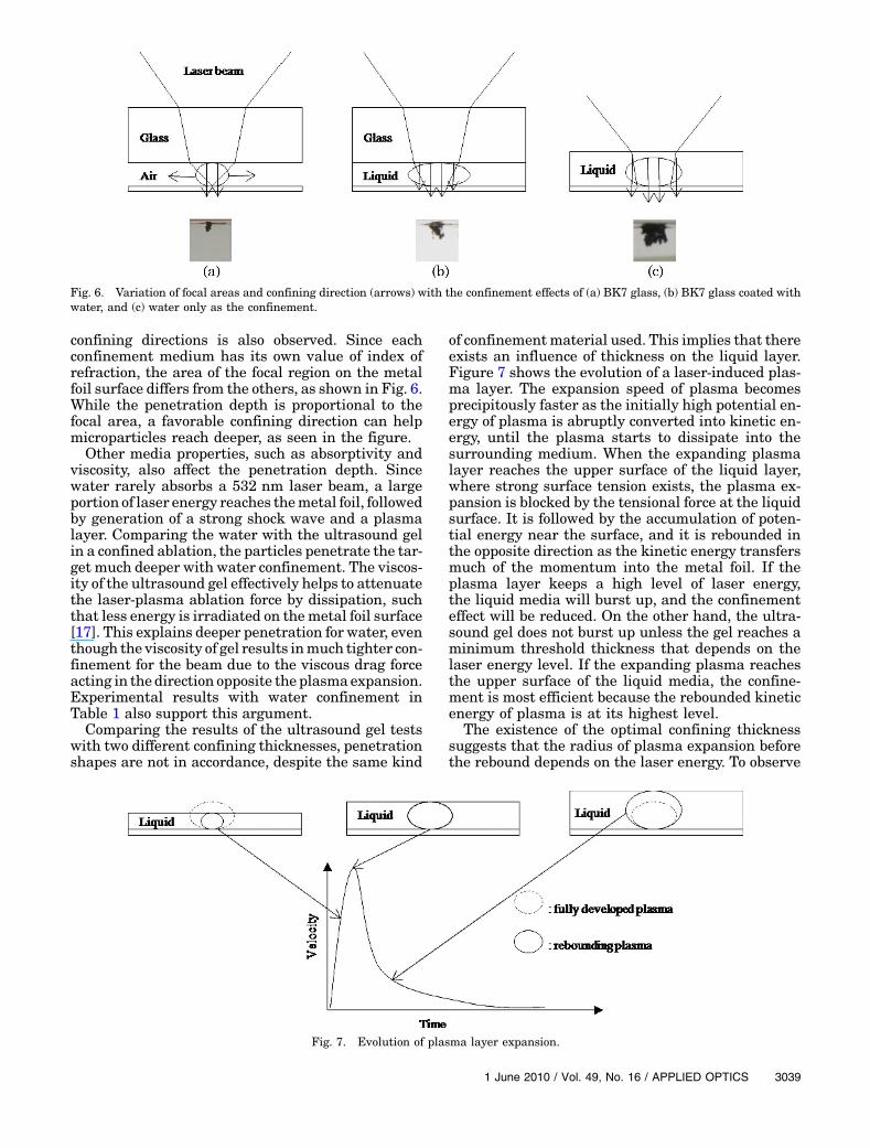
confining directions is also observed. Since eachconfinement medium has its own value of index ofrefraction, the area of the focal region on the metalfoil surface differs from the others, as shown in Fig. 6.While the penetration depth is proportional to thefocal area, a favorable confining direction can helpmicroparticles reach deeper, as seen in the figure.
Other media properties, such as absorptivity andviscosity, also affect the penetration depth. Sincewater rarely absorbs a 532 nm laser beam, a largeportion of laser energy reaches themetal foil, followedby generation of a strong shock wave and a plasmalayer. Comparing the water with the ultrasound gelin a confined ablation, the particles penetrate the tar-get much deeper with water confinement. The viscos-ity of the ultrasound gel effectively helps to attenuatethe laser-plasma ablation force by dissipation, suchthat less energy is irradiated on themetal foil surface[17]. This explains deeper penetration for water, eventhough the viscosity of gel results inmuch tighter con-finement for the beam due to the viscous drag forceacting in the direction opposite the plasmaexpansion.Experimental results with water confinement inTable 1 also support this argument.
Comparing the results of the ultrasound gel testswith two different confining thicknesses, penetrationshapes are not in accordance, despite the same kind
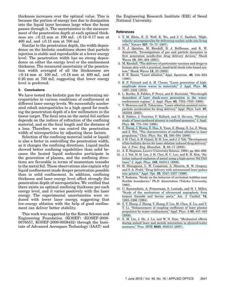
of confinementmaterial used. This implies that thereexists an influence of thickness on the liquid layer.Figure 7 shows the evolution of a laser-induced plas-ma layer. The expansion speed of plasma becomesprecipitously faster as the initially high potential en-ergy of plasma is abruptly converted into kinetic en-ergy, until the plasma starts to dissipate into thesurrounding medium. When the expanding plasmalayer reaches the upper surface of the liquid layer,where strong surface tension exists, the plasma ex-pansion is blocked by the tensional force at the liquidsurface. It is followed by the accumulation of poten-tial energy near the surface, and it is rebounded inthe opposite direction as the kinetic energy transfersmuch of the momentum into the metal foil. If theplasma layer keeps a high level of laser energy,the liquid media will burst up, and the confinementeffect will be reduced. On the other hand, the ultra-sound gel does not burst up unless the gel reaches aminimum threshold thickness that depends on thelaser energy level. If the expanding plasma reachesthe upper surface of the liquid media, the confine-ment is most efficient because the rebounded kineticenergy of plasma is at its highest level.
The existence of the optimal confining thicknesssuggests that the radius of plasma expansion beforethe rebound depends on the laser energy. To observe
Fig. 6. Variation of focal areas and confining direction (arrows) with the confinement effects of (a) BK7 glass, (b) BK7 glass coated withwater, and (c) water only as the confinement.
Fig. 7. Evolution of plasma layer expansion.
1 June 2010 / Vol. 49, No. 16 / APPLIED OPTICS 3039

it, the biolistic gun was tested with varyingthicknesses (1.0, 1.5, 2.0, 2.5, 3:0 mm) of ultrasoundgel at three different energy levels (100� 5 mJ,400� 10 mJ, 700 mJ� 10 mJ).
The results of penetration depth (Fig. 8) show thatthe optimal confining thicknesses of ultrasound gelat three energy levels are approximately 2:0 mm
at 700 mJ, 1:5–2:0 mm at 400 mJ, and about1:0 mm at 100 mJ. These are the averages of 15 pe-netration tests. The optimal confining thickness is apositively proportional function of the energy, and itis approximately equal to the distance that plasmagrows until it reaches a peak point of evolution.The penetration depth decreases as the confining
Fig. 8. Penetration depths and widths measurements for microparticle acceleration.
3040 APPLIED OPTICS / Vol. 49, No. 16 / 1 June 2010
thickness increases over the optimal value. This isbecause the portion of energy loss due to dissipationinto the liquid layer becomes large when the beampasses through it. The uncertainties in the measure-ment of the penetration depth at each optimal thick-ness are �0:12 mm at 100 mJ, �0:12–0:17 mm at400 mJ, and �0:15 mm at 700 mJ.
Similar to the penetration depth, the width depen-dence on the biolistic conditions shows that particleinjection is stable and reproducible at a lower energylevel. The penetration width has no strong depen-dence on either the energy level or the confinementthickness. The measured uncertainty of the penetra-tion width averaged of all the thicknesses are�0:14 mm at 100 mJ, �0:18 mm at 400 mJ, and0:20 mm at 700 mJ, suggesting that lower energylevel is preferred.
5. Conclusions
We have tested the biolistic gun for accelerating mi-croparticles in various conditions of confinement atdifferent laser energy levels. We successfully acceler-ated cobalt microparticles to a high speed for reach-ing the penetration depth of a few millimeters in thetissue target. The focal area on the metal foil surfacedepends on the indices of refraction of the confiningmaterial, and on the focal length and the distance ofa lens. Therefore, we can control the penetrationwidth of microparticles by adjusting these factors.
Selection of the confining material (solid or liquid)is also a factor in controlling the penetration width,as it changes the confining directions. Liquid mediashowed better confining capabilities than solid be-cause the heated liquid molecules participate inthe generation of plasma, and the confining direc-tions are favorable in terms of momentum transferto themetal foil. These observations also explain whyliquid confinementmade deeper penetration possiblethan in solid confinement. In addition, confiningthickness and laser energy level affect strongly thepenetration depth of microparticles. We verified thatthere exists an optimal confining thickness per eachenergy level, and it varies positively with the laserenergy. The experimental uncertainties were re-duced with lower laser energy, suggesting thatlow-energy ablation with the help of good confine-ment can deliver better stability.
This work was supported by the Korea Science andEngineering Foundation (KOSEF) (KOSEF-2008-0076557, KOSEF-2009-0059432) through the Insti-tute of Advanced Aerospace Technology (IAAT) and
the Engineering Research Institute (ERI) of SeoulNational University.
References1. T. M. Klein, E. D. Wolf, R. Wu, and J. C. Sanford, “High-
velocity microprojectiles for delivering nucleic acids into livingcells,” Nature 327, 70–73 (1987).
2. N. J. Quinlan, M. Kendall, B. J. Bellhouse, and R. W.Ainsworth, “Investigations of gas and particle dynamics infirst generation needle-free drug delivery devices,” ShockWaves 10, 395–404 (2001).
3. M. Kendall, “The delivery of particulate vaccines and drugs tohuman skin with a practical, hand-held shock tube-based sys-tem,” Shock Waves 12, 23 (2002).
4. R. E. Russo, “Laser ablation,” Appl. Spectrosc. 49, 14A–28A(1995).
5. B. P. Fairand and A. H. Clauer, “Laser generation of high-amplitude stress waves in materials,” J. Appl. Phys. 50,1497–1502 (1979).
6. L. Berthe, R. Fabbro, P. Perye, and E. Bartnicki, “Wavelengthdependent of laser shock-wave generation in the water-confinement regime,” J. Appl. Phys. 85, 7552–7555 (1999).
7. V. Menezes and K. Takayama, “Laser-ablation-assisted micro-particle acceleration for drug delivery,” Appl. Phys. Lett. 87,163504 (2005).
8. R. Fabbro, J. Fournier, P. Ballard, and D. Devaux, “Physicalstudy of laser-produced plasma in confined geometry,” J. Appl.Phys. 68, 775–784 (1990).
9. Z. Zheng, J. Zhang, Z. Hao, X. Yuan, Z. Zhang, X. Lu, Z. Wang,and Z. Wei, “The characteristics of confined ablation in laserpropulsion,” Chin. Phys. Soc. 15, 580–584 (2006).
10. J. H. Choi, A. B. Gojani, H. H. Lee, and J. J. Yoh, “Developmentof bio-ballistic device for laser ablation induced drug delivery,”Int. J. Prec. Eng. Manufact. 9, 68–71 (2008).
11. A. E. Siegman,Lasers (University Science, 1986), pp. 664–669.12. J. J. Yoh, H. H. Lee, J. H. Choi, K. C. Lee, and K. H. Kim, “Ab-
lation induced explosion of metal using a high-power Nd:YAGlaser,” J. Appl. Phys. 103, 043511 (2008).
13. H. Shangguan, L. W. Casperson, A. Shearin, K. W. Gregory,and S. A. Prahl, “Drug delivery with microsecond laser pulsesinto gelatin,” Appl. Opt. 35, 3347–3357 (1996).
14. T. Kodama, “Study on the behavior of cavitation bubbles nearflexible boundaries,” Ph.D. dissertation (Tohoku University,1992).
15. U. Rosenschein, A. Frimerman, S. Laniado, and H. I. Miller,“Study of the mechanism of ultrasound angioplasty fromhuman thrombi and bovine aorta,” Am. J. Cardiol. 74,1263–1266 (1994).
16. Z. Y. Zheng, J. Zhang, Y. Zhang, F. Liu, M. Chen, X. Lu, and Y.T. Li, “Enhancement of coupling coefficient of laser plasmapropulsion by water confinement,” Appl. Phys. A 85, 441–443(2006).
17. X. M. Liu, J. He, J. Lu, and W. N. Xiao, “Mechanical effectsduring pulsed laser and metals interaction in glycerol-watermixtures,” Proc. SPIE 6825, 682513 (2007).
1 June 2010 / Vol. 49, No. 16 / APPLIED OPTICS 3041
Copyright © 2022 FDOKUMEN