In vitro-in vivo correlation study on nimesulide loaded hydroxypropylmethylcellulose microparticles
134
Transcript of In vitro-in vivo correlation study on nimesulide loaded hydroxypropylmethylcellulose microparticles


药 学 学 报 第 45卷 第 6期 2010年 6月
综 述 聚乙二醇修饰脂质体的 ABC 现象研究进展 ………………………… 徐 缓, 王凯乾, 黄微崴, 邓意辉, 陈大为 (677)HIV-1病毒感染因子Vif及其相关抑制剂的研究进展 ……………………………………… 李震宇, 展 鹏, 刘新泳 (684)基于微流控芯片的酶及其抑制剂的研究进展 …………………………………… 侯凤华, 叶剑清, 陈缵光, 成志毅 (694)小檗碱调节血糖血脂代谢紊乱机制研究进展 …………………………………… 沈 宁, 李彩娜, 环 奕, 申竹芳 (699) 研 究 论 文
药理学
孕期酒精暴露对子鼠视皮质突触数量影响的体视学研究 …………… 席 艳, 张俊士, 臧建峰, 文曙光, 邓锦波 (705)贯众总多糖对空肠弯曲杆菌诱导的系统性红斑狼疮样综合征小鼠的作用
…………………………… 王 铮, 谢俊云, 徐 晗, 程小芹, 乐曦玲, 力 弘, 章蕴毅, 卢 燕, 陈道峰
(711)
一种大鼠慢性哮喘模型的建立与评价 ………………………………………………………………… 刘中成, 张艳芬 (718)神经突触核蛋白-γ过表达降低肝癌细胞对抗微管药物的敏感性
…………………………… 程世翔, 张 赛, 张 豪, 宋丹青, 王宇萍, 李玉环, 游雪甫, 王跃明, 蒋建东
(724)
药物化学 Synthesis and anti-inflammatory activity of 2-substituted-((N, N-disubstituted) - 1, 3-benzoxazole)-5-carboxamides
……………………………………………… Reena M, kiran G, Rajyalakshmi G, Venkateshwa Rao J, Sarangapani M
(730)新型组蛋白去乙酰化酶抑制剂的合成及抗肿瘤活性研究
………………………………………………… 程永浩, 郭彦伸, 韩海珠, 王 楠, 张国宏, 郭宗儒, 吴 松
(735)
虎耳草的化学成分研究 (英文) …………………………… 冯卫生, 李 振, 郑晓珂, 李原京, 苏芳谊, 张艳丽 (742)胆木叶生物碱类成分研究 ………………………… 范 龙, 范春林, 王 英, 张晓琦, 张庆文, 张俊清, 叶文才 (747) 药物分析与药物代谢 重组嵌合抗CD20 IgG1型单克隆抗体的结构验证 ………… 陶 磊, 饶春明, 高 凯, 史新昌, 赵 阳, 王军志 (752)人参与金银花、何首乌、黄芪配伍的化学成分变化研究及抗氧化活性测定
……………………………………………………………………… 杜芹芹, 张 旭, 宋凤瑞, 刘志强, 刘淑莹
(756)
毛橘红醇提物中柚皮苷、柚皮素在大鼠尿液和粪便中的代谢与排泄 ……………………………………………………………………… 孙国玲, 钱大玮, 段金廒, 李向明, 万建义
(761)
同位素稀释质谱法测定人血浆中奥氮平的方法学及生物等效性研究 (英文) ……… 张梦琪, 贾晶莹, 陆 川, 刘罡一, 余成寅, 桂雨舟, 刘 昀, 刘艳梅, 王 伟, 李水军, 余 琛
(767)
药剂学 In vitro-in vivo correlation study on nimesulide loaded hydroxypropylmethylcellulose microparticles
……………………………………………………………………………… Shujaat Ali KHAN, Mahmood AHMAD, Ghulam MURTAZA, Muhammad Naeem AAMIR, Rozina KOUSAR, Fatima RASOOL, Shahiq-u-ZAMAN
(772)
玻璃体腔注射伏立康唑缓释微球对兔烟曲霉菌性眼内炎的抗感染作用 ……… 杨丽娜, 辛 萌, 吴祥根, 姜皓然 (778) 生药学 丹参乙酰 CoA 酰基转移酶基因全长克隆和 SNP 分析 ………… 崔光红, 王学勇, 冯 华, 赵静雪, 黄璐琦 (785) 研 究 简 报
基于小鼠温度趋向行为学表征的左金丸及反左金丸寒热属性 ……………………………………… 杨宏博, 赵艳玲, 李宝才, 王伽伯, 李瑞生, 贾 雷, 程丹红, 肖小河
(791)
小分子化合物 ZL-004 升高白细胞的作用 …………………………… 孙海燕, 李春刚, 肖 璘, 王国平, 刘全海 (797)二甲双胍对 2型糖尿病大鼠肝纤维化形成的影响
……………………………………… 强桂芬, 张 莉, 宣 琪, 杨秀颖, 时丽丽, 张恒艾, 陈柏年, 杜冠华
(801)
信 息
(683) 第八届全国天然有机化学学术研讨会 (704) 《药学学报》第十一届编委会及学术报告会会议通知 (723) 《药学学报》入选WHO西太平洋地区医学索引 (WPRIM)

《药学学报》为我国自然科学核心期刊、中文核心期刊
1999年荣获首届“国家期刊奖”
2001年入选中国期刊方阵“双高”(高知名度、高学术水平)期刊
2002年被评为第二届“国家期刊奖百种重点科技期刊”,并荣获第三届“中国科技优秀期刊奖”二等奖
2002~2008年连续 7届荣获“百种中国杰出学术期刊”称号
2008,2009年获得中国科协精品科技期刊工程项目资助(B类)
本刊已被世界主要检索系统收录
美国《医学索引》(IM/Medline) 美国《化学文摘》(CA on CD)
美国《国际药学文摘》(IPA) 美国《生物学文摘》(BA)
俄罗斯《文摘杂志》(AJ, VINITI) 波兰《哥白尼索引》(IC)
美国《乌利希国际期刊指南》 (UIPD) 《制药及药品毒理生化数据库》 (TOXCENTER)
英国《国际农业与生物科学研究中心》(CABI) 日本《科学技术文献速报》(CBST)
根据《中国科技期刊引证报告(2009年度,核心版)》统计
2008年《药学学报》总被引频次为 2910,影响因子为 0.903
期刊基本参数: CN 11-2163/R*1953*m*A4*130*zh*P*¥30.00* *22*2010-06 本期责任编辑 范 颖
药学学报 (YAOXUE XUEBAO) ACTA PHARMACEUTICA SINICA (月刊, 1953年 7月创刊) (Monthly, Founded in 1953 July)
主管单位: 中国科学技术协会 Directed by: China Association for Science and Technology 主办单位: 中国药学会 http://www.cpa.org.cn Sponsored by: Chinese Pharmaceutical Association
http://www.cpa.org.cn 编辑出版: 中国医学科学院药物研究所 药学学报编辑部 (100050 北京市先农坛街 1号) 电话: 86-10-63035012, 63026192; 传真: 86-10-63026192; 电子信箱: [email protected]; 网址: http://www.yxxb.com.cn
Edited and Published by: Editorial Office of Acta Pharmaceutica Sinica, Institute of Materia Medica, Chinese Academy of Medical Sciences (1 Xiannongtan Street, Beijing 100050). Tel: 86-10-63035012, 63026192; Fax: 86-10-63026192; E-mail: [email protected]; http://www.yxxb.com.cn
主编: 王晓良 Editor-in-chief: WANG Xiao-liang 印刷: 北京科信印刷厂 Printed by: Beijing Kexin Printing House 国内订购: 全国各地邮电局 Domestic subscriptions: Local Post Offices 发行范围: 公开发行 Distribution 国 内: 北京报刊发行局 Domestic: Beijing Post Offices 国 外: 中国国际图书贸易总公司
(北京市 399信箱, 100044) Foreign: China International Book Trading Corporation,
PO Box 399, Beijing 100044, China
ISSN 0513-4870 2010年 第 45卷 第 6期 2010年 6月 12日出版 邮发代号: 2-233 CN 11-2163/R 2010, Vol. 45, No.6 Publication Date: 2010-06-12 Code number: M105 广告经营许可证:京西工商广字第 0437号 国内定价: 每期 30.00元

药学学报 Acta Pharmaceutica Sinica 2010, 45 (6): 677−683 · 677 ·
聚乙二醇修饰脂质体的 ABC现象研究进展
徐 缓 1, 王凯乾 2, 黄微崴 2, 邓意辉 2*, 陈大为 2
(1. 辽宁师范大学化学化工学院, 辽宁 大连 116029; 2. 沈阳药科大学药学院, 辽宁 沈阳 110016)
摘要: 通常聚乙二醇 (polyethylene glycol, PEG) 修饰脂质体被认为几乎没有或仅有很低的免疫原性。最新的文献报道, 重复注射 PEG 修饰脂质体发生了免疫反应。当向同一动物体内重复注射 (间隔几天) PEG 化脂质 体时, 二次注射的 PEG 化脂质体导致体内循环时间降低, 于肝和脾的聚集量增加, 这种现象称为“加速血液清除” (accelerated blood clearance, ABC) 现象。该免疫反应使 PEG化制剂的发展和临床应用面临严峻的挑战, 可能造成药物或基因治疗效率的下降, 甚至引起临床的毒副作用。本文综述了 ABC 现象的定义、验证 ABC 现象的方法和手段、ABC 现象成因的研究进展及影响因素, 并对其他 PEG 修饰载体是否也会发生 ABC 现象进行了探讨。
关键词: 聚乙二醇; 脂质体; 加速血液清除; 抗-聚乙二醇免疫球蛋白M 中图分类号: R943 文献标识码: A 文章编号: 0513-4870 (2010) 06-0677-07
Recent advances in the study of accelerated blood clearance phenomenon of PEGylated liposomes
XU Huan1, WANG Kai-qian2, HUANG Wei-wei2, DENG Yi-hui2*, CHEN Da-wei2
(1. College of Chemistry and Chemical Engineering, Liaoning Normal University, Dalian 116029, China; 2. China School of Pharmacy, Shenyang Pharmaceutical University, Shenyang 110016, China)
Abstract: It is generally believed that liposomes modified with polyethylene glycol (PEG) have no or lower
immunogenicity. However, based on many recent literatures, when the PEGylated liposomes were repeatedly applied to the same animal, the immune responses occurred. The first injection of PEGylated liposomes resulted in a reduction in the circulation time and an increase in hepatic and splenic accumulation of the second dose of PEGylated liposomes in a time-interval, which was called “accelerated blood clearance (ABC)” phenomenon. Such immunogenicity of PEGylated liposomes presents a barrier in the research of liposomal formulations and their use in the clinics. This review focused on the definition, the method of verification, the development of the reason for ABC phenomenon, influencing factors of ABC phenomenon, and discussed if other PEGylated nanocarriers also induce ABC phenomenon.
Key words: polyethylene glycol; liposome; accelerated blood clearance; anti- polyethylene glycol IgM
普通脂质体作为药物递送载体较易被单核巨噬
细胞系统 (mononuclear phagocyte system, MPS) 吞噬而迅速从血液循环中消失, 为了解决此问题, 研究人员将聚乙二醇 (polyethylene glycol, PEG) 类脂质衍生物修饰于脂质体表面, 利用 PEG 的亲水性和柔 收稿日期: 2009-10-13. *通讯作者 Tel: 86-24-23986316, Fax: 86-24-62561042,
E-mail: [email protected]
顺性, 延长脂质体体内循环时间, 减少 MPS 的分布量, 增强靶向性, 同时也明显提高了脂质体及药物的物理、化学和生物学稳定性。PEG 化脂质体的临床研究已有多年历史, PEG化脂质体以及相关产品的开发成为目前脂质体研究领域中的热点。
临床使用时 PEG 化脂质体需要重复注射, 然而目前关于体内重复注射 PEG 化脂质体的药动学研究资料比较缺乏。一直以来, PEG化脂质体都被认为没
·综述·

· 678 · 药学学报 Acta Pharmaceutica Sinica 2010, 45 (6): 677−683
有免疫原性, 但是脂质体在体内的行为可能比预想的更为复杂。有研究者发现, 当向同一动物体内重复注射 (间隔几天) PEG 化脂质体时, 二次注射的 PEG化脂质体丧失长循环特性, 这一现象被称之为 ABC现象[1−3]。这种脂质体药动学特性的改变在其临床应
用中是一个重大缺陷, 如果脂质体包封的药物具有细胞毒性, 那么脂质体在肝脏巨噬细胞的聚集会导致这些细胞的凋亡和坏死。而肝脏 Kupffer细胞的恢复需要2周时间, Kupffer细胞缺失的这段时间会引起菌血症, 对于癌症患者是致命的。ABC现象也会造成药物或基因治疗效率的下降, Semple等[4]的研究表明, 重复注射包封寡核苷酸、pDNA或 RNA核酶的 PEG化脂质体会诱导强烈的免疫应答, 导致制剂血液循环时间缩短和小鼠死亡率显著增加。 1 ABC现象的提出
PEG 化脂质体重复注射于同一动物体内时会发生异常的药代动力学的改变, 一般表现为二次注射的 PEG 化脂质体血液清除速度加快, 在肝和脾的聚集量增加。Dams等[1]以 99mTc标记脂质体, 研究大鼠重复注射 PEG 化脂质体的情况。结果发现, 二次注射的脂质体血浆水平明显降低, 且肝摄取量从 (8.1 ± 0.8) % 升至 (46.2 ± 9.8) % 。对恒河猴进行考察时发现
也产生明显的 ABC 现象, 脂质体的半衰期由 87.5 h减至 14.2 h, 而肝摄取量由 17.6% 增至 41.2%[2]。该项
研究者首次提出 ABC现象。 目前研究一般将 ABC 现象分为两相[2]: 诱导相
(induction-phase) ——首次注射脂质体后生物体已接触抗原 (表明某种可传输血清因子形成); 完成相 (effectuation-phase) ——二次注射后 PEG化脂质体从血液循环中迅速消除 (图 1)。
Figure 1 Schematic representation of the time frame of the two phases of the so-called enhanced clearance effect
2 验证 ABC现象的方法和手段
在研究 ABC 现象时, 研究人员通过将二次注射的 PEG化脂质体进行示踪标记——利用锝标记 PEG (99mTc-PEG)[1, 2]或 3H-CHE[3]标记脂质体中脂质组分
等手段来确定二次注射脂质体的药动学行为和组织
分布的变化。ABC 现象研究中应用的动物模型多为
大鼠[2, 3], 一般处理过程是经大鼠的右股静脉首次给药, 乙醚麻醉后左股静脉和动脉插管, 将标记的 PEG化脂质体通过股静脉插管注入, 进行二次给药。在不同的时间间隔经股动脉插管将血浆样品取出, 最后将大鼠处死, 取出脏器, 测定 PEG 化脂质体的组织分布情况。也有人采用小鼠、恒河猴等作为动物模型[1, 5]。
肝脏清除率能够反映 Kupffer细胞对脂质体的摄取活性, 是一个很好的比较 ABC现象程度的参数[6, 7]。
近年来普遍认为免疫球蛋白或补体与 ABC 现象的诱导有密切关系, 因此测定免疫球蛋白 M (IgM) 或补体也成为研究 ABC 现象的手段之一。可利用IgM 酶联免疫吸附定量试剂盒测定大鼠血清中与脂
质体结合的 IgM[3]。另外, 为了验证血清中是否含有诱导 ABC现象的血清因子, 有人将经预注射 PEG化脂质体的大鼠血清加热至 56 ℃ (30 min) 发现, 该血清可以抑制所有的补体活性[8]。 3 ABC现象成因的研究进展
有研究人员提出ABC现象是由于 PEG层从脂质体表面脱离引起, 认为摩尔比为 5% 的 PEG脂质修饰脂质体缺乏长循环特性的原因是: 当 PEG 化脂质体以这样的低浓度出现在大体积的血浆中时, 可能造成 PEG与脂质体的分离[6]。但是, Parr等[9]的研究表
明静注 PEG化脂质体 24 h后, PEG-二硬脂酰磷脂酰胆碱 (PEG-DSPE) 也很少或几乎没有从脂质体表面脱离, 该研究采用脂质体的处方与研究 ABC 现象时所用处方是相似的。这一结论被 Silvius 等[10]支持: PEG-DSPE (其中PEG的相对分子质量为1 900) 从脂质双分子层的交换半衰期为 70 h。因此, PEG脂质从脂质体表面的脱离不可能是产生 ABC 现象的主要原因。
Laverman等[2]推测 ABC现象的产生是由于肝脾巨噬细胞分泌了一种不耐热的血清因子, 而巨噬细胞对脂质体的吞噬与这种血清因子的产生有关。将 经过预注射脂质体的大鼠血清通过抗大鼠 IgM 琼脂
糖层析柱后, 得到 IgM-衰竭的血清, 洗脱琼脂糖层析柱则得到 IgM 级分。当向大鼠灌注该 IgM级分时并没有引起二次注射 PEG 化脂质体药动学的异常变化, 而注射 IgM-衰竭的血清则引起其清除加快, 因此得出结论: ABC 现象并不是由 IgM 诱导的。这与 一些 ABC 现象相关的研究结论[5, 11]相悖, 其原因可能是 IgM-衰竭血清中残留的 IgM (约 20%) 被浓缩在脂质体表面, 通过与 PEG的选择性结合, 激活了 C系统 —— 继而通过酶促反应串联被极大地放大。Ishida

徐 缓等: 聚乙二醇修饰脂质体的 ABC现象研究进展 · 679 ·
等 [11]通过二维聚丙烯酰胺凝胶电泳法 (2D-PAGE) 和十二烷基硫酸钠-聚丙烯酰胺凝胶电泳法 (SDS- PAGE) 证明在预处理血清中与PEG化脂质体结合的血清蛋白是 IgM。
可诱导免疫反应的二级抗原 (TI-2) 由细菌的细胞壁和荚膜多糖组成, 不依赖胸腺, 具有高度重复结构[5]。该抗原有可能与 B细胞表面的免疫球蛋白广泛交联, 导致 B 细胞分泌 IgM 和 IgG。当 TI-2 激活 B细胞时, 抗原决定簇的密度是关键。密度过低不足以激活细胞; 密度过高则细胞反应性反而降低。PEG聚合物也具有高度重复结构, 低剂量的 PEG化脂质体能够诱导ABC现象, 表明在此条件下抗原决定簇 (PEG) 的密度足以激活 B细胞。一旦 PEG化脂质体到达脾脏, 与被 PEG (或 PEG化脂质体) 激活的 B细胞表面的抗原结合、交联, 导致抗-PEG IgM的产生[3]。
众所周知, 脾在免疫反应中扮演着重要角色。首次注射 PEG 化脂质体之前将脾切除, 则二次注射PEG 化脂质体的 ABC 现象完全消失; 未切除脾的对照组中大鼠血清与脂质体结合的 IgM 量比脾切除组
高 8倍, 这表明脂质体与 IgM的结合是诱导 ABC现象的关键因素[12]。PEG 化脂质体充当脾脏中 B 细胞的活化剂。
综合目前的研究, ABC 现象是通过以下机制产生[5, 11−13]: 首次注射的脂质体在脾脏产生抗-PEG IgM, 该血清因子选择性结合到几天后注射的 PEG 化脂质体表面的PEG上, 并随后激活补体系统, 导致补体C3片段对脂质体的调理作用, 结果增强了肝脏 Kupffer细胞对脂质体的摄取, 于是产生了 ABC现象。
除此之外, 关于 ABC现象尚有其他的解释[4, 14]: 首次注射的 PEG化脂质体引发了胸腺和/或血液中淋
巴细胞的免疫应答, 导致细胞因子和/或化学增活素的大量产生[15, 16], 因此导致非调理素依赖的 Kupffer细胞吞噬作用, 引起试验剂量的 PEG 化脂质体摄取的增加, 而该过程中并没有抗 PEG IgM 的产生, 但是相关证据并不充足。 4 ABC现象的影响因素 4.1 首次注射脂质体是否有 PEG 包衣 ABC 现象的早期研究认为仅当首次注射 PEG 化脂质体才能引起二次注射 PEG 化脂质体的加速清除, 但是 Wang等[17]发现首剂量的常规脂质体 (conventional liposome, CL) 也能诱导强烈的 ABC现象。与 PEG化脂质体或CL结合的 IgM的量及其伴随的由这些脂质体激发的补体激活作用与脂质体的加速清除反应强度之间存
在着良好的对应关系。预先注射 CL (磷脂剂量 5 μmol·kg−1), 则二次注射 PEG 化脂质体的循环时间急剧减少, 最强的 ABC 反应出现在两次注射间隔为 7天的实验组。首次注射 CL 14天后进行二次给药仍能观察到明显的ABC现象, 并伴有脾聚集量的增加, 但其增加程度小于肝脏 (图 2)[6]。
ABC 现象的产生是由于首次注射脂质体诱导抗PEG IgM 产生, 以及二次注射的由 PEG修饰而不是CL激发的补体激活, 因此ABC现象可被多种脂质体组分诱导, 但仅 PEG 修饰脂质体对该现象的完成相敏感。另外, 注射 PEG2000-DSPE (无脂质体) 不会诱导 ABC 现象[1], 这进一步说明脂质体组分可能是激发免疫球蛋白分泌的主要原因。 4.2 磷脂剂量的影响 Ishida 等[5]发现 PEG 化脂质体的首剂量与 ABC 现象发生的程度存在明显的反相关。大鼠首次注射 PEG化脂质体, 磷脂剂量分别为 0 (HEPES盐缓冲液)、0.001、0.01、0.1、1和 5 μmol·kg−1,
Figure 2 Induction of the ABC phenomenon by non-PEGylated liposomes[6]. A: Blood clearance profile of subsequently injected radio-labeled mPEG2000-liposomes; B: Hepatic and splenic accumulation of subsequently injected radiolabeled mPEG2000-liposomes at 24 h following the injection. ***P < 0.001 vs control (liver); △P < 0.05, △△P < 0.01 vs control (spleen)

· 680 · 药学学报 Acta Pharmaceutica Sinica 2010, 45 (6): 677−683
5 d 后注射放射标记的 PEG 化脂质体 (磷脂剂量 5 μmol·kg−1), 结果表明二次注射 PEG化脂质体的血液清除程度与肝聚集量随首剂量的降低而明显增加。当
首剂量磷脂大于 1 μmol·kg−1时, 二次注射的 PEG化脂质体血液清除速度不再继续加快。在与 IgM 结合
的实验中发现, PEG化脂质体和 IgM的结合量与首次注射 PEG 化脂质体的浓度呈反相关[11]: 低首剂量 (磷脂剂量 0.001 μmol·kg−1), 每 1 μmol磷脂会产生约9 μg IgM, 而高首剂量 (磷脂剂量 5 μmol·kg−1) 时, 每 1 μmol磷脂仅产生约 4 μg IgM。首次注射 CL, 仅在高剂量时才会导致 IgM结合量明显增加。
而 Laverman 等[2]的研究与上述 Ishida 的结果存在一定矛盾。首次注射磷脂剂量分别为 0.05、0.5和5.0 μmol·kg−1, 二次注射 PEG 化脂质体 (磷脂剂量5.0 μmol·kg−1) 时均能产生 ABC现象[7]。当固定首次
注射磷脂剂量为5.0 μmol·kg−1, 二次注射的PEG化脂质体磷脂剂量为 15或 50 μmol·kg−1时, 产生的 ABC现象比 5.0 μmol·kg−1组明显减弱 (图 3)。
上述研究表明首剂量对 ABC 现象影响的结论并不一致。这两组研究采用的磷脂类型不同, 前者应用的是氢化卵磷脂 (HEPC), 后者是部分氢化卵磷脂 (PHEPC)。在 ABC现象的研究中, 脂质体的处方多由HEPC、胆固醇和 PEG-DSPE组成[2, 5, 12], 或以氢化大豆磷脂 (HSPC) 代替 HEPC[3], 但是目前还没有实验数据证明磷脂类型对 ABC 现象的影响, 值得进一步研究和确证。 4.3 PEG-DSPE 浓度的影响 目前能够诱导产生ABC现象的 PEG化脂质体均以 PEG-DSPE为长循环材料, 因此有研究者考察了 PEG-DSPE在处方中的比例对 ABC 现象的影响。首次注射 PEG 化脂质体中PEG-DSPE占处方脂质摩尔比分别为 0%、5%、10%
和 15% (磷脂剂量为 0.001 μmol·kg−1), 5 d后注射 PEG化脂质体 (磷脂剂量为 5.0 μmol·kg−1, PEG-DSPE在处方中的摩尔比例为 5%)。结果发现, ABC现象发生的程度与 PEG-DSPE 的比例存在明显的反相关。PEG-DSPE比例为 0的脂质体 (即普通脂质体) 不产生ABC现象; 含有 5% PEG-DSPE的脂质体可以诱导产生明显的 ABC 现象, 且伴随肝聚集量显著增加; 进一步增加 PEG-DSPE的含量 (10%和 15%), 与 5%
组相比, 二次注射脂质体的清除速率与肝聚集呈现降低趋势 (P < 0.01), 但仍显著高于对照组[6]。结果
表明 ABC现象发生的程度与 PEG的密度有关。 Ishida 等[18]也发现增加 PEG-DSPE 的比例能减
弱 PEG化脂质体对 ABC现象的诱导。与首次剂量中PEG-DSPE 含量低 (摩尔比小于 5%) 的脂质体相比, 若首次注射脂质体中 PEG-DSPE 含量高 (摩尔比大于10%), 则其后注射的 PEG 化脂质体的肝脏蓄积量减少。 4.4 PEG 的分子量 研究表明, 首次注射摩尔比均为 5% 的 PEG2000-DSPE和 PEG5000-DSPE修饰脂质体 (磷脂剂量 0.001 μmol·kg−1), 5 天后注射 PEG2000- DSPE修饰的标记脂质体 (磷脂剂量 5 μmol·kg−1), 均产生明显的 ABC 现象 (未考察二次注射 PEG5000- DSPE修饰脂质体)[6], 因此认为PEG的分子量对诱导ABC现象没有影响。
Ishida 等[18]的结论恰恰相反: 首次注射 PEG5000- DSPE 修饰脂质体诱导 ABC 现象的程度远远低于首次注射 PEG2000-DSPE 修饰脂质体, PEG5000-DSPE 和PEG2000-DSPE 修饰脂质体组的肝聚集量分别为 (35.3 ± 3.8) % 和 (78.7 ± 8.8) %[11], 表明首次注射脂质体中 PEG分子量的增加可以减弱 ABC现象。
以上两组实验的区别是前者的动物模型为大鼠,
Figure 3 Effect of lipid dose on the pharmacokinetics of 99mTc-labeled PEG-liposomes[2]. A: Blood levels of 99mTc-PEG-liposomes 4 h after a first dose of 0.05, 0.5, and 5.0 μmol·kg−1 and after a second dose of 5 μmol·kg−1 given 1 week later. Blood levels were measured 4 h p.i.; B: Blood level of 99mTc-PEG-liposomes 4 h after a first injection of 5 μmol·kg−1. One week later rats were injected with lipid doses of 5, 15, and 50 μmol·kg−1, respectively

徐 缓等: 聚乙二醇修饰脂质体的 ABC现象研究进展 · 681 ·
后者为小鼠。因此 PEG分子量对 ABC现象的影响尚没有统一的结论, 亟待对其进行进一步的研究。 4.5 粒径和表面电荷的影响 在ABC现象研究中所应用的 PEG 化脂质体 (首次和二次注射) 粒径多为100 nm左右[6, 11, 17, 18], 有人考察了粒径对 ABC现象的影响。Dams 等[1]研究表明, ABC 现象与首次注射PEG 化脂质体的粒径、表面性质和放射性标记均无关。未标记的小 (85 nm) 或大 (400 nm) PEG化脂质体或小的 (100 nm) 非 PEG 化脂质体均会引起二次注射的锝标记 PEG化脂质体产生相似的 ABC现象。
Wang 等[19]发现, 首次注射 CL 的理化性质对ABC现象的诱导相有显著的影响。与 PEG化脂质体相比, 首次注射中性或荷电的 CL (粒径 110 nm左右)
时, 都不会引起明显的 ABC 现象, 进一步减小荷电 (正电和负电) CL的粒径 (60 nm) 时, ABC现象明显增强, 中性 CL则没有此现象。结果表明, 当 CL诱导ABC现象时, 粒径是一个主要的影响因素。通过减小首剂量 CL的粒径使 ABC现象增强。 4.6 药物——阿霉素的影响 包封阿霉素的长循环脂质体已上市, 但首次注射 PEG 化阿霉素脂质体不会引起二次注射 PEG化脂质体的 ABC现象[3]。阿霉
素脂质体能够抑制 ABC 现象可能是因为阿霉素从脂质体释放后进入脾脏, 伤害脾细胞而减少了抗-PEG IgM 的产生, 进而抑制 B 细胞的增殖和/或杀害 B细胞, 阻止了二次注射 PEG 化脂质体的加速清除。因此在考察 ABC 现象的时候不能选择阿霉素作为模型药物。 4.7 注射时间间隔以及连续注射的影响 Dams等[1] 发现能够诱导 ABC 现象的两次注射最小时间间隔为5 d; Ishida等[5]证明两次注射间隔为 7 d时 ABC现象最强。另有研究报道, 大鼠间隔 35 d第 2次注射 PEG化脂质体后, 再间隔 4或 7 d进行第 3次注射 PEG化
脂质体, 其血液清除速度虽略有增加, 但无显著性差异, 而在肝脾的聚集量没有变化, 说明多次重复注射并不能继续引发 ABC 现象 (图 4)。而每天注射 PEG化脂质体直至第 4次均未产生 ABC现象。三次注射或两次注射的间隔时间延长都会使 ABC 现象消失, 表明接受 PEG化脂质体的动物不能产生ABC作用的免疫记忆, 或者它们获得了对 PEG化脂质体的免疫耐受性。具有长循环性质的 PEG 化脂质体可能导致一种缓和持续的对淋巴细胞或巨噬细胞的刺激[7]。 4.8 不同动物模型 Dams 等[1]研究表明, 大鼠和恒河猴均可以产生 ABC现象, 小鼠则不能。Ishida等[18]
的结果表明小鼠体内能够产生明显的 ABC 现象, Tagami等[20]也采用小鼠为动物模型, 验证重复注射包封小干扰RNA (siRNA) 的 PEG化脂质体产生了ABC现象。Goins等[21]报道家兔两次给药间隔为 6周时, 二次注射与首次注射后的药动学参数相似, 表明未产生ABC现象, 这与Dams的结果一致: ABC现象仅发生在两次给药间隔相对短的时间内 (4周)。Oussoren等[22, 23]报道的大鼠连续 4次给药, 间隔为 1或 2 d, 也未改变脂质体的药动学参数。这一结论与 Dams的实验并不矛盾: 两次注射间隔一周左右才能发生 ABC现象。不同种属对 ABC 现象的影响是存在的, 但是目前尚未见完整的数据。 5 其他 PEG修饰载体发生的 ABC现象
由 ABC 现象猜想其他 PEG 化纳米载体, 甚至PEG 化的蛋白质或 DNA 等制剂都可能发生这种意外的免疫反应。Lu等[24]研究表明, 重复注射阳离子牛血清白蛋白修饰的 PEG与 PLA交联形成的纳米粒 (CBSA-NP) 产生了 ABC现象。两次注射间隔时间越长 (10 d及10 d内), ABC现象越明显, 二次注射的纳米粒肝聚集量越多。免疫印记结果表明, 首次注射普通纳米粒或是CBSA-NP产生的 IgM能够识别纳米粒
Figure 4 Blood clearance profile (A) and hepatic and splenic accumulation (B) for the third dose of [3H]CHE-labeled PEGylated liposomes in rats[7]

· 682 · 药学学报 Acta Pharmaceutica Sinica 2010, 45 (6): 677−683
表面的 PEG结构中的决定子。Ishihara等[25]发现重复
注射 PEG 修饰的 PLA 纳米粒引起 ABC 现象: 间隔时间为 7 d时 ABC现象最明显, 而抗 PEG-IgM的量也是在注射 7 d后最高, 这些 PEG化纳米粒的研究与PEG化脂质体诱导 ABC现象的研究结论基本一致。 6 结语
药物或辅料具有免疫原性是一个非常严重的问
题, 因为抗体的产生会严重降低药物的安全性和效力, 这已经阻碍了一些药物的发展, 包括以蛋白质为基础的疗法, 如单克隆抗体和带有致免疫成分的病毒载体。随着基因药物治疗的发展, 脂质体作为基因药物载体得到了广泛而深入的研究, 甚至在美国已经进入Ⅰ期临床试验。ABC现象表明, 非病毒载体致免疫的潜在危险, 特别是当这些载体携带免疫刺激因子 (如质粒 DNA) 时, 可以充当强的免疫佐剂。因此, 深入细致地研究 PEG 化脂质体以及其他药物递送系统重复应用于生物体内时的药动学行为以及组
织分布具有十分重要的理论意义和实际应用价值。
References
[1] Dams ETM, Laverman P, Oyen WJG, et al. Accelerated blood
clearance and altered biodistribution of repeated injections of
sterically stabilized liposomes [J]. J Pharmacol Exp Ther,
2000, 292:1071−1079.
[2] Laverman P, Carstens MG, Boerman OC, et al. Factors
affecting the accelerated blood clearance of polyethylene
glycol-liposomes upon repeated injection [J]. J Pharmacol
Exp Ther, 2001, 298: 607−612.
[3] Ishida T, Atobe K, Wang XY, et al. Accelerated blood
clearance of PEGylated liposomes upon repeated injections:
effect of doxorubicin-encapsulation and high-dose first injection
[J]. J Control Release, 2006, 115: 251−258.
[4] Semple SC, Harasym TO, Clow KA, et al. Immunogenicity
and rapid blood clearance of liposomes containing polyethylene
glycol-lipid conjugates and nucleic acid [J]. J Pharmacol Exp
Ther, 2005, 312: 1020−1026.
[5] Ishida T, Masuda K, Ichikawa T, et al. Accelerated clearance
of a second injection of PEGylated liposomes in mice [J]. Int
J Pharm, 2003, 255: 167−174.
[6] Ishida T, Harada M, Wang XY, et al. Accelerated blood
clearance of PEGylated liposomes following preceding liposome
injection: effects of lipid dose and PEG surface-density and
chain length of the first-dose liposomes [J]. J Control Release,
2005, 105: 305−317.
[7] Ishida T, Maeda R, Ichihara M, et al. Accelerated clearance
of PEGylated liposomes in rats after repeated injections [J]. J
Control Release, 2003, 88: 35−42.
[8] Ishida T, Kashima S, Kiwada H. The contribution of
phagocytic activity of liver macrophages to the accelerated
blood clearance (ABC) phenomenon of PEGylated liposomes
in rats [J]. J Control Release, 2008, 126: 162−165.
[9] Parr MJ, Ansell SM, Choi LS, et al. Factors influencing the
retention and chemical stability of poly(ethylene glycol)-lipid
conjugates incorporated into large unilamellar vesicles [J].
Biochim Biophys Acta, 1994, 1195: 21−30.
[10] Silvius JR, Zuckermann MJ. Interbilayer transfer of phos-
pholipid-anchored macromolecules via monomer diffusion [J].
Biochemistry, 1993, 32: 3153−3161.
[11] Ishida T, Ichihara M, Wang XY, et al. Injection of PEGylated
liposomes in rats elicits PEG-specific IgM, which is responsible
for rapid elimination of a second dose of PEGylated liposomes
[J]. J Control Release, 2006, 112: 15−25.
[12] Ishida T, Ichihara M, Wang XY, et al. Spleen plays an
important role in the induction of accelerated blood clearance
of PEGylated liposomes [J]. J Control Release, 2006, 115:
243−250.
[13] Ishida T, Kiwada H. Accelerated blood clearance (ABC)
phenomenon upon repeated injection of PEGylated liposomes
[J]. Int J Pharm, 2008, 354: 56−62.
[14] Judge A, McClintock K, Phelps JR, et al. Hypersensitivity
and loss of disease site targeting caused by antibody responses
to PEGylated liposomes [J]. Mol Ther, 2006, 13: 328−337.
[15] Milner EC, Anolik J, Cappione A, et al. Human innate B
cells: a link between host defense and autoimmunity? [J].
Springer Semin Immuno Pathol, 2005, 26: 433−452.
[16] Mizoguchi A, Bhan AK. A case for regulatory B cells [J]. J
Immunol, 2006, 176: 705−710.
[17] Wang XY, Ishida T, Kiwada H. Anti-PEG IgM elicited by
injection of liposomes is involved in the enhanced blood
clearance of a subsequent dose of PEGylated liposomes [J]. J
Control Release, 2007, 119: 236−244.
[18] Ishida T, Ichikawa T, Ichihara M, et al. Effect of the
physicochemical properties of initially injected liposomes on
the clearance of subsequently injected PEGylated liposomes in
mice [J]. J Control Release, 2004, 95: 403−412.
[19] Wang XY, Ishida T, Ichihara M, et al. Influence of the
physicochemical properties of liposomes on the accelerated
blood clearance phenomenon in rats [J]. J Control Release,
2005, 104: 91−102.

徐 缓等: 聚乙二醇修饰脂质体的 ABC现象研究进展 · 683 ·
[20] Tagami T, Nakamura K, Shimizu T, et al. Effect of siRNA in
PEG-coated siRNA-lipoplex on anti-PEG IgM production [J].
J Control Release, 2009, 137: 234−240.
[21] Goins B, Philips WT, Klipper R. Repeat injection studies of
technetium-99m-labeled PEG-liposomes in the same animal [J].
J Liposome Res, 1998, 8: 265−281.
[22] Oussoren C, Storm G. Lymphatic uptake and biodistribution
of liposomes after subcutaneous injection: III. Influence of
surface modification with poly(ethyleneglycol) [J]. Pharm
Res, 1997, 14: 1479−1484.
[23] Oussoren C, Storm G. Effect of repeated intravenous
administration on circulation kinetics of poly(ethyleneglycol)-
liposomes in rats [J]. J Liposome Res, 1999, 9: 349−355.
[24] Lu W, Wan J, She ZJ, et al. Brain delivery property and
accelerated blood clearance of cationic albumin conjugated
pegylated nanoparticle [J]. J Control Release, 2007, 118: 38−53.
[25] Ishihara T, Takeda M, Sakamoto H, et al. Accelerated blood
clearance phenomenon upon repeated injection of PEG-
modified PLA-nanoparticles [J]. Pharm Res, 2009, 26: 2270−
2279.
第八届全国天然有机化学学术研讨会
经中国化学会批准, 决定于 2010 年 10 月 8 日至 10 日在济南召开第八届全国天然有机化学学术研讨会。本届研讨会将为我国的天然有机化学研究者提供一个良好的交流平台, 除邀请一批知名学者做大会报告及特邀报告外, 还将为从事天然有机化学研究的年轻学者提供学习和展示科研成果的机会, 将是一次学术盛会。 一、会议主题: 1. 具有生物活性的新颖结构的天然产物的发现及其功能; 2. 天然产物的结构修饰, 全合成和半合成; 3. 天然产物研究的新技术与新方法; 4. 天然产物的生物合成以及生物技术; 5. 其他天然有机化学相关领域。 二、会议组织机构 主 办 单 位: 中国化学会 协 办 单 位: 国家自然科学基金委员会 承 办 单 位: 山东大学药学院 大 会 主 席: 姚新生 院士 组委会主席: 娄红祥 教授 三、会议时间: 2010年 10月 8日至 10日 四、会议地点: 山东 济南 五、会议注册: 会议注册可采取网上注册或 E-mail注册, 截止时间: 2010年 8月 31日。 网上注册网址: http://www.nprmeeting.sdu.edu.cn E-mail注册: 请将参会回执发至 E-mail: [email protected]

· 684 · 药学学报 Acta Pharmaceutica Sinica 2010, 45 (6): 684−693
HIV-1 病毒感染因子 Vif 及其相关抑制剂的研究进展
李震宇, 展 鹏, 刘新泳*
(山东大学药学院药物化学研究所, 山东 济南 250012)
摘要: HIV-1 (human immunodeficiency virus type 1) 病毒感染因子 Vif (viral infectivity factor) 是高度保守的碱性磷酸化蛋白质, 是 HIV-1 的辅助调节蛋白之一。Vif 蛋白的主要功能是能够介导宿主细胞体内载脂蛋白 B mRNA 编辑酶催化多肽样蛋白 3G (apolipoprotein B mRNA editing enzyme catalytic polypeptide like 3G, APOBEC3G) 的降解, 从而增强病毒的感染性。此外, 它还具有调节病毒的逆转录和复制晚期以及诱导细胞 G2
期停滞等功能。目前, 许多实验室已经针对 Vif蛋白进行抑制剂的设计。本文简要叙述了 Vif蛋白的结构与功能, 并主要对其抑制剂的最新进展进行了综述。
关键词: HIV-1; AIDS; Vif; 三维结构; APOBEC3G; 抑制剂 中图分类号: R916 文献标识码: A 文章编号: 0513-4870 (2010) 06-0684-10
Progress in the study of HIV-1 Vif and related inhibitors
LI Zhen-yu, ZHAN Peng, LIU Xin-yong*
(Institute of Medicinal Chemistry, School of Pharmacy, Shandong University, Jinan 250012, China)
Abstract: Human immunodeficiency virus type 1 (HIV-1) viral infectivity factor (Vif), one of the accessory proteins, which is a small basic phosphoprotein, is essential for viral replication and pathogenesis. The best well-characterized function of Vif is its ability to neutralize the host cell antiviral factor, apolipoprotein B mRNA editing enzyme catalytic polypeptide like 3G (APOBEC3G), which makes the viral particles more infective. In addition, Vif can regulate the reverse transcription and the advanced stage of replication of the virus particle, as well as induce the termination of cell cycle at G2 stage and so on. The designed drug aimed directly at Vif can efficiently block the maturation and infectivity of HIV-1. In this review, the structure, function and especially the related inhibitors of Vif are reviewed.
Key words: HIV-1; AIDS; Vif; 3D structure; APOBEC3G; inhibitor
艾滋病亦称获得性免疫缺陷综合征 (acquired immunodeficiency syndrome, AIDS), 是由人类免疫缺陷病毒 HIV 感染引起的以 T 细胞免疫功能缺陷为主的综合征。自 1981 年在美国首次披露, 艾滋病在全球范围内迅速蔓延, 截止 2007年全球累计 HIV感染者近 7 000万, 其中 3 320万已经死亡[1, 2]。HIV分为两种亚型, HIV-1和HIV-2, 世界上大部分艾滋病患者都是由 HIV-1 感染的。HIV-1 是一种 RNA 病毒, 它的基因组由约 9 200 bp组成, 其基因组 (图 1) 编码 3 收稿日期: 2009-10-19. *通讯作者 Tel: 86-531-88380270, Fax: 86-531-88382264,
E-mail: [email protected]
种主要的结构蛋白 Gag、Pol 和 Env, 两种调节蛋白Tat和Rev, 以及 4种辅助蛋白Nef、Vpr、Vif和Vpu[3]。
其中, Nef 和 Vpu 对于感染性病毒的释放十分必要, 而 Vpr 和 Vif 能够在病毒的逆转录中促进病毒的转录。因此, 对于 HIV-1病毒辅助蛋白的研究可以更好地理解 HIV-1复制与感染的过程。目前针对这些辅助蛋白质进行药物设计已经成为艾滋病研究的热点之
一[4, 5]。 HIV-1 Vif是由保守的 vif基因编码的磷酸化蛋白,
它在产生感染性的病毒颗粒中起主要作用。Vif 蛋白不能表达或其功能受到限制, 会很大程度上减少病毒颗粒的产生。因此, 对 Vif蛋白的研究越来越受到

李震宇等: HIV-1病毒感染因子 Vif及其相关抑制剂的研究进展 · 685 ·
人们的关注[5]。
图 1 HIV-1基因组的结构[3]
1 HIV-1 Vif蛋白的结构
HIV-1病毒感染因子 Vif是高度保守的碱性磷酸化蛋白 (PI = 10.7) , 含有 192个氨基酸残基, 其分子质量为 23 kDa, 产生于 HIV-1病毒复制晚期。Vif存在于几乎所有的慢性病毒中 (马传染性贫血病毒除外)。
研究表明, 高同源性的 HIV-1毒株之间 Vif蛋白是高度保守的, 例如 HIV-1 HXB2和 HIV-1 MN毒株之间 Vif蛋白的序列有 91%的相似性; 而那些低同源性的慢性病毒中则差异很大, 例如 HIV-1 HXB2和猿
猴免疫缺陷病毒 (simian immunodeficiency virus, SIV) 毒株之间 Vif 蛋白的序列最大只有 30%的相似性。因此, 将 HIV-1、HIV-2和 SIV亚型 Vif蛋白的序列叠合 (图 2), 可以得到几个高度保守的区域, 例如N-端富含色氨酸延伸区域、锌结合区域 HCCH、下游区的 SOCS-box区域和多聚化区域 PPLP等 (图 3)[4]。
N-端富含色氨酸延伸区域 (残基 1~21) 是高 度保守的, 主要介导 Vif 对靶向分子 APOBEC3G 和 APOBEC3F 识别与拮抗。其中, 14DRMR17 序列 具有促使 APOBEC3G 和 APOBEC3F 降解的功能[6], 而 YRHHY 区域 (残基 40~44) 对 Vif 蛋白与APOBEC3G结合非常重要, 并加速其降解。EWRKKR区域 (残基 88~93) 是 Vif蛋白的核定位序列, 能够加强 Vif蛋白在宿主细胞中的稳定表达, 若该区域错误定位 Vif到细胞核, 则大大减少病毒颗粒的复制及传染性[4, 7]。HCCH区域 (残基 108~139) 是保守的, 它通过残基 H108、C114、C113和 H139与锌离子配位, 并
图 2 HIV-1、HIV-2和 SIV Vif蛋白的序列叠合[4]

· 686 · 药学学报 Acta Pharmaceutica Sinica 2010, 45 (6): 684−693
图 3 HIV Vif的功能域[4]
且与Cullin5直接结合[4]。BC-box区域即 144SLQYLA149
基序, 对于 APOBEC3G 蛋白的失活十分必要, 主要负责与 Elongin C 的结合 , Elongin C 进一步将APOBEC3G 抗病毒因子靶向到蛋白酶体[4]。多聚化
区域 161PPLP164 对病毒的感染力和阻止 APOBEC3G掺入病毒颗粒是必要的。在 T 细胞中所引起的此区域的突变能够降低病毒的感染性, 因此该区域对 Vif维持其正常功能十分重要[8]。
最新研究发现, 保守的 69YXXL72 区域能够介导
Vif与 APOBEC3G的结合, 使得 APOBEC3G降解。氨基酸残基Y69和L72对人类APOBEC3G与APOBEC3F的降解有着重要的调控作用[6]。为了研究 Vif蛋白与APOBEC3G/F 相互作用的结构域, Yamashita 等[9]克
隆了一系列 Vif特定位点突变的原病毒株, 发现 N-端残基 21~43对抑制APOBEC3G是重要的; 氨基酸E76
和W79对抑制APOBEC3F是重要的, 而对APOBEC3G则不是必须的。
在病毒的生命周期中, 许多病毒蛋白在各个不同阶段受到翻译后磷酸化的调节。HIV-1 Vif在体内和体外能够被细胞激酶磷酸化, 并且 Vif的磷酸化在HIV-1 的复制中起着重要的作用。目前, 已经确定了4个主要的磷酸化位点: T96、S144、T155和 T188, 其中S144、T155和T188位于Vif的C-端 (图 3)。研究表明, T96
和 S144在所有慢性病毒中高度保守。T96的突变可以
造成 Vif 活性的丧失, S144突变为丙氨酸同样能造成
Vif 活性的丧失, 说明这些磷酸化位点对 HIV-1 的复
制及感染力起着重要的调控作用。此外, 合成的 Vif多肽相对应的磷酸化位点不能被促分裂原活化蛋白
激酶 (mitogen activated protein kinase, MAPK) 磷酸化, 表明 MAPK 识别这些位点很可能需要结构性因素以外的磷酸化位点[4]。
到目前为止, 关于 Vif 蛋白结构的数据比较少, 尤其是还没有确定出 HIV-1 Vif蛋白的三维晶体结构。Lv等[10]以 VHL (Von Hippel-Lindau tumor suppressor protein) 和 NarL (PDB ID: 1A04) 为模板 (图 4), 通过比较建模得到了 HIV-1 Vif 蛋白的三维结构模型 (图 5)。研究表明, Vif的 C-端区域包含有 SOCS-box, 因此, 用 VHL 的 SOCS-box 作为模板构建了 Vif 的C-端。Vif 的 N-端与 NarL 的 N-端在二级结构上具 有高度相似性, 因此以NarL为模板得到Vif的N-端。分子动力学模拟显示, 该 Vif (SOCS-box)-ElonginB- ElonginC模型是稳定的, 位于Vif-ElonginC表面关键氨基酸的突变会导致模型的不稳定。这与突变分析实
验的结果是一致的。该模型提供了 Vif蛋白的结构信息, 使得在分子水平研究 Vif的功能成为可能。 2 HIV-1 Vif 蛋白的生化功能
Vif 蛋白的主要功能是通过介导宿主细胞体内抗病毒因子 APOBEC3G 的降解, 从而增强病毒的感染性。此外, 它还具有调节病毒的逆转录和复制晚期以及诱导细胞 G2期停滞等功能。 2.1 Vif蛋白拮抗体内抗病毒因子 APOBEC3G 2.1.1 关于 APOBEC3G 根据 Vif 缺失 (ΔVif) 的

李震宇等: HIV-1病毒感染因子 Vif及其相关抑制剂的研究进展 · 687 ·
图 4 (a) VHL与 5个 SOCS家族蛋白的序列叠合; (b) Vif的 SOCS-box和 NarL的 C-端区域的序列叠合[10]
图 5 HIV-1 Vif蛋白的结构模型[10]
HIV-1 是否能够在其内复制, 可将细胞分为“允许 细胞” (permissive cells) 和“非允许细胞” (non- permissive cells)。起初的研究发现一个有趣的现象: 缺失 Vif蛋白 (ΔVif) 的 HIV-1 病毒在非允许性细胞 (淋巴细胞、原代人 T-细胞、巨噬细胞及细胞系 HUT78) 中不能复制, 即没有感染性, 而在允许性细胞 (细胞系 SupT1、CEM-SS、293T、HeLa2CD4、COS7) 中产生的病毒则具有感染性。直到 2002 年, Sheehy等
从非允许性细胞系 CEM及与其密切相关的允许性细胞系CEM-SS的 cDNA文库中进行差减分析, 发现非允许性细胞中存在一种特异的细胞因子, 并将其命名为 CEM15, 即 APOBEC3G[11, 12]。由于 APOBEC3G仅在非允许性细胞中表达, 并且允许性细胞过表达APOBEC3G, 亦可变成非允许性细胞。因此 , APOBEC3G是决定非允许性细胞表型的充要条件。 2.1.2 Vif蛋白介导APOBEC3G的降解 研究发现, HIV-1 ΔVif在非允许细胞内不能顺利进行复制, 而含有 Vif的野生型 HIV-1病毒株则能顺利地复制, 提示Vif蛋白对 APOBEC3G的功能具有拮抗作用 (图 6)。Vif与APOBEC3G结合后, 通过 26S蛋白水解酶降解, 这个过程阻止了 APOBEC3G 包装进入病毒颗粒, 从而加强了病毒颗粒的感染性。基于这些研究, 提出了分析方法来检测 Vif 依赖性的 APOBEC3G 的降解。Vif与APOBEC3G的共同表达可导致APOBEC3G降解到几乎检测不到的水平 [ 1 4 , 1 5 ]。Vi f 蛋白介导APOBEC3G 的降解主要通过两个步骤 (图 7): 首先, Vif不可逆的与 Elongin B、Elongin C、Cul-5、Rbx-1
图 6 Vif拮抗 APOBEC3G图解[13]

· 688 · 药学学报 Acta Pharmaceutica Sinica 2010, 45 (6): 684−693
图 7 HIV-1 Vif蛋白介导 APOBEC3G的降解[10]
和 APOBEC3G 结合形成一个具有 E3泛素连接酶活性的蛋白复合物。接着, 该复合物通过泛素-蛋白酶
体途径介导 APOBEC3G 的泛素化 , 泛素化的APOBEC3G很快被蛋白酶体所降解[10, 16]。
总之, 如能有效阻断 Vif 蛋白与 APOBEC3G 的结合, 或阻断 APOBEC3G 结合后对泛素-蛋白酶体
途径的激活, 或设法抑制 Vif 蛋白的表达, 有效增强APOBEC3G 基因表达活性的方法, 人类就能有效抵御 HIV的感染。 2.2 Vif蛋白调节 HIV-1的复制
2.2.1 Vif 蛋白调节病毒的逆转录和复制晚期 Vif蛋白对于产生感染性的病毒颗粒至关重要, 它能使病毒的感染性增强 10~1 000 倍。起初的研究显示, 在病毒颗粒中 Vif 蛋白的含量很少, 因此推测 Vif 蛋白的包装是非特异性的, Vif 蛋白的功能受到质疑。Sheehy 等[12]报道, 在允许性和非允许 HIV-1 靶细胞中, Vif蛋白的包装依赖于病毒基因组 RNA。锌指结构区域的突变能够废除病毒基因组 RNA 的包装, 从而抑制 Vif的包装。包装进入 HIV-1核心的 Vif蛋白能够加强基质蛋白和逆转录酶与病毒 RNA与病毒核心的结合。研究表明, Vif蛋白的 C-端 56-位和 N-末端 43-位的氨基酸是调节逆转录的关键位点, 在 56-位和 43-位氨基酸发生突变的病毒颗粒内, Vif蛋白不能加速病毒的逆转录[11, 12]。
Vif 蛋白通过两种机制来调节病毒的逆转录: ① 加速病毒逆转录酶与诱发剂的结合速率, 降低形成逆转录复合物的热力学能垒; ② 增加 HIV-1 逆转录的聚合速率[11, 12]。
在非允许细胞中, HIV-1 ΔVif与野生型毒株含有的病毒 RNA 数量相当, 但是前者在感染靶细胞后不能合成病毒 DNA。Cancio等研究证明 HIV-1 ΔVif变异毒株在组装过程中产生异常的病毒核心。因此, Vif蛋白参与调节病毒颗粒的组装与成熟, 从而生成具有感染性的病毒颗粒[17, 18]。
2.2.2 Vif 蛋白诱导细胞 G2 期停滞 最近, DeHart 等[19]研究发现, Vif蛋白能够诱导细胞 G2期停滞。Vif蛋白与泛素连接酶的相互作用对破坏细胞周期是必
须的。APOBEC3 家族的存在对 Vif蛋白诱导细胞周期的改变没有任何影响。因此, DeHart等推断, Vif蛋白诱导细胞周期的停滞是一种未知细胞蛋白泛素化
和降解的结果。与野生型病毒相比, HIV-1 ΔVif或者缺失 Vpr(ΔVpr) 很大程度上降低了诱导细胞周期停滞的能力, 而 HIV-1 ΔVif和 ΔVpr则对细胞周期没有影响。此外, Vif单独表达诱导 G2期细胞的停滞具有
累积作用。细胞凋亡和 G2期细胞的停滞对于艾滋病
的发病十分必要[20, 21]。 总之, 深入了解 Vif蛋白在 HIV-1复制过程中的
功能, 为基于 Vif蛋白的抗 HIV-1药物的设计提供了必不可少的理论基础。 3 靶向 Vif-APOBEC3G相互作用的途径
与 Vif蛋白结合后, APOBEC3G直接从细胞膜转移至细胞质, 其复合物经过泛素化后很快被蛋白酶体所降解。抑制 Vif-APOBEC3G 相互作用可能导致两种结果: ① 更多的 APOBEC3G停留在细胞膜, 容易与病毒颗粒相结合; ② APOBEC3G被蛋白酶体的降解被阻止[22]。
研究表明, Vif干扰 APOBEC3G的功能是浓度依赖的, 在 Vif 蛋白的存在下, 高浓度的 APOBEC3G 仍然可以抑制 HIV 的复制。因此, 除了直接阻断Vif-APOBEC3G 相互作用外, 还可以通过改变 Vif: APOBEC3G的天然平衡而达到抑制 HIV的目的。可以通过以下两方面完成: 上调细胞内的 APOBEC3G水平; 下调 Vif蛋白的水平[22]。 3.1 上调细胞内的 APOBEC3G水平 3.1.1 增加 APOBEC3G 的合成 APOBEC3G 的转录调控研究表明, 佛波十四烷酯 (phorbol myristate acetate, PMA) 通过一系列细胞激酶的催化可以促进细胞内 APOBEC3G mRNA 水平的增加。这些细胞 激酶包括蛋白激酶 C (protein kinase C, PKC)、促分 裂原活化蛋白激酶 MAPK 和胞外信号调节激酶 (extracellular signal regulated kinase, ERK) [23]。相反, 如果刺激 PKC、MAPK或者 ERK这些激酶则可以上调 APOBEC3G 水平, 从而达到抑制 HIV 复制的目的。 3.1.2 抑制 Vif 介导的 APOBEC3G 的降解 Vif- APOBEC3G 相互作用能够促进 APOBEC3G 的多聚泛素化 , 直接导致其被蛋白酶体降解。如“增加

李震宇等: HIV-1病毒感染因子 Vif及其相关抑制剂的研究进展 · 689 ·
APOBEC3G 的合成”所述, 抑制 Vif-APOBEC3G 相互作用则可以减少 APOBEC3G的降解。这可以通过抑制 APOBEC3G的泛素化或者抑制蛋白酶体的降解途径而实现[24]。
APOBEC3G 的多聚泛素化可以导致其降解, 相反, APOBEC3G还能通过 Nedd4-1被单泛素化, 进而增加其包装进入病毒颗粒的数量[25]。 3.1.3 加强 APOBEC3G 的功能 (促进其与 Gag/ RNA 的结合或者促进其与病毒颗粒的结合) Vif 蛋白缺失时, APOBEC3G与Gag聚集在细胞膜上, 通过和 HIV Gag或者 RNA的相互作用而包装进入病毒颗粒。在细胞质中, 阻止或者减弱 Vif-APOBEC3G相互作用, 可能会有利于Gag/RNA-APOBEC3G在细胞膜上的相互作用, 间接导致 APOBEC3G 包装进入病毒颗粒数量增加, 提高抗病毒效应。此外, 直接加强Gag/ RNA-APOBEC3G 的相互作用, 会使得 APOBEC3G定向于细胞膜, 进而增加其包装进入病毒颗粒数量。APOBEC3G 包装进入病毒颗粒涉及与 Gag NC 区域的相互作用, 位于 APOBEC3G 两个锌指协调区域的残基 104~156对该相互作用是必须的[26]。上调细胞
内 APOBEC3G水平的途径见图 8 (A)。 3.2 下调 Vif蛋白的水平 3.2.1 抑制 Vif蛋白的合成 在细胞培养中, 使用反义核酸靶向 vif 序列的 5' 中端或者 3' 末端可以达到 抑制 HIV 复制的目的[27]。此外, 具有自身开裂发卡结构的核糖酶类被设计出来, 用于对抗 Vif mRNA。在感染的细胞中存在这些核酸会减少 p24 抗原的产生, 这表明减少 Vif RNA 的水平可以抑制 HIV 颗粒的产生[28]。这些策略需要基因治疗的方法, 为抗病毒研究提供了一个新的思路。 3.2.2 加强细胞内 Vif 蛋白的水解 当 Vif 与APOBEC3G结合, 进一步导致APOBEC3G被蛋白酶体降解, 与此同时, Vif 蛋白自身也会被 SCF E3 泛
素连接酶复合物泛素化, 并且通过相同的途径被降解[29]。相对于其他辅助蛋白, 细胞内的 Vif蛋白具有短的半衰期, 同样会经历正常的多聚泛素化和降解。Vif 蛋白泛素化介导的降解能够对抗与病毒相关的Vif水平的提高, 这对于病毒的复制是不利的[30]。
天然的 Vif突变株能够增加细胞内 Vif的溶蛋白性裂解。残基 63~70和 88~89与保持细胞内 Vif蛋白的水平有关[30]。然而, Vif一旦包装进入病毒颗粒, 它自身则以时间相关的方式水解开裂, 该步骤对于病毒的感染性是必需的。 3.2.3 干扰 Vif蛋白的功能 (改变细胞内 Vif蛋白的运输) 定位研究表明, Vif 大部分位于细胞质中, 少量位于细胞膜和细胞核中[31]。Vif也可以是单泛素化的, 这可以改变其在细胞内的定位。Vif 的主要功能 (即与 APOBEC3G相互作用) 就发生在细胞质中。可能的干涉重新定位使得大部分 Vif远离细胞质, 转移至细胞膜或者细胞核中, 可能会产生一个双重的影响: 减少细胞质中Vif介导的APOBEC3G降解; 增加Vif 的包装量, 从而降低病毒颗粒的感染性[32]。下调
Vif蛋白水平的途径见图 8 (B)。 4 HIV-1 Vif相关抑制剂研究进展
目前, 对于 HIV-1 Vif蛋白相关抑制剂的研究还处于探索阶段。按照前文所述的作用机制, 可以将 Vif相关抑制剂分为以下两大类: 上调细胞内 APOBEC3G水平的抑制剂; 下调 Vif蛋白水平的抑制剂[22, 33]。 4.1 上调细胞内的 APOBEC3G水平 4.1.1 小分子抑制剂 N, N, N ', N ' -四(2-吡啶甲基) 乙二胺 (TPEN, 1) 是一种薄膜渗透的螯合剂, 它通过抑制 Culli5的募集反应和 APOBEC3G的降解进而抑制HIV-1的复制。TPEN使得病毒颗粒对APOBEC3G敏感, 半数抑制浓度 (IC50) 为 1.79 µmol·L−1 [34]。此
外, TPEN 对细胞中 Cul5-SOCS3-E3 配体的形成没 有作用, 暗示其对 Vif蛋白具有专一性[34]。这对基于
图 8 靶向 Vif-APOBEC3G相互作用的途径 (A) 增加 APOBEC3G水平; (B) 降低 Vif蛋白水平[22]

· 690 · 药学学报 Acta Pharmaceutica Sinica 2010, 45 (6): 684−693
HIV-1 Vif蛋白抑制剂的设计提供了有益的启示。 Rana 等[33]通过基于荧光的筛选法, 对大量不同
种类的化合物 (商业购买或者合成) 进行筛选, 得到了许多 Vif相关抑制剂, 它们通过修复宿主细胞内蛋白的正常合成来抑制 HIV-1的复制。这些宿主细胞蛋白包括 APOBEC3G 和 APOBEC3F 以及其他潜在的防御蛋白。此外, 以 ΔVif/APOBEC3G为标准物, 进行Vif 蛋白活性抑制实验, 化合物 (2~7) 显示出与标准物对Vif蛋白抑制率相当的活性, 而化合物 (8~12) 显示出弱于标准物的活性 (表 1)[33]。
最近, Rana课题组对这些 Vif蛋白抑制活性高的化合物及其类似物继续研究发现, RN-18 (13) 通过增加细胞的 APOBEC3G水平而达到抑制 HIV-1增殖的目的。它还能特异性地增加 APOBEC3G 与 HIV-1的结合而不影响宿主细胞内其他正常蛋白酶体介导
的蛋白质降解。在非允许细胞 CEM和 H9中, RN-18的 IC50值分别为 4.5和 10 µmol·L−1, 而在允许细胞中IC50值大于 100 µmol·L−1, 说明 RN-18 是靶向于 Vif的抑制剂。研究发现, 只有在 APOBEC3G的存在下, RN-18 才能够起到对 Vif的降解作用, 同时能加强病毒基因组的胞苷脱氨基作用, 从而达到减少HIV-1复制的目的[35]。
表 1 化合物 2−12的抑制率[33]
化合物 抑制率/%
2 100
3 100
4 100
5 100
6 100
7 100
8 72
9 74.1
10 70.5
11 92.7
12 75.9
ΔVif /APOBEC3G 100
综上所述, 化合物TPEN和RN-18的 IC50值达到
微摩尔水平, 但目前这两个化合物进一步的结构修饰未见文献报道; 需要指出的是, 对于化合物 RN-18, 可以利用生物电子等排原理, 以及计算机辅助药物设计手段构建药效团模型和 3D-QSAR 模型, 进一步指导修饰和优化, 为抗 AIDS药物研究开辟一个新的方向。 4.1.2 抗体 2002 年, Goncalves 等[36]发现了一个
HIV-1 Vif 蛋白特异性的单链抗体, 并且在细胞内被

李震宇等: HIV-1病毒感染因子 Vif及其相关抑制剂的研究进展 · 691 ·
表达。该细胞内抗体能够有效地与 Vif蛋白结合, 抑制Vif使HIV-1感染力增强的功能。在宿主细胞中, 通过该抗体抑制Vif所得到的是一些没有完全逆转录的病毒颗粒。此外, 通过观察发现, 该抗体只在非允许细胞 (H9、CEM和 U38) 中起作用, 提示其对 Vif蛋白是特异的。直到 2005 年, 该课题组通过模仿骆驼科动物抗体得到了一个极小的具有细胞内抗体性质
的重链可变域 (heavy chain variable region, VH) 碎片。该抗体能够增加宿主细胞的 APOBEC3G 水平, 同时可以减少HIV-1前期病毒的整合和晚期的转录物来有效抑制 HIV-1的增殖[37]。 4.2 下调 Vif蛋白的水平 4.2.1 多肽 大多数实验表明: Vif 蛋白抑制剂所涉及的多肽和蛋白质与Vif的序列有不同程度的相关性。噬菌体展示试验[38]用来鉴别一系列包含一个 PXP 基序的十二肽。在这些富含脯氨酸的多肽中, 包含 PXP基序的多肽与 Vif 蛋白有较高的亲和力。前已述及, HIV-1 Vif的 151AALIKPKQIKPPLP164区域对Vif的多聚化起重要作用。因此, 推测这些多肽可能对 Vif的多聚化起抑制作用。Vif 的 161PPLP164区域已经被证
明在 Vif-Vif 相互作用中占有重要地位。任何含有PXP基序的多肽或者由 Vif衍生的含有 PPLP基序的多肽均能有效抑制 Vif-Vif 相互作用。此外, 一些经筛选的多肽能够抑制 Vif-Hck 结合, 作为控制触角基因同源结构域融合肽可以有效地进入被 HIV-1 感染的细胞, 并可以抑制 HIV-1的复制, 但是其活性数据未见文献报道[3]。
噬菌体生物筛选试验通过拮抗 Pr55Gag和Vif的相互作用区域, 鉴定出了 Gag和 Vif的竞争抑制剂。Gag 的竞争者可以扩展 Gag 蛋白中 H421和 T470之间
的连续区域。在 Vif竞争实验中, 50个独立的克隆体被分离并确定其序列, 大多数由此产生的噬菌体表位与 Vif 蛋白序列一致。它们在 Vif 蛋白中分布为 4个不连续区域, 其中两个位于分子的中央, 在残基T68~L81 和 W81~P100 之间; 另外两个位于 C-端, 在残基 P162~R167和 P177~M189之间。它们与 Vif蛋白的某些区域相一致, 这些区域可能具有高的亲水性和可及性。这些被分离得到的 Vif-噬菌体表位以脯氨酸、丝氨酸、苏氨酸和碱性氨基酸的高频出现为
特征, 其中三分之一的结果均显示出与Vif的C-端相一致的特征[3]。 4.2.2 Vif和APOBEC3G突变株 Vif蛋白的自然突变体 F-12 Vif已经被分离出来。在 F-12 Vif中, 45个氨基酸区域在 127、128、130、131、132和 142位携
带了 6个独特的氨基酸置换位点。该突变体能够抑制HIV-1的 CXCR4及 CCR5毒株在人类 T淋巴细胞、T 细胞的复制和传播[3]。T 细胞通过与 F12-Vif 进行转导, 可以减少 HIV-1 颗粒的释放, 降低病毒感染力。研究显示, HIV-1干扰需要野生型及 F12-Vif蛋白的共同存在, 暗示 F12-Vif 蛋白具有显性失活的特征。尽管 F12-Vif 突变体的作用机制尚不明确, 但是发现 F12-Vif蛋白不依赖于APOBEC3G功能的重建。2005 年, Bovolenta 等[39]发现 Vif 蛋白的其他部位 (22、29、41、48、66、80、109、185 及 186) 也对其活性起着重要作用, 相应的 Vif 突变体有望成为Vif 蛋白的抑制剂。该课题组还发现单突变位点的APOBEC3G仍然可以拮抗Vif蛋白, 并且该突变体在细胞中不会衰竭, 基于这种现象提出了基因疗法, 以达到抑制 HIV-1复制的目的[3, 39]。 4.2.3 核糖核苷酸类 核糖核苷酸 (RNA) 已经成功地通过不同途径来抑制 HIV-1 Vif蛋白的功能。起初, Lorentzen等[40]发现一个能够特异性靶向 Vif的核酶, 显示出使 vif RNA开链的作用。研究表明, 如果该核酶的杂交区域被部分删除或者突变, 其活性则大大地降低。接着, Barnor等[27, 41]报道了一个 HIV-1 Vif 的反义 RNA 片段, 能够很大程度上减少 Vif mRNA 的转录物, 从而抑制 HIV-1 的复制。该 RNA片段 5 561~5 705位的核酸与 Vif蛋白的 96~144位的氨基酸残基是对应的。HeLa-CD4+细胞转染得到 的病毒颗粒, 尤其是 HIV-1 Vif移码突变体 (3' ΔVif: 5 561~5 849) 会影响其剪接作用, 使得在MT-4细胞中的感染能力降低。此外, 在 H9 细胞中, 转染得到的病毒颗粒显示出二次感染力降低的现象。以上这些
数据表明, Vif 蛋白的中部到 3'末端区域对其在靶细胞中的生物活性起着关键作用[3]。
另外一个反义核酸通过靶向 vif来抑制 HIV-1的复制。这个抗病毒化合物叫做肽连接的多聚吗啉代寡
核苷酸。它由一个反义引物部分和富含精氨酸的部分
组成, 能够增强宿主细胞的摄取能力。反义引物起作用主要通过一个包含 12~40 个吗啉亚单元低聚体或者寡聚核苷酸和一个靶向于 vif基因的碱基序列。该反义引物通过局部带电的磷酸二酰胺与肽片段相连。
PMO 显示可以降低 Vif 蛋白的水平并促使APOBEC3G 掺和进入新生病毒颗粒, 这样可以大大降低 HIV-1的复制潜能[3]。
此外, siRNAs (small interfering RNAs) 已经被应用于抗 AIDS 感染。研究表明, 21~23 单位的双链RNA所构成的 siRNA若与被选RNA序列互补, 将会

· 692 · 药学学报 Acta Pharmaceutica Sinica 2010, 45 (6): 684−693
诱导目标 mRNA 分裂, 从而抑制蛋白质合成。然而, 到目前为止基于 siRNA 的抗 HIV-1 基因治疗仍没有进入临床试验阶段, 可能是由于 siRNA 活性要求完全互补性。事实上, HIV 的基因组在不断地突变, 从而导致了 siRNA 介导的抑制失败。另一种有效的方法是 miRNAs (microRNAs) 的应用, 它不需要完全互补性[3]。因此, 人类 miRNAs 将有可能影响 HIV-1基因的表达, 在不久的将来有望作为抗HIV-1治疗的新方法。 5 结语
目前 , 随着抗艾滋病药物种类和数量的增加 (主要是逆转录酶抑制剂和蛋白酶抑制剂), 以及高效抗逆转录疗法 (highly active anti-retroviral therapy, HAART) 的普遍实施, 使得艾滋病的发病率与死亡率大大降低[42, 43]。但是随之而来的耐药性问题, 药物不良反应问题和长期服用药物的费用问题, 使得寻找新作用机制和新作用靶点, 以及高效、低毒而且不易产生耐药性的抗艾滋病药物成为当务之急。Vif 蛋白作为 HIV-1 复制过程中不可或缺的病毒感染因子, 对 HIV-1的感染性有决定作用。因此, Vif在抗 AIDS治疗策略中成为药物研究的理想靶点。研究 Vif蛋白在病毒体内的生化过程 , 深入探讨其特异性拮抗APOBEC3G的机制, 为基于Vif蛋白的抗HIV-1病毒药物设计提供了理论基础。目前有多个实验室开展基
于 Vif蛋白进行小分子抑制剂设计的研究工作, 抑制Vif蛋白的药物前景非常广阔。
References
[1] Zhan P, Liu XY, Li Z. Recent advances in the discovery and
development of novel NNRTI platforms: 2006-2008 update [J].
Curr Med Chem, 2009, 16: 2876−2889.
[2] Zhan P, Li Z, Liu XY, et al. Sulfanyltriazole/tetrazoles: a
promising class of HIV-1 NNRTIs [J]. Mini Rev Med Chem,
2009, 9: 1014−1023.
[3] Richter SN, Frasson I, Palù G. Strategies for inhibiting
function of HIV-1 accessory proteins: a necessary route to aids
therapy? [J]. Curr Med Chem, 2009, 16: 267−286.
[4] Barraud P, Paillart J, Marquet R, et al. Advances in the
structural understanding of Vif proteins [J]. Curr HIV Res,
2008, 6: 91−99.
[5] Balaji S, Kalpana R, Shapshak P, et al. Paradigm development:
comparative and predictive 3D modeling of HIV-1 virion
infectivity factor (Vif) [J]. Bioinformation, 2006, 1: 290−309.
[6] Pery E, Rajendran KS, Brazier AJ, et al. Regulation of
APOBEC3 proteins by a novel YXXL motif in human immu-
nodeficiency virus type 1 Vif and simian immunodeficiency
virus SIVagm Vif [J]. J Virol, 2009, 83: 2374−2381.
[7] Farrow MA, Somasundaran M, Zhang C, et al. Nuclear
localization of HIV type 1 Vif isolated from a long-term
asymptomatic individual and potential role in virus attenuation
[J]. AIDS Res Hum Retroviruses, 2005, 21: 565−574.
[8] Donahue JP, Vetter ML, Mukhtar NA, et al. The HIV-1 Vif
PPLP motif is necessary for human APOBEC3G binding and
degradation [J]. Virology, 2008, 377: 49−53.
[9] Yamashita T, Kamada K, Hatcho K, et al. Identification of
amino acid residues in HIV-1 Vif critical for binding and
exclusion of APOBEC3G/F [J]. Microbes Infect, 2008, 10:
1142−1149.
[10] Lv W, Liu Z, Jin H, et al. Three-dimensional structure of
HIV-1 Vif constructed by comparative modeling and the
function characterization analyzed by molecular dynamics
simulation [J]. Org Biomol Chem, 2007, 5: 617−626.
[11] Khan MA, Aberham C, Kao S, et al. Human immunodefi-
ciency virus type 1 Vif protein is packaged into the nucleopro-
tein complex through an interaction with viral genomic RNA
[J]. J Virol, 2001, 75: 7252−7265.
[12] Sheehy AM, Gaddis NC, Choi JD, et al. Isolation of a human
gene that inhibits HIV-1 infection and is suppressed by the
viral Vif protein [J]. Nature, 2002, 418: 646−650.
[13] Adamson CS, Freed EO. Novel approaches to inhibiting
HIV-1 replication [J]. Antiviral Res, 2010, 85: 119−141.
[14] Kabat D, Marin M, Kozak SL, et al. Methods for identifying
inhibitors: US, 7220554 [P]. 2004-11-25.
[15] Greene WC, Stopak KS, deNoronha CMC, et al. Methods
for treating lentivirus infections: US, 20050053977A1 [P].
2005-03-10.
[16] Harris RS, Liddament MT. Retroviral restriction by APOBEC
proteins [J]. Nat Rev Immunol, 2004, 4: 868−877.
[17] Cancio R, Spadari S, Maga G. Vif is an auxiliary factor of the
HIV-1 reverse transcriptase and facilitates abasic site bypass
[J]. Biochem J, 2004, 383: 475−482.
[18] Carr JM, Coolen C, Davis AJ, et al. Human immunodeficiency
virus 1 (HIV-1) virion infectivity factor (Vif) is part of reverse
transcription complexes and acts as an accessory factor for
reverse transcription [J]. Virology, 2008, 372: 147−156.
[19] DeHart JL, Bosque A, Harris RS, et al. Human immunodefi-
ciency virus type 1 Vif induces cell cycle delay via recruitment
of the same E3 ubiquitin ligase complex that targets APOBEC3
proteins for degradation [J]. J Virol, 2008, 82: 9265−9272.
[20] Wang J, Shackelford JM, Casella CR, et al. The Vif accessory
protein alters the cell cycle of human immunodeficiency virus
type 1 infected cells [J]. Virology, 2006, 359: 243−252.
[21] Sakai K, Dimas J, Lenardo MJ. The Vif and Vpr accessory
proteins independently cause HIV-1-induced T cell cytopathicity
and cell cycle arrest [J]. Proc Natl Acad Sci USA, 2006, 103:
李震宇等: HIV-1病毒感染因子 Vif及其相关抑制剂的研究进展 · 693 ·
3369−3374.
[22] Carr JM, Davis AJ, Feng F, et al. Cellular interactions of
virion infectivity factor (Vif) as potential therapeutic targets:
APOBEC3G and more? [J]. Curr Drug Targets, 2006, 7:
1583−1593.
[23] Rose KM, Marin M, Kozak SL, et al. Transcriptional regula-
tion of APOBEC3G, a cytidine deaminase that hypermutates
human immunodeficiency virus [J]. J Biol Chem, 2004, 279:
41744−41749.
[24] Yu X, Yu Y, Liu B, et al. Induction of APOBEC3G ubiquiti-
nation and degradation by an HIV-1 Vif-Cul5-SCF complex [J].
Science, 2003, 302: 1056−1060.
[25] Dussart S, Douaisi M, Courcoul M, et al. APOBEC3G
ubiquitination by Nedd4-1 favors its packaging into HIV-1
particles [J]. J Mol Biol, 2005, 345: 547−558.
[26] Cen S, Guo F, Niu M, et al. The interaction between HIV-1
Gag and APOBEC3G [J]. J Biol Chem, 2004, 279: 33177−
33184.
[27] Barnor JS, Miyano-Kurosaki N, Yamaguchi K, et al. Intra-
cellular expression of antisense RNA transcripts complemen-
tary to the human immunodeficiency virus type-1 vif gene
inhibits viral replication in infected T-lymphoblastoid cells [J].
Biochem Biophys Res Commun, 2004, 320: 544−550.
[28] Wolkowicz R, Nolan GP. Gene therapy progress and
prospects: novel gene therapy approaches for AIDS [J]. Gene
Ther, 2005, 12: 467−476.
[29] Mehle A, Strack B, Ancuta P, et al. Vif overcomes the innate
antiviral activity of APOBEC3G by promoting its degradation
in the ubiquitin-proteasome pathway [J]. J Biol Chem, 2004,
279: 7792−7798.
[30] Fujita M, Akari H, Sakurai A, et al. Expression of HIV-1
accessory protein Vif is controlled uniquely to be low and
optimal by proteasome degradation [J]. Microbes Infect,
2004, 6: 791−798.
[31] Wichroski MJ, Ichiyama K, Rana TM. Analysis of HIV-1
viral infectivity factor-mediated proteasome-dependent depletion
of APOBEC3G: correlating function and subcellular localization
[J]. J Biol Chem, 2005, 280: 8387−8396.
[32] Akari H, Fujita M, Kao S, et al. High level expression
of human immunodeficiency virus type-1 Vif inhibits viral
infectivity by modulating proteolytic processing of the Gag
precursor at the p2/nucleocapsid processing site [J]. J Biol
Chem, 2004, 279: 12355−12362.
[33] Rana TM. Composition and synthesis of new reagents
for inhibition of HIV replication: WO, 2007044565 [P].
2007-04-19.
[34] Xiao Z, Ehrlich E, Luo K, et al. Zinc chelation inhibits HIV
Vif activity and liberates antiviral function of the cytidine
deaminase APOBEC3G [J]. FASEB J, 2007, 21: 217−222.
[35] Nathans R, Cao H, Sharova N, et al. Small-molecule inhibition
of HIV-1 Vif [J]. Nat Biotechnol, 2008, 26: 1187−1192.
[36] Goncalves J, Silva F, Freitas-Vieira A, et al. Functional
neutralization of HIV-1 Vif protein by intracellular immunization
inhibits reverse transcription and viral replication [J]. J Biol
Chem, 2002, 277: 32036−32045.
[37] Aires da Silva F, Santa-Marta M, Freitas-Vieira A, et al.
Camelized rabbit-derived VH single-domain intrabodies
against Vif strongly neutralize HIV-1 infectivity [J]. J Mol
Biol, 2004, 340: 525−542.
[38] Yang B, Gao L, Li L, et al. Potent suppression of viral
infectivity by the peptides that inhibit multimerization of
human immunodeficiency virus type 1 (HIV-1) Vif proteins
[J]. J Biol Chem, 2003, 278: 6596−6602.
[39] Bovolenta C. HIV Vif mutants: CA, 2599525 [P].
2006-10-26.
[40] Lorentzen EU, Wieland U, Kuhn JE, et al. In vitro cleavage
of HIV-1 vif RNA by a synthetic ribozyme [J]. Virus Genes,
1991, 5: 17−23.
[41] Barnor JS, Miyano-Kurosaki N, Yamaguchi K, et al. The
middle to 3' end of the HIV-1 vif gene sequence is important
for vif biological activity and could be used for antisense
oligonucleotide targets [J]. Nucleosides Nucleotides Nucleic
Acids, 2005, 24: 1745−1761.
[42] Zhan P, Liu XY, Li Z, et al. Design strategies of novel
NNRTIs to overcome drug resistance [J]. Curr Med Chem,
2009, 16: 3903−3917.
[43] Zhan P, Liu XY, Li Z, et al. Novel 1, 2, 3-thiadiazole
derivatives as HIV-1 NNRTIs with improved potency: synthesis
and preliminary SAR studies [J]. Bioorg Med Chem, 2009,
17: 5920−5927.

· 694 · 药学学报 Acta Pharmaceutica Sinica 2010, 45 (6): 694−698
基于微流控芯片的酶及其抑制剂的研究进展
侯凤华, 叶剑清, 陈缵光, 成志毅*
(中山大学药学院, 广东 广州 510006)
摘要: 近 20 年来, 随着微流控芯片加工技术的不断发展, 微流控分析已从一个概念发展为当前世界上最前沿的科技领域之一, 微流控芯片上酶及其抑制剂的研究也取得重要进展。微流控技术用于酶及其抑制剂研究时在减少试剂用量、缩短分析时间、自动化等方面提高了分析性能。本文综述了微流控芯片上酶及其抑制剂的研
究。 关键词: 酶; 酶抑制剂; 微流控芯片 中图分类号: Q523 文献标识码: A 文章编号: 0513-4870 (2010) 06-0694-05
Advances on enzymes and enzyme inhibitors research based on microfluidic devices
HOU Feng-hua, YE Jian-qing, CHEN Zuan-guang, CHENG Zhi-yi*
(School of Pharmaceutical Science, Sun Yat-Sen University, Guangzhou 510006, China)
Abstract: With the continuous development in microfluidic fabrication technology, microfluidic analysis has evolved from a concept to one of research frontiers in last twenty years. The research of enzymes and enzyme inhibitors based on microfluidic devices has also made great progress. Microfluidic technology improved greatly the analytical performance of the research of enzymes and enzyme inhibitors by reducing the consumption of reagents, decreasing the analysis time, and developing automation. This review focuses on the development and classification of enzymes and enzyme inhibitors research based on microfluidic devices.
Key words: enzyme; enzyme inhibitor; microfluidic chip
现代合成化学、组合化学和生物科学的迅速发展
使得大量新化合物迅速涌现, 为新药筛选提供了大量的候选化合物。针对特定的酶, 每一种对其活性具有抑制效果的化合物都可能在治疗与之相关的疾病
方面发挥潜在的作用。但是, 随着供筛选的化合物数目的骤然增多, 对传统的寻药方法也提出新的挑战。传统的筛选方法测试所需的样品量较大, 且效率很低, 很难实现对大量化合物进行即时准确地筛选。而现行的多种筛选方法多缺乏对混合物进行分离的程
序, 会给准确检测带来诸多阻碍。因此, 选择准确、快速、低耗又涵盖分离步骤的筛选方法将会促进这一
收稿日期: 2009-10-29. 基金项目: 国家自然科学基金资助项目 (20875105). *通讯作者 Tel: 86-20-39943047, E-mail: [email protected]
领域的发展和完善。 微流控芯片 (microfluidic chip) 技术是一项将
生物和化学等领域中所涉及的样品制备、生物与化学
反应、分离、检测及细胞培养等基本操作单元集成或
部分集成到一块数平方厘米的芯片上, 由微通道形成网络, 以可控流体贯穿整个体系, 以完成不同的生物或化学反应过程, 并对其产物进行分析的技术[1]。
近年来, 其应用领域已由简单的化学试样分析, 扩展到临床检验、新药合成与筛选、蛋白质组学等多个生
化分析领域, 具有巨大的应用潜力[2]。它以小体积进
样、快速高效检测、以及易于集成化和便携性的特点, 大大改善了传统生化分析技术试剂消耗量大、耗时
长、操作步骤复杂的缺点, 成为蛋白质和多肽分析中的研究热点。本文将重点讨论基于微流控芯片技术的
酶及相关抑制剂的研究。

侯凤华等: 基于微流控芯片的酶及其抑制剂的研究进展 · 695 ·
1 微流控芯片技术在酶及其抑制剂研究上的特点 与传统的分析方法相比, 基于微流控平台上的
酶催化反应动力学研究有着许多优点: ① 微芯片上集成了毛细管电泳 (capillary electrophoresis, CE) 和液相色谱 (liquid chromatography, LC), 使反应物与生成物分隔开而有利于分别测定。2006 年, Tang 和Kang 等[3]研究了一种新型的从混合物中筛选酶抑制剂
的方法, 实验运用离子结合技术把 CE和固定化酶微反应柱结合起来, 对血管紧张素转化酶 (angiotensin- converting enzyme, ACE) 进行了固定, 并研究和筛选 ACE 抑制剂, 结果表明用这种方法可保持酶的高活性和稳定性。② 微芯片尺度小, 大大减少了酶与反应物的用量, 首次体现这一优点的是 Xue与Yeung等[4]对乳酸脱氢酶在单细胞中酶活性的测定。由于 微流控设备的微小尺寸, 大大减少了对缓冲液的稀释, 且生成物浓度的快速增加有利于酶催化的检测。Zhang等[5]报道了有关在微流控芯片上对癌胚抗原和
甲胎蛋白的电化学酶免疫测定, 实验中的分离步骤耗时不到 60 s, 所需试液也大大减少, 预测芯片毛细管电泳的高灵敏性可用于生物样品的分析和临床的
快速诊断。③ 能快速地进行多步催化反应, 并能在线自动监测与分析。在毛细管电泳中, 电泳介导的微观分析 (electrophoretically mediated microanalysis, EMMA), 由于便捷而得到应用, Bao 和 Regnier 等[6]
在对 6-P 葡萄糖脱氢酶催化活性的研究中首次报道这项技术, 结合 EMMA 可实现酶与底物的混合以及底物与产物的分离。④ 由一些特定设备制成的集成化微流控芯片可用于样品制备、分离、检测及其衍生化
反应, 这一设计可灵活地运用于酶活性研究中。1997年, Hadd等[7]首次报道了在改造的微流控芯片上进行
酶抑制剂的研究。2005年, Koh和 Pishko等[8]把酶固
定在多层组装的微芯片上用来鉴定低级别的蛋白质。
实验把天然多糖、带正电荷的壳聚糖、带负电荷的透
明质酸等物质组装到微流控芯片上, 形成的微环境和生物相容性而有利于酶的固定。再如 Kawabata 和Wada等[9]在 2008年研究了一种在芯片上进行快速、灵敏的毛细管电泳免疫方法, 它集混合、反应、分离、检测于一体。实验用该芯片研究了甲胎蛋白, 三磷酸肌苷浓度的测定、通道反应等, 快速和高灵敏性说明了这种方法在研究和临床上有着广泛的应用前景。
基于微流控芯片的酶分析有着上述诸多优点, 但同时也存在着局限性。由于它是一项新的技术, 当前的微流控芯片系统集成度不够高, 多数检测器体积过大、成本较高、功能不全。但随着科技的日益发
展, 这些缺点正在逐步的完善。比如目前文献报道的芯片毛细管电泳分离蛋白质主要有区带电泳、凝胶电
泳、等电聚焦、胶束电动色谱及二维电泳等模式, 并且还在不断发展中。Renzi等[10]研制了手持式的微流
控芯片电泳分离蛋白质装置。该装置由电泳芯片、小
型激光诱导荧光检测系统以及高压电源等组成, 其体积仅为 11.5 cm × 11.5 cm × 19.0 cm, 可用于现场分析、床旁医学诊断以及取证分析。 2 基于微流控芯片上的酶抑制剂的研究方法
根据酶催化反应是否在芯片上进行, 把它分成两类, 即芯片外反应-芯片上分离和集成催化反应与
分离的微流控芯片[11]。 2.1 芯片外反应-芯片上分离
此法的特点是酶催化反应在芯片外完成, 再将催化反应的产物与剩余的反应物转移到芯片上进行
分离。主要的分离方法是毛细管电泳, 分离后的产物与反应物再通过薄层色谱 (TLC) 和HPLC进行测定, 因此只能算部分意义上的酶催化反应微流控芯片。
Hoffmann等[12]首次在芯片外, 用抗-二肽基肽酶 4抗体和其他的多肽来检测二肽基肽酶 4的抑制性。Cohen等[13, 14]在芯片外, 用蛋白激酶 A 催化磷酸根基团从ATP 到丝氨酸的转移, 而后在芯片上分离荧光标记的肽底物与产物, 由此进行酶反应动力学、抑制剂和底物的检测 (图 1)。类似这样的报道还有很多。此法有着许多的优点, 如高速、高效分离, 用量少等。但是这种方法没有充分体现微流控芯片的优点。如果酶
催化反应在芯片上进行, 则所需总体积更小, 大大减少所耗试剂。
图 1 荧光标记底物的酶催化反应液流经十字形进样通道示意图 2.2 集成催化反应与分离的微流控芯片
该系统的特点是酶催化反应、分离及检测在同一
微流控芯片上实现。这又可分为两类, 异相酶抑制反应和均相酶抑制反应。前者是将反应物 (酶、底物或抑制剂)的成分之一固定在微流控芯片固体载体表面,
· 696 · 药学学报 Acta Pharmaceutica Sinica 2010, 45 (6): 694−698
而酶催化反应在溶剂与之形成的界面上发生。而后者
是反应物 (酶、底物或抑制剂) 存在于同一液相中, 酶催化反应在其中进行。 2.2.1 异相酶抑制反应 大部分这种系统中, 酶是固定在毛细管或微通道的内壁上, 然后底物和酶抑制剂流经固定化酶, 酶催化反应的速率取决于生成物的产量或底物的用量。酶可以固定在毛细管或微通
道的内壁上, 或者连接在微芯片的固体载体表面[15]。 酶固定起来, 有优点也有不足。优点是在多步催
化反应中少量的酶就可以重复利用, 并且比较稳定。酶催化反应的开、关时间可以根据溶液中底物和酶抑
制剂与酶的接触时间来确定。固定化酶从其他的反 应中分离出来也很简单。但是它必须具有催化活性, 它的动力学应与溶液中的一样, 酶抑制剂在固定化酶上的动力学效果应与自然环境相同。如果酶一旦 失活, 则必须重新把酶固定或更换微流控芯片。现在应用最多的是把酶固定在毛细管内壁, 利用毛细管电泳传送底物和酶抑制剂, 待通过酶后由毛细管电泳对底物、产物、酶抑制剂进行分离以便分别测定。
Sakai-Kato 等 [16]把尿苷二磷酸葡萄糖醛酸 (uridine diphosphate glucuronosyltransferase, UGT) 以微粒的形式装入长 40 cm、内径 75 μm的溶胶-葡萄糖塑制毛细管中, 装量高为 2 cm。溶液包括底物、对乙酰氨基酚、酶抑制剂, 被引入毛细管孵育 10 min。然后用电动力驱动液流, 经由毛细管电泳, 底物与产物、对乙酰氨基酚葡萄糖苷酸、酶抑制剂分开。实验对 3种物质进行了研究: 4-硝基酚、丙磺舒和 2-丙基戊酸钠, 结果很好。Tang和 Kang等[3]对 ACE抑制剂卡托普利和西拉普利在微流控反应器上进行了研究, 酶固定在毛细管端口内壁表面, 采用毛细管电泳分离, 最后用 UV进行检测。这一实验的新颖之处在于运用离子键合的方法把酶固定在毛细管上, 制成了微反应器。
这一系统还可以采用压力驱动液流代替电渗流
的方式传送试样。Rashkovetsky 等[17]把 HIV 蛋白酶固定在 2~3 μm磁珠上, 而磁珠塞在内径为 75 μm的熔融石英毛细管内。混合物 (HIV蛋白酶抑制剂、乙酰胃蛋白酶抑制剂) 由 0.5 Pa压输送并流经磁珠, 然后停止 5 min以便更多的反应产物生成, 继续加压把溶液送往检测器进行检测。 2.2.2 均相酶抑制反应 反应试剂与待测物存在于同一相中, 因此要在微流控芯片上设计一定的结构使他们从不同的微通道流入反应微池或反应通道。由
于在微流控芯片上可设置多交叉的微流控通道以适
应不同的反应类型, 因此它在进行酶抑制反应时要优于毛细管。虽然目前在微流控芯片上进行酶抑制反
应的报道不多, 但发展迅速、前景很好。 在微流控芯片设备上利用多股液流交叉汇聚于
同一点的方法研究酶抑制剂动力学。最近 Burke 和Regnier等[18]运用此法对 β-牛乳糖酶抑制剂进行了研究, 选用苯乙基-β-D-硫代半乳糖苷为底物验证了这一理念。de Boer等[19]也用此法在 HPLC上对酶抑制剂进行分离。实验以 Z-苯丙氨基-精氨基-香豆素 (Z- Phe-Arg-AMC) 为底物, 分析组织蛋白酶 B1 的抑制
剂, 抑制剂采用 HPLC的方法进行分离, 柱流出物与三通道交叉点上的酶溶液相连。底物与酶由两个入口
泵入微通道中, 酶催化反应在微流控设备中进行, 所有不同溶液的混合物在微通道中径向扩散。终产物进
行MS检测。对亮肽素、抗蛋白酶、CA-074和 E-64的 IC50值进行了测定。Hadd 等[20]首次报道了在微流
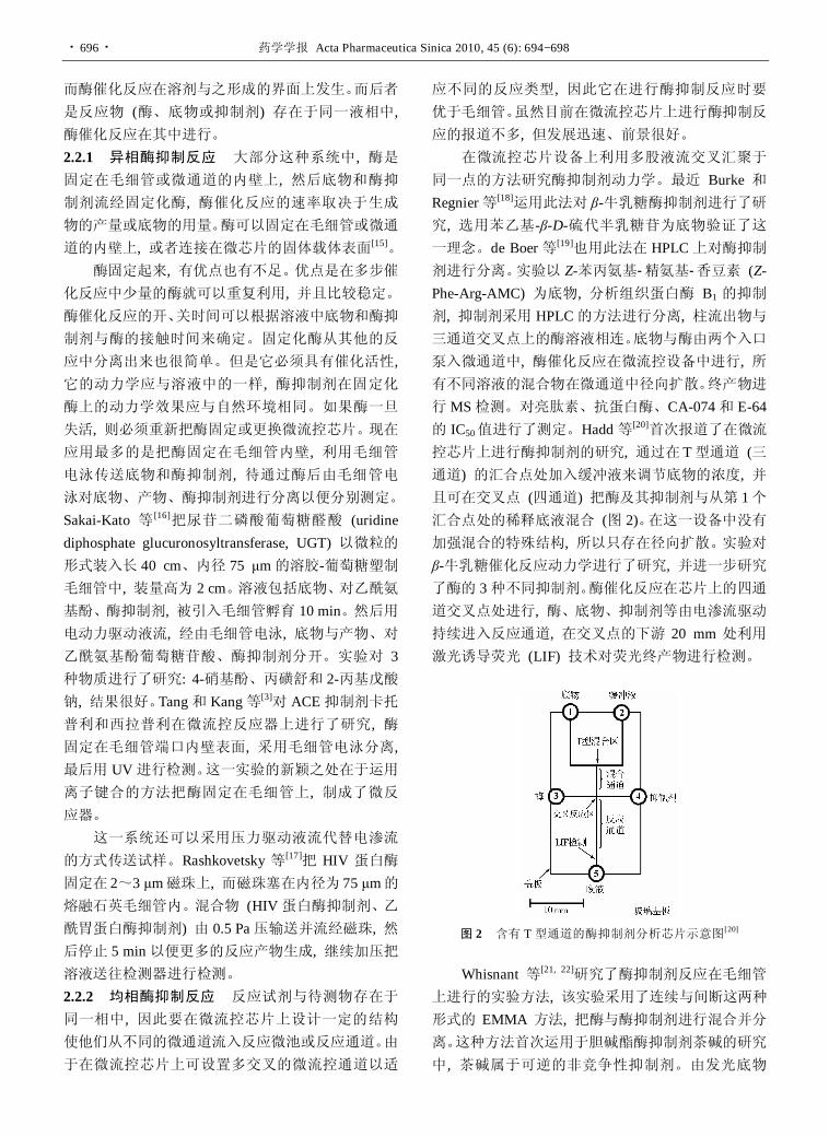
控芯片上进行酶抑制剂的研究, 通过在 T型通道 (三通道) 的汇合点处加入缓冲液来调节底物的浓度, 并且可在交叉点 (四通道) 把酶及其抑制剂与从第 1个汇合点处的稀释底液混合 (图 2)。在这一设备中没有加强混合的特殊结构, 所以只存在径向扩散。实验对β-牛乳糖催化反应动力学进行了研究, 并进一步研究了酶的 3种不同抑制剂。酶催化反应在芯片上的四通道交叉点处进行, 酶、底物、抑制剂等由电渗流驱动持续进入反应通道, 在交叉点的下游 20 mm 处利用激光诱导荧光 (LIF) 技术对荧光终产物进行检测。
图 2 含有 T型通道的酶抑制剂分析芯片示意图[20]
Whisnant 等[21, 22]研究了酶抑制剂反应在毛细管
上进行的实验方法, 该实验采用了连续与间断这两种形式的 EMMA 方法, 把酶与酶抑制剂进行混合并分离。这种方法首次运用于胆碱酯酶抑制剂茶碱的研究
中, 茶碱属于可逆的非竞争性抑制剂。由发光底物

侯凤华等: 基于微流控芯片的酶及其抑制剂的研究进展 · 697 ·
AttoPhos 经过酶催化反应而得的发光终产物来检测酶催化反应速率。实验通过电渗流把底物和缓冲液以
高浓度注入毛细管的含酶 (0.18 nmol·L−1, 3.0 s, 17.8 kV, 310 V·cm−1) 区域, 维持电压为 310 V·cm−1。当酶-
底物复合物流经毛细管时, 具有荧光的产物不断生成并通过 LIF检测器检测。运用这种方法还可以研究其他的胆碱酯酶抑制剂, 如钒酸钠、砷酸钠 (可逆的竞争性抑制剂) 和 EDTA (不可逆性抑制剂)。这种方法充分体现了在最简单的微流控设备 (内径为 50 μm的熔融石英毛细管) 上能够得到丰富的酶抑制剂反应数据。这种在线的 EMMA方法可以辨别可逆性抑制剂、不可逆性抑制剂, 根据电泳图的形状得到活性大小, 通过数据处理可以得到抑制剂的 Ki值。此外, 该方法采用荧光底物, 用 LIF测定, 从而酶的用量很低。 2.3 适用于高通量筛选的微流控芯片
酶抑制剂的高通量筛选包含着非常多的工作单
元, 如大量的样品溶液的稀释及连续的测定工作。应用微流控芯片技术有望得到改善, 利用微通道的流控特性和可集成化的特点, 使繁杂的操作自动化和简单化。
Garcia 等[23]研究了酶抑制剂筛选的微流控芯片, 设计为两个进样口的“T型”微流控扩散通道, 一个进样口注入底物, 另一进样口注入相同浓度的底物和酶抑制剂 (图 3)。当两条液流在主通道中汇合沿 y方向迁移时, 酶抑制剂会在 x方向形成扩散浓度梯度, 而底物在微流控装置的浓度相同则没有浓度梯度。为
了能够观察到酶活性转化产物, 注入的底物为荧光标记的。当酶抑制剂的扩散浓度梯度建立后, 主通道中的溶液与固定在通道表面上的酶 (图 3 中两条虚线之间的区域) 相遇, 导致荧光产物的形成。产物的形成速率随酶抑制剂的浓度 (即沿酶抑制剂的浓度梯度方向) 不同而改变。因此, 通过检测通道中荧光产物在 x方向的光密度, 就能测定酶的抑制率。微通道中产物的光密度可通过荧光显微镜上的数字照相
机将其记录成数字图像, 再通过软件处理成相应的密度数据和曲线图。该方法只要酶抑制剂的初始浓度
就可以得到 IC50值, 实现快速检测。 Kang 等[24]设计了一套用于酶活性分析的微流控
芯片系统, 芯片由 3 部分组成 (图 4), 其中 I 部分是等量分配微通道, II部分是酶反应池, III部分是溶液稀释网络通道。酶经 I被均匀分配注入 II中的 6个反应池; 与此同时, 两个高低不同浓度的底物溶液分别经 C1和 C2注入 III中的稀释网络通道, 被稀释为由高到低 6个浓度的溶液分别进入 II中的 6个反应池。
图 3 T型扩散微流控芯片酶抑制剂分析示意图 (虚线表示通道底部)
图 4 微流控芯片上单一酶分析示意图。俯视设备分 3个部分: I 部分含有等量分配微通道, 酶由此注入; 酶催化反应在 II 部分 (1~6) 进行, 0 室是控制室以确保酶样的均匀分布; III 部分是溶液稀释网络通道
该系统避免了手动稀释底物溶液, 可以自动进样, 很适应高通量酶活性筛选。作者将 4个芯片单元设计在一块相当于 96 孔微量滴定板尺寸的 PDMS 模板上, 由于每个芯片单元含 6个酶反应池, 一块模板上共有24 个酶反应池, 这样该模板就能在酶标仪上进行测定, 并以磷酸对硝基苯酯为测定底物, 完成了对碱性磷酸酯酶的活性测定。 3 展望
经过十几年的发展, 微流控分析芯片技术在酶及其抑制剂研究中的应用越来越广泛, 并不断取得积极和具有突破性的成果, 并由于其快速、高通量、低消耗、易于集成化、便携化的独特优势, 可能在不远的将来成为酶分析的主要工具, 为酶及其抑制剂的研究提供一个更为强大的平台。当然, 一项新技术高速发展的同时, 既存在着优势, 也面临着诸多挑战。当前报道的大部分微流控芯片分析系统功能仍不
够全[25−28], 仍处于实验室研究探索阶段。与微流控芯片系统配合的多数检测器的体积过大, 实现仪器集成化、商品化还有很长的路要走。如何更好地利用微
机电加工技术和微全分析系统 (μTAS) 技术, 发展实用性的微流控芯片分析平台, 应用于药物研发实际试样的分析, 还需不懈努力。

· 698 · 药学学报 Acta Pharmaceutica Sinica 2010, 45 (6): 694−698
References
[1] Lin BC, Qin JH. Laboratory on a Microfluidic Chip (微流控
芯片实验室) [M]. Beijing: Science Press, 2006.
[2] Kang LF, Chung BG, Langer R, et al. Microfluidics for drug
discovery and development: from target selection to product
lifecycle management [J]. Drug Discov Today, 2008, 13:
1−13.
[3] Tang ZM, Kang JW. Enzyme inhibitor screening by capillary
electrophoresis with an on-column immobilized enzyme
microreactor created by an ionic binding technique [J]. Anal
Chem, 2006, 78: 2514−2520.
[4] Xue Q, Yeung ES. Indirect fluorescence determination
of lactate and pyruvate in single erythrocytes by capillary
electrophoresis [J]. J Chromatogr A, 1994, 661: 287−295.
[5] Zhang S, Cao W, Li J, et al. MCE enzyme immunoassay
for carcinoembryonic antigen and alphafetoprotein using
electrochemical detection [J]. Electrophoresis, 2009, 30: 3427−
3435.
[6] Bao J, Regnier FE. Ultramicro enzyme assays in a capillary
electrophoretic system [J]. J Chromatogr, 1992, 608: 217−224.
[7] Hadd AG, Raymond DE, Halliwell JW, et al. Microchip
device for performing enzyme assays [J]. Anal Chem, 1997,
69: 3407−3412.
[8] Koh W, Pishko M. Immobilization of multi-enzyme micro-
reactors inside microfluidic devices [J]. Sens Actuators B,
2005, 106: 335−342.
[9] Kawabata T, Wada HG, Watanabe M, et al. “Electrokinetic
Analyte Transport Assay” for-fetoprotein immunoassay
integrates mixing, reaction and separation on-chip [J].
Electrophoresis, 2008, 29: 1399−1406.
[10] Renzi RF, Stamps J, Horn BA, et al. Hand-held microana-
lytical instrument for chip-based electrophoretic separations of
proteins [J]. Anal Chem, 2005, 77: 435−441.
[11] Rattikan C, Yan XY, Gilman SD. Microfluidics for Studying
Enzyme Inhibition [M]//Gomez FA. Biological Applications
of Microfluidics. Hoboken: John Wiley & Sons, Inc., 2008:
135−170.
[12] Hoffmann T, Reinhold D, Kähne T, et al. Inhibition of
dipeptidyl peptidase IV (DP IV) by anti-DP IV antibodies and
non-substrate X-X-Pro-oligopeptides ascertained by capillary
electrophoresis [J]. J Chromatogr A, 1995, 716: 355−362.
[13] Cohen CB, Dixon EC, Jeong S, et al. A microchip-based
enzyme assay for protein kinase A [J]. Anal Biochem, 1999,
273: 89−97.
[14] Leng C, Zhang XQ, Jü HX. Microfluidic chip-based immu-
noassay [J]. Prog Chem (化学进展), 2009, 21: 687−695.
[15] Krenková J, Foret F. Immobilized microfluidic enzymatic
reactors [J]. Electrophoresis, 2004, 25: 3550−3563.
[16] Sakai-Kato K, Kato M, Toyo'oka T. Screening of inhibitors
of uridine diphosphate glucuronosyltransferase with a minia-
turized on-line drug-metabolism system [J]. J Chromatogr A,
2004, 1051: 261−266.
[17] Rashkovetsky LG, Lyubarskaya YV, Foret F, et al. Automated
microanalysis using magnetic beads with commercial capillary
electrophoretic instrumentation [J]. J Chromatogr A, 1997,
781: 197−204.
[18] Burke BJ, Regnier FE. Stopped-flow enzyme assays on a
chip using a microfabricated mixer [J]. Anal Chem, 2003, 75:
1786−1791.
[19] de Boer AR, Bruyneel B, Krabbe JG, et al. A microfluidic-
based enzymatic assay for bioactivity screening combined with
capillary liquid chromatography and mass spectrometry [J].
Lab Chip, 2005, 5: 1286−1292.
[20] Hadd AG, Raymond DE, Halliwell JW, et al. Microchip
device for performing enzyme assays [J]. Anal Chem, 1997,
69: 3407−3412.
[21] Whisnant AR, Gilman SD. Studies of reversible inhibition,
irreversible inhibition, and activation of alkaline phosphatase
by capillary electrophoresis [J]. Anal Biochem, 2002, 307:
226−234.
[22] Whisnant AR, Johnston SE, Gilman SD. Capillary electro-
phoretic analysis of alkaline phosphatase inhibition by theo-
phylline [J]. Electrophoresis, 2000, 21: 1341−1348.
[23] Garcia E, Hasenbank MS, Finlaysonb B, et al. High-
throughput screening of enzyme inhibition using an inhibitor
gradient generated in a microchannel [J]. Lab Chip, 2007, 7:
249−255.
[24] Kang JH, Park JK. Development of a microplate reader
compatible microfluidic device for enzyme assay [J]. Sens
Actuators, 2005, 107B: 980−985.
[25] Wang J. On-chip enzymatic assays [J]. Electrophoresis,
2002, 23: 713−718.
[26] Vandaveer IV WR, Pasas-Farmer SA, Fischer DJ, et al.
Recent developments in electrochemical detection for microchip
capillary electrophoresis [J]. Electrophoresis, 2004, 25: 3528−
3549.
[27] Herrmann M, Veres T, Tabrizian M. Enzymatically-generated
fluorescent detection in micro-channels with internal magnetic
mixing for the development of parallel microfluidic ELISA [J].
Lab Chip, 2006, 6: 555−560.
[28] Miyazaki M, Maeda H. Microchannel enzyme reactors and
their applications for processing [J]. Trends Biotechnol, 2006,
24: 463−470.

药学学报 Acta Pharmaceutica Sinica 2010, 45 (6): 699−704 · 699 ·
小檗碱调节血糖血脂代谢紊乱机制研究进展
沈 宁, 李彩娜, 环 奕, 申竹芳*
(中国医学科学院、北京协和医学院药物研究所, 北京 100050)
摘要: 小檗碱是从中药黄连等中提取的异喹啉类生物碱, 长期以来用于治疗腹泻及消化道感染。近年来, 陆续有报道小檗碱可对糖尿病代谢紊乱状态发挥有益作用。其机制研究亦涉及疾病发生的多个环节, 包括调节血胆固醇、甘油三酯; 降低血糖; 改善胰岛素抵抗状态; 影响胰岛 β细胞功能等。
关键词: 小檗碱; 糖尿病; 胰岛 β细胞 中图分类号: R285 文献标识码: A 文章编号: 0513-4870 (2010) 06-0699-06
Advances of the mechanism study on berberine in the control of blood glucose and lipid as well as metabolism disorders
SHEN Ning, LI Cai-na, HUAN Yi, SHEN Zhu-fang*
(Institute of Materia Medica, Chinese Academy of Medical Sciences & Peking Union Medical College, Beijing 100050, China)
Abstract: Berberine, an isoquinoline alkaloid isolated from some Chinese medicinal herbs such as Coptidis
rhizoma, has been used for the treatment of diarrhea and other gastrointestinal infections as an antibacterial drug in Chinese medicine. In recent years, it was reported to have beneficial effects on the metabolism disorders states of diabetes. The mechanisms involve many aspects of the diabetes, including regulating the blood cholesterol and triglyceride, lowering blood glucose, ameliorating the insulin resistant state and influencing the function of the pancreatic β cell.
Key words: berberine; diabetes; pancreatic β cell
中药黄连在中国已有几千年的应用历史, 主要用于治疗消化道感染。小檗碱 (berberine) 是中药黄连的主要生物碱。近年来发现其药理作用广泛, 涉及糖尿病、肿瘤、感染、心血管疾病等多方面。自 1986年作者实验室首先报道了其降血糖作用以来, 小檗碱作为抗糖尿病药物的研究逐渐深入, 特别是近 5年, 国内外对其控制糖尿病血脂异常、降低血糖的药理作
用及机制进行了较深入的研究, 但目前仍存在一些分歧与争论, 现将相关研究结果综述如下。 1 小檗碱在脂质调节中的作用及机制
糖尿病不仅是血糖代谢的异常, 同时伴有血脂 收稿日期: 2009-11-10. 基金项目: 中央级公益性科研院所基本科研业务费专项基金资助项目
(2010ZD05). *通讯作者 Tel / Fax: 86-10-83172669, E-mail: [email protected]
增高、代谢紊乱。增高的血脂引起脂质在脂肪组织外
积聚, 引起氧化应激、炎症反应, 导致外周胰岛素抵
抗和 β细胞功能障碍。因此, 血脂异常不仅是糖尿病
的结果, 亦是糖尿病恶化的促进因素。 1.1 小檗碱对低密度脂蛋白受体的影响 低密度脂
蛋白 (LDL) 被认为与糖尿病和心血管疾病关系密
切, 而其肝脏受体 (LDLR) 是介导体内 LDL清除的
主要方式。高脂血症金黄地鼠在应用小檗碱后血浆胆
固醇降低 40%, LDL-胆固醇降低 42%, 肝脏 LDLR
mRNA 表达增加 2.6 倍。在 HepG2 细胞, 小檗碱对LDLR mRNA 的影响与细胞内胆固醇水平无关, 提
示其不同于他汀类药物。小檗碱不增加 LDLR 启动
子活性, 而是通过影响 mRNA 3' 非翻译区 (UTR) 的
5' 近端的 AU富含区 (AU-rich elements, ARE) 和四

· 700 · 药学学报 Acta Pharmaceutica Sinica 2010, 45 (6): 699−704
核苷酸 UCAU重复区, 增加 LDLR mRNA的稳定性发挥上述作用的。在相关信号通路的研究中, 应用MEK1、JNK、P38激酶、PI3K抑制剂后发现小檗碱对 LDLR mRNA 的作用仅依赖于 ERK 通路, 应用MEK1抑制剂U0126可以抑制小檗碱对 LDLR mRNA的上调作用[1]。
进一步的研究发现, 分别克隆 LDLR 编码序列 (cds)、cds+3' UTR、cds+5' UTR, 将上述片段连接至巨细胞病毒启动子并转染至 HepG2 细胞, 观察小檗碱对 LDLR基因片段表达的影响。结果显示, 含有 cds+3' UTR 的序列在转染细胞后, 与仅含有 cds 序列相比, 基因表达下降3.4倍, 但加入小檗碱8 h后, 基因表达明显增加, 这种增加作用能被 ERK抑制剂 U0126抑制。此外, 小檗碱能特异地增加肝细胞系 (HepG2) LDLR mRNA表达, 而对中国仓鼠卵细胞 (CHO)、人胚胎肾细胞 (HEK293) 和人成纤维细胞的 LDLR mRNA 表达影响不大。应用紫外交联技术结合
SDS-PAGE, 发现在小檗碱作用下, 分子质量约为 52 kD和 42 kD的两种不同蛋白与 3' UTR相互结合, 提示其可能为小檗碱发挥作用的调节蛋白[2]。
最近的相关研究发现 LDLR mRNA 的稳定性由复杂的 RNA 结合蛋白网络调控, 其中既有促稳定蛋白, 也有促降解蛋白。针对 46个候选蛋白, 应用 132个 siRNA 构建的文库进行筛选, 通过生物素化 RNA下拉 (biotinylated RNA pull-down)、质谱及功能分析最终鉴定 hnRNP D、hnRNP I 和 KSRP 与 LDLR mRNA 3' UTR特异相互作用。而小檗碱是通过减少上述蛋白与 mRNA的结合而发挥作用的[3]。
在应用多种信号通路激酶抑制剂研究小檗碱对
HepG2 细胞信号通路的影响时发现, 小檗碱在激活ERK通路的同时激活 JNK/c-jun通路, 应用 JNK抑制剂 SP600125能够抑制小檗碱诱导的 LDLR mRNA上调。此外, 小檗碱以浓度依赖的方式使 JNK 与 c-jun磷酸化增加[4]。这一结果与 Kong 等[1]的实验结果不
同, 其差异可能与实验中选用的 JNK 抑制剂不同有关 (Kong实验中应用的 JNK抑制剂为姜黄素)。
Proprotein convertase subtilisin/kexin type 9 (PCSK9)基因表达受细胞内胆固醇、固醇调节元件结合蛋白 (sterol regulatory element binding protein, SREBP) 调节, 其表达的蛋白能够使 LDLR 从内体 (endosome) 穿梭至溶酶体, 而使后者降解, 这是 LDLR蛋白在翻译后的一种调节方式。他汀类药物在增加 LDLR 表达的同时能够通过 SREBP旁路诱导 PCSK9表达。而
通过影响 SREBP的方法可以降低 PCSK9表达, 但同时可能降低 LDLR 的表达, 从而部分抵消这种方法的有益作用。小檗碱能够以浓度和时间依赖的方式, 使HepG2细胞 PCSK9 mRNA表达下降同时使 LDLR mRNA表达增加。小檗碱与美伐他汀合用亦使 LDLR mRNA及蛋白表达增加, 抑制了美伐他汀对PCSK9的诱导。此外, 小檗碱能够使 HMG-CoA还原酶 mRNA降低 39%, 但对法呢基二磷酸合成酶 (farnesyl- diphosphate synthetase, FDPS) 与脱氢胆固醇还原酶 (dehydrocholesterol reductase, DHCR7) 表达无明显影响。小檗碱 (15 μg·mL−1) 使 HepG2 细胞 PPARα mRNA表达增加 39%。RT-PCR实验表明, 小檗碱并 不影响 PCSK9 mRNA的稳定性, 其作用可能通过影响基因启动子转录或者通过激活 AMPK 继发激活过氧化物体增殖激活受体 α (peroxisome proliferator- activated receptors alpha, PPARα) 而发挥[5]。
肝细胞核因子 1α (HNF-1α) 与 SREBP是 PCSK9的重要调节因子, PCSK9启动子区存在保守的HNF-1结合区域, HNF-1α与该区域结合从而调节基因表达。小檗碱能够抑制 HNF-1α 的表达, 同时亦轻度降低SREBP2 的表达。其综合作用使 PCSK9 表达显著降低[6]。 1.2 小檗碱对脂肪细胞分化及脂肪激素的影响 脂
肪组织是机体重要的内分泌器官, 其分泌的细胞因子参与机体的脂质调节及机体对胰岛素的利用, 组织内脂质聚集直接影响胰岛素的敏感性并产生毒性
作用。 小檗碱能够抑制分化培养基条件下的 3T3-L1前
脂肪细胞分化为脂肪细胞, 这种抑制作用还表现在阻止已分化的脂肪细胞进一步分化。小檗碱明显使
3T3-L1前脂肪细胞在分化培养基条件下的 PPARα、β /
δ、γ和 CCAAT增强子结合蛋白 α (CCAAT-enhancer- binding proteins α, C/EBPα) mRNA表达下降, 并且对PPARγ转录前调控蛋白直接抑制。PPAR下游参与脂肪合成与脂肪酸合成的基因亦被抑制。通过细胞转染
报告基因的方法发现, 小檗碱在脂肪组织是 PPARα、γ的双重抑制剂, 且不是 PPAR的激动剂[7]。
内脂素 (visfatin) 是脂肪细胞产生的一种脂肪因子, 其能够增加外周葡萄糖摄取, 抑制肝脏葡萄糖释放, 模拟类似胰岛素作用在糖脂代谢中发挥作用。在一定浓度范围内, 小檗碱以浓度和时间依赖性的方式促进离体脂肪细胞内脂素 mRNA 及蛋白表达。在 0~10 μmol·L−1 浓度下小檗碱浓度依赖性地增加

沈 宁等: 小檗碱调节血糖血脂代谢紊乱机制研究进展 · 701 ·
内脂素 mRNA的表达, 其中 10 μmol·L−1时最明显。
小檗碱 (10 μmol·L−1) 作用 3 h后内脂素mRNA的表达开始明显增强, 作用在 12 h最明显。48 h蛋白电泳结果显示, 内脂素蛋白表达增加[8]。
脂联素 (adiponectin) 也是脂肪细胞分泌的一种脂肪因子, 其与胰岛素抵抗呈负相关。应用半定量RT-PCR方法测定小檗碱对 3T3-L1脂肪细胞脂联素表达的影响发现, 10 μmol·L−1小檗碱处理 48 h后 3T3-L1脂肪细胞脂联素 mRNA 表达明显升高, 与不同浓度胰岛素共孵育则使脂联素 mRNA 表达下降, 其原因可能与高胰岛素使磷脂酰肌醇-3激酶 (PI3K) 表达下
降有关[9]。 1.3 小檗碱对脂质的调节与 AMPK AMP激活的蛋白激酶 (AMPK) 在哺乳动物细胞能量平衡及细胞分化中发挥重要作用, 其活化后能影响很多脂质代谢相关酶, 如 HMG-CoA还原酶、乙酰辅酶 A羧化酶 (ACC) 等, 进而影响脂质代谢, ACC的磷酸化被认为是 AMPK激活的标志。
尽管 Kong等[1]的实验中显示了小檗碱能明显降
低糖尿病患者血甘油三酯 (triglyceride, TG) 达 35%, 但作者并未对这一结果做出明确解释。Brusq 等[10]
发现小檗碱能够以浓度依赖的方式抑制 HepG2细胞甘油三酯与胆固醇 (cholesterol, CHO) 的合成与分
泌, 同时能促进脂肪酸的氧化。应用 MAPK/ERK抑制剂 PD98059 能阻断小檗碱的上述作用, 这一抑制通路与先前的 LDLR 研究一致。随后研究发现, 小 檗碱对细胞的 TG 与 CHO 的上述作用与 AMPK 的 激活剂 AICAR 相似, 小檗碱以浓度依赖的方式使ACC 磷酸化, 随后引起脂肪酸氧化增加。但是应用PD98059 不能阻断 AICAR 对 ACC 的磷酸化, 说明AICAR激活 AMPK不依赖于 MAPK/ERK通路。研究还发现, 小檗碱使 AMPK 磷酸化增加而不改变总AMPK, PD98059 能够抑制小檗碱对 AMPK 的磷酸化。 除激活肝细胞AMPK, 小檗碱还能够使C57BLSK
小鼠脂肪组织内 AMPK及 ACC磷酸化水平增加, 脂肪细胞内参与脂质合成的基因如脂肪酸合成酶 (fatty acid synthase, FAS)、SREBP1c、PPARγ、11β羟基类固醇脱氢酶 1 (11β-hydroxysteroid dehydrogenase, 11β-HSD1) 表达受抑制, 同时体外实验亦证明小檗碱抑制 3T3-L1脂肪细胞参与脂质合成基因表达及细胞内脂滴形成, 上述作用与 AMPK 的活化及 P38 蛋白磷酸化有关[11]。
2 小檗碱对葡萄糖代谢的调节作用 抗糖尿病药物在胰腺外组织发挥降糖作用, 主
要依赖抑制外源葡萄糖的吸收及增加机体对葡萄糖
的利用两方面。 2.1 小檗碱对蔗糖酶及麦芽糖酶的影响 延缓肠道
内的葡萄糖吸收是有效的降糖手段, 其靶点是小肠内的蔗糖酶与麦芽糖酶。
小檗碱能够有效抑制 Caco-2 细胞蔗糖酶和麦芽糖酶的活性, 其对麦芽糖酶的抑制作用与阿卡波糖相当, 但是缺乏浓度依赖性, 这种对麦芽糖酶的作用可能是非特异的。小檗碱在高浓度时抑制蔗糖酶的活
性。但是 Caco-2细胞与小檗碱预孵育 72 h后, 蔗糖酶活性显著降低而麦芽糖酶活性无明显改变[12]。但
与上述结果不同, 作者实验室对大鼠和犬肠 α葡萄糖苷酶的离体酶学研究发现, 小檗碱对蔗糖酶与麦芽糖酶的活性无抑制作用 (未发表数据)。 2.2 小檗碱对外周组织胰岛素抵抗的影响 小檗碱
能改善高脂喂养的 Wistar 大鼠糖耐量异常并且降低动物体重, 正糖钳夹实验亦显示动物对葡萄糖利用明显改善。研究还发现小檗碱能够促进 L6肌管细胞GLUT4的转位, 这种作用由 AMPK快速介导并不被PI3K的抑制剂 wortmannin所阻断[11]。
小檗碱 (1及 10 μmol·L−1) 能够增加脂肪酸诱导胰岛素抵抗的 3T3-L1 脂肪细胞葡萄糖的摄取, 并提高抵抗状态下细胞 IRS-1 和 PI3KP85 的表达。此外, 小檗碱抑制核 NF-κB 的表达, 并抑制 NF-κB 转位进入细胞核。上述作用与小檗碱抑制 IKKβ 磷酸化有 关[13]。
小檗碱能够以浓度 (1~15 μg·mL−1) 和时间 (2~24 h) 依赖的方式促进HepG2细胞胰岛素受体 (InsR) 的表达, 同时使细胞表面的 InsR 数量增多。小檗碱促进 HepG2 细胞的葡萄糖消耗作用依赖于胰岛素信号通路, 应用 RNA干扰技术沉默 InsR基因则阻断上述作用。小檗碱通过依赖蛋白激酶 C (PKC) 的方式激活 InsR 基因启动子。动物实验表明, 小檗碱能够降低 2型糖尿病模型动物KKay小鼠的空腹血糖及胰岛素, 同时增加肝脏 InsR的表达及 PKC的活性[14]。 3 小檗碱对胰岛 β细胞的影响
β 细胞功能障碍与衰竭是从胰岛素抵抗状态发展至糖尿病状态的促发因素, 因此药物对胰岛 β细胞的影响已成为抗糖尿病药物研究的新的热点。如何延
缓或逆转 β细胞凋亡及促进β细胞功能是抗糖尿病药物研究的新方向。

· 702 · 药学学报 Acta Pharmaceutica Sinica 2010, 45 (6): 699−704
3.1 小檗碱对 β 细胞存活的影响 四氧嘧啶及高脂
饮食诱导的高糖高脂大鼠动物模型在应用小檗碱 (100及 200 mg·kg−1) 3周后, 胰腺组织 HE染色与对照组相比明显延缓损伤进展, 此作用与小檗碱对氧化应激的抑制有关[15]。
应用流式细胞术及原位末端核苷酸标记法观察
高脂 (含棕榈酸 0.5 mmol·L−1) 高糖 (葡萄糖 25 mmol·L−1) 培养基条件下, 小檗碱对 NIT-1 胰岛细胞存活的影响。结果发现, 在此环境下 NIT-1细胞凋亡 明显增加, 而小檗碱 (0.1、1 及 5 μmol·L−1) 能够抑制高脂高糖条件下的胰岛细胞凋亡[16]。小檗碱能够
降低高脂高糖条件下 NIT-1 细胞 bax 和 caspase-3 的表达, 提高 bcl-2 的表达, 这种抗凋亡作用与抗氧化应激有关[17]。 3.2 小檗碱对胰岛素分泌及胰岛素信号通路的影响
分泌胰岛素是 β细胞的主要功能, 但是小檗碱是否能促进胰岛的胰岛素分泌存在着争论。Leng 等[18]研究
小檗碱对 BALB/C小鼠血糖与胰岛素分泌的影响实验发现应用小檗碱不同剂量 2 h后, 小檗碱各用药组动物血浆胰岛素水平升高, 血糖下降, 并呈现剂量依赖关系。在培养基葡萄糖浓度分别为0、5及 10 mmol·L−1
的条件下, 小檗碱 (1~10 μmol·L−1) 以浓度依赖的
方式增加了 HIT-T15细胞胰岛素的分泌。葡萄糖 16.7 mmol·L−1 浓度下, 上述剂量的小檗碱亦增加了原代大鼠胰腺的胰岛素分泌, 并呈现浓度依赖效应。
在小檗碱对 NIT-1 细胞胰岛素分泌影响的实验中, 当葡萄糖浓度为 5.5 mmol·L−1时, 小檗碱与空白对照相比对NIT-1细胞的胰岛素分泌无显著促进作用; 当葡萄糖浓度为 16.5 mmol·L−1 时, 小檗碱对 NIT-1细胞胰岛素分泌有促进作用。小檗碱对葡萄糖激酶 (GK) 活性的影响也呈现相同的表现, 这种依赖葡萄糖刺激胰岛素分泌的作用与 GK的激活有关[19]。
在原代培养的 SD大鼠胰岛细胞实验中, 小檗碱能够以浓度依赖的方式 (1、3 及 10 μmol·L−1) 促进葡萄糖刺激的胰岛素分泌, 但不影响基础胰岛素分泌。此外, 小檗碱能够以浓度依赖的方式促进胰岛细胞 HNF4α 表达, 并显著提高胰岛细胞 GK 的活性。这种促胰岛素分泌作用依赖于 HNF-GK通路[20]。
在胰岛素刺激的葡萄糖摄取实验中发现, 小檗碱 (5及 50 μmol·L−1) 同时加以 0.2 nmol·L−1胰岛素
以浓度依赖的方式促进 3T3-L1 细胞的葡萄糖摄取, 小檗碱 (50 μmol·L−1) 加以 0.2 nmol·L−1胰岛素的作
用与单用 10 nmol·L−1胰岛素作用相当。由此推断小
檗碱的促糖摄取作用依赖胰岛素并通过胰岛素发挥
作用。小檗碱对胰岛素信号通路的研究中发现, 小檗碱加 0.2 nmol·L−1胰岛素能够使 IRS-1、AKT、PI3K磷酸化水平增高, 膜 GLUT-4 水平增高; 而在无胰岛素条件下, 上述蛋白磷酸化均无明显变化。在对胰岛素分泌影响的实验中发现 , 在低糖条件下 (2 mmol·L−1 葡萄糖), 小檗碱与 exendin-4 均不能增加Min6细胞胰岛素分泌, 但在 20 mmol·L−1葡萄糖条件
下以浓度依赖的方式促进葡萄糖刺激的胰岛素分泌。
应用 WST-1 试剂显示, 小檗碱以浓度依赖的方式促进MIN6细胞增殖。小檗碱的促增殖作用与 CREB活化后激活胰岛素/胰岛素样生长因子-1通路有关[21]。
但是 Kim 等[22]则发现, 小檗碱促进正常及胰岛素抵抗的 3T3-L1 细胞的葡萄糖摄取均不依赖于胰 岛素, 小檗碱并不影响胰岛素反应信号通路, 不增 加胰岛素受体数目及 IRS-1 磷酸化。PI3K 的抑制剂wortmannin 不能完全抑制小檗碱的促糖摄取作用, 但能完全抑制胰岛素的作用, 从而提示小檗碱的促糖消耗不依赖 PI3K通路。同时研究发现小檗碱亦不引起 PKCξ / λ 的磷酸化。小檗碱在不改变总 ERK 的情况下使 ERK 磷酸化明显增加, 这种作用能够被ERK 通路抑制剂 PD98059 抑制。此外小檗碱使AMPK 磷酸化明显增加。其处理的 3T3-L1 细胞GLUT1 的量明显增加, 而 GLUT4 的量无明显变化; 小檗碱的促糖摄取作用能够被腺苷激酶的抑制剂完
全抑制, 从而提示小檗碱通过ERK通路使GLUT1表达增加, 而 AMPK 活化后激活已存在于血浆膜面的GLUT1, 其共同作用使基础葡萄糖摄取增加。
应用高脂饮食诱导胰岛素抵抗大鼠动物模型分
析小檗碱对胰腺胰岛素分泌影响的实验中发现, 小檗碱给药 6周后, OGTT实验大鼠空腹血糖及血胰岛素水平显著下降, 与正常饮食大鼠水平相当, 在糖负荷后, 小檗碱用药组大鼠血糖及血胰岛素与对照组相比亦显著降低。细胞实验显示, MIN6 细胞在 25 mmol·L−1 葡萄糖条件下, 小檗碱以浓度依赖的方式减少胰岛素的分泌, 并能显著降低棕榈酸刺激下的MIN6细胞和大鼠胰岛细胞胰岛素的分泌。在小檗碱对MIN6细胞胰岛素分泌长期作用实验中显示, 小檗碱作用 24 h 的胰岛细胞基础胰岛素及葡萄糖刺激的胰岛素分泌均增加 , 小檗碱能部分抵消高糖 (25 mmol·L−1) 及棕榈酸 (0.4 mmol·L−1) 作用 24 h引起的MIN6细胞胰岛素分泌降低。小檗碱的上述作用不能被AMPK抑制剂阻断, cAMP通路参与小檗碱对胰

沈 宁等: 小檗碱调节血糖血脂代谢紊乱机制研究进展 · 703 ·
岛素分泌的调节[23]。 作者实验室对小檗碱抗糖尿病作用的研究已有
20 余年, 在小檗碱对正常昆明种小鼠胰岛素分泌及糖负荷后胰岛素释放的实验中, 未发现小檗碱对正常小鼠胰岛素分泌产生影响; 亦未发现小檗碱影响正常小鼠及 KK 小鼠肝细胞膜胰岛素受体的数目及与胰岛素的亲和力, 但发现小檗碱用药后动物血乳酸增加, 小檗碱降血糖的强度同血乳酸增加的程度有良好的相关性, 表明小檗碱通过促进葡萄糖酵解发挥降血糖作用 (未发表数据)。 4 结论与展望
自 1986年陈其明等[24]首先报道了小檗碱降低血
糖作用以来, 尽管国内外不断有药效学实验结果报道, 但是近 20 年小檗碱在糖尿病治疗中的作用机制研究进展缓慢, 直到近 5 年, 小檗碱成为抗糖尿病药物新的研究热点。其作用机制已经涉及了糖尿病发病
的多个环节, 动物实验中其对糖尿病模型动物外周组织如肝脏、肌肉、脂肪发挥的有益作用已被广泛认
可, AMPK的激活被认为与多数药理作用相关。然而, 小檗碱对胰岛 β细胞的影响目前存在很大争议, 在对胰岛素分泌影响及对胰岛素信号通路影响的问题上
甚至出现完全相反的实验结论。此外, 国内虽有文献报道小檗碱对高脂高糖环境下 β 细胞的凋亡有抑制作用, 但亦有文献报道AMPK在 β细胞的激活诱导 β细胞的凋亡。总之, 小檗碱对胰腺 β细胞的影响将仍然是研究的热点。此外, 根据小檗碱结构改造的化合物已被用来评价降脂作用, 因此, 小檗碱结构修饰及其代谢产物也是未来的研究方向。
References
[1] Kong W, Wei J, Abidi P, et al. Berberine is a novel cholesterol-
lowering drug working through a unique mechanism distinct
from statins [J]. Nat Med, 2004, 10: 1344−1352.
[2] Abidi P, Zhou Y, Jiang JD, et al. Extracellular signal-regulated
kinase-dependent stabilization of hepatic low-density lipoprotein
receptor mRNA by herbal medicine berberine [J]. Arterioscler
Thromb Vasc Biol, 2005, 25: 2170−2176.
[3] Li H, Chen W, Zhou Y, et al. Identification of mRNA-binding
proteins that regulate the stability of LDL receptor mRNA
through AU-rich elements [J]. J Lipid Res, 2009, 50: 820−
831.
[4] Lee S, Lim HJ, Park JH, et al. Berberine-induced LDLR
up-regulation involves JNK pathway [J]. Biochem Biophys
Res Commun, 2007, 362: 853−857.
[5] Cameron J, Ranheim T, Kulseth MA, et al. Berberine decreases
PCSK9 expression in HepG2 cells [J]. Atherosclerosis, 2008,
201: 266−273.
[6] Li H, Dong B, Park SW, et al. HNF1α plays a critical role
in PCSK9 gene transcription and regulation by a natural
hypocholesterolemic compound berberine [J]. J Biol Chem,
2009, 284: 28885−28895.
[7] Huang C, Zhang Y, Gong Z, et al. Berberine inhibits 3T3-L1
adipocyte differentiation through the PPARgamma pathway [J].
Biochem Biophys Res Commun, 2006, 348: 571−578.
[8] Liu Y, Wang WJ, Li YS, et al. Effect of berberine on visfatin
expression in 3T3-L1 adipocytes [J]. Chin J Endocrinol Metab
(中华内分泌代谢杂志), 2007, 4: 351−354.
[9] Gu W, Zeng WH, Hu HY, et al. Effects of berberine on
adiponectin mRNA expression in 3T3-L1 adipocyte [J].
China J Chin Mater Med (中国中药杂志), 2005, 4: 286−288.
[10] Brusq JM, Ancellin N, Grondin P, et al. Inhibition of lipid
synthesis through activation of AMP kinase: an additional
mechanism for the hypolipidemic effects of berberine [J]. J
Lipid Res, 2006, 47: 1281−1288.
[11] Lee YS, Kim WS, Kim KH, et al. Berberine, a natural plant
product, activates AMP-activated protein kinase with beneficial
metabolic effects in diabetic and insulin-resistant states [J].
Diabetes, 2006, 55: 2256−2264.
[12] Pan GY, Huang ZQ, Wang GJ, et al. The antihyperglycemic
activity of berberine arises from a decrease of glucose absorption
[J]. Planta Med, 2003, 69: 642−636.
[13] Yi P, Lu FE, Xu LJ, et al. Berberine reverses free-fatty-acid-
induced insulin resistance in 3T3-L1 adipocytes through targeting
IKKbeta [J]. World J Gastroenterol, 2008, 14: 876−883.
[14] Kong WJ, Zhang H, Song DQ, et al. Berberine reduces insulin
resistance through protein kinase C-dependent up-regulation of
insulin receptor expression [J]. Metab Clin Exp, 2009, 58:
109−119.
[15] Li SY, Yao YW, Wu R. Effects of berberine hydrochloridum
on streptozotocin induced B cells impairment in cultured
newborn rat islets [J]. China J Mod Med (中国现代医学杂
志), 2002, 18: 6−10.
[16] Liu WJ, Lu FE, Dong H, et al. Effects of berberine on the
apoptosis of NIT-1 cells induced by high glucose and saturated
fatty acids [J]. Chin J Pathophysiol (中国病理生理杂志),
2008, 24: 788−791.
[17] Sun H, Lu FE, Wang ZS, et al. Molecular mechanism of
berberine's inhibitory effects on the apoptosis of NIT-1 cells
induced by high glucose and saturated fatty acids [J]. Chin

· 704 · 药学学报 Acta Pharmaceutica Sinica 2010, 45 (6): 699−704
Pharmacol Bull (中国药理学通报), 2008, 24: 762−766.
[18] Leng SH, Lu FE, Xu LJ. Therapeutic effects of berberine in
impaired glucose tolerance rats and its influence on insulin
secretion [J]. Acta Pharmacol Sin, 2004, 25: 496−502.
[19] Wang ZS, Lu FE, Chen G, et al. Effect of berberine on
insulin secretion and glucokinase activity of NIT-1 cells [J].
Acta Pharm Sin (药学学报), 2007, 42: 1045−1049.
[20] Wang ZQ, Lu FE, Leng SH, et al. Facilitating effects of
berberine on rat pancreatic islets through modulating hepatic
nuclear factor 4 alpha expression and glucokinase activity [J].
World J Gastroenterol, 2008, 14: 6004−6011.
[21] Ko BS, Choi SB, Park SK, et al. Insulin sensitizing and
insulinotropic action of berberine from Coptidis rhizoma [J].
Biol Pharm Bull, 2005, 28: 1431−1437.
[22] Kim SH, Shin EJ, Kim ED, et al. Berberine activates
GLUT1-mediated glucose uptake in 3T3-L1 adipocytes [J].
Biol Pharm Bull, 2007, 30: 2120−2125.
[23] Zhou L, Wang X, Shao L, et al. Berberine acutely inhibits
insulin secretion from beta-cells through 3', 5'-cyclic adenosine
5'-monophosphate signaling pathway [J]. Endocrinology,
2008, 149: 4510−4518.
[24] Chen QM, Xie MZ. Studies on the hypoglycemic effect of
Coptis chinensis and berberine [J]. Acta Pharm Sin (药学学
报), 1986, 21: 401−406.
《药学学报》第十一届编委会及学术报告会会议通知
《药学学报》第十一届编委会及学术研讨会会议定于 2010年 7月 8日~11日在沈阳药科大学举行。本次会议主要内容包括: 编委会换届、《药学学报》发展中存在的问题及解决对策、《药学学报》在线英文版的出版及《药学学报》年度优秀论文的颁奖等。为丰富会议内容, 会议将邀请国内几位专家针对药学领域研究的热点做专题报告。详细会议通知将在 5月份发与各位编委, 欢迎各位积极参会。

药学学报 Acta Pharmaceutica Sinica 2010, 45 (6): 705−710 · 705 ·
孕期酒精暴露对子鼠视皮质突触数量影响的体视学研究
席 艳, 张俊士, 臧建峰, 文曙光, 邓锦波*
(河南大学神经生物学研究所, 河南 开封 475004)
摘要: 建立孕期酒精暴露 (PAE) 模型, 研究孕期酒精暴露对小鼠视皮质突触数量的影响。利用免疫荧光染色技术标记对照组与 PAE模型组 (低剂量和高剂量) 子鼠出生后 0、7、14及 30 d视皮质突触前体的 synaptophysin蛋白表达, 以此来代表突触, 观察其数密度变化, 并利用 Western blotting 检测对各实验组子代小鼠视皮质synaptophysin 的表达量进行半定量分析。突触数密度值统计学分析显示: 对照组、低剂量和高剂量组间比较差异显著 (P < 0.05), 0、7、14及 30 d差异显著 (P < 0.05), 年龄与剂量之间存在交互作用 (P < 0.05) 且剂量的影响作用更大。Western blotting检测结果与免疫荧光统计结果一致。这表明 PAE对突触的影响具有长时程效应和剂量相关性, 突触丢失的长时程放大效应可能是患儿精神发育迟滞和记忆力下降的主要原因。
关键词: 孕期酒精暴露;数密度; 突触; synaptophysin; 视皮质 中图分类号: R963 文献标识码: A 文章编号: 0513-4870 (2010) 06-0705-06
Stereological study on the synapse loss in visual cortex of mouse after prenatal alcohol exposure
XI Yan, ZHANG Jun-shi, ZANG Jian-feng, WEN Shu-guang, DENG Jin-bo*
(Institute of Neurobiology, Henan University, Kaifeng 475004, China)
Abstract: In order to understand the alcohol’s toxicity to the quantitative alternations of synapses in mouse visual cortex, the expression of synaptophysin after prenatal alcohol exposure was investigated. In present study, the experimental mice at P0, P7, P14 and P30 were grouped, as control, 2 g·kg−1 alcohol treatment and 4 g·kg−1 alcohol treatment. The pre-synaptic elements which were used to represent synapses were marked with synaptophysin (a synaptic vesicle associated protein) by immunocytochemistry technique. The synaptophysin positive boutons in layer VI of visual cortex were imaged under laser confocal microscope. With stereological methods, the number cal density of synapse in visual cortex was calculated in different groups at various ages. Moreover, Western blotting was carried out to detect the expression of synaptophysin in visual cortex. The results showed that prenatal alcohol exposure could cause synaptic loss with long-term effect and in a dose dependent manner. For instance, there were significant difference among the different treatment groups of P0, P14 and P30 as well (P < 0.05). Western blotting supported the results of immunofluorescent labeling. In conclusion, prenatal alcohol exposure can induce the synaptic loss dose dependently and with long-term effect. Our findings implicate that the synaptic loss with long term effect in CNS probably contributes to the lifelong mental retardation and memorial lowliness associated with childhood FAS.
Key words: prenatal alcohol exposure; number cal density; synapse; synaptophysin; visual cortex
收稿日期: 2009-11-26. 基金项目: 国家自然科学基金资助项目 (30771140); 河南省教育厅自
然科学研究项目 (2007180008); 河南省科技厅国际协作项目 (094300510044); 河南大学自然科学基础研究项目 (2008YBZR034).
*通讯作者 Tel / Fax: 86-378-3880292, E-mail: [email protected]
酒精、大麻和海洛因作为成瘾药物研究的重点, 受到科学工作者的广泛关注, 美国和其他西方国家投入大量资金推动酒精滥用相关的科学研究、临床治
疗和社会保障。近些年来中国酒类消费比例的显著上
升使我国将要面对酒精带来的越来越多的健康和社
·研究论文·

· 706 · 药学学报 Acta Pharmaceutica Sinica 2010, 45 (6): 705−710
会问题。孕期酒精暴露 (prenatal alcohol exposure, PAE) 会导致胎儿流产、死产、胎儿颜面部畸形、神经系统发育畸形以及出生后认知障碍等。临床上的胎
儿酒精综合征 (fetal alcohol syndrome, FAS) 是 PAE导致子代损害的一种典型表现, 其特征是患儿出生时伴有典型的颜面畸形和神经系统的发育异常, 成年后还会出现学习及社交能力下降等行为能力不足
和人格缺陷的问题, 并由此带来一系列社会问题[1]。
Sullivan[2]的研究表明, FAS的病因只和母亲在怀孕期间滥用酒精有关, 而与父辈和上一辈是否饮酒关系不大。国外对 PAE 的研究多集中于酒精对子代神经系统的毒性作用, 如能量代谢、受体功能、蛋白表达等方面[3], 而PAE对子代影响的长时程效应的报道尚不多见, 因此孕期的酒精暴露如何影响成年后子代的人格特征、行为及学习能力等问题难以得到合理解
释。 众所周知, 突触与学习记忆的关系极为密切, 故
研究酒精暴露后突触数量的变化有极其重要的意义, 作者采用 synaptophysin 荧光染色技术和数密度测量的方法进行研究[4, 5]。Synaptophysin是位于突触前体的囊泡膜蛋白, 功能与囊泡释放相关, 可以作为突触的特异性标记物, 在神经系统中稳定表达, 现已应用于老年痴呆、癫痫、精神分裂症等疾病突触数量的研
究[6−8]。本研究利用作者前期研究中建立的 PAE模型, 研究 PAE 子代小鼠视皮质突触的数密度变化, 旨在探讨 PAE对子代视皮质神经突触发育的影响及机制, 为深入理解孕期酒精暴露的危害, 解释孕期酒精暴露对子代的长时程影响及制定相关的卫生政策和保
健指南提供重要的理论依据。
材料与方法 动物模型与分组 选 2~3月龄健康 C57BL/6小
鼠 (由河南省实验动物中心提供, 合格证号SYXK豫2005-0012), 雌鼠 60 只 (未生育), 雄鼠 10 只, 体重20~30 g, 单笼饲养, 室温控制在 20~25 ℃, 相对湿度 60%~70%, 定期更换垫料消毒。采光控制为 8 h光照, 16 h黑暗 (5∶00 PM~9∶00 AM)。自由进食水, 食物为河南省实验动物中心提供的小鼠标准饲料, 饮用水选用实验室自制单蒸水。
将 60只雌鼠随机分为对照组 (10只)、低剂量组 (20 只) 和高剂量组 (30 只), 雌雄小鼠 1∶1 (5∶00 PM) 合笼交配, 次日 9∶00检查阴道栓塞情况, 将发现阴栓之日定为妊娠第 1天 (embryo day 0, E0)。E0
当日取出雄鼠, 妊娠雌鼠单笼如常饲养。随机选出 不同剂量的实验组, 即对照组 (control, C)、低剂量组 (low dosage, L) 和高剂量组 (high dosage, H)。对照组仍自由饮食, 其余各组雌鼠从孕五日 (E5) 到子鼠出生, 每日 9∶00禁食、水, 4 h后 25%酒精灌胃 (L组酒精剂量 2 g·kg−1·d−1、H组酒精剂量 4 g·kg−1·d−1), 灌胃后恢复正常饲养。子鼠出生后第 1个 24 h 内定为是生后第 0天 (postnatal day 0, P0), 收集各组别生后0 (P0)、7 (P7)、14 (P14) 及 30 d (P30) 子鼠, 每组不少于 5只进行免疫荧光染色; 每组不少于 5只小鼠进行Western blotting检测。
免疫荧光检测 Synaptophysin 聚集于突触前体, 可利用免疫荧光染色法显示, 并以荧光颗粒代表突触[9]。同时, 显示皮质 I-VI的片层化结构便于靶细胞定位, 由于 Foxp2 可以特异性地在皮质VI层神经细胞中表达[10], 结合 DAPI 衬染, 可实现皮质分层显色。免疫荧光染色具体步骤如下: 4% 苯巴比妥钠 (30 mg·kg−1) 腹腔注射麻醉, 开胸暴露心脏。剪开右心耳, 自心尖部插管, 4% 多聚甲醛 (pH 7.4) 25 mL缓慢灌流。断头取脑, 置 20 倍体积以上 4% 多聚甲醛 (pH 7.4) 固定 48 h (4 ℃)。蒸馏水冲洗, 4% 琼脂糖包埋。
取视皮制作为实验的靶组织, 震荡切片机 (vibratome 1000 plus, NatureGene Corp, USA) 切片, 每块组织切 5张切片, 厚度 50 μm。切片经 0.1 mol·L−1 PB漂洗。然后进行 synaptophysin 和 Foxp2 的荧光免疫双重标记 : 加入一抗 (鼠抗人 synaptophysin, 1∶500, Millipore, MAB368) 和Foxp2 (兔抗人Foxp2, 1∶300, abcam, ab16046), 置 4 ℃冰箱中孵育过夜, PB漂洗干净后加入二抗: 羊抗鼠 Alexa Fluor 568 (呈红色, 1∶600, Molecular probes, A11004) 和驴抗兔 Alexa Fluor 488 (呈绿色, 1∶300, Invitrogen, A11008) 室温下避光孵育 3 h, 后用 PB漂洗 3次, 最后用DAPI (呈蓝色, 1∶10 000, Roche, 10236276) 进行衬染, 荧光封片介质封片, 在Olympus荧光显微镜 (BX61, 日本) 下使用罗丹明、FITC 和紫外激发光进行观察、拍片。挑选质量较好的切片在激光扫描共聚焦显微镜 (Olympus, FV-1000) 下进行观察。
Western blotting检测 利用Western blotting方法对各组视皮质组织内的蛋白质进行半定量分析, 具体操作如下。颈椎脱位处死小鼠, 开颅取脑, 迅速剥离视皮质。按照试剂盒说明书提取膜蛋白 (膜蛋白提取试剂盒, Pierce, 89826) 至离心管中, 加入 9倍体积预冷的丙酮 (分析纯, 开封开化集团有限公司试剂厂), 静置过夜; 12 000 r·min−1离心 10 min (4 ℃),

席 艳等: 孕期酒精暴露对子鼠视皮质突触数量影响的体视学研究 · 707 ·
弃上清液; 室温干燥器中干燥; 加入 10 mol·L−1 Tris- HCl (pH 7.0) 缓冲液重悬沉淀。采用考马斯亮蓝法进行蛋白质定量 (Coomassie Protein Assay Kit, 上海美季生物技术有限公司, M20303); 各组样品取蛋白 15 μL加入 5×SDS上样缓冲液 5 μL, 100 ℃煮沸 5 min; 上样进行 SDS 聚丙烯酰胺凝胶电泳, 电转移蛋白至NC膜; 5%脱脂奶粉封闭, 4 ℃孵育过夜, 次日加入一抗 (羊抗兔 synaptophysin, 1∶500, abcam, ab9272) 室温孵育 3 h; 漂洗后入 HRP标记的二抗 (驴抗兔 IgG, 1∶2 000) 室温孵育 1 h, TBST缓冲液冲洗 3次, 每次5 min; ECL发光液孵育 2 min, 胶片曝光显影, 结果用Quality One分析, 并以 β-actin (江苏碧云天生物技术研究所, AA128) 做内对照, 以阳性条带与内对照条带光密度比值作为阳性条带的相对表达值。进行 5次重复试验。
突触计数与统计学处理 免疫荧光标记后, 视皮质锥体细胞表面可以见到大量的 synaptophysin 红色荧光颗粒, 一般认为, 每一荧光颗粒可代表一个突触前体, 也即一个突触[11]。同时, 依据 Foxp2与 DAPI荧光显色将体视学测量定位于皮质VI层, 原因是皮质各层中第VI层发育最早便于从 P0 开始进行比较 (图 1)。体视学方法可以通过二维结构信息推断实际的三维结构, 能够测量单位参照体积的突触数量, 是一种比较科学的计量学方法。本研究采用突触数密度 (单位: 个/μm3) 作为测量与统计参数。其具体测量步骤如下: 从实验图片中截取 13.81 μm×13.81 μm大小图片导入Word文档中, 绘制边长 4.14 μm×4.14 μm、6×6的正方格测试系统 (以每个测试小格左上角顶点为测试点), 正方格测试系统的尺寸依据图片放大倍数、荧光颗粒大小与实验要求误差共同选定[12]。测
量参照系内荧光颗粒的截面数、参照系测试点击中 的荧光颗粒数, 将来源于同一切片的多个测量数据代入数密度计算公式 Nv=[∑Nx]3/2/{1.382[31/2d2/2]3/2
[∑Pc][∑Px.n]} (Nv: 突触数密度, Pc: 参照系的测试点数, Nx: 参照系内被测粒子的截面数, Px.n: 参照系测试点击中 synaptophysin 颗粒的截面数, d: 参照系测试线长度) 得出数密度值。12组小鼠每组 5只, 每只小鼠测量 5 张切片, 共得到 12×25 个数密度数据, 均以 x ± s表示。采用 Spss 17.0统计分析软件, 进行Levene检验和主体间效应检验, 多组比较使用 Fisher LSD 检验和 Student-Newman-Keuls (SNK) 检验, 以了解剂量、年龄两个因素对子鼠视皮质突触发育的影
响。
结果 1 孕期酒精暴露对子鼠的影响
酒精处理组子代小鼠总体表现为早产多、发育缓
慢、体重低、个头小、抗病能力差、死亡率高及各种
畸形[13]; 对照组则没有发现畸形和发育异常。对子鼠出生后 0~7 d 死亡率进行分析发现, 高剂量组子鼠死亡率达 47.24%, 相应的低剂量组死亡率为 12.01%, 对照组子鼠的死亡率为 1.73%。结果表明, 孕期酒精暴露对子代存活率及发育有明显的影响。 2 Synaptophysin在视皮质的表达变化
激光共聚焦扫描显微镜显示, synaptophysin 在P0 以后各组视皮质各层均有表达, synaptophysin 荧光颗粒代表的突触数量随子鼠孕期酒精暴露剂量升
高而下降, 同时随子鼠年龄的增长而升高。横向比较 (剂量依赖性比较), 自对照组、低剂量组、高剂量组荧光颗粒密度呈下降趋势; 纵向比较 (长时程效应比较), 随年龄增加荧光颗粒密度呈上升趋势 (图 2)。子鼠视皮质突触数密度 (个/μm3) 采用正方格计数法进行体视学测量 (图 1), 各组数据如表 1所示。利用 SNK检验对数据进行统计分析发现, 对照组、低剂量组、高剂量组间差异显著 (P < 0.05), P0、P7、P14、P30年龄组间差异显著 (P < 0.05)。由于突触数密度受到年龄、剂量两个因素的影响, 对照组内 P0、P7、P14和 P30间存在显著差异 (P < 0.05), 但是在同一水平的酒精处理组内 P0、P7、P14 和 P30 的个别组无显著性差异 (P > 0.05)。为消除年龄因素对研究的影响, 还应进行主体间效应检验。检验结果发现, 年龄与剂量两个因素之间存在交互作用, 而且剂量 (MS = 9.75, F = 140.44, P < 0.05) 的影响比年龄 (MS = 6.55, F = 94.26, P < 0.05) 的影响作用稍大。Fisher LSD检验在有显著差异的两组之间进行均数差值比较 CP0与 LP0 (I−J = 0.15)、CP7与 LP7 (I−J = 0.18)、CP14与LP14 (I−J = 0.35)、CP30与 LP30 (I−J = 0.56)、CP0与HP0 (I−J = 0.36)、CP7与 HP7 (I−J = 0.52)、CP14与HP14 (I−J = 0.69)、CP30与 HP30 (I−J = 0.93), 发现年龄增高使对照组与酒精处理组的差异增大, 并且对照组与高剂量组的差异大于对照组与低剂量组的差
异。所以, 尽管在同一水平的酒精处理组内 P0、P7、P14和 P30的个别组, 受年龄因素影响无显著性差异, 但是通过 Fisher LSD 检验在不同组别不同剂量之间进行比较, 不影响得出长时程放大效应的结论。 3 Western blotting检测半定量分析 synaptophysin蛋白表达量
利用Western blotting检测对PAE模型组和对照组

· 708 · 药学学报 Acta Pharmaceutica Sinica 2010, 45 (6): 705−710
子鼠 P30、P14、P0进行 synaptophysin半定量检测, 以阳性条带与 β-actin条带光密度比值作为 synaptophysin的相对表达值 (图 3和表 2)。结果显示: ① synaptophysin
Figure 1 Lamination of visual cortex and the measurement area with stereological consulting system. (a) The lamination of visual cortex at P0 with Foxp2 immunocytochemistry (green) and DAPI counterstaining (blue). The layer I-VI was marked in the photo. (b) The high magnification of pyramidal cells with synaptophysin-positive puncta (red) at P14. The nuclei are visualized with DAPI staining. The synaptophysin puncta are located at the neuronal surface and dendrites, however, the cytoplasm of pyramidal cell lies between puncta and nucleus. The test system was overlapped on the photo, in order to show the stereological measurement. Bar = 60 μm for (a), 5 μm for (b)
Figure 2 The expressions of synaptophysin (red) among different treatment groups at various ages (P0, P7, P14 and P30, a−l). The control group (C), low dose treatment (L, 2 g·kg−1 alcohol treatment) and high dose treatment (H, 4 g·kg−1 alcohol treatment) are marked in the photo. Bar = 10 μm in (a−l)
Table 1 The number cal density in different groups and SNK tests (n = 25, x ± s)
Nv/punctum·μm−3 Treatment
P0 P7 P14 P30
Control 1.37 ± 0.27 1.58 ± 0.29▽ 1.84 ± 0.23▽ 2.38 ± 0.27▽
Alcohol 2 g·kg−1 1.21 ± 0.22* 1.40 ± 0.26*▽ 1.48 ± 0.28* 1.82 ± 0.28*▽
Alcohol 4 g·kg−1 1.00 ± 0.26*# 1.06 ± 0.27*# 1.15 ± 0.26*# 1.46 ± 0.27*#▽
P: Postnatal day; Nv: Number cal density of synapse in unit volume. *P < 0.05, if alcohol treatment group versus control at same age; #P < 0.05, if high dose group versus low dose at same age; ▽P < 0.05, if older age group versus young age group adjacent to it in the same alcohol treatment group; n = 25 means 25 sections for each group
Figure 3 The expression of synaptophysin in visual cortex detected with Western blotting. 1, 4 and 7 were 4 g·kg−1 alcohol treatment group at P30, P14 and P0, respectively; 2, 5 and 8 were 2 g·kg−1 alcohol treatment group at P30, P14 and P0, respectively; 3, 6 and 9 were control group at P30, P14 and P0, respectively Table 2 Semi-quantitative analysis of synaptophysin expression with Western blotting and SNK tests (n = 5, x ± s)
Synaptophysin/β-actin densitometric ratio Treatment
P0 P14 P30
Control 1.28 ± 0.08 1.92 ± 0.09 2.87 ± 0.10
Alcohol 2 g·kg−1 0.98 ± 0.09* 1.38 ± 0.09* 2.05 ± 0.10*
Alcohol 4 g·kg−1 0.73 ± 0.08* 0.94 ± 0.10* 1.64 ± 0.09* *P < 0.05, if high dose versus low dose or control, and if low dose versus control; n = 5 means 5 animals for each group 在胶片上显示为分子质量为 38 kD的活化片段, 这与文献[14]报道是一致的; ② 酒精处理组低于对照组 (P <
0.05), 高剂量组低于低剂量组 (P < 0.05), 即孕期酒精暴露使子鼠视皮质 synaptophysin 蛋白表达量下降且具有剂量依赖性; ③ synaptophysin 蛋白表达量随子鼠年龄增长而增高, 各年龄组的组间差异显著 (P < 0.05)。
讨论 研究孕期酒精暴露对胎儿的危害, 常选用动物
模型[15, 16]。作者的工作着重于利用 PAE模型研究年龄和酒精剂量的叠加对子鼠突触数量的影响, 从而达成研究孕期酒精暴露对子代神经发育长时程影响
的目的。Lancaster 等[17]也曾对孕期酒精暴露的子代
进行研究, 然其以突触的平均面积为统计对象, 这种二维空间上的统计方法可能受到突触形态变化的影
响, 因而不能反映三维空间上突触实际数量的变化。

席 艳等: 孕期酒精暴露对子鼠视皮质突触数量影响的体视学研究 · 709 ·
与之相比, 本研究采用体视学的方法进行组织定量, 利用正方测试系统和 Weibel 提出的数密度计算方法得出视皮质单位体积内的突触数量变化[18], 这种方法突破了二维空间的局限, 其结果更为准确, 有利于解释形态结构和功能的关系。Jiang 等[19]曾经报道孕
期酒精暴露造成视皮质神经元凋亡的现象, 然而突触是神经元发挥功能的重要单位, 一个神经元通常有很多突触, 这些突触与学习和记忆密切相关, 因此利用 synaptophysin 研究突触的改变更利于解释 PAE子代的神经行为改变。
本实验利用免疫荧光和体视学方法分析了 PAE模型子代视皮质突触数密度的变化。对照组、低剂量
组、高剂量组间差异显著 (P < 0.05) 且突触数密度依次降低, 说明 PAE 可以造成子代突触数量的丢失, 并且存在酒精剂量越高突触丢失越严重的剂量依赖
关系。这种剂量依赖关系提示, 孕妇在怀孕期间应绝对避免饮酒。Western blotting半定量分析结果与免疫荧光结果一致, 支持了上述结论, 说明PAE扰乱了子鼠突触的发育, 这也是造成突触丢失的主要原因。P0、P7、P14、P30年龄组间比较差异显著 (P < 0.05), 且突触数密度依次升高, 说明年龄增长是突触数量变化的另一原因。突触在年龄上的变化可影响对孕期酒
精暴露长时程效应的分析, 主体间效应检验可以排除这一干扰, 其结果显示年龄和剂量两个因素共同对子代突触数量产生影响 (交互作用), 剂量 (MS = 9.75, F = 140.44, P < 0.05) 的影响作用比年龄 (MS = 6.55, F = 94.26, P < 0.05) 稍大。这解释了 Fishier LSD结果中对照组内 P0、P7、P14、P30间差异显著 (P < 0.05), 而 2 g·kg−1与 4 g·kg−1酒精处理组内 P0、P7、P14、P30间无显著性差异 (表 1) 的现象, 正是年龄增长在一定程度上抵消了酒精暴露造成突触丢失的作用使酒
精处理组内各年龄组间无显著性差异。Fisher LSD检验还有一个重要发现, 即年龄增高会使对照组与酒精处理组的差异增大, 并且对照组与高剂量组的差异大于对照组与低剂量组的差异, 这说明 PAE 造成突触丢失的作用会随着子鼠年龄的增大而被放大, 这种放大效应可以解释 PAE 子代成年后 (相当于小鼠 P30) 社会行为能力低、易于罹患精神疾病等问题。同时子代突触丢失的长时程放大效应还揭示: 即便子代出生时不能被诊断为 FAS 综合征, 成年后酒精对神经系统的损害依然可以表现出来。从这点意义上
讲, 母体孕期饮酒对子代发育的损害是持久的、不可逆的。而且, 突触丢失可能是 PAE 对儿童学习记忆造成影响的病理学基础[20]。
基于对孕期酒精暴露造成突触丢失和长时程放
大效应的认识, 寻找合适的治疗药物以提高 PAE 子代的生存质量已经成为共识。目前, 国外已经开始了运用中枢神经系统兴奋剂对 PAE 子代进行干预治疗的探索。但是, 前期的研究工作表明由于 PAE子代脑的结构与神经递质传递都有异常, 故其对许多药物的反应也与常人不同[21]。因此在下一步的研究中应
该对突触丢失的机制做进一步探讨。然而 PAE 影响神经系统发育的机制目前还不十分清楚, 本研究所做的前期工作发现PAE对神经系统的影响与NMDA- NR1的功能有关[22]; 也有报道 PAE伴随PKC (protein kinase C) 激活并降低 GAP-43 蛋白的磷酸化[23], 但是这些学说单独都不足以解释本实验中观察到的
PAE对突触丢失的长时程放大效应。作者推测, 酒精影响突触发育受综合因素的影响, 如神经受体 (如AMPA、NMDA-NR1等)[24]和细胞因子 (如细胞间黏附分子)[25−27]等。PAE 的这些影响发生在突触发育的关键时期 (in utero), 由此引起的突触丢失会随年龄增长逐步显现出来, 呈现出长时程放大效应。
综上所述, 孕期酒精暴露对子代的神经系统的影响具有剂量相关性及长时程放大效应。一方面应该
充分认识到孕期饮酒对胎儿发育及家庭社会带来的
不利影响, 要求孕妇避免饮酒; 另一方面也应该看到孕期酒精暴露对子代神经系统影响的机制、FAS的产前诊断、孕期酒精暴露出生后胎儿治疗有关的药物开
发等诸多领域都大有可为。
References
[1] Guerri C, Bazinet A, Riley EP. Foetal alcohol spectrum
disorders and alterations in brain and behavior [J]. Alcohol
Alcohol, 2009, 44: 108−114.
[2] Spohr HL, Steinhausen HC. Fetal alcohol spectrum disorders
and their persisting sequelae in adult life [J]. Dtsch Arztebl
Int, 2008, 105: 693−698.
[3] Cummings AM, Kavlock RJ. Gene-environment interactions:
a review of effects on reproduction and development [J]. Crit
Rev Toxicol, 2004, 34: 461−485.
[4] Duffau H. Brain plasticity: from pathophysiological mecha-
nisms to therapeutic applications [J]. J Clin Neurosci, 2006,
13: 885−897.
[5] Xi Y, Wan JH, Deng JB. Synaptic plasticity: biophysical
basis and stereological measurements [J]. Chin Med Biotechnol
(中国医药生物技术), 2009, 4: 370−373.
[6] Leuba G, Savioz A, Vernay A. Differential changes in synaptic

· 710 · 药学学报 Acta Pharmaceutica Sinica 2010, 45 (6): 705−710
proteins in the Alzheimer frontal cortex with marked increase
in PSD-95 postsynaptic protein [J]. J Alzheimers Dis, 2008,
15: 139−151.
[7] Lorigados L, Orozco S, Morales L, et al. Neuronal death in
the neocortex of drug resistant temporal lobe epilepsy patients
[J]. Neurologia, 2008, 23: 555−565.
[8] Glantz LA, Lewis DA. Reduction of synaptophysin immuno-
reactivity in the prefrontal cortex of subjects with schizophrenia:
regional and diagnostic specificity [J]. Arch Gen Psychiatry,
1997, 54: 660−669.
[9] Masliah E, Ellisman M, Carragher B, et al. Three-dimensional
analysis of the relationship between synaptic pathology and
neuropil threads in Alzheimer disease [J]. J Neuropathol Exp
Neurol, 1992, 51: 404−414.
[10] Ferland RJ, Cherry TJ, Preware PO, et al. Characterization of
Foxp2 and Foxp1 mRNA developing and mature brain [J]. J
Comp Neurol, 2003, 460: 266−279.
[11] Masliah E, Terry RD, Alford M, et al. Quantitative immuno-
histochemistry of synaptophysin in human neocortex: an
alternative method to estimate density of presynaptic terminals
in paraffin sections [J]. J Histochem Cytochem, 1990, 38:
837−844.
[12] Yu YN, Liu GQ. Stereology: Quantitative Analysis of the
Theory and Application (体视学: 组织定量分析的原理和应
用) [M]. Beijing: Metallurgical Industry Press, 1989: 58−75.
[13] Hofer R, Burd L. Review of published studies of kidney,
liver, and gastrointestinal birth defects in fetal alcohol spectrum
disorders [J]. Birth Defects Res Part A Clin Mol Teratol,
2009, 85: 179−183.
[14] Honer WG, Kaufmann CA, Davies P. Characterization of a
synaptic antigen of interest in neuropsychiatric illness [J].
Biol Psychiatry, 1992, 31: 147−158.
[15] Cudd TA. Animal model systems for the study of alcohol
teratology [J]. Exp Biol Med (Maywood), 2005, 230: 389−393.
[16] Bonthius DJ, West JR. Blood alcohol concentration and
microencephaly: a dose-response study in the neonatal rat [J].
Teratology, 1998, 37: 223−231.
[17] Lancaster F, Delaney C, Samorajski T. Synaptic density of
caudate-putamen and visual cortex following exposure to
ethanol in utero [J]. Int J Dev Neurosci, 1989, 7: 581−589.
[18] Shen H, Shen ZY. Practical Biology Stereology Techniques (实
用生物体视学技术) [M]. Guangzhou: Zhongshan University
Press, 1991: 89−100.
[19] Jiang QY, Hu YQ, Wu P, et al. Prenatal alcohol exposure and
the neuroapoptosis with long-term effect in visual cortex of
mice [J]. Alcohol Alcohol, 2007, 42: 285−290.
[20] Singh AK, Gupta S, Jiang Y, et al. In vitro neurogenesis from
neural progenitor cells isolated from the hippocampus region
of the brain of adult rats exposed to ethanol during early
development through their alcohol-drinking mothers [J].
Alcohol Alcohol, 2009, 44: 185−198.
[21] Olney JW. New insights and new issues in developmental
neurotoxicology [J]. Neurotoxicology, 2002, 23: 659−668.
[22] Deng JB, Elberger AJ. Corpus callosum and visual cortex of
mice with deletion of the NMDA-NR1 receptor. II. Attenuation
of prenatal alcohol exposure effects [J]. Brain Res Dev Brain
Res, 2003, 144: 135−150.
[23] Tanner DC, Githinji AW, Young EA, et al. Fetal alcohol
exposure alters GAP-43 phosphorylation and protein kinase C
responses to contextual fear conditioning in the hippocampus
of adult rat offspring [J]. Alcohol Clin Exp Res, 2004, 28:
113−122.
[24] Valenzuela CF, Partridge LD, Mameli M, et al. Modulation
of glutamatergic transmission by sulfated steroids: role in fetal
alcohol spectrum disorder [J]. Brain Res Rev, 2008, 57:
506−519.
[25] Caldwell KK, Sheema S, Paz RD, et al. Fetal alcohol spectrum
disorder-associated depression: evidence for reductions in the
levels of brain-derived neurotrophic factor in a mouse model
[J]. Pharmacol Biochem Behav, 2008, 90: 614−624.
[26] Arevalo E, Shanmugasundararaj S, Wilkemeyer MF, et al.
An alcohol binding site on the neural cell adhesion molecule
L1 [J]. Proc Natl Acad Sci USA, 2008, 105: 371−375.
[27] Mellott TJ, Follettie MT, Diesl V, et al. Prenatal choline
availability modulates hippocampal and cerebral cortical gene
expression [J]. FASEB J, 2007, 21: 1311−1323.

药学学报 Acta Pharmaceutica Sinica 2010, 45 (6): 711−717 · 711 ·
贯众总多糖对空肠弯曲杆菌诱导的系统性红斑狼疮样 综合征小鼠的作用
王 铮 1, 谢俊云 1, 徐 晗 2, 程小芹 1, 乐曦玲 1, 力 弘 1*, 章蕴毅 1, 卢 燕 2, 陈道峰 2*
(复旦大学药学院 1. 药理学教研室, 2. 生药学教研室, 上海 201203)
摘要: 观察贯众总多糖 (Ms) 对空肠弯曲杆菌 CJ-S131 诱导的红斑狼疮样综合征小鼠的防治作用。BALB/c小鼠随机分为 6组, 即正常组、模型对照组、SLE模型组、贯众总多糖 (30和 15 mg·kg−1) 组以及泼尼松 (5 mg·kg−1) 组, 采用 CJ-S131免疫诱导小鼠疾病模型。检测贯众总多糖对小鼠体重和脏器指数、对抗自身抗体和总 IgG 水平以及尿蛋白和肾损伤的影响。结果表明, 与 SLE 模型组相比, 贯众总多糖可显著改善 SLE 模型小鼠体重降低 (P < 0.05), 显著降低小鼠抗自身抗体和总 IgG水平、抑制尿蛋白的升高, 改善肾病理损伤。结果表明, 贯众总多糖对狼疮样综合征小鼠有保护作用。
关键词: 贯众总多糖; 抗自身抗体; 尿蛋白; 肾损伤; 系统性红斑狼疮 中图分类号: R285 文献标识码: A 文章编号: 0513-4870 (2010) 06-0711-07
Effect of Matteuccia struthiopteris polysaccharides on systemic lupus erythematosus-like syndrome induced by
Campylobacter jejuni in BALB/c mice
WANG Zheng1, XIE Jun-yun1, XU Han2, CHENG Xiao-qin1, YUE Xi-ling1, LI Hong1*, ZHANG Yun-yi1, LU Yan2, CHEN Dao-feng2*
(1. Department of Pharmacology, 2. Department of Pharmacognosy, School of Pharmacy, Fudan University, Shanghai 201203, China)
Abstract: Matteuccia struthiopteris is a nature plant, which contains a lot of potential active components.
In the present study, we investigated the effect of polysaccharides extracted from Matteuccia struthiopteris on lupus-like syndrome induced by Campylobacter jejuni CJ-S131 in BALB/c mice. Mice were randomly divided into normal, model control, SLE model (vehicle treated), Matteuccia struthiopteris polysaccharides treated (30 and 15 mg·kg−1) groups and prednisone 5 mg·kg−1 treated groups. The effect of Matteuccia struthiopteris polysaccharides (Ms) on weight and organ index of BALB/c mice was detected. Autoantibodies and total IgG production were measured by enzyme linked immunosorbent assay. Proteinuria was measured and kidneys were examined by light microscopy. Compared with SLE model group, treatment with Matteuccia struthiopteris polysaccharides 30 and 15 mg·kg−1 reduced weight loss and Matteuccia struthiopteris polysaccharides 15 mg·kg−1 reduced spleen swelling (P < 0.05). The increased production of autoantibodies and total immunoglobulin G (IgG) were also significantly inhibited. Matteuccia struthiopteris polysaccharides protected kidney against glomerular injury in BALB/c mice with reduced immunoglobulin deposition and lowered proteinuria (P < 0.01).
收稿日期: 2009-12-28. 基金项目: 上海市科委中药现代化专项项目 (08DZ1971202); 国家杰出青年科学基金项目 (30925042); 科技部“重大新药创制”科技专项课题
(2009ZX09502-013). *通讯作者 Tel: 86-21-51980050, Fax: 86-21-51980040, E-amil: [email protected],
Tel: 86-21-51980135, Fax: 86-21-51980040, E-mail: [email protected]

· 712 · 药学学报 Acta Pharmaceutica Sinica 2010, 45 (6): 711−717
Matteuccia struthiopteris polysaccharides had a protective effect on lupus-like syndrome induced by CJ-S131 in BALB/c mice.
Key words: Matteuccia struthiopteris polysaccharides; autoantibody; urine protein; renal injury; systemic lupus erythematosus
系统性红斑狼疮 (systemic lupus erythematosus, SLE) 是一种典型的自身免疫性疾病, 致死率高, 确切病因至今还不明确, 研究表明狼疮性肾炎是 SLE患者肾衰和死亡的主要因素[1]。其临床表现为抗自身
抗体如抗双链 DNA (dsDNA)、抗单链 DNA (ssDNA)、抗组蛋白抗体的升高, T、B淋巴细胞高度活化[2, 3]。研
究发现, SLE 患者存在体内补体过度激活, 补体活化成分在肾小球、关节等器官的广泛沉积, 可引发组织损伤[4]。狼疮小鼠模型的建立, 对探讨 SLE的发病机制、研发防治药物十分重要。
狼疮鼠模型分为自发性和各种物质诱导两大类
型。自发性狼疮鼠模型存在多位点基因与 SLE 病因学有关。与自发性狼疮鼠模型相比, 诱导模型的实验条件易于控制, 发病时间均较早、疾病表现一致性高, 自身免疫病拟似性强, 经济易行, 实验周期短, 已成为研究自身免疫病机制, 尤其是治疗药物研究重要的实验支撑条件。
目前国内外用于诱导 SLE 样综合征模型小鼠的物质包括 Pristine[5]、特异性抗原[6]、机体自身组分[7, 8]、
空肠弯曲菌[9]等。空肠弯曲菌诱导的 SLE模型小鼠可表现出 3大症状: ① 小鼠体内有大量的抗核抗体, 如抗 dsDNA、ssDNA和抗组蛋白抗体等; ② 小鼠体重显著下降, 小鼠脾脏和胸腺指数显著增高; ③ 小鼠出现明显肾小球肿大、肾小球系膜增厚、小球细胞增
生多等肾损伤现象[10]。其机制可能是细菌成分作为
交叉抗原进入体内, 产生应激,引起一系列免疫反应, T 细胞活化后通过旁路途径辅助 B 细胞多克隆激活, 引起抗自身抗体的产生。而免疫细胞分泌的细胞因子
水平异常可能对病变的发生及发展起促进或加重作
用[9]。本文采用了空肠弯曲菌诱导的 SLE模型, 观察贯众总多糖对 SLE样小鼠的保护作用。
贯众为常用中药, 功能有清热解毒、凉血止血、杀虫, 主治风热感冒、温热斑疹、吐血、咯血、便血、崩漏、血痢、带下及肠道寄生虫病等[11], 现代研究表明, 贯众具有驱虫、抗病原微生物、抗肿瘤、保肝、抑制艾滋病毒、抗衰老、抗白血病及收缩平滑肌的作
用等[12]。贯众基源较为复杂, 除药典收载的绵马贯众 (鳞毛蕨科粗茎鳞毛蕨 Dryopteris crassirhizoma 的根茎及叶柄基 ) 外 , 各地也将蛾眉蕨 (Lunathyrium
acrostichoides)、狗脊蕨 (Woodwardia japonica)、紫萁 (Osmunda japonica)、苏铁蕨 (Brainea insignis)、乌毛蕨 (Blechnum orientale)、荚果蕨 (Matteuccia struthiopteris) 和东方荚果蕨 (M. orientalis) 的根茎及叶柄基作贯众入药。贯众含绵马酸类、三萜、鞣质、
挥发油、多糖等化学成分, 其中绵马酸类物质具有驱虫、抗病毒、抗癌等作用[13], 对其他成分的药理作用研究较少, 至今未见关于贯众总多糖对免疫功能影响的研究报道。作者的前期研究发现, 贯众总多糖在离体实验中具有抑制补体激活的作用 (待发表)。本研究通过观察贯众总多糖对 SLE 样模型小鼠体重和脏器指数、抗自身抗体、总 IgG水平、尿蛋白和肾损伤等的影响, 来探讨贯众总多糖对系统性红斑狼疮的防治作用。
材料与方法 主要药品及试剂 贯众总多糖 (Matteuccia
struthiopteris polysaccharides, Ms), 由复旦大学药学院生药学教研室徐晗博士制备 (专利申请号 : 200910045493.X); 贯众药材购自上海华宇药材公司 (批号 H2003121602), 经卢燕博士鉴定为荚果蕨贯众 (Matteuccia struthiopteris) 的根茎及叶柄基, 凭证标本保存于复旦大学药学院生药学教研室。卡介苗 (BCG) 由卫生部上海生物制品研究所提供, 批号: 200201001。甲醛化空肠弯曲菌 CJ-S131株菌悬液, 由上海市卫生防疫站提供。醋酸泼尼松购自天津天药药
业有限公司, 批号: PAAC030101。羊抗小鼠 IgG储备液由博士德生物工程有限公司提供, 批号: BA1038。二步法抗小鼠检测试剂盒 (HRP) 由上海长岛生物技术有限公司提供, 批号: 200702。 实验动物 雄性BALB/c小鼠, 6周龄, 体重 (20 ±
2) g, 由上海斯莱克实验动物有限责任公司提供, 许可证: SCXK (沪) 2007-0005。
实验仪器 离心沉淀机 (上海医用分析仪器厂); 显微镜 (重庆光学仪器厂); 隔水式电热恒温培养箱 (上海跃进医疗器械厂); 电热恒温鼓风干燥箱 (型号DHG-系列, 上海华连医疗器械有限公司); 手提式压力蒸汽消毒器 (上海医用核子仪器厂); 上皿电子天平 (上海天平仪器厂); 酶标仪 (型号Well scan MK3,

王 铮等: 贯众总多糖对空肠弯曲杆菌诱导的系统性红斑狼疮样综合征小鼠的作用 · 713 ·
芬兰雷勃公司)。 试剂与药物的配制
贯众总多糖的制备 取荚果蕨贯众粗粉, 以 95%乙醇冷浸脱脂, 滤过, 药渣于室温下置通风处晾干; 然后用 4倍体积热水提取 3次, 每次 2 h, 滤过, 合并提取液, 浓缩, 离心; 上清液以 15%三氯醋酸去游离蛋白, 离心; 上清液用水透析 3 d, 透析液浓缩至小体积后加乙醇至含醇量 80%, 离心; 沉淀冷冻干燥即得贯众总多糖组分, 收率 5.7%。采用硫酸-苯酚法测定多
糖含量为 56.8%, 采用间羟联苯法测定糖醛酸含量为12.3%。贯众总多糖的制备方法为常规中药总多糖制备的成熟工艺, 研究过程中严格按照相同工艺制备不同批次的总多糖样品, 而且原料均采用荚果蕨贯众, 保证每批次贯众总多糖样品的一致性。研究中制备了3批总多糖样品, 多糖含量平均为 (56.8 ± 3.32) %, RSD为 5.8%, 说明 3批样品基本一致。
药物的配制 取一定量贯众总多糖加生理盐水 (NS) 超声 12 min, 4 ℃冰箱放置过夜, 加入等体积1%羧甲基纤维素钠 (CMC) 磁力搅拌均匀; 再加入等体积 0.5% CMC 稀释, 贯众总多糖药物质量浓度 分别为 3和 1.5 mg·mL−1。取泼尼松 10 mg, 加入 1% CMC 10 mL碾磨均匀, 再加双蒸水 10 mL, 碾磨振荡得泼尼松悬液 0.5 mg·mL−1。 弗氏完全佐剂 (FCA) 羊毛脂 10 g, 液状石蜡 40
mL, 150 ℃高热灭菌 1 h, 配成弗氏不完全佐剂 (IFCA); 取 IFCA 加入 200 mg·mL−1 BCG, 配成 10 g·L−1弗氏完全佐剂。
含 CJ-S131的弗氏完全佐剂 甲醛化空肠弯曲菌株 (CJ-S131灭活菌株) 悬液, 用无菌生理盐水洗 3 次 (10 mL × 2 500 r·min−1 × 20 min), 调节菌悬液浓度, 在 721分光光度计 540 nm处测吸收度 (A) 值至 1.5 (菌浓度: 3.6 × 10⁹ CFU·L−1), 湿热消毒。免疫前, 取上述空肠弯曲菌悬液加入等量 FCA, 超声混匀, 完全乳化。
羊抗小鼠 IgG 储备液 称取羊抗小鼠 IgG 干粉 1 mg, 溶于羊抗小鼠 IgG稀释液 1 mL (0.01 mol·L−1 PBS) 配成 1 mg·mL−1储备液。0.1 mL分装, −20 ℃ 冻存。0.1 mL储备液加入 1.9 mL稀释液, 混匀 (50 µg·mL−1)。取 1 mL (50 µg·mL−1) 加入 9 mL稀释液, 混匀 (5 µg·mL−1), 用于包板。
二步法抗小鼠检测试剂盒 (HRP) 临用前取蒸馏水 1 mL, 先加二氨基联苯胺 (DAB) 储备液 (A液) 1滴, 混匀, 再加稀释液 (B液) 1滴, 混匀配成 DAB工作液。
SLE 小鼠模型的建立及给药方案 将小鼠随机分为 6组, 即正常组、模型对照组、SLE模型组、贯众总多糖 30和 15 mg·kg−1组、泼尼松 5 mg·kg−1组。
第 0天进行初次免疫, 正常组和模型对照组小鼠分别尾根部皮下注射生理盐水 (NS) 和 FCA 50 μL, 其余各组注射含 CJ-S131的 FCA 50 μL。第 14天进行再次免疫, 正常组和模型对照组小鼠尾静脉注射 NS 0.2 mL, 其余各组小鼠尾静脉注射 CJ-S131菌悬液 0.2 mL。第 0~34天灌胃给药, 正常组、模型对照组、SLE模型组均给予 0.5% CMC, 其余 3组分别给予贯众总多糖 (30 及 15 mg·kg−1) 和泼尼松 (5 mg·kg−1), 0.1 mL/10 g。隔天称小鼠体重。第 35天, 处死小鼠, 收集血清和尿样, −20 ℃冻存; 并取胸腺和脾脏称重, 求器官指数。器官指数 = 脏器重 (g) /体重 (g) (×100)。 血清抗 dsDNA、ssDNA、组蛋白抗体及总 IgG的
测定 dsDNA、ssDNA、组蛋白抗体的测定: 分别取dsDNA (50 μg·mL−1)、ssDNA (50 μg·mL−1)、组蛋白 (10 μg·mL−1), 100 μL/孔加入96孔酶标板 (COSTAR), 4 ℃过夜 (18~24 h)。第 2天, 倾去其上清液, 加洗涤液 200 μL/孔, 洗涤 3 次。待测血清加抗体稀释液1∶200稀释, 100 μL/孔, 每个样品做 2复孔, 4 ℃过夜。第 3天, 加洗涤液 200 μL/孔, 洗涤 5次, 甩干, 加羊抗小鼠 IgG-HRP (1∶2 000稀释) 100 μL/孔, 37 ℃孵育 2 h, 加洗涤液 200 μL/孔, 洗涤 5次。底物溶液100 μL/孔, 37 ℃孵育, 待 30 min显色后, 加 2 mol·L−1 H2SO4 50 μL/孔终止反应, 于自动酶标仪 (Labsystems Dragon) 492 nm处读取吸收度 (A) 值。分别测定血清
抗 dsDNA、ssDNA、组蛋白抗体。每次加板前具体步骤参照酶联免疫吸附法 (ELISA)[14, 15]。
总 IgG 测定的实验步骤同上, 取 5 μg·mL−1羊 抗小鼠 IgG, 100 μL/孔加入 96孔酶标板 (COSTAR), 第 2天加待测稀释血清 (1∶160 000) 100 μL/孔, 第 3天加羊抗小鼠 IgG-HRP (1∶40 000稀释) 100 μL/孔, 492 nm测定吸收度 (A) 值。具体步骤参照酶联免疫吸附法 (ELISA)[14, 15]。同时以标准量小鼠 IgG 作标准曲线, 计算小鼠总 IgG量。
尿蛋白含量测定[2] 考马斯亮蓝法检测尿蛋白。牛血清白蛋白作为标准蛋白溶液, 制作标准曲线。小鼠尿液离心 (6 000×g, 20 min)。上清液用生理盐水1∶10稀释。考马斯染液中加入待测样品 (v/v = 10∶1) 后, 于自动酶标仪 (Labsystems Dragon) 540 nm处读取吸收度 (A) 值。
肾脏损伤方法分析及免疫组化[16] 小鼠肾脏以甲醛固定, 石蜡切片, 苏木精-伊红染色 (HE), 采用

· 714 · 药学学报 Acta Pharmaceutica Sinica 2010, 45 (6): 711−717
双盲法进行病理学评分。肾脏损伤程度按 0~4评定, 标准如下: 0 = 正常; 1 = 仅有轻度灶性肾小球膜细胞
增多; 2 = 大量肾小球膜细胞增生; 3 = 大量肾小球膜
细胞增生, 肾小球小叶形成, 基底膜增厚; 4 = 肾小球
新月型形成, 硬化, 坏死, 小管萎缩, 脱落。对于一个肾脏中存在不同程度的损伤时, 以其最严重的级别进行评分统计。
5 μm石蜡切片, 二甲苯脱蜡。酒精水化。抗原修复。PBS洗切片组织 5次, 加入羊抗小鼠 IgG-HRP, 30 ℃湿盒中放置 10 min。PBS洗切片组织 3次, 加入二氨基联苯胺工作液, 30 ℃湿盒中放置 10 min。PBS洗切片组织 5次, 苏木精复染 10 s, 用流水冲至蓝化。酒精脱水, 二甲苯澄清。中性树胶封片, 400倍光学显微镜下观察, 每张切片中选取 20个损伤最严重的完整的肾小球, 通过 Image J 图像分析软件 (http://rsb.info.nih.gov/ij/index.html), 分别统计出每组肾小球面积 (μm2)、小球内细胞数、IgG 沉积 (黄染区域) 面积 (μm2)。 统计学方法 实验结果以 x ± s表示, 采用Statview
统计软件进行 One-way ANOVA分析, 结果显示组间差异有显著性时, 进一步采用 Fisher’s PLSD检验, 比较各组之间差异。在比较各组间病理学评分差异时, 采用非参数分析, 用 Mann-Whitney U-test进行检验。
结果 1 贯众总多糖对小鼠体重和脏器指数的影响 初次免疫后的第 14天, 再次用 CJ-S131免疫小鼠。
模型对照组与正常组相比, 小鼠体重无明显差异。与正常组和模型对照组相比, 第 2 次免疫使 SLE 模型 组小鼠体重有一个明显下降的过程 (day 15−29, P <
0.05)。与 SLE 模型组相比, 贯众总多糖 (30 和 15 mg·kg−1) 组小鼠体重明显上升 (day 15−18, P < 0.05)。泼尼松并不能显著改善小鼠体重降低现象。与正常 组相比, 泼尼松组小鼠给药末期体重显著降低 (day 32−35, P < 0.05), 见图 1。 模型对照组和正常组相比, 小鼠的脾指数有轻度
升高, 但两组的脾脏和胸腺指数均无明显差异 (P >
0.05)。与模型对照组相比, SLE模型组小鼠的胸腺指数显著升高 (P < 0.05), 脾脏指数也有一定的升高 (P =
0.059 2), 显示 SLE模型小鼠的脾脏和胸腺有一定的肿大。与 SLE 模型组相比, 贯众总多糖降低脾肿大, 15 mg·kg−1 组差异有显著性 (P < 0.05); 泼尼松显著降低小鼠脾脏和胸腺指数 (P < 0.001), 但其对脾肿大抑制过强, 显著低于正常组 (P < 0.01), 见图 2。
Figure 1 Effect of Matteuccia struthiopteris polysaccharides (Ms) on weight of BALB/c mice. Mice were grouped randomly and treated with Matteuccia struthiopteris polysaccharides 30 (n = 9) and 15 mg·kg−1·d−1, prednisone 5 mg·kg−1·d−1 or vehicle (SLE model group, n = 10) from day 0 to day 34; mice were sacrificed on day 35; data expressed as x ± s; n = 8 mice for each group. *P <0.05, **P <0.01, ***P <0.001 vs SLE model group, tested by ANOVA and Fisher’s PLSD
Figure 2 Effect of Matteuccia struthiopteris polysaccharides (Ms) on the indices of spleen and thymus of BALB/c mice. Mice were grouped randomly and treated with Matteuccia struthiopteris polysaccharides 30 (n = 9) and 15 mg·kg−1·d−1, prednisone 5 mg·kg−1·d−1 or vehicle (SLE model group, n = 10) from day 0 to day 34; mice were sacrificed on day 35; data expressed as x ± s; n = 8 mice for each group. *P < 0.05, ***P < 0.001 vs SLE model group, tested by ANOVA and Fisher’s PLSD
2 贯众总多糖对抗自身抗体和总 IgG水平的影响 模型对照组和正常组相比, 两组小鼠的抗自身
抗体水平无明显差异。与正常组和模型对照组相比, SLE 模型组小鼠的抗 dsDNA、ssDNA 和抗组蛋白抗体水平显著升高 (P < 0.01)。与 SLE模型组相比, 贯众总多糖和泼尼松均能显著降低小鼠血清抗 dsDNA抗体水平、抑制抗 ssDNA和抗组蛋白抗体水平的升高。
与正常组相比, 模型对照组及 SLE 模型组的血清总 IgG 水平显著升高 (P < 0.001)。与 SLE 模型组相比, 贯众总多糖 30 mg·kg−1 (P < 0.01) 和 15 mg·kg−1 (P < 0.05), 以及泼尼松 (P < 0.01) 均能显著抑制小鼠血清总 IgG水平的升高 (图 3)。

王 铮等: 贯众总多糖对空肠弯曲杆菌诱导的系统性红斑狼疮样综合征小鼠的作用 · 715 ·
3 贯众总多糖对尿蛋白和肾损伤的影响 模型对照组和正常组相比, 两组小鼠的尿蛋白
水平无明显差异。与正常组和模型对照组相比, SLE模型组小鼠的尿蛋白水平显著升高 (P < 0.01)。与 SLE模型组相比, 贯众总多糖和泼尼松均能抑制尿蛋白水平的升高 (P < 0.01), 见表 1。
SLE 模型组小鼠肾脏切片 (HE 染色) 中可观察到有明显的肾损伤, 有肾小球明显肿大、肾小球系膜增厚、细胞增生等异常病理现象产生。正常组和模型
对照组小鼠的肾脏切片中未见此类变化。贯众总多糖
和泼尼松均能明显改善肾损伤 (表 1和图 4)。 采用 Image J软件分析小鼠肾脏切片免疫组化染
色, 结果表明, SLE模型组小鼠肾小球有明显黄染、显示有明显的 IgG沉积; 肾小球面积增大、球内细胞数增多; 正常组和模型对照组没有此类病变。贯众总多糖和泼尼松均可抑制肾小球内的免疫复合物沉积, 肾小球面积的增大和球内细胞数的增多 (P < 0.01), 见表 1和图 4。
Figure 3 Effect of Matteuccia struthiopteris polysaccharides (Ms) on antinuclear antibodies and total IgG production of BALB/c mice. Mice were grouped randomly and treated with Matteuccia struthiopteris polysaccharides 30 and 15 mg·kg−1·d−1, prednisone 5 mg·kg−1·d−1 or vehicle (SLE model group, n = 10) from day 0 to day 34; mice were sacrificed on day 35; data expressed as x ± s; n = 8 mice for each group. *P < 0.05, **P < 0.01, ***P < 0.001 vs SLE model group, tested by ANOVA and Fisher’s PLSD
Table 1 Effect of Matteuccia struthiopteris polysaccharides (Ms) on kidney pathology and urine protein of BALB/c mice
Group Dose /mg·kg−1 n Nephritic lesion
scorea Cell number
in glomerular Glomerular
area/μm2 Area of stained
(IgG deposits)/μm2 Urine protein
/μg·mL−1 Normal − 8 1 (0−1)*** 32 ± 1*** 1 810 ± 95** 0 ± 0*** 52 ± 16**
Model control − 8 1 (1−2)*** 36 ± 4*** 1 685 ± 185*** 5 ± 6*** 51 ± 14**
SLE model − 10 3 (2−3) 49 ± 4 2 143 ± 288 176 ± 104 92 ± 15
Ms 30 9 2 (1−2)*** 22 ± 3*** 1 730 ± 190*** 7 ± 8*** 65 ± 19**
15 8 2 (2−3)** 23 ± 2*** 1 549 ± 113*** 2 ± 3*** 65 ± 12**
Prednisone 5 8 1 (0−1)*** 23 ± 2*** 1 805 ± 157** 1 ± 1*** 73 ± 14**
Mice were grouped randomly and treated with Matteuccia struthiopteris polysaccharides 30 and 15 mg·kg−1·d−1, prednisone 5 mg·kg−1·d−1 or vehicle from day 0 to day 34; mice were sacrificed on day 35; data expressed as x ± s. **P < 0.01, ***P < 0.001 vs SLE model group, tested by ANOVA and Fisher’s PLSD. aData of nephritic lesion score expressed as median (minimum-maximum), tested by Mann-Whitney U-test
Figure 4 Haematoxylin and eosin-stained (HE) and immunohistochemistry of kidney sections. Light microscopy 400×. Section of a normal and model control group mouse: normal kidney sections (HE and IgG). Section of a SLE model mouse: extensive glomerular hypercellularity, note glomerular enlargement (HE) and a patchy dense immunoperoxidase indicative of IgG deposition (IgG). Section from 30 and 15 mg·kg−1·d−1 Ms- and prednisone-treated mouse: note lack of glomerular enlargement and glomerular hypercellularity (HE) and IgG deposition (IgG)
· 716 · 药学学报 Acta Pharmaceutica Sinica 2010, 45 (6): 711−717
讨论 SLE 病理损伤与补体系统过度激活密切相关,
补体活化成分还是 SLE 病理过程中调节细胞因子产生, 参与炎症反应的重要成分[17]。SLE患者中多观察到由于补体过度激活造成的补体水平的降低以及血
浆中补体活化分解产物的出现, 抑制补体的过度激活的药物对进展性 SLE的病情有缓解作用[18, 19]。
多糖是一类由单糖组成的天然高分子化合物, 广泛存在于植物、动物和微生物中。目前已有近百种
植物的多糖被分离、提取、鉴定出来。植物源的多糖
类化合物拥有免疫调节、抗肿瘤活性以及降血糖、降
血脂活性和抗氧化等的独特功能, 免疫调节是植物多糖最重要和最主要的生物活性。植物多糖大多数毒
性较小, 在预防疾病上优于其他化合物, 因此其应用具有广阔的前景[20, 21]。
山田阳城等[22]发现日本柴胡果胶多糖有免疫调
节作用如抑制补体激活, 上调巨噬细胞 Fc受体表达, 降低 T、B淋巴细胞的高反应性等。柴胡均一多糖也有抑制补体系统激活的特性[23]。大多数植物多糖如
香菇多糖、枸杞多糖具有免疫调节作用, 但却是补体激活的增强剂[24, 25]。离体研究表明, 贯众总多糖可抑制补体激活 (待发表)。因此作者采用 SLE 样综合征小鼠模型, 初步观察具有补体抑制活性的贯众总多糖对 SLE样综合征的药效。
在实验结果中观察到, 与正常组相比, 模型对照组小鼠血清总 IgG水平也显著升高 (P < 0.001)。这种现象可能与弗氏完全佐剂所含 BCG 有关, 用弗氏完全佐剂免疫后的小鼠体内总 IgG 水平是用弗氏不完全佐剂免疫的两倍[26]。相对于正常组, 模型对照组小鼠的尿蛋白水平变化不明显, 且其肾脏切片中并没有出现 SLE 模型组小鼠那样的病理损伤, 说明模型对照组小鼠体内升高的总 IgG 可能大多数是针对BCG的抗体, 而非致病性 IgG。
免疫复合物在肾脏的沉积可激活补体, 引发局部组织病理损伤以及炎症。离体研究表明, 贯众总多糖可抑制补体激活。本次研究表明, 贯众总多糖对SLE 样综合征小鼠具有保护作用, 该作用与其抑制多种抗自身抗体的产生有关, 但是否还与其抑制补体过度活化有关, 还有待后续进一步实验的证实。 作者的研究表明, 贯众总多糖对空肠弯曲杆菌诱
导的 SLE样综合征小鼠有明显的保护作用。表现为: ① 显著改善 CJ-S131诱发的小鼠体重下降现象, 抑制小鼠脾肿大; ② 显著降低小鼠血清抗 dsDNA、ssDNA
和抗组蛋白抗体及总 IgG 含量; ③ 抑制小鼠尿蛋白水平的升高, 缓解 SLE模型小鼠肾病理损伤。本实验为贯众总多糖用于自身免疫性疾病的防治研究提供
了实验依据。
References
[1] Mok CC, Lau CS. Pathogenesis of systemic lupus erythema-
tosus [J]. J Clin Pathol, 2003, 56: 481−490.
[2] Li H, Zhang YY, Huang XY, et al. Induction and characteristics
of systemic lupus erythematosus-like syndrome in mice [J].
China J New Drugs Clin Rem (中国新药与临床杂志), 2004,
23: 480−484.
[3] Zhou HQ, Dong M, Ma L, et al. The relation between ANA,
anti-ds-DNA, ENA polypeptide antibody and autoimmune
disease [J]. Chin J Health Lab Technol (中国卫生检验杂志),
2008, 18: 2675−2676.
[4] Sturfelt G. The complement system in systemic lupus ery-
thematosus [J]. Scand J Rheumatol, 2002, 31: 129−132.
[5] Satoh M, Richards HB, Shaheen VM, et al. Wide spread
susceptibility among inbred mouse strains to the induction of
lupus autoantibodies by pristine [J]. Clin Exp Immunol, 2000,
121: 399−405.
[6] Brosh N, Eilat E, Zinger H, et al. Characterization and role
inexperimental systemic lupus erythematosus of T-cell line
specific to peptides based on complementarity-determining
region-1 and complementarity-determining region-3 of a
pathogenic anti-DNA monoclonal antibody [J]. Immunology,
2000, 99: 257−265.
[7] Dayan1 M, Segal R, Globersonl A, et al. Effect of aging on
cytokine production in normal and experimental systemic
lupus erythematosus afflicted mice [J]. Exp Gerontol, 2000,
35: 225−236.
[8] Qiao B, Wu J, Chu YW, et al. Induction of systemic lupus
erythematosus-like syndrome in syngeneic mice by immuniza-
tion with activated lymphocyte-derived DNA [J]. Rheuma-
tology, 2005, 44: 1108−1114.
[9] Jia YF, Li H, Zhang JP, et al. The model of high autoimmu-
nological reaction induced by campylobacter jejuni CF-1 and
adjuvant [J]. Chin Pharmacol Bull (中国药理学通报), 1998,
14: 564−566.
[10] Wang Z, Li H, Xu H. Beneficial effect of Bupleurum poly-
saccharides on autoimmune disease induced by Campylobacter
jejuni in BALB/c mice [J]. J Ethnopharmacol, 2009, 124:
481−487.
[11] Liu KQ, Wu CJ, Wan J, et al. Pharmacognostical research on
Guanzhong [J]. Chin Arch Tradit Chin Med (中医药学刊),
2004, 22: 1806−1871.
王 铮等: 贯众总多糖对空肠弯曲杆菌诱导的系统性红斑狼疮样综合征小鼠的作用 · 717 ·
[12] Wu YG, Song LR. Chinese Materia Medica Hand-picked
Anthology (中华本草 (上册) 精选本) [M]. Shanghai: Shanghai
Science and Technology Press, 1996: 219−219.
[13] Lou ZQ, Qin B. The Arrangement of Common Traditional
Chinese Drugs and Quality Research (northern edit) (常用中
药材品种整理和质量研究) [M]. Vol 2. Beijing: Beijing
University Medicine Press, 2003: 55−106.
[14] Li T, Wang H, He S. Induction of interleukin-6 release from
monocytes by serine proteinases and its potential mechanisms
[J]. Scand J Immunol, 2006, 64: 10−16.
[15] Wang JB, Zhou X, Wang ZW, et al. Effect of tetanus toxoid
powder needleless injection on the concentration of serum
antibody IgG in mice [J]. Acta Pharm Sin (药学学报), 2009,
44: 1406−1409.
[16] Nicoletti F, Di marco R, Zaccone P, et al. Dichotomic effects
of INF-γ on the development of systemic lupus erythematosus-
like syndrome in MRL-lpr/lpr mice [J]. Eur J Immunol, 2000,
30: 438−447.
[17] Yang LH, Qiu JD, Li HQ, et al. Effects of Astragalus hetero-
polysaccharides on erythrocyte immune adherence function of
mice with adjuvant-induced arthritis [J]. Acta Pharm Sin (药
学学报), 2009, 44: 1364−1370.
[18] Quigg RJ. Use of complement inhibitors in tissue injury [J].
Trends Mol Med, 2002, 8: 431−436.
[19] Sturfelt G, Truedsson L. Complement and its breakdown pro-
ducts in SLE [J]. Rheumatology, 2005, 44: 1227−1232.
[20] Leung MYK, Liu C, Koon JCM, et al. Polysaccharide
biological response modifiers [J]. Immunol Lett, 2006, 105:
101−114.
[21] Schepetkin IA, Quinn MT. Botanical polysaccharides:
macrophage immunomodulation and therapeutic potential [J].
Int Immunopharmacol, 2006, 6: 317−333.
[22] Matsumoto T, Guo YJ, Ikejima T, et al. Induction of cell
cycle regulatory proteins by murine B cell proliferating pectic
polysaccharide from the roots of Bupleurum falcatum L [J].
Immunol Lett, 2003, 1989:111−118.
[23] Xu H, Zhang Y, Zhang J, et al. Isolation and characterization
of an anti-complementary polysaccharide D3-S1 from the roots
of Bupleurum smithii [J]. Int Immunolpharmacol, 2007, 7:
175−178.
[24] Vetvicka V, Yvin JC. Effect of marine β-1, 3 glucan on
immune reactions [J]. Int Immunopharmacol, 2004, 4: 721−
730.
[25] Wang JH, Li HM. The study in the effect of immunomodu-
latory of Lyceum barbarum polysaccharides [J]. J Qiqihaer
Med Coll (齐齐哈尔医学院学报), 2002, 23: 1204.
[26] Hu JG, Ide A, Yokoyama T, et al. Studies on the optimal
immunization schedule of the mouse as an experimental
animal — The effect of antigen dose and adjuvant type [J].
Chem Pharm Bull, 1989, 37: 3042−3046.

· 718 · 药学学报 Acta Pharmaceutica Sinica 2010, 45 (6): 718−723
一种大鼠慢性哮喘模型的建立与评价
刘中成 1*, 张艳芬 2
(河北大学 1. 药学院, 2. 实验室管理办公室, 河北 保定 071002)
摘要: 本实验旨在探讨一种新的大鼠慢性哮喘模型建立方法。采用组胺激发对 SD大鼠进行筛选, 模型组经腹腔和皮下多点注射 OVA (卵清白蛋白) 致敏大鼠, 1% OVA雾化激发 1周建立急性模型, 0.1% OVA雾化激发 12周建立慢性模型, 对照组以生理盐水代替。通过检测大鼠肺组织病理切片、BALF (肺泡灌洗液) 炎性细胞构成、血清总 IgE 和 OVA 特异性 IgE、肺功能等指标变化情况及被动皮肤过敏试验对模型进行评价, 并对急、慢性模型进行了比较。结果显示, 慢性哮喘模型组气道平滑肌及基底膜厚度、杯状细胞增生及胶原沉积情况、皮肤蓝斑溶出物光吸收度值、血清总 IgE和 OVA特异性 IgE含量等指标均显著高于急性模型组, 但气道炎性细胞浸润程度及 BALF中嗜酸性粒细胞比例低于急性模型组; 急、慢性模型组呼吸速率及呼气流量间无显著性差异; 急、慢性哮喘模型组中以上各指标均显著高于正常对照组。因此, 本实验成功制备大鼠慢性哮喘模型, 基本复制了人哮喘病理生理特征。
关键词: 慢性哮喘; 大鼠; 气道重塑 中图分类号: R965.1 文献标识码: A 文章编号: 0513-4870 (2010) 06-0718-06
Establishment of a rat chronic asthma model and its evaluation
LIU Zhong-cheng1*, ZHANG Yan-fen2
(1. College of Pharmaceutical Science, 2. Lab Management Office, Hebei University, Baoding 071002, China)
Abstract: This study is to establish a rat chronic asthma model. Sensitive SD rats were selected through histamine challenge. The asthmatic groups were sensitized by ih and ip with OVA, aluminium hydroxide gel and inactivated bacillus pertussis on day 1 and 14. From day 21, acute asthmatic group was aerosolized 1% OVA for 1 week, chronic asthmatic group was aerosolized 0.1% OVA for 12 weeks. The control groups received saline as the substitution of OVA. Twenty four hours after the last provocation, physiological monitoring equipment was used to detect the pulmonary function, then the rats were sacrificed. Bronchoalveolar lavage fluid (BALF) was collected to calculate the ratio of different inflammatory cells. ELISA was used to detect total IgE and OVA-specific IgE in serum. Microscopy was conducted to observe the histopathology of lung stained with haematoxylin and eosin staining. Collagen fibers were detected using Picric acid-Sirius red staining technique. The optical density at 610 nm of extractive from locus caeruleus was detected by passive cutaneous anaphylaxis (PCA). The results showed that the asthmatic characteristics were significantly developed in model groups, but not in control groups. Chronic asthmatic group had significantly higher indexes than acute asthmatic group, including the thickness of airway smooth muscle and bronchial basement membrane, and goblet cell hyperplasia, the area of collagen in airways, A610 of extractive from locus caeruleus, the concentration of total IgE and OVA-specific IgE in serum. However, inflammatory cell infiltrate in lungs and the percentage of eosinophils of white blood cells in BALF were lower in chronic asthmatic group than those in acute asthmatic group. Respiratory rate and respiratory flow showed no significant difference in both model groups. In
收稿日期: 2009-12-31. 基金项目: 河北省自然科学基金资助项目 (C2010000248); 河北省教育厅项目 (Z20090307); 河北大学引进人才项目 (2008-140). *通讯作者 Tel / Fax: 86-312-5971107, E-mail: [email protected]

刘中成等: 一种大鼠慢性哮喘模型的建立与评价 · 719 ·
conclusion, the rat chronic asthma model is established by the way in this study, which is comparable to the physiopathologic characteristics of human asthma.
Key words: chronic asthma; rat; airway remodelling
哮喘是由多种因素参与的气道慢性炎症疾病, 气道炎症、气道重塑及气道高反应性是其临床的典型
特征。哮喘发病率及死亡率逐年升高, 但至今对其发病机制仍不十分清楚。鉴于人体实验的局限性, 目前对于哮喘的研究往往需要通过动物模型来进行。理想
的动物模型是研究哮喘病理生理学机制及探索新型
治疗策略的工作基础。 哮喘动物模型种类繁多, 其中鼠类应用最为普
遍[1]。但目前哮喘模型大多是经短期致敏、激发所建
立的急性模型, 往往形成气道急性炎症, 缺少上皮纤维化、胶原沉积、杯状细胞增生、平滑肌增厚及非特
异性 AHR 等结构重塑的慢性哮喘特征, 不能很好地模拟哮喘患者反复接触变应原使病情逐渐恶化的过
程[2, 3]。近年来, 国内外对慢性哮喘模型的研究报道逐渐增多, 但大多以小鼠为实验动物, 而以大鼠建立慢性哮喘模型的研究国内未见报道[4−6]。本实验结合
国内外文献报道, 选择国内常用的 SD 大鼠, 在传统模型制作方法基础上进行适当改良, 摸索出一种新的大鼠慢性哮喘模型建立方法。
材料与方法 实验动物 清洁级雌性Sprague-Dawley (SD) 大
鼠, 6~8周龄, 体重 (150 ± 15) g, 由军事医学科学院动物中心提供, 合格证书号: SCXK-军 2002-001。
实验试剂 卵清白蛋白 (OVA, Grade V, Sigma公司); 灭活百日咳杆菌 (北京天坛生物公司); 总IgE检测试剂盒 (R&D公司); 绵羊抗大鼠 IgE多克隆抗体 (Serotec公司); 兔抗绵羊 IgG (北京中杉金桥公司); 刘氏快速染液 (上海申雄公司)。其他均为国产分析纯试剂。
实验仪器 动物多功能生理仪及呼吸传感器 (中国泰盟科技公司, 型号: BL-420E); 医用超声雾化器 (北京亚都公司, 型号: YC-Y800); 显微镜 (日本Olympus, 型号 : BX51); 紫外分光光度计 (美国Beckman, 型号: DU800); 酶标仪 (美国 Bio-Rad 公司, 型号: 680); 病理切片系统 (德国 Leica 公司, 型号: RM2128); 离心机 (德国 Sigma公司, 型号: 1-14)。
实验动物筛选 将正常大鼠放于自制的密闭玻璃箱内, 超声雾化 0.5% 磷酸组胺盐生理盐水溶液刺
激大鼠, 观察大鼠吸入组胺后的呼吸变化情况, 选择
吸入组胺后 20 s 内出现呼吸急促、前肢缩抬、点头或腹式呼吸等哮喘症状的大鼠作为实验用鼠。
急、慢性哮喘模型建立 大鼠随机分为正常对照组 (15只)、急性模型组 (15只), 慢性模型组 (20只), 分笼饲养, 可以自由进食去 OVA的特殊饲料和水。模型组于第 1天、第 14天用新鲜配制的 OVA 1 mg与含 10%氢氧化铝凝胶的生理盐水混悬液共 1 mL, 在大鼠四肢内测皮下注射, 每点 0.2 mL, 腹腔注射 0.2 mL, 同时腹腔注射灭活百日咳杆菌 5×10⁹个; 正常对照组以生理盐水代替。急性模型组第 21天以 1% OVA进行雾化吸入, 激发大鼠哮喘, 每次雾化 30 min, 每天 1次, 连续 7天; 慢性模型组第 21天以 0.1% OVA进行雾化吸入, 每次雾化 30 min, 3次/周, 连续 12周; 正常对照组以生理盐水 (NS) 代替 OVA 激发。最后一次激发 24 h 后利用动物多功能生理仪和自制体描箱, 采用全体积描述法测定呼吸速率及呼气流量, 然后用戊巴比妥钠腹腔麻醉 (50 mg·kg−1), 75%乙醇浸泡 3 min消毒皮肤, 无菌心脏采血致死。
支气管肺泡灌洗液 (BALF) 采集及 BALF 中炎性细胞计数 心脏采血致死后, 立即充分暴露颈部, 分离气管, 将肺整体取出, 手术线结扎左肺后将左肺固定, 右肺用 37 ℃预热生理盐水 6 mL灌洗 3次, 回吸率在 80%左右。回收后, 1 500 r·min−1离心 10 min, 收集 BALF, 置−20 ℃冻存。吸取细胞沉渣 100 μL涂片。干燥固定后, 刘氏染色, 显微镜下分类记数 500个细胞, 计算炎性细胞百分比。
肺组织病理切片分析 取结扎未经灌洗的左肺, 石蜡切片, HE染色观察炎性细胞浸润; 苦味酸-天狼
猩红染色观察胶原沉积情况。每组内随机取不同直径
的支气管进行观测, 利用 CMIIAS 系列-多功能真彩
色病理图像分析系统分析肺部支气管基底膜及平滑
肌厚度变化情况。分别测支气管壁内周长 (Pi)、基底膜面积 (WAi)、气道平滑肌层面积 (WAm), 并使WAi、WAm与 Pi的比值作为标化测量值, 分别代表支气管基底膜厚度 (WAi/Pi) 和平滑肌层厚度 (WAm/Pi)。 被动皮肤过敏反应 (passive cutaneous anaphylaxis,
PCA) 参照文献方法略作改进, 大鼠最后一次激发后 5 h, 背部皮内注射含 OVA 20 μg的生理盐水溶液0.1 mL, 每只大鼠注射 4个点。4 h后静脉注射 0.5%伊文思蓝生理盐水溶液 0.5 mL, 30 min后处死大鼠,

· 720 · 药学学报 Acta Pharmaceutica Sinica 2010, 45 (6): 718−723
剥离皮肤, 将蓝斑皮片剪下, 浸泡于丙酮- 盐水 (7∶3) 中过夜, 离心后取上清液, 测定 610 nm 处的吸收度值。
血清中总 IgE 含量检测 参照 ELISA 试剂盒说明书进行竞争法检测。
血清中 OVA特异性 IgE含量检测 棋盘法确定最适抗原 OVA剂量、血清稀释度及最适 IgE多抗浓度, 常规 ELISA法检测。
统计学分析 所有数据均采用 x ± s 表示, 用SPSS11.0软件进行分析, 采用成组设计定量资料 t检验。
结果 1 大鼠的生理变化情况
模型鼠较正常鼠体重增长缓慢, 并表现为竖毛、毛色失去光泽。雾化激发时呼吸急促, 前肢缩抬, 点头或腹式呼吸, 节律不规则; 行动迟缓; 呈哮喘样表现, 并随着激发次数增加症状逐步加重。 2 大鼠肺组织病理切片 HE染色结果
正常对照组细、小支气管及肺泡结构正常, 支气管黏膜上皮完整, NS激发 12周组气道内可见少量炎症细胞浸润 (图 1A、1C); 哮喘模型组气道壁周围大量炎性细胞浸润, 细胞气道黏膜不规则增生, 皱褶明显减少, 上皮细胞有不同程度坏死且有脱落; OVA激发 12 周组较 1 周组肺泡间隔炎性细胞减少, 气道杯状细胞增生, 平滑肌增厚, 纤维化细胞增多等重塑特征明显加重 (图 1B、1D)。
哮喘模型组支气管平滑肌及基底膜厚度较正常
对照组显著增厚 (P < 0.01), 气道壁狭窄导致气道收
Figure 1 Airway sections of SD rat lungs (haematoxylin and eosin stained; original magnification, ×66). n = 10. A: Normal saline (1 week); B: Ovalbumin (1 week); C: Normal saline (12 weeks); D: Ovalbumin (12 weeks)
缩, 与病理检测一致; 慢性模型组支气管平滑肌及基底膜厚度显著高于急性模型组, 具有显著差异 (P < 0.01); 1周和 12周正常对照组间比较无显著性差异。结果表明, 经过低浓度 OVA 长时间激发, 使大鼠气道发生了结构性改变。见表 1。
Table 1 Airways thickness of different groups Group WAi / Pi (μm) WAm / Pi (μm)
1 week NS 12.77 ± 2.21 8.87 ± 1.20
OVA 35.19 ± 2.56** 20.00 ± 3.23**
12 weeks NS 16.23 ± 4.08 10.67 ± 1.83
OVA 42.59 ± 3.92△△ 39.21 ± 5.04△△
NS: Normal saline; OVA: Ovalbumin. n = 10. **P < 0.01 vs NS (1 week) group; △△P < 0.01 vs OVA (1 week) group 3 大鼠肺组织病理切片苦味酸-天狼猩红染色结果
组织切片经苦味酸-天狼猩红染色后, 在偏振光显微镜下可见, I 型胶原纤维紧密排列, 显黄色或红色; III型胶原纤维呈疏网状, 显绿色。正常对照组气道壁薄, 胶原染色浅, 着色面积小; 模型组气道壁明显增厚, 胶原沉积明显, 染色较深, 着色面积大; 慢性模型组中胶原沉积较急性模型组明显增厚, 1 周和12周正常对照组的胶原沉积情况较接近 (图 2)。
Figure 2 Airway sections of lungs of SD rats (Picric acid-Sirius red stained; original magnification, ×66). A: Normal saline (1 week); B: Ovalbumin (1 week); C: Normal saline (12 weeks); D: Ovalbumin (12 weeks) 4 大鼠 PCA反应结果
模型组皮肤蓝斑面积较正常对照组显著增大 (P < 0.01) (图 3), 慢性模型组皮肤蓝斑面积明显大于急性模型组, 其 A610值 (0.39 ± 0.04) 显著大于急性模型组 (0.27 ± 0.06) (P < 0.05, 图 4); 1周和 12周正常对照组间比较无显著性差异。说明随着反复激发, 慢性模型组可能处于更严重的致敏状态, 变态反应程度加深。

刘中成等: 一种大鼠慢性哮喘模型的建立与评价 · 721 ·
Figure 3 Locus caeruleus on the back among normal and model groups of SD rats by PCA. A: Model rat; B: Normal rat
Figure 4 Optical density at 610 nm of extractive from locus caeruleus by PCA. NS: Normal saline; OVA: Ovalbumin. n = 5. **P < 0.01 vs NS (1 week) group; △P < 0.05 vs OVA (1 week) group
5 大鼠血清中总 IgE和 OVA特异性 IgE含量
以 OVA 作为变态反应的致敏源, 模型组血清中应产生针对 OVA 特异性 IgE, 利用 ELISA 方法检测在 570 nm的吸收度值可间接反映血清中 OVA特异性 IgE 含量。模型组血清中总 IgE 和 OVA 特异性 IgE 含量均显著高于正常对照组, 激发 1 周时模型组值即显著高于正常对照组 (P < 0.01); IgE随激发次数增加而升高, 急、慢性模型组之间差异较大 (P < 0.01, 表 2)。 Table 2 Concentration of total IgE and OVA-specific IgE in serum of SD rats
Group Total IgE/ng·mL−1 A570
1 week NS 21.95 ± 2.27 0.224 8 ± 0.087 32
OVA 72.46 ± 8.11** 1.816 4 ± 0.120 1**
12 weeks NS 24.32 ± 5.01 0.209 4 ± 0.056 3
OVA 102.31 ± 14.24△△ 2.390 4 ± 0.206 5△△
NS: Normal saline; OVA: Ovalbumin. n = 10. **P < 0.01 vs NS (1 week) group; △△P < 0.01 vs OVA (1 week) group
6 大鼠肺功能变化情况 哮喘模型组呼吸明显加深、加快, 呼吸速率及呼
气流量均显著大于正常对照组 (P < 0.01); 慢性模型组与急性模型组间比较, 呼吸速率及呼气流量均未见显著性差异。说明慢性模型组虽发生了气道重塑, 但与急性模型组相比肺功能并未出现明显变化 (表 3)。
Table 3 Respiratory rate and flow of different groups Group Respiratory rate/time·min−1 Respiratory flow/mL·s−1
1 week NS 169 ± 14.36 0.049 ± 0.004
OVA 206 ± 10.57** 0.077 ± 0.008**
12 weeks NS 158 ± 20.82 0.053 ± 0.009
OVA 198 ± 17.76 0.072 ± 0.003
NS: Normal saline; OVA: Ovalbumin. n = 10. **P < 0.01 vs NS (1 week) group 7 大鼠肺泡灌洗液中炎性细胞组成
模型组 BALF 中嗜酸性粒细胞所占白细胞百分比明显增加, 显著高于正常对照组 (P < 0.01), 巨噬细胞却显著低于正常对照组 (P < 0.05); 慢性模型组中嗜酸性粒细胞及巨噬细胞所占比例显著低于急性
模型组 (P < 0.05, P < 0.01), 中性粒细胞比例显著高于急性模型组 (P < 0.01), 淋巴细胞所占比例在两者之间没有显著差异。1周和 12周正常对照组间比较, 仅中性粒细胞比例具有显著性差异 (表 4)。
讨论 稳定的慢性哮喘模型可真实复制人哮喘发病过
程, 对推动哮喘临床前实验研究具有重要意义。目前, 人们所建立的哮喘动物模型种类繁多, 其中鼠类应用最普遍。豚鼠易被致敏, 能产生 I 型变态反应, 反应程度强于其他动物, 但个体差异较大, 试验中死亡率较高, 以豚鼠建立慢性哮喘模型的报道较少。更重要的是, 豚鼠的变态反应由 IgG而非 IgE介导, 这与人类哮喘有所不同[7]; 小鼠虽然遗传背景较为清楚, 但由于体积小, 操作不便, 易造成非哮喘性气道阻力增高, 在国内研究中应用较少。大鼠品系纯, 价格便宜, 对抗原反应较为一致, 并有 EOS 浸润和气道反
Table 4 Cellular distribution (%) of white blood cells in bronchoalveolar lavage fluid (BALF)
Group Eosinophil Macrophage Neutrophil Lymphocyte
1 week NS 7.57 ± 2.04 49.42 ± 8.03 19.79 ± 2.20 23.21 ± 4.50
OVA 24.82 ± 1.30** 35.96 ± 6.65* 17.87 ± 3.33 21.34 ± 1.33
12 weeks NS 5.49 ± 1.53 44.92 ± 7.08 27.57 ± 6.14* 21.98 ± 3.72
OVA 19.05 ± 2.81△ 18.85 ± 3.49△△ 41.69 ± 7.52△△ 20.53 ± 2.94
NS: Normal saline; OVA: Ovalbumin. n = 10. *P < 0.05, **P < 0.01 vs NS (1 week) group; △P < 0.05, △△P < 0.01 vs OVA (1 week) group

· 722 · 药学学报 Acta Pharmaceutica Sinica 2010, 45 (6): 718−723
应性增加, 过敏反应由 IgE 介导, 可产生与人类哮喘相似的病症, 因此大鼠在哮喘实验研究中得到普遍应用。国外研究学者大多选择 BN 大鼠为实验动物, 但 BN 大鼠价格较昂贵, 在国内应用较少。因此, 本研究选用国内应用较普遍的 SD 大鼠为实验动物建立慢性哮喘模型, 最近, Hayashi等[8]研究显示, 雄性BALB/c小鼠其嗜酸性粒细胞和淋巴细胞浸润的支气管- 细支气管炎没有雌性 BALB/c 小鼠严重, 而且支气管肺泡液中炎症细胞数也较之雌性少。流行病学资
料也显示, 女性哮喘患者人数显著高于男性。因此本实验选择雌性 SD大鼠作为实验动物。OVA致敏前作者利用组胺对实验动物进行了筛选, 提高了模型成功率, 个体差异明显降低。
大剂量短期致敏、激发建立急性哮喘模型的方法
较为成熟, 但不能模拟慢性哮喘患者气道重塑等慢性特征。典型的哮喘模型主要是通过腹腔注射致敏原
及佐剂 1 次或 2次[2, 3]。作者采用 2次致敏, 经四肢皮下和腹腔加强致敏, 同时以灭活百日咳杆菌作为佐剂, 有文献证实灭活百日咳杆菌能有效激发机体Th2应答, 加速哮喘发病。本研究选择浓度仅为 0.1% 的 OVA长时间雾化激发, 连续长达 12周, 以形成典型的气道重塑症状。雾化期间作者曾分别于 4 周、8周和 14周随机选取 2只大鼠进行组织病理学分析发现, 雾化 12周大鼠气道重塑症状较 4周和 8周明显, 但与 14周较为接近, 因所选动物样本数较少, 结果未列出。参考 Lee 等的研究结果, 作者认为雾化 12 周可能是建模较适宜的时间[5, 6, 9]。
全体积描述法是近年来测定气道高反应性较常
用的方法[10]。本实验采用动物多功能生理仪、压力
传感器以及自制的体描仓来描记呼吸曲线, 可长时间连续测定呼吸功能变化, 能满足观察较长时间内生理或药理反应的需要。但由于实验操作过程中大 鼠具有一定的应激反应, 结果显示正常对照组的呼吸速率达到了 169 次/min, 高于理论呼吸速率; 但哮喘模型组的呼吸速率、呼气流速均明显高于正常对照
组, 说明此方法是切实可行的。本研究结果表明, 呼吸速率及呼气流量在急、慢性模型组之间无显著性差
异, 与 Palmans 等试验相一致, 这种结果可能是由于慢性模型大鼠气道重塑在一定程度上限制了气道收
缩, 也可能由于长时间激发, 使大鼠气道产生了抵抗作用[6, 11]。
气道炎性细胞浸润, 尤其是嗜酸性粒细胞增多是过敏型哮喘的典型特征。据文献报道, 在小鼠和
BN 大鼠模型中发现随雾化激发次数增多, 嗜酸性粒细胞浸润加重[2, 12]; 但杨汀和 Kumar等研究发现, 慢性哮喘模型 BALF 内嗜酸性粒细胞占白细胞比例显著低于急性模型, 这与本实验的结果相一致[11, 13]。分
析可能是由于致敏原反复激发, 使气道产生抵抗, 血管通透性下降, 炎性细胞浸润程度反而低于急性模型。文献表明, 目前人们所建急慢性哮喘模型气道内均存在不同程度的炎性细胞浸润, 尤其急性模型肺泡间隔及伴行血管周围均可见炎性细胞, 这与慢性哮喘患者症状是不一致的[14]。本实验所建慢性哮喘
模型气管周围仍可见少量炎性细胞, BALF 中嗜酸性粒细胞比例降低, 中性粒细胞所占白细胞比例提高, 但 Locke 等[15]认为气道炎症和气道重塑是造成哮喘
动物气道高反应性的主要因素, 气道胶原沉积加重了这种效应。
综上所述, 本实验成功复制了 SD 大鼠慢性哮喘模型。这种反复接触致敏原可导致机体多次发生变态
反应, 复制了哮喘的发生过程, 模拟了哮喘患者慢性气道炎症及气道重塑的特征。该模型的建立可为探索
哮喘发病机制及有效的治疗途径提供实验基础。
References
[1] Nials AT, Uddin S. Mouse models of allergic asthma: acute
and chronic allergen challenge [J]. Dis Model Mech, 2008, 1:
213−220.
[2] Fernandez-Rodriguez S, Ford WR, Broadley KJ, et al.
Establishing the phenotype in novel acute and chronic murine
models of allergic asthma [J]. Int Immunopharmacol, 2008, 8:
756−763.
[3] Xu S, Lü WH, Zhang HQ. Effect of ibudilast on apoptosis of
airway eosinophil in asthmatic guinea pigs [J]. Acta Pharm
Sin (药学学报), 2009, 44: 737−740.
[4] Yamagata S,Tomita K, Sato R, et al. Interleukin-18-deficient
mice exhibit diminished chronic inflammation and airway
remodelling in ovalbumin-induced asthma model [J]. Clin
Exp Immunol, 2008, 154:295−304.
[5] Kim SJ, Kim CH, Ahn JH, et al. Time sequence of airway
remodeling in a mouse model of chronic asthma: the relation
with airway hyperresponsiveness [J]. J Korean Med Sci,
2007, 22:183−191.
[6] Palmans E, Kips JC, Pauwels RA. Prolonged allergen
exposure induces structural airway changes in sensitized rats
[J]. Am J Respir Crit Care Med, 2000, 161:627−635.
[7] Ricciardolo FL, Nijkamp F, De Rose V, et al. The guinea pig
as an animal model for asthma [J]. Curr Drug Targets, 2008,

刘中成等: 一种大鼠慢性哮喘模型的建立与评价 · 723 ·
9: 452−465.
[8] Hayashi T, Adachi Y, Hasegawa K, et al. Less sensitivity for
late airway inflammation in males than females in BALB/c
mice [J]. Scand J Immunol, 2003, 57: 562−567.
[9] Lee SY, Kim JS, Lee JM, et al. Inhaled corticosteroid
prevents the thickening of airway smooth muscle in murine
model of chronic asthma [J]. Pulm Pharmacol Ther, 2008, 21:
14−19.
[10] Hoymann HG. Invasive and noninvasive lung function
measurements in rodents [J]. J Pharmacol Toxicol Methods,
2007, 55: 16−26.
[11] Kumar RK, Herbert C, Foster PS. The ‘classical’ ovalbumin
challenge model of asthma in mice [J]. Curr Drug Targets,
2008, 9: 485−494.
[12] Salmon M, Walsh DA, Koto H, et al. Repeated allergen
exposure of sensitized Brown-Norway rats induces airway cell
DNA syntheses and remodeling [J]. Eur Respir J, 1999, 14:
633−641.
[13] Yang T, Wang C, Pang BS, et al. Effect of airway remodeling
on airway responsiveness in asthmatic guinea pig [J]. Chin J
Pathophysiol (中国病理生理杂志) , 2004, 20: 2033−2036.
[14] Wenzel S, Holgate ST. The mouse trap: it still yields few
answers in asthma [J]. Am J Respir Crit Care Med, 2006,
174: 1173−1175.
[15] Locke NR, Royce SG, Wainewright JS, et al. Comparison of
airway remodeling in acute, subacute and chronic models of
allergic airways disease [J]. Am J Respir Cell Mol Biol, 2007,
36: 625−632.
《药学学报》入选WHO西太平洋地区医学索引 (WPRIM)
2009年 12月, 经过本刊编辑部申请, 以及WHO西太平洋地区医学索引 (The Western Pacific Region Index Medicus, WPRIM) 中国生物医学期刊评审委员会评审, 并经WPRIM期刊评审委员会审核, 《药学学报》已获准加入WPRIM。
为了促进卫生信息的全球共享与利用, 世界卫生组织 (WHO) 于 2005 年启动了全球卫生图书馆 (Global Health Library, GHL) 项目, 拟建立基于互联网的卫生虚拟图书馆, 旨在便捷地向全世界提供卫生相关信息。GHL的一项重要内容是建立全球医学索引 (Global Index Medicus, GIM) , 提供全世界的医学文献题录及文摘。WPRIM是 GHL项目的一个重要组成部分, 主要收录WHO西太平洋各成员国和地区所出版的覆盖卫生、生物医学领域的期刊及灰色文献的题录 (包括文摘) 信息。WPRIM检索服务平台将于 2010年 5月正式开通。

· 724 · 药学学报 Acta Pharmaceutica Sinica 2010, 45 (6): 724−729
神经突触核蛋白-γ过表达降低肝癌细胞对抗微管药物的敏感性
程世翔 1, 2, 张 赛 2, 张 豪 1, 宋丹青 1, 王宇萍 1, 李玉环 1, 游雪甫 1, 王跃明 1∗, 蒋建东 1*
(1. 中国医学科学院、北京协和医学院医药生物技术研究所, 北京 100050; 2. 中国人民武装警察部队医学院附属医院, 天津 300162)
摘要: 肝癌是中国最常见的恶性肿瘤之一。目前, 紫杉醇 (paclitaxel, PTX) 和长春新碱 (vincristine, VCR) 等抗微管药物是临床上肝癌化疗的常用药物, 然而此类药物对患者的疗效差别很大。因此, 发现影响抗微管药物作用的细胞内因子具有重要意义。本研究采用人肝癌细胞株 HepG2稳定过表达 synuclein-γ (SNCG) 模型, 研究SNCG是否影响肿瘤细胞对抗微管药物的敏感性。实时定量 PCR (real-time RT-PCR) 和蛋白免疫印迹 (Western blotting) 法检测 HepG2/SNCG细胞稳定高表达 SNCG , 与 HepG2/Neo细胞相比, SNCG mRNA表达上调 13.6倍, 增殖活性增高 (66 ± 5) %。经 PTX或 VCR分别处理, SNCG过表达可降低抗微管药物诱导的细胞有丝分裂期阻滞, HepG2/SNCG细胞的 G2/M期比例较 HepG2/Neo细胞分别下降 (19 ± 2.9) %和 (27 ± 1.7) %; HepG2/SNCG、HepG2/Neo和HepG2细胞对 PTX的有丝分裂指数分别为 0.47、0.64和 0.67, 对VCR的有丝分裂指数分别为 0.14、0.30和 0.34。HepG2/SNCG细胞对 PTX、VCR的敏感性显著降低, 耐药指数分别为 21和 15。研究结果表明, SNCG在肝癌细胞过表达后可显著降低其对抗微管药物的敏感性, 在肝癌化疗中 SNCG 是潜在的抗微管药物使用的生物标志物。
关键词: 神经突触核蛋白-γ; 抗微管药物; 耐药性; 肝癌 中图分类号: R963 文献标识码: A 文章编号: 0513-4870 (2010) 06-0724-06
Overexpression of synuclein-γ confers resistance to antimicrotubule drugs against human hepatoma cells
CHENG Shi-xiang1, 2, ZHANG Sai2, ZHANG Hao1, SONG Dan-qing1, WANG Yu-ping1, LI Yu-huan1, YOU Xue-fu1, WANG Yue-ming1∗, JIANG Jian-dong1*
(1. Institute of Medicinal Biotechnology, Chinese Academy of Medical Sciences & Peking Union Medical College, Beijing 100050, China; 2. Affiliated Hospital of Medical College of the Chinese People’s Armed Police Forces, Tianjin 300162, China)
Abstract: Liver cancer is one of the most common neoplastic diseases with high mortality in China.
Currently, antimicrotubule drugs such as paclitaxel (PTX) and vincristine (VCR), are used as the common agents in the clinical chemotherapy for liver cancer. However, the responses of patients to these drugs vary markedly. Successful identification of intracellular factors influencing liver cancer’s sensitivity to antimicrotubule drugs would be of great clinical importance. In this study, by engineering human hepatoma cell HepG2 to overexpress synuclein-γ (SNCG), we investigated if SNCG is a molecular factor associated with the sensitivity to antimicrotubule drug treatment. Real-time RT-PCR and Western blotting assays showed SNCG was successfully overexpressed in HepG2/ SNCG cells compared with HepG2/Neo cells. The overexpressed SNCG altered the proliferation activity in HepG2 cells, which was 66% higher than that of HepG2/Neo cells through MTT method. The overexpressed SNCG also reduced sensitivity of HepG2 cells to antimicrotubule drugs: after
收稿日期: 2010-03-29. 基金项目: 高等学校全国优秀博士学位论文作者专项资金 (200564); 中央级公益性科研院所基本科研业务专项基金 (IMBF200803). *通讯作者 Tel / Fax: 86-10-63188423, E-mail: [email protected]; [email protected]

程世翔等: 神经突触核蛋白-γ过表达降低肝癌细胞对抗微管药物的敏感性 · 725 ·
PTX or VCR treatment, the proportion of HepG2/SNCG cells in G2/M arrest was significantly lower than that in HepG2/Neo cells. Correspondingly, HepG2/SNCG cells showed significantly lower mitotic index than HepG2/Neo cells. Meanwhile, HepG2/SNCG cells showed higher resistance to PTX and VCR than HepG2/Neo cells, with resistance index 21 and 15 respectively. Our studies suggested that the overexpression of SNCG could confer resistance to antimicrotubule drugs in hepatoma cells; and it indicated that SNCG may be as a potential response marker for antimicrotubule drugs in liver cancer chemotherapy.
Key words: synuclein-γ; antimicrotubule drug; drug resistance; hepatocellular carcinoma
原发性肝细胞肝癌 (hepatocellular carcinoma, HCC) 是全球最常见的恶性肿瘤之一。我国是全世界肝癌发病率最高和死亡率最高的国家, 发病和死亡人数约占全球的 50%[1]。HCC恶性程度高, 侵润、转移性强, 多种细胞内分子的遗传学和表观遗传学表达变化与肝癌的发生和发展密切相关。最近研究表明, synuclein-γ (SNCG, 又称 BCSG1, breast cancer specific gene 1) 在正常人肝组织中不表达, 在肝癌组织中高表达, 而且表达水平同肝癌病理分级呈正相关。进一步的分子机制研究表明, 启动区 CpG 岛异常甲基化是造成 SNCG在肝癌中异常表达的主要原因[2]。目前, SNCG 已经成为潜在的肝癌分子诊断标志物。SNCG是从人乳腺癌 cDNA文库中比较研究发现的一个特异基因, 位于人第10号染色体 (10q23) 上, 转录mRNA长约 1 kb, 编码蛋白含 127氨基酸, 属于 synuclein蛋白家族。功能上, SNCG 可以抑制 BubR1 的功能。BubR1 是细胞有丝分裂检查点的重要激酶, 保证正常的细胞有丝分裂, BubR1受到抑制后, 会导致不正常的细胞分裂[3]。
抗微管药物 (antimicrotubule drugs) 是临床治疗中晚期肝癌的最常用药物之一[4], 如紫杉醇、长春新碱等。然而, 此类药物对不同肝癌患者的疗效差别很大。因为抗微管药物发挥作用依赖于正常的有丝分 裂检查点, 特别是 BubR1的功能[3], 所以 SNCG的过表达可能减弱抗微管药物作用。有研究表明, SNCG过表达的乳腺癌 MCF-7 细胞对紫杉醇和诺考达唑 (nocodazole) 药物敏感性降低[5], 因此 SNCG 可以影响抗微管药物的药效, 是潜在抗微管药物使用的生物标志物。本研究主要考察肝癌中高表达 SNCG 对抗微管药物治疗的意义, 通过建立 SNCG 高表达肝癌细胞模型, 在细胞水平检测 SNCG 作为预测抗微管药物治疗肝癌疗效的分子标志物的可能性及其意
义。
材料与方法 材料及试剂 真核表达载体 pcDNA3 购自
Invitrogen公司, 含 Neo抗性基因。BamH I、EcoR I限制性内切酶以及 T4 DNA 连接酶均购自 Promega公司; Power SYBR Green PCR购自 Applied Biosys-tems 公司; FuGENE HD Transfection Reagent 购自Roche Applied Sciences公司。长春新碱 (vincristine, VCR) 购自深圳万乐药业有限公司, 紫杉醇 (paclitaxel, PTX)、G418、四甲基偶氮唑盐 (MTT) 和鼠抗α-tubulin- FITC均购自Sigma公司; 鼠抗SNCG购自R&D公司; 鼠抗 β-actin和羊抗鼠二抗购自 Santa Cruz公司。 细胞培养 人肝癌HepG2细胞 (ATCC, Manassas,
VA) 为中国医学科学院医药生物技术研究所保存。将细胞培养在含 10% (v/v) 胎牛血清、1×10⁵ u·L−1青
霉素和 1×10⁵ u·L−1链霉素的 RPMI 1640 培养基中, 置于含 5 % CO2的 37 ℃培养箱。实验细胞均处于对数生长期。
克隆 SNCG cDNA 用 TRIzol法提取 HepG2细胞总 RNA, 测定 RNA含量及纯度, 1%琼脂糖凝胶电泳鉴定完整性。SNCG 基因上游引物: 5'-CGC GGA TCC ATG GAT GTC TTC AAG GGC T-3'; 下游引物5'-CCG GAA TTC TTA CTA GTC TCC CCC ACT CTG G-3', 长度 384 bp。Neo基因上游引物: 5'-ATG ACT GGG CAC AAC AGA CA-3'; 下游引物 5'-AGA ACT CGT CAA GAA GGC GA-3', 长度 725 bp。内参照 β-actin 上游引物: 5'-GGG ACC TGA CTG ACT ACC TCA-3'; 下游引物 5'-GAC TCG TCA TAC TCC TGC TTG-3', 长度 298 bp。引物序列由上海生工生物工程技术服务有限公司合成。细胞总 RNA (1 μg) 逆转录后行扩增反应, 产物经琼脂糖凝胶电泳鉴定, 切胶回收目的片段。
pcDNA3-SNCG载体构建与鉴定 采用 BamH I和 EcoR I酶切目的片段和空载质粒, 1%琼脂糖凝胶电泳鉴定并回收纯化目的片段。用 T4 DNA 连接酶16 ℃连接过夜,转化 DH5α感受态细胞, 通过氨苄青霉素抗性筛选并挑取阳性克隆, 提取质粒后, 进行双酶切和质粒 PCR 鉴定。取酶切鉴定正确的克隆进行测序分析。

· 726 · 药学学报 Acta Pharmaceutica Sinica 2010, 45 (6): 724−729
质粒转染 将HepG2细胞以细胞数 1×10⁸/L−1接
种于 6孔板, 按 FuGENE HD Transfection 说明书操 作, pcDNA3-SNCG 重组质粒和 pEGFP-C1 空载质粒以 10∶1比例共转染HepG2细胞 24 h, 荧光显微镜下观察。空载质粒 pcDNA3和重组质粒 pcDNA3-SNCG分别转染 HepG2细胞 24 h, 加 500 mg·L−1 G418进行抗性克隆筛选约 4周, 得到 HepG2/SNCG和 HepG2/ Neo细胞。
SNCG mRNA表达水平检测 RT-PCR检测细胞内 SNCG 与 Neo基因 mRNA 表达, β-actin 为内参基 因, 2%琼脂糖凝胶电泳, 凝胶成像系统扫描分析结果。应用 7300 Sequence Detection System (Applied Biosystems) 定量 PCR检测, 反应体系 25 μL。SNCG上游引物: 5'-ACA CTG TGG CCA CCA AGA C-3', 下游引物 5'-GCA GAT GGC CTC AAG TCC T-3', 长度 88 bp; 内参照GAPDH上游引物 5'-AGC CAC ATC GCT CAG ACA C-3', 下游引物 5'-GCC CAA TAC GAC CAA ATC C-3', 长度 66 bp。采用相对 CT值法
分析 SNCG mRNA表达变化 2−ΔΔCT, ΔΔCT = (CT 目的基因 −
CT 对照基因)实验组 − (CT 目的基因 − CT 对照基因)对照组。 SNCG 蛋白表达水平检测 Western blotting 检
测 SNCG蛋白表达。收集各组细胞并提取蛋白, BCA试剂盒测定各组蛋白浓度。每组各取蛋白 60 μg, 经SDS-PAGE电泳分离后电转移至硝酸纤维素膜, 用封闭液室温封闭 2 h, 加入 SNCG一抗, 4 ℃过夜, 洗膜后加入二抗, 室温 2 h。使用 ECL液显色, 最后进行显影, 定影。 细胞生长测定 将 HepG2细胞以细胞数 5×10³/孔
接种于 96孔板, 分别培养 0.5、1、2、3、4和 5 d (每组设 3 个复孔), 实验终止时每孔加入 500 mg·L−1 MTT继续培养 4 h, 然后加入DMSO 150 μL, 振荡 10 min, 490 nm测定吸收度 (A) 值, 细胞增殖促进率 = (AHepG2/SNCG − AHepG2/Neo) / AHepG2/Neo × 100%。
细胞周期分布 用不同浓度PTX和VCR分别处理细胞不同时间后, 收集细胞, 70%乙醇 4 ℃固定过夜。细胞经离心洗涤后, 加入 25 mg·L−1 RNA酶 37 ℃孵育 30 min, 然后加入 50 mg·L−1碘化丙啶, 避光 30 min, 流式细胞仪检测细胞周期变化, FCS Express V3.0软件分析数据。
细胞有丝分裂指数测定 用不同浓度 PTX 和VCR分别处理细胞, 无水甲醇−20 ℃固定细胞, 用含10% (v/v) 山羊血清的 PBS封闭, 加入鼠抗 α-tubulin- FITC抗体 4 ℃过夜, 1 mg·L−1 Hoechest 33342染色。荧光显微镜下在 5个不同视野随机选取 400~500个
细胞, 观察细胞间期、有丝分裂期和细胞凋亡的胞核形态学变化, 计算有丝分裂指数 (mitotic index) = 有丝分裂细胞数/总细胞数。
细胞耐药指数测定 将 HepG2 细胞以细胞数1×10⁴/孔接种于 96 孔板, 分别加入不同浓度的 PTX和 VCR培养 48 h, 细胞增殖活性检测方法同“细胞生长测定”实验。细胞生长抑制率 = (对照组 A − 加 药组 A) / (对照组 A − 空白组 A) × 100%, 耐药指数 (resistance index) = IC50 HepG2/SNCG / IC50 HepG2/Neo。
统计学处理 每组实验重复 3次, 计量资料采用x ± s表示, 应用 SPSS 12.0统计学软件处理, 组间比较采用 ANOVA检验。
结果 1 HepG2/SNCG细胞模型的建立和增殖活性变化
质粒 PCR 和双酶切位点鉴定结果均显示重组质粒符合设计要求 (图 1A), DNA测序结果证实 SNCG cDNA序列准确 (图 1B)。转染HepG2细胞 48 h后, 用光镜和荧光显微镜观察同一视野细胞, 结果显示视野下细胞呈现不同亮度绿色荧光 (图 1B)。RT-PCR结果显示, HepG2/SNCG细胞的 SNCG mRNA表达水平显著升高 (图 1C); 实时定量 RT-PCR证实, SNCG mRNA表达较HepG2/Neo细胞升高 13.6倍 (P < 0.01, 图 1C); Western blotting检测结果显示, HepG2/SNCG细胞的 SNCG蛋白表达水平显著升高 (图 1C)。细胞生长实验显示, HepG2/SNCG细胞增殖活性较 HepG2/ Neo细胞增高 (66 ± 5) % (P < 0.01, 图 1D)。 2 SNCG降低抗微管药物诱导的 HepG2细胞 G2/M期阻滞
首先检测抗微管药物处理 HepG2细胞后, SNCG水平对细胞周期的影响。0.02 μmol·L−1 PTX 作用HepG2、HepG2/Neo和 HepG2/SNCG细胞 12 h后, 检测细胞周期分布, 结果显示 (图 2A): SNCG过表达可升高G0/G1期细胞比例, 由 (8 ± 0.4) % 升至 (20 ± 0.5) %; 同时降低 G2/M期细胞比例, 由 (62 ± 0.4) %降至 (44 ± 0.4) %。以 0.2 μmol·L−1 VCR分别处理这 3种细胞, 得到类似结果 (图 2B): SNCG过表达可升高G0/G1期细
胞比例, 由 (17 ± 0.3) %升至 (35 ± 1.5) %, 降低 G2/M期细胞比例, 由 (66 ± 0.4) %降至 (37 ± 0.2) %。为进一步考察药物作用时间对细胞周期分布的影响, 接下来以 0.04 μmol·L−1 PTX或 0.7 μmol·L−1 VCR处理细胞不同时间 (12、14、16 和 18 h), 检测细胞周期变化。结果表明, 经PTX或VCR不同处理时间后 (图

程世翔等: 神经突触核蛋白-γ过表达降低肝癌细胞对抗微管药物的敏感性 · 727 ·
Figure 1 HepG2/SNCG cells overexpressed SNCG and overexpressed SNCG promoted proliferation of HepG2 cells. Recombinant plasmid pcDNA3-SNCG was identified with plasmid PCR (A, lane 1: pcDNA3-Neo; lane 2: pcDNA3-SNCG) and restrictive endonuclease analysis (A, lane 3: pcDNA3-SNCG; lane 4: pcDNA3-Neo, M: DNA molecular weight marker). SNCG gene sequencing (B, left), HepG2 cells co-transfected with pcDNA3-SNCG and pEGFP-C1 were observed with brightfield and fluorescence microscope (B, right, 100×). Overexpressed SNCG was found in HepG2/SNCG cells with RT-PCR, Western blotting (C, left), and Real-time PCR (C, right). HepG2/SNCG cells showed higher proliferative potential (D). *P < 0.05, **P < 0.01 vs HepG2/Neo cells
2C、2D), SNCG过表达的 HepG2 细胞 G2/M期比例分别下降为 (19 ± 2.9) %和 (27 ± 1.7) % (P < 0.01)。 3 SNCG 降低抗微管药物诱导的 HepG2 细胞有丝分裂期 (M期) 阻滞
抗微管药物作用的基本机制是通过破坏纺锤体
的正确装配, 触发有丝分裂检查点, 使肿瘤细胞受阻于有丝分裂期 (M期) 无法分裂, 最终导致肿瘤细胞凋亡。为进一步检验 SNCG 过表达是否可以特异降低抗微管药物引起的有丝分裂期 (M期) 阻滞, 使用抗微管药物处理细胞 18 h, 采用免疫荧光染色方法在荧光显微镜下计数 M 期细胞 (图 3), 计算有丝分裂指数。结果显示 (表 1), 0.02 μmol·L−1 PTX处理后HepG2、HepG2/Neo 和 HepG2/SNCG 细胞的有丝分裂指数分别为 0.67、0.64和 0.47; 0.2 μmol·L−1 VCR处理后有丝分裂指数分别为 0.34、0.30和 0.14。 4 SNCG降低 HepG2细胞对抗微管药物的敏感性
抗微管药物杀伤肿瘤细胞的作用与其 M 期阻滞作用相关, SNCG过表达可以减弱其引起的HepG2细胞M期阻滞, 因此 SNCG过表达可能减弱 HepG2细胞对抗微管药物的敏感性。细胞增殖活性实验结果表
明 (表 2), SNCG过表达的 HepG2/SNCG细胞对 PTX
和 VCR的 IC50值分别为 (0.41±0.03) 和 (3.34±0.10) μmol·L−1, 与 HepG2/Neo细胞相比, 敏感性显著降低。PTX和VCR对HepG2/SNCG细胞的耐药指数分别为21和 15, 这表明 SNCG过表达增加了 HepG2细胞对抗微管药物的耐药。 Table 1 Mitotic index of HepG2 cells after antimicrotubule drug treatment
Drug Cell line Number
(total cells) Number
(mitotic cells) Mitotic index
PTX HepG2 442 ± 12 295 ± 7 0.67
(0.02 μmol·L−1) HepG2/Neo 479 ± 50 308 ± 39 0.64
HepG2/SNCG 457 ± 18 215 ± 15** 0.47
VCR HepG2 475 ± 28 159 ± 16 0.34
(0.2 μmol·L−1) HepG2/Neo 433 ± 22 130 ± 12 0.30
HepG2/SNCG 414 ± 17 59 ± 5** 0.14
n = 5, x ± s. **P < 0.01 vs HepG2/Neo cells
Table 2 Overexpression of SNCG induced resistance to antimicrotubule drugs in HepG2 cells
IC50/μmol·L−1 Drug
HepG2/Neo HepG2/SNCG Resistant index
PTX 0.02 ± 0.06 0.41 ± 0.03* 21
VCR 0.22 ± 0.13 3.34 ± 0.10* 15
n = 3, x ± s. *P < 0.05 vs HepG2/Neo cells

· 728 · 药学学报 Acta Pharmaceutica Sinica 2010, 45 (6): 724−729
Figure 2 Overexpressed SNCG conferred higher resistance to antimicrotubule drug-induced G2/M phase arrest in HepG2 cells. HepG2/SNCG cells and control cells were treated with 0.02 μmol·L−1 PTX (A) or 0.2 μmol·L−1 VCR (B) for 12 h followed by fixation and cell cycle distribution analysis. Decreased G2/M arrest was found in HepG2/SNCG cells with the treatment of 0.04 μmol·L−1 PTX (C) or 0.7 μmol·L−1 VCR (D) for different duration. **P < 0.01 vs HepG2/Neo cells
Figure 3 Overexpressed SNCG conferred higher resistance to antimicrotubule drug-induced mitotic arrest in HepG2 cells. Cells grown on coverslips were treated with 0.02 μmol·L−1 PTX for 18 h followed by fixation and staining with Hoechest 33342 (blue) and anti-α-tubulin- FITC (green). For each cell line, 400−500 cells randomly chosen from five different views were scored for interphase and mitosis
讨论 本研究首次在细胞水平证实: SNCG过表达可以
降低肝癌细胞对抗微管药物的敏感性。现有抗肿瘤
抗微管药物产生耐药的原因有两种: 第一, 微管蛋白本身发生突变或异构, 药物结合位点改变而产生肿瘤化疗耐药[6]; 第二, 多药耐药基因产物 P-糖蛋白 (multidrug resistant P-glycoprotein, MDR/P-gp) 或多

程世翔等: 神经突触核蛋白-γ过表达降低肝癌细胞对抗微管药物的敏感性 · 729 ·
药耐药相关蛋白 (multidrug resistant-associated protein, MRP) 作为药物外排泵, 将底物分子泵出细胞而产生肿瘤化疗耐药性[7, 8]。作者的研究显示, 细胞内抑制有丝分裂检查点正常激活的机制也可能造成抗微
管药物的耐药。 SNCG是一种高度保守的可溶性小分子蛋白, 动
态定位于细胞质内, 是间期细胞中心体形成的相关蛋白, 在有丝分裂期定位于纺锤体[9]。功能上, SNCG可直接与有丝分裂检查点蛋白激酶 BubR1 结合, 抑制 BubR1 介导的有丝分裂检查点功能, 造成染色体不稳定, 促进肿瘤的发展和演进。肝癌组织中 SNCG基因 CpG 岛区的异常甲基化造成其高表达, 这不仅对于肝癌的分子诊断有潜在价值[2], 而且对于肝癌的药物治疗也具有意义。
抗微管类化疗药物发挥药效依赖于有丝分裂检
查点的正常激活[10], 而 SNCG 异常表达可能干扰了有丝分裂检查点的正常功能, 从而降低肿瘤细胞对此类药物的敏感性。Pan等[11]发现转染 SNCG的乳腺癌及卵巢癌细胞, 对紫杉醇和长春新碱等化疗耐药性增强; 陈家梁等[12]研究证实转染 SNCG 的前列腺癌 PC3 细胞对紫杉醇敏感性降低。相反, 如果抑制SNCG在肿瘤细胞中的作用, 可以提高抗微管类药物的疗效。使用肿瘤生长抑制因子 OM (oncostatin M) 下调SNCG表达, 可以提高过表达SNCG乳腺癌细胞对紫杉醇的敏感性[13]; SNCG 的小肽抑制剂 ANK 可以提高 SNCG过表达的乳腺癌细胞MCF-7对紫杉醇的敏感性[14]。
作者的研究证实, SNCG在肝癌细胞过表达后可以降低抗微管药物的敏感性, 基本机制可能与降低抗微管药物引起的肿瘤细胞有丝分裂期阻滞, 减少诱导肿瘤细胞凋亡相关。作者的研究结果表明, SNCG可以作为抗微管药物使用的潜在生物标志物。构建的
细胞模型可以作为开发跨越 SNCG 作用的新型小分子抗微管抗癌药物的有利工具[15]。降低 SNCG 在肿瘤细胞内的水平, 可能成为控制肿瘤耐药和开发抗肿瘤药物的新策略。
References
[1] EI-Serag HB, Rudolph KL. Hepatocellular carcinoma:
epidemiology and molecular carcinogenesis [J]. Gastroenterology,
2007, 132: 2557−2576.
[2] Zhao W, Liu HY, Liu W, et al. Abnormal activation of
the synuclein-gamma gene in hepatocellular carcinomas by
epigenetic alteration [J]. Int J Oncol, 2006, 28: 1081−1088.
[3] Gupta A, Inaba S, Wong OK, et al. Breast cancer-specific
gene 1 interacts with the mitotic checkpoint kinase BubR1 [J].
Oncogene, 2003, 22: 7593−7599.
[4] Li JN, Song DQ, Lin YH, et al. Inhibition of microtubule
polymerization by 3-bromopropionylamino benzoylurea (JIMB01),
a new cancericidal tubulin ligand [J]. Biochem Pharmacol,
2003, 65: 1691−1699.
[5] Zhou Y, Inaba S, Liu JW. Inhibition of synuclein-γ expression
increases the sensitivity of breast cancer cells to paclitaxel
treatment [J]. Int J Oncol, 2006, 29: 289−295.
[6] Wang Y, Yin S, Blade K, et al. Mutations at leucine 215
of β-tubulin affect paclitaxel sensitivity by two distinct
mechanisms [J]. Biochemistry, 2006, 45: 185−194.
[7] Wang YM, Hu LX, Liu ZM, et al. N-(2, 6-Dimethoxypyridine-
3-yl)-9-methylcarbazole-3-sulfonamide as a novel tubulin ligand
against human cancer [J]. Clin Cancer Res, 2008, 14: 6218−
6227.
[8] Ricardo PT. Multidrug resistance: retrospect and prospects in
anti-cancer drug treatment [J]. Curr Med Chem, 2006, 13:
1859−1876.
[9] Surgucheva I, McMahon B, Surguchov A. Gamma-synuclein
has a dynamic intracellular localization [J]. Cell Motil
Cytoskeleton, 2006, 63: 447−458.
[10] Li JN, Jiang JD. Biological characteristics of microtubule
and related drug research [J]. Acta Pharm Sin (药学学报),
2003, 38: 311−315.
[11] Pan ZZ, Bruening W, Giasson BI, et al. Gamma-synuclein
promotes cancer cell survival and inhibits stress- and
chemotherapy drug-induced apoptosis by modulating MAPK
pathways [J]. J Biol Chem, 2002, 277: 35050−35060.
[12] Chen JL, Wang B, Zhang LR, et al. Influence of SNCG
transfection on the effectiveness of anti-tumor drugs in PC-3
cell lines [J]. Natl J Androl (中华男科), 2008, 14: 1077−1082.
[13] Zhou Y, Inaba S, Liu JW. Inhibition of synuclein-γ expression
increase the sensitivity of breast cancer cells to paclitaxel [J].
Int J Oncol, 2006, 29: 289−295.
[14] Singh VK, Zhou Y, Marsh JA, et al. Synuclein-gamma
targeting peptide inhibitor that enhances sensitivity of breast
cancer cells to antimicrotubule drugs [J]. Cancer Res, 2007,
67: 626−633.
[15] Song DQ, Wang YM, Du NN, et al. Synthesis and activity
evaluation of benzoylurea derivatives as potential antiproliferative
agents [J]. Bioorg Med Chem Lett, 2009, 19: 755−758.

· 730 · 药学学报 Acta Pharmaceutica Sinica 2010, 45 (6): 730−734
Synthesis and anti-inflammatory activity of 2-substituted-((N, N- disubstituted) - 1, 3-benzoxazole)-5-carboxamides
Reena M1, kiran G1, Rajyalakshmi G2, Venkateshwa Rao J1, Sarangapani M2*
(1. Department of Pharmaceutical Chemistry, Talla Padmavati College of Pharmacy, Warangal 506 001, India; 2. Department of Pharmaceutical Chemistry, College of Pharmaceutical Sciences, Kakatiya University, Warangal 506 009, India)
Abstract: A series of 2-substituted-((N, N-disubstituted)-1, 3-benzoxazole)-5-carboxamides derivatives were synthesized by the reaction of 2-substituted-5-carbomethoxy benzoxazole with different secondary amines. The newly synthesized compounds were characterized on the basis of spectral (FT-IR, 1H NMR, MS) & elemental analysis. All these compounds were screened for anti-inflammatory activity using carrageenan induced rat paw edema method. All of these compounds exhibited significant activity. Among the tested compounds Ve, Vg, Vf and Va were considered to have potent anti-inflammatory activity and was comparable with standard.
Key words: benzoxazole; anti-inflammatory activity; carrageenan CLC number: R916 Document code: A Article ID: 0513-4870 (2010) 06-0730-05
Non-steroidal anti-inflammatory drugs are among the most widely used of all therapeutic agents. They are commonly prescribed for ‘rheumatic’ musculoskeletal complaints and are often taken without prescription for minor aches and pains. There are now more than 50 different NSAIDS on the market and none of these is ideal in controlling or modifying the signs and symptoms of inflammation. Virtually all currently available NSAIDS can have significant unwanted effects, especially in the elderly[1]. Therefore, discovery of new safer NSAIDS drugs represents a challenging goal in the research area. In our ongoing medicinal chemistry research work, the substituted benzoxazoles have attracted much attention due to their prominent utilization as anti-inflammatory[2], antiviral[3], antifungal[4], anti-bacterial[5], anticancer[6], anti-tubercular[7], anticonvulsant[8] and hypoglycemic[9] activity probably resulting from its planar and compact structure.
It has been reported that 2-oxo-3H-benzoxazoles bearing N-alkyl, N-acyl, N-diaminoalkyl and 6-acyl substitutes were reported to have higher analgesic and anti-inflammatory activity[10, 11]. Studies showed that these benzoxazole moieties exerted their in vivo activity by inhibiting the synthesis of prostaglandin E2
[12]. We have earlier reported that some lead molecules 2- substituted benzoxazol-5-carboxylic acid hydrazides[13],
Received 2009-08-21. *Corresponding author E-mail: [email protected]
2-(2-substituted benzoxazol-5-yl)-5-aryl-1, 3, 4-oxadi- azoles[14], 5-(2-substituted benzoxazole-5-yl)-2-methyl thio/benzyl thio/arylaminocarbonyl methyl thio-1, 3, 4- thiadiazoles[15], 5-(benzimidazol-2-yl) benzoxazoles[16] exhibited good anti-inflammatory activity. The present work is an extension of our ongoing efforts of development and identification of new molecules for anti-inflammatory activity with minimal side effects. Therefore, in the present study we have synthesized a series of 2-substituted-(N, N-disubstituted)-5-carboxamide benzoxazole derivatives with a view to screen the products for anti-inflammatory effect by carrageenan induced rat paw edema method.
2-Substituted-((N, N-disubstituted)-1, 3-benzoxazole)- 5-carboxamides (Va−Vg) were prepared by treating different amines with 2-substituted 5-carbomethoxy benzoxazole (IV) and which is obtained by cyclisation reaction 4-carbomethoxy-2-aminophenol (III) with an appropriate aliphatic acid. The III is obtained by reduction of 4-carbomethoxy-2-nitrophenol (II) with sodium dithionate in presence of alcohol. Methyl-p- hydroxybenzoate (I) on nitration with aluminium nitrate gives the corresponding compound II. The preparation of the title derivatives is outlined in Scheme 1. All the synthesized compounds were purified by column chromatography using ethyl acetate, chloroform and methanol as solvent and the reactions were monitored by TLC. The chemical structures of the synthesized compounds (Table 1) were confirmed by

Reena M, et al: Synthesis and anti-inflammatory activity of 2-substituted-((N, N-disubstituted)-1, 3-benzoxazole)-5-carboxamides· 731 ·
IR, 1H NMR and mass spectral analysis. All the compounds gave satisfactory elemental analysis. The logP values of all the compounds were computed and are portrayed in Table 1. From these data the compound Vh exhibited more lipophilic value of 4.26.
Scheme 1 Synthetic route of title compounds
Results and discussion The preliminary studies on pharmacological activity
of the new benzoxazole derivatives have generated some interesting data. Here, an attempt has been made to infer the results obtained from the study on anti-inflammatory activity. The anti-inflammatory activity of test compounds has been evaluated against carrageenan induced paw edema in rats and the results are presented in Table 2 using diclofenac as standard.
Based on these results, all the synthesized benzoxazole derivatives exhibited a significant anti- inflammatory activity against carrageenan induced paw edema. All the compounds with a 2-substituents are relatively more potent than their unsubstituted compounds. Compound Ve with a diethyl group is found to be a more potent one with an inhibition of paw volume of 77.47 per cent after 3 hr of administration. Compounds Vg, Vf and Va are next in the order of inhibition of paw volume. All the test compounds were
Table 2 Effect of 2-substituted-((N, N-disubstituted)-1, 3- benzoxazole)-5-carboxamides on the paw edema test in rats
Paw edema volume in mL at different time after carrageenan injection (Inhibitory rate/%) Compound
1 h 2 h 3 h 4 h
Control 0.36 ± 0.10 0.70 ± 0.15 0.73 ± 0.14 0.77 ± 0.12
Diclofenac 0.23 ± 0.04a 0.17 ± 0.02b 0.22 ± 0.006b 0.37 ± 0.05b
(36.11) (75.71) (69.86) (51.94)
Va 0.18 ± 0.05b 0.30 ± 0.04b 0.15 ± 0.05b 0.35 ± 0.04b
(50.00) (67.14) (61.69) (54.55)
Vb 0.27 ± 0.07a 0.29 ± 0.03b 0.35 ± 0.09a 0.60 ± 0.10a
(25.00) (58.57) (33.96) (22.07)
Vc 0.29 ± 0.10a 0.45 ± 0.14a 0.32 ± 0.12a 0.52 ± 0.09a
(19.44) (35.71) (39.62) (32.47)
Vd 0.31 ± 0.06a 0.49 ± 0.08a 0.32 ± 0.12b 0.61 ± 0.10a
(13.89) (30.00) (40.52) (20.78)
Ve 0.19 ± 0.03b 0.24 ± 0.04b 0.13 ± 0.06b 0.35 ± 0.03b
(47.22) (65.71) (77.47) (54.55)
Vf 0.15 ± 0.07b 0.23 ± 0.02b 0.19 ± 0.05b 0.34 ± 0.02b
(58.33) (67.14) (73.97) (55.85)
Vg 0.13 ± 0.05b 0.21 ± 0.02b 0.18 ± 0.05b 0.29 ± 0.02b
(63.38) (70.00) (75.34) (62.33)
All the values are expressed as x ± s, n = 6. aP < 0.05, bP < 0.01 vs control significantly (P < 0.01) reduced carrageenan induced paw edema and were comparable with standard anti- inflammatory drug, diclofenac sodium.
In our earlier studies[13−16] we observed that the presence of alkyl group at C-2 position shown more potent anti-inflammatory activity than the unsubstituted. Hence in C-2 position we also made a substitution in such a way to increase lipophilicity of the molecule. The placement of such a group enhanced the anti- inflammatory activity. The most active compound with a C-2 methyl and 3-methoxy-4-hydroxyphenyl groups at C-5 positions showed 73.70% of anti-inflammatory activity[13], whereas C-5 thiadiazole lead molecules showed 68.89% of anti-inflammatory activity[15]. Introduction of benzimidazole at C-5 position showed
Table 1 Physical data of 2-substituted-((N, N-disubstituted)-1, 3-benzoxazole)-5-carboxamides (V)
Compound R
Molecular formula
mp/℃ Yield /%
ClogP
Va H Dimethylamino C10H10N2O2 220−222 85 0.18
Vb CH3 Dimethylamino C11H12N2O2 200−202 80 0.45
Vc C2H5 Dimethylamino C12H14N2O2 160−162 65 0.98
Vd H Diethylamino C12H14N2O2 210−212 53 1.24
Ve CH3 Diethylamino C13H16N2O2 174−176 75 1.50
Vf C2H5 Diethylamino C13H16N2O2 110−113 68 2.03
Vg H Diphenylamino C20H14N2O2 185−187 71 3.59
Vh CH3 Diphenylamino C21H16N2O2 155−156 69 4.26

· 732 · 药学学报 Acta Pharmaceutica Sinica 2010, 45 (6): 730−734
53.92% of anti-inflammatory activity at a dose of 50 mg·kg−1 (b.w)[16]. The structural modification of benzoxazole derivatives at C-5 position has shown good promising anti-inflammatory activity. Therefore these lead molecules are planned for further COX enzymatic studies.
Carrageenan-induced rat paw oedema is a suitable test for evaluating anti-inflammatory drugs which has frequently been used to assess the anti-oedematous effect of natural products[17]. Development of oedema in the paw of the rat after injection of carrageenan is a biphasic event. The initial phase observed during the first hour is attributed to the release of histamine and serotonin. The second phase of oedema is due to the release of prostaglandins, protease and liposome[18, 19]. Diclofenac sodium, a COX-inhibitor at the dose of 20 mg·kg−1, po significantly reduced the paw oedema. This indicates action against release of histamine, serotonin and kinins in early phase, while later phases are suspected to be arachidonate metabolites producing an oedema dependent on mobilization of neutrophils[18]. The reduction in paw volume after administration of all the 2-substituted-((N, N-disubstituted)-1, 3-benzoxazole)- 5-carboxamides indicated the anti-inflammatory activity of the test compounds which might be due to the inhibition of either release or synthesis of prostaglandins, histamine and serotonin.
In conclusion, all the new 2-substituted benzoxazole derivatives showed promising anti-inflammatory activity when compared to the standard drug. It has been felt necessary, from the results of the present preliminary investigations that there is a need for further advanced studies, at least on the few of the test compounds which are found to be superior.
Experimental Melting points were determined in open capillaries,
using Toshniwal melting point apparatus, expressed in ℃ and are uncorrected. The IR spectra of the compounds were recorded on Thermo Nicolet Nexus 670S series, FT-IR spectrometer using KBr disc. 1H NMR were recorded on a Avance-300 MHz instrument using TMS as an internal standard, mass spectra were recorded on a LC-MSD-Trap-SL. Elemental analysis was performed on a Perkin-Elmer 2400 CHN analyzer (Perkin-Elmer). 1 Synthesis of 4-carbomethoxy-2-nitrophenol (II)
The synthesis of 4-carbomethoxy-2-nitrophenol was carried out with the same procedure described
earlier[20]. To a solution of aluminium nitrate (40 g) in acetic acid-acetic anhydride (1∶1, V/V) mixture (160 mL), 40 g of methyl-p-hydroxybenzoate (I) was added in small portions, while cooling and shaking occasionally. The reaction mixture was left at room temperature for 1.5 h while shaking the contents intermittently to complete the nitration. The resulting brown solution was diluted with ice cold water (500 mL) and acidified with concentrated HNO3 (40 mL) to get a bulky yellow precipitate of 4-carbomethoxy-2-nitrophenol. It was filtered and washed with small quantity of methanol and purified by recrystallization from alcohol to get a yellow crystalline solid (46 g, 87%), mp 73 ℃ (lit [21] mp 73 ℃) 2 Synthesis of 4-carbomethoxy-2-aminophenol (III)
The synthesis of 4-carbomethoxy-2-aminophenol was carried out according to the procedure described earlier[17]. 4-Carbomethoxy-2-nitrophenol (II, 10 g) was dissolved in boiling ethanol (100 mL) and sodium dithionite was added to this boiling alcohol solution until it becomes almost colorless. Then the solvent was reduced to one third of its volume by distillation and the residual liquid was triturated with ice cold water. The resulting colorless shiny product (III) was filtered and washed with cold water and dried. It is purified by recrystallization from benzene to get colourless, shiny scales (5.3g, 65%), mp143 ℃ (lit[20] mp143 ℃). 3 Synthesis of 2-substituted-5-carbomethoxy ben-zoxazole (IV)[22]
4-Carbomethoxy-2-aminophenol (III, 0.01 mol) was refluxed with an appropriate aliphatic acid in excess viz. formic acid, acetic acid and propionic acid for 2 hours. The reaction mixture was cooled and poured into crushed ice with stirring. The obtained product was purified by recrystallization from methanol.
The following compounds were prepared and their spectral data are:
5-Carbomethoxy benzoxazole (IVa): yield (80%), mp 231 ℃; The IR spectrum (KBr) of the compound exhibited characteristic absorption bands (cm−1): 1 705 (C=O), 1 576 (C=N), 1 120 (C-O-C).
2-Methyl-5-carbomethoxy benzoxazole (IVb): yield (80%), mp 162 ℃; IR (KBr cm−1) 1 700 (C=O), 1 588 (C=N), 1 118 (C-O-C). 4 Synthesis of 2-substituted-((N, N-disubstituted)- 1, 3-benzoxazole)-5-carboxamides (V)
A mixture of an appropriate 2-substituted-5-car- bomethoxy benzoxazole (IV, 0.01 mol) and appropriate secondary amines was heated under reflux for 4 h

Reena M, et al: Synthesis and anti-inflammatory activity of 2-substituted-((N, N-disubstituted)-1, 3-benzoxazole)-5-carboxamides· 733 ·
in the presence of absolute alcohol (25 mL). The separated product was filtered and washed with small portions of crushed ice repeatedly and filtered. The product was purified by recrystallization from suitable solvent (s).
The synthesized compounds were purified by column chromatography and were characterized by IR and 1H NMR.
N, N-Dimethyl-1, 3-benzoxazole-5-carboxamide (Va) IR υ (cm−1): 2 924 (C-H str), 1 691 (C=O), 1 604 (C=C), 1 480 (C=C, Ar), 1 462 (C=N) and 1 114 (ether, C-O-C); 1H NMR (DMSO-d6) δ: 7.89 (s, 1H, CH), 7.82 (d, 1H, J = 2 Hz, 4-H), 7.81 (dd, 1H, J = 2, 9 Hz, 6-H), 7.42 (d, 1H, J = 9 Hz, 7-H), 3.40 (s, 6H, N(CH3)2); EI-MS m/z: [M]+ 190.07. Elemental analysis for C10H10N2O2: calculated (%) C: 63.15, H: 5.30, N: 14.73; found: C: 63.10, H: 5.24, N: 14.68.
N, N, 2-Trimethyl-1, 3-benzoxazole-5-carboxamide (Vb) IR υ (cm−1): 3 014 (C-H str), 1 688 (C=O), 1 545 (C=N, cyclic), 1 488 (C=C, Ar); 1H NMR (DMSO-d6) δ: 7.84 (d, 1H, J = 2 Hz, 4-H), 7.83 (dd, 1H, J = 2, 9 Hz, 6-H), 7.46 (d, 1H, J = 8.7 Hz, 7-H), 3.40 (s, 6H, N(CH3)2), 2.63 (s, 3H, CH3); EI-MS m/z: [M]+ 204.09. Elemental analysis for C11H12N2O2: calculated (%) C: 64.69, H: 5.92, N: 13.72; found: C: 64.62, H: 5.89, N: 13.68.
N, N-Dimethyl-2-ethyl-1, 3-benzoxazole-5-carbo- xamide (Vc) IR υ (cm−1): 2 935 (C-H str), 1 601 (C=O), 1 563 (C=N, cyclic), 1 488 (C=C, Ar); 1H NMR (DMSO- d6) δ: 7.83 (d, 1H, J = 2 Hz, 4-H), 7.81 (dd, 1H, J = 2, 9 Hz, 6-H), 7.37 (d, 1H, J = 9 Hz, 7-H), 3.40 (s, 6H, N(CH3)2), 2.50 (m, 2H, CH2), 2.2 (m, 3H, CH3). Elemental analysis for C12H14N2O2: calculated (%) C: 66.04, H: 6.47, N: 12.84; found: C: 66.00, H: 6.41, N: 12.81.
N, N-Diethyl-1, 3-benzoxazole-5-carboxamide (Vd) IR υ (cm−1): 2 953 (C-H str), 1 691 (C=O), 1 604
(C=C), 1 438 (C=N) and 1 114 (ether, C-O-C); 1H NMR (DMSO-d6) δ: 7.83 (s, 1H, CH), 7.80 (dd, 1H, J = 2, 9 Hz, 6-H), 7.78 (d, 1H, J = 2 Hz, 4-H), 7.38 (d, 1H, J = 9 Hz, 7-H), 3.40 (m, 4H, N(CH2)2) and 2.30 (m, 6H, CH3); Elemental analysis for C12H14N2O2: calculated (%) C: 66.07, H: 6.47, N: 12.84; found: C: 68.24, H: 6.41, N: 12.76.
N, N-Diethyl-2-methyl-1, 3-benzoxazole-5-carbo- xamide (Ve) IR υ (cm−1): 2 917 (C-H str), 1 668 (C=O), 1 530 (C=N, cyclic), 1 485 (C=C, Ar), 1 021 (C-O-C); 1H NMR (DMSO-d6) δ: 7.4−8.0 (m, 3H, ArH), 3.40 (m, 4H, N(CH2)2), 3.10 (s, 3H, CH3) and 2.30 (m, 6H, CH3); Elemental analysis for C13H16N2O2: calculated (%) C:
67.22, H: 6.94, N: 12.08; found: C: 67.22, H: 6.93, N: 12.01.
N, N-Diethyl-2-ethyl-1, 3-benzoxazole-5-carbo- xamide (Vf) IR υ (cm−1): 2 990 (C-C str), 2 857 (C-H str), 1 730 (C-O str), 1 538 (C=N, cyclic), 1 490 (C=C, Ar), 1 021(C-O-C); 1H NMR (DMSO-d6) δ: 8.01 (d, 2H, Ar-H), 7.65 (s, 1H, Ar-H), 3.5 (m, 2H, CH2), 3.2 (m, 4H, N(CH2)2), 2.0 (t, 3H, CH3), 1.56 (m, 6H, CH3); Elemental analysis for C14H18N2O2: calculated (%) C: 68.27, H: 7.37, N: 11.37; found: C: 68.25, H: 7.34, N: 11.36.
N, N-Diphenyl-1, 3-benzoxazole-5-carboxamide (Vg) IR υ (cm−1): 3 095 (Ar-H str), 2 985 (C-C str), 1 720 (C-O str), 1 600 (C=N, cyclic), 1 021 (C-O-C); 1H NMR (DMSO-d6) δ: 6.2−8.1 (m, 13H, ArH), 2.10 (s, 1H, CH); Elemental analysis for C14H18N2O2: calculated (%) C: 76.42, H: 4.49, N: 8.91; found: C: 76.40, H: 4.46, N: 8.87. 5 Screening for anti-inflammatory activity by rat hind paw edema method
Animals Male Wistar albino rats weighing 200− 230 g were used for the experiment. The animals were housed in colony cages (six rats each), maintained on a standard pellet diet and water ad libitum and left for 2 days for acclimatization before the experimental sessions. The food was withdrawn on the day before the experiment, but they were allowed free access to water. The protocol of the present study was approved by the Institutional Animal Ethical Committee.
Test samples The suspension of all test samples were prepared using a mixture of distilled H2O and 0.5% sodium carboxymethyl cellulose (CMC) and were administered orally to the animals. All the test compounds were tested at a dose of 100 mg·kg−1 body weight.
Carrageenan induced rat hind paw edema method The normal paw volumes of all the rats were measured initially and were divided into nine groups of six animals each and were treated with the vehicle as control (0.5% sodium CMC), standard diclofenac sodium (20 mg·kg−1) and seven test compound (Va−Vg, 100 mg·kg−1) respectively. Carrageenan (0.1 mL, 1% suspension in saline) was injected into the sub plantar region of the right hind paw of each rat. All the test compounds were administered orally 30 min prior to the injection of carrageenan. The swelling (paw volumes) produced after injection of the phlogistic agent was measured in all the rats at 1, 2, 3, and 4 h

· 734 · 药学学报 Acta Pharmaceutica Sinica 2010, 45 (6): 730−734
after carrageenan treatment by using plethysmometer[23]. A significant reduction in the paw volume compared to vehicle treated control animals was considered an inflammatory response. % Inhibition = [(VT – V0)control − (VT – V0)treated groups] /
(VT – V0)control ×100 V0 = paw volume of the rat before administration of
carrageenan VT = paw volume of the rat after administration of
carrageenan at different time intervals Statistical analysis All the results were expressed
as Mean ± Standard deviation (SD). Data was analyzed using one-way ANOVA followed by Dunnett’s t-test. P-values < 0.05 were considered as statistically significant.
References
[1] Rang HP, Dale MM, Ritter JM, et al. Rang & Dale’s
Pharmacology [M]. 6th ed. London: Churchill Livingstone,
2009: 227−250.
[2] Sondhi SM, Singh S, Kumar A, et al. Synthesis, anti-
inflammatory, analgesic and kinase (CDK-1, CDK-5 and
GSK-3) inhibition activity evaluation of benzimidazole/
benzoxazole derivatives and some Schiff's bases [J]. Bioorg
Med Chem, 2006, 14: 3758−3765.
[3] Maurice M, Samia AM, Conrad B, et al. Synthesis of
new difluoromethylene benzoxazole and 1, 2, 4-oxadiazole
derivatives, as potent non-nucleoside HIV-1 reverse transcriptase
inhibitors [J]. J Fluor Chem, 2005, 126: 535−542.
[4] Vinsova J, Cermakova K, Tomeckova A, et al. Synthesis
and antimicrobial evaluation of new 2-substituted 5, 7-di-tert-
butylbenzoxazoles [J]. Bioorg Med Chem, 2006, 14: 5850−
5865.
[5] Oren-Yildiz I, Tekiner-Gulbas B, Yalcin I, et al. Synthesis
and antimicrobial activity of new 2-(p-substituted-benzyl)-5-
(substituted-carbonylamino) benzoxazoles [J]. Arch Pharm
Pharm Med Chem, 2004, 337: 402−410.
[6] Lage H, Aki-Sener E, Yalcin I. High antineoplastic activity
of new heterocyclic compounds in cancer cells with resistance
against classical DNA topoisomerase II-targeting drugs [J].
Int J Cancer, 2006, 119: 213−220.
[7] Rodriguez II, Rodriguez AD. Homopseudopteroxazole, a new
antimycobacterial diterpene alkaloid from Pseudopterogorgia
elisabethae [J]. J Nat Prod, 2003, 66: 855−857.
[8] Ucar H, Cacciaguerra S, Spampinato S, et al. 2(3H)-
Benzoxazolone and 2(3H)-benzothiazolone derivatives: novel,
potent and selective s1 receptor ligands [J]. Eur J Pharmacol,
1997, 355: 267−273.
[9] Arakawa K, Inamasu M, Matsumoto M, et al. Novel
benzoxazole 2, 4-thiazolidinediones as potent hypoglycemic
agents. Synthesis and structure–activity relationships [J].
Chem Pharm Bull, 1997, 45: 1984−1993.
[10] Gokhan N, Erdogan H, Tel B, et al. Analgesic and antiin-
flammatory activity screening of 6-acyl-3-piperazinomethyl-
2-benzoxazolinone derivatives [J]. Eur J Med Chem, 1996,
31: 625−628
[11] Pilli HG, Özkanlı F, Şafak C, et al. (6-Acyl-2-benzoxazolinone-
3-yl) acetamide and aceto-nitrile derivatives with analgesic
activities [J]. Pharmazie, 1994, 49: 63−64.
[12] Safak C, Erdogan H, Palaska E, et al. Synthesis of 3-(2-
pyridylethyl) benzoxazolinone derivatives: potent analgesic
and anti-inflammatory compounds inhibiting prostaglandin E2
[J]. J Med Chem, 1992, 35: 1296−1299.
[13] Gopalakrishna B, Raghunandan N, Rao VJ, et al. Synthesis
and anti-inflammatory activity of some new benzoxazole
Schiff bases [J]. Indian Drugs, 2005, 42: 369−374.
[14] Sridhar D, Arjun M, Jyothi M, et al. Synthesis of new
2-(2-substituted benzoxazol-5-yl)-5-aryl-1, 3, 4-xadiazoles [J].
Indian J Heterocycl Chem, 2006, 16: 61−62.
[15] Gopalakrishna B, Venkatesham A, Srinivas B, et al. Synthesis
of 5-(2-substituted benzoxazol-5-yl)-2-methylthio/benzylthio/
arylaminocarbonyl methylthio-1, 3, 4-thiadiazoles as antiin-
flammatory agents [J]. Indian J Heterocycl Chem, 2005, 15:
109−112.
[16] Sridhar D, Arjun M, Jyothi M, et al. Synthesis of 5-
(benzimidazol-2-yl) benzoxazoles as a new anti-inflammatory
agents [J]. Indian J Heterocycl Chem, 2006, 16: 99−100.
[17] Vinegar R, Schreiber W, Hugo R. Biphasic development of
carrageenan edema in rats [J]. J Pharmacol Exp Ther, 1969,
166: 96−103.
[18] Crunkhorn P, Meacock SER. Mediators of the inflammation
induced in the rat paw by carrageenan [J]. Br J Pharmacol,
1971, 42: 392−402.
[19] Just MJ, Recio MC, Giner RM, et al. Anti-inflammatory
activity of unusual lupine saponins from Bupleurum fruticescens
[J]. Planta Med, 1998, 64: 404−407.
[20] Sarangapani M, Sridhar D, Arjun M, et al. Synthesis of
some novel 2-substituted-N-aryl-benzoxazole-5-carboxamides
using cobalt dipyridine dichloride as a catalyst [J]. India J
Heterocyclic Chem, 2008, 45: 1187−1190.
[21] Waghulde S, Patil VR, Chaudhari RY, et al. Synthesis and
antiinflammatory activity of some new benzoxazole [J].
Asian J Chem, 2008, 20: 5367−5371.
[22] Vogel AI, Tatchell AR, Furnis BS, et al. Vogel’s Text Book of
Practical Organic Chemistry [M]. 5th ed. New Jersey:
Prentice Hall, 1996: 1163−1165.
[23] Turner RA. Screening Methods in Pharmacology [M]. New
York: Academic Press, 1965: 22−25.

药学学报 Acta Pharmaceutica Sinica 2010, 45 (6): 735−741 · 735 ·
新型组蛋白去乙酰化酶抑制剂的合成及抗肿瘤活性研究
程永浩 1, 郭彦伸 1, 韩海珠 1, 王 楠 1, 张国宏 2, 郭宗儒 1, 吴 松 1*
(1. 中国医学科学院、北京协和医学院药物研究所, 北京 100050; 2. 北京医药集团, 北京 100021)
摘要: 为寻找新型组蛋白去乙酰化酶抑制剂, 设计合成了酰胺类、脲类和酰肼类全新结构类型的 12 个目标化合物, 并测定了体外 HDAC酶抑制活性。目标化合物的结构经 1H MNR、元素分析及MS分析确证。体外 HDAC酶抑制活性评价结果表明, 化合物 Ia 的抑酶活性较好, 值得进一步研究, 化合物 IIa、IIb、IIIa~IIIi 具有一定的抑酶活性。
关键词: 组蛋白去乙酰化酶; 合成; 抗肿瘤活性 中图分类号: R916 文献标识码: A 文章编号: 0513-4870 (2010) 06-0735-07
Synthesis and activity of some new histone deacetylases inhibitors
CHENG Yong-hao1, GUO Yan-shen1, HAN Hai-zhu1, WANG Nan1, ZHANG Guo-hong2, GUO Zong-ru1, WU Song1*
(1. Institute of Materia Medica, Chinese Academy of Medical Sciences & Peking Union Medical College, Beijing 100050, China; 2. Beijing Pharmaceutical Group Co., Ltd., Beijing 100021, China)
Abstract: To explore novel histone deacetylase (HDAC) inhibitors with anti-tumor activity, twelve target
compounds were synthesized, and their structures were confirmed by 1H NMR, MS and elemental analyses. Evaluation results in vitro showed that compound Ia exhibited potent inhibition against HDAC and is worth for further investigation. And compounds IIa, IIb, IIIa−IIIi possessed moderate HDAC inhibitory activity.
Key words: histone deacetylase; synthesis; anti-tumor activity
恶性肿瘤是严重危害人类健康的重大疾病, 据美国国立卫生研究院 (National Institutes of Health, NIH) 2007年的数据显示, 心血管疾病、恶性肿瘤和脑血管疾病是导致人类死亡的前三位致命性疾病。传
统的细胞毒抗癌药物因为缺乏作用靶标的选择性, 在杀伤肿瘤细胞的同时, 也会对骨髓、消化道、肝、肾等某些正常组织和细胞带来损害, 产生较为严重的毒副反应, 极大地制约了临床效果的发挥。随着分子医学和分子生物学的发展, 抗癌药物的研究已从传统的、非特异的细胞毒药物向作用于多信号传导分
子、多环节的选择性靶向抗癌药物发展[1, 2]。 组 蛋 白 去 乙 酰 化 酶 (histone deacetylases,
收稿日期: 2010-01-20. 基金项目: 国家自然科学基金资助项目 (30472091). *通讯作者 Tel: 86-10-83163542, Fax: 86-10-63017757,
E-mail: [email protected]
HDAC)[3−6]是一种依赖锌离子的金属蛋白酶, 研究发现, 组蛋白的乙酰化与去乙酰化的调控与肿瘤的发病机制密切相关。组蛋白去乙酰化酶抑制剂可以抑 制细胞内 HDAC 的活性, 使细胞内组蛋白的乙酰化程度增加, 提高 p21、p53 等基因的表达, 进而抑制 肿瘤细胞的增殖, 诱导其分化、凋亡。目前有大量的HDAC 抑制剂研究报道, 分为 6 种结构类型: ① 异 羟肟酸 (hydroxamic acid) 类, 如曲古抑菌素 (TSA)、oxamflatin、suberoylanilide hydroxamic acid (SAHA); ② 包含环氧酮结构的环四肽类, 如 trapoxin A 和trapoxin B; ③ 不含环氧酮结构的环肽类 , 如depsipeptide (FK228, 原名 FR901228); ④ 苯甲酰胺 (benzamides) 类, 如 MS-275; ⑤ 短链和芳香脂肪酸类, 如丁酸钠、苯丁酸钠; ⑥ 杂环化合物类, 如 depudecin。其中 SAHA (Vorinostat) 已经上市, FK228、MS-275已进入 II 期临床试验。但现有的 HDACIs 存在生物

· 736 · 药学学报 Acta Pharmaceutica Sinica 2010, 45 (6): 735−741
利用度低、代谢快、选择性低等缺点。因此, 研究开发结构新颖、选择性高的 HDAC抑制剂, 具有重要的临床意义。
目前, 异羟肟酸类 HDAC 抑制剂 Trichostatin A和 SAHA 与组蛋白去乙酰化酶同源蛋白 (HDLP) 的复合物晶体结构 (1c3r和1c3s)[7]已经得到, 该同源蛋白的结合位点附近的氨基酸残基是高度保守的, 这就为进行基于结构设计结构全新的 HDAC 抑制剂奠定了基础。本研究从 Trichostatin A与 HDLP的晶体复合物结构出发, 整合组合化学的策略和计算机虚拟筛选技术, 设计出酰胺、脲和酰肼 3种结构新颖的12个目标化合物[8](图 1), 合成路线见合成路线图 1~
3。 以联苯磺酰氯 (1) 为原料, 与对苯二胺在三乙胺
的催化下缩合得到中间体 2, 2与 (1-chlorocarbonyl-2- phenyl-ethyl)-carbamic acid tert-butyl ester 缩合生成酰胺类目标化合物 Ia。
以苯甲酰氯 (3) 为原料, 与对苯二胺在三乙胺的催化下缩合得中间体 4, 4与 4-甲基-苯基异氰酸酯反应生成脲类目标化合物 IIa; 以 6-羟基-2-萘甲酸 (5) 为原料用硫酸二甲酯甲基化, 然后用二氯亚砜酰化得中间体 7, 对苯二胺 (8) 与 4-甲基-苯基异氰酸酯反应生成中间体9, 7与 9在三乙胺作用下缩合得脲类目标化合物 IIb。
Figure 1 Structures of target compounds
Scheme 1 Synthetic route of Ia
Scheme 2 Synthetic route of IIa−IIb

程永浩等: 新型组蛋白去乙酰化酶抑制剂的合成及抗肿瘤活性研究 · 737 ·
Scheme 3 Synthetic route of IIIh−IIIi
以 6-氨基己酸 (10) 为原料, 与取代的苯甲酰氯
或对甲苯磺酰氯缩合, 然后用甲醇酯化, 再用水合肼
肼解, 最后与取代的酰氯缩合生成酰肼类目标化合
物 IIIa~IIIi (合成路线 3)。
对合成的 12个目标化合物进行体外HDAC活性
筛选, 结果显示, 目标化合物均具有一定的HDAC酶
的抑制活性, 酰胺类化合物具有较好的生物活性, 脲
类、酰肼类化合物次之。
结果与讨论 1 结果
目标化合物的理化常数、波谱数据和生物活性数
据见表 1~3。 2 讨论 2.1 N-酰化缩合试剂的选择 在目标化合物 IIIa~IIIi合成中, 关于 N-酰化缩合反应, 文献[9]以辛二酸为原料, 使用价格昂贵的 BOP-Cl 为缩合试剂, 成本
Table 1 Physical properties, experimental data and elemental analyses of target compounds
Elemental analyses (%, calcd.) Compd. Formula Yield/% mp/℃
C H N
Ia C27H25N3O3S 75.1 146−147 69.06 (68.77) 5.18 (5.34) 9.21 (8.91)
IIa C21H19N3O2 59.3 123−124 73.29 (73.03) 5.23 (5.54) 12.39 (12.17)
IIb C26H23N3O3 60.6 173−174 73.21 (73.39) 5.67 (5.45) 9.64 (9.88)
IIIa C15H21N3O3 53.3 150−152 61.89 (61.84) 7.45 (7.27) 14.65 (14.42)
IIIb C20H23N3O3 66.4 163−164 67.68 (67.97) 6.79 (6.56) 12.11 (11.89)
IIIc C16H23N3O4 55.6 155−156 59.68 (59.80) 7.02 (7.21) 12.86 (13.08)
IIId C17H25N3O4 61.0 176−177 60.63 (60.88) 7.76 (7.51) 12.79 (12.53)
IIIe C21H25N3O4 42.0 133−134 66.02 (65.78) 6.29 (6.57) 11.17 (10.96)
IIIf C22H27N3O4 58.5 170−172 66.75 (66.48) 7.07 (6.85) 10.26 (10.57)
IIIg C16H22N4O5 54.0 183−184 55.06 (54.85) 6.17 (6.33) 16.24 (15.99)
IIIh C15H23N3O4S 57.8 189−190 52.95 (52.77) 6.57 (6.79) 12.06 (12.31)
IIIi C20H25N3O4S 48.7 148−149 59.79 (59.53) 5.96 (6.25) 10.59 (10.41)

· 738 · 药学学报 Acta Pharmaceutica Sinica 2010, 45 (6): 735−741
Table 2 Spectral data of target compounds Compd. 1H NMR (400 MHz, DMSO-d6) δ MS (m/z) [M]+
Ia 2.92−2.97 (m, 2H), 3.48−3.51 (m, 1H), 7.01-7.84 (m, 18H), 9.70 (br s, 1H) 471
IIa 2.23 (s, 3H,), 7.06-7.95 (m, 13H), 8.51 (s, 1H), 8.56 (s, 1H); 10.14 (s, 1H) 345
IIb 2.34 (s, 3H), 3.91 (s, 3H), 7.06−8.60 (m, 14H), 8.49 (s, 1H), 8.52 (s, 1H), 10.33 (s, 1H) 425
IIIa 1.27−1.35 (m, 2H), 1.46−1.58 (m, 4H), 1.84 (s, 3H), 2.08-2.12 (t, 2H, J = 6.0 Hz), 3.19−3.26 (m, 2H), 7.41−7.83 (m, 5H), 8.40−8.43 (t, J = 4.5 Hz, 1H), 9.63 (s, 1H), 9.68 (s, 1H)
291
IIIb 1.33−1.41 (m, 2H), 1.50−1.64 (m, 4H), 2.48−2.49 (t, 2H, J = 1.5 Hz), 3.22−3.29 (m, 2H), 7.41−7.87 (m, 10H), 8.42−8.45 (t, J = 4.5 Hz, 1H), 9.83 (s, 1H), 10.27 (s, 1H)
353
IIIc 1.26−1.33 (m,2H), 1.44−1.55 (m, 4H), 1.82 (s, 3H), 2.07−2.12 (t, 2H, J = 7.5 Hz), 3.17−3.23 (m, 2H), 3.79 (s, 3H), 6.95− 7.81 (m, 4H), 8.26−8.28 (t, J = 3.0 Hz, 1H), 9.63 (s, 1H), 9.67 (s, 1H)
321
IIId 0.97−1.02 (t, J = 7.5 Hz, 3H), 1.26−1.31 (m, 2H), 1.47−1.55 (m, 4H), 2.06−2.13 (m, 4H), 3.17−3.24 (m, 2H), 3.78 (s, 3H), 6.95−7.81 (m, 4H), 8.26 (s, 1H), 9.62 (s, 2H)
335
IIIe 1.35−1.37 (m, 2H), 1.50−1.61 (m, 4H), 2.16−2.21 (t, J = 7.5 Hz, 2H), 3.20−3.26 (m, 2H), 3.79 (s, 3H), 6.95−7.87 (m, 9H), 8.26−8.30 (t, J = 6.0 Hz, 1H), 9.82 (s, 1H), 10.27 (s, 1H)
383
IIIf 1.26−1.33 (m, 2H), 1.44−1.55 (m, 4H), 2.07−2.12 (t, J = 7.5 Hz, 2H), 3.17−3.23 (m, 2H), 3.44 (s, 3H), 3.78 (s, 3H), 6.95− 7.81 (m, 9H), 8.24−8.28 (t, J = 6.0 Hz, 1H), 9.73 (s, 1H), 9.99 (s, 1H)
397
IIIg 0.97−1.02 (t, J = 7.5 Hz, 3H), 1.26−1.31 (m, 2H), 1.47−1.55 (m, 4H), 2.08−2.12 (t, J = 6.0 Hz, 2H), 3.17−3.24 (m, 2H), 7.41−7.83 (m, 4H), 8.40−8.43 (t, J = 4.5 Hz, 1H), 9.64 (s, 1H), 9.67 (s, 1H)
350
IIIh 1.14−1.24 (m, 2H), 1.29−1.45 (m, 4H), 1.81 (s, 3H), 2.00−2.05 (t, J = 7.5 Hz, 2H), 2.37 (s, 3H), 2.62−2.69 (m, 2H), 7.36− 7.39 (d, J = 9.0 Hz, 2H), 7.41−7.47 (t, J = 9.0 Hz, 1H), 7.64−7.67 (d, J = 9.0 Hz, 2H), 9.61 (s, 1H), 9.66 (s, 1H)
341
IIIi 1.22−1.30 (m, 2H), 1.32−1.49 (m, 4H), 2.09−2.14 (t, J = 7.5 Hz, 2H), 2.37 (s, 3H), 2.65−2.71 (m, 2H), 7.37−7.40 (d, J = 9.0 Hz, 2H), 7.45−7.50 (m, 3H), 7.54−7.56 (t, J = 3.0 Hz, 1H), 7.64−7.68 (d, J = 6.0 Hz, 2H), 7.84−7.86 (d, J = 6.0 Hz, 2H), 9.80 (s, 1H), 10.25 (s, 1H)
403
Table 3 Bioactivities of target compounds
Inhibition ratio towards HDAC in vitro / % Compd.
0.1 µmol·L−1 1 µmol·L−1 10 µmol·L−1
Ia 8.56 24.27 41.15
IIa 4.60 17.35 29.18
IIb 3.80 11.43 18.54
IIIa 6.50 15.16 27.51
IIIb 4.13 12.40 14.67
IIIc 3.21 13.54 14.21
IIId 2.48 11.43 13.17
IIIe 1.52 9.27 11.89
IIIf 3.35 9.86 10.56
IIIg 3.60 8.20 13.54
IIIh 4.73 8.96 16.32
IIIi 3.78 10.7 15.40
SAHA 5.23 42.29 77.08
较高; 而文献[10]缩合反应条件苛刻、温度高、收率低。作者参考以上文献的方法, 改进合成路线, 以 6-氨基己酸为原料, 以三乙胺为缩合试剂, 反应条件温和, 收率提高, 本方法原料易得, 副反应少, 易于纯化, 操作简便。 2.2 酯化条件的选择 化合物 11、14进行甲酯化反应时, 常用强酸作催化剂, 但该方法反应温度高、副产物多。参考文献[9]的方法, 使用 Dowex 50W-X12强酸型树脂作催化剂, 反应条件温和, 副产物少, 收率高, 反应液减压蒸去溶剂后即可直接进行下一步反
应。
2.3 生物活性结果分析 对合成的 12个目标化合物进行体外抑 HDAC 酶活性试验, 实验结果如表 3 所 示, 0.1、1和 10 µmol·L−1 3个剂量下体外抑酶活性研究结果表明: 目标化合物均具有一定的 HDAC 酶抑制活性, 其中酰胺类化合物 Ia的活性较好, 脲类、酰肼类化合物 IIa、IIb、IIIa~IIIi活性普遍比较低。
从目标化合物与 HDLP对接结果 (图 2) 可以看出, 酰胺类化合物 Ia 中的羰基氧和 α 位的氨基氮原子与锌离子形成双齿配位, 同时羰基氧也和 Tyr297 形成氢键, α位的氨基氮原子上的氢分别与 His131和His132形成氢键, 保留了模板化合物 (Trichostatin A) 与 HDLP所形成的氢键; 脲类化合物 IIa中的羰基氧原子和锌离子形成络合, 同时氧原子和 Tyr297 形成氢键; 脲中氮原子上的氢分别和 His131、His132形成氢键, 模板化合物 (Trichostatin A) 所形成的氢键都得到部分保留。酰肼化合物 IIIa 中的一个羰基氧原子和一个酰肼氮原子与锌离子形成络合, 其中一个羰基氧原子和 Tyr297 形成氢键, 酰肼中一个氮原子上的氢能够和 His132 形成氢键, 模板化合物的氢键作用得到部分保留。因而预测目标化合物应具有
HDAC 酶抑制活性, 其中酰胺类化合物生物活性应该比较好, 脲类和酰肼类化合物因为部分保留了氢键, 生物活性较低。这与目标化合物体外抑酶活性试验结果一致。虽然酰胺类化合物 Ia 活性比较好, 但其结构需要进一步优化, 如间隔区链长的调整, 尾端
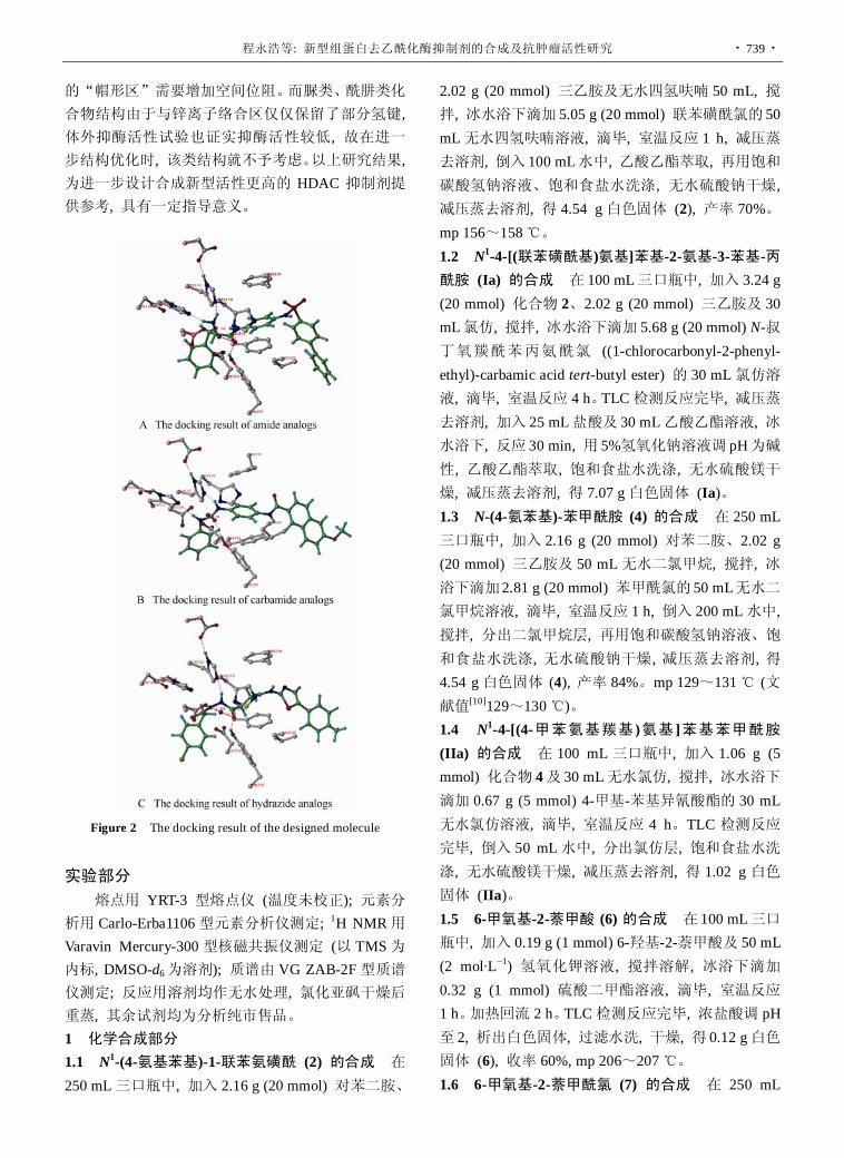
程永浩等: 新型组蛋白去乙酰化酶抑制剂的合成及抗肿瘤活性研究 · 739 ·
的“帽形区”需要增加空间位阻。而脲类、酰肼类化
合物结构由于与锌离子络合区仅仅保留了部分氢键, 体外抑酶活性试验也证实抑酶活性较低, 故在进一步结构优化时, 该类结构就不予考虑。以上研究结果, 为进一步设计合成新型活性更高的 HDAC 抑制剂提供参考, 具有一定指导意义。
Figure 2 The docking result of the designed molecule
实验部分 熔点用 YRT-3 型熔点仪 (温度未校正); 元素分
析用 Carlo-Erba1106型元素分析仪测定; 1H NMR用Varavin Mercury-300型核磁共振仪测定 (以 TMS为内标, DMSO-d6为溶剂); 质谱由 VG ZAB-2F型质谱仪测定; 反应用溶剂均作无水处理, 氯化亚砜干燥后重蒸, 其余试剂均为分析纯市售品。 1 化学合成部分 1.1 N1-(4-氨基苯基)-1-联苯氨磺酰 (2) 的合成 在250 mL三口瓶中, 加入 2.16 g (20 mmol) 对苯二胺、
2.02 g (20 mmol) 三乙胺及无水四氢呋喃 50 mL, 搅拌, 冰水浴下滴加 5.05 g (20 mmol) 联苯磺酰氯的 50 mL无水四氢呋喃溶液, 滴毕, 室温反应 1 h, 减压蒸去溶剂, 倒入 100 mL水中, 乙酸乙酯萃取, 再用饱和碳酸氢钠溶液、饱和食盐水洗涤, 无水硫酸钠干燥, 减压蒸去溶剂, 得 4.54 g白色固体 (2), 产率 70%。mp 156~158 ℃。 1.2 N1-4-[(联苯磺酰基)氨基]苯基-2-氨基-3-苯基-丙酰胺 (Ia) 的合成 在 100 mL三口瓶中, 加入 3.24 g (20 mmol) 化合物 2、2.02 g (20 mmol) 三乙胺及 30 mL氯仿, 搅拌, 冰水浴下滴加 5.68 g (20 mmol) N-叔丁氧羰酰苯丙氨酰氯 ((1-chlorocarbonyl-2-phenyl- ethyl)-carbamic acid tert-butyl ester) 的 30 mL氯仿溶液, 滴毕, 室温反应 4 h。TLC检测反应完毕, 减压蒸去溶剂, 加入 25 mL盐酸及 30 mL乙酸乙酯溶液, 冰水浴下, 反应 30 min, 用 5%氢氧化钠溶液调 pH为碱性, 乙酸乙酯萃取, 饱和食盐水洗涤, 无水硫酸镁干燥, 减压蒸去溶剂, 得 7.07 g白色固体 (Ia)。 1.3 N-(4-氨苯基)-苯甲酰胺 (4) 的合成 在 250 mL三口瓶中, 加入 2.16 g (20 mmol) 对苯二胺、2.02 g (20 mmol) 三乙胺及 50 mL无水二氯甲烷, 搅拌, 冰浴下滴加2.81 g (20 mmol) 苯甲酰氯的 50 mL无水二氯甲烷溶液, 滴毕, 室温反应 1 h, 倒入 200 mL水中, 搅拌, 分出二氯甲烷层, 再用饱和碳酸氢钠溶液、饱和食盐水洗涤, 无水硫酸钠干燥, 减压蒸去溶剂, 得4.54 g白色固体 (4), 产率 84%。mp 129~131 ℃ (文献值[10]129~130 ℃)。 1.4 N1-4-[(4-甲苯氨基羰基 )氨基 ]苯基苯甲酰胺 (IIa) 的合成 在 100 mL三口瓶中, 加入 1.06 g (5 mmol) 化合物 4及 30 mL无水氯仿, 搅拌, 冰水浴下滴加 0.67 g (5 mmol) 4-甲基-苯基异氰酸酯的 30 mL无水氯仿溶液, 滴毕, 室温反应 4 h。TLC 检测反应完毕, 倒入 50 mL水中, 分出氯仿层, 饱和食盐水洗涤, 无水硫酸镁干燥, 减压蒸去溶剂, 得 1.02 g 白色固体 (IIa)。 1.5 6-甲氧基-2-萘甲酸 (6) 的合成 在 100 mL三口瓶中, 加入 0.19 g (1 mmol) 6-羟基-2-萘甲酸及 50 mL (2 mol·L−1) 氢氧化钾溶液, 搅拌溶解, 冰浴下滴加0.32 g (1 mmol) 硫酸二甲酯溶液, 滴毕, 室温反应 1 h。加热回流 2 h。TLC检测反应完毕, 浓盐酸调 pH至 2, 析出白色固体, 过滤水洗, 干燥, 得 0.12 g白色固体 (6), 收率 60%, mp 206~207 ℃。 1.6 6-甲氧基-2-萘甲酰氯 (7) 的合成 在 250 mL

· 740 · 药学学报 Acta Pharmaceutica Sinica 2010, 45 (6): 735−741
三口瓶中, 加入 2.02 g (10 mmol) 化合物 6及 30 mL苯, 加热至 50 ℃, 缓慢滴加 20 mL二氯亚砜, 滴毕反应 3 h, TLC检测反应完毕, 减压蒸去溶剂, 得 2.10 g白色固体 (7), 收率 95%, mp 84~85 ℃ (文献值[11]
83~84 ℃)。 1.7 N-(4-氨基苯基)-N'-(4-甲基苯基) 脲 (9) 的合成 在 250 mL三口瓶中, 加入 1.08 g (10 mmol) 对苯二胺、50 mL二氯甲烷, 搅拌 10 min, 冰盐浴下缓慢滴加1.33 g (10 mmol) 4-甲基-苯基异氰酸酯的二氯甲烷溶液 50 mL, 反应 1 h。TLC检测反应完毕, 反应液减压浓缩, 柱色谱分离得1.95 g粉色固体 (9), 收率81%, mp 127~129 ℃ 1.8 N2-4-[(4-甲苯氨基羰基)氨基]苯基-6-甲氧基-2-萘甲酰胺 (IIb) 的合成 在 100 mL三口瓶中, 加入2.41 g (10 mmol) 化合物 9、1.01 g (10 mmol) 三乙胺及 20 mL 无水四氢呋喃, 搅拌, 冰浴下滴加 2.21 g (10 mmol) 化合物 7的 40 mL无水四氢呋喃溶液, 滴毕, 室温反应 1 h。TLC检测反应完毕, 减压浓缩反应液析出固体, 乙醇重结晶, 得 1.02 g白色固体 (IIb)。 1.9 6-苯甲酰基氨基-己酸 (11) 的合成 在 100 mL三口瓶中, 加入 5.25 g (40 mmol) 6-氨基己酸及 50 mL (2 mol·L−1) 氢氧化钠溶液, 搅拌溶解, 冰浴下滴加5.62 g (40 mmol) 苯甲酰氯的 10 mL四氢呋喃溶液, 滴毕, 室温反应 10 h。TLC检测反应完毕, 倒入 100 mL 水中, 浓盐酸调 pH 至 2.5, 析出白色固体, 过滤水洗, 干燥, 得 8.94 g白色固体 (11), 收率 95%, mp 80~81 ℃ (文献值[9]80~82 ℃)。 1.10 6-苯甲酰基氨基-己酸甲酯 (12) 的合成 在100 mL三口瓶中, 加入 4.71 g (20 mmol) 化合物 11、0.63 g Dowex50W-X12型强酸型树脂及 30 mL甲醇, 加热回流 12 h, TLC检测反应完毕, 抽滤, 滤液减压蒸去溶剂, 得 4.53 g 无色黏稠液体 (12), 收率 91%, 直接用于下步反应。 1.11 6-苯甲酰基氨基己酰肼 (13) 的合成 在 100 mL 三口瓶中, 加入 2.49 g (10 mmol) 化合物 12、 1.88 g (80%) 水合肼及30 mL乙醇, 加热回流 4 h, 有白色固体析出, TLC 检测反应完毕, 抽滤, 固体用乙醇重结晶, 得 2.10 g 白色固体 (13), 收率 84%, mp 50~52 ℃。 1.12 6-[(苯甲酰基) 氨基] 己酰基-2-乙酰肼 (IIIa) 的合成 在 100 mL三口瓶中, 加入 1.25 g (5 mmol) 化合物 13、0.51 g (5 mmol) 三乙胺及 20 mL无水甲苯, 搅拌, 冰浴下滴加 0.39 g (5 mmol) 乙酰氯的 10
mL无水甲苯溶液, 滴毕, 室温反应 4 h。TLC检测反应完毕, 倒入 100 mL冰水中, 析出固体, 过滤, 水洗, 乙醇重结晶, 得 0.77 g白色固体 (IIIa)。
IIIb~IIIg的合成参照 IIIa的合成方法, 理化参数及实验数据见表 1。 1.13 6-[(4-甲基苯基) 磺酰基] 氨基己酸 (14) 的合成 在 100 mL三口瓶中, 加入 5.25 g (40 mmol) 6-氨基己酸及 50 mL (2 mol·L−1) 氢氧化钠溶液, 搅拌溶解, 冰浴下滴加 7.63 g (40 mmol) 对甲基苯磺酰氯的 10 mL四氢呋喃溶液, 滴毕, 室温反应 10 h。TLC检测反应完毕, 倒入 500 mL水中, 浓盐酸调 pH至 2.5, 析出白色固体, 过滤, 水洗, 干燥, 得 10.32 g淡黄色固体 (14)。收率 90%, mp 107~108 ℃ (文献值[12] 106~108 ℃)。 1.14 6-[(4-甲基苯基) 磺酰基] 氨基己酸甲酯 (15)
的合成 在 100 mL三口瓶中, 加入 5.70 g (20 mmol) 化合物 14、0.63 g Dowex50W-X12型强酸型树脂及30 mL甲醇, 加热回流 12 h, TLC检测反应完毕, 抽滤, 滤液减压蒸去溶剂, 得 5.82 g白色固体 (15), 收率 97%, mp 43~44 ℃ (文献值[13] 45 ℃)。 1.15 6-[(4-甲基苯基) 磺酰基] 氨基己酰肼 (16) 的合
成 在 100 mL三口瓶中, 加入 3.0 g (10 mmol) 化合物 15、1.88 g (80%) 水合肼及 20 mL乙醇, 加热回流6 h, TLC检测反应完毕, 冷却至室温, 有淡黄色固体析出, 过滤, 用乙醇重结晶, 得 2.0 g 白色固体 (16), 收率 67%, mp 65~66 ℃。 1.16 6-[(苯磺酰基) 氨基] 己酰基-2-乙酰肼 (IIIh) 的
合成 在 50 mL三口瓶中, 加入 1.50 g (5 mmol) 化合物 15、0.51 g (5 mmol) 三乙胺及 10 mL无水甲苯, 搅拌, 冰浴下滴加 0.39 g (5 mmol) 乙酰氯的 10 mL无水甲苯溶液, 滴毕, 室温反应 6 h。TLC 检测反应完毕, 倒入 100 mL 冰水中, 析出固体, 过滤, 水洗, 乙醇重结晶, 得 0.38 g淡黄色固体 (IIIh)。
IIIi的合成参照 IIIh的合成方法, 理化参数及实验数据见表 1。 2 药理实验部分 2.1 HDAC 的制备 取小鼠肝脏, 剪碎, 加磷酸盐缓冲液 (PBS) 在 100 目筛网上研磨过滤, 得到的细胞悬液离心后用磷酸盐缓冲液清洗 2 次, 细胞沉淀, 加等体积的磷酸盐缓冲液, 振荡混匀, 冻融法裂解细胞, 离心 (4 ℃, 12 000 r·min−1, 10 min), 吸取上清液, 加入甘油至终体积为 30%, 蛋白浓度为 5 mg·mL−1, −20 ℃保存。 2.2 酶活性测定 在 PBS 为缓冲液的 50 µL 反应

程永浩等: 新型组蛋白去乙酰化酶抑制剂的合成及抗肿瘤活性研究 · 741 ·
体系中, 加入反应成分的终浓度分别为: MAL 20 μmol·L−1, 酶粗制剂 50%, Triton 0.1%, 待测样品0.1~10 μmol·L−1。SAHA 0.1~10 μmol·L−1为阳性对
照。加入样品后于 37 ℃水浴中反应 24 h。反应均作4个重复。以 100 μmol·L−1 SAHA阳性对照的荧光抑制率为 100%, PBS为阴性对照的抑制率为 0, 计算样品不同浓度的抑制率。目标化合物的 HDAC 酶抑制活性结果见表 3。
References
[1] Chen XG, Zhang Y. Recent advance in the study of novel
anti-tumor targets and drugs [J]. Acta Pharm Sin (药学学报),
2009, 44: 264−269.
[2] Li J, Li YT, Li XM. Advances on molecular targeted anti-
tumor drugs [J]. Lett Biotechnol, 2009, 20: 411−416.
[3] Minucci S, Pelicci PG. Histone deacetylase inhibitors and the
promise of epigenetic (and more) treatments for cancer [J].
Nat Rev Cancer, 2006, 6: 38−51.
[4] Bi GF, Jiang GS. The molecular mechanism of HDAC
inhibitors in anticancer effects [J]. Cell Mol Immunol, 2006,
3: 285−290.
[5] Glaser KB. HDAC inhibitors: clinical update and mechanism-
based potential [J]. Biochem Pharmacol, 2007, 74: 659−671.
[6] Bortugno OA, Santoro F, Minucci S. Histone deacetylase
inhibitors as a new weapon in the arsenal of differentiation
therapies of cancer [J]. Cancer Lett, 2009, 280: 134−144.
[7] Monneret C. Histone deacetylase inhibitors [J]. Eur J Med
Chem, 2005, 40: 1−13.
[8] Cheng YH, Guo YS, Wang N, et al. Structure based virtual
screening of HDAC inhibitors [J]. Comput Appl Chem, 2007,
24: 281−284.
[9] Jung M, Brosch G, Kolle D. Amide analogues of Trichostatin
A as inhibitors of histone deacetylase and inducers of terminal
cell differentiation [J]. J Med Chem, 1999, 42: 4669−4679.
[10] Weller H, Grellmann KH. On the photochemical synthesis
of N, N'-dimethylindolo[2, 3-c]carbazole and the mechanism
of its formation from N, N'-dimethyl-l-N, N'-diphenyl-1, 4-
henylenediamine [J]. J Am Chem Soc, 1983, 105: 6268−
6273
[11] Ananthakrishnanadar P, Varghesedharumaraj G. Kinetics
reaction of substituted naphthoyl chlorides with aniline [J].
Indian J Chem Sect B, 1983, 22: 506−507.
[12] Marvel CS, Moyer WW. Some derivatives of ε-caprolactam
[J]. J Org Chem, 1957, 22: 1065−1067.
[13] Schmidtchen FP. Synthese makrotricylischer amine [J].
Chem Ber, 1980, 113: 864−874.

· 742 · 药学学报 Acta Pharmaceutica Sinica 2010, 45 (6): 742−746
Chemical constituents of Saxifraga stolonifera (L.) Meeb.
FENG Wei-sheng*, LI Zhen, ZHENG Xiao-ke, LI Yuan-jing, SU Fang-yi, ZHANG Yan-li
(Henan University of Traditional Chinese Medicine, Zhengzhou 450008, China)
Abstract: To study the chemical constituents of Saxifraga stolonifera (L.) Meeb., chromatographic techniques were applied to separate and purify the compounds, and their structures were confirmed on the basis of physico-chemical properties and spectral data. Ten compounds were isolated and identified as 5-O-methylnorbergenin (1), 3, 4-dihydroxyallylbenzene-4-O-β-D-glucopyranoside (2), (7R, 8S)-4, 9, 9′-trihydroxyl-3-methoxyl-7, 8- dihydrobenzofuran-1′-propylneolignan-3′-O-β-D-glucopyranoside (3), quercetin-3-O-β-D-xylopyranosyl-(1→2)- β-D-galactopyranoside (4), kaempferol-3-O-α-L-rhamnopyranoside (5), (3S, 5R, 6R, 7E, 9R)-3, 5, 6, 9-tetrahydroxy- 7-megastigmane (6), benzyl-O-α-L-rhamnopyranosyl-(1→6)-β-D-glucopyranoside (7), p-hydroxyacetophenone (8), pyrogallic acid (9) and p-hydroxyphenol (10). Compound 1 is a new compound. Compounds 2 − 10 were isolated from this plant for the first time.
Key words: Saxifraga stolonifera (L.) Meeb.; chemical constituent; 5-O-methylnorbergenin CLC number: R284.1 Document code: A Article ID: 0513-4870 (2010) 06-0742-05
虎耳草的化学成分研究
冯卫生∗, 李 振, 郑晓珂, 李原京, 苏芳谊, 张艳丽
(河南中医学院, 河南 郑州 450008)
摘要: 为研究虎耳草 [Saxifraga stolonifera (L.) Meeb.] 的化学成分, 用柱色谱方法进行分离, 根据其理化性质和波谱数据鉴定化合物的结构。分离得到了 10 个化合物, 分别鉴定为 5-甲氧基异虎耳草素 (1)、3, 4-二羟基烯丙基苯-4-O-β-D-葡糖苷 (2)、(7R, 8S)-4, 9, 9′-三羟基-3-甲氧基-7, 8-二氢苯骈呋喃-1′-丙基新木脂素-3′-O-β-D-葡糖苷 (3)、槲皮素-3-O-β-D-木糖-(1→2)-β-D-半乳糖苷 (4)、山柰酚-3-O-α-L-鼠李糖苷 (5)、(3S, 5R, 6R, 7E, 9R)- 3, 5, 6, 9-四羟基-7-megastigmane (6)、苄基-O-α-L-鼠李糖-(1→6)-β-D-葡糖苷 (7)、对羟基苯乙酮 (8)、连苯三酚 (9) 和对羟基苯酚 (10)。其中化合物 1为新化合物, 化合物 2~10为首次从该植物中分离得到。
关键词: 虎耳草; 化学成分; 5-甲氧基异虎耳草素
Saxifraga stolonifera (L.) Meeb. (Family Saxifra- gaceae), a Chinese medicinal herb, is widely distributed in most parts of China. It is used to treat rubella, eczema, otitis media, erysipelas, hemoptysis, pulmonary abscess, uterine bleeding, and so on[1]. Recent studies have shown that it has many pharmacologic effects, such as anti-cancer[2], anti-oxidant and anti-aging[3]. Received 2009-12-21. Project supported by the Key Project of Chinese Ministry of Education
(2003078). *Corresponding author Tel / Fax: 86-371-65680011,
E-mail: [email protected]
Previous phytochemical investigations showed the presence of flavonoids, phenolic acids and aliphatics in this plant[2, 4]. In this paper, the isolation and structure elucidation of a new compound and nine known compounds were presented.
Results and discussion 50% aqueous acetone extract of the whole plants
of Saxifraga stolonifera (L.) Meeb. was subjected to a series of chromatographic procedures, including Diaion HP-20, Toyopearl HW-40C and Silica gel

FENG Wei-sheng, et al: Chemical constituents of Saxifraga stolonifera (L.) Meeb. · 743 ·
column chromatography, affording 10 compounds. Their structures were identified on the basis of spectroscopic analysis, including a new compound 5-O-methylnor- bergenin (1) (Figure 1), and nine known compounds, 3, 4-dihydroxyallylbenzene-4-O-β-D-glucopyranoside (2), (7R, 8S)-4, 9, 9′-trihydroxyl-3-methoxyl-7, 8-dihydro- benzofuran-1′-propylneolignan-3′-O-β-D-glucopyranoside (3), quercetin-3-O-β-D-xylopyranosyl-(1→2)-β-D-gala- ctopyranoside (4), kaempferol-3-O-α-L-rhamnopyrano- side (5), (3S, 5R, 6R, 7E, 9R)-3, 5, 6, 9-tetrahydroxy-7- megastigmane (6), benzyl-O-α-L-rhamnopyranosyl(1→ 6)-β-D-glucopyranoside (7), p-hydroxyacetophenone (8), pyrogallic acid (9) and p-hydroxyphenol (10). 1H and 13C NMR data for 1 were complete assigned by 1D and 2D NMR, including NOESY, HSQC and HMBC. Compounds 2 − 10 were isolated from this plant for the first time.
Compound 1 was obtained as light brown amorphous powder (CH3OH), mp 178 − 181 ℃, [α] 20
D
+13.3 (c 0.14, CH3OH). It gave a positive result for the ferric chloride reaction, revealing its phenolic nature. Its UV spectrum showed absorption at 223 nm and 291 nm. Its HR-ESI-MS showed [M+Na]+ at m/z 351.069 3 (calcd. for C14H16O9Na 351.069 2). Meanwhile, the IR spectrum showed the presence of hydroxyl (3 393 cm−1), aromatic ring (1 582 cm−1 and 1 475 cm−1) and ester carbonyl (1 698 cm−1). The 1H NMR spectrum (Table 1) of 1 exhibited one aromatic proton at δ 7.13 (1H, s), a C-glycosyl moiety at δ 3.44−4.86 and one methoxyl signal at 3.81 (3H, s). The 13C NMR spectrum (Table 1) of 1 exhibited 14 carbon signals, including a C-glycosyl moiety at δ 82.8 (C-5′), 71.8 (C-4′), 81.4 (C-3′), 75.8 (C-2′), 74.6 (C-1′), 62.7 (C-6′), which was characteristic of bergenin and norbergenin[5, 6]; one substituted benzene moiety at δ 118.6 (C-1), 107.5 (C-6), 150.8 (C-5), 151.4 (C-4), 144.9 (C-3), 107.6 (C-2); one ester carbonyl carbon at δ 167.8 (C-7) and one methoxyl signal at δ 56.3. The above information suggested that the basic structure of compound 1 is C-glucoside of gallic acid. The 1H NMR and 13C NMR spectrum characters were very similar to those of norbergenin[6] (Figure 1), except for a different substitution at C-5. In fact, the only difference between them consists in the hydroxyl at C-5 in norbergenin being replaced by a methoxyl group in compound 1, which was supported by the HMBC spectra. In the HMBC spectrum, the protons of methoxyl group (δ 3.81) correlated with the
carbon signal at δ 150.8 (C-5). This was further confirmed by NOE correlation between the methoxyl group and H-6. Thus, the methoxyl group was located at C-5 in compound 1. The relative stereochemistry of compound 1 was determined by comparison of the 1H and 13C NMR spectral data with those of bergenin and related compounds[5−7], the coupling constant observed in the 1H NMR spectrum, and the NOESY experiment. In the 1H NMR spectrum, the larger coupling constant (J = 10.3 Hz) between vicinal proton suggested their trans diaxial orientations. Additionally, in the NOESY spectrum there were no NOE correlations among H-1′, H-2′, H-3′, H-4′, and H-5′, which indicated that they were in trans-configuration. Based on the above evidence and according to the literature[6], the structure of 1 is determined to be 5-O-methylnor- bergenin (Figure 1).
Figure 1 Key HMBC and NOE correlations of compound 1 and structure of norbergenin Table 1 1H NMR and 13C NMR spectral data of compound 1 (CD3OD)
Position 1H NMR (400 MHz) 13C NMR (100 MHz)
1 118.6
2 107.6
3 144.9
4 151.4
5 150.8
6 7.13 (1H, s) 107.5
7 167.8
1′ 4.86 (1H, d, J = 10.3 Hz) 74.6
2′ 3.78 (1H, m) 75.8
3′ 3.95 (1H, m) 81.4
4′ 3.44 (1H, m) 71.8
5′ 3.62 (1H, m) 82.8
6′ 3.68 (1H, m) 62.7
4.00 (1H, m)
OCH3 3.81 (3H, s) 56.3

· 744 · 药学学报 Acta Pharmaceutica Sinica 2010, 45 (6): 742−746
Experimental General experimental procedures
Melting points were determined on a Kofler micro- melting point apparatus and uncorrected. Optical rotations were obtained using a Perkin-Elmer 341 polarimeter. UV spectra were measured with a Shimadzu UV-VIS 2201 spectrophotometer. IR spectra were measured with a Shimadzu FTIR-8201 PC spectrometer. The 1H and 13C NMR spectra were obtained on a Bruker DPX-400 spectrometer (400 MHz for 1H NMR and 100 MHz for 13C NMR) with TMS as internal reference. HR-TOF-MS were recorded on an APEX II spectrometer. Column chromatography was performed on Diaion HP-20 (Mitsubishi Chemical Corp., Japan), silica gel (160–200 mesh, Qingdao Haiyang Chemical Co., Ltd., China), Toyopearl HW-40C (TOSOH Corp., Japan). Sephadex LH-20 (Pharmacia, Sweden). TLC was conducted on self-made silica gel G (Qingdao Haiyang Chemical Co., Ltd., China) plates. The chemical reagents were purchased from Beijing Chemical Plant and Tianjin No. 3 Reagent Plant. Plant materials
Dried Saxifraga stolonifera (L.) Meeb. were collected in Xixia county, Henan Province in China, and identified by Prof. Dong Cheng-ming of Henan University of Traditional Chinese Medicine. Extraction and isolation
Air-dried whole plants of S. stolonifera (3.5 kg) were extracted two times with 50% aqueous acetone at room temperature and filtered. The combined extract was evaporated in vacuo to yield gross extract (256 g), which was dissolved in suitable amount of water, and then was subjected to Diaion HP-20 porous polymer resin and eluted with H2O and 10%, 20%, 30% CH3OH successively. The fraction eluted with H2O was subjected to Toyopearl HW-40C column chromatography and eluted with H2O and 10%, 20%, 30%, 40%, 50% CH3OH successively to afford 150 fractions. Then fractions 45-99 were rechromatographed on Toyopearl HW-40C, Sephadex LH-20 and silica gel to yield compound 7 (87 mg), 8 (11 mg), 9 (12 mg) and 10 (25 mg). The fraction eluted with 10% CH3OH was subjected to Toyopearl HW-40C column chromatography and eluted with H2O and 10%, 20%, 30%, 40%, 50% CH3OH successively to afford 280 fractions. Then fractions
25-40 and 86-110 were rechromatographed on Toyopearl HW-40C, Sephadex LH-20 and silica gel to yield compound 1 (13 mg) and 6 (12 mg) separately. The fraction eluted with 20% CH3OH was subjected to Toyopearl HW-40C column chromatography and eluted with H2O and 10%, 20%, 30%, 40%, 50% CH3OH successively to afford 200 fractions. Then fractions 31-60 were rechromatographed on Toyopearl HW-40C, Sephadex LH-20 and silica gel to yield compound 2 (18 mg), 3 (16 mg) and 4 (17 mg). The fraction eluted with 30% CH3OH was subjected to Toyopearl HW-40C column chromatography and eluted with H2O and 10%, 20%, 30%, 40%, 50% CH3OH successively to afford 158 fractions. Then fractions 71-120 were rechromatographed on Toyopearl HW-40C and Sephadex LH-20 gel to yield compound 5 (12 mg). Identification
Compound 1 Light brown amorphous powder, mp 178−181 ℃, [α] 20
D +13.3 (c 0.14, CH3OH). UV
(CH3OH) λmax: 223, 291 nm. IR (KBr): υmax cm−1: 3 393, 2 926, 1 697, 1 582, 1 475, 1 334, 868, 766. HR-ESI- MS m/z: 351.069 3 (calcd. for C14H16O9Na 351.069 2). The 1H NMR (400 MHz, CD3OD) and 13C NMR (100 MHz, CD3OD) spectral data see Table 1.
Compound 2 Colorless oil, 1H NMR (400 MHz, CD3OD) δ: 7.02 (1H, d, J = 1.6 Hz, H-2), 6.74 (1H, d, J = 8.1 Hz, H-5), 6.71 (1H, dd, J = 1.5, 8.1 Hz, H-6), 5.91 (1H, m, H-8), 5.01 (2H, m, H-9), 4.72 (1H, d, J = 7.3 Hz, H-1′), 3.25 (2H, d, J = 6.8 Hz, H-7). 13C NMR (100 MHz, CD3OD) δ: 133.1 (C-1), 119.2 (C-2), 146.6 (C-3), 146.6 (C-4), 116.9 (C-5), 124.7 (C-6), 40.5 (C-7), 139.2 (C-8), 115.6 (C-9), 104.5 (C-1′), 74.9 (C-2′), 77.6 (C-3′), 71.3 (C-4′), 78.3 (C-5′), 62.4 (C-6′). Combining literature[8] identified that the compound 2 was 3, 4- dihydroxyallylbenzene-4-O-β-D-glucopyranoside.
Compound 3 Colorless oil, 1H NMR (400 MHz, CD3OD) δ: 6.96 (1H, d, J = 1.7 Hz, H-2), 6.91(1H, br s, H-2′), 6.84 (1H, dd, J = 8.2, 1.7 Hz, H-2), 6.80 (1H, d, J = 8.2 Hz, H-5), 6.76 (1H, br s, H-6′), 5.50 (1H, d, J = 6.4 Hz, H-7), 4.97 (1H, d, J = 6.8 Hz, H-1′′), 3.80 (3H, s, -OCH3), 3.76 (2H, m), 3.52 (2H, t, J = 6.8 Hz, H-9′), 3.41 (1H, dd, J = 12.1, 6.3 Hz, H-8), 2.61 (2H, t, J = 8.0 Hz, H-7′), 1.80 (2H, m, H-8′). 13C NMR (100 MHz, CD3OD) δ: 134.3 (C-1), 110.7 (C-2), 149.1 (C-3), 147.7 (C-4), 116.2 (C-5), 119.9 (C-6), 89.4 (C-7), 55.2 (C-8), 64.8 (C-9), 137.1 (C-1′), 118.1 (C-2′), 142.5 (C-3′),

FENG Wei-sheng, et al: Chemical constituents of Saxifraga stolonifera (L.) Meeb. · 745 ·
147.5 (C-4′), 130.6 (C-5′), 119.6 (C-6′), 32.7 (C-7′), 35.6 (C-8′), 62.5 (C-9′), 102.9 (C-1′′), 74.9 (C-2′′), 77.7 (C-3′′), 71.4 (C-4′′), 78.2 (C-5′′), 62.2 (C-6′′), 56.4 (-OCH3). Its 1H NMR and 13C NMR data were in good agreement with those of literature[9] and identified the compound 3 to be (7R, 8S)-4, 9, 9′-trihydroxyl-3- methoxyl-7, 8-dihydrobenzofuran-1′-propylneolignan-3′- O-β-D-glucopyranoside.
Compound 4 Yellow amorphous powder, 1H NMR (400 MHz, CD3OD) δ: 7.71 (1H, d, J = 1.6 Hz, H-2′), 7.62 (1H, dd, J = 8.0, 1.6 Hz, H-6′), 6.85 (1H, d, J = 8.0 Hz, H-5′), 6.36 (1H, br s, H-8), 6.17 (1H, br s, H-6), 5.40 (1H, d, J = 7.2 Hz, H-1′′), 4.77 (1H, d, J = 6.4 Hz, H-1′′′). 13C NMR (100 MHz, CD3OD) δ: 158.3 (C-2), 135.2 (C-3), 179.6 (C-4), 163.2 (C-5), 99.8 (C-6), 165.9 (C-7), 94.6 (C-8), 158.2 (C-9), 105.7 (C-10), 123.1 (C-1′), 116.2 (C-2′), 145.9 (C-3′), 149.8 (C-4′), 117.4 (C-5′), 123.4 (C-6′), 101.4 (C-1′′), 80.0 (C-2′′), 74.7 (C-3′′), 70.3 (C-4′′), 75.1 (C-5′′), 61.9 (C-6′′), 105.3 (C-1′′′), 76.9 (C-2′′′), 77.0 (C-3′′′), 71.0 (C-4′′′), 66.5 (C-5′′′). Combining literature[10] identified that the compound 4 was quercetin-3-O-β-D-xylopyranosyl- (1→2)-β-D-galactopyranoside.
Compound 5 Yellow amorphous powder, 1H NMR (400 MHz, acetone-d6) δ: 7.86 (2H, d, J = 8.5 Hz, H-2′, 6′), 7.03 (2H, d, J = 8.6 Hz, H-3′, 5′), 6.48 (1H, d, J = 1.8 Hz, H-8), 6.28 (1H, d, J = 1.8 Hz, H-6), 5.55 (1H, d, J = 1.2 Hz, H-1′′), 0.92 (3H, d, J = 5.7 Hz, H-6′′). 13C NMR (100 MHz, acetone-d6) δ: 159.6 (C-2), 135.8 (C-3), 179.6 (C-4), 163.2 (C-5), 99.8 (C-6), 165.9 (C-7), 94.7 (C-8), 158.5 (C-9), 105.7 (C-10), 122.8 (C-1′), 132.4 (C-2′, 6′), 116.2 (C-3′, 5′), 161.8 (C-4′), 102.5 (C-1′′), 72.5 (C-2′′), 72.1 (C-3′′), 73.3 (C-4′′), 71.6 (C-5′′), 17.9 (C-6′′). Combining literature[11] identified that the compound 5 was kaempferol-3-O-α-L-rham- nopyranoside.
Compound 6 Colorless oil, 1H NMR (400 MHz, CD3OD) δ: 6.07 (1H, dd, J = 16.1, 1.2 Hz, H-7), 5.79 (1H, dd, J = 15.8, 6.1 Hz, H-8), 4.33 (1H, m, H-9), 4.03 (1H, m, H-3), 1.76 (2H, m, H-4), 1.66 (1H, t, H-2), 1.44 (1H, m, H-2), 1.27 (3H, d, J = 6.4 Hz, H-10), 1.19 (3H, s, H-11), 1.13 (3H, s, H-13), 0.83 (3H, s, H-12). 13C NMR (100 MHz, CD3OD) δ: 40.7 (C-1), 46.5 (C-2), 65.3 (C-3), 45.7 (C-4), 77.8 (C-5), 78.9 (C-6), 136.1 (C-7), 131.2 (C-8), 69.6 (C-9), 24.2 (C-10), 27.5 (C-11), 26.2 (C-12), 27.1 (C-13). Combining literature[12] identified that the compound 6 was (3S, 5R, 6R, 7E, 9R)-3, 5, 6,
9-tetrahydroxy-7-megastigmane. Compound 7 Pale yellow oil, 1H NMR (400
MHz, CD3OD) δ: 7.40 (2H, br d, J = 7.2 Hz, H-2, 6), 7.33 (2H, m, H-3, 5), 7.27 (1H, m, H-4), 4.78 (1H, br s, H-1′′), 4.33 (1H, d, J = 7.6 Hz, H-1′), 1.27 (3H, d, J = 6.5 Hz, H-6′′). 13C NMR (100 MHz, CD3OD) δ: 138.8 (C-1), 129.3 (C-2, 3, 5, 6), 128.7 (C-4), 71.7 (C-7), 103.1 (C-1′), 75.0 (C-2′), 77.9 (C-3′), 71.6 (C-4′), 76.9 (C-5′), 68.1 (C-6′), 102.2 (C-1′′), 72.1 (C-2′′), 72.2 (C-3′′), 73.9 (C-4′′), 69.8 (C-5′′), 18.0 (C-6′′). These 1H NMR and 13C NMR data were in basic agreement with those of literature[13, 14] and identified the compound 7 to be benzyl-O-α-L-rhamnopyranosyl (1→6)-β-D- glucopyranoside.
Compound 8 Colorless needles, 1H NMR (400 MHz, acetone-d6) δ: 7.84 (2H, d, J = 8.8 Hz, H-2, 6), 6.88 (2H, d, J = 8.8 Hz, H-3, 5), 2.46 (3H, s). 13C NMR (100 MHz, acetone-d6) δ: 196.4 (C=O), 129.1 (C-1), 131.4 (C-2, 6), 116.1 (C-3, 5), 164.2 (C-4), 26.1 (CH3). Combining literature[15] identified that the compound 8 was p-hydroxyacetophenone.
Compound 9 White needles, 1H NMR (400 MHz, acetone-d6) δ: 6.50 (1H, t, J = 8.1 Hz, H-5), 6.36 (2H, d, J = 8.1 Hz, H-4, 6). 13C NMR (100 MHz, acetone-d6) δ: 146.5 (C-1, 3), 133.5 (C-2), 119.7 (C-5), 107.8 (C-4, 6). Combining literature[16] identified that the compound 9 was pyrogallic acid.
Compound 10 Colorless needles, 1H NMR (400 MHz, acetone-d6) δ: 6.82 (2H, m, H-2, 6), 6.68 (2H, m, H-3, 5). 13C NMR (100 MHz, acetone-d6) δ: 145.8 (C-1, 4), 120.6 (C-2, 6), 116.0 (C-3, 5). Combining literature[17] identified that the compound 10 was p- hydroxyphenol.
References
[1] Jiangsu New Medical College. Dictionary of Traditional
Chinese Medicines (中药大辞典) [M]. Shanghai: Shanghai
Science and Technology Publisher, 1986: 1335.
[2] Chen Z, Liu YM, Yang S, et al. Studies on the chemical
constituents and anticancer activity of Saxifraga stolonifera
(L) Meeb [J]. Bioorg Med Chem, 2008, 16: 1337−1344.
[3] Yoon MY, Lim HW, Sim SS, et al. Anti-oxidant and anti-
aging activity on Saxifraga stolonifera Meerburgh ethanol
extract [J]. Yakhak Hoeji, 2007, 51: 343−349.
[4] Luo HW, Wu BJ, Chen JA, et al. Study on the effective
components of Saxifraga stolonifera Meerb. [J]. J Chin
Pharm Univ (中国药科大学学报), 1988, 19: 1−3.

· 746 · 药学学报 Acta Pharmaceutica Sinica 2010, 45 (6): 742−746
[5] Yoshida T, Seno K, Takama Y, et al. Bergenin derivatives
from Mallotus japonicus [J]. Phytochemistry, 1982, 21:
1180−1182.
[6] Taneyama M, Yoshida S, Michio K, et al. Isolation of
Norbergenin from Saxifraga stolonifera [J]. Phytochemistry,
1983, 22: 1053−1054.
[7] Ramaiah PA, Row LR, Reddy DS, et al. Isolation and
characterisation of bergenin derivatives from Mecaranga
peltata [J]. J Chem Soc Perkin Trans 1, 1979: 2313−2316.
[8] Zhou ZH, Wang JL, Yang CR. Chemical constituents of
Sanguis Draxonis made in China [J]. Chin Tradit Herb Drugs
(中草药), 2001, 32: 484−486.
[9] Feng WS, Zang XY, Zheng XK, et al. Isolation and Structure
Identification of Lignan from Rabdosia lophanthoides (Buch.-
Ham.ex D. Don) Hara [C]. Proceeding of the 2008 Chinese
Pharmaceutical Association Annual Symposium (2008 年中国
药学会学术年会论文集). Shijiazhuang: China Academic
Journal Electronic Publishing House, 2008: 1641−1647.
[10] Larsen LM, Nielsen JK, Sorensen H. Identification of 3-O-
2-O-(β-D-xylopyranosyl)-β-D-galactopyranosyl flavonoids in
horseradish leaves acting as feeding stimulants for a flea beetle
[J]. Phytochemistry, 1982, 21: 1029−1033.
[11] Jia Z, Zhang PF, Tao BQ, et al. Study on flavones of the
flower of Juglans regia [J]. Chin Pharm J (中国药学杂志),
2009, 44: 496−497.
[12] Otsuka H, Hirata E, Shinzato T, et al. Stereochemistry of
megastigmane glucosides from Glochidion zeylanicum and
Alangium premnifolium [J]. Phytochemistry, 2003, 62: 763−
768.
[13] Tommasi ND, Rastrelli J, Cumanda J, et al. Aryl and
triterpenic glycosides from Margyricarpus setosus [J].
Phytochemistry, 1996, 42: 163−167.
[14] Feng WS, Gao L, Zheng XK, et al. A new aryl glycoside
from Euphorbia helioscobia L. [J]. Chin Chem Lett, 2009,
20: 181−183.
[15] Kwon HC, Lee KR. Phytochemical constituents of Artemisia
japonica ssp. Littoricola [J]. Arch Pharm Res, 2001, 24:
194−197.
[16] Feng WS, Gao L, Zheng XK, et al. Polyphenols of Euphorbia
helioscopia [J]. Chin J Nat Med (中国天然药物), 2009, 7:
37−39.
[17] Zhao XM, Ye XQ, Zhu DY. A novel compound isolated from
the peels of Citrus changshan-huyou Y.B.Chang [J]. Acta
Pharm Sin (药学学报), 2008, 43: 1208−1210.

药学学报 Acta Pharmaceutica Sinica 2010, 45 (6): 747−751 · 747 ·
胆木叶生物碱类成分研究
范 龙 1, 3, 范春林 1, 3, 王 英 1, 3, 张晓琦 1, 3, 张庆文 2, 张俊清 4, 叶文才 1, 3*
(1. 暨南大学中药及天然药物研究所, 广东 广州 510632; 2. 澳门大学中华医药研究院, 澳门 沊仔; 3. 暨南大学中药药效物质基础及创新药物研究广东省高校重点实验室, 广东 广州 510632; 4. 海南医学院海南省热带药用植物研究开发重点实
验室, 海南 海口 571101)
摘要: 为了研究胆木叶中的化学成分, 采用硅胶、Sephadex LH-20、反相键合硅胶柱色谱等方法从胆木叶的95%乙醇提取物中分离得到 8 个生物碱类化合物, 并通过 NMR、MS 等波谱技术鉴定了化合物的结构, 分别为naucleactonin C (1)、strictosamide (2)、vincosamide (3)、pumiloside (4)、angustoline (5)、angustine (6)、18, 19- dihydroangustine (7) 和 naucleofficine D (8)。化合物 1为一新的吲哚类生物碱, 化合物 6和 7为首次从该植物中分离得到。
关键词: 胆木; 吲哚类生物碱; naucleactonin C 中图分类号: R284.1 文献标识码: A 文章编号: 0513-4870 (2010) 06-0747-05
Alkaloids from the leaves of Nauclea officinalis
FAN Long1, 3, FAN Chun-lin1, 3, WANG Ying1, 3, ZHANG Xiao-qi1, 3, ZHANG Qing-wen2, ZHANG Jun-qing4, YE Wen-cai1, 3*
(1. Institute of Traditional Chinese Medicine and Natural Products, Jinan University, Guangzhou 510632, China; 2. Institute of Chinese Medical Science, University of Macau, Macau; 3. Guangdong Province Key Laboratory of Pharmacodynamic
Constituents of Traditional Chinese Medicine and New Drugs Research, Jinan University, Guangzhou 510632, China; 4. Hainan Province Key Laboratory of R & D of Tropical Herbs, Hainan Medical College, Haikou 571101, China)
Abstract: To study chemical constituents of the leaves of Nauclea officinalis, eight alkaloids were isolated
from 95% ethanol extract by various chromatographic methods. The structures were elucidated on the basis of spectroscopic data (IR, UV, ESI-MS, 1D and 2D NMR) and identified as naucleactonin C (1), strictosamide (2), vincosamide (3), pumiloside (4), angustoline (5), angustine (6), 18, 19-dihydroangustine (7) and naucleofficine D (8). Compound 1 is a new indole alkaloid. Compounds 6 and 7 were isolated from this plant for the first time.
Key words: Nauclea officinalis; indole alkaloid; naucleactonin C
胆木 (Nauclea officinalis Pierre ex Pitard) 为茜草科 (Rubiaceae) 乌檀属乔木, 又名乌檀、药乌檀等, 在我国主要分布于海南、广西、广东等省区。胆木味
苦、性寒, 具清热解毒、消肿止痛之功效, 在我国南方民间常用于治疗感冒发烧、肺炎、肠炎、痢疾、湿
疹、皮疹等[1], 胆木提取物的两种制剂“胆木注射
收稿日期: 2009-12-24. 基金项目: 广东省自然科学基金团队项目 (8351063201000003); 澳门
特别行政区科学技术发展基金资助项目 (013/2008/A1). *通讯作者 Tel / Fax: 86-20-85221559, E-mail: [email protected]
液”和“胆木浸膏片”在临床上主要用于急性扁桃
体炎、急性咽喉炎、急性结膜炎及上呼吸道感染等疾
病的治疗, 疗效良好。文献[2−4]报道胆木中主要含有
生物碱、酚类及三萜类成分, 但其活性物质尚不明确。为进一步探索胆木中的活性成分, 本课题组对胆木叶的化学成分进行了研究。从胆木叶 95%乙醇提取物的乙酸乙酯萃取部位中分离鉴定出 8 个吲哚类生物碱, 其中, 化合物 1 为新化合物 (图 1), 化合物 6和 7为首次从该植物中分离得到。
化合物 1 黄色片状结晶 (CH3OH), 熔点 256~

· 748 · 药学学报 Acta Pharmaceutica Sinica 2010, 45 (6): 747−751
258 ℃。碘化铋钾试剂反应呈阳性, 推测该化合物可能为生物碱类成分。UV谱在 209、353和 370 nm处分别有最大吸收, 提示分子中有长共轭系统。IR谱显示该化合物结构中含有胺基 (3 241 cm−1)、酰胺羰基 (1 662 cm−1) 和芳环 (1 586、1 516 cm−1)。HR-ESI-MS谱显示出准分子离子峰 m/z 301.098 5 [M−H]− (计算值为301.098 3), 推断化合物1的分子式为C19H14N2O2, 计算不饱和度Ω = 14。化合物1的 1H NMR (DMSO-d6, 400 MHz) 谱中, 在低场区 δH 11.67处出现一个活泼质子信号; 在 δH 7.59 (1H, d, J = 8.0 Hz, H-9), 7.42 (1H, d, J = 8.0 Hz, H-12), 7.22 (1H, td, J = 8.0, 1.0 Hz, H-11) 和 7.07 (1H, td, J = 8.0, 1.0 Hz, H-10) 处为一个邻位双取代苯环上的 4 个芳香氢信号; 在 δH 6.80 (1H, dd, J = 18.0, 11.0 Hz, H-19), 5.97 (1H, d, J = 18.0 Hz, Ha-18) 和 5.47 (1H, d, J = 11.0 Hz, Hb-18)] 处为一组末端双键上的烯氢信号; 另外, 在 δH 4.40 (2H, t, J = 6.7 Hz, H-5) 和 3.07 (2H, t, J = 6.7 Hz, H-6) 处还出现了一组 -CH2CH2- 基团上的氢信号。化合物
1的 13C NMR (DMSO-d6, 100 MHz) 谱显示出 19个碳信号, 结合 DEPT谱信息, 可知包含 3个亚甲基、7个次甲基和 9个季碳, 其中在 δC 152.4 (C-22) 处为一个酰胺的羰基碳信号, 其余均为芳香碳信号。根据化合物 1的 1H NMR、13C NMR、1H-1H COSY、HSQC和 HMBC 谱信息, 对该化合物的碳氢信号进行了全归属 (表 1)。在化合物 1的 1H-1H COSY谱中, 显示出 3组质子自旋体系: δH 7.59 (H-9) ↔ 7.07 (H-10) ↔ 7.22 (H-11) ↔ 7.42 (H-12), 证实化合物 1中含有邻位双取代的苯环结构; δH 4.40 (H2-5) ↔ 3.07 (H2-6), 确证结构片段-CH2CH2-的存在; δH 5.97, 5.47 (Ha-18, Hb-18) ↔ 6.80 (H-19) 则进一步确证了末端双键的存在 (图1)。在化合物1的HMBC谱中, H-1 (δH 11.67) 与 C-2 (δC 128.5)、C-8 (δC 125.4)、C-12 (δC 111.5) 和C-13 (δC 137.8), H-6 (δH 3.07)与 C-2 (δC 128.5) 和 C-7 (δC 111.9), H-9 (δH 7.59) 与 C-7 (δC 111.9) 和 C-13 (δC
137.8) 分别有远程相关, 说明化合物 1 结构中含有tetrahydro-β-carboline环片段; H-14 (δH 7.33) 与 C-2 (δC 128.5)、C-3 (δC 133.6) 和 C-16 (δC 142.5) 有远程相关, 提示 tetrahydro-β-carboline 环与一个六元内酰胺环相骈合。此外, 在 HMBC谱中 H-21 (δH 8.23) 与C-15 (δC 129.7) 和 C-16 (δC 142.5) 分别有远程相关, 结合分子式和不饱和度信息, 提示六元内酰胺环的15和 16位还骈合有一个呋喃环。进一步在 HMBC谱中观察到 Ha-18 (δH 5.97) 和 Hb-18 (δH 5.47) 与 C-20
(δC 119.9), H-19 (δH 6.80) 与 C-15 (δC 129.7) 也分别有远程相关, 故可确定乙烯基连接在 C-20 位。综合以上信息, 鉴定化合物 1的结构如图 1所示, 并命名为 naucleactonin C。
Figure 1 1H-1H COSY and key HMBC correlations of 1
实验部分 熔点用X-5型显微熔点测定仪测定 (温度未校正);
紫外光谱用 JASCO V-550 紫外-可见光光度仪测定; 红外光谱用 JASCO FT/IR-480 Plus Fourier Transform型红外光谱仪测定 (KBr 压片); 核磁共振谱用 Bruker AV-400 FT型核磁共振仪测定; HR-ESI-MS用 Applied Biosystems MarinenTM 5140型质谱仪测定; ESI-MS用Finnigan LCQ Advantage MAX型质谱仪测定。
柱色谱用硅胶 (100~200 目和 200~300 目) 为青岛海洋化工厂产品; 硅胶 GF254薄层预制板为烟台
化学工业研究所产品; Sephadex LH-20柱色谱材料为Pharmacia公司产品; ODS柱层析材料为YMC公司产品; 所用试剂均为化学或分析纯。
胆木叶于 2008 年 7 月采于海南省三亚市, 由中国医学科学院药用植物研究所兴隆分所陈伟平教授
鉴定为胆木 (Nauclea officinalis Pierre ex Pitard) 的叶, 标本 (No. 090223) 保存于暨南大学中药及天然药物研究所。 1 提取与分离
干燥的胆木叶 4.8 kg, 粉碎成粗粉, 用 95%乙醇渗漉提取, 减压回收溶剂至无醇味, 得浸膏约 250 g。提取物用适量水混悬后, 依次用石油醚 (60~90)、乙酸乙酯和正丁醇萃取, 分别得到石油醚萃取物 (60.0 g)、乙酸乙酯萃取物 (85.0 g) 和正丁醇萃取物 (45.0 g)。取乙酸乙酯萃取物 60 g, 经硅胶柱色谱, 氯仿-甲
醇 (95∶5 → 50∶50) 梯度洗脱, 得到 7个流分 (Fr. 1−7)。Fr. 3经硅胶 (氯仿-甲醇, 100∶0.5 → 80∶20)、Sephadex LH-20 (CH3OH) 柱反复柱色谱分离纯化, 分别得到化合物 1 (8 mg)、6 (15 mg) 和 7 (9 mg); 取

范 龙等: 胆木叶生物碱类成分研究 · 749 ·
Table 1 1H and 13C NMR data of compound 1 (DMSO-d6) Position δC δH HMBC
1 − 11.67 (1H, s) C-2, 8, 12, 13
2 128.5 − −
3 133.6 − −
5 40.1 4.40 (2H, t, J = 6.7 Hz) C-7
6 19.1 3.07 (2H, t, J = 6.7 Hz) C-2, 5, 7
7 111.9 − −
8 125.4 − −
9 119.1 7.59 (1H, d, J = 8.0 Hz) C-7, 11, 13
10 119.4 7.07 (1H, td, J = 8.0, 1.0 Hz) C-8, 12
11 123.4 7.22 (1H, td, J = 8.0, 1.0 Hz) C-9, 12, 13
12 111.5 7.42 (1H, d, J = 8.0 Hz) C-8, 10
13 137.8 − −
14 93.3 7.33 (1H, s) C-2, 3, 16, 20
15 129.7 − −
16 142.5 − −
18 116.1 5.97 (1H, d, J = 18.0 Hz) Ha C-20
5.47 (1H, d, J = 11.0 Hz) Hb
19 125.9 6.80 (1H, dd, J = 18.0, 11.0 Hz) C-15
20 119.9 − −
21 147.5 8.23 (1H, s) C-15, 16, 20
22 152.4 − −
Fr. 4经硅胶柱色谱, 氯仿-甲醇 (100∶1 → 80∶20) 梯度洗脱, 得到化合物 2 (5.5 g) 和 4 (1.8 g); 取 Fr. 7经ODS 柱色谱分离, 甲醇-水系统 (10% → 85%) 梯度洗脱, 得到化合物 3 (11 mg); 取 Fr. 5经 ODS (甲醇-
水, 30% → 85%)、Sephadex LH-20 (CH3OH) 反复柱色谱分离纯化, 分别得到化合物 5 (13 mg) 和 8 (30 mg)。 2 结构鉴定
化合物 1 黄色片状结晶 (CH3OH), mp 256~258 ℃。UV (CH3OH) λmax nm: 209, 353, 370。IR (KBr) νmax cm−1: 3 241, 1 662, 1 586, 1 516。HR-ESI-MS m/z: 301.098 5 [M−H]− (计算值为 301.098 3), 分子式为C19H14N2O2。
1H NMR (DMSO-d6, 400 MHz) 和 13C NMR (DMSO-d6, 100 MHz) 数据见表 1。 化合物 2 白色无定形粉末, mp 179~180 ℃。UV
(CH3OH) λmax nm: 203, 226。IR (KBr) νmax cm−1: 3 412,
1 655, 1 583, 1 071, 746。ESI-MS m/z: 521 [M+Na]+。1H NMR (CD3OD, 400 MHz) δ: 7.37 (1H, d, J = 8.1 Hz, H-9), 7.36 (1H, s, H-17), 7.32 (1H, d, J = 8.1 Hz, H-12), 7.07 (1H, td, J = 8.1, 1.0 Hz, H-11), 6.99 (1H, td, J = 8.1, 1.0 Hz, H-10), 5.64 (1H, ddd, J = 17.0, 10.0, 10.0 Hz, H-19), 5.38 (1H, dd, J = 16.0, 2.0 Hz, Ha-18), 5.33 (1H, dd, J = 10.0, 2.0 Hz, Hb-18), 5.29 (1H, d, J = 2.0
Hz, H-21), 5.03 (1H, m, H-3), 4.93 (1H, dd, J = 5.0, 12.0 Hz, Ha-5), 4.56 (1H, d, J = 7.9 Hz, H-1'), 3.85 (1H, dd, J = 12.0, 1.9 Hz, Ha-6'), 3.62 (1H, dd, J = 12.0, 5.7 Hz, Hb-6'), 3.34~3.16 (3H, m, H-3', H-4', H-5'), 3.07 (1H, td, J = 4.0, 12.0 Hz, Hb-5), 2.96 (1H, m, H-2'), 2.93 (1H, m, Ha-6), 2.79 (1H, m, H-15), 2.69~2.63 (2H, m, Hb-6, H-20), 2.44 (1H, m, Ha-14), 2.02 (1H, ddd, J = 13.8, 13.8, 6.0 Hz, Hb-14)。13C NMR (CD3OD, 100 MHz) δ: 167.1 (C-22), 149.1 (C-17), 137.8 (C-13), 134.8 (C-2), 134.4 (C-19), 128.7 (C-8), 122.5 (C-11), 120.5 (C-18), 120.1 (C-10), 118.7 (C-9), 112.3 (C-12), 110.3 (C-7), 109.2 (C-16), 100.5 (C-1'), 98.1 (C-21), 78.2 (C-3'), 77.9 (C-5'), 74.3 (C-2'), 71.4 (C-4'), 62.6 (C-6'), 55.1 (C-3), 44.7 (C-5), 44.7 (C-20), 27.3 (C-14), 24.9 (C-15), 22.1 (C-6)。以上数据与文献[5]报道的
strictosamide一致, 故鉴定化合物 2为 strictosamide。 化合物 3 无色针状结晶 (CH3OH), mp 200~
202 ℃。UV (CH3OH) λmax nm: 205, 227。IR (KBr) νmax cm−1: 3 393, 1 653, 1 578, 1 438, 1 076, 1 018, 742。
ESI-MS m/z: 521 [M+Na]+。1H NMR (CD3OD, 400 MHz) δ: 7.44 (1H, d, J = 2.4 Hz, H-17), 7.41 (1H, d, J = 8.0 Hz, H-9), 7.29 (1H, d, J = 8.1 Hz, H-12), 7.07 (1H, td, J = 8.1, 1.0 Hz, H-11), 6.98 (1H, td, J = 8.1, 1.0 Hz, H-10), 5.52 (1H, m, H-19), 5.49 (1H, d, J = 2.0 Hz, H-21), 5.28 (1H, dd, J = 16.0, 2.0 Hz, Hb-18), 5.18 (1H, dd, J = 10.0, 2.0 Hz, Ha-18), 5.06 (1H, m, Hb-5), 4.93 (1H, m, H-3), 4.70 (1H, d, J = 7.9 Hz, H-1'), 3.90 (1H, dd, J = 12.0, 2.0 Hz, Hb-6'), 3.68 (1H, dd, J = 12.0, 5.7 Hz, Ha-6'), 3.39~3.30 (3H, m, H-3', H-4', H-5'), 3.20 (2H, m, H-2', H-15), 2.94 (1H, td, J = 4.0, 11.0 Hz, Ha-5), 2.75 (2H, m, H-6), 2.71 (1H, m, H-20), 2.46 (1H, dt, J = 12.0, 4.0 Hz, Hb-14), 1.45 (1H, q, J = 13.0 Hz, Ha-14)。13C NMR (CD3OD, 100 MHz) δ: 166.0 (C-22), 149.0 (C-17), 138.3 (C-13), 134.5 (C-2), 134.0 (C-19), 127.9 (C-8), 122.6 (C-11), 120.4 (C-18), 120.0 (C-10), 118.8 (C-9), 112.0 (C-12), 109.3 (C-16), 109.0 (C-7), 99.6 (C-1'), 97.4 (C-21), 78.3 (C-5'), 78.0 (C-3'), 74.8 (C-2'), 71.6 (C-4'), 62.7 (C-6'), 54.8 (C-3), 44.5 (C-20), 41.2 (C-5), 32.7 (C-14), 27.4 (C-15), 22.0 (C-6)。以上数据与文献[6]报道的 vincosamide一致, 故
鉴定化合物 3为 vincosamide。
化合物 4 白色无定形粉末, mp 236~238 ℃。UV (CH3OH) λmax nm: 213, 244, 316, 328。IR (KBr) νmax cm−1: 3 413, 1 629, 1 591, 1 514, 1 088。ESI-MS m/z: 535 [M+Na]+。1H NMR (DMSO-d6, 400 MHz) δ:

· 750 · 药学学报 Acta Pharmaceutica Sinica 2010, 45 (6): 747−751
8.12 (1H, d, J = 8.1 Hz, H-9), 7.67 (1H, td, J = 8.1, 1.3 Hz, H-11), 7.60 (1H, d, J = 8.1 Hz, H-12), 7.35 (1H, td, J = 8.1, 1.3 Hz, H-10), 7.05 (1H, d, J = 2.6 Hz, H-17), 5.81 (1H, dt, J = 17.0, 10.0 Hz, H-19), 5.48 (1H, dd, J = 17.0, 1.5 Hz, Ha-18), 5.40 (1H, d, J = 2.0 Hz, H-21), 5.35 (1H, dd, J = 10.0, 1.5 Hz, Hb-18), 4.77 (1H, d, J = 12.0 Hz, H-3), 4.55 (1H, d, J = 7.9 Hz, H-1'), 4.48 (1H, dd, J = 14.0, 2.5 Hz, Ha-5), 4.33 (1H, d, J = 14.0 Hz, Hb-5), 3.70 (1H, dd, J = 11.0, 5.7 Hz Ha-6'), 3.44 (1H, m, Hb-6'), 3.29 (1H, m, H-15), 3.21~3.16 (2H, m, H-3', H-5'), 3.07~2.97 (2H, m, H-2', H-4'), 2.66 (1H, dd, J = 9.0, 5.0 Hz, H-20), 2.50 (1H, m, Ha-14), 2.02 (1H, q, J = 12.0 Hz, Hb-14)。13C NMR (DMSO-d6, 100 MHz) δ: 173.0 (C-7), 163.9 (C-22), 149.7 (C-2), 145.1 (C-17), 140.4 (C-13), 132.5 (C-19), 131.6 (C-11), 125.3 (C-8), 124.7 (C-9), 123.2 (C-10), 120.5 (C-18), 118.3 (C-12), 113.0 (C-6), 108.9 (C-16), 97.8 (C-1'), 94.8 (C-21), 77.3 (C-3'), 76.5 (C-5'), 73.1 (C-2'), 70.1 (C-4'), 61.1 (C-6'), 59.4 (C-3), 47.4 (C-5), 43.6 (C-20), 28.1 (C-14), 23.7 (C-15)。以上数据与文献[7]报道的 pumiloside一致, 故鉴定化合物 4为 pumiloside。
化合物 5 橙红色无定形粉末, mp 275~277 ℃。
UV (CH3OH) λmax nm: 206, 290。IR (KBr) νmax cm−1:
3 393, 1 651, 1 384, 1 200, 1 059, 749。ESI-MS m/z:
332 [M+H]+。1H NMR (DMSO-d6, 400 MHz) δ: 11.84 (1H, s, H-1), 9.24 (1H, s, H-17), 8.76 (1H, s, H-21), 7.63 (1H, d, J = 7.9 Hz, H-9), 7.47 (1H, d, J = 8.2 Hz, H-12), 7.26 (1H, t, J = 7.2 Hz, H-11), 7.23 (1H, s, H-14), 7.10 (1H, t, J = 7.2 Hz, H-10), 5.55 (1H, d, J = 4.0 Hz, OH), 5.31 (1H, t, J = 6.4 Hz, H-19), 4.39 (2H, m, H-5), 3.11 (2H, t, J = 6.7 Hz, H-6), 1.51 (3H, d, J = 6.4 Hz, H-18)。13C NMR (DMSO-d6, 100 MHz) δ: 161.1 (C-22), 149.0 (C-17), 147.4 (C-21), 138.3 (C-15), 138.3 (C-13), 136.3 (C-3), 134.7 (C-20), 127.6 (C-2), 125.3 (C-8), 124.3 (C-11), 119.7 (C-10), 119.5(C-9), 118.5 (C-16), 114.5 (C-7), 111.8 (C-12), 93.7 (C-14), 63.7 (C-19), 40.2 (C-5), 25.1 (C-18), 19.1 (C-6)。以上
数据与文献[8]报道的 angustoline一致, 故鉴定化合物
5为 angustoline。 化合物 6 橙红色无定形粉末, mp 283~285 ℃。
UV (CH3OH) λmax nm: 206, 305, 380, 399。IR (KBr)
νmax cm−1: 3 229, 1 642, 1 601, 739。ESI-MS m/z: 314
[M+H]+。1H NMR (DMSO-d6, 400 MHz) δ: 11.78 (1H, s, H-1), 9.22 (1H, s, H-17), 8.85 (1H, s, H-21), 7.61 (1H, d, J = 8.0 Hz, H-9), 7.46 (1H, d, J = 8.0 Hz, H-12),
7.29~7.19 (3H, m, H-14, H-19, H-11), 7.09 (1H, t, J = 8.0 Hz, H-10), 6.05 (1H, d, J = 16.0 Hz, Ha-18), 5.62 (1H, d, J = 8.0 Hz, Hb-18), 4.39 (2H, t, J = 6.8 Hz, H-5), 3.12 (2H, t, J = 6.8 Hz, H-6)。13C NMR (DMSO- d6, 100 MHz) δ: 160.6 (C-22), 149.3 (C-17), 147.3 (C-21), 138.5 (C-15), 138.1 (C-13), 136.5 (C-3), 129.8 (C-19), 127.4 (C-2), 126.4 (C-20), 125.0 (C-8), 124.1 (C-11), 119.5 (C-10), 119.3 (C-9), 118.8 (C-16), 118.7 (C-18), 114.3 (C-7), 111.5 (C-12), 93.3 (C-14), 40.0 (C-5), 18.8 (C-6)。以上数据与文献[9]报道的 angustine
一致, 故鉴定化合物 6为 angustine。
化合物 7 黄色无定形粉末, mp 271~273 ℃。UV (CH3OH) λmax nm: 204, 245。IR (KBr) νmax cm−1: 3 418, 1 660, 1 595, 744。ESI-MS m/z: 316 [M+H]+。1H NMR (DMSO-d6, 400 MHz) δ: 11.83 (1H, s, H-1), 9.20 (1H, s, H-17), 8.54 (1H, s, H-21), 7.63 (1H, d, J = 8.0 Hz, H-9), 7.48 (1H, d, J = 8.0 Hz, H-12), 7.27 (1H, td, J = 8.0, 1.0 Hz, H-11), 7.21 (1H, s, H-14), 7.10 (1H, td, J = 8.0, 1.0 Hz, H-10), 4.40 (2H, t, J = 6.7 Hz, H-5), 3.13 (2H, t, J = 6.7 Hz, H-6), 2.93 (2H, q, J = 7.5 Hz, H-19), 1.32 (3H, t, J = 7.5 Hz, H-18)。13C NMR (DMSO-d6, 100 MHz) δ: 161.2 (C-22), 149.7 (C-17), 148.5 (C-21), 139.9 (C-13), 138.4 (C-15), 136.5 (C-3), 132.1 (C-20), 127.7 (C-2), 125.4 (C-8), 124.4 (C-11), 119.8 (C-10), 119.6 (C-9), 119.1 (C-16), 114.4 (C-7), 111.8 (C-12), 94.0 (C-14), 40.3 (C-5), 22.3 (C-19), 19.1 (C-6), 15.1 (C-18)。以上数据与文献[9]报道的18, 19-dihydroangustine一致, 故鉴定化合物 7为 18, 19-dihydroangustine。
化合物 8 淡黄色片状结晶 (CH3OH), mp 247~
249 ℃。UV (CH3OH) λmax nm: 205, 224, 280。IR (KBr)
νmax cm−1: 3 277, 1 728, 1 617, 1 470, 740。ESI-MS m/z:
361 [M+Na]+。1H NMR (DMSO-d6, 400 MHz) δ: 11.01 (1H, s, H-1), 7.37 (1H, d, J = 7.9 Hz, H-9), 7.32 (1H, d, J = 7.9 Hz, H-12), 7.04 (1H, t, J = 7.9 Hz, H-11), 6.97 (1H, t, J = 7.9 Hz, H-10), 6.53 (1H, d, J = 4.3 Hz, OH), 5.40 (1H, q, J = 6.5 Hz, H-19), 5.29 (1H, t, J = 4.1 Hz, H-17), 5.04 (1H, d, J = 5.6 Hz, H-3), 4.78 (1H, dd, J = 12.2, 5.2 Hz, Ha-5), 4.64 (1H, d, J = 12.6 Hz, Ha-21), 3.67 (1H, d, J = 12.6 Hz, Hb-21), 2.98~2.87 (2H, m, Hb-5, Ha-14), 2.80 (1H, m, Ha-6), 2.72 (1H, m, H-15), 2.59 (1H, dd, J = 9.0, 4.2 Hz, Hb-6), 2.41 (1H, t, J = 4.8 Hz, H-16), 2.25 (1H, d, J = 10.2 Hz, Hb-14), 1.45 (3H, d, J = 6.7 Hz, H-18)。13C NMR (DMSO-d6, 100 MHz) δ: 168.1 (C-22), 135.9 (C-13), 135.0 (C-2), 134.9 (C-20), 127.1 (C-8), 121.0 (C-11), 119.7 (C-19), 118.7 (C-10),

范 龙等: 胆木叶生物碱类成分研究 · 751 ·
117.6 (C-9), 111.3 (C-12), 108.7 (C-7), 91.1 (C-17), 60.4 (C-21), 53.7 (C-3), 46.4 (C-16), 42.4 (C-5), 28.4 (C-15), 27.0 (C-14), 20.8 (C-6), 11.6 (C-18)。以上数据
与文献[10]报道的 naucleofficine D 一致, 故鉴定化合
物 8为 naucleofficine D。
References
[1] Editorial Board of China Herbal, State Administration of
Traditional Chinese Medicine, China. China Herbal: Vol 6 (中
华本草: 第六卷) [M]. Shanghai: Shanghai Scientific and
Technical Publishers, 1999: 456.
[2] Ma WZ, Ling TJ, Zhang YH, et al. Chemical constituents
from Nauclea officinalis [J]. J Trop Subtrop Bot (热带亚热
带植物学报), 2005, 13: 167−170.
[3] Lin M, Li SZ, Liu X, et al. Studies on the structures of
two new alkaloidal glucosides of Nauclea officinalis Pierre ex
Pitard [J]. Acta Pharm Sin (药学学报), 1989, 24: 32−36.
[4] Lin M, Liu X, Yu DQ, et al. The structure determination of a
new alkaloid, nauclefiline, in Nauclea officinalis [J]. Acta
Pharm Sin (药学学报), 1985, 20: 902−905.
[5] Dhooghe L, Mesia K, Kohtala E, et al. Development
and validation of an HPLC-method for the determination of
alkaloids in the stem bark extract of Nauclea pobeguinii [J].
Talanta, 2008, 76: 462−468.
[6] Wang B, Yuan D, Ma B, et al. Chemical constituents of the
leaves of Uncaria rhynchophylla (Miq.) Jacks [J]. Chin J
Med Chem (中国药物化学杂志), 2006, 16: 369−372.
[7] Zhang ZZ, ElSohly HN, Jacob MR, et al. New indole
alkaloids from the bark of Nauclea orientalis [J]. J Nat Prod,
2001, 64: 1001−1005.
[8] Xuan WD, Bian J, Chen HS. Alkaloidal constituents of
Nauclea officinalis [J]. Chin Tradit Herb Drugs (中草药),
2007, 38: 170−173.
[9] Erdelmeier CAJ, Regenass U, Rali T, et al. Indole alkaloids
with in vitro antiproliferative activity from the ammoniacal
extract of Nauclea orientalis [J]. Planta Med, 1992, 58: 43−48.
[10] Sun JY, Lou HX, Dai SJ, et al. Indole alkaloids from Nauclea
officinalis with weak antimalarial activity [J]. Phytochemistry,
2008, 69: 1405−1410.

· 752 · 药学学报 Acta Pharmaceutica Sinica 2010, 45 (6): 752−755
重组嵌合抗 CD20 IgG1 型单克隆抗体的结构验证
陶 磊, 饶春明, 高 凯, 史新昌, 赵 阳, 王军志*
(中国药品生物制品检定所, 北京 100050)
摘要: 本文选择一种重组嵌合抗 CD20 IgG1型单抗, 应用液质联用仪及 N-末端测序仪对其进行结构验证。对该单抗进行还原、烷基化、酶解等处理后, 对其氨基酸序列、二硫键配对方式、糖链类型及糖基化位点进行分析测定。结果显示, 该单抗轻、重链氨基酸序列与理论一致。通过液质肽图的解析, 对单抗 10 条二硫键的配对方式进行了验证; 通过比较单抗重链切糖前、后的相对分子质量, 预测单抗所含糖链类型为岩藻糖化的双触角复杂型 N-糖, 糖基化位点位于重链的 Asn301上。本方法可为该类重组单抗制品的质量控制及其参考品的结构确证提供参考。
关键词: 抗 CD20 IgG1单克隆抗体; 结构验证; 高效液相色谱-质谱联用 中图分类号: R917 文献标识码: A 文章编号: 0513-4870 (2010) 06-0752-04
Structure verification of a recombinant chimeric anti-CD20 IgG1 monoclonal antibody
TAO Lei, RAO Chun-ming, GAO Kai, SHI Xin-chang, ZHAO Yang, WANG Jun-zhi*
(National Institute for the Control of Pharmaceutical and Biological Products, Beijing 100050, China)
Abstract: Structure of a recombinant chimeric anti-CD20 IgG1 monoclonal antibody was verified by the application of high-performance liquid chromatography-mass spectrometry (HPLC-MS) and N-terminal sequencer. Molecular masses, N-terminal sequences and peptide maps of the antibody treated with different reagents and enzymes were measured. Results indicate that the amino acid sequences of light and heavy chains and 10 disulfide bonds were consistent with theoretical structure. By comparison of molecular masses and peptide maps for the fully glycosylated and deglycosylated samples, the N-linked glycosylation site was identified. The method is simple, rapid, precise, and could be referred to the quality control and structure determination of other IgG1 products.
Key words: anti-CD20 IgG1 monoclonal antibody; structure verification; HPLC-MS
近年来, 重组单抗药物广泛应用于肿瘤、类风湿性关节炎以及移植排斥等的治疗, 是复合增长率最快的一类生物技术药物, 具有巨大的经济价值和社会价值。随着重组单抗药物的迅猛发展, 对其进行有效的质量控制成为迫切需要解决的问题。与小分子蛋
白药物不同, 单抗制品相对分子质量大、结构复杂,
收稿日期: 2009-11-15. 基金项目: 国家高技术研究发展谋划 (863 计划 ) 资助项目
(2007AA021601); 重大新药创制资助项目 (2009ZX09307). *通讯作者 Tel: 86-10-67095782, Fax: 86-10-67014382,
E-mail: [email protected]
结构分析是其质量控制中的难点问题。 20世纪 80 年代中期, 出现了两种新型质谱电离
技术: 电喷雾离子化 (electrospray ionization, ESI)[1]
和基质辅助激光解析离子化 (matrix-assisted laser desorption ionization, MALDI)[2], 新型质谱的出现使蛋白质、核酸等大分子物质精确相对分子质量的测定
成为可能, 其测定准确度高达 0.01%, 借助此项技术, 可以对单抗等复杂的大分子重组蛋白进行结构验证。
目前重组单抗药物多为 IgG1 型, 因此本文选择了一种重组嵌合抗 CD20 (cluster of differentiation 20) IgG1 型单抗进行分析, 该单抗主要用于治疗 B 细胞

陶 磊等: 重组嵌合抗 CD20 IgG1 型单克隆抗体的结构验证 · 753 ·
非霍奇金淋巴瘤, 其理论相对分子质量约为 146 000, 含有 16条二硫键, 包括 4条链间二硫键和 12条链内 二硫键, 单抗 Fc 段存在糖基修饰。本文通过对单抗进行不同的前处理、液质联用测定相对分子质量, 对该单抗的氨基酸序列、二硫键配对方式、糖链类型及
糖基化位点进行了分析验证。
材料与方法 材料 二硫苏糖醇 (dithiothreitol, DTT, 购自
INALCO); 碘乙酰胺 (iodoacetamide, IAM, 购自Acros); 肽N糖苷酶F (peptide N-glycosidase F, PNGase F, 购自 Sigma); 焦谷氨肽酶 (购自 TaKaRa); 胰蛋白酶试剂盒 (Trypsin Singles, 购自 Sigma); 待测单抗为供试品。 电泳仪 (Bio-Rad, Miniprotein3); 转膜仪 (Pharmacia);
聚偏二氟乙烯膜 (PVDF膜, 孔径 0.2 μm, Millipore); N-末端测序仪 (日本岛津); 高效液相色谱仪 (美国Waters Alliance 2695); 检测器 (Waters2996); 色谱柱
(Symmetry 300, C18, 3.9 mm × 150 mm, Waters); 质谱仪 (Q-TOF-Micro, Micromass); Masslynx 4.0数据处理软件。
单抗氨基酸序列验证 抗体轻、重链相对分子质量测定 将单抗样品以
超纯水充分透析并冻干, 用超纯水重溶后加入糖苷酶酶解去除糖基, 然后用 DTT 对样品进行充分还原, 之后液质联用测定相对分子质量。液相条件: 流动相A为 0.1% TFA水溶液, 流动相 B为 0.1% TFA乙腈溶液, 使用40 min连续梯度 (流动相B从10%到100%); 流速 1 mL·min−1。色谱柱后分流 80%进入二极管阵 列检测器。质谱条件: 毛细管电压 3 kV; Cone 电压 30 V; 去溶剂气体温度: 200 ℃; 源温: 80 ℃; 飞行管电压: 5 630 V; MCP电压: 2 300 V; 去溶剂气体流速: 400 L·h−1, 扫描范围 (m/z): 500~3 000。色谱柱后分流 20%进入质谱检测器。
N-末端氨基酸序列测定 用焦谷氨肽酶酶解缓冲液将透析冻干的单抗样品溶解成 2 mg·mL−1, 取 25 μL加入焦谷氨肽酶 10 μL, 60℃水浴过夜。将酶解后的单抗样品进行还原 SDS-PAGE并转印 PVDF膜, 将膜上的单抗轻、重链样品小心剪下, 分别上样 N-末端测序仪, 测定其 N-末端 14个氨基酸序列。
液质肽图测定 取单抗样品 300 μg, 用超纯水150 μL溶解, 用 DTT充分还原后以酶与蛋白质量比1∶50加入胰蛋白酶, 37 ℃水浴 13 h, 液质联用测定
肽图。液相条件: 0~60 min, 流动相 B从 5%到 40%; 60~90 min流动相 B从 40%到 100%, 质谱扫描范围(m/z): 100~3 000, 其余同“抗体轻、重链相对分子质量测定”。
二硫键配对方式验证 取单抗样品 300 μg, 用超纯水150 μL溶解, 以酶与蛋白质量比1∶50加入胰蛋白酶, 37 ℃水浴 14 h, 液质联用测定肽图。液相条件: 使用 60 min连续梯度 (流动相 B从 5%到 75%); 上样量 80 μL, 质谱扫描范围 (m/z): 100~3 000, 其余同“抗体轻、重链相对分子质量测定”。
糖链类型及糖基化位点分析 取冻干单抗样品用超纯水重溶, 用 DTT 充分还原后直接液质联用测定单抗重链的相对分子质量。液相及质谱条件同“抗
体轻、重链相对分子质量测定”。取冻干单抗样品
300 μg, 用超纯水 150 μL重溶并平分为两支, 分别加入糖苷酶 10 μL与超纯水 10 μL, 37 ℃水浴 3 h。还 原、烷基化后以酶与蛋白质量比 1∶50分别加入胰蛋白酶, 37 ℃水浴 13 h, 液质联用测定肽图。液相条件使用 100 min连续梯度 (流动相 B从 0到 50%), 质谱扫描范围 (m/z): 100~3 000, 其余同“抗体轻、重链相对分子质量测定”。
结果 1 单抗氨基酸序列验证 1.1 轻、重链相对分子质量 单抗轻、重链的理论相对分子质量分别为 23 039.71和 49 094.42, 实测相对分子质量分别为 23 040.38和 49 092.50, 相对误差均小于 0.01%, 初步说明单抗氨基酸序列与理论一致。 1.2 N-末端氨基酸序列 单抗轻、重链 N-末端氨基酸序列测定结果分别为: IVLSQSPAILSASP, VQLQQ PGAELVKPG, 与理论序列中 N-末端第 2~15位氨基酸序列一致。 1.3 液质肽图 图 1 为单抗的胰蛋白酶酶解液的液质肽图, 部分肽段解析结果见表 1, 其分别是轻链与重链的 N-末端及 C-末端肽段, 肽段的实测相对分子质量与理论值非常接近, 说明单抗轻、重链的 N-末端及 C-末端表达正确。通过对液质肽图的解析, 对单抗的 55 条肽段进行了氨基酸序列的验证, 氨基酸覆盖率达 90%。 2 二硫键配对方式验证
单抗直接经胰蛋白酶酶解后 , 有些肽段仍然 通过二硫键连接在一起, 通过对二硫键连接肽段的

· 754 · 药学学报 Acta Pharmaceutica Sinica 2010, 45 (6): 752−755
鉴定可验证单抗的二硫键连接方式。通过对液质 肽图 (图 2) 进行解析, 共发现 5种链内二硫键连接肽段: 2种位于轻链 (T2-T5; T9-T16), 3种位于重链
Figure 1 Peptide maps of IgG1 (reduced) digested by trypsin. A: Detected at 214 nm; B: Total ion current detected by MS
Table 1 Comparison of theoretical to measured masses of terminal peptides
Peptide Sequence Theoretical mass Measured mass Differ. L: T1a 1−18 1 806.97 1 807.07 0.10 L: T17−T18b 207−213 811.33 811.38 0.05
H: T1c 1−19 1 959.08 1 959.17 0.09 H: T40d 444−450 659.35 659.39 0.04
aN-terminal peptide of light chain; bC-terminal peptide of light chain; cN-terminal peptide of heavy chain; dC-terminal peptide of heavy chain
Figure 2 Peptide maps of IgG1 (unreduced) digested by trypsin. A: Detected at 214 nm; B: Total ion current detected by MS; L: Light chain; H: Heavy chain
(T2-T9; T20-T26; T34-T39), 由于单抗含有 2条轻链和重链, 因此对单抗 10 条二硫键的配对方式进行了验证。 3 糖链类型及糖基化位点分析结果
切除糖链前单抗重链有 3 种相对分子质量 (图3), 切除糖链后测得 1 种, 说明重链相对分子质量的不均一是由糖基化造成的。由切糖前、后重链的相对
分子质量得到糖链的相对分子质量分别为: 1 446.75、1 608.88和 1 770.00, 由此预测糖链类型如图 3所示, 为岩藻糖化的双触角复杂型 N-糖, 与文献[3]报道相符。单抗氨基酸序列中只有一个 N-糖序列子结构 (NST), 位于重链肽段 T23。在单抗切除糖链前、后所得液质肽图中, 分别发现了含糖链的肽段 T22-T23及不含糖链的肽段 T23 (图 4), 因此将糖基化位点定位于肽段 T23中的 Asn301。
Figure 3 MS spectrum (after transformation) of glycosylated heavy chain and estimated structure of the glycon
Figure 4 Peptide maps of glycosylated (A) and deglycosylated (B) IgG1 digested by trypsin

陶 磊等: 重组嵌合抗 CD20 IgG1 型单克隆抗体的结构验证 · 755 ·
讨论 有研究用 ESI-MS[4]或 MALDI-TOF-MS[5]测定了
相对分子质量约为 150 000的完整抗体分子的相对分子质量, 测定相对误差最低为 0.02%左右。本实验将单抗还原后分别测定了轻、重链的相对分子质量, 实测值与理论值的相对误差均小于 0.01%。
本文通过测定液质肽图对单抗的结构进行了分
析。在对未知蛋白进行液质肽图分析时, 只通过肽段相对分子质量的测定不能对其定性, 而需要进一步的串联质谱分析。但是在本实验中, 研究对象为重组蛋白制品, 理论序列已知、纯度很高 (>95%)、不存在未知杂蛋白的干扰 (或杂蛋白量很低, 可忽略不计), 另外胰蛋白酶的酶切位点特异性高, 因此, 如果肽段实测相对分子质量与理论值匹配 (相对分子质量差值<1), 即可对该肽段进行定性[6]。对单抗氨基酸
序列的验证、二硫键配对方式的验证以及糖基化位点
的测定都是基于上述前提进行的。 在单抗氨基酸序列验证中, 通过对液质肽图的
解析, 对单抗的55个肽段进行了验证, 还有 3个肽段 (轻链的 T5 及重链的 T9、T39) 未验证, 考虑原因可能是: ① 由于胰蛋白酶的非特异酶解或不完全酶解, 未能对该肽段进行解析; ② 肽段离子化困难, 不易被质谱检测到; ③ 属于疏水肽段, 实验过程中发生沉淀, 未能将其洗脱。
在对二硫键配对方式的验证中, 如果在肽图中能发现二硫键连接肽段, 则可以认为单抗绝大部分是以该方式连接的, 这是基于以下前提: 单抗由细胞表达产生, 细胞的内在机制能促使形成正确的二硫键配对方式; 其次, 单抗生产过程经过多步纯化, 纯度较高 (>95%), 均一性好。本文按理论配对方式[7]
对10条链内二硫键进行了验证, 未能对链间二硫键及2 条链内二硫键进行验证, 原因可能是未验证的二硫键将抗体的相关肽段连接形成一个比较紧密的结构, 导致胰蛋白酶无法酶切, 并且该结构离子化困难, 在质谱测定时不能检测到。
对糖链进行分析时, 常用的方法是先用凝集素亲和层析柱将糖肽纯化[8], 然后用质谱仪对其结构进行鉴定, 该方法的缺点是只能收集某些糖肽而不能将所有异型糖肽全部回收。还有研究者用多维色谱与
质谱仪联用的方法在线对某一蛋白的糖基化位点进
行鉴定[9], 过程较为复杂。本实验未将糖肽进行分离纯化而通过相对分子质量的测定预测了糖型, 并通过肽图的方法测定了糖基化位点, 方法更为简便、快
捷。但是, 本文所用糖链分析方法也有不足之处, 只能结合理论知识对糖链类型进行预测, 对糖链类型的确定还需要其他方法的进一步分析; 另外, 不能对糖链的连接方式及单糖组成进行测定。
总之, 本文通过相对分子质量的测定、N-末端测序及液质肽图分析对单抗的氨基酸序列进行了验证, 并进一步对单抗的二硫键配对方式及糖链结构进行
了分析验证, 所用方法简便、快速、准确, 能应用于单抗或其他复杂糖蛋白类蛋白制品的质量控制中, 并为该类制品理化参考品的结构分析提供参考。
References
[1] Fenn JB, Mann M, Meng CK, et al. Electrospray ionization
for mass spectrometry of large biomolecules [J]. Science,
1989, 246: 64.
[2] Karas M, Hillenkamp F. Laser desorption ionization of
proteins with molecular masses exceeding 10000 daltons [J].
Anal Chem, 1988, 60: 2299.
[3] Barbin K, Stieglmaier J, Saul D, et al. Influence of variable
N-glycosylation on the cytolytic potential of chimeric CD19
antibodies [J]. J Immunother, 2006, 29: 122−133.
[4] Wagner-Rousset E, Bednarczyk A, Bussat MC. The way
forward, enhanced characterization of therapeutic antibody
glycosylation: comparison of three level mass spectrometry-
based strategies [J]. J Chromatogr B, 2008, 872: 23−37.
[5] Ehrlich GK, Michel H, Chokshi HP, et al. Affinity purification
and characterization of an anti-PEG IgM [J]. J Mol Recognit,
2009, 22: 99−103.
[6] Wang L, Amphlett G, Lambert JM, et al. Structural charac-
terization of a recombinant monoclonal antibody by electro-
spray time-of-flight mass spectrometry [J]. Pharm Res, 2005,
22: 1338−1349.
[7] Chen WF. Medical Immunology (医学免疫学) [M]. 4th ed.
Beijing: People’s Medical Publishing House, 2004: 36.
[8] Zhao J, Simeone DM, Heidt D, et al. Comparative serum
glycoproteomics using lectin selected sialic acid glycoproteins
with mass spectrometric analysis: application to pancreatic
cancer serum [J]. J Proteome Res, 2006, 5: 1792−1802.
[9] Apffel A, Chakel JA, Hancock WS, et al. Application of
multidimensional affinity high-performance liquid chromato-
graphy and electrospray ionization liquid chromatography-mass
spectrometry to the characterization of glycosylation in single-
chain plasminogen activator initial results [J]. J Chromatogr
A, 1996, 750: 35−42.

· 756 · 药学学报 Acta Pharmaceutica Sinica 2010, 45 (6): 756−760
人参与金银花、何首乌、黄芪配伍的化学成分变化 研究及抗氧化活性测定
杜芹芹 1, 2, 张 旭 1, 宋凤瑞 1, 刘志强 1, 刘淑莹 1*
(1. 中国科学院长春应用化学研究所长春质谱中心, 吉林 长春 130022; 2. 中国科学院研究生院, 北京 100049)
摘要: 采用高效液相色谱 (HPLC) 与电喷雾质谱技术 (ESI-MS), 对人参分别与金银花、何首乌和黄芪配伍过程中人参皂苷的变化规律进行了系统研究; 同时应用铁离子还原/抗氧化能力测定 (FRAP) 法测定了人参单煎液及人参分别与金银花、何首乌和黄芪共煎液中正丁醇提取物和水提物的抗氧化活性。实验结果表明, 不同剂量的金银花与人参配伍, 对人参皂苷溶出量的影响不是线性变化; 加入少量的何首乌有利于人参皂苷的溶出; 人参-黄芪共煎液的人参皂苷含量明显高于单煎液。以抗坏血酸做对照, 人参与金银花、何首乌、黄芪配伍溶液的抗氧化活性比人参单煎液好。
关键词: 人参; 金银花; 何首乌; 黄芪; 配伍; 抗氧化 中图分类号: R917 文献标识码: A 文章编号: 0513-4870 (2010) 06-0756-05
Change of the content of chemical constituents and anti-oxidative action of the decoction of Radix Ginseng combined
with Flos Lonicerae, Radix Polygoni Multiflori and Radix Astragali
DU Qin-qin1, 2, ZHANG Xu1, SONG Feng-rui1, LIU Zhi-qiang1, LIU Shu-ying1*
(1.Changchun Center of Mass Spectrometry , Changchun Institute of Applied Chemistry, Chinese Academy of Science, Changchun 130022,China; 2. Graduate University of Chinese Academy of Science, Beijing 100049, China)
Abstract: Ginsenosides in the decoction of Radix Ginseng, Radix Ginseng with Flos Lonicerae, Radix
Polygoni Multiflori or Radix Astragali have been investigated by high performance liquid chromatography (HPLC) and electrospray ionization mass spectrometric method (ESI-MS). Change of the content of ginsenosides was nonlinear in diverse combinative proportion of Radix Ginseng with Flos Lonicerae, while the stripping of ginsenosides was promoted by a small amount of Radix Polygoni Multiflori. In the combinative decoction of Radix Ginseng with Radix Astragali, ginsenosides contents were increased compared to single decoction of Radix Ginseng. Besides, ferric reducing antioxidant power (FRAP) method was developed for determination of the total antioxidative activity of n-butanol and water-soluble extracts from the decoction. The experimental results showed that antioxidative activity was better in the combinative decoction than that in single decoction, and the FRAP values of n-butanol extract were also greater compared with that of water extract.
Key words: Radix Ginseng; Flos Lonicerae; Radix Polygoni Multiflori; Radix Astragali; combination; antioxidative activity
人参是五加科人参属植物 (Panax ginseng C.A.
Meyer) 的干燥根, 味甘、微苦、性温。人参活性成 收稿日期: 2009-11-20. 基金项目: 国家支撑计划资助项目 (2007BAI38B04-03); 吉林省科技
发展重大及重点项目 (20086043, 20090908). *通讯作者 Tel: 86-431-85262886, E-mail: [email protected]
分主要有人参皂苷、人参多糖、多肽、脂肪酸、维生
素和氨基酸等, 其中对人参皂苷的研究最多[1, 2]。人参
与不同中药配伍后药理作用不同, 如与金银花配伍,
可清热毒等。两者配伍比例一般根据病情需要而定,
《医林纂要》 收录的保肺汤中有此配伍; 与黄芪配伍,

杜芹芹等: 人参与金银花、何首乌、黄芪配伍的化学成分变化研究及抗氧化活性测定 · 757 ·
可补气健脾, 主治各种原因引起的气虚不足之症, 如补中益气汤, 人参与黄芪配伍多以等量入药或者黄芪用量多于人参[3, 4]; 人参- 何首乌合用可益气养血, 适用于气血两虚或气血不足者。何人饮中涉及二者相
须配伍, 人参与何首乌用量相等。总体来讲, 中药配伍在不同方剂中比例不同, 治疗的疾病也不尽相同。因此本文适当调整人参与金银花、何首乌、黄芪的配
伍比例, 采用高效液相色谱和质谱检测方法对其化学成分变化进行系统研究。
机体在代谢过程中会产生活性氧自由基, 过量的自由基会导致人体正常细胞和组织的损坏, 从而引起各种疾病。近年来, 从中药中寻找天然抗氧化剂已成为一种趋势。因此, 研究中药单味或复方提取物的抗氧化活性, 对中药的应用和开发有一定的实际意义。铁离子还原/抗氧化能力测定法 (ferric reducing antioxidant power, FRAP) 是基于氧化还原反应的比色法, 非酶抗氧化剂作为还原剂, 使氧化物质还原, 从而起到抗氧化的作用。它测定的是物质总的抗氧化
活性, 操作简单, 测定迅速并且重复性好[5]。本文在
研究人参皂苷变化规律的同时, 也分析比较了配伍前后溶液中正丁醇提取物和水提取物的抗氧化活性。
材料与方法 仪器和试剂 Waters 600 液相色谱仪 (美国
Waters 公司); 美国 Alltech 2000 型蒸发光检测器, 1100 Series 自动进样器 (美国 Agilent 公司); Dikma diamonsil C18色谱柱 (250 mm × 4.6 mm ID, 5 μm); LCQTM 型电喷雾多级串联质谱仪 (美国 Finnigan 公司)。
人参 (Radix Ginseng) 购自吉林抚松参场, 金银花 (Flos Lonicerae)、黄芪 (Radix Astragali)、何首乌 (Radix Polygoni Multiflori) 购自长春药店 (所有药材均粉碎过 40目筛), 均经长春中医药大学王淑敏教授鉴定; 三吡啶三嗪 (2, 4, 6-tripyridyl-s-triazine, TPTZ)、抗坏血酸 (L-ascorbic acid, Vc) 购于美国Sigma公司。
样品制备
人参单煎对照液的制备 精密称取人参 5 g, 加水 100 mL浸泡 45 min, 加热回流提取两次 (每次 2 h), 合并两次提取液, 离心, 取上清液浓缩至 100 mL, 加95%乙醇使溶液中乙醇含量达到 80%, 静置过夜, 过滤, 滤液浓缩, 用甲醇定容至 25 mL量瓶中, 备用。
人参与金银花共煎液的制备 精密称取 5 份人参各 5 g, 分别加入不同配伍剂量 (2、5、8、11和 14 g) 的金银花, 加水 150 mL, 浸泡 30 min, 加热回流
提取两次, 每次 2 h。合并两次提取液, 离心, 合并上清液, 浓缩至 100 mL, 加 95%乙醇使浓缩液中乙醇含量达到 80%, 静置过夜, 过滤去除沉淀, 滤液浓缩, 分别用甲醇定容至 25 mL量瓶中, 备用。
人参与何首乌共煎液的制备 精密称取 5 份人参各 5 g, 分别加入不同配伍剂量 (3、5、10、18 和27 g) 的何首乌, 加水 200 mL浸泡 45 min, 加热回流提取两次, 每次 2 h。合并两次提取液, 离心, 合并上清液, 浓缩至 150 mL, 加 95% 乙醇使浓缩液中乙醇
含量达到 80%, 静置过夜, 过滤去除沉淀, 滤液浓缩, 分别用甲醇定容至 25 mL量瓶中, 备用。
人参与黄芪共煎液的制备 精密称取 4 份人参各 5 g, 分别加入不同配伍剂量 (2、5、8和 11 g) 的黄芪, 加水 200 mL浸泡 45 min, 加热回流提取两次, 每次 2 h。合并两次提取液, 离心, 合并上清液, 浓缩至150 mL, 加95%乙醇使浓缩液中乙醇含量达到80%, 静置过夜, 过滤去除沉淀, 滤液浓缩, 分别用甲醇定容至 25 mL量瓶中, 备用。
抗氧化活性样品的制备 称取人参粉末 5 g, 加水 150 mL浸泡 30 min, 加热回流提取 2次, 每次 2 h, 过滤, 合并两次提取液, 离心, 合并上清液, 减压浓缩至 100 mL。用水饱和正丁醇 300 mL萃取 2次, 合并上层正丁醇液, 浓缩至干, 用甲醇定容至 25 mL量瓶中, 得到正丁醇提取物。水层溶液浓缩定容至 50 mL量瓶中, 备用。
称取金银花 11 g, 黄芪 2 g, 何首乌 5 g, 分别加人参 5 g, 按“抗氧化活性样品的制备”项下浸泡、煎煮、提取, 得到不同药对的正丁醇提取物和水提取物, 备用。
高效液相色谱与电喷雾质谱条件 色谱柱温度35 ℃; 漂移管温度 92 ℃, 气体流速 2.3 L·min−1。采
用二元线性梯度洗脱: 流动相为 0.2% 冰醋酸水溶液 (A)-乙腈 (B), 梯度设置: 0~2 min, 25% B; 2~47 min, 25%~52% B。流速为 0.6 L·min−1, 进样量 10 µL。质谱检测采用负离子模式 , 扫描范围为 m/z 200~1 500, 金属毛细管温度为 250 ℃, 电压为 10 V; 壳气流速 60 arb (1 arb = 0.33 L·min−1); 碰撞气流速12 arb。
酶标仪的条件 酶标仪 (Tecan GENios ELIASA, Austria), 检测波长 593 nm, 检测温度 37 ℃, 检测时间 5 min。
FRAP法测定化合物抗氧化活性 对照品溶液配制 精密称定 Vc对照品 8.80 mg,
甲醇定容于10 mL量瓶中, 得5 mmol·L−1的对照品溶

· 758 · 药学学报 Acta Pharmaceutica Sinica 2010, 45 (6): 756−760
液。将所得的对照品溶液稀释成 0.5 mmol·L−1的甲醇
溶液, 备用。 FRAP 法实验流程及计算方法 将醋酸钠缓冲
溶液 (300 mmol·L−1, pH 3.6), 10 mmol·L−1 TPTZ (溶解在 40 mmol·L−1盐酸溶液中) 和 20 mmol·L−1 FeCl3
以体积比 10∶1∶1比例混合, 配成FRAP试剂 (临用前新鲜配制)。取 Vc对照品或样品溶液 5 μL加入 96孔板中, 之后迅速加入 FRAP试剂 150 μL, 测定 593 nm波长下 0和 4 min对照品和样品溶液的吸收度的变化值 (ΔA), 每个对照品或样品平行测定 6个。FRAP测量值以 500 μmol·L−1 Vc为标准, 按公式 (1) 计算。
FRAP = ΔAsample/ΔAstandard×500 (1)
结果与讨论 1 煎煮液中人参皂苷的分离与鉴定
人参单独煎煮及人参分别与金银花、何首乌、黄
芪共同煎煮的 HPLC图如图 1所示。
Figure 1 HPLC profile of Radix Ginseng (RG), the decoction of RG with Flos Lonicerae (FL); the decoction of RG with Radix Polygoni Multiflori (RPM), and the decoction of RG with Radix Astragali (RA). A: RG 5 g; B: RG-FL (5 g∶11 g); C: RG-RPM (5 g∶5 g); D: RG-RA (5 g∶5 g). Peaks 1−10: Ginsenoside Rg1, Re, Rf, Rb1, Rc, Rb2, Rd, Ro, 20S-Rg3, and 20R-Rg3
利用色谱先将煎煮液中各人参皂苷进行色谱分
离, 然后在电喷雾一级谱中获得其相对分子质量信息, 通过多级串联质谱分析, 得到人参皂苷碎裂规律, 确定皂苷结构。下面以峰 7的ESI-MSn数据 (图 2) 为例来说明。
图 2A显示的是负离子模式下峰 7的 ESI-MS图, m/z 945 是峰 7 代表的人参皂苷的准分子离子峰[M−H]−, 推测其相对分子质量为 946。图 2B 是 m/z 945离子的 MS2谱图, 从图中观察到 m/z 783、621和459 三个碎片离子峰, 分别对应着 m/z 945 离子丢失1~3个六碳糖基, 其中 m/z 459离子是二醇型人参皂苷的特征碎片离子。与文献和标准品的数据对照, 可以确定峰 7 对应的化合物为 Rd。用同样的方法可以确定其他峰, 1#−10#
峰分别为人参皂苷 Rg1、Re、Rf、Rb1、Rc、Rb2、Rd、Ro、20S-Rg3和 20R-Rg3。
Figure 2 ESI-MS spectra of peak 7 (A) and ESI-MS2 spectrum of m/z 945 ion (B) 2 人参与金银花配伍过程中人参皂苷的研究
金银花中也含有三萜类皂苷等成分, 在相同的处理条件下对人参皂苷的分离具有一定的影响。从图
1B 中可以看出, 由于金银花中成分干扰, 在液相色谱中出峰较早的人参皂苷 Rg1 (1#
峰)、Re (2# 峰) 无法
有效分离, 所以在总皂苷面积对比时, 去掉了 Rg1 和
Re的量。 从药对的整体来看, 人参与金银花以 5∶8, 5∶11

杜芹芹等: 人参与金银花、何首乌、黄芪配伍的化学成分变化研究及抗氧化活性测定 · 759 ·
和 5∶14配伍时, 溶液中的人参皂苷含量要比人参单独煎煮时高, 其中比例为 5∶11 时皂苷的含量最高; 而以 5∶5比例配伍时溶液中人参皂苷的含量最低。从各单体皂苷的含量来看 , 皂苷的变化不是线性的 (表 1)。与人参单独煎煮相比, 齐墩果酸型皂苷 Ro的含量均增加, Rf、Rb1、Ro、20S-Rg3和 20R-Rg3的含
量在配伍比例是 5∶11的煎煮液中含量最高。在配伍比例是 5∶2和 5∶5时, 二醇型皂苷 Rb1、Rc的含量降低明显。金银花的加入使溶液中具有消炎作用、可
抑制 D-Glan、CCl4诱发的肝损伤的 Ro和具有抗癌活性的 Rg3增加。 Table 1 Peak area of ginsenosides during combination of RG and FL
Peak area (×105)
RG RG∶FL Ginsenoside [M−H]−
(m/z) 5 g 5∶2 5∶5 5∶8 5∶11 5∶14
Rf 799 1.66 2.39 2.35 2.32 2.96 2.68
Rb1 1 107 21.01 16.48 15.49 18.97 21.60 18.27
Rc 1 077 13.40 9.04 8.83 14.91 14.75 15.02
Rb2 1 077 10.28 10.88 10.75 12.10 13.16 13.39
Rd 945 8.22 7.70 7.21 8.55 6.62 8.46
Ro 955 4.54 5.93 5.62 9.27 11.71 10.23
Rg3 783 2.03 1.88 1.89 5.80 9.79 8.03
20R-Rg3 783 - 1.45 1.66 4.18 5.96 4.36
Sum 61.14 55.70 53.80 76.09 86.55 80.44
3 人参与何首乌配伍过程中人参皂苷的研究
与人参单独煎煮溶液中的皂苷相比, 加入少量的何首乌, 有利于人参皂苷的溶出。如图 1C 与表 2所示, 在人参与何首乌的配伍比例为 5∶5 时, 溶液中人参总皂苷的含量达到最高值。增大何首乌的加入量, 溶液中总皂苷的含量与人参单煎时相近。加入何首乌
后溶液中产生的新皂苷化合物是 20R-Rg3, 各单体皂苷在不同比例配伍溶液中的含量都有所改变。在人参
与何首乌配伍比例分别为 5∶3和 5∶5时, 煎煮液中相对分子质量较大的二醇型皂苷 Rb1、Rc、Rb2和 Rd的含量增加, 且有少量 20R-Rg3生成; 逐渐增大何首乌的加入量后, 大分子量的二醇型皂苷减少, Rg3及其
手性异构体的含量增加, 可能是糖苷键断裂生成。 4 人参与黄芪配伍过程中人参皂苷的研究
由表 3 可见, 加入黄芪后, 溶液中的人参皂苷总量明显增加, 说明黄芪的加入有利于人参皂苷的溶出; 但是各单体皂苷的溶出量不是随黄芪加入量的增加
而呈线性增长的。配伍比例是 5∶2时溶液中 Rf、Rc和Rb2的含量较高; Rb1和 20R-Rg3的含量在配伍比例
Table 2 Peak area of ginsenosides during combination of RG and RPM
Peak area (×105)
RG RG∶RPM Ginsenoside [M−H]−
(m/z) 5 g 5∶3 5∶5 5∶10 5∶18 5∶27
Rb1 1 107 21.01 24.56 21.91 18.09 20.10 17.38
Rc 1 077 13.40 14.53 17.92 10.47 10.63 12.60
Rb2 1 077 10.28 15.16 14.23 9.76 8.79 10.94
Rd 945 8.22 10.90 12.00 7.55 8.34 4.81
Ro 955 4.54 5.73 7.37 3.87 4.58 4.83
Rg3 783 2.03 2.42 2.88 4.37 5.69 5.47
20R-Rg3 783 - 1.59 1.47 2.50 3.17 2.33
Sum 59.48 74.89 77.78 56.61 61.30 58.36
Table 3 Peak area of ginsenosides during combination of RG and RA
Peak area (×105)
RG RG∶RA Ginsenoside [M−H]−
(m/z) 5 g 5∶2 5∶5 5∶8 5∶11
Rf 799 1.66 1.90 1.55 1.40 1.59
Rb1 1 107 21.01 24.51 22.46 27.03 26.70
Rc 1 077 13.40 15.96 14.18 14.72 15.30
Rb2 1 077 10.28 15.52 12.36 11.69 12.12
Rd 945 8.22 11.23 11.44 10.58 9.63
Ro 955 4.54 7.01 6.08 6.56 6.94
Rg3 783 2.03 1.79 1.99 2.39 2.52
20R-Rg3 783 - - 0.95 1.42 1.37
Sum 61.14 77.92 71.01 75.79 76.17
是 5∶8时达到最高, 而 Ro则在 5∶2时溶出量最大。
文献[6]报道, 人参、黄芪等量配伍的溶出物中存在人参总皂苷、黄芪甲苷、天门冬氨酸、γ-氨基丁酸等成分, 这证明两者配伍可增强疗效。从免疫生物学效应和免疫调节效应角度进行研究[7], 揭示了人参与黄芪的配伍在抗肿瘤及治疗免疫缺陷病方面具有疗
效, 同时还可提高机体非特异性和特异性免疫功能。 5 抗氧化活性的测定
利用 FRAP方法, 以 500 µmol·L−1抗坏血酸为对
照, 测定了人参单煎 (5 g)、人参与金银花共同煎煮 (5 g∶11 g)、人参与何首乌共同煎煮 (5 g∶5 g) 和人参与黄芪共同煎煮 (5 g∶2 g) 的正丁醇提取物和水提取物的 FRAP值, 结果如图 3所示。
从图 3可以看出, 人参单独煎煮液的水提取物的FRAP 值高于正丁醇提取物, 说明人参单煎液的水提取物的抗氧化活性高于正丁醇提取物。而在人参与何
首乌、金银花、黄芪的共煎液中, 正丁醇提取物的活性明显高于水提取物, 同时也高于人参、何首乌、金银花、黄芪单煎液正丁醇提取物的 FRAP值之和。何首乌、金银花、黄芪与人参配伍后, 能促进人参皂苷

· 760 · 药学学报 Acta Pharmaceutica Sinica 2010, 45 (6): 756−760
Figure 3 Chart of FRAP values of n-butanol (A) and water- soluble (B) extracts from RG and the decoction of RG with FL, RPM, RA, separately 的溶出, 使溶液中的人参皂苷含量增加。文献[8]报道, 人参总皂苷具有较强的清除自由基能力, 其抗氧化和抗糖基化活性较好。同时, 何首乌中的蒽醌类物 质[9], 金银花中的黄酮类物质[10], 黄芪中的黄酮类、皂苷类成分[11]也都具有很强的抗氧化活性。因此, 配伍后的正丁醇提取物的抗氧化活性不仅远远高于人
参单煎液, 也高于人参与何首乌、金银花、黄芪的单煎之和。
人参与何首乌、金银花、黄芪配伍后的水提取物
的抗氧化活性与单煎液抗氧化活性之和相近, 均比人参单煎液的水提物抗氧化活性高。可能的原因是何
首乌和黄芪的水溶性部位能够清除 O2−、OH−自由基, 表现出较强的抗氧化作用 [12, 13]; 金银花水提物对H2O2有直接的清除作用, 且呈线性量效关系[14]。
结论 通过比较人参及其与 3 种甘味中药配伍后的水
煎液中几种主要人参皂苷的含量变化, 发现人参与不同中药配伍对人参皂苷的影响不一致, 但从整体上看, 在各配伍溶液中均新生成了人参皂苷 20R-Rg3; 并且, FRAP结果显示, 人参与何首乌、金银花、黄芪配伍后, 提取物的抗氧化活性明显增强。
加入不同的中药对人参皂苷产生的影响各不相
同, 且同味药的加入量不同也可使各单体人参皂苷产生变化。应用 FRAP法快速有效地测定了人参与不同中药配伍后的抗氧化活性变化。通过这些变化的研
究, 结合人参皂苷的药理特点, 对于探讨组方配伍的原则和作用机制具有重要的参考意义。
References [1] Park JD, Rhee DK, Lee YH. Biological activities and
chemistry of saponins from Panax ginseng C.A.Meyer [J].
Phytochem Rev, 2005, 4: 159−175.
[2] Dou DQ, Jin L, Chen YJ. Advances and prospects of the
study on chemical constituents and pharmacological activities
of Panax ginseng [J]. J Shenyang Pharm Univ (沈阳药科大
学学报), 1999, 16: 151−156.
[3] Zhang S, Jia B. Issues on combination of Astragalus and
Ginseng [J]. Forum Tradit Chin Med (国医论坛), 2004, 3:
19−20.
[4] Deng ZJ. Science of Prescriptions (方剂学) [M]. Beijing:
China Press of Traditional Chinese Medicine, 2003.
[5] Benzie IFF, Strain JJ. The ferric reducing ability of plasma
(FRAP) as a measure of “antioxidant power”: the FRAP assay
[J]. Anal Biochem, 1996, 239: 70−76.
[6] Tang ZM. Studies on chemical constituents during the
combination of Ginseng and Astragalus [J]. J Yunnan Coll
Tradit Chin Med (云南中医学院学报), 1998, 21 Suppl: 31−32.
[7] Fang YS, Chu J. Advances in immunopharmacologic studies
on effect of composition of Renshen and Huangqi [J]. Chin
Arch Tradit Chin Med (中医药学刊), 2002, 4: 471, 476.
[8] Xi MM, Hai CX, Tang HF, et al. Antioxidant and antiglycation
properties of total saponins extracted from traditional Chinese
medicine used to treat diabetes mellitus [J]. Phytother Res,
2008, 22: 228−237.
[9] Lü LS, Lü HH, Ma HT. Study on extraction process of com-
ponents with free radical scavenging activity from Polygonum
multiflorum Thunb [J]. Food Sci (食品科学), 2008, 8: 197−200.
[10] Liu CP. Analysis on anti-oxidized activity of flavonoid from
honeysuckle [J]. J Anhui Agri Sci (安徽农业科学), 2009, 20:
9483−9484, 9505.
[11] Huang X, Liu Y, Song FR, et al. Studies on principal
components and antioxidant activity of different Radix Astragali
samples using high-performance liquid chromatography/
electrospray ionization multiple-stage tandem mass spectrometry
[J]. Talanta, 2009, 78: 1090−1101.
[12] Lü LS. Study on stilbene from roots of Polygonum multiflorum
Thunb antioxidant activities in vitro [J]. Food Sci (食品科
学), 2007, 1: 313−317.
[13] Zhang SZ, Li JX, Qi ZM, et al. ESR study on oxygen free
radicals scavenging activity of active fractions of Paeonia
veitchii Lynch and other five Chinese herbal medicine [J]. J
Tradit Chin Vet Med (中兽医医药杂志), 2001, 4: 6−9.
[14] Wang FG, Ye HG, Ma QX, et al. Honeysuckle flower and its
pharmacologic function [J]. Biol Bull (生物学通报), 2004, 5:
17−18.

药学学报 Acta Pharmaceutica Sinica 2010, 45 (6): 761−766 · 761 ·
毛橘红醇提物中柚皮苷、柚皮素在大鼠尿液和粪便中的代谢与排泄
孙国玲 1, 钱大玮 1*, 段金廒 1, 李向明 2, 万建义 2
(1. 南京中医药大学, 江苏 南京 210046; 2. 广东化州市绿色生命有限公司, 广东 广州 510016)
摘要: 为分析毛橘红醇提物中柚皮苷、柚皮素在大鼠尿液和粪便中的代谢与排泄, 本文给健康 SD大鼠连续灌胃毛橘红醇提物 3 天, 于第 3 天给药后, 收集 0~24 h 的粪便和 0~4、4~8 和 8~24 h 的尿液, 使用UPLC-Q-TOF/MS仪器与MetabolynxTM软件分析。结果显示, 在大鼠含药尿液中存在柚皮苷及其 6个代谢产物、柚皮素及其 4个代谢产物; 在含药粪便中存在柚皮苷及其 3个代谢产物、柚皮素及其 2个代谢产物。
关键词: 毛橘红; 柚皮苷; 柚皮素; 代谢产物; UPLC-MS 中图分类号: R969.1 文献标识码: A 文章编号: 0513-4870 (2010) 06-0761-06
UPLC-Q-TOF/MS analysis of naringin and naringenin and its metabolites in rat urine and feces after intragastric administration of alcohol extract
of Exocarpium Citri Grandis
SUN Guo-ling1, QIAN Da-wei1*, DUAN Jin-ao1, LI Xiang-ming2, WAN Jian-yi2
(1. Nanjing University of Chinese Medicine, Nanjing 210046, China; 2. Huazhou Green Life Co.,Ltd., Guangzhou 510016, China)
Abstract: To analyze naringin, naringenin and its metabolites in rat urine and feces after intragastric administration of alcohol extract of Exocarpium Citri Grandis, healthy SD rats were fed with alcohol extract of Exocarpium Citri Grandis for 3 days. On the last day, 0−24 h feces and 0−4 h, 4−8 h, 8−24 h urine were collected and analyzed by UPLC-Q-TOF/MS. The post-acquisition data were processed using MetabolynxTM. The result is that naringin and its 6 metabolites, naringenin and its 4 metabolites were detected in the urine of rat. Meanwhile, naringin and its 3 metabolites, naringenin and its 2 metabolites were detected in the feces of rat.
Key words: Exocarpium Citri Grandis; naringin; naringenin; metabolite; UPLC-MS
毛橘红为芸香科植物化州柚的未成熟或近成熟
的干燥外层果皮, 具有散寒、燥湿、利气、消痰的功效[1], 单独使用或配制成复方使用。黄酮是毛橘红的主要有效成分之一, 黄酮类成分中主要含有柚皮苷、野漆树苷、柚皮素等[2]。柚皮苷及其苷元柚皮素在降
血脂、镇静、抗氧化、抗动脉粥样硬化等方面具有较
强的生物活性[3]。本文采用 UPLC-Q-TOF/MS联用技术, 以及质量丢失滤过 (MDF) 处理方法快速检测大鼠灌胃毛橘红醇提物后在尿液和粪便中柚皮苷及柚
皮素的代谢物, 为毛橘红药效物质基础研究提供依
收稿日期: 2009-12-05. *通讯作者 Tel / Fax: 86-25-85811916, E-mail: [email protected]
据。
材料与方法 仪器与试药 UPLC Acquity系统 (Waters公司);
Synapt Q-TOF质谱仪 (Waters公司), 配有Lock-spray接口; 电喷雾离子源 (ESI); Masslynx 4.1质谱工作站软件 (Waters公司), Metabolynx分析软件 (Waters公司)。柚皮苷 (naringin) 和柚皮素 (naringenin) 购自 中国药品生物制品检定所 (批号分别为: 722-200107、05-2010, 供含量测定)。毛橘红药材由广东化州市绿色生命有限公司提供, 经南京中医药大学段金廒教授鉴定为芸香科植物化州柚的干燥外层果皮, 毛橘红醇提物由本实验室制备。
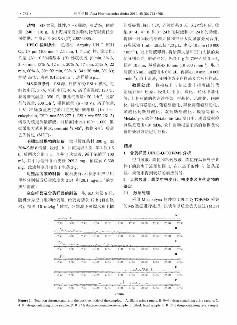
· 762 · 药学学报 Acta Pharmaceutica Sinica 2010, 45 (6): 761−766
动物 SD大鼠, 雄性, 7~8周龄, 清洁级, 体质量 (240 ± 10) g, 由上海斯莱克实验动物有限责任公司提供, 合格证号 SCXK (沪) 2007-0005。
UPLC 检测条件 色谱柱: Acquity UPLC BEH C18 1.7 µm (100 mm × 2.1 mm, 1. 7 μm) 柱; 流动相: 乙腈 (A) − 0.5%醋酸水 (B) 梯度洗脱 (0 min, 3% A, 3~8 min, 13% A, 12 min, 20% A, 17 min, 35% A, 22 min, 60% A, 30~32 min, 95% A, 34~36 min, 3% A); 柱温 35 ℃; 流速 0.4 mL·min−1, 进样量 5 μL。
MS检测条件 ESI源, 扫描方式: ESI ± 模式, 毛细管电压: 3 kV, 锥孔电压: 40 V, 离子源温度: 120 ℃, 脱溶剂气温度: 350 ℃, 锥孔气流量: 50 L·h−1, 脱溶剂气流量: 600 L·h−1, 碰撞能量 (6~40 V), 离子能量: 1 V; 准确质量测定采用亮氨酸−脑啡肽 (1eucine- enkephalin, ESI+: m/z 556.277 1, ESI−: m/z 555.261 5) 溶液为锁定质量溶液。扫描范围: m/z 100~1 000, 数据采集方式和模式: centroid与MSE, 数据分析: 质量丢失滤过 (MDF)。
毛橘红醇提物的制备 取毛橘红药材 300 g, 加70%乙醇 8倍量, 浸泡 1 h, 回流提取 3次, 第 1次 1.5 h, 后两次分别 1 h, 合并 3 次滤液, 减压浓缩至 100 mL, 其中每毫升含柚皮苷 269.3 mg、柚皮素 0.688 mg。此液每毫升相当于生药 3 g。
对照品溶液的制备 取柚皮苷、柚皮素对照品用甲醇分别制成质量浓度为 21.4 和 28.1 µg·mL−1的对
照品溶液。 空白样品及含药样品的制备 取 SD 大鼠 6 只,
随机分为空白组和给药组, 给药前禁食 12 h (自由饮水), 按照 16 mL·kg−1 体重, 分别灌予蒸馏水和毛橘
红醇提物。每日 1次, 连续给药 3天, 末次给药后, 收集 0~4、4~8 和 8~24 h的尿液和 0~24 h的粪便。将同一时间段的给药大鼠和空白大鼠尿液分别合并, 各取尿液 1 mL, 加乙腈 420 µL, 离心 10 min (10 000 r·min−1), 取上清液即得。将给药大鼠和空白大鼠的粪便分别合并, 碾碎混匀, 各取 1 g加 70%乙腈 3 mL, 超声 10 min, 然后离心 10 min (10 000 r·min−1), 取上清液 0.5 mL, 加蒸馏水 670 µL, 再离心 10 min (10 000 r·min−1), 取上清液, 分别作为空白样品及给药后样品。
数据处理 将柚皮苷与柚皮素Ⅰ相可能的代 谢途径如: 还原、羟化后还原、氧化、羟化甲基化 等; Ⅱ相可能的代谢途径如: 甲基化、乙酰化、磺酸化、羟化再磺酸化、葡糖醛酸化、羟化再葡糖醛酸化、
磺酸化葡糖醛酸化、双葡糖醛酸化、脱糖等输入
Metabolynx软件Metabolite List窗口中, 质谱数据检测误差范围<10 mDa, 软件自动根据采集的数据及设置的处理方法进行分析。
结果 1 含药样品 UPLC-Q-TOF/MS分析
空白尿液、粪便和给药尿液、粪便样品负离子条
件下的总离子流图如图 1, 在正离子条件下, 给药尿液、粪便未得到较好的响应信号。 2 大鼠尿液、粪便中柚皮苷、柚皮素及其代谢物的鉴定 2.1 数据处理
采用 Metabolynx 软件将 UPLC-Q-TOF/MS 采集的MS数据进行处理, 该软件以质量丢失滤过 (MDF)
Figure 1 Total ion chromatograms in the positive mode of the samples. A: Blank urine sample; B: 0−4 h drug-containing urine sample; C: 4−8 h drug-containing urine sample; D: 8−24 h drug-containing urine sample; E: Blank fecal sample; F: 0−24 h drug-containing fecal sample
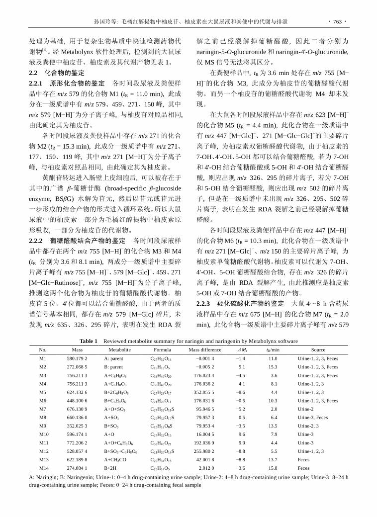
孙国玲等: 毛橘红醇提物中柚皮苷、柚皮素在大鼠尿液和粪便中的代谢与排泄 · 763 ·
处理为基础, 用于复杂生物基质中快速检测药物代谢物[4]。经 Metabolynx软件处理后, 检测到的大鼠尿液及粪便中柚皮苷、柚皮素及其代谢产物见表 1。 2.2 化合物的鉴定 2.2.1 原形化合物的鉴定 各时间段尿液及粪便样品中存在 m/z 579的化合物 M1 (tR = 11.0 min), 此成分在一级质谱中有 m/z 579、459、271、150峰, 其中 m/z 579 [M−H]−为分子离子峰, 与柚皮苷对照品相同, 由此确定其为柚皮苷。
各时间段尿液及粪便样品中存在 m/z 271的化合物M2 (tR = 15.3 min), 此成分一级质谱中有 m/z 271、177、150、119峰, 其中 m/z 271 [M−H]−为分子离子峰, 与柚皮素对照品相同, 由此确定其为柚皮素。 黄酮苷转运进入肠壁上皮细胞后, 可以被存在于
其中的广谱 β-葡糖苷酶 (broad-specific β-glucoside enzyme, BSβG) 水解为苷元, 然后以苷元或苷元进一步形成的结合产物的形式进入循环系统。所以大鼠
尿液中的柚皮素一部分为毛橘红醇提物中柚皮素原
形吸收, 一部分为柚皮苷的代谢物。 2.2.2 葡糖醛酸结合产物的鉴定 各时间段尿液样品中都存在两个 m/z 755 [M−H]−的化合物M3和 M4 (tR 分别为 3.6和 8.1 min), 两成分一级质谱中主要碎片离子峰有 m/z 755 [M−H]−、579 [M−Glc]−、459、271 [M−Glc−Rutinose]−, m/z 755 [M−H]−为分子离子峰, 推测这两个化合物为柚皮苷的葡糖醛酸代谢物。柚 皮苷 5位、4' 位都可以结合葡糖醛酸, 由于两者的质谱信号基本相同, 都存在 m/z 579 [M−Glc]−碎片, 未发现 m/z 635、326、295 碎片, 表明在发生 RDA 裂
解之前已经裂解掉葡糖醛酸 , 因此二者分别为naringin-5-O-glucuronide和 naringin-4'-O-glucuronide, 仅MS信号无法将其区分。 在粪便样品中, tR为 3.6 min处存在 m/z 755 [M−
H]−的化合物 M3, 此成分为柚皮苷的葡糖醛酸代谢物。而另一个柚皮苷的葡糖醛酸代谢物 M4 却未发现。 在大鼠各时间段尿液样品中存在 m/z 623 [M−H]−
的化合物M5 (tR = 4.4 min), 此化合物在一级质谱中有 m/z 447 [M−Glc]−、271 [M−Glc−Glc]−的主要碎片离子峰, 为柚皮素双葡糖醛酸代谢物, 由于柚皮素的7-OH、4'-OH、5-OH都可以结合葡糖醛酸, 若为 7-OH和 4'-OH结合葡糖醛酸或 5-OH和 4'-OH结合葡糖醛酸, 则应出现 m/z 326、295 的碎片离子, 若为 7-OH和 5-OH结合葡糖醛酸, 则应出现 m/z 502 的碎片离子, 但是在一级质谱中未出现 m/z 326、295、502碎片离子, 表明在发生 RDA 裂解之前已经裂解掉葡糖醛酸。 各时间段尿液及粪便样品中存在 m/z 447 [M−H]−
的化合物M6 (tR = 10.3 min), 此化合物在一级质谱中有 m/z 271 [M−Glc]−、m/z 150的主要碎片离子峰, 为柚皮素单葡糖醛酸代谢物。柚皮素可以代谢为 7-OH、4'-OH、5-OH葡糖醛酸结合物, 存在 m/z 326的碎片离子峰, 是由 RDA 裂解产生, 由此推测应是柚皮素5-OH或 7-OH结合葡糖醛酸的产物。 2.2.3 羟化硫酸化产物的鉴定 大鼠 4~8 h 含药尿液样品中存在 m/z 675 [M−H]−的化合物M7 (tR = 2.0 min), 此化合物一级质谱中主要碎片离子峰有 m/z 579
Table 1 Reviewed metabolite summary for naringin and naringenin by Metabolynx software
No. Mass Metabolite Formula Mass difference ⊿Mr tR/min Source
M1 580.179 2 A: parent C27H32O14 −0.001 4 −1.4 11.0 Urine-1, 2, 3, Feces
M2 272.068 5 B: parent C15H12O5 −0.005 2 5.1 15.3 Urine-1, 2, 3, Feces
M3 756.211 3 A+C6H8O6 C33H40O20 176.023 4 −4.5 3.6 Urine-1, 2, 3, Feces
M4 756.211 3 A+C6H8O6 C33H40O20 176.036 2 4.1 8.1 Urine-1, 2, 3
M5 624.132 6 B+2C6H8O6 C27H28O17 352.055 5 −8.6 4.4 Urine-1, 2, 3
M6 448.100 6 B+C6H8O6 C21H20O11 176.031 6 −0.5 10.3 Urine-1, 2, 3, Feces
M7 676.130 9 A+O+SO3 C27H32O18S 95.946 5 −5.2 2.0 Urine-2
M8 660.136 0 A+SO3 C27H32O17S 79.957 3 0.5 6.4 Urine-3, Feces
M9 352.025 3 B+SO3 C15H12O8S 79.953 4 −3.5 13.5 Urine-2, 3
M10 596.174 1 A+O C27H32O15 16.004 5 9.6 7.9 Urine-3
M11 772.206 2 A+O+C6H8O6 C33H40O21 192.036 9 9.9 4.4 Urine-3
M12 528.057 4 B+SO3+C6H8O6 C21H20O14S 255.980 2 −8.8 5.5 Urine-1, 2, 3
M13 622.189 8 A+CH3CO C29H34O15 42.001 8 −8.8 13.7 Feces
M14 274.084 1 B+2H C15H14O5 2.012 0 −3.6 15.8 Feces
A: Naringin; B: Naringenin; Urine-1: 0−4 h drug-containing urine sample; Urine-2: 4−8 h drug-containing urine sample; Urine-3: 8−24 h drug-containing urine sample; Feces: 0−24 h drug-containing fecal sample

· 764 · 药学学报 Acta Pharmaceutica Sinica 2010, 45 (6): 761−766
[M−O−SO3]−、271 [M−O−SO3−Rutinose]−, 为柚皮苷羟化硫酸结合物。柚皮苷发生氧化反应后被代谢成
3', 4'-二羟基黄酮[5]。硫酸结合物可能结合在 A环或 B环的-OH上, 碎片离子峰 m/z 230是柚皮苷先脱去芸香糖再发生 RDA裂解产生的, 所以推测柚皮苷 A环上的 5-OH结合了-SO3。 2.2.4 硫酸结合产物的鉴定 大鼠8~24 h含药尿液和粪便样品中存在 m/z 659 [M−H]−的化合物M8 (tR = 6.4 min), 此化合物有 m/z 579 [M−SO3]−、459、351 [M− Rutinose]−、271 [M−SO3−Rutinose]−的主要碎片离子峰, 为柚皮苷的代谢产物柚皮苷硫酸化物。柚皮苷 5-OH 与 4'-OH 易结合硫酸, 在质谱图中存在 m/z 199碎片峰, 此碎片峰来源于M8的 RDA裂解, 由此分析应是柚皮苷 4'-OH结合硫酸产生的碎片离子, 故推测M8为 naringin-4'-O-sulfate。
大鼠 4~8 h和 8~24 h含药尿液样品中存在 m/z 351 [M−H]−的化合物M9 (tR = 13.5 min), 此化合物的主要碎片离子峰有 m/z 271 [M−SO3−Rutinose]−、177、151, 为柚皮素硫酸化物。柚皮素 7-OH、5-OH与 4'-OH易结合硫酸, 存在 m/z 199 碎片峰, 此碎片峰来源于M9 的 RDA 裂解, 由此分析应是柚皮素 4'-OH 结合 硫酸产生的碎片离子, 故推测M9为 naringenin-4'-O- sulfate。 2.2.5 羟化产物的鉴定 大鼠8~24 h含药尿液样品中存在 m/z 595 [M−H]−的化合物 M10 (tR = 7.9 min), 此化合物的主要碎片离子峰有 m/z 579 [M−O]−、459、271 [M−O−Rutinose]−, 为柚皮苷羟化产物。根据 M7中的推论, 柚皮苷 3' 位会被氧化, 最终被氧化为 3', 4'- 二羟基黄酮。 2.2.6 羟化葡糖醛酸结合产物的鉴定 大鼠 8~24 h含药尿液样品中存在 m/z 771 [M−H]−峰的化合物M11 (tR = 4.4 min), 此化合物的主要碎片离子峰有m/z 595 [M−Glc]−、271 [M−O−Glc−Rutinose]−, 为柚皮苷羟化葡糖醛酸化结合物, 是体内酶的催化作用下同时发生羟化反应和葡糖醛酸结合反应而生成的
代谢物。柚皮苷发生氧化反应后被氧化成 3', 4'-二羟基黄酮。葡糖醛酸结合物可能结合在 A 环或 B 环的
-OH上, 碎片离子峰m/z 311是柚皮苷发生了RDA裂解产生的, 所以推测柚皮苷 B 环上结合了葡糖醛酸, 但是, 具体是 3' 还是 4' 位结合未能通过 MS来确定。 2.2.7 硫酸化葡糖醛酸结合产物的鉴定 各时间段尿液样品中存在 m/z 527 [M−H]−的化合物M12 (tR = 5.5 min), 此化合物的主要碎片离子峰有 m/z 447 [M−
SO3]−、351 [M−Glc]−、271 [M−Glc−SO3]−, 为柚皮素代谢产物柚皮素葡糖醛酸硫酸化结合物。此结合产 物是在动物体内由葡糖醛酸化酶和硫酸转移酶共同
催化下产生的。文献[6]报道, 给人服用葡萄柚汁后, 在人尿液中发现了柚皮素的代谢物柚皮素葡糖醛酸
硫酸化结合产物。柚皮素的 7-OH、4'-OH、5-OH 都可结合葡糖醛酸或硫酸分子。若是 A环上的 7-OH、5-OH结合葡糖醛酸和硫酸, 则质谱中应出现 m/z 406的碎片峰, 若是 A 环结合葡糖醛酸, B 环上的 4'-OH结合硫酸应出现 m/z 326、199 的碎片峰, 若是 A 环结合硫酸, B 环上的 4'-OH 结合葡糖醛酸应出现 m/z 230、295的碎片峰, 而在质谱中未出现 m/z 406、326、199、230、295 碎片峰, 表明在发生 RDA 裂解之前已经裂解掉葡糖醛酸和硫酸。 2.2.8 乙酰化结合产物的鉴定 大鼠含药粪便样品中存在 m/z 621 [M−H]−的化合物 M13 (tR = 13.7 min), 此化合物的主要碎片离子峰有 m/z 579 [M−Ac]−、459、271 [M−Ac−Rutinose]−, 为柚皮苷乙酰化结合物。柚皮苷 5-OH与 4'-OH容易结合乙酰基, 碎片峰m/z 501来源于 M13 的 RDA 裂解, 由此分析应是柚皮苷5-OH 结合乙酰基产生的碎片离子, 故推测 M13 为naringin-5-O-acetylate。
乙酰化反应是在酰基转移酶的催化下进行的, 以乙酰辅酶 A作为辅酶, 对羟基进行乙酰化反应。 2.2.9 被还原产物的鉴定 大鼠含药粪便中存在m/z 273 [M−H]−的化合物 M14 (tR = 15.8 min), 此化合物的主要碎片离子峰有 m/z 273、271、151、119, 为柚皮素还原化代谢物。 2.3 柚皮苷、柚皮素代谢物结构与主要的代谢途径
根据以上代谢物鉴定的结果, 推测柚皮苷、柚皮素代谢物结构与主要的代谢途径如图 2。
讨论 本文使用了UPLC-Q-TOF/MS联用技术, 能同时
得到准分子离子和不同撞击能量下子离子图谱的
MSE 数据采集方式, 并运用以质量丢失滤过 (MDF) 为基础的代谢物寻找软件 MetabolynxTM 来处理数 据。MDF 是一个数据处理方法, 它能将 LC-MS 采 集到的高分辨率数据和预先设定的可能代谢物的 质量亏损数据通过软件处理来推测目标代谢物[7]。
MetabolynxTM 软件是通过含药样品与空白样品比对
进行分析, 去除所有干扰离子而简化质谱图的解析, 从而能从复杂生物基质中快速检测药物代谢物, 使

孙国玲等: 毛橘红醇提物中柚皮苷、柚皮素在大鼠尿液和粪便中的代谢与排泄 · 765 ·
Figure 2 Chemical structures of naringin, naringenin metabolites identified in the drug-containing urine and fecal samples and their proposed major metabolic pathways. GlcUA = glucuronyl unit; Ac = acetyl group. a: Glucuronidation; b: Sulfation; c: Acetylation; d: Hydroxylation; e: Hydroxylation + glucuronidation; f: Hydroxylation + sulfation; g: Loss of rutinose; h: Reducion; i: Sulfation + glucuronidation; k: 2×glucuronidation 高分辨 LC-MS 成为快速筛选和确认代谢物的有用平台[8−10]。
在大鼠含药尿液中发现柚皮苷与柚皮素原形及
柚皮苷 1个Ⅰ相代谢产物 (M10) 和 5个Ⅱ相代谢产
物 (M3、M4、M7、M8和M11), 柚皮素 4个Ⅱ相代谢产物 (M5、M6、M9和M12)。柚皮苷在大鼠尿液中 0~4 h 主要以原形和葡糖醛酸结合物的形式被排泄, 4~8 h尿液中出现了羟化硫酸化代谢物, 8 h后,

· 766 · 药学学报 Acta Pharmaceutica Sinica 2010, 45 (6): 761−766
又产生了新的代谢物形式 (羟化、羟化葡糖醛酸化和硫酸化代谢物)。柚皮素在尿液中 0~4 h主要以原形和单葡糖醛酸、双葡糖醛酸、硫酸化葡糖醛酸结合物
的形式被排泄, 4~8 h以硫酸化代谢物的形式被排泄, 8 h后无新的代谢形式出现。
在大鼠含药粪便中发现柚皮苷与柚皮素原形及
柚皮苷 3 个Ⅱ相代谢产物 (M3、M8 和 M13), 柚皮 素 1 个Ⅰ相代谢产物 (M14) 和 1 个Ⅱ相代谢产物 (M6)。柚皮苷在大鼠粪便中 0~24 h以原形以及硫酸化、乙酰化、葡糖醛酸结合代谢产物的形式被排泄, 柚皮素以原形以及还原化、葡糖醛酸结合代谢产物的
形式被排泄。 其中, 柚皮苷、柚皮素代谢物柚皮素双葡糖醛酸
苷 (M5)、柚皮素葡糖醛酸 (M6)、柚皮素硫酸酯 (M9)、柚皮素葡糖醛酸基和硫酸酯基共价结合物 (M12) 已见报道[6, 11]。柚皮苷 5-O-葡糖醛酸苷和柚皮苷 4'-O-葡糖醛酸苷 (M3和M4)、羟基化柚皮苷硫酸酯 (M7)、柚皮苷 4'-O-硫酸酯 (M8)、羟基化柚皮苷甲醚 (M10)、羟基化柚皮苷葡糖醛酸苷 (M11)、柚皮苷乙酰化物 (M13)、柚皮素还原化代谢物 (M14) 未见报道。毛橘红中其他成分的代谢与排泄有待进一步
研究。
References
[1] State Pharmacopoeia Committee. Pharmacopoeia of the
People’s Republic of China (中华人民共和国药典) [S]. Part
I. Beijing: Chemical Industry Press, 2005: 51−52.
[2] Yuan XJ, Lin L, Chen ZX. Phenolic constituents in Epicarpium
Citri Grandis [J]. Chin Tradit Herb Drugs (中草药), 2004, 35:
498−500.
[3] Yang HL, Tian H, Li PB. Biological activity of naringin and
naringenin [J]. J Chin Med Mater (中药材), 2007, 30: 752−
754.
[4] Zhang XZ, Li Y. Advances in studies on the detection of drug
reactive metabolites by LC-MS [J]. J Int Pharm Res (国际药
学研究杂志), 2009, 36: 393−396.
[5] Pei LK, Guo BL. Advances in studies on absorption and
metabolism of flavonoids [J]. China J Chin Mater Med (中国
中药杂志), 2006, 41: 568−572.
[6] Zhang JM, Brodbelt JS. Screening flavonoid metabolites of
naringin and narirutin in urine after human consumption of
grapefruit juice by LC-MS and LC-MS/MS [J]. Analyst,
2004, 129: 1227−1233.
[7] Zhang H, Zhang D, Ray K. A software filter to remove
interference ions from drug metabolites in accurate mass liquid
chromatography/mass spectrometric analyses [J]. J Mass
Spectrom, 2003, 38: 1110−1112.
[8] Bateman KP, Castro-Perez J, Wrona M, et al. MSE with mass
defect filtering for in vitro and in vivo metabolite identification
[J]. Rapid Commun Mass Spectrom, 2007, 21: 1485−1496.
[9] Zhu M, Ma L, Zhang D, et al. Detection and characterization
of metabolites in biological matrices using mass defect filtering
of liquid chromatography/high resolution mass spectrometry
data [J]. Drug Metab Dispos, 2006, 34: 1722−1733.
[10] Tiller PR, Yu S, Castro-Perez J, et al. High-throughput,
accurate mass liquid chromatography/tandem mass spectrometry
on a quadrupole time-of-flight system as a ‘first-line’ approach
for metabolite identification studies [J]. Rapid Commun
Mass Spectrom, 2008, 22: 1053−1061.
[11] Silberberg M, Gil-Izquierdo A, Combaret L, et al. Flavanone
metabolism in healthy and tumor-bearing rats [J]. Biomed
Pharmacother, 2006, 60: 529−535.

药学学报 Acta Pharmaceutica Sinica 2010, 45 (6): 767−771 · 767 ·
Development and validation of a liquid chromatography-isotope dilution tandem mass spectrometry for determination of olanzapine in human plasma and its application to bioavailability study
ZHANG Meng-qi1, JIA Jing-ying1, LU Chuan1, LIU Gang-yi1, YU Cheng-yin2, GUI Yu-zhou3, LIU Yun1, LIU Yan-mei1, WANG Wei4, LI Shui-jun1, YU Chen1*
(1. Central Laboratory, Shanghai Xuhui Central Hospital, Shanghai 200031, China; 2. College of Traditional Chinese Medicine, Shanghai University of Chinese Traditional Medicine, Shanghai 201203, China; 3. Shanghai Clinical Research Center, Shanghai
200233, China; 4. Department of Emergency ICU, Shanghai Xuhui Central Hospital, Shanghai 200031, China)
Abstract: A simple, reliable and sensitive liquid chromatography-isotope dilution mass spectrometry (LC- ID/MS) was developed and validated for quantification of olanzapine in human plasma. Plasma samples (50 µL) were extracted with tert-butyl methyl ether and isotope-labeled internal standard (olanzapine-D3) was used. The chromatographic separation was performed on XBridge Shield RP 18 (100 mm × 2.1 mm, 3.5 μm, Waters). An isocratic program was used at a flow rate of 0.4 mL∙min−1 with mobile phase consisting of acetonitrile and ammonium buffer (pH 8). The protonated ions of analytes were detected in positive ionization by multiple reactions monitoring (MRM) mode. The plasma method, with a lower limit of quantification (LLOQ) of 0.1 ng∙mL−1, demonstrated good linearity over a range of 0.1 − 30 ng∙mL−1 of olanzapine. Specificity, linearity, accuracy, precision, recovery, matrix effect and stability were evaluated during method validation. The validated method was successfully applied to analyzing human plasma samples in bioavailability study.
Key words: olanzapine; liquid chromatography-isotope dilution mass spectrometry; plasma CLC number: R917 Document code: A Article ID: 0513-4870 (2010) 06-0767-05
同位素稀释质谱法测定人血浆中奥氮平的方法学及生物等效性研究
张梦琪 1, 贾晶莹 1, 陆 川 1, 刘罡一 1, 余成寅 2, 桂雨舟 3, 刘 昀 1, 刘艳梅 1, 王 伟 4, 李水军 1, 余 琛 1*
(1. 上海市徐汇区中心医院中心实验室, 上海 200031; 2. 上海中医药大学中药学院, 上海 201203; 3. 上海医药临床研究中心, 上海 200233; 4. 上海市徐汇区中心医院急诊监护室, 上海 200031)
摘要: 建立简便、可靠、灵敏的同位素稀释质谱法测定血浆中奥氮平的浓度。50 μL血浆样品中加入稳定同
位素 (奥氮平-D3) 为内标, 经特丁基甲醚提取后进样分析。色谱柱为 XBridge Shield RP 18 (100 mm × 2.1 mm, 3.5 μm, Waters)。乙腈-缓冲液 (氨水-醋酸铵, pH 8) 为流动相, 等度洗脱, 流速 0.4 mL∙min−1。扫描方式为正离
子多离子反应监测 (MRM)。定量下限均为 0.1 ng∙mL−1, 线性范围为 0.1~30 ng∙mL−1。对特异性、线性、准确度、
精密度、回收率、介质效应均进行了方法学考察, 且各项结果均符合要求, 可以用于奥氮平的生物等效性研究。 关键词: 奥氮平; 同位素稀释质谱; 血浆
Olanzapine (Figure 1) is an atypical antipsychotic, Received 2009-12-15. *Corresponding author Tel: 86-21-54030254, Fax: 86-21-54043676,
E-mail: [email protected]
approved by the FDA for the treatment of schizophrenia. Olanzapine is a serotonin-dopamine-receptor antagonist indicated for the treatment of schizophrenia and other psychotic disorders. The drug is well absorbed from

· 768 · 药学学报 Acta Pharmaceutica Sinica 2010, 45 (6): 767−771
Figure 1 Chemical structures of olanzapine (1) and D3-olanzapine (2)
the gastro-intestinal tract. Food has no effect on its absorption[1].
There are several determination methods, HPLC- UV[2], HPLC-ECD[3], GC-NPD[4], liquid chromatography/ tandem mass spectroscopy (LC-MS/MS)[5, 6] published for the measurement of olanzapine in previous researches. However, a main drawback of LC-MS/MS is matrix effect from urine and blood sample[7]. The use of stable isotope internal standards may effectively overcome the problem of matrix effect as they have almost identical chemical properties but are easily to be distinguished in the MS analysis. Therefore, isotope dilution mass spectrometry (IDMS) is capable of providing very accurate and precise results and suitable as reference method[8].
Most of the previous researches have been reported solid phase extraction (SPE) as the method of sample preparation, it is effective for remove interferences but may be too cost for large amount bioanalysis[9−12]. Liquid-liquid extraction was adopted in this study, which not only simplified the process but also saved the analytical time.
Based on previously published LC-MS/MS method[5, 6], a liquid chromatographic-isotope dilution tandem mass spectrometry (LC-ID/MS) was developed in this study and validated for the quantification of olanzapine in human plasma by spiking stable isotope olanzapine-D3 as an internal standard. A single step liquid-liquid extraction was adopted for sample prepa-ration. IDMS has been proved to be a definitive technique providing results with unchallenged precision and accuracy, and is well suited for building a reference method[13]. Here we proposed an IDMS method for simple, reliable and accurate determination for olanzapine in human plasma. The method has been successfully applied to a bioavailability study in 24 Chinese healthy volunteers following oral administration of two brands of olanzapine tablets.
Materials and methods Reagents Olanzapine (99.9% purity) was provided
at Dr. Reddy’s Laboratories Ltd. (India), and compound 2, olanzapine-D3 (internal standard, IS, 98% purity) was purchased from Toronto Research Chemicals Inc (North York, Canada).
Instrumentation The liquid chromatograph system (Shimadzu, Kyoto, Japan) was equipped with two LC-20ADvp pumps, a DGU-20AD3 vacuum degasser, a SIL-HTC autosampler and a controller module. Mass spectrometric detection was performed on an API 3000 triple quadruple instrument (ABI-SCIEX, Toronto, Canada). The data was acquired and processed with Analyst 1.4.2 software package.
Standard solutions Stock solutions of olanzapine (1.022 mg∙mL−1) and IS (25 μg∙mL−1) were prepared with methanol. Working standard solution of olanzapine was diluted with 80% methanol solution. All standard solutions were stored at 4 ℃. Calibration standards (0.1, 0.2, 1, 5, 10, 20, and 30 ng∙mL−1) were prepared by spiking the working standard solutions of olanzapine into human plasma. Dilutions were used to prepare four levels of QCs, 0.1, 0.25, 15, and 25 ng∙mL−1 in human plasma. QCs were stored at −30 ℃.
Sample preparation All frozen human plasma samples were thawed at ambient temperature. To a 1.5 mL polypropylene test tube, 50 μL of plasma sample, 5 µL of IS (25 ng∙mL−1) and 5 μL of sodium hydroxide solution (0.1 mol∙L−1) were added. It was subsequently spiked with 0.5 mL tert-butyl methyl ether and thoroughly mixed for 45 s. Following centrifugation at 16 000 r∙min−1 for 45 s, the upper- layer of organic solvent was transferred to a clean tube and evaporated to dryness at 40 ℃ under a stream of nitrogen. Afterwards, the residue was reconstituted with 100 μL of 80% methanol and thoroughly mixed for 30 s. Finally, 5 μL of the sample was injected into the LC-MS/MS system.
LC-MS/MS conditions The chromatographic separation was performed on a XBridge Shield RP 18 (100 mm × 2.1 mm, 3.5 μm, Waters, Ireland) at room temperature. An isocratic programming was used at a flow rate of 0.4 mL∙min−1 with mobile phase consisting of 80% acetonitrile and 20% ammonium buffer (0.02% ammonia water-5 mmoL∙L−1 ammonium acetate, pH 8). The samples were kept at 4 ℃ in autosampler and 5 μL was injected.
The mass spectrometer was operated in the positive ion detection mode with the spray voltage set at 1.1 kV.

ZHANG Meng-qi, et al: Development and validation of a liquid chromatography-isotope dilution tandem mass spectrometry for determination of olanzapine in human plasma and its application to bioavailability study · 769 ·
The heated capillary temperature was 450 ℃. The nitrogen sheath gas and the auxiliary gas were set at 12 and 8 psi (1 psi ≈ 6.9 kPa), respectively. Tandem mass spectrometry analysis was carried out on an API 3000 triple quadruple instrument (Applied Biosystems, America) using multiple reaction monitoring (MRM) detection.
Method validation The method was validated for specificity, linearity, lower limit of quantification (LLOQ), intra- and inter-assay precision and accuracy, matrix effect, recovery, and stability according to the recommendation for the bioanalytical method validation by FDA (2001).
The method specificity was evaluated by screening six different batches of blank human plasma prior to the main validation batches. These batches were spiked with known concentrations of olanzapine at 0.25, 15, and 25 ng∙mL−1, extracted and analyzed along with a calibration curve prepared in one of the six batches to demonstrate the lack of chromatographic interference from endogenous plasma components and batch-to- batch variation.
The linearity for olanzapine was evaluated over the range of 0.1 − 30 ng∙mL−1. A linear regression model with 1/x weighted factor was constructed based on the measured peak area ratio of olanzapine to the IS versus the nominal concentration, where x is the concentration of olanzapine, was fitted to each standard curve.
QCs at four concentration levels (0.1, 0.25, 15, and 25 ng∙mL−1) were analyzed to evaluate intra- and inter- assay precision and accuracy of the method, with six replicates for each of the three randomized batches. Precision was expressed as RSD% for replicate mea- surements and accuracy (%) by the percentage of devia- tion between nominal and calculated concentrations.
The matrix effect was assessed by comparing the peak areas of analytes from the standards spiked after extraction and the pure QC standards at the same concentration levels. The recovery was assessed by comparing the standards spiked before and after extraction at three concentration levels (0.25, 15, and 25 ng∙mL−1).
The autosampler stability was assessed by keeping the processed QCs at 4 ℃ for 18 h. The freeze-thaw stability was evaluated by analyzing the QCs after three cycles from −30 ℃ to room temperature. The plasma samples were analyzed for bench top stability after placed at room temperature for 20 h.
Clinical application The developed method was applied to determining the plasma concentrations of olanzapine from 24 Chinese young healthy subjects for a bioavailability study of two olanzapine tablets. The study was approved by the Ethics Committee of Shanghai Xuhui Central Hospital. Each subject was informed of the purpose of the study, and informed consent forms were obtained.
Results and discussion 1 Optimization of chromatographic conditions
Many reports described a liquid chromatographic mobile phase with acidic methanol and water[5, 9]. However, distorted peak tailing was observed in our LC-MS/MS system under such a mobile phase condition. In this study, ammonium acetate-ammonia was used to take the place of formic acid[7, 14, 15] which is widely used in previous research to adjust the pH to 8. The alkalescency mobile phase was proved to have a better peak without tailing peak. The precursor-to-product ion transitions 313→256 amu for olanzapine and 316→256 amu for IS were monitored with the collision energy set at 32 eV (Figure 2). The LC-MS/MS chromatograms of plasma samples are shown in Figure 3. 2 Optimization of sample preparation
In order to increase the extraction recovery, NaOH solution (0.1 mol∙L−1) was added to adjust the pH value to 10 as olanzapine is an alkaline compound. The recovery was enhanced 20% by using the alkaline modifier. In addition, different extracting agents were tried. Finally tert-butyl methyl ether was selected considering the volatility and recovery rate.
In the process of extraction, the toughest problem was the absorption of olanzapine. Different concen-trations of solvent, different tubes and different sample preparation times were tested to find the causes of
Figure 2 Product ion mass spectrum of protonated ions obtained from olanzapine

· 770 · 药学学报 Acta Pharmaceutica Sinica 2010, 45 (6): 767−771
Figure 3 Representative LC-MS/MS chromatograms of olanzapine (OZP) and D3-olanzapine (IS) in human plasma. A: Blank human plasma; B: LLOQ plasma sample with 0.1 ng∙mL−1 olanzapine; C: Human plasma sample collected 4.0 h after an oral dose of 10 mg olanzapine absorption. The result indicated that 80% MeOH was the best solvent (recovery 90.6%). 1.5 mL polypropylene centrifugal tube was the most suitable tube (30% signals loss). The reconstitution process which absorbed huge amount of olanzapine directly lead the recovery rate dropped from 87.5% to 67.2%, so the mix and cen-trifugation process was designed as quickly as possible. 3 Method validation 3.1 Specificity Blank plasma from six batches was tested for endogenous interferences. The olanzapine retention time region was free from interferences. The back-calculated values from all six batches, spiked at concentrations of 0.25, 15, and 25 ng∙mL−1 for
olanzapine, the RSD was less than 7% and the accuracy was 94.9% − 108%, respectively, indicating no significant batch-to-batch variation. 3.2 Linearity and LLOQ Linear regression analysis for olanzapine was performed by using internal standard method. Olanzapine fixed good linearity over a relatively wide concentration range, and the mean correlation coefficient (r) was 0.999 3. Based on the standard data presented here, it was concluded that the calibration curves used in this method were accurate for the determination of olanzapine. The LLOQ was 0.1 ng∙mL−1 for olanzapine with a signal to noise (S/N) over 6. Though LLOQ is adequately low for olanzapine
quantification, the sensitivity is achieved using a limited sample volume of 0.05 mL plasma. The extracts were diluted two folds in the reconstitution step and only 5 μL was injected onto LC-MS/MS for analysis. This indicates that a more sensitive linear range could be achieved if it is necessary for future studies. 3.3 Accuracy and precision A summary of the individual QC data is obtained in the three runs for the validation. The inter-batch precision RSD of the assay was no more than 11% at four concentration levels of the QC samples, the RSD for intra-batch precision was less than 4%. The inter- and intra-batch accuracy of the assay ranged from 93.7% to 105%. The assay for olanzapine was accurate and precise between runs and within individual run for each level. 3.4 Recovery and matrix effect The mean recoveries of olanzapine at 0.25, 15, and 25 ng∙mL−1 were 82.6%, 88.4% and 86.9%, respectively. The mean recoveries of IS were 76.3%. Both these replicates presented with a RSD of less than 12%.
The method demonstrated that ionization efficiency of analytes from plasma was suppressed with a mean peak area ratio ranging from 64.9% − 76.3%, 71.8% for olanzapine and IS obtained from the post-extracted samples. No interferences from other compounds present in plasma were observed to the analytes assay. But the accurate quantification could still be achieved even with the presence of the matrix effect under our experimental condition, mainly owing to the using of isotope dilution mass spectrometry. To date, no significant interferences from endogenous substances were observed during the process of method validation and sample assay. 3.5 Stability The olanzapine stabilities were inves-tigated in autosampler, after three freeze-thaw cycles, and on bench top, separately. The results expressed as RSD were 6.5%, 2.8% and 4.2%, respectively. The good stability of olanzapine simplified the precautions needed for laboratory manipulations during the assay procedures. In addition, stock solutions were shown to be stable at 4 ℃ for 4 weeks. 3.6 Application study The described LC-ID/MS method was successfully applied to determining the human plasma samples of olanzapine up to 144 h after a single oral dose administration of a 10 mg olanzapine tablet to 24 healthy volunteers. Figure 4 showed the concentration-time curve of olanzapine after an oral administration of 10 mg reference and test olanzapine tablet.
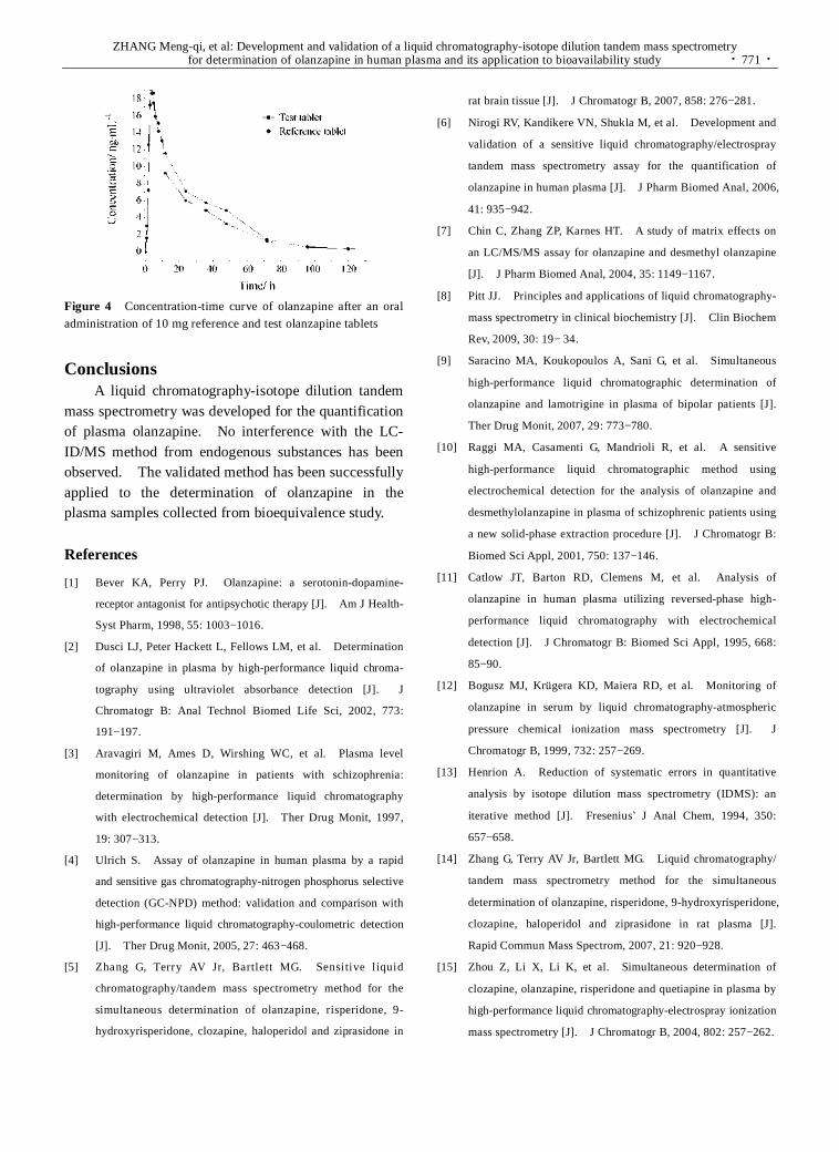
ZHANG Meng-qi, et al: Development and validation of a liquid chromatography-isotope dilution tandem mass spectrometry for determination of olanzapine in human plasma and its application to bioavailability study · 771 ·
Figure 4 Concentration-time curve of olanzapine after an oral administration of 10 mg reference and test olanzapine tablets
Conclusions A liquid chromatography-isotope dilution tandem
mass spectrometry was developed for the quantification of plasma olanzapine. No interference with the LC- ID/MS method from endogenous substances has been observed. The validated method has been successfully applied to the determination of olanzapine in the plasma samples collected from bioequivalence study.
References
[1] Bever KA, Perry PJ. Olanzapine: a serotonin-dopamine-
receptor antagonist for antipsychotic therapy [J]. Am J Health-
Syst Pharm, 1998, 55: 1003−1016.
[2] Dusci LJ, Peter Hackett L, Fellows LM, et al. Determination
of olanzapine in plasma by high-performance liquid chroma-
tography using ultraviolet absorbance detection [J]. J
Chromatogr B: Anal Technol Biomed Life Sci, 2002, 773:
191−197.
[3] Aravagiri M, Ames D, Wirshing WC, et al. Plasma level
monitoring of olanzapine in patients with schizophrenia:
determination by high-performance liquid chromatography
with electrochemical detection [J]. Ther Drug Monit, 1997,
19: 307−313.
[4] Ulrich S. Assay of olanzapine in human plasma by a rapid
and sensitive gas chromatography-nitrogen phosphorus selective
detection (GC-NPD) method: validation and comparison with
high-performance liquid chromatography-coulometric detection
[J]. Ther Drug Monit, 2005, 27: 463−468.
[5] Zhang G, Terry AV Jr, Bartlett MG. Sensitive liquid
chromatography/tandem mass spectrometry method for the
simultaneous determination of olanzapine, risperidone, 9-
hydroxyrisperidone, clozapine, haloperidol and ziprasidone in
rat brain tissue [J]. J Chromatogr B, 2007, 858: 276−281.
[6] Nirogi RV, Kandikere VN, Shukla M, et al. Development and
validation of a sensitive liquid chromatography/electrospray
tandem mass spectrometry assay for the quantification of
olanzapine in human plasma [J]. J Pharm Biomed Anal, 2006,
41: 935−942.
[7] Chin C, Zhang ZP, Karnes HT. A study of matrix effects on
an LC/MS/MS assay for olanzapine and desmethyl olanzapine
[J]. J Pharm Biomed Anal, 2004, 35: 1149−1167.
[8] Pitt JJ. Principles and applications of liquid chromatography-
mass spectrometry in clinical biochemistry [J]. Clin Biochem
Rev, 2009, 30: 19− 34.
[9] Saracino MA, Koukopoulos A, Sani G, et al. Simultaneous
high-performance liquid chromatographic determination of
olanzapine and lamotrigine in plasma of bipolar patients [J].
Ther Drug Monit, 2007, 29: 773−780.
[10] Raggi MA, Casamenti G, Mandrioli R, et al. A sensitive
high-performance liquid chromatographic method using
electrochemical detection for the analysis of olanzapine and
desmethylolanzapine in plasma of schizophrenic patients using
a new solid-phase extraction procedure [J]. J Chromatogr B:
Biomed Sci Appl, 2001, 750: 137−146.
[11] Catlow JT, Barton RD, Clemens M, et al. Analysis of
olanzapine in human plasma utilizing reversed-phase high-
performance liquid chromatography with electrochemical
detection [J]. J Chromatogr B: Biomed Sci Appl, 1995, 668:
85−90.
[12] Bogusz MJ, Krügera KD, Maiera RD, et al. Monitoring of
olanzapine in serum by liquid chromatography-atmospheric
pressure chemical ionization mass spectrometry [J]. J
Chromatogr B, 1999, 732: 257−269.
[13] Henrion A. Reduction of systematic errors in quantitative
analysis by isotope dilution mass spectrometry (IDMS): an
iterative method [J]. Fresenius’ J Anal Chem, 1994, 350:
657−658.
[14] Zhang G, Terry AV Jr, Bartlett MG. Liquid chromatography/
tandem mass spectrometry method for the simultaneous
determination of olanzapine, risperidone, 9-hydroxyrisperidone,
clozapine, haloperidol and ziprasidone in rat plasma [J].
Rapid Commun Mass Spectrom, 2007, 21: 920−928.
[15] Zhou Z, Li X, Li K, et al. Simultaneous determination of
clozapine, olanzapine, risperidone and quetiapine in plasma by
high-performance liquid chromatography-electrospray ionization
mass spectrometry [J]. J Chromatogr B, 2004, 802: 257−262.

· 772 · 药学学报 Acta Pharmaceutica Sinica 2010, 45 (6): 772−777
In vitro-in vivo correlation study on nimesulide loaded hydroxypropylmethylcellulose microparticles
Shujaat Ali KHAN, Mahmood AHMAD, Ghulam MURTAZA, Muhammad Naeem AAMIR, Rozina KOUSAR, Fatima RASOOL, Shahiq-u-ZAMAN
(Faculty of Pharmacy & Alternative Medicine, the Islamia University of Bahawalpur, Bahawalpur 63100, Pakistan)
Abstract: This study involves mathematical simulation model such as in vitro-in vivo correlation (IVIVC) development for various extended release formulations of nimesulide loaded hydroxypropylmethylcellulose (HPMC) microparticles (M1, M2 and M3 containing 1, 2, and 3 g HPMC, respectively and 1 g drug in each) having variable release characteristics. In vitro dissolution data of these formulations were correlated to their relevant in vivo absorption profiles followed by predictability worth analysis of these Level A IVIVC. Nimaran was used as control formulation to validate developed formulations and their respective models. The regression coefficients of IVIVC plots for M1, M2, M3 and Nimaran were 0.834 9, 0.831 2, 0.927 2 and 0.898 1, respectively. The internal prediction error for all formulations was within limits, i.e., <10%. A good IVIVC was found for controlled release nimesulide loaded HPMC floating M3 microparticles. In other words, this mathematical simulation model is best fit for biowaiver studies which involves study parameters as those adopted for M3 because the value of its IVIVC regression coefficient is the closest to 1 as compared to M1 and M2.
Key words: nimesulide; modelling; HPMC; Wagner-Nelson method CLC number: R943 Document code: A Article ID: 0513-4870 (2010) 06-0772-06
Due to the understanding of disease nature, particularly of chronic diseases, there is a growing need for the development of controlled and continuous release drug delivery systems for several biopharmaceutical, safety and patient compliance issues[1]. To know more about the relationship between chemical properties and movement of drugs throughout the body, drug discovery scientists are largely conducting pharmacokinetic stud-ies[2]. This situation necessitates the developments of simulation models for the prediction of pharmacoki-netic performance of drugs instead of repeatedly using living subjects.
In recent years, the concept and application of mathematical simulation such as in vitro-in vivo corre-lation (IVIVC) for pharmaceutical dosage forms have been a main focus of attention. Development and op-timization of formulation is an integral part of manu-facturing and marketing of any therapeutic agent which is indeed a time consuming and costly process. It would be, desirable, therefore, to develop in vitro tests that reflect bioavailability data[3]. A regulatory guid- Received 2010-01-19. *Corresponding author Tel: 0092-0314-2082826, Fax: 0092-62-9255565,
E-mail: [email protected]
ance for both immediate- and modified-release dosage forms has been, therefore, developed by the Food and Drug Administration (FDA) to minimize the need for bioavailability studies as part of the formulation design and optimization. IVIVCs could also be employed to establish dissolution specifications and to support and/or validate the use of dissolution methods. This is because the IVIVC includes in vivo relevance to in vitro dissolution specifications[4].
HPMC is a swellable and gel forming polymer in aqueous medium and the documented mechanism of hydrophobic drug release from its formulations is erosion of its hydrated outer layers[5]. HPMC (40 − 60 cps) is a medium viscosity grade of HPMC series of polymers. It is extensively employed for the devel-opment of gastroretentive formulations. Previous studies confirm extended release of nimesulide follow-ing anomalous mode[6].
Nimesulide (N-(4-nitro-2-phenoxy-phenyl)-metha- nesulfonamide) is a selective cyclooxygenase-2 (COX-2) inhibitor and non-steroidal anti-inflammatory drug with potent anti-inflammatory, antipyretic and analgesic properties. It is well tolerated gastro-intestinally[6]. Nimesulide is a hydrophobic drug that belongs to

Shujaat Ali KHAN, et al: In vitro-in vivo correlation study on nimesulide loaded hydroxypropylmethylcellulose microparticles · 773 ·
biopharmaceutics classification system class II and therefore is a good candidate for developing IVIVC based on FDA provisions. This study presents a mathematical simulation model such as IVIVC devel-opment and its validation for nimesulide loaded HPMC microparticles. Nimaran was used as control formula-tion to validate developed formulations and their respective models.
Materials and methods Materials Nimesulide was donated by Pharm-
Evo Pharma, Karachi, Pakistan. Hydroxypropyl-methylcellulose (HPMC, 4×10⁻² − 6×10⁻² Pa·s) was purchased from Sigma, USA. Other chemicals such as n-hexane, liquid paraffin and dichloromethane were of analytical grade supplied by Merck, Germany.
Preparation of microparticles Nimesulide and HPMC in a ratio of 1∶1, 1∶2 and 1∶3 were dissolved in 20 mL dichloromethane followed by the addition of liquid paraffin with continuous magnetic stirring. The microparticles were obtained, washed with n-hexane and dried. These nimesulide loaded HPMC floating microparticles were evaluated physico-chemically as presented previously[6] regarding drug-polymer com-patibility analysis, micromeritics and drug content analysis, and filled into capsules. Thus, three capsule filled microparticulate formulations with different release rates were used for IVIVC establishment based on FDA rules.
In vitro drug release study In vitro drug release study of nimesulide from microparticles was carried out in USP XXII basket method-I dissolution test apparatus. The volume of dissolution medium (phosphate buffer pH 6.8, distilled water pH 6.5 and 0.1 mol·L−1 HCl pH 1.2)[6] was 900 mL and temperature was maintained at (37 ± 1) ℃ through out study. Basket speed was adjusted to 100 r·min−1. Samples were withdrawn at 0, 0.5, 1.0, 1.5, 2.0, 3.0, 4.0, 6.0, 8.0, 10, 12, and 24 hours with an automated sample collector after filtering through 10 µm sinter filters and the volume was replaced immediately by fresh dissolution media. All samples were analyzed as such at 404 nm for nimesulide using an UV-spectrophotometer (Shimadzu UV-1601, Japan) as presented previously[6]. Results were expressed as mean (± SD) of three experiments.
In vivo experimental design The human volun-teers were divided into 4 groups (A, B, C and D) con-taining 6 volunteers in each. In first sampling, group A, B and C received M1, M2 and M3 formulations,
respectively, and 4th group received conventional one (Nimaran-Novartis, Pakistan) with water after a full night fast while complete sampling schedule is given in Table 1.
Table 1 In vivo sampling schedule for various formulations Formulation Sampling-1 Sampling-2 Sampling-3 Sampling-4
M1 Group-A Group-D Group-C Group-B
M2 Group-B Group-A Group-D Group-C
M3 Group-C Group-B Group-A Group-D
Nimaran Group-D Group-C Group-B Group-A
Experimental and sampling procedure This
single dose drug regimen was administered on an empty stomach. Each subject was instructed to keep fast overnight prior to the treatment visit. The subjects were allowed to drink water ad libitum. One week of wash out period was given for the next sampling treat-ment. The ethics of this study was approved by the Board of Advance Studies and Research, the Islamia University of Bahawalpur and was conducted according to Haleneski rules for human use in research.
A 20-gauge venous catheter was inserted into a fore-arm of each subject for the collection of blood samples and samples, each of 5 mL blood, were collected through the syringe before dosing (zero time) and at 0.5, 1, 1.5, 2, 3, 4, 6, 8, 10, 12, 18, and 24 hours after dosing of the nimesulide. Blood samples were centrifuged at 4 000 r·min−1 for 10 min. The plasma was harvested and frozen at −20 ℃ until assay was performed using methanol precipitation and reverse phase-high performance liquid chromatography (RP- HPLC) method attached with spectrophotometric detection at 404 nm and Nucleosil C18, 50 mm × 4 mm column as described previously[7]. The mobile phase composition was 30∶5∶65, v/v for acetonitrile∶methanol∶potassium dihydrogen phosphate buffer 15 mmol·L−1, pH 7.3, respectively. The linearity of calibration curve was found between 80 to 10 000 ng·mL−1. The intra- and inter-day precision was about 4%[7]. Plasma drug concentrations versus time profiles were analyzed by using Kinetica, 4.0 to determine various pharmacokinetic parameters.
In vitro-in vivo correlation development There are four levels of IVIVC that have been described in the FDA guidance, which include levels A, B, C, multiple C and D[8]. Level A is the highest level of point-to-point correlation between in vitro dissolution and in vivo data that is investigated by plotting the percent dissolved

· 774 · 药学学报 Acta Pharmaceutica Sinica 2010, 45 (6): 772−777
versus percent absorbed drug. Percent dissolved values were taken from in vitro release data and percent absorbed was determined by the Wagner-Nelson method using the following equation[9]:
Fa = [(Ct + ke AUC0−t) ke AUC0−∞] ×100 Where Fa is the fraction of drug absorbed, Ct is the drug plasma concentration at time t, ke is the overall elimina-tion rate constant, AUC0–t and AUC0–∞ are areas under the curve between time zero and time t and between time zero and infinity, respectively.
Statistical evaluation ANOVA based procedures were applied for statistical evaluation of obtained data at a significance level of 0.05.
Results The percentage of drug release in initial 4 h from
M1, M2, M3 and Nimaran was 35%, 40%, 44% and 96%, respectively, which confirms HPMC coating resists nimesulide release from microparticles. Based on these significantly (P < 0.05) different dissolution results, the developed formulations can be declared as extended
release (ER) having different release characteristics which fulfils FDA criteria for the development of IVIVC, i.e. three formulations with different release rates. The dissolution profiles (Figure 1) were also analyzed by pair-wise model independent approaches such as similarity factor (f2) test to elaborate any difference or similarity between the compared data of various formulations such as M1, M2, M3 and conventional tablet. It was found that M1 versus M2, M1 versus M3 and M2 versus M3 were similar in each intra-pair comparison but there was no similarity between Nimaran versus M1, Nimaran versus M2 and Nimaran versus M3 (Table 2). ER nature of microparticulate formulations is responsible for the observed difference between dissolution profiles of Nimaran versus floating microparticles based on percent drug release in initial 4 h.
The developed microparticles formulations (M1, M2 and M3) maintained a constant therapeutic concen tration of nimesulide in plasma, as compared to Nimaran, which showed a rapid decline in drug concentration
Figure 1 Drug dissolved (%) versus drug absorbed (%) for A = M1, B = M2 and C = M3 microparticles according to Wagner-Nelson method and drug concentration (%, dissolved and absorbed) versus time (h) for D = M1, E = M2 and F = M3 microparticles

Shujaat Ali KHAN, et al: In vitro-in vivo correlation study on nimesulide loaded hydroxypropylmethylcellulose microparticles · 775 ·
Table 2 ƒ2 Values determined from drug release data of different formulations
Comparison ƒ2-Value
M1 vs M2 68.036 4
M1 vs M3 54.731 6
M2 vs M3 66.985 0
Nimaran versus M1 21.485 5
Nimaran versus M2 19.229 8
Nimaran versus M3 17.612 2
with time. Pharmacokinetic parameters for various formulations are presented in Table 3. The developed formulations showed lower Cmax (but within therapeutic range) and higher Tmax values than that of Nimaran (Figure 2). Lower Cmax for formulations M1, M2 and M3 indicate avoidance of the risk of exceeding the maximum safe concentration. Higher Tmax for formulations M1, M2 and M3 is indicative of drug release occurring at a slower rate than from commercial tablets. The lowest value of Cmax for formulation M3 compared to M1 and M2 further indicate the superiority of the M3 formulation over conventional tablet, M1 and M2, in terms of providing controlled drug release for a longer time and improved bioavailability.
Table 3 Data of the in vivo bioavailability parameters Parameter M1 M2 M3 Nimaran
C max /μg·mL−1 3.18 3.05 2.94 3.98
Tmax /h 4.00 4.00 4.00 3.00
AUC0−∞/μg·h·mL−1 27.77 25.59 23.55 27.15
Figure 2 In vivo drug release data of different formulations (n = 24)
Drug dissolved (%) versus drug absorbed (%) plots (Figure 1) were plotted followed by their regression analysis (Table 4) for the establishment of mathematical simulation model such as IVIVC level A for nimesulide loaded HPMC microparticles. The values of regression coefficient (R2) for M1 and M2 (0.834 9 and 0.831 2,
respectively) were significantly (P > 0.05) similar to each other. It also indicates M1 and M2 behave differently in in vitro and in vivo environments. Internal prediction error for M1 and M2 was also less than 10 which confirms the predictability worth of these mathematical IVIVC models for M1 and M2. However, significantly (P < 0.05) different value of R2 (0.927 2) for IVIVC plot of M3 from M1 and M2 reveals its comparatively similar behaviour in both conditions, i.e., in vitro phosphate buffer pH 6.8 and normal body physiological conditions. The R2-value for Nimaran IVIVC plot was 0.898 1 which varied non-significantly (P > 0.05) from that of M3 but significantly from that of M1 and M2. It means the performance of M3 resembles Nimaran in in vitro and in vivo study conditions. Internal prediction errors for M3 and Nimaran were also less than 10. Table 4 Y-equations and R2 values after application of Wagner- Nelson method to IVIVC plots for all formulations
Formulation Regression equation R2
M1 3.014 2x + 14.11 0.834 9
M2 2.780 5x + 13.067 0.831 2
M3 3.255 6x + 8.177 4 0.927 2
Nimaran 4.970 8x + 14.973 0.898 1
Discussion Different approaches have been narrated in literature
for the development of gastroretentive dosage forms such as mucoadhesive, floating, high-density/sedimenting, swelling and expanding, and modified shape systems[10]. Floating dosage form (FDF) is one of the most appropriate approaches for achieving a prolonged and predictable drug delivery in the gastrointestinal tract (GIT). It is widely considered that the extent of drug absorption in GIT is related to its contact time with the small intestinal mucosa. On contact with the gastric fluid, the FDF swells to create a water impermeable colloidal gel barrier around its surface and maintains a bulk density of less than that of gastric fluids (i.e. 1)[11]. Therefore, it remains buoyant in the gastric fluid and prolongs GIT residence time so that it may release whole loading dose in an area of GIT where maximum drug molecules gather in the form of solution. FDF thus, assists in improving the bioavailability of drugs at the site of absorption. It is also useful for local drug delivery to the stomach and proximal small intestines. Floating microparticles have been developed by using various natural and synthetic polymers such as HPMC, acrylics, cellulose acetate, methylcellulose, chitosan and cellulose

· 776 · 药学学报 Acta Pharmaceutica Sinica 2010, 45 (6): 772−777
acetate butyrate/Eudragit RL100 mixture[12]. Single-unit dosage forms such as tablets of nime-
sulide have extensively been reported in the literature, however few studies are available for multi-unit systems such as microparticles of nimesulide to avoid the ‘‘all or none’’ or “toxic or under-therapeutic” effect[12]. In the present study, floating or hydro-dynamically balanced dosage form has been developed to control the release of nimesulide for prolonged period in stomach because its better absorption is acknowledged in the initial part of the small intestine[7]. FDF exhibits maximum release of drug before it reaches the absorp-tion site, thus resulting in optimum bioavailability. Therefore, present study was designed to develop a floating dosage form to deliver nimesulide in the stomach and the proximal part of small intestine for sustaining drug release and enhancing the efficiency of drug followed by the development of its in vitro-in vivo correlation.
Extended release floating microparticles were prepared by the coacervation non solvent addition method using HPMC as a polymer[6]. These microparticles were then filled into hard gelatin capsules (00 size) and analyzed for various quality control tests to make sure the presence of 100 mg nimesulide in each capsule. The formulations fulfilled all compendial requirements such as weight variation and drug content uniformity (data not given).
The dissolution results confirm that HPMC coating retards nimesulide release from microparticles. The significantly (P < 0.05) different dissolution profiles as elaborated from the percentage of drug release in initial 4 h and f2 test ensure that FDA guidelines for the establishment of IVIVC i.e. three formulations with different release rates were followed in this study.
Diffusion and erosion, anomalous diffusion, mechanism of drug release from prepared HPMC microparticles was observed. The results obtained from dissolution tests in different mediums elaborated that release of drug from phosphate buffer (pH 6.8) and distilled water (pH 6.5) occurred similarly but different in 0.1 mol·L−1 HCl (pH 1.2) which supports the view that an acidic drug dissolves slowly in acidic medium than an alkaline[6].
All prepared microparticulate formulations and Nimaran maintained a constant therapeutic concentra-tion of nimesulide in plasma. However, therapeutic concentration for developed formulations was prolonged as compared to conventional, Nimaran.
The values of R2 for M1 and M2 were significantly (P > 0.05) similar to each other which elaborates that they behave differently in in vitro and in vivo environ-ment. However, high value of R2 for IVIVC plot of M3 reveals its comparatively similar behaviour in in vitro phosphate buffer pH 6.8 and normal body physiological conditions. This variation suggests the possibility of low buoyancy for M3 having ① larger microparticle particle size ② more swell-ability and stick-ability with mucosal membrane[5] due to higher HPMC concentration. The R2-value for Nimaran IVIVC plot was 0.898 1 which vary non-significantly (P > 0.05) from that of M3 but significantly from that of M1 and M2. It means the performance of M3 resembles Nimaran in in vitro and in vivo study condi-tions. Internal prediction errors for M3 and Nimaran were also less than 10. This type of correlation is considered because it shows point to point relationship between in vitro dissolution data and its complementary in vivo absorption data of a formulation. Thus an in vitro dissolution plot can perform as a surrogate for in vivo behaviour of drug. It is evident from IVIVC plots that absorption rate of nimesulide were slower than its in vitro dissolution rate.
Conclusion This study confirms that there is a good in vitro-
in vivo correlation for controlled release nimesulide loaded HPMC floating microparticles, particularly for M3. This study provides discriminating dissolution test specifications that can be used to get useful information regarding in vivo absorption performance of such formulations. However, it is also concluded from results that this mathematical simulation model is best fit for biowaiver studies which involves study parameters as those adopted for M3 because the value of its IVIVC regression coefficient is the closest to 1 as compared to M1 and M2. Thus it is appropriate to say that the IVIVC level A is dosage form specific, not drug specific.
Acknowledgments: The authors would like to thank Pharm- Evo Pharma-Karachi, Pakistan for providing a gift sample of pure nimesulide and Higher Education Commission of Pakistan for proving financial support for research.
References
[1] Ratti E, Trist D. The continuing evolution of the drug dis-
covery process in the pharmaceutical industry [J]. Il Farmaco,
2001, 56: 13−19.

Shujaat Ali KHAN, et al: In vitro-in vivo correlation study on nimesulide loaded hydroxypropylmethylcellulose microparticles · 777 ·
[2] Henderson LS, Tenero DM, Campanile AM, et al. Ethanol
does not alter the pharmacokinetic profile of the controlled-
release formulation of carvedilol [J]. J Clin Pharmacol, 2007,
47: 1358−1365.
[3] Ahmad M, Pervaiz K, Murtaza G, et al. Pharmacokinetic
modelling of microencapsulated metronidazole [J]. Acta
Pharm Sin (药学学报), 2009, 44: 674−679.
[4] U.S. Department of Health and Human Services. Guidance for
industrial. Extended release oral dosage forms: development,
evaluation and application of In Vitro / In Vivo correlations [S].
Food and Drug Administration, Center for Drug Evaluation
and Research (CDER), 1997.
[5] Nellore RV, Rekhi GS, Hussain AS, et al. Development of
metoprolol tartrate extended-release matrix tablet formulations
for regulatory policy consideration [J]. J Control Release,
1998, 50: 247−256.
[6] Khan SA, Ahmad M, Murtaza G, et al. Low viscosity
hydroxypropylmethylcellulose coated nimesulide floating
microparticles [J]. Trop J Pharm Res, 2010, in press.
[7] Ptacek P, Macek J, Klima J. Rapid and simple high perform-
ance liquid chromatographic determination of nimesulide in
human plasma [J]. J Chromatogr B, 2001, 758: 183−188.
[8] Mendell HJ, Dowell J, Bigora S, et al. Nonlinear In vitro-In
vivo Correlation [M]. In: Young D, De Vane J, Butler J, eds.
In vitro-In vivo Correlations. New York: Plenum Press, 1997.
[9] Murtaza G, Ahmad M, Akhtar N. Biowaiver study of oral
tabletted ethylcellulose microcapsules of a BCS class I drug [J].
Bull Chem Soc Ethiop, 2009, 23: 1−16.
[10] Kshirsagar RV, Jain V, Wattamwar S. Effect of different
viscocity grade HPMC polymers on gastroretantive drug
delivery of metformin HCl [J]. Int J Appl Pharm, 2009, 1:
44−50.
[11] Bhisse SB, Allorkar NH. Formulation and in vitro evaluation
of floating capsules of theophylline [J]. Ind J Pharm Sci,
2008, 70: 224−227.
[12] Srivastava AK, Ridhurkar DN, Wadhwa S. Floating micro-
spheres of cimetidine: formulation, characterization and in
vitro evaluation [J]. Acta Pharm, 2005, 55: 277−285.

· 778 · 药学学报 Acta Pharmaceutica Sinica 2010, 45 (6): 778−784
玻璃体腔注射伏立康唑缓释微球对兔烟曲霉菌性 眼内炎的抗感染作用
杨丽娜, 辛 萌, 吴祥根*, 姜皓然
(山东省眼科研究所, 山东省眼科学重点实验室-省部共建国家重点实验室培育基地, 山东 青岛 266071)
摘要: 评价玻璃体腔内注射伏立康唑缓释微球 (voriconazole microspheres, VCZ-MS) 对兔烟曲霉菌性眼内炎的治疗作用, 为治疗真菌性眼内炎开辟新途径。制备 VCZ-MS 并对其理化性质进行考察。采用新西兰大白兔右眼玻璃体腔内注入 1×10³ CFU·mL−1烟曲霉孢子悬液建立烟曲霉菌性眼内炎模型, 建模成功的动物随机分为 A组 (空白对照组)、B 组 (玻璃体切除联合 100 μg·0.1 mL−1伏立康唑眼内注射)、C 组 (玻璃体切除联合 0.5 mg VCZ-MS注射)、D组 (玻璃体切除联合 1.0 mg VCZ-MS注射) 和 E组 (玻璃体切除联合 1.5 mg VCZ-MS注射)。术后不同时间点观察前房闪辉、房水细胞及玻璃体混浊程度, 评价治疗效果, 并进行组织病理学检查。制备的微球球形圆整, 微球载药量和包封率分别为 29.94%和 73.5%, 具有显著的体外缓释特性。药效结果表明, A组眼内炎发生率为 100%, 迅速发展为全眼球炎, 导致眼球萎缩。B、C、D、E组炎症反应均较 A组轻 (P <0.05)。B组玻璃体混浊度较 C、D、E组重 (P < 0.05)。C组有 2只眼在接种烟曲霉菌后 3~5 d出现后极部积脓, 失去控制。D和 E组接种烟曲霉菌后 8周, 所有炎症控制眼的眼底清晰, 无复发。组织病理学结果表明, 治愈眼结构正常; 炎症未控制眼多萎缩, 球壁结构破坏。该研究证实玻璃体腔内注射 VCZ-MS明显优于传统制剂玻璃体腔注射给药, 玻璃体腔注射 VCZ-MS能安全有效地抑制烟曲霉菌性眼内炎的发展, 以含伏立康唑 1.0 mg VCZ-MS较佳。
关键词: 伏立康唑; 微球; 烟曲霉菌; 眼内炎; 眼感染; 真菌性 中图分类号: R943 文献标识码: A 文章编号: 0513-4870 (2010) 06-0778-07
Anti-infectious activity of intravitreal injectable voriconazole microspheres on experimental rabbit fungal endophthalmitis
of Aspergillus fumigatus
YANG Li-na, XIN Meng, WU Xiang-gen*, JIANG Hao-ran
(State Key Laboratory Training Base, Shandong Provincial Key Laboratory of Ophthalmology, Shandong Eye Institute, Qingdao 266071, China)
Abstract: The therapeutic effect of sustained intravitreal injectable voriconazole microspheres (VCZ-MS)
on an experimental endophthalmitis of Aspergillus fumigatus was investigated. VCZ-MS was prepared successfully and its physico-chemical property was also evaluated. Right eyes of albino rabbits were infected with an intravitreal injection of 1 000 CFU·mL−1 of susceptible Aspergillus fumigatus. All fungal endophthalmitis models were randomly divided into five groups 48 hours later: Group A is control group with no treatment; in group B, vitrectomy was performed combined with intravitreal 3 times injections of 100 μg·0.1 mL−1 voriconazole every other day. In group C, D and E, vitrectomy was performed combined with intravitreal injection of 0.5 mg, 1.0 mg and 1.5 mg VCZ-MS respectively. The treatment effect was assessed by slit lamp and indirect ophthalmoscope funduscopy examination, using clinical grading system of inflammation in the anterior chamber and the vitreous opacity. The optical microscopy revealed that microspheres obtained from the experiment
收稿日期: 2010-02-03. *通讯作者 Tel / Fax: 86-532-85899351, E-mail: [email protected]

杨丽娜等: 玻璃体腔注射伏立康唑缓释微球对兔烟曲霉菌性眼内炎的抗感染作用 · 779 ·
design were opaque, discrete and spherical particles with smooth surfaces. The drug content and encapsulation efficiency of microspheres were 29.94% and 73.5%, respectively. Endophthalmitis occurred in all eyes of group A, and rapidly developed to panophthalmitis. The inflammation grade of group B, C, D or E was lower than that of group A (P < 0.05). The grade of vitreous opacity in group C, D, E is lower than group B (P < 0.05). Two eyes in group C developed to panophthalmitis. But in group D and E, all eyes whose inflammation was controlled had no recurrence with vitreous clear. Histopathological examination showed normal structures in the cured eyes, while most uncured eyes were atrophic and with eyeball destroyed. So, it can be safely concluded that the curative effect of intravitreal VCZ-MS is significantly better than that of routine intraocular injection of voriconazole. The optimal dose is the one containing 1.0 mg voriconazole.
Key words: voriconazole; microsphere; Aspergillus fumigatus; endophthalmitis; eye infection; fungal
真菌性眼内炎 (fungal endophthalmitis) 是由浅部、深部病原真菌或条件致病真菌引起的眼内感染, 真菌性眼内炎的发病率仅次于细菌性, 是高致盲性眼病之一, 具有发病隐匿, 病程长及易复发等特点, 可引起眼组织严重破坏, 以致视力丧失、眼球萎缩[1]。
其中烟曲霉菌是致病力最强的菌株 [2]。伏立康唑 (voriconazole) 是第二代三唑类广谱抗真菌药物, 对多种眼部常见致病真菌具有抗菌活性, 尤其对镰刀菌属、曲霉菌属和念珠菌属抗菌活性更为有效[3], 但是伏立康唑的玻璃体半衰期只有 2.5 h, 需连续玻璃体腔注射给药, 这又导致玻璃体出血、增生机化和视网膜脱离等并发症[4]。而生物降解型微球制剂, 混悬后直接进行玻璃体内注射, 不需要如植入剂等制剂的手术植入, 同时具有缓释作用。随着载体材料的 降解, 不要再次手术取出等优点, 在玻璃体给药中日益受到重视[5]。本实验制备伏立康唑缓释微球 (vori-conazole microspheres, VCZ-MS), 并模拟外源性烟曲霉菌性眼内炎模型, 观察对玻璃体腔注射不同剂量VCZ-MS烟曲霉菌性眼内炎的治疗效果。
材料与方法 仪器 NIKON/C1 PLUS激光共聚焦显微镜 (尼
康公司); Agilent 1100 HPLC (美国 Agilent 公司); Genesis-D 型手持眼底照相机 (日本 Kowa 公司); 900BQ裂隙灯显微镜, MD6002型玻璃体切割仪 (科林公司); S4型手术显微镜 (ZEISS公司); DSC204F1型差示扫描量热分析仪 (德国耐驰公司); VERTEX70型傅立叶变换红外光谱仪 (德国布鲁克公司)。
药品与试剂 伏立康唑 (深圳海滨制药有限公司提供, 纯度≥99.8%, 批号: 20080701); 乳酸-羟乙
酸共聚物 (PLGA, 乳酸与羟乙酸聚合比 75∶25, 黏均分子量 15 000, 中国科学院成都有机化学研究所); 聚乙烯醇 (PVA, Sigma公司, 平均摩尔分子量 30 000~70 000); 二氯甲烷为分析纯, 甲醇、乙腈为色谱纯,
水为蒸馏水。 菌株与动物 烟曲霉菌菌株 (Aspergillus.
fumigatus 3.772) 由中国医学科学院微生物研究所中国医学真菌保藏管理中心提供。在无菌条件下将烟曲
霉菌标准菌株接种于沙氏低糖琼脂培养基, 28 ℃温箱培育 7 d, 菌种生长良好后用塑料薄膜包裹于 4 ℃冰箱冷藏保存备用。真菌孢子悬液制备: 接种前一周从 4 ℃冰箱中取出烟曲霉菌的保藏培养基, 28 ℃培养2 d后转种于沙氏葡萄糖琼脂平板上, 28 ℃培育 4 d, 观察菌落生长良好后在无菌条件下用 1 mL生理盐水冲洗菌面, 震荡均匀, 制成生理盐水悬液, 红细胞计数板进行分生孢子计数, 调整真菌孢子浓度为 1×10³ CFU (colony forming unit)·mL−1。
健康成年新西兰大白兔, 体重 2.0~2.5 kg, 雌雄不限, 购自山东省农业科学院 (许可证号: SCXK (鲁) 20040013), 于山东省眼科研究所动物房喂养并用于实验。
微球的制备 采用 O/W 型乳化溶剂挥发法, 精密称取伏立康唑 80 mg和 PLGA 20 mg, 溶于 1 mL二氯甲烷作为油相, 在搅拌下将油相缓慢滴入 2% PVA水溶液 20 mL (外水相), 1 500 r·min−1搅拌 10 min, 加入水 80 mL, 将转速降至 1 000 r·min−1, 25 ℃继续搅拌10 h以固化微球, 离心收集微球, 用水反复洗涤后, 冷冻干燥 72 h即得 VCZ-MS。
药物的含量测定 选用 HPLC 测定微球的载药量、包封率和释放液中药量。色谱柱 Agilent SB- C18 (250 mm × 4.60 mm, 5 μm), 流动相为甲醇/0.02 mol·L−1
磷酸缓冲液 (0.1%三乙胺, pH 6.0): 75∶25, 流速 1 mL·min−1, 柱温 35 ℃, 检测波长 256 nm, 进样量 20 μL, 外标法定量。
称取微球约 5 mg, 用乙腈 1.0 mL溶解, 加入甲醇4.0 mL, 涡旋振荡2 min, 10 000 r·min−1离心10 min, 上清液进样。计算微球中的载药量, 同时以载药量与理论载药量的比值计算微球的包封率[6]。

· 780 · 药学学报 Acta Pharmaceutica Sinica 2010, 45 (6): 778−784
伏立康唑微球的外观形态观察及其分散状态 采用光学显微镜观察微球的外观形态, 共聚焦激光扫描显微镜 (confocal laser scanning microscope, CLSM) 观察微球中药物的分布, 激发波长为 488 nm。
差示扫描量热分析 分别取 PLGA、伏立康唑、PLGA与伏立康唑的物理混合物、空白微球和VCZ-MS样品, 在 20~160 ℃内, 以 10 ℃·min−1的升温速率加
热, 在氮气氛 (100 mL·min−1) 下进行 DSC分析。 红外光谱 对 PLGA、伏立康唑、PLGA与伏立
康唑的物理混合物、空白微球和 VCZ-MS 样品进行红外光谱分析 (溴化钾压片)。
体外释药特性 称取VCZ-MS约 5 mg于透析袋中, 加入 pH 7.4 的 0.05 mol·L−1磷酸盐缓冲液 (含0.02%叠氮化钠) 5 mL。两端扎紧后, 置入装有 45 mL释放介质的三角烧瓶中, 于 (37 ± 0.5) ℃的恒温振荡器中水平振荡, 频率为每分钟 72 次, 定时取样 400 μL, 同时补充等体积空白介质。测定释放液的药物浓度, 计算微球的累积释放度。
烟曲霉菌性眼内炎动物模型构建与治疗[7, 8] 健康成年新西兰大白兔, 在无菌条件下, 使用带有 30G针头的注射器行前房穿刺抽出房水 0.1 mL, 之后于鼻上方角巩膜缘后 2 mm行玻璃体腔穿刺术, 直视下将孢子浓度为 1×10³ CFU·mL−1的烟曲霉菌悬液 60 μL缓慢推入玻璃体腔中央。穿刺时针头斜面向前, 避免损伤晶状体。
兔眼成功接种烟曲霉菌悬液 48 h 后, 随机数字表法分为 5 组, 每组 6 只眼。A 组为空白对照组; B组为伏立康唑 100 μg·0.1 mL−1眼内注射, 隔日 1 次, 共 3次; C组为 0.5 mg VCZ-MS (以伏立康唑计) 组; D组为 1.0 mg VCZ-MS组; E组为 1.5 mg VCZ-MS组。A~E 组均联合玻璃体切除术。将兔麻醉后, 右眼常规消毒, 铺巾, 开睑器开睑。沿角膜缘剪开 10点钟至 2点钟位球结膜。角巩膜缘后 2 mm鼻上方做穿刺口, 5-0 尼龙线固定灌注管, 眼内灌注平衡盐溶液 (BSS, 美国 Alcon 公司)。再于角巩膜缘后 2 mm颞上方做穿刺口, 放置 15°斜面角膜接触镜清楚显示眼内结构后, 将 20G 玻璃体切割头伸入玻璃体腔内, 切速 750 r·min−1, 吸力 120 mmHg (1 mmHg = 0.133 kPa), 从后向前切割玻璃体中轴部共 10 min。玻璃体切除术完成后, 7-0 尼龙线缝合穿刺口, 调节灌注眼至眼压正常, 拉紧预置缝线关闭灌注口, 复位缝合球结膜。各微球组和空白对照组用 27G 针头于角巩膜缘后 2 mm 注射微球混悬液 0.1 mL。伏立康唑眼内 注射组用30G针头, 将 100 μg·0.1 mL−1伏立康唑缓慢
推入玻璃体腔中央。术后结膜囊涂 0.3%氧氟沙星眼膏每天 2次, 共 3 d。
疗效观察 术后 2周内隔天、3、4、6及 8周用裂隙灯和间接检眼镜观察各眼炎症情况, 重点观察前房闪辉、房水细胞及玻璃体混浊程度, 其分级标准如下[9]:
前房闪辉分级 (炎性渗出物进入房水, 在裂隙灯窄光带斜照下, 可见闪光及渗出颗粒在浮动, 这种现象称为房水闪辉): 0 级, 无房水闪辉 (光束透明发亮); 1级, 轻度房水闪辉 (微弱发白光束); 2级, 中度房水闪辉 (中度乳白光束, 可辨别虹膜及晶状体细节); 3 级, 显著房水闪辉 (明显乳白光束, 难以辨别虹膜及晶状体细节); 4 级, 严重的前房闪辉, 房水成凝固状态, 伴有大量纤维素性渗出物。
房水细胞分级 (在暗室内, 将光源和裂隙灯显微镜的角度调整为 45°~60°, 光束为 1 mm × 0.5 mm, 将光束通过瞳孔区计算所有光束内的细胞数): 0 级, 无细胞; 1级, 5~10个细胞; 2级, 11~20个细胞; 3级, 21~50个细胞; 4级, >50个细胞。
玻璃体混浊分级: 0级, 玻璃体清晰无混浊; 1级, 轻微混浊, 视神经、视网膜血管和神经纤维可分辩; 2级, 轻度混浊, 视神经、视网膜血管可以辨认, 但神经纤维模糊难辩; 3级, 中度混浊, 视神经、视网膜血管可以辨认, 但很模糊; 4级, 重度混浊, 后极部结构不能看到。
组织病理学检查 A~E 组玻璃体腔接种烟曲霉后第 8周, 各组随机数字表法选取 2只兔, 过量麻醉致死, 摘除实验眼, 浸入 10%甲醛溶液固定 24 h后取材, 梯度乙醇脱水, 60 ℃二甲苯透明处理 30 min, 浸蜡过夜, 石蜡包埋。切片后苏木素-伊红 (HE) 染色, 观察烟曲霉对眼内组织结构的影响。
统计学方法 应用 SPSS11.5 统计软件进行数据统计分析 , 其中呈正态分布的数据采用 One-way ANOVA 检验 , 呈非正态分布的数据采用 Mann- Whitney检验, 以 P <0.05作为差异有统计学意义。
结果 1 微球的理化性质
制备所得的微球呈类白色粉末状, 流动性良好, 光学显微镜下均光滑圆整, 呈良好的分散状态, 粒径约在 10~40 μm, 通针性良好 (图 1)。微球的实际载药量为 29.94%, 包封率为 73.5%, CLSM扫描结果表明伏立康唑在微球中均匀分布。

杨丽娜等: 玻璃体腔注射伏立康唑缓释微球对兔烟曲霉菌性眼内炎的抗感染作用 · 781 ·
Figure 1 Optical micrograph of voriconazole microspheres (VCZ-MS). (×400) 2 微球的体外释放
微球体外释药特征见图 2, 1 d 内的释放量约20.5%, 具有一定突释效应, 这有利于微球注入玻璃体后, 快速释放出药物且达到烟曲霉菌的MIC, 起到快速的抑制甚至直接杀灭病原菌的作用, 有利于真菌性眼内炎的有效控制和治疗。由曲线可知该微球具
有显著的缓释特性, 28 d的累积释放达 93.61%。微球的体外释放曲线可用一级方程描述, 释放百分数 (Q)与时间 (t) 的关系为 ln (100−Q) =−0.093t + 4.283 1 (r=0.987)。在体外释放条件下, 不同时间点用显微镜观察微球的形态变化如下: 微球开始数天可保持完整球形, 7 d后边缘呈现锯齿状, 随时间延长, 微球不断溶胀, 形态越来越不规则, 2 周后出现较明显的溶蚀, 粒径明显增大, 微球与界面的边缘越来越模糊, 28 d后成为一团胶状物。
Figure 2 In vitro drug release profile of voriconazole from microspheres (n = 5, x ± s)
3 伏立康唑与 PLGA的相互作用及伏立康唑在微球中的分散状态
通过分析 PLGA、伏立康唑、PLGA与伏立康唑的物理混合物、空白微球和 VCZ-MS的 DSC图谱可知, PLGA 和伏立康唑的吸热峰分别在 52.9 ℃和 133 ℃处, 二者的物理混合物在对应的熔点处也有吸热峰。与伏立康唑和二者物理混合物比较, VCZ-MS中伏立康唑的熔点下降至 122.8 ℃, 熔程也明显延
长。这一变化可能与伏立康唑晶体有序状态被打乱有
关。此外, 伏立康唑熔融焓为 106.3 J·g−1, PLGA与伏立康唑的物理混合物为 51.15 J·g−1, VCZ-MZ中伏立康唑为 33.21 J·g−1。微球中药物的熔融焓明显降低, 这说明伏立康唑的有序状态在微球中被进一步破坏。
以上两点反映出伏立康唑在微球中的分散状态。 而 IR图谱中, PLGA表现在 3 650 cm−1及 3 508
cm−1为-OH 伸缩振动峰、2 996~2 948 cm−1为饱和
C-H伸缩振动峰、1 747 cm−1为 C=O伸缩振动峰等红外特征峰。伏立康唑表现在 3 191~3 046 cm−1为 C-N伸缩振动峰、1 587~1 451 cm−1为芳香族 C=C伸缩峰、1 497~1 451 cm−1为 C-F伸缩振动峰等红外特征谱图。由 PLGA 与伏立康唑的物理混合物的红外谱图可看出, 各物质特征峰明显, 没有发生变化, 表明二者仅是物理混合。而空白微球的红外谱图与 PLGA的红外谱图基本一致, 表明将 PLGA制备成微球, 其化学结构并无明显变化。但 VCZ-MS 的红外谱图特征峰与空白微球基本一致, 无明显的伏立康唑特征峰检出。以上特征说明伏立康唑以非晶体状态存在于
微球中。 4 术后疗效观察
A 组 (空白对照组): 接种烟曲霉菌后 3~4 d 前房和玻璃体开始出现炎症反应并逐渐加重, 第 5~6天时, 虹膜中/重度充血水肿, 眼前房大量纤维素性渗出, 玻璃体浑浊度 3~4 级。其中 2 只眼第 7~10天时出现角膜浑浊并迅速变为白色 (已无法观察玻璃体), 第 14 天后角膜混浊最重, 大量新生血管自边缘长入, 约 1~4 mm, 眼内不能窥入, 失去观察价值, 4 周后至观察期结束, 受累角膜水肿逐渐消退, 但仍残留少量新生血管达角膜中央, 余处轻度浑浊, 虹膜亦见新生血管, 瞳孔闭锁。其余 4 只眼角膜未受累, 但在第 7~10天玻璃体混浊度继续加重, 第 14天后, 前房炎症开始消退, 晶状体后脓液开始机化, 第 4周后前房炎症均消失, 角膜尚透明, 前房极浅, 虹膜新生血管, 晶状体不同程度混浊。至观察期结束, A 组所有眼均发生萎缩。
B组 (注射剂组): 2只眼在接种 5 d内仅有轻微前房炎症反应, 1~2级, 之后消退未复发。但在第 14天时发生视网膜脱离, 至观察期结束, 未见明显玻璃体混浊。其他 4只眼接种真菌后 3 d均出现不同程度前房及玻璃体炎症反应, 5~6 d前房炎症反应较明显, 前房闪辉 0~3级, 房水细胞 0~2级, 虹膜充血水肿, 晶状体前、后囊膜均可见散在白色点状沉着物, 前段玻璃体内亦可见白色细点状或细颗粒状漂浮物, 甚

· 782 · 药学学报 Acta Pharmaceutica Sinica 2010, 45 (6): 778−784
至晶状体后半透明脓液层, 玻璃体浑浊度 0~3 级不等。其中 2只眼在第 10天及第 4周时分别于角膜重度水肿浑浊, 至观察期结束, 与A组角膜受累眼炎症发展相似; 第 3只眼至第 4周前房炎症消退, 玻璃体无明显混浊; 而第 4只眼虽至第4周前房炎症也消退, 但晶状体混浊, 眼底视不入。
C组 (0.5 mg VCZ-MS): 6只眼在 2周内前房反应为 1~2 级, 之后逐渐消退。有 2 只眼在接种烟曲霉菌后 3~5 d 出现后极部积脓, 其中 1 只眼于第 7天时局限为白色块状, 另 1只眼积脓逐渐增多, 并于第 3周时出现大片新生血管长入玻璃体腔。其余 4只眼 8周内玻璃体无明显炎症反应。所有眼注射给药后玻璃体腔内均见药物颗粒贴附于晶体后囊, 4~8 周后逐渐消失。
D组 (1.0 mg VCZ-MS): 前房 2 周内出现 0~2级的炎症反应, 2 周后均消退。注射给药后晶体后囊及玻璃体腔可见药物颗粒, 4~8周后逐渐消失。观察期间玻璃体腔未见明显炎症反应。
E组 (1.5 mg VCZ-MS): 前房与玻璃体炎症表现与 D 组类似, 但注射给药后晶体后囊及玻璃体腔可见更多的药物颗粒。
不同时间点各组前房闪辉等级变化、房水细胞等
级变化以及玻璃体混浊度等级变化见图 3, 玻璃体混浊度组间比较统计分析表明, 在观察期的两周内, C、D、E组前房炎症反应普遍较 A组轻 (P < 0.05), 而两周后随着 A组前房炎性反应的消退, A组炎性反应虽较 C、D、E组加重, 但没有统计学差异。而 B组在观察期的1周内前房炎症反应显著较A组轻 (P < 0.05), 此后的观察时间内也虽较 A 组轻, 但没有统计学差异。而玻璃体混浊度方面, 各组在术后 3 d内都存在一定的玻璃体混浊现象, 这时的玻璃体反应主要表现的是玻璃体切除术后的正常炎性反应。手术 3 d 后, C、D、E组玻璃体混浊度则呈现明显的好转, 而A 组的玻璃体混浊度因真菌感染作用而快速加重 (P < 0.05), B组玻璃体混浊度也呈加重趋势 (P < 0.05)。
而 C、D、E 组之间在玻璃体混浊度方面则没有统计学差异 (P > 0.05)。
Figure 3 The grade of anterior chamber flare (a), anterior chamber cells (b), and vitreous opacity (c) of different groups in selected intervals
5 组织病理学检查
接种烟曲霉菌后 8周, 炎症未控制眼均不同程度萎缩,大量白色脓性固体充满前房和/或玻璃体腔, 晶状体混浊, 其周围有不同程度炎性浸润。视网膜各层结构不能辨别, 巩膜尚有部分结构可辨认, 但亦有炎症细胞浸润 (图 4a)。所有炎症控制眼玻璃体腔内洁净, 角膜结构正常 (图 4b), 视网膜各层结构清晰, 巩膜结构致密, 均无炎症细胞浸润 (图 4c)。
Figure 4 Histopathological examination of eyes in control group (a) and 1.0 mg VCZ-MS (b and c) (×100)
杨丽娜等: 玻璃体腔注射伏立康唑缓释微球对兔烟曲霉菌性眼内炎的抗感染作用 · 783 ·
讨论 真菌性眼内炎对眼组织破坏极大。对 282例感染
性眼内炎进行调查, 结果显示真菌感染可达 30.63%, 是导致眼内感染的主要病原体[10]。在众多导致眼内
炎真菌菌株中, 烟曲霉菌对视力及眼内组织结构破坏最大, 常有黄斑部特有的视网膜脉络膜损伤, 并易累及角膜, 预后差。在发展中国家, 曲霉菌性眼内炎多为外源性因素引起。本实验中模拟外源性眼内炎感
染途径 , 建立烟曲霉菌性眼内炎动物模型 , 评价VCZ-MS 的疗效, 对此高致盲性眼病的治疗具有直接的指导意义。而玻璃体切除联合玻璃体腔注射药物
是公认有效的治疗方法。据此, 本研究中治疗组均联合玻璃体切除术, 以最大程度地模拟临床治疗。
真菌性眼内炎病程迁延, 易复发。为获得确切疗效, 需要反复玻璃体腔注射药物以维持药效。此外, 伏立康唑在玻璃体中的半衰期仅为 2.5 h[4]。因此, 普通伏立康唑注射剂用于治疗真菌性眼内炎时必须反
复玻璃体腔注射给药, 而这可导致玻璃体出血、增生机化、视网膜脱离等严重并发症。而本研究中的
VCZ-MS 单次玻璃体内注射后, 可有效维持药物治疗浓度达 3 周, 减少给药次数, 减少了玻璃体腔注射的严重并发症。
本研究中, 采用浓度为 1×10³ CFU·mL−1的烟曲
霉菌孢子悬液建立眼内炎模型。A组接种烟曲霉菌后, 眼内炎发生率为 100%, 且无自愈倾向, 说明玻璃体切除术不能干扰炎症进展。接种真菌孢子 48 h内, 无明显的前房及眼底炎症表现, 但可观察到部分兔眼畏光、流泪, 与临床眼内炎前期患者表现相似。真菌性眼内炎病情轻重、演变及结局取决于致病菌株的毒
力、是否能够早期诊断以及采取及时有效的治疗措
施。对于该模型, 48 h内是进行有效治疗的最佳时机。48 h后, 炎症发展迅速, 尤其 72 h后, 很快出现结膜与虹膜高度充血, 前房渗出、积脓或积血, 玻璃体腔大量积脓, 随即晶状体混浊, 角膜受累, 水肿混浊并逐渐新生血管化, 即全眼球炎。以上均证实, 烟曲霉菌对眼部组织具有极强的破坏性。
Gao等[11]研究表明小于 100 μg的伏立康唑对人眼内注射是安全的, 并参考文献[11−13], B组采用玻璃体切除联合 100 μg·0.1 mL−1伏立康唑玻璃体腔注射, 隔日 1次, 共 3次。B组早期前房反应及玻璃体混浊度较 A组轻, 差异有统计学意义 (P < 0.05), 为炎症初期, 说明玻璃体腔内的伏立康唑药物浓度在一定程度上能够抑制烟曲霉菌的生长与繁殖。第 7 天后,
B 组前房炎症及玻璃体混浊度与 A 组差异无统计学意义 (P > 0.05), 此时已停止玻璃体腔内注射药物。伏立康唑在兔玻璃体腔的药物半衰期为 2.5 h, 推测在无玻璃体眼中, 药物半衰期可能更短, 因此第 7天后兔玻璃体腔内药物浓度不足以抑制烟曲霉菌的生
长。目前该治疗方法在临床上应用逐渐增多, 并有较多治疗成功的病例[4, 13], 但对于病情延误或者炎症发展过快的病例, 其疗效不甚确切, 预后差[14]。B 组炎症控制较好的 2 只眼, 在第 14 天后均发生视网膜脱离, 可能是视网膜炎性渗出导致的渗出性视网膜脱离, 也可能是多次行玻璃体腔穿刺术导致的孔源性视网膜脱离, 还可能是以上两种因素共同作用的结果。因此, 多次玻璃体腔穿刺注射药物的利弊还有待进一步探讨。
C、D和 E组早期前房炎症程度及晚期玻璃体混浊度与 A组比较, 差异均有统计学意义 (P < 0.05)。三组之间前房炎症程度和玻璃体混浊度在评分上无
显著性差异。但 C组有 2只眼在接种烟曲霉菌后 3~5 d出现后极部积脓, 其中 1只眼于第 7天时局限为白色块状, 观察结束时呈后极部孤立机化团状, 其周围视神经及视网膜多可分辨, 脓块后方可见视网膜新生血管。另 1只眼积脓逐渐增多但进展较 A、B组缓慢, 并于第 3周时出现大片新生血管长入玻璃体腔, 6周后出现视网膜脱离。D和 E组治疗效果较好, 各眼炎症反应轻, 两组所有眼在第 14天后角膜透明, 房水清晰, 晶状体透明, 玻璃体无混浊, 无视网膜充血、水肿、裂孔或脱离发生, 病理组织学检查视网膜各层机构清晰, 无坏死灶, 至观察 8周结束均未见眼内炎复发倾向, 说明 VCZ-MS具有良好的组织相容性且治疗眼内炎的效果。C、D和 E组均可见微球颗粒黏附于晶体后囊, 剂量越高, 黏附量越多。随着微球的降解, 晶体后囊黏附的颗粒也慢慢降解最后消失。观察期结束后, 未见晶体后囊发生病变, 但 VCZ- MS对晶体后囊的影响还有待进一步的研究。综合考虑 VCZ-MS 治疗眼内炎疗效和微球颗粒黏附于晶体后囊可能带来视觉质量的影响, 含伏立康唑 1.0 mg的 VCZ-MS 是较为合适的玻璃体腔注射剂量。若病情需要, 可将注射剂量适当增加, 如增加至 1.5 mg。
综上所述, 本研究显示玻璃体腔注射 VCZ-MS明显优于传统玻璃体腔注射用药, VCZ-MS玻璃体腔注射后能够有效控制烟曲霉菌性眼内炎的进展。实验
观察期结束时, VCZ-MS 在玻璃体腔内完全降解, 未见明显的毒副作用, 有望成为安全有效的新型抗真菌药物。

· 784 · 药学学报 Acta Pharmaceutica Sinica 2010, 45 (6): 778−784
References
[1] Rao NA, Hidayat AA. Endogenous mycotic endophthalmitis:
variations in clinical and histopathologic changes in candidiasis
compared with aspergillosis [J]. Am J Ophthalmol, 2001,
132: 244−251.
[2] Callanan D, Scott IU, Murray TG, et al. Early onset
endophthalmitis caused by Aspergillus species following
cataract surgery [J]. Am J Ophthalmol, 2006, 142: 509−511.
[3] Marangon FB, Miller D, Giaconi JA, et al. In vitro investigation
of voriconazole susceptibility for keratitis and endophthalmitis
fungal pathogens [J]. Am J Ophthalmol, 2004, 137: 820−
825.
[4] Shen YC, Wang MY, Wang CY, et al. Clearance of intravitreal
voriconazole [J]. Invest Ophthalmol Vis Sci, 2007, 48: 2238−
2241.
[5] Herrero-Vanrell R, Refojo MF. Biodegradable microspheres
for vitreoretinal drug delivery [J]. Adv Drug Deliv Rev, 2001,
52: 5−16.
[6] Wu W, Yang X, Lu B. Intravitreally injectable poly (D, L-
lactide) microspheres containing dexamethasone acetate for
sustained release [J]. Acta Pharm Sin (药学学报), 2001, 36:
766−770.
[7] Yang X, Dong XG, Liu AM, et al. A pharmacodynamics
study of an intravitreal amphotericin B drug delivery system
for the treatment of experimental Aspergillus fumigatus
endophthalmitis [J]. Chin J Ophthal (中华眼科杂志), 2007,
43: 546−553.
[8] Chen N, Yang YT, Dong XG, et al. Anti-infectious effect of
sustained antravitreal amphotericin B drug delivery system on
experimental rabbit fungal endophthalmitis of Candida albicans
[J]. Chin J Ophthal (中华眼科杂志), 2006, 42: 420−425.
[9] Yang PZ, Li SZ. Uveitis (葡萄膜炎) [M]. Beijing: People’s
Health Publishing House, 1998: 431−432.
[10] Chen R, Xie LX, Sun SY, et al. Clinical analysis of 282
patients with infectious endophthalmitis [J]. Chin J Ocular
Fundus Dis (中华眼底病杂志), 2008, 24: 404−405.
[11] Gao H, Pennesi ME, Shah K, et al. Intravitreal voriconazole:
an electroretinographic and histopathologic study [J]. Arch
Ophthalmol, 2004, 122: 1687−1692.
[12] Zarkovic A, Guest S. Scedosporium apiospermum traumatic
endophthalmitis successfully treated with voriconazole [J].
Int Ophthalmol, 2007, 27: 391−394.
[13] Sen P, Gopal L, Sen PR. Intravitreal voriconazole for
drug-resistant fungal endophthalmitis: case series [J]. Retina,
2006, 26: 935−939.
[14] Tu EY, McCartney DL, Beatty RF, et al. Successful treatment
of resistant ocular fusariosis with posaconazole (SCH-56592)
[J]. Am J Ophthalmol, 2007, 143: 222−227.

药学学报 Acta Pharmaceutica Sinica 2010, 45 (6): 785−790 · 785 ·
丹参乙酰 CoA酰基转移酶基因全长克隆和 SNP 分析
崔光红 1, 王学勇 1, 2, 冯 华 1, 赵静雪 1, 黄璐琦 1*
(1. 中国中医科学院中药研究所, 北京 100700; 2. 北京中医药大学中药学院, 北京 100102)
摘要: 乙酰 CoA酰基转移酶 (acetyl-CoA C-acetyltransferase, AACT) 为萜类化合物生物合成甲羟戊酸 (MVA) 途径的起始酶, 催化使 2个分子的乙酰 CoA缩合为乙酰乙酰 CoA。利用基因芯片结合 RACE方法, 从丹参毛状根中得到一个 AACT基因 (SmAACT, accession No. EF635969), 该基因全长 1 623 bp, 含有 1个 1 200 bp的开放阅读框 (open reading frame, ORF), 编码 399个氨基酸, 对应基因组序列含有 9个内含子。序列同源性分析和分子进化分析表明 SmAACT是一种与植物类异戊二烯 MVA途径相关的蛋白, 其表达量受到生物和非生物诱导子酵母和 Ag+的诱导, 并伴随丹参酮类成分积累。在丹参根、茎、叶中均有表达, 根中的表达量明显高于茎和叶中的表达量。单核苷酸多态性分析表明, 在第 6至第 9内含子约 600 bp范围内, SmAACT共存在 33个多态位点, 且表现出产地特异基因型。该基因的克隆分析为研究丹参酮类成分的次生代谢调控机制奠定了基础。
关键词: 丹参; 乙酰 CoA酰基转移酶; 单核苷酸多态性 中图分类号: R931.5 文献标识码: A 文章编号: 0513-4870 (2010) 06-0785-06
Molecular cloning and SNP analysis of a acetyl-CoA C-acetyltransferase gene (SmAACT) from Salvia miltiorrhiza
CUI Guang-hong1, WANG Xue-yong1, 2, FENG Hua1, ZHAO Jing-xue1, HUANG Lu-qi1*
(1. Institute of Chinese Materia Medica, China Academy of Chinese Medical Sciences, Beijing 100700, China; 2. College of Chinese Materia Medica, Beijing University of Chinese Medicine, Beijing 100102, China)
Abstract: Acetyl-CoA C-acetyltransferase (AACT) is the first enzyme in the terpene synthesis pathway,
catalyzed two units of acetyl-CoA to acetoacetyl-CoA. In order to study the tanshinone biosynthesis in Salvia miltiorrhiza, a novel AACT gene, SmAACT, was cloned using cDNA microarray and RACE strategy. The full length cDNA of SmAACT is 1 623 bp (accession No. EF635969), which contained a 1 200 bp open reading frame (ORF) encoding a 399 amino acid protein. Nine introns were found in the genomic sequence. SmAACT was upregulated by YE and Ag+ elicitors both with cDNA microarray and quantitative RT-PCR analyses along with the accumulation of tanshinones. Sequence homology comparison and phylogenetic analysis all suggested that SmAACT belonged to the class of acetyl-CoA C-acetyltransferase. The transcription level of SmAACT was relatively higher in root than that in stem and leaf tissues. SNP analysis revealed that SmAACT was highly variable in the region of 6 to 9 introns with 33 SNPs in the 600 bp region, there are 5 SNPs in the cDNA region while they are all synonymous cSNPs. Some special genotypes were found in Salvia miltiorrhiza from different areas. SmAACT will be an useful gene for further analyze the mechanism of gene regulation among the tanshinones biosynthesis.
Key words: Salvia miltiorrhiza; acetyl-CoA C-acetyltransferase; single nucleotide polymorphism
收稿日期: 2009-11-06. 基金项目: 国家重点基础研究发展计划 (973计划) 资助项目 (2006CB504700); 国家高技术研究发展计划 (863计划) 资助项目 (2007AA02Z104);
国家自然科学基金资助项目 (30801517). *通讯作者 Tel: 86-10-84738625, Fax: 86-10-84027175, E-mail: [email protected]

· 786 · 药学学报 Acta Pharmaceutica Sinica 2010, 45 (6): 785−790
丹参 (Salvia miltiorrhiza Bge.) 中的主要活性成分丹参酮类化合物, 具有肝损伤保护[1]、抑制肿瘤细
胞增长[2]等药理作用。丹参酮类化合物是由异戊二烯
为基本结构单元构建的二萜醌类化合物。二萜的共同
前体牻牛儿基牻牛儿基焦磷酸 (GGPP) 可由两条 途径合成 , 一条为存在于胞质溶胶里的甲羟戊酸 (MVA) 途径, 另一条为存在于质体里的丙酮酸/磷 酸甘油醛途径 (DXP 途径)。乙酰 CoA 酰基转移酶 (acetyl-CoA C-acetyltransferase, AACT) 催化MVA途径上第一个酶促反应, 使 2 个分子的乙酰 CoA 缩合为乙酰乙酰 CoA, 属于硫解酶 (thiolase) 家族。
本文通过 cDNA 芯片技术获得丹参乙酰 CoA酰基转移酶 cDNA片段, 并克隆得到全长 cDNA和基因组序列。cDNA 芯片结果与实时荧光定量 PCR 结果一致, 均表明 SmAACT受到诱导子酵母提取物 (yeast extraction, YE) 和 Ag+的诱导, 并伴随丹参酮类成分增加, 提示SmAACT为丹参酮类成分生物合成MVA途径中的起始酶。单核苷酸多态性 (single nucleotide polymorphism, SNP) 分析初步显示, SmAACT具有丰富的变异, 并呈现出产地特异基因型。这些为从药用植物功能基因转录水平的角度探讨丹参酮类成分积
累的调控机制和次生代谢基因工程的研究奠定了基
础。
材料与方法 实验材料 丹参毛状根为发根农杆菌 15834 直
接感染的方式进行Ri-质粒转化诱导的毛状根。取 6~7 V固体培养基 (不含激素) 暗藏保存的丹参毛状根, 在无菌条件下将 2 g湿根接种于装有 200 mL无激素的 6~7 V液体培养基的 500 mL三角瓶中进行继代培养, 培养至18 d后作为试验材料。培养条件为25 ℃, 110~120 r·min−1, 黑暗条件下培养。
SmAACT单核苷酸多态性初步研究共采用了 3个不同产地的 18 个丹参样品, 其中四川中江大叶丹参 (SCDY) 5个、四川中江小叶丹参 (SCXY) 3个、陕西商洛丹参 (SL) 3个和山东莱芜丹参 (LW) 7个。 诱导子制备和含量测定 利用诱导子组合YE+Ag+
对上述毛状根进行处理, 分别于处理后 0、1、2、4、6和 9 d收获材料, 分别进行含量测定和 RNA提取[3]。
YE制备过程为取 25 g酵母提取物溶于 125 mL蒸馏水中, 加入 100 mL无水乙醇, 置于 4 ℃冰箱静置 4 d, 倾去上清液, 胶状沉淀溶于 125 mL 蒸馏水中, 加入无水乙醇 (乙醇含量达 80%) 2次沉淀, 离心, 沉淀溶
于 100 mL蒸馏水中, 120 ℃灭菌 20 min, 冷却后置于4 ℃冰箱备用。Ag+的制备为取 AgNO3 50.96 mg溶 于 100 mL蒸馏水中, 制备得 3 mmol·L−1的 Ag+。诱
导子处理浓度为 200 mL培养基中加入 2 mL YE和66.7 μL Ag+。
芯片制备和杂交 采用丹参 cDNA芯片[4, 5]克隆
SmAACT 基因。大致流程为 CTAB 法提取丹参根总RNA, 采用 Pharmacia公司QuichprepTM Micro mRNA PurifiCation Kit分离mRNA后, 通过在 cDNA分子两端加上 EcoRⅠ/NotⅠ接头, 在 T4多核苷酸激酶的作用下磷酸化, 与表达载体 λ ZAP Express Predigested Vector 连接, 然后用包装蛋白在体外包装连接产物, 感染 E.coli XL1-Blue MRF ' 构建成 cDNA文库。挑取分离良好的噬菌斑进行 PCR 扩增, 经过电泳检测、纯化、再次电泳检测后得到 4 354个克隆用于芯片点制。以丹参 actin基因作为阳性对照, 不含 DNA的点样液和 PolyA为阴性对照。
将YE+Ag+处理2 d的材料与未处理的材料进行芯片杂交, 探针标记采用间接标记法。芯片杂交进行两次重复并进行正反交试验。芯片用 LuxScan 1/A双通道激光扫描仪进行扫描, 用GenePix P4.0图像分析软件对芯片图像进行分析, 数据用 Lowess 方法进行归一化。采用两倍差异标准结合 T-test方法来确定差异表达基因。
cDNA全长克隆 采用 SMARTTM RACE cDNA Amplification Kit (Clontech 公司 ) 试剂盒进行SmAACT 5' 末端的扩增, 按照说明书进行操作。总RNA 用 Trizol (Invitrogene 公司) 试剂盒提取, 步骤详见试剂盒操作手册。以设计的 GSP1 (5'-CCTCAAA ACCCCCAAGAGAGTGACCAAG-3') 为 5' RACE 特异引物, 进行 cDNA 5' 末端的扩增。PCR条件为 94 ℃ 5 min, 94 ℃ 30 s、68 ℃ 30 s、72 ℃ 2 min (35个循环), 72 ℃ 7 min。扩增得到长约 1 100 bp的特异性 cDNA片段。琼脂糖凝胶回收试剂盒 (Takara公司) 回收目的片段, 按 pMD19T载体 (Takara公司) 试剂盒操作手册将上述片段克隆至 pMD19-T 载体中, 鉴定阳性克隆并测序 (北京三博远志生物有限公司)。
基因结构分析 采用 CTAB 法提取丹参总DNA。在 SmAACT的两端设计引物, 以丹参 DNA为模板, PCR反应体系: 100 ng DNA, 引物终浓度 0.2 μmol·L−1, 10 μL 2×Taq Plus Master Mix, 终体积 20 μL。反应条件: 94 ℃预变性 3 min, 然后进行 35个循环: 94 ℃ 30 s、55 ℃ 30 s、72 ℃ 2.5 min, 最后 72 ℃

崔光红等: 丹参乙酰 CoA酰基转移酶基因全长克隆和 SNP 分析 · 787 ·
延伸 5 min。切胶回收, 连接克隆, 测序, 结果与 cDNA序列比较。
SmAACT诱导表达水平的实时荧光定量 PCR检测 总RNA采用Trizol试剂盒 (Invitrogen) 提取, 取1 µg毛状根样本提取的总 RNA合成第一链 cDNA。其过程为: RNA模板 1 µg, Oligo dT (18) (50 µmol·L−1)引物 1 µL, 加不含 RNase 的去离子水至 6 µL, 70 ℃保温 10 min后, 冰上急冷 2 min以上, 离心, 然后在离心管中配制如下反应液: 2 µL 5×M-MLV buffer (含250 mmol·L−1 Tris-HCl (pH 8.3)、375 mmol·L−1 KCl、15 mmol·L−1 MgCl2及50 mmol·L−1 DTT (dithiothreitol), 0.5 µL dNTP mixture (10 mmol·L−1)), 0.25 µL RNase 抑制剂 (40 U·µL−1) 和 0.5 µL逆转录酶M-MLV (200 U·µL−1), 42 ℃保温 1 h, 70 ℃反应 15 min后置于冰中冷却, 直接用于 Real-time PCR扩增或 −20 ℃保存备用。
荧光定量 PCR 扩增: 以丹参 actin (GenBank accession number DQ243702) 基因作为内参, 采用荧光定量 PCR检测 SmAACT的表达情况。actin引物为Fa: 5'-GGTGCCCTGAGGTCCTGTT-3'和 Ra: 5'-AGG AACCACCGATCCAGACA-3'。SmAACT 引物为 Fs: 5'-ATGCTGAAGGACGGACTCTGGGATG-3'和 Rs: 5'-TTGTCAACAATGGTGGATGG-3'。取已知浓度的模板 cDNA溶液 1 µL, 按 1/50、1/100、1/500、1/1 000、1/2 000 的梯度加灭菌蒸馏水稀释成不同浓度的模板溶液。稀释后分装保存, 避免反复冻融, 用于制作相对标准曲线。在薄壁 96孔 PCR板 (ABI公司) 中每孔加入下述反应液 (25 µL 体系): SYBGREEN PCR master mix 10 µL、引物 F (20 µmol·L−1) 0.5 µL、引物 R (20 µmol·L−1) 0.5 µL、模板 cDNA 0.5 µL, 灭菌去离子水至 25 µL。实时定量 PCR的扩增条件为: 94 ℃ 5 min后, 进入 35个扩增循环: 94 ℃变性 30 s、55 ℃退火30 s、72 ℃延伸1 min, 最后72 ℃延伸7 min, 4 ℃保存。
采用标准曲线法计算出 SmAACT 基因与内参actin相比较而获得的相对表达量。诱导组 (YE+Ag+) 与对照组 (control) 之比表示基因诱导表达值。数理统计采用 SPSS 软件包分析, 统计结果以“平均值± SD”表示。
生物信息学分析 差异基因经单侧测序后, 经过 gap4 软件进行拼接聚类形成 Unigene 后进行BLASTX、BLASTN比对分析。采用 NCBI的开放阅读框寻找程序 (ORF finder) 确定该基因的开放阅读框。通过 Prosite数据库 (http://us.expasy.org/prosite/) 分析 SmAACT 蛋白的结构位点, 并对不同物种来源的 AACT用MEGA构建分子进化树[6]。
结果 1 芯片杂交试验结果
通过 cDNA 芯片杂交、差异基因测序、序列拼接及 BLAST 比对分析, 一个 cDNA (Chip30h04) 被注释为乙酰辅酶 A 酰基转移酶, 诱导子处理 2 d 中 的表达量明显高于对照, 平均为对照组的 3.199 7倍 (表 1), 表明该基因受诱导子诱导效果显著。同时, YE+Ag+能迅速刺激丹参酮类成分的积累, 在处理 9 d后, 隐丹参酮的含量增幅可高达 35.8倍[3]。提示该
基因可能与丹参酮类生物合成有关。 2 SmAACT全长克隆和序列分析
根据 cDNA芯片得到的核心序列, 通过 5'-RACE得到全长 cDNA 序列。该基因全长 1 623 bp。通过ORF finder 寻找该基因的开放阅读框, 结果显示在26~1 325 bp有一个 1 200 bp的ORF框, 推断其编码399个氨基酸 (图 1)。由 DNAMAN软件推算该编码蛋白的相对分子质量为 41 193.3, 等电点为 6.26。
通过 Prosite 数据库分析该蛋白质的结构位点, 发现 349~365 aa为硫解酶特异序列 NvhGGaVSlGH PlGcSG。 将此基因ORF序列通过BlastN在线比较, 结果显
示该序列与已报道的巴西橡胶树 Hevea brasiliensis (AB294686 和 AB294687)、萝卜 Raphanus sativus (X78116.1) 的一致度分别为 78%和 76%, 由此可初步断定所克隆获得的 cDNA序列编码的应为一个 AACT蛋白, 命名为 SmAACT, GenBank号为 EF635969。
使用MEGA软件, 采用邻结法 (neighbor-joining, NJ) 构建了 AACT 的系统进化树 (图 2)。选择 8 种植物、2种藻类和 4种真菌的 AACT进行进化分析。从进化树中可见, 藻类和植物聚为一个大的分支, 单
Table 1 Gene expression ratio of SmAACT by cDNA microarray
Gene expression level (ratio) cDNA
1 2 3 4 5 6 7 8 Mean (M)
Standard error (SE)
Chip30h04 2.800 0 3.760 0 4.383 8 4.158 4 2.348 6 2.027 1 2.872 2 3.248 0 3.199 7** 0.299 0 *Ratio1−ratio8 is gene expression level of YE+Ag+/control hybridization result; **Significantly different by T test of 99%

· 788 · 药学学报 Acta Pharmaceutica Sinica 2010, 45 (6): 785−790
Figure 1 Nucleotide, deduced amino acid sequences of SmAACT from Salvia miltiorrhiza. The SmAACT ORF encodes a putative protein of 399 amino acids including a thiolase domain which was underlined. Nine introns interrupt the coding region at the points signaled by arrowheads
子叶植物水稻 Oryza sativa、玉米 Zea mays与双子叶植物遗传距离较大, 这与传统的分类结果吻合。
SmAACT 与植物 AACT 的同源性比藻类高, 同时, 与双子叶植物烟草 Nicotiana tabacum、胡黄连Picrorhiza kurrooa的同源性比单子叶植物高。这些结果显示了 SmAACT 蛋白的分子进化过程, 同时表明SmAACT是一种与植物MVA途径相关的蛋白。 3 基因结构分析
从 SmAACT基因 ORF两侧设计引物对丹参基因组 DNA进行扩增, 得到约 2 700 bp序列, 克隆测序后与其 cDNA序列进行比对, 发现 SmAACT基因有 9个内含子, 分别为 152、180、80、113、130、122、91、121和 102 bp (图 1)。内含子/外显子的边界均符合 GT-AG剪切模式, 其中 7个内含子位于 0相位, 2个内含子位于 2相位。内含子 A、T含量为 64%, 明
显大于 G、C碱基含量, 符合一般内含子的规则。 4 丹参 SmAACT表达谱分析
利用实时荧光定量 PCR对 SmAACT基因在丹参植株不同器官及毛状根中的诱导表达进行了分析, 结果显示 SmAACT 基因在丹参器官中均有表达, 其中根中的表达量明显高于茎和叶中的表达量, 根中表达量为茎中的 4.84倍 (图 3A)。毛状根诱导表达分析表明, SmAACT 基因受到提取物和 Ag+的诱导, 在处理 1、2、4和 6 d分别为对照的 3.6、2、2.6和 2.4倍 (图 3B), 该结果与芯片杂交结果一致, 证明了芯片杂交试验的可靠性, 同时为诱导子提高丹参酮类含量的机制研究提供了基础。 5 SmAACT基因单核苷酸多态性分析
对来源于不同产地的丹参进行 PCR 扩增和测序分析, 结果表明 SmAACT 基因在不同个体中存在大

崔光红等: 丹参乙酰 CoA酰基转移酶基因全长克隆和 SNP 分析 · 789 ·
量的单核苷酸变异位点, 在第 6内含子到第 9内含子约 600 bp范围内, 共存在 33个多态位点。该 33个位点中, C-T、A-G转换分别为 6个和 21个, A-T、A-C、G-T 颠换分别为 3 个、2 个和 1 个 (表 2), 可见, 转换的发生频率 (81.81%) 显著高于颠换的频率。变异位点绝大多数 (28 个) 位于内含子区, 5 个位于编码区, 但均为同义突变, 未引起氨基酸序列的改变。从
Figure 2 Phylogenetic tree of SmAACT and their related sequences. The tree was constructed by the MEGA program (Kumar et al., 2004) using the neighbor-joining method. Num-bers on nodes indicate the bootstrap values after 1000 replicates. The AACT sequences were obtained from the GenBank database. Plant origin: PkAACT (Picrorhiza kurrooa; GenBank accession No. ABC74567), NtAACT (Nicotiana tabacum; AAU95618), MsAACT (Medicago sativa; ACX47470), HbAACT (Hevea brasiliensis; BAF98277), RsAACT (Raphanus sativus; CAA55006), OsAACT (Oryza sativa; BAB39872), ZmAACT (Zea mays; ACG34735), PtAACT (Phaeodactylum tricornutum; XP_002185228), TpAACT (Thalassiosira pseudonana; XP_002291097), CnAACT (Cryptococcus neoformans; XP_569717), CdAACT (Candida dubliniensis; CAX43676), ScAACT (Saccharomyces cerevisiae; EDN61111), UmAACT (Ustilago maydis; XP_759718)
Table 2 Distribution of SNPs in the 610 bp sequence of SmAACT from 4 populations. The exact position of the SNP is indicated. Heterozygotes are indicated by K (G/T), R (G/A), S (G/C), W (A/T), Y (T/C),M (A/C) and were detected by double peak in the electropherograms
Population Single nucleotide polymorthysm site (bp) SCDY SCXY SL LW
59 A A A A/R
62 T T T T/W
77 A A A/G A/M
98 G G G A/G/R
107 A A A A/R
116 A A A A/R
170 T T T A/T
175 A A A A/R
188 G G G G/R
196 T T T T/Y
198 R R A A
207 A A A A/G/R
242 A A A A/R
244 C C C C/Y
277 C C C C/M
328 G G G A/R/G
338 R R G/R G
347 A A A G/R
356 C C C C/T/Y
361 T T T T/W
369 G G G A/G/R
373 G G G A/R/G
400 A A A A/G/R
405 T T T K/T
425 A A A A/G/R
497 C C C C/Y
595 T T T/Y C/T/Y
596 G G G/R A/G/R
598 Y Y C C
604 A A A/R G/R
605 A A A/R G/R
609 R R G/R G
610 G G G/R A/G/R
Figure 3 Real-time quantitative RT-PCR analyses of the SmAACT in leaf, stem, and root tissues of Salvia miltiorrhiza (A), and expression patterns of SmAACT in hairy root of Salvia miltiorrhiza after treated with yeast extraction and Ag+ (B). Data were normalized to Salvia miltiorrhiza actin expression level. The mean expression value was calculated with three independent replicates. Vertical bars represent the standard deviation

· 790 · 药学学报 Acta Pharmaceutica Sinica 2010, 45 (6): 785−790
表 2 可见, 四川大叶型 (SCDY) 和小叶型 (SCXY) 丹参所有碱基序列均一致, 且 33 个位点中仅有 4 个杂合位点。陕西商洛 (SL) 丹参有 7个杂合位点, 而山东莱芜丹参的杂合位点达到 29 个, 表明山东丹参遗传多样性高, 为一个高度杂合的群体。同时, 部分位点表现出产地特异基因型, 如四川中江的大叶和小叶型丹参在 198 bp和 598 bp均为AG和 CT型, 而其他产地的均为 AA和 CC型。
讨论 AACT 基因已经从银杏[7]、喜树[8]等植物克隆和
进行了相关分析, 但丹参 AACT基因研究未见报道。在本研究中报道了丹参中克隆的 SmAACT 基因, 该基因编码的氨基酸与其他植物的 AACT 的同源性分析和分子进化分析表明推导的 SmAACT是一种与植物类异戊二烯MVA途径相关的蛋白, 其表达量受到生物和非生物诱导子酵母提取物和 Ag+
的协同诱导, 并伴随丹参酮类成分积累, 表明 SmAACT 编码一个功能性蛋白。
诱导子可用来提高植物次生代谢产物的含量, 其作用机制可概括为诱导子作为一种外界信号被植
物细胞膜上的受体所识别, 并与之结合, 从而引起细胞膜上及细胞内一系列反应, 使与次生代谢产物有关的酶合成或活性发生变化, 从而引起植物基因表达发生变化 , 最终导致次生代谢产物的合成和积 累[9]。诱导子能显著提高丹参酮类成分含量已有大量
报道, 但其作用机制研究较少涉及[10, 11]。SmAACT为萜类合成起始酶基因, 其表达量的提高将为丹参酮类成分的积累提供充足的“原料”。利用基因芯片技
术分析诱导子处理前后基因表达谱的变化, 得到了大量与诱导子和丹参酮生物合成相关的基因, 为从分子水平研究诱导子的调控机制奠定了基础。
SmAACT单核苷酸多态性分析表明, SmAACT基因具有丰富的单核苷酸变异, 虽编码区变异未引起氨基酸功能的改变, 但内含子区域的改变也有可能引起基因表达的差异, 从而引发次生代谢产物的改变。SmAACT丰富的单核苷酸变异表明丹参为一个遗传变异较大的物种, 产地特异基因型的发现提示基因型可能是影响其质量的主要因素, 而这些变异能否导致基因表达差异及其作用机制则是值得深入研
究的课题。
References
[1] Xu JK, Hiroshi K, Zheng JJ, et al. Protective effect of
tanshinones against liver injury in mice loaded with restraint
stress [J]. Acta Pharm Sin (药学学报), 2006, 41: 631−635.
[2] Jing J, Zheng H, Wang J, et al. Growth inhibition and
multidrug resistance-reversing effect of tanshinone A on Ⅱ
human breast cancer cell with estrogen receptor negative [J].
J Sichuan Univ (Med Sci Ed) (四川大学学报 医学版), 2007,
38: 39l−395.
[3] Wang XY, Cui GH, Huang LQ, et al. The effect of elicitors
on the accumulation of tanshinones in hairy root of Salvia
miltiorrhiza [J]. China J Chin Mater Med (中国中药杂志),
2007, 32: 976−978.
[4] Cui GH, Huang LQ, Tang XJ, et al. Functional genomics
studies of Salvia miltiorrhiza . Ⅰ Establish cDNA microarray
of Salvia miltiorrhiza [J]. China J Chin Mater Med (中国中
药杂志), 2007, 32: 1137−1141.
[5] Cui GH, Huang LQ, Qiu DY, et al. Functional genomics
studies of Salvia miltiorrhiza . Ⅱ Gene expression profiling of
different stage of hairy root [J]. China J Chin Mater Med (中
国中药杂志), 2007, 32: 1267−1272.
[6] Kumar S, Tamura K, Nei M. MEGA3: integrated software
for molecular evolutionary genetics analysis and sequence
alignment [J]. Brief Bioinform, 2004, 5: 150−163.
[7] Xiao CQ, Cao H, Chi RA. Progress in the application
of elicitors to promote the production of plant secondary
metabolites [J]. Nat Prod Res Dev, 2004, 16: 472−476.
[8] Kim SM, Kuzuyama T, Kobayashi A, et al. 1-Hydroxy-2-
methyl-2-(E)-butenyl 4-diphosphate reductase (IDS) is encoded
by multicopy genes in gymnosperms Ginkgo biloba and Pinus
taeda [J]. Planta, 2008, 227: 287−298.
[9] Wang Q, Pi Y, Hou R, et al. Molecular cloning and charac-
terization of 1-hydroxy-2-methyl-2-(E)-butenyl 4-diphosphate
reductase (CaHDR) from Camptotheca acuminata and its func-
tional identification in Escherichia coli [J]. BMB Rep, 2008,
41: 112−118.
[10] Yan Q, Hu Z, Tan RX, et al. Efficient production and recovery
of diterpenoid tanshinones in Salvia miltiorrhiza hairy root
cultures with in situ adsorption, elicitation and semi-continuous
operation [J]. J Biotechnol, 2005, 119: 416−424.
[11] Shi M, Kwok KW, Wu JY. Enhancement of tanshinone
production in Salvia miltiorrhiza Bunge (red or Chinese sage)
hairy-root culture by hyperosmotic stress and yeast elicitor [J].
Biotechnol Appl Biochem, 2007, 46: 191−196.

药学学报 Acta Pharmaceutica Sinica 2010, 45 (6): 791−796 · 791 ·
基于小鼠温度趋向行为学表征的左金丸及反左金丸寒热属性
杨宏博 1, 2, 赵艳玲 1, 李宝才 2, 王伽伯 1, 李瑞生 3, 贾 雷 1, 4, 程丹红 1, 4, 肖小河 1*
(1. 解放军第三〇二医院解放军中药研究所, 北京 100039; 2. 昆明理工大学生命科学与技术学院, 云南 昆明 650224; 3. 解放军第三〇二医院动物实验中心, 北京 100039; 4. 成都中医药大学, 四川 成都 610075)
关键词: 寒热属性; 动物行为学; 温度趋向性; 药对; 左金丸和反左金丸 中图分类号: R285 文献标识码: A 文章编号: 0513-4870 (2010) 06-0791-06
COLD or HOT natural attribute of Zuojinwan and Fanzuojinwan based on temperature tropism of mice
YANG Hong-bo1, 2, ZHAO Yan-ling1, LI Bao-cai2, WANG Jia-bo1, LI Rui-sheng3, JIA Lei1, 4, CHENG Dan-hong1, 4, XIAO Xiao-he1*
(1. China Military Institute of Chinese Materia Medica, 302 Military Hospital, Beijing 100039, China; 2. Academy of Life Science and Technology, Kunming University of Science and Technology, Kunming 650224, China; 3. Animal Laboratory Center,
302 Military Hospital, Beijing 100039, China; 4. Chengdu University of Traditional Chinese Medicine, Chengdu 610075, China)
Abstract: This study is to investigate the authenticity between COLD and HOT natural attribute in the famous Chinese medicine formulas —— Zuojinwan (Coptis-Evodia 6∶1) and Fanzuojinwan (Coptis-Evodia 1∶6) based on mice temperature tropism, and establish an objective method to estimate the difference of two natural attribute by using a cold/hot plate differentiating technology. The results indicated that the COLD nature Zuojinwan could decrease significantly the remaining rate of HOT-symptom rat on warm pad (P < 0.05). That was not notable to COLD-symptom rat. The interference result of COLD-HOT temperature tropism to COLD/HOT symptom rat in Fanzuojinwan was the reverse with the COLD nature Zuojinwan. Meanwhile, biochemical indicators which are relative to energy metabolism such as ATPase enzyme activity and total anti-oxidant capability (T-AOC), had corresponding change in the organism. In the study, the COLD and HOT natural tendency in Zuojinwan and Fanzuojinwan which were composed by the same herbs with different proportion could be expressed qualitatively, quantitatively, objectively and directly with applying animal temperature tropism, and be verified to philosophical idea of treating disease theory with “expelling heat with cold herbs and cryopathy requiring warm prescription”, not “expelling heat with heat herbs and cryopathy requiring cold prescription” in ancient traditional Chinese medicine, which brings a new approach in investigation of the nature theory of traditional Chinese medicine.
Key words: COLD and HOT natural attribute; ethology; temperature tropism; herb couple; Zuojinwan and Fanzuojinwan
中药四性又称“四气”, 是指药物寒、热、温、凉四种不同的药性, 它反映了药物对人体阴阳盛衰、 收稿日期: 2009-12-22. 基金项目: 国家重点基础研究发展计划 (973 计划 ) 资助项目
(2007CB5-12607); 国家自然科学基金资助项目 (30772740). *通讯作者 Tel: 86-10-66933322, E-mail: [email protected]
寒热变化的作用倾向。四性理论作为中医理论体系的
重要组成部分, 是在前人长期的医疗实践中, 对各种药物的性质及其疗效不断深化认识的基础上, 结合药物作用于人体的不同表现进行概括和总结而来。 其中, “寒和凉”与“温和热”属性相同而程度有异, 热是大温, 凉即微寒[1]。因此, 所谓“四性”, 实质上
·研究简报·

· 792 · 药学学报 Acta Pharmaceutica Sinica 2010, 45 (6): 791−796
可以看作是寒凉、温热两个方面。中医的辨证施治的
重要原则是“疗寒以热药, 疗热以寒药”, 从而调整机体阴阳平衡。例如古用左金丸 (黄连-吴茱萸 6∶1) 治胃热证和由其变化而来的反左金丸 (黄连-吴茱萸
1∶6) 治胃寒证[2], 又如治疗风寒感冒的麻黄汤和治疗风热感冒的麻杏石甘汤, 均主要依据寒热辨证理论进行配伍获得, 一直沿用至今。那么前人提出的以寒热药性辨证论治理论是否正确, 如何用现代技术手段直观和客观地加以验证, 长期以来一直是中医药理论研究的难点和热点, 至今尚未有成熟的经验可循。
现代研究认为机体在药物作用下对热变化的一
种生理或病理感受[3], 既可能是药物本身蕴涵的能量或热量物质在体内正常转化 (代谢), 也可能是药物所含有的内生致热物质或相关物质作用于机体后产
生的一系列生理或病理反应, 这些反应均伴有能量转移和热变化。无论哪种形式的能量转移和热变化, 均可使机体呈现寒、热差异, 从而表现为对寒热环境的趋向性 (选择性) 的行为学差别[4]。研究表明从原
生动物到哺乳动物, 都倾向选择最适宜环境温度进行生活, 而这种趋向性称之为温度趋向性 (temperature tropism), 并且认为这种温度趋向性受机体具体能量状态变化影响, 当机体的能量状态发生变化时, 可能会使动物的温度趋向选择性发生变化[5]。作者通过给
予机体相应的药物干预, 使其能够重新恢复到原来的能量状态, 从而可以认为, 机体对环境温度的趋向性可能是中药对机体能量代谢整体干预的结果, 也可能是中药寒热药性的具体表达方式之一。基于以上
的思考, 近年来本课题组采用首创的冷热板示差法, 从动物温度趋向行为学角度已对中药大黄-附子[6]、
黄连-吴茱萸[7]和红参-西洋参[8]寒热药性的评价进行
了探索性的研究, 取得了一定的成果。 本研究利用该方法进一步考察中医经典类方
——左金丸 (黄连-吴茱萸 6∶1) 和反左金丸 (黄连-
吴茱萸 1∶6) 对胃寒/胃热证小鼠机体的干预作用, 验证古代中医运用中药的寒热属性辩证论治理论的
正确性和科学性, 寻找寒热药性与动物温度趋向行为学的可能的内在联系和作用机制, 探讨建立中药寒热属性的客观真实性评价方法, 为中药寒热属性现代化研究提供新的视角与技术手段, 也为中药药性客观评价研究提供了思路和参考。
材料与方法 仪器及试剂 FA21004A电子天平 [精度 0.1 mg,
梅特勒-托利多仪器 (上海) 有限公司]; T10-basic型动物组织匀浆机 (德国 IKA公司); TGL-16G 高速台式离心机 (上海安亭科学仪器厂); 白洋 B320型低速离心机 (北京瑞泰行科技发展有限公司); Cary50 Bio紫外分光光度计 (Varian Australia PTY LTD); 动物温度趋向性行为学智能监测系统由解放军第三〇二医院
中药研究所自主设计 (专利号 ZL2008200004444.2, 北京中交仪器公司协助研发)。视频识别软件参考了文献[9]算法, 数据分析软件采用 Visual Basic 6.0语言编制。
氢氧化钠 (北京化学试剂公司, 批号: 060109); 95%乙醇 (北京化工厂, 批号: 080522); 去离子水 (自制); 新鲜小辣椒 (购自北京农贸市场); 考马斯亮蓝蛋白测定盒 (批号: 20090522)、ATP酶试剂盒 (批号: 20090522) 和 T-AOC测试盒 (批号: 20090522) 均
购于南京建成生物工程研究所。其他试剂均为分析
纯。 动物及饲养条件 清洁级雄性昆明种小鼠, 体
重 14~16 g, 购于军事医学科学院, 许可证号: SCKX- (军) 2007004。动物自由摄食饮水, 饲养室光照 12 h, 黑暗 12 h, 室温为 23~25 ℃。
受试药物的制备 黄连和吴茱萸匀购自北京绿
野药业有限公司, 经解放军第三〇二医院中药研究所肖小河研究员鉴定黄连 (Coptis chinensis Franch.) 为毛茛科多年生草本植物黄连的干燥根茎, 吴茱萸 [Evodia rutaecarpa (Juss.) Benth.] 为芸香科植物吴茱萸的干燥近成熟果实。
左金丸及类方总生物碱提取物的制备 按照黄连与吴茱萸的 6∶1 (左金丸) 和 1∶6 (反左金丸) 的剂
量配比, 称取两者粗粉共 210 g, 10倍量去离子水浸泡 60 min, 回流提取 3次 (10倍量水 1.5 h, 8倍量水1 h, 6倍量水 0.5 h), 合并提取液, 浓缩, 真空减压干燥分别得先配伍后共煎的左金丸和反左金丸干浸膏, 作为供试药品备用。 造模药物的制备 0.3 mol·L−1氢氧化钠溶液 (4 ℃):
取氢氧化钠固体 0.60 g溶于去离子水 0.50 L中, 不间断搅拌至固体全部溶解后, 静置放冷到室温, 然后将其放入提前设定好的 4 ℃冰箱中备用。10%乙醇的辣椒溶液的制备: 取市售的新鲜小辣椒 100 g, 反复用清水冲洗, 洗干净后, 用 10 倍量的 70%乙醇提取两次, 将两次提取液合并, 减压浓缩, 得油状物[10]。将
其溶于 10%乙醇 30 mL中。 模型的建立[11] 冷氢氧化钠溶液灌胃模拟胃寒证模型 小鼠自
杨宏博等: 基于小鼠温度趋向行为学表征的左金丸及反左金丸寒热属性 · 793 ·
由饮食 3 d, 同时按 20 mL·kg−1灌服 4 ℃冷水 3次 (1次/6 h), 共灌胃 3 d。造模前小鼠禁食不禁水 24 h, 然后给小鼠灌胃 0.3 mol·L−1 氢氧化钠溶液 (4 ℃) 10 mL·kg−1, 制成胃寒证模型。
10%乙醇的辣椒溶液灌胃模拟胃热证模型 小鼠自由饮食 3 d, 造模前小鼠禁食不禁水 24 h, 然后按 0.02 mL·g−1给小鼠灌胃 10%乙醇的辣椒溶液, 每天 1次, 共灌胃 3 d, 制成小鼠胃热证模型。
分组及给药 预养及造模期间, 使小鼠感受不同温区的温度, 每天 30 min, 学习记忆冷热板 (温度控制板) 不同温区的位置。造模前测定小鼠肛温, 淘汰体温差异较大者, 然后随机分为 8组 (n = 6), 即空白对照组 (control)、冷氢氧化钠溶液灌胃模型组 (COLD model, CM)、冷氢氧化钠溶液灌胃+左金丸组 (CM+ZJW, 5.0 g·kg−1) 和冷氢氧化钠溶液灌胃+反左金丸组 (CM+FZJ, 5.0 g·kg−1); 10%乙醇的辣椒溶液灌胃模型组 (HOT model, HM), 10%乙醇的辣椒溶液灌胃+左金丸组 (HM+ZJW, 5.0 g·kg−1) 和 10%乙醇的辣椒溶液灌胃+反左金丸组 (HM+FZJ, 5.0 g·kg−1)。以上给药量均为原药材的生药量。冷氢氧化钠溶液灌
胃组: 采用“防+治”方法, 将各给药组先灌服受试药, 30 min后再给予 0.3 mol·L−1氢氧化钠溶液 (4 ℃); 10%乙醇的辣椒溶液灌胃组: 将各给药组先灌服 10%乙醇的辣椒溶液, 2 h后灌服受试药。空白对照组: 给予同体积生理盐水, 连续灌胃 7 d。
小鼠体重的测定 在给予受试药物的前一天早晨, 称量并记录各组小鼠的体重, 并给予一定量的食物及水。第 2天重复记录小鼠的体重, 记录完后补给足量的食物和水。以上工作共重复 7 d, 结束记录。
小鼠冷热板温度趋向性的监测及数据处理 在室温 (20 ± 2) ℃下, 设置自动温控系统不同温区的底板温度 (低温板 25 ℃, 高温板 40 ℃), 待冷热板的温度达到预设温度后, 将已给药 30 min待测组的6只小鼠按编有 1→6 号的顺序分别放入相应的冷热板中的各通道内, 运行摄像跟踪软件 (15帧/s), 监测其冷热板冷热温区趋向活动, 并对活动轨迹进行全程记录。停留比例 = 高温区停留时间 (s) /总监测时间 (s) × 100%
小鼠肝组织各生化指标的测定 称取肝组织制备 10%肝组织匀浆, 按各试剂盒说明书操作, 分别测定 Na+-K+、Mg2+和 Ca2+-ATP 酶和总抗氧化能力 (T-AOC) 生化指标。
统计学处理 实验数据以 x ± s 表示, 小鼠冷热板各温区活动分布率和肝组织总抗氧化能力采用
SPSS统计软件进行单因素方差分析和 t检验。
结果 1 各组小鼠冷热板温度趋向性的比较
与空白对照组比较, 冷氢氧化钠溶液灌胃模型组小鼠在高温区的停留比例 (remaining ratio, RR) 明显提高, 表现出典型的寒证特点; 而 10%乙醇的辣椒溶液灌胃模型组明显降低, 与典型的热证特点相符合 (图 1), 且两模型随造模时间延续 (P < 0.01)。在冷氢氧化钠溶液灌胃模型中, 反左金丸降低了小鼠在高温区的停留比例 (接近空白对照组), 而与其模型组比较从第 4 天开始具有显著性差异 (P < 0.05), 而左金丸对冷氢氧化钠溶液灌胃模型小鼠高温区的停
留比例无显著影响。在 10%乙醇的辣椒溶液灌胃模型中, 左金丸提高了小鼠在高温区的停留比例 (接近空白对照组), 与其模型组比较从第 4 天开始具有显著性差异 (P < 0.05); 而反左金丸对 10%乙醇的辣椒溶液灌胃模型小鼠高温区的停留比例无显著影响, 使热证却有进一步增加的趋势。冷氢氧化钠溶液灌 胃/10%乙醇的辣椒溶液灌胃模型小鼠经左金丸与反左金丸干预后, 虽然均由黄连、吴茱萸及两者不同比例的组合, 但干预效果迥异。 2 小鼠一般生理指标的比较
在造模及给药饲养过程中, 冷氢氧化钠溶液灌胃组小鼠在造模后, 稀便现象严重, 毛色枯白, 喜热聚集成团, 精神萎靡, 四肢和尾部偏凉, 饮水量逐渐减少, 表现出“胃寒畏饮”的特征, 体重生长缓慢、停滞, 且在造模第 6 天开始, 部分小鼠腹部开始胀大 (经实验完成后解剖, 该现象是胃肠胀气导致), 体重迅速减轻; 10%乙醇的辣椒溶液灌胃组小鼠在造模后, 排便干燥, 颗粒小而坚硬, 毛色不顺, 分散且活跃, 体重轻, 易出汗, 随造模时间的延续, 以上现象随之严重, 体重增加缓慢, 饮水量明显提高, 表现出“胃热欲饮”的特征。冷氢氧化钠溶液灌胃/10%乙醇的辣椒溶液灌胃模型在不断给予受试药的过程中, 反左金丸能提高冷氢氧化钠溶液灌胃小鼠的体重, 从第 5天开始与空白对照组接近; 左金丸能降低 10%乙醇的辣椒溶液灌胃小鼠的体重, 结果与空白对照组的差异最小。结果表明, 冷氢氧化钠溶液灌胃/10%乙醇的辣椒溶液灌胃病理状态和给予受试药物干预, 均对小鼠体重有较大的影响 (图 2)。 3 小鼠肝组织 Na+-K+、Mg2+和 Ca2+-ATP酶水平的比较 与空白对照组比较, 冷氢氧化钠溶液灌胃模型组
小鼠的 Na+-K+、Mg2+和 Ca2+-ATP酶活性均显著下降(P < 0.01), 而 10%乙醇的辣椒溶液灌胃模型组显著增

· 794 · 药学学报 Acta Pharmaceutica Sinica 2010, 45 (6): 791−796
Figure 1 Dynamic effects of temperature tropism on mice treated with Zuojinwan and Fanzuojinwan (n = 6). A: Administered orally cold sodium hydroxide solution; B: Administered orally 10% ethanol cayenne pepper solution. CM: COLD model; CM+ZJW: CM mice treated with Zuojinwan 5.0 g·kg−1; CM+FZJ: CM mice treated with Fanzuojinwan 5.0 g·kg−1; HM: HOT model; HM+ZJW: HM mice treated with Zuojinwan 5.0 g·kg−1; HM+FZJ: HM mice treated with Fanzuojinwan 5.0 g·kg−1. Using SPSS statistics software, *P < 0.05, **P < 0.01 vs control group; △P < 0.05, △△P < 0.01 vs model group
Figure 2 Effects of Zuojinwan and Fanzuojinwan on the body weight of mice. A: Administered orally cold sodium hydroxide solution; B: Administered orally 10% ethanol cayenne pepper solution. CM: COLD model; CM+ZJW: CM mice treated with Zuojinwan 5.0 g·kg−1; CM+FZJ: CM mice treated with Fanzuojinwan 5.0 g·kg−1; HM: HOT model; HM+ZJW: HM mice treated with Zuojinwan 5.0 g·kg−1; HM+FZJ: HM mice treated with Fanzuojinwan 5.0 g·kg−1
强 (P < 0.05)。在冷氢氧化钠溶液灌胃模型给药组中, 反左金丸使 3 种 ATP 酶的活性显著增强, 接近正常水平, 与模型组比较具有显著性差异 (P < 0.05), 而左金丸对冷氢氧化钠溶液灌胃模型的 3 种 ATP 酶活性均无显著作用。在 10%乙醇的辣椒溶液灌胃模型给药组中, 左金丸使 3种ATP酶的活性显著下降, 接近正常水平, 与模型组比较具有显著性差异 (P < 0.05); 而反左金丸对 10% 乙醇的辣椒溶液灌胃模型 Na+-K+
和 Mg2+-ATP 酶活性也有明显的下调作用 (P < 0.05), 但对 Ca2+-ATP酶活性无显著作用 (表 1)。 4 小鼠肝组织总抗氧化能力 (T-AOC) 的比较
与空白对照组比较, 冷氢氧化钠溶液灌胃模型小鼠肝组织总抗氧化能力显著下降 (P < 0.05), 而 10%乙醇的辣椒溶液灌胃模型显著增强 (P < 0.01)。与模型组比较, 左金丸使 10%乙醇的辣椒溶液灌胃模型小
Table 1 Effects of Zuojinwan and Fanzuojinwan on ATPase activity of mice
Group Na+-K+-ATPase
activity /μmol·mg−1·h−1
Mg2+-ATPase activity
/μmol·mg−1·h−1
Ca2+-ATPase activity
/μmol·mg−1·h−1 Control 4.38 ± 0.72 3.96 ± 0.48 3.72 ± 0.63 CM 2.58 ± 0.44** 1.97 ± 0.23** 2.57 ± 0.25**
CM+ZJW 2.43 ± 0.38** 1.87 ± 0.36** 2.45 ± 0.49*
CM+FZJ 4.73 ± 0.51△△ 4.35 ± 0.85△△ 3.56 ± 0.38△
Control 4.96 ± 0.64 3.84 ± 0.51 3.94 ± 0.67
HM 5.63 ± 0.77* 5.36 ± 0.66** 4.97 ± 0.46*
HM+ZJW 4.72 ± 0.70△ 3.38 ± 0.50*△△ 3.90 ± 0.54△ HM+FZJ 5.18 ± 0.56△ 4.64 ± 0.82* △ 5.09 ± 0.72*
n = 6, x ± s. CM: COLD model; CM+ZJW: CM mice treated with Zuojinwan 5.0 g·kg−1; CM+FZJ: CM mice treated with Fanzuojinwan 5.0 g·kg−1; HM: HOT model; HM+ZJW: HM mice treated with Zuojinwan 5.0 g·kg−1; HM+FZJ: HM mice treated with Fanzuojinwan 5.0 g·kg−1. Using SPSS statistics software, *P < 0.05, **P < 0.01 vs control group; △P < 0.05, △△P < 0.01 vs model group

杨宏博等: 基于小鼠温度趋向行为学表征的左金丸及反左金丸寒热属性 · 795 ·
鼠肝组织总抗氧化能力显著下降 (P < 0.01), 且接近正常水平, 对冷氢氧化钠溶液灌胃组无明显增强作用; 反左金丸使冷氢氧化钠溶液灌胃组小鼠肝组织总抗氧化能力显著增强 (P < 0.01), 且接近正常水平, 对 10%乙醇的辣椒溶液灌胃模型无明显下降作用, 反而显著增强 (图 3)。
Figure 3 Effects of Zuojinwan and Fanzuojinwan on T-AOC activity of mice (n = 6). CM: COLD model; CM+ZJW: CM mice treated with Zuojinwan 5.0 g·kg−1; CM+FZJ: CM mice treated with Fanzuojinwan 5.0 g·kg−1; HM: HOT model; HM+ZJW: HM mice treated with Zuojinwan 5.0 g·kg−1; HM+FZJ: HM mice treated with Fanzuojinwan 5.0 g·kg−1. Using SPSS statistics software, *P < 0.05, **P < 0.01 vs control group; △△P < 0.01 vs model group
讨论 本研究采用冷氢氧化钠溶液灌胃模拟胃寒和 10%
乙醇的辣椒溶液灌胃模拟胃热的方法建立胃寒/胃热证模型, 在现代中医药研究中已有大量文献报道。报道中对胃寒/胃热证模型建立的机制做了详细阐述[12], 同时也从动物的整体状态表现、生化指标、病理组 织观察等方面研究了胃寒/胃热证模型的方法学基 础[13−15], 甚至有学者对该模型进行相关的方证对应研究[11]。以上文献报道的结果均表明, 可利用冷氢氧化钠溶液灌胃模拟胃寒和 10% 乙醇的辣椒溶液灌胃模
拟胃热, 与中医胃寒/胃热证相符。因此本文借鉴了胃寒证、胃热证的造模方法, 同时通过冷热板示差法, 从生物热动力学角度考察和印证了胃寒证和胃热证
模型的客观性。从实验结果中可以看出, 冷氢氧化钠溶液灌胃模型小鼠在高温区的停留比例显著提高 (P < 0.05), 10% 乙醇的辣椒溶液灌胃模型小鼠显著降
低 (P < 0.05); 经给药后, 药性属寒的左金丸和药性属热的反左金丸, 分别可使冷氢氧化钠溶液灌胃小鼠和 10% 乙醇的辣椒溶液灌胃小鼠在高温区的停留
比例恢复并接近正常水平, 而与其相应的模型产生显著性的差异。
在生理状态下, 机体的寒热可反映能量生成及利用的平衡关系, 当机体处在病理状态或是受到药物的干预作用, 都将使机体的寒热发生变化 (与能量代谢相关的生化指标产生变化), 其中主要是 ATP的生成、利用及产热作用。在这方面已有相关研究报
道, 赵兴业[16]在寒热中药对机体的生理生化指标影
响研究中发现, 机体的寒热药性与机体的体重、体温、ATP酶活力等 12个生化指标具有密切的相关性。另外, 李盛清[17]研究左金丸对热证大鼠的影响中发
现, 左金丸可使肝细胞的超微结构恢复正常, 说明了左金丸对热证肝脏有一定的恢复作用。本研究选择 了内在的与机体能量代谢相关的 Na+-K+和Mg2+-ATP 酶活力和 T-AOC 等生化指标, 作为宏观动物温度趋向行为学中药寒热药性考察的辅助验证, 对获得的实验结果进行了分析。结果表明, 左金丸显著降低 了热证小鼠的 Na+-K+和 Mg2+-ATP 酶活力和 T-AOC (P < 0.05), 反左金丸显著提高了寒证小鼠的Na+-K+和
Mg2+-ATP 酶活力和 T-AOC (P < 0.05)。该结果反映 了两复方对寒热证机体内在的生理指标影响的差异
性, 也表明与宏观温度趋向行为学结果有一定的相关性。
由此可见, 本实验以动物温度趋向行为作为主要评价指标, 并辅以相关生化指标和一般生理指标, 多角度综合评判寒性的左金丸和热性的反左金丸对
胃寒/胃热证小鼠寒热趋向改变的影响, 上述结果符合中医“制热以寒, 制寒以热”理论, 且与传统本草记载一致。因此可以认为, 本研究所采用的研究方法与思路更接近于千百年来中医药临床疗病实践和理
论思维方法实际, 可为中药寒热药性现代化研究提供帮助。
References
[1] Wang CY, Wang P, Wang ZG. Origin and changes of four
natural theory in traditional Chinese medicine [J]. J Zhejiang
Univ Tradit Chin Med (浙江中医药大学学报), 2009, 33: 8−10.
[2] Qing·Sun XY. Shen Nong’s Herbal (神农本草经 ) [M].
Beijing: People Health Publishing House, 1982: 26.
[3] Campbell I. Body temperature and its regulation [J]. Anaesth
Intensive Care, 2008, 9: 259−263.
[4] Xiao XH, Wang YY, Liu Y. Study TCM from the view of
thermodynamics (从热力学角度审视和研究中医药 ) [C].
Beijing: China Association of Traditional Chinese Medicine,
2005: 74−79.
[5] Liu JS, Wang DH, Sun RY. Metabolism and thermoregulation

· 796 · 药学学报 Acta Pharmaceutica Sinica 2010, 45 (6): 791−796
in three species of rodent from Northeastern China [J]. J
Therm Biol, 2004, 29: 177−183.
[6] Zhao HP, Zhao YL, Wang JB, et al. Study on difference of
both COLD and HOT natures in Rhubarb and Radix Aconite
praeparata based on cold-hot plate differentiating technology [J].
Sci Sin C (中国科学 C 辑), 2009, 39: 803−808.
[7] Ren YS, Wang JB, Zhao YL, et al. COLD and HOT nature of
Coptis & Evodia and their prescriptions investigated with diet
restriction/COLD-water swimming mice models [J]. Acta
Pharm Sin (药学学报), 2009, 44: 1221−1227.
[8] Zhang XR, Zhao YL, Wang JB, et al. Differences between
COLD and HOT natures of processed Radix Ginseng Rubra
and Panax quinquefolius based upon mice temperature tropism
[J]. Chin Med J (中华医学杂志), 2009, 89: 1994−1998.
[9] Aguiar P, Mendona L, Galhardo V, et al. OpenControl: a free
opensource software for video tracking and automated control
of behavioral mazes [J]. J Neurosci Methods, 2007, 166: 66−
72.
[10] Li WS, Chen X, Shi KJ, et al. Industrialized extraction
technology of capsaicin [J]. Res Pract Chin Med (现代中药
研究与实践), 2004, 18: 55−57.
[11] Li RL, Chen YF, Chen WW. Review and research significance
to developed animal model of concordance of formulae and
symptom in Zuojin Wan [J]. J Guangzhou Univ Tradit Chin
Med (广州中医药大学学报), 2005, 22: 236−238.
[12] Hu YL. Basic reflux bastritis [J]. Foreign Med Sci (Surg)
(国外医学 外科学分册), 1988, 15: 78.
[13] Chen YF, Chen WW, Li RL, et al. Influence of Zuojin Pill
and Retro-zuojin Pill on inflammatory and protective factors in
rat with gastric mucosa lesion of COLD and HOT Type [J].
Chin J Integr Tradit West Med Dig (中国中西医结合消化杂
志), 2003, 11: 133−135.
[14] ZhaoYL, Shi WL, Shan LM, et al. Differences in effects of
Zuojin Pills and its similar formulaes on Wei COLD Model in
rats [J]. Chin J Integr Med (中国中西医结合杂志), 2009,
15: 293−298.
[15] ZhaoYL, Shi WL, Shan LM, et al. Influence of Zuojin Pill
and it homoioplastic prescriptions on rat with COLD Type ( )Ⅱ
[J]. Chin J Exp Tradit Med Form (中国实验方剂学杂志),
2009, 15: 74−77.
[16] Zhao XY. Initial study on evolution of physiological and
biochemical index to COLD and HOT natural attribute of
Chinese medicine (中药寒热药性生理生化评价指标的初步
研究) [D]. Beijing: Beijing University of Chinese Medicine,
2007.
[17] Li SQ. Investigation on natural attribute of Zuojin Wan
(左金丸药性的实验研究 ) [D]. Guangzhou: Guangzhou
University of Traditional Chinese Medicine, 2001.

药学学报 Acta Pharmaceutica Sinica 2010, 45 (6): 797−800 · 797 ·
小分子化合物 ZL-004升高白细胞的作用
孙海燕*, 李春刚, 肖 璘, 王国平, 刘全海
(上海医药工业研究院, 上海 200437)
关键词: 小分子化合物 ZL-004; 白细胞减少; 升高白细胞 中图分类号: R965 文献标识码: A 文章编号: 0513-4870 (2010) 06-0797-04
Effect of ZL-004 on raising leukocyte count
SUN Hai-yan*, LI Chun-gang, XIAO Lin, WANG Guo-ping, LIU Quan-hai
(Shanghai Institute of Pharmaceutical Industry, Shanghai 200437, China)
Abstract: This study is to investigate the effect of ZL-004 on normal mouse and mice with leukopenia induced by chemotherapeutic agents. 5-Fluorouracil were administered intraperitoneally to mice to develop leucopenia, and the mice were treated with ZL-004. The number of peripheral leukocytes and the percentage of granulocyte in total WBC were examined. The results are that ZL-004 markedly raise peripheral blood leukocytes in the normal mice and the mice model of leukopenia. So, ZL-004 could protect mice against 5-fluorouracil damage and raise peripheral blood leukocyte. Features of bone marrow smears is myeloproliferative hyperactivity in the mice, particularly the matured granulocytic series were observed. The mechanism of ZL-004 is to act on the mouse bone marrow causing proliferation and differentiation.
Key words: small molecular chemical compound ZL-004; leucopenia; raising leukocyte count
化疗、放疗、手术治疗单独或联合是目前治疗恶
性肿瘤的主要手段。放疗、化疗引起的骨髓抑制, 造成外周血白细胞减少, 是常见和较严重的合并症[1]。
临床上除了一般的支持治疗, 控制感染, 必要时输注粒细胞外, 最主要的是应用重组人粒细胞集落刺激因子 (rhG-CSF, 如赛格力)和重组人粒细胞-巨噬细
胞集落刺激因子 (rhGM-CSF, 如特尔立), 缩短白细胞低下维持的时间, 促使白细胞尽快恢复, 使化疗或放疗顺利按计划进行[2, 3]。但是 G-CSF和 GM-CSF属于蛋白多肽类药物, 其储藏、运输不便, 容易失活而影响疗效, 价格也较为昂贵, 目前尚未有疗效明确的小分子化合物应用于临床。因此积极研制具有防治骨
髓抑制, 促进白细胞升高, 疗效确切的小分子化合物, 是非常必要的。由上海医药工业研究院化学部自行合
收稿日期: 2009-12-28. *通讯作者 Tel: 86-21-55514600-332, Fax: 86-21-65179533,
E-mail: [email protected]
成的系列二硫杂环并吡咯酮类小分子化合物中, 发现 ZL-004 具有升高外周血白细胞的作用, 已申请发明专利[4]。曾有文献报道二硫杂环并吡咯酮类化合物
具有抗微生物的活性和抗癌活性[5, 6], 国内外尚无升高外周血白细胞活性的报道。作者对 ZL-004升高外周血白细胞, 改善细胞毒类化疗药物所导致的白细胞减少, 以及是否促进骨髓粒系细胞的成熟分化进行了研究。
材料与方法 实验动物及试剂 雄性BALB/c小鼠, 体重18~
20 g; 昆明种小鼠, 雌雄各半, 18~20 g (由中国科学院上海实验动物中心提供, 许可证号: SCXK (沪):

· 798 · 药学学报 Acta Pharmaceutica Sinica 2010, 45 (6): 797−800
2007-0005)。动物架笼饲养, 动物房保持安静通风, 室温 20~25 ℃, 湿度 50%~60%, 光暗周期 (12 h/12 h)。实验动物使用许可证号: SYXK (沪): 2007-0015。给予标准小鼠饲料, 所用饲料为中国科学院上海实验动物中心提供, 饮水为高压灭菌自来水。
原药 ZL-004 由本院王国平教授课题组合成, 为棕黄色粉末, 纯度 > 96% (批号: 20060323)。配制时用Tween 80 (用量小于总液体量的 4%) 研磨后, 使用 0.5% CMC-Na稀释至所需浓度。注射用 5-氟尿嘧啶 (5-FU), 由江苏恒瑞制药有限公司提供 (批号: 061125)。赛格力 (重组人粒细胞集落刺激因子注射液), 上海三维生物技术有限公司生产 (批号: 051001)。
实验分组
正常小鼠 取 BALB/c 小鼠 50 只, 随机分成 5组 (n = 10), 分别为空白对照组、阳性对照组、ZL-004 (20、10和 5 mg·kg−1) 组。阳性对照组给予 rhG-CSF (22.5 μg·kg−1) 皮下注射, 每天 1 次, 连续给药 7 d; ZL-004各组灌胃给药 (1次/d, 0.5 mL/只), 连续给药7 d。空白对照组小鼠灌胃等体积0.5% CMC-Na溶液。
5-FU诱导白细胞减少模型小鼠 取BALB/c小鼠60只, 随机分为 6组 (n = 10), 分别为空白对照组、阳性对照组、ZL-004 (20、10和 5 mg·kg−1) 组、5-FU模型组。空白对照组未做任何处理, 其他组均给予 5-FU腹腔注射 (150 mg·kg−1) 1次, 于第 4天用动物血液分析仪测定各组小鼠的白细胞总数, 结果均低于空白对照组 (P < 0.01), 说明 5-FU诱导小鼠白细胞减少模型建立成功。阳性对照组在第 4 天开始给予赛格力 (22.5 μg·kg−1) 皮下注射, 每天 1次, 连续给药 7 d。ZL-004各组从第4天开始每天灌胃给药1次 (0.5 mL), 连续给药 7 d。5-FU模型组和空白对照组小鼠于第 4天开始灌胃等体积 0.5% CMC-Na溶液。
观测指标
外周血的血细胞计数及骨髓涂片 分别于给药前和给药后的第 3、5、7天, 各“正常小鼠”实验组按常规方法小鼠眼眶静脉取血 (取血量约 50 μL), 用EDTA-K2抗凝, 动物血液分析仪 (日本 DREW 公司, 型号: HEMAVET950; 仪器分析所需血量 20 μL) 进行常规血液学分析, 包括红细胞、白细胞、血小板计数以及白细胞的分类等, 分析 ZL-004 对正常小鼠外周血细胞的影响。于第 7天麻醉后, 颈椎脱臼每组处死 3只小鼠, 分离股骨后用微量胎牛血清冲出骨髓进行骨髓涂片, HE染色显微镜下观察。每组剩余 7只小鼠于停药 2 d后, 即第 10天眼眶静脉取血, 进行血液学分析。
分别于第 0、4、6、8、11天, 对“5-FU诱导白细胞减少模型小鼠”实验组按常规方法小鼠眼眶静
脉取血, 动物血液分析仪检测外周血白细胞总数以及嗜中性粒细胞占白细胞总数的百分比 , 分析ZL-004对 5-FU诱导白细胞减少模型小鼠外周血白细胞的影响情况。
急性毒性试验 取昆明种小鼠 100只, 参照《化学药物急性毒性试验技术指导原则》(编号: [H]GPT1-1) 进行试验。给药体积: 一次性灌胃给与 0.5 mL。给 药剂量: 预试选定雌雄动物的剂量均在 252~460 mg·kg−1。给药后观察动物反应, 并于给药后 14 d 内每天记录动物死亡数。采用卫生部新药统计程序中 的 bliss法, 计算出小鼠灌胃给药的 LD50值。
统计学分析 使用 SPSS 12.0软件对数据进行统计分析, 计量资料采用 x ± s表示, 组间比较采用 t检验。
结果 1 ZL-004 对正常小鼠外周血的血细胞计数及骨髓涂片的影响
与给药前以及与空白对照组比较, ZL-004 (20和10 mg·kg−1) 组的外周血白细胞总数均明显增高 (P < 0.01), 5 mg·kg−1 剂量组未见明显的外周血白细胞升
高现象 (表 1)。ZL-004 (20和 10 mg·kg−1) 组的嗜中性粒细胞占白细胞总数的百分比与空白对照组比较
明显增加 (P < 0.01)。白细胞分类结果发现, 白细胞总数升高主要是由于嗜中性粒细胞数量的增加, 淋巴细胞、单核细胞数未见明显变化, 对红细胞及血小板数未见明显影响 (文中未列出数据)。停药 2 d后发现, ZL-004 各组以及阳性对照组的白细胞总数及嗜中性粒细胞百分比均明显下降, 且恢复至正常范围 (4~12 ×103·μL−1)。 骨髓涂片结果 (图 1) 显示: 阳性对照组和 ZL-004
(20 和 10 mg·kg−1) 组均增生极度活跃, 主要是粒系细胞的增殖分化增加; ZL-004 (5 mg·kg−1) 组较空白对照组骨髓增生活跃。粒红比值 : 阳性对照组 2.666∶1, ZL-004 20 mg·kg−1组 3.524∶1, 10 mg·kg−1
组 2.721∶1, 5 mg·kg−1 组 1.862∶1, 空白对照组1.384∶1。 2 ZL-004对 5-FU诱导白细胞减少模型小鼠外周血白细胞的影响
ZL-004 (20和10 mg·kg−1) 组明显升高5-FU诱导白细胞减少模型小鼠的外周血白细胞总数, 主要是

孙海燕等: 小分子化合物 ZL-004升高白细胞的作用 · 799 ·
Table 1 Effect of ZL-004 on WBC count of normal mice peripheral blood and percentage of neutrophil (NE) of total WBC (WBC unit: ×103·μL−1)
d 0 d 3 d 5 d 7 d 10 Group
WBC NE/% WBC NE/% WBC NE/% WBC NE/% WBC NE/%
G-CSF 7.4 ± 0.7 29.7 ± 7.2 19.6 ± 1.8** 52.9 ± 4.3* 17.5 ± 1.1** 60.7 ± 5.4** 21.5 ± 1.7** 65.9 ± 6.2** 7.9 ± 0.8 36.3 ± 2.6
(22.5 μg·kg−1) sc×7 d
ZL-004 7.8 ± 0.7 34.2 ± 5.6 19.5 ± 1.6** 76.8 ± 8.9** 28.3 ± 1.7** 88.5 ± 10.2** 26.2 ± 1.8** 86.1 ± 9.6** 11.4 ± 1.1* 52.0 ± 4.7*
(20 mg·kg−1) ig×7 d
ZL-004 8.2 ± 0.7 30.3 ± 6.4 14.3 ± 1.6** 62.0 ± 7.8** 15.6 ± 1.4** 66.4 ± 10.6** 18.8 ± 1.2** 65.8 ± 7.7** 9.3 ± 1.0 44.7 ± 3.9
(10 mg·kg−1) ig×7 d
ZL-004 8.5 ± 0.6 28.9 ± 5.1 9.5 ± 0.9 35.1 ± 5.6 9.8 ± 1.1 38.0 ± 6.7 9.2 ± 1.0 41.4 ± 8.4 7.1 ± 0.6 28.9 ± 3.7
(5 mg·kg−1) ig×7 d
Control 7.4 ± 0.6 30.4 ± 7.9 8.4 ± 0.6 32.1 ± 6.7 7.9 ± 0.7 29.9 ± 8.1 7.2 ± 0.7 31.3 ± 5.8 7.5 ± 0.6 34.1 ± 6.0
n = 10, x ± s. *P < 0.05, **P < 0.01 vs control group
Figure 1 Effect of ZL-004 on bone marrow smear of mice (HE, ×200). A: ZL-004 (20 mg·kg−1, ig); B: ZL-004 (10 mg·kg−1, ig); C: ZL-004 (5 mg·kg−1, ig); D: G-CSF (22.5 μg·kg−1, sc); E: Control
Table 2 Effect of ZL-004 on WBC count of 5-FU model mice peripheral blood and percentage of neutrophil (NE) of total WBC (WBC unit: ×103·μL−1)
d 0 d 4 d 6 d 8 d 11 Group
WBC NE/% WBC NE/% WBC NE/% WBC NE/% WBC NE/%
5-FU model 7.6 ± 0.3 26.6 ± 2.3 3.0 ± 0.2 6.8 ± 1.0 0.4 ± 0.1 2.6 ± 1.0 4.2 ± 0.6 0.6 ± 0.2 6.0 ± 0.4 12.6 ± 1.8
(150 mg·kg−1, ip)
G-CSF 7.5 ± 0.5 27.4 ± 2.9 3.0 ± 0.3 6.2 ± 0.9 4.3 ± 0.4 9.6 ± 1.3 10.8 ± 1.9* 30.2 ± 4.0** 22.1 ± 4.2** 82.5 ± 9.2**
(22.5 μg·kg−1) sc×7 d
ZL-004 7.2 ± 0.6 30.4 ± 3.4 2.8 ± 0.2 7.5 ± 0.9 4.4 ± 0.3 10.8 ± 2.4* 10.5 ± 2.7* 28.9 ± 4.8** 18.4 ± 2.1** 86.7 ± 10.7**
(20 mg·kg−1) ig×7 d
ZL-004 6.9 ± 0.5 29.8 ± 3.0 3.1 ± 0.1 5.9 ± 0.5 3.7 ± 0.4 7.4 ± 1.9* 9.6 ± 1.9* 30.3 ± 3.7** 14.6 ± 2.0** 47.2 ± 6.7**
(10 mg·kg−1) ig×7 d
ZL-004 7.0 ± 0.4 24.8 ± 2.2 2.7 ± 0.1 7.2 ± 0.8 1.6 ± 0.2 2.2 ± 1.9 5.3 ± 1.2 1.1 ± 0.3 8.6 ± 1.1 16.4 ± 3.0
(5 mg·kg−1) ig×7 d
Control 6.8 ± 0.3 30.5 ± 4.3 6.9 ± 1.1* 29.9 ± 2.9* 7.8 ± 0.7** 30.4 ± 4.2** 7.7 ± 1.0* 26.7 ± 3.3** 6.9 ± 0.9 30.1 ± 6.5*
n = 10, x ± s. *P < 0.05, **P < 0.01 vs 5-FU model group
嗜中性粒细胞百分比相应增加 (P < 0.01); 5 mg·kg−1
剂量组未见明显的升高现象, 而且在第 11 天时已经恢复正常 (表 2)。因此 ZL-004能改善细胞周期特异性化疗药物引起白细胞下降的最低点, 尽快恢复外
周血白细胞至正常, 对受损伤骨髓有修复和保护作用。 3 急性毒性试验结果 昆明种小鼠灌胃给药后的 LD50值为 356 mg·kg−1。

· 800 · 药学学报 Acta Pharmaceutica Sinica 2010, 45 (6): 797−800
讨论 骨髓抑制是癌症患者化疗或放疗后最常见的不
良反应, 是主要的剂量限制性毒性反应; 粒细胞平均生存时间最短, 约为 6~8 h, 因此骨髓抑制常最先表现为粒细胞下降, 从而造成白细胞总数的减少, 重度白细胞减少症可导致严重感染的发生, 甚至死亡[7]。临
床上, 使用造血细胞集落刺激因子 (colony stimulating factors, CSFs) 可以通过作用于造血干细胞, 促进其分化、增殖和成熟, 并增加其功能, 从而达到升高白细胞的作用, 控制感染, 保证化疗顺利进行。粒细胞集落刺激因子 (G-CSF) 是调节骨髓中粒系造血的主要细胞因子之一, 选择性作用于粒系造血祖细胞, 促进其增殖、分化, 并可增加粒系终末分化细胞的功能。本研究证实了, 小分子化合物 ZL-004 能有效升高正常小鼠以及细胞毒类化疗药物诱导白细胞减少
模型小鼠的外周血白细胞数, 主要是嗜中性粒细胞百分比相应增加, 从而促进造血功能的恢复, 并有一定的剂量依赖性, 对红细胞数和血小板数未见明显影响。骨髓涂片结果显示, 给药组小鼠骨髓增生活跃, 主要是粒系增殖、分化成熟, 由骨髓释放至外周血液, 发挥升高外周血白细胞功效。ZL-004 (20 和 10 mg·kg−1) 组能升高正常小鼠白细胞和纠正用化疗药后白细胞减少, 而且数量超过正常对照组; 但是停药后观察白细胞数发现, 与临床使用赛格力一样, 虽然数量超过正常对照组, 停药后能恢复至正常范围, 其作用与重组粒细胞集落刺激因子类似。本研究也使用
了细胞毒类细胞周期非特异性化疗药物 CTX 诱导小鼠白细胞减少, 然后给予 ZL-004 进行治疗, 得到了
同样的结果 (文中未列出数据)。药物对于造血系统的作用也是多环节、多途径的, ZL-004的作用机制尚需进一步探索。ZL-004 能改善细胞毒类化疗药物引起的白细胞下降, 且其小鼠灌胃的 LD50 值为 356 mg·kg−1, 最低有效剂量是 10 mg·kg−1, 初步认为是较为安全的, 有望为癌症患者带来福音, 因此具有广阔的开发和应用前景。
References
[1] Gao J. Discussion about factors of the leukopenia [J]. Chin
Gen Pract (中国全科医学), 2004, 7: 1170−1171.
[2] Wolach B, Gavrieli R, Pomeranz A. Effect of granulocyte
and granulocyte macrophage colony stimulating factors (G-CSF
and GM-CSF) on neonatal neutrophil functions [J]. Pediatr
Res, 2000, 48: 369−373.
[3] Straka C, Oduncu F, Hinke A, et al. Responsiveness to G-CSF
before leukopenia predicts defense to infection in high-dose
chemotherapy recipients [J]. Blood, 2004, 104: 1989−1994.
[4] Wang GP, Liu QH, Sun HY, et al. Dithiolopyrrolones
compounds and preparation method and application: CN,
200710045600.X [P]. 2009-03-11.
[5] Li J, Chen G, Webster JM, et al. Antimicrobial metabolites
from a bacterial symbiot [J]. Nat Prod, 1995, 58: 1081−1086.
[6] Chen GH, Li B, Li JX, et al. Therapeutic effect of
dithiolopyrrolones compounds: CN, 03806882.6 X [P].
2005-07-20.
[7] Da WM, Pei XT. Modern Hematology (现代血液学) [M].
Beijing: People’s Military Medical Press, 2003: 575−581.

药学学报 Acta Pharmaceutica Sinica 2010, 45 (6): 801−806 · 801 ·
二甲双胍对 2 型糖尿病大鼠肝纤维化形成的影响
强桂芬 1, 张 莉 1, 宣 琪 2, 杨秀颖 1, 时丽丽 1, 张恒艾 1, 陈柏年 1, 杜冠华 1*
(1. 中国医学科学院、北京协和医学院药物研究所, 北京 100050; 2. 首都医科大学宣武医院, 北京 100053)
关键词: 二甲双胍; 2型糖尿病; 肝纤维化; 非酒精性脂肪性肝病 中图分类号: R965 文献标识码: A 文章编号: 0513-4870 (2010) 06-0801-06
Effect of metformin on the formation of hepatic fibrosis in type 2 diabetic rats
QIANG Gui-fen1, ZHANG Li1, XUAN Qi2, YANG Xiu-ying1, SHI Li-li1, ZHANG Heng-ai1, CHEN Bai-nian1, DU Guan-hua1*
(1. Institute of Materia Medica, Chinese Academy of Medical Sciences & Peking Union Medical College, Beijing 100050, China; 2. Xuanwu Hospital, Capital Medical University, Beijing 100053, China)
Abstract: The aim of this study is to investigate the effects of the metformin on the formation of hepatic
fibrosis in type 2 diabetic rats and discuss its mechanism of liver-protecting activity. After SD rats were fed with high-fat and high-sucrose diet for four weeks, low-dose streptozotocin (STZ) was injected intraperitoneally to make the animal mode of type 2 diabetes. Then, all diabetic rats was fed with the high-fat diet and metformin (ig, 100 mg·kg−1) was given orally to metformin group for four months. After the last administration, fasting blood glucose was determined. The livers were removed to calculate the hepatic coefficient and to make HE and Picro acid-Sirius red staining, immunohistochemistry (α-SMA and TGFβ1) and TUNEL staining in order to evaluate the effect of metformin on the hepatic fibrosis. The animal model of type 2 diabetes with hepatic fibrosis was successfully made. Metformin can significantly alleviate the lesions of hepatic steatosis and fibrosis, markedly reduce the expressions of α-SMA and TGFβ1 in liver tissue of type 2 diabetic rats. However, TUNEL staining result suggested that metformin could not reduce apoptosis of hepatocytes. The results suggest that meformin can inhibit the formation of hepatic fibrosis in type 2 diabetes.
Key words: metformin; type 2 diabetes; hepatic fibrosis; non-alcoholic fatty liver disease
近年来, 糖尿病的发病率日益增加, 目前全球糖尿病患者已达 2.85亿[1]。糖尿病主要表现为高血糖, 以及由高血糖引起的糖尿病大血管、微血管并发症, 糖尿病肝纤维化是糖尿病慢性并发症之一, 有学者认为高血糖对肝纤维化有明显的促进作用[2]。
二甲双胍是临床常用的治疗糖尿病的药物, 自1957 年应用于临床以来, 在糖尿病治疗中发挥了重
收稿日期: 2010-02-26. 基金项目: 科技部“重大新药创制”科技重大专项 (2009ZX09102-123);
科技部国际合作项目 (2006DFA31420); 中央公益性研究院所科研基础专项资助 (2009ZHD-LCH02, 2007ZD02).
*通讯作者 Tel / Fax: 86-10-63165184, E-mail: [email protected]
要作用。近年来除可靠的降糖作用外, 还有研究表明二甲双胍对糖尿病引起的多种并发症都有改善作用, 其中对非酒精性脂肪性肝病 (non-alcoholic fatty liver disease, NAFLD) 显示了较好的疗效[3, 4]。NAFLD是一种常见的慢性肝病, 以脂肪在肝内堆积为特点, 但患者不过量饮酒。近年来发现, NAFLD与肥胖、2型糖尿病、血脂异常和代谢综合征有关, 可发展为肝硬化、终末期肝病和肝细胞肝癌[5−7], 因此及早预防与有效治疗成为关键。
目前临床上治疗 NAFLD主要通过减重、控制饮食与锻炼等非药物治疗, 抗氧化剂、胰岛素增敏剂噻唑烷二酮类 (TZDs)、二甲双胍、他汀类等药物治疗

· 802 · 药学学报 Acta Pharmaceutica Sinica 2010, 45 (6): 801−806
和手术治疗。有研究表明, 二甲双胍可逆转肥胖、瘦素缺乏小鼠的脂肪肝, 其作用可能与改善胰岛素抵抗有关[4]。但是, 针对 2 型糖尿病合并肝纤维化病变的研究较少, 且由于试验周期短、肝脏活检难度大等原因至今尚未得出明确一致的结论[8, 9]。因此, 本研究以肝脏病理检查观察肝纤维化改善情况, 考察二甲双胍对 2型糖尿病合并肝纤维化形成的影响。
材料与方法 药品和试剂 盐酸二甲双胍片购自天津太平洋
制药有限公司 (批号: 080907); 链脲佐菌素 (STZ)、天狼猩红 (Sirius Red F3B) 购自 Sigma公司; 苏木素-
伊红 (HE) 购自武汉博士德生物工程有限公司; 兔抗大鼠 α-SMA 多克隆抗体 (1∶400) 购自 Abcam公司; 兔抗大鼠TGFβ1多克隆抗体 (1∶200) 购自 Santa Cruz公司; 聚合物增强剂 (KIT-9901) 和酶标羊抗兔聚合物 (Polymerized HRP-Anti Ms/Rb IgG) 购自福州迈新公司; 罗氏全活力血糖仪、罗氏全活力血糖试纸和 TUNEL试剂盒均购自罗氏公司。
实验动物 SPF 级 Sprague-Dawley (SD) 大鼠, 雌雄各半, 体重 (200 ± 20) g, 由北京维通利华实验动物技术有限公司提供 [清洁级, 许可证编号 SCXK (京) 2007-0001]。
2 型糖尿病动物模型肝纤维化病变制备 参考文献[10−12]的造模方法, 略作调整。取大鼠 80 只, 适应性喂养 1周后, 随机抽取20只作为正常对照组, 其余作为糖尿病造模组。正常对照组大鼠喂养标准大鼠
饲料, 糖尿病造模组大鼠喂养高糖高脂饲料 (饲料中含: 10%猪油, 10%蔗糖, 2.0%胆固醇, 0.5%胆酸盐, 77.5%基础饲料)。4 周后测定大鼠血脂水平, 选择血脂水平升高的大鼠继续实验。高血脂大鼠禁食 12 h后, 腹腔注射小剂量 STZ (30 mg·kg−1, 以 pH 4.4的0.1 mol·L−1 柠檬酸-柠檬酸钠缓冲液溶解, 避光, 现用现配), 正常对照组仅注射柠檬酸-柠檬酸钠缓冲
液。注射第 6天, 大鼠自晨 8∶00开始禁食, 4 h后尾尖取血, 采用罗氏全活力血糖仪测定禁食血糖值。选择血糖值大于 10 mmol·L−1的动物 (40只) 为造模成功的 2型糖尿病大鼠模型。
动物分组: 除正常对照组外, 模型动物随机分为模型对照组、二甲双胍组 (100 mg·kg−1), 每组 20只, 模型对照组与二甲双胍组持续喂饲高脂饲料。盐酸二
甲双胍片用0.5%羧甲基纤维素钠 (CMC-Na) 配制成混悬液, 每天灌服 2次, 每周给药 6 d, 周日停药 1 d, 正常对照组与模型对照组灌服等量溶剂。造模后连续
给药 4个月。 终末期处理 给药 4个月后, 大鼠自晨 8∶00开
始禁食, 4 h 后尾尖取血, 采用罗氏血糖仪测定禁食血糖值。末次给药后, 动物禁食过夜, 自由饮水, 称重, 25%乌拉坦 (5 mL·kg−1, ip) 麻醉大鼠, 取肝组织称重, 计算肝脏系数 (肝重/体重)。
HE 染色 取大鼠肝组织, 10%中性福尔马林固定, 石蜡包埋, 4 μm切片脱蜡水化, 苏木素染 5 min, 1%盐酸酒精分化 30 s, 伊红染色 3 min, 脱水、二甲苯透明, 中性树胶封片。光学显微镜下 (Olympus BX51) 观察肝脏组织病理学变化。
苦味酸-天狼猩红染色 参考黎小间等 [13]的方
法, 取 6 μm石蜡切片经二甲苯脱蜡, 逐级乙醇脱水。入天青石蓝染液 15 min, 双蒸水冲洗, 0.1% 饱和苦味
酸-天狼猩红溶液中染色 20 min, 无水乙醇分化, 镜下控制显色, 逐级乙醇脱水, 二甲苯透明, 中性树胶封片。染色后的切片胶原纤维着色, 偏振光显微镜 (Olympus BX51) 观察并随机选取 5个视野采集图像, 用 Image-Pro Plus 6.0图像分析软件分别对Ⅰ型、Ⅲ型胶原进行光密度分析[14]。
α-SMA免疫组化染色 取6 μm石蜡组织切片脱蜡水化, 用 3% H2O2溶液室温阻断 10 min, 滴加兔抗大鼠 α-SMA多克隆抗体 (1∶400) 溶液, 4 ℃孵育过夜。滴加聚合物增强剂 50 μL, 室温静置 20 min。加酶标羊抗兔聚合物 50 μL, 室温孵育 30 min。PBS冲洗。用 0.02% DAB显色 10 min; PBS冲洗, 5 min × 3次; 苏木素复染 5 min, 自来水洗 1 min, 梯度酒精分化并脱水。二甲苯透明, 中性树胶封固。每组选取 5例, 每例在 400倍光学显微镜下 (Olympus BX51) 随机选取 5个视野拍照, 用 Image-Pro Plus 6.0图像分析软件进行光密度分析。
TGFβ1 免疫组化染色 一抗为兔抗大鼠 TGFβ1多克隆抗体 (1∶200), 染色方法及图像分析程序同α-SMA免疫组化染色。
TUNEL原位细胞凋亡检测 按罗氏公司提供的
试剂盒说明书操作。每组选取 5例, 每例在 400倍荧光显微镜下 (Nikon 80i, 激发光波长为 450~500 nm, 检测波长为 515~565 nm) 随机选取 5个非重叠的视野进行观察, 以细胞核固缩呈不规则形或分解碎裂成数块并有绿色颗粒者为阳性细胞 , 同时计数TUNEL阳性凋亡细胞数。
统计学处理 实验数据采用 SPSS 10.0软件进行统计分析, 数据以 x ± s 表示, 组间比较采用单因素方差分析 (one-way ANOVA) 检验。

强桂芬等: 二甲双胍对 2型糖尿病大鼠肝纤维化形成的影响 · 803 ·
结果 1 二甲双胍对糖尿病大鼠血糖、体重和肝脏系数的影响
2 型糖尿病大鼠造模成功后, 经高脂饲料持续喂养 4 个月 (期间因血糖过高及其他因素导致动物死亡, 终末期每组动物例数10−15只), 可见血糖和肝脏系数明显升高, 体重明显降低; 应用二甲双胍治疗后禁食血糖较模型组略有降低, 体重较模型组增加, 但由于标准差偏大, 差异无统计学意义, 对肝脏系数未见明显影响 (表 1)。 Table 1 Effect of metformin (100 mg·kg−1) on blood glucose, body weight and hepatic coefficient in diabetic rats
Group Fasting blood
glucose/mmol·L−1 Body weight/g Hepatic coefficient
Normal 5.04 ± 0.51 565.9 ± 153.3 0.023 7 ± 0.002 1
Model 24.13 ± 6.65# # 368.5 ± 78.8# # 0.104 2 ± 0.030 0# #
Metformin 21.23 ± 5.92# # 394.0 ± 90.6# # 0.105 4 ± 0.023 8# #
n = 10−15, x ± s. ##P < 0.01 vs normal group
2 二甲双胍对糖尿病大鼠肝脏脂肪变性及纤维化的影响
2 型糖尿病大鼠造模成功后, 经高脂饲料持续喂养 4个月, 肝脏出现脂肪变性和纤维化改变。如图 1所示: 正常对照组肝小叶结构规则, 无明显脂肪变性及纤维化; 模型对照组病变严重, 可见肝小叶结构破坏, 汇管区及间质纤维性结缔组织增多, 纤维间隔形成假小叶, 大量肝细胞脂肪变性, 斑纹状分布, 以中央静脉周围较重, 汇管区相对较轻, 整体评价为重度脂肪肝、肝纤维化; 二甲双胍组肝小叶结构破坏较轻, 纤维化程度也较轻, 可见肝细胞脂肪变性, 但较模型组少, 为轻度脂肪肝、肝纤维化。提示 2型糖尿病大鼠脂肪肝、肝纤维化模型成功, 二甲双胍治疗后可明显减轻糖尿病性脂肪肝及纤维化病变程度。 3 二甲双胍对糖尿病大鼠肝组织纤维化的影响
肝组织切片经苦味酸-天狼猩红染色后, 在偏振光显微镜下可见随肝纤维化程度的不同清晰地显示
嗜酸性胶原蛋白纤维束, 其中Ⅰ型胶原纤维为粗大明亮的黄或红色纤维, 显示很强的双折光性; Ⅲ型胶原纤维为绿色细纤维, 呈疏松网状, 显示弱的双折光性。如图 2所示: 正常对照组未见明显纤维化现象。模型对照组Ⅰ型及Ⅲ型胶原均增多, 并构成纤维性间隔, 破坏肝板, 形成假小叶, 证实存在严重纤维化。二甲双胍组可见部分区域轻度纤维化现象, 两种胶原纤维均较少, 与模型对照组相比, 光密度分析结果显示差异均具有统计学意义 (P < 0.01)。提示二甲双胍可明显减轻糖尿病肝纤维化病变程度。 4 二甲双胍对糖尿病大鼠肝组织 α-SMA 表达的影响
如图 3所示, α-SMA表达部位在细胞质, 呈棕黄色。正常对照组血管壁有少量阳性细胞。模型对照组
α-SMA 阳性细胞数明显增加, 分布于汇管区、纤维 间隔和肝窦, 部分细胞深染, 有多个突起, 为肝星状细胞。与模型对照组比较, 二甲双胍组阳性细胞数明显减少, 光密度分析结果显示差异具有统计学意义 (P < 0.01)。提示二甲双胍可明显减少HSC的激活, 对糖尿病肝纤维化具有改善作用。 5 二甲双胍对糖尿病大鼠肝组织 TGFβ1 表达的影响
如图 4所示, TGFβ1表达部位在细胞质, 呈棕黄色。正常对照组肝组织表达较少, 仅见于汇管区、中央静脉周围细胞和肝窦内皮细胞, 着色浅。模型对 照组表达明显升高, 阳性细胞数增多, 除上述部位外, 还可见于部分肝细胞、星状细胞和纤维间隔内。应 用二甲双胍治疗后 TGFβ1 表达较模型对照组明显减轻, 光密度分析结果显示差异具有统计学意义 (P < 0.01)。提示二甲双胍可减少肝组织 TGFβ1的表达, 对
糖尿病肝纤维化具有改善作用。 6 二甲双胍对糖尿病大鼠肝细胞凋亡的影响
正常对照组无明显细胞凋亡, 模型对照组肝细胞凋亡明显, 细胞核固缩呈新月形、团块状、不规则形或分解碎裂成数块, 形成凋亡小体。二甲双胍组凋
Figure 1 Effect of metformin (100 mg·kg−1) on liver tissue of diabetic rats detected with HE staining. A: Normal group; B: Model group; C: Metformin group. Scale bar: 10 µm

· 804 · 药学学报 Acta Pharmaceutica Sinica 2010, 45 (6): 801−806
Figure 2 Effect of metformin (100 mg·kg−1) on collagen fibers in liver tissue of diabetic rats detected with Picro acid-Sirius red staining (polarized light microscope). A: Normal group; B: Model group; C: Metformin group; n = 5, x ± s. **P < 0.01 vs model group. Scale bar: 10 µm
Figure 3 Effect of metformin (100 mg·kg−1) on the expression of α-SMA in liver tissue of diabetic rats detected with immunohisto-chemistry staining. A: Normal group; B: Model group; C: Metformin group; n = 5, x ± s. **P < 0.01 vs model group. Scale bar: 10 µm
亡细胞较模型组减少, 但差异无统计学意义 (图 5)。提示二甲双胍对糖尿病肝细胞凋亡无减轻作用, 可能与改善糖尿病肝纤维化作用无关。
讨论 二甲双胍作为传统的降糖药, 在治疗 2型糖尿病
中发挥重要的作用, 是美国糖尿病联合会 (ADA) 和欧洲糖尿病研究学会 (EASD) 认可的一线用药[15]。
近年来, 关于二甲双胍在改善脂代谢、保护血管、治疗非酒精性脂肪肝和多囊卵巢综合征等疾病中的作
用的研究较多, 其对糖尿病肝纤维化的作用报道较
少。本研究从病理学角度观察了二甲双胍对糖尿病肝
纤维化形成的影响, 结果显示, 2 型糖尿病大鼠继续喂养高脂饲料 4个月后, HE及苦味酸-天狼猩红染色
明确已出现脂肪肝、早期肝纤维化病变。应用盐酸二
甲双胍治疗 4个月, 禁食血糖较模型对照组略有降低, 病理学检查显示脂肪肝病变减轻, 肝脏Ⅰ型及Ⅲ型胶原纤维减少, 纤维化病变程度明显减轻。
肝星状细胞 (hepatic stellate cell, HSC) 的激活和增殖是肝纤维化的中心环节。激活的 HSC 转变为“肌成纤维细胞”, 表达 α-SMA, 合成细胞外基质 (extra cellular matrix, ECM) 等[16]。甘油三酯 (TG) 和

强桂芬等: 二甲双胍对 2型糖尿病大鼠肝纤维化形成的影响 · 805 ·
Figure 4 Effect of metformin (100 mg·kg−1) on the expression of TGFβ1 in liver tissue of diabetic rats detected with immunohisto-chemistry staining. A: Normal group; B: Model group; C: Metformin group; n = 5, x ± s. **P < 0.01 vs model group. Scale bar: 10 µm
Figure 5 Effect of metformin (100 mg·kg−1) on hepatocytes apoptosis in liver tissue of diabetic rats detected with TUNEL staining. A: Normal group; B: Model group; C: Metformin group; n = 5, x ± s. **P < 0.01 vs model group. Scale bar: 10 µm
极低密度脂蛋白 (VLDL) 可通过脂质过氧化, 促进HSC增殖和激活, 可能与脂肪肝和肝纤维化有关, 通过抑制 HSC 的激活和增殖可望改善肝纤维化[17]。
Sugimoto 等[18]在高糖环境下培养 HSC, 发现Ⅰ型胶原 mRNA 和蛋白表达的增加, 提示高糖环境下 HSC合成 ECM增多。因此, 肝纤维化病变程度与 α-SMA的表达水平存在正相关。本实验结果显示, 糖尿病大鼠肝脏α-SMA表达明显增加, 提示血脂升高促进脂质过氧化, 肝脏大量 HSC被激活, 表达更多的 α-SMA。二甲双胍治疗可明显降低肝脏 α-SMA 表达, 这一作用可能与其抑制 HSC 的激活与增殖有关, 从而使胶原纤维合成减少, 肝脏纤维化病变减轻。
TGFβ1 是促进肝纤维化形成过程中起中心作用
的细胞因子。当肝实质受损后, 肝细胞释放大量的TGFβ1, TGFβ1从转录水平调控增加其下游元件结缔组织生长因子 (CTGF) 合成, CTGF诱导 HSC激活、增殖、迁移, 诱导合成 ECM, 产生胶原, 引起肝纤维化[19, 20]。因此肝脏 TGFβ1表达可反映 HSC活化及肝纤维化的程度。本研究显示糖尿病大鼠肝脏 TGFβ1表达增加, 应用二甲双胍治疗 4 个月后可明显降低TGFβ1的表达 (P < 0.01), 从而使肝纤维化的病变程度减轻。其机制可能是通过增加肝脏对胰岛素的敏感
性, 减轻高胰岛素血症和胰岛素抵抗状态, 从而中断与甘油三酯和游离脂肪酸 (FFA) 升高、进行性脂肪变性、肝细胞超微结构线粒体病变, 最终导致肝细胞死亡之间的恶性循环[4]。

· 806 · 药学学报 Acta Pharmaceutica Sinica 2010, 45 (6): 801−806
肝细胞凋亡在NAFLD发病中扮演了非常重要的角色, 肝细胞凋亡体会发生自溶, 产生炎症、纤维化刺激因子, 反过来激活 HSC, 促进肝纤维化发生[21, 22]。
有研究表明肝细胞脂质过载会激发肝细胞凋亡、肝 损伤 , 进而发展为肝脏脂肪变性 [23]。本研究采用
TUNEL 染色方法发现, 二甲双胍并不能减轻肝细胞的凋亡程度, 可能与改善糖尿病肝纤维化病变无关, 与以往研究结果不同。
综上所述, 二甲双胍可明显减少肝脏 α-SMA 和TGFβ1表达, 对 2型糖尿病肝纤维化的病理进程具有一定的抑制作用, 这种作用可能与减少肝脏 α-SMA和 TGFβ1的表达有关。
References
[1] International Diabetes Federation. Latest diabetes figures paint
grim global picture. Montreal, Canada, 19 October 2009 [EB].
http://www.idf.org/latest-diabetes-figures-paint-grim-global-pi
cture.
[2] Ban CR, Twigg SM. Fibrosis in diabetes complications:
pathogenic mechanisms and circulating and urinary markers [J].
Vasc Health Risk Manag, 2008, 4: 575−596.
[3] Sanyal AJ, Campbell-Sargent C, Mirshahi F, et al. Nonalco-
holic steatohepatitis: association of insulin resistance and
mitochondrial abnormalities [J]. Gastroenterology, 2001, 120:
1183−1192.
[4] Lin HZ, Yang SQ, Chuckaree C, et al. Metformin reverses
fatty liver disease in obese, leptin-deficient mice [J]. Nat
Med, 2000, 6: 998−1003.
[5] Rafiq N, Younossi ZM. Effects of weight loss on nonalcoholic
fatty liver disease [J]. Semin Liver Dis, 2008, 28: 427−433.
[6] Utzschneider KM, Kahn SE. Review: the role of insulin
resistance in nonalcoholic fatty liver disease [J]. J Clin
Endocrinol Metab, 2006, 91: 4753−4761.
[7] Adams LA, Harmsen S, St Sauver JL, et al. Nonalcoholic
fatty liver disease increases risk of death among patients
with diabetes: a community-based cohort study [J]. Am J
Gastroenterol, 2010, in press.
[8] Akyuz F, Demir K, Ozdil S, et al. The effects of rosiglitazone,
metformin, and diet with exercise in nonalcoholic fatty liver
disease [J]. Dig Dis Sci, 2007, 52: 2359−2367.
[9] Lingvay I, Raskin P, Szczepaniak LS. Effect of insulin-
metformin combination on hepatic steatosis in patients with
type 2 diabetes [J]. J Diabetes Complications, 2007, 21:
137−142.
[10] Mu SN, Kang LM, Xie YH. Establishment of rat model of
type 2 diabetes [J]. Acta Acad Med Jiangxi (江西医学院学
报), 2009, 49: 16−19.
[11] Wang CM, Li J, Lü XY, et al. Ameliorative effect of berberine
on endothelial dysfunction in diabetic rats induced by high-fat
diet and streptozotocin [J]. Eur J Pharmacol, 2009, 620: 131−
137.
[12] Zhou JY, Zhou SW. Effect of berberine on PPAR α / β / γ
expression in type 2 diabetic rat retinae [J]. Acta Pharm Sin
(药学学报), 2007, 42: 1243−1249.
[13] Li XJ, Lei T, Gao JH. Detection of collagen in hypertrophic
scars by picrosiriuspolarization method [J]. J First Mil Med
Univ (第一军医大学学报), 2002, 22: 217−219.
[14] Li T, Fan Y, Liu F. Standard method on immunohistochemical
image analytics of luminous density in immunohistochemistry
image [J]. Chin J Anat (解剖学杂志), 2008, 31: 727−728.
[15] Chen HH, Liu C. The 50 years history of metformin [J]. Int
J Intern Med (国际内科学杂志), 2008, 35: 573−577.
[16] Nouchi T, Tanaka Y, Tsukada T, et al. Appearance of alpha-
smooth-muscle-actin-positive cells in hepatic fibrosis [J].
Liver, 1991, 11: 100−105.
[17] Lu LG, Zeng MD, Li JQ, et al. Effect of lipid on proliferation
and activation of rat hepatic stellate cells (I) [J]. World J
Gastroenterol, 1998, 4: 497−499.
[18] Sugimoto R, Enjoji M, Kohjima M, et al. High glucose
stimulates hepatic stellate cells to proliferate and to produce
collagen through free radical production and activation of
mitogen-activated protein kinase [J]. Liver Int, 2005, 25:
1018−1026.
[19] Saile B, Matthes N, EI Armouche H, et al. The bcl, NFkappaB
and p53/p21WAF1 systems are involved in spontaneous
apoptosis and in the anti-apoptotic effect of TGF-beta or TNF-
alpha on activated hepatic stellate cells [J]. Eur J Cell Biol,
2001, 80: 554−561.
[20] Zhang C, Zhu Y, Wan J, et al. Effects of Ginkgo biloba extract
on cell proliferation, cytokines and extracellular matrix of
hepatic stellate cells [J]. Liver Int, 2006, 26: 1283−1290.
[21] Hang HJ, Sheng JF. Apoptosis and liver fibrosis [J]. Inter J
Epidemiol Infect Dis (国际流行病学传染病学杂志), 2006, 33:
118−120, 128.
[22] Kashi MR, Torres DM, Harrison SA, et al. Current and
emerging therapies in nonalcoholic fatty liver disease [J].
Semin Liver Dis, 2008, 28: 396−406.
[23] Li ZZ, Berk M, Mclntyre TM, et al. Hepatic lipid partitioning
and liver damage in nonalcoholic fatty liver disease: role of
stearoyl-CoA desaturase [J]. J Biol Chem, 2009, 284: 5637−
5644.

ACTA PHARMACEUTICA SINICA Volume 45 Number 6 2010 June
REVIEWS 677 Recent advances in the study of accelerated blood
clearance phenomenon of PEGylated liposomes XU H, WANG KQ, HUANG WW, DENG YH, CHEN DW
684 Progress in the study of HIV-1 Vif and related inhibitors LI ZY, ZHAN P, LIU XY
694 Advances on enzymes and enzyme inhibitors research based on microfluidic devices HOU FH, YE JQ, CHEN ZG, CHENG ZY
699 Advances of the mechanism study on berberine in the control of blood glucose and lipid as well as metabolism disorders SHEN N, LI CN, HUAN Y, SHEN ZF
ORIGINAL ARTICLES
Pharmacology 705 Stereological study on the synapse loss in visual
cortex of mouse after prenatal alcohol exposure XI Y, ZHANG JS, ZANG JF, WEN SG, DENG JB
711 Effect of Matteuccia struthiopteris polysaccharideson systemic lupus erythematosus-like syndrome induced by Campylobacter jejuni in BALB/c mice WANG Z, XIE JY, XU H, CHENG XQ, YUE XL, LI H, ZHANG YY, LU Y, CHEN DF
718 Establishment of a rat chronic asthma model and its evaluation LIU ZC, ZHANG YF
724 Overexpression of synuclein-γ confers resistance to antimicrotubule drugs against human hepatoma cells CHENG SX, ZHANG S, ZHANG H, SONG DQ, WANG YP, LI YH, YOU XF, WANG YM, JIANG JD
Medicinal Chemistry 730 Synthesis and anti-inflammatory activity of 2-
substituted-((N, N-disubstituted)-1, 3-benzoxazole)-5-carboxamides Reena M, kiran G, Rajyalakshmi G, Venkateshwa Rao J, Sarangapani M
735 Synthesis and activity of some new histone deacetylases inhibitors CHENG YH, GUO YS, HAN HZ, WANG N, ZHANG GH, GUO ZR, WU S
742 Chemical constituents of Saxifraga stolonifera(L.) Meeb. FENG WS, LI Z, ZHENG XK, LI YJ, SU FY, ZHANG YL
747 Alkaloids from the leaves of Nauclea officinalis FAN L, FAN CL, WANG Y, ZHANG XQ, ZHANG QW, ZHANG JQ, YE WC
Pharmaceutical Analysis and Pharmacokinetics 752 Structure verification of a recombinant chimeric
anti-CD20 IgG1 monoclonal antibody TAO L, RAO CM, GAO K, SHI XC, ZHAO Y, WANG JZ
756 Change of the content of chemical constituents and anti-oxidative action of the decoction of Radix Ginseng combined with Flos Lonicerae, Radix Polygoni Multiflori and Radix Astragali DU QQ, ZHANG X, SONG FR, LIU ZQ, LIU SY
761 UPLC-Q-TOF/MS analysis of naringin and naringenin and its metabolites in rat urine and feces after intragastric administration of alcohol extract of Exocarpium Citri Grandis SUN GL, QIAN DW, DUAN JA, LI XM, WAN JY
767 Development and validation of a liquid chromato-graphy-isotope dilution tandem mass spectrometry for determination of olanzapine in human plasma and its application to bioavailability study ZHANG MQ, JIA JY, LU C, LIU GY, YU CY, GUI YZ, LIU Y, LIU YM, WANG W, LI SJ, YU C
Pharmaceutics 772 In vitro-in vivo correlation study on nimesulide
loaded hydroxypropylmethylcellulose microparticles Shujaat Ali KHAN, Mahmood AHMAD, Ghulam MURTAZA, Muhammad Naeem AAMIR, Rozina KOUSAR, Fatima RASOOL, Shahiq-u-ZAMAN
778 Anti-infectious activity of intravitreal injectable voriconazole microspheres on experimental rabbit fungal endophthalmitis of Aspergillus fumigatus YANG LN, XIN M, WU XG, JIANG HR
Pharmacognosy 785 Molecular cloning and SNP analysis of a acetyl-
CoA C-acetyltransferase gene (SmAACT) from Salvia miltiorrhiza CUI GH, WANG XY, FENG H, ZHAO JX, HUANG LQ
COMMUNICATIONS 791 COLD or HOT natural attribute of Zuojinwan and
Fanzuojinwan based on temperature tropism of mice YANG HB, ZHAO YL, LI BC, WANG JB, LI RS, JIA L, CHENG DH, XIAO XH
797 Effect of ZL-004 on raising leukocyte count SUN HY, LI CG, XIAO L, WANG GP, LIU QH
801 Effect of metformin on the formation of hepatic fibrosis in type 2 diabetic rats QIANG GF, ZHANG L, XUAN Q, YANG XY, SHI LL, ZHANG HA, CHEN BN, DU GH
Abstracted / indexed in Chemical Abstracts (CA), Index Medicus (IM, Medline), SciFinder Scholar, Elsevier Scopus, Abstract Journal (AJ), Biological Abstracts (BA), Centre for Agriculture and Bioscience International (CABI), Current Bibliography on Science and Technology (CBST), Excerpta Medica (EM), Index Copurnicus (IC), International Pharmaceutical Abstracts (IPA), Cambridge Scientific Abstracts (CSA), TOXCENTER, Ulrich’s International Periodicals Directory, Chinese Pharmaceutical Abstracts (CPA), Abstracts of Chinese Medicine (ACME), etc.