Trans-acting glmS catalytic riboswitch: Locked and loaded
10
LETTER TO THE EDITOR Trans-acting glmS catalytic riboswitch: Locked and loaded REBECCA A. TINSLEY, JENNIFER R.W. FURCHAK, and NILS G. WALTER Department of Chemistry, University of Michigan, Ann Arbor, Michigan 48109-1055, USA ABSTRACT A recently discovered class of gene regulatory RNAs, coined riboswitches, are commonly found in noncoding segments of bacterial and some eukaryotic mRNAs. Gene up- or down-regulation is triggered by binding of a small organic metabolite, which typically induces an RNA conformational change. Unique among these noncoding RNAs is the glmS catalytic riboswitch, or ribozyme, found in the 59-untranslated region of the glmS gene in Gram-positive bacteria. It is activated by glucosamine-6- phosphate (GlcN6P), leading to site-specific backbone cleavage of the mRNA and subsequent repression of the glmS gene, responsible for cellular GlcN6P production. Recent biochemical and structural evidence suggests that the GlcN6P ligand acts as a coenzyme and participates in the cleavage reaction without inducing a conformational change. To better understand the role of GlcN6P in solution structural dynamics and function, we have separated the glmS riboswitch core from Bacillus subtilis into a trans-cleaving ribozyme and an externally cleaved substrate. We find that trans cleavage is rapidly activated by nearly 5000-fold to a rate of 4.4 min 1 upon addition of 10 mM GlcN6P, comparable to the cis-acting ribozyme. Fluorescence resonance energy transfer suggests that this ribozyme–substrate complex does not undergo a global conformational change upon ligand binding in solution. In addition, footprinting at nucleotide resolution using terbium(III) and RNase V1 indicates no significant changes in secondary and tertiary structure upon ligand binding. These findings suggest that the glmS ribozyme is fully folded in solution prior to binding its activating ligand, supporting recent observations in the crystalline state. Keywords: catalytic RNA; conformational change; fluorescence resonance energy transfer; footprinting; ribozyme INTRODUCTION Over the past few years, it has been shown that noncoding RNAs can serve as molecular switches to regulate gene expression. These riboswitches are highly structured domains found in the 59-untranslated region (59-UTR) of mRNAs of Gram-positive bacteria, where it is thought that expression of at least 4% of all genes are riboswitch controlled (Winkler 2005), as well as in the 39 UTRs and introns of certain eukaryotic mRNAs. A riboswitch with a novel, catalytic mode of action was recently discovered and termed the glmS ribozyme. It resides in the 59 UTR of the mRNA of the glmS gene in at least 18 Gram-positive bacteria, including Bacillus subtilis and the pathogenic Bacillus anthracis and Bacillus cereus (Winkler et al. 2004). The glmS ribozyme self-cleaves upon specific bind- ing of glucosamine-6-phosphate (GlcN6P), the metabolic product of the enzyme glutamine-fructose-6-phosphate amidotransferase (also referred to GlcN6P synthase), which is encoded by the glmS gene. Self-cleavage of the 59 UTR correlates with down-regulation of glmS gene expression, providing for a negative feedback loop in response to metabolite accumulation (Winkler et al. 2004; Winkler 2005). Recent studies focused on the role of GlcN6P in glmS ribozyme function (McCarthy et al. 2005; Jansen et al. 2006; Lim et al. 2006). Several structurally related amine- containing compounds, such as glucosamine (GlcN), ser- inol, serine, and even the buffer tris(hydroxymethyl) aminomethane, were found to partially activate the ribo- switch. In addition, nucleotide analog interference mapping and suppression studies place the ligand close to the cleavage site (Jansen et al. 2006). These observations, along with enzymology data (McCarthy et al. 2005; Link et al. 2006), led to the hypothesis that GlcN6P functions as a coenzyme for general acid–base catalysis in self-cleavage rather than as an allosteric activator (McCarthy et al. 2005). Such a function is consistent with the observation that a Reprint requests to: Nils G. Walter, Department of Chemistry, University of Michigan, 930 N. University, Ann Arbor, MI 48109-1055, USA; e-mail: [email protected]; fax: (734) 647-4865. Article published online ahead of print. Article and publication date are at http://www.rnajournal.org/cgi/doi/10.1261/rna.341807. 468 RNA (2007), 13:468–477. Published by Cold Spring Harbor Laboratory Press. Copyright Ó 2007 RNA Society.
-
Upload
independent -
Category
Documents
-
view
0 -
download
0
Transcript of Trans-acting glmS catalytic riboswitch: Locked and loaded

LETTER TO THE EDITOR
Trans-acting glmS catalytic riboswitch: Locked
and loaded
REBECCA A. TINSLEY, JENNIFER R.W. FURCHAK, and NILS G. WALTERDepartment of Chemistry, University of Michigan, Ann Arbor, Michigan 48109-1055, USA
ABSTRACT
A recently discovered class of gene regulatory RNAs, coined riboswitches, are commonly found in noncoding segments ofbacterial and some eukaryotic mRNAs. Gene up- or down-regulation is triggered by binding of a small organic metabolite,which typically induces an RNA conformational change. Unique among these noncoding RNAs is the glmS catalytic riboswitch,or ribozyme, found in the 59-untranslated region of the glmS gene in Gram-positive bacteria. It is activated by glucosamine-6-phosphate (GlcN6P), leading to site-specific backbone cleavage of the mRNA and subsequent repression of the glmS gene,responsible for cellular GlcN6P production. Recent biochemical and structural evidence suggests that the GlcN6P ligand acts asa coenzyme and participates in the cleavage reaction without inducing a conformational change. To better understand the roleof GlcN6P in solution structural dynamics and function, we have separated the glmS riboswitch core from Bacillus subtilis into atrans-cleaving ribozyme and an externally cleaved substrate. We find that trans cleavage is rapidly activated by nearly 5000-foldto a rate of 4.4 min�1 upon addition of 10 mM GlcN6P, comparable to the cis-acting ribozyme. Fluorescence resonance energytransfer suggests that this ribozyme–substrate complex does not undergo a global conformational change upon ligand binding insolution. In addition, footprinting at nucleotide resolution using terbium(III) and RNase V1 indicates no significant changes insecondary and tertiary structure upon ligand binding. These findings suggest that the glmS ribozyme is fully folded in solutionprior to binding its activating ligand, supporting recent observations in the crystalline state.
Keywords: catalytic RNA; conformational change; fluorescence resonance energy transfer; footprinting; ribozyme
INTRODUCTION
Over the past few years, it has been shown that noncodingRNAs can serve as molecular switches to regulate geneexpression. These riboswitches are highly structureddomains found in the 59-untranslated region (59-UTR) ofmRNAs of Gram-positive bacteria, where it is thought thatexpression of at least 4% of all genes are riboswitchcontrolled (Winkler 2005), as well as in the 39 UTRs andintrons of certain eukaryotic mRNAs. A riboswitch with anovel, catalytic mode of action was recently discovered andtermed the glmS ribozyme. It resides in the 59 UTR of themRNA of the glmS gene in at least 18 Gram-positivebacteria, including Bacillus subtilis and the pathogenicBacillus anthracis and Bacillus cereus (Winkler et al.2004). The glmS ribozyme self-cleaves upon specific bind-
ing of glucosamine-6-phosphate (GlcN6P), the metabolicproduct of the enzyme glutamine-fructose-6-phosphateamidotransferase (also referred to GlcN6P synthase), whichis encoded by the glmS gene. Self-cleavage of the 59 UTRcorrelates with down-regulation of glmS gene expression,providing for a negative feedback loop in response tometabolite accumulation (Winkler et al. 2004; Winkler2005).
Recent studies focused on the role of GlcN6P in glmSribozyme function (McCarthy et al. 2005; Jansen et al.2006; Lim et al. 2006). Several structurally related amine-containing compounds, such as glucosamine (GlcN), ser-inol, serine, and even the buffer tris(hydroxymethyl)aminomethane, were found to partially activate the ribo-switch. In addition, nucleotide analog interference mappingand suppression studies place the ligand close to thecleavage site (Jansen et al. 2006). These observations, alongwith enzymology data (McCarthy et al. 2005; Link et al.2006), led to the hypothesis that GlcN6P functions as acoenzyme for general acid–base catalysis in self-cleavagerather than as an allosteric activator (McCarthy et al. 2005).Such a function is consistent with the observation that a
rna3418 Tinsley et al. LETTER TO THE EDITOR RA
Reprint requests to: Nils G. Walter, Department of Chemistry,University of Michigan, 930 N. University, Ann Arbor, MI 48109-1055,USA; e-mail: [email protected]; fax: (734) 647-4865.
Article published online ahead of print. Article and publication date areat http://www.rnajournal.org/cgi/doi/10.1261/rna.341807.
468 RNA (2007), 13:468–477. Published by Cold Spring Harbor Laboratory Press. Copyright � 2007 RNA Society.
JOBNAME: RNA 13#4 2007 PAGE: 1 OUTPUT: Wednesday March 7 19:46:35 2007
csh/RNA/131733/rna3418

trans-acting glmS ribozyme does not show changes inhydroxyl radical footprinting pattern upon ligand binding(Hampel and Tinsley 2006). Most recently, crystal struc-tures of the reaction precursor of a glmS ribozyme from athermophile in the absence and presence of a modifiedligand have confirmed this hypothesis but have alsorevealed a different secondary structure than previouslypostulated, leading to a particularly compact and rigiddouble-nested pseudoknot structure (Fig. 1; Klein andFerre-D’Amare 2006). This new crystallographic informa-tion now facilitates interpretation of solution-based struc-ture probing data, while it will benefit, in turn, fromsolution-based techniques to verify its functional relevance(Nelson and Uhlenbeck 2006).
We set out to apply biophysical structure probingtechniques in combination with footprinting to the glmSribozyme to better understand its structural dynamics in
solution. To this end, we developed a trans-cleaving versionof just the catalytic core of the B. subtilis glmS ribozyme, inwhich the substrate is bound and cleaved externally. We findthat this trans-acting variant is rapidly activated by nearly5000-fold to a rate of 4.4 min�1 upon addition of 10 mMGlcN6P, comparable to the cis-acting ribozyme. Usingfluorescence resonance energy transfer (FRET) to monitorglobal structure, we show that this core trans-acting glmScatalytic riboswitch also does not undergo a global confor-mational change upon GlcN6P binding and maintains asubstrate end-to-end distance of 52 A, in agreement withthe crystal structure, and therefore confirming its relevancefor solution conditions. Nucleotide-resolution structuralprobing methods including terbium(III) and RNase V1-mediated footprinting further confirm that neither second-ary nor tertiary structure significantly change upon ligandbinding. Our data underscore that the glmS ribozyme isprefolded prior to binding and activation by ligand in boththe crystalline and solution states, and they establish a shorttrans-acting variant as a suitable model for biophysicalstudies and bioanalytical applications.
RESULTS AND DISCUSSION
Trans cleavage by the minimal glmS catalyticriboswitch is activated 5000-fold uponaddition of GlcN6P
Figure 1A shows the secondary structure of the trans-actingglmS ribozyme from B. subtilis, as modeled from the crystalstructure of the glmS ribozyme from Thermoanaerobactertengcongensis represented in Figure 1B. Early truncationanalysis showed that the nucleotides downstream of C75are nonessential for catalytic activity of the B. subtilisribozyme (Winkler et al. 2004), although recent kineticanalyses indicate that a pseudoknot in this region enhancesself-cleavage activity by stabilizing the core structure atlimiting Mg2+ concentrations (Wilkinson and Been 2005;Roth et al. 2006). For our studies, in which we wereparticularly interested in the structural impact of ligandbinding on the catalytic core, we therefore focused on thestructurally presumably more labile catalytic core of theriboswitch. We further modified the riboswitch by remov-ing the closing loop of helix P1, thus generating a two-strand trans-cleaving ribozyme termed G1 (Fig. 1; Barricket al. 2004; Hampel and Tinsley 2006). This variant has thedistinct advantage over a cis-acting (one-strand) constructin that chemical modifications may be more easily intro-duced, such as a 29 O-methyl group at the cleavage site ofthe substrate strand to block catalysis and thus representthe catalytic precursor form. It also allows for a donor–acceptor fluorophore pair to be placed on the 59 and39 termini, respectively, of the substrate strand for FRET-based distance measurements (Fig. 1A). To test the validity
FIGURE 1. Structure of the trans-acting glmS catalytic riboswitchfrom Bacillus subtilis as derived by homology modeling based on thecrystal structure of the glmS riboswitch from Thermoanaerobactertengcongensis (Klein and Ferre-D’Amare 2006). (A) Modeled second-ary structure of the catalytic core ribozyme used in this study, colorcoded by structure element. The outlined nucleotides of the closingloop of helix P1 were removed to obtain a trans-acting ribozyme, thesmall ‘‘c’’ at the 39 terminus was added to stabilize P1. The substratestrand is shown in red with the cleavage site indicated by an openarrow. For FRET studies ‘‘(G)’’ on the 59 end was removed and adonor (‘‘D,’’ fluorescein) and an acceptor (‘‘A,’’ tetramethylrhod-amine) fluorophore were attached to the substrate 59 and 39 termini,respectively, as indicated. To obtain a noncleavable substrate analog(ncS3) for structural studies of the precursor form, the 29-hydroxyl ofthe underlined A-1 nucleotide was modified to 29-methoxy. (B) Stickand ribbon depiction of the glmS precursor crystal structure fromThermoanaerobacter tengcongensis bound to glucose-6-phosphate(PDB ID 2H0Z; Klein and Ferre-D’Amare 2006) that shows onlythe elements contained in the trans-acting ribozyme studied here,using the same color code as in panel A. The Glc6P ligand isrepresented in space filling, a chelated Mg2+ ion is shown as a greensphere. Rendered using PyMol (DeLano 2002).
Prefolded glmS trans-ribozyme
www.rnajournal.org 469
JOBNAME: RNA 13#4 2007 PAGE: 2 OUTPUT: Wednesday March 7 19:46:36 2007
csh/RNA/131733/rna3418
Fig. 1 live 4/C

of such a construct, we sought to compare its catalyticactivity with the previously characterized full-length, cis-acting glmS ribozyme (Winkler et al. 2004; McCarthy et al.2005).
First, the ribozyme concentration dependence of cleavageby our trans-acting catalytic core construct was measuredby varying the concentration of the ribozyme strandbetween 10 and 100 nM (Fig. 2A) under standard single-turnover (pre-steady-state) reaction conditions in thepresence of 10 mM GlcN6P. The first-order rate constantswere plotted as a function of ribozyme (Rz) concentrationand fit with a binding equation, yielding a rate constant atsaturation for the rate-limiting step of cleavage (kcleav) of4.95 min�1 (Rz1/2 = 14 nM). This rate constant compareswell with those reported for the cis-acting full-length glmSribozyme under similar conditions, which typically rangefrom 1.1 to 3.0 min�1 (Winkler et al. 2004; McCarthy et al.2005; except for a recent study, conducted at elevatedtemperature [37°C] in the absence of monovalent ions,which found a small burst phase with further acceleratedcleavage [at up to 15 min�1]; Wilkinson and Been 2005).
Based on our titration, we chose 100 nM as our standardsaturating trans-acting ribozyme concentration.
Next, under our standard single-turnover reaction con-ditions, we tested how rapidly the glmS ribozyme isactivated by addition of GlcN6P. This was achieved byincubating the riboswitch in standard buffer (50 mMHEPES-KOH at pH 7.5, 200 mM KCl, and 10 mM MgCl2at 25°C; Materials and Methods) for 10 min, followed bythe addition of GlcN6P to a final concentration of 10 mM;reaction aliquots were analyzed throughout this period.While no detectable cleavage was observed over the initial10 min in the absence of GlcN6P, addition of 10 mMGlcN6P resulted in z25% cleavage within 3 sec withoutany noticeable lag time, indicating that GlcN6P rapidlyactivates the riboswitch (Fig. 2B). The resulting curve waswell fit to yield a first-order single-exponential rate con-stant of 5.0 min�1. This rapid activation, with no detectablelag phase, suggests that our trans-acting catalytic coreconstruct forms its active state without the need for apreceding slow conformational search, similar to the cis-acting construct (Winkler et al. 2004) and consistent with
the notion that it has a prefolded struc-ture poised to bind ligand (Hampel andTinsley 2006; Klein and Ferre-D’Amare2006).
The cis-acting glmS ribozyme haspreviously been observed to cleave veryslowly in the absence of GlcN6P(Winkler et al. 2004). To test whetherthis is the case for our trans-actingcatalytic core construct and establish abaseline for potential biosensor applica-tions, we measured cleavage activity inthe absence and presence of 10 mMGlcN6P over an extended time course.Under these conditions, the riboswitchonly cleaves at a very slow rate constantof 0.0009 min�1 in the absence ofGlcN6P, while the presence of GlcN6Pactivates the riboswitch z5000-fold toa (biphasic) rate constant of 4.4 min�1
(Fig. 2B). These data further confirmthat the ligand is integral to catalyticactivity of the glmS riboswitch, and onlyslow, if site-specific, background cleav-age occurs in its absence.
Cleavage of the glmS catalyticriboswitch has a strong dependenceon GlcN6P concentration, ionicconditions, and pH
To further characterize the trans-actingcore glmS ribozyme relative to its cis-acting full-length parent, we measured
FIGURE 2. Cleavage activity of the trans-acting glmS catalytic riboswitch. (A) Dependence ofthe cleavage time constant, kobs, on ribozyme concentration under standard buffer conditionsat 25°C in the presence of 10 mM GlcN6P. The data were fit with a simple binding equation(Materials and Methods), yielding a kcleav (maximum cleavage rate at saturating ribozymeconcentration) of 4.95 min�1 (Rz1/2 = 14 nM). (B) Rapid activation of the glmS ribozymeupon the addition of 10 mM GlcN6P carried out under standard buffer conditions and aribozyme concentration of 100 nM. The dotted line signals the addition of GlcN6P. Data werefit with a single-exponential decrease function (—) to yield the reported rate constant kobs =5.0 min�1. (C) Cleavage time course under standard buffer conditions with a ribozymeconcentration of 100 nM with (d) or without (s) 10 mM GlcN6P. Data were fit with eithera single- (- - -) or double-exponential (—) increase function to yield the (fast-phase) rateconstants kobs reported in the graph; over such an extended time course, cleavage in thepresence of GlcN6P showed a slow-phase rate constant of 0.011 6 0.003 min�1, representingz25% of the total cleaved substrate. This rate constant is well separated from the fast-phaserate constant so that we were able to fit shorter time-scale reactions with single exponentials toobtain very similar rate constants, within error.
Tinsley et al.
470 RNA, Vol. 13, No. 4
JOBNAME: RNA 13#4 2007 PAGE: 3 OUTPUT: Wednesday March 7 19:46:46 2007
csh/RNA/131733/rna3418

the dependence of cleavage activity on GlcN6P concentra-tion under standard single-turnover conditions (100 nMribozyme strand) with increasing GlcN6P concentration(Fig. 3A). The observed rate constant increases from 0.0009to 5.26 min�1 as the GlcN6P concentration increases from10 mM to 14 mM, yielding a GlcN6P half-titration point(GlcN6P1/2) of 4.8 6 0.4 mM (with n = 1, i.e., nocooperativity of GlcN6P binding, consistent with theobserved single GlcN6P binding site; Klein and Ferre-D’Amare 2006). The apparent GlcN6P affinity of ourtrans-acting catalytic core construct is thus z24-foldweaker than that reported for the cis-acting full-lengthglmS ribozyme (Winkler et al. 2004). This may be due toremoval of the downstream pseudoknot, which is thoughtto play a role in stabilizing the overall RNA fold (Wilkinsonand Been 2005) or due to loss of the capping loop on helixP1. Yet the activity of the trans-acting ribozyme stilldifferentiates GlcN6P concentrations in the tens of micro-
molar range (Fig. 3A), making it a suitable biosensorcandidate.
Next, we measured the cleavage activity of our trans-acting catalytic core construct with increasing Mg2+ con-centration under standard single-turnover conditions in thepresence of 10 mM GlcN6P (Fig. 3B). The observed rateconstant increases significantly from 0.009 to 18.7 min�1
as the Mg2+ concentration is raised from 2.5 to 100 mM,yielding a Mg2+ half-titration point (Mg1/2) of 18 6 1 mM(cooperativity coefficient n = 2). This apparent Mg2+
binding affinity is z10-fold weaker than that of the cis-acting glmS riboswitch but retains some of its cooperativity(Winkler et al. 2004). This higher dependence on Mg2+ isconsistent with the notion of a destabilized tertiary struc-ture due to removal of the downstream pseudoknot(Wilkinson and Been 2005). The high activity of nearly20 min�1 attained at saturating Mg2+ concentrationssuggests that both folding into an active structure and
reaction chemistry of the trans-actingglmS ribozyme are quite fast comparedto other trans-acting ribozymes (Pereiraet al. 2002; Zamel and Collins 2002;Rueda et al. 2004; Blount and Uhlen-beck 2005).
The pH dependency of a rate con-stant can help establish the role offunctional groups in the catalytic mech-anism of an RNA, which led to, forexample, the proposal that GlcN6Ppromotes general acid–base catalysis inthe cis-acting glmS ribozyme (McCarthyet al. 2005). We therefore tested the pHdependence of our trans-acting catalyticcore ribozyme for comparison with thecis-acting version (Fig. 3C). Our G1construct indeed displays a log-linearpH profile that saturates above a pH of9, similar to the one of the cis-actingfull-length glmS ribozyme. From the pHdependency we were able to extract asingle pKa of 7.7. This value is inagreement with the pKa observed in apH profile of the cis-acting glmS ribo-zyme using nonsaturating concentra-tions of GlcN6P, but is slightly lowerthan that of free GlcN6P (pKa = 8.2)(McCarthy et al. 2005). Thus, if GlcN6Pindeed acts as a cofactor in the reaction,this slight shift toward neutrality in thecontext of the ribozyme’s activity makesit a better suitable general acid or baseduring catalysis at a physiological pH of7.4 (McCarthy et al. 2005).
Finally, we wanted to test whetherMg2+ is directly participating in catalytic
FIGURE 3. Characteristics of glmS catalytic riboswitch cleavage in trans. (A) Dependence ofcleavage rate, kobs, on GlcN6P concentration under standard buffer conditions at 25°C and aribozyme concentration of 100 nM. Data were fit with a cooperative binding equation(Materials and Methods) to yield the reported dissociation constant (cooperativity constantn = 1). (B) Observed cleavage rate constants, kobs, as a function of Mg2+ concentration in 50 mMHEPES-KOH (pH 7.5) and 200 mM KCl at 25°C in the presence of 10 mM GlcN6P and aribozyme concentration of 100 nM. The experimental data were fit with a binding equationto yield the reported apparent Mg2+ dissociation constant Mg1/2 = 18 mM (cooperativityconstant n = 2). (C) pH dependence of the observed cleavage rate constants, kobs, understandard buffer conditions in the presence of 10 mM GlcN6P and 100 nM ribozyme. The datawere fit as described in Materials and Methods (solid line) to yield a pKa of 7.7. (D) Cleavagetime course of the glmS ribozyme in the presence of 1 mM Co(NH3)6
3+ in 50 mM HEPES-KOH (pH 7.5), 200 mM KCl, at 25°C, and a ribozyme concentration of 100 nM in thepresence of 10 mM GlcN6P. Data were fit with a single-exponential increase function to yieldthe reported rate constant kobs.
Prefolded glmS trans-ribozyme
www.rnajournal.org 471
JOBNAME: RNA 13#4 2007 PAGE: 4 OUTPUT: Wednesday March 7 19:46:52 2007
csh/RNA/131733/rna3418

activity of our G1 construct or primarily plays a structuralrole, as recently suggested for the cis-acting ribozyme (Kleinand Ferre-D’Amare 2006; Roth et al. 2006). Therefore, wemeasured cleavage activity under buffer conditions of50 mM HEPES-KOH (pH 7.5), 200 mM KCl, 2 mMEDTA, and 1 mM Co(NH3)6
3+ in the presence of 10 mMGlcN6P at 25°C. Cobalt(III) hexammine is a mimic ofhydrated Mg2+ with largely exchange-inert ammonialigands that allow it only to establish electrostatic outer-sphere contacts with RNA, but no inner-sphere contacts asnecessary for any direct participation in reaction chemistry(Cowan 1993). Our G1 construct cleaves to a final extent ofz50% in the presence of only Co(NH3)6
3+, albeit at adrastically slower rate of 0.0009 min�1 (Fig. 3D). This rateconstant is considerably slower than that of the cis-actingribozyme in Co(NH3)6
3+ (Roth et al. 2006) and is compa-rable to that found for the trans-acting ribozyme in theabsence of GlcN6P in 10 mM Mg2+. These results suggestthat while Mg2+ is not absolutely obligatory for the reactionit is more important for catalysis in a trans-acting core glmSribozyme than it is for catalysis in the cis-acting full-lengthform.
Structurally similar metabolites are stronglydiscriminated against
One important characteristic of riboswitches is that theydemonstrate remarkable specificity, even discriminatingagainst metabolites that differ by only one functionalgroup. To test whether our trans-acting catalytic coreconstruct can be activated by metabolites structurallysimilar to GlcN6P, we compared cleavage under standardsingle-turnover conditions in the presence of GlcN6P,glucosamine (GlcN), or glucose-6-phosphate (Glc6P).The latter two metabolites differ from GlcN6P by removalof only one functional group; GlcN lacks the 6-phosphatefunctionality, while Glc6P lacks the 2-amino group. Addi-tion of 10 mM GlcN activates the riboswitch, although toan z160-fold lower rate constant (0.03 min�1) than doesGlcN6P. By contrast, Glc6P does not significantly activatethe riboswitch (data not shown). These data show that ourtrans-acting catalytic core construct discriminates againststructurally similar metabolites and has a specificity com-parable to that previously reported for the cis-acting glmSribozyme (Winkler et al. 2004; McCarthy et al. 2005).
Fluorescence resonance energy transfer analysissuggests that the global structure is formedprior to binding of the GlcN6P ligand
We utilized FRET between a donor–acceptor fluorophorepair to test whether a conformational change occurs uponbinding of the GlcN6P ligand. We labeled the substratestrand of our trans-acting core glmS ribozyme with a59 fluorescein and 39 tetramethylrhodamine donor–acceptor
pair. This particular labeling scheme has the potential todetect distance changes along the central P1:P2.2 helicalaxis that encompasses the cleavage site and the GlcN6Pbinding site (Fig. 1; Klein and Ferre-D’Amare 2006). Thedistance between the labeling sites is expected from thecrystal structure to be 54 A, close to the Forster distance ofthe fluorescein–tetramethylrhodamine pair (z55 A), wheredistance sensitivity of FRET is at a maximum.
We first performed steady-state FRET experiments thatallow for real-time observations of structural changes.When saturating concentrations of the ribozyme strandwere added to either a labeled noncleavable substrateanalog or cleavable substrate (Rz addition in Fig. 4), theacceptor-to-donor ratio decreased due to binding of theribozyme, indicative of the expected FRET decrease uponextension of the substrate in the complex. After equilibra-tion, addition of 10 mM GlcN6P to the noncleavablesubstrate analog–ribozyme (precursor) complex resultedin no further change in FRET efficiency (Fig. 4, top panel).By contrast, a significant change observed in an analogouscontrol experiment with the cleavable substrate–ribozymecomplex is due to cleavage and dissociation of the 59
substrate sequence, which contains the donor fluorophore(Fig. 4, bottom panel). These data suggest that addition ofGlcN6P does not cause a significant change in the fluo-rophore distance in the absence of cleavage and that theglobal fold of the trans-acting catalytic core construct isadopted prior to ligand binding. It should be noted that wecannot rule out that the 29-methoxy modification at the
FIGURE 4. Changes over time in donor fluorescence (fluorescein,dark gray), acceptor fluorescence (tetramethylrhodamine, light gray),and acceptor:donor fluorescence ratio (black) of 10 nM doublylabeled substrate (analog) strand upon addition of 100 nM glmSribozyme (Rz), and subsequently 10 mM GlcN6P under standardbuffer conditions of 50 mM HEPES-KOH (pH 7.5), 200 mM KCl, and10 mM MgCl2 at 25°C, supplemented with 25 mM DTT as an oxygenscavenger.
Tinsley et al.
472 RNA, Vol. 13, No. 4
JOBNAME: RNA 13#4 2007 PAGE: 5 OUTPUT: Wednesday March 7 19:47:06 2007
csh/RNA/131733/rna3418
cleavage site slightly modulates ligand affinity, which is whywe used only high GlcN6P concentrations in our experi-ments, although it is reassuring that 29-amino and 29-deoxymodifications in the same substrate position are fullycompatible with binding of the weaker Glc6P ligand tothe crystalline glmS ribozyme from Thermoanaerobactertengcongensis (Klein and Ferre-D’Amare 2006).
To obtain a more quantitative measure of the fluoro-phore distance, we employed time-resolved FRET (trFRET)to measure the donor–acceptor distances in the FRET-labeled glmS catalytic riboswitch as previously described(Pereira et al. 2002; Rueda et al. 2003). Time-resolveddonor decay curves of the precursor complex wererecorded in the absence and presence of GlcN6P, each ofthem singly labeled with donor as well as doubly labeledwith donor and acceptor. We found that the donor–acceptor distance for the complex in the absence of GlcN6Pligand is 52 A (with a full width at half maximum,[FWHM] of the distance distribution of 21 A) and remainsat 52 A upon addition of 10 mM GlcN6P (FWHM of 19 A),within error of the ligand-free measurement and inexcellent agreement with the crystal structure that yields adistance between the labeling sites of 54 A (Fig. 1). In bothcases a single distance distribution fit the trFRET data well,as judged by the residuals and reduced x2 values (<1.2).Finally, the fluorescence anisotropies of 0.10 for fluoresceinand 0.23 for tetramethylrhodamine show no significantchange upon addition of any of the three ligands, GlcN6P,GlcN, or Glc6P, to the precursor complex, indicating nodetectable change in rotational mobility of the fluorophoresupon ligand binding and justifying the assumption ofisotropic fluorophore orientation for our distance analysis(Pereira et al. 2002; Rueda et al. 2003). Taken together,these data further support the notion that the trans-actingcatalytic core glmS ribozyme does not undergo a globalstructural change along the P1/P2.2 helical axis uponbinding of GlcN6P.
The footprinting pattern of the trans-acting glmScatalytic riboswitch at nucleotide resolution is notmodulated upon binding of ligand
Footprinting techniques are often used to examine local orglobal structural changes of RNA at nucleotide resolution.There are many different types of footprinting assays thatcan give valuable information on changes in secondary andtertiary structure. For example, to identify single-strandedor non-Watson–Crick base-paired regions, higher (milli-molar) concentrations of terbium(III) have previously beenemployed that slowly cut the RNA phosphodiester back-bone in a largely sequence-independent manner. Double-stranded regions and regions involved in tertiary interac-tions are less accessible to solvent and thus less likely to becut (Hargittai and Musier-Forsyth 2000; Walter et al. 2000;Harris and Walter 2005). In contrast, RNase V1 specifically
cuts regions of RNA that are double stranded or areinvolved in base-stacking interactions, also largely inde-pendent of sequence. We therefore set out to utilize Tb3+
and RNase V1 as complementary footprinting agents toprobe and compare the structures of the trans-acting glmSribozyme precursor in the absence and presence of ligand.
To this end, trace amounts of 59-32P-radiolabeled ribo-zyme strand were preannealed with a saturating excess ofan unlabeled noncleavable substrate analog under standardbuffer conditions, followed by addition of 10 mM GlcN6P(see Materials and Methods). Next, increasing concentra-tions of terbium(III) were added to initiate slow backbonescission at 25°C over the course of 2 h (under theseconditions, only a small fraction [<5%] of RNA is cut,thus avoiding secondary hits). We found that the RNAbecomes strongly globally protected from Tb3+ scission inthe presence of 10 mM GlcN6P. This protection is pre-sumably due to sequestration of Tb3+ by the negativelycharged phosphate group on GlcN6P, as it is not observedin the presence of GlcN, which lacks that phosphate group(Fig. 5A). In order to ensure that we indeed inducebackbone scission in the presence of GlcN6P, we increasedthe concentration of TbCl3 in the reaction to 15 or 20 mMand compared these footprinting patterns to that obtainedfrom 1 mM Tb3+ in the absence of GlcN6P. As a control,we performed terbium(III)-mediated footprinting in thepresence of 1 mM Tb3+ and increasing concentrations ofGlcN, which also activates the trans-acting glmS ribozyme(Fig. 4) and is known to bind to the cis-acting form (Jansenet al. 2006). Under all conditions we found evidence forformation of the proposed secondary structure, as helicesP2.2, P2, P2.1, and P1 are protected relative to the formallysingle-stranded regions such as the G36–A37, G41–U44,U51–A52 regions and the capping loop of helix P2.1 (Fig.5A). Remarkably, the footprinting patterns appear essen-tially the same in the absence and presence of GlcN6P orGlcN (Fig. 5A).
To better quantify the scission patterns and detectmore subtle differences we calculated the relative percen-tages of RNA cut at each resolved nucleotide of theribozyme strand in the presence and absence of GlcN6Pand GlcN. Figure 5B shows the ratios of the relative extentof scission (P values) in the absence and presence ofligand for all resolved positions. A ratio of unity signifiesno change, while we consider ratios of 0.5–2 as the samewithin error. The terbium(III)-mediated footprintingpatterns in the presence and absence of ligand are indeedidentical within error (Fig. 5B, GlcN6P: top panel, GlcN:middle panel), providing further evidence that the trans-acting catalytic core ribozyme is folded prior to bindingof the ligand.
To complement our terbium(III)-mediated footprinting,we performed RNase V1 footprinting in the absence andpresence of 10 mM GlcN6P. The reaction was prepared instandard buffer (see Materials and Methods) essentially as
Prefolded glmS trans-ribozyme
www.rnajournal.org 473
JOBNAME: RNA 13#4 2007 PAGE: 6 OUTPUT: Wednesday March 7 19:47:13 2007
csh/RNA/131733/rna3418

described above except that 0.2 mg/mL of tRNA carrier andincreasing concentrations of RNase V1 were added in placeof Tb3+ and the sample was reacted for just 10 min. Weobserved RNase V1 backbone scission above background at0.00125 units and saturation at 0.0025 units (data notshown). The overall cleavage pattern was again consistentwith the secondary structure of the glmS ribozyme in thatexpected double-stranded regions were more strongly cutthan single-stranded regions. Consistent with our resultswith terbium(III), addition of GlcN6P does not change theRNase V1 cleavage pattern. A plot of the ratio of the relativepercentage cut in the presence and absence of GlcN6P for allresolved positions illustrates that the footprinting patterns inthe presence and absence of GlcN6P are identical withinerror, with all ratios around unity (Fig. 5B, bottom panel).Our footprinting results therefore further support the notionthat the glmS ribozyme precursor does not undergo struc-tural changes upon addition of GlcN6P, which is consistent
with the model of a glmS riboswitch that is prefolded insolution prior to binding and activation by ligand, assuggested in solution for the extended B. subtilis glmSribozyme with a downstream pseudoknot by hydroxylradical footprinting (Hampel and Tinsley 2006) and forthe Thermoanaerobacter tengcongensis glmS ribozyme byX-ray crystallography (Klein and Ferre-D’Amare 2006).
CONCLUSIONS
Our results further support the notion that the glmSribozyme(–substrate complex) is prefolded to recognizeand bind its GlcN6P ligand in solution, and that catalyticactivation occurs via a mechanism, presumably involvingparticipation in reaction chemistry, which is distinct fromthe allostery found in other riboswitches. In addition, wehave characterized a previously undescribed, particularlysmall, trans-acting form of the glmS ribozyme (Fig. 1),
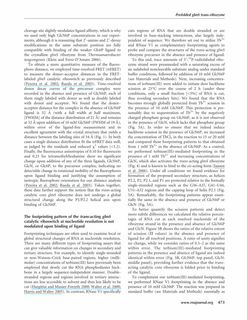
FIGURE 5. Footprinting of the trans-acting glmS ribozyme. (A) Terbium(III)-mediated footprinting of a 59-32P-labeled glmS ribozyme strandupon incubation with Tb3+ for 2 h in 50 mM HEPES-KOH (pH 7.5), 200 mM KCl, and 10 mM MgCl2 at 25°C. From left to right are shown, asindicated; freshly labeled ribozyme strand without further incubation, footprint with 1 mM Tb3+ and increasing concentration of GlcN, RNase T1digests, alkali (OH�) ladder, ribozyme strand incubated in buffer without Tb3+, footprint with increasing Tb3+ concentrations in the presence ofGlcN6P, and footprint with increasing Tb3+ concentrations in the absence of ligand. (B) Histogram plots showing relative change in scissionintensity from with ligand to without ligand, expressed as a ratio between the normalized intensities of scission (P values, see Materials andMethods). The top panel represents terbium(III)-induced backbone scission in the presence (at 20 mM Tb3+) or absence (at 1 mM Tb3+) of10 mM GlcN6P, the middle panel represents terbium(III)-induced backbone scission in the presence or absence of 20 mM GlcN (both at 1 mMTb3+), and the bottom panel represents backbone scission by RNase V1 (0.0025 units) in the presence or absence of 10 mM GlcN6P. A ratio of1 (continuous horizontal line) signifies no change in scission intensity.
Tinsley et al.
474 RNA, Vol. 13, No. 4
JOBNAME: RNA 13#4 2007 PAGE: 7 OUTPUT: Wednesday March 7 19:47:13 2007
csh/RNA/131733/rna3418

which we find to be activated z5000-fold by its specificligand GlcN6P (Fig. 2C). Addition of comparable amountsof GlcN, lacking the 6-phosphate moiety, is able to activatethe riboswitch by only 30-fold, and Glc6P, lacking the2-amino functionality, only results in background activity.This high-level specificity of our trans-activating glmSribozyme, together with its rapid response time and highdynamic range, compares well with most aptazymes, whichwere engineered and/or in vitro evolved for applications asbiosensor components (Breaker 2002). The fact that ourglmS ribozyme construct utilizes an external substrateenables multiple-turnover assays (i.e., amplified productformation; data not shown) and reloading with substrate,virtues sought in biosensor applications. Only futurestudies will reveal how suitable the glmS ribozyme is forsuch applications and whether its effective activationmechanism can be engineered into other aptazymes.
MATERIALS AND METHODS
Preparation of RNA oligonucleotides
The substrate strand, termed gS (see Fig. 1 for sequence), waspurchased from the Howard Hughes Medical Institute Biopolymer/Keck Foundation Biotechnology Resource RNA Laboratory at theYale University School of Medicine. The RNA contained 29-protection groups and was deprotected as recommended by themanufacturer and purified as previously described (Harris et al.2002; Pereira et al. 2002). For FRET measurements, gS wasmodified on the 59 and 39 ends with fluorescein (donor) andtetramethylrhodamine (acceptor), respectively (FgST), as also pre-viously described (Walter 2001, 2002). To obtain the chemicallyblocked, noncleavable substrate analog for structural studies of theprecursor form, the substrate was additionally modified with a 29-methoxy group at the cleavage site (ncFgST; ncgS is the unlabeledform). RNA concentrations were calculated from their absorptionat 260 nm and corrected for the additional absorption of fluores-cein and tetramethylrhodamine by using the relations A260/A492 =0.3 and A260/A554 = 0.49, respectively. The ribozyme strand wasgenerated by run-off transcription from a double-stranded, PCR-amplified template that encoded an upstream T7 promoter. Tran-scriptions were purified as previously described (Harris et al. 2004),and the RNA concentration was calculated as described above.
Cleavage reactions
Cleavage activity of the riboswitch was determined using the two-strand glmS ribozyme construct depicted in Figure 1. A 39-32P-labeled substrate was prepared by ligation with [32P]pCp using T4RNA ligase, followed by desalting using a CentriSep spin column(Princeton Separations). All cleavage reactions were conductedunder single-turnover (pre-steady-state) conditions. Standardbuffer was 50 mM HEPES-KOH (pH 7.5), 200 mM KCl, and10 mM MgCl2, unless otherwise stated. In general, ribozyme andsubstrate strands were annealed separately in standard buffer byheating to 70°C for 2 min and cooling to room temperature overthe course of 5 min. After preincubation for 15 min at 25°C, a
trace (<1 nM) amount of 39-32P-labeled substrate in standardbuffer was added to the ribozyme strand. After combiningribozyme and substrate solutions and incubating for 2 min, analiquot of glucosamine-6-phosphate was added to a final concen-tration of 10 mM (unless otherwise stated), thereby diluting theribozyme to a final concentration of 100 nM. Aliquots (5 mL) weretaken at appropriate time intervals and the reactions quenchedwith 10 mL of 80% formamide, 0.025% bromophenol blue, 50 mMEDTA, and 7 M urea. The radiolabeled 39 cleavage product wasseparated and analyzed as previously described (Pereira et al.2002). Time traces of product formation were fit, unless otherwisenoted, with the single-exponential first-order rate equation y =y0 + A(1�e�t/t), employing Marquardt–Levenberg nonlinearregression (Igor Pro 5.03), where A is the amplitude and t�1 isthe pseudo-first-order rate constant kobs. The ribozyme depen-dency of cleavage was fit with a noncooperative binding equation,while the Mg2+ and GlcN6P dependencies of cleavage were fit witha cooperative binding equation, yielding the cleavage rate constantkcleav under standard conditions, the ribozyme and metal ionhalf-titration points Rz1/2 and Mg1/2, respectively, and thecooperativity coefficient n as described (Pereira et al. 2002).For the GlcN6P concentration dependence, n = 1 gave the bestfit. The pH dependency was fit with equation kobs = kmax
�
ð1 + 10pKa1�pH + 10pH�pka2Þ as previously described (Harris et al.2002; Pereira et al. 2002; Tinsley et al. 2003).
To study rapid activation of the glmS ribozyme by GlcN6P,separate solutions of 100 nM ribozyme (final concentration) andtrace amounts of 59-32P-labeled glmS substrate were prepared instandard buffer as described above. After preincubation for 15 minat 25°C, the ribozyme and substrate were mixed together andallowed to incubate for 10 min while 5 mL aliquots were removedat appropriate time intervals, quenched, and analyzed as describedabove. To initiate the reaction after 10 min, a final concentrationof 10 mM GlcN6P was added to the ribozyme–substrate solutionand reaction aliquots were quenched and analyzed as describedabove.
Steady-state FRET measurements
Steady-state FRET measurements of the glmS ribozyme doublylabeled with fluorescein and tetramethylrhodamine were per-formed on our AB2 spectrofluorometer in a manner similar tothat of previously described experiments (Pereira et al. 2002).Typically, stock solutions for a final concentration of 10 nMdoubly labeled noncleavable substrate analog ncFgST or cleavablesubstrate FgST and 100 nM ribozyme were incubated separatelyin standard buffer supplemented with 25 mM dithiothreitol asa radical quencher at 25°C for at least 15 min. The substrate(145 mL) was then transferred to a 150 mL cuvette and theribozyme was manually added to a saturating 10-fold excess toform the glmS riboswitch. After complex formation, as indicatedby reaching a fluorescence plateau, GlcN6P was added to a finalconcentration of 10 mM to initiate the reaction. Throughout theseadditions fluorescein was excited at 490 nm (4 nm bandwidth),and fluorescence emission was recorded simultaneously at thefluorescein (520 nm, 8 nm bandwidth) and tetramethylrhodamine(585 nm, 8 nm bandwidth) wavelengths, by shifting the emissionmonochromator back and forth. A FRET ratio was calculated asF585/F520.
Prefolded glmS trans-ribozyme
www.rnajournal.org 475
JOBNAME: RNA 13#4 2007 PAGE: 8 OUTPUT: Wednesday March 7 19:47:25 2007
csh/RNA/131733/rna3418

Time-resolved FRET measurements
The global structure of the glmS ribozyme in the presence andabsence of 10 mM GlcN6P was studied by trFRET. Preannealedcomplexes (75 mL, 1 mM noncleavable substrate analog ncFgST,and a saturating excess of 3 mM ribozyme strand) were incubated at25°C for at least 15 min in standard buffer supplemented with25 mM dithiothreitol, prior to collecting time-resolved donor emis-sion profiles using time-correlated single-photon counting. Donor–acceptor distances were measured and analyzed following pre-viously described procedures yielding a three-dimensional Gaussiandistribution in distance R (Pereira et al. 2002; Rueda et al. 2003). Inall cases, a single-distance distribution gave a good fit, as judged bylow, reduced x2 values (<1.2) and evenly distributed residuals. Toextract absolute distances, a value of 55 A for the Forster distanceR0 of fluorescein and tetramethylrhodamine was used (Pereira et al.2002), and a value of 2/3 was assumed for the orientation factor,based on the relatively high mobility of the fluorophores as evidentfrom their moderate fluorescence anisotropies.
Fluorescence anisotropy measurements
Binding of ligand to the trans-acting glmS ribozyme was analyzedusing fluorescence anisotropy measurements performed on aFusion Universal Microplate Analyzer (Packard Instrument Com-pany). For detection of fluorescein, fluorescence was excited withpolarized light at 485 6 10 nm and emission was collected at 535 6
12 nm through a polarizer. For detection of tetramethylrhodamine,fluorescence was excited with polarized light at 535 6 10 nm andemission was collected at 580 6 10 nm through a polarizer. Apreannealed, noncleavable substrate analog–ribozyme complex(100 mL, with a 10 nM ncFgST noncleavable substrate strand and a100 nM ribozyme strand) was incubated at 25°C for at least 15 minin standard buffer. Anisotropy values were automatically measured10 times either in the presence or absence of 10 mM GlcN6P,10 mM GlcN, or 10 mM Glc6P on two different samples and averaged.The factory G value (0.9) was used for all assays for correction.
Terbium(III)-mediated and RNase V1 footprinting
To observe the slow backbone scission mediated by the aqueousTb3+ derived species Tb(OH)(aq)2+ and RNA footprinting medi-ated by the double-strand specific RNase V1, a purified ribozymestrand was 59-32P-phosphorylated with T4 polynucleotide kinaseand [g-32P]ATP, repurified as described above. The labeled RNAstrand (250,000 cpm per 10 mL reaction volume) was preannealedwith a final concentration of 500 nM noncleavable substrateanalog ncgS, denatured at 70°C for 2 min, and slowly cooled toroom temperature over 5 min. After cooling, 0.2 mg/mL of tRNAcarrier were added to the RNase V1 reactions. The sample wassplit into two halves, and GlcN6P was added to only one half to afinal (saturating) concentration of 10 mM (unless otherwisestated). Terbium(III)-mediated scission was initiated by mixingwith 2 mL of an appropriate serial dilution of TbCl3 to achieve thedesired final Tb3+ concentration (typically, 1–20 mM) andincubated at 25°C for 2 h. RNase V1 degradation was initiatedby mixing 2 mL of the RNA solution with RNase V1 to achieve thedesired final nuclease concentration (0.00125–0.125 units perreaction) and incubated at 25°C for 10 min. Reaction mixtureswere stopped by addition of an equal volume of 50 mM EDTA
(pH 8.0) and ethanol precipitation at �20°C. The precipitatedRNA was redissolved in urea loading buffer (80% formamide,0.025% xylene cyanol, 0.025% bromophenol blue, 50 mM EDTA),and analyzed on a wedged 20% polyacrylamide, 7 M urea,sequencing gel, alongside sequencing ladders from partial diges-tion with G-specific RNase T1 under denaturing conditions andfrom alkaline hydrolysis. Product bands were visualized asdescribed above and analyzed as previously described in theliterature (Walter et al. 2000; Jeong et al. 2003; Harris et al.2004; Harris and Walter 2005).
ACKNOWLEDGMENTS
We thank Ken Hampel for sharing preliminary data, Adrian Ferre-D’Amare for sending his coordinates on the day they werepublished, Robert Kennedy for use of his Microplate analyzer,and all members of the Walter laboratory for stimulatingdiscussions and thoughtful suggestions.
Received October 9, 2006; accepted December 19, 2006.
REFERENCES
Barrick, J.E., Corbino, K.A., Winkler, W.C., Nahvi, A., Mandal, M.,Collins, J., Lee, M., Roth, A., Sudarsan, N., Jona, I., et al. 2004.New RNA motifs suggest an expanded scope for riboswitches inbacterial genetic control. Proc. Natl. Acad. Sci. 101: 6421–6426.
Blount, K.F. and Uhlenbeck, O.C. 2005. The structure–functiondilemma of the hammerhead ribozyme. Annu. Rev. Biophys.Biomol. Struct. 34: 415–440.
Breaker, R.R. 2002. Engineered allosteric ribozymes as biosensorcomponents. Curr. Opin. Biotechnol. 13: 31–39.
Cowan, J.A. 1993. Metallobiochemistry of RNA. Co(NH3)63+ as a
probe for Mg2+(aq) binding sites. J. Inorg. Biochem. 49: 171–175.DeLano, W. 2002. The PyMOL molecular graphics system. DeLano
Scientific, San Carlos, CA.Hampel, K.J. and Tinsley, M.M. 2006. Evidence for preorganization of
the glmS ribozyme ligand binding pocket. Biochemistry 45: 7861–7871.
Hargittai, M.R. and Musier-Forsyth, K. 2000. Use of terbium as aprobe of tRNA tertiary structure and folding. RNA 6: 1672–1680.
Harris, D.A. and Walter, N.G. 2005. Terbium(III) footprinting as aprobe of RNA structure and metal-binding sites. In Handbook ofRNA biochemistry (eds. R.K. Hartmann et al.), pp. 205–213. Wiley-VCH, Weinheim, Germany.
Harris, D.A., Rueda, D., and Walter, N.G. 2002. Local conformationalchanges in the catalytic core of the trans-acting hepatitis delta virusribozyme accompany catalysis. Biochemistry 41: 12051–12061.
Harris, D.A., Tinsley, R.A., and Walter, N.G. 2004. Terbium-mediatedfootprinting probes a catalytic conformational switch in the anti-genomic hepatitis delta virus ribozyme. J. Mol. Biol. 341: 389–403.
Jansen, J.A., McCarthy, T.J., Soukup, G.A., and Soukup, J.K. 2006.Backbone and nucleobase contacts to glucosamine-6-phosphate inthe glmS ribozyme. Nat. Struct. Mol. Biol. 13: 517–523.
Jeong, S., Sefcikova, J., Tinsley, R.A., Rueda, D., and Walter, N.G.2003. Trans-acting hepatitis delta virus ribozyme: Catalytic coreand global structure are dependent on the 59 substrate sequence.Biochemistry 42: 7727–7740.
Klein, D.J. and Ferre-D’Amare, A.R. 2006. Structural basis of glmSribozyme activation by glucosamine-6-phosphate. Science 313:1752–1756.
Lim, J., Grove, B.C., Roth, A., and Breaker, R.R. 2006. Characteristicsof ligand recognition by a glmS self-cleaving ribozyme. Angew.Chem. Int. Ed. Engl. 45: 6689–6693.
Tinsley et al.
476 RNA, Vol. 13, No. 4
JOBNAME: RNA 13#4 2007 PAGE: 9 OUTPUT: Wednesday March 7 19:47:26 2007
csh/RNA/131733/rna3418

Link, K.H., Guo, L., and Breaker, R.R. 2006. Examination of thestructural and functional versatility of glmS ribozymes by using invitro selection. Nucleic Acids Res. 34: 4968–4975.
McCarthy, T.J., Plog, M.A., Floy, S.A., Jansen, J.A., Soukup, J.K., andSoukup, G.A. 2005. Ligand requirements for glmS ribozyme self-cleavage. Chem. Biol. 12: 1221–1226.
Nelson, J.A. and Uhlenbeck, O.C. 2006. When to believe what you see.Mol. Cell 23: 447–450.
Pereira, M.J., Harris, D.A., Rueda, D., and Walter, N.G. 2002.Reaction pathway of the trans-acting hepatitis delta virus ribo-zyme: A conformational change accompanies catalysis. Biochem-istry 41: 730–740.
Roth, A., Nahvi, A., Lee, M., Jona, I., and Breaker, R.R. 2006.Characteristics of the glmS ribozyme suggest only structural rolesfor divalent metal ions. RNA 12: 607–619.
Rueda, D., Wick, K., McDowell, S.E., and Walter, N.G. 2003. Diffuselybound Mg2+ ions slightly reorient stems I and II of the hammer-head ribozyme to increase the probability of formation of thecatalytic core. Biochemistry 42: 9924–9936.
Rueda, D., Bokinsky, G., Rhodes, M.M., Rust, M.J., Zhuang, X., andWalter, N.G. 2004. Single-molecule enzymology of RNA: Essentialfunctional groups impact catalysis from a distance. Proc. Natl.Acad. Sci. 101: 10066–10071.
Tinsley, R.A., Harris, D.A., and Walter, N.G. 2003. Significantkinetic solvent isotope effects in folding of the catalytic RNAfrom the hepatitis delta virus. J. Am. Chem. Soc. 125: 13972–13973.
Walter, N.G. 2001. Structural dynamics of catalytic RNAhighlighted by fluorescence resonance energy transfer. Methods25: 19–30.
Walter, N.G. 2002. Probing RNA structural dynamics and function byfluorescence resonance energy transfer (FRET). Curr. ProtocolsNucleic Acid Chem. 11.10: 11.10.11–11.10.23.
Walter, N.G., Yang, N., and Burke, J.M. 2000. Probing nonselectivecation binding in the hairpin ribozyme with Tb(III). J. Mol. Biol.298: 539–555.
Wilkinson, S.R. and Been, M.D. 2005. A pseudoknot in the 39 noncoreregion of the glmS ribozyme enhances self-cleavage activity. RNA11: 1788–1794.
Winkler, W.C. 2005. Metabolic monitoring by bacterial mRNAs. Arch.Microbiol. 183: 151–159.
Winkler, W.C., Nahvi, A., Roth, A., Collins, J.A., and Breaker, R.R.2004. Control of gene expression by a natural metabolite-respon-sive ribozyme. Nature 428: 281–286.
Zamel, R. and Collins, R.A. 2002. Rearrangement of substratesecondary structure facilitates binding to the Neurospora VSribozyme. J. Mol. Biol. 324: 903–915.
Prefolded glmS trans-ribozyme
www.rnajournal.org 477
JOBNAME: RNA 13#4 2007 PAGE: 10 OUTPUT: Wednesday March 7 19:47:28 2007
csh/RNA/131733/rna3418