Squalene Loaded Nanoparticles Effectively Protect Hepatic ...
Upload
khangminh22Category
view
0download
0
�����������������
Citation: Alshaman, R.; Alattar, A.;
El-Sayed, R.M.; Gardouh, A.R.;
Elshaer, R.E.; Elkazaz, A.Y.; Eladl,
M.A.; El-Sherbiny, M.; Farag, N.E.;
Hamdan, A.M.; et al. Formulation
and Characterization of
Doxycycline-Loaded Polymeric
Nanoparticles for Testing
Antitumor/Antiangiogenic Action in
Experimental Colon Cancer in Mice.
Nanomaterials 2022, 12, 857. https:/
/doi.org/10.3390/nano12050857
Academic Editor: Lisheng Wang
Received: 26 January 2022
Accepted: 26 February 2022
Published: 3 March 2022
Publisher’s Note: MDPI stays neutral
with regard to jurisdictional claims in
published maps and institutional affil-
iations.
Copyright: © 2022 by the authors.
Licensee MDPI, Basel, Switzerland.
This article is an open access article
distributed under the terms and
conditions of the Creative Commons
Attribution (CC BY) license (https://
creativecommons.org/licenses/by/
4.0/).
nanomaterials
Article
Formulation and Characterization of Doxycycline-LoadedPolymeric Nanoparticles for Testing Antitumor/AntiangiogenicAction in Experimental Colon Cancer in MiceReem Alshaman 1, Abdullah Alattar 1 , Rehab M. El-Sayed 2, Ahmed R. Gardouh 3,4 , Rabie E. Elshaer 5,Amany Y. Elkazaz 6,7, Mohamed Ahmed Eladl 8,* , Mohamed El-Sherbiny 9 , Noha E. Farag 10,11,Ahmed Mohsen Hamdan 12 and Sawsan A. Zaitone 13,*
1 Department of Pharmacology and Toxicology, Faculty of Pharmacy, University of Tabuk,Tabuk 71491, Saudi Arabia; [email protected] (R.A.); [email protected] (A.A.)
2 Department of Pharmacology and Toxicology, Faculty of Pharmacy, Sinai University, El-Arish 45511, Egypt;[email protected]
3 Department of Pharmaceutics and Industrial Pharmacy, Faculty of Pharmacy, Suez Canal University,Ismailia 41522, Egypt; [email protected]
4 Department of Pharmacy, Faculty of Pharmacy, Jadara University, Irbid 21110, Jordan5 Department of Pathology, Faculty of Medicine, Al-Azhar University, Cairo 11651, Egypt;
[email protected] Biochemistry and Molecular Biology Department, Faculty of Medicine, Suez Canal University,
Ismailia 41522, Egypt; [email protected] Biochemistry and Molecular Biology Department, Faculty of Medicine, Port Said University,
Port Said 42526, Egypt8 Basic Medical Science Department, University of Sharjah, Sharjah 27272, United Arab Emirates9 Department of Basic Medical Sciences, College of Medicine, AlMaarefa University,
Riyadh 71666, Saudi Arabia; [email protected] Department of Physiology, Faculty of Medicine, Suez Canal University, Ismailia 41522, Egypt;
[email protected] Department of Physiology, College of Medicine, Taif University, Taif 21974, Saudi Arabia12 Department of Pharmacy Practice, Faculty of Pharmacy, University of Tabuk, Tabuk 71491, Saudi Arabia;
[email protected] Department of Pharmacology and Toxicology, Faculty of Pharmacy, Suez Canal University,
Ismailia 41522, Egypt* Correspondence: [email protected] (M.A.E.); [email protected] (S.A.Z.);
Tel.: +20-1068916396 (S.A.Z.)
Abstract: Nanotherapeutics can enhance the characteristics of drugs, such as rapid systemic clearanceand systemic toxicities. Polymeric nanoparticles (PRNPs) depend on dispersion of a drug in anamorphous state in a polymer matrix. PRNPs are capable of delivering drugs and improving theirsafety. The primary goal of this study is to formulate doxycycline-loaded PRNPs by applying thenanoprecipitation method. Eudragit S100 (ES100) (for DOX-PRNP1) and hydroxypropyl methylcellulose phthalate HP55 (for DOX-PRNP2) were tested as the drug carrying polymers and theDOX-PRNP2 showed better characteristics and drug release % and was hence selected to be testedin the biological study. Six different experimental groups were formed from sixty male albino mice.1,2,-Dimethylhydrazine was used for 16 weeks to induce experimental colon cancer. We compared theoral administration of DOX-PRNP2 in doses of 5 and 10 mg/kg with the free drug. Results indicatedthat DOX-PRNP2 had greater antitumor activity, as evidenced by an improved histopathologicalpicture for colon specimens as well as a decrease in the tumor scores. In addition, when compared tofree DOX, the DOX-PRNP2 reduced the angiogenic indicators VEGD and CD31 to a greater extent.Collectively, the findings demonstrated that formulating DOX in PRNPs was useful in enhancingantitumor activity and can be used in other models of cancers to verify their efficacy and compatibilitywith our study.
Nanomaterials 2022, 12, 857. https://doi.org/10.3390/nano12050857 https://www.mdpi.com/journal/nanomaterials
Nanomaterials 2022, 12, 857 2 of 20
Keywords: angiogenesis; mouse colon cancer; eudragit S100; hydroxypropyl methylcellulose phtha-late; doxycycline polymeric nanoparticles; nanoprecipitation method
1. Introduction
Nanotherapeutics is a developing field within the field of nanomedicine and it aims toenhance the characteristics of drugs [1]. There are various types of nanotherapeutics suchas solid-lipid nanoparticles, gold nanoparticles and polymeric nanoparticles (PRNPs) [2–5].
PRNPs improve solubility, bioavailability and retention time of drugs and enhancetheir therapeutic effect [6–9]. PRNPs have advantages over conventional drugs in that theyincrease drug efficacy, protect the drug from degradation when in contact with biologicalfluids and control the drug release [10] due to their advantages resulting from their tinysize. PRNPs have gained a lot of attention in recent years [11,12]. PRNPs are composedof amorphous drugs dispersed within a polymer matrix [13]. Controlling drug releasein the gastrointestinal tract is possible with pH-sensitive materials that release the drugselectively in the small intestine adjacent to the absorption location [14,15]. PRNPs can beutilized to be loaded with various types of medications for treatment of different typesof cancer [16,17].
Cancer is an important cause of death in the world [18]. Obstacles for cancer therapyare the toxicity of drugs, multidrug resistance, and low drug uptake by cancer cells [19].Colorectal cancer is in second place for women-diagnosed cancers and in third placefor men-diagnosed cancers [20,21]. Angiogenesis is the generation of capillary bloodvessels [22] that is stimulated in tumor growth and metastasis [23]. Capillaries are requiredto supply nutrients and oxygen to a growing tumor. Vascular endothelial growth factor(VEGF) is a key factor in angiogenesis that enhances microvascular hyperpermeability [24].
In cancer therapy, the critical question is how to achieve the desired level of chemother-apy in tumors [25]. Contemporary cancer treatment modalities continue to face significantchallenges, including low drug concentrations at the tumor areas, intolerable adverse effectsand development of chemo-resistance [26]. These and other constraints have mandateddevelopment of the recent advancements in cancer therapeutics.
Doxycycline (DOX) is a wide-spectrum antibiotic that is a tetracycline derivative [27]that was first approved by the FDA in the 1960s [28,29]. It is mainly used in the treatmentof acne and acne rosacea [30]. This side effect of DOX was repurposed as a therapeuticeffect in treatment of cancer by inhibition of mitochondrial biogenesis in cancer cells [31,32].Moreover, it may potentially be harmful to healthy cells due to enzymatic inhibition orchanges in protein synthesis [33]. The use of nanotechnology is one possible solution tothis challenge [34] and to improving the efficiency of chemotherapeutic agents [35].
Since the stability of drugs can be improved utilizing pH-sensitive nanoparticles. Theprimary goal of this study is to formulate pH-sensitive PRNPs loaded with DOX using thenanoprecipitation technique and the polymers eudragit S100 (ES100) or hydroxypropylmethyl cellulose phthalate HP55 (HPMCP HP55) and to test their therapeutic potentialagainst experimentally induced colon cancer in mice.
2. Materials & Methods2.1. Synthesis of Doxycycline Polymeric Nanoparticles2.1.1. Materials
Poly(methacrylic acid, methyl methacrylate) 1:2 (Eudragit S100®) was kindly providedby Heinrich’s Commercial Agency (EVONIK, batch # B101205222, Cairo, Egypt) (ES100).Cellulose, 2-hydroxypropyl methyl ether and phthalic acid ester were purchased fromShin-Etsu Chemical Co. (Tokyo, Japan). HPMCP HP55 was supplied by Acino Pharm(batch #0000168267, Acino International AG Thurgauerstrasse 36/38 CH-8050, Zurich,Switzerland). α-Hydro-ω-hydroxypoly (oxyethylene) poly (oxypropylene) poly (oxyethy-lene) block copolymers (Poloxamer 407®) were obtained from BASF, (Cairo, Egypt, batch

Nanomaterials 2022, 12, 857 3 of 20
#WPHF555C). Acetone was purchased from El-Nasr chemical company (Qalyub, Egypt,batch #2013/7). Doxycycline helicate was a gift from Tabuk Pharmaceutical Company(Tabuk, KSA).
2.1.2. Formulation of DOX-PRNPs with Full Characterization
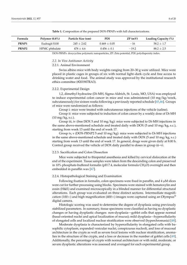
Preparation of polymeric nanoparticles containing doxycycline with full characteri-zation was performed according to [36], who prepared DOX-PRNPs with hydroxypropylmethyl cellulose polymer and Tween 80 as surfactant utilizing different ratios, with littlemodification. Briefly, PRNPs formulations were prepared by the nanoprecipitation tech-nique [37] for hydrophilic and hydrophobic drug encapsulation in polymer nanoparticles.We used 0.8 g % of the polymer, eudragit S100® (PRNP1) or HPMCP HP55® (PRNP2), and1% Poloxamer 407® as surfactant. Drop wise addition of aqueous phase with drug andsurfactant added to a certain concentration of polymer dissolved in acetone (water-miscibleorganic solvent) was used for forming an organic phase (the final organic: aqueous phaseratio equals 1:8) as shown in Figure 1. The mixture was stirred at room temperature untilturning into a milk-like mixture [38] and was left overnight with continuous stirring [39].The drug polymer ratio in final formulations was 2:1. The efficiency of encapsulation andloading capacity were tested by an indirect method. The amount of the non-capsulateddrug in the supernatant was estimated spectrophotometrically at 269 nm, utilizing anultraviolet spectrophotometer (Shimadzu, Yokohama, Japan). Different characterizationssuch as zeta potential (ZP), particle size as well as the polydispersity index (PDI) of theformulated nanoparticles were measured using a Zetasizer (Malvern Instruments Ltd.,Malvern, UK) [40,41], while morphology of the surface of the nanoparticles was confirmedusing transmission electron microscopy (TEM) (model JTEM-2100, Tokyo, Japan) [42].Release of the drug from different trials was carried out using dialysis bag method [43]at simulated gastric fluid containing 0.1 N HCl and final pH equalsing 1.2 for 2 h andthen substituted with phosphate buffer (pH = 6.8) for the next 10 h. Temperature waskept at 37 ± 1 ◦C, and drug was assayed spectrophotometrically at 269 nm (UV-visiblespectrophotometer, Shimadzu, Yokohama, Japan) [44]. Detailed ratios of drug to polymerwith surfactant concentration and full characterizations are listed in Table 1. From thesedata, PRNP2 showed better characteristics and was selected to be tested for biologicalactivity in the in vivo study.
Figure 1. Formulation of doxycycline polymeric nanoparticles.
Nanomaterials 2022, 12, 857 4 of 20
Table 1. Composition of the prepared DOX-PRNPs with full characterizations.
Formula Polymer (0.8%) Particle Size (nm) PDI ZP (mV) Loading Capacity (%)
PRNP1 Eudragit S100 245 ± 2.62 0.469 ± 0.05 −16 58.2 ± 1.7
PRNP2 HPMC phthalate 478 ± 4.6 0.456 ± 0.1 −19.2 88.2 ± 2.5
DOX-PRNPs: doxycycline polymeric nanoparticles, ZP: Zeta potential, PDI: polydispersity index.
2.2. In Vivo Antitumor Activity2.2.1. Animal Environment
Swiss albino mice with body weights ranging from 20–30 g were utilized. Mice wereplaced in plastic cages in groups of six with normal light–dark cycle and free access todrinking water and food. The animal study was approved by the institutional researchethics committee (#201907RA3).
2.2.2. Experimental Design
1,2, dimethyl hydrazine (Di-MH, Sigma-Aldrich, St. Louis, MO, USA) was employedto induce experimental colon cancer in mice and was administered (10 mg/kg/week,subcutaneously) for sixteen weeks following a previously reported schedule [45,46]. Groupsof mice were randomized as follows:
Group i: mice were treated with subcutaneous injections of the vehicle (saline).Group ii: mice were subjected to induction of colon cancer by a weekly dose of Di-MH
(10 mg/kg, s.c.).Group iii, iv (free DOX 5 and 10 mg/kg): mice were subjected to Di-MH injections in
the same above-mentioned schedule and treated daily with DOX (5 and 10 mg/kg, s.c.),starting from week 13 until the end of week 17.
Group iv, v (DOX-PRNP2 5 and 10 mg/kg): mice were subjected to Di-MH injectionsin the same above-mentioned schedule and treated daily with DOX (5 and 10 mg/kg, s.c.)starting from week 13 until the end of week 17. In general, drugs were given daily at 8:00 h.Control group received the vehicle of DOX daily parallel to doses in group iii–vi.
2.2.3. Sacrification and Colon Dissection
Mice were subjected to thiopental anesthesia and killed by cervical dislocation at theend of the experiment. Tissue samples were taken from the descending colon and preservedin 10% phosphate-buffered formalin (pH 7.4, molecular formula CH2O) overnight and thenembedded in paraffin wax [47].
2.2.4. Histopathological Staining and Examination
Following fixation in formalin, colon specimens were fixed in paraffin, and 4 µM sliceswere cut for further processing using blocks. Specimens were stained with hematoxylin andeosin (H&E) and examined microscopically in a blinded manner for differential structuralalterations. Each group was evaluated on three distinct sections. Intermediate magnifi-cation (100×) and high magnification (400×) images were captured using an Olympus®
digital camera.Histologic scoring was used to determine the degree of dysplasia using previously
stabilized parameters. In summary, tissue specimens were classified as having no dysplasticchanges or having dysplastic changes: non-dysplasia—goblet cells that appear normal(basal-oriented nuclei and apical localization of mucus); mild dysplasia—hypercellularityof elongated cells and localized nuclear stratification were observed (hyperchromasia) [48].
Moderate dysplasia is characterized by hypercellularity in elongated cells with ba-sophilic cytoplasm, expanded vesicular nuclei, conspicuous nucleoli, and loss of mucosalarchitecture in the crypts as well as severe focal lesions with nuclear stratification, anoma-lies in the structure of the crypts, and a loss or decrease in the number of goblet cells [49].Additionally, the percentage of crypts with normal architecture or with mild, moderate, orsevere dysplastic alterations was assessed and averaged for each experimental group.

Nanomaterials 2022, 12, 857 5 of 20
2.2.5. Immunohistochemical Staining for VEGF
After antigen retrieval, 5% normal goat serum was applied to the slides for 45 min forpreventing non-specific antibody binding. At 4 ◦C overnight, slides were treated with thedesired primary antibodies. At a dilution of 1:100, primary rabbit polyclonal antibodiesagainst VGEF (diluted 1:100, #A17877, ABclonal, Woburn, MA, USA) was employed.Slides were cleaned, treated with secondary antibody, washed again, and then coveredwith DAB and counterstained. Finally, slides were blindly viewed under an Olympusmicroscope. The images were captured at magnifications of 100× and 400× using anOlympus® built-in digital camera. The percentage of stained area was determined usingImageJ (NIH, Bethesda, MD, USA), and the percentages were digitalized to obtain the areaof immunostaining.
2.2.6. Western Blot Analysis
In RIPA buffer containing protease and phosphatase inhibitors, isolated colon tissueswere homogenized. Homogenates were centrifuged at 14,000× g for 20 min at 4 ◦C toremove insoluble material. Transferring the supernatant to a new microcentrifuge tube,5 µL was used to determine the protein concentration using the Bio-Rad Quick StartTM
Bradford Protein Assay kit. After initial denaturation with 4x Laemmli Sample Buffer, simi-lar amounts of protein from colon tissues homogenate were loaded onto sodium dodecylsulfate polyacrylamide gel (Bio-Rad, Hercules, CA, USA). Following electrophoresis sepa-ration of proteins, the proteins from the gels were transferred to nitrocellulose membranes.Incubation in 5% nonfat dried milk blocking the free sites on the membranes (Bio-Rad,Hercules, CA, USA) were performed for 1 h. This was followed by washing of the blockedmembranes and incubating them with primary antibodies specific for the targeted proteins:a rabbit recombinant polyclonal anti-cluster of differentiation 31 (CD31) antibody (RM1006)(ab281583 Abcam) at 1/1000 dilution, a mouse monoclonal antibody to VEGF (sc-7269)from Santa Cruz Biotechnology Inc. (Santa Cruz, CA, USA) at dilution 1:200 and antibodyto β-actin (sc-8432) (SantaCruz Biotechnology, Santa Cruz, CA, USA) overnight at 4 ◦C withgentle agitation. Following that, the blots were washed and incubated with the appropriatesecondary antibody conjugated to horseradish peroxidase (HRP) and goat anti-mouse, fol-lowed by enhanced chemiluminescence detection using the enhanced chemiluminescenceECL AdvanceTM Western blotting detection kit. Densitometry was used to quantify theintensity of immunoreactivity using the ImageJ software (NIH, Bethesda, MD, USA).
2.2.7. Enzyme Linked-Immunosorbent Assay for the Proangiogenic Factors
The colon homogenates were assayed for IL-6, TNF-α and VEGF using ELISA kits:mouse IL-6 ELISA kit (Sunred Biological Technology Company, Shanghai, China), mouseTNF-α ELISA kit (Cloud-Clon Crop Company, Katy, TX, USA) and mouse VEGF ELISAkit (Cloud-Clon Crop Company, Katy, TX, USA). The optical density of the reactions wasmeasured at 450 nm.
2.3. Statistical Analysis
The statistical tests were conducted using the social sciences statistical package. (SPSSSoftware, SPSS Inc., Chicago, IL, USA). Finally, the differences were considered significantin any of the statistical tests when p < 0.05. The Shapiro–Wilks test was used to determinethe normality of the data distribution. For data with a Gaussian distribution, one-wayanalysis of variance, ANOVA, was applied for analyzing the measurements. Additionally,post hoc analysis was used to compare the study groups. The Mann-Whitney U test wasused to examine data with a non-Gaussian distribution. Two-tailed data are expected.
3. Results3.1. Polymeric Nanoparticles Characterizations
Nanoprecipitation technique was suitably used for preparation of pH responsiveDOX-PRNPs to control its release and modulate its pharmacodynamic influence. Figures 2

Nanomaterials 2022, 12, 857 6 of 20
and 3 show that particle size of formulated nanoparticles were below micrometers andshowed moderate stability owing to values of ZP (below 30 mV). Loading capacity ofPRNP2 was higher than that of PRNP1 while maintaining the same formulation conditions.PDI, which is an indicator of uniformity of prepared particle size, showed a value below 0.5,indicating homogeneity and monodisperse feature. The TEM micrographs of the preparednanoparticles seemed spherical with a smooth surface as shown by Figure 4.
The prepared PRNPs revealed negative ZPs, which gives an indication about stabilitydue to low tendency for aggregation. Upon studying DOX release form prepared PRNPs,results showed that the type of the polymer significantly impacts drug release, wherePRNP1 presented greater release than PRNP2 (Figure 5). Further, PRNP2 dissolved atgreater pH than that of PRNP1, and the two formulations showed more delayed releasethan the pure drug.
Figure 2. Particle size by laser diffraction technique of prepared polymer nanoparticles of doxycyclineprepared by nanoprecipitation technique (displaced for better comparison).

Nanomaterials 2022, 12, 857 7 of 20
Figure 3. Zeta potential values for the prepared polymer nanoparticles of doxycycline prepared bynanoprecipitation technique.
Figure 4. Transmission electron micrographs of prepared polymer nanoparticles of doxycycline pre-pared by nanoprecipitation technique, showing the particle size of prepared nanoparticles (displacedfor better visualization).

Nanomaterials 2022, 12, 857 8 of 20
Figure 5. Release profile of doxycycline from prepared polymeric nanoparticles prepared by nano-precipitation technique.
3.2. Biological Activity against Colon Cancer3.2.1. Histopathological Examination
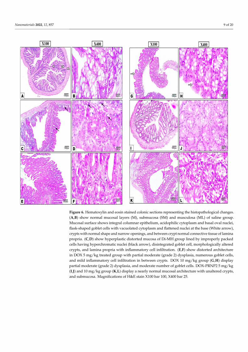
As shown in Figure 6, the histopathological examinations of the colon tissues fromsaline group (Figure 6A,B) stained with H&E showed the normal mucosa, submucosaand musculosa. The mucosa showed columnar epithelium with intact surface, acidophiliccytoplasm, and basal oval nuclei. Crypts show normal shape with narrow openings, whilegoblet cells are flask in shape with basal flattened nuclei and vacuolated cytoplasm. Con-versely, the Di-MH control group (Figure 6C,D) showing hyperplasia with irregular shapedmucosa and slightly raised showed lining by inappropriate crowded hyperchromatic nu-clei, distortion in crypts with disintegrated goblet cell and appearance of laminar cellularinfiltration. Moreover, colon tissue sections from DOX 5 mg treated group (Figure 6E,F)and DOX 10 mg group (Figure 6G,H) showed moderate dysplasia (grade 2) with highernumber of goblet cells but fewer inflammatory cells. Furthermore, using the 5 mg/kgof DOX-PRNP2 (Figure 6I,J) or 10 mg/kg group (Figure 6K,L) showed an almost normalappearance for crypts, mucosa and submucosa.
Microscopic examination of stained colonic sections showed minimal scattered chronicinflammatory cells in control negative group (Figure 7A). Meanwhile, the Di-MH group(Figure 7B) showed positive dense inflammatory infiltrate with lymphoid follicles. Colonicsections from treated groups showed moderate inflammation with lymphoid aggregatein group DOX 5 mg/kg (Figure 7C), mild inflammation with mild aggregates in groupDOX 10 mg/kg (Figure 7D) and few inflammatory cells in DOX-PRNP2 5 and 10 mg/kg(Figure 7E,F).
Nanomaterials 2022, 12, 857 9 of 20
Figure 6. Hematoxylin and eosin stained colonic sections representing the histopathological changes.(A,B) show normal mucosal layers (M), submucosa (SM) and musculosa (ML) of saline group.Mucosal surface shows integral columnar epithelium, acidophilic cytoplasm and basal oval nuclei,flask-shaped goblet cells with vacuolated cytoplasm and flattened nuclei at the base (White arrow),crypts with normal shape and narrow openings, and between crypt normal connective tissue of laminapropria. (C,D) show hyperplastic distorted mucosa of Di-MH group lined by improperly packedcells having hyperchromatic nuclei (black arrow), disintegrated goblet cell, morphologically alteredcrypts, and lamina propria with inflammatory cell infiltration. (E,F) show distorted architecturein DOX 5 mg/kg treated group with partial moderate (grade 2) dysplasia, numerous goblet cells,and mild inflammatory cell infiltration in between crypts. DOX 10 mg/kg group (G,H) displaypartial moderate (grade 2) dysplasia, and moderate number of goblet cells. DOX-PRNP2 5 mg/kg(I,J) and 10 mg/kg group (K,L) display a nearly normal mucosal architecture with unaltered crypts,and submucosa. Magnifications of H&E stain X100 bar 100, X400 bar 25.

Nanomaterials 2022, 12, 857 10 of 20
Figure 7. In the control negative group (A), microscopic images of H&E stained colonic sections showonly a few scattered chronic inflammatory cells. Conversely, the Di-MH group (B) has a markedpositive inflammatory infiltrate with prominent lymphoid follicles (arrows). Moderate inflammationwith lymphoid aggregates in the DOX 5 mg/kg group (C), mild inflammation with minor aggregatesin the DOX 10 mg/kg group (D), and few inflammatory cells in the DOX-PRNP2 5 and 10 mg group(E,F). Magnifications of H&E stain X100 bar 100, X400 bar 25.
Figure 8 demonstrates the tumor scores given to colon specimens. Scores for crypticdistortion were greater in the colon cancer control group than in the saline group. The scoreswere significantly reduced in mice treated with DOX-PRNP2 5 or 10 mg/kg (Figure 8A).Scores for hyperplasia, goblet cell depletion and dysplasia were also significantly higherin the colon cancer control group compared to the saline group (Figure 8B–D); these threescores were significantly reduced in the DOX-PRNP2 10 mg/kg group.

Nanomaterials 2022, 12, 857 11 of 20
Figure 8. Histologic scores given to colon specimens stained with H&E. (A) Cryptic distortion,(B) hyperplasia, (C) goblet cell depletion and (D) dysplasia score. Scoring was performed for eachitem from 0–3 by an experienced pathologist. Data were analyzed by Kruskal-Wallis ANOVA andDunn’s post hoc test for intergroup comparison at p < 0.05. *: versus saline group, #: versus coloncancer control group, p < 0.05. PRNPs: PRNP2 prepared from hydroxypropyl methyl cellulosephthalate HP55 as a polymer.
3.2.2. Immunohistochemistry for VEGF
Concerning the immunohistochemistry, the VEGF-immunostained colonic sectionsshowed negative immune reaction in colonic mucosa of the saline group (Figure 9A,B)whereas the Di-MH-induced colon cancer control group demonstrated strong positivebrown immune reaction in the affected mucosal layers (Figure 9C,D). The DOX 5 and 10mg/kg and DOX-PRNP2 5 and 10 mg/kg treated groups showed weaker positive reactionsin the affected mucosal layers (Figure 9E–L).

Nanomaterials 2022, 12, 857 12 of 20
Figure 9. Microscopic pictures of immunostained colonic sections against VEGF. Images show nega-tive expression of colonic mucosa in the saline group (A,B). Control group (C,D) presents markedbrown positive expression in afflicted colonic mucosa of cancer colon (white arrows), while DOX 5and 10 and DOX-PRNP2 5 and 10 mg/kg treated groups (E–L) indicate mild brown positive expres-sion in affected mucosa (white arrows). Mayer’s hematoxylin was used as counterstain with IHC.Magnifications of VEGF immune stain X100 bar 100, X400 bar 25. (M) Column chart representingmean ± SDM for area % of VEGF immunostaining, *: versus saline group, #: versus colon cancercontrol, &: versus colon cancer + DOX 5 mg/kg and $: versus colon cancer + DOX 10 mg/kg,p < 0.05. PRNPs: PRNP2 prepared from hydroxypropyl methyl cellulose phthalate HP55 asa polymer.
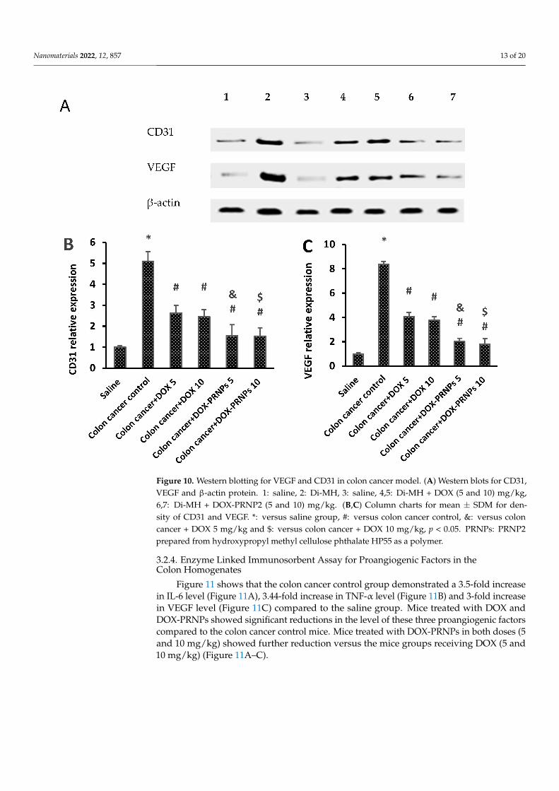
3.2.3. Western Blot Analysis for CD31 and VEGF
Figure 10A shows the WB gels for the bands of CD31, VEGF and β-actin. Currentresults showed significant augmentation in CD31 and VEGF protein expression inDi-MH mice compared to control negative group (Figure 10B,C). Conversely, micetreated with DOX and DOX-PRNP2 showed a significant reduction in the proteinexpression compared to the Di-MH mice group. Mice treated with DOX-PRNP2 inboth doses (5 and 10 mg/kg) showed further reduction versus the mice groups treatedwith DOX (5 and 10 mg/kg), respectively.
Nanomaterials 2022, 12, 857 13 of 20
Figure 10. Western blotting for VEGF and CD31 in colon cancer model. (A) Western blots for CD31,VEGF and β-actin protein. 1: saline, 2: Di-MH, 3: saline, 4,5: Di-MH + DOX (5 and 10) mg/kg,6,7: Di-MH + DOX-PRNP2 (5 and 10) mg/kg. (B,C) Column charts for mean ± SDM for den-sity of CD31 and VEGF. *: versus saline group, #: versus colon cancer control, &: versus coloncancer + DOX 5 mg/kg and $: versus colon cancer + DOX 10 mg/kg, p < 0.05. PRNPs: PRNP2prepared from hydroxypropyl methyl cellulose phthalate HP55 as a polymer.
3.2.4. Enzyme Linked Immunosorbent Assay for Proangiogenic Factors in theColon Homogenates
Figure 11 shows that the colon cancer control group demonstrated a 3.5-fold increasein IL-6 level (Figure 11A), 3.44-fold increase in TNF-α level (Figure 11B) and 3-fold increasein VEGF level (Figure 11C) compared to the saline group. Mice treated with DOX andDOX-PRNPs showed significant reductions in the level of these three proangiogenic factorscompared to the colon cancer control mice. Mice treated with DOX-PRNPs in both doses (5and 10 mg/kg) showed further reduction versus the mice groups receiving DOX (5 and10 mg/kg) (Figure 11A–C).

Nanomaterials 2022, 12, 857 14 of 20
Figure 11. Level of the proangiogenic factors in the colon homogenates of the experimental groups.(A) IL-6, (B) TNF-α and (C) VEGF. *: versus saline group, #: versus colon cancer control group,&: versus colon cancer + DOX 5 mg/kg and $: versus colon cancer + DOX 10 mg/kg, p < 0.05. PRNPs:PRNP2 prepared from hydroxypropyl methyl cellulose phthalate HP55 as a polymer.
4. Discussion
Colorectal cancer is a major public serious health risk due to its prevalence andhigh death rates [50,51], Chemotherapies have significantly improved survival rates ofindividuals suffering from locally advanced stage malignancies [52–54]. Unfortunately,their efficacy is restricted due to cancer cells developing multidrug resistance, as well assignificant side effects and dose-limiting toxicities [55,56]. Nanomedicaments are ableto broadly drug load into cancer cells without depending on cell surface transporters,exterminating drug metabolism and efflux, while also reducing adverse effects associatedwith tissue-dependent drug uptake [16] (Figure 12).
Figure 12. Release of drugs from nanoparticles to reach cancer cells through circulation.
4.1. Polymeric Nanoparticle Characterizations
The nanoprecipitation technique was used for preparation of pH responsive DOX-PRNPs to control its release and modulate its pharmacodynamic influence. The formulatedparticle size was below micrometers and showed moderate stability owing to values of

Nanomaterials 2022, 12, 857 15 of 20
ZP. The loading capacity of PRNP2 was higher than that of PRNP1; this may be due tothe type of the utilized polymer. While maintaining the same formulation conditions,PRNP1 showed a smaller size than those prepared using PRNP2. This observation maytake place due to the fact that “the molecular weight of the polymer impacts nanoparticlesize”; thus, a higher polymer molecular weight will result in nanoparticles with smallersize [57]. Accordingly, ES100 molecular weight (150,000 g/mole) [58] was greater thanHPMCP HP55 molecular weight (78,000 g/mole) [58], and hence, ES100 nanoparticles(PRNP1) were smaller in size compared to HPMCP HP55 nanoparticles (PRNP2).
The prepared DOX-PRNPs revealed negative ZP, which may give an indication ofhigh stability of the prepared particles because of the low tendency for aggregation. Drugloading was also affected by viscosity of the polymer, where PRNP1, made of ES100 withhigher molecular weight, demonstrated lower loading capacity value than that preparedwith HPMCP HP55 of lower molecular weight [58]. Upon studying release of DOX-PRNPs,results showed that the type of polymer affects drug release, where PRNP1, formulatedusing ES100, showed higher release than PRNP2, those formulated using HPMCP HP55;this may be due to that PRNP1 possesses a smaller size compared to PRNP2, resultingin greater surface area. Furthermore, HPMCP HP55 has the ability to dissolve at higherpH than ES100, and both formulations showed more delayed release than the pure drug.Further pharmacodynamic studies were carried out on PRNP2 for the most delayed release.
In the current study, the authors depended on physiochemical characterizationbased on measuring particle size, ZP, PDI and loading capacity %. In agreement withour study, many recent studies used these values for physicochemical characterizationof nanoparticles [59–63].
4.2. In Vivo Antitumor and Anti-Angiogenic Activity
Although DOX is a cytotoxic antibiotic with a broad antibacterial range and exerts anti-cancer effects through inhibition of mitochondrial protein synthesis consequently inhibitingcell proliferation, and induces apoptosis of cancer cells [64], its uses in the long term isrestricted in that it is attributed to dose-dependent organ toxicities such as cardiotoxicity,nephrotoxicity and hepatotoxicity [65–67]. To overcome this obstacle, we suggested astrategy of PRNP development for DOX delivery and compared it to the effect of free DOX.The evolutionary conservation between PRNP design for a loading drug and their potentialapplication for in colon cancer therapy was illustrated in this study.
Nanotherapeutic physicochemical characteristics (for example, size, geometry, surfacefeatures, elasticity, stiffness, porosity, composition, targeting ligand, and drug-releasekinetics) enhance systemic transport to tumors, increasing permeability and retention andtherapeutic results [68].
The current results demonstrated that the proangiogenic factors IL-6, TNF-α andVEGF were elevated in the Di-MH control group compared to saline and were downreg-ulated relatively by the therapeutic remedies. IL-6 is one of the major cancerous mediacytokines that controls the pattern of tumor proliferation, apoptosis, metabolism, pro-gression, metastasis, and angiogenesis. Protection of cancer cells by IL-6 from apoptosisand inflammation, DNA damage, antiproliferative, antimetastatic, and antiangiogenicgains of chemotherapy is considered as a major cause of cytotoxic drugs resistance [69–71].IL-6 collectively regulates the tumor proliferation target genes as inflammatory cytokinesas TNF-α, and several angiogenic growth factors as VEGF [72]. To mirror the paracrineand autocrine cancer signaling, IL-6 was inoculated with colon cancer cells SW620,wherein Doxycycline conquered the IL-6-induced proliferative and metastatic activities.Moreover, the degree of tumor IL-6 levels is inversely correlated with the cancer prog-nosis and aggressiveness, through manipulation of extracellular matrix proteins andcancer-associated fibroblasts [73].
VEGF is a potent guide for pro- and neogenesis in cancer environment. It guides theendothelial cell migration and proliferation to the cancer avascular areas. This ensures bloodsupply to the newly proliferative cancer cells and enhances the generation and stabilization

Nanomaterials 2022, 12, 857 16 of 20
of cancer stem cells [74–76]. Previous studies disclosed the angiogenic capabilities of VEGFin Ehrlich solid carcinomas, with correlation to the chemotherapeutic efficacy and prognosisof the tested drug [77–80]. Herein, DOX-PRNP2 ameliorated the elevated tumor levels ofIL-6, TNF-α and VEGF more efficiently than free DOX. These conducted results configurethe utility of PRNPs as a useful tool for drug formulation.
Angiogenesis is crucial in tumor invasion; new blood arteries deliver nutrients duringtumor cell growth [81] and its inhibition has been considered as a significant target forcancer treatment. CD31 or CD34 are utilized in malignancies as a micro vessel densitymarker and are taken in to account as a clear indicator of neo-angiogenesis severity [82]found on the surfaces of hematopoietic stem cells, progenitor cells, and endothelial cellsof tiny blood vessels [83]. We observed upregulation in the VEGF and CD31 expressionsin the colon cancer tissue of DMH mice. Herein, our results are in line with the previousstudy [84] showing that CD34 expression was augmented in colorectal cancer tissue. Previ-ous evidence has reported that a close relationship between VEGF and malignant tumorswith a severe prognosis, such as colon cancer, are linked to high levels of VEGF [85–88].
However, the observed enhancement of VEGF and CD31 was suppressed by DOXand a higher dose of DOX-PRNP2 showed further inhibition of VEGF that indicated thatPRNP2 increased the anti-tumor efficacy of doxycycline on colon cancer. This suggests thatencapsulating DOX in PRNP2 enhances its anticancer activity, possibly due to increasedtransport into tumor cells [89]. These results were consistent with those reported by [90],developing flexible folate-targeted and oxygen/indocyanine green-loaded lipid nanopar-ticles (FA-OINPs) for dual-mode imaging-guided therapy in ovarian cancer cells, whichreduced VEGF and microvessel density expression as well as CD68 expression.
Similarly, doxorubicin- and indocyanine-synthesized green nanoparticles that areloaded on poly(lactic-co-glycolic acid)–lecithin–PEG (DINPs) were evaluated for biologicactivity in prior research, which agreed with these findings. In comparison to free medicines,the DINPs had steady spectrum characteristics, greater stability and worthy dispersity.Besides, the DINPs remained in the tumors for a longer period of time [91]. In linewith our results, polyester poly(DL-lactide-co-glycolide) (PLGA)/poloxamer nanoparticlesloaded with EPAS1 siRNA suppressed pancreatic tumor development and substantiallyreduced VEGF and CD31 expression [92]. By understanding the mechanisms of PRNPs,several studies were performed utilizing PRNP-based chemotherapy, which revealed thatPRNPs loaded with chemotherapies showed increased drug entry to tumors and persistentlocalization; this leads to significant increases in their anticancer potential [93].
5. Conclusions
We are fast gaining a much greater knowledge of the difficulties and potential thatcancer nanomedicine presents. This study investigated the significance of the confluenceof nanotechnology and tumor biology in overcoming the barrier of chemotherapy. Ourstudy concluded that the DOX-PRNP2 showed better characteristics and drug release %and hence was selected to be tested in the biological study. DOX-PRNP2 inhibited theformation of tumor microvessels and mitigated colon cancer growth in mice to a greaterextent compared to the free DOX preparations. Further studies are warranted to confirmgreater activity in other animal cancer models. We anticipate that nanomedicines willchange the cancer therapy paradigm and that the actual objective of cancer nanomedicinewill become a reality in the near future.
Author Contributions: Conceptualization, R.A., A.A., A.R.G. and S.A.Z.; Data curation, R.E.E.,M.E.-S., N.E.F. and S.A.Z.; Formal analysis, N.E.F., S.A.Z. and A.M.H.; Funding acquisition, R.A.and A.A.; Investigation, R.M.E.-S., A.R.G., M.A.E., R.E.E., A.Y.E., M.E.-S. and S.A.Z.; Methodology,R.M.E.-S., A.R.G., A.Y.E. and S.A.Z.; Project administration, R.A. and A.A.; Software, M.A.E., M.E.-S.and A.M.H.; Visualization, R.A., A.A., R.M.E.-S., A.R.G., M.A.E., R.E.E., A.Y.E., M.E.-S., N.E.F., S.A.Z.and A.M.H.; Writing—original draft, R.M.E.-S., A.R.G. and M.E.-S.; Writing—review and editing,R.A., A.A., M.A.E., R.E.E., A.Y.E., N.E.F., S.A.Z. and A.M.H. All authors have read and agreed to thepublished version of the manuscript.
Nanomaterials 2022, 12, 857 17 of 20
Funding: The authors wish to acknowledge the financial support for this work from the Deanship ofScientific Research (DSR) at the University of Tabuk, Tabuk, Saudi Arabia (grant number S-1441-0164).
Institutional Review Board Statement: The protocol of this research paper was approved by theresearch ethics committee at the Faculty of Pharmacy, Suez Canal University (approval number201907RA3, in July 2019).
Informed Consent Statement: Not applicable.
Data Availability Statement: Data are available from the corresponding author upon request.
Acknowledgments: The authors are thankful for Tabuk Pharmaceutical Company for the generousgift of doxycycline helicate.
Conflicts of Interest: Authors declare no conflict of interest.
References1. Nethi, S.K. Progress, challenges, and future opportunities for green nanomaterials as cancer nanomedicine. In Biogenic Nanoparti-
cles for Cancer Theranostics; Elsevier: Amsterdam, The Netherlands, 2021; pp. 249–264.2. Chidambaram, M.; Manavalan, R.; Kathiresan, K. Nanotherapeutics to overcome conventional cancer chemotherapy limitations.
J. Pharm. Pharm. Sci. A Publ. Can. Soc. Pharm. Sci. Soc. Can. Des Sci. Pharm. 2011, 14, 67–77. [CrossRef] [PubMed]3. Chidambaram, M.; Kandasamy, K. Nanotoxicology: Toxicity of engineered nanoparticles and approaches to produce safer
nanotherapeutics. Int. J. Pharm. Sci. 2012, 2, 117–124.4. Kesisoglou, F.; Panmai, S.; Wu, Y. Nanosizing—Oral formulation development and biopharmaceutical evaluation. Adv. Drug
Deliv. Rev. 2007, 59, 631–644. [CrossRef] [PubMed]5. Moorthi, C.; Kathiresan, K. Curcumin–Piperine/Curcumin–Quercetin/Curcumin–Silibinin dual drug-loaded nanoparticulate
combination therapy: A novel approach to target and treat multidrug-resistant cancers. J. Med. Hypotheses Ideas 2013, 7, 15–20.[CrossRef]
6. Kamaly, N.; Yameen, B.; Wu, J.; Farokhzad, O.C. Degradable Controlled-Release Polymers and Polymeric Nanoparticles:Mechanisms of Controlling Drug Release. Chem. Rev. 2016, 116, 2602–2663. [CrossRef]
7. Lima, T.; Bernfur, K.; Vilanova, M.; Cedervall, T. Understanding the Lipid and Protein Corona Formation on Different SizedPolymeric Nanoparticles. Sci. Rep. 2020, 10, 1129. [CrossRef]
8. Jain, A.K.; Thareja, S. In vitro and in vivo characterization of pharmaceutical nanocarriers used for drug delivery. Artif. CellsNanomed. Biotechnol. 2019, 47, 524–539. [CrossRef]
9. Pinelli, F.; Perale, G.; Rossi, F. Coating and Functionalization Strategies for Nanogels and Nanoparticles for Selective DrugDelivery. Gels 2020, 6, 6. [CrossRef]
10. Ulbrich, K.; Hola, K.; Subr, V.; Bakandritsos, A.; Tucek, J.; Zboril, R. Targeted drug delivery with polymers and magneticnanoparticles: Covalent and noncovalent approaches, release control, and clinical studies. Chem. Rev. 2016, 116, 5338–5431.[CrossRef]
11. Soppimath, K.S.; Aminabhavi, T.M.; Kulkarni, A.R.; Rudzinski, W.E. Biodegradable polymeric nanoparticles as drug deliverydevices. J. Control. Release 2001, 70, 1–20. [CrossRef]
12. Cano, A.; Ettcheto, M.; Chang, J.-H.; Barroso, E.; Espina, M.; Kühne, B.A.; Barenys, M.; Auladell, C.; Folch, J.; Souto, E.B.Dual-drug loaded nanoparticles of Epigallocatechin-3-gallate (EGCG)/Ascorbic acid enhance therapeutic efficacy of EGCG in aAPPswe/PS1dE9 Alzheimer’s disease mice model. J. Control. Release 2019, 301, 62–75. [CrossRef]
13. Zhang, Q.; Chuang, K.T. Adsorption of organic pollutants from effluents of a Kraft pulp mill on activated carbon and polymerresin. Adv. Environ. Res. 2001, 5, 251–258. [CrossRef]
14. Seitz, J.A.; Mehta, S.P.; Yeager, J.L. Tablet coating. In The Theory and Practice of Industrial Pharmacy; Lachman, L., Lieberman, H.A.,Kanig, J.L., Eds.; Lea & Febiger: Philadelphia, PA, USA, 1986; pp. 346–373.
15. Chambliss, W.G. Enteric coatings. In Encyclopedia of Pharmaceutical Technology; Swarbrick, J., Boylan, J.C., Eds.; Marcel Dekker,Inc.: New York, NY, USA, 1992; pp. 189–200.
16. Masood, F. Polymeric nanoparticles for targeted drug delivery system for cancer therapy. Mater. Sci. Eng. C 2016, 60, 569–578.[CrossRef]
17. Lim, E.-K.; Chung, B.H.; Chung, S.J. Recent advances in pH-sensitive polymeric nanoparticles for smart drug delivery in cancertherapy. Curr. Drug Targets 2018, 19, 300–317. [CrossRef]
18. Mishra, S.; Tamta, A.K.; Sarikhani, M.; Desingu, P.A.; Kizkekra, S.M.; Pandit, A.S.; Kumar, S.; Khan, D.; Raghavan, S.C.;Sundaresan, N.R. Subcutaneous Ehrlich Ascites Carcinoma mice model for studying cancer-induced cardiomyopathy. Sci. Rep.2018, 8, 5599. [CrossRef]
19. Hilbig, J.; de Britto Policarpi, P.; de Souza Grinevicius, V.M.A.; Mota, N.S.R.S.; Toaldo, I.M.; Luiz, M.T.B.; Pedrosa, R.C.; Block, J.M.Aqueous extract from pecan nut [Carya illinoinensis (Wangenh) C. Koch] shell show activity against breast cancer cell line MCF-7and Ehrlich ascites tumor in Balb-C mice. J. Ethnopharmacol. 2018, 211, 256–266. [CrossRef]

Nanomaterials 2022, 12, 857 18 of 20
20. Keum, N.; Giovannucci, E. Global burden of colorectal cancer: Emerging trends, risk factors and prevention strategies. Nat. Rev.Gastroenterol. Hepatol. 2019, 16, 713–732. [CrossRef]
21. Jemal, A.; Siegel, R.; Ward, E.; Hao, Y.; Xu, J.; Murray, T.; Thun, M.J. Cancer statistics, 2008. CA A Cancer J. Clin. 2008, 58, 71–96.[CrossRef]
22. Folkman, J. Angiogenesis in cancer, vascular, rheumatoid and other disease. Nat. Med. 1995, 1, 27–30. [CrossRef]23. Hanahan, D. A flanking attack on cancer. Nat. Med. 1998, 4, 13–14. [CrossRef]24. Hoeben, A.N.N.; Landuyt, B.; Highley, M.S.; Wildiers, H.; Van Oosterom, A.T.; De Bruijn, E.A. Vascular endothelial growth factor
and angiogenesis. Pharmacol. Rev. 2004, 56, 549–580. [CrossRef] [PubMed]25. Singhal, S.; Nie, S.; Wang, M.D. Nanotechnology applications in surgical oncology. Annu. Rev. Med. 2010, 61, 359–373. [CrossRef]
[PubMed]26. Sakhrani, N.M.; Padh, H. Organelle targeting: Third level of drug targeting. Drug Des. Dev. Ther. 2013, 7, 585.27. Federici, T.J. The non-antibiotic properties of tetracyclines: Clinical potential in ophthalmic disease. Pharmacol. Res. 2011, 64,
614–623. [CrossRef]28. Griffin, M.O.; Ceballos, G.; Villarreal, F.J. Tetracycline compounds with non-antimicrobial organ protective properties: Possible
mechanisms of action. Pharmacol. Res. 2011, 63, 102–107. [CrossRef]29. Garrido-Mesa, N.; Zarzuelo, A.; Gálvez, J. Minocycline: Far beyond an antibiotic. Br. J. Pharmacol. 2013, 169, 337–352. [CrossRef]30. Scatena, C.; Roncella, M.; Di Paolo, A.; Aretini, P.; Menicagli, M.; Fanelli, G.; Marini, C.; Mazzanti, C.M.; Ghilli, M.; Sotgia, F.
Doxycycline, an inhibitor of mitochondrial biogenesis, effectively reduces cancer stem cells (CSCs) in early breast cancer patients:A clinical pilot study. Front. Oncol. 2018, 8, 452. [CrossRef]
31. Lamb, R.; Fiorillo, M.; Chadwick, A.; Ozsvari, B.; Reeves, K.J.; Smith, D.L.; Clarke, R.B.; Howell, S.J.; Cappello, A.R.; Martinez-Outschoorn, U.E. Doxycycline down-regulates DNA-PK and radiosensitizes tumor initiating cells: Implications for more effectiveradiation therapy. Oncotarget 2015, 6, 14005. [CrossRef]
32. Lamb, R.; Ozsvari, B.; Lisanti, C.L.; Tanowitz, H.B.; Howell, A.; Martinez-Outschoorn, U.E.; Sotgia, F.; Lisanti, M.P. Antibioticsthat target mitochondria effectively eradicate cancer stem cells, across multiple tumor types: Treating cancer like an infectiousdisease. Oncotarget 2015, 6, 4569. [CrossRef]
33. Misra, R.; Sahoo, S.K. Antibacterial activity of doxycycline-loaded nanoparticles. In Methods in Enzymology; Elsevier: Amsterdam,The Netherlands, 2012; Volume 509, pp. 61–85.
34. Silvero, C.M.J.; Rocca, D.M.; de la Villarmois, E.A.; Fournier, K.; Lanterna, A.E.; Perez, M.F.; Becerra, M.C.; Scaiano, J.C. Selectivephotoinduced antibacterial activity of amoxicillin-coated gold nanoparticles: From one-step synthesis to in vivo cytocompatibility.ACS Omega 2018, 3, 1220–1230. [CrossRef]
35. Gardouh, A.R.; Barakat, B.M.; Qushawy, M.K.E.; El-Kazzaz, A.Y.; Sami, M.M.; Zaitone, S.A. Antitumor activity of a molecularlyimprinted nanopreparation of 5-flurouracil against Ehrlich’s carcinoma solid tumors grown in mice: Comparison to free 5-flurouracil. Chem.-Biol. Interact. 2018, 295, 52–63. [CrossRef]
36. Gardouh, A.R.; Attia, M.A.; Enan, E.T.; Elbahaie, A.M.; Fouad, R.A.; El-Shafey, M.; Youssef, A.M.; Alomar, S.Y.; Ali, Z.A.-E.;Zaitone, S.A. Synthesis and antitumor activity of doxycycline polymeric nanoparticles: Effect on tumor apoptosis in solid ehrlichcarcinoma. Molecules 2020, 25, 3230. [CrossRef]
37. Markwalter, C.E.; Pagels, R.F.; Wilson, B.K.; Ristroph, K.D.; Prud’homme, R.K. Flash nanoprecipitation for the encapsulation ofhydrophobic and hydrophilic compounds in polymeric nanoparticles. JoVE (J. Vis. Exp.) 2019, 143, e58757. [CrossRef]
38. Nasef, A.M.; Gardouh, A.R.; Ghorab, M.M. Formulation and in-vitro evaluation of pantoprazole loaded pH-sensitive polymericnanoparticles. Future J. Pharm. Sci. 2017, 3, 103–117. [CrossRef]
39. Rivas, C.J.M.; Tarhini, M.; Badri, W.; Miladi, K.; Greige-Gerges, H.; Nazari, Q.A.; Rodríguez, S.A.G.; Román, R.Á.; Fessi, H.;Elaissari, A. Nanoprecipitation process: From encapsulation to drug delivery. Int. J. Pharm. 2017, 532, 66–81. [CrossRef]
40. Salatin, S.; Barar, J.; Barzegar-Jalali, M.; Adibkia, K.; Kiafar, F.; Jelvehgari, M. Development of a nanoprecipitation method for theentrapment of a very water soluble drug into Eudragit RL nanoparticles. Res. Pharm. Sci. 2017, 12, 1. [CrossRef]
41. Qushawy, M.; Nasr, A. Solid lipid nanoparticles (slns) as nano drug delivery carriers: Preparation, characterization and application.Int. J. Appl. Pharm. 2020, 12, 1–9. [CrossRef]
42. Prabahar, K.; Udhumansha, U.; Qushawy, M. Optimization of Thiolated Chitosan Nanoparticles for the Enhancement of in VivoHypoglycemic Efficacy of Sitagliptin in Streptozotocin-Induced Diabetic Rats. Pharmaceutics 2020, 12, 300. [CrossRef]
43. Singh, Y.; Srinivas, A.; Gangwar, M.; Meher, J.G.; Misra-Bhattacharya, S.; Chourasia, M.K. Subcutaneously Administered UltrafinePLGA Nanoparticles Containing Doxycycline Hydrochloride Target Lymphatic Filarial Parasites. Mol. Pharm. 2016, 13, 2084–2094.[CrossRef]
44. Dutta, R.S.; Hauzel, L.; Roy, P.K.; Kalita, P.; Devi, T.B.; Deka, D.; Pachuau, L. Nanoprecipitated ethylcellulose-curcumin particlesfor controlled release and enhanced antioxidant activity. Curr. Nanosci. 2018, 14, 298–306. [CrossRef]
45. Zaafar, D.K.; Zaitone, S.A.; Moustafa, Y.M. Role of metformin in suppressing 1,2-dimethylhydrazine-induced colon cancer indiabetic and non-diabetic mice: Effect on tumor angiogenesis and cell proliferation. PLoS ONE 2014, 9, e100562. [CrossRef]
46. Attia, M.A.; Enan, E.T.; Hashish, A.A.; Mh El-kannishy, S.; Gardouh, A.R.; Tawfik, M.K.; Faisal, S.; El-Mistekawy, A.; Salama, A.;Alomar, S.Y. Chemopreventive Effect of 5-Flurouracil Polymeric Hybrid PLGA-Lecithin Nanoparticles against Colon DysplasiaModel in Mice and Impact on p53 Apoptosis. Biomolecules 2021, 11, 109. [CrossRef]

Nanomaterials 2022, 12, 857 19 of 20
47. Bahr, H.I.; Ibrahiem, A.T.; Gabr, A.M.; Elbahaie, A.M.; Elmahdi, H.S.; Soliman, N.; Youssef, A.M.; El-Sherbiny, M.; Zaitone,S.A. Chemopreventive effect of α-hederin/carboplatin combination against experimental colon hyperplasia and impact on jnksignaling. Toxicol. Mech. Methods 2021, 31, 138–149. [CrossRef]
48. El-Fadeal, A.; Noha, M.; Nafie, M.S.; El-kherbetawy, M.K.; El-mistekawy, A.; Mohammad, H.M.F.; Elbahaie, A.M.; Hashish, A.A.;Alomar, S.Y.; Aloyouni, S.Y. Antitumor Activity of Nitazoxanide against Colon Cancers: Molecular Docking and ExperimentalStudies Based on Wnt/β-Catenin Signaling Inhibition. Int. J. Mol. Sci. 2021, 22, 5213.
49. Suzui, M.; Morioka, T.; Yoshimi, N. Colon preneoplastic lesions in animal models. J. Toxicol. Pathol. 2013, 26, 335–341. [CrossRef]50. Ait Ouakrim, D.; Dashti, S.G.; Chau, R.; Buchanan, D.D.; Clendenning, M.; Rosty, C.; Winship, I.M.; Young, J.P.; Giles, G.G.;
Leggett, B. Aspirin, ibuprofen, and the risk for colorectal cancer in Lynch Syndrome. JNCI J. Natl. Cancer Inst. 2015, 107, djv170.[CrossRef]
51. Siegel, R.L.; Fedewa, S.A.; Anderson, W.F.; Miller, K.D.; Ma, J.; Rosenberg, P.S.; Jemal, A. Colorectal cancer incidence patterns inthe United States, 1974–2013. JNCI J. Natl. Cancer Inst. 2017, 109, djw322. [CrossRef]
52. André, T.; Bensmaine, M.A.; Louvet, C.; François, E.; Lucas, V.; Desseigne, F.; Beerblock, K.; Bouché, O.; Carola, E.; Merrouche, Y.Multicenter phase II study of bimonthly high-dose leucovorin, fluorouracil infusion, and oxaliplatin for metastatic colorectalcancer resistant to the same leucovorin and fluorouracil regimen. J. Clin. Oncol. 1999, 17, 3560–3568. [CrossRef]
53. De Gramont, A.; Figer, A.; Seymour, M.; Homerin, M.; Hmissi, A.; Cassidy, J.; Boni, C.; Cortes-Funes, H.; Cervantes, A.; Freyer, G.Leucovorin and fluorouracil with or without oxaliplatin as first-line treatment in advanced colorectal cancer. J. Clin. Oncol. 2000,18, 2938–2947. [CrossRef]
54. Grothey, A.; Van Cutsem, E.; Sobrero, A.; Siena, S.; Falcone, A.; Ychou, M.; Humblet, Y.; Bouché, O.; Mineur, L.; Barone,C. Regorafenib monotherapy for previously treated metastatic colorectal cancer (CORRECT): An international, multicentre,randomised, placebo-controlled, phase 3 trial. Lancet 2013, 381, 303–312. [CrossRef]
55. Van Schaeybroeck, S.; Karaiskou-McCaul, A.; Kelly, D.; Longley, D.; Galligan, L.; Van Cutsem, E.; Johnston, P. Epidermal growthfactor receptor activity determines response of colorectal cancer cells to gefitinib alone and in combination with chemotherapy.Clin. Cancer Res. 2005, 11, 7480–7489. [CrossRef] [PubMed]
56. Kotelevets, L.; Chastre, E.; Desmaele, D.; Couvreur, P. Nanotechnologies for the treatment of colon cancer: From old drugs to newhope. Int. J. Pharm. 2016, 514, 24–40. [CrossRef] [PubMed]
57. Zambaux, M.F.; Bonneaux, F.; Gref, R.; Maincent, P.; Dellacherie, E.; Alonso, M.J.; Labrude, P.; Vigneron, C. Influence ofexperimental parameters on the characteristics of poly(lactic acid) nanoparticles prepared by a double emulsion method. J.Control. Release 1998, 50, 31–40. [CrossRef]
58. Rowe, R.C.; Sheskey, P.; Quinn, M. Handbook of Pharmaceutical Excipients; Libros Digitales-Pharmaceutical Press: London,UK, 2009.
59. Massadeh, S.; Omer, M.E.; Alterawi, A.; Ali, R.; Alanazi, F.H.; Almutairi, F.; Almotairi, W.; Alobaidi, F.F.; Alhelal, K.; Almutairi, M.S.Optimized polyethylene glycolylated polymer–lipid hybrid nanoparticles as a potential breast cancer treatment. Pharmaceutics2020, 12, 666. [CrossRef]
60. Ciro, Y.; Rojas, J.; Alhajj, M.J.; Carabali, G.A.; Salamanca, C.H. Production and characterization of chitosan–polyanion nanoparti-cles by polyelectrolyte complexation assisted by high-intensity sonication for the modified release of methotrexate. Pharmaceuticals2020, 13, 11. [CrossRef]
61. Liu, W.-Y.; Lin, C.-C.; Hsieh, Y.-S.; Wu, Y.-T. Nanoformulation development to improve the biopharmaceutical properties of fisetinusing design of experiment approach. Molecules 2021, 26, 3031. [CrossRef]
62. Aldawsari, H.M.; Alhakamy, N.A.; Padder, R.; Husain, M.; Md, S. Preparation and characterization of chitosan coated plgananoparticles of resveratrol: Improved stability, antioxidant and apoptotic activities in H1299 lung cancer cells. Coatings 2020,10, 439. [CrossRef]
63. Alhakamy, N.A. Development and Evaluation of Icariin-Loaded PLGA-PEG Nanoparticles for Potentiation the ProapoptoticActivity in Pancreatic Cancer Cells. AAPS PharmSciTech 2021, 22, 252. [CrossRef]
64. Dijk, S.N.; Protasoni, M.; Elpidorou, M.; Kroon, A.M.; Taanman, J.-W. Mitochondria as target to inhibit proliferation and induceapoptosis of cancer cells: The effects of doxycycline and gemcitabine. Sci. Rep. 2020, 10, 4363. [CrossRef]
65. Bulucu, F.; Ocal, R.; Karadurmus, N.; Sahin, M.; Kenar, L.; Aydin, A.; Oktenli, C.; Koc, B.; Inal, V.; Yamanel, L. Effects ofN-acetylcysteine, deferoxamine and selenium on doxorubicin-induced hepatotoxicity. Biol. Trace Elem. Res. 2009, 132, 184–196.[CrossRef]
66. Injac, R.; Boskovic, M.; Perse, M.; Koprivec-Furlan, E.; Cerar, A.; Djordjevic, A.; Strukelj, B. Acute doxorubicin nephrotoxicity inrats with malignant neoplasm can be successfully treated with fullerenol C60OH24 via suppression of oxidative stress. Pharmacol.Rep. 2008, 60, 742–749.
67. Zhang, S.; Liu, X.; Bawa-Khalfe, T.; Lu, L.-S.; Lyu, Y.L.; Liu, L.F.; Yeh, E.T.H. Identification of the molecular basis of doxorubicin-induced cardiotoxicity. Nat. Med. 2012, 18, 1639–1642. [CrossRef]
68. Shi, J.; Kantoff, P.W.; Wooster, R.; Farokhzad, O.C. Cancer nanomedicine: Progress, challenges and opportunities. Nat. Rev. Cancer2017, 17, 20–37. [CrossRef]
69. Kumari, N.; Dwarakanath, B.S.; Das, A.; Bhatt, A.N. Role of interleukin-6 in cancer progression and therapeutic resistance. TumorBiol. 2016, 37, 11553–11572. [CrossRef]

Nanomaterials 2022, 12, 857 20 of 20
70. Liu, Q.; Yu, S.; Li, A.; Xu, H.; Han, X.; Wu, K. Targeting interlukin-6 to relieve immunosuppression in tumor microenvironment.Tumor Biol. 2017, 39, 1010428317712445. [CrossRef]
71. Masjedi, A.; Hashemi, V.; Hojjat-Farsangi, M.; Ghalamfarsa, G.; Azizi, G.; Yousefi, M.; Jadidi-Niaragh, F. The significant role ofinterleukin-6 and its signaling pathway in the immunopathogenesis and treatment of breast cancer. Biomed. Pharmacother. 2018,108, 1415–1424. [CrossRef]
72. Chonov, D.C.; Ignatova, M.M.K.; Ananiev, J.R.; Gulubova, M.V. IL-6 activities in the tumour microenvironment. Part 1. OpenAccess Maced. J. Med. Sci. 2019, 7, 2391. [CrossRef]
73. Ham, I.-H.; Oh, H.J.; Jin, H.; Bae, C.A.; Jeon, S.-M.; Choi, K.S.; Son, S.-Y.; Han, S.-U.; Brekken, R.A.; Lee, D. Targeting interleukin-6as a strategy to overcome stroma-induced resistance to chemotherapy in gastric cancer. Mol. Cancer 2019, 18, 68. [CrossRef]
74. Chung, A.S.; Lee, J.; Ferrara, N. Targeting the tumour vasculature: Insights from physiological angiogenesis. Nat. Rev. Cancer2010, 10, 505–514. [CrossRef]
75. Ferrara, N.; Gerber, H.-P.; LeCouter, J. The biology of VEGF and its receptors. Nat. Med. 2003, 9, 669–676. [CrossRef]76. Zhao, D.; Pan, C.; Sun, J.; Gilbert, C.; Drews-Elger, K.; Azzam, D.J.; Picon-Ruiz, M.; Kim, M.; Ullmer, W.; El-Ashry, D. VEGF
drives cancer-initiating stem cells through VEGFR-2/Stat3 signaling to upregulate Myc and Sox2. Oncogene 2015, 34, 3107–3119.[CrossRef]
77. Abo-Elmatty, D.M.; Ahmed, E.A.; Tawfik, M.K.; Helmy, S.A. Metformin enhancing the antitumor efficacy of carboplatin againstEhrlich solid carcinoma grown in diabetic mice: Effect on IGF-1 and tumoral expression of IGF-1 receptors. Int. Immunopharmacol.2017, 44, 72–86. [CrossRef]
78. Amin, A.H.; El-Missiry, M.A.; Othman, A.I.; Ali, D.A.; Gouida, M.S.; Ismail, A.H. Ameliorative effects of melatonin against solidEhrlich carcinoma progression in female mice. J. Pineal Res. 2019, 67, e12585. [CrossRef]
79. Elgharabawy, R.M.; El Sayed, I.E.T.; Rezk, N.A.-A.; Tousson, E. Therapeutic Impact of Costus (Saussurea lappa) against EhrlichSolid Tumor-Induced Cardiac Toxicity and DNA Damage in Female Mice. Front. Pharmacol. 2021, 12, 708785. [CrossRef] [PubMed]
80. Khedr, N.F.; Khalil, R.M. Effect of hesperidin on mice bearing Ehrlich solid carcinoma maintained on doxorubicin. Tumor Biol.2015, 36, 9267–9275. [CrossRef]
81. Hicklin, D.J.; Ellis, L.M. Role of the vascular endothelial growth factor pathway in tumor growth and angiogenesis. J. Clin. Oncol.2005, 23, 1011–1027. [CrossRef]
82. Ajili, F.; Kacem, M.; Tounsi, H.; Darouiche, A.; Enayfer, E.; Chebi, M.; Manai, M.; Boubaker, S. Prognostic impact of angiogenesis innonmuscle invasive bladder cancer as defined by microvessel density after immunohistochemical staining for CD34. Ultrastruct.Pathol. 2012, 36, 336–342. [CrossRef]
83. Gangenahalli, G.U.; Singh, V.K.; Verma, Y.K.; Gupta, P.; Sharma, R.K.; Chandra, R.; Luthra, P.M. Hematopoietic stem cell antigenCD34: Role in adhesion or homing. Stem Cells Dev. 2006, 15, 305–313. [CrossRef] [PubMed]
84. Ma, Y.-L.; Peng, J.-Y.; Zhang, P.; Liu, W.-J.; Huang, L.; Qin, H.-L. Immunohistochemical analysis revealed CD34 and Ki67 proteinexpression as significant prognostic factors in colorectal cancer. Med. Oncol. 2010, 27, 304–309. [CrossRef]
85. Takahashi, Y.; Kitadai, Y.; Bucana, C.D.; Cleary, K.R.; Ellis, L.M. Expression of vascular endothelial growth factor and its receptor,KDR, correlates with vascularity, metastasis, and proliferation of human colon cancer. Cancer Res. 1995, 55, 3964–3968.
86. Ahluwalia, A.; Jones, M.K.; Matysiak-Budnik, T.; Tarnawski, A.S. VEGF and colon cancer growth beyond angiogenesis: DoesVEGF directly mediate colon cancer growth via a non-angiogenic mechanism? Curr. Pharm. Des. 2014, 20, 1041–1044. [CrossRef]
87. Liang, J.-F.; Wang, H.-K.; Xiao, H.; Li, N.; Cheng, C.-X.; Zhao, Y.-Z.; Ma, Y.-B.; Gao, J.-Z.; Bai, R.-B.; Zheng, H.-X. Relationship andprognostic significance of SPARC and VEGF protein expression in colon cancer. J. Exp. Clin. Cancer Res. 2010, 29, 71. [CrossRef]
88. Zhang, Y.; Liu, X.; Zhang, J.; Li, L.; Liu, C. The expression and clinical significance of PI3K, pAkt and VEGF in colon cancer. Oncol.Lett. 2012, 4, 763–766. [CrossRef]
89. Chen, B.; Yang, J.-Z.; Wang, L.-F.; Zhang, Y.-J.; Lin, X.-J. Ifosfamide-loaded poly(lactic-co-glycolic acid) PLGA-dextran polymericnanoparticles to improve the antitumor efficacy in Osteosarcoma. BMC Cancer 2015, 15, 752. [CrossRef]
90. Liu, Y.; Chen, S.; Sun, J.; Zhu, S.; Chen, C.; Xie, W.; Zheng, J.; Zhu, Y.; Xiao, L.; Hao, L. Folate-targeted and oxygen/indocyaninegreen-loaded lipid nanoparticles for dual-mode imaging and photo-sonodynamic/photothermal therapy of ovarian cancerin vitro and in vivo. Mol. Pharm. 2019, 16, 4104–4120. [CrossRef]
91. Zheng, M.; Yue, C.; Ma, Y.; Gong, P.; Zhao, P.; Zheng, C.; Sheng, Z.; Zhang, P.; Wang, Z.; Cai, L. Single-step assembly of DOX/ICGloaded lipid–polymer nanoparticles for highly effective chemo-photothermal combination therapy. ACS Nano 2013, 7, 2056–2067.[CrossRef]
92. Pan, X.; Zhu, Q.; Sun, Y.; Li, L.; Zhu, Y.; Zhao, Z.; Zuo, J.; Fang, W.; Li, K. PLGA/poloxamer nanoparticles loaded with EPAS1siRNA for the treatment of pancreatic cancer in vitro and in vivo. Int. J. Mol. Med. 2015, 35, 995–1002. [CrossRef]
93. Thakur, S.; Pramod, K.S.; Malviya, R. Utilization of polymeric nanoparticle in cancer treatment: A review. J. Pharm. Care HealthSyst. 2017, 4, 172.
Copyright © 2022 FDOKUMEN