Chitosan films containing mesoporous SBA-15 supported silver nanoparticles for wound dressing
Upload
khangminh22Category
view
4download
0
Assessing modified chitosan wound dressings to
enhance wound healing in the porcine model
by
Bongai Khathide
A thesis submitted in partial fulfilment of the requirements for the degree
Doctor of Philosophy
in
Pharmacology
in the
Faculty of Health Sciences
at the
University of Pretoria
Supervisor
Prof AD Cromarty
Co-supervisor
Dr M Balogun (Council for Scientific and Industrial Research)
2020
©© UUnniivveerrssiittyy ooff PPrreettoorriiaa

i
Abstract
Dressings enhancing wound healing can improve the outcome of wounds where tissue
replacement is required, like for burns and ulcers. Treatment of these wounds is complex due
to their depth and excessive tissue loss. Replacement of the lost tissue and delivery of growth
factors could enhance healing and reduce scarring. The natural biomaterial; chitosan is
reported to bind growth factors, with reduced wound healing times when used in dressings.
This study aimed to modify chitosan into a wound dressing filler that would optimise growth
factor delivery to full-thickness wounds and overall reduce healing times with minimum
scarring.
Lipophilic modified chitosan was chemically synthesised by addition of different percentages
(10%, 20%, and 34%) of lauric acid residues into three lauroyl chitosan (LCs) derivatives (LCs10,
LCs20, LCs34). Lauric acid was the fatty acid of choice due to its superior antimicrobial
properties among the saturated fatty acids.1 The loading densities selected were based on
commonly used concentration ranges as found in literature. The three derivatives were then
characterised using Nuclear magnetic resonance (NMR) and Fourier-transform infrared (FT-
IR) spectroscopy. Thereafter, swelling tests and water drop shape analysis followed to assess
the physical characteristics of the derivatives. Cytotoxicity/proliferation assays using primary
fibroblasts and sulphorhodamine-B for cell enumeration were performed followed by a
preliminary skin sensitivity test. The acid phophatase assay was used to measure platelet
adhesion while the enzyme-linked immunosorbent assay (ELISA) measured the release profile
of platelet derived growth factor AB (PDGF-AB) over 24 hr. These assays assisted with
determining which derivative had the optimum lauric acid loading density for wound healing.
After determining the derivative with the optimum loading density, porcine collagen was
extracted from skin and added to the selected LCs derivative at the ratio 1:4 to make a wound
filler paste that would increase cellular ingrowth. Wound healing studies using LCs10 enriched
with collagen fibres (Co/LCs10) alone and with platelet-rich plasma (Co/LCs10/PRP) as dressing
material were performed using the porcine full skin thickness wound healing protocol. Finally,
histological analysis of the cellular events taking place in the wounds at different stages of
healing were done using the Haematoxylin and Eosin and the Masson’s Trichrome stains.
©© UUnniivveerrssiittyy ooff PPrreettoorriiaa

ii
Evidently, the FT-IR and NMR, displayed successful modification of chitosan with the lauric
acid side chains with a visible aliphatic group in both spectra. Comparison of the LCs
derivatives to the underivatized chitosan using the drop shape analysis, showed increased
contact angles with increased hydrophobicity. It appeared that as the molar concentration
of lauric acid increased, the contact angle also increased. In the swelling tests, LCs34 had the
highest swelling capacity.
Results from the in vitro assays showed that hydrophobic modification of chitosan reduced
the adhesion capacity of platelets to chitosan as the lauric acid density on the underivatized
chitosan increased. Cytotoxicity assays indicated that neither LCs nor chitosan were toxic to
primary fibroblast cells, with the LCs34 significantly (43%) promoting fibroblast proliferation
compared to the control. A preliminary skin sensitivity test comparing LCs34 to chitosan
showed that LCs34 was compatible with human skin.
From the ELISA study the LCs10 sample exhibited a sustained release of growth factors over
24 hr compared to both chitosan and collagen.
Consequently, the LCs10 derivative was then selected for further analysis and for final analysis
in the wound study.
Sixteen full-thickness skin wounds were thereafter made along the dorsum of each of four
pigs with two treatments and a control (Jelonet®) randomly applied as dressing material:
Co/LCs10, Co/LCs10/PRP and the Jelonet® treatment. The differences in wound healing were
observed with biopsies taken at 3-day intervals over 21 days. By the 12th day, all wounds had
completely healed with little scarring. The Co/LCs10/PRP dressing significantly induced
haemostasis, wound contraction and accelerated wound closure and healing from the wound
bed. Results from histological examinations demonstrated advanced granulation tissue
formation, collagen deposition and epithelialisation in the wounds treated with Co/LCs10/PRP.
This study therefore revealed that hydrophobically modified chitosan at 10% loading density
provided a wound dressing material that allowed sustained growth factor release. The
Co/LCs10/PRP dressing also demonstrated that it was an improved wound dressing due to
acceleration of wound healing, promotion of fibroblast proliferation with increased collagen
deposition and minimal scarring. These materials may significantly reduce healing times of
full-thickness wounds and should be studied further in in vivo models.
©© UUnniivveerrssiittyy ooff PPrreettoorriiaa

iii
Declaration
University of Pretoria
Faculty of Health Sciences
Department of Pharmacology
I Bongai Khathide
Student number: 15412068
Declaration:
1. I understand what plagiarism is and am aware of the University’s policy in this
regard.
2. I declare that this Thesis is my own original work. Where other people’s work
has been used (either from a printed source, Internet or any other source),
this has been properly acknowledged and referenced in accordance with
departmental requirements.
3. I have not used work previously produced by another student or any other
person to hand in as my own.
4. I have not allowed, and will not allow, anyone to copy my work with the
intention of passing it off as his or her own work.
SIGNATURE OF STUDENT:..
SIGNATURE OF SUPERVISOR:…..
©© UUnniivveerrssiittyy ooff PPrreettoorriiaa

iv
Acknowledgements
I would like to recognise and give my heartfelt thanks to the following people with whom this PhD was made possible:
• Prof Cromarty, my supervisor, for your expert advice and constant support throughout this study. The gentle reminders that it was achievable really encouraged me. Many times, your positive attitude when I thought things were falling apart kept me going. Special thanks to Elza for accommodating me when I came for help on weekends.
• My co-supervisor, Dr Balogun for assisting with the Chemistry work. I appreciate how you tirelessly helped me get to where it is.
• CSIR colleagues and friends; William, Sindi, Jubril, Vusi, Lindani, Lethula for helping with all that had to do with Chemistry; from data analysis to getting software and proof reading. My chemistry chapter would not have been possible without your help.
• Wits Central Animal Services (CAS) staff (Prof Candy, Prof Erlwanger, Mary-Ann, Amelia, Patrick and Nico for assisting with the animal study.
• Doreen Swartz, Ampath for helping with the platelet counts.
• Jacques Snyman for culturing and donating human fibroblasts for the in vitro assays.
• Mamoulosi and Patrick (University of Pretoria Veterinary Division) for preparing the histology slides in record time.
• Dr Ntokozo Zulu, Dr Temitope Sokoya, Dr Yeukai Shoko, Dr Muyunda Mutemwa; my friends who picked me up a million times during this study. I am so grateful that you allowed your shoulders to get soggy with my tears without complaining.
• Dr Lizyben Chidamba for tirelessly reviewing my work whenever I needed help. I really appreciate the time you and your family made for me. Thanks to Charity Chidamba for allowing you all the time to help me.
• Dr Craig Grobbelaar, Dr Ntokozo Zulu and the University of Pretoria Clinical Research Unit staff for assisting with drawing blood. Special thanks to Nkanyiso Zulu for being available every time we needed a donor after hours.
• Prof Meghan Bester and Dr June Serem for assisting with interpreting the histology.
• Kim, Machel, Hafiza and Channel, my fellow PhD ‘martyrs’. Oh, my friends! The WhatsApp PhD Therapy group picked me up so often. It was nice to realise that I was actually not insane; you were also going through the same stress as me.
• Aunty Amina, for keeping the kids busy every night, weekend, holiday and all the days you were supposed to be free. You did this without complaining. I really would not have managed without your help.
• Mama Patricia and Mbongeni for coming to my house during the holidays to babysit my kids and do my laundry while I studied. I really appreciate how you kept asking with pride how far I was.
• My parents and sisters (Vigi and Zandi) for the prayers and the support. Thank you to my dad, for showing me that it was possible.
• Our children; Nala, Nqabakazi and Enzi, for enduring the neglect for so many years. Oh, I pray I can make it up to you going forward. I pray this was all worth it!
• My dear husband, Joy Khathide; for supporting me from day one until the last. You went through my pain with me, you saw me break down repeatedly but you still believed I could! You prayed for me, you went to the lab with me and stayed way after midnight so many times. Thank you for walking this journey with me.
• Finally, James 1:5; all praise be to God for His faithfulness!
©© UUnniivveerrssiittyy ooff PPrreettoorriiaa

v
“And once the storm is over, you won’t remember how you made it through, how you
managed to survive. You won’t even be sure, whether the storm is really over. But one thing
is certain. When you come out of the storm, you won’t be the same person who walked in.
That’s what this storm’s all about.”
― Haruki Murakami
©© UUnniivveerrssiittyy ooff PPrreettoorriiaa

vi
Table of Contents
Abstract ................................................................................................................................... i
Declaration ............................................................................................................................ iii
Acknowledgements ............................................................................................................... iv
List of Figures.......................................................................................................................... x
List of Tables ......................................................................................................................... xii
Abbreviations ...................................................................................................................... xiii
1 Chapter 1 ........................................................................................................................... 1
1.1 Introduction................................................................................................................. 1
1.2 Wounds ....................................................................................................................... 2
1.3 Socio-economic impact of wounds ............................................................................. 5
1.4 Pathophysiology of wounds ........................................................................................ 7
1.4.1 Haemostasis ......................................................................................................... 8
1.4.2 Inflammation phase ............................................................................................. 8
1.4.3 Proliferative phase ............................................................................................. 10
1.4.4 Tissue remodelling phase .................................................................................. 11
1.5 Types of wound healing ............................................................................................ 12
1.5.1 Primary healing (healing by first intention) ....................................................... 12
1.5.2 Secondary healing (healing by second intention) .............................................. 12
1.5.3 Healing by tertiary intention .............................................................................. 12
1.6 Chronic wounds ......................................................................................................... 12
1.6.1 Types of chronic wounds ................................................................................... 17
1.7 Full-thickness wounds ............................................................................................... 20
1.8 Keloids and hypertrophic scars ................................................................................. 22
1.9 Wound dressings ....................................................................................................... 22
1.9.1 The history of wound healing ............................................................................ 22
1.9.2 Wound dressings; current trends ...................................................................... 23
©© UUnniivveerrssiittyy ooff PPrreettoorriiaa

vii
1.9.3 Wound dressings facilitating debridement ....................................................... 25
1.9.4 Wound dressings regulating moisture levels ..................................................... 28
1.9.5 Wound dressings controlling bacterial load ...................................................... 29
1.10 Skin grafts .................................................................................................................. 30
1.11 Characteristics of the ideal wound dressing ............................................................. 31
1.12 pH and wound healing .............................................................................................. 31
1.13 Biomaterials as wound dressing material ................................................................. 32
1.13.1 Chitin and chitosan ............................................................................................ 32
1.13.2 Modified chitosan-based materials in wound healing applications .................. 41
1.13.2.2 Modification to improve physical properties of chitosan ................................. 41
1.14 Hydrophobically modified wound dressings ............................................................. 45
1.15 Lauric acid .................................................................................................................. 46
1.16 Collagen ..................................................................................................................... 46
1.17 Platelet-rich plasma and growth factors ................................................................... 47
1.18 Problem statement ................................................................................................... 50
1.19 Hypotheses, aims and objectives .............................................................................. 50
1.19.1 Hypothesis.......................................................................................................... 50
1.19.2 Aim ..................................................................................................................... 50
1.19.3 Research objectives ........................................................................................... 51
2 Chapter 2 ......................................................................................................................... 52
2.1 Introduction............................................................................................................... 52
2.2 Materials ................................................................................................................... 53
2.3 Methods .................................................................................................................... 54
2.3.1 Chemical synthesis of lauroyl chitosan .............................................................. 54
2.3.2 Characterisation of lauroyl chitosan .................................................................. 54
2.3.3 Scaffold preparation .......................................................................................... 55
2.3.4 Drop shape analysis ........................................................................................... 55
2.3.5 Swelling index .................................................................................................... 56
©© UUnniivveerrssiittyy ooff PPrreettoorriiaa

viii
2.4 Results and discussion ............................................................................................... 56
2.5 Conclusion ................................................................................................................. 68
3 Chapter 3 ......................................................................................................................... 69
3.1 Introduction............................................................................................................... 69
3.1.1 Platelet-rich plasma ........................................................................................... 71
3.1.2 Fibroblasts .......................................................................................................... 71
3.1.3 Objectives of the bioassays ................................................................................ 72
3.2 Materials and reagents ............................................................................................. 73
3.3 Methods .................................................................................................................... 75
3.3.1 PRP preparation ................................................................................................. 75
3.3.2 Platelet counting ................................................................................................ 75
3.3.3 Collagen extraction ............................................................................................ 76
3.3.4 Cell line preparation........................................................................................... 76
3.3.5 Acid phosphatase assay for platelet adhesion .................................................. 77
3.3.6 Growth factor release assay .............................................................................. 78
3.3.7 Enzyme-linked immunosorbent assay Protocol (As supplied by Cloud-Clone kit)78
3.3.8 Scaffold sample sterilisation .............................................................................. 79
3.3.9 Sulphorhodamine (SRB) assay ........................................................................... 79
3.3.10 Skin sensitivity test............................................................................................. 80
3.3.11 Fibroblast infiltration ......................................................................................... 81
3.3.12 TEM sample resin embedding ........................................................................... 82
3.4 Statistics .................................................................................................................... 82
3.5 Results and discussion ............................................................................................... 82
3.6 Conclusion ............................................................................................................... 102
4 Chapter 4 ....................................................................................................................... 103
Wound healing study using porcine model ........................................................................... 103
4.1 Introduction............................................................................................................. 103
4.2 Materials ................................................................................................................. 104
4.3 Animals .................................................................................................................... 104
©© UUnniivveerrssiittyy ooff PPrreettoorriiaa

ix
4.4 Methods .................................................................................................................. 105
4.4.1 Co/LCs10 paste preparation .............................................................................. 105
4.4.2 Sample sterilisation .......................................................................................... 105
4.4.3 Animal acclimatisation ..................................................................................... 105
4.4.4 Surgical procedures.......................................................................................... 105
4.4.5 Platelet-rich plasma preparation ..................................................................... 106
4.4.6 Wound dressings .............................................................................................. 108
4.4.7 Housing after surgery....................................................................................... 110
4.4.8 Wound healing ................................................................................................. 110
4.4.9 Biopsy sampling ............................................................................................... 111
4.4.10 Histology .......................................................................................................... 111
4.5 Results and discussion ............................................................................................. 113
4.6 Conclusion ............................................................................................................... 138
5 Chapter 5 ....................................................................................................................... 139
5.1 General discussion and conclusion ......................................................................... 139
5.2 Limitations and recommendations ......................................................................... 143
6 References .................................................................................................................... 145
Annexure 1 ......................................................................................................................... 163
Annexure 2 ......................................................................................................................... 168
Annexure 3 ......................................................................................................................... 173
Annexure 4 ......................................................................................................................... 174
Annexure 5 ......................................................................................................................... 175
Annexure 6 ......................................................................................................................... 176
Annexure 7 ......................................................................................................................... 177
Annexure 8 ......................................................................................................................... 178
©© UUnniivveerrssiittyy ooff PPrreettoorriiaa

x
List of Figures
Figure 1.1 Phases of wound healing…………. ........................................................................................ 8
Figure 1.2 The pattern of leukocyte infiltration into wounds. ........................................................... 10
Figure 1.3 Cellular and molecular differences between acute and chronic wounds ........................ 14
Figure 1.4 Wound classification according to the depth of the wound in relation to skin
layers. ................................................................................................................................................. 21
Figure 1.5 Structures of cellulose, chitin, chitosan and hyaluronic acid ............................................ 34
Figure 1.6 Chemical production of chitosan from chitin. .................................................................. 35
Figure 1.7 Schematic representation of the properties of a chitosan wound dressing material. ..... 37
Figure 1.8 Factors affecting stability of chitosan based products ..................................................... 42
Figure 1.9 Schematic representation of the gelation of blood by hm-chitosan. ............................... 44
Figure 1.10 Effect of hm-chitosan and chitosan on heparinised human blood. ................................ 44
Figure 1.11 Model of platelet production from megakaryocytes. ..................................................... 48
Figure 2.1 Scaffold samples soaked in PBS pH 7.4 ............................................................................. 56
Figure 2.2 FT-IR of chitosan and LCs34 from both Method A and Method B ..................................... 59
Figure 2.3 NMR peaks of (A) chitosan and (B) lauroyl chitosan ......................................................... 61
Figure 2.4 Schematic representation of a sessile drop contact angle ............................................... 62
Figure 2.5 Contact angle images of water.......................................................................................... 63
Figure 2.6 Scaffolds prepared in 48-well plates after lyophilisation. ................................................ 65
Figure 2.7 Non-neutralised and neutralised scaffolds swollen in PBS pH 7.4. .................................. 65
Figure 2.8 Swelling behaviour of chitosan and lauroyl chitosan in PBS ............................................. 66
Figure 3.1 Comparison of average white blood cells, red blood cells and platelet counts
between whole blood and PRP of 10 participants ............................................................................. 84
Figure 3.2 FT-IR of collagen extracted from porcine skin compared to Buffalo skin collagen .......... 86
Figure 3.3 Standard curve of PRP in NaCl .......................................................................................... 89
Figure 3.4 Platelet adhesion to the samples: collagen (Col), chitosan (Cs), lauroyl chitosan ........... 90
Figure 3.5 Microscopic images of platelet adhesion to chitosan (A) and LCs10 (B). .......................... 90
Figure 3.6 (A) Standard curve for ELISA (B) ELISA PDGF-AB release profiles for chitosan, LCs10
and collagen over 72 hr ...................................................................................................................... 94
Figure 3.7 Patch test results from P13 after removing patch ............................................................ 97
©© UUnniivveerrssiittyy ooff PPrreettoorriiaa

xi
Figure 3.8 Patch test of sensitivity of skin to LCs34, 1% acetic acid (control) and the OpSite®
dressing .............................................................................................................................................. 98
Figure 3.9 Effect of chitosan (Cs) and LCs on the cell density of fibroblast cells over a 72 hr
period of exposure. ............................................................................................................................ 99
Figure 3.10 SEM images of chitosan and lauroyl chitosan at 34% lauroyl loading density
(LCs34) after culturing with fibroblast cells for 72 hr. ...................................................................... 101
Figure 4.1 Wound creation ............................................................................................................... 107
Figure 4.2 Dressed wounds .............................................................................................................. 108
Figure 4.3 Wound treatments used (Black: Jelonet®; Blue: Co/LCs10; Red: Co/LCS10/PRP. ............. 109
Figure 4.4 Sterilised Co/LCs10 paste (beige area in lower centre) after 72 hr incubation in
DMEM at 37°C. ................................................................................................................................. 113
Figure 4.5 The same wounds photographed on Day 1 and Day 3 of wound healing. ..................... 117
Figure 4.6 (A) Day 1, immediately after wounding and applying wound dressings, skin cut
using dermatome ............................................................................................................................. 118
Figure 4.7 Microscopic images of a Jelonet®-treated wound (black) on Day 3 ............................... 119
Figure 4.8 Masson’s trichrome stained biopsy of a Cs/LCs10/PRP treated wound on Day 5 ........... 122
Figure 4.9 Photographs of the three treatment groups on Days 5 and 8. ....................................... 124
Figure 4.10 Masson’s Trichrome stained biopsy images of the treatment groups on Days 5
and 8. ................................................................................................................................................ 125
Figure 4.11 Masson’s Trichrome histology images of the wounds on Day 10. ............................... 127
Figure 4.12 Wound 5 (black) on Day 12 – Collagen deposition, fibloblast cells (green arrows)
visible, little inflammation. ............................................................................................................... 128
Figure 4.13 Photographs of wounds on Days 12 and 19 after wounding. ....................................... 130
Figure 4.14 Microscopic images of MT stained wounds at 10x magnification on Day 15 ............... 131
Figure 4.15 Images of a Jelonet®-treated wound taken on different days showing the loss of
square shape as wounds re-epithelialised as a result of the scab breaking up in a non-
symmetrical way. ............................................................................................................................. 133
Figure 4.16 Progression of Jelonet®-treated wounds from Days 1 to 15. ....................................... 134
Figure 4.17 Progression of Co/LCs10 treated wounds from Days 1 to 15. ....................................... 135
Figure 4.18 Progression of Co/LCs10/PRP treated wounds from Days 1 to 15. ................................ 136
Figure 4.19 Percent wound area Days 1 up to 15. ........................................................................... 137
©© UUnniivveerrssiittyy ooff PPrreettoorriiaa

xii
List of Tables
Table 1.1 Wound healing associated cells and their function ............................................................. 7
Table 1.2 Cutaneous ECM changes that affect wound healing over the human life span. ............... 16
Table 1.3 Growth factors and cytokines and their effects on wound repair .................................... 49
Table 2.1 Contact angles of chitosan and lauroyl chitosan films....................................................... 63
Table 3.1 Recording of patch test reactions according to the ICDRG ............................................... 81
Table 3.2 Amide band frequencies (cm-1) of native and denatured collagen dry films .................... 87
Table 3.3 Skin sensitivity test results ................................................................................................. 96
Table 4.1 Day to day schedule of procedures .................................................................................. 110
©© UUnniivveerrssiittyy ooff PPrreettoorriiaa

xiii
Abbreviations
°C Degrees Celsius
% Percent
AA Acetic acid
ANOVA Analysis of variance
ATP Adenosine triphosphate
bFGF Basic fibroblast growth factor
BSA Bovine Serum Albumin
CMC Carboxymethylchitosan
CMCS Carboxymethyl chitosan sulphate
Co Collagen
CoLCs Collagen/Lauroyl chitosan
Cs Chitosan
DD Deacetylation degree
DFUs Diabetic foot ulcers
DMEM Dulbecco’s Modified Eagle Medium
DMF Dimethylformamide
ECGF Epithelial-cell growth factor
ECM Extracellular matrix
EDC.HCl 1-ethyl-3-(3-dimethylaminopropyl) carbodiimide hydrochloride
EGF Epidermal growth factor
ELISA Enzyme-linked immunosorbent assay
EtOH Ethanol
FCS Foetal calf serum
FDA Food and Drug Administration of the United States of America
FGF Fibroblast growth factor
GA Gluteraldehyde
GAA Glacial acetic acid
GP General Practitioner
HDMS Hexamethylsilazane
H & E Haematoxylin and Eosin stain
HGB Haemoglobin
HCT Haematocrit
HGF Hepatocyte growth factor
Hm-Cs Hydrophobically modified chitosan
©© UUnniivveerrssiittyy ooff PPrreettoorriiaa
xiv
HS Healthy skin
ICDRG International Contact Dermatitis Research Group
IDF International Diabetes Federation
IGF Insulin-like growth factor
IL Interleukin
IR Infrared
KGF Keratinocyte growth factor
LCs Lauroyl chitosan
LD Loading density
LSCS Lauroyl sulphated chitosan
LMW Low molecular weight
MCH Mean corpuscular haemoglobin
MCHC Mean corpuscular haemoglobin concentration
MCV Mean corpuscular volume
ml Millilitre
mm Millimetre
MIC Minimum inhibitory concentration
min Minutes
MMP Matrix metalloproteinase
MT Masson’s Trichrome
MW Molecular weight
MWT Maggot wound therapy
NADH 1,4-dihydronicotinamide adenine dinucleotide
NaOH Sodium hydroxide
NHS N-hydroxysuccinimide
nm Nanometer
NMR Nuclear magnetic resonance
OCNP Oleoyl-chitosan nanoparticles
OH Hydroxy
P. aeruginosa Pseudomonas aeruginosa
PBS Phosphate Buffered Saline
PDGF Platelet derived growth factor
pg/ml Picogram per millilitre
PMNs Polymorphonuclear cells
PML Polymorphonuclear leukocytes
p-NPP para-nitrophenyl phosphate
PRP Platelet-rich plasma
©© UUnniivveerrssiittyy ooff PPrreettoorriiaa

xv
PPP Platelet-poor plasma
RNA Ribonucleic acid
ROS Reactive oxygen species
RBC Red blood cell
S. aureus Staphylococcus aureus
SD Standard deviation
SEM Standard error of the mean
SEM Scanning Electron Microscopy
SRB Sulphorhodamine-B
TCA Trichloroacetic acid
TEM Transmission Electron Microscopy
TGF-β Transforming growth factor beta
TIMP Tissue inhibitor of matrix metalloproteinase
TNF-α Tumour necrosis factor alpha
Tris Tris(hydroxymethyl)aminomethane
UK United Kingdom
USA United States of America
UV Ultraviolet
VEGF Vascular endothelial growth factor
VLU Venous leg ulcer
WA Wound area
WBC White blood cells
WHS Wound Healing Society
w/v Weight per volume
w/w Weight per weight
©© UUnniivveerrssiittyy ooff PPrreettoorriiaa

1
1 Chapter 1
Literature review
1.1 Introduction
The high burden of wound management is challenging to healthcare systems as these wounds
often become chronic, significantly affecting the quality of life and work productivity of the
afflicted persons. The cost of managing chronic wounds in the United States of America is
nearly US$32 billion.2 In South Africa, however, prevalence and cost studies are lacking but it
is estimated that R250 million was reportedly claimed from medical insurance providers in
2016 for wound care.3 Furthermore, in Africa, treatment of chronic wounds poses a major
challenge due to the lack of suitable resources. In addition to trauma related wounds,
infectious tropical wounds such as leishmaniasis, buruli ulcer, phagedenic ulcer and leprosy
are common occurrences.4 These however do not receive the attention that they should, with
the available treatments relying essentially on wound disinfection and drying out,
subsequently delaying healing which in turn increases the cost of treatment.4
Regardless of the wound aetiology, the outcome of wound healing is restoration of the
function and integrity of damaged tissues. It is an intricate process that involves the
synchronised activity of various types of cells including endothelial cells, fibroblasts,
keratinocytes, platelets and macrophages. Wound dressings are crucial in the management
of serious wounds and these serve several purposes like protecting the wound area,
maintaining an optimal moist environment which promotes healing while preventing
microbial growth. To date, the wound dressing market has several polyurethane foam
products and a number of biomaterials including those comprising of collagen, pectin,
hydrocolloids, chitosan, alginates, and hyaluronic acid.5-9
Chitosan; which is the focus of this study, is a deacetylated derivative of the abundant natural,
chitin. Chitin is readily available and economical as the molecule that forms the exoskeleton
of invertebrates and the cell wall of fungi. It was first identified in 1811 by the French of
natural history professor; Prof Henri Broconnot.10 After isolation from ants, the name ‘chitin’
was then assumed in the 1830’s. This deacetylated product, chitosan, is widely used in
medical and industrial applications and has been proven non-toxic, biodegradable,
©© UUnniivveerrssiittyy ooff PPrreettoorriiaa

2
biocompatible, hydrating and an antimicrobial agent showing positive properties on wound
healing. Various materials derived from chitosan include: micro/nanoparticles, hydrogels,
films, foam, fibres and powders have been successfully used.11-14 The presence of the reactive
primary amine and primary and secondary hydroxyl groups make chitosan of interest for
chemical modification to improve certain functions including wound healing properties. The
high proportion of protonated amino groups on the glucosamine units of chitosan explains its
solubility in dilute aqueous acidic solvents.15 Several investigations on the improvement of
chitosan’s wound healing properties through chemical modification have been reported.16-20
The specific characteristics of chitosan can be modified in several ways including modification
with lauroyl chloride or combining with platelet-rich plasma (PRP) both of which have been
touted as promoting wound healing. There are however no reports on the efficacy of this
combination, which motivated the investigations in this study. This study thus aimed to
prepare a chitosan based wound filling paste that would provide a matrix that enhances
infiltration of fibroblasts while promoting growth factor release, angiogenesis and a reduction
in overall wound healing times.
1.2 Wounds
According to the Merriam Webster dictionary, the Greeks use the word ‘trauma’ with
reference to physical wounds.21 The American Wound Healing Society (WHS) defines a
wound as ‘the disruption of cellular, anatomic and functional integrity of a living tissue’.22 An
expansive definition includes the disruption of the integrity of the skin as well as mucous
membranes and organ tissues. A simple definition by Medline plus states that ‘wounds are
injuries that break the skin or other body tissues’.23
Traditionally there was a clear distinction between wounds and ulcers. Wounds were said to
result from a break in the continuity of any of the bodily tissues due to violence while ulcers
were from internal aetiology; characterised by inflammation and/or chronicity.24 Wounds due
to external injury were described as acute; healing in a timely and orderly manner.25 However,
the distinction between ulcers and wounds is seldom used. Regardless of aetiology (trauma
or disease process), it is clear that any break in the integrity of the skin is regarded a wound.
©© UUnniivveerrssiittyy ooff PPrreettoorriiaa

3
Defined as the environment in direct contact with the wound surface but exterior to the
wound, 26 the external wound microenvironment plays a key role in skin homeostasis. Its
impact on the rate, duration and quality of healing (e.g. scar formation) is significant. The
notion that wounds need fresh air to dry out and prevent infection was dispelled in 1962
when it was discovered that wounds re-epithelialise more rapidly under occlusion.27 The
interior of wound microenvironments are regulated by a variety of cells, cytokines and growth
factors. During the inflammation phase; platelets, macrophages, polymorphonuclear cells
(PMNs), and mast cells actively facilitate haemostasis and autolytic debridement while
directly influencing the inflammation process. The proliferative phase through the mediation
of growth factors such as fibroblast growth factor (FGF), platelet derived growth factor
(PDGF), keratinocyte growth factor (KGF), epidermal growth factor (EGF) and vascular
endothelial growth factor (VEGF) sees the formation of new cells by fibroblasts, keratinocytes,
myofibroblasts and angioblasts.28
Invasion of pathogenic organisms in viable tissue around the acute or chronic wound is a
common occurrence. However, it is factors such as wound location, type, quality, depth, the
antimicrobial efficacy of the host immune response and the level of tissue perfusion that
influence the microbial colonisation in any wound.29 Both gram-negative and gram-positive
bacteria are implicated in wound infection with the most commonly isolated pathogens being
Klebsiella spp, Escherichia coli (E. coli), Staphylococcus spp, and Pseudomonas spp.30 Fungi
such as Candida albicans. Candida tropicalis, Candida parapsilosis, Trichosporon asahii,
and Aspergillus species 31 have also been reportedly isolated from wounds. Patients with
highly glycosylated haemoglobin levels present with significantly higher fungal infections.31 It
is thus no surprise that healing of diabetic foot ulcers (DFUs) is complicated by the
opportunistic fungi and by the immunocompromised status of these patients.
The normal pH of the skin which is between 4.2 and 5.6 becomes alkaline with wound
infection 32, with most bacteria favouring a pH > 6.33 For healing to occur, the pH has to
progress to a slightly acidic state that favours healing. This implies that the pH in the wound
influences infection control, oxygen release, antimicrobial activity, angiogenesis and protease
activity.
©© UUnniivveerrssiittyy ooff PPrreettoorriiaa

4
Ranging from superficial cuts, limited to the epithelium, to deep wounds, extending into
subcutaneous tissue and underlying organs; wounds can be of accidental or intentional
aetiology or can result from a disease process (dermatological diseases, diabetes mellitus, and
venous/arterial insufficiency). The numerous causes and nature of wounds result in many
ways of classifying them. Characteristics such as inflammation, depth, duration, blood flow,
repetitive trauma, nutrition, systemic factors and wound metabolism are used to describe
wounds.25 A further description of wounds is according to the type of wound healing; first
intention, secondary intention or tertiary intention. Wounds are also classified according to
whether they are open or internal. Open wounds have four classifications (abrasion,
laceration, puncture, and avulsion) that are dependent on their cause.
Wounds may also be classified according to their appearance; sloughy, necrotic, granulating,
malodorous/infected, epithelising. Classification according to depth gives; superficial, partial
thickness and full-thickness wounds.34 Superficial wounds only affect the epidermal layer of
the skin, while partial thickness affect both the epidermis and dermis. Full-thickness wounds
extend to the subcutaneous fat sometimes progressing to the bone. The time span and nature
of the repair process leads to the classification of wounds into acute or chronic. The cause
and type of wound influence the healing time. Acute wounds heal with minimum scarring
within four weeks while chronic wounds heal over extended periods in a disorderly manner.
Chronic wounds have the greatest impact on the quality of life and work productivity of the
afflicted persons. Prevalence of chronic wounds varies depending on diagnosis, year and
country. Chronic wounds affect almost 8.2 million (15%) Medicare beneficiaries in the United
States of America (USA)2 per year, while statistics show that Canada has an estimated 4-7%
cases35; Germany: 1.03 - 1.05%36; China: 1.7%37 and the Indian population: 0.45%.38 A global
increase in chronic wounds was reported from 2012 – 2017 due to a rapidly aging population
and a sharp rise in diabetes and obesity. In South Africa however, prevalence and cost studies
are lacking, but it is known that the main cause of injury is trauma, with gunshot wounds
costing the public healthcare system billions. Approximately 25% of emergencies in public
hospitals in Kwazulu-Natal are due to trauma 39, while 23% of mortality in the Western Cape
is a result of trauma. 40
©© UUnniivveerrssiittyy ooff PPrreettoorriiaa

5
1.3 Socio-economic impact of wounds
Wound care is not a specialised field; dermatologists, podiatrists, vascular surgeons and
geriatricians may be involved at some point of care of these wounds.41 Successful wound care
frequently involves a multidisciplinary team and regular visits to healthcare professionals.
These basic wound related visits to health professionals contribute to the overall cost of
wound care. The cost of wound care is also notably influenced by; the frequency of dressing
change, duration of care and occurrence of complications.
The socio-economic impact of wound care is separated into direct costs and indirect costs.
Direct costs are incurred directly by the healthcare provider from treating the wound e.g.
wound dressings while indirect costs are those incurred by the patient, his/her family
including the losses to society caused by the disease and its treatment e.g. inability to work.
Indirect factors such as monthly income, number of dependents, cost of consultation, and
cost of medication etc. contribute to the type of care sought and/or received by patients and
the quality of life.42 In a tertiary hospital in India, although the actual cost of managing the
wound was not high, it was observed that patients with lower economic status did not come
for follow up visits for regular wound management compared to the higher economic status
patients. This was attributed to additional costs such as transport, attendant fees etc.43 Thus,
the socio-economic status of the patient had an indirect effect on the wound management.
The cost of managing wounds is enormous. The severity of the wound and the duration of
treatment are also cost drivers associated with wound care. Uncomplicated wounds require
basic resources and staff times, while chronic wounds that are complicated by several factors
such as infection, low blood perfusion etc. sometimes require hospital stay or surgical
intervention and longer term use of dressings/devices. Besides lowering the quality of life of
the patients, chronic wounds often lead to serious life events such as limb amputations
and/or premature death.41 Up to 85% of all amputations in diabetics are preceded by DFUs.
Needless to say, persons with an amputation may have difficulty being rehabilitated and may
lose their employment further impacting their quality of life.
A few articles document the disproportionate cost of managing wounds. In the United
Kingdom (UK) a relatively uncomplicated pressure ulcer will cost an estimated £1,200 to
manage while a complicated one will have costs escalate to over £14,000.44 In general, the
©© UUnniivveerrssiittyy ooff PPrreettoorriiaa

6
United Kingdom (UK) had an estimated 2.2 million wounds managed by the National Health
Service (NHS) in 2012/2013. The annual NHS cost of managing these wounds and associated
comorbidities was approximately £5.3 billion with these costs including 18.6 million practice
nurse visits, 10.9 million community nurse visits, 7.7 million GP visits and 3.4 million hospital
outpatient visits.45 In the United States of America (USA), the cost of managing wounds for
Medicare beneficiaries is just over US$32 billion. The highest cost drivers being surgical
wounds and DFUs while hospital outpatients also accounted for a great proportion of the
costs.2 Hospital inpatient costs were found to be about half the hospital outpatient costs.2
The total cost of wound care in Australia was estimated to be US$2.85 billion in 2014. Most
of the costs were incurred in the hospital system while community care incurred lower
costs.46 In South Africa, an estimated R250 million was reportedly claimed from medical
insurance providers in 2016 for wound care.3
Wounds do not only have medical and economic implications on the lives of those affected.
While hand injuries have apparent consequences on the patient’s ability to work, other types
of wounds may result in mobility issues; moreover the physical effects of the wound such as
pain, excessive exudate, mobility issues have a psychological impact and also have a bearing
on the well-being of those affected47 The consequences thereof is social isolation, stress,
sleep disturbances, low self-esteem and negative mood.47-49 Consistent with these findings,
Jones et al, showed that excessive exudate and odour with leakages resulted in feelings of
disgust, self-loathing and low self-esteem thus hampering their social lives, leading to social
isolation and depression.50 This vicious cycle of psychological stress leading to impaired
healing is well researched.47,51 Detillion et al, showed the effect a positive psychological state
had on wound healing in rodents. They determined that social isolation impairs wound
healing, while treatment with oxytocin blocked stress-induced surges in cortisol
concentrations and expedited wound healing. 52
Having a chronic wound can be a life-changing event that alters even the family of the affected
individual. The obvious costs impose a financial hardship on the family by increasing expenses
related to the cost of care and supplies.48 Moreover, the family with the affected individual
may have to adapt to meet the demands of the hardship of taking care of an ill individual. 48
©© UUnniivveerrssiittyy ooff PPrreettoorriiaa

7
1.4 Pathophysiology of wounds
Despite various classification methods being used for wounds, the cellular and extracellular
interactions that take place in wounds are similar. Restoration attempts of the damaged
tissues begin very early after injury. Wounds go through four overlapping stages of healing
that are not mutually exclusive. These four phases; haemostasis, inflammation, proliferation
and maturation are coordinated and regulated by the action of several mediators including
epidermal cells, dermal cells, inflammatory cells, growth factors, cytokines, platelets, (matrix
metalloproteinases) MMPs and their inhibitors.53 (Table 1.1)
Acute wounds heal timeously through an intricate, well-orchestrated sequence of the
overlapping phases (Figure 1.1) that include cellular activities such as chemotaxis,
phagocytosis, mitogenesis and synthesis of extracellular matrix (ECM) constituents. While the
healing process is continuous, the division into different phases assists with understanding
the physiology of the wound and surrounding tissue.54
Table 1.1 Wound healing associated cells and their function 55
Cell type Function related to wound healing
Platelets
• Thrombus formation
• Inflammatory mediators including cytokines (e.g. TGF-β, PDGF,
β-thromboglobulin, platelet factor-4) released by α granules
• Key early stimulus for inflammation
Neutrophils • First cells to infiltrate site of injury
• Phagocytosis and intracellular killing of invading bacteria
Monocytes
(macrophages)
• Phagocytise and destruction of invading bacteria
• Clear debris and necrotic tissue
• Rich source of inflammatory mediators including cytokines
• Stimulate fibroblast division, collagen synthesis and
angiogenesis
Lymphocytes • Not clearly defined
• May produce cytokines in certain types of wound
Fibroblasts
• Produce various components of the ECM, including collagen,
fibronectin, hyaluronic acid, proteoglycans
• Synthesise granulation tissue
• Help to reorganise the ‘provisional’ ECM
©© UUnniivveerrssiittyy ooff PPrreettoorriiaa
8
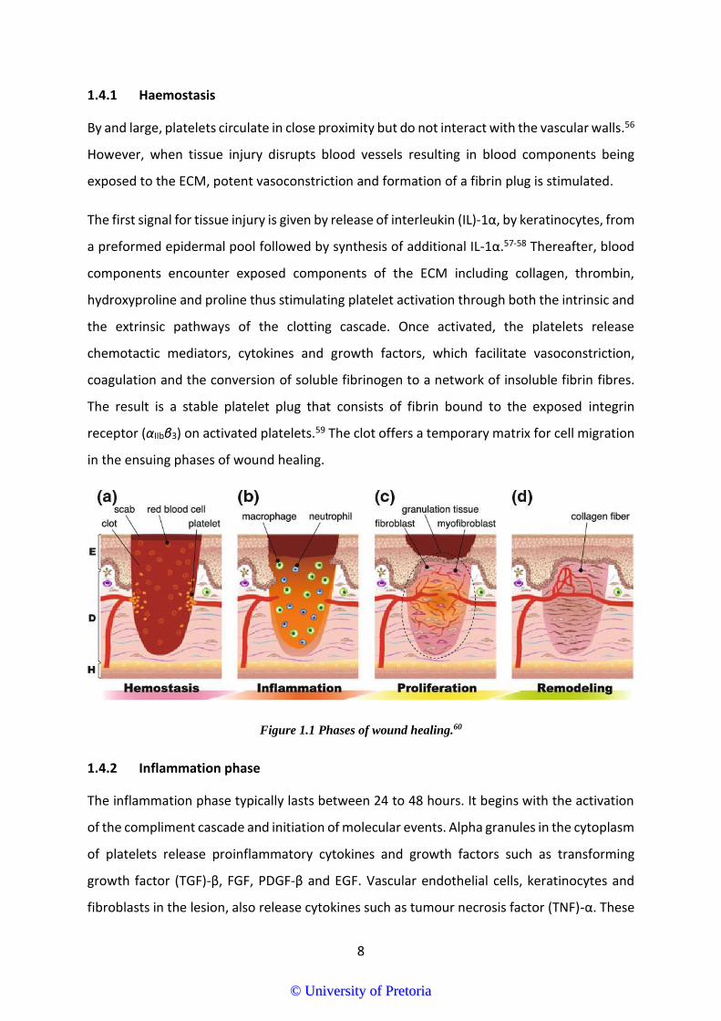
1.4.1 Haemostasis
By and large, platelets circulate in close proximity but do not interact with the vascular walls.56
However, when tissue injury disrupts blood vessels resulting in blood components being
exposed to the ECM, potent vasoconstriction and formation of a fibrin plug is stimulated.
The first signal for tissue injury is given by release of interleukin (IL)-1α, by keratinocytes, from
a preformed epidermal pool followed by synthesis of additional IL-1α.57-58 Thereafter, blood
components encounter exposed components of the ECM including collagen, thrombin,
hydroxyproline and proline thus stimulating platelet activation through both the intrinsic and
the extrinsic pathways of the clotting cascade. Once activated, the platelets release
chemotactic mediators, cytokines and growth factors, which facilitate vasoconstriction,
coagulation and the conversion of soluble fibrinogen to a network of insoluble fibrin fibres.
The result is a stable platelet plug that consists of fibrin bound to the exposed integrin
receptor (αIIbβ3) on activated platelets.59 The clot offers a temporary matrix for cell migration
in the ensuing phases of wound healing.
Figure 1.1 Phases of wound healing.60
1.4.2 Inflammation phase
The inflammation phase typically lasts between 24 to 48 hours. It begins with the activation
of the compliment cascade and initiation of molecular events. Alpha granules in the cytoplasm
of platelets release proinflammatory cytokines and growth factors such as transforming
growth factor (TGF)-β, FGF, PDGF-β and EGF. Vascular endothelial cells, keratinocytes and
fibroblasts in the lesion, also release cytokines such as tumour necrosis factor (TNF)-α. These
©© UUnniivveerrssiittyy ooff PPrreettoorriiaa

9
cytokines and growth factors promote chemotaxis of leukocytes to the site of injury
facilitating the elimination of debris, bacteria and damaged tissue.61 Ritsu et al, studied the
effect of suppressing TNF-α activity by using neutralising monoclonal antibodies (mAb). It
was evident that TNF-α is critical early in wound healing while neutralisation of TNF-α delayed
wound closure by interfering with fibroblast proliferation and the formation of new ECM.62
Neutrophils, which rid the wound area of invading microbes and cellular debris, are active
during the early stages of inflammation. Thereafter, monocytes infiltrate the wound site and
mature into macrophages in a process mediated by IL-8. Macrophages play various crucial
roles that include release of proinflammatory cytokines (IL-1, TNF, IL-6 etc.), resolution of
inflammation in the later stages of inflammation by removing apoptotic cells and sustaining
the proliferation of cells and restoration of tissue (Figure 1.2).61 Macrophages also release
prostaglandins, chemokines, leukotrienes, and complement which collectively induce
increased vascular permeability and recruitment of inflammatory cells.63 A study by Leivobich
and Ross investigating the role of macrophages in wound healing showed that macrophage
depleted wounds had defective repair with signs such as a severe lack of debridement and a
delay in fibrosis.64
©© UUnniivveerrssiittyy ooff PPrreettoorriiaa

10
Figure 1.2 The pattern of leukocyte infiltration into wounds.
Inflammatory cells are present at all stages of wound healing.65 The four most prominent types of
leukocytes in wounds (mast cells, neutrophils, macrophages and lymphocytes) are depicted with their
relative densities in each phase.
1.4.3 Proliferative phase
Once haemostasis and the immune system are effectively set, the proliferation phase,
mediated mainly by fibroblasts, keratinocytes and endothelial cells begins 3 - 5 days after
injury. The activity of these cells progressively modify the wound microenvironment from one
that is inflammatory to a synthesis-driven phase.66 Its onset is marked by the degranulation
of macrophages resulting in the release of soluble mediators (e.g. FGF, TGF-β, EGF, PDGF) that
activate and recruit endothelial cells for angiogenesis and fibrillogenesis.67 The essential
growth factor TGF-β stimulates matrix contraction68 by fibroblasts and conversion of
monocytes to macrophages. Fibroblasts and macrophages locally produce VEGF which
stimulates new tissue and blood vessel formation, with the blood vessels formed sustained
by VEGF.69 Fibroblasts and myofibroblasts also produce the major extracellular matrix (ECM)
components; collagen, glycosaminoglycans, fibronectin and proteoglycans which support the
formation of granulation tissue. Fibronectin, a large major glycoprotein is found throughout
all phases of wound healing; from the fibrin clot, the papillary dermis, to the newly
©© UUnniivveerrssiittyy ooff PPrreettoorriiaa

11
synthesised collagen in the granulation tissue.70-71 Being the primary constituent of the
provisional matrix, fibronectin provides migratory cells with a provisional matrix for collagen
deposition and myofibroblast driven wound contraction.71 This provisional matrix is
eventually replaced by a more mature matrix. Thus, prolonged expression of fibronectin in
chronic wounds might be a contributing factor to delayed healing. Re-epithelialisation begins
within hours with the migration of keratinocytes (stimulated by growth factors such as EGF
and TGF- β) and epidermal cells from the wound edge to the temporary matrix. This process
may last up to 3 months.
1.4.4 Tissue remodelling phase
The final phase of wound healing; the remodelling phase initiates within the third week after
injury and may continue for years. Fibroblasts play a key role in this phase with the production
of fibronectin, proteoglycans, hyaluronic acid and collagen all of which are essential for
cellular migration.
The fibrin clot from the haemostasis phase forms the premature composition of the
regenerated matrix. This is followed by a significant event in connective tissue healing which
is the differentiation of fibroblasts into myofibroblasts. Myofibroblasts then synthesise
glycosaminoglycans, proteoglycans, and other proteins which form a temporary framework
for the new matrix.72-73 Eventually, a stronger and more organised matrix made of collagen
replaces this temporary matrix.73
Collagen type III is remodelled to type I while the ECM is remodelled to bear a resemblance
to normal tissue (scar tissue). Apoptosis rids the healing wound of the cells that were
necessary in the repair process but are no longer needed. Collagen fibres realign and form
crosslinks resulting in greater tensile strength and less scarring. However, scar tissue only
attain up to only 80% tensile strength compared normal skin55, while excessive collagen
synthesis produces a hypertrophic scar or keloid.
©© UUnniivveerrssiittyy ooff PPrreettoorriiaa

12
1.5 Types of wound healing
1.5.1 Primary healing (healing by first intention)
Wounds that cause loss of a moderate amount of connective tissue cells and underlying
epithelial cells heal by first intention. These types of wound which are a result of e.g. a surgical
incision only cause focal damage to the skin and thus the wound edges are easily
approximated with staples, sutures or adhesive and within 12-24 hours of its creation, the
wound is closed. These wounds heal through re-epithelisation with minimum scar formation.
1.5.2 Secondary healing (healing by second intention)
Wounds that occur as result of major trauma causing extensive tissue loss (e.g. burns)
undergo secondary healing. Due to the great loss in tissue resulting in full-thickness wounds,
new granulation tissue ingrowth is necessary for restoration of the integrity of the skin.
Healing is first by contraction and then re-epithelisation.
1.5.3 Healing by tertiary intention
Dehisced or infected wounds are cleaned, debrided and observed for a prescribed period
(typically 4 - 5 days) to clear up any infection and allow new tissue growth before
approximating the edges.
1.6 Chronic wounds
Disruption of the complex healing process results in chronic wounds, with some wounds
recurring frequently and others progressing by involving new surrounding tissue. Mediators
such as inflammatory cells, growth factors and proteases influence the healing process while
exogenous factors such as disease, immunocompromised state, malnutrition, smoking and
radiation exposure also have a significant impact on wound healing.74 In contrast to acute
wounds, chronic wounds are not only unsuccessful in advancing through an orderly and
timely reparative process but also advance without creating a sustained anatomic and
functional result.22
Different aetiologies and the various and complicated pathophysiology of these wounds yield
poor or inappropriate management. However, it is commonly known that chronic wounds get
trapped in the inflammation phase that becomes a continuous cycle with mutual features
©© UUnniivveerrssiittyy ooff PPrreettoorriiaa

13
such as high levels of reactive oxygen species (ROS), repeated tissue injury, platelet derived
factors e.g. TGF-β, proinflammatory cytokines e.g. IL-1β and TNF, ECM fragments, proteases,
senescent cells, and in some cases a persistent infection. 75-78 These factors, although
impairing chronic wound healing; serve an important role in acute wound healing. (Figure 1.3)
Oxygen is essential for cellular respiration and subsequently production of adenosine
triphosphate (ATP) by cells; providing the high energy required for angiogenesis and
regeneration of damaged wound tissue. The role oxygen plays in wound healing depends on
whether the wound is in a hypoxic, normoxic or hyperoxic state. In general, the wound
oxygenation state influences collagen deposition, fibroplasia, angiogenesis, resistance to
infection, and epithelialisation. 79 The oxygenation state of wounds frequently guides some
treatment planning such as the decision to amputate a limb.80 It is important to note that
both high oxygen levels and too little may delay healing. Chronic and nonhealing wounds are
often hypoxic as a result of poor blood perfusion from sympathetically induced
vasoconstriction. The hypoxic environment created by the ischaemia and consumption of
oxygen by facultative bacteria is ideal for the proliferation of anaerobes.81 Adequate
vascularisation is vital for proper oxygenation and nutrient supply to the healing tissue in the
wound bed. Reduced blood supply to the wound aggravates it. PMNs, when in contact with
specific stimuli; produce ROS that are derived from oxygen e.g. superoxide anion (O2-•); that
are essential for defence against bacteria and other pathogens. 82-83 In fact, all wound healing
stages may be regulated by ROS. However, in chronic wounds there are numerous sources of
ROS, which may lead to elevated levels. Inflammatory cells accumulated inside the chronic
wound produce high levels of ROS.
These enhanced levels of ROS may result in oxidative stress, which further destroys the ECM
and prolongs the inflammatory phase gives rise to the nonhealing wound. High MMP levels
suppress angiogenesis and cell proliferation while aggravating the proteolytic process and
thus uncontrolled tissue degradation.
©© UUnniivveerrssiittyy ooff PPrreettoorriiaa

14
Figure 1.3 Cellular and molecular differences between acute and chronic wounds84
Acute wounds have a short-lived inflammatory response where granulation tissue is formed. Chronic wounds however, frequently present with stalled re-
epithelialisation because of infection and persistent inflammation. Elevated matrix metalloproteinases (MMPs) together with poor infiltration of blood vessels and
fibroblasts are a common occurrence. Open access article distributed under the terms of the Creative Commons Attribution License
(http://creativecommons.org/licenses/by/3.0).
©© UUnniivveerrssiittyy ooff PPrreettoorriiaa

15
Activated PMNs and macrophages are the leading sources of proinflammatory cytokines (e.g.
IL-1β) in wounds. The cytokines (TNF-α and IL-1β) are powerful mediators of various
inflammatory processes at all phases of wound healing.65 Wound macrophages also facilitate
the phagocytosis of neutrophils during the proliferative and remodelling phase. In the early
phases of healing, neutrophils facilitate effective decontamination and also produce a variety
of growth factors that could promote revascularisation and repair of injured tissue. However,
once decontamination is set, if neutrophils are not removed, they negatively influence repair,
as they are capable of destroying healthy ECM components such as clotting factors,
complement, immunoglobulins, and cytokines. Furthermore, neutrophils release
collagenases and their inhibitors which prevent the accumulation of a collagen-rich matrix.85
Delay in collagen deposition is also a common occurrence in DFUs where although collagen
gene expression is elevated, there is decreased collagen deposition in the wounds because of
the glycosylated collagen. Comparison of the changes in the ECM in DFUs and venous leg
ulcers (VLUs) shows that these chronic wounds have in common the loss of collagen and
elastin.86
Proteases hydrolyse the peptide bonds between amino acid residues in a polypeptide chain.
During proteolysis, excess ECM components are edited, the ECM structure remodelled, and
ECM assembly is regulated creating an equilibrium between ECM degradation and
deposition.87 Tissue metalloproteinase inhibitors (TMPI) inhibit and regulate protease activity
in acute wounds; however, in chronic wounds there is overexpression of proteases and down
regulation of TMPI85 resulting in ECM and growth factor degradation, suppression of
angiogenesis and cell proliferation while aggravating the proteolytic process and thus
uncontrolled tissue degradation.
The elderly are also known to have higher incidences of chronic wounds. With advancing age,
normal skin undergoes distinctive changes that have an effect on wound healing.88 There are
age-related alterations (Table 1.2) that influence wound healing during the human life span
as well as susceptibility to conditions such as diabetes and vascular disease which adversely
affect wound healing.88 In contrast with neonates who after an injury are able to regenerate
the lost tissue with minimum scarring, adult human wounds most often result in prolonged
healing and scar formation.89
©© UUnniivveerrssiittyy ooff PPrreettoorriiaa

16
Table 1.2 Cutaneous ECM changes that affect wound healing over the human life span.89-90
Age Properties
Foetal Highly regenerative skin
Large amount of cell mobility in a fragile ECM
ECM rich in collagen III and hyaluronic acid
Little inflammation in response to injury
Scarless healing
Juvenile Massive production of type I collagen
Moderate and transient inflammation
Cellular response in compliant ECM
Early adult Scarring properties at maximum
High production of type I collagen
Fibrotic response in stiff ECM
Aged adult Prolonged inflammation
High matrix metalloproteinase and elastase expression
Low expression of transforming growth factor beta
Weakened cellular response in an atrophic ECM
Aged skin is more prone to cellular senescence than younger skin. In vitro, most cells have a
finite life span before replication is arrested.91 These cells are characterised by enlargement
and spreading of the cells, an accumulation of lipofuscine, expression of senescence
associated β-galactosidase (SA-β-gal), and an increase in polynucleation.92 The above factors
amplify the severity of wounds and may result in inadequate vascularisation, a lengthy
inflammatory response, and a failure of re-epithelialisation.
The high significance of the ECM in wound healing is attributed to it being a major component
of the dermal skin layer particularly in wounds that have a significant loss of tissue and cannot
heal by primary intention. Scientific evidence has dismissed the notion that the ECM is only a
scaffold that offers passive structural support for cells.93 Some ECM interactions with cells are
critical for cell regulation, adhesion, motility, growth, differentiation and even ECM synthesis.
The mediation of ECM activities is regulated by the glycoprotein transmembrane receptors;
integrins. Integrins are cell surface glycoproteins that have α and β subunit types that
facilitate cell - ECM adhesion. In chronic wounds, there is defective ECM composition and
remodelling. The provisional fibronectin matrix formed during proliferation is replaced by a
©© UUnniivveerrssiittyy ooff PPrreettoorriiaa

17
more mature matrix in normal wound healing. Consequently, prolonged expression of
fibronectin in wounds might be a cause of delayed healing.
Prevalence of chronic wounds varies depending on diagnosis, year and country. As with other
diseases, it is worth noting that in developing countries, due to lack of proper nutrition and
medical care, chronic wounds have a higher prevalence.94 Statistics show that Canada has an
estimated 4-7% cases35; Germany: 1.03 - 1.05%36; China: 1.7%37 and 0.45% in the Indian
population.38
1.6.1 Types of chronic wounds
The American Wound healing Society (WHS) identifies four types of chronic wounds; DFUs,
VLUs, arterial insufficiency ulcers and pressure ulcers.89 Evidently, chronic wounds frequently
result from an underlying pathologic condition.95
1.6.1.1 Diabetic foot ulcers
The International Consensus on the Diabetic Foot defined the DFU as a ‘full-thickness wound
below the ankle, in a diabetic, regardless of the duration’.96 A purulent discharge, fever,
vasculopathy, neuropathy, foul smell, osteomyelitis, cellulitis, gangrene and crepitus are all
common in DFUs.97
Hyperglycaemia, a biochemical abnormality may accelerate neuropathy and vascular disease,
thus inducing vascular damage through any of four pathways: (1) enhanced polyol activity
leading to accumulation of fructose and sorbitol; (2) augmented glycation end product
formation; (3) nuclear factor ƘB and protein kinase C activation; and (4) increased hexosamine
pathway flux.98 The process of high production of superoxide by the mitochondrial electron-
transport chain activates all these harmful metabolic events. Thus, oxidative stress as a result
of hyperglycemia is a contributing factor to the pathogenesis of diabetic complications,99-100
including DFUs. Inflammatory cytokines and susceptibility to infection and other factors also
contribute to the pathophysiology of DFUs. An estimated 15% of diabetic patients form DFUs.
DFUs can be classified into three groups: neuropathic, ischaemic or a combination of the two
which is neuroischaemic.101 Ischaemia leads to a lack of oxygen and nutrients74, ultimately
tissue necrosis therefore a common risk factor for amputation.
©© UUnniivveerrssiittyy ooff PPrreettoorriiaa
18
Due to their complicated pathophysiology and slow healing, DFUs are associated with long
term disability and premature mortality.102
Neuropathy plays a significant role in the pathophysiology of most DFUs, with more than 60%
of DFUs estimated to occur as a result of underlying neuropathy.103 Neuropathy causes
muscle weakness and numbness which allow the foot to be subjected to abnormal loading
which the skin is unable to withstand because of diabetic ischaemia.104 The resultant callus
leads to further abnormal loading and oftentimes subcutaneous haemorrhage, which may
eventually become infected. The infection complicates both neuropathy and ischaemia, thus
increasing the risk of amputation of the limb.105
In order to control the diabetes epidemic and as a result effectively treat DFUs and reduce
the number of amputations, prevention and early intervention is critical. Once a foot ulcer
develops, the patient should be assessed for neuropathy and arterial blood supply to facilitate
healing and if infection is present, it should be treated appropriately. Management of diabetic
ulcers, includes debridement, restoration of arterial circulation to the limb to ensure
adequate oxygen and nutrient delivery to the ulcerated area, application of medication and
dressings, taking pressure off the area and administration of systemic antibiotics (if there is
an infection) and improving plasma glucose control.
1.6.1.2 Venous leg ulcers (VLU)
Although the pathophysiology of venous ulcers remains ambiguously defined, they are
characterised by venous incompetence and consequently venous hypertension,
dysregulation of various cytokines, excessive deposition of fibrin around capillary beds.106
With a 1% global prevalence, VLUs are the cause of up to 80% of the known leg ulcer cases.107
The most common risk factors include female sex, increase in age, trauma, obesity,
immobility, deep vein thrombosis and phlebitis.
Physical examination of the ulcers presents a shallow and irregular wound with the presence
of granulation tissue and fibrin at the base of the ulcer. The study of the cellular infiltrate and
ECM of chronic VLU and DFU compared to acute wounds showed that the chronic wounds
had lower numbers of CD4+ T cells, significantly higher B cells, plasma cells and macrophages
compared to acute wounds.108
©© UUnniivveerrssiittyy ooff PPrreettoorriiaa

19
Venous ulcers are managed by elevation of the affected limb, compression therapy to correct
impaired venous return, antibiotic treatment if necessary and most importantly constant
monitoring of the ulcer by wound specialists. As already mentioned, wounds that fail to heal
within four weeks are considered chronic. In the case of venous ulcers, it is recommended to
reassess their pathophysiology and treat them further with topical or systemic agents.
1.6.1.3 Arterial insufficiency ulcers
Arterial insufficiency ulcers, frequently known as ischaemic ulcers are commonly caused by
microangiopathy and macroangiopathy resulting in poor perfusion to the lower extremities.
Insufficient oxygen and nutrient supply if left untreated ultimately lead to tissue necrosis.
Characterised by deep wounds extending into the underlying tendons, these wounds are
commonly deficient of new tissue growth. They present as black, yellow, brown or grey
wounds that do not haemorrhage when debrided.
Like DFUs, arterial insufficiency ulcers of the lower limb increase the risk of limb loss. Like
other chronic ulcers arterial insufficiency ulcers can be treated by debridement, management
of infection, revascularisation and management of a moist wound environment.109
1.6.1.4 Pressure ulcers
The National Pressure Ulcer Advisory Panel defines chronic pressure ulcers (now known as
pressure injury) as localised injury to soft tissues usually occurring over a bony prominence as
a result of pressure and/or shear or friction.110 The common predisposing factors are impaired
mobility and exposure to undisturbed pressure for long periods. Patients, such as those with
spinal cord injuries or bed-ridden patients who are unable to change positions on their own
are at highest risk.
As with other chronic ulcers, management of these ulcers involves offloading, debridement
to remove necrotic tissue, infection management and maintenance of a moist wound
environment.
1.6.1.5 Tropical wounds
Tropical ulcers, also called phagedenic ulcer or malabar ulcer are unique to tropical and
subtropical areas. They are large, infected (polymicrobial infection) ulcers that develop on
skin predisposed to trauma (e.g. a leg with pre-existing sores, scratches or abrasions).
©© UUnniivveerrssiittyy ooff PPrreettoorriiaa

20
Communities with poor hygiene, malnutrition, underlying chronic diseases, low socio-
economic conditions, poverty and lack of protection from insect bites are more susceptible
to tropical wounds. 111
The wound presents usually as a solitary ulcer with raised edges, foul-smelling discharge and
oedema in the surrounding tissues. 112 It also features extensive necrosis of the skin,
subcutaneous tissue, gangrene of muscles, and even osteomyelitis. Spontaneous healing is
possible but in some cases the wounds are aggravated by superinfection with
Fusobacterium, spirochetes and/or anaerobic microorganisms 113 resulting in deep wounds
that penetrate into muscles, tendons, and bone. Infection control with systemic antibiotics is
frequently indicated for these ulcers.
1.7 Full-thickness wounds
Full-thickness wounds are a consequence of great damage to the skin resulting in deep
wounds that heal by simultaneous contraction and re-epithelialisation. They present with loss
of tissue extending below the dermis into the subcutaneous tissues (Figure 1.4).
The pathophysiology of full-thickness wounds is diverse, but the extent of tissue damage is
similar in these wounds. These wounds are deep and differ significantly from other wounds
in appearance, yet the mechanism of wound healing proceeds through the same phases of
healing viz haemostasis, inflammation, proliferation and tissue remodelling. 60
If not complicated by underlying disease, full-thickness wounds will heal naturally by
secondary intention.114 However, it is imperative to take into consideration the location of
the wound and the functionality of the tissue after healing. In areas where scarring could
reduce the functionality, repair of the damaged tissue is a necessary intervention to minimise
scarring and unwanted effects.
Deeper wounds re-epithelialise slowly and frequently require autologous skin grafting to
prevent extensive scarring that results in reduced cosmetic and functional outcomes. The first
line of treatment of these deep wounds includes the use of epidermal sheets or split-skin
mesh grafts. However, suitable autologous skin grafts may not always be available, thus
laboratory engineered skin substitutes have become a viable alternative.
©© UUnniivveerrssiittyy ooff PPrreettoorriiaa

21
Figure 1.4 Wound classification according to the depth of the wound in relation to skin layers.115
Full-thickness wounds extend deeper than the epidermis and dermis right into the subcutaneous fat and in some cases up to the bone. They present as deep wounds
with excessive tissue loss.
©© UUnniivveerrssiittyy ooff PPrreettoorriiaa

22
1.8 Keloids and hypertrophic scars
Scars are anticipated consequences of wound healing. Conversely, some wounds undergo
excessive/dysfunctional scarring which results in the formation of keloids or hypertrophic
scars. This is a consequence of excessive synthesis of connective tissue and excessive
angiogenesis during the wound healing process. In mammals, the most important outcome
of wound healing is restoration of the skin’s protective and functional integrity, as rapidly as
possible, rather than restoration of complete biological function116 often resulting in scar
tissue that lacks sweat and sebaceous glands, hair and melanocytes.116
Histopathologically, the differentiation between hypertrophic scars and keloids is difficult.
While normal skin contains distinct collagen bundles that run parallel to the epidermis,
collagen bundles in hypertrophic scars are flatter, less demarcated, and are arranged in a
wavy pattern, although, still orientated to the epithelial surface. In keloids, collagen bundles
are virtually non-existent, and the fibres lie in loose, haphazardly orientated sheets.
The management of wounds takes into account the outcome that comes from the use of the
treatment option selected in relation to the specific wound treated. An example, as
mentioned in Section 1.7, is the requirement of autologous skin grafting for extensive full-
thickness wounds to prevent scarring that results from contraction over a large surface. The
outcome-based approach of wound healing could decrease the potential for pathological
scarring that could require further interventions.
1.9 Wound dressings
1.9.1 The history of wound healing
It is said that the history of wound healing is as old as history itself.117 Beginning with the
caveman who tended to his wounds acquired in battle; to the ancient Egyptians, Greeks,
Romans and other ancient civilizations who used magical incantations, ointments and
potions; wound healing evolved to a methodical script of wound care and surgery from
Hippocrates and Celsus.118 It is notable that some of the principles of wound healing applied
prehistorically are still useful today.
The earliest recorded manuscripts related to wounds were papyri attributed to the ancient
Egyptians from around 3000 BC; where honey was recorded as a wound healing agent. Other
©© UUnniivveerrssiittyy ooff PPrreettoorriiaa

23
records are the clay tablets discovered in Mesopotamia; dating back to about 2500 BC. The
Edwin Smith papyrus, dated about 1700 BC, unquestionably is one of the most significant
known ‘surviving scientific treatise’.119 Forty-eight wounds or trauma cases and their
treatment were described in descending anatomical order. The Egyptians also used bandages
made of linen with texture varying from the finest silk-like gauze to a canvas-like coarseness
to cover and keep in place any medications applied.120 In addition to being masters in applying
and arranging bandages, the ancient Egyptians recognised the basic signs of infection and
inflammation. The Ebers papyrus, also known as the most famous plant medicine
“encyclopaedia” of ancient Egypt described and recommended several herbal remedies like
Aloe vera and acacia which were used to treat skin diseases.121
The Greeks stressed the importance of handling wounds hygienically. They used boiled clean
water, wine or vinegar (acetic acid) for cleaning the wounds. It was however only in the 19th
century that the surgeon, Joseph Lister stressed the importance of antiseptic treatment and
prevention of infection.122 This was after he observed that pyaemia, erysipelas and gangrene
were prevalent in open wounds of patients in some parts of the hospital building, especially
those on the ground floor. At some point, the mortality in the male accident ward on the
ground floor was so excessive that they shut down the ward and investigations led to the
finding of a foul drain. In contrast to this, Lister showed that where the ‘antiseptic system’
was used, no cases of erysipelas, pysemia or hospital gangrene reported. 122
The Romans were the first to describe the four cardinal signs of inflammation—rubor, tumour,
calor, et dolor (redness, swelling, heat, and pain). 123 It is notable that some of the principles
of wound healing applied prehistorically are still useful today.
1.9.2 Wound dressings; current trends
With over 6000 wound dressings currently available, the wound dressing market has shown
considerable progress. However, only a few dressings have all the characteristics of an ideal
dressing. It is important to use the wound environment as a guide to selecting a suitable
wound dressing.124
As cited in Section 1.9.1, before the 1960s, wound dressings were considered to play a passive
role in wound healing125, creating a dry scab which functioned as a protective barrier against
©© UUnniivveerrssiittyy ooff PPrreettoorriiaa

24
bacterial contamination and also aided exudate absorption. Studies by Winter et al in the
domestic pig showed that a moist wound environment significantly augmented re-
epithelialisation rates when compared to wounds exposed to the air.27,126 These findings
were substantiated by Hinman and Maibach127 who later did a parallel study comparing air
exposed experimental split thickness wounds to wounds occluded with a polyethylene film in
humans. As expected, occluded wounds re-epithelialised significantly faster than air exposed
wounds.127
A moist wound environment ensures: improved interaction of growth factors and their target
cells, accelerated angiogenesis, inhibits tissue dehydration and cell death, improves
breakdown of dead tissue, and provides a significant reduction in pain.128 The belief that the
moist wound environment provided a medium for bacterial growth, was dismissed by a
review of over 100 studies comparing wound infection in occlusive with non-occlusive
dressings. 129 The results showed the overall infection rate under conventional dressings was
7.1% compared to 2.6% in the wounds under occlusive dressings. 129 These findings heralded
the development of modern wound dressings.
Progression of research has brought an in-depth understanding of the underlying cellular and
molecular mechanisms of wound healing, thus wound dressings have advanced from being
passive to active material that manipulates the wound environment into an optimum milieu
conducive for healing.130 However, 50% of the wound dressing market is still accounted for
by traditional dressings such as bandages, woven and non-woven sponges, etc.131 with some
of them modified and/or medicated. Various strategies are currently employed for enhancing
the healing of wounds, ranging from skin grafts, to the inclusion of growth factors and gene
delivery as well as cell therapy. Modern wound healing devices are continually progressing.
1.9.2.1 Gauze
Gauze is a timeless wound dressing material that encompasses woven or non-woven material.
It was used for several wound types; from the dry wound to packing bleeding and exuding
wounds. The common belief was that a dry wound environment was conducive for healing as
it supposedly stalled the growth of bacteria.132 Notwithstanding published evidence of the
benefits of a moist wound environment, gauze is still commonly used worldwide. Several
studies have even shown that modern dressings such as foam, hydrocolloids, etc. affording
©© UUnniivveerrssiittyy ooff PPrreettoorriiaa

25
superior healing, have lower infection rates, greater patient satisfaction and are overall more
cost effective compared to gauze, as they require less frequent changes.133 As cited above,
modern wound management takes the wound environment into consideration to determine
the suitability of the wound material selected. Traditional plain gauze dressings were known
to adhere to the wound bed and thus result in trauma and bleeding upon dressing
removal.132,134 Consequently, impregnated gauzes were then developed to provide low
adherence, antiseptic properties and moisture retention for the wound. The first modern low
adherence dressing to be made was ‘tulle gras’; developed in 1915 during World War I by the
inventor Lumiere. This was gauze fabric impregnated with paraffin and balsam of Peru.135 To
date, gauze has also been impregnated with iodine, zinc, or petrolatum to avoid desiccation
and afford nonadherence to the wound bed.136
Sood et al proposed three comprehensive wound dressing categories, each with a specific
purpose.134 The first interacting with the tissue itself, thus facilitating autolytic debridement.
Wound dressings that regulate moisture level of the wound bed are in the second category,
while the third category consists of dressings that control bacterial load.134 Although the
classes are sometimes not mutually exclusive, this classification is useful.
1.9.3 Wound dressings facilitating debridement
Foreign material in wounds promote an environment conducive for infection, extending the
inflammatory period, and subsequently delaying contraction and epithelialisation of the
wound.137 Key to wound management is the use of debridement to prepare the wound bed
for re-epithelialisation. Initially, wound care is aimed at reducing the incidence of foreign
material, bacterial load, and damaged or necrotic tissue.
For ages, many methods of debridement were used. Mechanical debridement using sharp
equipment has the disadvantage of lacking specificity for devitalised tissue. Surgical excision
has the disadvantage of the trauma it causes, and the risk associated with the use of
anaesthesia. Both surgical and mechanical debridement have increased risks of bleeding. A
few modern wound dressings were designed to promote autolytic debridement by creating
an environment conducive for proteases within the wound to liquefy necrotic tissue.
Typically, a process that promotes natural wound debridement by endogenous phagocytic
©© UUnniivveerrssiittyy ooff PPrreettoorriiaa

26
cells and proteolytic enzymes. The highly selective method of autolytic debridement is
indicated for necrotic, non-infected wounds.
Other wound dressings promote enzymatic debridement while biologic debridement using
maggots is used with success in some refractory chronic wounds. Conversely, one
debridement method may not be suitable/adequate for some wounds, e.g. infected wounds
may need antibiotics while some wounds may require surgical debridement. The clinician
who examines the wound determines the debridement method to use. Characteristics such
as patient commodities, time, resources and available skills all impact the debridement
method selected.138 In many cases, a suitably qualified clinician is not always available to
perform mechanical debridement, making autolytic/enzymatic debridement an important
alternative. 138
1.9.3.1 Hydrogels
A common debriding agent, hydrogel is a network of semisolid, hydrophilic polymers that are
chemically or physically cross-linked to give a soft contour following three-dimensional
structure.139 Available as either amorphous or sheet hydrogels; they keep the wound
environment moist140 while their hydrophilic properties allow them to absorb large volumes
of exudate without dissolving because of the crosslinks between their network chains.141
Research data comparing hydrogel dressings with other dressings shows that debridement
followed by use of hydrogel is more effective and less complicated compared to standard
wound care. Dumville and colleagues showed that hydrogel dressings heal lower grade DFUs
more effectively than basic wound contact dressings.142 However, comparison of
debridement efficacy of collagenase vs. hydrogels shows that enzymatic debridement is
superior to hydrogels in the debriding efficacy138 Hydrogel dressings are important for
promoting a moist wound environment in dry wounds and helps remove excess moisture in
weeping wounds.
1.9.3.2 Hydrocolloid dressings
A combination of pectin, carboxymethylcellulose and gelatine143 (e.g. Granuflex®),
hydrocolloid dressings are widely used to absorb relatively large volumes of exudate and
facilitate autolytic debridement 144 in sloughy wounds. However, there have been higher
incidents of infection with their use, with Foster and colleagues even concluding that
©© UUnniivveerrssiittyy ooff PPrreettoorriiaa

27
hydrocolloid dressings should never be used on deep, infected, exudating diabetic wounds.145
A podiatry study at the University of Huddersfield, concludes that the use of hydrocolloid
dressings is controversial146 while a review by Dumville and colleagues presented no research
evidence proving that hydrocolloid wound dressings are superior to other types of dressing
in healing DFUs.147 Besides autolytic debridement, other advantages of hydrocolloid dressings
include soothing of pain; autolytic debridement encouraged by the gel, the dressing can be
retained on the wound for up to a week and easy removal without causing wound trauma.
1.9.3.3 Enzymatic dressings
Collagenase and proteolytic enzyme preparations are significant instruments in debridement
of wounds as they break down non-viable tissue. Collagenase is specific for collagen and
elastin and thus will not digest fibrin. It has been used for decades for enzymatic
debridement.148 These enzymes ultimately result in collagen fragments that are chemotactic
for fibroblasts and macrophages. In addition to faster debridement, Palmieri et al’s study
showed that enzymatic debridement using Iruxol mono® (a collagenase wound ointment)
gave a quicker reduction in inflammation, enhanced granulation tissue formation and
improved epithelisation compared to the placebo dressing that comprised of liquid paraffin
and white soft paraffin.149 Even when compared to hydrogel, collagenase’s significant
debridement efficacy was apparent.138
1.9.3.4 Maggot wound therapy
Biological debridement using live, sterile larvae of the green fly (Lucilia sericata) is effectively
employed in large wounds where painless debridement is required. Currently regulated and
approved by the US Food and Drug Administration (FDA) since 2004150, the use of maggots in
wound healing is effective for necrotic wounds such as diabetic ulcers151, pressure ulcers152,
venous ulcers153 and postoperative wounds. Maggot wound therapy (MWT) significantly
reduces treatment costs, healing times154 and increases granulation tissue proliferation, while
eradicating methicillin-resistant Staphylococcus aureus in infected wounds.155
The mechanism of action includes direct ingestion of necrotic tissue by maggots, leaving
healthy tissue that promotes proliferation of granulation tissue. Maggots seem to indirectly
stimulate healing by secreting protease inhibitor resistant digestive enzymes
©© UUnniivveerrssiittyy ooff PPrreettoorriiaa

28
(chymotrypsin 1) which contain serine proteases, collagenase, leucine aminopeptidase and
carboxypeptidase enzymes.156
Maggot therapy is a highly effective debridement technique; however, it does not replace
surgical treatment of large wounds, those that require amputation and those with large
amounts of necrotic tissue.156
1.9.4 Wound dressings regulating moisture levels
Winter’s studies showed that a moist environment is pivotal to wound healing as it increases
the rate of epithelialisation.27,126 Most modern wound dressings were developed to promote
a moist wound environment in addition to other factors of wound healing, e.g. hydrogels and
enzymatic dressings provide moisture while promoting wound debridement.
Wound exudate is a liquid containing high amounts of protein, nutrients, electrolytes and
inflammatory cells. Increased permeability and blood vessel dilatation as a result of the
inflammatory response leads to high levels of exudate. In acute wounds, wound exudate is a
crucial constituent of the reparative process.157 However, wound exudate is often believed to
negatively impact wound healing. Deeper understanding of the constituents of exudate,
shows that an exuding wound at optimum levels provides the wound with moisture,
proteinases for autolytic debridement, tissue inhibitors of matrix metalloproteinases (TIMP)
to regulate the autolytic debridement and cytokines.157 High amounts of wound exudate
however lead to maceration of surrounding tissues further impact the wound negatively.
Thus, regulating the amount of exudate in a wound is key to effective wound management.
1.9.4.1 Foam dressings
Foam dressings are best suited for moderate to highly exuding wounds to facilitate faster
healing.135 They induce a moist wound environment, provide thermal insulation, have
increased absorbency, promote gaseous exchange and increase hyaluronan synthesis in the
epidermis which also promotes healing.158 Commonly made from polyurethane or silicone,
foam dressings can be used alone or in combination with an outer layer that acts as an
antibacterial layer.
A review on foam dressings, showed that foam dressings do not have an advantage over other
dressings in the healing of DFUs.142 However, after consulting with several wound care
©© UUnniivveerrssiittyy ooff PPrreettoorriiaa

29
specialists, this study found that foam dressings were a dressing of choice for most full-
thickness wounds because of their ability to absorb high quantities of exudate and to conform
to the different wound shapes and aid in offloading pressure. They are suitable during the
inflammatory phase especially after debridement. Foam dressings however, if left on the
wound too long, can cause maceration of the wound due to absorbing large amounts of
exudate, thus they need to be changed frequently.
1.9.4.2 Alginates
Alginates are gel-forming polysaccharides. Their mechanism of action involves absorbing
exudate leading to the formation of a moist gel on the surface of the wound that facilitates
healing of the wound. They are highly absorbent and can be packed into the wound easily;
therefore, they are normally used as a primary layer on the surface of the wound, under a
more absorbent dressing. Although these dressings are very effective for heavily exuding
wounds,159 there is no research evidence suggesting that they are more effective in healing
chronic ulcers than other types of dressing.
1.9.5 Wound dressings controlling bacterial load
Wound dressings loaded with antimicrobial agents such as silver, ciprofloxacin,160 zinc
oxide161 and mupirocin are viable alternatives to diminish wound bacterial colonisation and
infection. Other therapies such as honey and silver sulphadiazine are already extensively
used.
1.9.5.1 Honey
Since the beginning of time, honey has been known to have antimicrobial properties.
Substantial evidence, both in vitro and in vivo has supported the positive wound healing
effects of honey. With potent antibacterial activity, honey provides a moist wound
environment favouring autolytic debridement. Furthermore, its viscosity protects the wound
from additional bacterial colonisation. 162 Its antibacterial activity is even effective against
multi-drug resistant bacterial such as S. aureus, Acinetobacter baumanni, S. typhi, E. coli and
P. aeruginosa.163
©© UUnniivveerrssiittyy ooff PPrreettoorriiaa

30
Initially the limitation to the use of honey was licencing; however, there are many licenced
sterile honey formulations now available for use.162 Melladerm®, a modern honey gel, was
found to heal wounds 26% faster than other high tech dressings.164
1.9.5.2 Silver
Silver is widely used for its broad antibacterial properties. Most commonly incorporated in
creams e.g. Flamazine (10mg/ml silver sulphadiazine), these dressings are favoured for burn
wounds. In vitro assessments of the activity of silver sulphadiazine against 409 bacterial
strains showed activity against multi-resistant bacterial species such as methicillin-resistant
S. aureus and Acinetobacter spp.165 Slow release silver has recently been incorporated into
wound dressings like actisorbTM and acticoatTM.166
1.9.5.3 Mupirocin
Mupirocin (pseudomonic acid A), first isolated in 1971 from Pseudomonas fluorescens, is a
potent topical bactericidal agent with in vitro activity against gram-positive bacteria that
cause skin infections. Gram-negative bacterial are generally resistant to mupirocin. 167
Commonly marketed as Bactroban®, its mechanism of action involves inhibition of isoleucyl-
transfer RNA synthetase, thus inhibition of RNA synthesis and bactericidal protein
synthesis.167
Initially, mupirocin had activity against methicillin-resistant S. aureus; however, recent
evidence has shown increasing bacterial resistance especially with repeated use.168
1.10 Skin grafts
Skin grafts are indicated for deep wounds with extensive tissue loss. The first skin graft was
performed by Jaques-Louis Reverdin in 1869.169 Since then, this technique has undergone
significant development including the use of fibrin glue/tissue adhesives to approximate the
edges.
The skin grafting procedure generally involves obtaining autologous skin from a donor site
with minimum damage. After harvesting and transferring to the desired location, grafts do
not have their own nutrient and blood supply and as a result, depend fully on the wound bed’s
nutrient and blood supply via absorption and diffusion respectively. Within the few
subsequent days after placement, an intact vascular supply growing from inside the bed is
©© UUnniivveerrssiittyy ooff PPrreettoorriiaa

31
normally established.114 Although grafts are frequently used to prevent the contracture
observed in secondary intention healing, they do however demonstrate contracture although
to a lesser extent. 114
Skin grafting causes considerable pain for both the injury site and the donor site. Biomaterials
are thus a desirable alternative for wound management in wounds with excessive skin loss.
1.11 Characteristics of the ideal wound dressing
No single dressing addresses all the ideal properties for optimal wound healing. Physical
protection from external factors and conditions are a common feature of wound dressing
materials. The ideal dressing should maintain some moisture, remove excess exudate, be
impermeable to bacteria, should not delay healing, be free of particles or any contaminants,
be non-allergenic and non-toxic, protect the wound from further trauma, thermally insulate
the wound, allow gaseous exchange, be comfortable and also conformable, allow changing
without causing further trauma and be able to release pharmacological agents to the
wound.145
Not one single dressing has all these characteristics. Oftentimes a combination of wound
dressings is used at the different stages of the wound healing cascade.
1.12 pH and wound healing
The pH of wounds which is a value expressing the negative logarithmic scale for the
concentration of H+-ions in the interstitial environment of wounds, has both a direct and
indirect influence on all biochemical reactions taking place. The pH value is therefore an
important factor for metabolism during wound healing and thus a significant parameter for
therapeutic interventions in wound-care.170 Healthy skin under normal conditions has a pH
ranging between 4 and 6 which plays a critical role in the skin’s barrier function. Different pH
values are recorded for different stages of healing. In chronic wounds, the pH changes from
alkaline to acidic as the wound heals.
For wound dressings, an acidic pH has been shown to have antibacterial and antifungal
properties. A study by Basavraj et al showed that an acidic wound milieu resulting from the
use of acidic wound dressings such as acetic acid and citric acid helps in wound healing by
©© UUnniivveerrssiittyy ooff PPrreettoorriiaa

32
increasing antimicrobial activity, releasing oxygen, controlling wound infection, altering
protease activity, reducing toxicity of bacterial end products, and enhancing epithelialization
and angiogenesis.171 It follows that wounds with a high pH heal slower than wounds with a
slight acidic environment and that the pH of chronic wounds decreases as the wound heals.
These and other findings with regards to pH of wounds, should guide treatment selection.
1.13 Biomaterials as wound dressing material
The first ever biomaterial recorded in history were sutures used by the ancient Greeks,
Indians, Romans and the Egyptians. The material used ranged from silk, hair, wool threads,
tendons to plant fibres and later the discovery of the absorbable catgut in the 1960s.
Over the past three decades, a number of investigations have been done on wound healing
material and technology with biomaterials emerging as excellent material for wound healing.
Biomaterials are biocompatible, biodegradable, non-toxic and bioactive. There are several
commercially available wound dressing biomaterials made from natural or synthetic polymers
or a mixture of both.172 These materials include tissue-engineered products such as
collagen173, chitosan174, sericin173, starch, alginates8, hyaluronic acid and many others.
Chitosan is the most extensively used due to its biocompatibility and biodegradability and
almost unlimited supply of medicinal quality starting material.
Chitosan is structurally similar to hyaluronic acid (Figure 1.5) which is a common ECM
component.175 Hyaluronic acid, a glycosaminoglycan plays a vital role in the structure and
organisation of the ECM.176 Prominence of this polysaccharide is seen whenever prompt
proliferation, regeneration, and repair of tissues is needed. 176
1.13.1 Chitin and chitosan
In 1811, Henri Braconnot, a Frenchman who was director of the national botanical garden at
that time, discovered a material in mushroom that would not dissolve in sulphuric acid. 177 He
named this material ‘fungine’. A few years later the term ‘chitin’ was coined after similar
material was isolated from beetle cuticles by another French scientist.
Chitin forms structural components of the exoskeletons of fungi and arthropods and naturally
presents itself as an organised crystalline. Chitin essentially means ‘the skeletal material of
invertebrates’. An insoluble, inelastic, hard, white linear polysaccharide occurring naturally in
©© UUnniivveerrssiittyy ooff PPrreettoorriiaa

33
the crystalline state, chitin is composed of β-(1→4)-linked N-acetyl-D-glucosamine (d-GlcNAc)
residues. The chitin chains are organised in sheets which are tightly bonded by a number of
intra-sheet and inter-sheet hydrogen bonds178, giving a highly hydrophobic semi-crystalline
material that is insoluble in aqueous acetic acid and most organic solvents.179
The chitin structure closely resembles cellulose (Figure 1.5), with chitin having the acetamide
group (-NHCOCH3) in place of the hydroxyl group (-OH) at the C2 position. Like cellulose, chitin
has low chemical reactivity and is not freely soluble in water or organic solvents. It is the
deacetylated derivative; chitosan that is used abundantly for biomedical applications.
©© UUnniivveerrssiittyy ooff PPrreettoorriiaa

34
n
nCellulose
n
Chitosan
Chitin
n
Hyaluronic acid
O
O
OH
CH2OHO
CH2OH
OH
O
O
O
OH
CH2OH
CH3COHN
OOH
CH2OH
NHCOCH 3
O
O
O
OH
NH2
CH2OH
O
CH2OH
NHCOCH3
OH
O
O
CH2OH
O
OH
OH
COOH
O
OHNHCOCH 3
O
Figure 1.5 Structures of cellulose, chitin, chitosan and hyaluronic acid
©© UUnniivveerrssiittyy ooff PPrreettoorriiaa

35
Despite its wide abundance, chitin is mostly collected from the shrimp and crab canning
industry and processing involves demineralisation followed by deproteinization,
decolourisation and deacetylation180 (Figure 1.6). The natural source and biological function
of chitin determines its structural form; which is either an α, β or γ allomorph that can be
characterised by infrared and solid-state NMR spectroscopy, and X-ray diffraction.181-182 The
most stable and abundant form is α-chitin which is found in nematode and rotifer egg shells,
arthropod cuticles and hydrozoan calyces. β-chitin occurs in cuttlefish bone, the squid pen,
mollusc and brachiopod shells and the pogonophora tubes.
Crustacean shells Washing and grinding Demineralisation
Figure 1.6 Chemical production of chitosan from chitin.180
Chitosan exists naturally in some microorganisms and in the cell wall of some fungi
(Mucoraceae). However, commercial chitosan is produced from the partial deacetylation of
chitin to chitosan through alkali N-deacetylation183-184, enzymatic hydrolysis in the presence
of a chitin deacetylase or by steam explosion. Chitosan is a copolymer of N-acetylglucosamine
and D-glucosamine coupled through β-(1→4) glycosidic bonds, which when depolymerised
gives a compound with lower molecular weight (MW)185 and lower viscosity. It is also defined
as a group of deacetylated chitins with an acetylation value of < 40% and nitrogen content >
7%.186 The deacetylation of chitin into chitosan occurs in a highly concentrated base (NaOH)
at high temperatures. Despite these harsh conditions, chitosan remains a semi-crystalline
polymer.
HCl CaCO3
NaOH
Protein
Deproteinization Bleaching Washing and drying
Chitin Deacetylation
Washing and drying Chitosan
NaOH N-acetyl groups
©© UUnniivveerrssiittyy ooff PPrreettoorriiaa

36
Chitosan’s NH2 and OH groups result in a compound with high chemical reactivity; thus, the
versatility of chitosan depends on its degree of deacetylation. Chitosan contains amino groups
which strongly complex various species, such as metal ions, and are often used for waste
water treatment, to remove heavy metal contaminants.178 Several reports have mentioned
chitosan’s mucoadhesive187, haemostatic188, antifungal and antibacterial properties. Its
excellent mucoadhesive properties result in chitosan being widely used in developing drug
delivery systems. The mechanism for chitosan mucoadhesion as explained by Sogias et al
appears to be through electrostatic attraction, hydrogen bonding and hydrophobic effects.187
Other scholars reported that chitosan’s positively charged amino groups and the negatively
charged moieties of the mucus gel layer interact via hydrogen bonds and ionic interactions.189
1.13.1.1 Deacetylation degree of chitosan
The deacetylation degree (DD) is defined as the ratio between acetylated (GluNAc) and non-
acetylated (GluN) glucosamine units. It is a process that involves nucleophilic substitution at
the primary acetamido group of chitin, resulting in chitosan that has free amino groups (-NH2)
with deacetylation ranges from 40 to 98%.190
The DD is an important factor that determines various biological and physicochemical
properties of chitosan, e.g. toxicity191, degradation, crystallinity, solubility and hydrophilicity.
Chitosans with higher DD have more positively charged amine groups when dissolved in acid
solution compared to chitosans with a lower DD192 and this has a direct effect on the biological
activity of the chitosan. Schipper and colleagues compared eight chitosans with varying DD
and MWs. Their results showed that chitosans with low DD displayed a clear dose-dependent
toxicity, while those with high DD showed low toxicity.191 Other studies indicated that the
radical scavenging activities of chitosans,193 and the effect on fibroblast proliferation and
collagenase activity194 depend on the degree of deacetylation and concentration.193
Prasitsilp and colleagues’ study showed that their high DD chitosans regardless of the source
(shrimp or cuttlefish) supported adhesion and proliferation of two different cell-lines (L929
and BHK21) under investigation; while the lower DD chitosans did not favour this
attachment of these cells.195 It is thus important to accurately determine the DD of each
chitosan sample used.
©© UUnniivveerrssiittyy ooff PPrreettoorriiaa

37
Various methods196 are used to measure the DD of chitosan including: IR spectroscopy, UV
spectrophotometry, X-ray diffraction, colloidal titration, potentiometric titration, circular
dichroism (CD), ninhydrin assay, NMR spectroscopy, elemental analysis, gel permeation
chromatography, conductimetry and N-acetyl group hydrolysis.196 Most of the methods
mentioned above are too costly for routine analysis, time consuming or destructive to the
sample. However, the 1H NMR spectroscopy has proven to give more accurate results with
the use of less resources.197
1.13.1.2 Wound healing properties of chitosan
Chitosan derivatives in the form of gels, sponges, powders, scaffolds, nanoparticles or films
have been used successfully to accelerate wound healing.198 Ranging from hydrogels to
scaffolds/membranes, there are various kinds of chitosan wound dressings available
commercially.199-200 Chitosan promotes wound healing using different mechanisms (Figure
1.7) at different stages of the wound healing process.
Figure 1.7 Schematic representation of the properties of a chitosan wound dressing material.201
©© UUnniivveerrssiittyy ooff PPrreettoorriiaa

38
Previous studies have shown that chitosan based dressings can expedite contraction of
wounds, accelerate healing of different tissues,202 accelerate the infiltration of the PMNs,
activate migration and proliferation of fibroblasts203, promote granulation tissue formation,
stimulate production of cytokines204 and attenuate production of proinflammatory cytokines
such as IL-1β and TNF-α formation but increases the concentration of the anti-inflammatory
cytokine; IL-10.205
Haemostasis
Considerable research data shows the haemostatic effect of chitosan in a normal
environment206 and in the absence of platelets or coagulation factors.207 While some scholars
have proven that chitin and chitosan both enhance blood coagulation188, He and his
colleagues’ study showed that chitosan’s positive charge retarded blood coagulation on
chitosan films.208 They further proved that chitosan’s positive charge resulted in inhibited
activation of the contact system.208 However, the exact mechanism by which chitosan causes
haemostasis is not fully elucidated. The haemostatic efficacy is highly influenced by
physicochemical properties like DD, MW, polymeric structure, cationic properties and
intrinsic viscosity. Both high viscosity and a high degree of deacetylation increase chitosan’s
haemostatic efficiency.209 It is suggested that the haemostatic effect of chitosan is mainly due
to the agglutination of the red blood cell component rather than interaction with the normal
clotting cascade.210 The cationic chitosan interacts with the negatively charged molecules at
the cell surface in its agglutination.210
Janvikul et al showed a significant reduction in the whole blood clotting time (WHBCT) with
the use of either chitin and chitosan, while only chitin significantly shortened platelet
recalcification time.211 Rao and Sharma also showed that chitosan brought down the WHBCT
by up to 40 % of the normal. They proceeded to study the effectiveness of the interaction
between red blood cells and chitosan and concluded that even small amounts of chitosan can
cause agglutination of erythrocytes from any species.210 In another study, chitin aggregated
platelets more than chitosan. The conclusion was therefore made that platelet aggregation
did not necessarily reflect blood coagulation.188
Thus, there is sufficient evidence from literature supporting the use of chitosan as a
haemostat.209,212 The FDA approved HemConTM chitosan dressing when used in pre-hospital
©© UUnniivveerrssiittyy ooff PPrreettoorriiaa

39
combat patients; successfully arrested bleeding in > 90% of cases.213 Other haemostatic
chitosan wound dressings already approved by the FDA for external use include TraumaStat
and Celox dressings.214 TraumaStat, a silica and chitosan based dressing is proven to have
superior haemostatic properties than HemConTM .215
Inflammation
A study investigating the effects of chitosan on wound healing in dogs showed accelerated
migration of macrophages and severe infiltration of PMNs on the 3rd day.203 Inan and Saraydin
also showed that during the early phases of wound healing, chitosan stimulates inflammatory
cells and the immune-localisation of the growth factor; VEGF and the receptor; FGFR3.216
In another study however, evaluation with the enzyme-linked immunosorbent assay (ELISA)
showed that water soluble chitosan oligosaccharides with MW < 5 kDa had the ability to
reduce inflammation by inhibiting the secretion of proinflammatory cytokines; IL-6 and TNF-
α.217
Proliferation
Ueno and colleagues showed that from the 9th to the 15th day after treatment of wounds with
a chitosan based dressing; there was a great amount of collagen deposition indicating that
chitosan promotes granulation tissue formation. 203 In another study, chitosan stimulated
fibroblast proliferation in vitro at a rate dependent on the DD. 218 However, in the same study,
the higher DD (89%) chitosan inhibited keratinocyte proliferation compared to the lower DD
(37%) which had no effect on them .218 This proliferation of fibroblasts induced angiogenesis
and collagen formation in the wound area.218 These findings were consistent with Prasitsilp
et al whose study exhibited high DD chitosans regardless of the source (shrimp or cuttlefish)
as supportive of cell adhesion and hence proliferation of two different cell-lines (L929 and
BHK21) under investigation; while the lower DD chitosans did not favour this attachment of
these cells.195 Another study showed an increased synthesis of type III collagen with the use
of chitosan.203 In yet another study, Zhou et al prepared biocompatible carboxyethyl
chitosan/poly (vinyl alcohol) nanofiber wound dressings through electrospinning of aqueous
carboxyethyl chitosan/poly(vinyl alcohol) solution. Cell culture results showed that these
fibrous mats successfully promoted attachment and proliferation of L929 mouse fibroblasts
without any cytotoxicity towards these cells.219
©© UUnniivveerrssiittyy ooff PPrreettoorriiaa

40
Tissue remodelling
Scar formation is a clinical feature of the remodelling phase of wound healing. A balance
between collagen deposition and degradation or collagen remodelling from type III to type I
is of paramount importance as it has a bearing on the tensile strength of the scar. As
mentioned in Section 1.8, excessive collagen deposition results in hypertrophic scars or the
formation of keloids.220 While chitosan is known to induce collagen synthesis during the early
phases of wound healing, further investigations on chitosan’s role in the remodelling phase
are ongoing.
A study by Wang and colleagues showed superior inhibition of scar formation in wounds
treated with chitosan than those treated with hyaluronate while those treated with saline
solution and free fat did have some scar formation.221 Consistent with these findings, research
data from Hilmi et al demonstrated that use of a skin substitute derived from chitosan in
irradiated wounds yields significant tissue-engineered skin with significantly smaller scar sizes
when compared to the biomaterial Duoderm® CGFTM.222
Stone’s study did not show a difference between wound size reduction of wounds treated
with chitosan compared to their gold standard wound dressing (Kaltostat®). However,
another important dimension of wound healing was shown; the colour of the scar site treated
with chitosan showed an earlier return to normal skin colour compared to the site treated
with the Kaltostat® control.223
In an attempt to explain the mechanism of action of inhibition or reduction in scar formation
Lv et al cultured hypertrophic scar fibroblasts (HSF), keloid fibroblasts (KF) and normal dermal
fibroblasts (NDF) in the presence or absence of different doses of chitosan.224 They found that
to a small extent chitosan exhibited proliferation-inhibiting effect on NDF while inhibiting
proliferation of HSF and KF in a dose-dependent manner. Their findings further demonstrated
that chitosan could significantly (p < 0.01) suppress the expression of type I and III procollagen
in both HSF and KF.224
Chitosan is also said to play a role in decreasing wound fibrosis225 thereby, decreasing the
chances of hypertrophic or dysfunctional scarring.
©© UUnniivveerrssiittyy ooff PPrreettoorriiaa

41
1.13.2 Modified chitosan-based materials in wound healing applications
1.13.2.1 Modification to enhance solubility of chitosan
The solubility of chitosan is influenced by its DD, the distribution of free amino and N-acetyl
groups along the polymer chain, MW, pH and temperature of the solvent.178 Chitosan being
a weak base is completely soluble in aqueous acidic media which come with several
complications such as toxicity, chain hydrolysis and the removal of residual solvents.226
Various studies to modify chitosan to improve its solubility in organic solvents179 and
water179,186,227 are reported. Crustacean chitosan, carboxylated in equimolar ratio glyoxylic
acid: amino groups, produced the water soluble anionic derivative; carboxymethylchitosan
(CMC).228 Sometimes CMC is formed from chitosan’s reaction with monocholoroacetic acid in
alkaline medium.16 A porous, physically stable, self-sustaining CMC membrane was prepared
using the natural crosslinker, genipin. CMC with high MW permitted the preparation of a
lightly cross-linked membrane which showed improved swelling capacity and good
mechanical properties.17 Recent data has shown possible solubility of chitosan at neutral pH.
From a histological perspective, wounds treated with water soluble chitosan have fine
matured collagen fibres, arranged in a similar fashion to those in normal skin and an overall
higher tensile strength than those treated by chitin or normal chitosan.226
1.13.2.2 Modification to improve physical properties of chitosan
The hygroscopic chitosan is susceptible to degradation when exposed to extreme
temperatures during processing, e.g. high temperatures. Furthermore, the diverse sources of
chitosan mean that the chitosan material differ significantly in terms of MW and stability.
These and several other factors affect chitosan’s stability (Figure 1.8), thus application of
chitosan might be limited.
©© UUnniivveerrssiittyy ooff PPrreettoorriiaa

42
Figure 1.8 Factors affecting stability of chitosan based products229
1.13.2.3 Modified antibacterial chitosan
In recent years, extensive attention has been drawn on the antimicrobial activity of chitosan
and its derivatives against different groups of microorganisms, such as bacteria, yeast, and
fungi.230 The physical characteristics of chitosan such as DD and MW have a great influence
on the antimicrobial activity of chitosan. Liu et al showed low MW chitosans to have higher
antibacterial activity than that of high MW samples; however, the middle MW chitosans
promoted the growth of bacteria.231
Chitosan’s antimicrobial activity occurs through three proposed mechanisms. One means is
the interaction of charged groups in the polymer backbone with bacteria wall constituents,18
leading to hydrolysis of the peptidoglycans in the microorganism wall, causing the escape of
intracellular electrolytes, resulting in the death of the microorganism. The other mechanism,
is the charge on the bacterial surface which was shown to influence the activity of chitosan in
in vitro experiments.232 Some authors reported that chitosan had less effect on gram-negative
bacteria (Salmonella typhymurium, E. coli, Vibrio parahaemolyticus, etc.) compared to gram-
positive strains (S. aureus, Lactobacillus plantarum, Listeria monocytogenes, etc). However,
Internal
Factors affecting chitosan solubility
External
Environment Processing
• Humidity
• Temperature
• Involving acidic
dissolution
• Sterilisation
• Thermal processing
• Physical methods
(e.g. centrifugation,
compression force)
• Purity level
• Molecular weight
• Polydispersity index
• Deacetylation degree
• Pattern of deacetylation
• Moisture content
©© UUnniivveerrssiittyy ooff PPrreettoorriiaa

43
several in vitro experiments proved that gram-negative bacteria were more sensitive to
chitosan due to their hydrophilicity compared to the gram-positive strains.
In 2009, Xing and his colleagues prepared oleoyl-chitosan nanoparticles (OCNP) whose
antimicrobial properties they investigated against E. coli and S. aureus. They found that
OCNP bound to intracellular targets (like DNA and RNA or extracellular targets such as
phosphate groups) and damaged the cell membrane.19 In another study; Huang et al prepared
chemically modified chitosan sulphate derivatives and also investigated their antibacterial
activity against E. coli and S. aureus using the optical density method. The results showed
that the acylated chitosan sulphates not only increased the S. aureus inhibition activities but
also marginally inhibited the growth of E. coli.233 Tao et al20 prepared a carboxymethyl
chitosan sulphate (CMCS) derivative and that they evaluated for its effects on the
proliferation of fibroblasts using the 3-(4,5-dimethylthiazol-2-yl)-2,5-diphenyltetrazolium
bromide (MTT) method. The results showed that the CMCS promoted the proliferation of
fibroblasts. Vo and Lee234 prepared a very stable hydrophobically modified chitosan sponge
by reacting chitosan with dodecyl aldehyde. This hydrophobic sponge had an improved ability
to capture E. coli cells (∼4.0 × 10⁸ cells/mg sponge) by intercalating into the outer membrane
of E. coli cells compared to a sponge made with chitosan alone. Although this was a water
treatment study, the evidence substantiated the notion that hydrophobically modified
chitosan has antibacterial activity.234
1.13.2.4 Modified haemostatic chitosan
Various in vivo experiments have reported enhanced haemostasis using chitosan.235-237
However, chitosan has been modified to further enhance its haemostatic potential through
chemical modification. Several chitosan derivatives with different structures have been
discovered. Ong and colleagues fabricated a dressing incorporating polyphosphate which is a
pro-coagulant and the antimicrobial silver.238 The haemostatic efficacy was evaluated on full-
thickness wounds on a mouse model. At the optimal chitosan-polyphosphate concentration,
there was increased platelet adhesion, an acceleration in blood clotting, faster thrombin
generation and more blood absorption compared to the plain chitosan.238
In a more recent study, the use of chitosan modified to produce an amphiphilic biopolymer
as a “reversible” haemostatic agent was explored. A few hydrophobic tails were attached to
©© UUnniivveerrssiittyy ooff PPrreettoorriiaa

44
the backbone of chitosan, thus producing a hydrophobically modified chitosan (hm-chitosan)
(Figure 1.9). Upon addition of hm-chitosan to liquid blood a liquid gel was instantly observed.
When the same experiment was repeated using unmodified chitosan, the blood remained a
flowing liquid, with only an increase in viscosity. (Figure 1.10)239
Figure 1.9 Schematic representation of the gelation of blood by hm-chitosan.
The left showing the polymer with its hydrophilic backbone in blue and the attached benzyloctadecyl
hydrophobes in purple. Insertion of hydrophobes into blood cell membranes (as depicted in the top inset);
resulted in the polymer chains bridging the cells into a self-supporting network. Upon addition to liquid
blood, the components assembled into a three-dimensional network (gel), as shown on the right.239 (with
permission)
Figure 1.10 Effect of hm-chitosan and chitosan on heparinised human blood.
The hm-chitosan/blood mixture formed a gel that held its weight in the inverted tube while the
chitosan/blood mixture remained a freely flowing mixture. (with permission)
Shi and colleagues240 developed a haemostatic powder consisting of microspheres made from
CMC, collagen and sodium alginate. Collagen was added because of its ability to combine with
platelets, activate the intrinsic pathway and accelerate wound healing at different stages of
©© UUnniivveerrssiittyy ooff PPrreettoorriiaa

45
the wound healing phases. The result was a powder, which when tested for swelling rate,
platelet adhesion strength and platelet aggregation, and toxicity illustrated respectively; that
this material exhibited very high water absorption ability (> 50 self-weight) and very active
haemostatic function, in addition to good biodegradability and non-cytotoxicity.240
In another study241, azide and lactose moieties were introduced to the chitosan and cross-
linked to an insoluble hydrogel using UV irradiation. Addition of the lactose moieties
increased the water solubility of the chitosan. Overall, a hydrogel with the effectiveness that
compares to fibrin glue was produced. To assess the sealing ability of chitosan, a rabbit model
was used. The chitosan hydrogel when used to seal punctures made on the carotid artery and
lungs, was able to stop the bleeding and air leakage respectively.241
An exceptional polymer solution containing chitosan and glycerol phosphate when mixed
with whole blood forms a solid polymer-blood implant.242 Research by Marchand et al
investigated the mechanism of solidification of the chitosan–GP/blood implant and found that
it solidifies through coagulation mechanisms involving activation of thrombin, platelets, FXIII
and through fibrin polymerisation, resulting in a dual fibrin–polysaccharide clot scaffold that
has more tensile strength than normal blood clots and resists lysis.243
Increasing amino groups on chitosan may improve blood-clotting properties of chitosan.
Ogino and colleagues focused on introducing amino groups to the surface of chitosan using
O2, Ar, NH3 and NH3–He mixed gas plasmas.244 The results showed a significant reduction in
blood clotting time in the sample pre-treated with O2 and then treated with NH3 and He
plasma compared to the untreated sample.
1.14 Hydrophobically modified wound dressings
In the last decade, wound dressings with a hydrophobic contact layer have been used to
reduce infection by adsorbing bacteria onto the dressing surface.245 A study by Bowler and
his colleagues, showed that the chemical nature and the surface properties of bacteria
influenced the interactions with the wound dressings. They proved that particularly the gram-
negative P. aeruginosa bound more to the wound dressing compared to the gram-positive
bacteria.245
©© UUnniivveerrssiittyy ooff PPrreettoorriiaa

46
An invention by Tomas Fabo emphasised on a hydrophobic contact layer preventing the
wound dressing from sticking to the wound bed.246 Hydrophobic dressings are ideal for
wounds with exudate as hydrophobic material has poor solubility in aqueous wound
exudate.246-247
1.15 Lauric acid
Lauric acid, a saturated 12 carbon medium chain fatty acid is found in coconut, palm kernel
and several other vegetable oils. It presents as a white, soapy odour powder, that is widely
used in the production of cosmetics and soap.
Lauric acid’s strong bactericidal properties are reported in various studies including acne
treatment248, burn wound healing249 and as a general wound treatment.250 Lauric acid has
also been shown to have 5α-reductase enzyme activity making it a suitable treatment for
testosterone induced hyperplasia.251 Lauric acid’s bactericidal properties are consequently
beneficial in wound healing.
1.16 Collagen
Collagen, a fibrous material is the most abundant protein in the body; being the major
component in skin and bones. It is a material that is extensively studied and applied in tissue
engineering due to its biocompatibility and biodegradability. Its use alone, is however limited
due to the rapid biodegradability and low mechanical strength. Collagen is produced mostly
by fibroblasts in most connective tissues. Fibroblasts in turn also exhibit a chemotactic
response to type I, II, and III human collagens.252 There are several types of collagen identified
in the human body with the most common types found in the dermal and epidermal layers
being type I-IV.
It is biodegradable and biocompatible thus making it a very good material for wound healing.
Within normal tissue the collagen in the ECM has been shown to provide binding sites for
cells, cytokines and growth factors in addition to acting as a scaffold in the intracellular space.
Collagen plays a role in all the phases of wound healing. It can protect and deliver to the
wound, healing promoting factors such as growth factors253 while providing physical support
and allowing infiltration of cells into the injury site.
©© UUnniivveerrssiittyy ooff PPrreettoorriiaa

47
There is renewed interest in the use of collagen material for tissue engineering and in wound
dressings. Promogran®, a novel, spongy, collagen dressing containing oxidised cellulose is
patented for treating exuding ulcers in the USA and Europe.254 Micronized collagen is
beneficial in remodelling of the ECM and accelerating wound closure thus stabilising the
environment in a chronic wound, resulting in healing,255 thus collagen is a widely used
material for wound dressings.
1.17 Platelet-rich plasma and growth factors
Presently, platelet derived growth factors, including platelet derived growth factor-BB (PDGF-
BB), basic fibroblast growth factor (bFGF), and granulocyte macrophage-colony stimulating
factor (GM-CSF) are used for wound healing in the USA. Of these growth factors, only PDGF-
BB has successfully completed randomised clinical trials for treatment of chronic DFUs.256
Platelet-rich plasma is a concentrated source of platelets with up to 8-fold increase in platelet
concentration compared to that of normal plasma.257 Platelets are small anucleate cells
ranging from 1-3 µm in diameter; produced by large polyploid megakaryocytes (MKs) in the
bone marrow (Figure 1.11). This process involves endomitosis of the MKs and a maturation
phase in which the greater part of the cytoplasm is packed into 10-20 blunt tube-like
protrusions called proplatelets, and the nucleus is extruded.258 The blunt proplatelets
elongate over time and eventually platelets form at their tips.259
Preplatelet fragments are released into the blood where maturation occurs. All the platelet
constituents are derived from the megakaryocytes. Their outer membrane encompasses the
cytoplasm and its constituents which include the mitochondria, lysosomes, storage granules,
microfilaments, endoplasmic reticulum or Golgi apparatus.
Platelets play a significant role in innate immunity, haemostasis, thrombosis and
inflammation and are an important source of growth factors. Initially it was thought that
platelets are only involved in thrombosis and haemostasis but there is overwhelming
evidence that they have other important functions. Published data demonstrates that growth
factors and their receptors are major mediators of tissue repair260; however, it is the
synchronised effects of a blend of growth factors that produces the maximum tissue
response.261
©© UUnniivveerrssiittyy ooff PPrreettoorriiaa

48
Figure 1.11 Model of platelet production from megakaryocytes.259
(Stage 1) Polyploidisation and cytoplasmic maturation of MKs. (Stage 2) Remodelling of megakaryocyte cytoplasm into thick pseudopodia containing bundles of
microtubules (Stage 3) Blunt pseudopodia stretch into proplatelet processes, with thick bundles of microtubules in their core. (Stage 4) Bending of proplatelets to form a branched structure from which new processes extend. (Stage 5) Constrictions on length of proplatelets give the beaded appearance to proplatelets. (Stage 6)
Retraction to release proplatelets from the megakaryocyte body and may undergo further fragmentation to yield preplatelets.
©© UUnniivveerrssiittyy ooff PPrreettoorriiaa

49
Table 1.3 gives a summary of the function of some cytokines and growth factors in wound
healing. The cytokine IL-1 is the first cytokine to be released from keratinocytes after tissue
injury, signalling barrier damage.260,262 Immediately after injury, platelets infiltrating the
wound site are activated and trigger the clotting cascade.263 The platelet plug formed provides
a matrix for the influx of inflammatory cells. Thereafter, the platelets degranulate releasing
alpha granules which secrete growth factors such as: PDGF, TGF-β and EGF including the
proinflammatory cytokines IL-1 which are chemotactic for neutrophils.
TGF-β promotes the conversion of monocytes to macrophages which augment inflammation
and tissue debridement. Macrophages in turn secrete IL-1, IL-6, FGF, EGF, TGF-β and PDGF.
Growth factors FGF, TGF-β and PDGF promote the infiltration of fibroblasts. Phenotypic
changes of fibroblasts to myofibroblasts which align themselves at wound edges to facilitate
wound closure. 53,264
Table 1.3 Growth factors and cytokines and their effects on wound repair53
Process Growth factors/cytokines involved
Neutrophil infiltration
TGF-β, MCP-1, MIP2/GRO-α, IL-8, IL-6, IL-10 (-)
Macrophage infiltration
TGF-β, MCP-1, MIP-1α, IL-10 (-)
Angiogenesis
VEGF-A, PLGF, FGF2, Angiopoietins, HGF, Cyr61, MCP-1, IL-8,
GRO-α, GM-CSF, IP-10 (-)
Fibroplasia
PDGF, TGF-β, CTGF, GM-CS
F, IGFs
Matrix deposition
FGF2, IGF-1, NGF, TGF-β, Activin, MCP-1, CTGF, Cyr61
Scarring
IGF-I, TGF-β, Activin, CTGF, IL-6, IL-10 (-)
Re-epithelialisation
FGF2, FGF7, FGF10, EGF, TGF-α, HB-EGF, NDF, IGFs, NGF, Activin,
MCP-1, IL-6, GM-CSF, Leptin, TGF-β, BMP-6 (-), IP-10 (-)
©© UUnniivveerrssiittyy ooff PPrreettoorriiaa

50
1.18 Problem statement
Although wound healing strategies have evolved significantly over the ages, the basic
principles have remained the same. Wounds go through similar phases of healing; however,
the duration of healing is influenced by several factors including aetiology and management
of the wounds. Evidently, not all wounds are the same, therefore the treatment plan for each
wound is specific for the patient.
Deep chronic wounds still remain a challenge for clinicians. Skin grafts and other autologous
technologies are currently used for these wounds; nevertheless, biopolymers offer a feasible
substitute. Biopolymers are important alternatives as wound healing material as their
availability, biocompatibility and efficacy is proven. Chitosan is a naturally abundant polymer
that is already used in some wound healing materials. The hydrophobic material, lauric acid
is known to have antibacterial properties but its wound healing potential for full-thickness
wounds has not been reported.
As full-thickness wounds present with excessive tissue loss, minimum growth factor activity
and impaired angiogenesis, dysfunctional scarring emanating from these factors is inexorable,
unless some skin substitutes are used. A wound filler that promotes tissue growth,
angiogenesis and combats infection is seen as a viable option for treating these wounds.
1.19 Hypotheses, aims and objectives
1.19.1 Hypothesis
Wound management using hydrophobically modified chitosan-based dressings combined
with collagen and platelet-rich plasma will enhance the wound healing rate of full-thickness
wounds measured using the porcine wound model.
1.19.2 Aim
The aim of this study was to determine which of three lauric acid conjugated chitosan
derivatives (LCs10, LCs20, LCs34) showed the best physicochemical properties for use in a
wound dressing and to test the treatment efficacy of a wound filling dressing material based
on the selected conjugate in a full-thickness wound model.
©© UUnniivveerrssiittyy ooff PPrreettoorriiaa

51
To prepare a chitosan based wound filling paste that would provide a matrix that enhances
infiltration of fibroblasts while promoting growth factor release, angiogenesis and a reduction
in overall wound healing times.
1.19.3 Research objectives
The properties of a chitosan based wound dressings with and without added platelet-rich
plasma (Co/LCs10 and Co/LCs10/PRP) were compared to the standard of care wound dressings
where healing rates and tissue characteristics were compared.
To achieve the aim of this study, the following objectives were set:
• Synthesis of three different hydrophobic modified chitosan products with different
percentages of lauric acid loading densities.
• Chemical characterisation of the lauroyl chitosan derivative (LCs).
• In vitro assays to determine optimum loading density of lauric acid on chitosan based
on platelet binding and growth factor loading.
• Assessing different lauroyl chitosan conjugates to determine desirable physical
properties and compare these as new improved wound dressings.
• Preparation of a space filling wound dressing using the most suitable modified
chitosan product combined with collagen and platelet-rich plasma.
• Testing the modified dressing in synergism with autologous PRP on a porcine full skin
thickness wound model.
• Identify significant histological differences in the wound bed and the wound edges of
chitosan dressings compared to the control.
©© UUnniivveerrssiittyy ooff PPrreettoorriiaa

52
2 Chapter 2
Lauroyl chitosan synthesis and characterisation
2.1 Introduction
Chitosan is a significantly versatile polymer existing naturally in some microorganisms and in
the cell wall of some fungi (Mucoraceae). However, to supply chitosan’s enormous market,
commercial chitosan is produced from the partial deacetylation of its natural form, chitin
through alkali N-deacetylation.183-184 Chitin forms structural components of the exoskeletons
of fungi and arthropods and naturally presents itself as an organised crystalline. Its physical
characteristics include; a white, hard, inelastic, insoluble linear polysaccharide. It has low
chemical reactivity and is not readily soluble in water or organic solvents, thus compared to
its deacetylated form, chitosan is the preferred form. While chitin is composed of β-(1→4)-
linked N-acetyl-D-glucosamine (d-GlcNAc) residues, deacetylation yields chitosan, which is
composed of N-acetyl-D-glucosamine and D-glucosamine residues. Chitosan is therefore
defined as ‘a group of deacetylated chitins with an acetylation value of < 40% and nitrogen
content > 7%’.186
Chitosan has been used in a vast number of applications in different fields ranging from water
treatment, cosmetic, weight loss, drug delivery and wound healing.265 Several modification
strategies of chitosan have been applied to improve the physical characteristics,
processability and in some cases suitability for a specific biological application. Hydrophobic
modification of chitosan increases its processability, drug delivery properties and interaction
with biological tissues.
This chapter reports on the chemical modification of chitosan with lauric acid to produce the
hydrophobic derivative lauroyl chitosan. The synthesis of lauroyl chitosan involved the
formation of amide linkages between the primary amine groups of chitosan and the carboxylic
acid functional group of lauric acid. Three loading densities (10%, 20% and 34%) of lauric acid
on chitosan were synthesised to give LCs10, LCs20 and LCs34, respectively. These loading
densities were selected according to the commonly used concentrations in literature.
The lauroyl chitosan was extensively characterised with the Drop Shape Analyser (DSA),
Moisture Analyser, Fourier-Transform Infrared (FT-IR) Spectroscopy and Proton Nuclear
©© UUnniivveerrssiittyy ooff PPrreettoorriiaa

53
Magnetic Resonance (1H NMR) Spectroscopy. DSA provided information on the contact angle
of a drop of water deposited on the surface of a thin compressed disc of lauroyl chitosan as a
measure of the hydrophobicity of the material. It is a simple method for analysing the
relationship between the wetting of a liquid and the solid properties. The swelling index,
measured using the Moisture Analyser, is an important parameter used in the production of
wound dressing materials. It was used to evaluate the capacity of the material to absorb
moisture and in wounds this will be the exudate. FT-IR spectroscopy offered a molecular
‘fingerprint’ of the material by exposing a sample to infrared radiation. Each different
functional group in the compound produces specific spectral signal. NMR spectroscopy is used
to determine a compound’s unique structure by defining, often, the hydrogen and carbon
composition of the compound.
2.2 Materials
Acetic acid was purchased from SAARchem` Modderfontein, South Africa. Chitosan (Mw: 50
- 190 kDa and degree of deacetylation: 75% – 85%), dimethylformamide (DMF), 1-ethyl-3-(3-
dimethylaminopropyl) carbodiimide hydrochloric acid (EDC.HCl), lauric acid, lauroyl chloride
(d = 0.946 g/ml), N-hydroxysuccinimide (NHS) and ninhydrin solution (90%, d = 0.788 g/ml)
were purchased from Sigma-Aldrich, South Africa. Methanol (99.9%), sodium hydroxide
(NaOH), ethanol (99.5%) and acetone (99.5%) were purchased from Merck (Pty) (Ltd)
Modderfontein, South Africa.
The BBATM FTA Hemagglutination buffer consisting of sodium chloride 7.65 g/l, disodium
phosphate 1268.8 mg/l, monosodium phosphate 0.1 g/l and monopotassium phosphate
211.3 mg/l at pH 7.2 purchased was from Becton, Dickson and Company, Sparks, USA. It was
prepared to a volume of 1 l and stored.
Deuterium chloride (DCl) 35 wt% was obtained from Sigma-Aldrich, South Africa and was
diluted to a 2% solution using D2O. The 2 wt% DCl/D2O was made up by diluting 0.057 ml 35
wt% DCl with 1 ml D2O).
Distilled water (ddH2O) was used in the experiments.
©© UUnniivveerrssiittyy ooff PPrreettoorriiaa

54
2.3 Methods
2.3.1 Chemical synthesis of lauroyl chitosan
Method A
Conjugation of lauric acid to chitosan using carbodiimide chemistry
Chitosan, 1 g, was dissolved in a binary mixture of aqueous acetic acid (100 ml 1% v/v) and
DMF (40 ml), and left to stir for 24 hr. In a separate reaction flask, EDC.HCl and NHS were
added to a solution of lauric acid (0.422 g, which was equal to 34% loading density) dissolved
in DMF (10 ml) and left to react for 2 hr. The activated solution of lauric acid was added drop
wise to the chitosan solution under constant stirring to prevent precipitation and left to react
for 72 hr at ~23 °C. At the end of the experiment, the reaction was quenched by precipitating
the chitosan with 100 ml of an ethanol/NaOH (0.5 M) solution (60 ml/40 ml). The precipitate
was then washed copiously with acetone and water. The gelatinous product obtained was rid
of residual solvent using a Buchi Interface R-300 Pro Rotary Vaporizer (790 mbar at 49 °C),
followed by freeze-drying for 72 hr at -55 °C.
Method B
Conjugation of lauric acid to chitosan using acyl chloride chemistry
Chitosan, 1 g, was dissolved in a binary mixture (100 ml) of aqueous acetic acid (1% v/v) (57
ml) and methanol (43 ml) and left to stir for 24 hr. Thereafter, lauroyl chloride at 143 µl, 286 µl
and 486 µl (which equated to 10%, 20% and 34% loading densities, respectively) was added
drop wise to the chitosan solution under constant stirring to prevent precipitation and left to
react for 24 hr at ~23 °C. A precipitate of the conjugate was extracted using acetone (100 ml)
and washed copiously with acetone and water. The material was then centrifuged at 6000 x
g for 10 min. The gelatinous product obtained was rid of residual solvent using the Buchi
Rotary evaporator (790 mbar at 49 °C), followed by freeze-drying for 72 hr at -55 °C using the
Telstar LyoAlfa 10 (2013) freeze-dryer instrument.
2.3.2 Characterisation of lauroyl chitosan
FT-IR analysis
The Perkin Elmer Spectrum 100 Fourier-Transform Infrared (FT-IR) Spectrometer was used to
obtain the infrared absorption spectra. Briefly, a dry sample of lauroyl chitosan (2 mg) was
©© UUnniivveerrssiittyy ooff PPrreettoorriiaa
55
scanned at wave numbers between 4000 cm-1 and 650 cm-1. A standard 32 scans procedure
at a resolution of 4 cm-1 was ran at room temperature.
1H NMR analysis
Proton (1H) nuclear magnetic resonance (1H NMR) experiments were conducted at 60 °C on a
Bruker AscendTM 500 MHz NMR Spectrometer with Prodigy Probe. Dry lauroyl chitosan (8 mg)
was dissolved in 1 ml of 2 wt % deuterated HCl (DCl) in deuterated water (D2O) and then
added to a 5 mm NMR tube. The tube was kept in a water bath at 60 °C prior to being
analysed. Tetramethylsilane (TMS) was used as an internal reference and chemical shifts (δ)
were reported in ppm. Hirai et al method266 was used for structural analysis.
2.3.3 Scaffold preparation
The lauroyl chitosan samples obtained after freeze-drying were dissolved in distilled water to
give a 2% solution. This was seeded onto 48-wells plates at 500 µl/well and left to stand for
+/- 30 min until the air bubbles disappeared. Thereafter, the plates were frozen at -80 °C for
2 hr after which they were freeze-dried for 24 hr. Each scaffold, obtained as a soft foam, was
then soaked in 0.5 M NaOH for 10 min and thereafter soaked twice in distilled water for
20 min each time. This was followed by freezing at -80 °C for 2 hr followed by freeze-drying
for 24 hr to give the scaffolds.
2.3.4 Drop shape analysis
The wettability of lauroyl chitosan was evaluated using the Kruss contact angle video-based
goniometer system (DSA100), which utilises a dispensing needle for dosing and an on-board
camera for image acquisition. All measurements were made at room temperature (~23 °C)
using deionised distilled water as the probe liquid. A mass of ~7.5 mg of each sample was
pressed into a disc using a manually-operated hydraulic press. A 10 µl droplet of ddH2O was
deposited onto the test sample material using a luerlock syringe. Within 10 s of droplet
deposition the image of the water droplet was captured. The contact angle between the drop
and the material’s surface was used to determine their relative hydrophilicities.
Measurements were made by the sessile drop method on three randomly selected surface
positions for each sample. For each specimen of modified chitosan, three different samples
were used. Hydrophilicities of chitosan and lauroyl chitosan were compared.
©© UUnniivveerrssiittyy ooff PPrreettoorriiaa

56
2.3.5 Swelling index
Approximately 100 mg sample of the scaffold of each loading density was completely
immersed in 5 ml of phosphate buffered saline solution (PBS) pH 7.4 at 37 °C (Figure 2.1) and
weighed at predetermined time intervals, i.e. 1 hr, 2 hr, 3 hr, 6 hr, 12 hr for up to 24 hr. The
swelled samples were removed, and the excess liquid was carefully wiped off with paper
towels and placed on the aluminium weighing pan of a Radwag® IRDW MA 50.R Moisture
Analyser (Poland) balance for mass determination. The average of three measurements for
the samples of chitosan and lauroyl chitosan scaffolds was used to determine the swelling
index.
Figure 2.1 Scaffold samples soaked in PBS pH 7.4
The following equation was used to calculate the swelling index of the scaffolds. Where Wi is
the initial weight of a dry sample and Wf is the final weight of a soaked sample.267
𝑺𝒊 (%) =𝐖𝒇−𝑾𝒊
𝐖𝒊 X 100%
2.4 Results and discussion
Chemical synthesis of lauroyl chitosan
Both Methods A and B for conjugating lauric acid to chitosan yielded an off white coloured,
fluffy foam which was soluble in dilute aqueous acetic acid (1% v/v). The samples for each of
the lauric acid loading densities; 10%, 20% and 34%, were each named LCs10, LCs20 and LCs34.
It was observed that as the loading density of lauric acid increased, the material became more
plasticised with a hard textured material that was almost yellow in colour. This was observed
predominantly for the LCs34 sample. This sample did not easily go into solution in 1% v/v acetic
acid compared to the LCs10 sample due to its increased lipophilicity. It was therefore
immersed in a water bath at ~55 °C and further vortexed to finally get it into solution.
©© UUnniivveerrssiittyy ooff PPrreettoorriiaa

57
The hydroxyl and primary amino functional groups of chitosan offer a multi-nucleophilic
property to the polymer which enables several chemical and physical modification strategies
to improve its application. In this study, the chemical modification strategy involved coupling
lauric acid to the primary amine of chitosan to yield an amide. Discrimination between the
nucleophilic amino and hydroxyl groups was achieved because the acidic medium (1% acetic
acid) used readily protonates the more nucleophilic amino groups than the –OH which makes
the former the preferred site for substitution.268 Furthermore, the reaction was carried out at
23 °C to prevent O-acylation. Initially, Method A adopted from Chen et al269, linked lauric acid
to chitosan using carbodiimide chemistry. The chloride salt of EDC was used to activate the
carboxylic group of lauric acid. The EDC salt is one of the most commonly used agents for the
activation of the carboxyl or phosphate groups of organic acids for conjugation to primary
amines or alcohols. It is particularly attractive for biopolymer functionalisation because of its
water solubility. The mechanism of action involves a nucleophile, in most cases a primary
amine functional group, reacting with the electrophilic EDC-activated carboxyl functional
group to form an O-acyl urea ester intermediate. Although commonly used in aqueous-based
coupling reactions, the reagent (EDC) rapidly degrades in water while the complex formed
with the acid functional group is relatively unstable and hydrolyses to an inactive water
soluble by-product iso-urea. NHS is included in the reaction to rapidly react with the EDC-
activated complex to form a stable NHS-ester.270 The rate of hydrolysis of this NHS-ester is
slower than that of the O-urea derivative of EDC making it possible to even isolate and purify
the NHS-ester.271 NHS-ester formation thus allows for increased coupling efficiency. In the
presence of chitosan, the NHS ester of lauric acid reacts further with the nucleophilic primary
amine to form an amide of lauric acid and chitosan (Scheme 2.1).
The final product was washed twice using methanol and NaOH (5:2) and further washed with
acetone to remove the iso-urea by-products.
©© UUnniivveerrssiittyy ooff PPrreettoorriiaa

58
OH
O
Lauric acid EDC.HCl
O
O N
NH
NH+
Chitosan
Lauroyl Chitosan
Unstable O_acylisourea ester intermediate
+
NOH
O
O
NHS
O
O
N
O
OSemi_stable amine reactive ester
n
N N NH+
Cl-
Cl-
OOH
CH2OH
CH3COHN
O
OH
NH2
CH2OHO
O
OH
O
HO
OH
NH
CH2OH
O
O
CH2OH
O
OH
NHCOCH3
n
Scheme 2.1. Conjugation of lauric acid to chitosan using carbodiimide chemistry.
©© UUnniivveerrssiittyy ooff PPrreettoorriiaa

59
While the carbodiimide coupling chemistry involving EDC is a widely used method of
conjugating carboxylic acids for biopolymers, acid chlorides are significantly more reactive
than the NHS esters. The chloride is an excellent leaving group. A second method using
commercially available lauroyl chloride (Method B). The highly reactive lauroyl chloride
significantly reduced the reaction time from 72 hr to just 24 hr.
The products were characterised using FT-IR to confirm the conjugation of the acyl groups
onto the chitosan. Consistent with literature reports, some of chitosan’s specific peaks
included the amide I (1656 cm-1), amide II (1548 cm-1), and the amide III (1320 cm-1).272
4000 3500 3000 2500 2000 1500 1000 500
Tra
nsm
itta
nce
(%
)
Wavenumber (cm-1
)
Chitosan
LCs34
(Method B)
LCs34
(Method A)
Figure 2.2 FT-IR of chitosan and LCs34 from both Method A and Method B
The FT-IR spectra for both Method A and B (Figure 2.2) showed that lauroyl chitosan when
compared to the underivatised chitosan had increased intensity of absorption peaks at
2842 cm-1 and 2932 cm-1 which were attributed to symmetric and asymmetric C-H stretching,
respectively. These stretching vibrations accounted for the methyl, methylene and methine
3295
2842 2932 1722 1656
O-H N-H
stretch
C-H
C=O N-H
©© UUnniivveerrssiittyy ooff PPrreettoorriiaa

60
groups in NHCOCH3, CH2OH and CH in the pyranose ring, respectively.273 An increase in the
intensity of these peaks in the lauroyl chitosan compared to the chitosan spectra in both
Methods A and B confirmed successful coupling of the saturated aliphatic fatty acid chain to
chitosan. Both methods A and B showed an increase in the carbonyl band at 1656 cm-1 of
lauroyl chitosan confirming the formation of an amide linkage between amino groups of
chitosan and carboxyl groups of lauroyl chloride.
The FT-IR also displayed very slight O-acylation in the LCs34 from Method A (1722 cm-1) while
this phenomena did not occur using Method B, as the typical ester band, above 1700 cm-1
was absent in the spectra of all the Method B samples.274 It was rather the amide carbonyl at
~1656 cm-1 that was present in all samples. It was suggested that N-acylation rather than
O-acylation of chitosan occurred. The FT-IR peaks showed clear differences between chitosan
and the lauroyl modified chitosan.
This broadening around 3319/3327 cm-1 was assigned to the OH stretching and this
overlapped with the NH2 stretching for both chitosan and lauroyl chitosan. The OH were more
than the NH in both chitosan and lauroyl chitosan; hence, the OH peak overshadowed the
NH. In this case, coupling of lauric acid to chitosan resulted in a decrease in NH2 groups which
presented as a decrease in the intensity of the absorption band at 3000 – 3600 cm-1. The N-H
bending vibrations for the deacetylated primary amines in chitosan were seen at ~1521 cm-1.
Furthermore, the intensity of the band at 1548 cm-1, did not change for chitosan before and
after modification to lauroyl chitosan. Greater intensity of the band than that of the carbonyl
peak at the 1649/1656 cm-1 region typically suggested a highly deacetylated chitosan.273
The FT-IR spectra obtained for lauroyl chitosan was highly consistent with the results of
Chiandotti et al. 268
NMR analysis
The 1H NMR spectra of chitosan and lauroyl chitosan from Method B were given in Figure 2.3.
The Cs 1H NMR showed: δ=4.7, δ=3.1, δ= 3.4-3.9 δ= 1.8, δ= 0.9. The 1H NMR spectra of lauroyl
chitosan showed: δ=4.6, δ=3.0, δ= 3.4-3.8 δ= 1.9.
©© UUnniivveerrssiittyy ooff PPrreettoorriiaa

61
(A) Chitosan
PROTON_01
9 8 7 6 5 4 3 2 1
Chemical Shift (ppm)
0
0.0005
0.0010
0.0015
0.0020
0.0025
0.0030
0.0035
0.0040
0.0045
0.0050
0.0055
0.0060
0.0065
0.0070
No
rma
lize
d I
nte
nsity
0.701.194.516.00
(B) Lauroyl chitosan PROTON_01
5.5 5.0 4.5 4.0 3.5 3.0 2.5 2.0 1.5 1.0 0.5 0
Chemical Shift (ppm)
0
0.005
0.010
0.015
0.020
0.025
0.030
0.035
0.040
0.045
0.050
0.055
No
rma
lize
d I
nte
nsity
1.431.002.301.592.804.95
Figure 2.3 NMR peaks of (A) chitosan and (B) lauroyl chitosan
©© UUnniivveerrssiittyy ooff PPrreettoorriiaa

62
Consistent with the spectrum of Hirai et al266 and Shelma & Sharma275, 1H NMR (Figure 2.3)
analysis showed a peak at 4.95 ppm which was assigned to the solvent (D2O. DCl). The peaks
resonating at 3.6 ppm and 3.75 ppm were considered to be from the ring protons. The singlet
peak at 1.80 was a result of the methyl groups of the N-acetylglucosamine residue of chitosan.
Consistent with the FT-IR spectrum, the 1H NMR spectrum of lauroyl chitosan displayed a
more intense peak than that of chitosan at 0.9 ppm that was attributed to the aliphatic
protons of lauric acid.266
Drop shape analysis
The degree of water repellency confered on chitosan by hydrophobical modification with
lauric acid was assessed by determination of the contact angle of a drop of water deposited
onto the material using a DSA. The basic principle of this being that the shape or angle ‘θ’ at
the base of a sessile drop of liquid on a solid surface gives the surface energy of the solid
surface. A large contact angle indicates a more hydrophobic surface, while a smaller contact
angle indicates a greater surface hydrophilicity (Figure 2.4).
Figure 2.4 Schematic representation of a sessile drop contact angle
Contact angle also gives valuable information with regards to surface roughness, energetics
and chemical heterogeneity.276 Contact angle measurements of the samples before and after
pressing with the hydraulic press gave different measurements.
Hydrophobic
~50°
Fully wetting
~100°
Hydrophilic
~25°
©© UUnniivveerrssiittyy ooff PPrreettoorriiaa

63
(A)
(B)
(C)
Figure 2.5 Contact angle images of water
(A) non-compacted (non-pressed) chitosan. (B) Chitosan pressed with a hydraulic press gave a
106.38° angle. (C) compacted (pressed) LCs34 showed a 149° angle.
Table 2.1 Contact angles of chitosan and lauroyl chitosan films
Lauroyl chitosan
Chitosan LCs10 LCs20 LCs34
Contact angle 106.38° 94° 104° 149°
The LCs34 material was found to have increased water repellence as seen in the high contact
angles compared to chitosan. The chitosan surface prior to modification had a contact angle
of 106.38° and increased considerably to 149° for the LCs34 sample (Table 2.1).
The elucidation of surface characteristics like the wettability studies are important in
determining the potential biological properties materials may have.277 The surface wettability
of biomaterial substances which can be characterised using water contact angle
measurements, has a great influence on cell adhesion and spreading. Proteins are reported
to have a greater affinity for hydrophobic surfaces compared to hydrophilic surfaces.278 The
©© UUnniivveerrssiittyy ooff PPrreettoorriiaa

64
sessile drop method was used to give information on the contact angle of a small drop of
deionised water placed on the sample.
An increase in water repellence with increasing concentration of the lauroyl groups on
chitosan led to an increased contact angle as a proportion to the loading density. Chitosan’s
contact angle before modification was 106.38 ° which is an indication of significant water
repellence. This high contact angle might be partly due to the physical processing of the
material into a disc before analysis and relatively high degree of acetylation (as much as 25%)
of the chitosan used. Compacting with a hydraulic press was necessary because loosed
lyophilised chitosan was not suitable for a sessile drop experiment as the water percolated
through the test sample (Figure 2.5). Significantly high contact angle for chitosan has also
been reported in the literature.279-281 These reports gave a minimum of 88.4°.
The highest contact angle of 149° for the sessile drop of water on the modified chitosan
samples was observed, expectedly, for LCs34. The lower loading concentrations of LCs10 and
LCs20 actually demonstrated contact angles lower than unmodified chitosan, 94° and 104°,
respectively.
Research findings showed that as the concentration of lauroyl groups on chitosan increase,
so does the hydrophobicity, while the positive charge disappears282, consequently, the degree
of substitution of chitosan with the hydrophobic aliphatic chain affects its swelling
properties283 and subsequently, the preservation of the material’s shape284, therefore the
swelling ratio was used to measure the performance of the modified polymer in physiological
media solution.267
©© UUnniivveerrssiittyy ooff PPrreettoorriiaa

65
Figure 2.6 Scaffolds prepared in 48-well plates after lyophilisation.
The cylindrical scaffolds measured 7 mm in diameter, with a thickness of 4 mm.
Swelling index
(A)
(B)
Figure 2.7 Non-neutralised and neutralised scaffolds swollen in PBS pH 7.4.
(A) Non-neutralised scaffolds in PBS. From the left showing: chitosan, lauroyl chitosan (LCs10, LCs20 and LCs34), respectively. Scaffolds before neutralisation were unstable in PBS and fully
disintegrated within 4 hr (B) Neutralised chitosan before (left) and 24 hr after (right) saturation in
PBS, pH 7.4.
Chitosan LCs10 LCs20 LCs34
©© UUnniivveerrssiittyy ooff PPrreettoorriiaa

66
(A)
(B)
Swelling ratio profiles
0 10 20 300
5
10
15
Chitosan LCs10 LCs20 LCs34
Time (hr)
Ma
ss i
ncr
eas
e r
ati
o
Figure 2.8 Swelling behaviour of chitosan and lauroyl chitosan in PBS
(A) Mass comparison of chitosan and lauroyl chitosan after 1 hr and 24 hr saturation in PBS, pH 7.4 at
37 °C. (B) Swelling profile of chitosan compared to lauroyl chitosan in PBS at pH 7.4 (37 °C). Triplicates
(n = 3) of each type of scaffold were measured and swelling percentage was calculated ± SD. *, ** and
*** represent significant difference p < 0.05, p < 0.01 and p < 0.001, respectively.
*
*
*
*
Mass comparison
Chitosan LCs10 LCs20 LCs34
0
50
100
150
200
1hr 24hr
Time
Mass (
mg
)
©© UUnniivveerrssiittyy ooff PPrreettoorriiaa

67
Swelling of chitosan is dependent on the temperature and pH of the media.285-286 An acidic
medium would result in dissolution or degradation of chitosan through protonation of the
amino groups (NH2) or hydrolysis.286 Swelling experiments were therefore conducted in PBS
(pH 7.4) at 37 °C. A 2% w/v chitosan solution was lyophilised into a scaffold as control material.
Since deformations in the material structure have an effect on cell proliferation and
migration284, the scaffolds were stabilised in 0.5 M NaOH to enable them to maintain their
morphologies when soaked in PBS. Not doing so resulted in the lauric acid-modified materials
dissolving into gelatinous forms. The scaffolds were then lyophilised once again before use.
Chitosan scaffolds were able to maintain their shapes although from physical assessment they
appeared unstable. The LCs20 scaffold almost completely dissolved into a gel within 2 hr and
no further readings could be taken beyond this time point (Figure 2.7) (A).
By the fourth hour of the experiment, the scaffolds had absorbed moisture to a maximum,
ranging between 271% and 1400% mass increase. The swelling of all modified chitosan
scaffolds had stabilised and further exposure to PBS did not cause any significant change in
swelling of the scaffolds (Figure 2.7) (B)).
In Figure 2.8 (A) comparison between the swelling ratio of chitosan and lauroyl chitosan after
1 hr and 24 hr showed that chitosan almost doubled its initial weight after 24 hr while LCs34
swelled almost to its maximum potential within an hour followed by slow constant swelling
thereafter. After 24 hr saturation, both chitosan and lauroyl chitosan had absorbed PBS, with
lauroyl chitosan showing a greater swelling degree than chitosan. This was contrary to what
is observed in literature. It was expected that as the concentration of the aliphatic chains on
chitosan increased, the swelling of the scaffold would decrease as a result of the increase in
their hydrophobic character.
The LCs10 sample swelled more than LCs20 which made the trend not clear cut. Overall,
hydrophobic modification of chitosan increased its swelling capacity up to a certain extent. At
higher lauric acid concentrations, the swelling capacity decreased. This was accounted for by
the rigid nature of the chitosan as the hydrophobic loading increased. However, the
hydrophobically modified scaffolds did not show obvious differences regardless of the
concentration.
©© UUnniivveerrssiittyy ooff PPrreettoorriiaa

68
2.5 Conclusion
Hydrophobic modification of chitosan with lauric acid was achieved successfully as shown by
the presence of lauric acid’s aliphatic chain in both the FT-IR and 1H-NMR spectra of LCs
compared to the underivatized chitosan. The conjugation was completed successfully using
the lauroyl chloride addition method. This method significantly shortened the reaction time
from 72 hr for the carbodiimide chemistry method to 24 hr. The reaction conditions favoured
N-acylation rather than O-acylation as shown by the FT-IR results.
Further characterisation using drop shape analysis and swelling ratio were performed. Results
from the water drop shape analysis showed that the LCs34 sample made a larger contact angle
with water compared to chitosan while the swelling ratio showed that the hydrophobicity of
the lauroyl conjugates affected the degree of swelling to a great extent especially for the
conjugate with the highest lauric acid loading density (LCs34). The swelling ratio also showed
that prior neutralisation of the scaffolds with 0.5 M NaOH as the unneutralised forms
disintegrated in water.
Contact angle measurements were useful for predicting the adhesive properties of platelets
to LCs. The derivatives with the lower loading densities did not show significant differences in
contact angles to the underivatized chitosan. It was only the LCs34 derivative that showed a
significantly higher contact angle compared to chitosan indicating its increased
hydrophobicity. It was estimated that LCs10 with a contact angle of 94° would give the best
adhesion, however this would be determined with in vitro studies.
©© UUnniivveerrssiittyy ooff PPrreettoorriiaa

69
3 Chapter 3
Bioassays of chitosan and lauroyl chitosan samples 3.1 Introduction
Platelet-rich plasma (PRP) is an autologous whole blood product which has a high
concentration of platelets suspended in plasma and may contain additional leukocytes.
Platelets play an important role in the blood clotting and initiation of damaged tissue repair
and thus have significance for promoting healing of internal and external injuries.
Immediately after an injury, the first reaction is binding of platelet glycoprotein receptors to
ECM collagen followed by platelet adhesion, which is mediated by von Willebrand factor
(vWf). This is followed by platelet activation and degranulation as a consequence of a
combination of factors including the platelets releasing activating agents such as adenosine
diphosphate (ADP), localised thrombin activation and contact with exposed ECM proteins,
e.g. collagen.287 Platelet activation subsequently results in the release of peptide type growth
factors, which together with other factors; mediate all phases of wound healing (haemostasis,
inflammation, proliferation and maturation).
Throughout the entire healing process, it is important to maintain an optimum concentration
of growth factors at the wound site. To this end several biomaterials have been used as
platelet carriers to aid in the delivery of these PDGFs. The adhesion and activation properties
of these biomaterials for platelets are a very important measure of the ability of the material
to increase bioavailability of growth factors and important cytokines that regulate the
immune response during the phases of wound healing. Protein–surface interactions of
biomaterials play a major role in the biocompatibility or lack thereof on medical devices and
have been a major focus area for several decades.288 A number of these biomaterials including
alginates, sericin, collagen, chitosan and starch have mostly been applied in enhancing skin
related wound healing. Among these biomaterials, chitosan, which is the deacetylated form
of chitin, has emerged as a popular biomaterial owing to its biocompatibility, ease of use,
relatively low cost and availability.
©© UUnniivveerrssiittyy ooff PPrreettoorriiaa

70
The modification of chitosan to form a more hydrophobic material alters its interaction with
blood239 and other biological tissues. Chitosan was modified by conjugation of lauric acid to
increase the hydrophobic properties. Lauric acid is a saturated 14-carbon fatty acid
demonstrating strong bactericidal properties.248,289
Platelet adhesion to polymer surfaces occurs after initial adsorption of plasma proteins such
as fibrinogen and thrombin on the foreign surface290, however, the most abundant plasma
protein, albumin does not favour platelet adhesion as much as fibrinogen.291 Thus, the
adsorption of specific proteins onto the biomaterial can greatly influences platelet adhesion.
There are conflicting reports regarding the benefits of using PRP; however, growth factor
delivery for promoting tissue regeneration has increased in popularity. Platelets are reported
to be more mitogenic for endothelial cells than whole blood.292 While platelet adhesion plays
a defined role in haemostasis and thrombosis, it also provides significant improvement in
wound healing times.
Chitosan has haemostatic activity that is independent of the normal clotting cascade210 but
its degree of deacetylation influences its affinity for different plasma proteins with the highly
deacetylated chitosan preferentially binding fibrinogen that leads to platelet adhesion and
activation.293 As chitosan has natural haemostatic properties235 and has been reported to
expedite contraction of wounds, this study set out to investigate the effect of various loading
densities (LCs10, LCs20 and LCs34) of lauric acid during conjugation of chitosan by assessing
which sample had the best activity in appropriate bioassays. It was hypothesised that
hydrophobically modified chitosan biomaterial would increase interaction with platelets and
therefore enhance delivery of growth factors to a treated wound while stimulating fibroblast
infiltration and an overall increase in collagen deposition at the wound site. Since not much
data with respect to the effects that hydrophobicity or hydrophilicity of chitosan-based
materials have on platelet activation and growth factor release has been reported, it was thus
one of our objectives to distinguish and quantify the interaction of the platelets with the
modified hydrophobic material. In vitro assays were performed to determine the feasibility
of utilisation of the prepared LCs samples (LCs10, LCs20 and LCs34) as wound dressing material
and which lauric acid conjugation densities of chitosan provided an optimum material for
wound healing.
©© UUnniivveerrssiittyy ooff PPrreettoorriiaa

71
3.1.1 Platelet-rich plasma
Platelet-rich plasma (PRP) is a small volume of plasma where most blood cells have been
removed but when compared to whole blood have up to an 8-fold increase in platelet
concentration.257 It was first used for its wound healing properties in the 1980s.294 However,
it was only in 1999 that a method was described that allowed the quick and relatively easy
preparation of PRP for outpatient use.295 Of the numerous methods of separating PRP and
platelet-poor plasma (PPP) from red blood cells (RBCs) described in subsequent years,296-298
the buffy coat and density gradient centrifugation separation PRP methods are most
commonly used.
In the PRP method, PRP is extracted by concentrating platelets using two gradient density
centrifugation steps to ensure complete separation.299 The first centrifugation step separates
the platelets in plasma from the erythrocytes. The second centrifugation of the plasma layer
(including the buffy coat) then separates the PRP from the PPP. The buffy coat method utilises
one centrifugation step. Whole blood is centrifuged at relatively high centrifugal force and
the thin layer of buffy coat is subsequently collected.
Thrombin or calcium addition to the PRP activates platelets to degranulate; releasing the
growth factors essential for tissue repair. Collagen also has platelet-activating properties
upon surface contact with the key receptor; glycoprotein (GP) Ia/IIa, integrin [alpha]2[beta]
taking part in the adhesion.300 In one study, soluble or micronized type 1 collagen was used
to activate platelets.301 However, Scherer et al showed that non-activated PRP could be more
effective in wound healing than activated PRP.302 Based on this report, non-activated PRP was
used in all the assays in this study.
3.1.2 Fibroblasts
When observed under a microscope, fibroblasts appear as flattened and elongated cells with
cytoplasm filled with spherical lipid droplets that compress the nucleus.303 They take an
oblong or triangular shape when attached to a flat surface, while detachment gives rise to a
change in shape to a spherical shaped cell. Actively modulated by a number of growth factors
and cytokines like EGF, fibroblasts play an active role in promoting angiogenesis during wound
healing.303 Fibroblasts also play an important role in collagen architecture. In fact, fibroblasts
©© UUnniivveerrssiittyy ooff PPrreettoorriiaa

72
are responsible for synthesising glycosaminoglycans, collagen fibres, elastic and reticular
fibres.
Fibroblasts are important in wound healing as they are the predominant cell type of the ECM
and provides a scaffold that maintains the structure of the ECM in healthy tissue. The critical
role they play in wound healing and their presence in healthy wounds from the inflammation
phase right up until the wound is fully epithelialized304 confirms their significant role.
Fibroblast infiltration is an assay that measures the extent of infiltration by fibroblasts into
the different LCs scaffolds being tested.
Chitosans with high degree of deacetylation stimulate fibroblast proliferation while samples
with lower degree of deacetylation show less activity218, the effect of the hydrophobic
modification on fibroblast proliferation was investigated.
3.1.3 Objectives of the bioassays
Three samples of the modified lipophilic chitosan with different loading densities of lauric acid
were tested in vitro, while a skin sensitivity patch assay was performed to determine if any of
the three LCs elicited an immune response and which biomaterial showed the best properties
to evaluate further in vivo.
The following were the aims and objectives set for the in vitro assays performed.
1. Platelet adhesion assay
To measure the effect that hydrophobicity had on the adhesion of platelets to the
sample surface.
2. Growth factor release assay
To investigate the release kinetics of PDGF-AB from the hydrophobic derivatives
compared to the starting chitosan and collagen using the ELISA.
3. Fibroblast infiltration
To evaluate the extent to which fibroblasts infiltrate the different hydrophobically
modified chitosan.
4. SRB assay
To assess potential cytotoxicity or proliferation enhancement of primary fibroblasts
using the Sulphorhodamine-B for cell enumeration.
5. Skin sensitivity test
©© UUnniivveerrssiittyy ooff PPrreettoorriiaa

73
To assess the sensitivity of human skin to LCs.
3.2 Materials and reagents
Dulbecco's Modified Eagle's Medium (DMEM) (with low glucose concentration) powder, was
obtained from Sigma-Aldrich (St. Louis, Mo, USA). A 1.04% solution was prepared in
autoclaved, pyrogen-free, ultra-pure de-ionised water and adjusted to pH 7.4 using sodium
hydrogen carbonate was obtained from Merck Chemicals (Darmstadt, Germany). The solution
was filtered three times using in vacuo filtration (Sartorius, 0.22 µm). It was enhanced with
addition of 1% streptomycin/penicillin, L-glutamine and 10% foetal calf serum and stored at
4°C until use.
Foetal calf serum (FCS) was obtained from The Scientific Group (South Africa), and heat
inactivated by heating at 56 °C for 45 min. It was then stored at – 20°C until use. Human PDGF-
AB ELISA kit was purchased from Reliable Diagnostic Supplies (Pty) Ltd, Midrand, South Africa.
The 96-well antibody coated plate, standard, detection Reagent A and detection Reagent B
were stored at -20 °C while all the other reagents were stored at 4 °C until use.
Researchers from the Pharmacology Department cell culture laboratory kindly donated
primary human fibroblasts isolated from human skin biopsies. Third or fourth passage cells
were used for all the assays in this study.
PBS (BBATM FTA Hemagglutination buffer) consisted of sodium chloride 7.65 g/l, disodium
phosphate 1.27 g/l, monosodium phosphate 0.1 g/l, monopotassium phosphate 0.21 g/l at
pH 7.2. An amount of 9.32 g buffer salt was reconstituted to 1 l, autoclaved at 121°C for
25 min and stored between 2°C to 8°C. Glacial acetic acid was sourced from Merck
(Darmstadt, Germany) and stored at room temperature. For the purposes of our research 1%
AA was made by adding 1 ml GAA to 100 ml of distilled water and stored at room temperature
in a glass container until use. Human PRP was prepared in the laboratory using freshly drawn
whole venous blood obtained from healthy donors (see Section 3.3.1).
Lauroyl substituted chitosan with the three lauric acid loading densities (10%, 20%, 34%) were
prepared as described in Chapter 2 and stored as the freeze-dried sponges at 4°C until use.
Para-nitrophenyl phosphate (p-NPP, P4744) powder was obtained from Sigma-Aldrich, South
Africa and stored at -20 °C until use.
©© UUnniivveerrssiittyy ooff PPrreettoorriiaa

74
Penicillin/streptomycin (10 000 U/10 000 µg/ml respectively) solution was acquired from
BioWhittaker (Walkersville, USA) and was added to the DMEM to a final concentration of 1%.
BD Vacutainer® Sodium Citrate 8 ml Glass CPT™ tubes with blue /black conventional closure,
16 x 125 mm were obtained from The Scientific Group (PTY) LTD, Randburg, South Africa.
Saponin was purchased from Sigma-Aldrich (St. Louis, USA) and a final concentration of 1%
w/v was prepared using deionised water and stored between 2 and 8 °C. Sodium hydroxide
was obtained as pellets from Merck, (Darmstadt, Germany). The 2 M solution was made up
of 8 g NaOH in 100 ml deionised water. Sodium chloride crystals were obtained from Merck
(Darmstadt, Germany).
Sulphorhodamine-B (SRB) powder was purchased from Sigma-Aldrich (St. Louis, USA) and
285 mg dissolved in 500 ml 1% aqueous acetic acid and stored in a dark plastic container until
use. Trypan blue powder was from Sigma-Aldrich (St. Louis, USA). A 0.1% was prepared with
distilled water and stored at room temperature in a dark plastic tube. Triton-X 100 was
obtained from Fluka Biochemica (Switzerland).
Acid phosphatase assay reagents305
Acid phosphatase buffer (APH) was made up using 0.1 M sodium acetate and 45 ml of 0.1%
Triton-X. This was made up to 40 ml and could be stored at 2-8 °C for up to 4 weeks. A 1.5 ml
volume of 3 M sodium acetate buffer was added to 45 μl Triton-X and this was made up to 45
ml with sterile deionised water. APH assay solution was prepared immediately before the
assay by dissolving 30 mg of 4-nitrophenyl phosphate in 15 ml APH buffer. The prepared
solution was protected from light and used immediately.
Sodium acetate 3 M solution was made up by dissolving 20.41 g sodium acetate trihydrate in
40 ml deionised water. Thereafter, the pH was adjusted to 5.2 with glacial acetic acid followed
by making up the volume to 50 ml and finally filter sterilising using a 0.22 μm filter.
Reagents required for SEM and TEM: (provided by the microscopy unit)
The following reagents were supplied by the University of Pretoria’s Microscopy Unit:
2.5 % Glutaldehyde/formaldehyde fixative, 0.075M sodium phosphate wash buffer, several
solutions ranging from 30 - 100% ethanol in H2O at 10% intervals for sample dehydration.
1% Osmium tetroxide, Hexamethyldisilazane (HMDS) and resin.
©© UUnniivveerrssiittyy ooff PPrreettoorriiaa

75
3.3 Methods
3.3.1 PRP preparation
This study was structured in accordance with the Declaration of Helsinki (last update: October
2013), and was approved by the University of Pretoria’s Ethics committee (173/2018)
(Annexure 4). From each of 10 healthy volunteers who gave informed consent (Annexure 2),
16 ml of blood was collected from a forearm vein by venepuncture using a 21-gauge BD
Vacutainer® Safety-Lok™ blood collection set (SLBCS). The blood was drawn directly into two
8 ml BD Vacutainer® CPT™ tubes containing sodium citrate as the anticoagulant and a wax
separating layer, then mixed by gentle inversion several times to ensure that the blood and
anticoagulant were completely mixed. The whole blood was centrifuged at 1800 x g in an
AllegraTM X-12R centrifuge for 20 min at 22 °C. The platelet-poor plasma (upper half of the
plasma layer) was removed using a Pasteur® pipette and care was taken not to remove any
PRP layer. Carefully, PRP (buffy coat) was collected with another Pasteur® pipette and
transferred to a 1.5 ml Eppendorf® tube leaving the RBCs in the bottom layer. Aliquots of the
PRP was sent to Ampath (Centurion, RSA) for counting using a Sysmex XS-1000i haematology
analyser. A haemocytometer was also used to confirm the platelets counts. It was not
necessary to activate the PRP as chitosan has platelet-activating properties306, and exposure
of platelets to collagen; a constituent of the modified dressing, also activates platelets.307
Platelets were stored at room temperature with gentle agitation using a VRN-200 rocker
(Taiwan) for up to 48 hr before use as Moore et al validated that platelets are stable at room
temperature for up to five days.307
3.3.2 Platelet counting
Before counting the platelets, extracted PRP was rocked on a VRN-200 shaker (Taiwan) to
ensure even mixing and prevent clumping. Manual platelet counts were performed using an
improved Neubauer haemocytometer (Beight-line, USA) and were viewed on a Reichert-Jung
Microstar 110 phase contrast microscope at 40 x magnification. These counts were validated
by comparing the platelet counts with those made at Ampath using a Sysmex XS-1000i (Kobe,
Japan) Automated Haematology Analyser device.
©© UUnniivveerrssiittyy ooff PPrreettoorriiaa

76
For the haemocytometer method, platelets were diluted in PBS to give a 1/10 dilution and
thereafter four of the 16-set squares in the Neubauer chamber were counted and averages
calculated. The following equation was used to give the platelet count per ml:
𝑪𝒆𝒍𝒍 𝒄𝒐𝒖𝒏𝒕 = 𝒙 x 𝟏𝟎𝟓 x 𝟒
Where x is the platelet count for four of the 16 squares. Platelet counts obtained were
compared using the student t-test in Microsoft Excel 2010 and the minimum statistical
significance value was set at p < 0.05.
3.3.3 Collagen extraction
Collagen type 1 was prepared using modified methods of Martinez-Ortiz and Sheu et al.308-309
Freshly harvested porcine skin was scraped with a scalpel to remove hair and fat and cut into
squares of about 0.5 cm2 in size. A 2:1 chloroform: methanol solution was used to extract the
remaining fat at 23 °C for 18 hr (the chloroform: methanol solution was changed every 6 hr).
Thereafter, the skin was rinsed with methanol to remove residual chloroform. The treated
skin was suspended in 0.5 M acetic acid at a ratio of 1:10 (w/v) then homogenised with an
Ultra-turrax homogeniser at 15 000 rpm. This was then digested at pH 2 for 20 hr at 23 °C
using pepsin (1 g/L) mixed with the NaN3 (1 g/L) preservative. Finally, to precipitate the
collagen out, the pH was adjusted to 10 using 0.5 M NaOH after which it was made to stand
for 24 hr at ambient temperature. The pH was then readjusted to pH 7 with hydrochloric acid
(HCl). The precipitate was filtered out and then centrifuged for 10 min at 6000 x g in an
AllegraTM X-12R centrifuge. The precipitated collagen was then dissolved in a 0.5 M acetic acid
solution and freeze-dried.
3.3.4 Cell line preparation
Jacques Snyman of the University of Pretoria (Pharmacology Department) who isolated
fibroblasts from human skin tissue biopsies kindly donated the cells used in this study. The
fibroblasts were incubated in DMEM supplemented with 10% FCS, 1% Glutamax and 1%
pen/strep at 37°C, 5% CO2, 95% humidity incubator with medium changes every three days.
Cells were passaged at approximately 80% confluence and third or 4th passage fibroblasts
were used in this study.
©© UUnniivveerrssiittyy ooff PPrreettoorriiaa

77
To harvest the cells from the flasks, media was poured off and the cells quickly washed with
DMEM without any additives before adding commercial trypsin/EDTA solution and incubated
at 37°C for 4–5 min and the detached cells diluted in supplemented DMEM and harvested by
centrifugation at 120 x g for 5 min.
3.3.5 Acid phosphatase assay for platelet adhesion
The platelets were prepared as described in Section 3.3.1. A method described by Chou et
al310 was modified to evaluate the platelet adhesion. Furthermore, the Bellavite and
colleagues’311 colorimetric acid phosphatase method was suitably modified to confirm
platelet numbers. The activity of the platelet enzyme; acid phosphatase, is independent of
platelet stimulation thus rendering it stable. Acid phosphatase enzyme in platelets reacts with
the p-nitrophenyl phosphate (p-NPP) substrate. The platelet enzyme; acid phosphatase,311
catalyses the breakdown of the phosphate into p-nitrophenol and phosphoric acid (H3PO4) as
illustrated in the equation below. The yellow p-nitrophenol obtained is then measured at 405
nm using a spectrophotometer.
p-Nitrophenyl phosphate + H2O + Phosphatase → p-Nitrophenol + H3PO4 + Phosphatase
To quantitatively measure the amounts of platelets adhered, a standard curve was prepared
using a platelet suspension with a known platelet count. This calibration curve was used to
estimate actual platelet counts of the adhered platelets. The assay was performed in 96-well
culture plates that were coated with a volume of 100 μl of 1% acetic acid (negative control),
2% collagen in 1% acetic acid as positive control or LCs (LCs10, LCs20 and LCs34) at 2% for 1 hr
at 37°C. This was followed by washing twice in 0.9% NaCl by plate inversion. Thereafter, 50 μl
of the platelet suspension was added at 3.175 x 10⁷ platelets/well followed by incubation for
60 min at room temperature without agitation. The non-adherent platelets were then
removed by aspiration and the wells once again washed three times with 0.9% NaCl by plate
inversion. This was followed by addition of 100 μl of 1 mg/ml p-nitrophenyl phosphate (Sigma-
Aldrich) dissolved in 0.1 mol/l sodium citrate/0.1 mol/l citric acid and 0.1% (w/v) Triton-X-100
(pH 5.4). The blank value consisted of 50 μl 0.5% aqueous acetic acid solution. To stop the
reaction and develop colour, 100 μl of 2 M NaOH was added to all wells followed by
measurement of optical densities (ODs) at 405 nm of the yellow p-nitrophenol product using
©© UUnniivveerrssiittyy ooff PPrreettoorriiaa

78
a microplate reader. Background absorbance was measured at 605 nm and these values were
subtracted from the readings obtained.
3.3.6 Growth factor release assay
The release of PDGF AB was measured following the protocol described by Shimojo et al.312
To prepare PRP/LCs samples, PRP immediately after isolation was dripped onto LCs at a
concentration of 200 μl of PRP/10 mg of LCs in 48-well plates. Chitosan samples were used as
control and each sample was assayed in duplicate. This was allowed to interact with the
scaffolds for one hour before adding 1.5 ml PBS. Thereafter, the plates were placed in an
incubator at 37 °C with 5% CO2 and after 3 hr, 6 hr, 12 hr and 72 hr, 1.5 ml samples of the
culture medium were taken out of the well and stored at -80 C until further use. Fresh culture
medium was added to each well. The ELISA was performed according to the Cloud Nine
Protocol as seen in Section 3.3.5.
3.3.7 Enzyme-linked immunosorbent assay Protocol (As supplied by Cloud-Clone kit)
Before use, all kit components and samples were brought to room temperature (18 - 23°C).
Standards were prepared 15 min before the assay. Thereafter, the standards were
reconstituted with 0.5 ml of standard diluent and kept at room temperature while shaking
gently. The stock solution concentration was 5.000 pg/ml. Half dilution series in 0.25 ml
standard diluent were prepared in Eppendorf (EP) tubes. The 7-point diluted standards set
had concentrations of: 0 pg/ml, 78 pg/ml, 156 pg/ml, 312 pg/ml, 625 pg/ml, 1 250 pg/ml, 2
500 pg/ml and finally 5 000 pg/ml. After clarifying detection, Reagent A and detection Reagent
B by brief centrifugation at 12 000 x g, they were diluted 100 times for working concentration
with assay diluent A and B.
Previously frozen supernatant samples were brought to room temperature and thereafter
100 µL of each added to appropriate wells in duplicate, including blank and the standard
series were added to the wells of the ELISA kit plate. This was incubated at 37°C for 1 hr while
covered with the plate sealer. Liquid from each well was aspirated and 100 µL of Detection
Reagent A working solution was added followed by incubation at 37° C for another hour while
covered. The solution was aspirated and thereafter wells were washed with 350 µL wash
solution for 2 min before removing by decanting. This wash was repeated two times,
removing final wash buffer residue using absorbent paper by inverting plate and blotted
©© UUnniivveerrssiittyy ooff PPrreettoorriiaa

79
against absorbent paper. Working solution of detection Reagent B was then added to each
well and covered with a plate sealer and incubated again for 30 min at 37°C. The aspiration
and wash procedures were repeated five times as conducted above. A volume of 90 µL
substrate solution was added to each well and covered with a new plate sealer. This was
incubated for 20 min while protected from light. The solution turned blue as a result of adding
substrate solution. Stop solution (50 µL) was then added to each well with the solution turning
yellow. To ensure thorough mixing, the side of the plate was tapped gently. Readings were
conducted immediately on a microplate reader at 450 nm. The PDGF-AB ELISA kit has high
sensitivity. The minimum PDGF-AB quantification detection dose of the kit as specified in the
manufacturer’s manual is typically less than 31.2 pg/ml.
3.3.8 Scaffold sample sterilisation
Prior to seeding the well plates, LCs scaffolds were sterilised using 70% ethanol for 12 hr then
rinsed 3 times using sterile PBS via solvent exchange for 15 min each time. This was followed
by overnight incubation in fortified DMEM.
3.3.9 Sulphorhodamine (SRB) assay
The SRB assay, developed in 1990 by Skehan et al,313 is a rapid, sensitive, simple and cost
effective in vitro colorimetric assay for cell enumeration used for cytotoxicity testing.21
Basically, SRB, a negatively charged, bright pink aminoxanthene dye binds to basic amino
acids in cells22 that have been fixed on culture plates with trichloroacetic acid (TCA). The
stoichiometric binding of SRB to cells under mild acidic conditions allows the quantity of
bound dye to be used as a measure of relative cell number and overall, as a measure of cell
proliferation or cytotoxicity.23
A slightly modified method of Orellana and Kansiki314 was used to describe the proliferation
of fibroblasts when exposed to LCs. A volume of 50 µL of human fibroblasts at a concentration
of 2 × 10⁴ cells/well were seeded into a 96 well plate and allowed to attach overnight. The
blank wells contained 100 µL 10% FCS-supplemented DMEM while the cells were exposed to
50 µL non-supplemented DMEM (negative control), 1% acetic acid (vehicle control), 1%
saponin (positive cytotoxicity control) or the LCs samples (LCs10, LCs20 and LCs34) in
supplemented DMEM for 72 hr. The cells were then fixed with 50 µL cold 50% (w/v) TCA and
incubated for 1 hr at 4 °C. This was followed by washing the plate 4 times in slow running
©© UUnniivveerrssiittyy ooff PPrreettoorriiaa

80
water. The plate was left to air-dry at room temperature. A volume of 50 µL of 0.04% (w/v)
SRB was then added to each well. This was left at room temperature for 1 hr and thereafter
rinsed 4 times with 200 µL 1% (v/v) aqueous acetic acid to remove unbound SRB dye. A 10
mM tris(hydroxymethyl)aminomethane (Tri base) solution (pH 10.5) (100 µL) was added to
each well and plates rocked for 10 min to dissolve the dye. Absorbance was measured at
510 nm using a microplate reader. Percentage proliferation or cytotoxicity of the cells was
expressed relative to the negative control.
3.3.10 Skin sensitivity test
A 2% lauroyl chitosan solution was tested on 16 healthy participants lacking any skin irritation
that might be confused with an allergic reaction. Each participant was provided the
information and allowed to ask questions before signing a written consent (Annexure 1)
Ethics approval (99/2016) (Annexure 3) was obtained from the University of Pretoria ethics
committee prior to the commencement of the study.
The inclusion criteria below were followed:
• 18 to 70 years
• Volunteers not participating in another clinical trial already
• Volunteers not on any topical or oral corticosteroid or antihistamine medication up to
7 days before the test.
Exclusion criteria were as follows:
• Volunteers who have a history of allergies to any cosmetics could not take part in this
study.
An aliquot of 300 mg lauroyl chitosan (LCs ) was weighed and dissolved in 1% acetic acid. A
total of 16 healthy volunteers aged 25 – 57; 6 males and 10 females participated in the study.
The mean age of the participants was 34. Each participant signed an informed consent form
(Annexure 1). The patch consisted of 1 cm2 gauze soaked in 0.5 ml of 2% lauroyl chitosan
dissolved in 1% acetic acid solution. The control was the same gauze soaked in 0.5 ml 1%
acetic acid solution. The patches were applied directly to the inner forearm at about 2 cm
apart and covered with Opsite® Flexifit film dressing and left on for 48 hr. After removal of
©© UUnniivveerrssiittyy ooff PPrreettoorriiaa

81
the patches the skin was observed for signs of irritation, swelling, discolouration and observed
for a further 24 hr after removal.
Any changes to the skin, e.g. itching, erythema were recorded according to the interpretation
adapted from the International Contact Dermatitis Research Group (ICDRG). (Error!
Reference source not found.)
Table 3.1 Recording of patch test reactions according to the ICDRG
3.3.11 Fibroblast infiltration
TEM and SEM were used to assess the extent of infiltration of human fibroblasts seeded on
the surface of chitosan, LCs10, LCs20 and LCs34. Briefly, sterilised scaffolds were incubated in
supplemented DMEM for 24 hr. A volume of 200 µL of human fibroblast suspension at a
concentration of 2 × 10⁴ cells/well were seeded directly onto each of the different LCs
samples. A volume of 300 µL media was added to the wells 4 hr later. The samples were
maintained in supplemented DMEM for 7 days. The medium was changed every third day.
3.3.11.1 Sample preparation for SEM and TEM
The samples were washed with the sodium phosphate buffer (0.07 M) and after removing the
buffer, fixed for 1 hr with 2.5% GA/FA solution. The fixative solution was removed and
thereafter the sample was washed three times with sodium phosphate buffer for 15 min each
time. After removal of the buffer, the samples were post fixed in the fume hood with 1%
osmium tetroxide for one hour. The samples were once again washed three times for 15 min
each time and finally dehydrated using a graded series of ethanol (30%, 50%, 70%, 90% and
Notation Description Interpretation
- No changes to the skin Negative
+? Faint erythema Doubtful reaction
+ Palpable erythema - moderate oedema or
infiltrate
Weak reaction
++ Numerous papules and vesicles present Strong reaction
+++ Coalescing vesicles, bullae or ulceration Extreme reaction
©© UUnniivveerrssiittyy ooff PPrreettoorriiaa

82
3 x 100%) for 15 min each. The last ethanol was left for 30 min. The ethanol was removed and
half the samples were then prepared for SEM while half were prepared for TEM.
3.3.11.2 SEM sample preparation
A 1:1 mixture of HDMS and 100% ethanol was added to the samples and left for one hour
with the samples covered. After removal of this mixture, HDMS was added and left for
another hour. This was removed, fresh HDMS was added, and the container left open for the
samples to dry. The samples were then mounted on suitable grids and carbon coated using
an automated coater.
3.3.12 TEM sample resin embedding
After removal of ethanol from the samples, a 1:2 mixture epoxy resin:ethanol was added to
the samples and left for an hour. This was replaced with a 1:1 mixture of epoxy resin:ethanol
and left for another hour. This was followed by a 2:1 mixture of epoxy resin:ethanol and left
for another hour. Finally, the mixture was replaced by 100% epoxy resin and was left to stand
for 4 hr. After removal of the resin, fresh resin was added and a sample number was assigned
to the samples before embedding them in moulds. The samples were placed in the oven for
36 hr for polymerisation and thereafter block trimmed and sectioned.
3.4 Statistics
For all the in vitro experiments, means of triplicates were used for calculations while the
Student t-test and one-way analysis of variance (ANOVA) in Microsoft Excel 2010 and
GraphPad 5 software were used unless otherwise indicated. The p-values of 0.05 or less were
considered statistically significant.
3.5 Results and discussion
Biological response to biomaterials is significantly influenced by surface properties such as
topography, chemistry, elasticity, surface free energy and charge which may affect protein
and cell interactions, and eventually the host response.278,315 The current in vitro studies
investigated interaction between hydrophobically modified chitosan with platelets and
fibroblasts. The use of PRP for wound healing has been studied extensively and proven
exceptional in wound healing. Initial adhesion of platelets to the surface has a great impact
on their performance in vivo/in vitro. Fibroblasts, a basic component of a healthy healing
©© UUnniivveerrssiittyy ooff PPrreettoorriiaa

83
wound, are a significant factor in the wound healing process. Their role in fundamental
processes such as breaking down the fibrin clot, synthesising collagen, generating a new ECM
along with contracting the newly formed granulation tissue is well elucidated68,304 and was
assayed further.
Haematology analysis of platelet-rich plasma
Platelet counts from whole blood and platelet-rich plasma samples from each study
participant gave on average 18 ± 2 percent PRP extract, with an average increased platelet
count from 146 x 109 platelets/L in blood to 733 x 109 platelets/L in the PRP extract. The
median platelet count in PRP was 765.5 x 10⁹ platelets/L. Platelet counts from whole blood
were all within the normal human range; ranging from 100 to 209 x 109 platelets/L. Contrary
to literature reports, PRP preparation resulted in an average 5-fold increase in platelet
concentration which was less than the 8-fold expected.257 Only 16.27% participants gave an
8-fold increase in the platelet count before and after extraction.
Further factors measured of the complete blood count compared to the PRP demonstrated
that RBC, HDG and HCT concentration decreased by 99% while WBC in the concentrated
plasma had an increase of close to 60% of the initial value (Figure 3.1). The high WBC
concentration is an added benefit of PRP. In the early phases of wound healing, platelets
release mediators, which are chemoattractant to leukocytes, which assist in cleaning and
removing damaged tissue debris and foreign particles.
©© UUnniivveerrssiittyy ooff PPrreettoorriiaa

84
(A)
WBC RBC HDG HCT MCV MCH MCHC PLT0
200
400
600
800WB
PRP
Parameter
Valu
e %
Blood Parameters
(B)
WBC RBC HDG HCT MCV MCH MCHC PLT-200
0
200
400
600
800
Parameter
Percent Increase in Blood Parameters
Incr
ease
%
Figure 3.1 Comparison of average white blood cells, red blood cells and platelet counts between whole
blood and PRP of 10 participants
©© UUnniivveerrssiittyy ooff PPrreettoorriiaa

85
Collagen extraction
Collagen, the main activator of platelet response after injury, was the positive control for all
platelet assays performed. Types I, III, and VI promote platelet adhesion and aggregation316
resulting in platelet activation and growth factor release.
Type 1 collagen extracted from porcine skin and was prepared and characterised using FT-IR.
Infrared spectra of diverse collagens extracted differently showed strong peak similarities for
the major amides (Table 3.2) with however, fine structural differences in the amides I and II.
The FT-IR spectrum of the extracted collagen had several similarities with the FT-IR spectra of
collagen in other animals (Figure 3.2)(A).317 CoP’s Amide A peak was at 3307 signalling strong
binding to a hydrogen as the typical N-H stretching vibration occurs around 3310-3270 cm-1.
Consistent with literature reports, the amide B peak was at 3084 cm-1. Amide I band’s
frequency typically occurs at 1700-1600 cm-1 and exhibits a strong protein signal in this
region.318 Approximately 80% of Amide I is the stretching vibration of the C=O peptide
bonds319 with minor contribution from the C-N stretching vibration.320 Amide I at 1640 cm-1
showed a weak band due to the lower frequency showed a weak band. The downward shift
of the band was a result of many reasons including a possible higher concentration of imine
groups compared to amino groups. Between the 1510 and 1580 cm-1 region the amide II
frequency occurs as a consequence of the N-H bending vibration and the C-N stretching
vibration. CoP’s amide II which occurred at a higher frequency of 1543 cm-1 could have had
an upward shift as a result of a partially hydrated collagen sample as suggested previously by
Susi et al.321 Amide III band at 1326 cm-1 was easier to determine compared to Amide I as
water vibrations do not interfere in this region.322
©© UUnniivveerrssiittyy ooff PPrreettoorriiaa

86
4000 3500 3000 2500 2000 1500 1000
Tran
smitt
ance
(%)
cm-1
Figure 3.2 FT-IR of collagen extracted from porcine skin compared to Buffalo skin collagen
(A) FT-IR of extracted collagen (B) Extracted collagen after lyophilisation at -55°C (C) Rizk and Mustafa
2016. Extraction and Characterisation of Collagen from Buffalo Skin for Biomedical Applications
3307
3084
2927
1640
1543 1405
1326
1230
1078
1012
2963
(A) (B)
(C)
©© UUnniivveerrssiittyy ooff PPrreettoorriiaa

87
Table 3.2 Amide band frequencies (cm-1) of native and denatured collagen dry films.317
Band
Native Collagen
RTT Section
Earthworm
cuticle
Cartilage
ex H2O
RTT ex 1%
acetic acid
Denatured
Collagen RTT ex
hexafluoro-
isopropanol
Amide A 3326 (perp) 3330 3321 3329 3307
Amide B 3076 (perp) 3079 3074 3072 3073
Amide I 1661 (perp) 1663 1659 1661 1656
Amide II 1529 (par) 1533 1533 1540 1533
Consistent with peaks shown in Table 3.2 and collagen from buffalo skin (Figure 3.2); CoA
showed Amide A at 3307 cm-1, Amide B was at 3084 cm-1, Amide I at 1640 cm-1 and Amide II
at 1543 cm-1.
Acid phosphatase assay for platelet adhesion Albeit the mechanism of chitosan’s haemostatic activity remains abstract, its ability to
interact with blood is well known and reported. Some research findings suggest that this
activity is independent of platelets or coagulation factors323 and also independent of the
intrinsic pathway.293 In fact, the positive charge on chitosan was found to inhibit contact
system activation.208 Electrostatic interactions between chitosan’s positively charged amino
groups and the negatively charged moieties of the mucus gel layer have however been shown
to proceed via hydrogen bonds and ionic interactions.189 Chitosan’s influence on adsorption
of certain proteins like fibronectin or perlecan results in augmented platelet adhesion and
activation324; thus, increasing the interaction of growth factors with the wound.
Plasma protein adherence to foreign surfaces is a major drawback in the use of cardiovascular
and other implants290; however, when it comes to wound healing material, it is a positive
attribute. The next objective of this study was to investigate how derivatisation with different
concentrations of the lipophilic lauric acid moiety affected platelet binding capacity of
chitosan. The adhesion of platelets to the conjugated LCs surfaces compared to chitosan was
examined. The expectation was that hydrophobically modified chitosan paste used
©© UUnniivveerrssiittyy ooff PPrreettoorriiaa

88
synergistically with PRP would have high adhesion properties for platelets and would thus be
used as a platelet delivery vehicle for the wounds. In some studies, in line with the study’s
hypothesis, platelets adhered more strongly onto the hydrophobic surfaces than to the
hydrophilic surfaces.277 Wu et al found that surfaces with contact angles above 65 °C favoured
higher fibrinogen adsorption and hence, platelet adhesion.325 Fibrinogen and von Willebrand
factor are essential proteins in platelet adhesion and aggregation. Adsorption of fibrinogen
onto a surface creates a suitable environment for the adhesion of platelets.326
Assessments were made to determine if the adhesion was a result of the modified material
or came about spontaneously and/or if hydrophobic modification resulted in superior
adhesion of platelets.
Initially, the adhesion was measured by coating a 6-well plate with the different material and
followed by the addition of PRP for an hour, finally washing out with PBS and thereafter
quantifying the adhesion microscopically. The interference of the dried PBS crystals with the
adhered platelets made viewing of the platelets microscopically a challenge. The dried
refractive PBS crystals sometimes overlapped with the platelets obscuring the view. The
protocol of Chou et al310 was also followed; however, similar challenges with the PBS were
experienced. The present study omitted the washing of platelets in order to decrease the
occurrence of spontaneous activation of platelets327 and the alteration of platelet function.
Additionally, washed platelets are devoid of fibrinogen and other important clotting factors328
that also influence platelet adhesion. Use of PRP also gave a better estimate of platelet
function in the physiological environment.284 PRP used eliminated the need to count
platelets. However, platelet concentration was still determined for each blood sample using
a haemocytometer and verified using an automated platelet counter at Ampath. Platelets
were viewed as small, round refractile bodies at 40 x magnification. Manual platelet counts
gave values of 59.4 x 10⁶ platelets/ml. These platelet values were significantly low compared
to what is commonly seen in PRP. Although Umshakar’s study showed that unstained
platelets counts give reliable results,329 samples were still sent to Ampath to validate these
findings. The average count was approximately 733 x 106 platelets/ml in PRP when counts
were done at Ampath. Ampath platelet counts were consistent with the average platelet
counts seen in PRP.
©© UUnniivveerrssiittyy ooff PPrreettoorriiaa

89
Spectrophotometric assay comparison of collagen, the positive control to Cs and the three
conjugated LCs samples (LCs10, LCs20 and LCs34) gave more reliable results, using para-
nitrophenyl phosphate (p-NPP) substrate in the acid phosphatase assay. In the said assay,
phosphatase enzymes hydrolyse the p-NPP substrate releasing p-nitrophenol, which is then
measured in a colorimetric assay. A standard curve (Figure 3.3) drawn using known platelet
concentrations was used to calculate platelet concentration from each sample.
The chitosan material adhered more than 90% platelets than the collagen positive control
(Figure 3.4). The acetic acid vehicle control adhered the least concentration of platelets while
adhesion capacity decreased as the hydrophobicity increased. No significant differences in
adhesion capacity was seen between LCs20 and LCs34, while all the LCs surfaces adhered
significantly less than the chitosan surface.
Platelets adhering to chitosan had pseudopods while those adhering to LCs10 were firmly
adhered whereas maintaining their initial shape (Figure 3.5).
Standard curve for Platet-rich plasma
0.0 0.1 0.2 0.30.0
0.2
0.4
0.6
Concentration (%)
Ab
so
rba
nc
e (
40
5 n
m)
Figure 3.3 Standard curve of PRP in NaCl
Concentrations ranged from 0.78%, 1.56%, 3.13%, 6.25%, 12.50% and up to 25.00%. The curve displayed
that p-NPP absorbance was directly proportional to platelet count from 159 × 106 to 5 × 10⁶ platelets/ml.
Total platelet concentration before dilution was 635 x 106 platelets/ml.
©© UUnniivveerrssiittyy ooff PPrreettoorriiaa

90
Vc Cs LCs10 LCs20 LCs34 Col0
200
400
600
800
***
***
*** ***
*
Platelet adhesion assayA
dh
esi
on
re
lati
ve t
o t
he
Veh
icle
co
ntr
ol (
%)
Figure 3.4 Platelet adhesion to the samples: collagen (Col), chitosan (Cs), lauroyl chitosan
The three loading densities (10%, 20% and 34%) (LCs10, LCs20 and LCs34) were compared to the vehicle control (Vc). The Cs sample had the most pronounced concentration of adhered platelets. The average
total platelet count used was 635 x 109 platelets/Litre. Results were expressed as mean +/- standard
deviation of the mean *p < 0.05; **p < 0.01; and ***p < 0.001 vs. the vehicle control.
.
Figure 3.5 Microscopic images of platelet adhesion to chitosan (A) and LCs10 (B).
Of the initial 3.175 x 10⁷ platelets per well, only an average of 47.7% adhered. The expectation
was that as sample hydrophobicity increased, so would platelet adhesion. Contrary to this,
chitosan had the highest concentration of adherent platelets followed by LCs10. Evidently;
A B
©© UUnniivveerrssiittyy ooff PPrreettoorriiaa

91
increased hydrophobicity of the samples seemed to cause a decrease in platelet adhesion. A
90% increase in platelet adhesion was seen with chitosan compared to collagen, while LCs34
had a 38.35% increase in adhesion. These findings contradicted the study’s hypothesis and
were inconsistent with the commonly perceived observations. However, they were consistent
with Neumann and colleagues’ study on the adhesion of platelets to different polymer
surfaces. Neumann found that with increasing hydrophobicity as well as water contact angle,
the density of adhered platelets declined.291 In line with these findings, Yeh and Lin also
contradicted this study’s hypothesis, with their hydrophilic sulphonated chitosan membranes
attaching more platelets than the hydrophobic membranes. They attributed this attachment
to the indigenous characters of sulphonate/sulphonic acid groups.330
In some studies, in line with the study’s hypothesis, platelets adhered more strongly onto the
hydrophobic surfaces than to the hydrophilic surfaces.277 Wu et al found that surfaces with
contact angles above 65 °C favoured higher fibrinogen adsorption and hence, platelet
adhesion.325 Fibrinogen and von Willebrand factor are essential proteins in platelet adhesion
and aggregation. Adsorption of fibrinogen onto a surface creates a suitable environment for
the adhesion of platelets.326
These results also contradicted the study by Xu et al 277 on the adhesive force of albumin,
fibrinogen and FXII. Their findings presented a step increase in adhesion force of all the
samples as the surface contact angles increased above θ∼60° (τ<36.4 dyn/cm). Xu et al thus
proceeded to suggest that the data they obtained could possibly be used as a standard for
distinguishing a surface as either ‘protein adherent’ or ‘protein non-adherent’. In a study by
Shi and colleagues, a hydrophilic scaffold with a water contact angle value of 34.81 ± 2.8°
hardly adhered any platelets at all while however promoting the adhesion and proliferation
of human umbilical vein endothelial cells.331
Research data suggested that chitosan induced platelet adhesion and activation proceeds via
augmented expression of GPIIb/IIIa complex and increased [Ca2+]i mobilisation.310 Fibrinogen
is binds to the GPIIb/IIIa receptor on platelets332 while the same receptor increases [Ca2+]I;
thus, leading to increased platelet adhesion. Chitosan’s adhesive potential lies mainly on its
positive charge that interacts with the negatively charged platelets and plasma proteins. It
was therefore suggested that the lauroyl modified chitosan because of the hydrophobic
©© UUnniivveerrssiittyy ooff PPrreettoorriiaa

92
lauroyl groups resulted in a negatively charged polymer surface that repelled the negatively
charged platelets. Increased hydrophobicity is also known to reduce the shear stress of a
surface, decrease the surface area available for adhesion and overall reduce the concentration
of adherent platelets.333 Fatty acids induce platelet aggregation, however, when it comes to
adhesion, fatty acids most likely form a smooth surface that repels platelets.
The platelets adhered to the Cs sample had pseudopodia indicating activation, while those
adhered to LCs10 were firmly adhered but still maintained their discoid shape (Figure 3.5) (b)).
The formation of dendritic pseudopodia marks one of the initial events in platelet adhesion
onto foreign substances. Based on Hattori and Ishikara’s results306, chitosan activates
platelets and releases growth factors but the current hypothesis was that the hydrophobic
chitosan would enhance binding of platelets and thereafter activate them. Hattori and
Ishikara’s study of 13 types of chitosan to see how their varying MWs and DD would affect
platelet activation showed that chitosan with a DD of > 75% increased the release of platelet
factor 4 into the plasma.306 The MW and DD, however, had an impact on platelet activation
which in turn influenced the quantities of growth factors released into the plasma. Their
results suggested that the chitosan with a similar DD and MW (DD of 75–85% and an MW of
50,000–190,000 Da) to the chitosan in the present study was the most effective for PRP
therapy.306
PDGF-AB release kinetics
Growth factors play an important role in the regulation of cellular events involved in
granulation tissue formation and wound healing.334 Their release into the wound is essential
for healing to occur. PDGF and TGF-β are significant in the initial phase of wound healing
chemo-attracting neutrophils and macrophages and also stimulating the proliferation of
fibroblasts.335 PDGF-AB plays a key role in the formation of blood vessels as it enhances cell
growth and division.
After determining that the LCs10 exhibited overall superior in vitro properties, the study
proceeded to measure its PDGF-AB release kinetics compared to Cs and collagen. As cited
previously, chitosan’s positive charge interacts with the positive charge on biological
membranes; thus, activating or binding to them. Hattori and Ishikara investigated the
feasibility of optimising PRP therapy by using chitosan in synergism with PRP to improve
©© UUnniivveerrssiittyy ooff PPrreettoorriiaa

93
platelet activation ability. 306 Their findings displayed an increased growth factor release and
increased proliferation of fibroblasts and adipose tissue-derived stromal cells which
depended on the MW and DD of chitosan.306 Results from the ELISA assay showed that LCs10
exhibited a sustained release of PDGF-AB over 24 hr compared to both chitosan and collagen.
In the first hour, chitosan and collagen had a burst of growth factors released while LCs10 only
released 41% when compared to collagen. Within 3 hr, the released PDGF-AB from both
chitosan and collagen had stabilised; however, the LCs10 sample released growth factors for
a further 18 hr at a sustained rate. Collagen and chitosan both activate platelets immediately
upon contact301, thus the burst release of growth factors within the first hour was possibly a
consequence of the activated platelets. The LCs10 sample because of the hydrophobic groups
did not immediately activate the platelets; thus, the initial slow release of the growth factors.
The use of PRP with a carrier provides a controlled release system for growth factors.
Chitosan, when modified is able to control growth factor release when used as a carrier for
platelet-rich plasma.312
©© UUnniivveerrssiittyy ooff PPrreettoorriiaa

94
(A)
Standard curve for ELISA
0.5 1.0 1.5
-2000
0
2000
4000
6000
r2 = 0.9785
Optical Density
Co
nc
en
trati
on
(p
g/m
L)
(B)
3 6 12 24 72 0
2000
4000
6000
8000
10000Col
Cs
LCs10
Time (hr)
PDGF AB release kinetics
PD
GF
AB
(p
g/m
L)
Figure 3.6 (A) Standard curve for ELISA (B) ELISA PDGF-AB release profiles for chitosan, LCs10 and
collagen over 72 hr
©© UUnniivveerrssiittyy ooff PPrreettoorriiaa

95
Toxicity assays
One of the key benefits of the use of the polymer chitosan is its biocompatibility. Used
extensively in several fields and even as a drug excipient336, chitosan is generally accepted as
a non-toxic biopolymer. Lauric acid on the other hand is non-toxic but rather induces cell
proliferation and has strong bactericidal properties.248,289 With a plethora of background
knowledge on the biocompatibility of our material, the authors proceeded to conduct toxicity
studies as the chemical modification of chitosan to LCs could possibly alter its
pharmacodynamics. A skin sensitivity test and an in vitro toxicity assay were done as
preliminary studies to evaluate the possible adverse reactions and skin sensitivity to the LCs34
material.
Skin sensitivity test
The solvent; 0.5% acetic acid (CH3COOH), used for solubilising the samples, is the second
simplest carboxylic acid and an important chemical reagent in industrial chemistry. In the food
industry, it is the main ingredient in vinegar besides water, making up 3 - 9% acetic acid by
volume. Glacial acetic acid is corrosive to skin, however, 1% acetic acid is safe and non-toxic
in the treatment of wounds.337-338 The acidic nature of acetic acid and other varied
mechanisms that include protein denaturation, account for acetic acid’s antibacterial
properties. For the purposes of wound healing, the commonly used acetic acid
concentrations range between 0.5 and 5.0%. Therefore, no skin toxicity was expected to
result from the use of 0.5% acetic acid. However, one study participant; P13 had a rash
showing sensitivity to acetic acid even in the 0.5% solution. The fact that P13 showed no
reaction to the LCs34 patch suggested that the 2% LCs34 mixture formed a barrier that
protected P13 from the harmful effects of the dilute acetic acid. Further investigations into
this matter are important. Reduction in pH as a result of the exposure to 0.5% acetic acid for
an extended period possibly caused dry skin experienced after removal of the patches.
The inclusion criteria included males and females between the ages 18 to 65. Although it is
reported that the level of skin sensitivity between males and females varies339, no significant
differences between the male and the female responses were observed. The sex and age of
the participants were not determinants of the reaction of the skin to LCs34. Contrary to typical
expectations relating to age and sex, the most severe response displayed in a 25-year old
©© UUnniivveerrssiittyy ooff PPrreettoorriiaa

96
male who was sensitive to both LCs34 and the control. Overall, 56.25% of the participants did
not respond to the test material nor the control (Figure 3.8).
Table 3.3 Skin sensitivity test results
Participant
Age
Sex
Results
Control Test
Itching?
P1 57 F - -
P2 34 F - -
P3 33 F - +?
P4 30 M - -
P5 51 M - -
P6 35 M - -
P7 35 F - -
P8 30 M - -
P9 25 F - -
P10 29 F - -
P11 41 F - -
P12 23 M - -
P13 25 M +? +?
P14 35 M - -
P15 26 F - -
©© UUnniivveerrssiittyy ooff PPrreettoorriiaa

97
Figure 3.7 Patch test results from P13 after removing patch
(A) skin immediately after removing the patch (B) skin 24 hr after removal of patch (C) P3 within 30 min of applying patch (D) P5 after removal of patch (E) P12 24 h after treatment initiation (F) P12
after removal of patch
A B
D C
E F
©© UUnniivveerrssiittyy ooff PPrreettoorriiaa

98
Itching
Doubtful reaction
Stripping of skin
Reddening of skin
No reaction
0
20
40
60 Control
Test
Opsite
Inci
de
nce
(%
)Skin sensitivity patch test
Figure 3.8 Patch test of sensitivity of skin to LCs34, 1% acetic acid (control) and the OpSite® dressing
Two participants’ skin, P3 and P13 reddened within an hour after applying the patches with
P3’s reaction resolving after a few minutes while P13 was slightly red until removal of the
patches. P13 presented with erythema in the control after removal of the patch, even 24 hr
after (Figure 3.7). After disappearance of the rash, dry scaly skin remained. Six participants
reported itching after removing the patch. This itching disappeared within 24 hr. P5 reported
smooth looking skin for a few hours after removal of the patch. Overall, the majority of the
skin reactions seemed to be allergic reactions to the Opsite®Flexifit film dressing or the glue
that the participants were exposed to for a prolonged period. Out of the 16 participants, 10
reported no reaction at all to any of the material that was applied on them.
The patch test results relied mainly on self-diagnosis as skin sensitivity is largely a subjective
sensory skin discomfort.339 Another drawback of the present study was the small group of
participants, which cannot be used conclusively to give an indication of the safety of the
material. However, further toxicity tests were performed in vitro using the sulphorhodamine-
B (SRB) assay.
The SRB assay was used to measure cell viability as it quantifies cell density based on the
uptake of the negatively charged pink aminoxanthine dye, sulphorhodamine-B (SRB) by amino
©© UUnniivveerrssiittyy ooff PPrreettoorriiaa

99
acids in the cells. This method is more accurate than the commonly used MTT assay which
relies on activation of mitochondrial activity which may be inhibited by varied glucose levels
in the cells, presence of NADH and other factors. The exposure of chitosan to fibroblast cells
for 72 hr resulted in cell density reduction of not less than 50% in all the cell-lines. Consistent
with the reports of Shelma and Sharma who studied the toxicity of their LCs using the MTT
assay L929 mouse fibroblast cells274, all the conjugated LCs samples (LCs10, LCs20, and LCs34)
including the underivatised chitosan were non-toxic. In fact, after 72 hr they had all stimulated
fibroblast cell proliferation significantly with increasing lauric acid concentration compared to
the chitosan sample. Our findings were also consistent with Shelma and colleagues’ toxicity
study results from the MTT assay on their amphiphilic lauroyl sulphated chitosan (LSCS)
derivatives on L929 cells.275 They found that chitosan and LSCS displayed 85 ± 1.5% and 100%
cell viability after the experiment confirming its biocompatibility. 275
Nc Sa Cs LCs10 LCs20 LCs34 Vc0
100
200
300
400
***
* *
Sulphohordamine-B assay
Cel
l den
sity
rel
ativ
e to
th
e n
egat
ive
con
tro
l (%
)
Figure 3.9 Effect of chitosan (Cs) and LCs on the cell density of fibroblast cells after exposure for 72 hr
(NC – negative control, Sa – saponin, Cs – chitosan, Vc – vehicle control. ***p < 0.001. All samples showed increased protein density compared to the negative control. The LCs34 sample had a
43% increase in cellular protein compared to the control. Comparison of the proliferation of Cs, LCs10
and LCs34 exhibited non-significant differences in the proliferation of the fibroblasts.
The next objective was to measure the effect the hydrophobicity had on the proliferation and
infiltration of fibroblasts. In 1994, Frederick Grinnell suggested that fibroblast migration at
the wound edges produced enough force to promote wound contraction340 thus fibroblast
migration and subsequently infiltration would be a good measure of wound contractibility;
consequently, wound healing. Infiltration and proliferation of cells on the biomaterial are key
©© UUnniivveerrssiittyy ooff PPrreettoorriiaa

100
for tissue regeneration. Contrary to what was expected, the SEM and TEM images did not
show any fibroblasts on the scaffolds. The infiltration of fibroblasts into the scaffolds was
most likely influenced by the porosity of the scaffold rather than by the DD and MW. Hamilton
et al found no clear relationship between chitosan material characteristics such as MW, DD
and wettability on cell attachment or proliferation.282 Groth and Altankov’s study showed
that surfaces with contact angles of 60 °C and above resulted in decreased adhesion of
fibroblasts due to the considerable reduction in the deposition of endogenous fibronectin on
the substratum as a consequence of surface hydrophobicity.341 With this published data as
reference, the expectation was that the fibroblast cells would bind less to the more
hydrophobic scaffold compared to the Cs sample. As expected, chitosan showed the highest
adhesion, even surpassing the positive control (collagen). This is consistent with research data
that suggests that a low hydrophobicity results in a larger pore size which favours fibroblast
adhesion; however, as the hydrophobicity increases, adhesion decreases.
Finally, infiltration into the scaffolds was measured by culturing human fibroblasts for 72 hr
on the LCs scaffolds. From the SEM images (Figure 3.10), it was not easy to differentiate the
concentration of fibroblasts on the scaffold as both the SEM and TEM images did not show
any fibroblast attachment. Furthermore, the SEM images showed only the fibrous structure
of the scaffolds. It was disappointing not to be able to view the fibroblasts tightly adhered on
the walls of the scaffold. The TEM images likewise did not yield any good results.
©© UUnniivveerrssiittyy ooff PPrreettoorriiaa

101
Figure 3.10 SEM images of chitosan and lauroyl chitosan at 34% lauroyl loading density (LCs34) after culturing with fibroblast cells for 72 hr.
Cells were expected to attach, proliferate and infiltrate the scaffold material within 3 days of culture. Cells did not show significant proliferation into the scaffold
and SEM images after culture did not indicate cell infiltration but showed only the morphology of the scaffolds.
Cs LCs20 LCs34
©© UUnniivveerrssiittyy ooff PPrreettoorriiaa

102
3.6 Conclusion
An optimum platelet concentration should bind to the biomaterial to enable a positive healing
effect of the platelets. However, a very high platelet adhesion does not necessarily yield
positive results.342 Platelets adhered firmly to chitosan compared even to the positive control,
collagen. Hydrophobic modification of chitosan diminished its platelet adhesion capacity with
increasing hydrophobicity. Proving that hydrophobic modification would not necessarily
result in increased platelet binding. The FT-IR peaks of CoP strongly correlated with several
collagen extracts seen in literature demonstrating successful isolation of collagen while the
ELISA showed that the LCs10 sample exhibited a sustained release of growth factors over 24 hr
compared to both chitosan and collagen.
Cytotoxicity assays showed that neither LCs nor chitosan was toxic to primary fibroblast cells,
with the LCs34 significantly (43%) promoting fibroblast proliferation compared to the control.
From the skin sensitivity assay, LCs34 did not cause significant irritation to the skin. However,
from the results obtained from P13 1% acetic acid is a possible skin irritant. P13 results
showed no positive reaction to the test patch, which can prove that lauroyl chitosan, could
have protected the skin from irritation that the 1% acetic acid caused. Fibroblast infiltration
tests did not show any visible growth into all the different scaffolds; thus, findings from this
assay were inconclusive.
In summary, hydrophobic modification of chitosan reduced the adhesion capacity of chitosan
to platelets as the lauric acid density on the underivatised chitosan increased. Although this
was contrary to the hypothesis of the project, an optimum concentration of platelets would
still be required to deliver a sufficient concentration of growth factors for healing. Platelet
counts of the extracted PRP were reliably done using an automated system at Ampath as the
haemocytometer gave very low counts. Lauroyl chitosan at 10% loading density (LCs10)
allowed sustained release of growth factors over 24 hr and from the skin sensitivity test and
SRB assay showed that it is a safe material to use for a wound dressing thus this was selected
for further analysis in a wound healing study.
©© UUnniivveerrssiittyy ooff PPrreettoorriiaa

103
4 Chapter 4
Wound healing study using porcine model
4.1 Introduction
An ideal skin substitute should have suitable mechanical properties, be safe to use, be
biodegradable while retaining sufficient durability to allow cell infiltration and growth and
should support vascularisation.343 In short, the success of a biomaterial for wound healing
depends on its ability to stimulate healing, assuage inflammation and infection and result in
less scar formation or prevent dysfunctional scarring. A wound filler paste that would
function as a skin substitute while the body regenerates its own skin was hypothesised to
reduce healing times, while reducing wound contraction and scar formation. The lauric acid
conjugated chitosan paste that was shown in the earlier characterisation studies to have
both good chemical and cell growth characteristics was tested as an in vivo wound healing
enhancer on full-thickness wounds in the porcine model.
It is not possible to create an in vitro model of a full-thickness wound that would closely
mimic the characteristics of an in vivo wound environment and its complexities344; hence,
animal models are frequently used for wound healing studies. To date, a number of wound
healing models have been used in biomedical research and regenerative medicine. The
porcine model was selected for this study because it had the best chance of answering the
hypothesis. It is widely used because it closely resembles human skin345 both anatomically
and physiologically.126 As in humans, porcine skin has sparse body hair and very similar
structural and functional characteristics. Anatomically, the epidermis of the pig closely
matches the thickness of human skin with the thickness varying according to the body area.
The epidermis of the pig ranges from 30 to 140 μm while that of humans from 50 to 120
μm.346 Porcine wounds like in humans, heal through re-epithelisation as compared to
contraction in small animal wound models.
Debeer et al’s study highlighted the main immunohistochemical differences and similarities
between porcine and human skin. They found that keratinocytes as well as Langerhans cells
are similarly organised in human and porcine epidermis.347 The ECM of both humans and
pigs has about 95% collagen and 2% elastic fibres, which affects the elasticity of the skin,
©© UUnniivveerrssiittyy ooff PPrreettoorriiaa

104
which is vital in wound contraction. Porcine dermal collagen is similar to human collagen.348
The main differences between the porcine and human skin as shown by Debeer et al were
less melanin and melanocytes in the basal cell layer, smaller and sparsely distributed
sebaceous glands, a thinner granular cell layer and wider distribution of apocrine (vs eccrine)
sweat glands in the porcine skin.347
Collagen fillers and wound healing accelerators have been used extensively as implants in
cosmetics and reconstructive surgery and in complicated bone fractures; thus, verifying that
collagen is non-immunogenic and non-toxic in vivo.349-350 Washed and soluble porcine
collagen type 1 was extracted from abdominal porcine skin (Section Error! Reference source
not found.) and then incorporated into the modified chitosan at relatively high content
(25%) to form a thick paste that could be spread and formed into the wound. The collagen-
lauroyl chitosan (CoLCs10) paste was made to provide a template that would contain
collagen, which is produced by fibroblasts and a major protein of the ECM, and is a significant
component of a healing wound.
The animal study was carried out at the Central Animal Services (CAS) at the University of
Witwatersrand under the supervision of Profs Kennedy and AD Cromarty. The Animal Ethics
Committees of both the Universities of Pretoria (H009-17) (Annexure 5) and Witwatersrand
(Animal Ethics Screening Committee) (2018/01/06/C) (Annexure 6) approved the
experimental protocols.
4.2 Materials
Jelonet® dressing was purchased from Smith and Nephew, Hull, UK, while collagen was
extracted from porcine abdominal skin (refer to Section Error! Reference source not found.)
and the lauroyl chitosan conjugate (LCs10) was synthesised at the Council for Scientific and
Industrial Research (CSIR) (Chapter 2). All drugs and material required for wounding and
biopsies were provided by the Wits CAS.
4.3 Animals
Four “large white” female pigs weighing +/- 32 kg were sourced from a single breeding farm
in Pretoria and were kept in a large pen for 14 days for acclimatisation and humanising prior
to the study.
©© UUnniivveerrssiittyy ooff PPrreettoorriiaa

105
4.4 Methods
4.4.1 Co/LCs10 paste preparation
Lyophilised collagen and lyophilised LCs10 were weighed and mixed at the ratio 1:4
(Co:LCs10). Thereafter the Co/LCs10 powder was dispersed in distilled water 10% w/v to form
a thick gel-like solid paste. The paste was stirred and manipulated well until smooth. The pH
was adjusted to between 5 - 6 with 0.5 M NaOH before transferring into 15 ml centrifuge
tubes.
4.4.2 Sample sterilisation
Samples were sterilised using gamma rays at a dose of 5.52 kGy/hour for 4 hr 15 min. No
significant physical changes in the colour or consistency of the paste were observed.
To confirm full sterilisation, aliquots of sterilised samples were suspended in DMEM and
incubated at 37 °C for 72 hr and no bacterial or fungal growth was seen when observed
under a light microscope nor any change in medium colour.
4.4.3 Animal acclimatisation
On arrival, the pigs were housed in large pens at the Wits CAS for fourteen days for
acclimatisation. The pigs were all exposed to 12 hr light/dark cycle in their individual cages.
Their diet consisted of commercial feed (Pig Growth and Sow, Epol, Johannesburg) once daily
and water ad libitum. The investigators tended the animals while constant monitoring was
provided for by the trained staff from Wits CAS. The pigs got regular physical contact, and as
outlined in the facility’s standard operating procedures, their straw bedding was sufficient
and allowed for “snuffling” for food pellets or raisins.
4.4.4 Surgical procedures
The surgery was performed by a qualified veterinarian at Wits. Prior to surgery, the animals
were anaesthetised using injectable Dormicum® (Midazolam) (0.3 mg/kg) and AnaketV
(11 mg/kg). Isofluor was used to maintain anaesthesia during surgery.
All procedures were carried out in a theatre under sterile conditions. The initial procedures
were to sterilise the complete dorsal area using chlorhexidine gluconate (Hibiscrub surgical
scrub) and finally with F10 antiseptic fluid. This was followed by marking the skin with a
©© UUnniivveerrssiittyy ooff PPrreettoorriiaa

106
permanent marker using a template cut from a used X-ray plate. Sixteen full skin thickness
wounds (1 cm2 each) were created using a scalpel blade (1.1 x 1.1 cm) on the dorsum of
4 pigs, 8 on each psoas muscle ridge (Figure 4.1). The wounds were created to a depth of
the dermal layer but not extending into the subcutaneous layers. For pain management
post-operatively, the analgesic Temgesic (buprenorphine) (0.3 - 0.6 ml) and Flunixin
meglumine (2.2 mg/kg i.m.) were administered.
4.4.5 Platelet-rich plasma preparation
While under anaesthesia, 16 ml whole blood was drawn from the jugular vein of each pig
using 8 ml sodium citrate containing CPT vacutainer tube and a gauge 22 needle and “needle
holder device”. No negative physiological effects resulting from the phlebotomy were
observed for any of the animals. To isolate the PRP, the blood was centrifuged at 1800 x g
for 20 min using a Beckman Allegra 22 centrifuge. Three layers; PPP, PRP and blood cells
were obtained after centrifugation. The PRP layer was collected using a plastic pipette and
transferred to a tube with Co/LCs10 paste at a concentration of 1:5 PRP:Co/LCs10 (v/v) and
mixed with a sterile wooden tongue depressor. This mixture was kept at ambient
temperature (22°C) until used. The paste formed was applied within 30 min of PRP
separation.
©© UUnniivveerrssiittyy ooff PPrreettoorriiaa
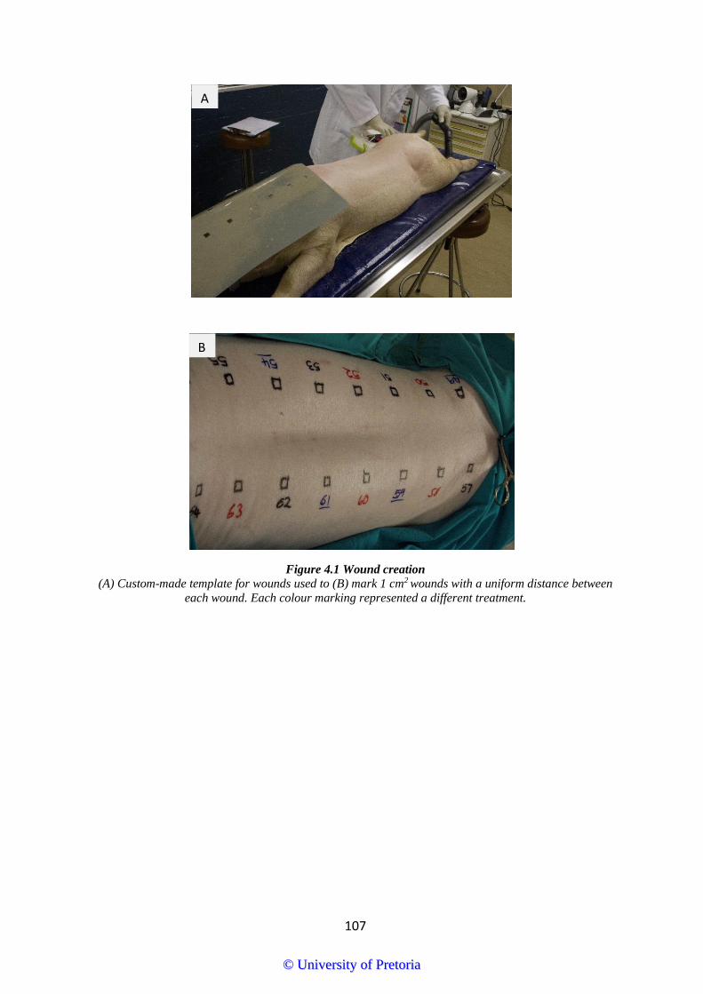
107
Figure 4.1 Wound creation
(A) Custom-made template for wounds used to (B) mark 1 cm2 wounds with a uniform distance between
each wound. Each colour marking represented a different treatment.
A
B
©© UUnniivveerrssiittyy ooff PPrreettoorriiaa
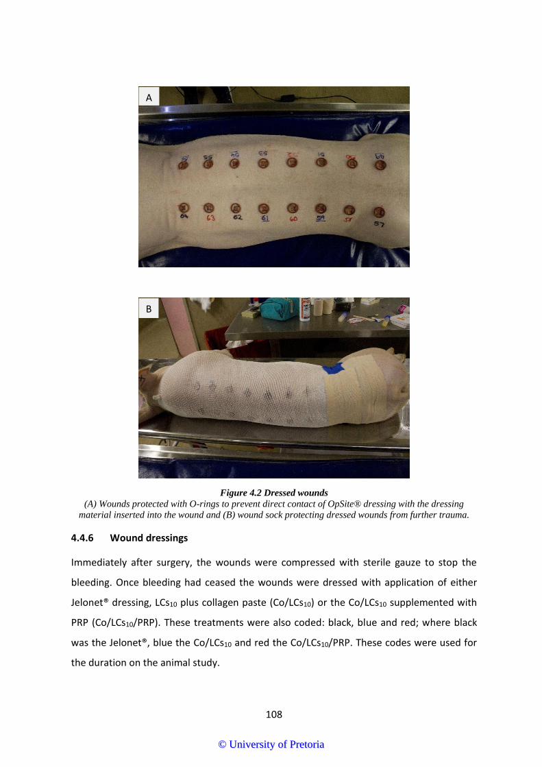
108
Figure 4.2 Dressed wounds
(A) Wounds protected with O-rings to prevent direct contact of OpSite® dressing with the dressing material inserted into the wound and (B) wound sock protecting dressed wounds from further trauma.
4.4.6 Wound dressings
Immediately after surgery, the wounds were compressed with sterile gauze to stop the
bleeding. Once bleeding had ceased the wounds were dressed with application of either
Jelonet® dressing, LCs10 plus collagen paste (Co/LCs10) or the Co/LCs10 supplemented with
PRP (Co/LCs10/PRP). These treatments were also coded: black, blue and red; where black
was the Jelonet®, blue the Co/LCs10 and red the Co/LCs10/PRP. These codes were used for
the duration on the animal study.
A
B
©© UUnniivveerrssiittyy ooff PPrreettoorriiaa
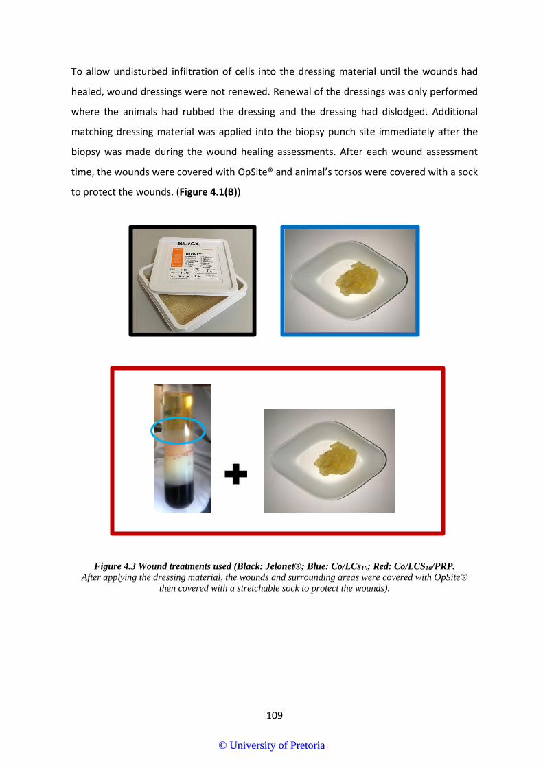
109
To allow undisturbed infiltration of cells into the dressing material until the wounds had
healed, wound dressings were not renewed. Renewal of the dressings was only performed
where the animals had rubbed the dressing and the dressing had dislodged. Additional
matching dressing material was applied into the biopsy punch site immediately after the
biopsy was made during the wound healing assessments. After each wound assessment
time, the wounds were covered with OpSite® and animal’s torsos were covered with a sock
to protect the wounds. (Figure 4.1(B))
Figure 4.3 Wound treatments used (Black: Jelonet®; Blue: Co/LCs10; Red: Co/LCS10/PRP.
After applying the dressing material, the wounds and surrounding areas were covered with OpSite®
then covered with a stretchable sock to protect the wounds).
©© UUnniivveerrssiittyy ooff PPrreettoorriiaa

110
Table 4.1 Day to day schedule of procedures
Day 1 2 3 4 5 6 7 8 9 10 11 12 13 14 15 16 17 18 19
Blood collection for PRP preparation
X
Dressing change (if necessary)
X X
Digital photograph
X X X X X X
X
X
X
PRP application (Group C-Day 1 and all biopsy days)
X X X X X X
Biopsy (full-thickness wound)
X X X X X
4.4.7 Housing after surgery
Each pig was housed in its own pen to prevent mutual interference with the wound sites of
the other animals. They each had visual contact to stimulate social interaction. Their straw
bedding was cleaned daily and they also had overhead infrared heating lamps. Animal
enrichment items were used to keep the animals busy and treats like raisins were buried in
the bedding to enhance their interest.
4.4.8 Wound healing
Macroscopic appearance of the wounds was assessed and wounds were photographed as
scheduled on Table 4.1. Standardised photographs were taken using a Canon EOS 7D camera
that was modified with a light tube with an internal ruler and evenly spaced light-emitting
diode (LED) lighting to give a fixed uniform focal distance and scale and for all wounds.
Autofocus at a focal distance of 55 mm was used for all images.
To quantitatively measure wound size, images were assessed using Image J software. The
change in wound area due to both re-epithelialisation and contraction was measured using
Image J software. To calculate percent change in wound area, the following equation was
used:
©© UUnniivveerrssiittyy ooff PPrreettoorriiaa

111
Percent wound area = (𝑫𝒂𝒚 𝒙 𝒘𝒐𝒖𝒏𝒅 𝒂𝒓𝒆𝒂
𝑫𝒂𝒚 𝟏 𝒘𝒐𝒖𝒏𝒅 𝒂𝒓𝒆𝒂 ) ∗ 𝟏𝟎𝟎
4.4.9 Biopsy sampling
For biopsy sampling, the animals were anesthetised using Dormicum® (0.3 mg/kg) and
AnaketV (11 mg/kg). Isofluor was used to maintain anaesthesia during surgery. To manage
pain post-operatively, Temgesic (0.3 – 0.6 ml) and Flunixin meglumine (2.2 mg/kg) were
administered intramuscularly. The body sock and dressings were removed and the wounds
photographed. The skin was sprayed with 70% alcohol to ensure sterility and wiped with
sterile gauze. Three wound biopsies were obtained from each pig (one from each treatment
group) using a 4 mm biopsy punch. The area of each biopsy included approximately half
healthy skin at the edge of the wound with half from the adjacent wound area. The
specimens were immediately snap-frozen in liquid nitrogen and stored at -80 °C until
required for use.
4.4.10 Histology
For the preparation and staining of histology samples, the samples were kindly sent to the
Pathology Section, Faculty of Veterinary Sciences, University of Pretoria, Onderstepoort.
Full-thickness skin biopsies were fixed in 10% buffered formalin for 48 hr at room
temperature. Care was taken to ensure that the tissues were completely immersed in the
fixative. Fixed biopsies were trimmed into appropriate size and shape and placed in
embedding cassettes in the appropriate orientation. The following paraffin embedding
schedule was used; 70% ethanol, two changes of 1 hr each; 80% ethanol, once, 1 hr; 5%
ethanol, once, 1 hr; 100% ethanol, three changes, 1.5 hr; xylene or xylene substitute (i.e.
Clear Rite 3), three changes, 1.5 hr each; paraffin wax (58 - 60 °C), two changes, 2 hr each.
Tissues were then embedded into paraffin blocks. The paraffin blocks were trimmed as
necessary and cut at 4 µm sections from epidermis to subdermal layers through the centre
of the wound biopsy. The paraffin wax ribbon was floated in a water bath at about 40 – 45 °C
and individual sections mounted onto glass microscope slides and allowed to air-dry for
30 min followed by baking in a 45 - 50°C oven overnight. Adjacent 4 µm sections were
stained with either Haematoxylin and Eosin (H & E) or Masson’s trichrome (MT).
©© UUnniivveerrssiittyy ooff PPrreettoorriiaa

112
4.4.10.1 H & E staining
For close to a century H & E staining has been used to differentiate morphologic changes in
tissues and the various tissue types. The H & E stains nuclei blue and cytoplasm pink. Briefly,
the adjacent sections were dewaxed in xylene for 5 min followed by staining for 1 min each
in ethanol; 100%, 96%, 70% and finally brought to distilled water. Thereafter, the sections
were stained in Haematoxylin for 10 min followed by rinsing in tap water. The sections were
then differentiated using one dip in Acid Alcohol. The samples were left to turn blue in
running tap water for 10 min, followed by 70% alcohol for 3 min. thereafter, the samples
were counterstained in Eosin for 2 ½ min and finally dehydrated for 3 min each in alcohol;
96% and 100%, respectively. The samples were cleared in Xylol and mounted in Entellan.
4.4.10.2 Masson’s trichrome staining
As inferred by the name, the MT method employs three dyes which selectively stain collagen
blue, cell nuclei black, and the cytoplasm, muscles, erythrocytes and fibrin red. The principle
of this method is the selective colouring of the tissue based on their porosity and the size of
the dye molecule. For the purposes of the current study, the sections were dewaxed in
xylene for 5 min followed by staining for 1 min each in alcohol; 100%, 96%, 70% and finally,
brought to distilled water. They were then stained in filtered Celestine blue solution for
15 min followed by rinsing three times in distilled water. Thereafter, the samples were
stained in Lily-Mayer Haematoxylin for 1 min and placed in blue tap water for 10 min. Next,
they were stained in Biebrich Scarlet Fuchsin solution for 15 min followed by rinsing in
distilled water. This was followed by mordanting in Phosphotunstic-phosphormolybdic acid
for 3 min and then dehydrated in 96% and 100% alcohol respectively. Finally, the samples
were cleared in xylene and mounted in Entellan.
4.4.10.3 Microscopic images
Images of all stained sections were taken using bright field microscopy (Olympus BX51;
Olympus, Tokyo, Japan) and measurements made at x10 and x20 and x40 magnification.
Epithelialisation, collagen deposition, fibroblast proliferation, neovascularisation, and
collagen deposition were assessed. Digital images with the magnifications were collected
using the microscope camera and stored on an external hard drive for later reassessment by
an independent observer.
©© UUnniivveerrssiittyy ooff PPrreettoorriiaa

113
4.5 Results and discussion
The preparation of a wound filler paste provided a matrix that was expected to promote
cellular and vascular infiltration into the wound area and promote collagen deposition and
myofibroblast driven wound contraction. Lauroyl chitosan was combined with micronized
collagen in the ratio 4:1 (LCs10:Collagen). The consistency of Co/LCs10 was a thick paste
(Figure 4.4). After the paste was prepared it was exposed to high dose gamma irradiation to
ensure sterility. Gamma irradiation sterilisation utilises the high energy of gamma rays to
disrupt bacterial DNA; thus, inhibiting bacterial division and decontaminating the
samples.351 The irradiation required for sterilization has the potential to also degrade the
structure of proteins and polysaccharides, thus PRP was added to the already sterilised
Co/LC10 paste.
The gamma irradiation dose utilised in this study theoretically disrupts the physicochemical
properties of collagen and chitosan352, however, physical examination of the paste did not
show significant changes in the morphology and viscosity of the material.353 In line with a
study by Singh et al, the benefits of using gamma irradiation were predicted to outweigh the
costs. Singh et al reported that radiation sterilised amniotic membranes favoured
epithelialisation when applied to burn wounds.353
Figure 4.4 Sterilised Co/LCs10 paste (beige area in lower centre) after 72 hr incubation in DMEM at
37°C.
The main objective of the animal study was to evaluate the wound healing properties of the
collagen/lauroyl chitosan (Co/LCs10) paste and Co/LCs10 paste supplemented with PRP
(Co/LCs10/PRP) in the treatment of full-thickness wounds in the well described porcine
©© UUnniivveerrssiittyy ooff PPrreettoorriiaa

114
model. The wound dressings were not changed from initial application until the end of the
study. The expectation was to have epidermal cells infiltrate the paste with the paste
providing a scaffold for the growth of new tissue. Collagen and chitosan have independently
shown wound healing properties. Collagen is as versatile as chitosan also with diverse
applications in tissue engineering. However, despite its biocompatibility, collagen’s rapid
biodegradation rate and low mechanical strength are the major limitations of using collagen
alone as a wound dressing material.354 Collagen and chitosan were thus combined to
increase the biostability of the material while potentiating the wound healing effect.
Previous wound healing studies using collagen and chitosan composites have shown
increased mechanical strength of the final wound dressing. In one study, a collagen/chitosan
(9:1) porous scaffold cross-linked with glutaraldehyde was invented by Ma et al to increase
its tensile strength by functioning as a crosslinking bridge in the crosslinking treatment with
glutaraldehyde.354 Chen et al fabricated composite nanofibrous membranes (NFM) from
type I collagen, chitosan, and polyethylene oxide by electrospinning. To prevent the
solubility of the NFM in water, they were further crosslinked using glutaraldehyde vapour.355
This NFM showed excellent skin regeneration potential when compared to gauze and
commercial collagen sponge.355 Analysis of these two studies reveal that the low ratio of
chitosan to collagen and the solubility of the underivatised chitosan and collagen in water
obliged the addition of a crosslinking material to further stabilise the material. In another
study, the physical, thermal and biological properties of a collagen/chitosan gel resulted in
a novel type of composite gel prepared by Li et al to enhance wound healing. This gel
contained collagen/chitosan supplemented with a cell-penetrating peptide (oligoarginine)
(collagen/chitosan/CPPs). The result was improved antibacterial activity, while
histopathological examination showed that collagen/chitosan/CPPs improved granulation
tissue formation, promoted angiogenesis and augmented collagen deposition in the wound
tissue.356 In yet another study, collagen and chitosan were enzymatically conjugated using
microbial transglutaminase to get a product with superior antioxidant activity compared to
carboxymethyl chitosan.272
While composites of collagen and chitosan have been extensively subjected to wound
healing analysis, LCs has been used for many other applications but has not been applied
expansively in wound healing. LCs’ haemostatic properties have however been reported.
©© UUnniivveerrssiittyy ooff PPrreettoorriiaa

115
Shelma and Sharma successfully developed an amphiphilic derivative (lauroyl sulphated
chitosan, LSCS) that significantly improve chitosan’s haemo-compatibility. 275
Some of the characteristics of an ideal wound dressing include maintaining moisture,
removing excess exudate, provision of an impermeable barrier to bacteria, protection of the
wound from further trauma, comfortable and shape conforming.145 These parameters were
used as markers of the success of the wound dressings in the management of the wounds.
For histological assessments, samples of the skin immediately after cutting with the
dermatome were snap-frozen and stored at -80 °C until required for histological analysis.
Other samples were collected at predetermined time points were examined by H & E and
MT staining. The MT stained collagen fibres blue, the cytoplasm in pink, the nuclei very dark
blue, and the red blood cells in cherry red. The H&E stained collagen fibres pale pink, the
cytoplasm purple, the nuclei in blue, and the red blood cells cherry red.84 MT stained
histological samples could show the presence of inflammatory cells in the skin samples
immediately upon wounding, even before the wound dressings were applied.
Approximately 10 min after applying the dressings, the Jelonet®-treated wounds appeared
to have continued haemorrhaging after removal of the compressive gauze, when comparing
to the wounds treated with Co/LCs10 and Co/LCs10/PRP used on day one. The first response
to an injury within the first few minutes is haemostasis. This study supported the fact that
chitosan is a haemostatic agent with several FDA approved haemostatic chitosan based
dressings already in use.213-214 Furthermore, the rapid arrest of the haemorrhaging especially
in the Co/LCs10/PRP group was attributed to PRP’s haemostatic and tissue sealing properties.
Platelet-rich plasma is a concentrated source of platelets, which after aggregation and
activation, achieves haemostasis through the formation of a fibrin clot. According to the
normal wound healing process, platelets play a pivotal role during the haemostasis phase.
In this case, the platelets, activated by chitosan, were understood to have released
chemotactic mediators, cytokines and growth factors which facilitated vasoconstriction,
coagulation and the conversion of soluble fibrinogen to a network of insoluble fibrin fibres.
The result was a stable chitosan based plug that had fibrin exposed surfaces due to the
activated platelets.59 The plug functioned as both a tissue sealant and growth factor delivery
©© UUnniivveerrssiittyy ooff PPrreettoorriiaa

116
system with chemotactic and mitogenic properties as was seen during the subsequent
phases of wound healing.
Within 10 min of wounding, inflammatory cells had already infiltrated the tissue directly
around the wound edges. During early inflammation, platelets release alpha granule
proinflammatory cytokines and growth factors while vascular endothelial cells,
keratinocytes and fibroblasts in the lesion, release cytokines such as TNF-α. These cytokines
promote chemotaxis of leukocytes to the injury site. Neutrophils are the dominant
leukocytes in the wound area during the early stages and facilitate elimination of debris,
bacteria and damaged tissue61 but also produce cytokines that attract and activate various
inflammatory cells in the wound. However, prolonged neutrophil infiltration was observed
in chronic wounds.
By the 3rd day of treatment (Figure 4.5), wounds treated with Co/LCs10 and Co/LCs10/PRP
material had formed a distinct scab that reduced the volume of exudate. Wounds treated
with Co/LCs10 had a dark, almost dry scab covering the wound while the Co/LCs10/PRP group
showed a tan coloured scab.
©© UUnniivveerrssiittyy ooff PPrreettoorriiaa

117
Jelonet® Co/LCs10 Co/LCs10/PRP
Day 1
Day 3
Figure 4.5 The same wounds photographed on Day 1 and Day 3 of wound healing.
Notice the macerated Jelonet-treated wounds on Day 3 while the other treatment groups already had scab formation.
©© UUnniivveerrssiittyy ooff PPrreettoorriiaa

118
Figure 4.6 (A) Day 1, immediately after wounding and applying wound dressings, skin cut using dermatome
Note the hair follicle (purple arrow), inflammatory cell accumulation, well-formed epidermal layers. (B) Magnified view of the area indicated in A: Accumulation of inflammatory cells could be seen within minutes after wounding. Inflammatory cells also infiltrated healthy skin. Notice the well organised structure of the stratum
corneum (SC) and epidermis (E), the accumulation of inflammatory cells (dotted black arrows), and the arrangement of the mature collagen fibres in the dermal
layers of the skin.
E
SC
SC
SC
A
B
©© UUnniivveerrssiittyy ooff PPrreettoorriiaa

119
.
Figure 4.7 Microscopic images of a Jelonet®-treated wound (black) on Day 3
Notice increased accumulation of inflammatory cells (dotted black arrows) in wound area (WA) compared to healthy skin (HS). Note the lack of any visible collagen
within the WA
WA
WA
HS
HS
©© UUnniivveerrssiittyy ooff PPrreettoorriiaa

120
A moist wound environment allows cell migration and proliferation providing an overall
optimum environment for healing.27 However, an equilibrium needs to be established for
optimum moisture to prevent maceration of surrounding tissues. In acute wounds, wound
exudate is produced because of vasodilation during inflammation, which is an important
factor for healing. Conversely, a prolonged inflammatory phase leads to delayed and
complicated wound healing. Exudate from chronic wounds contains proteolytic enzymes
capable of breaking down ECM proteins and these enzymes are absent in acute wounds.
Proteolytic enzymes are required in early stages of wound healing to degrade proteins and
clear the wound environment of debris during the healing processes. Chronic wound
exudate decreases cell proliferation and migration and may result in maceration of
surrounding tissue. On Day 3, moderate exudate which resulted in low grade maceration
was observed in the Jelonet®-treated wounds (Figure 4.5). This was coupled with bleeding
that was not completely arrested on the first day. Overall, this resulted in loose attachment
of Jelonet® on the wound. All the wounds from this control group were carefully cleaned to
remove excess exudate and blood that had clotted within the rubber ring with gauze dipped
in sterile saline followed by reapplication of Jelonet®. The same procedure was followed for
all other wounds that appeared macerated and had lost the dressing material. On Day 3,
only three wounds from the Co/LCs10/PRP required redressing, while the Co/LCs10 group had
five wounds. From Day 5 only the Jelonet® group required cleaning and retreatment and
from Day 8 none of the wounds had to be cleaned or redressed.
The lower volume of exudate in the Co/LCs10 group and Co/LCs10/PRP group was attributed
to the LCs10 in the dressings. The air permeable and moisture absorbing LCs10 in the dressings
provided a sterile, moist environment under a dry scab protecting the wound from
dehydration and contamination thus enhancing the healing conditions.177 Chitosan reduces
inflammation by absorbing the secreted of proinflammatory cytokines; TNF-α and IL-6.217
Inflammatory cells rid the wound area of debris during the inflammation phase of wound
healing; thereafter, inflammatory cells play a minor role. The anti-inflammatory properties
of lauric acid are known. It thus confirmed that the Jelonet®-treated wounds seemed to have
been in a prolonged inflammatory phase when compared to the other treatment groups.
The Jelonet®-treated wounds had higher concentrations of inflammatory cells until Day 8 as
seen from the histological analysis (Figure 4.7 and Figure 4.8), where inflammatory cells
©© UUnniivveerrssiittyy ooff PPrreettoorriiaa

121
were visible on Day 3 and even on Day 5 compared to the Co/LCs10 and the Co/LCs10/PRP
wounds. The inflammatory cells also appear to invade the healthy tissue adjacent to the
wounds. The scab covering the Co/LCs10 and Co/LCs10 treatment groups was also highly
concentrated with inflammatory cells and erythrocytes (Figure 4.8). It is believed that the
Co/LCs10 scab remains chemotactic to the inflammatory cells and thus facilitated the
removal of inflammatory cells from the wound area. The chemokine properties of chitosan
to neutrophils are reported to be via firstly, the stimulation of persistent IL-8 secretion by
fibroblasts which also results in increased angiogenesis.357 The second mechanism is through
the binding and subsequent activation of blood complement proteins resulting in a surface
that promotes neutrophil chemotaxis.293 This poses a paradoxical effect for the use of
chitosan as a wound healing material as high levels of inflammatory cells in the wound may
result in a chronic wound. Nonetheless, Park et al elucidated this topic further as they
showed that a decrease in the degree of deacetylation leads to increased hydrophobicity
and a shift from a positive charge to a negative charge which may have been the reason for
the increased secretion of IL-8 following exposure to their acetylated chitosan.358
According to Diegelmann, chitosan is said to be chemo-attractive to PMN cells.359 It is
possible that the cells seen in the scab on Day 3 were PMN cells. The inflammatory cells;
PMN cells together with neutrophils migrate into the wound area during the early stages of
the inflammation phase.
The Co/LCs10 and Co/LCs10/PRP pastes were intended to form a provisional matrix that would
promote collagen deposition and myofibroblasts driven wound contraction. Contrary to this
hypothesis, all the wounds appeared to heal from the dermal interface beneath the scab
without much infiltration into the chitosan-based dressing material. (Figure 4.8). The scab
which consisted mostly of entrapped dry blood and the inflammatory cells did not
incorporate into the new tissue as visible in the histology by Day 5 (Figure 4.8). The MT
stained slides showed a clear distinction between new epithelium and the scab in all the
healing wounds.
©© UUnniivveerrssiittyy ooff PPrreettoorriiaa

122
Figure 4.8 Masson’s trichrome stained biopsy of a Cs/LCs10/PRP treated wound on Day 5
(A) High concentration of inflammatory cells and erythrocytes (dotted black arrow) in the scab (S). (B) Notice separation between scab and wound area and the lower concentration of inflammatory cells in the wound area compared to the scab. (B) Day 12 showing a distinct epithelial layer with a thin layer of stratum
corneum already formed. Notice that the dressing material has not been incorporated and the thin collagen fibres in the wound area (Lower left).
E
HS
S
E
S
WA
WA
HS
A B
©© UUnniivveerrssiittyy ooff PPrreettoorriiaa

123
On Day 5, the Jelonet®-treated wounds still showed a high volume of cloudy yellow/brown
exudate exhibiting the presence of fibrin strands as a result of inflammation (Figure 4.9).
Despite this exudate, the histological images shown in Figure 4.10 revealed increased
collagen deposition in all three treatments, but with a higher abundance of collagen
deposition in the Co/LCS10/PRP treated wounds. The number of inflammatory cells had
decreased in the two treatments and the wound was clearly in the proliferative phase.
Consistent with what happens in the proliferative phase, capillaries and fibroblast cells were
now clearly visible. Fibroblasts synthesise the ECM components; glycosaminoglycans,
proteoglycans, fibronectin and collagen which support granulation tissue formation.70-71
Diegelmann’s study did not give promising results from the use of chitosan in subcutaneous
wounds in rats. This was a result of the chitosan sponge significantly delaying the appearance
of macrophages and also diminishing the infiltration of fibroblasts, capillaries, and the
deposition of mature collagen fibres compared to the control.359 In contrast with these
findings, studies by Ueno et al in a dog model203 and Ishihara et al in a mouse model130
showed that chitosan promoted granulation tissue formation, fibroblast infiltration and
collagen deposition which was consistent with the results found in this study.
The explanation for the current findings was initially thought to lie in the lauric acid
incorporated into the chitosan used for dressings. The increased collagen deposition and
fibroblast infiltration may be due to the micronized collagen incorporated into the dressings.
Collagen has been demonstrated in vitro to exhibit a chemotactic response to fibroblast
cells252 while fibroblast cells synthesise the new collagen.
©© UUnniivveerrssiittyy ooff PPrreettoorriiaa

124
Jelonet®
Co/LCs10
Co/LCs10/PRP
Day 5
Day 8
Figure 4.9 Photographs of the three treatment groups on Days 5 and 8.
Jelonet®-treated wounds were still exuding on Day 5 and only showed granulation tissue formation on Day 8. The Co/LCs10 and Co/LCs10/PRP treatment groups
both mentioned the scab throughout the treatment period.
©© UUnniivveerrssiittyy ooff PPrreettoorriiaa

125
Jelonet® Co/LCs10 Co/LCs10/PRP Day 5
Day 8
Figure 4.10 Masson’s Trichrome stained biopsy images of the treatment groups on Days 5 and 8.
©© UUnniivveerrssiittyy ooff PPrreettoorriiaa

126
Fibronectin, a large major glycoprotein is found throughout all phases of wound healing;
from the fibrin clot, the papillary dermis, to the newly synthesised collagen in the granulation
tissue.70-71 Granulation tissue is also composed of collagen and fibroblasts that are involved
in the migration of cells into the wound area and overall in the contraction of the wound. In
addition to this, granulation tissue also has monocytes and macrophages that are active
inflammatory cells and newly formed blood vessels. There was no visible granulation tissue
formation in Jelonet®-treated wounds until Day 8 (Figure 4.9). The granulation tissue
appeared grey brown, shiny and granular as a result of the new capillaries; thus, indicating
a healthy wound. Histological analysis with the MT stain (Figure 4.10) showed fibroblast
infiltration and collagen deposition was more pronounced on Day 8. It was then postulated
that despite the two chitosan based treatment groups (Co/LCs10 and the Co/LCs10/PRP
groups) showing scab formation by Day 3, which is commonly regarded as a signal that
granulation tissue formation has been initiated, the Jelonet®-treated wounds possibly only
started contracting around the 8th day when visible granulation tissue formation could be
seen.
By Day 10 it was evident that the wounds were in the tissue remodelling phase. In this phase,
the temporary matrix formed in the proliferative phase is replaced by thicker fibres and
stronger more organised collagen matrix.73 The dense collagen observed from Day 10
showed fibres that were possibly remodelling from the more gel-like type III collagen to
more fibrous type I collagen. Furthermore, PRP is known to enhance cell proliferation while
promoting angiogenesis.360 The presence of fully-formed blood vessels on Day 10 in the
Co/LCS10/PRP treated wounds, substantiated already published data. PRP was postulated to
have promoted angiogenesis in the Co/LCs10/PRP treatment group as a consequence of the
continuous slow release of exogenous growth factors in the dressings.
On Day 10, after biopsy sampling, one of the pigs was found dead in its pen. Autopsy results
(
©© UUnniivveerrssiittyy ooff PPrreettoorriiaa

127
Annexure 7) revealed an intestinal torsion, which was a result of the handling of the pigs for
each procedure.
©© UUnniivveerrssiittyy ooff PPrreettoorriiaa

128
Jelonet®
Co/LCs10
Co/LCs10/PRP
Day 10
Figure 4.11 Masson’s Trichrome histology images of the wounds on Day 10.
Wound area (WA) shows increased collagen density especially in the Co/LCs10/PRP treatment. Collagen fibres were still not fully mature as in the HS.
WA
WA
WA
HS HS
HS
©© UUnniivveerrssiittyy ooff PPrreettoorriiaa

129
Figure 4.12 Wound 5 (black) on Day 12 – Collagen deposition, fibloblast cells (green arrows) visible, little inflammation.
Notice collagen deposition in the wound area (WA)and the large hair follicle (HF).
WA
HS
WA
HF E
S
©© UUnniivveerrssiittyy ooff PPrreettoorriiaa

130
It was only on Day 12 that clear wound healing was observed after gentle removal of the
loose scabs from wounds. The scabs that were semi-detached or falling off revealed healed
skin with only minor scarring. Regrowth of hair was observed macroscopically as well as
microscopically as indicated by the hair shafts in Figure 4.12.
During the entire duration of the study, the outward appearance of CoLCs10 and
Co/LCs10/PRP treated wounds did not change except for the darkening and slight reduction
in the size of the scab as the wounds progressed through the healing phases. These wounds
appeared not to be reducing in size macroscopically for the entire 15 days despite seeing
that the scab was becoming loose at the edges. Histological analysis gave a clearer view of
what was happening under the dry scab.
When all the wounds were observed on Day 19 not much scarring was observed (Figure
4.13). Co/LCs10 paste was observed to have aided in preventing the formation of
dysfunctional scars.
©© UUnniivveerrssiittyy ooff PPrreettoorriiaa
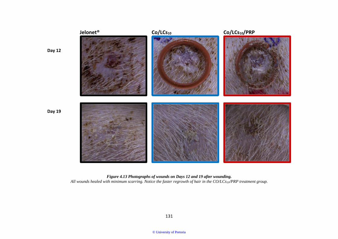
131
Jelonet® Co/LCs10 Co/LCs10/PRP Day 12
Day 19
Figure 4.13 Photographs of wounds on Days 12 and 19 after wounding.
All wounds healed with minimum scarring. Notice the faster regrowth of hair in the CO/LCs10/PRP treatment group.
©© UUnniivveerrssiittyy ooff PPrreettoorriiaa

132
Figure 4.14 Microscopic images of MT stained wounds at 10x magnification on Day 15
Wound area (WA) in the Co/LCs10 (Blue boarder) had a well-developed HF (purple arrow). Jelonet®-treated wounds (Black boarder) and Co/LCs10/PRP wounds
(Red boarder).
WA WA
WA
HS
HS HS
©© UUnniivveerrssiittyy ooff PPrreettoorriiaa

133
Consistent with the findings of Agyingi et al who simulated the process of epithelialisation and
angiogenesis using the Eden model, it was found that as the wounds healed, they lost the
original square shape (Figure 4.15) as a result of migration of epidermal cells from the wound
edges propagating towards the wound centre.361 The areas with the highest curvature in
wounds heal first362 as seen by the initial contraction of the wound corners (Figure 4.15). It was
thus apparent that wounds in the porcine model heal by simultaneous re-epithelialisation and
contraction. However, on microscopic examination, the wounds in this study healed more by
re-epithelialisation from the bottom of the wound bed up rather than migration from the
wound edges. Therefore, the wound measurements taken gave an indication of the healing
effect due to contraction, which in this case could have been hidden under the well-established
scab.
When measuring the wound area, it appeared as if the Jelonet®-treated wounds were
decreasing in wound area more rapidly than the other treatments (Figure 4.19). This was
because of the scab covering the two treatment groups that obstructed wound measurements.
When the scabs eventually fell off, the two treatment groups showed the most favourable
healing outcomes. These included little or no scarring, quick return of skin to its original colour
and morphology and functionality. Histologically this was confirmed by the presence of more
mature and thicker collagen fibres and angiogenesis as explained above.
Overall, the Co/LCs10 treated wounds on macroscopic examination did not appear to show
great healing progress due to the scab that masked the underlying changes. Microscopic
examination showed wounds progressing well through each of the phases of wound healing
with a high concentration of inflammatory cells initially which then decreased during the later
stages of healing as collagen was deposited (Figure 4.16). In summary, the wounds showed a
similar healing pattern (healing from the bottom of the wound bed up) with an overall higher
and more mature collagen deposition in the Co/LCs10/PRP treatment group.
©© UUnniivveerrssiittyy ooff PPrreettoorriiaa

134
Day 1 Day 5
Day 10 Day 12
Figure 4.15 Images of a Jelonet®-treated wound taken on different days showing the loss of square shape as wounds re-epithelialised as a result of the scab
breaking up in a non-symmetrical way.
©© UUnniivveerrssiittyy ooff PPrreettoorriiaa

135
Day 1 Day 5 Day 10 Day 15
H & E
Masson’s Trichrome
Figure 4.16 Progression of Jelonet®-treated wounds from Days 1 to 15.
First row showing the macroscopic appearance of the wounds, second row showing H & E stained wounds and third row showing MT stained wounds.
©© UUnniivveerrssiittyy ooff PPrreettoorriiaa

136
Day 1 Day 5 Day 10 Day 15
H & E
Masson’s Trichrome
Figure 4.17 Progression of Co/LCs10 treated wounds from Days 1 to 15.
First row showing the macroscopic appearance of the wounds, second row showing H & E stained wounds and third row showing MT stained wounds.
©© UUnniivveerrssiittyy ooff PPrreettoorriiaa

137
Day 1 Day 5 Day 10 Day 15
H & E
Masson’s Trichrome
Figure 4.18 Progression of Co/LCs10/PRP treated wounds from Days 1 to 15.
First row showing the macroscopic appearance of the wounds, second row showing H & E stained wounds and third row showing MT stained wounds.
©© UUnniivveerrssiittyy ooff PPrreettoorriiaa
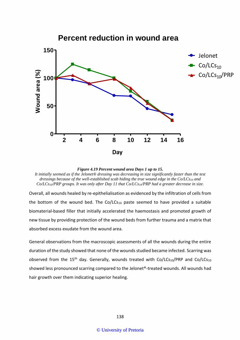
138
Percent reduction in wound area
2 4 6 8 10 12 14 160
50
100
150Jelonet
Co/LCs10
Co/LCs10/PRP
Day
Wo
un
d a
rea
(%)
Figure 4.19 Percent wound area Days 1 up to 15.
It initially seemed as if the Jelonet® dressing was decreasing in size significantly faster than the test dressings because of the well-established scab hiding the true wound edge in the Co/LCs10 and
Co/LCs10/PRP groups. It was only after Day 11 that Co/LCs10/PRP had a greater decrease in size.
Overall, all wounds healed by re-epithelialisation as evidenced by the infiltration of cells from
the bottom of the wound bed. The Co/LCs10 paste seemed to have provided a suitable
biomaterial-based filler that initially accelerated the haemostasis and promoted growth of
new tissue by providing protection of the wound beds from further trauma and a matrix that
absorbed excess exudate from the wound area.
General observations from the macroscopic assessments of all the wounds during the entire
duration of the study showed that none of the wounds studied became infected. Scarring was
observed from the 15th day. Generally, wounds treated with Co/LCs10/PRP and Co/LCs10
showed less pronounced scarring compared to the Jelonet®-treated wounds. All wounds had
hair growth over them indicating superior healing.
©© UUnniivveerrssiittyy ooff PPrreettoorriiaa

139
4.6 Conclusion
The use of a wound filling paste containing collagen, lauroyl chitosan and platelet-rich plasma
resulted in improved healing outcomes compared to those treated with Jelonet®. Within
10 min after applying the different treatments on the wounds, it was seen that the wounds
treated with Co/LCs10 and Co/LCs10/PRP had stopped bleeding while the Jelonet®-treated
wounds continued bleeding slowly and had a red tinged exudate. By Day 3, this bleeding
caused high moisture content of the Jelonet® treatment group resulting in macerated wounds
while the LCs10 and Co/LCs10/PRP treatment groups appeared to have a solid healthy-looking
scab, thus showing the haemostatic effect of chitosan. Furthermore, the two chitosan-based
treatment groups appeared to encourage accumulation of inflammatory cells and
erythrocytes on the dressing material surface yet reduced inflammation. It was evident that
the Co/LCs10 paste allowed adsorption of cells onto its surface as seen from the MT histology
of these scabs.
The time for re-epithelialisation between topically applied Co/LCs10 and Co/LCs10/PRP pastes
compared to the Jelonet®-treated wounds was not significantly different; however, the
Co/LCs10/PRP treatment increased collagen deposition considerably more than the Co/LCs10
without the PRP. The scab on the two chitosan-based treatment groups was shown to
completely cover and to some extent distort the appearance of the wounds, thus accurate
measurement the wound size as the wound healing progressed was not possible.
Two pigs died, one each on the 10th and 16th days of the study. This was a surprising event
and the animals were sent for autopsy to determine the cause of death. The first pig was
found to have died from an intestinal torsion that was possibly due to the handling
procedures used when transferring the pigs while under anaesthesia. The second pig died of
pulmonary distress. It was concluded from the autopsies that none of the deaths could be
attributed to the wound dressings used. However, the handling method of the pigs was
identified as a causative factor and further handling procedures were all performed on the
same mattress for the remaining pigs for the rest of the study.
The wound areas from the Co/LCs10 and Co/LCs10/PRP groups quickly appeared similar to the
surrounding normal skin, with hair growth compared to the Jelonet®-treated wounds.
©© UUnniivveerrssiittyy ooff PPrreettoorriiaa

140
5 Chapter 5
5.1 General discussion and conclusion
The focus of the present study was on full-thickness wounds. These wounds present with
excessive tissue loss, minimum growth factor activity and impaired angiogenesis.
Dysfunctional scarring emanating from impaired healing in full-thickness wounds is
inexorable unless some skin substitutes are used. A wound filler that promotes tissue growth,
angiogenesis and combats infection was considered a viable option for treating these
wounds.
Thus, it was hypothesised that wound management using hydrophobically modified chitosan-
based dressings combined with collagen and platelet-rich plasma would enhance the wound
healing rate and minimise dysfunctional scarring of full-thickness wounds when measured
using the porcine wound model.
The field of wound healing is diverse and well researched, however hydrophobically modified
chitosan is under-researched as a wound dressing. In this study, chitosan was hydrophobically
modified to give three different loadings of lauric acid conjugated chitosan, which were
chemically and physically characterised together with in vitro assays to determine the product
with the best physicochemical properties for use in a wound dressing. The chosen conjugate
was then tested on a full-thickness wound model for efficacy as a wound filling dressing
material. This study thus provides an important foundation for further studies into the
efficacy of hydrophobically modified chitosan in improving full-thickness wound dynamics
including platelet delivery, growth factor release, exudate absorption, wound contraction and
avoiding dysfunctional scarring.
First, hydrophobic modification of chitosan using lauric acid was completed successfully using
an addition-elimination type reaction. This proceeded through the formation of amide
linkages between the primary amine groups of chitosan and the carboxylic acid functional
group of lauric acid. Initially, lauric acid was conjugated to chitosan using carbodiimide
chemistry, with the EDC salt activating the carboxylic group of lauric acid. However, a second
method using the highly reactive lauroyl chloride was used. This method significantly reduced
the reaction time from 72 hr to just 24 hr. Characterisation of the LCs conjugates using NMR
©© UUnniivveerrssiittyy ooff PPrreettoorriiaa

141
and FT-IR confirmed the conjugation with the appearance of the aliphatic proton peak at
0.9 ppm in the NMR spectrum, and the carboxylic group at 1655 cm-1 and 1722 cm-1 in the FT-
IR. The FT-IR absorption peaks at 2842 cm-1 and 2932 cm-1, were attributed to symmetric and
asymmetric C-H stretching, respectively. These stretching vibrations accounted for the
methyl, methylene and methine groups in NHCOCH3, CH2OH and CH in the pyranose ring,
respectively.273
The degree of lauroyl chain conjugation to chitosan demonstrated increased hydrophobicity
as the molar concentration of lauric acid increased. When compared to the underivatised
chitosan using the drop shape analysis, it appeared that as the molar concentration of lauric
acid increased, the contact angle also increased. In the swelling tests, LCs34 had the highest
swelling capacity. However, despite this good characteristic, LCs34 was physically rigid, hard
and plastic which reduced its processability. Consequently, LCs34 could not provide a good
malleable wound fitting matrix for space filling and absorption of excess exudate and for this
reason was eliminated as a possible wound healing material. The swelling index studies also
showed the LCs20 scaffold almost completely dissolving into a gel within 2 hr and no further
readings could be taken beyond this time point. This was another reason why LCs10 was
selected for the wound healing study.
In preliminary studies, platelet adhesion to the conjugated chitosan was done by coating of
the wells of tissue culture plates with the different chitosan conjugate material and adding
PRP with aspiration of the non-adherent platelets after an hour. The adherent platelets were
then quantified microscopically by platelet counting. After realising the impracticality of
complete uniform aspiration of non-adherent platelets, and the difficulty in counting the
adhered platelets accurately, this method was discontinued and the colorimetric acid
phosphatase assay was followed. The findings from the acid phosphatase assay showed that
as hydrophobicity of the sample increased, platelet adhesion decreased. Chitosan adhered
the highest platelet concentration, even higher than the collagen positive control. The lauroyl
substitution of chitosan resulted in a negatively charged polymer surface that repelled the
negatively charged platelets. Increased hydrophobicity is also known to reduce the “shear
stress” of a surface, decrease the surface area available for adhesion and reduce the overall
concentration of adherent platelets.333 Fatty acids induce platelet aggregation, yet fatty acids
also form a smooth surface that repels platelets. The platelet adhesion assay validated the
©© UUnniivveerrssiittyy ooff PPrreettoorriiaa

142
concept that the higher molar loading of lauroyl groups in LCs34 and in this case also the LCs20
were not the best candidates to increase platelet delivery to wound sites and thus based on
these findings it was decided that LCs10 gave the most suitable characteristics for a wound
healing dressing and was therefore further investigated in the animal study.
Next, an ELISA kit was used to assess PDGF-AB release over 24 hr. The positive control,
collagen type 1 extracted from porcine skin was characterised using FT-IR. The FT-IR peaks
corresponded with that found in literature. The other determinant for the collagen was its
ability to form a gel in 1% aqueous acetic acid.354 Since LCs10 gave superior swelling and
platelet adhesion properties, it was assayed for platelet adhesion and growth factor release
using the ELISA. After binding platelets, the LCs10 conjugate gave a sustained release of PDGF-
AB over 24 hr. The release of PDGF-AB from native chitosan and collagen displayed a burst
release within the first hour. This was attributed to the platelet activation by these two
materials. Hydrophobic modification of Cs resulted in a material with reduced platelet-
activating properties.
Finally, a wound filler paste comprising of 20% w/w collagen in LCs10 (Co/LCs10) was prepared
and sterilised using gamma irradiation. The energy released during gamma irradiation causes
damage to biological products like PRP while potentially causing physico-chemical changes to
collagen and chitosan. To reduce the potential damage to PRP which would alter its efficacy,
the CoLCs10 was sterilised and stored before adding PRP. PRP was only added to the paste just
before applying to the wounds on the day of treatment. The Co/LCs10 and Co/LCs10 with PRP
(Co/LCs10/PRP) were compared to Jelonet®-treated controls to compare wound healing rates
using the full-thickness porcine wound model. On the 3rd and 5th days after applying these
dressings, macroscopic observations showed Co/LCs10/PRP to have superior haemostatic
properties compared to the Co/LCs10 treatment. The Jelonet®-treated wounds appeared to
have macerated edges because of high exudate levels. Chitosan’s haemostatic properties
were a contributing factor to the low exudate levels in the two chitosan-based treatments
with PRP adding further benefit to this property in the Co/LCs10/PRP treatment. Histologically,
the MT stain showed prolonged inflammation on Jelonet®-treated wounds continuing up to
the 8th day. However, inflammatory cells on Co/LCs10 and Co/LCs10/PRP treated wounds were
adsorbed into the chemoattractant LCs10 scab; thus facilitating the removal of inflammatory
cells from the wound bed.
©© UUnniivveerrssiittyy ooff PPrreettoorriiaa
143
Contrary to the aim of the study, which set out to provide a matrix for the infiltration and
growth of new cells within the wound bed, all the wounds appeared to heal from the dermal
interface beneath the scab without significant cell infiltration into the chitosan-based dressing
material. The scab which consisted mostly of entrapped dry blood and the inflammatory cells
also incorporated the LCs10 material which did not incorporate into the newly formed granular
tissue as clearly visible in the biopsy histology by Day 5. The MT stained slides showed a clear
distinction between newly formed epithelium and the scab in all the healing wounds. The
wounds healed within 12 to 16 days with minimal scarring and the histology of the edge
biopsies indicated extensive new collagen deposition within the wound bed and re-
establishment of the characteristic skin structure with clear dermal and epidermal layer.
In summary, three hydrophobically modified LCs derivatives were synthesised using the acyl
halide chemistry method. They were then physically and chemically characterised followed
by in vitro analysis to determine which of these three derivatives would give optimum platelet
binding and growth factor release and overall give the best-improved wound dressing. The
LCs10 derivative proved to have optimum in vitro properties as shown especially in the platelet
binding assay. It was further assayed using the ELISA where it showed sustained release of
the PDGF-AB. Finally, LCs10 was combined with collagen and PRP to provide a wound filler
paste that was tested on full-thickness wounds in the porcine model. The wounds treated
with Co/LCs10 and Co/LCs10/PRP formed a plug immediately after application, which proved
to be haemostatic compared to the Jelonet®-treated wounds. In the later stages of healing,
The Co/LCs10/PRP treated wounds appeared to have healed more rapidly with less scarring
than the Jelonet®-treated wounds. Finally, histological analysis using the MT and H & E
showed highest and most mature collagen deposition in the Co/LCs10/PRP treatment group.
Based on the findings from this study, the Co/LCs10/PRP paste used as a space filling wound
dressing material provided a protective scaffold that prevented wound constriction and
provided a sustained release of growth factors from the added platelet-rich plasma, was
haemostatic and absorbed excess exudate from the wound while providing a moist wound
environment with fewer inflammatory cells thus preventing chronic inflammation in wounds.
The use of PRP appeared to have an additional benefit of increased collagen density and
growth of new epithelial cells. Further research on the Co/LCs10/PRP is recommended to
validate its efficacy as a wound filler.
©© UUnniivveerrssiittyy ooff PPrreettoorriiaa

144
5.2 Limitations and recommendations
Blood from only ten participants was used to determine the blood parameters and the
average platelet count. On average, platelet concentrations increased five-fold in the PRP
compared to the platelet count in whole blood. This platelet count was lower than the
average eight-fold recorded platelet counts in literature. This could possibly be improved by
decreasing the final volume of plasma harvested per sample and confirmed by increasing the
number of study participants.
Due to cost limitations of the PDGF-AB kit, only the LCs10 conjugate compared to the collagen
and native chitosan controls were assayed. Further PDGF-AB release assays on all the
conjugates would give a good comparison of the impact of increased hydrophobicity on
growth factor release.
The platelet adhesion assay used a blank well surface treated with 1% acetic acid. Future
studies using albumin-coated surfaces for the blank would give a better comparison.
The SEM and TEM images from the fibroblast infiltration assay did not show clear infiltration
of the fibroblasts in the images. One limitation was the adherence of fibroblasts onto the
culture plates with proliferation on the plate surface rather than growing into the scaffolds.
This could be prevented in future by prior coating of the plate surface with agarose to prevent
adhesion of the fibroblasts.
The pH is an important factor when preparing wound dressings as it has both a direct and
indirect effect on all biochemical reactions taking place. During the different stages of
preparing the CoLCs10 paste, different pH values were used. For example, the swelling index
was done at pH of 7.4 while the Co/LCs10 paste was adjusted to a pH range of 5 – 6. This
impacted accuracy in reporting the efficacy of the final product. Future studies should
consider consistency with the pH range at every step.
During the course of the animal study, two pigs died. Full autopsies were performed on these
two animals and it was found that the first pig had died due to an intestinal torsion, potentially
due to the handling procedures of the pigs during the anaesthesia and transfers onto the
theatre table and transfer trolley. The second pig appeared to have died due to undetermined
pulmonary causes. The autopsy reports (see
©© UUnniivveerrssiittyy ooff PPrreettoorriiaa

145
Annexure 7) also determined that death was highly unlikely to have been due to the wounds
or any of the material applied on the wounds. After the death of the first pig, it was
recommended immediately that the handling of the pigs change from picking up and rolling
onto the operating table to keeping the pig in the same position while transporting onto the
operating table. It is also recommended that future animal studies reduce the frequency of
the anaesthesia for biopsy purposes as this increased the handling frequency.
This being one of the first studies on the use of hydrophobic chitosan as a wound dressing
material, the above-mentioned recommendations should be considered for further
preclinical studies on these compounds.
©© UUnniivveerrssiittyy ooff PPrreettoorriiaa

146
6 References
1. Kabara JJ, Swieczkowski DM, Conley AJ, Truant JP. Fatty acids and derivatives as antimicrobial agents. Antimicrobial agents and chemotherapy. 1972; 2(1):23-8.
2. Nussbaum SR, Carter MJ, Fife CE, DaVanzo J, Haught R, Nusgart M, et al. An economic evaluation of the impact, cost, and medicare policy implications of chronic nonhealing wounds. Value in Health. 2018; 21(1):27-32. doi:10.1016/j.jval.2017.07.007
3. UspikedTM [Internet]. The chronic wounds: Money-spinner! As practitioners and device makers run amok. Cape town2017, May 1 [2018, Dec 18]. Available from: https://www.uspiked.com/health/2017/05/01/the-chronic-wounds-money-spinner!/.
4. Comte E. Treatment of chronic wounds: An issue for africa. European Wound Management Association Journal. 2009;
5. Rangaraj A, Harding K, Leaper D. Role of collagen in wound management. Wounds UK. 2011; 7(2):54-63.
6. Birch NP, Barney LE, Pandres E, Peyton SR, Schiffman JD. Thermal-responsive behavior of a cell compatible chitosan/pectin hydrogel. Biomacromolecules. 2015; 16(6):1837-43.
7. Leek MD, Barlow YM. Tissue reactions induced by hydrocolloid wound dressings. Journal of Anatomy. 1992; 180 ( Pt 3)(Pt 3):545-51.
8. Paul W, Sharma CP. Chitosan and alginate wound dressings: A short review. Trends in Biomaterials and Artificial Organs. 2004; 18(1):18-23.
9. Lee SM, Park IK, Kim YS, Kim HJ, Moon H, Mueller S, et al. Physical, morphological, and wound healing properties of a polyurethane foam-film dressing. Biomaterials research. 2016; 20:15-. doi:10.1186/s40824-016-0063-5
10. Syifa Z, Fazli WM. Biodegradable mushroom-based transparent paper. Research Communication in Engineering Science & Technology. 2018; 1:25-.
11. Wang T, Zheng Y, Shen Y, Shi Y, Li F, Su C, et al. Chitosan nanoparticles loaded hydrogels promote skin wound healing through the modulation of reactive oxygen species. Artificial Cells, Nanomedicine, and Biotechnology. 2017:1-12. doi:10.1080/21691401.2017.1415212
12. Campos M, Rawls H, Mei L, Satsangi N. In vitro wound healing model: Effects of chitosan films loaded with gentamicin and silver sulfadiazine on the wound filling rate. Advances in Tissue Engineering & Regenerative Medicine. 2017; 2(3):00031.
13. Michailidou G, Christodoulou E, Nanaki S, Barmpalexis P, Karavas E, Vergkizi-Nikolakaki S, et al. Super-hydrophilic and high strength polymeric foam dressings of modified chitosan blends for topical wound delivery of chloramphenicol. Carbohydrate Polymers. 2019; 208:1-13. doi:https://doi.org/10.1016/j.carbpol.2018.12.050
14. Andres Y, Giraud L, Gerente C, Le Cloirec P. Antibacterial effects of chitosan powder: Mechanisms of action. Environmental Technology. 2007; 28(12):1357-63. doi:10.1080/09593332808618893
15. Fiamingo A, de Moura Delezuk JA, Trombotto S, David L, Campana-Filho SP. Extensively deacetylated high molecular weight chitosan from the multistep ultrasound-assisted deacetylation of beta-chitin. Ultrasonics Sonochemistry. 2016; 32:79-85.
16. Fei Liu X, Lin Guan Y, Zhi Yang D, Li Z, De Yao K. Antibacterial action of chitosan and carboxymethylated chitosan. Journal of Applied Polymer Science. 2001; 79(7):1324-35. doi:10.1002/1097-4628(20010214)79:7<1324::AID-APP210>3.0.CO;2-L
17. Fiamingo A, Campana-Filho SP. Structure, morphology and properties of genipin-crosslinked carboxymethylchitosan porous membranes. Carbohydrate polymers. 2016; 143:155-63.
18. Goy RC, Britto Dd, Assis OB. A review of the antimicrobial activity of chitosan. Polímeros. 2009; 19(3):241-7.
19. Xing K, Chen XG, Liu CS, Cha DS, Park HJ. Oleoyl-chitosan nanoparticles inhibits escherichia coli and staphylococcus aureus by damaging the cell membrane and putative binding to extracellular or intracellular targets. International Journal of Food Microbiology. 2009; 132(2):127-33. doi:https://doi.org/10.1016/j.ijfoodmicro.2009.04.013
©© UUnniivveerrssiittyy ooff PPrreettoorriiaa

147
20. Tao S, Gao S, Zhou Y, Cao M, Xie W, Zheng H, et al. Preparation of carboxymethyl chitosan sulfate for improved cell proliferation of skin fibroblasts. International Journal of Biological Macromolecules. 2013; 54:160-5. doi:https://doi.org/10.1016/j.ijbiomac.2012.12.009
21. Dictionary MW. Trauma. 2019; 22. Lazarus GS, Cooper DM, Knighton DR, et al. Definitions and guidelines for assessment of wounds
and evaluation of healing. Archives of Dermatology. 1994; 130(4):489-93. doi:10.1001/archderm.1994.01690040093015
23. Medicine USNLo [Internet]. Wounds and injuries. 14 August, 2018. Available from: https://medlineplus.gov/woundsandinjuries.html.
24. Hermans MH. Wounds and ulcers: Back to the old nomenclature. Wounds. 2010; 22(11):289-93. 25. Lazarus GS, Cooper DM, Knighton DR, Margolis DJ, Percoraro RE, Rodeheaver G, et al. Definitions
and guidelines for assessment of wounds and evaluation of healing. Wound Repair and Regeneration. 1994; 2(3):165-70.
26. Junker JP, Caterson E, Eriksson E. The microenvironment of wound healing. Journal of Craniofacial Surgery. 2013; 24(1):12-6.
27. Winter GD. Formation of the scab and the rate of epithelization of superficial wounds in the skin of the young domestic pig. 1962;
28. Swezey L [Internet]. The microenvironment of wounds. WoundEducators.com; 2014 [27 Dec 2018]. Available from: https://woundeducators.com/wound-microenvironment/.
29. Bowler PG, Duerden BI, Armstrong DG. Wound microbiology and associated approaches to wound management. Clinical microbiology reviews. 2001; 14(2):244-69. doi:10.1128/CMR.14.2.244-269.2001
30. Sule AM, Thanni L, Sule Odu OA, Olusanya O. Bacterial pathogens associated with infected wounds in ogun state university teaching hospital, sagamu, nigeria2002.
31. Chellan G, Shivaprakash S, Karimassery Ramaiyar S, Varma AK, Varma N, Thekkeparambil Sukumaran M, et al. Spectrum and prevalence of fungi infecting deep tissues of lower-limb wounds in patients with type 2 diabetes. Journal of Clinical Microbiology. 2010; 48(6):2097-102. doi:10.1128/jcm.02035-09
32. Ono S, Imai R, Ida Y, Shibata D, Komiya T, Matsumura H. Increased wound ph as an indicator of local wound infection in second degree burns. Burns. 2015; 41(4):820-4. doi:https://doi.org/10.1016/j.burns.2014.10.023
33. Stewart CM, Cole MB, Legan JD, Slade L, Vandeven MH, Schaffner DW. Staphylococcus aureus growth boundaries: Moving towards mechanistic predictive models based on solute-specific effects. Applied and Environmental Microbiology. 2002; 68(4):1864-71.
34. Percival NJ. Classification of wounds and their management. Surgery (Oxford). 2002; 20(5):114-7. 35. Denny K, Lawand C, Perry SD. Compromised wounds in canada. Healthcare Quarterly. 2014; 17(1) 36. Heyer K, Herberger K, Protz K, Glaeske G, Augustin M. Epidemiology of chronic wounds in germany:
Analysis of statutory health insurance data. Wound Repair and Regeneration. 2016; 24(2):434-42.
37. Jiang Y, Huang S, Fu X, Liu H, Ran X, Lu S, et al. Epidemiology of chronic cutaneous wounds in china. Wound Repair and Regeneration. 2011; 19(2):181-8.
38. MS NG, S.K. Gupta MS F, V.K. Shukla Ms M, MD SPS. An indian community-based epidemiological study of wounds. Journal of Wound Care. 2004; 13(8):323-5. doi:10.12968/jowc.2004.13.8.26657
39. Hardcastle TC, Oosthuizen G, Clarke D, Lutge E. Trauma, a preventable burden of disease in south africa: Review of the evidence, with a focus on kwazulu-natal. South African Health Review. 2016; 2016(1):179-89.
40. Cummins P. Access to health care in the western cape. The Lancet. 2002; 360:s49-s50. 41. Järbrink K, Ni G, Sönnergren H, Schmidtchen A, Pang C, Bajpai R, et al. The humanistic and
economic burden of chronic wounds: A protocol for a systematic review. Systematic Reviews. 2017; 6(1):15-. doi:10.1186/s13643-016-0400-8
©© UUnniivveerrssiittyy ooff PPrreettoorriiaa

148
42. International W [Internet]. International consensus: Making the case for cost-effective wound management. Kathy Day; 2013 [07-01-2019]. Available from: https://www.woundsinternational.com/resources/details/international-consensus-making-case-cost-effective-wound-management.
43. Paul-Satyaseela M, Hussain AC, Philip V. Socio-economic impact on wound management—a pilot study at a tertiary care indian hospital. Wound Medicine. 2013; 2-3:3-5. doi:https://doi.org/10.1016/j.wndm.2013.10.003
44. Turner J [Internet]. The hidden costs of wound care. 2012 [08/01/2019]. Available from: https://www.hospitalmanagement.net/features/featureppc-wound-care-costs-paul-trueman-smith-nephew/.
45. Guest J, Ayoub N, McIlwraith T, Uchegbu I, Gerrish A, Weidlich D, et al. Health economic burden that wounds impose on the national health service in the uk2015.
46. Graves N, Zheng H. Modelling the direct health care costs of chronic wounds in australia. Wound Practice & Research: Journal of the Australian Wound Management Association. 2014; 22(1):20.
47. Upton D, Penn F. The psychological consequences of wounds - a vicious circle that should not be overlooked2011.
48. Pittman J. The chronic wound and the family. Ostomy Wound Manage. 2003; 49(2):38-46. 49. Fox C. Living with a pressure ulcer: A descriptive study of patients’ experiences. British Journal of
Community Nursing. 2002; 7(Sup1):10-22. doi:10.12968/bjcn.2002.7.Sup1.12954 50. Jones JE, Carlisle C. Impact of exudate and odour from chronic venous leg ulceration. Nursing
standard. 2008; 22(45) 51. Gouin J-P, Kiecolt-Glaser JK. The impact of psychological stress on wound healing: Methods and
mechanisms. Immunology and Allergy Clinics. 2011; 31(1):81-93. doi:10.1016/j.iac.2010.09.010 52. Detillion CE, Craft TK, Glasper ER, Prendergast BJ, DeVries AC. Social facilitation of wound healing.
Psychoneuroendocrinology. 2004; 29(8):1004-11. doi:10.1016/j.psyneuen.2003.10.003 53. Werner S, Grose R. Regulation of wound healing by growth factors and cytokines. Physiological
Reviews. 2003; 83(3):835-70. 54. Richardson M. Acute wounds: An overview of the physiological healing process. Nursing Times.
2004; 100(4):50-3. 55. Enoch S, Price P. Cellular, molecular and biochemical differences in the pathophysiology of healing
between acute wounds, chronic wounds and wounds in the aged. World Wide Wounds. 2004; 13:1-17.
56. Rumbaut RE, Thiagarajan P. Platelet-vessel wall interactions in hemostasis and thrombosis. Synthesis Lectures on Integrated Systems Physiology: From Molecule to Function. 2010; 2(1):1-75.
57. Wood LC, Elias PM, Calhoun C, Tsai JC, Grunfeld C, Feingold KR. Barrier disruption stimulates interleukin-1α expression and release from a pre-formed pool in murine epidermis. Journal of Investigative Dermatology. 1996; 106(3):397-403. doi:https://doi.org/10.1111/1523-1747.ep12343392
58. Kupper TS, Ballard DW, Chua A, McGuire J, Flood P, Horowitz M, et al. Human keratinocytes contain mrna indistinguishable from monocyte interleukin 1 alpha and beta mrna. Keratinocyte epidermal cell-derived thymocyte-activating factor is identical to interleukin 1. Journal of Experimental Medicine. 1986; 164(6):2095-100.
59. Fang J, Hodivala-Dilke K, Johnson BD, Du LM, Hynes RO, White GC, 2nd, et al. Therapeutic expression of the platelet-specific integrin, alphaiibbeta3, in a murine model for glanzmann thrombasthenia. Blood. 2005; 106(8):2671-9. doi:10.1182/blood-2004-12-4619
60. Kawasumi A, Sagawa N, Hayashi S, Yokoyama H, Tamura K. Wound healing in mammals and amphibians: Toward limb regeneration in mammals. New perspectives in regeneration: Springer; 2012. p. 33-49.
©© UUnniivveerrssiittyy ooff PPrreettoorriiaa

149
61. Mast BA, Schultz GS. Interactions of cytokines, growth factors, and proteases in acute and chronic wounds. Wound Repair and Regeneration. 1996; 4(4):411-20. doi:10.1046/j.1524-475X.1996.40404.x
62. Ritsu M, Kawakami K, Kanno E, Tanno H, Ishii K, Imai Y, et al. Critical role of tumor necrosis factor-α in the early process of wound healing in skin. Journal of Dermatology & Dermatologic Surgery. 2017; 21(1):14-9. doi:https://doi.org/10.1016/j.jdds.2016.09.001
63. Arango Duque G, Descoteaux A. Macrophage cytokines: Involvement in immunity and infectious diseases. Frontiers in Immunology. 2014; 5:491-. doi:10.3389/fimmu.2014.00491
64. Leibovich SJ, Ross R. The role of the macrophage in wound repair. A study with hydrocortisone and antimacrophage serum. The American Journal of Pathology. 1975; 78(1):71-100.
65. Koh TJ, DiPietro LA. Inflammation and wound healing: The role of the macrophage. Expert Reviews in Molecular Medicine. 2011; 13:e23-e. doi:10.1017/S1462399411001943
66. Werner S, Krieg T, Smola H. Keratinocyte–fibroblast interactions in wound healing. Journal of Investigative Dermatology. 2007; 127(5):998-1008. doi:https://doi.org/10.1038/sj.jid.5700786
67. Lee YS, Wysocki A, Warburton D, Tuan TL. Wound healing in development. Birth Defects Research Part C: Embryo Today: Reviews. 2012; 96(3):213-22. doi:doi:10.1002/bdrc.21017
68. Montesano R, Orci L. Transforming growth factor beta stimulates collagen-matrix contraction by fibroblasts: Implications for wound healing. Proceedings of the National Academy of Sciences. 1988; 85(13):4894. doi:10.1073/pnas.85.13.4894
69. Nissen NN, Polverini PJ, Koch AE, Volin MV, Gamelli RL, DiPietro LA. Vascular endothelial growth factor mediates angiogenic activity during the proliferative phase of wound healing. The American Journal of Pathology. 1998; 152(6):1445-52.
70. Grinnell F, Billingham RE, Burgess L. Distribution of fibronectin during wound healing in vivo. Journal of Investigative Dermatology. 1981; 76(3):181-9.
71. Lenselink EA. Role of fibronectin in normal wound healing. International wound journal. 2015; 12(3):313-6.
72. Li B, Wang JHC. Fibroblasts and myofibroblasts in wound healing: Force generation and measurement. Journal of Tissue Viability. 2011; 20(4):108-20. doi:10.1016/j.jtv.2009.11.004
73. Broughton G, 2nd, Janis JE, Attinger CE. The basic science of wound healing. Plastic and Reconstructive Surgery. 2006; 117(7 Suppl):12s-34s. doi:10.1097/01.prs.0000225430.42531.c2
74. Guo S, DiPietro LA. Factors affecting wound healing. Journal of Dental Research. 2010; 89(3):219-29.
75. James GA, Swogger E, Wolcott R, Pulcini Ed, Secor P, Sestrich J, et al. Biofilms in chronic wounds. Wound Repair and Regeneration. 2008; 16(1):37-44. doi:doi:10.1111/j.1524-475X.2007.00321.x
76. Frykberg RG, Banks J. Challenges in the treatment of chronic wounds. Advances in Wound Care. 2015; 4(9):560-82. doi:10.1089/wound.2015.0635
77. Kim HS, Sun X, Lee J-H, Kim H-W, Fu X, Leong KW. Advanced drug delivery systems and artificial skin grafts for skin wound healing. Advanced Drug Delivery Reviews. 2018; doi:https://doi.org/10.1016/j.addr.2018.12.014
78. Trengove NJ, Stacey MC, Macauley S, Bennett N, Gibson J, Burslem F, et al. Analysis of the acute and chronic wound environments: The role of proteases and their inhibitors. Wound Repair and Regeneration. 1999; 7(6):442-52. doi:doi:10.1046/j.1524-475X.1999.00442.x
79. Hopf HW, Rollins MD. Wounds: An overview of the role of oxygen. Antioxidants & Redox Signaling. 2007; 9(8):1183-92.
80. Niinikoski JH. Clinical hyperbaric oxygen therapy, wound perfusion, and transcutaneous oximetry. World Journal of Surgery. 2004; 28(3):307-11. doi:10.1007/s00268-003-7401-1
81. Bowler P, Duerden B, Armstrong DG. Wound microbiology and associated approaches to wound management. Clinical Microbiology Reviews. 2001; 14(2):244-69.
82. Meier B. Superoxide generation of phagocytes and nonphagocytic cells. Protoplasma. 2001; 217(1):117-24. doi:10.1007/bf01289420
©© UUnniivveerrssiittyy ooff PPrreettoorriiaa

150
83. Babior BM. Oxygen-dependent microbial killing by phagocytes. New England Journal of Medicine. 1978; 298(12):659-68.
84. Nunan R, Harding KG, Martin P. Clinical challenges of chronic wounds: Searching for an optimal animal model to recapitulate their complexity. Disease Models & Mechanisms. 2014; 7(11):1205-13. doi:10.1242/dmm.016782
85. Nwomeh BC, Liang H-X, Cohen IK, Yager DR. Mmp-8 is the predominant collagenase in healing wounds and nonhealing ulcers. Journal of Surgical Research. 1999; 81(2):189-95.
86. Sutcliffe JES, Thrasivoulou C, Serena TE, Madden L, Richards T, Phillips ARJ, et al. Changes in the extracellular matrix surrounding human chronic wounds revealed by 2-photon imaging. International Wound Journal. 2017; 14(6):1225-36. doi:doi:10.1111/iwj.12789
87. McCarty SM, Percival SL. Proteases and delayed wound healing. Advances in Wound Care. 2013; 2(8):438-47. doi:10.1089/wound.2012.0370
88. Gosain A, DiPietro LA. Aging and wound healing. World Journal of Surgery. 2004; 28(3):321-6. doi:10.1007/s00268-003-7397-6
89. Gould L, Abadir P, Brem H, Carter M, Conner-Kerr T, Davidson J, et al. Chronic wound repair and healing in older adults: Current status and future research. Journal of the American Geriatrics Society. 2015; 63(3):427-38. doi:10.1111/jgs.13332
90. Gurtner GC, Werner S, Barrandon Y, Longaker MT. Wound repair and regeneration. Nature. 2008; 453(7193):314.
91. Sherr CJ, DePinho RA. Cellular senescence: Minireview mitotic clock or culture shock? Cell. 2000; 102(4):407-10. doi:10.1016/S0092-8674(00)00046-5
92. Bernard D, Gosselin K, Monte D, Vercamer C, Bouali F, Pourtier A, et al. Involvement of rel/nuclear factor-κb transcription factors in keratinocyte senescence. Cancer Research. 2004; 64(2):472-81.
93. Agren MS, Werthen M. The extracellular matrix in wound healing: A closer look at therapeutics for chronic wounds. The International Journal of Lower Extremity Wounds. 2007; 6(2):82-97. doi:10.1177/1534734607301394
94. de-Graft Aikins A, Unwin N, Agyemang C, Allotey P, Campbell C, Arhinful D. Tackling africa's chronic disease burden: From the local to the global. Globalization and Health. 2010; 6:5-. doi:10.1186/1744-8603-6-5
95. Robson MC, Barbul A. Guidelines for the best care of chronic wounds. Wound Repair and Regeneration. 2006; 14(6):647-8.
96. Apelqvist J BK, van Houtom WH, Nabuurs-Fransen MH, Schaper NC. International consesus on the diabetic foot. International working group on the Diabetic foot. Maastritcht.1999.
97. Halpati A, Desai KJ, Jadeja R, Parmar M. A study of aerobic and anaerobic bacteria in diabetic foot ulcer and in vitro sensitivity of antimicrobial agent. International Journal of Medical Science and Public Health. 2014; 3(7):818-21.
98. Brownlee M. Biochemistry and molecular cell biology of diabetic complications. Nature. 2001; 414(6865):813-20.
99. Du X-L, Edelstein D, Rossetti L, Fantus IG, Goldberg H, Ziyadeh F, et al. Hyperglycemia-induced mitochondrial superoxide overproduction activates the hexosamine pathway and induces plasminogen activator inhibitor-1 expression by increasing sp1 glycosylation. Proceedings of the National Academy of Sciences. 2000; 97(22):12222-6.
100. Nishikawa T, Edelstein D, Du XL, Yamagishi S-i, Matsumura T, Kaneda Y, et al. Normalizing mitochondrial superoxide production blocks three pathways of hyperglycaemic damage. Nature. 2000; 404(6779):787-90.
101. Moulik PK, Mtonga R, Gill GV. Amputation and mortality in new-onset diabetic foot ulcers stratified by etiology. Diabetes Care. 2003; 26(2):491-4.
102. Abbas Z, Archibals L. The diabetic foot in sub-saharan africa: A new management paradigm. Diabetic Foot. 2007; 10(3):128.
103. Bowering CK. Diabetic foot ulcers. Pathophysiology, assessment, and therapy. Canadian Family Physician. 2001; 47(5):1007-16.
©© UUnniivveerrssiittyy ooff PPrreettoorriiaa
151
104. Jeffcoate WJ, Harding KG. Diabetic foot ulcers. The Lancet. 2003; 361(9368):1545-51. doi:http://dx.doi.org/10.1016/S0140-6736(03)13169-8
105. Gershater M, Löndahl M, Nyberg P, Larsson J, Thörne J, Eneroth M, et al. Complexity of factors related to outcome of neuropathic and neuroischaemic/ischaemic diabetic foot ulcers: A cohort study. Diabetologia. 2009; 52(3):398-407.
106. Vasudevan B. Venous leg ulcers: Pathophysiology and classification. Indian Dermatology Online Journal. 2014; 5(3):366-70. doi:10.4103/2229-5178.137819
107. O'Meara S, Al-Kurdi D, Ologun Y, Ovington LG, Martyn-St James M, Richardson R. Antibiotics and antiseptics for venous leg ulcers. Cochrane Database of Systematic Reviews. 2014; (1)
108. Loots MAM, Lamme EN, Zeegelaar J, Mekkes JR, Bos JD, Middelkoop E. Differences in cellular infiltrate and extracellular matrix of chronic diabetic and venous ulcers versus acute wounds. Journal of Investigative Dermatology. 1998; 111(5):850-7. doi:https://doi.org/10.1046/j.1523-1747.1998.00381.x
109. Hopf HW, Ueno C, Aslam R, Burnand K, Fife C, Grant L, et al. Guidelines for the treatment of arterial insufficiency ulcers. Wound Repair and Regeneration. 2006; 14(6):693-710.
110. Panel NPUA. National pressure ulcer advisory panel (npuap) announces a change in terminology from pressure ulcer to pressure injury and updates the stages of pressure injury. 2016;
111. Rathnayake D, Sinclair R. Tropical and exotic dermatoses and ulcers2014. 112. Gupta SK, Shukla VK. Leg ulcers in the tropics. The International Journal of Lower Extremity
Wounds. 2002; 1(1):58-61. 113. Adriaans B, Drasar B. The isolation of fusobacteria from tropical ulcers. Epidemiology & Infection.
1987; 99(2):361-72. 114. Rivera AE, Spencer JM. Clinical aspects of full-thickness wound healing. Clinics in Dermatology.
2007; 25(1):39-48. 115. [Internet]. [cited 2019 13 August 2019]. Available from:
http://suprathelu.com/row/#Publication. 116. O'leary R, Wood E, Guillou P. Pathological scarring: Strategic interventions. European Journal of
Surgery. 2002; 168(10):523-34. 117. Cohen IK. The evolution of wound healing. In: Veves A, Giurini JM, Logerfo FW, editors. The
diabetic foot. Totowa, NJ: Humana Press; 2006. p. 51-7. 118. Broughton GI, Janis JE, Attinger CE. A brief history of wound care. Plastic and Reconstructive
Surgery. 2006; 117(7S):6S-11S. doi:10.1097/01.prs.0000225429.76355.dd 119. Feldman RP, Goodrich JT. The edwin smith surgical papyrus. Child's Nervous System. 1999;
15(6):281-4. doi:10.1007/s003810050395 120. Sipos P, Gyõry H, Hagymási K, Ondrejka P, Blázovics A. Special wound healing methods used in
ancient egypt and the mythological background. World Journal of Surgery. 2004; 28(2):211. doi:10.1007/s00268-003-7073-x
121. Aboelsoud NH. Herbal medicine in ancient egypt. Journal of Medicinal Plants Research. 2010; 4(2):082-6.
122. Lister J. On the effects of antiseptic system of treatment upon the salubrity of a surgical hospital. `. The Lancet. 1870; 95(2418):4-6.
123. Rocha e Silva M. A brief survey of the history of inflammation. Agents and Actions. 1978; 8(1):45-9. doi:10.1007/bf01972401
124. Dabiri G, Damstetter E, Phillips T. Choosing a wound dressing based on common wound characteristics. Advances in Wound Care. 2016; 5(1):32-41. doi:10.1089/wound.2014.0586
125. Sai K P, Babu M. Collagen based dressings — a review. Burns. 2000; 26(1):54-62. doi:https://doi.org/10.1016/S0305-4179(99)00103-5
126. Winter GD. A study of wound healing in the domestic pig1966. 127. Hinman CD, Maibach H. Effect of air exposure and occlusion on experimental human skin wounds.
Nature. 1963; 200:377-8.
©© UUnniivveerrssiittyy ooff PPrreettoorriiaa

152
128. Field FK, Kerstein MD. Overview of wound healing in a moist environment. The American Journal of Surgery. 1994; 167(1a):2s-6s.
129. Hutchinson J, McGuckin M. Occlusive dressings: A microbiologic and clinical review. American Journal of Infection Control. 1990; 18(4):257-68.
130. Ishihara M, Nakanishi K, Ono K, Sato M, Kikuchi M, Saito Y, et al. Photocrosslinkable chitosan as a dressing for wound occlusion and accelerator in healing process. Biomaterials. 2002; 23(3):833-40. doi:https://doi.org/10.1016/S0142-9612(01)00189-2
131. Dermasciences [Internet]. Traditional wound care. [16-01-2019]. Available from: http://www.dermasciences.com/traditional-wound-care.
132. Jones VJ. The use of gauze: Will it ever change? International Wound Journal. 2006; 3(2):79-86. 133. Jones AM, San Miguel L. Are modern wound dressings a clinical and cost-effective alternative to
the use of gauze? Journal of Wound Care. 2006; 15(2):65-9. doi:10.12968/jowc.2006.15.2.26886 134. Sood A, Granick MS, Tomaselli NL. Wound dressings and comparative effectiveness data.
Advances in wound care. 2014; 3(8):511-29. doi:10.1089/wound.2012.0401 135. Thomas S. Surgical dressings and wound management: Dr Stephen Thomas; 2010. 136. Murphy PS, Evans GRD. Advances in wound healing: A review of current wound healing products.
Plastic Surgery International. 2012; 2012:190436-. doi:10.1155/2012/190436 137. Davidson JR. Current concepts in wound management and wound healing products. Veterinary
Clinics of North America: Small Animal Practice. 2015; 45(3):537-64. doi:https://doi.org/10.1016/j.cvsm.2015.01.009
138. Milne CT, Ciccarelli AO, Lassy M. A comparison of collagenase to hydrogel dressings in wound debridement. Wounds. 2010; 22(11):270-4.
139. Wichterle O, Lim D. Hydrophilic gels for biological use. 1960; 140. Wulff T, Aagren SPM, Nielsen PS. Hydrocolloid wound gel. Google Patents; 2001. 141. Okay O. General properties of hydrogels. Hydrogel sensors and actuators: Springer; 2009. p. 1-
14. 142. Dumville JC, O'Meara S, Deshpande S, Speak K. Hydrogel dressings for healing diabetic foot ulcers.
The Cochrane Library. 2013; 143. Heenan A. Frequently asked questions: Hydrocolloid dressings. World Wide Wounds. 1998; 144. Pudner R. Hydrocolloid dressings in wound management. Journal of Community Nursing. 2001;
15(4):44-8. 145. Foster A, Spencer S, Edmonds M. Deterioration of diabetic foot lesions under hydrocolloid
dressings. Practical Diabetes International. 1997; 14(2):62-4. 146. McIntosh C. Are hydrocolloid dressings suitable for diabetic foot ulcers? Wound Essentials. 2007;
2:170-2. 147. Dumville JC, Deshpande S, O'Meara S, Speak K. Hydrocolloid dressings for healing diabetic foot
ulcers. Cochrane Database of Systematic Reviews. 2012; (2) 148. Barrett D, Klibanski A. Collagenase debridement. The American Journal of Nursing. 1973;
73(5):849-51. doi:10.2307/3422824 149. Palmieri B, Magri M. A new formulation of collagenase ointment (iruxol® mono) in the treatment
of ulcers of the lower extremities. Clinical Drug Investigation. 1998; 15(5):381-7. doi:10.2165/00044011-199815050-00002
150. Sherman R. Age-old therapy gets new approval. Advances in Skin & Wound Care. 2005; 18(1):12-5. doi:10.1097/00129334-200501000-00003
151. Sherman RA. Maggot therapy for treating diabetic foot ulcers unresponsive to conventional therapy. Diabetes Care. 2003; 26(2):446-51.
152. Sherman RA, Wyle F, Vulpe M. Maggot therapy for treating pressure ulcers in spinal cord injury patients. The Journal of Spinal Cord Medicine. 1995; 18(2):71-4.
153. Wayman J, Nirojogi V, Walker A, Sowinski A, Walker MA. The cost effectiveness of larval therapy in venous ulcers. Journal of Tissue Viability. 2000; 10(3):91-4.
©© UUnniivveerrssiittyy ooff PPrreettoorriiaa

153
154. Wilasrusmee C, Marjareonrungrung M, Eamkong S, Attia J, Poprom N, Jirasisrithum S, et al. Maggot therapy for chronic ulcer: A retrospective cohort and a meta-analysis. Asian Journal of Surgery. 2014; 37(3):138-47. doi:https://doi.org/10.1016/j.asjsur.2013.09.005
155. Bowling FL, Salgami EV, Boulton AJ. Larval therapy: A novel treatment in eliminating methicillin-resistant staphylococcus aureus from diabetic foot ulcers. Diabetes Care. 2007; 30(2):370-1.
156. du Plessis HJ, Pretorius JP. Maggot therapy in pretoria, south africa: An update. Wound Healing Southern Africa. 2017; 10(2):15-9.
157. Cutting KF. Wound exudate: Composition and functions. British Journal of Community Nursing. 2003; 8(Sup3):S4-S9.
158. Yamane T, Nakagami G, Yoshino S, Muramatsu A, Matsui S, Oishi Y, et al. Hydrocellular foam dressing promotes wound healing along with increases in hyaluronan synthase 3 and pparα gene expression in epidermis. PloS one. 2013; 8(8):e73988.
159. Motta G. Calcium alginate topical wound dressings: A new dimension in the cost-effective treatment for exudating dermal wounds and pressure sores. Ostomy/wound management. 1988; 25:52-6.
160. Unnithan AR, Barakat NA, Pichiah PT, Gnanasekaran G, Nirmala R, Cha Y-S, et al. Wound-dressing materials with antibacterial activity from electrospun polyurethane–dextran nanofiber mats containing ciprofloxacin hcl. Carbohydrate Polymers. 2012; 90(4):1786-93.
161. Kumar P, Lakshmanan V-K, Biswas R, Nair SV, Jayakumar R. Synthesis and biological evaluation of chitin hydrogel/nano zno composite bandage as antibacterial wound dressing. Journal of Biomedical Nanotechnology. 2012; 8(6):891-900.
162. Molan PC. Using honey in wound care. 2006; 163. Tumin N, Halim N, Shahjahan M, Noor Izani N, Sattar MA, Khan A, et al. Antibacterial activity of
local malaysian honey. Malaysian Journal of Pharmaceutical Sciences. 2005; 3(2):1-10. 164. Nestjones D, Vandeputte J. Clinical evaluation of melladermr plus: A honey-based wound gel.
Wounds UK. 2012; 8(2) 165. Hamilton-Miller J, Shah S, Smith C. Silver sulphadiazine: A comprehensive in vitro reassessment.
Chemotherapy. 1993; 39(6):405-9. 166. Silver S, Phung LT, Silver G. Silver as biocides in burn and wound dressings and bacterial resistance
to silver compounds. Journal of Industrial Microbiology and Biotechnology. 2006; 33(7):627-34. doi:10.1007/s10295-006-0139-7
167. Ward A, Campoli-Richards DM. Mupirocin. Drugs. 1986; 32(5):425-44. doi:10.2165/00003495-198632050-00002
168. Antonov NK, Garzon MC, Morel KD, Whittier S, Planet PJ, Lauren CT. High prevalence of mupirocin resistance in <span class="named-content genus-species" id="named-content-1">staphylococcus aureus</span> isolates from a pediatric population. Antimicrobial Agents and Chemotherapy. 2015; 59(6):3350-6. doi:10.1128/aac.00079-15
169. Farina-Perez LA. Jaques-louis reverdin (1842-1929): The surgeon and the needle. Arch Esp Urol. 2010; 63(4):269-74.
170. Schneider LA, Korber A, Grabbe S, Dissemond J. Influence of ph on wound-healing: A new perspective for wound-therapy? Archives of dermatological research. 2007; 298(9):413-20.
171. Nagoba B, Suryawanshi N, Wadher B, Selkar S. Acidic environment and wound healing: A review. Wounds: a compendium of clinical research and practice. 2015; 27:5-11.
172. Dragostin OM, Samal SK, Dash M, Lupascu F, Panzariu A, Tuchilus C, et al. New antimicrobial chitosan derivatives for wound dressing applications. Carbohydrate Polymers. 2016; 141:28-40. doi:10.1016/j.carbpol.2015.12.078
173. Akturk O, Tezcaner A, Bilgili H, Deveci MS, Gecit MR, Keskin D. Evaluation of sericin/collagen membranes as prospective wound dressing biomaterial. Journal of Bioscience and Bioengineering. 2011; 112(3):279-88.
©© UUnniivveerrssiittyy ooff PPrreettoorriiaa

154
174. Jayakumar R, Prabaharan M, Sudheesh Kumar PT, Nair SV, Tamura H. Biomaterials based on chitin and chitosan in wound dressing applications. Biotechnology Advances. 2011; 29(3):322-37. doi:http://dx.doi.org/10.1016/j.biotechadv.2011.01.005
175. Uppal R, N Ramaswamy G, Arnold C, Goodband R, Wang Y. Hyaluronic acid nanofiber wound dressing--production, characterization, and in vivo behavior2011.
176. Meyer LJ, Stern R. Age-dependent changes of hyaluronan in human skin. Journal of Investigative Dermatology. 1994; 102(3):385-9.
177. Dai T, Tanaka M, Huang Y-Y, Hamblin MR. Chitosan preparations for wounds and burns: Antimicrobial and wound-healing effects. Expert Review of Anti-infective Therapy. 2011; 9(7):857-79. doi:10.1586/eri.11.59
178. Rinaudo M. Chitin and chitosan: Properties and applications. Progress in Polymer Science. 2006; 31(7):603-32. doi:http://dx.doi.org/10.1016/j.progpolymsci.2006.06.001
179. Dutta PK, Dutta J, Tripathi V. Chitin and chitosan: Chemistry, properties and applications. 2004; 180. Klinkesorn U. The role of chitosan in emulsion formation and stabilization. Food Reviews
International. 2013; 29(4):371-93. 181. Faria RR, Guerra RF, de Sousa Neto LR, Motta LF, de Faria Franca E. Computational study of
polymorphic structures of α-and β-chitin and chitosan in aqueous solution. Journal of Molecular Graphics and Modelling. 2016; 63:78-84.
182. Jang MK, Kong BG, Jeong YI, Lee CH, Nah JW. Physicochemical characterization of α-chitin, β-chitin, and γ-chitin separated from natural resources. Journal of Polymer Science Part A: Polymer Chemistry. 2004; 42(14):3423-32.
183. Muzzarelli RA. Chitin: Elsevier; 2013. 184. Pires CTGVMT, Vilela JAP, Airoldi C. The effect of chitin alkaline deacetylation at different
condition on particle properties. Procedia Chemistry. 2014; 9:220-5. doi:https://doi.org/10.1016/j.proche.2014.05.026
185. Mao S, Shuai X, Unger F, Simon M, Bi D, Kissel T. The depolymerization of chitosan: Effects on physicochemical and biological properties. International Journal of Pharmaceutics. 2004; 281(1):45-54. doi:https://doi.org/10.1016/j.ijpharm.2004.05.019
186. Kumar MR, Muzzarelli RA, Muzzarelli C, Sashiwa H, Domb A. Chitosan chemistry and pharmaceutical perspectives. Chemical Reviews. 2004; 104(12):6017-84.
187. Sogias IA, Williams AC, Khutoryanskiy VV. Why is chitosan mucoadhesive? Biomacromolecules. 2008; 9(7):1837-42. doi:10.1021/bm800276d
188. Okamoto Y, Yano R, Miyatake K, Tomohiro I, Shigemasa Y, Minami S. Effects of chitin and chitosan on blood coagulation. Carbohydrate Polymers. 2003; 53(3):337-42.
189. Lehr C-M, Bouwstra JA, Schacht EH, Junginger HE. In vitro evaluation of mucoadhesive properties of chitosan and some other natural polymers. International journal of Pharmaceutics. 1992; 78(1-3):43-8.
190. Mourya VK, Inamdar NN. Chitosan-modifications and applications: Opportunities galore. Reactive and Functional Polymers. 2008; 68(6):1013-51. doi:https://doi.org/10.1016/j.reactfunctpolym.2008.03.002
191. Schipper NG, Vårum KM, Artursson P. Chitosans as absorption enhancers for poorly absorbable drugs. 1: Influence of molecular weight and degree of acetylation on drug transport across human intestinal epithelial (caco-2) cells. Pharmaceutical Research. 1996; 13(11):1686-92.
192. Alsarra IA, Betigeri SS, Zhang H, Evans BA, Neau SH. Molecular weight and degree of deacetylation effects on lipase-loaded chitosan bead characteristics. Biomaterials. 2002; 23(17):3637-44.
193. Park P-J, Je J-Y, Kim S-K. Free radical scavenging activities of differently deacetylated chitosans using an esr spectrometer. Carbohydrate Polymers. 2004; 55(1):17-22.
194. Minagawa T, Okamura Y, Shigemasa Y, Minami S, Okamoto Y. Effects of molecular weight and deacetylation degree of chitin/chitosan on wound healing. Carbohydrate Polymers. 2007; 67(4):640-4.
©© UUnniivveerrssiittyy ooff PPrreettoorriiaa

155
195. Prasitsilp M, Jenwithisuk R, Kongsuwan K, Damrongchai N, Watts P. Cellular responses to chitosan in vitro: The importance of deacetylation. Journal of Materials Science: Materials in Medicine. 2000; 11(12):773-8.
196. Kasaai MR. Various methods for determination of the degree of n-acetylation of chitin and chitosan: A review. Journal of Agricultural and Food Chemistry. 2009; 57(5):1667-76. doi:10.1021/jf803001m
197. Lavertu M, Xia Z, Serreqi AN, Berrada M, Rodrigues A, Wang D, et al. A validated 1h nmr method for the determination of the degree of deacetylation of chitosan. Journal of Pharmaceutical and Biomedical Analysis. 2003; 32(6):1149-58. doi:https://doi.org/10.1016/S0731-7085(03)00155-9
198. Patrulea V, Ostafe V, Borchard G, Jordan O. Chitosan as a starting material for wound healing applications. European Journal of Pharmaceutics and Biopharmaceutics. 2015; 97:417-26.
199. Brown MA, Daya MR, Worley JA. Experience with chitosan dressings in a civilian ems system. The Journal of Emergency Medicine. 2009; 37(1):1-7.
200. Lee J-S, Hsu C-Y, Lin W-H. Composite sponge wound dressing made of β-chitin and chitosan and method for producing the same. Google Patents; 2004.
201. Paul W, Sharma CP. Chitosan and alginate wound dressings: A short review. Trends Biomater Artif Organs. 2004; 18(1):18-23.
202. Azad AK, Sermsintham N, Chandrkrachang S, Stevens WF. Chitosan membrane as a wound-healing dressing: Characterization and clinical application. Journal of Biomedical Materials Research Part B: Applied Biomaterials. 2004; 69B(2):216-22. doi:10.1002/jbm.b.30000
203. Ueno H, Yamada H, Tanaka I, Kaba N, Matsuura M, Okumura M, et al. Accelerating effects of chitosan for healing at early phase of experimental open wound in dogs. Biomaterials. 1999; 20(15):1407-14. doi:http://dx.doi.org/10.1016/S0142-9612(99)00046-0
204. Nishimura K, Ishihara C, Ukei S, Tokura S, Azuma I. Stimulation of cytokine production in mice using deacetylated chitin. Vaccine. 1986; 4(3):151-6.
205. Chou T-C, Fu E, Shen EC. Chitosan inhibits prostaglandin e2 formation and cyclooxygenase-2 induction in lipopolysaccharide-treated raw 264.7 macrophages. Biochemical and Biophysical Research Communications. 2003; 308(2):403-7. doi:http://dx.doi.org/10.1016/S0006-291X(03)01407-4
206. Klokkevold PR, Lew DS, Ellis DG, Bertolami CN. Effect of chitosan on lingual hemostasis in rabbits. Journal of Oral and Maxillofacial Surgery. 1991; 49(8):858-63. doi:https://doi.org/10.1016/0278-2391(91)90017-G
207. Klokkevold PR, Fukayama H, Sung EC, Bertolami CN. The effect of chitosan (poly-n-acetyl glucosamine) on lingual hemostasis in heparinized rabbits. Journal of Oral and Maxillofacial Surgery. 1999; 57(1):49-52. doi:https://doi.org/10.1016/S0278-2391(99)90632-8
208. He Q, Gong K, Ao Q, Ma T, Yan Y, Gong Y, et al. Positive charge of chitosan retards blood coagulation on chitosan films. Journal of Biomaterials Applications. 2013; 27(8):1032-45.
209. Gu R, Sun W, Zhou H, Wu Z, Meng Z, Zhu X, et al. The performance of a fly-larva shell-derived chitosan sponge as an absorbable surgical hemostatic agent. Biomaterials. 2010; 31(6):1270-7. doi:https://doi.org/10.1016/j.biomaterials.2009.10.023
210. Rao SB, Sharma CP. Use of chitosan as a biomaterial: Studies on its safety and hemostatic potential. Journal of Biomedical Materials Research. 1997; 34(1):21-8.
211. Janvikul W, Uppanan P, Thavornyutikarn B, Krewraing J, Prateepasen R. In vitro comparative hemostatic studies of chitin, chitosan, and their derivatives. Journal of Applied Polymer Science. 2006; 102(1):445-51. doi:10.1002/app.24192
212. Kavitha Sankar PC, Rajmohan G, Rosemary MJ. Physico-chemical characterisation and biological evaluation of freeze dried chitosan sponge for wound care. Materials Letters. 2017; 208:130-2. doi:https://doi.org/10.1016/j.matlet.2017.05.010
213. Wedmore I MJ, Pusateri AE, Holcomb JB. A special report on the chitosan-based hemostatic dressing: Experience in current combat operations. The Journal of Trauma, Injury and Critical care. 2006; 60(3):655-68.
©© UUnniivveerrssiittyy ooff PPrreettoorriiaa

156
214. Hatamabadi HR, Asayesh Zarchi F, Kariman H, Arhami Dolatabadi A, Tabatabaey A, Amini A. Celox-coated gauze for the treatment of civilian penetrating trauma: A randomized clinical trial. Trauma Monthly. 2015; 20(1):e23862-e. doi:10.5812/traumamon.23862
215. Englehart MS, Cho SD, Tieu BH, Morris MS, Underwood SJ, Karahan A, et al. A novel highly porous silica and chitosan-based hemostatic dressing is superior to hemcon and gauze sponges. The Journal of Trauma. 2008; 65(4):884-90; discussion 90-2. doi:10.1097/TA.0b013e318187800b
216. Inan ZDS, Serpil U. Investigation of the wound healing effects of chitosan on fgfr3 and vegf ımmunlocalization in experimentally diabetic rats. International Journal of Biomedical Materials Research. 2013; 1(1):1-8.
217. Kim M-S, Sung M-J, Seo S-B, Yoo S-J, Lim W-K, Kim H-M. Water-soluble chitosan inhibits the production of pro-inflammatory cytokine in human astrocytoma cells activated by amyloid β peptide and interleukin-1β. Neuroscience Letters. 2002; 321(1):105-9. doi:https://doi.org/10.1016/S0304-3940(02)00066-6
218. Howling GI, Dettmar PW, Goddard PA, Hampson FC, Dornish M, Wood EJ. The effect of chitin and chitosan on the proliferation of human skin fibroblasts and keratinocytes in vitro. Biomaterials. 2001; 22(22):2959-66. doi:https://doi.org/10.1016/S0142-9612(01)00042-4
219. Zhou Y, Yang D, Chen X, Xu Q, Lu F, Nie J. Electrospun water-soluble carboxyethyl chitosan/poly(vinyl alcohol) nanofibrous membrane as potential wound dressing for skin regeneration. Biomacromolecules. 2008; 9(1):349-54. doi:10.1021/bm7009015
220. Alster TS, Tanzi EL. Hypertrophic scars and keloids. American Journal of Clinical Dermatology. 2003; 4(4):235-43. doi:10.2165/00128071-200304040-00003
221. Wang Q, Xiang Y, Hou C, Wu Y, Yang X, Gu X, et al. [the effect of chitosan in prevention of fibrous scar tissue formation after laminectomy]. Zhonghua Wai Ke Za Zhi. 1998; 36(6):379-81.
222. Hilmi M, Bakar A, Halim AS, Jaafar H, Asiah AB, Hassan A. Chitosan dermal substitute and chitosan skin substitute contribute to accelerated full-thickness wound healing in irradiated rats. BioMed Research International. 2013; 2013
223. Stone CA, Wright H, Devaraj VS, Clarke T, Powell R. Healing at skin graft donor sites dressed with chitosan. British Journal of Plastic Surgery. 2000; 53(7):601-6. doi:https://doi.org/10.1054/bjps.2000.3412
224. Lv C, Dai H, Xing X, Zhang J. The systematic effects of chitosan on fibroblasts derived from hypertrophic scars and keloids. Indian Journal of Dermatology, Venereology and Leprology. 2012; 78(4):520.
225. Bartone FF, Adickes ED. Chitosan: Effects on wound healing in urogenital tissue: Preliminary report. The Journal of Urology. 1988; 140(5, Part 2):1134-7. doi:https://doi.org/10.1016/S0022-5347(17)41980-X
226. Cho Y-W, Cho Y-N, Chung S-H, Yoo G, Ko S-W. Water-soluble chitin as a wound healing accelerator. Biomaterials. 1999; 20(22):2139-45. doi:https://doi.org/10.1016/S0142-9612(99)00116-7
227. Toh EK-W, Chen H-Y, Lo Y-L, Huang S-J, Wang L-F. Succinated chitosan as a gene carrier for improved chitosan solubility and gene transfection. Nanomedicine: Nanotechnology, Biology and Medicine. 2011; 7(2):174-83. doi:https://doi.org/10.1016/j.nano.2010.07.007
228. Muzzarelli RA, Ilari P, Petrarulo M. Solubility and structure of n-carboxymethylchitosan. International Journal of Biological Macromolecules. 1994; 16(4):177-80.
229. Szymańska E, Winnicka K. Stability of chitosan—a challenge for pharmaceutical and biomedical applications. Marine Drugs. 2015; 13(4):1819.
230. Goy RC, Morais STB, Assis OBG. Evaluation of the antimicrobial activity of chitosan and its quaternized derivative on e. Coli and s. Aureus growth. Revista Brasileira de Farmacognosia. 2016; 26(1):122-7. doi:https://doi.org/10.1016/j.bjp.2015.09.010
231. Liu N, Chen X-G, Park H-J, Liu C-G, Liu C-S, Meng X-H, et al. Effect of mw and concentration of chitosan on antibacterial activity of escherichia coli. Carbohydrate Polymers. 2006; 64(1):60-5. doi:https://doi.org/10.1016/j.carbpol.2005.10.028
©© UUnniivveerrssiittyy ooff PPrreettoorriiaa

157
232. Raafat D, von Bargen K, Haas A, Sahl H-G. Insights into the mode of action of chitosan as an antibacterial compound. Applied and Environmental Microbiology. 2008; 74(12):3764. doi:10.1128/AEM.00453-08
233. Huang R, Du Y, Zheng L, Liu H, Fan L. A new approach to chemically modified chitosan sulfates and study of their influences on the inhibition of escherichia coli and staphylococcus aureus growth. Reactive and Functional Polymers. 2004; 59(1):41-51. doi:https://doi.org/10.1016/j.reactfunctpolym.2003.11.014
234. Vo D-T, Lee C-K. Antimicrobial sponge prepared by hydrophobically modified chitosan for bacteria removal. Carbohydrate Polymers. 2018; 187:1-7. doi:https://doi.org/10.1016/j.carbpol.2018.01.082
235. Malette WG, Quigley HJ, Gaines RD, Johnson ND, Rainer WG. Chitosan: A new hemostatic. The Annals of Thoracic Surgery. 1983; 36(1):55-8.
236. Brandenberg G, Leibrock LG, Shuman R, Malette WG, Quigley H. Chitosan: A new topical hemostatic agent for diffuse capillary bleeding in brain tissue. Neurosurgery. 1984; 15(1):9-13.
237. Pusateri AE, McCarthy SJ, Gregory KW, Harris RA, Cardenas L, McManus AT, et al. Effect of a chitosan-based hemostatic dressing on blood loss and survival in a model of severe venous hemorrhage and hepatic injury in swine. Journal of Trauma and Acute Care Surgery. 2003; 54(1):177-82.
238. Ong SY, Wu J, Moochhala SM, Tan MH, Lu J. Development of a chitosan-based wound dressing with improved hemostatic and antimicrobial properties. Biomaterials. 2008; 29(32):4323-32. doi:10.1016/j.biomaterials.2008.07.034
239. Dowling MB, Kumar R, Keibler MA, Hess JR, Bochicchio GV, Raghavan SR. A self-assembling hydrophobically modified chitosan capable of reversible hemostatic action. Biomaterials. 2011; 32(13):3351-7. doi:https://doi.org/10.1016/j.biomaterials.2010.12.033
240. Xinyi S, Qiang F, Miao D, Jing W, Fei Y, Zhengbing L, et al. Microspheres of carboxymethyl chitosan, sodium alginate and collagen for a novel hemostatic in vitro study. Journal of Biomaterials Applications. 2015; 30(7):1092-102. doi:10.1177/0885328215618354
241. Ishihara M, Ono K, Saito Y, Yura H, Hattori H, Matsui T, et al. Photocrosslinkable chitosan: An effective adhesive with surgical applications. International Congress Series. 2001; 1223:251-7. doi:https://doi.org/10.1016/S0531-5131(01)00430-7
242. Hoemann CD, Sun J, McKee MD, Chevrier A, Rossomacha E, Rivard GE, et al. Chitosan–glycerol phosphate/blood implants elicit hyaline cartilage repair integrated with porous subchondral bone in microdrilled rabbit defects. Osteoarthritis and Cartilage. 2007; 15(1):78-89. doi:https://doi.org/10.1016/j.joca.2006.06.015
243. Marchand C, Rivard GE, Sun J, Hoemann CD. Solidification mechanisms of chitosan–glycerol phosphate/blood implant for articular cartilage repair. Osteoarthritis and Cartilage. 2009; 17(7):953-60. doi:https://doi.org/10.1016/j.joca.2008.12.002
244. Ogino A, Kral M, Yamashita M, Nagatsu M. Effects of low-temperature surface-wave plasma treatment with various gases on surface modification of chitosan. Applied Surface Science. 2008; 255(5, Part 1):2347-52. doi:https://doi.org/10.1016/j.apsusc.2008.07.119
245. Bowler PG, Jones SA, Davies BJ, Coyle E. Infection control properties of some wound dressings. Journal of Wound Care. 1999; 8(10):499-502. doi:10.12968/jowc.1999.8.10.26356
246. Fabo T. Wound dressing. Google Patents; 1994. 247. Langen G, Jung H. Knitted hydrophobic web wound dressing. Google Patents; 1991. 248. Nakatsuji T, Kao MC, Fang J-Y, Zouboulis CC, Zhang L, Gallo RL, et al. Antimicrobial property of
lauric acid against propionibacterium acnes: Its therapeutic potential for inflammatory acne vulgaris. Journal of Investigative Dermatology. 2009; 129(10):2480-8. doi:https://doi.org/10.1038/jid.2009.93
249. Silalahi J, Surbakti C. Burn wound healing activity of hydrolyzed virgin coconut oil. USA. IJPRIF. 2015; 8(1):67-73.
©© UUnniivveerrssiittyy ooff PPrreettoorriiaa

158
250. Karahaliloglu Z, Kilicay E, Denkbas EB. Antibacterial chitosan/silk sericin 3d porous scaffolds as a wound dressing material. Artificial Cells, Nanomedicine, and Biotechnology. 2017; 45(6):1172-85. doi:10.1080/21691401.2016.1203796
251. Veeresh Babu SV, Veeresh B, Patil AA, Warke YB. Lauric acid and myristic acid prevent testosterone induced prostatic hyperplasia in rats. European Journal of Pharmacology. 2010; 626(2):262-5. doi:https://doi.org/10.1016/j.ejphar.2009.09.037
252. Postlethwaite AE, Seyer JM, Kang AH. Chemotactic attraction of human fibroblasts to type i, ii, and iii collagens and collagen-derived peptides. Proceedings of the National Academy of Sciences of the United States of America. 1978; 75(2):871-5.
253. Cullen B, Watt PW, Lundqvist C, Silcock D, Schmidt RJ, Bogan D, et al. The role of oxidised regenerated cellulose/collagen in chronic wound repair and its potential mechanism of action. Int J Biochem Cell Biol. 2002; 34(12):1544-56.
254. Vin F, Teot L, Meaume S. The healing properties of promogran in venous leg ulcers. Journal of Wound Care. 2002; 11(9):335-41. doi:doi:10.12968/jowc.2002.11.9.26438
255. Jridi M, Bardaa S, Moalla D, Rebaii T, Souissi N, Sahnoun Z, et al. Microstructure, rheological and wound healing properties of collagen-based gel from cuttlefish skin. Int J Biol Macromol. 2015; 77:369-74. doi:10.1016/j.ijbiomac.2015.03.020
256. Steed DL, Study Group tDU. Clinical evaluation of recombinant human platelet – derived growth factor for the treatment of lower extremity diabetic ulcers. Journal of Vascular Surgery. 1995; 21(1):71-81. doi:https://doi.org/10.1016/S0741-5214(95)70245-8
257. Eppley BL, Woodell JE, Higgins J. Platelet quantification and growth factor analysis from platelet-rich plasma: Implications for wound healing. Plastic and Reconstructive Surgery. 2004; 114(6):1502-8.
258. Machlus KR, Italiano JE. The incredible journey: From megakaryocyte development to platelet formation. J Cell Biol. 2013; 201(6):785-96.
259. Italiano JE, Lecine P, Shivdasani RA, Hartwig JH. Blood platelets are assembled principally at the ends of proplatelet processes produced by differentiated megakaryocytes. The Journal of Cell Biology. 1999; 147(6):1299-312. doi:10.1083/jcb.147.6.1299
260. Bennett NT, Schultz GS. Growth factors and wound healing: Part ii. Role in normal and chronic wound healing. The American Journal of Surgery. 1993; 166(1):74-81. doi:https://doi.org/10.1016/S0002-9610(05)80589-6
261. Lynch SE, Colvin RB, Antoniades HN. Growth factors in wound healing. Single and synergistic effects on partial thickness porcine skin wounds. The Journal of Clinical Investigation. 1989; 84(2):640-6.
262. Sauder DN, Kilian PL, McLane JA, Quick TW, Jakubovic H, Davis SC, et al. Interleukin-1 enhances epidermal wound healing. Lymphokine Research. 1990; 9(4):465-73.
263. Smith SA, Travers RJ, Morrissey JH. How it all starts: Initiation of the clotting cascade. Critical Reviews in Biochemistry and Molecular Biology. 2015; 50(4):326-36. doi:10.3109/10409238.2015.1050550
264. Hantash BM, Zhao L, Knowles JA, Lorenz HP. Adult and fetal wound healing. Frontiers in Bioscience. 2008; 13:51-61.
265. Jennings JA, Bumgardner JD. Chitosan based biomaterials volume 1: Fundamentals: Woodhead Publishing; 2016.
266. Hirai A, Odani H, Nakajima A. Determination of degree of deacetylation of chitosan by 1 h nmr spectroscopy. Polymer Bulletin. 1991; 26(1):87-94.
267. Srivastava G, Walke S, Dhavale D, Gade W, Doshi J, Kumar R, et al. Tartrate/tripolyphosphate as co-crosslinker for water soluble chitosan used in protein antigens encapsulation. International Journal of Biological Macromolecules. 2016; 91:381-93.
268. Chiandotti RS, Rodrigues PC, Akcelrud L. Grafting ofchitosan with fatty acyl derivatives. Journal of the Brazilian Chemical Society. 2010; 21(10):1910-6.
©© UUnniivveerrssiittyy ooff PPrreettoorriiaa

159
269. Chen X-G, Lee CM, Park H-J. O/w emulsification for the self-aggregation and nanoparticle formation of linoleic acid modified chitosan in the aqueous system. Journal of Agricultural and Food Chemistry. 2003; 51(10):3135-9.
270. Bartczak D, Kanaras AG. Preparation of peptide-functionalized gold nanoparticles using one pot edc/sulfo-nhs coupling. Langmuir. 2011; 27(16):10119-23. doi:10.1021/la2022177
271. Kafedjiiski K, Föger F, Werle M, Bernkop-Schnürch A. Synthesis and in vitro evaluation of a novel chitosan–glutathione conjugate. Pharmaceutical Research. 2005; 22(9):1480-8. doi:10.1007/s11095-005-6248-6
272. Hu W, Liu M, Yang X, Zhang C, Zhou H, Xie W, et al. Modification of chitosan grafted with collagen peptide by enzyme crosslinking. Carbohydrate Polymers. 2019; 206:468-75.
273. Zvezdova D. Synthesis and characterization of chitosan from marine sources in black sea. Annual Proceedings," Angel Kanchev" University of Ruse. 2010; 49(9.1):65-9.
274. Shelma R, Sharma CP. Acyl modified chitosan derivatives for oral delivery of insulin and curcumin. Journal of Materials Science: Materials in Medicine. 2010; 21(7):2133-40.
275. Shelma R, Sharma CP. Development of lauroyl sulfated chitosan for enhancing hemocompatibility of chitosan. Colloids and Surfaces B: Biointerfaces. 2011; 84(2):561-70. doi:https://doi.org/10.1016/j.colsurfb.2011.02.018
276. Tavana H, Lam CNC, Grundke K, Friedel P, Kwok DY, Hair ML, et al. Contact angle measurements with liquids consisting of bulky molecules. Journal of Colloid and Interface Science. 2004; 279(2):493-502. doi:https://doi.org/10.1016/j.jcis.2004.06.090
277. Xu L-C, Siedlecki CA. Effects of surface wettability and contact time on protein adhesion to biomaterial surfaces. Biomaterials. 2007; 28(22):3273-83. doi:https://doi.org/10.1016/j.biomaterials.2007.03.032
278. Dee KC, Puleo DA, Bizios R. An introduction to tissue-biomaterial interactions: John Wiley & Sons; 2003.
279. Rivero S, Garc¨ªa MA, Pinotti A. Physical and chemical treatments on chitosan matrix to modify film properties and kinetics of biodegradation. Journal of Materials Physics and Chemistry. 2013; 1(3):51-7. doi:10.12691/jmpc-1-3-5
280. Choi W, Park HJ, Ahn DJ, Lee J, Lee C. Wettability of chitosan coating solution on ‘fuji’apple skin. Journal of Food Science. 2002; 67(7):2668-72.
281. Almeida EVR, Frollini E, Castellan A, Coma V. Chitosan, sisal cellulose, and biocomposite chitosan/sisal cellulose films prepared from thiourea/naoh aqueous solution. Carbohydrate Polymers. 2010; 80(3):655-64. doi:https://doi.org/10.1016/j.carbpol.2009.10.039
282. Hamilton V, Yuan Y, A Rigney D, Puckett A, Ong J, Yang Y, et al. Characterization of chitosan films and effects on fibroblast cell attachment and proliferation2007.
283. Rodrigues MR. Synthesis and investigation of chitosan derivatives formed by reaction with acyl chlorides. Journal of Carbohydrate Chemistry. 2005; 24(1):41-54. doi:10.1081/CAR-200049412
284. Eriksson AC, Whiss PA. Measurement of adhesion of human platelets in plasma to protein surfaces in microplates. Journal of Pharmacological and Toxicological Methods. 2005; 52(3):356-65.
285. Pradhan AK, Rana PK, Sahoo PK. Biodegradability and swelling capacity of kaolin based chitosan-g-phema nanocomposite hydrogel. International Journal of Biological Macromolecules. 2015; 74(Supplement C):620-6. doi:https://doi.org/10.1016/j.ijbiomac.2014.12.024
286. Abdeen ZI, El Farargy AF, Negm NA. Nanocomposite framework of chitosan/polyvinyl alcohol/zno: Preparation, characterization, swelling and antimicrobial evaluation. Journal of Molecular Liquids. 2018; 250:335-43. doi:https://doi.org/10.1016/j.molliq.2017.12.032
287. Gawaz M. Role of platelets in coronary thrombosis and reperfusion of ischemic myocardium. Cardiovascular Research. 2004; 61(3):498-511. doi:10.1016/j.cardiores.2003.11.036
288. Latour RA. Biomaterials: Protein-surface interactions. Encyclopedia of Biomaterials and Biomedical Engineering. 2005; 1:270-8.
©© UUnniivveerrssiittyy ooff PPrreettoorriiaa

160
289. Nitbani FO, Siswanta D, Solikhah EN. Isolation and antibacterial activity test of lauric acid from crude coconut oil (cocos nucifera l.). Procedia Chemistry. 2016; 18:132-40.
290. Kim SW, Lee RG, Oster H, Coleman D, Andrade JD, Lentz DJ, et al. Platelet adhesion to polymer surfaces. American Society for Artificial Internal Organs Journal. 1974; 20(1):449-55.
291. Neumann A, Moscarello M, Zingg W, Hum O, Chang S, editors. Platelet adhesion from human blood to bare and protein-coated polymer surfaces. Journal of Polymer Science: Polymer Symposia; 1979: Wiley Online Library.
292. Fréchette J-P, Martineau I, Gagnon G. Platelet-rich plasmas: Growth factor content and roles in wound healing. Journal of Dental Research. 2005; 84(5):434-9. doi:10.1177/154405910508400507
293. Benesch J, Tengvall P. Blood protein adsorption onto chitosan. Biomaterials. 2002; 23(12):2561-8.
294. Knighton DR, Ciresi KF, Fiegel VD, Austin LL, Butler EL. Classification and treatment of chronic nonhealing wounds. Successful treatment with autologous platelet-derived wound healing factors (pdwhf). Ann Surg. 1986; 204(3):322-30.
295. Anitua E. Plasma rich in growth factors: Preliminary results of use in the preparation of future sites for implants. Int J Oral Maxillofac Implants. 1999; 14(4):529-35.
296. Landesberg R, Roy M, Glickman RS. Quantification of growth factor levels using a simplified method of platelet-rich plasma gel preparation. Journal of Oral and Maxillofacial Surgery. 2000; 58(3):297-300. doi:http://dx.doi.org/10.1016/S0278-2391(00)90058-2
297. Kececi Y, Ozsu S, Bilgir O. A cost-effective method for obtaining standard platelet-rich plasma. Wounds. 2014; 26(8):207-13.
298. Dhurat R, Sukesh MS. Principles and methods of preparation of platelet-rich plasma: A review and author's perspective. Journal of Cutaneous and Aesthetic Surgery. 2014; 7(4):189-97. doi:10.4103/0974-2077.150734
299. Amable PR, Carias R, Teixeira M, da Cruz Pacheco I, Correa do Amaral R, Granjeiro JM, et al. Platelet-rich plasma preparation for regenerative medicine: Optimization and quantification of cytokines and growth factors. Stem Cell Res Ther. 2013; 4(3):67.
300. Siljander P, Lassila R. Studies of adhesion-dependent platelet activation: Distinct roles for different participating receptors can be dissociated by proteolysis of collagen. Arteriosclerosis, Thrombosis, and Vascular Biology. 1999; 19(12):3033-43.
301. Fufa D, Shealy B, Jacobson M, Kevy S, Murray MM. Activation of platelet-rich plasma using soluble type i collagen. Journal of Oral and Maxillofacial Surgery. 2008; 66(4):684-90. doi:http://dx.doi.org/10.1016/j.joms.2007.06.635
302. Scherer SS, Tobalem M, Vigato E, Heit Y, Modarressi A, Hinz B, et al. Nonactivated versus thrombin-activated platelets on wound healing and fibroblast-to-myofibroblast differentiation in vivo and in vitro. Plastic and reconstructive surgery. 2012; 129(1):46e-54e.
303. Tettamanti G, Grimaldi A, Rinaldi L, Arnaboldi F, Congiu T, Valvassori R, et al. The multifunctional role of fibroblasts during wound healing in hirudo medicinalis (annelida, hirudinea). Biology of the Cell. 2004; 96(6):443-55.
304. Bainbridge P. Wound healing and the role of fibroblasts. Journal of Wound Care. 2013; 22(8) 305. Ivanov DP, Grabowska AM, Garnett MC. High-throughput spheroid screens using volume,
resazurin reduction, and acid phosphatase activity. Cell Viability Assays: Methods and Protocols. 2017:43-59.
306. Hattori H, Ishihara M. Feasibility of improving platelet-rich plasma therapy by using chitosan with high platelet activation ability. Experimental and Therapeutic Medicine. 2017; 13(3):1176-80. doi:10.3892/etm.2017.4041
307. Moore GW, Maloney JC, Archer RA, Brown KL, Mayger K, Bromidge ES, et al. Platelet-rich plasma for tissue regeneration can be stored at room temperature for at least five days. British Journal of Biomedical Science. 2017; 74(2):71-7. doi:10.1080/09674845.2016.1233792
©© UUnniivveerrssiittyy ooff PPrreettoorriiaa

161
308. Martínez-Ortiz MA, Hernández-Fuentes AD, Pimentel-González DJ, Campos-Montiel RG, Vargas-Torres A, Aguirre-Álvarez G. Extraction and characterization of collagen from rabbit skin: Partial characterization. CyTA - Journal of Food. 2015; 13(2):253-8. doi:10.1080/19476337.2014.946451
309. Sheu M-T, Huang J-C, Yeh G-C, Ho H-O. Characterization of collagen gel solutions and collagen matrices for cell culture. Biomaterials. 2001; 22(13):1713-9.
310. Chou T-C, Fu E, Wu C-J, Yeh J-H. Chitosan enhances platelet adhesion and aggregation. Biochemical and Biophysical Research Communications. 2003; 302(3):480-3.
311. Bellavite P, Andrioli G, Guzzo P, Arigliano P, Chirumbolo S, Manzato F, et al. A colorimetric method for the measurement of platelet adhesion in microtiter plates. Analytical Biochemistry. 1994; 216(2):444-50. doi:https://doi.org/10.1006/abio.1994.1066
312. Shimojo AAM, Perez AGM, Galdames SEM, Brissac ICS, Santana MHA. Stabilization of porous chitosan improves the performance of its association with platelet-rich plasma as a composite scaffold. Materials Science and Engineering: C. 2016; 60:538-46. doi:https://doi.org/10.1016/j.msec.2015.11.080
313. Skehan P, Storeng R, Scudiero D, Monks A, McMahon J, Vistica D, et al. New colorimetric cytotoxicity assay for anticancer-drug screening. JNCI: Journal of the National Cancer Institute. 1990; 82(13):1107-12.
314. Orellana EA, Kasinski AL. Sulforhodamine b (srb) assay in cell culture to investigate cell proliferation. Bio-protocol. 2016; 6(21)
315. Xu L-C, Bauer J, Siedlecki CA, Group ACftHaBIR. Proteins, platelets, and blood coagulation at biomaterial interfaces. Colloids and Surfaces. B, Biointerfaces. 2014; 124:49-68. doi:10.1016/j.colsurfb.2014.09.040
316. Farndale RW, Sixma JJ, Barnes MJ, De Groot PG. The role of collagen in thrombosis and hemostasis. Journal of Thrombosis and Haemostasis. 2004; 2(4):561-73. doi:10.1111/j.1538-7836.2004.00665.x
317. Doyle BB, Bendit E, Blout ER. Infrared spectroscopy of collagen and collagen-like polypeptides. Biopolymers: Original Research on Biomolecules. 1975; 14(5):937-57.
318. Cai S, Singh BR. Identification of β-turn and random coil amide iii infrared bands for secondary structure estimation of proteins. Biophysical Chemistry. 1999; 80(1):7-20.
319. Grdadolnik J. Saturation effects in ftir spectroscopy: Intensity of amide i and amide ii bands in protein spectra. Acta Chimica Slovenica. 2003; 50(4):777-88.
320. Barth A. Infrared spectroscopy of proteins. Biochimica et Biophysica Acta (BBA)-Bioenergetics. 2007; 1767(9):1073-101.
321. Susi H, Ard J, Carroll R. The infrared spectrum and water binding of collagen as a function of relative humidity. Biopolymers: Original Research on Biomolecules. 1971; 10(9):1597-604.
322. Singh BR, DeOliveira DB, Fu F-N, Fuller MP. Fourier transform infrared analysis of amide iii bands of proteins for the secondary structure estimation: SPIE; 1993.
323. Malette WG, Quigley Jr HJ. Method of achieving hemostasis. Google Patents; 1983. 324. Lord MS, Cheng B, McCarthy SJ, Jung M, Whitelock JM. The modulation of platelet adhesion and
activation by chitosan through plasma and extracellular matrix proteins. Biomaterials. 2011; 32(28):6655-62. doi:https://doi.org/10.1016/j.biomaterials.2011.05.062
325. Wu Y, Simonovsky FI, Ratner BD, Horbett TA. The role of adsorbed fibrinogen in platelet adhesion to polyurethane surfaces: A comparison of surface hydrophobicity, protein adsorption, monoclonal antibody binding, and platelet adhesion. Journal of Biomedical Materials Research Part A: An Official Journal of The Society for Biomaterials, The Japanese Society for Biomaterials, and The Australian Society for Biomaterials and the Korean Society for Biomaterials. 2005; 74(4):722-38.
326. Chinn JA, Horbett TA, Ratner BD. Baboon fibrinogen adsorption and platelet adhesion to polymeric materials. Thrombosis and Haemostasis. 1991; 66(05):608-17. doi:10.1055/s-0038-1648198
©© UUnniivveerrssiittyy ooff PPrreettoorriiaa

162
327. Schoenfeld H, Muhm M, Doepfmer U, Exadaktylos A, Radtke H. Platelet activity in washed platelet concentrates. Anesthesia & Analgesia. 2004; 99(1):17-20. doi:10.1213/01.Ane.0000116930.28250.15
328. Zucker MB, Vroman L. Platelet adhesion induced by fibrinogen adsorbed onto glass. Proceedings of the Society for Experimental Biology and Medicine. 1969; 131(2):318-20. doi:10.3181/00379727-131-33866
329. Dr UT, Mary Thomas B, Sahana P. Estimation of platelet count in unstained peripheral blood smears in comparison with stained smears and evaluation of its efficacy2014.
330. Yeh H-Y, Lin J-C. Surface characterization and in vitro platelet compatibility study of surface sulfonated chitosan membrane with amino group protection–deprotection strategy. Journal of Biomaterials Science, Polymer Edition. 2008; 19(3):291-310.
331. Shi C, Yuan W, Khan M, Li Q, Feng Y, Yao F, et al. Hydrophilic pcu scaffolds prepared by grafting pegma and immobilizing gelatin to enhance cell adhesion and proliferation. Materials Science and Engineering: C. 2015; 50:201-9. doi:https://doi.org/10.1016/j.msec.2015.02.015
332. Bennett JS. Platelet-fibrinogen interactions. Annals of the New York Academy of Sciences. 2001; 936:340-54. doi:10.1111/j.1749-6632.2001.tb03521.x
333. Jokinen V, Kankuri E, Hoshian S, Franssila S, Ras RHA. Superhydrophobic blood-repellent surfaces. Advanced Materials. 2018; 30(24):1705104. doi:10.1002/adma.201705104
334. Steenfos HH. Growth factors and wound healing. Scandinavian Journal of Plastic and Reconstructive Surgery and Hand Surgery. 1994; 28(2):95-105. doi:10.3109/02844319409071186
335. Falanga V. Growth factors and wound healing. The Journal of dermatologic surgery and oncology. 1993; 19(8):711-4.
336. Sabnis S, Block LH. Chitosan as an enabling excipient for drug delivery systems: I. Molecular modifications. International Journal of Biological Macromolecules. 2000; 27(3):181-6. doi:https://doi.org/10.1016/S0141-8130(00)00118-5
337. Sloss J, Cumberland N, Milner S. Acetic acid used for the elimination of pseudomonas aeruginosa from burn and soft tissue wounds. Journal - Royal Army Medical Corps. 1993; 139:49-.
338. Madhusudhan VL. Efficacy of 1% acetic acid in the treatment of chronic wounds infected with pseudomonas aeruginosa: Prospective randomised controlled clinical trial. International Wound Journal. 2016; 13(6):1129-36. doi:10.1111/iwj.12428
339. Willis CM, Shaw S, De Lacharrière O, Baverel M, Reiche L, Jourdain R, et al. Sensitive skin: An epidemiological study. British Journal of Dermatology. 2001; 145(2):258-63. doi:10.1046/j.1365-2133.2001.04343.x
340. Grinnell F. Fibroblasts, myofibroblasts, and wound contraction. The Journal of Cell Biology. 1994; 124(4):401-4. doi:10.1083/jcb.124.4.401
341. Altankov G, Groth T. Fibronectin matrix formation by fibroblasts on surfaces varying in wettability1996.
342. Hoffman AS. Blood—biomaterial interactions: An overview. Biomaterials: Interfacial phenomena and applications: American Chemical Society; 1982. p. 3-8.
343. Yildirimer L, Thanh NT, Seifalian AM. Skin regeneration scaffolds: A multimodal bottom-up approach. Trends in Biotechnology. 2012; 30(12):638-48.
344. Ud-Din S, Bayat A. Non-animal models of wound healing in cutaneous repair: In silico, in vitro, ex vivo, and in vivo models of wounds and scars in human skin. Wound Repair and Regeneration. 2017; 25(2):164-76. doi:10.1111/wrr.12513
345. Sullivan TP, Eaglstein WH, Davis SC, Mertz P. The pig as model for human wound healing. Wound Repair and Regeneration. 2001; 9(2):66-76. doi:10.1046/j.1524-475x.2001.00066.x
346. Meyer W, Schwarz R, Neurand K. The skin of domestic mammals as a model for the human skin, with special reference to the domestic pig1. Skin-drug application and evaluation of environmental hazards: Karger Publishers; 1978. p. 39-52.
©© UUnniivveerrssiittyy ooff PPrreettoorriiaa
163
347. Debeer S, Le Luduec J-B, Kaiserlian D, Laurent P, Nicolas J-F, Dubois B, et al. Comparative histology and immunohistochemistry of porcine versus human skin. European Journal of Dermatology. 2013; 23(4):456-66.
348. Heinrich W, Lange P, Stirtz T, Iancu C, Heidemann E. Isolation and characterization of the large cyanogen bromide peptides from the α1-and α2-chains of pig skin collagen. FEBS letters. 1971; 16(1):63-7.
349. Macleod T, Williams G, Sanders R, Green C. Histological evaluation of permacol™ as a subcutaneous implant over a 20-week period in the rat model. British Journal of Plastic Surgery. 2005; 58(4):518-32.
350. MacLeod T, Sarathchandra P, Williams G, Sanders R, Green C. Evaluation of a porcine origin acellular dermal matrix and small intestinal submucosa as dermal replacements in preventing secondary skin graft contraction. Burns. 2004; 30(5):431-7.
351. da Silva Aquino KA. Sterilization by gamma irradiation. Gamma radiation: IntechOpen; 2012. 352. Harrell CR, Djonov V, Fellabaum C, Volarevic V. Risks of using sterilization by gamma radiation:
The other side of the coin. International journal of medical sciences. 2018; 15(3):274. 353. Singh R, Purohit S, Chacharkar MP, Bhandari PS, Bath AS. Microbiological safety and clinical
efficacy of radiation sterilized amniotic membranes for treatment of second-degree burns. Burns. 2007; 33(4):505-10. doi:https://doi.org/10.1016/j.burns.2006.08.004
354. Ma L, Gao C, Mao Z, Zhou J, Shen J, Hu X, et al. Collagen/chitosan porous scaffolds with improved biostability for skin tissue engineering. Biomaterials. 2003; 24(26):4833-41. doi:https://doi.org/10.1016/S0142-9612(03)00374-0
355. Chen J-P, Chang G-Y, Chen J-K. Electrospun collagen/chitosan nanofibrous membrane as wound dressing. Colloids and Surfaces A: Physicochemical and Engineering Aspects. 2008; 313-314:183-8. doi:https://doi.org/10.1016/j.colsurfa.2007.04.129
356. Li M, Han M, Sun Y, Hua Y, Chen G, Zhang L. Oligoarginine mediated collagen/chitosan gel composite for cutaneous wound healing. International Journal of Biological Macromolecules. 2019; 122:1120-7. doi:https://doi.org/10.1016/j.ijbiomac.2018.09.061
357. Mori T, Okumura M, Matsuura M, Ueno K, Tokura S, Okamoto Y, et al. Effects of chitin and its derivatives on the proliferation and cytokine production of fibroblasts in vitro. Biomaterials. 1997; 18(13):947-51. doi:https://doi.org/10.1016/S0142-9612(97)00017-3
358. Park CJ, Gabrielson NP, Pack DW, Jamison RD, Wagoner Johnson AJ. The effect of chitosan on the migration of neutrophil-like hl60 cells, mediated by il-8. Biomaterials. 2009; 30(4):436-44. doi:https://doi.org/10.1016/j.biomaterials.2008.09.060
359. Diegelmann RF, Dunn JD, Lindblad WJ, Cohen IK. Analysis of the effects of chitosan on inflammation, angiogenesis, fibroplasia, and collagen deposition in polyvinyl alcohol sponge implants in rat wounds. Wound Repair and Regeneration. 1996; 4(1):48-52. doi:10.1046/j.1524-475X.1996.40109.x
360. Dong Z, Li B, Liu B, Bai S, Li G, Ding A, et al. Platelet-rich plasma promotes angiogenesis of prefabricated vascularized bone graft. Journal of Oral and Maxillofacial Surgery. 2012; 70(9):2191-7.
361. Agyingi E, Wakabayashi L, Wiandt T, Maggelakis S. Eden model simulation of re-epithelialization and angiogenesis of an epidermal wound. Processes. 2018; 6(11):207.
362. Zahouani H, Assoul M, Janod P, Mignot J. Theoretical and experimental study of wound healing: Application to leg ulcers. Medical and Biological Engineering and Computing. 1992; 30(2):234-9.
©© UUnniivveerrssiittyy ooff PPrreettoorriiaa

164
Annexure 1
Faculty of Health Sciences Department of Pharmacology
6th floor, Basic Medical Sciences Building 9 Bophelo Road
Prinshof Campus Pretoria
Patch test using modified chitosan
Participant consent forms for skin sensitivity test using chitosan
Principal investigator: Bongai Khathide
Supervisor: Prof AD Cromarty
Co-supervisor: Dr MO Balogun
©© UUnniivveerrssiittyy ooff PPrreettoorriiaa

165
PATIENT PARTICIPATION AND INFORMED CONSENT
Introduction
My name is Bongai Khathide. I am a PhD student at the University of Pretoria
(Pharmacology Department). I would like to invite you to participate in my research
study entitled: Patch test using a modified chitosan. Please note that it is not compulsory
to take part in this study. You will need to read and understand this document as it
explains all you will need to know about this study. Before you participate you will need
to sign informed consent forms.
Purpose of the study
A modified dressing will be made using chitosan to improve the healing of diabetic foot
ulcers. Chitosan is a material found in the outer skeleton of shellfish including crab,
shrimp, and lobster. It is used for many things including treating obesity and high
cholesterol. In this study it will be used to make a new dressing that will increase the
healing of diabetic foot ulcers.
The purpose of this is to document the safety of these two compounds before a dressing
is made. No adverse reactions are expected.
Length of the study and the number of participants
The duration of this study is 3 days. This study will have 15 participants, from the
University of Pretoria.
Procedures
• Within 3 days before the beginning of the study, you will be screened to make
sure you can take part in the study. You will be assigned a study number so that
your identity will be kept confidential.
• We will expect you to come back after 3 days. A chitosan foam will be applied to
your skin. It will be covered with OpSite® Flexifit.
• A photograph of your skin before the foam is applied and after the 2-day
exposure will be recorded so we can be able to track any reactions to the
chitosan.
©© UUnniivveerrssiittyy ooff PPrreettoorriiaa

166
Risks
No known risks have been recorded with the use of both the OpSite® dressing and
chitosan. Chitosan has been used orally and thus is safe. However, if you experience any
adverse effects at any time of the day, feel free to call me as soon as possible on this
number: 078 018 6511. If I am not available, you can call Prof AD Cromarty on 073 306
4220. The University of Pretoria will cover insurance of each participant.
Benefits
The benefits of using chitosan include:
1. It is non-toxic.
2. It provides material that keeps wounds moist.
3. It has antibacterial properties.
4. It has antifungal properties.
Participants’ rights
It is your right to drop out of this study at any point. We would like to know if the reason
is due to the chitosan paste or not.
Confidentiality
You will be assigned a unique study number that will assure that all the information we
gather from you is kept confidential. Only the team I am working with and I will have
access to your information. If any of the data is published in scientific journals, your
identity will not be used. All the information we obtain from you will be held with strict
confidence.
Emergency care and hospitalisation
If you happen to be hospitalised for any reason or visit any health care practitioner,
please tell them that you are involved in this study.
Ethical approval
This clinical trial Protocol was submitted to the Faculty of Health Sciences Research
Ethics Committee, University of Pretoria, telephone numbers 012 354 1677 / 012 354
©© UUnniivveerrssiittyy ooff PPrreettoorriiaa

167
1330. Ethical approval was obtained for this patch test. The study has been structured
in accordance with the Declaration of Helsinki (last update: October 2013), which deals
with the recommendations guiding doctors in biomedical research involving human
subjects. A copy of the Declaration may be obtained from the investigator should you
wish to review it.
Do you have any questions regarding the study?
Yes/No
If yes, please record them below:
1.
2.
3.
4.
5.
©© UUnniivveerrssiittyy ooff PPrreettoorriiaa

168
INFORMED CONSENT
1. Participant
I …………………………………………. hereby confirm that I have read and understood
the contents of this document and therefore understand the benefits, risks, and
the nature of this clinical study.
• I also understand that the data collected from this research will be
published in a scientific journal. This will not include personal
information.
• I agree for a photograph to be taken of the area where the study material
is applied.
• I understand that I can withdraw from participating in this study at any
stage.
• I have asked all the necessary questions.
Signature: Date:
2. Study Doctor
I Dr…………………………………………confirm that the above participant has been fully
informed about the nature, conduct and risks of this study.
Signature: Date:
3. Principal Investigator
I Bongai Khathide confirm that I have explained fully the nature of this study and
the risks/ benefits.
Signature: Date:
4. Witness
Name:
Signature: Date:
©© UUnniivveerrssiittyy ooff PPrreettoorriiaa

169
Annexure 2
Faculty of Health Sciences Department of Pharmacology
6th floor, Basic Medical Sciences Building 9 Bophelo Road
Prinshof Campus Pretoria
Research title: Assessing modified chitosan wound
dressings to enhance wound healing in the porcine
model
Participant consent forms for blood donations from healthy voluntary
participants at the University of Pretoria
Principal investigator: Bongai Khathide
Supervisor: Prof AD Cromarty
Co-supervisor: Dr MO Balogun
©© UUnniivveerrssiittyy ooff PPrreettoorriiaa

170
PATIENT PARTICIPATION AND INFORMED CONSENT
Introduction
My name is Bongai Khathide. I am a PhD student at the University of Pretoria (Pharmacology
Department). I would like to invite you to participate in my research study entitled: Assessing
modified chitosan wound dressings to enhance wound healing on the porcine model. Please
note that it is not compulsory to take part in this study. You will need to read and understand
this document as it explains all you will need to know about this study. Before you participate
you will need to sign informed consent forms.
Purpose of the study
A modified dressing will be made using chitosan to improve the healing of deep wounds.
Platelets obtained from blood will then be added to the modified dressing to maximize the
wound healing effect. Chitosan is a material found in the outer skeleton of shellfish including
crab, shrimp, and lobster. It is used for many things including treating obesity and high
cholesterol. Platelets are small cells in the blood that are involved in forming a clot to stop
bleeding. Platelets are also rich in growth factors that have been shown to play a big role in
wound healing.
This study will have the first phase where the material will be tested in the laboratory before
being tested on wounds on pigs. Thus, we ask for you to donate blood so we can prepare the
platelet-rich plasma required for the laboratory tests.
Who can take part in this study?
• If your age is between 18 and 60.
Who cannot take part in this study?
• If you are anaemic. (In the event that you do not know if you are anaemic or not, you
will be tested by having your finger pricked and testing the blood droplet density)
• If you are pregnant
• If you are a participant in another clinical study
• If you are on medication that interferes with platelet function i.e. aspirin, heparin
©© UUnniivveerrssiittyy ooff PPrreettoorriiaa

171
Procedure
• The World Health Organisation has guidelines for drawing blood and these will be
followed.
• Blood samples will be drawn from 10 healthy participants. A needle and two blood
collection tubes of 8 ml each will be used to draw blood from a vein in your arm.
• A trained person such as a nurse or a doctor will draw the blood. A restriction cuff will
be placed on your upper arm, the area cleaned with a sterilising solution tissue and a
needle will be inserted into your vein near the inside of your elbow. Your blood will be
drawn into the two blood tubes and this will take about 5 minutes.
• The blood samples will only be used to isolate platelets for testing how they bind to
the wound healing material. Any leftover blood will be discarded in the biohazard
containers in the lab.
Risks
Slight pain and bruising in some cases will be felt from the needle prick. It is possible to feel
lightheaded just after donating the blood.
Benefits
This study has no direct benefits for you; however it is part of a bigger study that will assist in
getting an improved wound dressing.
Participants’ rights
It is your right to drop out of this study at any point as you will be donating blood voluntarily.
Confidentiality
Your blood sample will be assigned a unique study number that will assure that all the
information we gather from you is kept confidential. Only the team I am working with and I
will have access to your information. If any of the data is published in scientific journals, your
identity will not be used. All the information we obtain from you will be held with strict
confidence.
Emergency care and hospitalisation
If you happen to be hospitalised for any reason or visit any health care practitioner, please tell
them that you are involved in this study.
©© UUnniivveerrssiittyy ooff PPrreettoorriiaa

172
Ethics approval
This Protocol was submitted to the Faculty of Health Sciences Research Ethics Committee,
University of Pretoria, telephone numbers 012 3541677 / 012 3541330. Ethical approval was
obtained for obtaining this blood and for the experimental work. The study has been
structured in accordance with the Declaration of Helsinki (last update: October 2013), which
deals with the recommendations guiding doctors in biomedical research involving human
subjects. A copy of the Declaration may be obtained from the investigator should you wish
to review it.
Do you have any questions regarding the study?
Yes/No
If yes, please record them below:
1.
2.
3.
4.
For any further questions or queries you can contact any one of these people:
Principal investigator: Bongai Khathide
Cell: 078 018 6511
Email address: [email protected]
Supervisor of Study: Prof AD Cromarty
Tel: 012 319 2622
Email address: [email protected]
Co- supervisor: Dr M Balogun
Tel: 012 841 2340
Email address: [email protected]
©© UUnniivveerrssiittyy ooff PPrreettoorriiaa

173
INFORMED CONSENT
1. Participant
I …………………………………………. hereby confirm that I have read and understood the
contents of this document and therefore understand the benefits, risks and the nature
of this study.
• I also understand that the data collected from this research will be published in
a scientific journal. This will not include personal information.
• I understand that I am allowed to withdraw from participating in this study at
any stage.
• I have asked all the necessary questions.
Signature: Date:
2. Principal Investigator
I Bongai Khathide confirm that I have explained fully the nature of this study and the
risks/ benefits.
Signature: Date:
3. Witness
Name:
Signature: Date:
©© UUnniivveerrssiittyy ooff PPrreettoorriiaa
174
Annexure 3
©© UUnniivveerrssiittyy ooff PPrreettoorriiaa

175
Annexure 4
©© UUnniivveerrssiittyy ooff PPrreettoorriiaa
176
Annexure 5
©© UUnniivveerrssiittyy ooff PPrreettoorriiaa

177
Annexure 6
©© UUnniivveerrssiittyy ooff PPrreettoorriiaa

178
Annexure 7
©© UUnniivveerrssiittyy ooff PPrreettoorriiaa

179
Annexure 8
7
©© UUnniivveerrssiittyy ooff PPrreettoorriiaa
Copyright © 2022 FDOKUMEN