WOUND ManageMent guide - Universal Specialities Limited
157
WOUND MANAGEMENT GUIDE
-
Upload
khangminh22 -
Category
Documents
-
view
0 -
download
0
Transcript of WOUND ManageMent guide - Universal Specialities Limited

WOUNDManageMent
guide

OUR COMPANY AT A GLANCE:
• uSL is a limited liability company, proudly new Zealand owned
• Our annual sales exceed $nZ 48 million
• Our head office and main distribution centre are based in auckland
• the South island is serviced from our Christchurch distribution centre
• We service 3,750 customers at all locations throughout new Zealand including all 20 of the national district Health Boards
• Public Hospitals • Private Health• general Practitioners• nurse Practitioners• Private Hospitals (Medical, Surgical)• aged Care• accident and emergency Clinics• emergency Services• Occupational Health Services• Community trusts• Physiotherapists and Sports Medicine Clinics
• Sports Clubs and Organising Bodies• Veterinary Services• First aid Suppliers• Medical Laboratories• Specialist Clinics• Community and Public Health Services• Pharmacy Retail• industrial and Safety• dermatologists and Skin Clinics
WHAT YOU CAN EXPECT FROM US:
• We offer our customers dependability and experience they can rely on and trust.
• We deliver under iFOtiS: in-Full, On-time, in-Specification.
• We operate a management reporting system that provides information on the frequency, recency, amount and type of purchases for all customers, and thereby enhancing their business purchasing efficiency.
• normal guarantees apply and are rigorously honoured, and we guarantee to supply our key clients with critical stock items under iFOtiS.
• Our product range is comprehensive. it meets our customers’ needs, so they can confidently have a one supplier relationship for all medical products.
…with people in mind
SONICAIDFETAL MONITORS
PROudLy nZ OWned.USL has been built by creating seamless relationships. Since 1984 we’ve built enduring partnerships with customers, suppliers and our community. We set out to create the very best environment for our team so that they might deliver their unique skills to our markets.
In addition to the products featured in this catalogue, we also supply wound care consumables from global manyfacturers as follows:
2

3
WOUND MANAGEMENT
the Skin . . . . . . . . . . . . . . . . . . . . . . . . . . . . . . . . . . . . . . . . . . . . . . . 6
Wounds and Wound Healing . . . . . . . . . . . . . . . . . . . . . . . . . . . . 7
Phases of Wounds and Wound Healing . . . . . . . . . . . . . . . . . . 8
tissue types of Wounds . . . . . . . . . . . . . . . . . . . . . . . . . . . . . . . . 9
types of Wounds . . . . . . . . . . . . . . . . . . . . . . . . . . . . . . . . . . . . . 10
Chronic Wounds . . . . . . . . . . . . . . . . . . . . . . . . . . . . . . . . . . . . . . . 11
infected Wounds . . . . . . . . . . . . . . . . . . . . . . . . . . . . . . . . . . . . . . 14
Moist Wound Healing . . . . . . . . . . . . . . . . . . . . . . . . . . . . . . . . . . 15
Patient assessment and Wound documentation
Patient and Wound assessment . . . . . . . . . . . . . . . . . . . . . . . . 17
Wound documention Chart . . . . . . . . . . . . . . . . . . . . . . . . . . . . 18
Pain and Wound management
Pain - the Fifth Vital Sign . . . . . . . . . . . . . . . . . . . . . . . . . . . . . .20
nutrition and Wound Healing
nutrition and Wound Healing . . . . . . . . . . . . . . . . . . . . . . . . . . 24
skin tears
Skin tears Overview . . . . . . . . . . . . . . . . . . . . . . . . . . . . . . . . . . . 26
leg ulcers
Venous Leg ulcers . . . . . . . . . . . . . . . . . . . . . . . . . . . . . . . . . . . .30
Leg ulcer Management . . . . . . . . . . . . . . . . . . . . . . . . . . . . . . . . 32
arterial ulcers . . . . . . . . . . . . . . . . . . . . . . . . . . . . . . . . . . . . . . . . 33
diabetic ulcers . . . . . . . . . . . . . . . . . . . . . . . . . . . . . . . . . . . . . . . .34
Venous vs arterial vs neuropathic ulcers . . . . . . . . . . . . . . . . 35
comPression tHeraPy
Compression therapy Overview . . . . . . . . . . . . . . . . . . . . . . . . 37
Pressure ulcers
Pressure ulcers Overview . . . . . . . . . . . . . . . . . . . . . . . . . . . . . .40
Burns
Burns Overview . . . . . . . . . . . . . . . . . . . . . . . . . . . . . . . . . . . . . . .46
dressing categories and Functions
Woundcare guidelines / Summary Chart . . . . . . . . . . . . . . . . 51
Wound assessment tools . . . . . . . . . . . . . . . . . . . . . . . . . . . . . . 52
What dressing For What Wound? . . . . . . . . . . . . . . . . . . . . . . 54
CO
NT
EN
TS

4
Plasters . . . . . . . . . . . . . . . . . . . . . . . . . . . . . . . . . . . . . . . . . . . . . .60
Gauze Swabs . . . . . . . . . . . . . . . . . . . . . . . . . . . . . . . . . . . . . . . . . 62
Contact Layers . . . . . . . . . . . . . . . . . . . . . . . . . . . . . . . . . . . . . . . 63
Island Dressings . . . . . . . . . . . . . . . . . . . . . . . . . . . . . . . . . . . . . .64
Films . . . . . . . . . . . . . . . . . . . . . . . . . . . . . . . . . . . . . . . . . . . . . . . . . 65
Hydrogels . . . . . . . . . . . . . . . . . . . . . . . . . . . . . . . . . . . . . . . . . . . .66
Alginates . . . . . . . . . . . . . . . . . . . . . . . . . . . . . . . . . . . . . . . . . . . . . 67
Wound Bed Preparation . . . . . . . . . . . . . . . . . . . . . . . . . . . . . .68
Hydrocolloids . . . . . . . . . . . . . . . . . . . . . . . . . . . . . . . . . . . . . . . . .70
Multifunctional Dressings . . . . . . . . . . . . . . . . . . . . . . . . . . . . . . 71
Foams . . . . . . . . . . . . . . . . . . . . . . . . . . . . . . . . . . . . . . . . . . . . . . .80
Exudate Management . . . . . . . . . . . . . . . . . . . . . . . . . . . . . . . . . 81
New Technology . . . . . . . . . . . . . . . . . . . . . . . . . . . . . . . . . . . . . . 82
MEBO® Ointments . . . . . . . . . . . . . . . . . . . . . . . . . . . . . . . . . . . . . 83
Medihoney™ . . . . . . . . . . . . . . . . . . . . . . . . . . . . . . . . . . . . . . . . .84
coloPlast® Woundcare Products . . . . . . . . . . . . . . . 92
Biatain® Foam dressings . . . . . . . . . . . . . . . . . . . . . . . . . . . . . . . 93
Comfeel® Hydrocolloid dressings . . . . . . . . . . . . . . . . . . . . . . . 97
Biatain® alginate dressings (Previously Seasorb) . . . . . . . . 100
retention and Fixation dressings . . . . . . . . . . . . . . 103
skin integrity Protection and Wound maPPing
Skin Protection . . . . . . . . . . . . . . . . . . . . . . . . . . . . . . . . . . . . . . 108
Wound Mapping . . . . . . . . . . . . . . . . . . . . . . . . . . . . . . . . . . . . . . 111
Patient Hygiene . . . . . . . . . . . . . . . . . . . . . . . . . . . . . . . . . . . . . . . 112
comPression Bandaging . . . . . . . . . . . . . . . . . . . . . . . . . . 117
casting and Bandages . . . . . . . . . . . . . . . . . . . . . . . . . . . 123
instruments . . . . . . . . . . . . . . . . . . . . . . . . . . . . . . . . . . . . . . 129
draPes . . . . . . . . . . . . . . . . . . . . . . . . . . . . . . . . . . . . . . . . . . . . 133
HuntleigH doPPlers and Pulse oximeters . . . . . . 135
Pressure relieving devices . . . . . . . . . . . . . . . . . . . . . . .141
Beds . . . . . . . . . . . . . . . . . . . . . . . . . . . . . . . . . . . . . . . . . . . . . . . . 142
Mattresses . . . . . . . . . . . . . . . . . . . . . . . . . . . . . . . . . . . . . . . . . . . 143
scar management . . . . . . . . . . . . . . . . . . . . . . . . . . . . . . . . 145
negative Pressure tHeraPy . . . . . . . . . . . . . . . . . . . . . . 147
sigvaris comPression stockings . . . . . . . . . . . . . . . . 150
Postgraduate Wound courses . . . . . . . . . . . . . . . . . . 154
WOUNDCARE PRODUCTS
CO
NT
EN
TS

5
WOundManageMent
dermisthe dermis - the thick, deeper layer of the skin is composed of collagen and elastin fibres, and an extra cellular matrix, which contributes to the skin’s strength.
it is very vascular and contains nerve fibres, hair follicles, and the fibroblast cells which are critical for the formation of collagen and elastin.
it is also composed of two layers of connective tissue
• the papillary (collagen and reticular fibres)
• reticular dermis (network of collagen bundles)
HyPodermisHypodermis forms a subcutaneous layer below the dermis. this is made up of adipose tissue which in turn provides insulation for the body. a ready energy reserve, providing additional cushioning and skin mobility over underlying structures (e.g. joints/bones).
a wound is defined by any break to the skin’s surface, resulting in tissue damage.
THE SKIN
the skin is the largest organ in the body. it accounts for 2.5 - 3.5kg of a person’s body weight and has a surface area of more than 2 square metres. Maintaining its integrity is a complex process.
tHe layersthe skin is divided into two primary layers; epidermis (outermost layer) and dermis (innermost layer). these two layers are separated by a structure called the basement membrane. Beneath the dermis is a layer of connective tissue called the hypodermis. Major functions of the skin are protection, immunity, thermoregulation, sensation metabolism and communication. the skin forms a protective barrier from the external environment while maintaining a haemostatic internal environment. Skin also reflects the body’s general physical health.
ePidermis• epidermis is avascular and is made up of five layers.
• stratum corneum (horny layer)
• stratum licidum (clear layer)
• stratum granulosum (granular layer)
• stratum spinosum
• stratum basale
1 Horny layer
2 Prickle cell layer
3 Basal cell layer
4 Meissner’s tactile corpuscles
5 Sweat gland
6 Hair follicle with hair muscle
7 Sebaceous gland
8 Free nerve ending
6

TYPES OF WOUND HEALINGa wound is classified by the way it closes. a wound can close by three ways; primary, secondary or tertiary.
PRIMARY Re-epithelialisation, in which the outer layer grows closed. Mostly superficial involving only the epidermis with no loss of tissue. Heals within 4-14 days with minimal scarring.
SECONDARYinvolves some degree of tissue loss with edges that can’t be easily brought together. depending on depth of damage determines whether it is a partial or full thickness wound. Wounds that heal by secondary intention fill with granulation tissue, then a scar forms and re-epithelialisation occurs. Primarily from the wound edges.
Pressure ulcers, burns, dehisced surgical wounds and traumatic injuries are all examples of this type of wound.
typically these wounds take longer to heal, result in scarring and have more complications.
tertiary (delayed Primary)Wounds that are intentionally kept open to allow oedema or infection to resolve. these wounds are then later closed with either staples, sutures or adhesive skin closures, and can result in more scarring, due to more tissue damage.
WOUNDS AND WOUND HEALING
WOUND MANAGEMENT PRINCIPLES• define aetiology - work towards a diagnosis
• develop a management plan in conjunction with patient/family/caregiver
• assess and manage factors affecting wound and patient
• choose appropriate dressing regimen
• plan for maintenance
“to maximise healing, minimise pain and prevent cross infection
through wound management that is supported by current research and best practice”
7

PHASES OF WOUNDS AND WOUND HEALING
Inflammation phase (0-3 days)• starts at the first moment of
injury when capillaries contract and thrombose to facilitate haemostasis (clean-up phase)
• inflammatory response occurs following haemostasis
• influx of polymorphs protect against invasion of pathogens
• key cells are released into wound
• exudate nourishes cells and flushes out debris
• destruction and debridement by macrophages, neutrophils breakdown debris
• release of growth factors that activate fibroblasts (growth cell) and endothelial cells
• crucial phase for wound healing
Inflammation signs and symptoms• erythema
• heat
• oedema
• discomfort
• functional disturbance
Proliferation phase (4-24 days)• macrophages stimulate and
regulate the production and work of fibroblasts
• fibroblasts produce collagen and other substances to produce new tissue
• collagen synthesis occurs to assist the formation of granulation tissue
• wound edges contract reducing wound size and epithelialisation occurs with cell migrating from wound edges and undamaged hair follicles
• angiogenesis (new capillary growth)
• granulation tissue formation (fibroblasts)
• wound contraction
• epithelialisation
Maturation phase (24 days-2 years)• third phase of healing - main
function is to increase tensile strength of wound
• collagen is converted and reorganised
• cellular activity and blood supply reduced
• decrease in vascularity and size of scar
• final stage of healing begins when the wound is covered with epithelial tissue
• maturation/ remodelling phase, lasts 6-24 months after injury
• healed tissue regains about 80% of its original strength
• Collagen fibres function is to
• reorganise
• remodel
• mature, and gain strength
PHASES OF WOUND HEALING
the healing process begins at the instant of injury and proceeds through a repair “cascade” until healing occurs following epithelialisation. the wound healing process involves four phases which tend to overlap:
• haemostasis
• inflammation
• proliferation
• maturation
Haemostasis occurs immediately after injury and releases a multitude of growth factors into the wound to begin the healing process.
8

TISSUE TYPES OF WOUNDS
Necrotic (Black)
these wounds contain blackened areas, which are made up of dead tissue. this tissue needs to be debrided (removed) to allow healing to take place. debridement can be achieved by surgical, mechanical, chemical means or by promoting autolysis (the breakdown of necrotic tissue by enzymes and white blood cells naturally found in tissue and wound fluid). Wound bed preparation facilitated by moist wound environment provided by some dressings, can be provided by some wound management products.
Sloughy (Yellow/Grey)Slough is formed by the accumulation of dead cells within the wound exudate. it is important that this sloughy tissue is treated within a moist healing environment, to prevent hardening and facilitate removal.
desloughing a wound is critical to encourage the wound bed cells to grow and heal.
Granulating (Red)granulation tissue is red, moist, healthy tissue that fills the wound cavity to allow for epithelialisation. it has an uneven surface due to the development of new capillaries. it requires exudate management, a moist environment, protection and support to encourage and maximise healing.
Epithialising (Pink)Pink, translucent tissue that wrinkles when pressed. Matt finish and minimal exudate, it requires some hydration and protection especially against shear friction, and support against any further damage.
HYPERGRANULATIONthis is often very vascular and bleeds easily. it has a jelly like consistency and may be quite wet. Some success has been reported using foams, antibacterial wound contact layer dressings and hypertonic saline dressings. Biopsy is necessary if it doesn’t resolve with local management to rule out carcinoma.
9

ACUTE WOUNDSacute wounds heal in predictable phases and have excellent potential to heal, despite dressing choice. Complications are rare and there is good patient compliance. they usually heal within six weeks.
acute wound fluid contains metabolically active cells, growth factors, appropriate levels of pro inflammatory cytokines and is biochemically balanced.
Examples of acute wounds• traumatic wounds
• minor burns
• surgical wounds
Acute Wounds
Acute wounds are very different from chronic wounds. However a chronic wound can start off as a traumatic wound eg skin tears. Then develop into chronic wound as a result of variables associated with the healing. Acute wounds are usually caused by either surgery (intentional), and/or traumatic or burns. Assessment and categorisation of an acute wound should include the timeframes over which they have occurred. Is it healing in a timely, predictable, and measurable sequence. They usually heal easily without any complications.
Regardless of the cause of the acute wound restoring anatomical structure, physiological function and the wounds normal wound appearance is the focus for acute wounds.
Surgical Wounds
An acute Surgical Wound is a healthy and uncomplicated break in the skin resulting from surgery.
Surgical procedures are the commonly categorized by urgency, type of procedure, body system involved, degree of invasiveness, and special instrumentation.
• Based on timing eg:Elective surgery, Emergency surgery • Based on purpose eg Exploratory surgery • By type of procedure eg amputation, reconstructive surgery, cosmetic surgery
Factors that affect the healing of a post operative wound include:
• Age • Nutrition • Illness • Infection • Oxygen and circulatory status of the patient
Traumatic Wounds
A traumatic wound is a sudden accidental injury to the skin. This can be mild or severe depending on the trauma causing incident. Examples of the types of traumatic wounds include:
• Lacerations • Skin tears • Burns • Bites • Abrasions • Penetrating wounds
Foot abrasion
Laceration
Skin tear
TYPES OF WOUNDS
Traumatic wound
WOUNDS CAN bE DIvIDED INTO TWO bROAD CATEGORIES, ACUTE AND CHRONIC.
acute wounds are very different from chronic wounds. However a chronic wound can start off as a traumatic wound eg skin tears, then develop into chronic wound as a result of variables associated with the healing. acute wounds are usually caused by either surgery (intentional), and/or trauma or burns. assessment and categorisation of an acute wound should include the timeframes over which they have occurred. is it healing in a timely, predictable, and measurable sequence? they usually heal easily without any complications.
Regardless of the cause of the acute wound, restoring anatomical structure, physiological function and the wound’s normal wound appearance is the focus for acute wounds.
SURGICAL WOUNDSan acute surgical wound is a healthy and uncomplicated break in the skin resulting from surgery.
Surgical procedures are commonly categorised by urgency, type of procedure, body system involved, degree of invasiveness, and special instrumentation.
• based on timing eg: elective surgery, emergency surgery
• based on purpose eg exploratory surgery
• by type of procedure eg amputation, reconstructive surgery, cosmetic surgery
Factors that affect the healing of a post operative wound include:• age
• nutrition
• illness
• infection
• oxygen and circulatory status of the patient
TRAUMATIC WOUNDSa traumatic wound is a sudden accidental injury to the skin. this can be mild or severe depending on the trauma causing incident. examples of the types of traumatic wounds include:
• lacerations
• skin tears
• burns
• bites
• abrasions
• penetrating wounds
Acute Wounds
Acute wounds are very different from chronic wounds. However a chronic wound can start off as a traumatic wound eg skin tears. Then develop into chronic wound as a result of variables associated with the healing. Acute wounds are usually caused by either surgery (intentional), and/or traumatic or burns. Assessment and categorisation of an acute wound should include the timeframes over which they have occurred. Is it healing in a timely, predictable, and measurable sequence. They usually heal easily without any complications.
Regardless of the cause of the acute wound restoring anatomical structure, physiological function and the wounds normal wound appearance is the focus for acute wounds.
Surgical Wounds
An acute Surgical Wound is a healthy and uncomplicated break in the skin resulting from surgery.
Surgical procedures are the commonly categorized by urgency, type of procedure, body system involved, degree of invasiveness, and special instrumentation.
• Based on timing eg:Elective surgery, Emergency surgery • Based on purpose eg Exploratory surgery • By type of procedure eg amputation, reconstructive surgery, cosmetic surgery
Factors that affect the healing of a post operative wound include:
• Age • Nutrition • Illness • Infection • Oxygen and circulatory status of the patient
Traumatic Wounds
A traumatic wound is a sudden accidental injury to the skin. This can be mild or severe depending on the trauma causing incident. Examples of the types of traumatic wounds include:
• Lacerations • Skin tears • Burns • Bites • Abrasions • Penetrating wounds
Surgical wounds
“to maximise healing, minimise pain and prevent cross
infection through wound management. Regardless of
the cause of the acute wound restoring anatomical structure,
physiological function and the wound’s normal wound
appearance is the focus”
10

a chronic wound is a wound that does not heal in an orderly set of stages and in a predictable amount of time. Wounds that do not heal within three months are often considered chronic. in 1992, Lazarus et al defined chronic wounds as those that “fail to progress through a normal orderly and timely sequence of repair or wounds that pass through the repair process without restoring anatomic and functional results.”
the most common types of chronic wounds include lower extremity leg ulcers, diabetic ulcers, and pressure ulcers. Other types of chronic wounds include skin cancers, nonhealing surgical wounds, fistulae, dermatitis or vasculitis wounds radiation wounds and burns.
Chronic wounds seem to be detained in one or more of the phases of wound healing. For example, chronic wounds often remain in the inflammatory stage for too long. differentiated from acute wounds, there is a precise balance between production and degradation of molecules/cells such as collagen. in chronic wounds this balance is lost and degradation occurs.
today considering wound healing as the only goal of management is short sighted. each wound and each host are unique and have their own set of problems
Chronic wounds may never heal or may take years to do so. these wounds cause patients severe emotional and physical stress as well as creating a significant financial burden on patients and the healthcare system.
acute and chronic wounds are at opposite ends of a spectrum of wound healing types that progress toward being healed at different rates. Critical to the management of chronic wounds is a comprehensive assessment of both patient and wound. When in doubt refer patient to specialist services for a complete wound review and managed intervention.
Classificationthe vast majority of chronic wounds can be classified into three categories: venous ulcers, diabetic, and pressure ulcers. a small number of wounds that do not fall into these categories may be due to causes such as ischemia.
Venous and arterial ulcersVenous ulcers, usually occur in the legs, account for about 70% to 90% of chronic wounds and mostly affecting the elderly. although having stated this, there appears to be a down-trend with younger people suffering from hypertension. they are thought to be due to venous hypertension caused by improper function of valves that exist in the veins to prevent blood from flowing backward. ischemia results from the dysfunction and, combined with reperfusion injury, causes the tissue damage that leads to the wounds.
Venous ulcer
Pressure ulcer
Diabetic ulcersanother major cause of chronic wounds, diabetes, is increasing in prevalence. diabetics have a 15% higher risk for amputation than the general population due to chronic ulcers. diabetes causes neuropathy, which inhibits nociception and the perception of pain. thus patients may not initially notice small wounds to legs and feet, and may therefore fail to prevent infection or repeated injury. Further, diabetes causes immune compromise and damage to small blood vessels, preventing adequate oxygenation of tissue, which can cause chronic wounds. Pressure also plays a role in the formation of diabetic ulcers.
Diabetic ulcer
CHRONIC WOUNDS
Pressure ulcers
another leading type of chronic wound is pressure ulcers, which usually occur in people with conditions such as paralysis that inhibit movement of body parts that are commonly subjected to pressure such as the heels, shoulder blades, and sacrum. Pressure ulcers are caused by ischemia that occurs when pressure on the tissue is greater than the pressure in capillaries, and thus restricts blood flow into the area. Muscle tissue, which needs more oxygen and nutrients than skin does, can show the worst effects from prolonged pressure. as in other chronic ulcers, reperfusion injury damages tissue.
11

CHRONIC WOUNDS
Ischemiaischemia is an important factor in the formation and persistence of wounds, especially when it occurs repetitively (as it usually does) or when combined with a patient’s old age. ischemia causes tissue to become inflamed and cells to release factors that cause a repeated inflammatory process.
Bacterial colonisationthe host’s immune response to the presence of bacteria prolongs inflammation, delays healing, and damages tissue. infection can lead not only to chronic wounds but also to gangrene, loss of the infected limb, and death of the patient.
Treatmentthough treatment of the different chronic wound types varies slightly, appropriate treatment seeks to address the problems at the root of chronic wounds, including ischemia, bacterial load, and imbalance of proteases. Various methods exist to ameliorate these problems, including antibiotic and antibacterial use, debridement, irrigation, vacuum-assisted closure, warming, oxygenation, moist wound healing, removing mechanical stress, and adding cells or other materials to secrete or enhance levels of healing factors.
Preventing and treating infectionto lower the bacterial count in wounds, therapists may use topical antimicrobials, which kill bacteria and can also help by keeping the wound environment moist, which is important for speeding the healing of chronic wounds. a greater amount of exudate and necrotic tissue in a wound increases likelihood of infection by serving as a medium for bacterial growth away from the host’s defenses.
Signs and symptoms of chronic woundsChronic wound patients often report pain as dominant in their lives. it is recommended that healthcare providers handle the pain related to chronic wounds as one of the main priorities in chronic wound management (together with addressing the cause). Six out of ten venous leg ulcer patients experience pain with their ulcer, and similar trends are observed for other chronic wounds.
Persistent pain (at night, at rest, and with activity) is the main problem for patients with chronic ulcers. Frustrations regarding ineffective analgesics and plans of care that they were unable to adhere to were also identified.
Causein addition to poor circulation, neuropathy, and difficulty moving, factors that contribute to chronic wounds include systemic illnesses, age, poor nutrition, and repeated trauma. Comorbid ailments that may contribute to the formation of chronic wounds include vasculitis (an inflammation of blood vessels), immune suppression, pyoderma gangrenosum, and diseases that cause ischemia. immune suppression can be caused by illnesses or medical drugs used over a long period, for example steroids. emotional stress can also negatively affect the healing of a wound, possibly by raising blood pressure and levels of cortisol, which lowers immunity.
What appears to be a chronic wound may also be a malignancy; for example, cancerous tissue can grow until blood cannot reach the cells and the tissue becomes an ulcer. Cancer, especially squamous cell carcinoma, may also form as the result of chronic wounds, probably due to repetitive tissue damage that stimulates rapid cell proliferation.
another factor that may contribute to chronic wounds is old age. the skin of older people is more easily damaged, and older cells do not proliferate as fast.
Repeated physical trauma plays a role in chronic wound formation by continually initiating the inflammatory cascade.
PathophysiologyChronic wounds may affect only the epidermis and dermis, or they may affect tissues all the way to the fascia. they may be formed originally by the same situation that cause acute wounds, such as surgery or accidental trauma, or they may form as the result of systemic infection, vascular, immune, or nerve insufficiency. the reason a wound becomes chronic is that the body’s ability to deal with the damage is overwhelmed by factors such as repeated trauma, continued pressure, ischemia, or illness. Current research now understand some of the major factors that lead to chronic wounds, among which are ischemia, reperfusion injury, and bacterial colonisation.
Deep pressure ulcer12

Treating trauma and painful woundsPersistent chronic pain associated with non-healing wounds is caused by tissue (nociceptive) or nerve (neuropathic) damage and is influenced by dressing changes and chronic inflammation. Chronic wounds take a long time to heal and patients can suffer from chronic wounds for many years. Chronic wound healing may be compromised by coexisting underlying conditions, such as venous valve backflow, peripheral vascular disease, uncontrolled oedema and diabetes mellitus.
if wound pain is not assessed and documented it may be ignored and/or not addressed properly. it is important to remember that increased wound pain may be an indicator of wound complications that need treatment, and therefore practitioners must constantly reassess the wound as well as the associated pain.
Optimal management of wounds requires holistic assessment. documentation of the patient’s pain experience is critical and may range from the use of a patient diary, (which should be patient driven), to recording pain entirely by the healthcare professional or caregiver. effective communication between the patient and the healthcare team is fundamental to this holistic approach. the more frequently healthcare professionals’ measure pain, the greater the likelihood of introducing or changing pain management practices.
at present there are few local options for the treatment of persistent pain, whilst managing the exudate levels present in many chronic wounds. important properties of such local options are that they provide an optimal wound healing environment, while providing a constant local low dose release of ibuprofen during weartime.
if local treatment does not provide adequate pain reduction, it may be necessary for patients to seek different interventions.
“it is sad but true that there are only four facts about leg ulcers that can
be stated without contradiction: they are common, their treatment is time consuming and tedious, they are not life threatening, and most surgeons would prefer someone else to be looking after them.”
(negus 1991)
LymphoedemaLymphoedema is swelling that results from impaired normal flow of lymph into the venous circulatory system because of a blockage. Lymphatic ulcers occur mostly on the arms and legs. these patients are vulnerable and prone to infection due to skin folds and moisture.
The goal of managing lymphatic ulcers is to:• reduce oedema
• prevent infection
Wound managementWound care for lymphatic ulcers is very similar to vascular ulcers. infection is a much greater risk for the patients with Lymphoedema. Choose a dressing that can if necessary handle large amounts of fluid. Protection of the surrounding skin is critical in maintaining skin integrity.
• treat infection
• appropriate dressing selection
• multidisciplinary team approach
Lymphoedema is a chronic condition with no known cure at this stage. So positive clinical outcomes are dependent on early diagnosis and an appropriate treatment plan.
Lymphoedema 13
INFECTED WOUNDS
CLASSIFICATION OF INFECTED WOUNDS
DIAGNOSIS AND MANAGEMENT
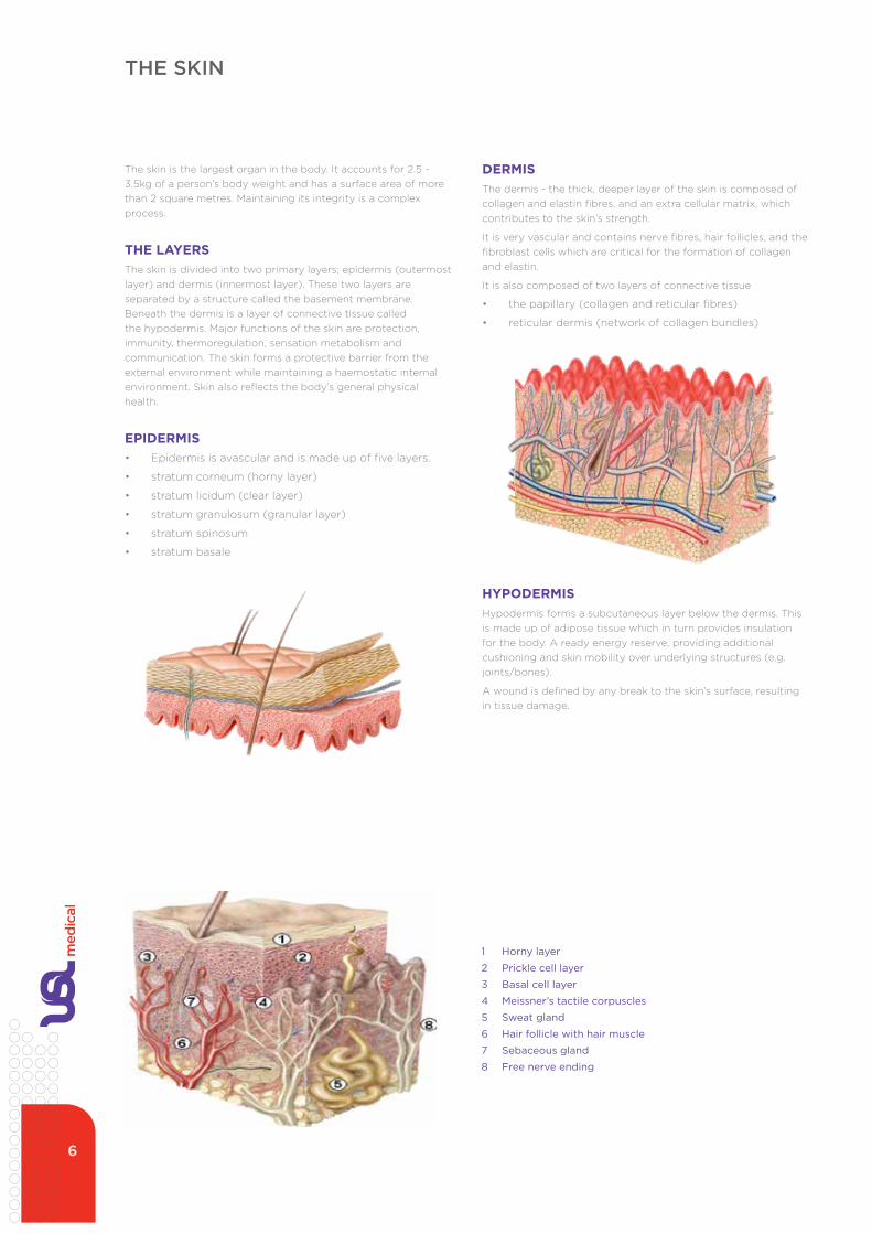
Wound infection is a serious problem. all wounds are considered contaminated as bacteria exist as part of the body’s natural flora. However, this does not necessarily mean infection or sepsis will develop.
acute and chronic wounds are both at risk of infection.
Risk factorsRisk factors can be local or systemic, these include:
Local• foreign material
• trauma
• hypoxia
• swelling
• location of wound e.g. peri-anal
• size of wound
Systemic• underlying disease
• smoking
• poor nutrition
• immunosupression
• alcoholism
• poor standard of hygiene
• poor general health
• multiple wounds
Bioburdenthe presence of bacteria in the wound creates a burden on the wound and its ability to heal. this burden is due to the fact that bacteria compete for the limited supply of oxygen and nutrients in the wound. achieving sterility in a wound is not possible, so the objective needs to achieve a host manageable bioburden.
Clinical signs of wound infection• pain
• heat
• swelling
• redness
• exudate (type, consistency and/or increase in amount)
Clinical signs of systemic infection• abnormal blood tests
• increased tiredness
• elevated temperature
Acute Wounds
Acute wounds are very different from chronic wounds. However a chronic wound can start off as a traumatic wound eg skin tears. Then develop into chronic wound as a result of variables associated with the healing. Acute wounds are usually caused by either surgery (intentional), and/or traumatic or burns. Assessment and categorisation of an acute wound should include the timeframes over which they have occurred. Is it healing in a timely, predictable, and measurable sequence. They usually heal easily without any complications.
Regardless of the cause of the acute wound restoring anatomical structure, physiological function and the wounds normal wound appearance is the focus for acute wounds.
Surgical Wounds
An acute Surgical Wound is a healthy and uncomplicated break in the skin resulting from surgery.
Surgical procedures are the commonly categorized by urgency, type of procedure, body system involved, degree of invasiveness, and special instrumentation.
• Based on timing eg:Elective surgery, Emergency surgery • Based on purpose eg Exploratory surgery • By type of procedure eg amputation, reconstructive surgery, cosmetic surgery
Factors that affect the healing of a post operative wound include:
• Age • Nutrition • Illness • Infection • Oxygen and circulatory status of the patient
Traumatic Wounds
A traumatic wound is a sudden accidental injury to the skin. This can be mild or severe depending on the trauma causing incident. Examples of the types of traumatic wounds include:
• Lacerations • Skin tears • Burns • Bites • Abrasions • Penetrating wounds
ContaminationContamination is the presence of non-replicating micro-organisms on the wound surface. Contaminating micro-organisms are derived from normal flora (skin) external environment (linen) and contaminants (urine/faeces). there is no host reaction.
ColonisationRefers to the presence of replicating bacteria without a host reaction or clinical signs and symptoms of infection. Bacteria in this phase do not necessitate treatment with antibiotics. inappropriate use of antibiotics in this phase is one of the many factors contributing to the prevalence of antiobiotic-resistant organisms.
Critical colonisationas the wound bioburden increases and further overwhelms the host, the wound reaches a period of critical colonisation. this means the wounds healing has been impeded, as a result of the bioburden.
Infectioninfection is present when the micro-organisms invade the tissues and there is a systemic response to them. the clinical appearance of the wound at this time depends on whether the wound is acute or chronic and the corresponding inflammatory responses.
14

MOIST WOUND HEALING
george Winter, Phd, university of London questioned if allowing wound to dry out was the best method of healing.
in 1962, george Winter published his landmark study in which he demonstrated that wounds healed faster with occlusive dressings than by air drying. a warm moist environment is necessary to encourage regranulation of epithelial tissue and local production of vascular endothelial growth factor (VegF). Occlusive dressings are designed to create a moist micro-environment that promotes wound healing.
Results: Wounds that had been covered by polymer film, epithelialised twice as quickly as the wounds exposed to air.
Winter postulated that epithelial cells in dry wounds have to negotiate the scab, consuming energy and time, whereas in moist wounds they migrate freely across a moist, vascular wound surface. Winter’s theory has been supported by other studies in addition other studies provided evidence that a moist environment can accelerate the inflammatory response, leading to faster cell proliferation and wound healing in deeper dermal wounds.
the principle of moist wound healing mimics the function of the epidermis. Our body is mainly composed of water, and the natural environment of a cell is moist; therefore, a dry cell is a dead cell. the diagram below demonstrates the benefits of moist wound healing from use of an occlusive dressing.
The benefits of a moist environment for wound healing are:• increases the rate of healing and improves the cosmetic
result
• better manages exudate
• decreases pain - moist wound bed insulates and protects nerve endings thereby reducing pain
• enhanced autolytic debridement – debrides the wound effectively
• prevents scab formation - scabs form a physical barrier to healing - decreased dehydration and cell death (neutrophils, macrophages, and fibroblast necessary for wound healing cannot thrive in a dry environment)
• increased angiogenesis
• increased re-epithelialisation (dry crusted wounds decrease supply of blood and nutrients which thus results in a barrier to cell migration and slowing of epithelialisation.)
• reduces scarring
Moist wound healing is considered to be the ideal environment for optimal wound healing. the work of zoologist george Winter (1927–1981) stimulated a great deal of research into the development of new types of dressings that completely revolutionised the care of wounds. Research before and since Winter’s work suggests that moisture under occlusive dressings promotes healing through moisture itself, some of the components of wound exudate, and the presence of low oxygen tension. Occlusive dressings increase cell proliferation and activity by retaining vital proteins and cytokines contained within wound exudate produced in response to injury. although there have been some concerns regarding increased infection in a moist wound environment, these concerns appear to be unfounded.
Diagram of a protected and an unprotected skin wound
Cell stripping and trauma caused during dry dressing change
Wound dressings
• the ideal dressing is one that:
• maintains a moist environment/moisture balance
• allows gaseous exchange
• provides thermal insulation
• provides a barrier to pathogens
• does not promote infection
• does not shed fibres or leak toxic substances
• protects against mechanical trauma eg; shearing and friction
• allows removal without traumatising new tissue
• is easy to apply, comfortable to wear and adaptable to body parts
• does not interfere with body function
• is cost effective
15

16
Surrounding Skin:
Healthy:
Erythema:
Oedematous:
Macerated:
Fragile:
Dry:
Dermatitis/Eczema:
Other:
Pain: (Pain Scale 1/10)
Severity:
Frequency:
Treatment Plan
Objectives:
Wound Cleansing:
Sterile Normal Saline:
Shower:
Tap Water:
Dressing Products:
Primary Dressing:
Secondary Dressing:
Dressing Retention Aid:
Notes:
Wound Assessment
Wound Type:
Acute:
Chronic:
Aetiology:
Duration Of Wound:
Location:
Dimensions:
Width-cm:
Length-cm:
Depth-cm:
Clinical Appearance:
Necrotic:
Sloughy:
Granulating:
Epitheliating:
Hypergranulating:
Exudate Amount:
Nil:
Low:
Moderate:
Heavy:
Exudate Type:
Serous:
Haemoserous:
Blood:
Purulent:
Condition Of Wound:
Clean:
Contaminated:
Infected:
Odour:
Nil:
Present:
General Assessment
Influencing Factors:
Age:
Underlying Disease:
Malignancy:
Diabetes:
Vascular/Arterial Function:
Smoking:
Psychological State:
Obesity/Cachexia:
Radiation Therapy:
Other:
Medication:
Steroids:
Cytotoxics:
Immunosuppressants:
Antibiotics:
NSAIDS:
Other:
Nutritional Status:
Good:
Average:
Poor:
Other:
Mobility:
Mobile:
Slight Impairment:
Gross Impairment:
Immobile:
Diagnostic Investigations:
Wound Documentation
Surname:
First Name:
Date:
Patient aSSeSSMent and WOund dOCuMentatiOn

PATIENT AND WOUND ASSESSMENT
ACCURATE ASSESSMENT OF THE PATIENTAND THE WOUNDassessment provides the foundation for the wound management plan for the patient.
accurate assessment and regular reassessment of all aspects of the wound and patient should be undertaken on admission and at each dressing change in order to monitor effectiveness of treatment regime and optimise wound healing. this can also be effectively achieved by the use of periodic photographs giving a visual record to support written documentation.
Reduce or eliminate causative factors• pressure, shearing forces, friction, circulatory
impairment, etc
• identify and manage infection
Provide systematic support for wound healing• good nutrition – dietary supplements may be required
• control of systemic conditions. e.g. blood sugar levels, oedema, cardiac disease, circulatory disorders
ASSESSMENT OF THE PATIENTan important aspect of wound management is to assess the patient as a whole and should include:-
• general physical condition and age
• activity level
• urinary and faecal continence
• other diseases e.g. diabetes, cardiac function
• current medications, treatments
• self care ability
• level of pain/tolerance
• location of wound/ability to reach dressing site
• skin condition
DESCRIbING AND DOCUMENTING A WOUND• mobility level
• nutritional status
• sensory functioning
• compliance with treatment
the initial cause of the wound and duration of treatment when describing a wound in the patient’s clinical record/wound assessment chart should include:
• type of wound and location
• aim of treatment; to promote healing/palliative
• wound size
• presence of necrotic tissue and/or slough
• condition of wound bed, surrounding skin and wound edges
• amount of exudate/drainage (noting any odour)
• presence of pain
• current dressing regime
• the progress of the wound healing according to the resident and/or carer, (this can further be evidenced with the use of photographs)
• ability to educate patients/family/caregiver
(T.I.M.E. FRAMEWORK)tiMe is an acronym to identify and manage the wounds imbalances therein improving patient outcomes.
TIME includes:
• Tissue management
• Inflammation and infection control
• Moisture balance and wound
• Edge assessment
this simple tool is a great reference to clearly establish and prioritise the care and treatment regimes required for each individual patient and wound.
(H.E.I.D.I. FRAMEWORK)Heidi is a holistic assessment tool which encompasses the patient and the wound, and all those factors influencing healing.
HEIDI includes:
• History
• Examination
• Investigate
• Diagnose
• Indicators for healing
Prior to assessing the wound thorough assessment of the patient to understand all the systemic and local factors that will affect healing is essential.
CHARACTERISTICS OF EXUDATE
Amountexudate is difficult to measure accurately, often described as light, moderate or heavy. Heavy exudate may result in electrolyte imbalance. it may be useful to use a wound drainage bag if possible.
dry: Wound bed is dry. there is no visible moisture. this may be the environment of choice for ischaemic wounds
Moist: Small amounts of fluid are visible when the dressing is removed. dressing changes are appropriate for dressing type
Wet: Small amounts of fluid are visible when dressing is removed; the primary dressing is extensively marked
Saturated: Primary dressing is wet and strike through is occurring; dressing change is required more frequently or the dressing type needs to change
Leaking: dressings are saturated and exudate is escaping from primary/secondary dressings; dressing change is required more frequently or the dressing type needs to change.
WOUND ASSESSMENT TOOLS
17

WOUND DOCUMENTATION CHART
Surrounding Skin:Healthy:Erythema:Oedematous:Macerated:Fragile:Dry:Dermatitis/Eczema:Other:
Pain: (Pain Scale 1/10)Severity:Frequency:
Treatment Plan
Objectives:
Wound Cleansing:Sterile Normal Saline:Shower:Tap Water:
Dressing Products:Primary Dressing:
Secondary Dressing:
Dressing Retention Aid:
Notes:
Wound Assessment
Wound Type:Acute:Chronic:
Aetiology:
Duration Of Wound:
Location:
Dimensions:Width-cm:Length-cm:Depth-cm:
Clinical Appearance:Necrotic:Sloughy:Granulating:Epitheliating:Hypergranulating:
Exudate Amount:Nil:Low:Moderate:Heavy:
Exudate Type:Serous:Haemoserous:Blood:Purulent:
Condition Of Wound:Clean:Contaminated:Infected:
Odour:Nil:Present:
General Assessment
Influencing Factors:Age:Underlying Disease:
Malignancy:Diabetes:Vascular/Arterial Function:
Smoking:Psychological State:
Obesity/Cachexia:Radiation Therapy:Other:
Medication:Steroids:
Cytotoxics:
Immunosuppressants:
Antibiotics:
NSAIDS:
Other:
Nutritional Status:Good:Average:Poor:Other:
Mobility:Mobile:Slight Impairment:Gross Impairment:Immobile:
Diagnostic Investigations:
Wound DocumentationSurname: First Name: Date:
Patient and Wound Document Chart
When performing a thorough wound and skin assessment, a pictorial record is helpful to help identify the wound site or sites. using the patient and wound document chart example shown here helps to document and record the patient’s wound and healing progress.
Dry:Moist:Wet:Saturated:Leaking:
Hydrate:
Decrease bioburden:
Absorption:
Debridement:
Protection:
Other:
18

19
Pain and WOund ManageMent

PAIN - THE FIFTH vITAL SIGN
THE FIvE vITAL SIGNS• blood pressure
• pulse
• respiration
• temperature
• pain
WHAT IS PAIN?Pain is an unpleasant sensory and emotional experience associated with actual or potential tissue damage or described in terms of such damage. international association for the Study of Pain [iaSP] the fact remains that pain is an alarm signal telling us that something in the body is wrong.
TWO MAjOR TYPES OF PAIN
Nociceptive (tissue) pain• results from tissue damage (mechanical, thermal or
chemical trauma to peripheral nerve fibres)
• is mediated at nociceptors widely distributed in skin tissue, bone, muscle and connective tissue
Nociceptive pain is described as sharp, dull, aching or throbbing pain.
Neuropathic (nerve) pain • damage or dysfunction to the peripheral or central
nervous system
• faulty signals are sent to the brain and experienced as pain
Neuropathic pain is described as a burning, tingling, shooting, electric-like, or lightning-like pain.
SENSORY RECEPTORS IN THE SKIN• pain
• touch/pressure
• temperature
• vibration
Every cm2 of skin contains around • 200 pain receptors (nociceptors)
• 15 receptors for pressure
• 6 for cold
• 1 for warmth
WHAT CAUSES PAIN?• injury to cells result in chemical release which in turn
leads to inflammation
• trauma/injury initiates immediate nerve impulses to brain
Clinical symptoms associated with nociceptor (inflammatory) response to tissue injury• increased temperature
• pain
• bruising
• swelling all beyond the immediate zone of injury.
This wound shows classic signs of inflammation with the reaction depicted beyond the injury site.
“Pain is a personal and subjective experience
that can only be felt by the sufferer.”
Katz and Melzack
“Pain is whatever the experiencing person says it is and exists
whenever they say it does.” McCaffery
20
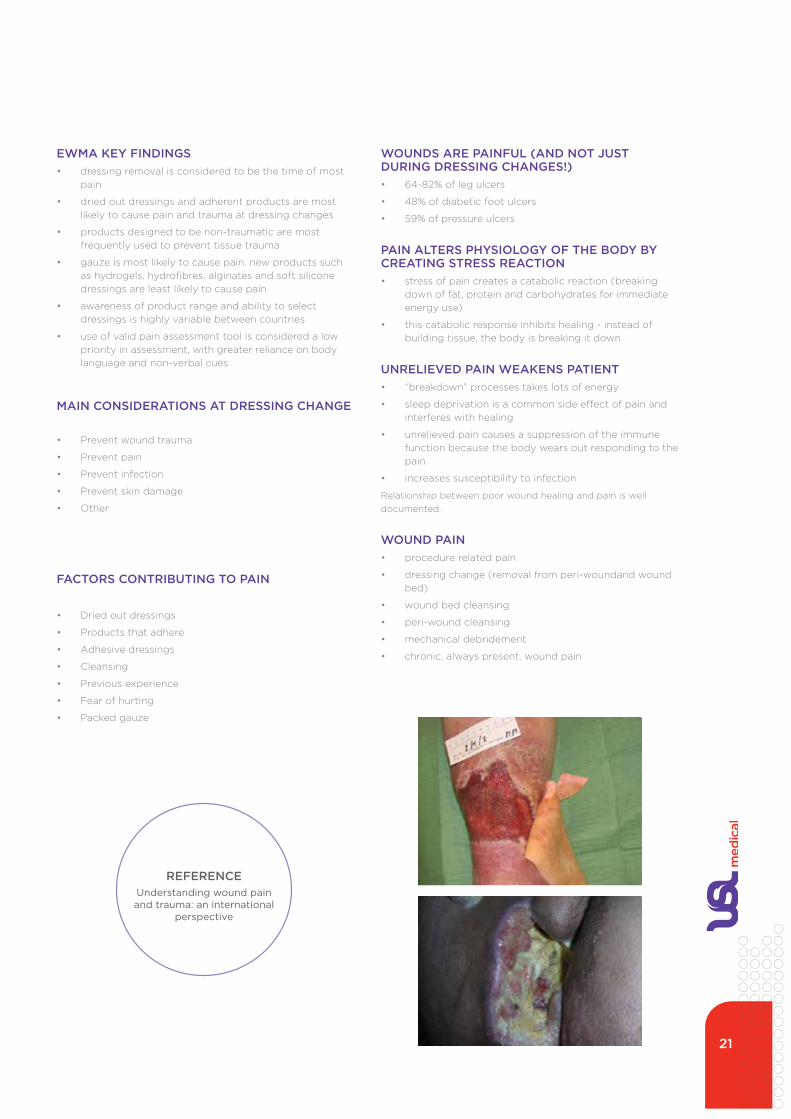
WOUNDS ARE PAINFUL (AND NOT jUST DURING DRESSING CHANGES!)• 64-82% of leg ulcers
• 48% of diabetic foot ulcers
• 59% of pressure ulcers
PAIN ALTERS PHYSIOLOGY OF THE bODY bY CREATING STRESS REACTION• stress of pain creates a catabolic reaction (breaking
down of fat, protein and carbohydrates for immediate energy use)
• this catabolic response inhibits healing - instead of building tissue, the body is breaking it down
UNRELIEvED PAIN WEAKENS PATIENT• “breakdown” processes takes lots of energy
• sleep deprivation is a common side effect of pain and interferes with healing
• unrelieved pain causes a suppression of the immune function because the body wears out responding to the pain
• increases susceptibility to infection
Relationship between poor wound healing and pain is well documented.
WOUND PAIN• procedure related pain
• dressing change (removal from peri-woundand wound bed)
• wound bed cleansing
• peri-wound cleansing
• mechanical debridement
• chronic, always present, wound pain
EWMA KEY FINDINGS• dressing removal is considered to be the time of most
pain
• dried out dressings and adherent products are most likely to cause pain and trauma at dressing changes
• products designed to be non-traumatic are most frequently used to prevent tissue trauma
• gauze is most likely to cause pain. new products such as hydrogels, hydrofibres, alginates and soft silicone dressings are least likely to cause pain
• awareness of product range and ability to select dressings is highly variable between countries
• use of valid pain assessment tool is considered a low priority in assessment, with greater reliance on body language and non-verbal cues
• Prevent wound trauma
• Prevent pain
• Prevent infection
• Prevent skin damage
• Other
• dried out dressings
• Products that adhere
• adhesive dressings
• Cleansing
• Previous experience
• Fear of hurting
• Packed gauze
MAIN CONSIDERATIONS AT DRESSING CHANGE
FACTORS CONTRIbUTING TO PAIN
REFERENCEUnderstanding wound pain and trauma: an international
perspective
21

PAIN - THE FIFTH vITAL SIGN
Increases metabolic rate of
tissue cells
Brings more nutrients and
oxygen to area
Fibrin barrier
Removal of damaged/dead tissue cells and pathogens
from area
Clotting proteins enter
area
Oedema (fluid in tissue spaces)
Increased blood flow into area
Blood vessels dilate
Capillaries become “leaky”
Neutrophils and then monocytes (and other
WBCs) enter area
PHYSIOLOGICAL PAIN PATHWAY
Injurious Agents
Cells damaged
Release kinins, histamine and other chemicals
Redness Heat Pain Swelling
Healing
Possible temporary limitation of
joint movement
22
23
nutRitiOn and WOund HeaLing
NUTRITION AND WOUND HEALING
nutrition is a fundamental part of normal cell function, integrity and tissue repair. Lack of adequate nutrition has been associated with increased morbidity and mortality in medical and surgical patients.
nursing has the primary responsibility for the initial nutritional assessment across all settings - hospital primary care and aged care facilities. early nutritional assessment is critical in an effort to identify those individuals at-risk for and experiencing malnutrition. it’s vital in providing positive wound healing outcomes.
DEFINITIONMalnutrition is undernutrition or overnutrition that is caused by a deficit or excess of nutrients in the diet.
NUTRIENT NEEDS FOR HEALINGnormal healing requires adequate protein, fat, and carbohydrates, as well as vitamins and minerals. deficiencies in any of these nutrients may result in delayed or impaired healing.
With wound injuries, more calories are needed than in an uninjured state. Calories are needed purely for energy alone and normal bodily functions. When an injury is part of the patient, then their nutritional needs should be assessed and revised accordingly.
nutritional assessment provides the basis for developing a nutritional plan. a physical examination and patient history are essential in obtaining the overall nutritional status of each patient.
nutritional support should provide a balanced intake of necessary nutrients based on the person’s energy and protein requirements.
the patient at risk or with existing malnutrition needs to be brought to the attention of the health care team so appropriate support can be provided. this includes referral to a dietician and/or physician.
NUTRITION AND WOUND HEALINGMalnutrition causes delays in wound healing and substantially can impact on the wound’s ability to heal. When a patient is debilitated by their wound their need for extra nutrition increases proportionally.
Some critical factors to consider when assessing a patient’s nutritional status are:
amino acids – derived from protein found in meat, fish, poultry, diary products and lentils. arginine (see table below) exisits as an amino acid and supports collagen formation.
Lipids – are fats and an excellent source of energy and can either be saturated or non-saturated. Saturated fats are responsible for raising blood cholesterol and mono or poly-unsaturated fats help lower blood cholesterol. essential fatty acids can be obtained from whole grains, mono or poly-unsaturated oils, nuts and fish.
Carbohydrates – are essential for the functioning of cellular energy. Critical for fibroblasts, collagen, leucocytes, synthesis of dna and Rna and nutrient absorption.
Vitamin C – is also known as absorbic acid. important in fibroblast production and immune responses thereby help enhance wound healing. Can gain this vitamin from kiwifruit, citrus fruits, capsicum, strawberries and rockmelon.
also iron and vitamins a, B, d (sunlight derived), K, and e play an important role in nutrition and wound healing, as well as trace elements.
SUMMARYnutritional assessment and support play an important role in successful wound healing. all patients with wounds should have their nutritional status evaluated and a plan put in place, which should be re-assessed regularly for effectiveness.
NUTRIENTS ESSENTIAL TO WOUND HEALING
Arginine• helps new skin tissue production
• helps increase blood flow to wound bed
• enhances immune system activity
Protein• provides high levels of protein to help wound healing.
wounds require 25-50% more protein intake
• helps build and maintain lean muscle mass
Vitamins A, C and antioxidants• enhance wound healing by reducing cellular damage
caused by chemical reactions
• play an important role on collagen synthesis
Zinc• required for wound repair
• required for tissue growth, skin integrity, cell mediated immunity and generalised host defence
24
25
SKin teaRS

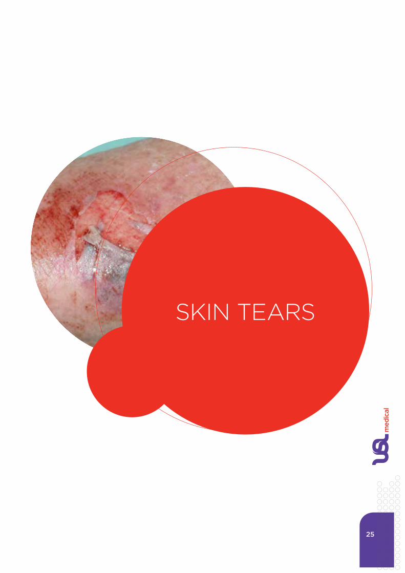
SKIN TEARS
RATIONALESkin tears are usually quite painful, and are the most common wound type amongst the elderly. it is estimated that at least 1.5 million skin tears occur annually among institutionalised adults in the uSa, with 14% of patients suffering from skin tears each month. a combined analysis suggests that the average distribution of skin tears is: head 4%, biceps area 16%, elbow 15%, forearm 29%, top of hand 18%, trunk/back 2%, shin 13%, top of foot 3%, and other sites 1%. they can become infected and may heal slowly in compromised patients. as our population ages, it is becoming increasingly important for all health care providers to learn how to manage skin tears to promote quick, pain-free healing while preventing infection and other complications.
DEFINITION in 1993, Payne and Martin published their revised definition of a skin tear:
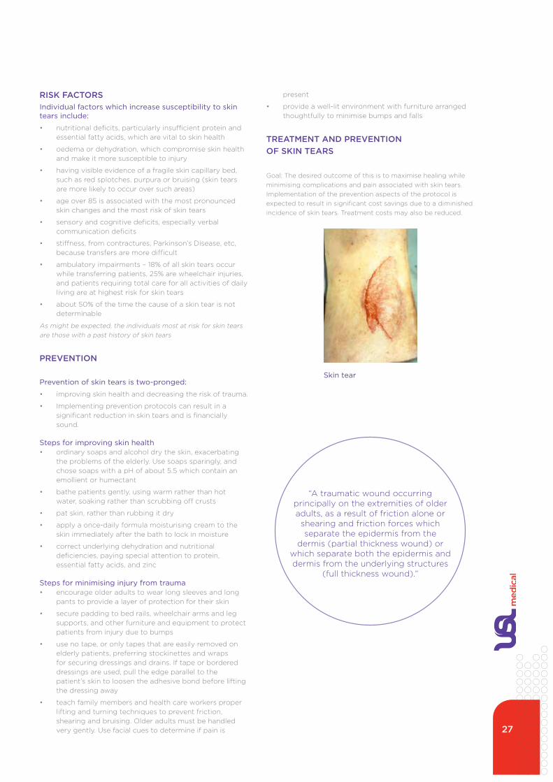
“a traumatic wound occurring principally on the extremities of older adults, as a result of friction alone or shearing and friction forces which separate the epidermis from the dermis (partial thickness wound) or which separate both the epidermis and dermis from the underlying structures (full thickness wound).”
Payne and Martin also developed the standard system for categorising skin tears (detailed on page 30).
PatHoPHysiology oF skin tears
Skin tears become increasingly common with age because:• the skin thins internally and contains less subcutaneous
fat, especially at the shins, face and back of the hands
• elasticity is reduced because collagen and elastin in the skin degenerates
• natural lubrication is diminished due to reduced functioning of the sweat and sebaceous glands
• the capillaries become fragile and disorganised, leading to easy bruising and reduced blood supply
• the dermal-epidermal junction is weakened because the rete ridges that keep the dermis and epidermis locked together are flattened, making aging skin especially susceptible to injury from friction and shear; when the epidermis is moved, the dermis may remain stationary rather than moving with it
Steroid use thins the skin and anticoagulants damage capillaries, so younger persons on these medications are also at increased risk for skin tears.
SKIN TEAR PROTOCOL
RATIONALE Skin tears are usually quite painful,1 and are the most common wound type among the elderly.2 It is estimated that at least 1.5 million skin tears occur annually among institutionalized adults in the USA,1,2 with 14% of patients suffering from skin tears each month.3,4 A combined analysis suggests that the average distribution of skin tears is: head 4%, biceps area 16%, elbow 15%, forearm 29%, top of hand 18%, trunk/back 2%, shin 13%, top of foot 3%, and other sites 1%.2,5 They can become infected and may heal slowly in compromised patients.4,6 As our population ages, it is becoming increasingly important for all health care providers to learn how to manage skin tears to promote quick, pain-free healing while preventing infection and other complications. DEFINITION In 1993, Payne and Martin published their revised definition of a skin tear: “A traumatic wound occurring principally on the extremities of older adults, as a result of friction alone or shearing and friction forces which separate the epidermis from the dermis (partial thickness wound) or which separate both the epidermis and dermis from the underlying structures (full thickness wound).”7 Payne and Martin also developed the standard system for categorizing skin tears (detailed below).7 PATHOPHYSIOLOGY OF SKIN TEARS Skin tears become increasingly common with age because: • the skin thins internally and contains less subcutaneous fat,8 especially at the shins, face and back of the hands1 • elasticity is reduced because collagen and elastin in the skin degenerates7,8,9 • natural lubrication is diminished due to reduced functioning of the sweat and sebaceous glands9 • the capillaries become fragile and disorganized, leading to easy bruising and reduced blood supply7,8,9 • the dermal-epidermal junction is weakened because the rete ridges that keep the dermis and epidermis locked together are
flattened, making aging skin especially susceptible to injury from friction and shear; when the epidermis is moved, the dermis may remain stationary rather than moving with it 7,8,9
Steroid use thins the skin and anticoagulants damage capillaries,6 so younger persons on these medications are also at increased risk for skin tears.5 RISK FACTORS Individual factors which increase susceptibility to skin tears include: • nutritional deficits, particularly insufficient protein and essential fatty acids, which are vital to skin health5,9 • edema or dehydration, which compromise skin health and make it more susceptible to injury3,5,8 • having visible evidence of a fragile skin capillary bed, such as red splotches, purpura or bruising9 (skin tears are more likely to
occur over such areas)5 • age over 85 is associated with the most pronounced skin changes and the most risk of skin tears5,9,10 • sensory and cognitive deficits,5 especially verbal communication deficits • stiffness, from contractures, Parkinson’s Disease, etc., because transfers are more difficult5 • ambulatory impairments – 18% of all skin tears occur while transferring patients,2,5 25% are wheelchair injuries,2 and patients
requiring total care for all activities of daily living are at highest risk for skin tears3 • about 50% of the time the cause of a skin tear is not determinable1,2
As might be expected, the individuals most at risk for skin tears are those with a past history of skin tears1,2,5 PREVENTION Prevention of skin tears is two-pronged: improving skin health and decreasing the risk of trauma. Implementing prevention protocols can result in a significant reduction in skin tears and is financially sound.4
Steps for Improving Skin Health: 1. Ordinary soaps and alcohol dry the skin, exacerbating the problems of the elderly.9 Use soaps sparingly,9 and chose soaps with a
pH of about 5.59 which contain an emollient12 or humectant.9 2. Bathe patients gently, using warm rather than hot water13, soaking rather than scrubbing off crusts.14,15 3. Pat skin, rather than rubbing it dry.14,15 4. Apply a once-daily formula moisturizing cream to the skin immediately after the bath to lock in moisture.4 5. Correct underlying dehydration and nutritional deficiencies, paying special attention to protein, essential fatty acids, and zinc.5,9
Section 1, Page 1 of 4
DISTRIbUTION OF SKIN TEARS
26
RISK FACTORSIndividual factors which increase susceptibility to skin tears include:
• nutritional deficits, particularly insufficient protein and essential fatty acids, which are vital to skin health
• oedema or dehydration, which compromise skin health and make it more susceptible to injury
• having visible evidence of a fragile skin capillary bed, such as red splotches, purpura or bruising (skin tears are more likely to occur over such areas)
• age over 85 is associated with the most pronounced skin changes and the most risk of skin tears
• sensory and cognitive deficits, especially verbal communication deficits
• stiffness, from contractures, Parkinson’s disease, etc, because transfers are more difficult
• ambulatory impairments – 18% of all skin tears occur while transferring patients, 25% are wheelchair injuries, and patients requiring total care for all activities of daily living are at highest risk for skin tears
• about 50% of the time the cause of a skin tear is not determinable
as might be expected, the individuals most at risk for skin tears are those with a past history of skin tears
PREvENTION
Prevention of skin tears is two-pronged:
• improving skin health and decreasing the risk of trauma.
• implementing prevention protocols can result in a significant reduction in skin tears and is financially sound.
Steps for improving skin health• ordinary soaps and alcohol dry the skin, exacerbating
the problems of the elderly. use soaps sparingly, and chose soaps with a pH of about 5.5 which contain an emollient or humectant
• bathe patients gently, using warm rather than hot water, soaking rather than scrubbing off crusts
• pat skin, rather than rubbing it dry
• apply a once-daily formula moisturising cream to the skin immediately after the bath to lock in moisture
• correct underlying dehydration and nutritional deficiencies, paying special attention to protein, essential fatty acids, and zinc
Steps for minimising injury from trauma• encourage older adults to wear long sleeves and long
pants to provide a layer of protection for their skin
• secure padding to bed rails, wheelchair arms and leg supports, and other furniture and equipment to protect patients from injury due to bumps
• use no tape, or only tapes that are easily removed on elderly patients, preferring stockinettes and wraps for securing dressings and drains. if tape or bordered dressings are used, pull the edge parallel to the patient’s skin to loosen the adhesive bond before lifting the dressing away
• teach family members and health care workers proper lifting and turning techniques to prevent friction, shearing and bruising. Older adults must be handled very gently. use facial cues to determine if pain is
present
• provide a well-lit environment with furniture arranged thoughtfully to minimise bumps and falls
TREATMENT AND PREvENTIONOF SKIN TEARS
goal: the desired outcome of this is to maximise healing while minimising complications and pain associated with skin tears. implementation of the prevention aspects of the protocol is expected to result in significant cost savings due to a diminished incidence of skin tears. treatment costs may also be reduced.
Skin tear
“a traumatic wound occurring principally on the extremities of older adults, as a result of friction alone or
shearing and friction forces which separate the epidermis from the
dermis (partial thickness wound) or which separate both the epidermis and dermis from the underlying structures
(full thickness wound).”
27

SKIN TEARSPayne-Martin classification system (revised 1993):
Category I: Skin tears without tissue loss
A. LINEAR TYPE
a linear skin tear is a full thickness wound occuring in a wrinkle or furrow of the skin. Both the epidermis and the dermis are pulled apart as if an incision has been made, exposing the tissue below
b. FLAP TYPE a flap type skin tear is a partial thickness wound in which the epidermal flap can be completely approximated or approximated so that no more than one (1) millimetre of dermis is exposed
Category II: Skin tears with partial tissue loss
A. SCANT TISSUE LOSS TYPE
a skin tear with scant tissue loss is a partial thickness wound in which 25% or less of the epidermal flap is lost and in which at least 75% or more of the dermis is covered by the flap
b. MODERATE-TO-LARGE TISSUE LOSS TYPE
a skin tear with moderate-to-large tissue loss is a partial thickness wound in which more than 25% of the epidermal flap is lost and in which more than 25% of the dermis is exposed
Category III: Skin tears with complete tissue loss
a skin tear with complete tissue loss is a partial thickness wound in which the epidermal flap is absent
PAYNE-MARTIN CLASSIFICATION SYSTEM (revised 1993):7 Category I: Skin tears without tissue loss A. Linear type A linear skin tear is a full thickness wound
occurs in a wrinkle or furrow of the skin. Both the epidermis and the dermis are pulled apart as if an incision has been made, exposing the tissue below.
B. Flap type
A flap type skin tear is a partial thickness wound in which the epidermal flap can be completely approximated or approximated so that no more than one (1) millimeter of dermis is exposed.
Category II: Skin tears with partial tissue loss A. Scant tissue loss type A skin tear with scant tissue loss is a partial
thickness wound in which 25 percent or less of the epidermal flap is lost and in which at least 75 percent or more of the dermis is covered by the flap.
B. Moderate-to-large tissue loss type
A skin tear with moderate-to-large tissue loss is a partial thickness wound in which more than 25 percent of the epidermal flap is lost and in which more than 25 percent of the dermis is exposed.
Category III: Skin tears with complete tissue loss
A skin tear with complete tissue loss is a partial thickness wound in which the epidermal flap is absent.
Section 1, Page 3 of 4
PAYNE-MARTIN CLASSIFICATION SYSTEM (revised 1993):7 Category I: Skin tears without tissue loss A. Linear type A linear skin tear is a full thickness wound
occurs in a wrinkle or furrow of the skin. Both the epidermis and the dermis are pulled apart as if an incision has been made, exposing the tissue below.
B. Flap type
A flap type skin tear is a partial thickness wound in which the epidermal flap can be completely approximated or approximated so that no more than one (1) millimeter of dermis is exposed.
Category II: Skin tears with partial tissue loss A. Scant tissue loss type A skin tear with scant tissue loss is a partial
thickness wound in which 25 percent or less of the epidermal flap is lost and in which at least 75 percent or more of the dermis is covered by the flap.
B. Moderate-to-large tissue loss type
A skin tear with moderate-to-large tissue loss is a partial thickness wound in which more than 25 percent of the epidermal flap is lost and in which more than 25 percent of the dermis is exposed.
Category III: Skin tears with complete tissue loss
A skin tear with complete tissue loss is a partial thickness wound in which the epidermal flap is absent.
Section 1, Page 3 of 428
29
Leg uLCeRS
OvERvIEW
Causesit is thought that alterations in blood flow due to venous hypertension from chronic venous insufficiency (CVi) cause white blood cells to adhere to the walls of the smaller vessels of the lower leg. the white blood cells may plug the vessels or leak from them and release tissue destroying enzymes and inflammatory mediators into the tissues. Venous ulcers are likely caused by a prolonged and chaotic local inflammatory state induced by this aspect of venous hypertension. Mild trauma can also precipitate venous leg ulcers in patients with CVi. Venous hypertension causes fluid to leak into the tissues; this oedema compresses the capillaries, decreasing blood flow to the skin and increasing damage. Chronic inflammation prevents venous ulcers from healing at the pace of acute wounds.
CharacteristicsVenous ulcers are predominantly in the ankle or lower calf (gaiter
area). the medial ankle is by far the most common site because the long saphenous vein is more superficial and has the greatest curvature there and because this is the area of the highest pressure within the entire venous system. Venous ulcers tend to produce copious exudate when uncompressed and are usually
vENOUS LEG ULCERS
Varicose veins resulting in venous hypertension
Venous leg ulcers
shallow, irregular, and often have a yellow fibrous bed.
Risk factors for venous ulcersPatients with venous ulcers can have an increased tendency for blood to clot (thrombophilia), which is often a congenital condition. Other risk factors for ulceration include:
• history of phlebitis or deep vein thrombosis (dVt)
• history of leg trauma, such as a fracture, but even minor trauma can cause vessel damage
• vigorous exercise (which may cause microtrauma)
• CVi or varicose veins in the patient or maternal family history
• pregnancy, obesity or ascites, all of which cause increased abdominal pressure, femoral vein compression, and decreased mobility
• multiple pregnancies
• greater height, which increases venous system pressure
• recent lower leg oedema, which may be caused by chronic heart failure or medications
• diabetes, which decreases skin health
• increasing age, because valve damage is permanent and cumulative
• poor calf muscle function or decreased range of motion (ROM) in the foot. Standing or sitting for prolonged periods of time with the feet dependent dramatically increases susceptibility to ulceration.
Assessing oedemaLower leg circumferences should be measured at the widest point and at 2.5cm above the malleolus. it is important to rule out systemic and reversible causes of oedema, such as medication reactions, heart or liver failure and low protein. Suspect accompanying lymphoedema, if skin at the base of the second toe is so thick that you cannot pick up a fold of it (Stemmer Sign). Oedema caused by lymphoedema does not respond significantly to limb elevation. With standard compression, fluid from lymphoedema may simply move to proximal areas. Refer patients for testing to rule out lymphoedema if oedema does not improve despite elevation and compression.
Biopsyalthough venous disease is the most common cause of leg ulcers, it is by no means the only cause. Sickle cell, arterial insufficiency, rheumatologic disorders, cancer, infection, medication reactions, vasculitis and a host of other systemic illnesses can lead to leg ulcers which mimic venous ulcers but require different interventions. it is important to note that the chronic inflammation in venous ulcers can cause them to degenerate into a malignancy. So, if there is no significant decrease in size or increase in granulation of the wound after six weeks of treatment as described in this protocol, a biopsy of the wound base and margin should be considered to confirm the pathology of the leg ulcer.
Improve nutritionProtein deficiency can result in leg oedema. adequate levels of protein, vitamin a, vitamin C and zinc are needed for wound healing. Wound patients require 25–35 kcal/kg/day with 1.0–1.2 g/kg/day protein. Supplementation should be individualised.
30
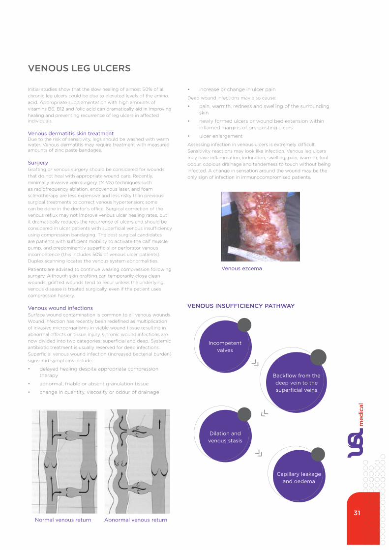
initial studies show that the slow healing of almost 50% of all chronic leg ulcers could be due to elevated levels of the amino acid. appropriate supplementation with high amounts of vitamins B6, B12 and folic acid can dramatically aid in improving healing and preventing recurrence of leg ulcers in affected individuals.
Venous dermatitis skin treatmentdue to the risk of sensitivity, legs should be washed with warm water. Venous dermatitis may require treatment with measured amounts of zinc paste bandages.
Surgerygrafting or venous surgery should be considered for wounds that do not heal with appropriate wound care. Recently, minimally invasive vein surgery (MiVS) techniques such as radiofrequency ablation, endovenous laser, and foam sclerotherapy are less expensive and less risky than previous surgical treatments to correct venous hypertension; some can be done in the doctor’s office. Surgical correction of the venous reflux may not improve venous ulcer healing rates, but it dramatically reduces the recurrence of ulcers and should be considered in ulcer patients with superficial venous insufficiency using compression bandaging. the best surgical candidates are patients with sufficient mobility to activate the calf muscle pump, and predominantly superficial or perforator venous incompetence (this includes 50% of venous ulcer patients). duplex scanning locates the venous system abnormalities.
Patients are advised to continue wearing compression following surgery. although skin grafting can temporarily close clean wounds, grafted wounds tend to recur unless the underlying venous disease is treated surgically, even if the patient uses compression hosiery.
Venous wound infectionsSurface wound contamination is common to all venous wounds. Wound infection has recently been redefined as multiplication of invasive microorganisms in viable wound tissue resulting in abnormal effects or tissue injury. Chronic wound infections are now divided into two categories: superficial and deep. Systemic antibiotic treatment is usually reserved for deep infections. Superficial venous wound infection (increased bacterial burden) signs and symptoms include:
• delayed healing despite appropriate compression therapy
• abnormal, friable or absent granulation tissue
• change in quantity, viscosity or odour of drainage
vENOUS LEG ULCERS
Normal venous return Abnormal venous return
• increase or change in ulcer pain
deep wound infections may also cause:
• pain, warmth, redness and swelling of the surrounding skin
• newly formed ulcers or wound bed extension within inflamed margins of pre-existing ulcers
• ulcer enlargement
assessing infection in venous ulcers is extremely difficult. Sensitivity reactions may look like infection. Venous leg ulcers may have inflammation, induration, swelling, pain, warmth, foul odour, copious drainage and tenderness to touch without being infected. a change in sensation around the wound may be the only sign of infection in immunocompromised patients.
vENOUS INSUFFICIENCY PATHWAY
Venous ezcema
Incompetent valves
Backflow from the deep vein to the superficial veins
Dilation and venous stasis
Capillary leakage and oedema
31

LEG ULCER MANAGEMENT
MANAGEMENTManagement of venous hypertension helps prevent venous ulcer formation and is the foundation of venous wound treatment.
ElevationContinuous bed rest with leg elevation will decrease oedema from CVi, but this is rarely practical.
ExerciseStanding or sitting for prolonged periods of time with the feet dependent should be avoided, as should vigorous exercise, but a daily walking program (30 minutes of brisk walking twice a day) should be encouraged to improve circulation and for quality of life. gait training and simple restorative exercises to increase the ankle joint range of motion and strength (see diagrams) address the underlying causes of CVi.increases in the depth of breathing during exercise aid in venous and lymphatic return. even nonambulatory patients can increase blood flow and decrease oedema through passive foot/ankle/lower limb exercises and weight bearing with assistance. Rocking in a chair, calf pump action while lying supine, and deep breathing all help with venous return. in contrast, prolonged standing or sitting, wearing high heels and crossing of the legs reduces venous return.
Compression
the cornerstone of chronic venous insufficiency therapy is
managementManagement of venous hypertension helps prevent venous ulcer formation and is the foundation of venous wound treatment.
Elevation:37
Continuous bed rest with leg elevation will decrease edema from CVI, but this is rarely practical.1 If at all possible, to decrease edema and increase white blood cell velocity, legs should be elevated above the heart level for 30 minutes, 3-4 times daily.57 In addition, the foot of the patient’s bed should be raised 15-20 cm (6–8 inches) if tolerated.1,4,5,27,45,57
Exercise:37
Standing or sitting for prolonged periods of time with the feet dependent should be avoided, as should vigorous exercise, but a daily walking program (30 minutes of brisk walking twice a day) should be encouraged to improve circulation and for quality of life.1,5,15,28,40,45 Gait training and simple restorative exercises to increase the ankle joint range of motion and strength (see diagrams) address the underlying causes of CVI.12,15,28,45,52,58 Increases in the depth of breathing during exercise aid in venous and lymphatic return.2,4 Even nonambulatory patients can increase blood flow and decrease edema through passive foot/ankle/lower limb exercises and weight bearing with assistance.4,15,59 Rocking in a chair, calf pump action while lying supine, and deep breathing all help with venous return.2,15 In contrast, prolonged standing or sitting, wearing high heels and crossing of the legs reduces venous return.15
Compression: The cornerstone of chronic venous insufficiency therapy is graduated lower leg compression.1,4,15,37,48,59 Graduated compression increases the blood flow velocity and lymph drainage, so it works on the most probable cause of the venous ulceration (leakage of injurious substances from the vessels) as well as decreasing superficial venous system pressure and reducing edema.1,4,20 The reduction in venous hypertension and edema that compression affords results in decreased leakage of exudate and increased cutaneous blood flow.1 Valves that are not touching because of venous distension are
pushed back into approximation by compression.1,20 Compression enhances fibrinolysis and may improve lipodermatosclerosis.1,4,13,48,60 Patients who wear graduated compression consistently have significantly improved healing rates and decreased recurrence rates.1,15,61 Many also report relief of venous pain.13,18,32 Compression for life is essential to prevent recurrence.1,5,15,27,38,48,52 Two basic types of compression systems are used for treating chronic venous insufficiency: elastic and inelastic. Research has not shown the superiority of any one system of high graduated compression over the others.48,61,62
Specific methods of applying graduated compression are discussed on pages Sec 1:8-9.
Medication and co-morbidity management: Patients may need alternatives to medications that cause edema, such as certain hormones, antihypertensives, NSAIDs and COX-2 inhibitors.39,40 Monitor PT times and anticoagulant therapy.15 Diabetic patients should control glucose levels.37 The oral medication pentoxifylline, which improves blood flow in the small vessels, is an effective adjunct in treating venous ulcers and should be added if needed.3,4,7,13,15,52,63,64
Weight loss: A weight reduction program is advisable for obese patients,5,15,27,40 and bariatric surgery may be indicated for super-obese (BMI>60) venous ulcer patients.65
Avoid vasoconstriction: Vasoconstriction decreases peripheral blood flow. Causes of vasoconstriction, such as cold environment, dehydration, stress, pain,5,52 and cigarette smoking should be eliminated.4,5,15,37,40,52
Pain control: Pain in venous ulcers is usually caused by tissue damage and the resultant inflammation.66,67 Inflammation leads to more pain, even with usually non-painful stimuli, by exciting the nociceptors.67 Pain causes decreased mobility, which causes increased ulceration, which causes more pain. Pain causes a stress reaction,14,68 which increases the risk of ulcer infection14,40,69 and creates another destructive cycle because infection is a major cause of pain.67,70
Great gains can be achieved by breaking these pain cycles. Controlling excessive activities of inflammatory mediators may reduce wound pain and enhance healing.67 PolyMem and PolyMem Silver wound dressings are not only atraumatic to the wound bed when applied and removed, they also help inhibit the nociceptor response at the wound site,71,72 which can decrease both pain and the additional inflammation caused by inflammatory mediators.72,73,74
Assess location, intensity, quality, and duration of pain, as well as pain precipitators and pain relievers.5 Negative pressure therapy has been associated with increased wound pain60,67 and some dressings cause burning on contact with wounds.18,50 Cooling the ulcer during dressing changes is painful and should be avoided.18 Manage pain with leg elevation and compression,15 PolyMem dressings,71,72,74,75,76,77,78
analgesics, relaxation, etc.5
Every hour, do 30 foot raises
Every hour do 30 foot extensions
External CompressionVein Support
Sec1:5
Every hour do 30 foot raises
Every hour do 30 foot extensions
for treating chronic venous insufficiency: elastic and inelastic.
Specific methods of applying graduated compression are discussed further in this section.
Medication and co-morbidity managementPatients may need alternatives to medications that cause oedema.
Weight lossa weight reduction program is advisable for obese patients, and bariatric surgery may be indicated for super-obese (BMi>60) venous ulcer patients.
Avoid vasoconstrictionVasoconstriction decreases peripheral blood flow. Causes of vasoconstriction, such as cold environment, dehydration, stress, pain, and cigarette smoking should be eliminated.
Pain controlPain in venous ulcers is usually caused by tissue damage and the resultant inflammation. inflammation leads to more pain, even with usually non-painful stimuli, by exciting the nociceptors. Pain causes decreased mobility, which causes increased ulceration, which causes more pain. Pain causes a stress reaction, which increases the risk of ulcer infection and creates another destructive cycle because infection is a major cause of pain.
assess location, intensity, quality, and duration of pain, as well as pain precipitators and pain relievers. Manage pain with leg elevation and compression, PolyMem® dressings, analgesics, relaxation, etc.
graduated lower leg compression. graduated compression increases the blood flow velocity and lymph drainage, so it works on the most probable cause of the venous ulceration (leakage of injurious substances from the vessels) as well as decreasing superficial venous system pressure and reducing oedema. the reduction in venous hypertension and oedema that compression affords results in decreased leakage of exudate and increased cutaneous blood flow. Valves that are not touching because of venous distension are pushed back into approximation by compression. Patients who wear graduated compression consistently have significantly improved healing rates and decreased recurrence rates. Many also report relief of venous pain. Compression for life is essential to prevent recurrence. two basic types of compression systems are used
managementManagement of venous hypertension helps prevent venous ulcer formation and is the foundation of venous wound treatment.
Elevation:37
Continuous bed rest with leg elevation will decrease edema from CVI, but this is rarely practical.1 If at all possible, to decrease edema and increase white blood cell velocity, legs should be elevated above the heart level for 30 minutes, 3-4 times daily.57 In addition, the foot of the patient’s bed should be raised 15-20 cm (6–8 inches) if tolerated.1,4,5,27,45,57
Exercise:37
Standing or sitting for prolonged periods of time with the feet dependent should be avoided, as should vigorous exercise, but a daily walking program (30 minutes of brisk walking twice a day) should be encouraged to improve circulation and for quality of life.1,5,15,28,40,45 Gait training and simple restorative exercises to increase the ankle joint range of motion and strength (see diagrams) address the underlying causes of CVI.12,15,28,45,52,58 Increases in the depth of breathing during exercise aid in venous and lymphatic return.2,4 Even nonambulatory patients can increase blood flow and decrease edema through passive foot/ankle/lower limb exercises and weight bearing with assistance.4,15,59 Rocking in a chair, calf pump action while lying supine, and deep breathing all help with venous return.2,15 In contrast, prolonged standing or sitting, wearing high heels and crossing of the legs reduces venous return.15
Compression: The cornerstone of chronic venous insufficiency therapy is graduated lower leg compression.1,4,15,37,48,59 Graduated compression increases the blood flow velocity and lymph drainage, so it works on the most probable cause of the venous ulceration (leakage of injurious substances from the vessels) as well as decreasing superficial venous system pressure and reducing edema.1,4,20 The reduction in venous hypertension and edema that compression affords results in decreased leakage of exudate and increased cutaneous blood flow.1 Valves that are not touching because of venous distension are
pushed back into approximation by compression.1,20 Compression enhances fibrinolysis and may improve lipodermatosclerosis.1,4,13,48,60 Patients who wear graduated compression consistently have significantly improved healing rates and decreased recurrence rates.1,15,61 Many also report relief of venous pain.13,18,32 Compression for life is essential to prevent recurrence.1,5,15,27,38,48,52 Two basic types of compression systems are used for treating chronic venous insufficiency: elastic and inelastic. Research has not shown the superiority of any one system of high graduated compression over the others.48,61,62
Specific methods of applying graduated compression are discussed on pages Sec 1:8-9.
Medication and co-morbidity management: Patients may need alternatives to medications that cause edema, such as certain hormones, antihypertensives, NSAIDs and COX-2 inhibitors.39,40 Monitor PT times and anticoagulant therapy.15 Diabetic patients should control glucose levels.37 The oral medication pentoxifylline, which improves blood flow in the small vessels, is an effective adjunct in treating venous ulcers and should be added if needed.3,4,7,13,15,52,63,64
Weight loss: A weight reduction program is advisable for obese patients,5,15,27,40 and bariatric surgery may be indicated for super-obese (BMI>60) venous ulcer patients.65
Avoid vasoconstriction: Vasoconstriction decreases peripheral blood flow. Causes of vasoconstriction, such as cold environment, dehydration, stress, pain,5,52 and cigarette smoking should be eliminated.4,5,15,37,40,52
Pain control: Pain in venous ulcers is usually caused by tissue damage and the resultant inflammation.66,67 Inflammation leads to more pain, even with usually non-painful stimuli, by exciting the nociceptors.67 Pain causes decreased mobility, which causes increased ulceration, which causes more pain. Pain causes a stress reaction,14,68 which increases the risk of ulcer infection14,40,69 and creates another destructive cycle because infection is a major cause of pain.67,70
Great gains can be achieved by breaking these pain cycles. Controlling excessive activities of inflammatory mediators may reduce wound pain and enhance healing.67 PolyMem and PolyMem Silver wound dressings are not only atraumatic to the wound bed when applied and removed, they also help inhibit the nociceptor response at the wound site,71,72 which can decrease both pain and the additional inflammation caused by inflammatory mediators.72,73,74
Assess location, intensity, quality, and duration of pain, as well as pain precipitators and pain relievers.5 Negative pressure therapy has been associated with increased wound pain60,67 and some dressings cause burning on contact with wounds.18,50 Cooling the ulcer during dressing changes is painful and should be avoided.18 Manage pain with leg elevation and compression,15 PolyMem dressings,71,72,74,75,76,77,78
analgesics, relaxation, etc.5
Every hour, do 30 foot raises
Every hour do 30 foot extensions
External CompressionVein Support
Sec1:5
External compression vein support
32

ARTERIAL ULCERS
OvERvIEWarterial ulcers are also known as ischemic ulcers. they occur as a result of severe tissue ischemia due to arterial insufficiency. they are known to be extremely painful and have potential to represent potential limb loss. they account for approximately 5-20% of all leg ulcers. the management of these ulcers is multifaceted and unless tissue perfusion is restored or improved progress onto extensive infection and/or gangrene is likely.
arterial insufficiency occurs when arterial blood flow is restricted or obstructed. Blockage can occur in any artery and appear anywhere along the arterial branches from the aorta to the capillary.
Causesthe most common cause of arterial ulcers is atherosclerosis involving the peripheral circulation. the most commonly affected vessels are the aortic, femoral, iliac, and popliteal arteries. Patients at high risk for developing atherosclerosis include men, cigarette smokers, and those individuals with hypertension, diabetes mellitus or hyperlipidemia. advanced age places the patient at even more risk.
atherosclerotic disease causes plaque formation and a hardening of the vessel wall. an occluded artery results in a chronic reduction in blood flow which impacts on the limb tissues.
Indicators of critical limb ischaemia• nonpalpable pulse
• ankle brachial index <4
• rest pain
• ulceration or gangrene
Assessmentassessment of a patient with an arterial ulcer needs to be very thorough. a complete patient history needs to be taken along with a physical examination. History of the pain caused by arterial insufficiency is important to document. gather relevant information about the type of pain experienced - is the pain at rest or when the foot is elevated or hanging.
diagnostic testing for arterial ulcers should be completed to assess arterial flow to extremities before deciding on best management plan. Some commonly used testing methods are:
• duplex ultrasonography
• ankle-brachial index
• arteriography
Management of arterial ulcersthe goal of management is to improve perfusion to the ulcer supplying essential oxygenation and nutrients to support the healing process.
the prognosis for the wound to heal is directly related to the ability to provide reperfusion of the tissues.
the specific interventions for the patient are determined by the severity of the ischaemia and the patient’s overall health. Some of the interventions include:
• surgical options eg arterial bypass grafts
• medication to reduce thrombotic events eg anticoagulants
• angioplasty and stents
• amputation (usually last resort)
Arterial ulcers
33

DIAbETIC ULCERS
DIAbETIC ULCERSdiabetes Mellitus is a metabolic disorder that results in hyperglycaemia as a result of increased insulin. due to this impaired perfusion and other complications eg infection, neuropathy, repeated trauma, healing of the diabetic ulcer is extremely challenging. the lower extremity metabolic changes that occur as a result of diabetes exacerbate neuropathy (nerve pain).
insulin is critical for transporting glucose into the cells where it is used as fuel or stored as glycogen. an insulin deficiency compromises important functions and therefore diabetics are much more susceptible to developing foot ulcers.
Peripheral neuropathy is the primary cause of diabetic foot ulcers. neuropathy is a nerve disorder that results in impaired or lost function in the tissues served by the affected nerve. this results in loss of sensation, loss of motor function and loss of autonomic functions. typically this starts affecting the feet and hands first then progresses towards the knees and elbows respectively.
as sensation declines the patient risks injury. their normal reactions to pain and stimuli do not function as before so injury occurs particularly of the feet.
Mechanical forcesPressure friction and shear are the mechanical forces that can contribute to diabetic ulcers.
Bony prominences are most commonly affected including the metatarsal heads, great toe and the heel. Or the shearing effect of a loose shoe against the skin and underlying structures.
Peripheral vascular diseaseis a major problem in patients with diabetes because the accompanying lack of oxygen and perfusion of tissues significantly impairs healing.
Risk factorsidentifying risk factors is an important part of prevention. Loss of sensation is the biggest factor but other risk factors include:
• foot deformities
• calluses
• ill-fitting footwear
• limited joint mobility
• prolonged history of diabetes
• renal disease
Preventiondiabetic ulcer prevention begins with identifying the patients at risk factors and then education to minimise or eliminate these risks, e.g. proper foot care.
Assessmenta thorough assessment of the wound and patient’s history is paramount in providing the proper care and maintenance plan. due to the complex nature of the diabetic foot ulcers and the numerous co-morbidities that can occur a multidisciplinary approach is recommended. When in doubt always refer for a team approach and support.
Wound managementWound cleansing is key to managing the diabetic foot ulcer. Priorities for this wound are:
• aggressively treat infection
• is ischemia present
• relieve pressure
• sharp wound debridement
• appropriate dressing selection
• multidisciplinary team approach
Off-loadingPressure reduction is a basic principle involved in the prevention of foot ulcers. enlist the help of specialists involved in this to provide the best means of relieving pressure eg podiatrists, orthotics dept.
Complications the most common complication of the diabetic foot ulcer is infection. this can cause the wound be become chronic and either be limb threatening or non-limb threatening. non-limb threatening can be superficial and resolve with topical antibiotics, sharp debridement from a specialist and wound cleaning every day. Limb threatening infection involves deep tissue and bone. Surgical debridement and intervention is necessary.
34

vENOUS vS ARTERIAL vS NEUROPATHIC ULCERS
FACTORS TO BE ASSESSED
ARTERIAL VENOUS NEUROPATHIC
CHARACTERISTICSintermittent claudication
nocturnal pain
Rest pain
dull
aching
Burning/tingling
“Pins and needles”
“Shooting”
SEvERITYVariable
Frequently severe
Variable
typically moderate to severe
Variable
Commonly severe
EXACERbATING FACTORS
elevation
activity
infection
dependency
increased oedema
infection
Variable
inactivity sometimes a precipitating factor
RELIEvING FACTORSdependency
Rest
Reduction of bacterial burden
elevation
Oedema control
Reduction of bacterial burden
activity such as walking
FACTORS TO BE ASSESSED
ARTERIAL VENOUS NEUROPATHIC
LOCATION
tips of toes
Pressure points (e.g., heel or lateral foot)
areas of trauma (non healing wounds)
Between ankles and knees ‘classic’ location is malleolus
Plantar surface over metatarsal heads
areas of foot exposed to repetitive trauma (toes and sides of feet)
WOUND bEDPale or necrotic dark red, ‘ruddy’
May be covered with fibrinous slough
typically red (if no coexisting ischemia)
EXUDATEMinimal Moderate to large amounts Moderate to large amounts
WOUND EDGESWell-defined Poorly defined; irregular Well defined : frequently
associated with callous formation
OTHER
infection common
typically painful
typically associated with other indicators: ischemia; diminished/absent pulses; elevational pallor and dependent rubor; thin fragile skin
Oedema common
Hyperpigmentation surrounding skin common
Feet typically warm with good pulses (if no coexisting arterial disease)
infection common but signs and symptoms may be muted
May or may not have coexisting ischemia
tables kind courtesy of: acute & Chronic Wounds, Current Management Concepts, third edition. Ruth a. Bryant and denise P. nix
Pain assessment for patient with lower leg ulcer
Characteristics of ischemic vs venous vs neuropathic ulcers
35
36
COMPReSSiOntHeRaPy
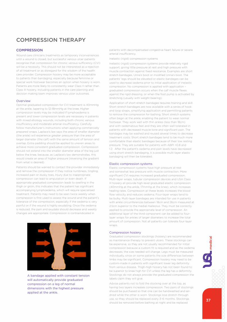
COMPRESSION THERAPY
COMPRESSIONWound care clinicians treatments as temporary inconveniences until a wound is closed, but successful venous ulcer patients recognise that compression for chronic venous sufficiency (CVi) will be a necessity. this should not be interpreted as a rejection of all treatment or as disregard for the wisdom of the health care provider. Compression hosiery may be more acceptable to patients than bandaging, especially because feminine or special work footwear becomes an option when hosiery is worn. Patients are more likely to consistently wear Class ii rather than Class iii hosiery. including patients in the care planning and decision making team improves venous ulcer outcomes.
OverviewOptimal graduated compression for CVi treatment is 40mmHg at the ankle, tapering to 12-18mmHg at the knee. Higher compression levels may be indicated if lymphoedema is present and lower compression levels are necessary in patients with mixed etiology wounds, including both chronic venous insufficiency and moderate arterial insufficiency. Carefully follow manufacturer’s instructions when applying commercially prepared wraps. Laplace’s law says the area of smaller diameter (the ankle) will experience greater pressure than the area of larger diameter (the calf) with the same amount of tension and overlap. extra padding should be applied to uneven areas to achieve more consistent graduated compression. Compression should not extend into the smaller diameter area of the leg just below the knee, because, as Laplace’s law demonstrates, this would create an area of higher pressure (reversing the gradient from what is desired).
Patients should be warned to contact the provider immediately and remove the compression if they notice numbness, tingling, increased pain or dusky toes. injury due to inappropriate compression can lead to amputation or even death. if appropriate graduated compression leads to swelling in the thigh or groin, this indicates that the patient has significant accompanying lymphoedema, which will require specialised treatment. Patients may need to be seen twice weekly when compression is first used to assess the wound and the patient’s tolerance of the compression, especially if the oedema is very painful or if the wound is highly exudating. Once the oedema is reduced, the pain and exudate should decrease and weekly changes are appropriate. Compression is contraindicated in
patients with decompensated congestive heart failure or severe arterial insufficiency.
inelastic (rigid) compression systems
inelastic (rigid) compression systems provide relatively rigid support, giving little pressure at rest and high pressure with muscle contraction against fixed resistance. examples are short stretch bandages, unna’s boot or modified unna’s boot. the patients’ legs should be elevated or elastic bandages can be used to decrease oedema prior to initial application of inelastic compression. no compression is applied with application – graduated compression occurs when the calf muscle flexes against the rigid dressing, or when the foot pump is activated by stretching (usually with weight bearing).
application of short stretch bandages requires training and skill. Short stretch bandages are now available with a series of hook and loop straps, simplifying application and permitting patients to remove the compression for bathing. Short stretch systems often begin at the ankle, enabling the patient to wear normal footwear. they work well with thin ankles (less than 18cm) and with oedematous feet and they are often well tolerated in patients with decreased muscle tone and significant pain. the bandages may be washed and reused several times to decrease treatment costs. Short stretch bandages tend to be much more comfortable than elastic bandages because of their low resting pressure. they are suitable for patients with aBPi >0.8 and 1.2. after the patient’s oedema and pain levels have decreased using short stretch bandaging, it is possible multi-layer elastic bandaging will then be tolerated.
Elastic compression systemselastic compression systems have high pressure at rest and somewhat less pressure with muscle contraction. More significant CVi requires increased graduated compression. Multi-layer wraps, tubular compression devices and uSa Class iii hosiery all provide high-level graduated elastic compression (40mmHg at the ankle, 17mmHg at the knee), which increases healing rates. Compression at these levels increases the blood flow velocity and reduces oedema. Four-layer wraps tend to be bulky. Multi-layer bandages are intended for use in patients with ankle circumferences between 18cm and 26cm measured at 2.5cm superior to the medial malleolus. they must be correctly applied to provide the appropriate level of compression. an additional layer of the third component can be added to four-layer wraps for ankles of larger diameters to increase the total amount of compression. not all patients can tolerate four-layer wraps.
Compression hosierygraduated compression stockings (hosiery) are recommended as maintenance therapy to prevent ulcers. these stockings can be expensive, so they are not usually recommended for initial compression because a custom fit is required and as the oedema decreases, the size needed will change. Legs must be measured individually, since on some patients the size differences between limbs may be significant. Compression hosiery may need to be custom-made in patients with significant lower leg deformity from venous disease. thigh-high hosiery has not been found to be superior to knee high for CVi unless the leg has a deformity. Stockings do not always provide the graduated compression the labels claim they will give.
advise patients not to fold the stocking over at the top, as having two layers increases compression. two pairs of stockings should be purchased so that one can be handwashed and air-dried while the other is worn. Stockings lose stretch through use, so they should be replaced every 3–6 months. Stockings should be removed before bathing at night and be replaced
A bandage applied with constant tension will automatically provide graduated compression on a leg of normal dimensions with the highest pressure applied at the ankle.
37

these patients. an aBPi of 1.0-1.2 is considered venous, 1.0 is considered normal and 0.8-1.0 has an arterial component. an aBPi of >0.8 indicates mixed venous and arterial insufficiency, so low compression should be used cautiously and only after further medical assessment and supervision. Patients with an aBPi of less than 0.5 should not have any type of compression and require a referral to a vascular surgeon. arterial disease may develop over time, so aBPi’s should be re-assessed every three to six months.
Doppler arterial diagnostic toolLocating the pulses:
• dorsalis pedis - central dorsal part of foot
• posterior tibial - behind medial malleolus
• popliteal - behind the knee
• femoral - in the groin
COMPRESSION THERAPY CONTINUED
in the morning before the patient rises from the bed (before oedema has a chance to develop). apply non-sensitizing, 24-hour moisturiser after bathing to be absorbed by the skin during the night to prevent itching from dryness. ankle and knee flexion exercises are advisable even with the use of graduated compression stockings.
graduated compression stockings may be very difficult to put on. applying a silk bootee or other device first to decrease friction, or using a frame over which the stocking is stretched may help. Stockings with zippers and two piece stockings (separate foot and leg pieces that overlap at the ankle) are also available. two layers of Class i hosiery produce higher resting pressures than a single class iii stocking and may be easier to put on. another option is a low compression liner with a higher compression overstocking. in either case, the patient can take the outer stocking off at night. the patient may, however, find the increase in stiffness caused by the friction between the two layers to be unacceptable. Orthotic compression devices using Velcro are another option.
Hosiery alternativesSingle-layer elastic (long-stretch) wraps do not provide graduated compression and are not recommended. antiembolic stockings and standard support hose are not appropriate for patients with CVi. Multilayer bandaging systems more effectively maintain the leg’s volume. in general, high and multi-layer compression is more effective than lower compression or single layer wraps. Patients with a significant arterial component to their disease (aBPi<0.8) must use modified levels of compression. But, the most supportive compression the patient will tolerate should be applied consistently when treating venous ulcers without accompanying arterial disease.
Assessing circulatory statusit is essential to assess a venous ulcer patient’s circulatory status to rule out significant arterial insufficiency prior to treatment for chronic venous insufficiencies (CVi). arterial insufficiency usually causes the lower leg to be cold and to hurt when it is elevated, while in CVi, legs are usually warm and pain is relieved with elevation. But, mixed disease is common, and assessing a patient’s ankle-brachial index (aBPi) using a hand-held doppler is necessary prior to compression treatment for CVi.
Warning: Palpable pedal pulses are NOT proof that a patient has adequate circulation for compression, and the absence of palpable pulses does not always indicate arterial occlusion.
Evaluating a patient’s ABPIthe aBPi is assessed after the patient has been supine for 15 minutes. Brachial blood pressures are taken on both arms. then the BP cuff is placed on the leg just superior to the ankle bone and the doppler probe (using ultrasound gel) is placed at a 45-degree angle to the dorsalis pedis or posterior tibial artery. the cuff is inflated until the doppler signal is obliterated, then deflated slowly. When the doppler signal returns, the number is recorded as the ankle systolic pressure. each ankle’s pressure divided by the higher of the two brachial systolic pressures gives that limb’s aBPi.
Interpretation of ABPIan aBPi of more than 1.2 indicates an invalid test due to non-compressible vessels. in older patients and patients with renal disease or diabetes mellitus, the aBPi is also unreliable due to possible arterial calcification, so, further vascular testing should be considered to rule out arterial compromise in all of
ABPI is calculated by dividing ankle pressure by the higher of the two brachial systolic pressures.
Assessing circulatory status It is essential to assess a venous ulcer patient’s circulatory status to rule out significant arterial insufficiency prior to treatment for CVI. Arterial insufficiency usually causes the lower leg to be cold and to hurt when it is elevated, while in CVI, legs are usually warm and pain is relieved with elevation. But, mixed disease is common, and assessing a patient’s ankle-brachial index (ABI) using a hand-held Doppler is necessary prior to compression treatment for CVI.46
(Warning: Palpable pedal pulses are NOT proof that a patient has adequate circulation for compression, and the absence of palpable pulses does not always indicate arterial occlusion.15,46,47)
Evaluating a patient’s ABI (ABPI)The ABI is assessed after the patient has been supine for 15 minutes.48 Brachial blood pressures are taken on both arms.48 Then the BP cuff is placed on the leg just superior to the ankle bone and the Doppler probe (using ultrasound gel) is placed at a 45-degree angle to the dorsalis pedis or posterior tibial artery.48 The cuff is inflated until the Doppler signal is obliterated, then deflated slowly. When the Doppler signal returns, the number is recorded as the ankle systolic pressure. Each ankle’s pressure divided by the higher of the two brachial systolic pressures gives that limb’s ABI.47,48,49
Interpretation of ABIAn ABI of more than 1.2 indicates an invalid test due to non-compressible vessels.45,50 In patients older than 70 and patients with renal disease or diabetes mellitus,1,45 the ABI is also unreliable due to possible arterial calcification, so, further vascular testing should be considered to rule out arterial compromise in all of these patients.5,48 An ABI of 0.9
– 1.2 is considered normal, and patients with an ABI>0.8 who are not in congestive heart failure can safely wear standard compression.5,48,49 An ABI of 0.6–0.8 indicates mixed venous and arterial insufficiency, so low compression should be used cautiously and only after further medical assessment.15,16,45,48,51 Patients with an ABI of less than 0.6 should not have any type of compression and require a referral to a vascular surgeon.5,15,48,51
Arterial disease may develop over time, so ABI’s should be re-assessed every six months.45
Assessing edemaLower leg circumferences should be measured at the widest point and at 2.5 cm above the malleolus.45 It is important to rule out systemic and reversible causes of edema, such as medication reactions, heart or liver failure and low protein.2 Usually patients with systemic causes of their edema have symmetrical swelling, while CVI produces some asymmetry.2 Diuretics, medication change, or diet change will be needed with systemic causes of edema, but diuretics are not appropriate for primary CVI.2 Suspect accompanying lymphedema, which will require additional treatment modalities, if skin at the base of the second toe is so thick that you cannot pick up a fold of it (Stemmer Sign).2,7 Edema caused by lymphedema does not respond significantly to limb elevation. With standard compression, fluid from lymphedema may simply move to proximal areas.4 Refer patients for testing to rule out lymphedema if edema does not improve despite elevation and compression.4
BiopsyAlthough venous disease is the most common cause5,7 of leg ulcers, it is by no means the only cause.7,52 Sickle cell,52 arterial insufficiency,1,13 rheumatologic disorders,13,53 cancer,13 infection,1 medication reactions,54 vasculitis1,7,13 and a host of other systemic illnesses can lead to leg ulcers which mimic venous ulcers but require different interventions.1,21,55,56 It is important to note that the chronic inflammation in venous ulcers can cause them to degenerate into malignancy.5,7,20 So, if there is no significant decrease in size or increase in granulation of the wound after six weeks of treatment as described in this protocol, a biopsy of the wound base and margin should be considered to confirm the pathology of the leg ulcer.1,5,7,15,40,52
assessmentInitial lab tests for venous wound patients should include nutritional parameters (CBC, Albumin and Prealbumin, HgbA1C, glucose), total lymphocyte count, kidney and liver function tests, ESR,15 Hgb and Hct,41 PT15 and possibly homocysteine (Hcy).42,43,44 The complete physical assessment should include a focused look at the lower extremities, noting abnormalities and asymmetry. Record BP, ABI (ABPI: Ankle-Brachial Index) and lower leg circumferences (see details below), pain and overall health history15 (with medications) and weight with BMI.45 Wound assessment includes size, tissue, margin,
periwound skin and drainage.5 Assess wound pain45 and sensation so that a change can be noted. Wound duration and the history of previous wound treatments, recurrences and compression tolerance is also important.15 Tracing the wound shape onto a transparent film (see photo) makes it easy for caregivers and patients to visualize wound size changes.45
Sec1:4
N.B. Whenever necessary, always be guided by your specialist unit when managing your patient’s leg ulcer with compression therapy.
38

39
PReSSuRe uLCeRS
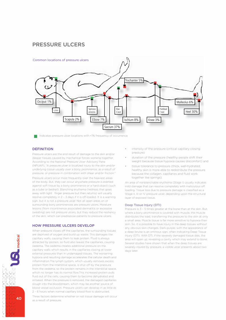
PRESSURE ULCERS
Sec1:1
INTRODUCTION
This protocol, procedure, decision tree, treatment kit and pocket guide are all designed to facilitate preparation of educational materials for communities that include families, patients and staff in order to increase the implementation of evidence-based recommendations.1,2 Implementation of pressure ulcer prevention and treatment protocols can result in a signifi cant reduction in the number of newly developed pressure ulcers, as well as substantial litigation savings.3 Guideline implementation is most likely to be successful when it includes reminder systems, education and a toolkit of multiple interventions.1,2 Over 2400 research articles have been published and at least a dozen evidence-based guidelines for prevention and treatment have been distributed, but pressure ulcers (previously called bedsores, decubitus ulcers or pressure sores)4,5 are still a global health concern.2,5,6 This integrated educational package summarizes the tremendous volume of information about pressure ulcers so that this body of recommendations can easily be transferred into practice.1,2
Historical prevalence and incidence fi gures cannot be compared to one another because of differences in both pressure ulcer defi nitions and which patients were included or excluded by the researchers.5,7 It is known that pressure ulcers develop more frequently with aging. The most common sites in adults are the sacrum and the heels, and intensive care patients are at higher risk than less severely ill patients.7 About 50% of all pressure ulcers are Stage I; Stages II, III and IV are progressively less common.7
The cost of treating pressure ulcers is staggering. Stage IV pressure ulcers often require a year or more to heal.8 In addition to primary and secondary wound dressings and caregiver time, pressure ulcer patients often require: wound cleansers, tape, special support surfaces, radiology, consultations, surgery (including debriding and grafting), pain medications and antibiotics.1 Facility associated pressure ulcers increase the patient’s risk for developing complications as well as increasing the length of stay.4 Pressure ulcer patients spend extra time in a more debilitated state and may develop additional complications.1 In the USA in 2003, hospitalizations primarily for pressure ulcers cost an average of $37,800,9 but numbers cannot refl ect the impact of a pressure ulcer on a patient’s quality of life or an institution’s reputation.5
DEFINITION
Pressure ulcers are the end result of damage to the skin and/or deeper tissues caused by mechanical forces working together.5
According to the NPUAP, “A pressure ulcer is localized injury to the skin and/or underlying tissue usually over a bony prominence, as a result of pressure, or pressure in combination with shear and/or friction. A number of contributing or confounding factors are also associated with pressure ulcers; the signifi cance of these factors is yet to be elucidated.”8 These contributing factors (detailed in CHART A on page Sec1:6) should not be dismissed lightly – we simply do not fully understand the complex dynamic process that culminates in the formation of a pressure ulcer.6
Pressure ulcers occur most frequently over the heaviest areas of the body.2,4,5 But, they can occur anywhere pressure is exerted against soft tissue by a bony prominence or a hard object (such as a tube or bedrail).1,4 Blanching erythema (redness that goes
away with light fi nger pressure but then returns) will usually resolve completely in 2 – 3 days if it is off-loaded.10 It is a warning sign, but it is not a pressure ulcer.4
Not all open areas on or surrounding bony prominences are pressure ulcers. Moisture lesions (from incontinence associated dermatitis or excessive sweating) are not pressure ulcers, but they reduce the resiliency of the skin, which can predispose patients to pressure ulcers.1,4 Herpetic and candidiasis lesions may be misclassifi ed as pressure ulcers.4 Kennedy Terminal Ulcers are probably not true pressure ulcers.11
PretibialCrestPrCr
Malleolus 6%
Heel 30%
Knee 3%Elbow 7%
IliacCrest
Scapula 2%
Chin
Elbow 7%
Spinousprocess
Sacrum 37%
Ischium 8%
Trochanter 5%
Indicates pressure ulcer locations with <1% frequency of occurence
Chi
Occiput 1%
COMMON LOCATIONS OF PRESSURE ULCERSCommon locations of pressure ulcers
indicates pressure ulcer locations with <1% frequency of occurrence
DEFINITIONPressure ulcers are the end result of damage to the skin and/or deeper tissues caused by mechanical forces working together. according to the national Pressure ulcer advisory Pane (nPuaP), “a pressure ulcer is localized injury to the skin and/or underlying tissue usually over a bony prominence, as a result of pressure, or pressure in combination with shear and/or friction.”
Pressure ulcers occur most frequently over the heaviest areas of the body. But, they can occur anywhere pressure is exerted against soft tissue by a bony prominence or a hard object (such as a tube or bedrail). Blanching erythema (redness that goes away with light finger pressure but then returns) will usually resolve completely in 2 – 3 days if it is off-loaded. it is a warning sign, but it is not a pressure ulcer. not all open areas on or surrounding bony prominences are pressure ulcers. Moisture lesions (from incontinence associated dermatitis or excessive sweating) are not pressure ulcers, but they reduce the resiliency of the skin, which can predispose patients to pressure ulcers.
HOW PRESSURE ULCERS DEvELOPWhen pressure closes off the capillaries, the surrounding tissues are deprived of oxygen and build up waste. this damages the capillary walls, causing them to leak protein. Fluid is always attracted by protein, so fluid also leaves the capillaries, causing oedema. the oedema creates additional pressure on the capillary walls which results in the capillaries closing at lower external pressures than in undamaged tissues. the worsening hypoxia and resulting damage accelerates the cellular death and inflammation.the lymph system, which usually removes excess protein from the interstitial space, is shut off by the pressure from the oedema, so the protein remains in the interstitial space, which no longer has its normal flow.this increased protein pulls fluid out of the cells, causing them to become dehydrated and irritated. When the pressure is removed, the damaged capillaries slough into the bloodstream, which may be another source of blood vessel occlusion. Pressure ulcers can develop in as little as 2 – 6 hours when normal capillary blood flow is obstructed.
three factors determine whether or not tissue damage will occur as a result of pressure:
• intensity of the pressure (critical capillary closing pressure)
• duration of the pressure (healthy people shift their weight because tissue hypoxia causes discomfort) and
• tissue tolerance to pressure (thick, well-hydrated, healthy skin is more able to redistribute the pressure because the collagen, capillaries and fluid work together like springs).
an area of nonblanchable erythema (Stage i) usually indicates mild damage that can resolve completely with meticulous off loading. tissue loss due to pressure damage is classified as a Stage ii, iii or iV pressure ulcer, depending upon the structural layer of exposed tissue.
Deep Tissue Injury (DTI)Pressure is 3 – 5 times greater at the bone than at the skin. But, where a bony prominence is covered with muscle, the muscle distributes the load, transferring the pressure to the skin at only a small area. Muscle tissue is far more sensitive to hypoxia than skin. So, it is possible to have injury in the deep tissues without any obvious skin changes. dark purple, with the appearance of a deep bruise is an ominous sign, often indicating deep tissue injury (dti). With dti, if the severely damaged tissue dies, the area will open up, revealing a cavity which may extend to bone. Several studies have shown that when the deep tissues are severely injured by pressure, a visible ulcer presents about two days later.
40
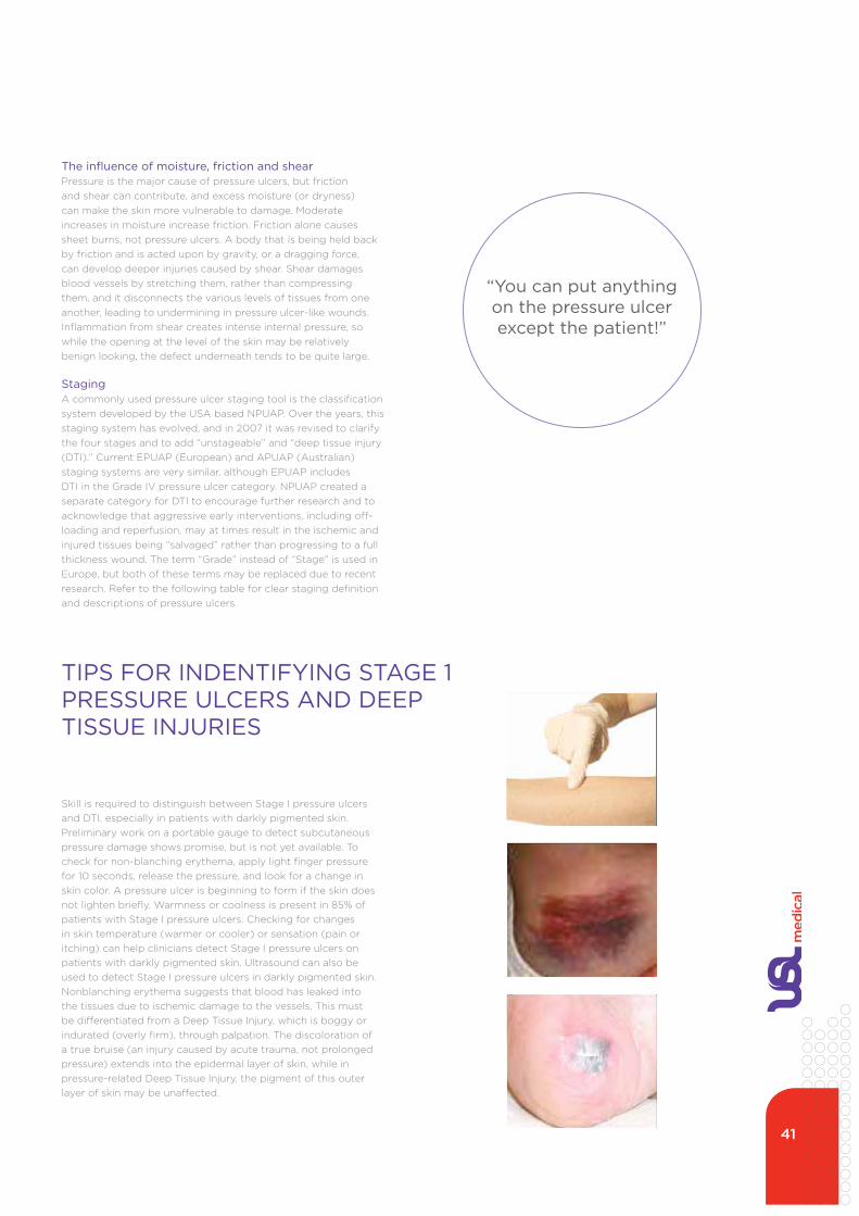
The influence of moisture, friction and shearPressure is the major cause of pressure ulcers, but friction and shear can contribute, and excess moisture (or dryness) can make the skin more vulnerable to damage. Moderate increases in moisture increase friction. Friction alone causes sheet burns, not pressure ulcers. a body that is being held back by friction and is acted upon by gravity, or a dragging force, can develop deeper injuries caused by shear. Shear damages blood vessels by stretching them, rather than compressing them, and it disconnects the various levels of tissues from one another, leading to undermining in pressure ulcer-like wounds. inflammation from shear creates intense internal pressure, so while the opening at the level of the skin may be relatively benign looking, the defect underneath tends to be quite large.
Staginga commonly used pressure ulcer staging tool is the classification system developed by the uSa based nPuaP. Over the years, this staging system has evolved, and in 2007 it was revised to clarify the four stages and to add “unstageable” and “deep tissue injury (dti).” Current ePuaP (european) and aPuaP (australian) staging systems are very similar, although ePuaP includes dti in the grade iV pressure ulcer category. nPuaP created a separate category for dti to encourage further research and to acknowledge that aggressive early interventions, including off-loading and reperfusion, may at times result in the ischemic and injured tissues being “salvaged” rather than progressing to a full thickness wound. the term “grade” instead of “Stage” is used in europe, but both of these terms may be replaced due to recent research. Refer to the following table for clear staging definition and descriptions of pressure ulcers.
Skill is required to distinguish between Stage i pressure ulcers and dti, especially in patients with darkly pigmented skin. Preliminary work on a portable gauge to detect subcutaneous pressure damage shows promise, but is not yet available. to check for non-blanching erythema, apply light finger pressure for 10 seconds, release the pressure, and look for a change in skin color. a pressure ulcer is beginning to form if the skin does not lighten briefly. Warmness or coolness is present in 85% of patients with Stage i pressure ulcers. Checking for changes in skin temperature (warmer or cooler) or sensation (pain or itching) can help clinicians detect Stage i pressure ulcers on patients with darkly pigmented skin. ultrasound can also be used to detect Stage i pressure ulcers in darkly pigmented skin. nonblanching erythema suggests that blood has leaked into the tissues due to ischemic damage to the vessels. this must be differentiated from a deep tissue injury, which is boggy or indurated (overly firm), through palpation. the discoloration of a true bruise (an injury caused by acute trauma, not prolonged pressure) extends into the epidermal layer of skin, while in pressure-related deep tissue injury, the pigment of this outer layer of skin may be unaffected.
Sec1:3
Advisory Panel (NPUAP).5 Over the years, this staging system has evolved, and in 2007 it was revised to clarify the four stages and to add “unstageable” and “deep tissue injury (DTI).”8 Current EPUAP (European) and APUAP (Australian) staging systems are very similar, although EPUAP includes DTI in the Grade IV pressure ulcer category.8,17 NPUAP created a separate category for DTI to encourage further research and to acknowledge that aggressive early interventions, including off-loading and reperfusion, may at times result in the ischemic and injured tissues being “salvaged” rather than progressing to a full thickness wound.8 The term “Grade” instead of “Stage” is used in Europe, but both of these terms may be replaced because they have been obstacles for some clinicians, since “grade” or “stage” may imply that a larger score represents a more serious wound.11 In fact, staging alone does not
indicate the seriousness of a pressure ulcer.5 It is also important to note that pressure ulcers do not necessarily progress from one stage to another – the various stages of pressure ulcers may have differing causes.8,13,18 Staging defi nitions for pressure ulcers involving mucous membranes, which have unique underlying tissue, are being formulated.11
STAGE IIIDEFINITION: Full thickness tissue loss. Subcutaneous fat may be visible but bone, tendon or muscle are not exposed. Slough may be present but does not obscure the depth of tissue loss. May include undermining and tunneling.
FUTHER DESCRIPTION: The depth of a stage III pressure ulcer varies by anatomical location. The bridge of the nose, ear, occiput and malleolus do not have
subcutaneous tissue and stage III ulcers can be shallow. In contrast, areas of signifi cant adiposity can develop extremely deep stage III pressure ulcers. Bone/tendon is not visible or directly palpable.
STAGE IVDEFINITION: Full thickness tissue loss with exposed bone, tendon or muscle. Slough or eschar may be present on some parts of the wound bed. Often include undermining and tunneling.
FUTHER DESCRIPTION: The depth of a stage IV pressure ulcer varies by anatomical location. The bridge of the nose, ear, occiput and malleolus do not have subcutaneous tissue and these ulcers can be shallow. Stage IV ulcers can extend into muscle and/or supporting structures (e.g.,
fascia, tendon or joint capsule) making osteomyelitis possible. Exposed bone/tendon is visible or directly palpable.
UNSTAGEABLEDEFINITION: Full thickness tissue loss in which the base of the ulcer is covered by slough (yellow, tan, gray, green or brown) and/or eschar (tan, brown or black) in the wound bed.
FUTHER DESCRIPTION: Until enough slough and/or eschar is removed to expose the base of the wound, the true depth, and therefore stage, cannot be determined. Stable (dry, adherent, intact without erythema or fl uctuance) eschar on the heels serves as “the body’s natural
(biological) cover” and should not be removed.
Diagrams of pressure ulcer stages are used here with permission from the NPUAP. Reproduction of the National Pressure Ulcer Advisory Panel (NPUAP) materials in this document does not imply endorsement by the NPUAP of any products, organizations, companies, or any statements made by any organization or company.
Tips for Identifying Stage I Pressure Ulcers and Deep Tissue Injuries:Skill is required to distinguish between Stage I pressure ulcers and DTI, especially in patients with darkly pigmented skin.8 Preliminary work on a portable gauge to detect subcutaneous pressure damage shows promise, but is not yet available.19 To check for non-blanching erythema, apply light fi nger pressure for 10 seconds, release the pressure, and look for a change in skin color.17 A pressure ulcer is beginning to form if the skin does not lighten briefl y.17 Warmness or coolness is present in 85% of patients with Stage I pressure ulcers.5 Checking for changes in skin temperature (warmer or cooler) or sensation (pain or itching) can help clinicians detect Stage I pressure ulcers on patients with darkly pigmented skin.5,7 Ultrasound can also be used to detect Stage I pressure ulcers in darkly pigmented skin.5 Nonblanching erythema suggests that blood has leaked into the tissues due to ischemic damage to the vessels. This must be differentiated from a Deep Tissue Injury, which is boggy or indurated (overly fi rm), through palpation.4 The discoloration of a true bruise (an injury caused by acute trauma, not prolonged pressure) extends into the epidermal
layer of skin, while in pressure-related Deep Tissue Injury, the pigment of this outer layer of skin may be unaffected.11
Example of bruise on hip
Example of DTI on heel
TIPS FOR INDENTIFYING STAGE 1 PRESSURE ULCERS AND DEEP TISSUE INjURIES
“You can put anything on the pressure ulcer except the patient!”
41

PRESSURE ULCERS
Sec1:2
HOW PRESSURE ULCERS DEVELOP When pressure closes off the capillaries, the surrounding tissues are deprived of oxygen and build up waste. This damages the capillary walls,4 causing them to leak protein. Fluid is always attracted by protein, so fl uid also leaves the capillaries, causing edema. The edema creates additional pressure on the capillary walls which results in the capillaries closing at lower external pressures than in undamaged tissues.4 The worsening hypoxia and resulting damage accelerates the cellular death and infl ammation.4 The lymph system, which usually removes excess protein from the interstitial space, is shut off by the pressure from the edema, so the protein remains in the interstitial space, which no longer has its normal fl ow.4 This increased protein pulls fl uid out of the cells, causing them to become dehydrated and irritated.4 When the pressure is removed, the damaged capillaries slough into the bloodstream, which may be another source of blood vessel occlusion.4 Pressure ulcers can develop in as little as 2 – 6 hours when normal capillary blood fl ow is obstructed.12
Three factors determine whether or not tissue damage will occur as a result of pressure:
• intensity of the pressure (critical capillary closing pressure)
• duration of the pressure (healthy people shift their weight because tissue hypoxia causes discomfort) and
• tissue tolerance to pressure (thick, well-hydrated, healthy skin is more able to redistribute the pressure because the collagen, capillaries and fl uid work together like springs).1,4
An area of nonblanchable erythema (Stage I) usually indicates mild damage that can resolve completely with meticulous offl oading.11 Tissue loss due to pressure damage is classifi ed as a Stage II, III or IV pressure ulcer, depending upon the structural layer of exposed tissue.8,13
Deep Tissue Injury (DTI)Pressure is 3 – 5 times greater at the bone than at the skin.4,5 But, where a bony prominence is covered with muscle, the muscle distributes the load, transferring the pressure to the skin at only a small area.4,5 Muscle tissue is far more sensitive to hypoxia than skin.4,5,14 So, it is possible to have injury in the deep tissues without any obvious skin changes.5,15 Dark purple, with the appearance of a deep bruise is an ominous sign, often indicating Deep Tissue Injury (DTI).8 With DTI, if the severely damaged tissue dies, the area will open up, revealing a cavity which may extend to bone.8 Several studies have shown that when the deep tissues are severely injured by pressure, a visible ulcer presents about two days later.2
The Influence of Moisture, Friction and ShearPressure is the major cause of pressure ulcers, but friction and shear can contribute, and excess moisture (or dryness) can make the skin more vulnerable to damage.1 Moderate increases in moisture increase friction.4,16 Friction alone causes sheet burns, not pressure
ulcers.4 A body that is being held back by friction and is acted upon by gravity, or a dragging force, can develop deeper injuries caused by shear.13 Shear damages blood vessels by stretching them, rather than compressing them, and it disconnects the various levels of tissues from one another, leading to undermining in pressure ulcer-like wounds.4 Infl ammation from shear creates intense internal pressure, so while the opening at the level of
the skin may be relatively benign-looking, the defect underneath tends to be quite large.13
Staging: A commonly used pressure ulcer staging tool is the classifi cation system developed by the USA-based National Pressure Ulcer
SUSPECTED DEEP TISSUE INJURY
DEFINITION: Purple or maroon localized area of discolored intact skin or blood-fi lled blister due to damage of underlying soft tissue from pressure and/or shear. The area may be preceded by tissue that is painful, fi rm, mushy, boggy, warmer or cooler as compared to adjacent tissue.
FUTHER DESCRIPTION: Deep tissue injury may be diffi cult to detect in individuals with dark skin tones. Evolution may include a thin blister over a dark wound bed. The wound may further evolve and
become covered by thin eschar. Evolution may be rapid exposing additional layers of tissue even with optimal treatment.
STAGE IDEFINITION: Intact skin with non-blanchable redness of a localized area usually over a bony prominence. Darkly pigmented skin may not have visible blanching; its color may differ from the surrounding area.
FUTHER DESCRIPTION: The area may be painful, fi rm, soft, warmer or cooler as compared to adjacent tissue. Stage I may be diffi cult to detect in individuals with dark skin tones. May indicate “at risk” persons (a heralding sign of risk)
STAGE IIDEFINITION: Partial thickness loss of dermis presenting as a shallow open ulcer with a red pink wound bed, without slough. May also present as an intact or open/ruptured serum-fi lled blister.
FUTHER DESCRIPTION: Presents as a shiny or dry shallow ulcer without slough or bruising.* This stage should not be used to describe skin tears, tape burns, perineal dermatitis, maceration or excoriation.
*Bruising indicates suspected deep tissue injury
NPUAP STAGING SYSTEM:8
Suspected deep tissue injurydefinition: Purple or maroon localised area of discoloured intact skin or blood-filled blister due to damage of underlying soft tissue from pressure and/or shear. the area may be preceded by tissue that is painful, firm, mushy, boggy, warmer or cooler as compared to adjacent tissue.
deep tissue injury may be difficult to detect in individuals with dark skin tones. evolution may include a thin blister over a dark wound bed. the wound may further evolve and become covered by thin eschar. evolution may be rapid exposing additional layers of tissue even with optimal treatment.
Stage Idefinition: intact skin with non-blanchable redness of a localized area usually over a bony prominence. darkly pigmented skin may not have visible blanching; its color may differ from the surrounding area.
the area may be painful, firm, soft, warmer or cooler as compared to adjacent tissue. Stage i may be difficult to detect in individuals with dark skin tones. May indicate “at risk” persons (a sign of risk).
Stage IIdefinition: Partial thickness loss of dermis presenting as a shallow open ulcer with a red pink wound bed, without slough. May also present as an intact or open/ruptured serum-fi lled blister.
Presents as a shiny or dry shallow ulcer without slough or bruising.* this stage should not be used to describe skin tears, tape burns, perineal dermatitis, maceration or excoriation.
*Bruising indicates suspected deep tissue injury
PreventionPrevention of pressure ulcers requires two things: improving skin health to promote increased tissue tolerance and decreasing exposure to excessive pressure, friction, moisture and sheer. this can be accomplished through a formal, evidence-based pressure ulcer prevention program, which usually includes:
• a risk assessment, such as the Braden Scale norton, Waterlow, Braden Q or similar
• a systematic skin assessment
• reducing risk factors
• patient, family and staff education and involvement
• evaluation and maintenance
it is important to note, that introducing a formal risk assessment tool linking levels of risk to prevention protocols can dramatically decrease the incidence and severity of pressure ulcers in a facility.
STAGING OF PRESSURE ULCERS
42

Stage IIIdefinition: Full thickness tissue loss. Subcutaneous fat may be visible but bone, tendon or muscle are not exposed. Slough may be present but does not obscure the depth of tissue loss. May include undermining and tunneling.
the depth of a stage iii pressure ulcer varies by anatomical location. the bridge of the nose, ear, occiput and malleolus do not have subcutaneous tissue and stage iii ulcers can be shallow. in contrast, areas of significant adiposity can develop extremely deep stage iii pressure ulcers. Bone/tendon is not visible or directly palpable.
Sec1:3
Advisory Panel (NPUAP).5 Over the years, this staging system has evolved, and in 2007 it was revised to clarify the four stages and to add “unstageable” and “deep tissue injury (DTI).”8 Current EPUAP (European) and APUAP (Australian) staging systems are very similar, although EPUAP includes DTI in the Grade IV pressure ulcer category.8,17 NPUAP created a separate category for DTI to encourage further research and to acknowledge that aggressive early interventions, including off-loading and reperfusion, may at times result in the ischemic and injured tissues being “salvaged” rather than progressing to a full thickness wound.8 The term “Grade” instead of “Stage” is used in Europe, but both of these terms may be replaced because they have been obstacles for some clinicians, since “grade” or “stage” may imply that a larger score represents a more serious wound.11 In fact, staging alone does not
indicate the seriousness of a pressure ulcer.5 It is also important to note that pressure ulcers do not necessarily progress from one stage to another – the various stages of pressure ulcers may have differing causes.8,13,18 Staging defi nitions for pressure ulcers involving mucous membranes, which have unique underlying tissue, are being formulated.11
STAGE IIIDEFINITION: Full thickness tissue loss. Subcutaneous fat may be visible but bone, tendon or muscle are not exposed. Slough may be present but does not obscure the depth of tissue loss. May include undermining and tunneling.
FUTHER DESCRIPTION: The depth of a stage III pressure ulcer varies by anatomical location. The bridge of the nose, ear, occiput and malleolus do not have
subcutaneous tissue and stage III ulcers can be shallow. In contrast, areas of signifi cant adiposity can develop extremely deep stage III pressure ulcers. Bone/tendon is not visible or directly palpable.
STAGE IVDEFINITION: Full thickness tissue loss with exposed bone, tendon or muscle. Slough or eschar may be present on some parts of the wound bed. Often include undermining and tunneling.
FUTHER DESCRIPTION: The depth of a stage IV pressure ulcer varies by anatomical location. The bridge of the nose, ear, occiput and malleolus do not have subcutaneous tissue and these ulcers can be shallow. Stage IV ulcers can extend into muscle and/or supporting structures (e.g.,
fascia, tendon or joint capsule) making osteomyelitis possible. Exposed bone/tendon is visible or directly palpable.
UNSTAGEABLEDEFINITION: Full thickness tissue loss in which the base of the ulcer is covered by slough (yellow, tan, gray, green or brown) and/or eschar (tan, brown or black) in the wound bed.
FUTHER DESCRIPTION: Until enough slough and/or eschar is removed to expose the base of the wound, the true depth, and therefore stage, cannot be determined. Stable (dry, adherent, intact without erythema or fl uctuance) eschar on the heels serves as “the body’s natural
(biological) cover” and should not be removed.
Diagrams of pressure ulcer stages are used here with permission from the NPUAP. Reproduction of the National Pressure Ulcer Advisory Panel (NPUAP) materials in this document does not imply endorsement by the NPUAP of any products, organizations, companies, or any statements made by any organization or company.
Tips for Identifying Stage I Pressure Ulcers and Deep Tissue Injuries:Skill is required to distinguish between Stage I pressure ulcers and DTI, especially in patients with darkly pigmented skin.8 Preliminary work on a portable gauge to detect subcutaneous pressure damage shows promise, but is not yet available.19 To check for non-blanching erythema, apply light fi nger pressure for 10 seconds, release the pressure, and look for a change in skin color.17 A pressure ulcer is beginning to form if the skin does not lighten briefl y.17 Warmness or coolness is present in 85% of patients with Stage I pressure ulcers.5 Checking for changes in skin temperature (warmer or cooler) or sensation (pain or itching) can help clinicians detect Stage I pressure ulcers on patients with darkly pigmented skin.5,7 Ultrasound can also be used to detect Stage I pressure ulcers in darkly pigmented skin.5 Nonblanching erythema suggests that blood has leaked into the tissues due to ischemic damage to the vessels. This must be differentiated from a Deep Tissue Injury, which is boggy or indurated (overly fi rm), through palpation.4 The discoloration of a true bruise (an injury caused by acute trauma, not prolonged pressure) extends into the epidermal
layer of skin, while in pressure-related Deep Tissue Injury, the pigment of this outer layer of skin may be unaffected.11
Example of bruise on hip
Example of DTI on heel
Stage IVdefinition: Full thickness tissue loss with exposed bone, tendon or muscle. Slough or eschar may be present on some parts of the wound bed. Often include undermining and tunneling.
the depth of a stage iV pressure ulcer varies by anatomical location. the bridge of the nose, ear, occiput and malleolus do not have subcutaneous tissue and these ulcers can be shallow. Stage iV ulcers can extend into muscle and/or supporting structures (e.g., fascia, tendon or joint capsule) making osteomyelitis possible. exposed bone/tendon is visible or directly palpable.
Sec1:3
Advisory Panel (NPUAP).5 Over the years, this staging system has evolved, and in 2007 it was revised to clarify the four stages and to add “unstageable” and “deep tissue injury (DTI).”8 Current EPUAP (European) and APUAP (Australian) staging systems are very similar, although EPUAP includes DTI in the Grade IV pressure ulcer category.8,17 NPUAP created a separate category for DTI to encourage further research and to acknowledge that aggressive early interventions, including off-loading and reperfusion, may at times result in the ischemic and injured tissues being “salvaged” rather than progressing to a full thickness wound.8 The term “Grade” instead of “Stage” is used in Europe, but both of these terms may be replaced because they have been obstacles for some clinicians, since “grade” or “stage” may imply that a larger score represents a more serious wound.11 In fact, staging alone does not
indicate the seriousness of a pressure ulcer.5 It is also important to note that pressure ulcers do not necessarily progress from one stage to another – the various stages of pressure ulcers may have differing causes.8,13,18 Staging defi nitions for pressure ulcers involving mucous membranes, which have unique underlying tissue, are being formulated.11
STAGE IIIDEFINITION: Full thickness tissue loss. Subcutaneous fat may be visible but bone, tendon or muscle are not exposed. Slough may be present but does not obscure the depth of tissue loss. May include undermining and tunneling.
FUTHER DESCRIPTION: The depth of a stage III pressure ulcer varies by anatomical location. The bridge of the nose, ear, occiput and malleolus do not have
subcutaneous tissue and stage III ulcers can be shallow. In contrast, areas of signifi cant adiposity can develop extremely deep stage III pressure ulcers. Bone/tendon is not visible or directly palpable.
STAGE IVDEFINITION: Full thickness tissue loss with exposed bone, tendon or muscle. Slough or eschar may be present on some parts of the wound bed. Often include undermining and tunneling.
FUTHER DESCRIPTION: The depth of a stage IV pressure ulcer varies by anatomical location. The bridge of the nose, ear, occiput and malleolus do not have subcutaneous tissue and these ulcers can be shallow. Stage IV ulcers can extend into muscle and/or supporting structures (e.g.,
fascia, tendon or joint capsule) making osteomyelitis possible. Exposed bone/tendon is visible or directly palpable.
UNSTAGEABLEDEFINITION: Full thickness tissue loss in which the base of the ulcer is covered by slough (yellow, tan, gray, green or brown) and/or eschar (tan, brown or black) in the wound bed.
FUTHER DESCRIPTION: Until enough slough and/or eschar is removed to expose the base of the wound, the true depth, and therefore stage, cannot be determined. Stable (dry, adherent, intact without erythema or fl uctuance) eschar on the heels serves as “the body’s natural
(biological) cover” and should not be removed.
Diagrams of pressure ulcer stages are used here with permission from the NPUAP. Reproduction of the National Pressure Ulcer Advisory Panel (NPUAP) materials in this document does not imply endorsement by the NPUAP of any products, organizations, companies, or any statements made by any organization or company.
Tips for Identifying Stage I Pressure Ulcers and Deep Tissue Injuries:Skill is required to distinguish between Stage I pressure ulcers and DTI, especially in patients with darkly pigmented skin.8 Preliminary work on a portable gauge to detect subcutaneous pressure damage shows promise, but is not yet available.19 To check for non-blanching erythema, apply light fi nger pressure for 10 seconds, release the pressure, and look for a change in skin color.17 A pressure ulcer is beginning to form if the skin does not lighten briefl y.17 Warmness or coolness is present in 85% of patients with Stage I pressure ulcers.5 Checking for changes in skin temperature (warmer or cooler) or sensation (pain or itching) can help clinicians detect Stage I pressure ulcers on patients with darkly pigmented skin.5,7 Ultrasound can also be used to detect Stage I pressure ulcers in darkly pigmented skin.5 Nonblanching erythema suggests that blood has leaked into the tissues due to ischemic damage to the vessels. This must be differentiated from a Deep Tissue Injury, which is boggy or indurated (overly fi rm), through palpation.4 The discoloration of a true bruise (an injury caused by acute trauma, not prolonged pressure) extends into the epidermal
layer of skin, while in pressure-related Deep Tissue Injury, the pigment of this outer layer of skin may be unaffected.11
Example of bruise on hip
Example of DTI on heel Sec1:3
Advisory Panel (NPUAP).5 Over the years, this staging system has evolved, and in 2007 it was revised to clarify the four stages and to add “unstageable” and “deep tissue injury (DTI).”8 Current EPUAP (European) and APUAP (Australian) staging systems are very similar, although EPUAP includes DTI in the Grade IV pressure ulcer category.8,17 NPUAP created a separate category for DTI to encourage further research and to acknowledge that aggressive early interventions, including off-loading and reperfusion, may at times result in the ischemic and injured tissues being “salvaged” rather than progressing to a full thickness wound.8 The term “Grade” instead of “Stage” is used in Europe, but both of these terms may be replaced because they have been obstacles for some clinicians, since “grade” or “stage” may imply that a larger score represents a more serious wound.11 In fact, staging alone does not
indicate the seriousness of a pressure ulcer.5 It is also important to note that pressure ulcers do not necessarily progress from one stage to another – the various stages of pressure ulcers may have differing causes.8,13,18 Staging defi nitions for pressure ulcers involving mucous membranes, which have unique underlying tissue, are being formulated.11
STAGE IIIDEFINITION: Full thickness tissue loss. Subcutaneous fat may be visible but bone, tendon or muscle are not exposed. Slough may be present but does not obscure the depth of tissue loss. May include undermining and tunneling.
FUTHER DESCRIPTION: The depth of a stage III pressure ulcer varies by anatomical location. The bridge of the nose, ear, occiput and malleolus do not have
subcutaneous tissue and stage III ulcers can be shallow. In contrast, areas of signifi cant adiposity can develop extremely deep stage III pressure ulcers. Bone/tendon is not visible or directly palpable.
STAGE IVDEFINITION: Full thickness tissue loss with exposed bone, tendon or muscle. Slough or eschar may be present on some parts of the wound bed. Often include undermining and tunneling.
FUTHER DESCRIPTION: The depth of a stage IV pressure ulcer varies by anatomical location. The bridge of the nose, ear, occiput and malleolus do not have subcutaneous tissue and these ulcers can be shallow. Stage IV ulcers can extend into muscle and/or supporting structures (e.g.,
fascia, tendon or joint capsule) making osteomyelitis possible. Exposed bone/tendon is visible or directly palpable.
UNSTAGEABLEDEFINITION: Full thickness tissue loss in which the base of the ulcer is covered by slough (yellow, tan, gray, green or brown) and/or eschar (tan, brown or black) in the wound bed.
FUTHER DESCRIPTION: Until enough slough and/or eschar is removed to expose the base of the wound, the true depth, and therefore stage, cannot be determined. Stable (dry, adherent, intact without erythema or fl uctuance) eschar on the heels serves as “the body’s natural
(biological) cover” and should not be removed.
Diagrams of pressure ulcer stages are used here with permission from the NPUAP. Reproduction of the National Pressure Ulcer Advisory Panel (NPUAP) materials in this document does not imply endorsement by the NPUAP of any products, organizations, companies, or any statements made by any organization or company.
Tips for Identifying Stage I Pressure Ulcers and Deep Tissue Injuries:Skill is required to distinguish between Stage I pressure ulcers and DTI, especially in patients with darkly pigmented skin.8 Preliminary work on a portable gauge to detect subcutaneous pressure damage shows promise, but is not yet available.19 To check for non-blanching erythema, apply light fi nger pressure for 10 seconds, release the pressure, and look for a change in skin color.17 A pressure ulcer is beginning to form if the skin does not lighten briefl y.17 Warmness or coolness is present in 85% of patients with Stage I pressure ulcers.5 Checking for changes in skin temperature (warmer or cooler) or sensation (pain or itching) can help clinicians detect Stage I pressure ulcers on patients with darkly pigmented skin.5,7 Ultrasound can also be used to detect Stage I pressure ulcers in darkly pigmented skin.5 Nonblanching erythema suggests that blood has leaked into the tissues due to ischemic damage to the vessels. This must be differentiated from a Deep Tissue Injury, which is boggy or indurated (overly fi rm), through palpation.4 The discoloration of a true bruise (an injury caused by acute trauma, not prolonged pressure) extends into the epidermal
layer of skin, while in pressure-related Deep Tissue Injury, the pigment of this outer layer of skin may be unaffected.11
Example of bruise on hip
Example of DTI on heel
Unstageabledefinition: Full thickness tissue loss in which the base of the ulcer is covered by slough (yellow, tan, gray, green or brown) and/or eschar (tan, brown or black) in the wound bed.
until enough slough and/or eschar is removed to expose the base of the wound, the true depth, and therefore stage, cannot be determined. Stable (dry, adherent, intact without erythema or fluctuance) eschar on the heels serves as “the body’s natural (biological) cover” and should not be removed.
PRESSURE ULCERS
STAGING OF PRESSURE ULCERS CONTINUED
43
PRESSURE ULCER TREATMENT PROGRAMME
Addressing the underlying causesthe existence of a pressure ulcer should trigger the implementation of the entire prevention section of this protocol with increased intensity to prevent further damage and facilitate wound healing. Special attention should be given to
• managing incontinence and diaphoresis (excessive perspiration or sweating)
• maintaining skin hygiene and moisture
• optimising hydration and nutrition
• choosing appropriate support surfaces
• repositioning, avoiding friction and sheer
When repositioning the patient, avoid putting pressure over the area of the wound as much as possible.
ASSESSMENT
Perform an initial complete health history and physical, following up on deficits that can be corrected;
Weekly wound assessments should include:
• anatomic location of the wound
• stage or grade of the ulcer and documentation
• size in centimetres, including any tunnels or tracts with locations
• type of tissue in the wound base as a percentage of the whole
• exudate amount, consistency and type
• odour
• wound edges, or margins
• periwound condition – texture, colour, temperature, any rash
• wound pain (both persistent and incident-related)
Debridement and wound bed cleansing
Initial debridement• do not debride wounds on patients who are terminally
ill or have insufficient arterial flow for healing, such as dry stable eschar on noninfected heels. assess these wounds daily.
• if cellulitis or sepsis is present, immediate surgical debridement of eschar and slough is indicated.
• in all other cases, use moisture-retentive dressings to cleanse the wound bed through autolytic debridement. improvement should be seen in 72 – 96 hours.
Cleansing• initial wound cleansing should be as thorough as the
patient’s condition permits.
• obvious loose wound bed debris can be removed at dressing changes. Change dressings after showering to protect the wound.
• refer for surgical debridement if appropriate
Pain managementWound pain usually includes both persistent (background) pain
PRESSURE ULCERS
and incident-related pain (from dressing changes, repositioning, debridement, etc.). new pain may indicate a developing infection.
• manage pain by eliminating the source (cover wound, adjust support surfaces, reposition patient, etc).
• when local measures are not able to eliminate or control the source of pain, analgesics should be provided as needed
Dressingsdressings should absorb excess exudate, fill dead space, maintain moisture balance, maintain a moist wound environment, allow gaseous exchange, provide thermal insulation, protect the wound from contamination and relieve pain.
Evaluation• evaluate pressure ulcer healing
• partial-thickness pressure ulcers (Stage i and ii) should show evidence of healing within 1 – 2 weeks
• full-thickness pressure ulcers (Stage iii and iV) should show a reduction in size within 2 – 4 weeks
• if the goal of care is healing and no progress is being made after two weeks of appropriate care, reassess the overall plan and look for complications, such as infection or fluid collection in the soft tissues
• refer as necessary
Infection• local signs of infection in a chronic wound include:
strong odour, purulent exudate, induration, friable or discolored granulation tissue, pocketing of the wound base, increased pain and/or delayed healing
• if a clean-appearing wound is not healing despite four weeks of optimal wound care and patient management, a two week trial of a topical antimicrobial is maybe appropriate
• a wound swab culture should be obtained if signs of infection increase
• when clinical signs of infection do not respond to treatment, osteomyelitis and joint infection should be ruled out
44

45
BuRnS

bURNS
Nomenclature Layer involved Appearance Texture Sensation Time to healing Complications
First degree epidermisRedness (erythema)
dry Painful 1wk or less none
Second degree (superficial partial thickness)
extends into superficial (papillary) dermis
Red with clear blister. Blanches with pressure
Moist Painful 2-3wksLocal infection/cellulitis
Second degree (deep partial thickness)
extends into deep (reticular) dermis
Red-and-white with bloody blisters. Less blanching.
Moist PainfulWeeks - may progress to third degree
Scarring, contractures (may require excision and skin grafting)
Third degree (full thickness)
extends through entire dermis
Stiff and white/brown
dry, leathery
Painless Requires excisionScarring, contractures, amputation
Fourth degree
extends through skin, subcutaneous tissue and into underlying muscle and bone
Black; charred with eschar
dry Painless Requires excision
amputation, significant functional impairment
THE RULE OF NINES
Estimating extent of burn injurythis is determined by the Rule of nines which assigns a percentage to a specific body part and the total surface are then can be calculated more easily.
18%
9% 9%
6.7% 6.7%6.7%
18%
4.5% 4.5%4.5% 4.5%
1%
bURN SUMMARY TAbLEthe following tables provides a summary of burn injuries:
46

dressing in position until it was secured. These are common observations for most dressings.
0
1
2
3
4
5
6
7
8
9
10
The Use of Polymeric Membrane Silver Dressings in Chronic Burns and Burn Related Wounds
IntroductIonIn the past, an acute wound healing model has been applied to chronic wounds, but it is now known that chronic wound healing is different from acute wound healing1. Whilst it is now being recognised that chronic wounds heal differently to acute wounds and often `stick` in the inflammatory and proliferative phases, there is little or no recognition of chronicity in burn or surgical wounds. Teot & Otman2 argue that these wounds can evolve, when poorly managed or in specific situations, into chronic wounds. Chronic burns or donor sites often continue to be managed as acute-wounds and are often dressed with conventional “burn” products. These wounds have similar problems in terms of quality of life, cost and nursing time, however little is written about this in the literature. Dressings commonly used in other chronic wounds may offer better alternatives for these burn related chronic wounds, and may allow for more continuous care when patients return to the community.
AImThis poster sets out to evaluate polymeric membrane silver dressings on chronic burn-wounds through a prospective evaluation. A Likert-scale was used to assess pain and ease of application, pain in-situ, pain on-removal, ease of removal and acceptability of the dressings to staff and patients.
methodSeven patients with ten chronic non healing burn and burn related wounds were identified. (Table1). They were treated with polymeric membrane silver dressings. These dressings have a polyurethane matrix which contains components that draw and concentrate healing substances from the body into the wound bed to promote rapid healing while facilitating autolytic debridement. The liquefied slough is absorbed by the dressing, eliminating the need for manual wound bed cleansing. The surfactant, glycerol and starch copolymer work synergistically promoting wound-cleansing and healing. The nanocrystaline silver-particles are embedded in the foam matrix and are not released onto the wound surface3. At each dressing change, patient comfort levels, ease of application and removal, conformability and ability to manage exudate were assessed using a 10 point Likert scale with 0 being poor and 10 being excellent. Pain levels were also measured using a 10 point Likert scale, with 0 being extreme pain and 10 being no pain. Nursing staff also documented the acceptability of the dressings.
resultsThe evaluations showed that the Polymeric Membrane silver dressings were easy to apply and remove, with good conformability and little or no trauma on removal.
PolyMem® Silver Wound dressing, Manufactured by Ferris Mfg Corp, Burr Ridge, IL 60527 USA. This case study was unsponsored. Ferris Mfg. Corp. contributed to this poster design and presentation.
conclusIonPolymeric Membrane dressings seem to offer good clinical advantages when used in chronic burn and burn related wounds:
Patients reported an overall reduction in pain when using • these dressings. Given that chronic wounds are painful injuries; this reduction in pain could have a positive effect on wound healing and overall return to normal function. Many wounds showed healing despite having been treated • with numerous other dressings in the past.
Increased recognition of chronicity within Burn Care needs to be developed and as demonstrated in this initial study, products traditionally used in chronic wound management may have an advantageous place in managing the chronic burn wound. Larger sizes are needed to improve the ease of use of this product in this patient group, and we understand from the manufacturer, larger sizes are likely to be made available.
Jacky Edwards, Clinical Nurse Specialist and Sally Ann Mason, Burns Outreach SisterManchester Burns Service, University Hospital of South Manchester
[email protected]@uhsm.nhs.uk
References
Dowsett, C. & Ayello, E. (2004) TIME principles of chronic wound bed 1. preparation and treatment. British Journal of Nursing, 13:15, Teot, L & Otman S (2002) Burn wound management: how to prevent a burn 2. becoming a chronic wound. In Chronic Wound Management: The evidence for Change, Mani R (Ed). Parthenon PublishingBurd A, Kwok CH, Hung SC, Chan HS, Gu H,Lam WK, Huang L. A 3. comparative study of the cytotoxicity of silver based dressings in monolayer cell, tissue explant, and animal models. Wound Repair and Regeneration 2007;15:97-104.Collier, M. (2004) Recognition and Management of wound infections. World-4. Wide-Wounds, Foertsch, C. E., O’Hara, M. W., Stoddard, F.J., & Kealey, G. P. (1998) 5. Treatment-resistant pain and distress during pediatric burn-dressing changes. Journal of Burn care and Rehabilitation. 19: 3: 219-24
dIscussIonThis evaluation demonstrates that polymeric membrane silver dressings are a useful adjunct in the management of chronic non-healing burn-wounds. Pain is a major issue with these injuries, and it is difficult to manage5 and can negatively impact on the patients’ well-being and also on healing. It appears from the results that the Polymeric Membrane Silver dressing is beneficial in pain management. It caused minimum pain on application and removal. A number of patients experiencing pain in their wounds were included on the trial, in all cases; the patient’s overall pain experience was improved. The significant levels of exudate generally produced by these wounds can become dried and crusty on the wound surface and is painful to remove. The surfactant present in these dressings precludes this, diminishing the need for potentially painful manual wound cleansing procedures.
Infections of Pseudomonas Aeruginosa and Staphylococus Aureus are common problems when managing burns and chronic non-healing wounds4. Antimicrobial dressings are used as appropriate to help minimise this problem. The polymeric membrane silver dressings performed as required, as none of the patients in these evaluations developed infections.In the chronic group, one patient appeared to have a reaction to the adhesive, found on the border of the polymeric membrane silver oval dressing, which caused further breakdown of the wound. Following this, investigations revealed the patient had an allergy to adhesives which was previously unknown.
Out of the ten chronic wounds, 3 healed completely, 6 made a significant improvement and one remained unhealed. However, this wound had been present for over 18 months with no improvement from other types of dressings, and is undergoing further investigation.
All nursing staff were happy with the product, but some nurses felt that it would be beneficial if the dressing had an adherent layer, making application easier, as it was difficult to get the polymeric membrane silver dressing to stay in place on awkward sites whilst the retention dressing was being applied in the awkward to access areas such as axilla, upper arm, buttocks etc.
Chronic BurnsNumber of Patients 7 patients with 10 wounds
Mean Age Mean Age 49 yearsMale:Female Ratio 4:3
Range of duration of chronic burns 3-18 months
Table 1: Demographics Chronic Burns
Conformability was generally good, however on awkward areas, such as joints, it was difficult to keep in place. Some nurses commented that, with larger areas, a second nurse was needed to help keep the
The evaluations showed low pain levels on not just application and removal, but whilst the dressing was in situ, and overall the patients found the dressing very comfortable.
0
1
2
3
4
5
6
7
8
9
10
Mean Likert scores for the use of polymeric membrane dressings
Feedback from the Staff
Mean Likert scores for Pain
Chronic wound Healing Results
Four month old donor site. Previous treatments included various silicone and silver dressings as well as silver sulphadiazine but kept on deteriorating.
Right: The donor sited closed completely after 12 weeks treatment with polymeric membrane silver dressings. The patient commented during the treatment over the reduction of pain.
Left: Donor site after polymeric membrane silver dressings have been used for 5 weeks. 40% of the donor site is now covered with healthy granulation tissue.
Left: Photo shows the wound after 1 weeks treatment.
Right: It closed completely three weeks later.
Extravasation injury on a burn patient. After 2 surgical debridements followed by topical negative therapy it was still deteriorating. Photo shows the wound prior to commencing polymeric membrane dressings.
47

bURNS
a burn is a type of injury to flesh caused by heat, electricity, chemicals, light, radiation or friction. Most burns only affect the skin (epidermal tissue and dermis). deeper tissues, such as muscle, bone, and blood vessels can also be injured. Burns may be treated with first aid, in an out-of-hospital setting, or may require more specialised treatment such as those available at specialised burn centres.
Managing burns is important because they are common, painful and can result in disfiguring and disabling scarring, amputation of affected parts or death in severe cases. Complications such as shock, infection, multiple organ dysfunction syndrome, electrolyte imbalance and respiratory distress may occur. the treatment of burns may include the removal of dead tissue (debridement), applying dressings to the wound, administering large volumes of intravenous fluids, administering antibiotics and skin grafting.
CLASSIFICATIONBurns can be classified by mechanism of injury, depth, extent and associated injuries and comorbidities.
By depthCurrently, burns are described according to the depth of injury to the dermis and are loosely classified into first, second, third and fourth degrees. note that an alternative form of reference to burns may describe burns according to the depth of injury to the dermis.
it is often difficult to accurately determine the depth of a burn. this is especially so in the case of second degree burns, which can continue to evolve over time. as such, a second-degree partial-thickness burn can progress to a third-degree burn over time even after initial treatment. distinguishing between the superficial-thickness burn and the partial-thickness burn is important, as the former may heal spontaneously, whereas the latter often requires surgical excision and skin grafting.
Epidermal burn (first-degree) • erythematous, pink or red
• usually caused by ultraviolet radiation
• no blisters
• dry surface
• painful
• slight oedema
• spontaneous healing
• no scarring
Superficial dermal thickness burn (second-degree)• erythematous, bright pink or red
• thin-walled blisters
• extremely painful
• moist, almost weeping surface
• moderate oedema
• spontaneous healing (14-21 days)
By severityin order to determine the need for referral
to a specialised burn unit, the american
Burn association devised a classification
system to aid in the decision-making
process. under this system, burns can be
classified as major, moderate and minor.
this is assessed based on a number of
factors, including total body surface area
(tBSa) burnt, the involvement of specific
anatomical zones, age of the person and
associated injuries.
Wound caredebridement cleaning and then dressings
are important aspects of wound care. the
wound should then be regularly re-evalu-
ated until it is healed. in the management
of first and second degree burns little
quality evidence exists to determine which
type of dressing should be used.
Complicationsinfection is a major complication of burns. infection is linked to impaired resistance from disruption of the skin’s mechanical integrity and generalized immune sup-pression. the skin barrier is replaced by eschar. this moist, protein rich avascular environment encourages microbial growth. Migration of immune cells is hampered, and there is a release of intermediaries that impede the immune response. eschar also restricts distribution of systemically administered antibiotics because of its
avascularity.
CLASSIFICATION OF bURNS
48

SIGNS OF INFECTION
• erythema and oedema at wound edges
• increasing pain
• odour
• drainage
• colour changes of wound
• fever
infection can involve the depth and extent of a burn, converting a superficial partial-thickness burn into a deep partial-thickness burn.
CLASSIFICATION OF bURNS
Mid dermal (second-degree) • mixed red or waxy white
• broken blisters
• exposed surface moist
• marked oedema
• painful
• slow to heal (4-6 weeks)
• scarring
Deep dermal (third degree)• white, yellow, brown
• poor distal circulation
• dry and rigid
• no sensation
• skin graft required
• scarring, contractures
Full thickness (fourth degree)• charred, devitalised
• loss of function
• no sensation
• skin graft or flap required
• scarring
49

dReSSing CategORieS
and FunCtiOnS
50

WOUNDCARE GUIDELINES / SUMMARY CHART
aim Comments Primary dressings include
Secondary dressings include
Black
Necrotic
Wound
to debride necrotic tissue if wound has potential to heal.
Careful assessment and management is important. debridement should reveal viable tissue. Sharp debridement should only be performed by a skilled clinician
• interactive Wet dressing - tenderWet® active
• Hydrosorb® gel
• Purilon gel
• For tenderWet® see “guidelines and expectations” Film - Hydrofilm®
• Hydrogel
Yellow
Sloughy
- Wound Cavity
Remove slough; encourage granulation; absorb
excess exudates, manage cavity.
a range of dressings will need to be combined to treat this type of wound.
generally dressings will require changing every 24-48 hours.
• Hydrosorb® gel interactive Wet dressing - tenderWet® active Cavity
• Calcium alginate – Sorbalgon®
• Cavity Foam - PermaFoam™ Cavity
• PolyMem® Silver
• Wic Rope or Cavity
• SeaSorb
• Film - Hydrofilm®
• Foam - PermaFoam™, PermaFoam™ Comfort
• Polyacrylate sheet - Omnifix®
• Relevo®
Yellow
Sloughy
Wound - Superficial
Remove slough; encourage granulation, absorb excess exudate.
Rehydration and exudate management.
• Hydrosorb® gel
• interactive Wet dressing - tenderWet® 24 active
• Hydrocolloid - Hydrocoll®
• Foam - PermaFoam™
• exudate management - Zetuvit® Plus
• PolyMem® Silver
• Relevo®
• Comfeel®
• Purilon®
• Polyacrylate Sheet fixation - Omnifix®
• Relevo®
Green/Yellow
Malodorous Wound
to manage odour and exudate. assess for infection.
Careful assessment and management is important whenever infection is suspected or proven.
Systemic antibiotics should be considered for infected wounds.
• Silver dressing - atrauman® ag
• PolyMem® Silver
• interactive wet dressing - tenderWet® active
• Biatain ag
• exudate Management - Zetuvit®
• Foam - PermaFoam™
• tenderWet®
• PolyMem® silver WiC or Rope
• MediHoney dressings; antibacterial gel, Medical Honey, apinate and tulle
Red
Granulating Wound
- Cavity Wound
to protect granulating tissue; encourage epithelialisation; absorb excess exudate; provide a moist wound environment.
it is important to select a dressing that absorbs exudate but does not place excess pressure on the healing tissue.
• interactive Wet dressing - tenderWet® active Cavity
• Calcium alginate -Sorbalgon - sheet or rope
• Cavity Foam - PermaFoam™ Cavity
• PolyMem® Cavity
• PermaFoam™ adhesive
• exudate Management - Zetuvit®
• Polyacrylate Sheet - Omnifix®
• Peha-Haft® if located on limb etc
• Relevo®
Red
Granulating Wound
to protect granulating tissue; encourage epithelialisation; absorb excess exudate: provide a moist wound environment.
exudate levels can vary.
depending on the choice of dressing and the exudate level, wounds can be dressed every 1-7 days.
• Hydrocolloid - Hydrocoll®
• Foam -PermaFoam™, PermaFoam™ Comfort
• PolyMem®
• Polyacrylate fixation sheet - Omnifix®
• Film - Hydrofilm®
Pink
Epithelialising Wound
to protect and promote epithelialisation.
epithelialising minimal Wounds normally only produce minimal exudate.
depending on dressing choice wounds can be dressed every 3-7 days.
• Hydrogel sheet - aquaClear
• Hydrocolloid - Hydrocoll®, Hydrocoll® thin
• Film - Hydrofilm®, Hydrofilm® Plus
• Films -Omnifix®
n.B. these recommendations are a guide only as proper wound and patient assessment should be conducted as to appropriate dressing selection and use. 51

ObjECTIvE WOUND TYPE ACTION DRESSING CHOICE
WOUND bED PREPARATION
Remove nectrotic tissue
Necrotic
Black cap - soft or hard eschar
Aim: Rehydrate and debride, manage exudate
debride necrotic tissue debriding agent
Manage cavity wounds Cavity dressing
SloughyYellow/greyish devitalised tissue
Aim: Remove slough, encourage granulation and manage exudate
Remove dry, sloughy, fibrinous tissue debriding agent
Manage bacterial colonisation Silver anti-microbial agent
Remove bioburden
InfectedGreen. High exudate
Aim: Manage infection and exudate
Remove purulent exudate absorbent dressing
Continue management of bacterial colonisation Silver anti-microbial agent
Manage exudate absorb excess exudate exudate management agent
SUPPORT AND PROTECTION
Support granulation
Granulating
Red granulating tissue
Light to moderate exudate
Aim: support and protect granulation, manage exudate
Manage exudating wounds absorbent dressing
Manage bacterial colonisation Silver anti-microbial agent
Manage cavity wounds Cavity dressing
Support mature granulation tissue Moist wound dressing
Support epithelialisation Epithelialising
Epithelial tissue
Minimal exudate
Aim: Provide hydration and protection
Maintain moisture Moist wound dressing
WOUND ASSESSMENT TOOLS
52

ObjECTIvE WOUND TYPE ACTION DRESSING CHOICE
WOUND bED PREPARATION
Remove nectrotic tissue
Necrotic
Black cap - soft or hard eschar
Aim: Rehydrate and debride, manage exudate
debride necrotic tissue debriding agent
Manage cavity wounds Cavity dressing
SloughyYellow/greyish devitalised tissue
Aim: Remove slough, encourage granulation and manage exudate
Remove dry, sloughy, fibrinous tissue debriding agent
Manage bacterial colonisation Silver anti-microbial agent
Remove bioburden
InfectedGreen. High exudate
Aim: Manage infection and exudate
Remove purulent exudate absorbent dressing
Continue management of bacterial colonisation Silver anti-microbial agent
Manage exudate absorb excess exudate exudate management agent
SUPPORT AND PROTECTION
Support granulation
Granulating
Red granulating tissue
Light to moderate exudate
Aim: support and protect granulation, manage exudate
Manage exudating wounds absorbent dressing
Manage bacterial colonisation Silver anti-microbial agent
Manage cavity wounds Cavity dressing
Support mature granulation tissue Moist wound dressing
Support epithelialisation Epithelialising
Epithelial tissue
Minimal exudate
Aim: Provide hydration and protection
Maintain moisture Moist wound dressing
53

WHAT DRESSING FOR WHAT WOUND?
NameGeneric specifications
Dressing products/ranges Advantages Disadvantages
HYDROGEL
Amorphous Gels
Please note: manufacturer’s recommendation for storage and single use application
exudate Capacity: nil - low
Colloids consisting of polymers that expand in water, but are insoluable. available in gels, sheets, or hydrogel impregnated dressings
Hydrosorb® gel - HaRtMann
Purilon® - COLOPLaSt
Provides moist wound environment for cell migration
Reduces pain, keeps nerve endings moist
Re-hydrates eschar, autolytic debriding
easily irrigated
gel will require secondary dressing
May macerate surrounding skin with liberal use
Some patients may experience a sensitivity to the preservative agents
HYDROGELGEL SHEET
exudate Capacity:
nil - low
dressings can be left up to 5-7 days, subject to exudate
Hydrosorb® gel Sheet HaRtMann
Medihoney HCS -
deRMa SCienCeS
May not require secondary dressing
Can remain in-situ for up to 7 days
allows for easy wound inspection through the dressing
Should not be used on infected wounds
HYDROCOLLOID
exudate Capacity:
Low - moderate
incorporate hydroactive or hydrophillic particles bound to a hydrophobic polymer
Moisture retentive dressing as the hydrophillic particles absorb moisture and converts to a gel at the wound interface. Polymer outer layer of the adhesive hydrocolloid dressing is either semi-occlusive or occlusive
Dressing frequency: May be left intact for up to 5 - 7 days, subject to the amount of exudate (a week, or a leak)
Hydrocoll® - HaRtMann
Comfeel® - COLOPLaSt
Hydroactive particles absorb exudate
gel formation at wound surface provides moist wound environment. Waterproof and bacteria proof, allows patient to shower
Conforms to wound surface
Hydrocolloid interaction cleans and debrides by autolysis
Safe debridement, granulation and epithelisation can occur in the wound at the same time
Can be used with hydrocolloid pastes and powders, for greater absorbency and reduction of dead space in the wound
deep wounds require a cavity filling paste
edges may roll
difficult to keep in place if affected by friction - may require taping
not recommended on wounds clinically infected
APPROPRIATE DRESSING SELECTION AND APPLICATION CHART
54

NameGeneric specifications
Dressing products/ranges
Advantages Disadvantages
CALCIUM ALGINATE
exudate Capacity: Moderate - high
n.B do not use on dry wounds, or wet, prior to use
natural polysacchiride from seaweed
Various sizes, ribbons and ropes
Dressing frequency: up to 5 days subject to the amount of exudate
Sorbalgon® - HaRtMann
SeaSorb -
COLOPLaSt
active ion exchange at wound surface forms soluble sodium alginate that provides a moist wound environment
May be used to pack sinuses and cavities in order to reduce dead space in wounds
absorbent - useful in exudating wounds
Promotes haemostasis in bleeding wounds
Low allergenic
alginates attached to semipermeable or occlusive dressing are not recommended for anaerobic infected wounds
gels may be confused with pus or slough
not suitable for dry wounds or in presence of hardened eschar
FOAM
PolyMem® exudate Capacity: Moderate - High
Biatain® exudate Capacity: Moderate - High
Perma Foam® exudate Capacity: Moderate
Polyurethane foam dressing in sheets or cavity filling shapes. Some incorporate a semipermeable, waterproof, adhesive layer as an outer layer of the dressing
Dressing frequency: up to 4-5 days, subject to the amount of exudate
PermaFoam®- HaRtMann
PolyMem® Ferris
Biatain®-COLOPLaSt
available as non-adherent and adherent dressings
Facilitates a moist wound environment
absorbent
Provides protection
FOaM CaVity dReSSingS: reduce dead space in wounds; conform to cavity shape, absorb large amounts of exudate, reducing the need for frequent dressing changes
Moist wound environment may not be sufficient to allow autolysis to occur
EXUDATE MANAGEMENT
exudate Capacity: Moderate - High
a super absorbent polymer dressing for heavily exudating wounds
Dressing frequency: Change when the dressing saturates depending on the level of the exudate
Relevo®
Zetuvit® Plus
designed specifically to absorb high levels of exudate
doesn’t leak
Can be used under compression
Shouldn’t be used with film dressing
May cause maceration if dressing not changed frequently enough
INTERACTIvE WET THERAPYDRESSINGS
exudate Capacity: nil - High
a multi-layered dressing with a core of super absorbent polyacrylate that is pre-activated with ringers solution. this solution is continuously released from the dressing as debris is taken up into the dressing
Dressing frequency: daily 24 Hourly
tenderWet® 24 active - HaRtMann
tenderWet® active Cavity - HaRtMann
Can be used on chronic, infected wounds as well as diabetic ulcers or over bone and tendon
activated ready for use
Can be used on wounds with all exudate levels
no contraindications
Cannot be used with occlusive dressing
Requires daily dressing change
Cavity dressing must have the tenderWet 24 active placed on top of it to make it a daily dressing
Peri wound area may become macerated so need to provide skin barrier protection
Must choose appropriate size as dressing not able to be cut
55
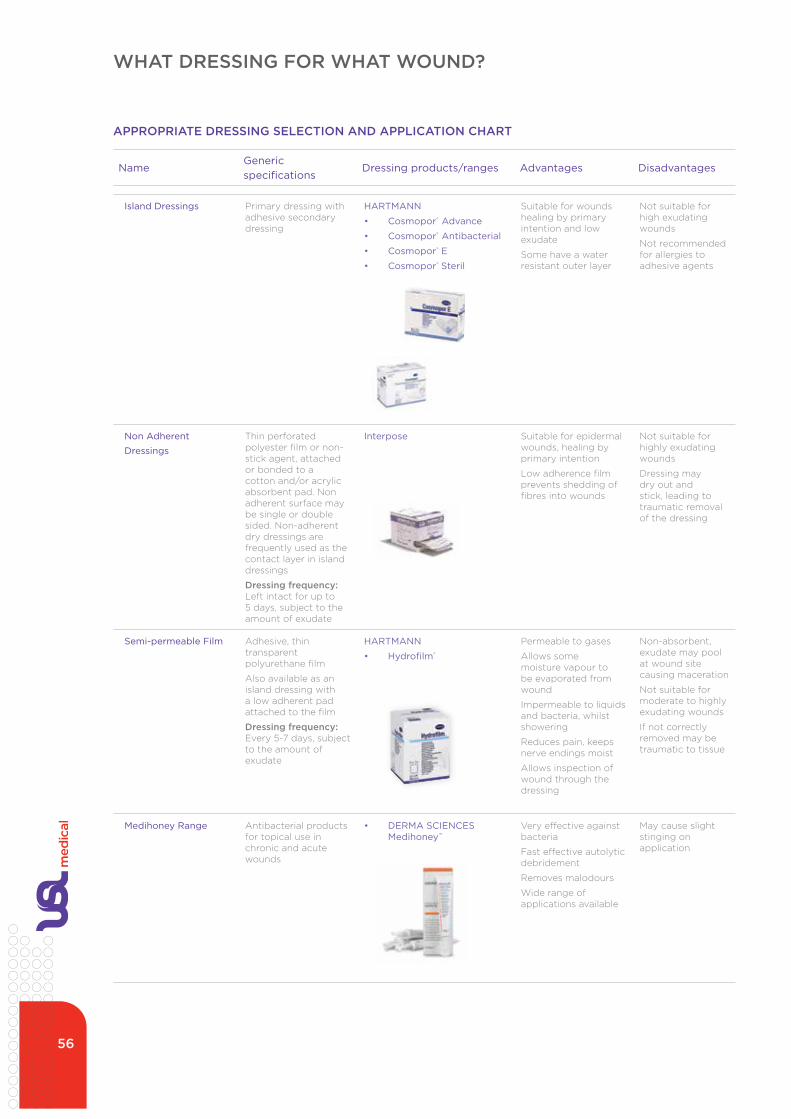
Island Dressings Primary dressing with adhesive secondary dressing
HARTMANN
• Cosmopor® Advance
• Cosmopor® Antibacterial
• Cosmopor® E
• Cosmopor® Steril
Suitable for wounds healing by primary intention and low exudate
Some have a water resistant outer layer
not suitable for high exudating wounds
not recommended for allergies to adhesive agents
Non Adherent
Dressings
thin perforated polyester film or non-stick agent, attached or bonded to a cotton and/or acrylic absorbent pad. non adherent surface may be single or double sided. non-adherent dry dressings are frequently used as the contact layer in island dressings
Dressing frequency: Left intact for up to 5 days, subject to the amount of exudate
Interpose Suitable for epidermal wounds, healing by primary intention
Low adherence film prevents shedding of fibres into wounds
not suitable for highly exudating wounds
dressing may dry out and stick, leading to traumatic removal of the dressing
Semi-permeable Film adhesive, thin transparent polyurethane film
also available as an island dressing with a low adherent pad attached to the film
Dressing frequency: every 5-7 days, subject to the amount of exudate
HARTMANN
• Hydrofilm®
Permeable to gases
allows some moisture vapour to be evaporated from wound
impermeable to liquids and bacteria, whilst showering
Reduces pain, keeps nerve endings moist
allows inspection of wound through the dressing
non-absorbent, exudate may pool at wound site causing maceration
not suitable for moderate to highly exudating wounds
if not correctly removed may be traumatic to tissue
Medihoney Range antibacterial products for topical use in chronic and acute wounds
• DERMA SCIENCES Medihoney™
Very effective against bacteria
Fast effective autolytic debridement
Removes malodours
Wide range of applications available
May cause slight stinging on application
WHAT DRESSING FOR WHAT WOUND?
APPROPRIATE DRESSING SELECTION AND APPLICATION CHART
NameGeneric specifications
Dressing products/ranges Advantages Disadvantages
56
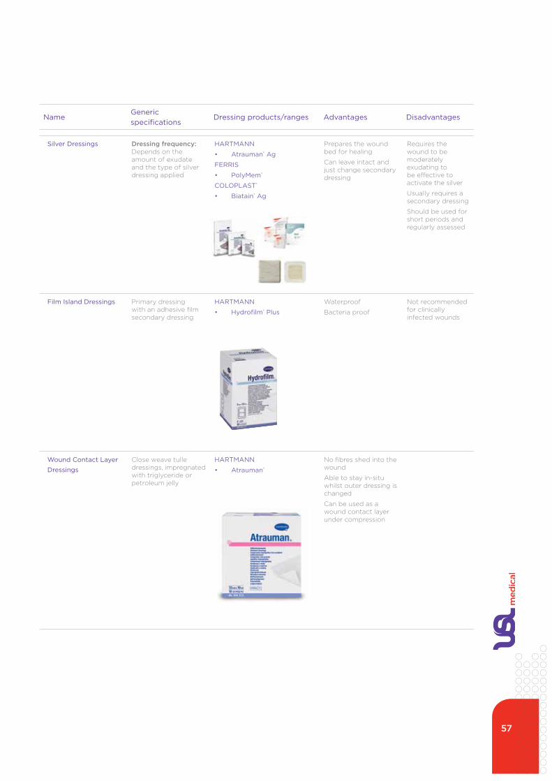
Silver Dressings Dressing frequency: depends on the amount of exudate and the type of silver dressing applied
HARTMANN
• Atrauman® Ag
FERRIS
• PolyMem®
COLOPLAST®
• Biatain® Ag
Prepares the wound bed for healing
Can leave intact and just change secondary dressing
Requires the wound to be moderately exudating to be effective to activate the silver
usually requires a secondary dressing
Should be used for short periods and regularly assessed
Film Island Dressings Primary dressing with an adhesive film secondary dressing
HARTMANN
• Hydrofilm® Plus
Waterproof
Bacteria proof
not recommended for clinically infected wounds
Wound Contact Layer
Dressings
Close weave tulle dressings, impregnated with triglyceride or petroleum jelly
HARTMANN
• Atrauman®
no fibres shed into the wound
able to stay in-situ whilst outer dressing is changed
Can be used as a wound contact layer under compression
NameGeneric specifications
Dressing products/ranges Advantages Disadvantages
57
58
WOundCaRePROduCtS

59
REMOvE NECROTIC TISSUE• Hydrosorb® gel
• tenderWet® 24 active and Cavity
• Purilon gel
REMOvE bIObURDEN• Comvita MediHoney™ Range
• Sorbalgon® and Sorbalgon® t
• atrauman® ag
• PolyMem® Silver
• SeaSorb
• Biatain ag
• Cosmopor antibacterial
MANAGE EXUDATE• Relevo®
• Zetuvit® Plus
• PolyMax®
• PermaFoam®
• Biatain
SUPPORT GRANULATION• atrauman®
• Hydrosorb gel Sheet®
• Hydrocoll® and Hydrocoll® thin
• PolyMem®
• Biatain®
• Comfeel®
SUPPORT EPITHELIALISATION• Hydrosorb gel Sheet®
• Hydrocoll® thin
• Cosmopor® e and Cosmopor® advance
• Comfeel
• Hydrofilm® and Hydrofilm® Plus
• Zetuvit®
SKIN CLOSURE• Omnistrip®
SKIN INTEGRITY & PROTECTION• niltac™
• Silesse™
• Cavilon™
• SKin-PReP® and nO-Sting SKin-PReP®
• ReMOVe®
RETENTION/FIXATION• Omnistrip®
• PehaHaft®
• Lastotel®
• Omnifix®
• tegaderm®
• OpSite®
NEGATIvE PRESSURE WOUND THERAPY (NPWT)
• SnaP®
SCAR MANAGEMENT• Kelo-cote®
COMPRESSION STOCKINGS• Sigvaris®
USL WOUNDCARE PRODUCTS
WO
UN
DC
AR
E P
RO
DU
CT
LIS
T
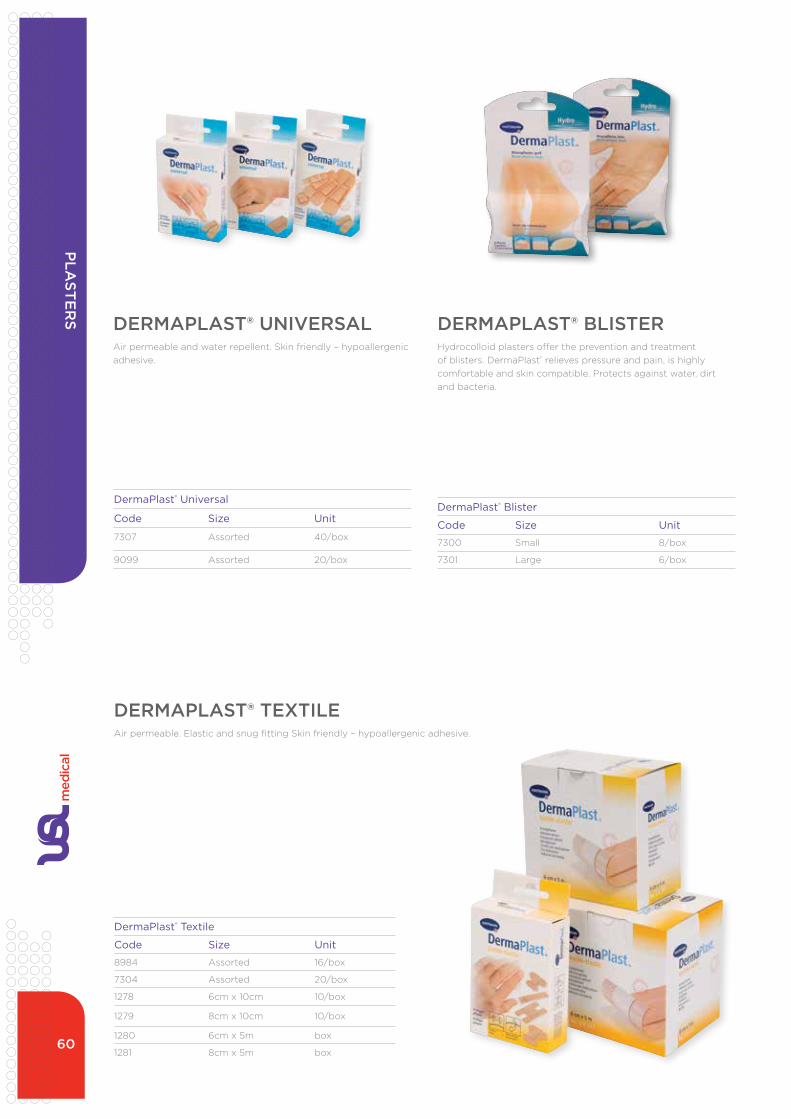
60
DERMAPLAST® TEXTILEair permeable. elastic and snug fitting Skin friendly – hypoallergenic adhesive.
DERMAPLAST® bLISTERHydrocolloid plasters offer the prevention and treatment of blisters. dermaPlast® relieves pressure and pain, is highly comfortable and skin compatible. Protects against water, dirt and bacteria.
DERMAPLAST® UNIvERSALair permeable and water repellent. Skin friendly – hypoallergenic adhesive.
DermaPlast® Universal
Code Size Unit
7307 assorted 40/box
9099 assorted 20/box
DermaPlast® Textile
Code Size Unit
8984 assorted 16/box
7304 assorted 20/box
1278 6cm x 10cm 10/box
1279 8cm x 10cm 10/box
1280 6cm x 5m box
1281 8cm x 5m box
DermaPlast® Blister
Code Size Unit
7300 Small 8/box
7301 Large 6/box
PLA
STE
RS

61
DermaPlast® Aqua
Code Size Unit
8983 30 x 40mm and 40 x 60mm 20/box
DERMAPLAST® SPORTair permeable, cushioned protection.Water resistant and dirt repellent. Skin friendly.
DERMAPLAST® AqUAtotally waterproof and breathable. Skin friendly – hypoallergenic adhesive.
USL UNIvERSAL PLASTIC AND FAbRIC PLASTERSPlastic • everyday protection
• hypoallergenic adhesive
• water and dirt resistant
Fabric• extra thick
• everyday protection
• superior elastic fabric
USL Universal Plastic Plasters
Code Size unit
9763 19mm x 72mm 100/box
USL Universal Fabric Plasters9764 19mm x 72mm 100/box
DermaPlast® Sport
Code Size Unit
9101 6cm x 10cm 10/box
PLA
STE
RS

62
MEDICOMP®
Medicomp® non-woven swabs may be used as an alternative to traditional gauze swabs in many clinical settings on the ward and in outpatient treatment.
Medicomp® non-woven swabs, made of 66% viscose and 34% polyester fibres, have an open, gauze-like structure. they are very absorbent, soft and permeable to air.
the fabric is bonded mechanically and does not contain binding agents or optical brighteners.
Medicomp® non-woven swabs are available in a variety of sizes and piles, sterilised for immediate use and in a non-sterile presentation.
Medicomp®
Code Size unit
1265 10 x 10cm, 4ply 100/pkt
1263 5 x 5cm, 4ply 100/pkt
1264 7.5 x 7.5cm, 4ply 100/pkt
Kompressen®
Code Size unit
1254 10 x 10cm, 12 ply 100/pkt
1252 5 x 5cm, 12 ply 100/pkt
1253 7.5 x 7.5cm, 12 ply 100/pkt
KOMPRESSEN®
Kompressen® is an unsterile cotton gauze which may be used in a variety of clinical settings.
Kompressen® swabs are available in a variety of sizes in a non-sterile presentation.
ZETUvIT® An absorbent dressing pad specifically developed for light-moderately exudating wounds
Zetuvit® is a multi-layered exudate management dressing pad. it is soft, comfortable, cost effective and can be used under compression bandages.
Zetuvit® is covered in a soft, non-woven, hydrophobic polyamide fibre. the inner surface of the cover has a high capillary activity, providing wicking of wound exudate into the central core of cellulose fluff.
the bleached cellulose tissue layer rapidly disperses fluid to maximise the use of the central absorbent layer.
the backing layer of hydrophobic cellulose is permeable to air and is moisture-repellent to help prevent strike-through.
Zetuvit® can be used to manage light-moderately exudating wounds and as a primary dressing on surgical wounds.
Zetuvit® Sterile, individually sealed
Code Size unit
1255 10 x 10cm 25/box
1256 10 x 20cm 25/box
1257 20 x 20cm 15/box
GA
UZ
ES/SW
Ab
S

63
ATRAUMAN® A low adherent primary contact layer
atrauman® is a low adherent, fine-weave, primary contact layer, impregnated with a non-medicated, non-petroleum based triglyceride for atraumatic wound treatment.
the soft, thin support material of atrauman® is made of polyester and ensures close contact with the whole surface of the wound, allowing an easy passage for exudate and reducing the risk of maceration.
the fine weave and hydrophobic surface of atrauman® counteracts adhesion to the wound by helping to prevent new tissue from penetrating the dressing. this is further enhanced by the triglyceride, which keeps the wound edges soft and supple.
atrauman® can be combined with numerous secondary dressings including Zetuvit® and Zetuvit® Plus, PermaFoam™ for wounds requiring excessive exudate management.
Indications: • abrasions and lacerations
• leg ulcers
• pressure ulcers
• burns and scalds
• skin graft sites
• nail extraction or wedge resection
Atrauman® Sterile, individually sealed
Code Size unit
1413 5 x 5cm 10/box
7366 5 x 5cm 50/box
1414 7.5 x 10cm 10/box
7367 7.5 x 10cm 50/box
Atrauman® Ag Silver Sterile, individually sealed
Code Size unit
8903 5 x 5cm 3/box
8904 5 x 5cm 10/box
8905 10 x 10cm 3/box
8906 10 x 10cm 10/box
8907 10 x 20cm 3/box
8908 10 x 20cm 10/box
ATRAUMAN® Ag SILvERVersatile, economic, low adherent triglyceride impregnated silver dressing
atrauman® ag is a low adherent, wound contact layer dressing which contains silver. the dressing consists of a soft, thin polyamide fabric which is chemically bound with metallic silver.
the silver tulle is coated with a non-petroleum, triglyceride based ointment. the triglyceride based ointment creates a hydrophobic surface enhancing the dressing’s low adherent properties, therefore protecting, caring and nourishing the peri-wound area.
When in direct contact with wound exudate, atrauman® ag forms silver ions on its metallic surface. the silver ions are kept within the dressing where they bind to and destroy a broad spectrum of bacteria.
the wound exudate together with the dead bacteria and endotoxins, pass easily through the dressing’s fine weave and are absorbed and retained in the secondary dressing. Removal of the bioburden into the secondary dressing helps with wound bed preparation and reduces the risk of maceration.
With these characteristics atrauman® ag manages contaminated, traumatic wounds, helping to reduce the risk of bacterial colonisation. atrauman® ag is effective against a wide spectrum of bacteria including gram positive and gram negative bacteria and is suitable for use in chronic wounds of various aetiologies.
it is contraindicated for full thickness burns and wounds that are non exudating.
atrauman® ag can be used with a variety of secondary dressings depending on the wound requirements. For highly exudating wounds, it can be used in combination with Zetuvit® Plus.
atrauman® ag can remain on a wound for up to 3-4 days depending on clinical assessment.
CO
NTA
CT
LA
YE
R/S
ILv
ER
CO
NTA
CT
LA
YE
R
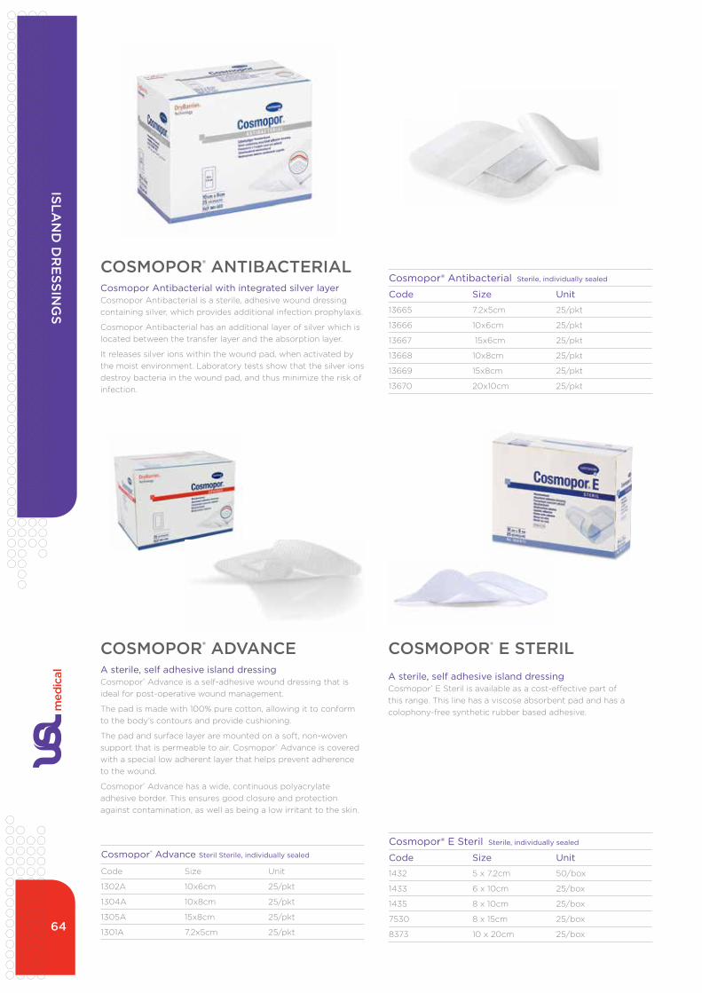
64
COSMOPOR® ADvANCEA sterile, self adhesive island dressingCosmopor® advance is a self-adhesive wound dressing that is ideal for post-operative wound management.
the pad is made with 100% pure cotton, allowing it to conform to the body’s contours and provide cushioning.
the pad and surface layer are mounted on a soft, non-woven support that is permeable to air. Cosmopor® advance is covered with a special low adherent layer that helps prevent adherence to the wound.
Cosmopor® advance has a wide, continuous polyacrylate adhesive border. this ensures good closure and protection against contamination, as well as being a low irritant to the skin.
COSMOPOR® E STERILA sterile, self adhesive island dressingCosmopor® e Steril is available as a cost-effective part of this range. this line has a viscose absorbent pad and has a colophony-free synthetic rubber based adhesive.
Cosmopor® Advance Steril Sterile, individually sealed
Code Size unit
1302a 10x6cm 25/pkt
1304a 10x8cm 25/pkt
1305a 15x8cm 25/pkt
1301a 7.2x5cm 25/pkt
Cosmopor® E Steril Sterile, individually sealed
Code Size Unit
1432 5 x 7.2cm 50/box
1433 6 x 10cm 25/box
1435 8 x 10cm 25/box
7530 8 x 15cm 25/box
8373 10 x 20cm 25/box
Cosmopor® Antibacterial Sterile, individually sealed
Code Size Unit
13665 7.2x5cm 25/pkt
13666 10x6cm 25/pkt
13667 15x6cm 25/pkt
13668 10x8cm 25/pkt
13669 15x8cm 25/pkt
13670 20x10cm 25/pkt
COSMOPOR® ANTIbACTERIALCosmopor Antibacterial with integrated silver layer Cosmopor antibacterial is a sterile, adhesive wound dressing containing silver, which provides additional infection prophylaxis.
Cosmopor antibacterial has an additional layer of silver which is located between the transfer layer and the absorption layer.
it releases silver ions within the wound pad, when activated by the moist environment. Laboratory tests show that the silver ions destroy bacteria in the wound pad, and thus minimize the risk of infection.
ISLAN
D D
RE
SSING
S
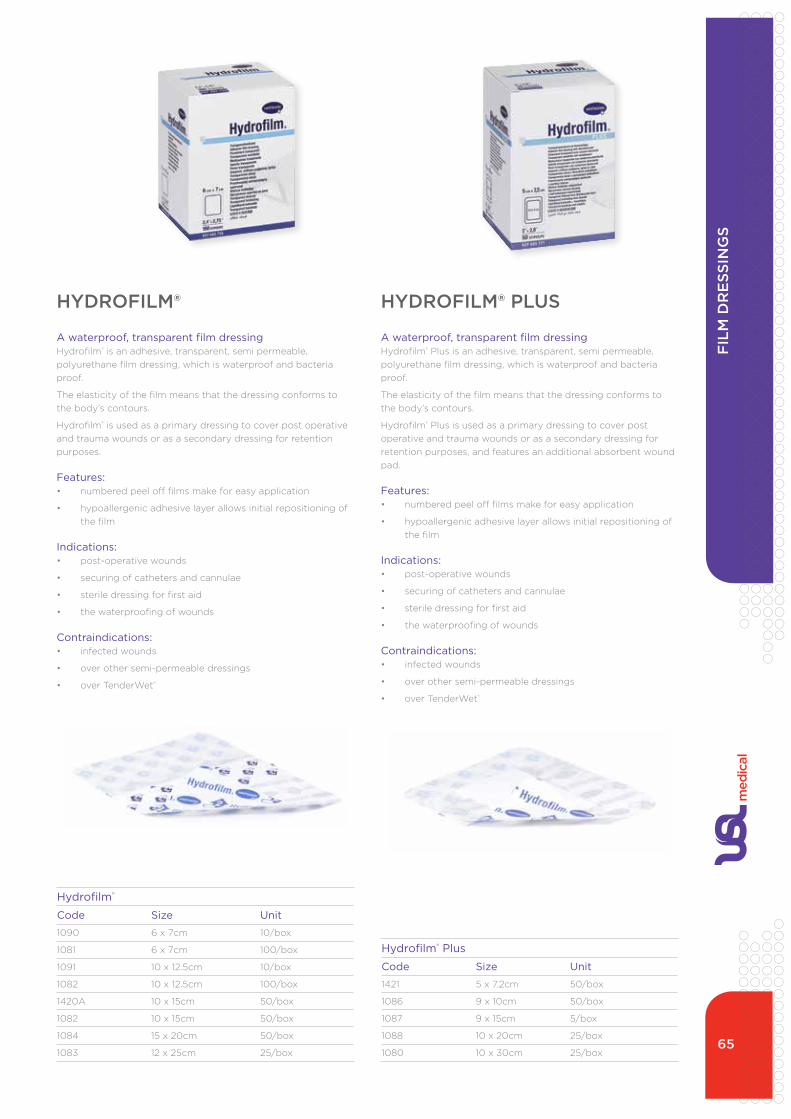
65
Hydrofilm® Plus
Code Size Unit
1421 5 x 7.2cm 50/box
1086 9 x 10cm 50/box
1087 9 x 15cm 5/box
1088 10 x 20cm 25/box
1080 10 x 30cm 25/box
Hydrofilm®
Code Size Unit
1090 6 x 7cm 10/box
1081 6 x 7cm 100/box
1091 10 x 12.5cm 10/box
1082 10 x 12.5cm 100/box
1420a 10 x 15cm 50/box
1082 10 x 15cm 50/box
1084 15 x 20cm 50/box
1083 12 x 25cm 25/box
HYDROFILM®
A waterproof, transparent film dressingHydrofilm® is an adhesive, transparent, semi permeable, polyurethane film dressing, which is waterproof and bacteria proof.
the elasticity of the film means that the dressing conforms to the body’s contours.
Hydrofilm® is used as a primary dressing to cover post operative and trauma wounds or as a secondary dressing for retention purposes.
Features:• numbered peel off films make for easy application
• hypoallergenic adhesive layer allows initial repositioning of the film
Indications:• post-operative wounds
• securing of catheters and cannulae
• sterile dressing for first aid
• the waterproofing of wounds
Contraindications:• infected wounds
• over other semi-permeable dressings
• over tenderWet®
HYDROFILM® PLUS
A waterproof, transparent film dressingHydrofilm® Plus is an adhesive, transparent, semi permeable, polyurethane film dressing, which is waterproof and bacteria proof.
the elasticity of the film means that the dressing conforms to the body’s contours.
Hydrofilm® Plus is used as a primary dressing to cover post operative and trauma wounds or as a secondary dressing for retention purposes, and features an additional absorbent wound pad.
Features:• numbered peel off films make for easy application
• hypoallergenic adhesive layer allows initial repositioning of the film
Indications:• post-operative wounds
• securing of catheters and cannulae
• sterile dressing for first aid
• the waterproofing of wounds
Contraindications:• infected wounds
• over other semi-permeable dressings
• over tenderWet®
FIL
M D
RE
SSIN
GS

66
Hydrosorb® Gel
Code Size Unit
11205 8g 5/box
1048 15g each
Hydrosorb® Gel Sheet
Code Size unit
13179 5 x 7.5cm 5/box
13180 10 x 10cm 5/box
Hydrosorb® Gel Sheet Adhesive
13181 7.5 x 10cm 5/box
13182 12.5 x 12.5cm 5/box
HYDROSORb® GEL SHEETA transparent, solid hydrogel sheet that supports granulation and epithelialisation
Hydrosorb gel Sheet® is a transparent, solid hydrogel sheet dressing with a semi-permeable outer layer.
the dressing’s 60% water content makes it particularly effective for stimulating tissue growth by keeping the young epithelium and granulation tissue moist.
the soft elastic property of Hydrosorb gel Sheet® creates a cushioning effect, which provides protection to the wound. it has a soothing and cooling effect on superficial burns and minor skin irritations.
Hydrosorb gel Sheet® does not adhere to the wound, and can be removed without pain or disturbance to the wound bed. For nil to low exudating wounds, Hydrosorb gel Sheet® can stay on the wound for up to seven days, making it a very cost-effective dressing.
Wound tracing film backing:
Hydrosorb gel Sheet® has a ‘write on’ removable transparent film for wound tracing. this peel off film can be placed in the patients’ notes for monitoring wound progress.
Indications:• leg ulcers and pressure ulcers
• superficial or partial thickness burns
• pain management in arterial leg ulcers
• abrasions
Contraindications:• wounds with moderate to high exudate
• infected wounds
Hydrosorb®gel Sheet is available in two forms – Hydrosorb®gel Sheet and Hydrosorb®gel Sheet adhesive. Hydrosorb®gel Sheet will require a retention tape or bandage while Hydrosorb®gel Sheet adhesive features its own adhesive film border.
Hydrosorb® gel Sheet adhesive
HYDROSORb® GEL Preservative-free hydrogel in convenient application
Hydrosorb® gel is a clear, viscous, sterile gel which is used for the treatment of dry chronic wounds. it contains Ringers solution, glycerol, hydroxy-ethyl cellulose, carboxy-methyl cellulose. it does not contain polyethylene glycol.
Hydrosorb® gel provides a moist wound environment helping to promote wound healing. it softens dry necrotic tissue, while facilitating the removal of devitalised tissue and absorbing wound debris and exudate.
the syringe presentation enables gel application to be delivered directly into the wound for safe, clean dosi ng. the reverse scale plunger allows accurate measuring of dispelled contents to the nearest millimetre, thus minimising the risk of application errors and enabling simple documentation.
the gel may be left on a wound for up to three days depending on wound and requires a suitable secondary dressing to prevent drying out, e.g. Hydrofilm®.
Indications: • rehydration and debridement of slough and necrotic
tissue
• to provide a moist environment in wounds of various aetiology
• pain relief
Contraindications:• full thickness burns
Hydrosorb® gel Sheet
HY
DR
OG
ELS
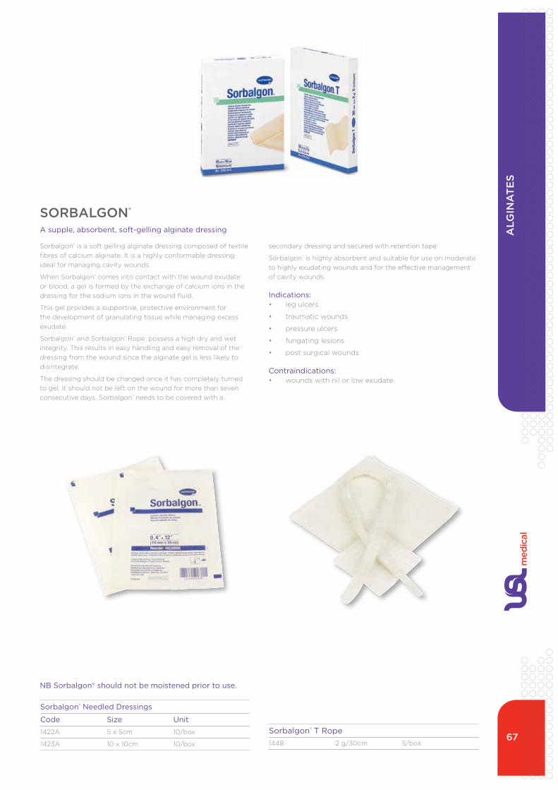
67
Sorbalgon® is a soft gelling alginate dressing composed of textile fibres of calcium alginate. it is a highly conformable dressing ideal for managing cavity wounds.
When Sorbalgon® comes into contact with the wound exudate or blood, a gel is formed by the exchange of calcium ions in the dressing for the sodium ions in the wound fluid.
this gel provides a supportive, protective environment for the development of granulating tissue while managing excess exudate.
Sorbalgon® and Sorbalgon® Rope, possess a high dry and wet integrity. this results in easy handling and easy removal of the dressing from the wound since the alginate gel is less likely to disintegrate.
the dressing should be changed once it has completely turned to gel. it should not be left on the wound for more than seven consecutive days. Sorbalgon® needs to be covered with a
Sorbalgon® Needled Dressings
Code Size Unit
1422a 5 x 5cm 10/box
1423a 10 x 10cm 10/box
Sorbalgon® T Rope1448 2 g/30cm 5/box
NB Sorbalgon® should not be moistened prior to use.
secondary dressing and secured with retention tape.
Sorbalgon® is highly absorbent and suitable for use on moderate to highly exudating wounds and for the effective management of cavity wounds.
Indications:• leg ulcers
• traumatic wounds
• pressure ulcers
• fungating lesions
• post surgical wounds
Contraindications:• wounds with nil or low exudate
SORbALGON®
A supple, absorbent, soft-gelling alginate dressing ALG
INA
TE
S

68
the unique properties of tenderWet® 24 active and tenderWet® active Cavity produce a continuous ‘rinsing effect’ at the wound bed, supporting effective wound bed preparation.
this multi-layered, autolytic debriding dressing comprises of:
• a central core of super absorbent polyacrylate activated with tenderWet® (Ringers) solution
• a hydrophobic cover with a high degree of conformity for direct contact with the wound bed
• a moisture-resistant top layer on tenderWet® 24 active to enhance the 24 hour action and prevent strike-through of tenderWet® solution and exudate
tenderWet® 24 active and tenderWet® active Cavity are the ideal dressings for wounds requiring wound bed preparation including debridement.
Code Size Unit
Tenderwet Active® 24hr Dressing10546 4cm diameter 8/box
10852 4 x 7cm 8/box
10851 5.5cm diameter 8/box
10853 7.5 x 7.5cm 8/box
10854 10 x 10cm 8/box
Tenderwet Cavity® 8917 4cm diameter 8/box
8918 5.5cm diameter 8/box
Indications: • necrotic, sloughy wounds
• infected wounds
• dehisced surgical wounds
• chronic wounds such as pressure ulcers
• leg ulcers
tenderWet® 24 active should be changed daily. unless used with tenderWet® 24 active, tenderWet® active Cavity should be changed every 12 hours. tenderWet® can be used in conjunction with a secondary dressing such as Zetuvit®/Zetuvit® Plus, then fixed into place with a bandage or Omnifix®.
tenderWet® 24 active and tenderWet® active Cavity come ready-to-use. they should be used until clean granulation tissue is established. granulating tissue can continue to be managed with Hydrocolloid dressings such as Hydrocoll®.
TENDERWET® 24 ACTIvE/TENDERWET® ACTIvE CAvITYInteractive moist wound dressings that provide continuous debriding and conditioning for effective wound bed preparation
tenderWet® solution is displaced into the wound
Wound debris and exudate is taken up into the dressing
Rinsing action provides effective debridement in an interactive moist environment
Sssn.B. tenderWet® guidelines for use must be adhered to for appropriate wound healing outcomes. Please ask for your copy.
WO
UN
D b
ED
PR
EPA
RA
TIO
N/D
Eb
RID
ME
NT
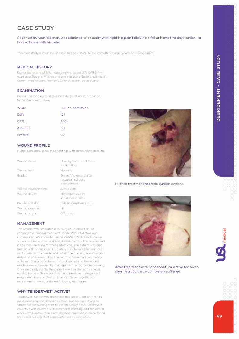
69
MEDICAL HISTORYdementia, history of falls, hypertension, recent uti, CaBg five years ago. Roger’s wife reports one episode of fever since his fall. Current medications: Ramipril, Coloxyl, aspirin, paracetamol.
EXAMINATIONdelirium secondary to sepsis, mild dehydration, constipation. no hip fracture on X-ray.
WCC: 13.6 on admission
ESR: 127
CRP: 280
Albumin: 30
Protein: 70
WOUND PROFILEMultiple pressure sores over right hip with surrounding cellulitis.
Wound swab: Mixed growth + coliform, ++ skin flora
Wound bed: necrotic
grade: grade iV pressure ulcer (ascertained post debridement)
Wound measurement: 8cm x 7cm
Wound depth: not obtainable at initial assessment
Peri-wound skin: Cellulitis, erythematous
Wound exudate: nil
Wound odour: Offensive
MANAGEMENTthe wound was not suitable for surgical intervention, so conservative management with tenderWet® 24 active was commenced. We chose to use tenderWet® 24 active because we wanted rapid cleansing and debridement of the wound, and it’s an ideal dressing for these situations. the patient was also treated with iV flucloxacillin, dietary supplementation and oral multivitamins. the tenderWet® 24 active dressing was changed daily, and after seven days the necrotic tissue had completely softened. Sharp debridement was attended and the wound exudate was subsequently managed with a hydrofibre dressing. Once medically stable, the patient was transferred to a local nursing home with a wound plan and pressure management programme in place. Oral metronidazole, amoxycillin and multivitamins were continued following discharge.
WHY TENDERWET® ACTIvE?tenderWet® active was chosen for this patient not only for its rapid cleansing and debriding action, but because it was so simple for the nursing staff to use on a daily basis. tenderWet® 24 active was covered with a combine dressing and secured in place with Hypafix tape. each dressing remained in place for 24 hours and nursing staff commented on its ease of use.
CASE STUDYRoger, an 80 year old man, was admitted to casualty with right hip pain following a fall at home five days earlier. He lives at home with his wife.
this case study is courtesy of Fleur trezise, Clinical nurse consultant Surgery/Wound Management.
Prior to treatment necrotic burden evident.
After treatment with TenderWet® 24 Active for seven days necrotic tissue completely softened.
DE
bR
IDE
ME
NT
- C
ASE
ST
UD
Y
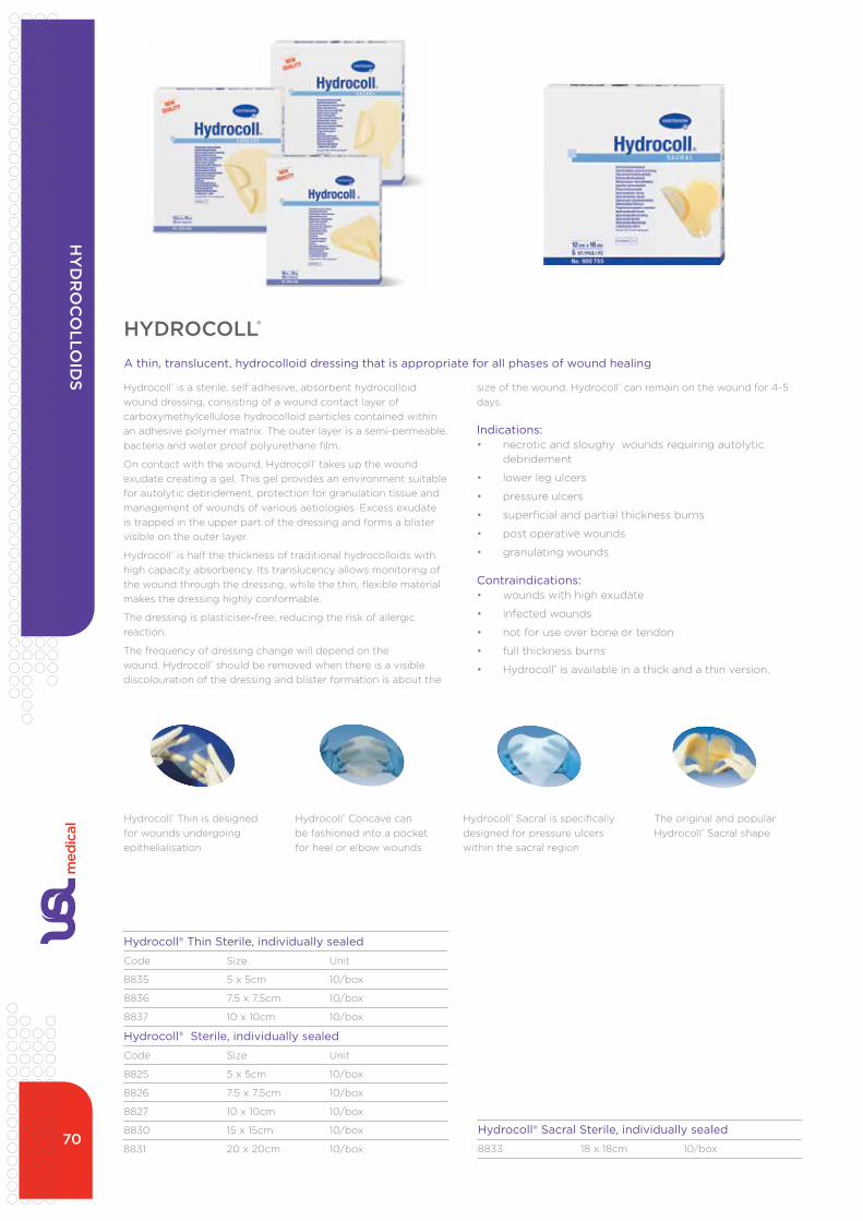
70
Hydrocoll® Thin Sterile, individually sealed
Code Size unit
8835 5 x 5cm 10/box
8836 7.5 x 7.5cm 10/box
8837 10 x 10cm 10/box
Hydrocoll® Sterile, individually sealed
Code Size unit
8825 5 x 5cm 10/box
8826 7.5 x 7.5cm 10/box
8827 10 x 10cm 10/box
8830 15 x 15cm 10/box
8831 20 x 20cm 10/box
Hydrocoll® Sacral Sterile, individually sealed
8833 18 x 18cm 10/box
Hydrocoll® is a sterile, self adhesive, absorbent hydrocolloid wound dressing, consisting of a wound contact layer of carboxymethylcellulose hydrocolloid particles contained within an adhesive polymer matrix. the outer layer is a semi-permeable, bacteria and water proof polyurethane film.
On contact with the wound, Hydrocoll® takes up the wound exudate creating a gel. this gel provides an environment suitable for autolytic debridement, protection for granulation tissue and management of wounds of various aetiologies. excess exudate is trapped in the upper part of the dressing and forms a blister visible on the outer layer.
Hydrocoll® is half the thickness of traditional hydrocolloids with high capacity absorbency. its translucency allows monitoring of the wound through the dressing, while the thin, flexible material makes the dressing highly conformable.
the dressing is plasticiser-free, reducing the risk of allergic reaction.
the frequency of dressing change will depend on the wound. Hydrocoll® should be removed when there is a visible discolouration of the dressing and blister formation is about the
Hydrocoll® thin is designed for wounds undergoing epithelialisation
Hydrocoll® Concave can be fashioned into a pocket for heel or elbow wounds
Hydrocoll® Sacral is specifically designed for pressure ulcers within the sacral region
the original and popular Hydrocoll® Sacral shape
size of the wound. Hydrocoll® can remain on the wound for 4-5 days.
Indications: • necrotic and sloughy wounds requiring autolytic
debridement
• lower leg ulcers
• pressure ulcers
• superficial and partial thickness burns
• post operative wounds
• granulating wounds
Contraindications:• wounds with high exudate
• infected wounds
• not for use over bone or tendon
• full thickness burns
• Hydrocoll® is available in a thick and a thin version.
HYDROCOLL®
A thin, translucent, hydrocolloid dressing that is appropriate for all phases of wound healing
HY
DR
OC
OLLO
IDS

71
POLYMEM®Not just a foam...PolyMem® now belongs to an innovative class of adaptable multifunctional wound care dressings. PolyMem® dressings effectively cleanse, fill, absorb and moisten wounds throughout the healing continuum. no other single wound dressing combines these four key wound healing capabilities like PolyMem®.
Cleanses: Contains a mild non toxic cleansing agent activated by moisture that is gradually released into the wound bed. Built-in cleansing capabilities reduce the need to cleanse wounds during dressing changes, so you can avoid disrupting the growth of healthy tissue as the wound heals.
Fills: gently expands to fill and conform to the wound.
absorbs: Wicks away up to ten times its weight in exudate.
Moistens: Keeps the wound bed moist and soothes traumatised tissues, reducing wound pain and providing comfort at the wound site. the moisturiser also keeps the dressing pad from adhering to the wound so it is able to be removed virtually atraumatically.
How does PolyMem® work?inhibits the actions of the pain-sensing nerve endings under the dressings (“nociceptors”)
these same nerves, when activated, create the series of events that result in; bruising, swelling, oedema and pain (inflammation)
evidence suggests that the dressing might absorb sodium ions, by capillary action, from the skin and from the subcutaneous tissues
if this is true, then this local decrease in sodium ions would result in reduced nociceptor nerve conduction, which could account for the observed pain relief.
Continual cleansingPolyMem® contains a wound-cleansing agent (F-68 surfactant) which is released into the wound when activated by moisture. this reduces the interfacial tension between healthy tissue and debris, loosens eschar and necrotic tissue, and supports autolytic debridement – all while keeping the wound bed clean during healing.
Wound comfort and healthglycerin, a moisturiser contained in the dressing, keeps the dressing from adhering to the wound bed and provides comfort at the wound site. glycerin also reduces odour, conserves living fat cells, reduces hypergranulation, soothes traumatised tissues and supports autolytic debridement.
Impressive wicking powerthe polyurethane membrane matrix wicks away up to ten times its weight in exudate. it will not fragment and leaves no residue in the wound bed.
the superabsorbent starch co-polymer in PolyMem® absorbs and binds the water molecules from the wound fluid, allowing the
First time only
• clean wound per facility protocol
• place PolyMem® dressing on wound
• change when exudate reaches wound margin (see above)
• dressing changes
• remove old dressing (do not disturb wound bed)
• if infected, treat accordingly
• place new PolyMem® dressing on wound site
PAIN - THE FIFTH vITAL SIGN• PolyMem® dressings help reduce wound pain
associated with dressing changes
• PolyMem® dressings are completely non-adherent to the wound bed. dressings which stick to the wound bed cause wound pain and trauma when they are removed during dressing changes and are also associated with delayed healing
• cleansing wounds is known to cause wound pain during dressing changes. PolyMem® dressings usually eliminate the need for wound bed cleansing during dressing changes
• PolyMem® dressings facilitate effective autolytic debridement, reducing the need for more painful debridement options
natural growth factors and nutrients to concentrate in the wound bed. Liquid barrier and gas exchange the semi-permeable thin film backing provides a liquid barrier while allowing gaseous exchange and maintaining an ideal moisture vapour transmission rate (MVtR). the transparent film backing allows for visual inspection of the membrane to determine the need for a dressing change.
Indications for use:• skin tears
• pressure and diabetic ulcers
• dermatological disorders
• leg ulcers
• donor and graft sites
• surgical wounds and tube sites
Multiple configurations available for a variety of wound needs:PolyMem®, PolyMax®, PolyMem® Silver, PolyMem® WiC (cavity dressing), PolyMem® WiC Silver (cavity dressing), PolyMem® Shapes, Oval.
Dressing protocol
PolyMem Surgical Dressings
DRESSING CHANGES:• Change when the strike through area
is equal to or less than the circumference of the wound bed
• If the strike through is noted on 75% of the dressing
• If the dressing becomes wetIf the dressing becomes wet N.B. Change dressing when 75% saturated.
MU
LTIF
UN
CT
ION
AL
DR
ESS
ING
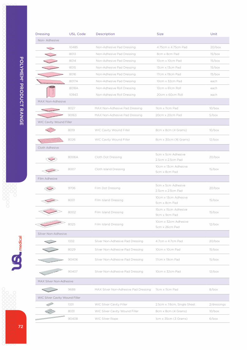
72
Dressing USL Code Description Size Unit
Non- Adhesive
10485 non-adhesive Pad dressing 4.75cm x 4.75cm Pad 20/box
8013 non-adhesive Pad dressing 8cm x 8cm Pad 15/box
8014 non-adhesive Pad dressing 10cm x 10cm Pad 15/box
8015 non-adhesive Pad dressing 13cm x 13cm Pad 15/box
8016 non-adhesive Pad dressing 17cm x 19cm Pad 15/box
8017a non-adhesive Pad dressing 10cm x 32cm Pad each
8018a non-adhesive Roll dressing 10cm x 61cm Roll each
10943 non-adhesive Roll dressing 20cm x 60cm Roll each
MAX Non-Adhesive
8027 MaX non-adhesive Pad dressing 11cm x 11cm Pad 10/box
90163 MaX non-adhesive Pad dressing 20cm x 20cm Pad 5/box
WIC Cavity Wound Filler
8019 WiC Cavity Wound Filler 8cm x 8cm (4 grams) 10/box
8026 WiC Cavity Wound Filler 8cm x 30cm (16 grams) 12/box
Cloth Adhesive
8006a Cloth dot dressing5cm x 5cm adhesive
2.5cm x 2.5cm Pad20/box
8007 Cloth island dressing10cm x 13cm adhesive
5cm x 8cm Pad15/box
Film Adhesive
9706 Film dot dressing5cm x 5cm adhesive
2.5cm x 2.5cm Pad20/box
8001 Film island dressing10cm x 13cm adhesive
5cm x 8cm Pad15/box
8002 Film island dressing15cm x 15cm adhesive
9cm x 9cm Pad15/box
8025 Film island dressing10cm x 32cm adhesive
5cm x 26cm Pad12/box
Silver Non-Adhesive
1332 Silver non-adhesive Pad dressing 4.7cm x 4.7cm Pad 20/box
8029 Silver non-adhesive Pad dressing 10cm x 10cm Pad 15/box
90406 Silver non-adhesive Pad dressing 17cm x 19cm Pad 15/box
90407 Silver non-adhesive Pad dressing 10cm x 32cm Pad 12/box
MAX Silver Non-Adhesive
9688 MaX Silver non-adhesive Pad dressing 11cm x 11cm Pad 8/box
WIC Silver Cavity Wound Filler
1331 WiC Silver Cavity Filler 2.5cm x 7.6cm, Single Sheet 2/dressings
8031 WiC Silver Cavity Wound Filler 8cm x 8cm (4 grams) 10/box
90408 WiC Silver Rope 1cm x 35cm (3 grams) 6/box
PO
LYM
EM
® PR
OD
UC
T R
AN
GE

73
POLYMEM® SKIN TEARS PROTOCOL
Manage skin tears step-by-step
• assess the cause and dimensions of the skin tear. Categorise the skin tear using the Payne-Martin Classification System
• Cleanse very gently with normal saline to remove clots and debris and rehydrate any remaining flap. Stop any bleeding with light direct pressure. Pat dry.
• approximate edges of any remaining flap without applying tension using a few thin adhesive strips, allowing space for exudate to drain from the edge of the wound. gently manipulate the flap using a moist cotton-tipped applicator, rather than hard instruments.
• Cover with an appropriately-sized PolyMem® or Shapes® by PolyMem® island dressing, or a non-adhesive PolyMem® Wound dressing held in place with a self-adhering wrap, gauze wrap or stockinette.
• document pain, location, size, depth, classification, treatment, teaching, and proposed prevention strategies.
• Follow-up, checking the dressing daily, or as appropriate. Leave it in place for three days unless exudate visible through the dressing reaches the approximate edge of the wound, indicating the need for an earlier dressing change. this is most common in Category iiB and iii skin tears with large amounts of tissue loss. do a routine dressing change* after three days, and then every four days until the wound is completely closed.
• Prevent recurrence by implementing the applicable changes to improve skin health and prevent trauma.
*With PolyMem® formulations, the dressing change process is simple – just remove the old dressing and place a new dressing on the wound.
• PolyMem® dressings contain a moisturizer and are non-adherent to the wound surface, assuring virtually pain-free removal and reducing the risk of disrupting healing tissues during the dressing change.
• PolyMem® absorbs up to ten times its weight in exudate, decreasing the risk of maceration.
• no wound cleansing is routinely performed during the dressing change process because PolyMem® dressings provide continuous cleansing of the wound.
• the semipermeable backing optimises oxygen and moisture vapor passage while protecting the wound from liquids and bacteria.
• usually patients experience dramatic pain relief when PolyMem® dressings are applied. animal studies suggest that the PolyMem® dressings interrupt the pain pathways at the wound site while enhancing healing.
• the PolyMem® formulation has also been shown in animal studies to reduce oedema and bruising while decreasing the spread of inflammation into surrounding uninjured tissues.
Always assess for pain and infection and follow-up appropriately.
Dressing USL Code Description Size Unit
PolyMem® Shapes - Latex Free
9679 #3 Oval dressing 5cm x 7.6cm Oval adhesive / 2.5cm x 5cm Pad 20/box
9682 Sacral dressing 18.4cm x 20cm Sacral adhesive / 11.4cm x 12cm Pad 10/box
10944 tube dressing 8cm x 8cm Pad 15/box
13480 nursicare Sterile PolyMem Breast Pads 6/box
PO
LYM
EM
® P
RO
DU
CT
RA
NG
E

74
POLYMEM® FINGER/TOE DRESSINGHelp improve recovery after injurythe PolyMem® Finger/toe dressings help reduce oedema, bruising, pain and inflammation when applied to open or closed injuries. When applied to open wound the dressings continuously cleanse, fill, absorb and moisten wounds. the dressings help relieve both persistent and procedure related pain associated with the injury throughout the healing process.
the ideal choice for managing:
• sprains
• strains
• contusions
• abrasions
• lacerations
• burns
• ulcers
• matricectomies
Dressing Code Description Size Unit
Polymem® Finger/Toe Dressings
11164 #1 Finger/toe dressing Small 6/box
11165 #2 Finger/toe dressing Medium 6/box
11166 #3 Finger/toe dressing Large 6/box
11167 #4 Finger/toe dressing extra Large 6/box
13441 #5 Finger/toe dressing extra extra Large 6/box
Polymem® Finger/Toe Dressings
11168 #1 Silver Finger/toe dressing Small 6/box
11169 #2 Silver Finger/toe dressing Medium 6/box
11170 #3 Silver Finger/toe dressing Large 6/box
11171 #4 Silver Finger/toe dressing extra Large 6/box
13442 #5 Silver Finger/toe dressing extra extra Large 6/box
Measure to determine length of dressing needed, cut off excess.
1
3
Insert the finger into the rolled end of the dressing.
5
Roll the dressing on the finger.
6
The dressing should fit securely on finger or toe.
Remove the insert from the rolled end and discard.
2
This is an overview. Please see package for insert for complete instructions.
4
Push the finger into the dressing and begin rolling.
MU
LTIF
UN
CT
ION
AL D
RE
SSING
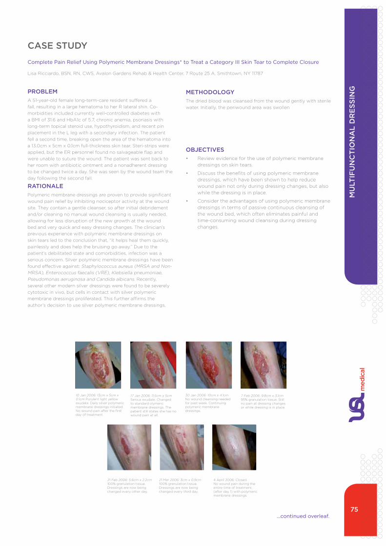
75
PRObLEMa 51-year-old female long-term-care resident suffered a fall, resulting in a large hematoma to her R lateral shin. Co-morbidities included currently well-controlled diabetes with a BMi of 31.6 and Hba1c of 5.7, chronic anemia, psoriasis with long-term topical steroid use, hypothyroidism, and recent pin placement in the L leg with a secondary infection. the patient fell a second time, breaking open the area of the hematoma into a 13.0cm x 5cm x 0.1cm full-thickness skin tear. Steri-strips were applied, but the eR personnel found no salvageable flap and were unable to suture the wound. the patient was sent back to her room with antibiotic ointment and a nonadherent dressing to be changed twice a day. She was seen by the wound team the day following the second fall.
RATIONALEPolymeric membrane dressings are proven to provide significant wound pain relief by inhibiting nociceptor activity at the wound site. they contain a gentle cleanser, so after initial debridement and/or cleaning no manual wound cleansing is usually needed, allowing for less disruption of the new growth at the wound bed and very quick and easy dressing changes. the clinician’s previous experience with polymeric membrane dressings on skin tears led to the conclusion that, “it helps heal them quickly, painlessly and does help the bruising go away.” due to the patient’s debilitated state and comorbidities, infection was a serious concern. Silver polymeric membrane dressings have been found effective against: Staphylococcus aureus (MRSa and non-MRSa), enterococcus faecalis (VRe), Klebsiella pneumoniae, Pseudomonas aeruginosa and Candida albicans. Recently, several other modern silver dressings were found to be severely cytotoxic in vivo, but cells in contact with silver polymeric membrane dressings proliferated. this further affirms the author’s decision to use silver polymeric membrane dressings.
CASE STUDY
Lisa Ricciardo, BSn, Rn, CWS, avalon gardens Rehab & Health Center, 7 Route 25 a, Smithtown, ny 11787
C A S E S T U D Y
Complete Pain Relief Using Polymeric Membrane Dressings*to Treat a Category III Skin Tear to Complete ClosureLisa Ricciardo, BSN, RN, CWS, Avalon Gardens Rehab & Health Center, 7 Route 25 A, Smithtown, NY 11787
PROBLEM
A 51-year-old female long-term-care resident suffered a fall, resulting in a large hematoma to her R lateral shin. Co-morbidities included currently well-controlled diabetes with a BMI of 31.6 and HbA1c of 5.7, chronic anemia, psoriasis with long-term topical steroid use, hypothyroidism, and recent pin placement in the L leg with a secondary infection. The patient fell a second time, breaking open the area of the hematoma into a 13.0 cm x 5 cm x 0.1 cm full-thickness skin tear. Steri-strips were applied, but the ER personnel found no salvageable flap and were unable to suture the wound. The patient was sent back to her room with antibiotic ointment and a non-adherent dressing to be changed twice a day. She was seen by the wound team the day following the second fall.
This case study was unsponsored. Ferris Mfg. Corp.
contributed to the presentation of this poster.
*PolyMem® Dressings, PolyMem Silver™ Dressings
Ferris Mfg. Corp., Burr Ridge, IL 60527
RATIONALE
Polymeric membrane dressings are proven to provide significant wound pain relief by inhibiting nociceptor activity at the wound site. They contain a gentle cleanser, so after initial debridement and/or cleaning no manual wound cleansing is usually needed, allowing for less disruption of the new growth at the wound bed and very quick and easy dressing changes. The clinician's previous experience with polymeric membrane dressings on skin tears led to the conclusion that, “it helps heal them quickly, painlessly and does help the bruising go away.”
Due to the patient’s debilitated state and co-morbidities, infection was a serious concern. Silver polymeric membrane dressings have been found effective against: Staphylococcus aureus (MRSA and Non-MRSA), Enterococcus faecalis (VRE), Klebsiella pneumoniae, Pseudomonas aeruginosa and Candida albicans. Recently, several other modern silver dressings were found to be severely cytotoxic in vivo, but cells in contact with silver polymeric membrane dressings proliferated. This further affirms the author's decision to use silver polymeric membrane dressings.
METHODOLOGY The dried blood was cleansed from the wound gently with sterile water. Initially, the periwound area was swollen with induration and the wound drained a moderate amount of purulent light yellow exudate. So, a silver polymeric membrane dressing was applied and changed daily for the first week of treatment. When the exudate was no longer purulent, standard polymeric membrane dressings were used instead. These were initially changed daily, then every-other-day, every three days and finally every five days. Polymeric membrane dressings were used to complete wound closure. The wound bled often during the first two weeks, so it was cleansed daily then, but it was not cleansed throughout the treatment as would have been necessary with other dressings.
CONCLUSION The polymeric membrane dressings were effective and easy to apply. After the initial cleansing and dressing of the wound by the wound team, the patient remained completely free from wound pain. The wound cleaned up quickly and healed in only three months, much faster than the clinician would have expected when using other dressings.
BIBLIOGRAPHY
1. Fleck, CA. Managing wound pain: today and in the future. Advances in Skin and Wound care 2007;20:3,138-145.
2. Hess CT. Wound Care Clinical Guide. Lippincott Williams & Wilkins. Ambler, PA. 2005; 275-6.
3. Fowler E, Papen JC. Clinical evaluation of a polymeric membrane dressing in the treatment of dermal ulcers. Ostomy/Wound Manage. 1991;35:35-38,40-44.
4. Burd A, Kwok CH, Hung SC, Chan HS, Gu H, Lam WK, Huang L. A comparative study of the cytotoxicity of silver-based dressings in monolayer cell, tissue explant, and animal models. Wound Repair and Regeneration 2007 15:94-104.
5. Beitz AJ, Newman A, Kahn AR, Ruggles T, Eikmeier L. A polymeric membrane dressing with antinociceptive properties: analysis with a rodent model of stab wound secondary hyperalgesia. J Pain. 2004 Feb;5(1):38-47.
OBJECTIVES 1. Review evidence for the use of polymeric membrane dressings on skin tears.2. Discuss the benefits of using polymeric membrane dressings, which have been shown to help
reduce wound pain not only during dressing changes, but also while the dressing is in place. 3. Consider the advantages of using polymeric membrane dressings in terms of passive
continuous cleansing of the wound bed, which often eliminates painful and time-consuming wound cleansing during dressing changes.
RESULTS Granulation tissue formed quickly, with complete wound closure in only three months. The patient did not have wound pain at any time after the first day, despite the depth of the wound. Slough was drawn into the dressing, revealing a clean wound bed at dressing changes. So, after the first two weeks, manual wound bed cleansing was not performed.
10 Jan 2006: 13 cm x 5 cm x 0.1 cm Purulent light yellow exudate. Daily silver
polymeric membrane dressings initiated. No wound pain after the first day of treatment.
17 Jan 2006: 11.5 cm x 5 cmSerous exudate. Changed to standard
polymeric membrane dressings. The patient still states she has no wound pain at all.
30 Jan 2006: 10 cm x 4.1 cmNo wound cleansing needed for past week. Continuing polymeric membrane dressings.
7 Feb 2006: 9.8 cm x 3.1 cm95% granulation tissue. Still no pain at
dressing changes or while dressing is in place.
21 Feb 2006: 5.6 cm x 2.2 cm100% granulation tissue. Dressings are now
being changed every other day.
21 Mar 2006: 3 cm x 0.9 cm100% granulation tissue. Dressings are now
being changed every third day.
4 April 2006: Closed. No wound pain during the entire time of treatment (after day 1) with
polymeric membrane dressings.
...continued overleaf.
10 Jan 2006: 13cm x 5cm x 0.1cm Purulent light yellow exudate. daily silver polymeric membrane dressings initiated. no wound pain after the first day of treatment.
21 Feb 2006: 5.6cm x 2.2cm 100% granulation tissue. dressings are now being changed every other day.
17 Jan 2006: 11.5cm x 5cm Serous exudate. Changed to standard olymeric membrane dressings. the patient still states she has no wound pain at all.
21 Mar 2006: 3cm x 0.9cm 100% granulation tissue. dressings are now being changed every third day.
30 Jan 2006: 10cm x 4.1cm no wound cleansing needed for past week. Continuing polymeric membrane dressings.
4 april 2006: Closed. no wound pain during the entire time of treatment (after day 1) with polymeric membrane dressings.
7 Feb 2006: 9.8cm x 3.1cm 95% granulation tissue. Still no pain at dressing changes or while dressing is in place.
Complete Pain Relief Using Polymeric Membrane Dressings* to Treat a Category III Skin Tear to Complete Closure
METHODOLOGYthe dried blood was cleansed from the wound gently with sterile water. initially, the periwound area was swollen
ObjECTIvES• Review evidence for the use of polymeric membrane
dressings on skin tears.
• discuss the benefits of using polymeric membrane dressings, which have been shown to help reduce wound pain not only during dressing changes, but also while the dressing is in place.
• Consider the advantages of using polymeric membrane dressings in terms of passive continuous cleansing of the wound bed, which often eliminates painful and time-consuming wound cleansing during dressing changes.
MU
LTIF
UN
CT
ION
AL
DR
ESS
ING

76
with induration and the wound drained a moderate amount of purulent light yellow exudate. So, a silver polymeric membrane dressing was applied and changed daily for the first week of treatment. When the exudate was no longer purulent, standard polymeric membrane dressings were used instead. these were initially changed daily, then every other day, every three days and finally every five days. Polymeric membrane dressings were used to complete wound closure. the wound bled often during the first two weeks, so it was cleansed daily then, but it was not cleansed throughout the treatment as would have been necessary with other dressings.
RESULTSgranulation tissue formed quickly, with complete wound closure in only three months. the patient did not have wound pain at any time after the first day, despite the depth of the wound. Slough was drawn into the dressing, revealing a clean wound bed at dressing changes. So, after the first two weeks, manual wound bed cleansing was not performed.
CONCLUSIONthe polymeric membrane dressings were effective and easy to apply. after the initial cleansing and dressing of the wound by the wound team, the patient remained completely free from wound pain. the wound cleaned up quickly and healed in only three months, much faster than the clinician would have expected when using other dressings.
bIbLIOGRAPHY 1. Fleck, Ca. Managing wound pain: today and in the future. advances in Skin and Wound care 2007;20:3,138-145. 2. Hess Ct. Wound Care Clinical guide. Lippincott Williams & Wilkins. ambler, Pa. 2005; 275-6. 3. Fowler e, Papen JC. Clinical evaluation of a polymeric membrane dressing in the treatment of dermal ulcers. Ostomy/Wound Manage. 1991;35:35-38,40-44. 4. Burd a, Kwok CH, Hung SC, Chan HS, gu H, Lam WK, Huang L. a comparative study of the cytotoxicity of silverbased dressings in monolayer cell, tissue explant, and animal models. Wound Repair and Regeneration 2007 15:94-104. 5. Beitz aJ, newman a, Kahn aR, Ruggles
t, eikmeier L. a polymeric membrane dressing with antinociceptive properties: analysis with a rodent model of stab wound secondary hyperalgesia. J Pain. 2004 Feb;5(1):38-47.
This case study was unsponsored. Ferris Mfg. Corp. contributed to the presentation of this poster. *PolyMem® Dressings, PolyMem Silver™ Dressings Ferris Mfg. Corp., Burr Ridge, IL 60527
CASE STUDY CONTINUED
Complete Pain Relief Using Polymeric Membrane Dressings* to Treat a Category III Skin Tear to Complete ClosureLisa Ricciardo, BSn, Rn, CWS, avalon gardens Rehab & Health Center, 7 Route 25 a, Smithtown, ny 11787
POLYMEM® DRUG-FREE DRESSINGS PROvIDE THE FOLLOWING• significant reduction in pain
• significant reduction in the spread of the inflammatory reaction into the uninjured surrounding tissues
• significant reduction in oedema
• significant reduction in bruising
• reduced injury healing timeM
ULT
IFU
NC
TIO
NA
L DR
ESSIN
G

77
Dressing Code Description Size Unit
SportsWrap®
10942 SportsWrap® (with White Fabric Over Wrap) 7cm x 40cm 6/box
10488 SportsWrap® (with White Fabric Over Wrap) 10cm x 76cm 4/box
10489 SportsWrap® (with White Fabric Over Wrap) 12cm x 76cm 4/box
10487 SportsWrap® Roll 7cm x 91cm 8/box
10490 SportsWrap® Roll 12cm x 91cm 4/box
10491 SportsWrap® Roll 12cm x 121cm 3/box
getting your athletes back in the game is one of your main objectives. you know how important it is to control the spread of the acute inflammatory process and regain full range of motion, muscle strength and power. What you may not know is that SportsWrap® by PolyMem® has become the preferred wrap for world renowned sports medicine professionals and elite athletes.
SportsWrap® is the world’s first wrap created specifically to manage athletic injuries. For you, that means quicker (more effective) management of the injury. For your athlete, that means a quicker return to game level performance.
Sports medicine professionals worldwide in professional and amateur sports are using SportsWrap®, including:
• professional sports organisations
• collegiate and high school teams
• elite athletes
HERE’S HOW IT WORKS:
Strains, sprains and contusions – where the skin is intactRigorous animal research studies show that SportsWrap®and PolyMem dressings inhibit the action of nociceptors, the populous raw nerve endings found in the epidermis, thereby interrupting the mechanism of inflammation and pain.
Study results revealed that, “there is robust, reproducible and statistically significant decrease in both secondary mechanical and thermal hyperalgesia” when wounds were wrapped with polymeric membrane dressings. Human case studies have
revealed dramatic reductions in oedema, ecchymosis (bruising) and pain in a wide array of athletic injuries.
SportsWrap® is nonsterile and should not be used on broken skin.
Benefits of SportsWrap® by PolyMem®• supports the tissue, limb or joint
• cushions and helps to protect from reinjury
• helps to provide compression and stabilisation
• provides gentle counterpressure to skin surface
• is comfortably snug without restricting blood flow
• insulates from thermal and mechanical shock
• retains warmth to encourage blood flow
• protects skin from irritating stimulation and abrasion
• absorbs perspiration from skin for comfort
• moisturises, lubricates and softens skin
• long lasting, drugfree pain relief
POLYMEM® SPORTSWRAP®Get them back in the game faster
MU
LTIF
UN
CT
ION
AL
DR
ESS
ING

78
POLYMEM® SURGICAL• the PolyMem dressing will expand and conform to the wound
• the mild, non-ionic, non-toxic, tissue-friendly wound cleanser/surfactant and the glycerin incorporated in the dressing will be released to the wound bed/incision site, while the starch co-polymer and the foam will bind fluid in the dressing
• the semi-permeable film cover helps maintain appropriate moisture balance
PolyMem surgical has the ability to rapidly reduce inflammation, resulting in the following benefits:
Clinicians• improved compliance
• post-operatively
• Less chance of infection and wound breakdown
• Less reliance on analgesia
• Overall cost-benefit
PatIents• Reduced pain
• Reduced edema and swelling
• improved rehabilitation
• overall improved clinical outcome
PolyMem® Surgical
Code Description Size Unit
13437 Surgical Knee dressing PolyMem Surgical 13x23cm BX5
13439 PolyMem Surgical dressing Hip PolyMem Surgical 14x32cm BX5
13438 PolyMem Surgical Silver Knee PolyMem Surgical Silver 13x23cm BX5
13440 Surgical Hip dressing PolyMem Surgical Silver 14x32cm BX5
MU
LTIF
UN
CT
ION
AL
DR
ESS
ING

MethodsEighty consecutive patients undergoing arthroscopic ACL reconstruction were dressed with:
1. Intraoperative Primary Sterile Dressing Application A. Aseptic application of 8cm x 30cm PMD cavity fi ller cut to cover the proximal tibial wound & medial and lateral portals.
B. Circumferential wrapping of the knee utilising sterile 20cm x 60cm PMD rolled on, without tension, in contact with the skin from the upper third of the proximal tibia to the distal third of the femur. Care taken to overlap wraps for complete skin surface cover because direct skin contact is required in order for the infl ammation modulating actions to be realized.
Circumferential wrap technique with polymeric membrane dressings after arthroscopic ACL reconstruction reduces blistering, infl ammation and bruising; rapid recovery and improved patient satisfaction: 80 prospective patient series
Julian Stoddart - MBChB with Distinction (Otago), FRACS (Orth) - Orthopaedic Surgeon, Adult Lower Limb Reconstruction and Sports SurgeonNew Plymouth, New Zealand
ProblemOur practice annually performs approximately 50 arthroscopic anterior cruciate ligament (ACL) reconstructions, with hamstring grafting. The sites are covered with traditional adhesive island dressings in conjunction with wool and then covered with thin wrap applied around the leg. A compressive dressing is then applied over the knee. Marked swelling and bruising often accompany these procedures. Swelling around the surgical wounds often leads to blistering under the adhesive dressings. When these blisters decompress, the risk of infection is increased. Swelling, blistering and bruising can often delay patients’ rehabilitation initiation, compliance, progression, and their rapid return to normal activity and sport. These negative outcomes affect the patients’ initial perception of the operation as well as making recovery more uncomfortable. ObjectivesDrug-free, multifunctional polymeric membrane dressings* (PMD) were formally evaluated because other clinicians had reported that, when placed on a surgical site, the skin and incision in contact with the PMD pad did not bruise or swell and the reduced swelling was accompanied by elimination of blistering. We had also observed these phenomena personally when using PMD dressings for hip and knee arthroplasties. Our goal was to:
• Reduce blistering and maceration associated with blister decompression. • Reduce the spread of infl ammation which results in bruising, pain and swelling; pain is
recognized to slow healing and swelling is recognized to increase the risk of infection. • Enhance infl ammation resolution. This will improve patients’ ability to engage in
rehabilitation and reduce the risk of infection.• Improve patients’ initial rehabilitation recovery phase and ultimate return to sport and
normal activity. • Minimize surgical site infections (SSI) leading to post-operative complications.
In addition to age, gender, surgical closure detail, compliance with post-operative dressing change at 24 hours post-op and non-sterile PMD† applied, the key outcomes tracked were: swelling; bruising; pain levels at 1 and 12 days post-operatively; maceration; blistering; time to physiotherapy initiation; time to hospital discharge; average number of days to full weight bearing; days post-op full range of motion achieved; subjective patient impression.
C. Secured with a 10cm width sterile cohesive bandage. Note – bandage gently applied circumferentially with minimal / no stretch. Goal is to assure the dressing stays in place, but not to apply compression to the limb. The purpose is to allow the dressing, without compression, to reduce the swelling and bruising through interaction with the contacted skin. Self-physiotherapy begun within the fi rst 24 hours.
2. Postoperative Initial Dressing Change at 24 hours Prior to Discharge- All intraoperative dressings removed.- Application of PMD Film Island to the proximal tibial wound.- Application of 10cm x 77cm non-sterile PMD† applied circumferentially over the knee. This wrap to be worn day & night except for showering. If wrap becomes wet from activity, change. Patients or family members apply at home.
Results
REFERENCES:1. Beitz AJ, Newman A, Kahn AR, Ruggles T, Eikmeier L. A polymeric membrane dressing with antinociceptive properties: analy-
sis with a rodent model of stab wound secondary hyperalgesia. J Pain. 2004 Feb; 5(1): 38-472. Dawson N, Lewis C, Bock R. Total joint replacement surgical site infections eliminated by using multifunctional dressing. 900
cases report over 4 years. Poster. Australian College of Operating Room Nurses (ACORN) May 19-22, 2010. Perth Australia.Acknowledgment: T. Wolfe (Physiotherapist) provided invaluable assistance during follow-up
*PolyMem Wic, *PolyMem, †SportsWrap are manufactured by Ferris Mfg. Corp. Fort Worth, Texas 76106 USA
This case study was unsponsored. Ferris Mfg. Corp. contributed to poster design and presentation.
1A.
1B.
1C.
2.
ConclusionUse of a PMD wound dressing protocol lead to objectively and subjectively improved outcomes. Bruising and swelling was noticeably reduced, particularly in the proximal tibial region. Blistering was almost entirely eliminated. It was postulated that this was due to reduced swelling under the PMD. Patient comfort, lack of wound problems and rapid infl ammation resolution allowed an early rapid rehabilitation of the reconstructed knee.
DiscussionPMD dressings, combined with the accompanying dressing change and wrap protocols, were found to be exceptional in arthroscopic ACL reconstruction with hamstring grafting surgery at our hospital. The infl ammatory process was concentrated on the surgical site but the surrounding tissues showed no infl ammation at all. This ensured there was also a greatly reduced chance of a post-operative infection occurring.
The patient’s perception of the technical success of an operation is often coloured by the amount of bruising, swelling and blistering. Improvement in these parameters seemed to increase the patients’ confi dence in the outcome of their surgery and thus proceed more rapidly to full rehabilitation.
The dressing proved cost-effective for the hospital in terms of number of dressings used versus the old protocol as well as the nursing time saved in dressing changes and lack of complications. All those factors added up to an overall cost benefi t to the hospital.
Staff involved with these patients reported that the drug-free multifunctional dressings combined with the dressing change protocols provided increased comfort for the patient, improved patient compliance and participation in rehabilitation resulting in more rapid and more cost-effective return to usual activities of daily life.
On-going commitment to the use of these dressings and dressing change protocol by medical and nursing has been reinforced by these results, as we strive to promote best practice in order to continuously improve outcomes for our patients.
Outcome PMD Results Compared to Previous Approach
1. Swelling Reduction of swelling 30% at proximal tibial compared to previous dressing using circumference measure
2 Bruising Marked reduction in bruising immediately adjacent to the tibial wound where PMD had been in direct contact with the skin. Less bruising tracking distally
3 Pain levels at 1 and 12 days post operatively
Pain levels similar at day 1 (as similar analgesic regime including use of local anaesthetic delivery system). At 12 days patients appeared more comfortable when compliance with use of non-sterile PMD wrap was high
4 Blistering 1 case (1.25%) of mild blistering associated with adhesive tape use. The previous dressings had blistering associated in around 15% of all the patients undergoing the surgery.
5 Maceration Maceration eliminated. Previously maceration was a problem with the dressings used.
6 Time to self-physiotherapy initiation
Unchanged at 12 hours for PMD and original dressing group
7 Time to hospital discharge
All patients discharged after one night in hospital. Previously there was an extra night required if complications were present.
8 Average # of days to full of weight bearing
All patients discharged fully weight bearing after 1 day. With the previous dressing protocol, some patients were not able to achieve this due to the complications.
9 Days post-op full range of motion achieved
All patients (100%) flexing to at least 90 degrees by 12 days. This was not always the case with the previous dressing solution of Telfa and Wool Wrap and bandage. Only 80% were able to achieve this with the old protocol.
10. Subjective patient impression
Marked increase in patient satisfaction at first dressing change and at 12-day mark. This increase in satisfaction appeared to lead to greater confidence in initiating rehabilitation program at the level planned.

80
PermaFoam™ is a highly absorbent foam dressing that manages exudate whilst facilitating a moist wound environment. this allows for autolytic debridement and supports the development of granulating tissue.
the dressing is a combination of two differently structured foams, an absorbent layer and an outer layer. PermaFoam™’s absorbent layer consists of a hydrophilic polyurethane foam and the outer layer consists of soft, flexible, semi permeable polyurethane foam, which is bacteria and water proof.
PermaFoam™’s capillary structure promotes rapid vertical wicking of exudate from the wound surface, minimising the risk of maceration at wound edges.
the dressing has a 90% fluid retention capacity under standard compression therapy, making it suitable for effective management of exudating lower leg ulcers of venous aetiology.
PermaFoam™ can be used for any moderately exudating wound and can remain on a wound for 3-5 days.
Indications:
PermaFoam™ Cavity
Sterile, individually sealed
8912 10 x 10cm 3/box
PermaFoam™
Sterile, individually sealed, non adhesive
Code Size Unit
8518 10 x 10cm 3/box
8519 10 x 10cm 10/box
PermaFoam™ Comfort
Sterile, individually sealed, adhesive
8524 11 x 11cm 3/box
8525 11 x 11cm 10/box
8529 20 x 20cm 3/box
• pressure ulcers
• leg ulcers
• superficial burns
• superficial abrasions
Contraindications:• partial and full thickness burns
• PermaFoam™ comfort is contraindicated in the treatment of infected wounds as with any other semi-permeable dressing
PermaFoam™ is available in adhesive and non adhesive versions and a number of shapes.
PermaFoam™ non-adhesive will require a retention tape or bandage while PermaFoam™ Comfort has a self adhesive polyacrylate border, which is gentle on the skin.
PERMAFOAM™ The absorbent, polyurethane foam dressing
FO
AM
S

81
Relevo®
Code Size Unit
1019 10 x 10cm 10/box
1020 10 x 10cm 50/box
1021a 10 x 20cm 10/box
1021 10 x 20cm 50/box
1022a 20 x 20cm 10/box
1022 20 x 20cm 50/box
RELEvO®
The super absorbent dressing for heavily exudating wounds
Indications for use:• chronically and heavily exudating wounds
• leg ulcers
• decubitus ulcers
Application• apply a wound contact layer first (not always
necessary)
• place appropriate sized Relevo® either side down (Relevo® should be bigger than the wound itself)
• secure with an appropriate secondary dressing and tape or bandage in place
• change when saturation or leakage has occurred
Contraindications/precautions• wounds with minimal or nil exudate
• dO nOt cut the dressing
Product properties:• Relevo® is made up of cellulose and super polymers,
with a cover of polypropylene
• Relevo® absorbs exudates and secretions while keeping a moist environment in the wound area. the excess exudate is absorbed and binds in the dressing without leaking
• Relevo® absorbs well under compression
• the performance of Relevo® contributes to fewer dressing changes and offers a more cost effective exudate management dressing option
Zetuvit® Plus
Code Size Unit
1045 10 x 10cm 10/box
1044 10 x 20cm 10/box
1049 15 x 20cm 10/box
1043 20 x 25cm 10/box
1050 20 x 40cm 5/box
ZETUvIT® PLUSAn extra absorbent dressing pad for heavily exudating woundsZetuvit® Plus is an extension of Zetuvit®, featuring Super absorbent Polymers (SaP) for higher absorbency. the dressing binds wound exudate rapidly and reliably, retaining it within the absorbent core.
the improved absorption and retention of fluid may reduce the frequency of dressing changes in some wounds, and offer better fluid handling capacity in difficult to manage, highly exudating wounds, such as fungating tumours and wounds associated with lymphoedema.
Indications:• heavily exudating wounds
• primary dressing on surgical wounds
the hydrophobic outer surface of the non-woven polyamide prevents it from sticking to the wound, while the hydrophilic cellulose fibers of the inner surface create a capillary action and passes exudate rapidly into the absorbent core.
the super absorbent core contains cellulose fluff bound with a polyacrylate which helps bind and trap exudate.
On the side facing away from the wound, the product features a special water-repellent, non-woven material that is permeable to air. this side is marked in green to ensure the correct application of the dressing.
non-woven material on the backside of the dressing, prevents fluid strike-through.
EX
UD
AT
E M
AN
AG
EM
EN
T

82
Altrazeal™
Code Size Unit
13184 0.75gm sachet (10x10cm) each
ALTRAZEAL
altrazeal® is an advanced engineering material that is unique among all other wound dressings in terms of properties and performance. it is designed to provide an optimized wound bed environment to promote wound healing.
the first and only sterile wound dressing that transforms from a powder, to a flexible, protective, solid gel dressing in the presence of wound exudate, filling and sealing the wound. the nanoflex technology combines biocompatible, non-toxic and safe materials, including hydroxyethylmethacrylate (HeMa), that have been used extensively in contact lenses.
On exposure to wound exudate, the altrazeal powder particles irreversibly aggregate to form a moist, flexible dressing and an ideal moist wound healing environment. this transformation occurs without a chemical reaction.
the material is non-resorbable and the pores are too small for bacteria to penetrate. the capillary forces of the dressing allow management of exudate through vapour transpiration and create a negative pressure at the wound bed.
Benefits• Micro-contouring to the moist wound bed without bio-
adhesion
• High MVtR (12L per metre squared in 24 hrs2) with low pressure at the wound-dressing interface stimulating healthy granulation tissue
• impermeable to bacteria
• Continuous total moisture content of 68% at the wound surface
• decreased risk of peri-wound maceration
• Flexibility with tensile strength
• improved patient comfort with improvements in pain levels
• Secondary dressing may not be required
• use for 7 days; can remain in place up to 30 days if exudate is present
• Reduced dressing changes and nursing intervention
ALT
RA
ZE
AL
PO
WD
ER
DR
ESS
ING
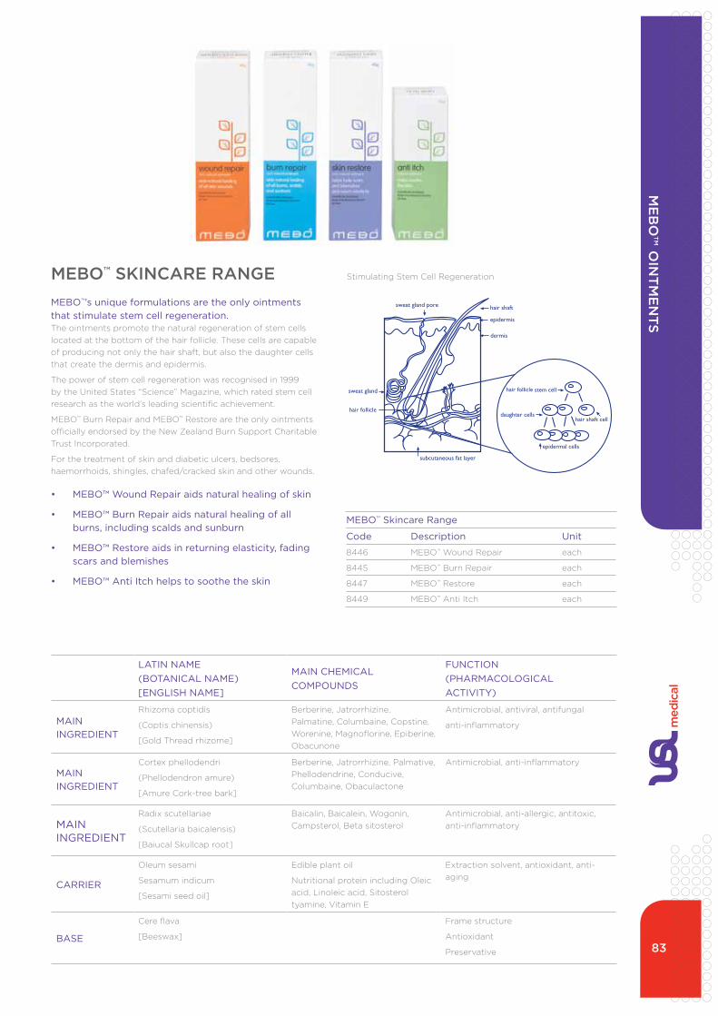
83
MEBO™ Skincare Range
Code Description Unit
8446 MeBO™ Wound Repair each
8445 MeBO™ Burn Repair each
8447 MeBO™ Restore each
8449 MeBO™ anti itch each
Stem cell regeneration
MEBO products accelerate the regeneration of stem cells located at the bottom of the hair follicle. These cells are capable of regenerating not only the hair shaft but also the daughter cells that create the dermis and epidermis. Stem cells can repair injured and defective skin by restructuring and regenerating new skin according to the original skin’s physiological structure. This speeds healing as well as minimising scarring. Research has shown the healing process is significantly faster using MEBO ointments.
MEBO products have proven to accelerate stem cell regeneration.
MEbO™ SKINCARE RANGEMEBO™’s unique formulations are the only ointments that stimulate stem cell regeneration. the ointments promote the natural regeneration of stem cells located at the bottom of the hair follicle. these cells are capable of producing not only the hair shaft, but also the daughter cells that create the dermis and epidermis.
the power of stem cell regeneration was recognised in 1999 by the united States “Science” Magazine, which rated stem cell research as the world’s leading scientific achievement.
MeBO™ Burn Repair and MeBO™ Restore are the only ointments officially endorsed by the new Zealand Burn Support Charitable trust incorporated.
For the treatment of skin and diabetic ulcers, bedsores, haemorrhoids, shingles, chafed/cracked skin and other wounds.
• MEBO™ Wound Repair aids natural healing of skin
• MEBO™ Burn Repair aids natural healing of all burns, including scalds and sunburn
• MEBO™ Restore aids in returning elasticity, fading scars and blemishes
• MEBO™ Anti Itch helps to soothe the skin
Stimulating Stem Cell Regeneration
LATIN NAME(BOTANICAL NAME)[ENGLISH NAME]
MAIN CHEMICALCOMPOUNDS
FUNCTION(PHARMACOLOGICALACTIVITY)
MAIN INGREDIENT
Rhizoma coptidis
(Coptis chinensis)
[gold thread rhizome]
Berberine, Jatrorrhizine, Palmatine, Columbaine, Copstine, Worenine, Magnoflorine, epiberine, Obacunone
antimicrobial, antiviral, antifungal
anti-inflammatory
MAIN INGREDIENT
Cortex phellodendri
(Phellodendron amure)
[amure Cork-tree bark]
Berberine, Jatrorrhizine, Palmative, Phellodendrine, Conducive, Columbaine, Obaculactone
antimicrobial, anti-inflammatory
MAIN INGREDIENT
Radix scutellariae
(Scutellaria baicalensis)
[Baiucal Skullcap root]
Baicalin, Baicalein, Wogonin, Campsterol, Beta sitosterol
antimicrobial, anti-allergic, antitoxic, anti-inflammatory
CARRIER
Oleum sesami
Sesamum indicum
[Sesami seed oil]
edible plant oil
nutritional protein including Oleic acid, Linoleic acid, Sitosterol tyamine, Vitamin e
extraction solvent, antioxidant, anti-aging
BASE
Cere flava
[Beeswax]
Frame structure
antioxidant
Preservative
ME
bO
™ OIN
TM
EN
TS

84
it has been specifically formulated combining 80% Medihoney™ antibacterial Honey with natural waxes and oils to provide a high viscosity gel that is easy to apply with good wash off characteristics when dressings are changed.
Medihoney™ Antibacterial Wound Gel™ is proven to be clinically effective for:• inhibiting bacteria at the wound bed (effective against
more than 200 clinical isolates)
• fast, effective autolytic debridement on sloughy and necrotic tissue in an antibacterial environment
• rapidly removing malodour
• providing a moist wound healing environment thus reducing trauma and pain at dressing change
Indications:• surgical wounds
• burns
• pressure sores
• acute and chronic wounds
• leg / foot ulcers
• donor and recipient graft sites
Superior wound bed preparationthe high osmotic potential created by Medihoney™ antibacterial Wound gel causes a mass flow of bacteria, endotoxins and necrotic material away from the wound bed. this material is then contained in an antibacterial matrix for easy removal at dressing change.
MEDIHONEY™ ANTIbACTERIAL PRODUCTS™ are topical preparations for chronic and acute wound care.
Medihoney™
Medihoney® Antibacterial Wound Gel
Code Description Unit
10786antibacterial Wound gel 10g tube
tube
CASE STUDY
initial wound.
after 10 days treatment with Medihoney™ antibacterial Wound gel™.
BurnsANTIBACTERIALWOUND GEL™
MEDIHONEY™ ANTIBACTERIAL WOUND GEL™ IS ATOPICAL PREPARATION FOR CHRONIC AND ACUTEWOUND CARE.
It has been specially formulated combining 80% Medihoney™Antibacterial Honey with natural waxes and oils to provide a highviscosity gel that is easy to apply with good wash off characteristicswhen dressings are changed.
Medihoney™ Antibacterial Wound Gel™ is proven to be clinicallyeffective for:
• Inhibiting bacteria at the wound bed (effective against morethan 200 clinical isolates);
• Fast, effective autolytic debridement on sloughy and necrotictissue in an antibacterial environment;
• Rapidly removing malodour;
• Providing a moist wound healing environment thus reducingtrauma and pain at dressing change.
INDICATIONS:• Surgical wounds • Acute & Chronic wounds• Burns • Leg / foot ulcers• Pressure sores • Donor and recipient graft sites
SUPERIOR WOUND BED PREPARATION:The high osmotic potential createdby Medihoney™ AntibacterialWound Gel™ causes a mass flow ofbacteria, endotoxins and necroticmaterial away from the wound bed.This material is then contained in anantibacterial matrix for easy removalat dressing change.
Clinical and In Vitro research has shown that Medihoney™ Antibacterial Honey at low concentrations is effective against abroad spectrum of bacteria, including antibiotic-resistant strains.
ONEPRODUCT,
THREEACTIONS.
WOUNDPROTECTION
WOUNDCLEANING
WOUNDHEALING
MIC OF 127 DRUG-RESISTANT CLINICAL ISOLATES (1990-2004) Narelle George, Qld Health Pathology and Scientific Services, Royal Brisbane Hospital
Concentration of Medihoney™ (%)Antibacterial Honey
* nm: non-multiresistant
MRSA / nm* MRSA
ESBL
VRE
Acinetobacter
Pseudomonas aeruginosa
0% 2% 4% 6% 8% 10% 12% 14% 16%
Bacteria Minimum Inhibitory Concentration (MIC).
ANTIbACTERIAL WOUND GELME
DIH
ON
EY
™ R
AN
GE
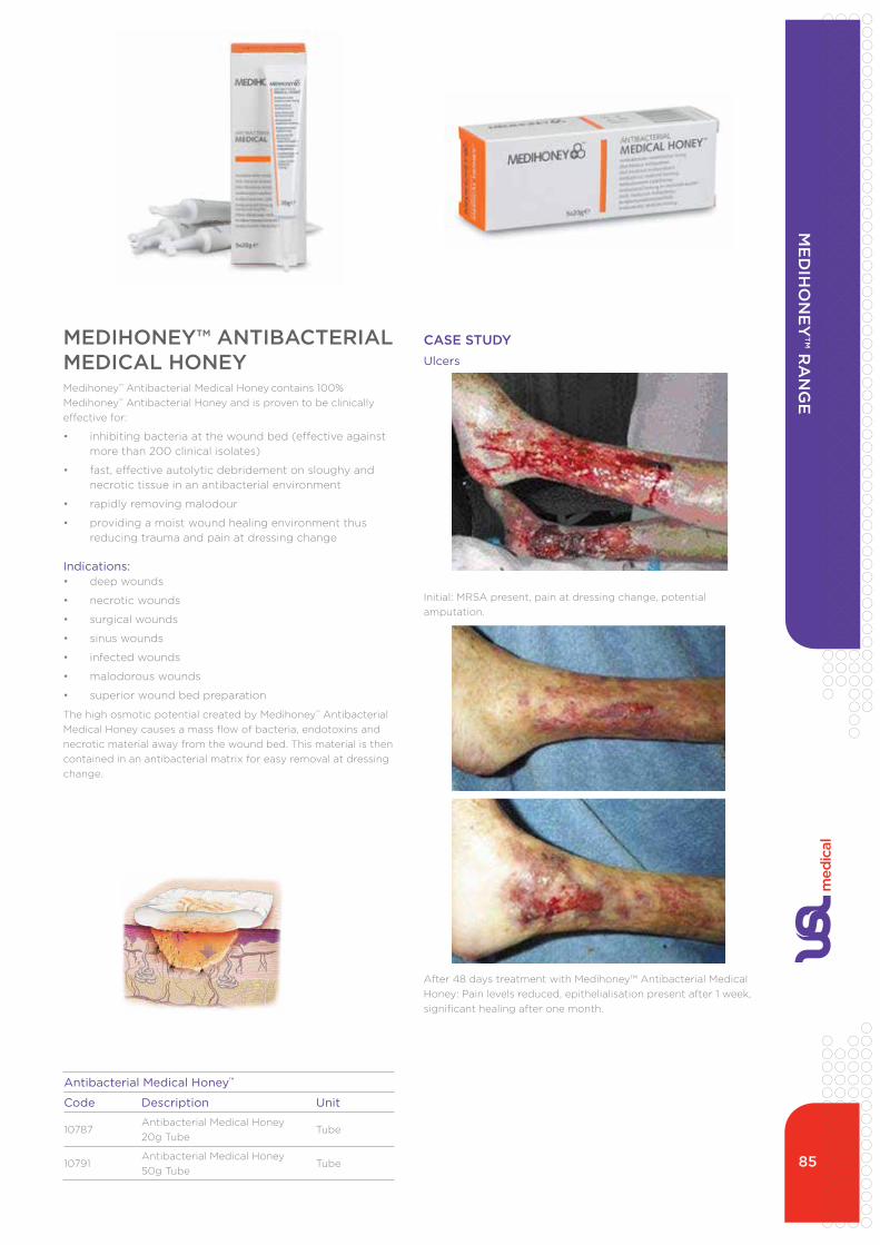
85
Antibacterial Medical Honey™
Code Description Unit
10787antibacterial Medical Honey 20g tube
tube
10791antibacterial Medical Honey 50g tube
tube
CASE STUDY
initial: MRSa present, pain at dressing change, potential amputation.
after 48 days treatment with Medihoney™ antibacterial Medical Honey: Pain levels reduced, epithelialisation present after 1 week, significant healing after one month.
UlcersANTIBACTERIALWOUND GEL™
MEDIHONEY™ ANTIBACTERIAL WOUND GEL™ IS ATOPICAL PREPARATION FOR CHRONIC AND ACUTEWOUND CARE.
It has been specially formulated combining 80% Medihoney™Antibacterial Honey with natural waxes and oils to provide a highviscosity gel that is easy to apply with good wash off characteristicswhen dressings are changed.
Medihoney™ Antibacterial Wound Gel™ is proven to be clinicallyeffective for:
• Inhibiting bacteria at the wound bed (effective against morethan 200 clinical isolates);
• Fast, effective autolytic debridement on sloughy and necrotictissue in an antibacterial environment;
• Rapidly removing malodour;
• Providing a moist wound healing environment thus reducingtrauma and pain at dressing change.
INDICATIONS:• Surgical wounds • Acute & Chronic wounds• Burns • Leg / foot ulcers• Pressure sores • Donor and recipient graft sites
SUPERIOR WOUND BED PREPARATION:The high osmotic potential createdby Medihoney™ AntibacterialWound Gel™ causes a mass flow ofbacteria, endotoxins and necroticmaterial away from the wound bed.This material is then contained in anantibacterial matrix for easy removalat dressing change.
Clinical and In Vitro research has shown that Medihoney™ Antibacterial Honey at low concentrations is effective against abroad spectrum of bacteria, including antibiotic-resistant strains.
ONEPRODUCT,
THREEACTIONS.
WOUNDPROTECTION
WOUNDCLEANING
WOUNDHEALING
MIC OF 127 DRUG-RESISTANT CLINICAL ISOLATES (1990-2004) Narelle George, Qld Health Pathology and Scientific Services, Royal Brisbane Hospital
Concentration of Medihoney™ (%)Antibacterial Honey
* nm: non-multiresistant
MRSA / nm* MRSA
ESBL
VRE
Acinetobacter
Pseudomonas aeruginosa
0% 2% 4% 6% 8% 10% 12% 14% 16%
Bacteria Minimum Inhibitory Concentration (MIC).
MEDIHONEY™ ANTIbACTERIAL MEDICAL HONEY Medihoney™ antibacterial Medical Honey contains 100% Medihoney™ antibacterial Honey and is proven to be clinically effective for:
• inhibiting bacteria at the wound bed (effective against more than 200 clinical isolates)
• fast, effective autolytic debridement on sloughy and necrotic tissue in an antibacterial environment
• rapidly removing malodour
• providing a moist wound healing environment thus reducing trauma and pain at dressing change
Indications:• deep wounds
• necrotic wounds
• surgical wounds
• sinus wounds
• infected wounds
• malodorous wounds
• superior wound bed preparation
the high osmotic potential created by Medihoney™ antibacterial Medical Honey causes a mass flow of bacteria, endotoxins and necrotic material away from the wound bed. this material is then contained in an antibacterial matrix for easy removal at dressing change.
ME
DIH
ON
EY
™ R
AN
GE

86
Medihoney™
Antibacterial Honey Tulle Dressing
Code Description Unit
10788 tulle dressing 10 x 10cm 5/box
Medihoney™
Antibacterial Honey Gel Sheet
Code Description Unit
10789 gel Sheet 5 x 5cm 10/box
10790 gel Sheet 10 x 10cm 10/box
MEDIHONEY™ ANTIbACTERIAL HONEY TULLE DRESSING• antibacterial Leptospermum (Manuka) honey
(minimum 20g)
• non adherent
Indications:• leg/foot ulcers
• pressure ulcers
• infected wounds
• sloughy wounds
• neucrotic wounds
• malodorous wounds
• donor and recipient graft sites
• burns
• surgical wounds
• diabetic wounds
• abrasions
Medihoney™ dressings have been shown to improve healing rates by up to 30% compared to standard dressings and to provide faster debridement than some hydrogels.
dressings may be folded or cut with sterile scissors to the shape of the wound.
Frequency of change:
Medihoney™ antibacterial Honey tulle dressing requires changing when the dressing has been diluted by wound exudate. the dressing may be left in place for up to seven days depending on wound exudate. Heavily exudating wounds require close monitoring and may require daily dressing changes.
MEDIHONEY™ ANTIbACTERIAL HONEY GEL SHEETMedihoney™ gel Sheet is a sterile non-adherent wound dressing comprising Medihoney™ antibacterial Honey (80% w/w) and Sodium alginate for wound care (20% w/w).
Medihoney™ gel Sheet is for use on mild to moderately exudating wounds.
Contraindications:do not use on individuals who have had an allergic reaction to honey or sodium alginate.
Cautions
due to the properties of honey, some patients may experience discomfort such as a stinging or drawing sensation upon application. this will subside over time. if discomfort is of an unacceptable level, remove the dressing and wash the affected area.
this product works optimally on mild to moderately exudating wounds. dry wounds should ideally be managed with Medihoney™ antibacterial Medical Honey™ or Medihoney™ antibacterial Wound gel.
to be used with caution and under close observation on heavily exudating wounds, arterial ulcers or heavy bleeds.
not recommended for use in body cavities.
in the event of a slow or non-responding wound, the treatment regime should be reviewed.
ME
DIH
ON
EY
™ R
AN
GE

87
MEDIHONEY™ANTIbACTERIAL HONEY APINATE™ DRESSING• antibacterial Leptospermum (Manuka) honey
• Calcium alginate fabric
• Protects delicate tissue
Indications:• leg/foot ulcers
• pressure ulcers
• infected wounds
• sloughy wounds
• malodorous wounds
• donor and recipient graft sites
• burns
• surgical wounds
• diabetic wounds
• abrasions
Frequency of change:the Medihoney™ antibacterial Honey apinate™ dressing requires changing when the dressing has been diluted by wound exudate. the dressing may be left in place for up to seven days depending on wound exudate. if maceration of the surrounding skin occurs more frequent changing of the dressing is required.
Medihoney™ Antibacterial Honey Apinate™ Dressing
Code Description Unit
11973antibacterial Honey apinate™ dressing, 5x5cm
10/box
9465antibacterial Honey apinate™ dressing, 10 x 10cm
5/box
11974antibacterial Honey apinate™ dressing, 2g Rope
5/box
tissue and debris from the wound. Additionally, the high
sugar-to-water ratio noted in undiluted honey, which directly
results in the antibacterial properties, has been demonstrated
to be maintained even when the honey has been diluted
by constantly exudating wounds.7 Honey contains the en-
zyme glucose oxidase, which produces gluconic acid and
minute amounts of hydrogen peroxide when in contact with
the wound surface. The concurrent activation of bee-derived
glucose oxidase promotes the release of hydrogen peroxide
from the glucose. The generation of hydrogen peroxide is
currently being correlated to the stimulation of angiogenesis
and fibroblast proliferation.9
Tonks et al9 confirmed that the 5.8-kDa component of
Manuka honey stimulated monocyte cells in cell culture to
release important intermediates that augmented the produc-
tion of several cytokines, including TNF-alpha and IL-L,
which notably help eradicate infection and facilitate tissue
repair. Minimizing protease levels within the wound envi-
ronment is paramount as elevated protease levels can de-
grade important growth factors and fibronectin, leading to a
prolonged inflammatory phase of wound healing.
Calcium alginate, which compromises approximately 5%
of each Medihoney wound dressing, has been shown to
improve fibroblastic proliferation and was noted to decrease
fibroblast motility.13 It has been suggested that the effects
of calcium alginate on the proliferation of fibroblasts may
be related to the release of calcium ions.13 Thus, alginate
incorporation with Medihoney provides a synergistic effect.
With the emergence of increasingly resistant strains of
bacteria, the attraction of medical-grade honey to stimulate
Table 1.
MEDIHONEY CASE SERIES
Patient # LocationPrior Durationof Ulcer
Initial WoundSize Bacterial Burden Previous Treatment History
1 Medial Ankle 4 months 9.7 cm Klebsiella pneumoniae, Enterobacter cloacae 4-layer compression2 Medial Ankle 4 years 6.9 cm methicillin-resistant Staphylococcus aureus 4-layer compression, IV Vancomycin, Topical Silver3 Medial Ankle/Heel 4 months 3.6 cm No growth 4-layer compression, hyperbaric oxygen therapy,
Genetically engineered skin equivalents, endovenouslaser treatment
4 Medial Ankle 6 months 4.7 cm No growth 4-layer compression5 Anterior Proximal Leg 6 months 14.8 cm No growth Short-stretch bandaging6 Medial Ankle 22 months 11.80 cm Staphylococcus aureus Short-stretch bandaging7 Lateral Ankle 8 months 6.7 cm No growth Nonadherent dressing8 Anterior Ankle 6 months 4.0 cm Staphylococcus epidermiditis Nonadherent dressing9 Medial Ankle 19 months 5.4 cm No growth 4-layer compression10 Lateral Ankle 11 months 3.2 cm No growth 4-layer compression11 Lateral Ankle 8 months 6.2 cm No growth 4-layer compression
Figure 3.
PATIENT #5 WITH MEDIHONEY APPLIED FOR 29 DAYS UNDER A 4-LAYER COMPRESSIVE DRESSING
CHANGED WEEKLY
ADVANCES IN SKIN & WOUND CARE & VOL. 22 NO. 2 70 WWW.WOUNDCAREJOURNAL.COM
9Copyright @ 200 Lippincott Williams & Wilkins. Unauthorized reproduction of this article is prohibited.
CASE STUDY
after 29 days, with Medihoney™ applied under a 4-layer compressive dressing. Changed weekly.
Ulcers
ME
DIH
ON
EY
™ R
AN
GE

88
MEDIHONEY® HCSHydrogel Dressing with Super Absorbent Polymer• antibacterial Leptospermum (Manuka) honey
• Calcium alginate fabric
• Protects delicate tissue
Indications:• leg/foot ulcers
• pressure ulcers
• infected wounds
• sloughy wounds
• malodorous wounds
• donor and recipient graft sites
• burns
• surgical wounds
• diabetic wounds
• abrasions
Frequency of change:the Medihoney™ antibacterial Honey apinate™ dressing requires changing when the dressing has been diluted by wound exudate. the dressing may be left in place for up to seven days depending on wound exudate. if maceration of the surrounding skin occurs more frequent changing of the dressing is required.
Medihoney™ Antibacterial Honey Hydrogel Colloidal Sheet Dressing
Code Description Unit
12818Medihoney HCS non-adhesive dressing 6x6cm
10/box
12819Medihoney HCS non-adhesive dressing 11x11cm
10/box
12820Medihoney HCS adhesive dressing 11x11cm
10/box
12817Medihoney HCS adhesive dressing 15x15cm
10/box
ME
DIH
ON
EY
™ R
AN
GE

89
ME
DIH
ON
EY
™ R
AN
GE
Cerebo Vascular Incidience, friable granulation tissue, oedema, and pain
Non-healing with compression therapy and a plain alginate dressing
MEDIHONEY™ initiated
Compression therapy continued
Wound heals in 28 days
Week #4 after adding MEDIHONEY® under compression
Matthew Regulski, DPM: Case #3 of 8 presented at 2008 Symposium of Advanced Wound Care
Non-healing, painful leg ulcer. Week #12 under compression
Dehisced pectoral flap procedure
Progress stalled under Negative Pressure Wound Therapy
MEDIHONEY™ initiated Rapid healing noted after initial daily applications
Complete wound closure achieved by Week #3
Non-Healing Post-Op Wound
StalledVenous Leg Ulcer
Stalled dehisced post-op wound Week #3 after MEDIHONEY® applications
Paul Liguori, MD and Kim Peters, RN, CWS: Case #1 of 8 presented at 2008 World Union of Wound Healing Societies
[Look to MEDIHONEY® as first-line of defense]
for stalled, non-progressing wounds

90
THE ORIGINAL WOUND CARE SOLUTION
WOUND CARE & PRODUCT SELECTION CHART
WOUND TYPE INFECTED BLACK, NECROTIC YELLOW, SLOUGHY RED, GRANULATION PINK, EPITHELIAL CAVITY OR SINUS/FISTULA MALODOROUS
TREATMENT OBJECTIVE Reduce infection and colonisation
Assists wound healing
Clean wound
Rehydrate
Debride
Wound protection
Remove slough
Rehydrate
Wound protection
Manage exudate
Maintain moist wound bed
Assist wound healing
Wound protection
Manage exudate
Maintain moist wound bed
Wound protection
Assist wound healing
Aid granulation
Assist wound healing
Wound protection
Remove malodour
MODE OF ACTION High osmotic potential helps clean the wound and disperse biofi lms.
Cleaning the wound reduces bacteria loadings, which are a source of infl ammatory endotoxins.
Provides an antibacterial barrier to protect the wound bed.
The osmotic action produces an outfl ow of body fl uid helping to lift debris and remove necrotic tissue.
The moist wound environment reduces the risk of eschar formation.
Provides an antibacterial environment that helps protect the wound.
The high osmotic potential of Antibacterial Medihoney™ wound dressings causes a mass fl ow of bacteria, endotoxins and sloughy material away from the wound bed. This material is then contained in an antibacterial matrix for easy removal at dressing change.
Osmotic action brings fl uid into the wound providing a moist wound environment to assist the granulation process
Antibacterial barrier protects the wound from infection by bacteria.
Provides moist wound environment to encourage the epithelialisation process.
Antibacterial barrier to protect the wound from contamination and reduces risk of colonisation.
Helps reduce oedema, pain and exudate levels and protects exposed tissue from infecting bacteria, reducing the risk of colonisation.
Inhibits the bacteria that cause odour.
Helps clean the wound and lift debris to rapidly reduce odour.
EXUDATE LEVEL Heavy/Moderate
Low/None Heavy/Moderate
Low/None Heavy/Moderate
Low/None Heavy/Moderate
Low/None Heavy/Moderate
Low/None Heavy/Moderate
Low/None Heavy/Moderate
Low/None
Medihoney™ Antibacterial Medical Honey™ ✓ ✓ ✓ ✓ ✓ ✓ ✓ ✓ ✓ ✓ ✓ ✓ ✓ ✓Medihoney™ Antibacterial Wound Gel™ ✓ ✓ ✓ ✓ ✓ ✓ ✓ ✓ ✓ ✓ ✓ ✓Medihoney™ Antibacterial Honey Gel Sheet ✓ ✓ ✓ ✓ ✓ ✓Medihoney™ Antibacterial Honey Alginate ✓ ✓ ✓ ✓ ✓ ✓Medihoney™ Antibacterial Honey Tulle ✓ ✓ ✓ ✓ ✓ ✓ ✓ ✓ ✓ ✓ ✓ ✓
Medihoney (Europe) LtdPost Box 2205 High StreetMaidenheadSL6 1JNUK Toll free: 0800 071 3912Int Tel: +44 1628 779461 www.medihoney.com
NB Secondary dressings should be suffi ciently absorbent to manage exudate levels
It’s not just any honey
It’s MEDIHONEY™
For skin protection around exudating wounds use Medihoney™ Barrier Cream
MEDIHONEY WOUNDCARE AND PRODUCT SELECTION CHART
ME
DIH
ON
EY
™ W
OU
ND
CH
AR
T

91
THE ORIGINAL WOUND CARE SOLUTION
WOUND CARE & PRODUCT SELECTION CHART
WOUND TYPE INFECTED BLACK, NECROTIC YELLOW, SLOUGHY RED, GRANULATION PINK, EPITHELIAL CAVITY OR SINUS/FISTULA MALODOROUS
TREATMENT OBJECTIVE Reduce infection and colonisation
Assists wound healing
Clean wound
Rehydrate
Debride
Wound protection
Remove slough
Rehydrate
Wound protection
Manage exudate
Maintain moist wound bed
Assist wound healing
Wound protection
Manage exudate
Maintain moist wound bed
Wound protection
Assist wound healing
Aid granulation
Assist wound healing
Wound protection
Remove malodour
MODE OF ACTION High osmotic potential helps clean the wound and disperse biofi lms.
Cleaning the wound reduces bacteria loadings, which are a source of infl ammatory endotoxins.
Provides an antibacterial barrier to protect the wound bed.
The osmotic action produces an outfl ow of body fl uid helping to lift debris and remove necrotic tissue.
The moist wound environment reduces the risk of eschar formation.
Provides an antibacterial environment that helps protect the wound.
The high osmotic potential of Antibacterial Medihoney™ wound dressings causes a mass fl ow of bacteria, endotoxins and sloughy material away from the wound bed. This material is then contained in an antibacterial matrix for easy removal at dressing change.
Osmotic action brings fl uid into the wound providing a moist wound environment to assist the granulation process
Antibacterial barrier protects the wound from infection by bacteria.
Provides moist wound environment to encourage the epithelialisation process.
Antibacterial barrier to protect the wound from contamination and reduces risk of colonisation.
Helps reduce oedema, pain and exudate levels and protects exposed tissue from infecting bacteria, reducing the risk of colonisation.
Inhibits the bacteria that cause odour.
Helps clean the wound and lift debris to rapidly reduce odour.
EXUDATE LEVEL Heavy/Moderate
Low/None Heavy/Moderate
Low/None Heavy/Moderate
Low/None Heavy/Moderate
Low/None Heavy/Moderate
Low/None Heavy/Moderate
Low/None Heavy/Moderate
Low/None
Medihoney™ Antibacterial Medical Honey™ ✓ ✓ ✓ ✓ ✓ ✓ ✓ ✓ ✓ ✓ ✓ ✓ ✓ ✓Medihoney™ Antibacterial Wound Gel™ ✓ ✓ ✓ ✓ ✓ ✓ ✓ ✓ ✓ ✓ ✓ ✓Medihoney™ Antibacterial Honey Gel Sheet ✓ ✓ ✓ ✓ ✓ ✓Medihoney™ Antibacterial Honey Alginate ✓ ✓ ✓ ✓ ✓ ✓Medihoney™ Antibacterial Honey Tulle ✓ ✓ ✓ ✓ ✓ ✓ ✓ ✓ ✓ ✓ ✓ ✓
Medihoney (Europe) LtdPost Box 2205 High StreetMaidenheadSL6 1JNUK Toll free: 0800 071 3912Int Tel: +44 1628 779461 www.medihoney.com
NB Secondary dressings should be suffi ciently absorbent to manage exudate levels
It’s not just any honey
It’s MEDIHONEY™
For skin protection around exudating wounds use Medihoney™ Barrier Cream
ME
DIH
ON
EY
™ W
OU
ND
CH
AR
T

92
COLOPLaSt WOundCaRe
PROduCtS

93
Biatain non-adhesive Foam dressing
Code Size unit
13276 5cm x 7cm 10/box
13277 10cm x 10cm 10/box
13278 10cm x 20cm 5/box
13279 15cm x 15cm 5/box
13280 20cm x 20cm 5/box
bIATAIN NON-ADHESIvE FOAM DRESSINGBiatain non-adhesive is a soft and flexible, easy to wear foam with bevelled edges, significantly lowering the risk of pressure marks.
Biatain is indicated for a wide range of moderate to highly exuding acute and chronic wounds such as leg ulcers, pressure ulcers, diabetic ulcers, second degree burns and donor sites.
Biatain adhesive Foam dressing
Code Size unit
13281 7.5cm x 7.5cm 10/box
13282 12.5cm x 12.5cm 10/box
13283 18cm x 18cm 5/box
13285 23cm x 23cm Sacral 5/box
13284 19cm x 20cm Heel 5/box
bIATAIN ADHESIvE FOAM DRESSINGthe Biatain adhesive dressing consists of the soft absorbent polyurethane foam pad located centrally upon a larger polyurethane membrane coated with a hydrocolloid adhesive. the film backing is permeable to water vapour, but impermeable to microorganisms and water, forming an effective bacterial and waterproof dressing.
Biatain adhesive is indicated for a wide range of moderate to highly exuding acute and chronic wounds such as leg ulcers, pressure ulcers, diabetic ulcers, second degree burns and donor sites.
Biatain Foam dressings provide superior exudate management for faster wound healing* and are soft, flexible and easy to wear. the superior absorption and retention properties of Biatain minimise the risk of leakage and maceration*. the unique 3-d structure of Biatain serves as a partial fluid lock, retaining the exudate – even under compression therapy. Wound exudate is absorbed locally with minimal lateral dispersal, thus reducing leakage, skin maceration and risk of infection, while providing an optimal moist wound healing environment for up to 7 days. When absorbing exudate, Biatain foam swells and conforms to the wound for intimate wound contact, resulting in superior absorption and a moisture balance that improves the healing process*.
*data on file
bIATAIN FOAM DRESSINGS
CO
LOP
LAST
WO
UN
DC
AR
E

94
Biatain Silicone Foam Dressing
Code Size Unit
13287 10cm x 10cm 10/box
13289 15cm x 15cm 5/box
bIATAIN SILICONE FOAM DRESSINGSilicone dressings are gentle on the skin, and easy to remove and reposition with minimal pain to the patient. Biatain Silicone provides superior absorption, fluid management, softness and flexibility. Biatatain Silicone combines the gentleness of silicone and the effectiveness of foam.
Biatain Silicone is indicated for the healing of most non-infected acute and chronic moist wounds, including leg ulcers, diabetic foot ulcers, second degree burns, donor sites, post-operative wounds, skin abrasions and pressure ulcers.
Biatain Soft-Hold Foam Dressing
Code Size Unit
13292 10cm x 10cm 5/box
13293 10cm x 20cm 5/box
13291 15cm x 15cm 5/box
bIATAIN SOFT-HOLD FOAM DRESSINGBiatain Soft-Hold is an additional variant in the non-adherent range. Biatain Soft-Hold features a light hydrocolloid adhesive on 50% of the dressing surface to assist with keeping the dressing in place for difficult to dress areas. the light adhesive coating does not affect the absorption capabilities of the dressing.
Biatain Soft-Hold is indicated for a wide range of moderate to highly exuding acute and chronic wounds such as leg ulcers, pressure ulcers, diabetic ulcers and second degree burns.
CO
LOP
LAST
WO
UN
DC
AR
E

95
Biatain Ag Non-Adhesive Dressing
Code Size Unit
13294 5cm x 7cm 5/box
11860 10cm x 10cm 5/box
13295 15cm x 15cm 5/box
13301 5cm x 8cm Cavity 5/box
13280 20cm x 20cm 5/box
bIATAIN AG NON-ADHESIvE FOAM DRESSINGin addition to the advantages of the soft and flexible non-adhe-sive foam, Biatain ag dressings are prepared with hydro-acti-vated silver ions, which work both in the foam and in the wound bed. the sustained and controlled release of silver ions ensures fast killing of all bacteria with the added benefit of effective exudate management. Biatain ag dressings are effective for up to 7 days.
Biatain ag is suitable for use on leg ulcers, pressure ulcers, diabetic foot ulcers, second degree burns, donor sites, post-op-erative wounds and skin abrasions. Biatain ag can be used on infected or at-risk wounds.
Biatain Ag Adhesive Dressing
Code Size Unit
13296 7.5cm x 7.5cm 5/box
13297 12.5cm x 12.5cm 5/box
bIATAIN AG ADHESIvE FOAM DRESSINGBiatain ag adhesive has a reduced thickness, softer foam and discreet design with the same absorbency as the non-adhesive version. the dressing uses a hydrocolloid product as the adhesive component, ensuring skin friendliness.
Biatain ag dressings are prepared with hydro-activated silver ions, which work both in the foam and in the wound bed. the sustained and controlled release of silver ions ensures fast killing of all bacteria with the added benefit of effective exudate management. . Biatain ag dressings are effective for up to 7 days.
Biatain ag is suitable for use on leg ulcers, pressure ulcers, diabetic foot ulcers, second degree burns, donor sites, post-operative wounds and skin abrasions. Biatain ag can be used on infected or at-risk wounds.
CO
LOP
LAST
WO
UN
DC
AR
E
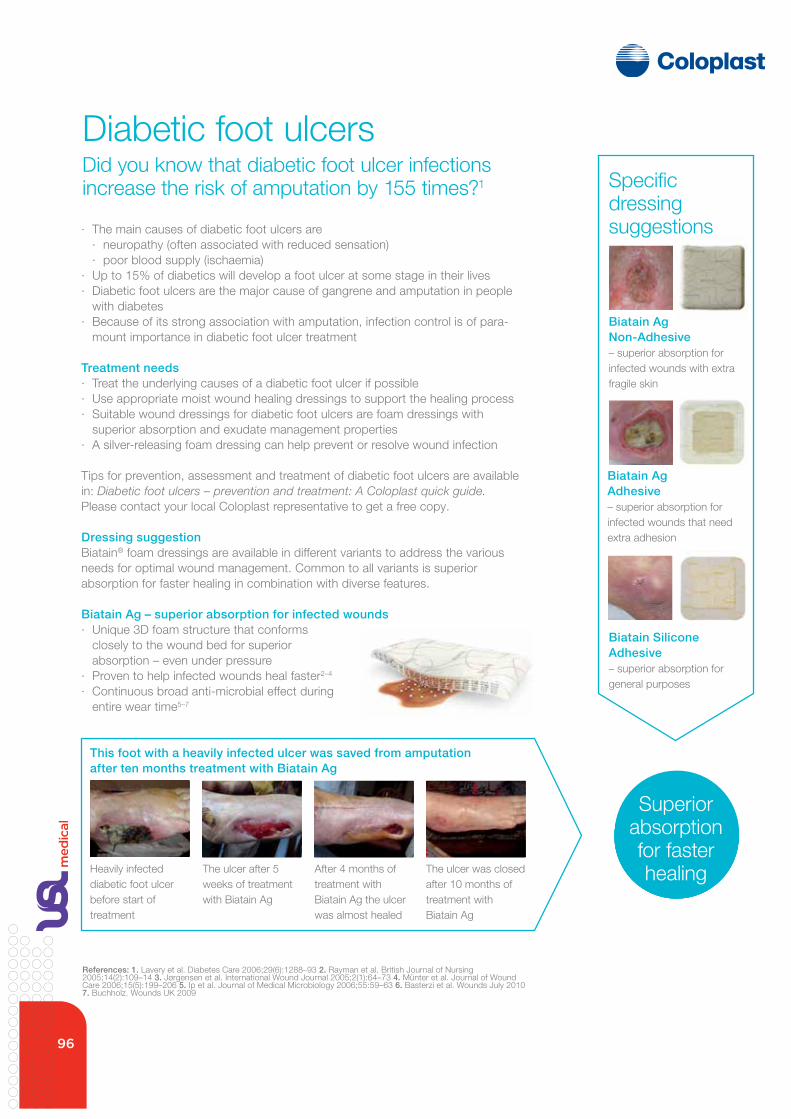
References: 1. Lavery et al. Diabetes Care 2006;29(6):1288–93 2. Rayman et al. British Journal of Nursing 2005;14(2):109–14 3. Jørgensen et al. International Wound Journal 2005;2(1):64–73 4. Münter et al. Journal of Wound Care 2006;15(5):199–206 5. Ip et al. Journal of Medical Microbiology 2006;55:59–63 6. Basterzi et al. Wounds July 2010 7. Buchholz. Wounds UK 2009
Diabetic foot ulcersDid you know that diabetic foot ulcer infectionsincrease the risk of amputation by 155 times?1
Superior absorption for faster healing
· The main causes of diabetic foot ulcers are· neuropathy (often associated with reduced sensation)· poor blood supply (ischaemia)
· Up to 15% of diabetics will develop a foot ulcer at some stage in their lives· Diabetic foot ulcers are the major cause of gangrene and amputation in people
with diabetes· Because of its strong association with amputation, infection control is of para-
mount importance in diabetic foot ulcer treatment
Treatment needs· Treat the underlying causes of a diabetic foot ulcer if possible· Use appropriate moist wound healing dressings to support the healing process· Suitable wound dressings for diabetic foot ulcers are foam dressings with
superior absorption and exudate management properties· A silver-releasing foam dressing can help prevent or resolve wound infection
Tips for prevention, assessment and treatment of diabetic foot ulcers are available in: Diabetic foot ulcers – prevention and treatment: A Coloplast quick guide. Please contact your local Coloplast representative to get a free copy.
Dressing suggestionBiatain® foam dressings are available in different variants to address the various needs for optimal wound management. Common to all variants is superior absorption for faster healing in combination with diverse features.
Biatain Ag – superior absorption for infected wounds· Unique 3D foam structure that conforms
closely to the wound bed for superior absorption – even under pressure
· Proven to help infected wounds heal faster2–4
· Continuous broad anti-microbial effect during entire wear time5–7
This foot with a heavily infected ulcer was saved from amputation after ten months treatment with Biatain Ag
Heavily infected diabetic foot ulcer before start of treatment
The ulcer after 5 weeks of treatment with Biatain Ag
After 4 months of treatment with Biatain Ag the ulcer was almost healed
The ulcer was closed after 10 months of treatment with Biatain Ag
Biatain Ag Adhesive – superior absorption for infected wounds that need extra adhesion
Biatain Silicone Adhesive– superior absorption for general purposes
Specific dressing suggestions
Biatain Ag Non-Adhesive– superior absorption for infected wounds with extra fragile skin
Coloplast develops products and services that make life easier for people with very personal and private medical conditions. Working closely with the people who use our products, we create solutions that are sensitive to their special needs. We call this intimate healthcare. Our business includes ostomy care, urology and continence care and wound and skin care. We operate globally and employ more than 7,000 people.
The Coloplast logo is a registered trademark of Coloplast A/S. © 2012-12. WOU150 All rights reserved Coloplast A/S, 3050 Humlebæk, Denmark.
Coloplast Pty Ltd 33 Gilby Road
Mount Waverley VIC 3149 Australia
www.coloplast.com.au
96

COMFEEL PLUS DRESSINGSComfeel Plus dressings have a unique combination of alginate and hydrocolloid for superior exudate management, resulting in even fewer dressing changes and less disruption to the wound. the dressing surface is covered by a semi-permeable and elastic polyurethane film, providing a low profile and waterproof dressing. Comfeel Plus dressings are a newer generation of hydrocolloid. new technology has made it possible to develop a unique wound dressing. Optimal moisture is maintained in the wound during healing and is achieved through a unique interplay between absorption and evaporation. Optimal moisture is maintained in the wound during healing and is achieved through a unique interplay between absorption and evaporation. the superior exudate management is due to improved absorption and the intelligent semi-permeable surface film.
the intelligence consists of the film’s ability to regulate the evaporation in accordance with the volume of exudate. during periods of heavy exudation, the pores of the film open widely to allow for its evaporation. this provides space for additional exudate. Conversely, during periods of light exudation, the pores contract to limit the evaporation. this gives a dressing that regulates the moisture in the wound. the moisture simplifies the clearing away of dead tissue and encourages epithelialisation. the colour guide indicates best time to change dressing and the no touch application system increases ease of use. the quadrate window pattern on the surface is a built in measuring guide, making it possible to measure the wound size on top of the dressing. Comfeel Plus dressings are also latex free and sterile.
COMFeeL®HydROCOLLOid
dReSSingS
97

98
Comfeel Plus Ulcer
Code Size Unit
29103146 6 x 4cm 30/box
29103110 10x10cm 10/box
29103115 15 x 15cm 5/box
29103120 20 x 20cm 5/box
COMFEEL® PLUS ULCER DRESSING Comfeel Plus ulcer dressings provides good exudate handling capacity, increased wear time and improved adhesion for low to moderate exuding wounds.
the improved exudate management means fewer dressing changes. this has a positive effect on the healing process and offers the patient increased freedom. Comfeel Plus ulcer dressings are soft and flexible.
the dressing changes its colour to milky white when it absorbs wound secretions. When the white colour reaches about 1cm from the edge, it’s time to change the dressing.
a dressing change can be done quickly and simply, because there is no residue left in the wound.
Comfeel Plus ulcer dressing is primarily indicated for the treatment of low to moderately exudating leg ulcers and pressure sores.
it may also be used for superficial burns, superficial partial-thickness burns, donor sites, post-operative wounds and skin abrasions. Hydrocolloids are not indicated for use on full thickness wounds or exposed bone or tendon.
Comfeel Plus Transparent Dressing
Code Size Unit
29103530 5cm x 7cm 10/box
29103533 10cm x 10cm 10/box
29103536 9cm x 14cm 10/box
29103542 15cm x 20cm 5/box
29103548 5cm x 25cm 5/box
COMFEEL® PLUS TRANSPARENT DRESSINGComfeel Plus transparent dressings are thin, transparent dressings, providing good exudate management for low exuding wounds, while allowing for visual inspection of the wound. they are also soft and flexible.
the transparent dressing facilitates inspection of the wound without removing it. the healing process can therefore be monitored closely. the quadrate window pattern on the surface of the dressing makes it easy to measure the size of the wound.
the dressing can stay on up to seven days which means less wound disruption and increased patient comfort.
Comfeel Plus transparent dressings are used for treatment of low exudating chronic wounds and superficial acute wounds, such as pressure sores, leg ulcers, superficial burns, donor sites, traumatic wounds and post-operative wounds. the dressing is used to protect the skin in specially vulnerable areas, in the final stages of wound healing and in difficult to dress areas.
CO
LOP
LAST
WO
UN
DC
AR
E

99
Comfeel Ulcer Dressing
Code Size Unit
3233 Comfeel ulcer 6x4cm 30/box
3213 Comfeel ulcer 10x10cm 10/box
3218 Comfeel ulcer 15x15cm 5/box
COMFEEL® ULCER DRESSINGComfeel ulcer dressing is a hydrocolloid dressing with high flexibility and elasticity, with bevelled edges for increased comfort.
Comfeel ulcer dressings will help manage exudate and are suitable for use on leg ulcers, pressure sores, donor sites, superficial burns and minor injuries.
COMFEEL® PLUS CONTOUR DRESSINGthis versatile dressing is designed for difficult to dress areas, combining a centre of Comfeel Plus ulcer dressing and a unique hydrocolloid framework. Offers all the benefits of the Comfeel Plus ulcer dressings.
Comfeel Plus Contour Dressing
Code Size Unit
29103280 6cm x 8cm Heel/Sacral 5/box
29103283 9cm x 11cm 5/box
CO
LOP
LAST
WO
UN
DC
AR
E

100
Biatain Alginate Dressing
Code Size Unit
29103705 5cm x 5cm 30/box
29103710 10cm x 10cm 10/box
29103715 15cm x 15cm 10/box
Biatain Alginate Rope Dressing (Previously SeaSorb Rope)
Code Size unit
291037402 40cm/2g rope 6/box
SeaSorb Soft alginate dressings are made of a unique
combination of alginate and carboxymethylcellulose (CMC)
for superior absorption and one-piece removal. SeaSorb Soft
dressings are comfortable and soft with excellent gelling
properties, conforming to any wound size and shape. SeaSorb
dressings support wound healing capabilities by providing a
moist wound healing environment.
they have superior vertical absorption to reduce maceration
and form a cohesive soft gel ensuring easy, pain-free one-piece
removal. When SeaSorb Soft dressing and Filler come into
contact with wound exudate, the calcium ions from the dressing
are replaced by sodium ions present in the exudate. this ion
exchange causes the dressing to form a soft, cohesive gel which
locks the exudate inside ensuring less risk of leakage or damage
to the peri-wound skin. SeaSorb Soft alginate dressings are
sterile and latex free.
SeaSorb Soft is indicated for moderately to heavily exudating
wounds such as leg ulcers, pressure ulcers, diabetic foot ulcers,
second degree burns and donor sites. SeaSorb Soft Filler is
indicated for heavily exudating wounds, in particular, deep
cavity wounds. SeaSorb Soft dressings should be used with a
secondary dressings.
bIATAIN® ALGINATE DRESSINGS
Biatain®SuPeRiOR
aBSORPtiOn(PReViOuSLy SeaSORB)
CO
LOP
LAST
WO
UN
DC
AR
E

101
PURILON® GELPurilon gel is a clear gel made of natural ingredients without additives. it consists of more than 90% purified water sodium
carboxymethylcellulose and calcium alginate.
Purilon gel is used for wounds that need effective autolytic debridement of necrotic tissue thus removing this barrier from healing. it
provides effective and gentle debridement, ease of use and superior rehydrating, gelling and cohesion properties without any additives.
the viscous texture allows for controlled application and reduces the risk of peri-wound maceration.
Purilon gel is indicated for dry, sloughy, necrotic wounds, as well as wounds with a mix of necrotic and granulated tissue such as leg
ulcers, pressure ulcers, non-infected diabetic foot ulcers and first and second degree burns. the gel may also be used throughout the
healing process to provide a moist healing environment.Purilon gel should be used in conjunction with a secondary dressing. the gel
changing interval may be up to 3 days. For removal of the gel, use saline or tap water.
Purilon Gel
Code Size Unit
29103900 15 grams each
CO
LOP
LAST
WO
UN
DC
AR
E

RetentiOn and FiXatiOn
dReSSingS
102

103
Omnifix® Elastic - Individually boxed, 10 metre length
Code Size Unit
1293 2.5cm 2 rolls
1297 5cm 1 roll
1298 10cm 1 roll
1300 15cm 1 roll
9423 20cm 1 roll
OMNIFIX® ELASTICThe self-adhesive, polyacrylate non-woven fabric retention dressingOmnifix® elastic is a soft, self-adhesive fabric for secure retention of wound dressings. the hypoallergenic acrylic adhesive reduces the risk of allergic reaction.
Omnifix® elastic is permeable to both air and water vapour, enabling large areas to be covered without the risk of maceration.
the widthways elasticity of Omnifix® elastic allows its smooth application to joints and angular parts of the body, with no inhibition of movement.
Omnifix® elastic covering paper has a ‘wave’ cut to aid in its removal from the backing paper, and a measuring grid printed on it to facilitate cutting to size.
Omnistrip® Sterile
Code Size Unit Per Box
7426 3 x 76mm Pouch of 5 50
7423 6 x 38mm Pouch of 6 50
7425 6 x 76mm Pouch of 3 50
7424 6 x 101mm Pouch of 10 50
7427 12 x 101mm Pouch of 6 50
OMNISTRIP®
The adhesive wound closure strip made of non-woven, flexible fabric Omnistrip® is a wound closure strip made of skin coloured non-woven fabric.
the low-irritant properties of Omnistrip® are enhanced by the high permeability to air and water vapour of the non-woven material.
Features include:• flexible, non-woven material allows
for expansion arising from oedema and flexion
• acrylic adhesive reduces the possibility of allergic reaction
• rounded edges help prevent roll-up
• does not absorb x-rays and may be left in place for radiological exposure
Indications:• atraumatic closure of minor wounds
and surgical incisions (healing by primary intention)
• wound support post operatively and following suture removal
RE
TE
NT
ION
/FIX
AT
ION

104
Opsite™ Flexifix™
Code Size Unit
1745 10cm x 10m each
1750 15cm x 10m each
1743 5cm x 10m each
OPSITE™ FLEXIFIX™
OpSite™ Flexifix™ is a roll of transparent adhesive film ideal for use in dressing fixation, tube fixation, dressing reinforcement and the treatment of painful peripheral neuropathy. it combines all the advantages of Opsite™ film with a unique application system, to allow easy and secure fixation of all kinds of dressings. OpSite™’s strong, flexible film also offers protection against skin breakdown due to friction and moisture.
Features:• transparent
• conformable
• unique Opsite™ on a roll format
• many clinical applications
What is Opsite™ Flexifix™ used for?• retention of primary dressings, eg Melolin non-
adherent dressings or allevyn™ hydrocellular hydrophilic polyurethane wound dressing
• fixation of tubing
• treatment of painful peripheral neuropathy
• skin protection under leg bags, stoma devices, etc
• reduction of shearing forces on unbroken skin, eg in pressure sore
TEGADERM™ TRANSPARENT FILM ROLLtegaderm™ Roll provides maximum versatility and is the affordable choice for skin protection and waterproof cover dressings.
What is Tegaderm™ used for?tegaderm™ Film provides a protective cover over at-risk, including newly healed, intact skin helping to prevent skin breakdown caused by friction and body fluids.
tegaderm™ Film is breathable, letting oxygen in and moisture vapour out, allowing the skin to function normally.
the perfect solution whenever a waterproof cover dressing is the appropriate choice.
Tegaderm™ Transparent Film Roll
Code Size Unit
9278 10cm x 10m roll
9377 15cm x 10m roll
9277 5cm x 10m roll
RE
TE
NT
ION
/FIX
AT
ION

105
LASTOTEL®
The conformable dressing retention bandageLastotel® is a dressing retention bandage with an extensibility of approximately 140%. it does not restrict or impede blood flow or joint movement when applied correctly.
Lastotel® is 60% polyamide (warp threads). the crepe-like surface structure means the bandage is non slip and easy to apply. Complicated bandaging techniques are not required and angular parts of the body or joint can be covered without reverse turns.
Lastotel® is soft and permeable to air. it is resistant to ointments, oils, greases and perspiration.
Lastotel® can be used as a universal dressing retention bandage.
Indications:• useful for joints and angular or round parts of the body
Lastotel® Unstretched length 2m, stretched length 4m, individually wrapped
Code Size unit
1319 4cm each
1320 6cm each
1321 8cm each
1322 10cm each
7183 12cm each
PEHA-HAFT® LATEX FREEA latex free light cohesive bandage to reduce the risk of allergiesPeha-haft® is now available as a 100% latex, chlorine and acrylate free bandage which is gentle to the skin with significantly reduced risk of allergic reactions.Peha-haft® latex free is cohesive and is suitable for use on fragile or hairy skin as the bandage does not adhere to skin, hair or clothes. Peha-haft® is more permeable to air than conventional cohesive bandages, limiting heat accumulation and subsequent maceration from perspiration.
Peha-haft® Unstretched length 2m, stretched length 4m, individually wrapped
Code Size unit
1346 2.5cm each
1347 4cm each
1348 6cm each
1349 8cm each
1350 10cm each
Peha-haft® Unstretched length 10m, stretched length 20m, not individually wrapped
1351 6cm each
1352 8cm each
1353 10cm each
Tapes and Bandages
PAUL HARTMANN Pty Ltd. ABN 35 000 099 589, Unit 27-28 Homebush Business Village 11-21 Underwood Road Homebush NSW 2140. ® Registered trademark PAUL HARTMANN AG.
1800 805 839 www.hartmann.com.au
Peha-haft® latex freeA latex free light cohesive bandage to reduce the risk of allergies
Peha-haft® is now available as a 100%latex, chlorine and acrylate free bandagewhich is gentle to the skin with significantlyreduced risk of allergic reactions.
Peha-haft® latex free is cohesive and issuitable for use on fragile or hairy skin asthe bandage does not adhere to skin, hairor clothes.
Peha-haft® is more permeable to air thanconventional cohesive bandages, limitingheat accumulation and subsequentmaceration from perspiration.
Product Width Unit Code Unitsper Case
Peha-haft® latex free 4 cm 8 bandages 932 441 90
Stretched length 4m 6 cm 1 bandage 932 442 90
8 cm 1 bandage 932 443 120
10 cm 1 bandage 932 444 135
12 cm 1 bandage 932 44 5 84
Stretched length 20m 4 cm 1 bandage 932 446 12
6 cm 1 bandage 932 447 90
8 cm 1 bandage 932 448 90
10 cm 1 bandage 932 449 120
12 cm 1 bandage 932 450 135
RE
TE
NT
ION
/FIX
AT
ION
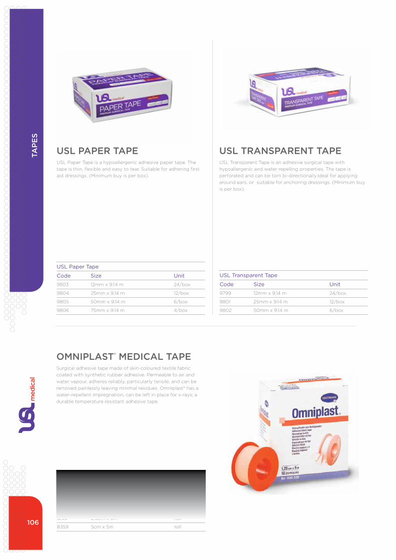
106
USL Transparent Tape
Code Size Unit
9799 12mm x 9.14 m 24/box
9801 25mm x 9.14 m 12/box
9802 50mm x 9.14 m 6/box
USL Paper Tape
Code Size Unit
9803 12mm x 9.14 m 24/box
9804 25mm x 9.14 m 12/box
9805 50mm x 9.14 m 6/box
9806 75mm x 9.14 m 4/box
Omniplast Medical Tape
Code Size Unit
1289 1.25cm x 5m roll
1294 1.25cm x 9m 5/box
1290 2.5cm x 5m roll
1295 2.5cm x 9m roll
8359 5cm x 5m roll
USL TRANSPARENT TAPEuSL transparent tape is an adhesive surgical tape with hypoallergenic and water repelling properties. the tape is perforated and can be torn bi-directionally.ideal for applying around ears, or suitable for anchoring dressings. (Minimum buy is per box).
USL PAPER TAPEuSL Paper tape is a hypoallergenic adhesive paper tape. the tape is thin, flexible and easy to tear. Suitable for adhering first aid dressings. (Minimum buy is per box).
OMNIPLAST® MEDICAL TAPESurgical adhesive tape made of skin-coloured textile fabric coated with synthetic rubber adhesive. Permeable to air and water vapour, adheres reliably, particularly tensile, and can be removed painlessly leaving minimal residues. Omniplast® has a water-repellent impregnation, can be left in place for x-rays; a durable temperature resistant adhesive tape.
TAP
ES

107
SKin integRity/PROteCtiOn/WOund
MaPPing and Patient Hygiene PROduCtS

108
Silesse™
Code Description Unit
11174 50ml aerosol each
11173 Wipes 30/pkt
SILESSE™
all-silicone protection…sometimes, your skin needs a little help
Skin is the largest organ of your body and is usually an excellent barrier. But for many people, keeping skin healthy is critical. We have used the latest in silicone-based technology to protect skin and prevent the damage and irritation caused by adhesives and body fluids.
Product features• protects, moisturises and soothes
• does not sting
• dries quickly – apply the next adhesive in seconds
• will not cause build-up
• Silesse™ is hypoallergenic
Stoma care – pouches: used at every pouch change, the soothing and hypoallergenic Silesse™ formulation creates a comfortable but durable barrier between the skin and the wafer. unlike some skin barriers, Silesse™ contains only silicones, so it will not cause any build-up on the skin surface. Feel the difference after only a few pouch changes. Continence care – sheath catheters and pads: you want the grip, but you need it to be gentle. to improve skin care and create a good seal use Silesse™ quick wipe or soft-pump spray before replacing the sheath catheter or tube holder. For pad users – the Silesse™ soft-pump spray protects quickly and will not clog the pad. Wound care – dressings and tapes: Peri-wound skin can become macerated and delicate. Silesse™ sting free skinbarrier is formulated to protect from exudate and adhesives.
50ml soft-pump spraySilesse™ contains only silicone-based ingredients. this creates the best protection without causing build-up or bonding skin folds. So with Silesse™, you will always have a consistently high level of protection…and the spray will never clog!
• easy-to-use soft-pump spray
• spray will never clog
• does not bond skin folds
• will not cause build-up
NILTAC™
For fast, painless and atraumatic removal of medical adhesives
What can you remove with Niltac™?Woundcare - dressing and tapes: the new, proven, silicone-based technology of niltac releases adhesive dressings and tapes in seconds. now you can easily remove dressings without causing pain or trauma.
Stoma care - pouches: a good seal between the wafer and the skin is important to prevent leakage, so a strong adhesive is necessary. But removing the wafer can cause skin trauma and pain. niltac™ has been carefully designed to gently release even high-tack adhesives in seconds.
Continence care: Sheaths and tube holders are held in place using strong adhesives. even these high-tack adhesives are released quickly and painlessly with a quick spray of niltac.
Product features• does not sting, even on sore skin
• releases adhesives quickly
• dries in seconds – no residue
• does not affect adhesion of next appliance or dressing
• moisturises
• niltac™ is hypoallergenic
To remove sticky adhesive residueMost medical adhesives are designed to adhere to undamaged, clean skin. gently remove sticky residues with niltac™ wipes to prepare the skin for the next adhesive product.
• creates a clean, fresh surface for the next appliance or dressing
• 30 large,soft wipes
Niltac™
Code Description Unit
11149 50ml aerosol each
11172 Wipes 30/pkt
SKIN
PR
OT
EC
TIO
N

109
3M™ CAvILON™ NO STING bARRIER FILMa completely alcohol-free, sting-free liquid barrier film that dries quickly to form a breathable coating to protect damaged or intact skin from bodily fluids, adhesive trauma, friction and incontinence. Cavilon™ no Sting Barrier Film is transparent, allowing continuous visualisation and monitoring of skin at risk for breakdown. the unique formulation provides long-lasting protection. it does not transfer to, or interfere with, diapers or continence briefs possibly minimising the need to change failed briefs, linens and associated products.
• protects intact or damaged skin from bodily fluids, adhesives and friction
• long-lasting, for more comfortable and cost-effective care
• sterile, non-cytotoxic
• alcohol-free, sting-free
• hypoallergenic
• CHg compatible
Strong clinical evidence3M sponsored the largest health economic study for incontinence dermatitis prevention and found that use of Cavilon™ no Sting Barrier Film three times weekly is as effective in preventing incontinence-associated dermatitis as products that require application after each episode of incontinence. the same study found that a skin damage prevention regimen using Cavilon™ no Sting Barrier Film three times per week had a significantly lower total cost than other product regimens.
3M™ CAvILON™ DURAbLE bARRIER CREAMa concentrated cream that provides durable, long-lasting protection from body fluids while moisturising the skin. unique, polymer-based formula provides a breathable barrier to promote skin health in patients at risk for skin damage from incontinence. the cream vanishes into the skin, does not clog briefs or diapers, and resists wash off. using Cavilon™ durable Barrier Cream regularly on intact skin can help prevent breakdown caused by regular exposure to urine and stool.
• unique, polymer-based formulation
• resists wash off—eliminates need for frequent reapplication
• allows tapes and dressings to adhere
• contains dimethicone for skin protection
• hypoallergenic
• CHg compatible
• concentrated - requires less frequent application and less product with each application
• now fragrance-free
• now available in a single-use sachet
3
3M™ Cavilon™ No Sting Barrier FilmA completely alcohol-free, sting-free liquid barrier film that dries quickly to form a breathable coating to protect damaged or intact skin from bodily fluids, adhesive trauma, friction and incontinence. Cavilon
No Sting Barrier Film is transparent, allowing continuous visualisation and monitoring of skin at risk for breakdown. The unique formulation provides long-lasting protection. It does not transfer to, or interfere with, diapers or continence briefs possibly minimising the need to change failed briefs, linens and associated products.
• Protects intact or damaged skin from bodily fluids, adhesives and friction
• Long-lasting, for more comfortable and cost-effective care • Sterile, non-cytotoxic• Alcohol-free, sting-free• Hypoallergenic• CHG compatible
Strong Clinical Evidence3M sponsored the largest health economic study for incontinence dermatitis prevention and found that use of Cavilon No Sting Barrier Film three times weekly is as effective in preventing incontinence-associated dermatitis as products that require application after each episode of incontinence.* The same study found that a skin damage prevention regimen using Cavilon No Sting Barrier Film three times per week had a significantly lower total cost than other product regimens.*
Cavilon No Sting Barrier Filmhas more clinical evidence
than any other moisture barrier or barrier film.
* Bliss D, et al. An economic evaluation of four skin damage prevention regimens in nursing home residents with incontinence. Journal of Wound Ostomy and Continence Nursing. 2007; 34(2): 143-152.
Proven Protection
Take Comfort in
Cavilon™ No Sting Barrier Film
Code Description Unit
4819 1ml Wand 25/box
4822 28ml Spray each
4820 Wipes 25/box
Cavilon™ Durable Barrier Cream
Code Description Unit
8669 28g each
2008 92g each
SKIN
PR
OT
EC
TIO
N
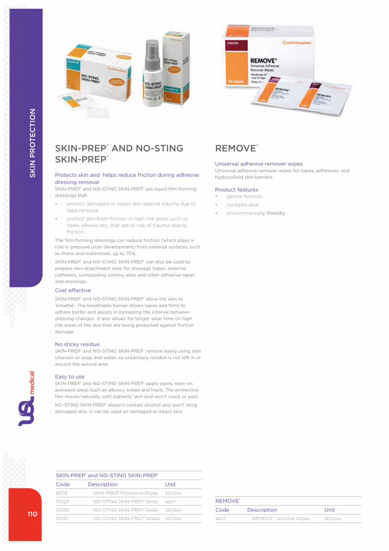
110
SKIN-PREP® and NO-STING SKIN-PREP®
Code Description Unit
6678 SKin-PReP® Protective Wipes 50/box
10529 nO-Sting SKin-PReP® Spray each
10530 nO-Sting SKin-PReP® Swab 50/box
10531 nO-Sting SKin-PReP® Wipes 50/box
REMOVE®
Code Description Unit
4012 ReMOVe® unisolve Wipes 50/box
SKIN-PREP® AND NO-STING SKIN-PREP®
Protects skin and helps reduce friction during adhesive dressing removalSKIN-PREP® and NO-STING SKIN-PREP® are liquid film-forming dressings that:
• protect damaged or intact skin against trauma due to tape removal
• protect skin from friction in high risk areas such as heels, elbows etc, that are at risk of trauma due to friction
The film-forming dressings can reduce friction (which plays a role in pressure ulcer development) from external surfaces, such as linens and mattresses, up to 75%.
SKIN-PREP® and NO-STING SKIN-PREP® can also be used to prepare skin attachment sites for drainage tubes, external catheters, surrounding ostomy sites and other adhesive tapes and dressings.
Cost effective
SKIN-PREP® and NO-STING SKIN-PREP® allow the skin to ‘breathe’. The breathable barrier allows tapes and films to adhere better and assists in increasing the interval between dressing changes. It also allows for longer wear time on high risk areas of the skin that are being protected against friction damage.
No sticky residueSKIN-PREP® and NO-STING SKIN-PREP® remove easily using skin cleanser or soap and water, so unsanitary residue is not left in or around the wound area.
Easy to useSKIN-PREP® and NO-STING SKIN-PREP® apply easily, even on awkward areas such as elbows, knees and heels. The protective film moves naturally with patients’ skin and won’t crack or peel.
NO-STING SKIN-PREP® doesn’t contain alcohol and won’t sting damaged skin. It can be used on damaged or intact skin.
REMOvE®
Universal adhesive remover wipesUniversal adhesive remover wipes for tapes, adhesives, and hydrocolloid skin barriers.
Product features• gentle formula
• contains aloe
• environmentally friendly
SKIN
PR
OT
EC
TIO
N

111
3M™ Wound Mapping Grid
Code Size Unit
11346 10x10cm 50/box
11347 15x20cm 50/box
3M WOUND MAPPING GRID™
Measure and record wound size. Hygienic and simple to use.
Application InstructionsApplication Instructions
1. Place the 3M™ Wound Mapping Grid over the wound
2. Trace the wound areas on to the grid (head to toe)
3. Peel off the top layer 4. Place the top layer in the patient’s notes and discard the contaminated backing sheet in the appropriate disposal unit
Product Code Grid Size Grids/Roll Rolls/Case GRID1 10cm x 10cm 50 10
GRID2 15cm x 20cm 50 10
Ordering Information
For more information, contact your 3M Health Care Representative or call 3M Health Care Customer Helpline on 0800 80 81 82
Please recycle. Printed in New Zealand.© 3M 2011. All rights reserved.
Please note: the 3M Wound Mapping Grid is not sterile
3M Australia Pty LimitedABN 90 000 100 096Building A1 Rivett RoadNorth Ryde NSW 21131300 363 878www.3M.com.au/healthcare
3M Health Care
3M New Zealand Limited94 Apollo Drive Rosedale, Auckland 0632 Customer Service: 0800 80 81 82 www.3M.com/SkinWoundCare
WO
UN
D M
AP
PIN
G

112
• as “poor oral health can profoundly decrease appetite and
the ability to eat, which in turn may lead to poor nutrition...
compromised nutritional status may result in an impaired
immune response and resistance to infection, retarded
wound healing, poor oral health, and, ultimately, general
ill health”. (Papas aS, et al. Longitudinal Relationships
between nutrition and Oral Health. annals of the new york
academy of Sciences, 561:124-142)
• “the role of the WOC nurse focuses on persons with
wound, ostomy needs, many who are undergoing cancer
therapy, have multisystem organ failure, are in the terminal
stages … or are malnourished for other reasons and if
they are elderly or disabled are unable to perform routine
oral care; as the WOC nurse often initiates a nutritional
consult they should also initiate or enquire about oral
hygiene program to maximise the impact of nutritional
interventions”. (aronovitch S. Oral care and its role in WOC
nursing. Volume 24, issue 2, March 1997, Pages 79–85)
Poor oral health and periodontal disease are known to increase the risk for respiratory infections, diabetes, stroke and cardiovascular disease; this knowledge is driving changes from ‘tradition based care” to evidenced based oral hygiene in acute, long-term and home health sectors.
Colonisation of dental plaque (biofilm) in compromised patients, including those with high dependency needs and those at risk for aspiration contribute to development of pneumonia (VaP / HaP) in both intubated and non-intubated persons; studies show that poor oral care and proliferation of oral biofilm also plays a significant role in pneumonia rates among the elderly and dependent residents in long term care.
For WOC nurses’ oral hygiene’s relevance to wound healing and its impact on a patient’s nutritional status and ability to fight infection adds to these concerns. WOCn organisations have long recognised that “oral care, in addition to promoting wound healing, reduces the risk for respiratory infections, relieves discomfort produced by inflammation of the oral mucosa; improves nutritional status, speech and outcomes for persons with wounds, ostomy or incontinence”.
as the relationship between oral health, wound healing and nutritional intake is dynamic, clinical citations of note include:
“nutrition has been recognised as a very important factor that affects wound healing. Most obvious is that malnutrition or specific nutrient deficiencies can have a profound impact on wound healing”. (guo S, diPietro L. Factors affecting wound healing. J dent Res. 2010 March; 89(3): 219–229.)
• the “involvement of the WOC nurse in preventative and
therapeutic oral care can ensure a healthier gastrointestinal
tract, improved nutritional status, and improved outcomes
in caring for the person with a wound, ostomy, or
incontinence. an assessment of the patient’s oral cavity
is necessary when managing the patient with wound,
ostomy or incontinence and a suboptimal nutritional
status. in addition to promoting wound healing, the
provision of oral care benefits the patient by reducing risk
for lower respiratory tract infection and by relieving the
discomfort produced by inflammation of the oral mucosa….”
(aronovitch S. Oral Care and its Role in WOC nursing J
WOCn, 1997; 24:79-85)
“THE MOUTH IS THE MIRROR OF THE bODY, bUT OFTEN OvERLOOKED IN HEALTHCARE”
(anonymous)
ORAL HYGIENEPA
TIE
NT
HY
GIE
NE

113
Oral Hygiene and Pneumonia: the critical linkaspiration pneumonia, which is typically caused by anaerobic organisms (commonly S. aureus, P. aeruginosa or one of the enteric species) arises from the gingival crevice and seen in both the community and institutionalised settings. Pneumonia develops when pathogens are aspirated from the oropharyngeal cavity and other sites (gi tract, sinuses) into the lower airway.
increased risks for aspiration pneumonia occur when a sequence of periodontal disease, dental decay and poor oral hygiene is compounded by the presence of dysphagia, feeding problems and poor functional status all of which are found in vulnerable dependent persons. But modifiable risks of aspiration pneumonia do exist; these include implementation of standardised evidenced based practices (eBP) in oral hygiene, routine oral assessment, mechanical cleansing to reduce biofilm and strict adherence to aspiration precaution protocols. Consequently, an urgent need exists to improve the state of oral care for vulnerable and dependent persons, particularly those who are dentate.
Current state of oral health and carethe level of oral care provided to patients / residents is a key indicator on the quality of care that is provided in general; the appearance of dry cracked lips, coated tongue, vegetation on teeth and halitosis indicate a lack of oral care and flagrant disregard for the well-being and comfort of the dependent person. a study of 71 edentulous elderly persons in Japan assessed ‘tongue coating’ as a predictor or risk indicator in the development of aspiration pneumonia; the number of elderly patients developing aspiration pneumonia was larger in patients with a poor tPi score (tongue plaque index) than those with better or good tPi scores. as a result “tongue coating was associated with a number of viable salivary bacterial cells and development of aspiration pneumonia in dentate persons”*.
With aspiration pneumonia accounting for 13% to 48% of all infections in nursing homes, growing evidence supports the causal links between oral hygiene, mechanical cleansing, aspiration risks and development of pneumonia in ‘high risk’ patient populations. However this awareness is not new with literature going back to the 1980’s documenting links between poor oral hygiene and illness.the importance of oral care as a preventative measure in maintaining systemic health has been so compelling that in 2003 the CdC and HiCPaC released “ guidelines for Preventing Health Care associated Pneumonia”. this stated that healthcare facilities must “...develop and implement a comprehensive oral-hygiene program (that might include use of an antiseptic agent) for patients in acute-care settings or residents in long-term care facilities who are at risk for health-care associated pneumonia (ii)*.”
Unsubstantiated “Tradition-based”
Practices in Oral Care
nursing literature abounds with support for mouth moisture, adequate salivary flow and control of plaque formation to preserve oral health but is not always adhered to; the goal of oral care is to maintain mouth cleanliness and prevent infection, moisturise the oral cavity, preserve mucosal integrity and promote healing. Mechanical and chemical debridement of the oral mucosa and teeth reduces dental plaque and mouth bacteria whilst stimulating production of saliva. Because of changes in salivary properties and production, alteration in oral flora from antibiotics, difficulties or obstacles in providing oral care, use of instruments not designed for mouth care and lack of standardised practice in oral care, critically ill or debilitated patients are at increased risk for colonisation of dental plaque aka biofilms.
unfortunately, traditional oral care, at best, has been fragmented and creative. For patients or residents unable to perform self-care with a toothbrush, nurses and other professional or lay caregivers (such as family members) have used a myriad of creative ‘implements’ to provide oral care; these include Lemon-glycerin swabs (dries out the mouth and can pit dentition), cotton tip swabs dipped in sodium bicarbonate or dentifrice, gauze sponges wrapped on forceps, tongue depressors or even wrapped around fingers, or hard edged “implements” to pry open or prop open the mouth all in the attempt to perform oral
hygiene. these practices offer no true ‘hygiene care” and are potentially dangerous as cotton tipped applicators leave behind small fibres that patients swallow, inhale or get trapped in the oral cavity causing bacterial seeding; and wood or hard edged implements can cause wounds to the mucosa, gums, teeth, lips and alarmingly, fingers can be bitten.
Solutions to optimising careif the patient is dentate (has teeth), diligent oral hygiene should provide cleaning with a toothbrush in the aM and PM. those at higher risk for pneumonia (i.e. intubated patients) or with moderate to severe ‘oral dysfunction’ require additional cleaning every 2-4 hours using foam oral swabs with at minimum, sodium bicarbonate to stimulate and moisturise the oral mucosa, lift debris and neutralise plaque acids.toothbrushes are the most effective means for removing and reducing dental plaque from teeth but biofilm also clings to the oral mucosa, particularly in the ‘cheeks’ (buccal cavity), gums and tongue. to clean those areas use a foam oral swab (commonly referred to as a toothette®) that contain deep “grooves or ridges” to mimic the actions of a toothbrush to clean the surface and in between the teeth and to stimulate - cleanse the oral mucosa, buccal areas, gum line, temporal-mandibular ridges, hard pallet and lips.
ORAL HYGIENEPA
TIE
NT
HY
GIE
NE
114
There are four components to providing evidenced based oral care:
1. Routine oral health assessment using a standardised tool, minimum of once per day and on admission; an oral dysfunction ‘scoring’ system is preferred over those that use only ‘tick marks’
2. Oral cleansing using appropriate cleansing agents to maintain mucosal integrity and pH
3. debridement of oral structures using appropriate implements to reduce presence of dental plaque and debris
4. Moisturising of the oral cavity and its’ structures particularly in those with significant xerostomia
Mechanical debridement / brushing:
Toothbrushes:• use an ultra-soft, small headed or paediatric sized
toothbrush or, flexible three-headed toothbrush (aka Surround® toothbrush) for those with short tolerance due to behavioural issues as all surfaces are brushed simultaneously in a shorter period of time
• For those with high risk for aspiration during oral care, a suction toothbrush and suction oral swab is preferred
Toothbrush ‘alternatives’ / adjuncts to oral care:• Foam oral swabs (aka toothette Swabs®) to stimulate and
cleanse the oral mucosa, buccal areas, gum line, temporal-mandibular ridges, tongue, hard pallet and lips
• Oral swabs don’t replace toothbrushes in those who are dentate or can tolerate brushing and have no contraindications such as low platelets, oral trauma, etc.
• Oral swabs are recommended for in-between aM and PM tooth brushing and used every 2-4 hours for those with moderate to severe oral dysfunction.
• Frequent swabbing stimulates and moisturises the mucosal tissue to minimise oral dysfunction.
• Foam swabs impregnated with sodium bicarbonate avoids under or overdosing with incorrectly prepared sodium bicarbonate solutions mixed / stored at the bedside; aids in cleansing, lubricating, preventing crusting and gently removing debris between brushing.
• Foam swabs used with 1.5% hydrogen peroxide and sodium bicarb increases effectiveness of the debriding action and helps thin ropey, thickened secretions found in those with severe oral dysfunction and aids in removing oral secretions.
• Plain / non-impregnated foam oral swabs are preferable over “jumbo swabs” or gloved fingers to apply mouth gels or medications.
Lip and Mouth Moisturisers • Only ‘muco-adhesive’ water-soluble mouth moisturiser for
lips and oral cavity should be used.
• Petroleum based moisturisers inflame open wounds and should not be used inside the mouth, particularly for those
ORAL HYGIENERECOMMENDED APPROACHES AND TOOLS
at high risk for aspiration
• Petroleum based lip moisturisers are contradicted during oxygen therapy
Other adjuncts to oral care• Protective mouth props (i.e. Open-Wide® Mouth Prop) to
facilitate care in those who are unable to fully cooperate during oral hygiene
• Made from dense foam, provides measure of safety for both patient and caregiver reducing injuries to the teeth, mouth or gums.
• Protective mouth prop essential for safe oral care in problematic patients such as those with alzheimer’s, dementia, strokes or spasticity issues who cannot keep their mouths fully open during oral care or might unexpectedly bite down during care.
the use of routine antibacterial mouthwashes has not been addressed in this article as differing opinions exist regarding routine use of Chlorhexidine versus other agents such as Cetylpyridium Chloride, 1.5% Hydrogen Peroxide, thymol or gLLL. that being said, physician or dentist directed use of daily antibacterial agents should be included in patients’ plan of care. Whilst the use of antibacterial’s (i.e. low strength CHg gel) is advocated by many, staff must be aware that the use of any antiseptic or antibacterial agent does not preclude proper mechanical debridement of tooth surfaces.
Recommendations for Best Practice Oral Hygiene Interventions the need for diligent and more frequent oral – dental care is greater in those with dysphagia and must play a part in any aspiration pneumonia prevention programs.
• Change from tradition based practices to evidence based practices
• demand 100% compliance with daily oral care practices
• everyone is to be trained in evidence based practice to include oral assessment, oral hygiene care and caring for dentures (dirty dentures recolonise the mouth with pathogens)
• involve dentist, dental hygienist, oral health nurse as part of the healthcare team.
Summaryas compliance plays a large part in oral care and patient outcomes, WOC nurses should recommend products that facilitate oral cleansing and make it easier and more comfortable for patient and staff. to encourage the delivery of good oral hygiene it is essential to have all necessary tools and equipment readily at hand to foster frequent and standardised care. using informed, reasoned choices does drive healthcare objectives and shift providers away from “tradition based” practices. unfortunately talking about ‘best practice’ does not always guarantee the application and performance of best practice models. Without the development of on-going educational programs for nursing personnel and regular involvement by dental professionals, oral hygiene will continue to take a “back seat” in many acute and long-term care facilities.
*footnotes available on request
PAT
IEN
T H
YG
IEN
E

115
SHORT TERM SWAb SYSTEM WITH PEROX-A-MINT ® SOLUTION• 20 swabs with sodium bicarbonate
• 1 ultra-Soft toothbrush
• 1 .33 oz./9g tube of sodium bicarbonate mouthpaste
• 1 1.5 fl. oz./44ml Bottle of Perox-a-Mint solution
• 1 .5 oz./14g tube of mouth moisturizer
TOOTHETTE ® AND TOOTHETTE ® PLUSORaL SWaBS are alternatives in oral care when toothbrushes cannot be used. Features soft foam heads with distinct ridges that lift and remove debris and mucus, clean between teeth and safely apply debriding agents or topical oral solutions. Mimicking the mechanical actions of a toothbrush, toothettes soft and gentle foam swabs stimulate oral tissue without injuring fragile tissue and sensitive mucosa. toothettes are avalible plain or impregnated with SOdiuM BiCaRBOnate to help dissolve thickened ropey saliva and deodorise the mouth.
“Effective Cleansing and Moisturizing”
Short term swab system with Perox-A-Mint®Solution
Code Description
90003 Short term Swab System
Short term swab system with Perox-A-Mint®Solution
Code Description
10124 untreated toothette x 200
10125 treated Soda-Bic toothette x 200
90008 Open wide mouth prop eachPA
TIE
NT
HY
GIE
NE

116
MOUTH MOISTURISER PEROX-A-MINT ® SOLUTION
Features
• Compact head with soft, end-rounded bristles for comfortable and safer brushing
• Side bristles positioned at a 45 degree angle recommended by the ada to clean both teeth and gums
• Flexible structure and soft tactile surface makes gripping easier and helps protect oralenviroment from trauma in sudden unexpected movement during brushing.
• Covered in a non latex rubbery material with padded head and neck.
• Head and neck of brush designed to make brushing safer even with those who tend to bite down on the toothbrush
Designed for “Special Needs” elderly and orthodontic patients
SURROUND ® TOOTHbRUSH
Features:
• Moisturises lips and oral tissue
• Minty flavoured
• Water based formula
• Vitamin e
• Coconut oil
• 0.5 oz tube
Features:
• Oral debriding agent
• aids in the removal of phlegm, mucus, or other secretions associated with occasional sore mouth
• Releases oxygen bubbles by the enzymatic action
when peroxide comes into contact with tissues
Surround ® Toothbrush
Code Description
90014 Surround toothbrush
Surround ® Toothbrush
Code Description
90011 Perox-a-Mint
Surround ® Toothbrush
Code Description
90007 Mouth Moisturiser
PAT
IEN
T H
YG
IEN
E

117
COMPReSSiOnBandaging

118
vENO4®
The latex-free four-layer compression bandaging kit for the management of chronic venous insufficiency and related conditionsVeno4® is a latex-free four-layer compression bandage system, suitable for most patients with venous leg ulcers. When applied correctly, Veno 4 can provide compression for up to seven days.
Veno4® consists of four latex-free bandages:
• Layer 1, viscose padding bandage – absorbs exudate and redistributes pressure around the bony prominence of the ankle
• Layer 2, cotton crepe bandage – smoothes padding bandage
• Layer 3, light elastic compression bandage – conforms to leg contours. Classified as a type 3a light compression bandage by BS 7505:7995 this will deliver 17mmHg
• Layer 4, latex free cohesive bandage – adds to the compression effect and helps to keep the bandages in place for up to seven days. a type 3a light compression bandage. When applied at mid stretch with a 50% overlap, this bandage gives an ankle pressure of 23mmHg
all bandages are non sterile, if necessary layer 1, layer 2 and layer 3 can be sterilised in the autoclave at 121˚C for 20 minutes.
Indications:• venous leg ulcers and related conditions
Contraindications:• should not be used on diabetic patients
• patients with known arterial disease
Four-layer compression
Veno4® Four layer, 18 - 25cm compression bandaging kit in polythene bag
Code Unit
9644 Veno 4 (18 - 25cm ankles)
Layer 1: Viscous Padding Bandage
application - Spiral technique
Overlap – 50%
Stretch – n/a
apply the bandage without tension from the base of the toes up to the knee. if the ankle circumference is less than 18cm, apply an extra layer of padding to increase ankle circumference to 18cm.
Layer 3: Light elastic Compression Bandage
application – Foot lock then figure of eight technique
Overlap – 50%
Stretch – 50%
Bandage from base of the toes with enough tension to secure the bandage around the foot and ankle. Begin compression at the ankle using a figure of eight technique finishing just below the knee.
Layer 2: Cotton Crepe Bandage
application – Foot lock then spiral
Overlap – 50%
Stretch – 50%
Bandage from the base of the toes to create a smooth surface.
Layer 4: Cohesive Bandage
application – Spiral technique
Overlap – 50%
Stretch – 50%
apply from the base of the toes to the knee, only commencing compression once at the ankle. apply gentle pressure to the bandage to ensure the layers bind together.
Four-layer Compression Bandaging System:
CO
MP
RE
SSION
bA
ND
AG
ING

119
Medical and social historyirene has never smoked, and worked as a shopkeeper and factory floor worker until retirement. She had bilateral total knee reconstructions 10 years ago.
Over the past seven years irene has had several venous leg ulcers, most recently one year ago. the ulcers have been treated at various times with anti-inflammatories, antibiotics and painkillers. Previous dressings included Cutifilm, Coban, iodosorb, Melolin, Replicare, Steripaste and Bactigras.
Wound and leg profilean area of extremely superficial skin ulceration was visible on the gaiter area of both lower legs. there was evidence of atrophy blanche and scarring from previous ulcerations. the legs had the classic inverted champagne bottle appearance.
Capillary return was normal, foot pulses good, legs warm.
Pre treatment ankle circumference: 28cm (both legs)
Managementatrauman® ag was used as a protective wound contact layer and Veno4® compression bandaging was applied in order to improve venous return. the dressing of the superficial ulcer is only part of treatment: in the case of venous disease, compression bandaging is the key element in wound healing. the Veno4® compression bandage system was left in place for seven days.
One week laterBandages remained intact; had slipped approximately 2cm from their original position.
ankle circumference after one week: 24cm (both legs)
Superficial skin ulceration: 90% resolved
Why Veno4®?Most clinicians in wound care have used a four-layer bandage system with good results. the Veno4® system performed very well and the patient remained relatively comfortable during the week of sustained compression. irene’s ankle circumference was reduced after seven days, indicating efficacy in oedema management.
CASE STUDY
Irene, a 77 year old woman, presented with areas of superficial skin ulceration and oedema in both lower legs. She also has intermittent discomfort in both legs, which is generally relieved with elevation.
this case study is courtesy of Jan Rice, Manager education & Clinical Service, Wound Foundation of australia, Monash university. ViC.
Prior to treatment ankle circumference 28cm.
Following sustained compression with Veno4® for seven days ankle circumference reduced to 24cm.
CO
MP
RE
SSION
bA
ND
AG
ING

120
Veno4® Crepe - Layer Two
Code Size Unit
9653 10cm x 4.5 m each
vENO 4® CREPE - LAYER TWO(KOB Crepe)Cotton crepe bandage – smoothes padding bandage - very soft crepe. available separately. Part of Veno 4®, 18-25cm Compression Bandaging Kit (Layer 2).
Veno4®Padding - Layer One
Code Size Unit
9652 10cm x 3.5 m each
vAROLAST®
The two-way elastic zinc bandageVarolast® is a ready to use, two-way stretch zinc elastic bandage. the two-dimensional stretch of the fabric facilitates accurate bandaging without any slitting or cutting of the bandage, even around difficult contours of the leg.
Varolast® is kind to the skin and dries quickly. the zinc paste in Varolast® contains 15% zinc oxide.
Indications:• decongestion and reduction of persistent oedema
• follow up treatment after thrombosis
• dry leg ulcers
• support bandage in general orthopaedics and after orthopaedic surgery
• phlebitis of deep and superficial veins
Varolast® 10cm wide x 7m
Code Size Unit
8374 10 m 1 bandage
vENO 4® PADDING - LAYER ONE(KOB Natural Rolta Soft)Viscose padding bandage – absorbs exudate and redistributes pressure around the bony prominence of the ankle. Made out of natural cotton so reduces sensitivity reactions. available separately. Part of Veno 4®, 18-25cm Compression Bandaging Kit (Layer 2).
CO
MP
RE
SSIO
N b
AN
DA
GIN
G
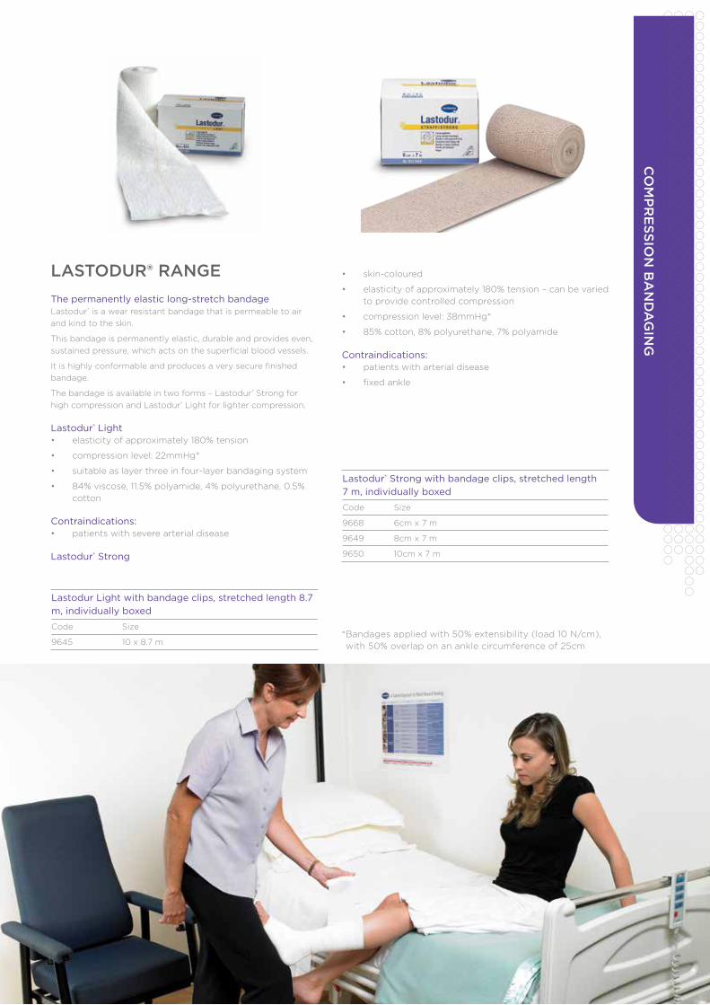
121
* Bandages applied with 50% extensibility (load 10 n/cm), with 50% overlap on an ankle circumference of 25cm
Lastodur® Strong with bandage clips, stretched length 7 m, individually boxed
Code Size
9668 6cm x 7 m
9649 8cm x 7 m
9650 10cm x 7 m
Lastodur Light with bandage clips, stretched length 8.7 m, individually boxed
Code Size
9645 10 x 8.7 m
LASTODUR® RANGEThe permanently elastic long-stretch bandageLastodur® is a wear resistant bandage that is permeable to air and kind to the skin.
this bandage is permanently elastic, durable and provides even, sustained pressure, which acts on the superficial blood vessels.
it is highly conformable and produces a very secure finished bandage.
the bandage is available in two forms – Lastodur® Strong for high compression and Lastodur® Light for lighter compression.
Lastodur® Light • elasticity of approximately 180% tension
• compression level: 22mmHg*
• suitable as layer three in four-layer bandaging system
• 84% viscose, 11.5% polyamide, 4% polyurethane, 0.5% cotton
Contraindications:• patients with severe arterial disease
Lastodur® Strong
• skin-coloured
• elasticity of approximately 180% tension – can be varied to provide controlled compression
• compression level: 38mmHg*
• 85% cotton, 8% polyurethane, 7% polyamide
Contraindications:• patients with arterial disease
• fixed ankle
CO
MP
RE
SSION
bA
ND
AG
ING

122
LASTOLAN®
A strong, short-stretch bandage for oedema reduction and supportLastolan® is a short-stretch cotton crepe bandage with approximately 90% extensibility high compression. it generates a high working pressure and a low resting pressure. this allows decongestion of deep sub-fascial venous areas.
Lastolan® adapts to changes in circumference caused by oedema subsidence, so can remain in place for 1-2 days, except in the acute phase of severe oedema.
Lastolan® is made of 100% cotton and is air-permeable. it can be washed up to 25 times at temperatures up to 95°C and steam sterilised.
Lastolan® is ideal for treating conditions that require a high working pressure, including:
lymphoedema
acute and chronic venous stasis
venous leg ulcers
thrombophlebitis
post-sclerotherapy
musculoskeletal injuries
Contraindications:• diabetic patients
• patients with known arterial disease
Lastolan® Stretched length 5 m, individually boxed
Code Size Unit
9671 6cm 1 bandage
9672 8cm 1 bandage
9651 10cm 1 bandage
9673 12cm 1 bandage
CO
MP
RE
SSIO
N b
AN
DA
GIN
G

123
CaSting and Bandaging
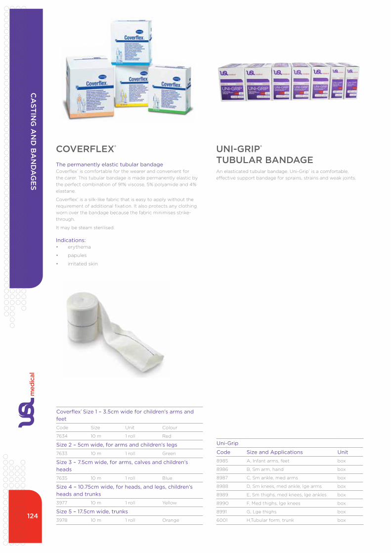
124
COvERFLEX®
The permanently elastic tubular bandageCoverflex® is comfortable for the wearer and convenient for the carer. this tubular bandage is made permanently elastic by the perfect combination of 91% viscose, 5% polyamide and 4% elastane.
Coverflex® is a silk-like fabric that is easy to apply without the requirement of additional fixation. it also protects any clothing worn over the bandage because the fabric minimises strike-through.
it may be steam sterilised.
Indications:• erythema
• papules
• irritated skin
Coverflex® Size 1 – 3.5cm wide for children’s arms and feet
Code Size unit Colour
7634 10 m 1 roll Red
Size 2 – 5cm wide, for arms and children’s legs
7633 10 m 1 roll green
Size 3 – 7.5cm wide, for arms, calves and children’s heads
7635 10 m 1 roll Blue
Size 4 – 10.75cm wide, for heads, and legs, children’s heads and trunks
3977 10 m 1 roll yellow
Size 5 – 17.5cm wide, trunks
3978 10 m 1 roll Orange
UNI-GRIP® TUbULAR bANDAGEan elasticated tubular bandage. uni-grip® is a comfortable, effective support bandage for sprains, strains and weak joints.
Uni-Grip
Code Size and Applications Unit
8985 a, infant arms, feet box
8986 B, Sm arm, hand box
8987 C, Sm ankle, med arms box
8988 d, Sm knees, med ankle, lge arms box
8989 e, Sm thighs, med knees, lge ankles box
8990 F, Med thighs, lge knees box
8991 g, Lge thighs box
6001 H,tubular form, trunk box
CA
STIN
G A
ND
bA
ND
AG
ES

125
STüLPA®
Seamless knitted tubular bandageStülpa® is a seamless knitted tubular bandage with high two-way stretch. not only does it provide very secure dressing retention, it is particularly useful for lining and covering zinc paste and plaster of Paris bandages. the high elasticity makes Stülpa® bandages quick and easy to apply without additional fixation.
Stülpa® bandages fit all body parts without creasing, slipping or causing a tourniquet effect. Joint movements are not impaired.
Stülpa® bandages provide a smooth surface. this means that they do not loosen even under mechanical stress. Wounds or sensitive parts of the skin are reliably protected. Stülpa® is made of 70% viscose and 30% cotton, making it pleasant to wear, absorbent and air-permeable. it may also be sterilised. Stülpa® can be cut without fraying.
Stülpa®
Code Size Unit
15m rolls
8378 Children’s finger and toe bandages Size 0 R roll
8377 Finger bandages Size 1 R roll
8376 arm and children’s leg bandages Size 2 R roll
8379Foot, leg, children’s head and axilla bandages
Size 3 R roll
8380 Head, leg and axilla bandages, face masks Size 4 R roll
STüLPA®-FIXElastic tubular net bandageStülpa®-fix is a soft, white, latex-free elastic net bandage knitted from cotton and polyamide yarns. this construction and the size range allows it to stretch and fit on all parts of the body.
the range of six widths makes Stülpa®-fix easy to apply and highly versatile to fit a broad range of indications.
Stülpa®-fix is available in a roll format that can be cut or slit to size without further tearing or fraying. the open net design holds dressings in position, without restricting movement. easier to wash and change than retention bandages, it provides cool, conformable low-bulk retention.
Indications:• light fixation of bandages and dressings
• dressing retention
Stülpa®-fix
Code Size Unit
Stretched length 25 m
2741 For fingers Size 1 roll
2742 For hands, arms and feet Size 2 roll
2743 For legs and children’s head Size 3 roll
2744 For head and children’s trunk Size 4 roll
2745 For trunk Size 5 roll
CA
STIN
G A
ND
bA
ND
AG
ES

126
PLASTRONA®
Fast-setting plaster of Paris bandagePlastrona® is a plaster of Paris bandage. the unique wave-like cut prevents fraying as well as cracking around the edges and improves the unrolling properties.
Plastrona® performs because it is made from a blend of quality types of plaster. it is available as a bandage, as a four-layer slab and as slab rolls for a variety of casting functions, including splints and jackets.
With an immersion time of 2-3 seconds and a setting time of approximately 5 minutes.
Plastrona®
Bandage
Code Size Unit
1235 6cm x 2m 5 x 2 bandages
1236 8cm x 4m 5 x 2 bandages
1237 10cm x 4m 5 x 2 bandages
1238 12cm x 4m 5 x 2 bandages
1239 15cm x 4m 5 x 2 bandages
1240 20cm x 4m 5 x 2 bandages
Plastrona®
Slab four-layer
1241 10cm x 20m slab
1242 15cm x 20m slab
1243 20cm x 20m slab
USL CREPE bANDAGEThe cotton crepe bandage for support and retentionuSL crepe bandage can be used as universal dressing retention bandages.
all three bandaging techniques offer the benefits of support and relief of injuries while not inhibiting mobility.
alternate uses include:
• light support
• sports bandaging
• securing splints
• when muscle activity is absent
• permeable to air and resistant to grease, oils, ointments and perspiration
• an economical latex free cotton crepe retention bandage made from unbleached cotton. 98% cotton and 2% elastane.
The cotton crepe bandage for support and retention
USL crepe bandage
Unstretched length 1.6 m, stretched length 4 m, individually wrapped
Code Size Unit
7356 5cm roll
7355 7.5cm roll
7357 10cm roll
7358 15cm roll
bANDAGING TECHNIqUESthe three most commonly used techniques of bandaging are spiral technique, ascending spica technique and diverging spica technique.
Spiral bandaging is the simplest of the roller bandaging techniques. While rolling the bandage, in this method, the turns are done in spiral method, wherein each turn covers the two-third part of the preceding turn. Spiral technique of bandaging is most often used on body parts with uniform circumference, such as leg or forearm.
diverging spica or reverse spiral bandaging technique is most often used on body parts with varying circumference. although the turns are made in spiral direction in this technique, the bandage is reversed on itself so that it stays firm on body parts with varying perimeters. Once the bandage is secured, after a few spiral binds, the bandage is rolled with the thumb being placed over the lower border of the bandage on the outer side of the limb. eventually the bandage is reversed downwards, and after passing it over the fixed thumb it is carried to the opposite side from under the limb, and rolled in reverse spiral technique above the preceding bandage wrap.
ascending spica or figure eight bandaging is considered to be the most useful roller bandaging technique. in this method, the bandage is alternately passed upwards and downwards over and under the C
AST
ING
AN
D b
AN
DA
GE
S

127
RHENA®THERMThe thermoplastic synthetic casting tape that can be applied without glovesRhena®therm is a thermoplastic casting tape made of a flexible textile fabric, in combination with a thermoplastic polyester polymer (free of isocyanides/polyurethane resins).
Rhena®therm casting tape is activated and transformed into a mouldable state by heat (eg in a water bath) at temperatures of 65°C plus.
the bandage is not sticky at room temperature and requires no special storage.
Indications:• external immobilisation
• functional stabilisation of limbs
• moulding orthopaedic splints and prostheses
Rhena®therm Length 1.8 m
Code Size Unit
9394 2.5cm 12 bandages
Rhena®therm Length 3.6 m
9396 7.5cm roll
9397 10cm roll
9398 12.5cm roll
HYGIA CASTPolyester Orthopaedic Casting TapeHygia Cast offers all the strength, rigidity and durability that you expect from a fibreglass tape, with the added advantages of being fibreglass-free.
Hygia Cast is comprised of a knitted polyester fabric which features extensible yarns which shape easily to body contours, providing superior moulding capabilities for both primary and secondary casting applications.
• strong and durable
• superior lamination
• excellent conformability
• excellent finish for smooth soft edges
• less dust than fibreglass products
• x-ray radiolucency
• excellent colour range
Hygia Cast
Code Size Colour Unit90165 Cast Hygia Plus 2' White Polyester roll90166 Cast Hygia Plus 2' green Polyester roll90167 Cast Hygia Plus 2' Pink Polyester roll90168 Cast Hygia Plus 2' Blue Polyester roll90169 Cast Hygia Plus 2' Red Polyester roll90170 Cast Hygia Plus 2' yellow Polyester roll90171 Cast Hygia Plus 2' Black Polyester roll90172 Cast Hygia Plus 2' Purple Polyester roll 90193 Cast Hygia Plus 2' Rainbow Polyester 10/box90174 Cast Hygia Plus 3' White Polyester roll90175 Cast Hygia Plus 3' green Polyester roll90177 Cast Hygia Plus 3' Blue Polyester roll90176 Cast Hygia Plus 3' Pink Polyester roll90178 Cast Hygia Plus 3' Red Polyester roll90179 Cast Hygia Plus 3' yellow Polyester roll90180 Cast Hygia Plus 3' Black Polyester roll90181 Cast Hygia Plus 3' Purple Polyester roll90182 Cast Hygia Plus 3' Orange Polyester roll90194 Cast Hygia Plus 3' Rainbow Polyester 10/box90183 Cast Hygia Plus 4' White Polyester roll90184 Cast Hygia Plus 4' green Polyester roll90185 Cast Hygia Plus 4' Pink Polyester roll90186 Cast Hygia Plus 4' Blue Polyester roll90187 Cast Hygia Plus 4' Red Polyester roll90188 Cast Hygia Plus 4' yellow Polyester roll90189 Cast Hygia Plus 4' Black Polyester roll90190 Cast Hygia Plus 4' Purple Polyester roll90191 Cast Hygia Plus 4' Orange Polyester roll90195 Cast Hygia Plus 4' Rainbow Polyester 10/box90192 Cast Hygia Plus 5' White Polyester roll
SONICLEAN HEAT PANthe Soniclean Heat Pan is specifically designed as a water heater for the activation of thermoplastic materials; it comes complete with a temperature control and digital display.
Soniclean Heat Pan
Code description
9584 Soniclean Heat Pan
CA
STIN
G A
ND
bA
ND
AG
ES

128
OSCIMED CAST SAWS AND EXTRACTION UNITSthe Oscimed 2000 is a very low noise level cast saw with patented anti-noise technology. the Oscimed 2000 consists of a brushless, fanless 100W-24V motor.
it has been ergonomically designed with a modern shape for single handed use. the saw has a instant blade change ability with a patented non-slip lock nut.
Oscimed technology includes saw blades with a noise-suppressing central core.
this vacuum cleaner possesses four levels of filtration (including two filters to HePa standards) and filters dust down to 3 microns. it has a tough, powerful construction with a support for the saw and vacuum hose. the unit comes complete with an electricity supply socket for the saw. available in 1-speed version (1000W).
• Sound level: 62dB in speed 1
• Suction: 2200mm water gauge, 50 litres/sec
• Length of vacuum hose: 3 metres
• Capacity of stainless steel tank: 20 litres
• Weight: 12 kg
PLASTER INSTRUMENTS
Oscimed Cast Saw and Extraction Vacuum Unit
Code Description
20101 Oscimed Cast saw
20102 Oscimed extraction Vacuum unit
20102B Oscimed Blade 65mm Synthetic Blade
20102C Oscimed Blade for POP
20102d Oscimed Blade - titanium
20102a Oscimed dust Bags 10/box
13413 Oscimed Cast Spreader
Plaster Intruments
Code Description Size Unit
id00573 Rocket Cast Shear Stille 23cm each
825201 Rocket Cast Shear Stille 38cm each
id00481 Rocket Cast Spreader daws 46cm each
id00480Rocket Cast Spreader Henning
28cm each
id00753Rocket Cast Spreader uS Model
23cm each
820421 Rocket utility Scissor Pink 14cm each
82042 Rocket utility Scissor Black 16.5cm each
CA
STIN
G A
ND
bA
ND
AG
ES

129
inStRuMentS

130
INSTRUMENT KITS
WOUND PRObES
850000 Suture Kit
Code Description
820101 dressing Scissor 13cm
820688adson Forceps 1 x 2 toothed 13cm
822021Mayo Hegar needle Holder 15cm
Wound Probes
Code Description Size
822365 Probe Jobson Horne 14cm
822366 Probe Jobson Horne 18cm
822370 Probe With eye, Silver plated 13cm
822371 Probe With eye, Silver Plated 15cm
850001 Suture Removal Kit
Code Description
820202iris Scissor Straight 11.5cm
820683adson Forceps Plain 13cm
850002 Dressing Kit
Code Description
820121Sharp/Blunt Scissor 13cm
820602Standard dressing Forcep 14.5cm
822370Silver Plated Probe with eye 13cm
INST
RU
ME
NTS

131
DISSECTING FORCEPSDissecting Forceps
Code Description Size
820688 Forcep adson 1x2 teeth 13cm
820683 Forcep adson Plain Jaw 13cm
820610 Forcep Continental narrow end 13cm
820625 Forcep Continental narrow end 1x2 teeth 13cm
820617 Forcep Continental Standard end 1x2 teeth 14.5cm
820602 Forcep Continental Standard end 15cm
820710 Forcep iris Fine 1x2 teeth 11.5cm
820705 Forcep iris Fine Straight 11.5cm
820700 Forcep gillies 1 x 2 teeth 15cm
820674 Forcep Fine Point 11.5cm
820727 Forcep Mcindoe 15cm
822430 Forcep Splinter Martin 7.5cm
INST
RU
ME
NTS

132
Scissors
Code Description Size
820121 Scissor Standard Sharp/Blunt Straight 13cm
820101 Scissor Standard Sharp/Sharp Straight 13cm
820141 Scissor Standard Blunt/Blunt Straight 13cm
820201 Scissor iris Straight 10cm
820202 Scissor iris Straight 11.5cm
820203 Scissor iris Straight 13cm
820206 Scissor iris Curved 10cm
820207 Scissor iris Curved 11.5cm
820421 utility Scissor Pink 14cm
820422 utility Scissor Black 16.5cm
Stitch Cutters
Code Description Size
820196 Scissor Stitch Cutter Spencer 9cm
820195 Scissor Stitch Cutter 11.5cm
SCISSORS AND STITCH CUTTERS
INST
RU
ME
NTS

133
dRaPeS

134
FOLIODRAPE® DRAPE SHEETSMade of soft, three-layer material; impermeable to moisture and bacteria; abrasion resistant and extremely low linting; highly conformable; also available as an adhesive drape with adhesive area for rapid and secure positioning at the incision site.
UsesFor sterile draping of the patient and equipment in the operating theatre, in the outpatient department, on the ward and in doctors’ offices.
Foliodrape® surgical drapes are available in a variety of sizes to enable reliable and rapid draping of the patient and equipment.
FOLIODRAPE® FENESTRATED DRAPESMade of soft, three-layer material; impermeable to moisture and bacteria; abrasion resistant and extremely low linting; highly conformable; also available as an adhesive drape with adhesive area around the fenestration for rapid and secure positioning at the incision site.
UsesFor sterile draping of the patient in the operating theatre, in the outpatient department, on the ward and in doctors’ offices.
Our range of Foliodrape® Fenestrated drapes enable rapid and accurate positioning in a broad range of surgical interventions.
Foliodrape® Drape Sheets
Code Description Unit
341152 Foliodrape® drape Sheet 50 x 50cm 90/box
341172 Foliodrape® drape Sheet 75cm x 90cm 30/box
1303Foliodrape® drape Sheet Self adhesive
75 x 90cm30/box
Foliodrape® Drape Sheets
Code Description Unit
341171 Foliodrape® Fenestrated drape 50 x60 65/box
1311Foliodrape® 7cm Fenestrated drape
50 x 60cm65/box
341153Foliodrape® adjustable Fenestration
45 x 75cm65/box
DR
AP
ES

135
HuntLeigHdOPPLeRS and
PuLSe OXiMeteRS

136
D900 Vascular Doppler
Code Description
10044 Huntleigh doppler Vascular d900
MD2 Vascular Doppler
Code Description
10215 Huntleigh doppler Vascular Md2
D900 vASCULAR DOPPLER*non-directional doppler
The Dopplex® D900 is a low cost audio only, non-directional doppler, principally used for ABPI measurements by community nurses and doctors in their role of leg ulcer management.
• clear audio sounds of blood flow or fetal heart
• enhanced battery management
• compatible with the complete range of vascular probes. When used with eZ8 probe it is excellent for the infrequent user in locating brachial and pedal arteries
• large carry bag and headphones included
*Probe sold separately
MD2 vASCULAR DOPPLER*Bi-directional doppler
the dopplex® Md2 is one of the most advanced pocket dopplers on the market. it provides the ability for high level vascular assessment, and is ideal for the detection of peripheral arterial disease. When used with the high sensitivity vascular probes it provides quality bi-directional blood flow information. it can be linked to the dopplex® Reporter software package for high quality waveform reports or the dopplex® Printa for single waveform recordings.
• connects with all high sensitivity probes (2, 3, 4, 5, 8, 10 MHz)
• provides bi-directional blood flow information and documentation
• integral battery management
• enhanced audio output
• large carry bag will hold sphyg and cuffs
*Probe sold separately
vA
SCU
LAR
DO
PP
LER
S

137
SD2 Vascular Doppler
Code Description
id00817 Huntleigh doppler Vascular Sd2
DFK
Code Description
10088 Huntleigh diabetic Foot assessment Kit
SD2 vASCULAR DOPPLER*Bi-directional doppler
the dopplex® Sd2 provides bi-directional vascular information to the vascular professional including the display of flow direction. it is ideal for clinical specialists wishing to conduct advanced doppler studies.
• compatible with all high sensitivity vascular probes (audio only with OP2 and OP3)
• ideal for aBPi assessment
• provides bi-directional blood flow information
• enhanced stereo audio output
• large carry bag stereo
• headphones included
*Probe sold separately
DFKdiabetic Foot assessment Kit
the diabetic Foot assessment Kit provides the professional vascular or diabetes specialist with a system to help in the assessment of neuropathy, ankle brachial pressure index (aBPi) and toe brachial pressure index (tBPi).
• bi-directional doppler (Md2) and probe (VP8HS)
• neuropen (includes 10g monofilament)
• trigger operated sphyg with a range of latex free cuffs (arm/ankle, large and small toe)
• educational dVd
• hard carry case
• set of guides (aBPi/tBPi)
• headphones
• box of neuropen tips
vA
SCU
LAR
DO
PP
LER
S

138
HUNTLEIGH DOPPLER ACCESSORIESHigh sensitivity doppler probesBased on over 20 years’ experience in this field, the latest generation of the world renowned dopplex® hand-held doppler range offers even greater performance, quality and value for money.
The range now has significantly enhanced features including:• improved probe design with 50% greater efficiency to
give increased sensitivity for easier detection of smaller vessels and calcified arteries
• an eZ8 wide beam probe for easy vessel location
• a reusable intraoperative probe for cost effective reassurance during surgery
• improved, audio performance, battery power management, carry bag design which includes space for a sphyg and cuff
eZ8 Probe: the new 8MHz high sensitivity eZ8 doppler probe incorporates Wide Beam technology to allow easy location of the vessel. it is also easier to maintain vessel contact during inflation and deflation.
VP4HS: the 4MHz high sensitivity doppler probe for detection of deep lying vessels.
VP5HS: the 5MHz high sensitivity doppler probe for oedematous limbs and deep lying vessels. the ideal probe as an adjunct to the eZ8 for aBi measurements.
VP8HS: the 8MHz high sensitivity doppler probe for easier detection of peripheral vessels and calcified arteries.
VP10HS: the 10MHz high sensitivity doppler probe for detecting smaller vessels in superficial applications.
Huntleigh Doppler Accessories
Code Description
10160 Huntleigh doppler Stand
10081 Huntleigh doppler Fetal Probe
10247 Huntleigh doppler Probe VP4HS
10295 Huntleigh doppler Probe VP5HS
10045 Huntleigh doppler eZ8 Probe
10163 Huntleigh doppler Probe VP8HS
10233 Huntleigh doppler Probe VP10HS
10202 Huntleigh interoperative
vA
SCU
LAR
DO
PP
LER
S
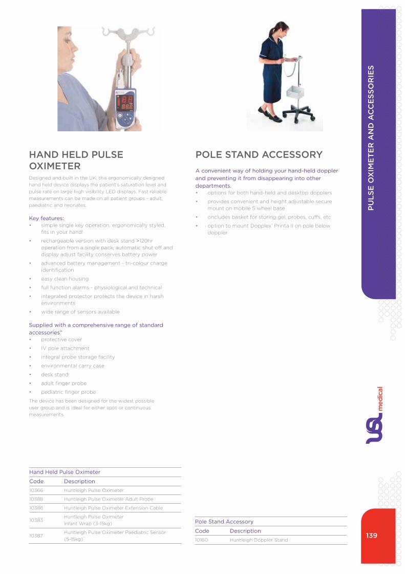
139
Pole Stand Accessory
Code Description
10160 Huntleigh doppler Stand
Hand Held Pulse Oximeter
Code Description
10366 Huntleigh Pulse Oximeter
10388 Huntleigh Pulse Oximeter adult Probe
10386 Huntleigh Pulse Oximeter extension Cable
10383Huntleigh Pulse Oximeter infant Wrap (3-15kg)
10387Huntleigh Pulse Oximeter Paediatric Sensor (5-15kg)
POLE STAND ACCESSORYA convenient way of holding your hand-held doppler and preventing it from disappearing into other departments.• options for both hand-held and desktop dopplers
• provides convenient and height adjustable secure mount on mobile 5 wheel base
• oncludes basket for storing gel, probes, cuffs, etc
• option to mount dopplex® Printa ii on pole below doppler
HAND HELD PULSE OXIMETERdesigned and built in the uK, this ergonomically designed hand held device displays the patient’s saturation level and pulse rate on large high visibility Led displays. Fast reliable measurements can be made on all patient groups - adult, paediatric and neonates.
Key features:• simple single key operation, ergonomically styled,
fits in your hand!
• rechargeable version with desk stand >120hr operation from a single pack, automatic shut off and display adjust facility conserves battery power
• advanced battery management - tri-colour charge identification
• easy clean housing
• full function alarms - physiological and technical
• integrated protector protects the device in harsh environments
• wide range of sensors available
Supplied with a comprehensive range of standard accessories”• protective cover
• iV pole attachment
• integral probe storage facility
• environmental carry case
• desk stand
• adult finger probe
• pediatric finger probe
the device has been designed for the widest possible user group and is ideal for either spot or continuous measurements.
smartsigns jáåámìäëÉ
Hand Held Pulse Oximeter
.
NE
W
®
PU
LSE
OX
IME
TE
R A
ND
AC
CE
SSO
RIE
S

140
Automatic Ankle Brachial Index System
Code Description
11296 Huntleigh ability unit
11298 Huntleigh ability Carry Bag
11297 Huntleigh ability trolley
11302 Huntleigh ability Fixing Plate
11301 Hunteligh ability Label Paper
11300 Hunteligh ability Paper
11299 Huntleigh ability Sleeves
AUTOMATIC ANKLE bRACHIAL INDEX SYSTEMRevolutionising Ankle Brachial Pressure Index Measurementsankle Brachial index System has never been simpler or quicker. it’s portability enables measurements to be made more efficiently in the primary care clinic, hospital or patient’s home. this can lead to the prioritisation of clinical services by improving clinical pathways.
aBis are calculated easily and accurately in three minutes without the need to rest the patient. Results are automatically calculated, interpreted and displayed with Pulse Volume waveforms on the LCd panel. the test results can be printed on either thermal paper or adhesive backed label paper via the integral printer.
HUNTLEIGH AbILITY
integral easy grip handle
integrated thermal
printer (can be used
with paper or label rolls)
Clear display showing
test status, numerical
results, waveform and
interpretation.
Simple user interface.
Colour coded, easy
connect tubing.
DO
PP
LEX
Ab
ILITY
141
PReSSuReReLieVing
deViCeS
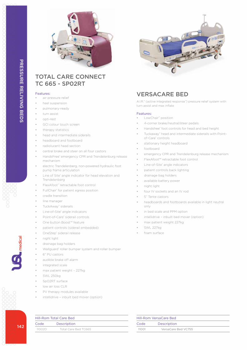
142
TOTAL CARE CONNECTTC 665 - SP02RTFeatures:• air pressure relief
• heel suspension
• pulmonary-ready
• turn assist
• opti-rest
• gCi colour touch screen
• therapy statistics
• head and intermediate siderails
• headboard and footboard
• radiolucent head section
• central brake and steer on all four castors
• HandsFree® emergency CPR and trendelenburg release mechanism
• electric trendelenberg, non-powered hydraulic foot pump frame articulation
• Line of Site® angle indicator for head elevation and trendelenberg
• Flexafoot™ retractable foot control
• FullChair® for patient egress position
• cradle transition
• line manager
• tuckaway™ siderails
• Line-of-Site® angle indicators
• Point-of-Care® siderail controls
• One button Boost™ feature
• patient controls (siderail embedded)
• OneStep® siderail release
• night light
• drainage bag holders
• Wallguard® roller bumper system and roller bumper
• 6” Pu castors
• audible brake off alarm
• integrated scale
• max patient weight – 227kg
• SWL 250kg
• SpO2Rt surface
• low air loss CLR
• PV therapy modules available
• intellidrive – inbuilt bed mover (option)
Hill-Rom Total Care Bed
Code Description
11002d total Care Bed tC665
vERSACARE bEDa.i.R.™ (active integrated response™) pressure relief system with turn assist and max inflate
Features:• LowChair™ position
• 4-corner brake/neutral/steer pedals
• Handsfree® foot controls for head and bed height
• tuckaway™ head and intermediate siderails with Point-of-Care® controls
• stationary height headboard
• footboard
• emergency CPR and trendelenburg release mechanism
• Flexafoot™ retractable foot control
• Line-of-Site® angle indicators
• patient controls back lighting
• drainage bag holders
• available battery power
• night light
• four iV sockets and an iV rod
• 5” tente castors
• headboards and footboards available in light neutral only
• in bed scale and PPM option
• intellidrive – inbuilt bed mover (option)
• max patient weight 227kg
• SWL 227kg
• foam surface
Hill-Rom VersaCare Bed
Code Description
11001 VersaCare Bed VC755
PR
ESSU
RE
RE
LIvIN
G b
ED
S

143
CLINACTIv™
Mattress replacement system comes complete with a user-friendly control unit, an-easy-to-connect double hose, a coated, two-way-stretch, detachable top cover enclosing an air therapy mattress with multiple, highly conformable air cells.
Features: • air mattress underlay for enhanced comfort
• Vario™ advanced sensor system for automatic pressure regulation
• aC power chord management system
• choice of alternating low pressure or continuous low pressure module (either module can be purchased as an option therefore you have one mattress with two therapy options)
• easy-to-connect quiet pump with pressure level display
• intuitive controls including: PMax, Pt egress alarm, Control Lock out, automatic transport Mode, CPR activation Led, alarm Silence
the standard Clinactiv™ mattress replacement system is delivered with specifically designed carry bags for both the control unit and the mattress.
Hill-Rom ClinActiv™
Code Description
11005d air therapy Pressure Care Mattress
DUO II™
Fully enclosed mattress replacement system with a coated, two-way-stretch, detachable protex top cover enclosing an air therapy mattress with multiple, highly conformable air cells.
Features:• air mattress underlay for enhanced comfort
• deteq™ sensor system
• quiet compressor enclosed in the system
• small compact hand control with the choice of either alternating low pressure or continuous low pressure at the touch of a button
• instant CPR
• instant 1 minute re-inflation
• alarm silence
• P-Max
• transport mode
• power connection
Hill-Rom Duo II™
Code Description
11020d air therapy Pressure Care Mattress
PR
ESS
UR
E R
ELI
vIN
G M
AT
TR
ESS
ES

144
AccuMax™ Quantum™ Convertible Mattress
Code description
90016 accumax QC Mattress
90120 Premium Control unit
ACCUMAX™ qUANTUM™
CONvERTIbLE MATTRESSthe top of the line accuMax™ Quantum™ Convertible is a non-powered pressure relief mattress that can be converted into a powered pressure relief mattress. Some special features include a hand-crafted co-polymer and fibre topper, designed therapeutic pressure zones, plus a gentle heel slope.
these features all provide unsurpassed patient benefits and comfort. the Convertible also includes our exclusive airport™ connection system as standard equipment to allow quick and easy conversion to a powered system with gentle, alternating pressure capability - as easy as 1, 2, 3!
PR
ESS
UR
E R
ELI
vIN
G b
ED
S

145
SCaRManageMent

146
Kelo-Cote
Code Description
12832 Kelo-Cote 6g tube
5-7cm linear scar coverage
12834 Kelo-Cote 15g tube with SPF30
10-17cm linear scar coverage
12837 KeloCote 100ml Spray
12x12cm scar coverage
KELO-COTEKelo-cote advanced formula scar therapy products may be used to manage, reduce, soften and help prevent the following types of scars: keloid scars, c-section scars, hypertrophic scars, mastectomy scars, burn scars, mohs surgery scars, plastic surgery scars, hysterectomy scars, breast augmentation scars, body contouring scars, breast reduction scars, body piercing scars, breast implant scars, tattoo removal scars, tummy tuck scars, cleft lip scars, cosmetic surgery scars, small cuts and injury scars, scar revision or scar surgery scars, and more.
* Sebastian g et. al. akt dermatol 2004; Bd. 30:450
• transparent and odourless 100% silicone gel
• Works on old and new scars
• Softens and flattens raised scars
• Reduces redness and discoloration
• Relieves itching and discomfort associated with scars
• the only 100% silicone gel with a patented, self-drying formula
• acts like a second layer of skin providing 24-hours efficacy
• easy and convenient twice daily application
• the only scar treatment available in a gel and spray
• Self-dries within 4-5 minutes forming a flexible, transparent sheet
• Cosmetics or sunscreen can be applied once Kelo-cote has dried
• Suitable for use with all ages and skin types, including children and patients with sensitive skin
• ideal for scars of all shapes and locations
• Over 2,000 patients in published clinical trials
1 day post-trauma 12 weeks post-trauma with Kelo-cote® treatment
Facial trauma, 8 year old
trauma to the palm
SCA
R T
HE
RA
PY

147
negatiVe PReSSuRe tHeRaPy
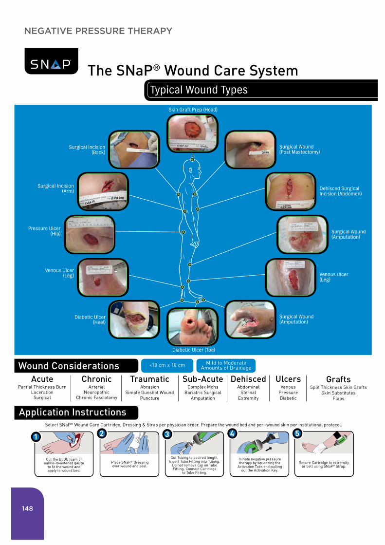
148
Wound Considerations
Application Instructions
Cut the BLUE foam or saline-moistened gauze
to fit the wound and apply to wound bed.
Cut Tubing to desired length. Insert Tube Fitting into Tubing.
Do not remove cap on Tube Fitting. Connect Cartridge
to Tube Fitting.
Initiate negative pressure therapy by squeezing the
Activation Tabs and pulling out the Activation Key.
Secure Cartridge to extremity or belt using SNaP® Strap.
Select SNaP® Wound Care Cartridge, Dressing & Strap per physician order. Prepare the wound bed and peri-wound skin per institutional protocol.
For detailed instructions and product information which includes specific indications, warnings and precautions, please refer to the SNaP® Wound Care System Instructions for Use. www.spiracur.com Additional questions? Please call: 1.877.774.7228
Spiracur, Spiracur logo, SNaP, and SNaP & Design are registered trademarks of Spiracur Inc. U.S. and certain foreign patents pending. www.spiracur.com/legal ©2013 Spiracur Inc. All rights reserved.
L23225 rev. 130730
The SNaP® Wound Care System Typical Wound Types
1
Place SNaP® Dressing over wound and seal.
2 3 4 5
Acute Partial Thickness Burn
Laceration Surgical
Chronic Arterial
Neuropathic Chronic Fasciotomy
Traumatic Abrasion
Simple Gunshot Wound Puncture
Sub-Acute Complex Mohs
Bariatric Surgical Amputation
Dehisced Abdominal
Sternal Extremity
Ulcers Venous
Pressure Diabetic
Grafts Split Thickness Skin Grafts
Skin Substitutes Flaps
<18 cm x 18 cm Mild to Moderate Amounts of Drainage
Surgical Wound (Post Mastectomy)
Skin Graft Prep (Head)
Surgical Incision (Back)
Surgical Incision (Arm) Dehisced Surgical
Incision (Abdomen)
Pressure Ulcer (Hip) Surgical Wound
(Amputation)
Surgical Wound (Amputation)
Venous Ulcer (Leg)
Venous Ulcer (Leg)
Diabetic Ulcer (Heel)
Diabetic Ulcer (Toe)
NEGATIvE PRESSURE THERAPY

149
Spiracur Inc.1180 Bordeaux Drive Sunnyvale, CA 94089Toll Free 1.877.774.7228www.spiracur.com
L22072 rev. 130513
Spiracur, Spiracur logo, SNaP, SNaP & Design, and BioLock are registered trademarks of Spiracur Inc. U.S. and certain foreign patents pending. www.spiracur.com/legal
©2013 Spiracur Inc. All rights reserved.
· Disposable components with off-the-shelf convenience
· Proprietary hydrocolloid dressing and antimicrobial gauze offer periwound protection and easy removal
· Fully integrated microport enables flexibility and a tight bending radius for wounds located in difficult areas
· Cut-to-length tubing and integrated one-way flow valve for improved safety
A closer look reveals the innovative features that create powerful wound therapy, without any power.
With the SNaP® Wound Care System, perfection can be found in the details.
Silent Cartridge
· Clinically proven negative pressure offered at 125 mmHg, 100 mmHg and 75 mmHg
· Small, silent, lightweight design disappears under clothes
· Proprietary spring mechanism generates consistent, even levels of pressure
· No noisy motors, electronics or batteries to interfere with daily living
· Visual indicator displays when cartridge is full or discharged
· BioLock® technology gels exudate for improved exudate containment
Advanced Wound Dressing
· Strap enables device to be conveniently and discreetly worn for maximum discretion
Comfort Strap
NEGATIvE PRESSURE THERAPY

150
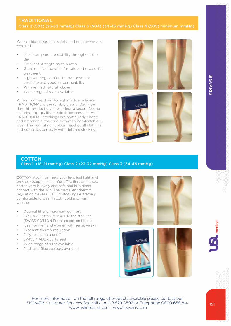
151
When a high degree of safety and effectiveness is required.
• Maximum pressure stability throughout the day
• excellent strength-stretch ratio• great medical benefits for safe and successful
treatment• High wearing comfort thanks to special
elasticity and good air permeability• With refined natural rubber• Wide range of sizes available
When it comes down to high medical efficacy, tRaditiOnaL is the reliable classic. day after day, this product gives your legs a secure feeling, ensuring top-quality medical compression. as tRaditiOnaL stockings are particularly elastic and breathable, they are extremely comfortable to wear. the neutral skin colour matches all clothing and combines perfectly with delicate stockings.
COttOn stockings make your legs feel light and provide exceptional comfort. the fine, processed cotton yarn is lovely and soft, and is in direct contact with the skin. their excellent thermo-regulation makes COttOn stockings extremely comfortable to wear in both cold and warm weather.
• Optimal fit and maximum comfort• exclusive cotton yarn inside the stocking
(SWiSS COttOn Premium cotton fibres)• ideal for men and women with sensitive skin• excellent thermo-regulation• easy to slip on and off• SWiSS Made quality seal• Wide range of sizes available • Flesh and Black colours available
TRADITIONAL Class 2 (503) (23-32 mmHg) Class 3 (504) (34-46 mmHg) Class 4 (505) minimum mmHg)
COTTONClass 1 (18-21 mmHg) Class 2 (23-32 mmHg) Class 3 (34-46 mmHg)
For more information on the full range of products available please contact our SigVaRiS Customer Services Specialist on 09 829 0592 or Freephone 0800 658 814
www.uslmedical.co.nz www.sigvaris.com
SIGv
AR
IS

152
For more information on the full range of products available please contact our SigVaRiS Customer Services Specialist on 09 829 0592 or Freephone 0800 658 814
www.uslmedical.co.nz www.sigvaris.com
SIGv
AR
IS
the women’s compression stocking for every requirement.
• a delicate, slightly transparent stocking with a natural, elegant look
• Perfect fit and exceptional degree of comfort• Simple handling• Provides optimum leg support• Wide range of sizes available • Flesh and Black colours available
MagiC, the versatile compression stocking, is exceptionally easy to handle and has a perfect fit. this has been confirmed by intensive tests carried out with compression stocking wearers under scientific conditions. their natural look and subtle transparency also appeal to the senses.
innovation meets tradition.
treat venous ulcers successfully.
• Significantly higher healing rate and shorter healing time compared with conventional bandages
• Particularly easy to put on and take off thanks to patented understockings
• SigVaRiS uLCeR X can be applied by patients themselves
• the use of natural yarns lends an especially comfortable feeling
Compression therapy with uLCeR X has considerable objective and subjective advantages compared with bandages.
MAGICClass 1 (18-21 mmHg) Class 2 (23-32 mmHg)
ULCER XClass 2 (503) (23-32 mmHg)

153For more information on the full range of products available please contact our
SigVaRiS Customer Services Specialist on 09 829 0592 or Freephone 0800 658 814 www.uslmedical.co.nz www.sigvaris.com
SIGv
AR
IS
the perfect travel companion.
traveno will come in handy wherever you go.
• intensive graduated support• Reduces swelling• Breathable and easy to clean
• ideal for long journeys
travel safely with traveno
every time you sit for a long period of time without moving your legs, the risk of travel thrombosis increases – whether you’re travelling by air, car or train. Swelling and painful legs are possible symptoms. traveno promotes circulation in your legs by applying gentle pressure.
a scientific study has proven that SigVaRiS traveno provides considerable health benefits.
natural Rubber Quality
indications
• For recent lymphodemas that will subside rapidly and are soft
• Post-mastectomy o• dema occurring late following radiotherapy• Post-operative and post-traumatic odema
after completion of basic treatment• Polyarthritis in certain cases• treatment by elastic compression for burns
and extensive scarring
Contraindications
• unreduced Lymphodema• inflammation
ARM SLEEvES
TRAvENO - Travel compression
154
adVanCed WOund
COuRSeS

155www.fmhs.auckland.ac.nz 06/12
All of our courses lead to qualifications that are approved by the Nursing Council of New Zealand and therefore are eligible for funding by Health Workforce New Zealand. Contact your local DHB HWNZ Co-ordinator.
Postgraduate Study at the School of Nursing
Postgraduate Wound Care Nursing Course–2013
Introducing: NURSING 730 Specialty Nursing Knowledge and Practice Wound Care Nursing (30 points)
In Semester Two, 2013, a Postgraduate Wound Care nursing course (Level 800) is being offered at the School of Nursing and can be combined with other Postgraduate Nursing courses to make up a Postgraduate Diploma in Health Sciences (Advanced Nursing) (120 points).
The Wound Care course is designed for Registered Nurses working within a range of clinical settings which may include specialist nursing roles, practice nurses, chronic conditions management nurses, acute care nurses and primary care nurses. The aim of the course is to:
•Explore the care and experience of clients with wounds across the continuum of care.
•Increase nurses’ specialty knowledge and skills in order to enable them to plan and deliver client-centred care to clients with wounds and other specific health care needs.
•Increase knowledge around advanced nursing roles, pathophysiology, client assessment and client management
•Increase the understanding around the impact of wounds and the health care experience on the client and family
•Increase nurses’ understanding and application of core nursing concepts such as ethics, caring and culture to specialty practice areas of nursing and in a wider socio-political and economic context.
Postgraduate Diploma (120 points)• NURSING 773 Advanced Assessment and Clinical Reasoning (30 points)
• NURSING 742 Biological Science for Practice (30 points)
• NURSING 730 Specialty Nursing Knowledge and Practice Wound Care Nursing (30 points)
• A 30 point course which reflects personal choice.
All Students are encouraged to seek individual academic advice to ensure nursing courses reflect personal preferences for their Postgraduate Diploma (Advanced Nursing).
For information about the Wound Care Course, Postgraduate Diploma courses or Individual Academic Pathways contact:
Lesley Doughty [email protected]
For information about courses, dates, study skill workshops see our website:
www.fmhs.auckland.ac.nz/son

With special acknowledgement to elizabeth (Liz) Milner, nZROn, BHS/nsg, Post diploma/advanced nsg, Post Cert Clinical/ed. for all her support and involvement with producing this woundcare catalogue.

HLE 8000 03/2008
Technical SpecificationsOverall dimensions 2160mm (length) x 1025mm (width)
Nurse lockout and hand control Standard
Castors 100mm Central locking, single wheels, premium
Minimum-maximum height 235-765mm
High-low adjustment Electronic standard
Backrest adjustment Electronic standard with quick release (optional)
Head and footboard Standard style provided
Sleeping surface 1960 x 860mm
Sleeping surface base Sheet metal or optional removable infection control panels for easy cleaning
IV pole, self help pole Both optional
Back up battery Optional – available for peace of mind
Mattress retainer is available Use without a footboard if preferred
SWL 250kgs
Premium package As above plus:
Safety side rails Included for added patient safety
Knee-break adjustment Electronic for therapy and comfort positions
Head and foot boards available Queen Anne, Colonial, Arch or Gentle Arch
Sleeping surface Removable light panels for infection control
and easy cleaning (ICO)
Back up battery Standard for peace of mind
The perfect solution to aged care or homecare bedding
For details on the full range of our hospital beds, trolleys and ward furniture please visit:
www.medicrafthill-rom.com.au or call 02 9569 0255
Medicraft Hill-Rom, 72-84 Shepherd Street, Marrickville NSW 2204
THE HLE 8000 SUPERLOW BED
15468 - HLE 8000 Bro 7/7/08 12:45 PM Page 1
Order By Freephone: auckland: 0800 658 814 Christchurch: 0800 804 546
Order By Freefax: auckland: 0800 830 660 Christchurch: 0800 804 547
auckland Head Office 494 Rosebank Road , avondale, auckland 1026 email: [email protected]
MaRCH 2014