Hyaluronate derivatives and their application to wound healing: Preliminary observations
Journal of Cellular Biochemistry 26: 107-116 (1984) Extracellular Matrix: Structure and Function 1-10
Fibronectin and Wound Healing Frederick Grinnell
Department of Cell Biology, University of Texas Health Science Center, Dallas, Texas 75235
Critical to the continued existence of all multicellular organisms is their ability to respond to and repair traumatic injuries. In vertebrates, particularly mammals, the response to injury has been studied in great detail, and the various cells involved in wound healing have been identified (recently reviewed in [ l ] and [ 2 ] ) . While the overall features of wound healing are now well known, many important details remain to be clarified. For instance, the biochemical signals that initiate and terminate the wound healing response are still subjects of considerable debate. Cells of the organism recognize and migrate to the wound interface, and the wound healing response persists at least until this abnormal interface is replaced by new tissue.
Several years ago, I reviewed the evidence indicating that fibronectin is impor- tant in cutaneous wound healing [ 3 ] . Since then, considerable additional information in support of this idea has been forthcoming, and it now appears that fibronectin plays numerous roles in the wound healing situation, which will be discussed in this brief review.
CUTANEOUS WOUND HEALING
Skin normally is composed of a stratified epithelium (epidermis) separated from an underlying connective tissue stroma (dermis) by a basement membrane. Following formation of a full thickness wound, the defect produced in the skin is filled by a blood clot composed of platelets trapped in a fibrin meshwork. Platelets are important, not only in recognizing and physically occluding the defect, but also in promoting blood coagulation and in secreting growth factors for fibroblasts and possibly other cells involved in subsequent stages of wound healing. Following formation of the blood clot, the wound region is invaded almost immediately by neutrophils. These cells are responsible primarily for controlling infection, but in the absence of infection neutrophils do not seem to be necessary for normal healing of wounds. Next, monocytes migrate into the wound region where they are involved in removing tissue debris and secreting factors that promote the growth and biosynthetic activity of
Received April 22, 1984; accepted June 1, 1984.
0 1984 Alan R. Liss, Inc.

1OI:JCB Grinnell
fibroblasts. Shortly after the appearance of monocytes, the region is invaded by fibroblasts and endothelial cells from the subdermis. The fibroblasts synthesize granulation tissue which is vascularized by the endothelial cells. Finally, the granula- tion tissue is covered by a neo-epidermis formed by keratinocytes migrating in from the wound edges. The keratinocytes migrate beneath the dried out portion of the blood clot through the upper region of the granulation tissue. This entire process takes about 1-2 weeks, and is followed by a gradual remodeling of the granulation tissue to a more normal neo-dermis, during which time the increased fibroblast population characteristic of granulation tissue markedly diminishes [ 1,2].
The above description (summarized in Table I) is somewhat superficial, and does not take into account important features such as wound contracture [4], but is sufficient to point out the main cell types that participate in the organism’s response to wounding and their primary functions in wound healing. In the remainder of this paper the role of fibronectin will be described in relationship to the cells and functions listed in Table I.
THROMBOSIS
Platelets normally are nonadhesive to the intact endothelial surface of blood vessels. When there is a defect in the endothelium, platelet attachment to the under- lying connective tissue appears to occur by at least two mechanisms, one involving direct interactions with polymerized collagen fibrils [5] and the other involving a von Willebrand factor-mediated interaction with the subendothelium [6]. While resting platelets do not normally express fibronectin on their surfaces, it has been shown that fibronectin can be detected on the surfaces of platelets attached to collagen [7] or enmeshed in blood clots [8] (Fig. 1). Apparently, there are specific and saturable receptors for fibronectin on the platelet surface 19,101 that are exposed after platelet activation [ 11,121.
Initial studies demonstrated that addition of fibronectin promoted platelet spreading on collagen substrata but did not enhance platelet attachment [ 13,141. Fibronectin also has been found to enhance platelet spreading associated with von Willebrand factor-mediated platelet attachment to the subendothelium [ 151. On the other hand, fibronectin does not appear to be important in platelet aggregation induced by ADP 1121 or collagen [ 161. The physiological significance of fibronectin-mediated platelet spreading has yet to be adequately explained, but studies on platelets isolated from a patient with a new form of Ehlers-Danlos syndrome [17] and patients with Glanzmann’s thrombasthenia [ 181 have raised the possibility that fibronectin is nec- essary for normal platelet function.
TABLE I. Cells Involved in Cutaneous Wound Healing
Function Cell type
Platelets Neutrophils Management of infection Monocytes Fibroblasts Endothelial Cells Neo-vascularization Keratinocytes Re-epithelialization
-
Thrombosis, coagulation, secretion of growth factors
Removal of tissue debris, secretion of growth factors Synthesis and remodeling of extracellular matrix
2: EMSF

Fibronectin and Wound Healing JCB:109
Fig. 1. Fibronectin distribution in blood clots formed ex vivo. Indirect irnmunofluorescence analysis with antifibronectin shows fibronectin coating fibrin strands (arrows) and platelets (arrowheads). A) Fluorescence. B) Phase contrast. See [8] for other details.
One other interesting possibility is that the presence or absence of fibronectin on material surfaces might determine the thrombogenicity of the surfaces [ 191. If this were the case, it would be of considerable importance in understanding tissue reac- tions to artificial implant materials. Both the attachment and spreading of platelets on polystyrene is promoted by fibronectin 1201, and the presence of fibronectin on a variety of material surfaces has been shown to promote thrombus deposition in an ex vivo shunt model [21].
FIBRONECTIN AT THE WOUND INTERFACE
The major structural component of the blood clot that fills the wound defect is a fibrin polymer cross-linked by the plasma enzyme transglutaminase (factor XIII), and fibronectin also can be covalently linked to the fibrin polymer by this enzyme [22]. As shown in Figure 1, the fibrin in blood clots formed ex vivo is uniformly coated with fibronectin [8], and a similar result has been found following wounding in vivo (Fig. 2) [23]. While the presence of fibronectin coating the fibrin does not appear to change the mechanical properties of clots formed from platelet-rich or platelet-free plasma [24], it does play an important role in the adhesive interactions of cells migrating into the clot region (see below).
One point of particular importance is that the fibronectin content of the wound matrix is much higher than that of the adjacent tissue [23]. This is consistent with the low concentration of fibronectin in the dermis of unwounded skin except where fibronectin is associated with dermal fibroblasts [23,25]. Although fibronectin has been found to bind collagen type I , the major collagenous component of dermis, this binding is relatively weak unless the collagen is denatured [26,27].
NEUTROPHIL FUNCTION
Several different studies have shown that fibronectin promotes neutrophil motil- ity, chemotaxis [28] and adhesion to endothelial cells [29] or material surfaces [21].
EMSF: 3

1lO:JCB Grinnell
Fig. 2. Fibronectin distribution in blood clots formed in vivo after wounding. Indirect irnrnunofluores- cence analysis with antifibronectin shows that fibronectin was present throughout the fibrin clot (fc) (A,B), and at higher magnification fibronectin was found coating individual fibrin strands (C). In the reticular dermis (rd) adjacent to the blood clot, fibronectin was found associated with cells but not with the collagen matrix. See [23] for other details.
It also has been reported that neutrophils can enzymatically modify fibronectin to an altered form that enhances neutrophil adhesion [30]. Thus, although there are some conflicting data 1311, it seems likely that the presence of fibronectin may be an important signal for neutrophil movement into the wound region.
The role of fibronectin in neutrophil phagocytosis is less clear. While fibronectin promoted binding of Streptococcus pyogenes to neutrophils [32], other evidence suggested that phagocytosis of bacteria was not enhanced by fibronectin [33,34]. In addition, although fibronectin promoted the phagocytosis of latex or yeast particles by neutrophils, opposite data were found regarding the activation of postphagocytotic metabolic activities [28,35].
MONOCYTE FUNCTION
In the case of monocytes, fibronectin appears to be important in both cell migration and phagocytosis. Fibronectin has been reported to promote monocyte adhesion to material surfaces [36], and both the intact fibronectin molecule [37] as well as fibronectin fragments [38] were found to be chemotactic for monocytes. In addition, although fibronectin does not itself function as an opsonin for phagocytosis by monocytes [33,36], it does enhance phagocytosis of particles opsonized by immu- noglobulin [36] or complement [39,40]. In addition, fibronectin fragments have been found to augment the opsonin-independent pathway of phagocytosis by monocytes [41]. Finally, fibronectin also stimulated secretion of monocyte derived growth factor for fibroblasts [42].
4: EMSF

Fibronectin and Wound Healing JCB:lll
While fibronectin promotes phagocytosis of particles by macrophages, the significance of this activity requires clarification. Several studies demonstrated that fibronectin promoted binding of gelatin-coated particles to macrophage surfaces [43,44], but when phagocytosis was directly measured it turned out that heparin was important and sometimes necessary as an additional cofactor for phagocytosis in this system [4547]. On the other hand, fibronectin has not been shown to promote phagocytosis of bacteria by macrophages under any conditions [33,34]. It is likely that fibronectin is important in macrophage phagocytosis of denatured collagen and fibrin debris found in the wound region, but further evidence to establish this point is necessary.
FIBRONECTIN AND INFECTION
The idea that fibronectin might function as a nonimmune opsonin for the phagocytosis of certain bacteria by neutrophils and cells of the monocyte/macrophage lineage seemed reasonable when it was found that fibronectin bound specifically to the surfaces of Staphylococcus aureus [48-501. This would have fit in well with the proposed role of fibronectin in reticulo-endothelial cell function [5 11. Nevertheless, as indicated above, present evidence suggests that fibronectin is not opsonic for bacterial phagocytosis. More recently, the idea has emerged that bacteria might use fibronectin as a mechanism for colonizing tissues. For instance, fibronectin was found to promote the attachment of Staphylococcus aureus and several Streptococcus strains to endothelial [52] and epithelial cells [53]. In addition, several antibiotics have been demonstrated to interfere with fibronectin binding to Staphylococcus aureus [54]. The possibility must be considered, therefore, that the presence of fibronectin in the wound bed might be one factor that enhances infection. In this regard, it may be that fibronectin-coated wound dressings could decrease infection by attracting bacteria out of wounds.
FIBROBLAST FUNCTION
The role of fibronectin in fibroblast function during wound healing has been well documented. Fibronectin promotes fibroblast adhesion to material surfaces [55] and denatured collagen [56,57]. In addition, the adhesion of fibroblasts to fibrin has been shown to require fibronectin, and maximal adhesion required that fibronectin be covalently crosslinked by Factor XI11 to the fibrin [58]. Moreover, fibronectin has been found to be chemotactic (or haptotactic) for fibroblasts [59-611. Taken together, these findings suggest that fibronectin is very important for fibroblast migration into the wound bed.
The fibroblasts that migrate into the wound region rapidly secrete an extensive fibronectin matrix and shortly thereafter large amounts of type I11 collagen are deposited in association with this matrix [23,62,63]. Fibronectin has a stronger affinity for native type 111 collagen than other collagen types [26,27]. In vitro studies have shown that collagen and fibronectin are codeposited in the matrix [64-661, and evidence has been presented indicating that the fibronectin matrix forms a scaffold on which the collagen is organized [67]. In addition, other matrix components such as heparin sulfate proteoglycan and chondroitin sulfate are codeposited with fibronectin
EMSFS

112: JCB Grinnell
in vitro [68]. These results suggest that fibronectin may be essential in the organiza- tion of the granulation tissue matrix.
With time, the granulation tissue is replaced with neo-dermis, which has a decreased content of fibronectin an4 type I11 collagen and an increased content of type I collagen [62,63]. If, however, the granulation tissue reaction persists, such as is the case in scleroderma [69] and hypertrophic scars [70], then the high levels of fibronectin and type I11 collagen do not decrease.
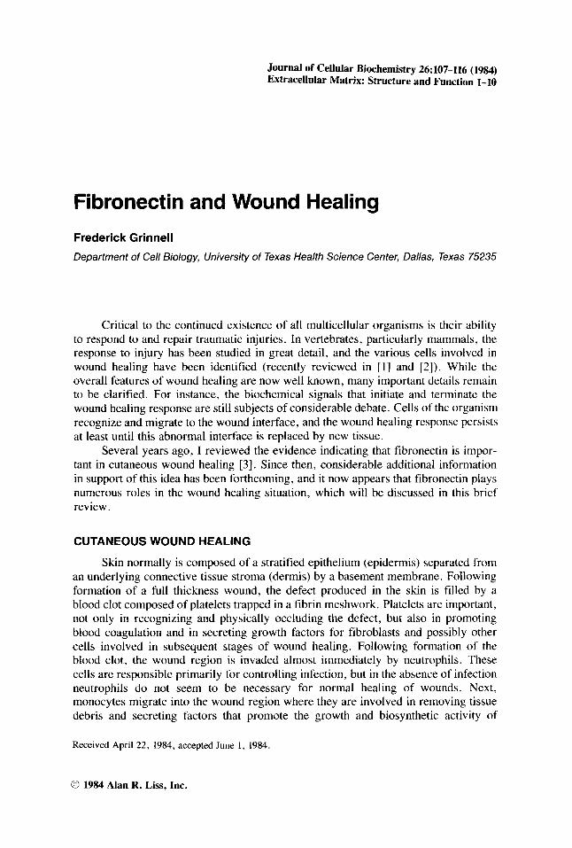
The mechanism by which the granulation tissue is remodeled to neo-dermis is unclear. One possibility, however, is that fibroblast phagocytosis of the fibronectin- coated collagen is involved [71-731. In support of this idea are recent experiments demonstrating that fibronectin-coated latex beads are avidly ingested by fibroblasts [74] (Fig. 3). In fact, it has been shown that fibroblast spreading on or phagocytosis of fibronectin-coated surfaces are similar cell responses differing according to the size of the particles 1751.
ENDOTHELIAL CELL FUNCTION
The role of fibronectin in endothelial cell function has not been studied as extensively as for other cell types. Nevertheless, it has been reported that fibronectin promotes endothelial cell adhesion [76-781 and chemotaxis [78]. These results indi- cate that fibronectin may be important for the migration of endothelial cells in the wound region 1791.
KERATINOCYTE FUNCTION
As already mentioned, the epidermis is separated from the dermis by a basement membrane, which has been shown to contain fibronectin, laminin, Type IV collagen, and proteoglycans [go]. While initial studies suggested that laminin, not fibronectin, was important for epidermal cell adhesion [56,81], more recent findings have shown that fibronectin also is an attachment and spreading factor for keratinocytes [82-841. In examining the surface of the granulation tissue over (through) which the keratino- cytes migrate, it has been shown that fibronectin is present [23,85], but laminin and type IV collagen are absent [86]. Also, fibronectin-coated material surfaces provide a suitable substratum for epidermal cell migration in situ [87].
The above results suggested the possibility that fibronectin is the substrate for keratinocyte migration during wound healing. Strong support for this notion has been provided by studies on wound healing in the cornea. The general structure of the cornea is analogous to that of the skin with a stratified epithelial layer separated from the underlying connective tissue by the basement membrane, although the cornea is avascular. Nevertheless, following wounding, a fibrin-fibronectin matrix forms at the wound site and persists until the defect is repaired [88]. As with migrating skin keratinocytes, the corneal epithelial cells move in direct contact with the fibronectin- rich matrix, and laminin and type IV collagen are absent [89]. Moreover, studies with corneal explants have shown that exogenously added fibronectin promotes epithelial migration and anti-fibronectin antibodies inhibit migration [90]. Additional evidence regarding the possible importance of fibronectin in epithelial migration comes from
6: EMSF
Fibronectin and Wound Healing JCR:113
Fig. 3. Interaction of baby hamster kidney cells with fibronectin-coated latex beads. Cells were found to phagocytose large numbers of 1.90-pm beads (A) and also could phagocytose beads that were 5.7 pm (B). With 15.8-pm beads, however, the cells spread around the bead surface but could not complete phagocytosis (C). See [75] for other details.
EMSF: 7

114: JCB Grinnell
TABLE 11. Functions of Fibronectin in Cutaneous Wound Healing
Cell type Function
Platelets Spreading Neutrophils Adhesion, migration. chemotaxis Monocytes Fibroblasts Endothelial cells Adhesion, migration, chemotaxis Epidermal cells Bacteria Tissue colonization
Adhesion, migration, chemotaxis, phagocytosis, secretion of growth factors Adhesion, migration, chemotaxis, phagocytosis, matrix organization
Adhesion, migration, phagocytosis, basement membrane organization
animal studies in which ulceration of the cornea (lack of reepithelialization) has been correlated with degradation of fibronectin at the corneal surface [91]. From a thera- peutic point of view, the possibility of using fibronectin as a treatment for corneal ulcers in humans is now under investigation [92].
An important unresolved issue is how migrating skin keratinocytes “cut” through the upper layer of the granulation tissue. One possibility, suggested by electron micrographic studies, is that the keratinocytes are able to phagocytose fibrin debris [93] and thereby clear their own path. Consistent with this possibility, fibronectin now has been shown to promote the phagocytosis of latex particles by keratinocytes [84]. Finally, the reorganization of the basement membrane beneath the epidermal cells after their migratory activites have ceased [86,89] may be promoted by fibronec- tin [94].
SUMMARY
I have tried to briefly review the evidence (summarized in Table 11) indicating that fibronectin is important in cutaneous wound healing. Fibronectin appears to be an important factor throughout this process. It promotes the spreading of platelets at the site of injury, the adhesion and migration of neutrophils, monocytes, fibroblasts, and endothelial cells into the wound region, and the migration of epidermal cells through the granulation tissue. At the level of matrix synthesis, fibronectin appears to be involved both in the organization of the granulation tissue and basement membrane. In terms of tissue remodeling, fibronectin functions as a nonimmune opsonin for phagocytosis of debris by fibroblasts, keratinocytes, and under some circumstances, macrophages. Fibronectin also enhances the phagocytosis of immune-opsonized par- ticles by monocytes, but whether this includes phagocytosis of bacteria remains to be determined. In general, phagocytosis of bacteria has not appeared to involve fibronec- tin. On the contrary, the presence of fibronectin in the wound bed may promote bacterial attachment and infection.
Because of the ease of experimental manipulations, wound healing experiments have been carried out on skin more frequently than other tissues. As a result, the possible role of fibronectin has not been investigated thoroughly in the repair of internal organs and tissues. Nevertheless, it seems reasonable to speculate that fibronectin plays a central role in all wound healing situations. Finally, the wound healing problems of patients with severe factor XI11 deficiencies may occur becuase of their inability to incorporate fibronectin into blood clots [22].
8:EMSF
Fibronectin and Wound Healing JCB:115
ACKNOWLEDGMENTS
Drs. R.E. Billingham, W. Snell, and A. Takashima made helpful comments regarding this manuscript. The author’s research has been supported by grants from the NIH (CA 14609, HL 24221, GM 31321).
REFERENCES
1. Hunt TK, Dunphy JE: “Fundamentals of Wound Management.” New York: Appleton-Century- Crofts, 1979.
2. Hunt TK: “Wound Healing and Wound Infection: Theory and Surgical Practice.” New York: Appleton-Century-Crofis, 1980.
3. Grinnell F: Am J Dermatopathol 4:185, 1980. 4. Montandon D, D’andiran G, Gabbiani G: Clinics Plast Surg 4:325, 1977. 5 . Muggli R, Baumgartner HR: Thromb Res 3:715, 1973. 6. Sakariassen KS, Bolhus PA, Sixma JJ: Nature 279:636, 1979. 7. Hynes RO, Ali IU, Destree AT, Mautner V, Perkins ME, Senger DR, Wagner DD, Smitt KK: Ann
NY Acad Sci 312:317, 1978. 8. Grinnell F, Feld M: Thromb Res 24:397. 1981. 9. Plow EF. Ginsberg MH: J Biol Chem 256:9477, 1981.
10. Hansen MS, Clemmensen I: Biochern J 201:629, 1982. 11. Ginsberg MH, Painter RG, Birdwell C, Plow EF, Forsyth J: J Supramol Struct 11:167, 1979. 12. Zucker MB, Mosesson MW, Brockman MJ, Kaplan KL: Blood 54:8, 1979. 13. Grinnell F, Feld M, Snell W: Cell Biol Int Rep 3:585, 1979. 14. Koteliansky VE, Leytin VL, Sviridov DD, Repin VS, Smirnov VN: FEBS Lett 123:59, 1981. 15. Booyse FM. Feder S, Quarfoot AJ: Thromb Res 28:299, 1982. 16. Santoro SA, Cunningham LW: Proc Natl Acad Sci USA 76:2644, 1979. 17. Arneson MA, Hammerschmidt DE, Furcht LT, King RA: J Am Med Assoc 244: 144, 1980. 18. Ginsberg MH, Forsyth J, Lightsey A, Chediak J, Plow EF: J Clin Invest 71:619, 1983. 19. Grinnell F: In Szycher M (ed): “Synthetic Biomedical Polymers: Concepts and Applications,” 2nd
ed. Westport, CT: Technomic Publishing Co., 1983. pp 673-699. 20. Grinnell F, Phan TV: Thrornb Res (submitted). 2 I , Barber TA. Mathis T, Thlenfeld JV, Cooper SL, Mosher DF: Scan Electron Microsc 2:43 I , 1978. 22. Mosher DF: Prog Hemost Thromb 5 : 11 1. 1980. 23. Grinnell F, Billingham RE, Burgess L: J Invest Dermatol 76:181, 1981. 24. Chow TW, McIntire LV, Peterson DM: Thromb Res 29:243, 1983. 25. Fryand 0: Br J Dermatol 101:263, 1979. 26. Ruoslahti E, Engvall E, Haymdn EG: Coll Res 1:95, 1981. 27. Yamada KM: Annu Rev Biachem 52:761, 1983. 28. Jarstrand C, Ahlgren T, Berghen L: J Clin Lab Immunol 8:59, 1982. 29. Wall RT, Cooper SL, Kosek JC: Exp Cell Res 140:105, 1982. 30. Vercellotti GM, McCarthy J, Furcht LT. Jacob HS, Moldown CF: Blood 62: 1063, 1983. 31. Brown AF, Lackie JM: Exp Cell Res 136:225, 1981. 32. Simpson WA, Hasty DL, Mason JM, Beachey EH: Infect Irnmun 36:805, 1982. 33. Verbrugh HA, Peterson PK, Smith DE. Nguyen B-YT, Hoidal JR, Wilkinson BJ, Verhoef J, Furcht
LT: Infect Immun 33:811, 1981. 34. Van de Water L, Destree AT, Hynes RO: Science 220:201, 1983. 35. Gudewicz PW, Beezhold DH, Van Alten P, Molnar J: J Reticuloendothel Soc 32: 143, 1982. 36. Bevilacqua MP, Amrani D, Mosesson MW, Bianco C: J Exp Med 153:42, 1981. 37. Yonemasu K, Nakanishi A, Sasaki T. Kashiba S: Microbiol Immunol 27:283, 1983. 38. Norris DA, Clark RAF, Swigart LM, Huff JC, Weston WL, Howell SE: J Immunol 129:1612.
1982. 39. Pomrnier CG, Inada S, Fries LF, Takahashi T, Frank MM, Brown EJ: J Exp Med 157:1844, 1983. 40. Wright SD, Craigmyle LS, Silverstein SC: J Exp Med 158: 1338, 1983. 41. Czop JK, Kadish JL, Austen KF: Proc Natl Acad Sci USA 78:3649, 1981.
EMSF: 9

116:JCB Grinnell
42. Martin BM, Gimbrone MA Jr, Majeau GR, Unanue ER, Cotran RS: Am J Pathol 11 1 :367, 1983. 43. Doran JE, Mansberger AR, Reese AC: J Reticuloendothel SOC 27:471, 1980. 44. Blumenstock FA, Saba TM. Roccario E, Cho E. Kaplan JE: J Reticuloendothel Soc 30:61, 1981. 45. Gudewicz PW, Molnar J , Lai MZ. Beczhold DW, Siefring GE Jr, Credo RB, Lorand L: J Cell Biol
46. Van de Water L 111, Schroeder S , Crenshaw EB 111, Hynes RO: J Cell Biol 90:32, 1981. 47. Doran JE, Mansberger AR. Edmondson HT. Reese AC: J Reticuloendothel Soc 29:285, 1981, 48. Kuusela P: Nature 276:718, 1978. 49. Ryden C, Rubin K , Speziale P, Hook M, Lindberg M, Wadstrom T: J Biol Chem 258:3396, 1983. 50. Bibel DJ, Aly R, Shinefield HR, Maibach HI: J Invest Dermatol 80:494. 1983. 51. Saba TM, Jaffe E: Am J Med 68:577, 1980. 52. Vercellotti GM, Lussenhop D, Peterson PK, Furcht LT, McCarthy JB, Jacob HS, Moldow CF: J
Lab Clin Med 103:34, 1984. 53. Simpson WA, Beachey EH: Infect lmmun 39:275, 1983. 54. Proctor RA, Olbrdntz PJ, Mosher DF: Antimicrob Agents Chemother 24:823, 1983. 55. Grinnell F: Int Rev Cytol 53:65, 1978. 56. Kleinman HK, Klebe RJ, Martin GR: J Cell Biol 88:473, 1981. 57. Grinnell F: In Guroff G (ed): "Growth and Maturation Factors." New York: John Wiley & Sons,
58. Grinnell F. Feld M. Minter D: Cell 19:517, 1980. 59. Gauss-Muller V, Kleinman HK, Martin GR, Schiffman E: J Lab Clin Med 96: 1071, 1980. 60. Postlethwaite AE, Keski-oja J, Balian G, Kang AH: J Exp Med 153:494. 1981. 61. Tsukamoto Y. Helsel WE, Wahl SM: J Immunol 127:673, 1981. 62. Kurkinen M, Vdheri A, Roberts PJ, Stenman S: Lab Invest 43:47, 1980. 63. Viljanto J, Penttinen R. Raekallio J: Acta Chi, Scand 147:7, 1981. 64. Bornstein P, Ash JR: Proc Natl Acad Sci USA 74:2480, 1977. 65. Furcht LT, Smith D, Wendelschafer-Crabb, Mosher DF, Foidart JM: J Histocheni Cytochem
28: 1319, 1980. 66. Vaheri A, Kurkinen M, Lehto V-P, Linder E, Timpl R: Proc Natl Acad Sci USA 75:4944, 1978. 67. McDonald JA, Kelley DG, Broekelmann TJ: J Cell Biol 92:485, 1982. 68. Hedman K, Johansson S , Vartio T, Kjellen L, Vaheri A. Hook M: Cell 28:663, 1982. 69. Fleischmajer R, Dessau W, Timpl R. Krieg T, Luderschmidt C, Wiestner M: J Invest Dermatol
75:270. 1980. 70. Kischer CW, Hendrix MJC: Cell Tissue Res 231:29, 1983. 71. Svoboda ELA, Shiga A. Deporter DA: Anat Rec 199:473, 1981. 72. Melcher AH. Chan J: J Ultrastruct Res 77: I, 1981. 73. McGaw WT, Ten Cate AR: J Invest Dermatol 81:375, 1983. 74. Grinnell F: J Cell Biol 86: 104, 1980. 75. Grinnell F: J Cell Physiol 11958, 1984. 76. Maciag T, Hoover GA, Stemerman MB, Weinstein R: J Cell Biol 91:420, 1981. 77. Macarak EJ. Howard PS: J Cell Physiol 116:76. 1983. 78. Bowersox JC, Sorgente N: Cancer Res 42:2547, 1982. 79. Clark RAF, DellaPell P, Manseau E, Lanigan JM, Dvorak HF, Colvin RB: J Invest Dermatol
76:269. 1982. 80. Laurie GW, Leblond CP, Martin GR: J Cell Biol 95:340, 1982. 81. Terranova VP, Rohrbach DH, Martin GR: Cell 22:719, 1980. 82. Gilchrest BA, Calhoun JK, Maciag T: J Cell Physiol 112: 197, 1982. 8 3 . Stenn KS, Madri JA, Tinghitella T, Terranova VP: J Cell Biol 96:63. 1983. 84. Takashima A, Grinnell F: J Invest Dermatol (in press). 85. Clark RAF, Winn HJ, Dvorak HF, Colvin RB: J Invest Dermatol 80:26S, 1983. 86. Stanley JR, Alvarez OM, Bere EW, Eaglestein WH. Katz SI: J Invest Dermatol 77:240, 1981. 87. Donaldson DJ, Mahan JT: J Cell Sci 62: 117, 1983. 88. Fujikawa LS, Foster CS, Harrist TJ, Lanigan JM, Covin RB: Lab Invest 45: 120, 1981. 89. Fujikawa LS, Foster CS. Gipson IK, Colvin RB: J Cell Biol 98: 128, 1984. 90. Nishida T. Nakagawa S, Awata T, Ohashi Y, Watanabe K, Manabe R: J Cell Biol 97: 1653, 1983. 91. Berman M, Manseau E, Law M, Aiken D: Invest Ophthalmol Vis Sci 24:1358, 1983. 92. Nishida T, Ohashi Y, Awata T, Manabe R: Arch Ophthalmol 101: 1046, 1983. 93. Odland G, Ross R: J Cell Biol 39: 135, 1968. 94. Brownel AG. Bessem CC, Slavkin HC: Proc Natl Acad Sci USA 78:3711, 1981.
10 :EMSF
87:427, 1980.
1983, pp 454-486.
Copyright © 2022 FDOKUMEN