Study of Phyto- and Physicochemical Analysis, Antimicrobial ...
Upload
khangminh22Category
view
0download
0
J Pharm Pharm Sci (www.cspsCanada.org) 16(5) 760 - 820, 2013
760
Phyto-Extracts in Wound Healing Prasanta K Ghosh1 and Anjali Gaba2
1 Managing Director and Head (R&D), KEE GAD Biogen Pvt. Ltd., Community Centre, Naraina Industrial Area Phase II, New Delhi, India; 2 Manager (R&D), KEE GAD Biogen Pvt. Ltd., Community Centre, Naraina Industrial Area Phase II, New Delhi, India. Received September 28, 2013; Revised November, 22, 2013; Accepted December 13, 2013, Published December 16, 2013. ABSTRACT - Data generated through systematic investigation, carried out on the evaluation of phyto-extracts on wound healing research during the last 20 years have been compiled. About 450 plant species having wound healing properties have been identified. The present knowledge of the wound healing process comprise coagulation, inflammation, proliferation, formation and accumulation of fibrous tissues, collagen deposition, epithelialization, contraction of wound with formation of granulation tissues, remodeling and maturation. The constituents of the plant extracts modulate one or more of the above stages. It was the endeavor to identify the active constituents responsible for antimicrobial activity, free radical scavenging properties, stimulators of enhanced collagen production and/or angiogenesis promoters with identification of lead scaffold chemical structures. Multiple phytochemicals concentrated and blended in optimal concentrations, are expected to be available in future years to carry out multi-tasking efforts in wound healing as more knowledge about the properties of the key constituents are unveiled. This article is open to POST-PUBLICATION REVIEW. Registered readers (see “For Readers”) may comment by clicking on ABSTRACT on the issue’s contents page. __________________________________________________________________________________________ INTRODUCTION Wounds can be major causes of physical disabilities and may lead to losses of many productive man-hours. Wounds are essentially the disruption of functional continuity of cells and tissues at the site of injury, and can be caused by insults to the tissue sites by physical, chemical, microbiological or immunological process. Humans and all animals have in situ capabilities of healing wounds in their body parts through continuous tissue repair and tissue regeneration. However, such capabilities are impaired by age, stress situation, obesity, sex, habits of the patient(such as smoking , alcoholism etc.), conditions of health and immunity status, severity and types of wounds, patient’s medication status , disastrous nature of the assault- environment around the site of the wounds and potentials of serious microbial infection (1). Curing of acute and chronic wounds proceed through common basic phases of hemostatis, inflammation, proliferation, fibroplasias, collagen deposition, epithelialization, contraction, remodeling and maturation.
Phases in Wound Healing During the wound healing process, a series of events encompass the repair especially through the presence and actions of activated platelets, neutrophils and macrophages. Increased vascular permeability and angiogenesis are the consequences of the healing, where multiple cellular and cytokine-mediated events are recruited. The endothelial cells are up-regulated by the actions of secreted soluble factors from the activated cells which include the fibroblast growth factors, transforming growth factors, epidermal growth factors and vascular endothelial growth factors among others (2 - 4). The platelets also get activated by the contents from the vascular wall; the main activators such as fibronectin, fibrillar collagen and other matrix proteins cause the kick-off. _________________________________________ Corresponding Author: Prasanta K Ghosh, Managing Director and Head(R&D), KEE GAD Biogen Pvt. Ltd., Community Centre, Naraina Industrial Area Phase II, New Delhi, India; E-Mail: [email protected]
J Pharm Pharm Sci (www.cspsCanada.org) 16(5) 760 - 820, 2013
761
The inflammed skin tissues at the wound site release several prostaglandins, some of which are considered to be the mediators for platelet activation and functioning (5-7). Once activated, the platelets commence aggregation and adhesion; concomitantly these release several mediators including chemotactic factors as well as adhesive proteins. Each factor has a role in the healing cascade (8-10). The mast cells surrounding the blood vessels at the wound site release histamine proteases, tumor necrosis factor, leukotrins and cytokines. These work as chemotactic signals for the recruitment of white blood cells or leukocytes (11) at the site of the wound. While coagulation of blood and vasoconstriction at the wound site are events completed in minutes, the repair process of polymorphonuclear cell migration manifested through vasodilation and inflammation followed by epithelialization, granulation and new tissue formation take about a week. Usually by the end of the first week, fibroblasts are the main cells accumulating at the wound. These cells are involved in differentiation in the wound healing process (12). During the initial phases usually type III collagen is synthesized and laid down at the wound site; during the later phase the stronger type I collagen gets produced (13). Type III collagen, which were in abundance during the proliferation stage gets degraded and is replaced by type I collagen during the maturation stage (14). The collagen fibers get cross-linked by the action of specific enzymes, properly rearranged and aligned for providing maximum rigidity and toughness (15). The maturation phase can vary from three weeks to two years. If the healing process does not move in a foreseeable manner, then the wound may turn into a chronic wound (16).
Direction of Wound Healing Research using Active Phyto-Extracts
The basic understanding that platelets and the fibrins produced from fibrinogen at the wound site set off several biochemical processes which include collagen synthesis, cell migration, fibroplasias and angiogenesis have been significantly investigated. (17-21). The events at the wound site, especially those including release of platelet factors and others such as cell adhesion, cell proliferation, mitogenesis, angiogenesis, fibroplasia, epithelialization, wound contraction, maturation and
remodeling of the wound site have been researched upon by several investigators. The platelets are the cause of release of more than sixty biologically active substances (9) and that each such factor is involved at specific time and in specific concentrations, contributing to specific activities in the wound healing process cascade, recruiting different cell types and coordinating complex interactions among the different actors during the wound healing process. Several factors appear and disappear at different stages of the healing cascade and their quantification along with the identification of involved cells has not yet been generally possible. The complexity of the situation can be gauged when one realizes that there is considerable variation in the concentration of such factors in healthy individuals; the quantities of transforming growth factor (TGF), vascular endothelial growth factor (VEGF) and fibroblast growth factor (FGF) as an example, in blood samples obtained from 20 different donors showed substantial variation (9). More understanding of the wound healing process especially in terms of quantification of factors are expected to evolve in the coming years. In this context, quantitative insight into the interaction of the various components of the plant extracts with the particularized substances present at the wound site needs also to be studied to scientifically assess their worth. Such studies would require semi-quantitative if not quantitative analysis of the active substances present in different classes of plant extracts. This area is yet to evolve although the usefulness of various kinds of plant extracts in curing wounds of different types is real and many of the plant extracts have been in use in traditional practices for several years in different societies. Active Plant Parts Studied for Wound Healing
During the last two decades, there has been increased interest to assess the utility of plant extracts in wound healing and to gain more insight into the active constituents that promote or modulate the healing process. We have reviewed the literature for the last 20 years. Table 1 provides information about the plants along with their families, which had shown wound healing properties studied in different models; the table also contains information about those wound healing plants that are extensively used in folk medicine.
J Pharm Pharm Sci (www.cspsCanada.org) 16(5) 760 - 820, 2013
762
Based on the information furnished in the literature, the main effects of the active constituents of the plant extracts towards wound healing are summarized as under:
1) Phyto-chemical constituents contributing to antimicrobial activity
2) Phyto-chemical constituents working as antioxidants and as free radical scavengers
3) Active components having enhanced mitogenic activity (contributing to increased cell proliferation), angiogenesis, enhanced collagen production and increased DNA synthesis.
Ideally, active substances present in the plant extracts are anticipated to interfere with one or more phases of the wound healing process in a positive manner in proper sequence and at the right time frame to show improved efficacy. There should also be minimization of substances that deteriorate the healing process. Since in actual experiments and usage, all the plant products as cited in the table have shown efficacious results, there are increased needs to isolate and investigate each active ingredient that has a positive role in the healing process. Unfortunately, such data presently are not plentiful. The active ingredients obtained from the plant materials have been analyzed for the presence of alkaloids, carbohydrates, glycosides, terpenoids, diterpenes, sesquiturpenes and phytosterols, phenolic compounds and multiple kinds of tannins, proteins, flavonoids, saponins, lignins, alkaloids and essential oils. (29,32,37,60,98,99 ,139 ,155,357,389,457,500). The identification of secondary metabolites in plant extracts that could bind to cellular receptors at wound site to initiate modulation of wound healing process was recently reviwed (501). In a couple of investigations, the principal active ingredients have been isolated to study especially their anti-microbial properties e.g. terpenes and terpenoids like gentiopicroside, sweroside and swertiamarine from Gentiana lutea; certain pentacyclic triterpenes (502-503) ; essential oil containing concentrates of eucalyptol (28) ; flavones such as kaempferol and quercetin and their derivatives (473); phenylpropanoid glycosides like verbascoside and teupolioside (504); cyanogenic glycosides such as sambunigrin as well as gallic acid and its derivatives (496).Wound healing substances isolated from Termalia arjuna were tannins (457);
oleanoic acid from Anredra diffusa (70); polysaccharides from Opuntia ficus-indica (365); shikonin derivatives including deoxyshikonin, acetylshikonin, 3- hydroxyl isovaleryl shikonin and 5,8-O-dimethylacetylshikonin from Onosma argentatum (361); asiaticoside, asiatic acid and madecassic acid from Centella asiatica (146-147); quercetin , isorhammetin and kempferol from Hippophae rhamnoides (261) ; and curcumin from Curcuma longa (168). The list of well-charaterized newer active ingredients is increasing with a galloping speed.
Wound repair process follows a set of biochemical reactions. At the wound site, increased amounts of superoxide anion radicals are produced by activated platelets, neutophils and the macrophases as well as by the fibroblasts, stimulated by the pro-inflammatory cytokines during the inflammation phase. These radicals are part of the inate immune system and are generated to destroy the invading microbes at the wound site. However, the oxidative stress requires careful manipulation and control as increased amounts are detrimental to the surrounding tissues and can cause heavy damage. While the system has its checks and measures in place and utilizes superoxide dismutases, catalases, glutathione peroxidases and peroxiredoxins, secreted by the adjoining cells, the impairment of such cells in certain wounds calls for use of extraneous agents that are more appropriate radical scavengers, working in synergy or independently. Several plants extracts containing proanthocyanidins, polyphenolic flavonoids and polyphenols in such situations are expected to provide enabling support to the healing process initially by the moderation of superoxide anions and later by enhancing the expression of vascular endothelial growth factor (VEGF), thereby enhancing angiogenesis and flow of blood as the repair process advances. The plant components having some such properties are described further below.
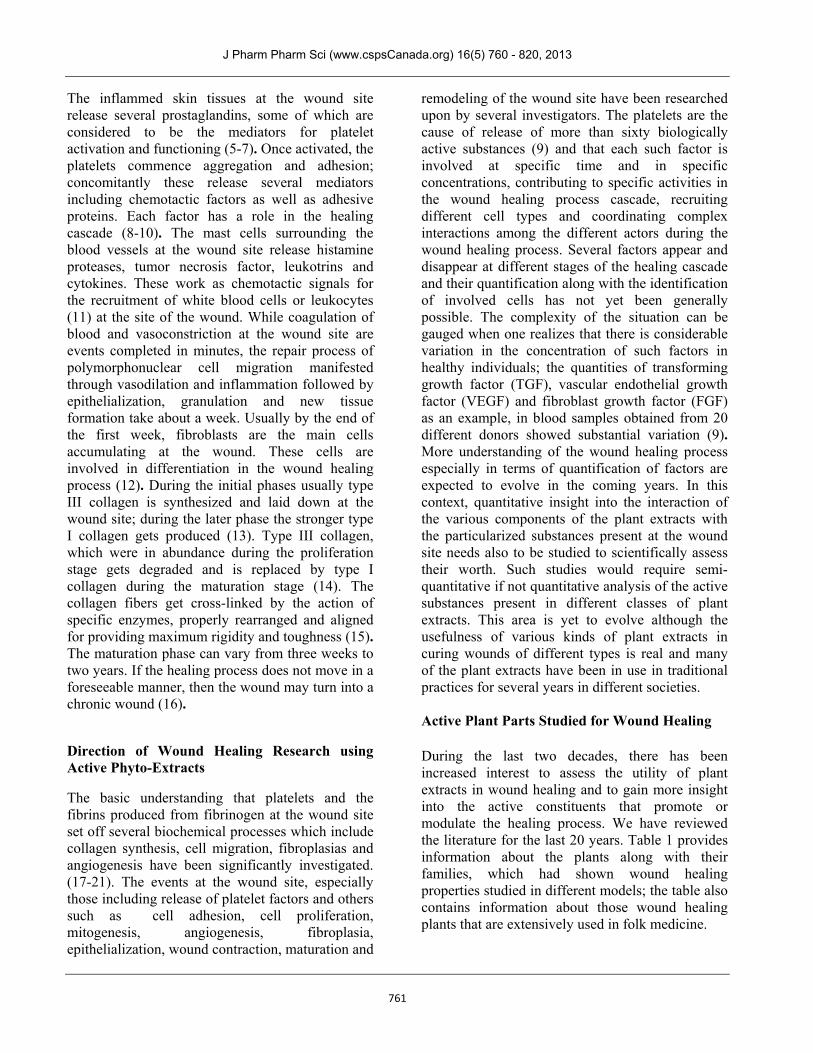
Among the soluble compounds in the plant extracts, the flavonoids, quinones, phenolic acids and phenyl propanoids have been found to possess considerable anti-microbial as well as anti-oxidant properties. A large number of flavonoids having the general structure as given in Figure 1 were found to possess antimicrobial and/or antioxidant properties.
J Pharm Pharm Sci (www.cspsCanada.org) 16(5) 760 - 820, 2013
763
Flavonoids are strong scavengers of reactive oxygen species .In wounds there is a tendency for sharp rise in the concentration of reactive oxygen species due to the activation of platelets, neutrophils, macrophages, lymphocytes and fibroblasts at different time points of the healing process. Infection from microbes also adds to the
woes. In such situations, plant flavonoids would benefit the healing process by modulating the concentrations of reactive oxygen species. Quantitative information and correlationships are yet inadequate however.
Plants such as Allamanda cathartica (50),
Artemisia absinthium (77), Coronopus didymus (176), Cuminum cyminum (187), Flaveria trinervia (233), Heliotropium indicum (253-255), Hippophae rhamnoides (261-263), Ipomoea Carnea (277), Jatropha curcas (187, 281,283), Lawsonia alba (298), Litsea glutinosa (307), Rosmarinus officinalis (416), Moringa oleifera (336-338), Olea europaea (357-359), Pedilanthus tithymaloides (376), Sambucus ebulus (422), Scorzonera (427-428) species and many others are used extensively
in traditional practices in wound healing and these plants are also rich in a wide range of flavonoid compounds.
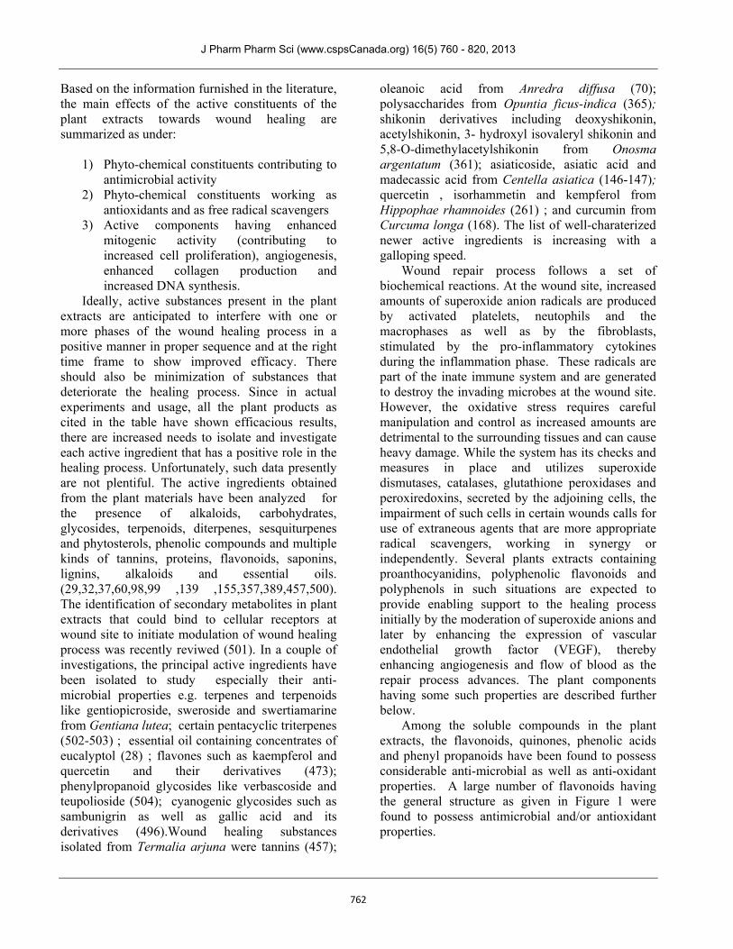
Anthocyanins synthesized in plants via the phenylpropanoid pathway are compounds based on flavylium ion which is a kind of oxonium ion. Anthocyanins have strong radical scavenging properties and many of these compounds also exhibit anti-bacterial properties. Some of the compounds found in wound healing plants are described with general formula as in Figure 2.
J Pharm Pharm Sci (www.cspsCanada.org) 16(5) 760 - 820, 2013
764
Anthocyanins from Black Soyabean seed coat (Glycine max) was found to have enhanced wound healing properties (243). Extracts from Anadenanthera colubrina rich in proanthocyanidins were effective in cutaneous wound healing in rats (62). Caralla brachiata rich in proanthocyanidins is also expected to be useful for such purposes.
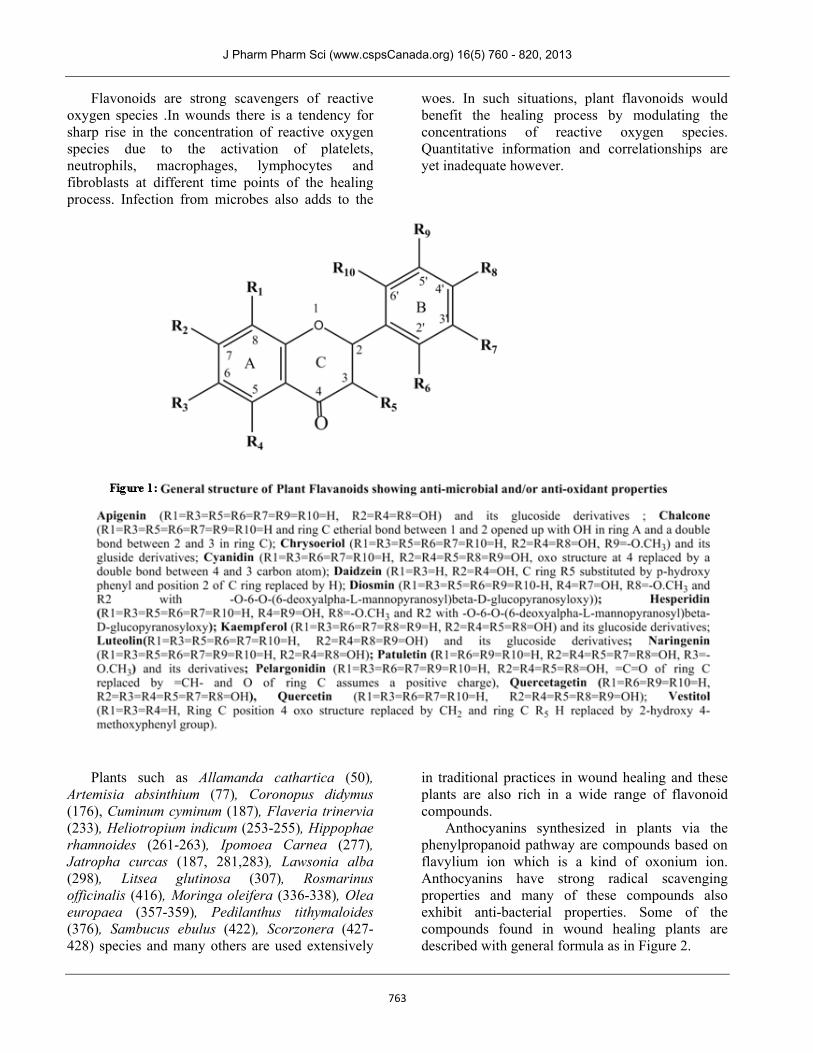
Several soluble quinones present in the roots of plants such as Alkanna tinctoria, Arnebia densiflora and Arnebia euchroma and many others were also found to possess antimicrobial propertie (49, 79, 80); these also had some antioxidant properties. The general structures of such soluble quinones are indicated schematically in Figure 3:
J Pharm Pharm Sci (www.cspsCanada.org) 16(5) 760 - 820, 2013
765
Structures of several of these compounds with ocurenace and biological properties have been reviewed (503). Emodin from Rheum officinale Baill showed encouraging results of repair of excision wounds in rats (413). Embelin from extracts of leaves of Embelia ribes Burm was effective in healing wound in excision, incision and dead space model on Swiss Albino rats (213).
Terpenoids of different structures including the monocyclic and the multicyclic ones have been identified to possess antimicrobial activity; these compounds are anticipated to manifest their antimicrobial effects through the process of synergy with other compounds present in the plant extract. Besides the monocyclic terpenoids, several dicyclic, tricyclic and the pentacyclic terpenoids of plant origin have been identified which possess considerable antimicrobial activities. Terpenoids are present in essential oils of a variety of trees, citrus fruits and herbs. Wound healing activities of Achillea species like Achillea biebersteinii (28) , A. millefolium (31), A. oxyodonta (29), A. setacea and A. teritifolia (506), A. vermicularis (29) etc., Achyranthes aspera (32-33), Allamanda cathartica (50), Alternanthera sessilis (59), Anredera diffusa (70), Arnebia densiflora (79), Berberis lycium (96), Caesalpinia benthamiana (116), Celastrus paniculatus (141), Centella asiatica (146-149), Cissus quadrangularis (159), Croton bonplandianum (182), Croton stellatopilosus Ohba (183), Elephantopus scaber (211) , Heliotropium indicum (254-255), Laurus nobilis (50), Paullinia pinnata (375), Vernonia arborea Hk. (480) etc. are
substantially attributed to the presence of a wide range of terpenoids. Since the structures of each group of terpenoids including mono and multi-cyclic ones vary considerably, a generalized structure could not be assigned to describe a general class of terpenoids having antimicrobial activities. However, assessing the chemical structures of terpenoids present in the above plants for identifying specific mono or multi-cyclic skeletons for in-vitro modification with a view to develop newer compounds is anticipated to be facilitated from the study of the listing of plants.
Phenolics including tannins, substituted cinnamic acids, phenolic acids and phenyl propanoids have also shown antimicrobial as well as antioxidant properties. Tannins from Phyllanthus muellerianus (380), Terminalia arjuna (457), Terminalia avicennioides (458), Terminalia bellirica (459), Terminalia chebula (460) and Terminalia coriacea (461) are reported to promote wound healing. Tannins are polyphenolic compounds containing considerable numbers of hydroxyls, carboxyls and other hydrophilic structures and are considered to be macromolecules. All natural tannins could not be included in one generic structure although there are considerable resemblances in chemical properties among different tannins.
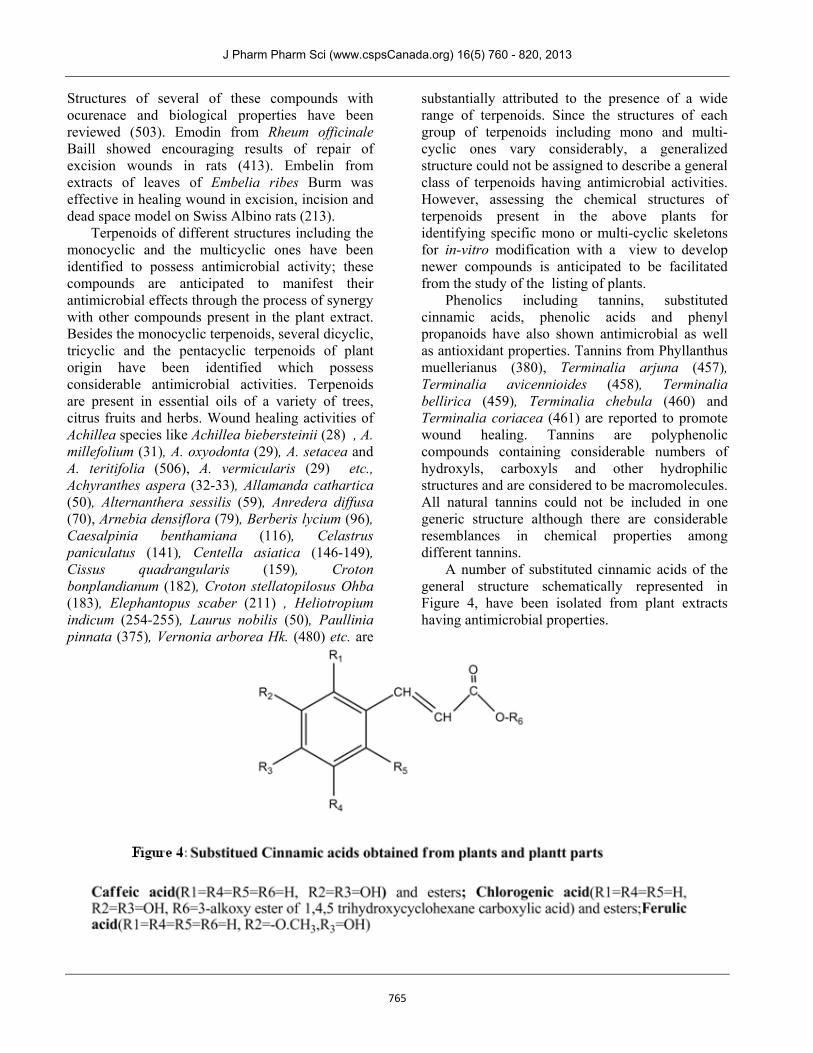
A number of substituted cinnamic acids of the general structure schematically represented in Figure 4, have been isolated from plant extracts having antimicrobial properties.
J Pharm Pharm Sci (www.cspsCanada.org) 16(5) 760 - 820, 2013
766
Caffeic acid, chlorogenic acid and ferulic acid are documented to have wound healing activities (507-509); these substances also work as free radical scavengers. Plant materials like Buddleja globosa (110) leaves containing caffeic acid derivatives, Scorzonera cana var. jacquiniana and S. eriophora (427) containing chlorogenic acids and Angelica sinensis (510) containing ferulic acid have been found to be effective in wound healing.
Several phenolic acids were also found to possess sound antimicrobial properties. In addition, many phenolic acids also had profound radical scavenging properties. Since the structures of phenolic acids vary considerably, it was not possible to represent all of them with one generic structure. However, plants such as Ageratum conyzoides (44,46), Emblica officinalis (214), Punica granatum (30,404), Salvia hypoleuca (420), Schinus lentiscifolius (423), Strobilanthes crispus (446), Quercus infectoria (407), Ximenia Americana (496) contain tannins and gallic acid and these have excellent wound healing activituies. Various mixed phenolic acids are present in plants such as gall nuts, tea leaves, oak bark etc. (511); although these plant materials have not been used in wound healing, it is anticipated that use of these would have beneficial effects as radical scavengers and therefore could be useful in wound healing.
Phenyl propanoids, especially in the form of glycosides, are natural polyphenols which are widely distributed in the plant kingdom. The roots and aerial parts of the families of Asteraceae, Labiateae, Liliceae, Oleaceae and related ones contain phenylpropanoid glycosides (sometimes also incorporating glucose, galactose and rhamnose in these compounds). Such substances are powerful antioxidants. Utilizing plant cells from Ajuga reptans and Syringa vulgaris two phenylpropanoid glycosides namely teupolioside and verbascoside were producued which had profound anti-inflammatory and wound healing properties (504).
Water soluble alkaloids including quinazolines, isoquinazolines, indole derivatives including betalains and eumelanins from a diverse range of plants have been found to possess antioxidant properties and many of these have also antimicrobial characteristics. Adhatoda vasica (36), Adhatoda zeylanica (38), Berberis lycium (512), Catharanthus roseus (139) etc. are rich in certain alkaloids which have antimicrobial properties. These plant extracts are useful in wound healing
purposes and are traditionally used by various societies. Since the compounds from such plants considerably vary in chemical structure, a generalized structure for all these alkaloids could not be presented. However, presently much work is being done to synthesize newer antimicrobial compounds utilizing quinazoline and indole backbones (513-515).
Several natural heteropolysaccharides such as arabinogalactans and rhamnogalacturonans are present in large quantities in certain plants. Hot water extracts of Alstonia boonei De Wild (57), Biophytum petersianum Klotzch (99), Cochlospermum tinctorium Perr (517), Glinus oppositifolius(242), Opuntia ficus-indica (364, 365), and Parquetina nigrescens (57) containing mainly water soluble polysaccharides have been used in traditional practices for treating external wounds. Although exact structure activity relationships are not yet understood, it is believed that the polysaccharides accelerate the phases of reepithelialization and remodeling by influencing interactions in the cell matrix and by moderating the deposition of laminin (365). Polysaccharides are also believed to exhibit immunomodulatory action on the cells around the wound site (57), which stimulate cell proliferation.
Mitogenic properties are anticipated to enhance healing process as phytochemicals possessing such properties exhibited in a structured manner are expected to enhance cell division. Whole plant extracts from Achyrenthus aspera (33) and ‘Cal-proteins’ from Calatropis procera (123) were believed to posssess constituents having mitogenic activities. Extracts of Calendula officinalis flowers have increased proliferation potential for endothelial progenitor cells (518). Extracts of leaves from Datura alba (195) and Euphorbia heterophylla (220) are believed to have strong mitogenic potentials contributing to the healing process. In most of the claims however, the specific compounds responsible for the mitogenic activities have not been identified. Like mitogens, substances promoting angiogenesis would also promote healing process by supply of blood around the wound sites. Extracts of Aloe vera (54), Alternanthera brasiliana (58), mixtures of extracts from Astragali radix and Rehmanniae radix (519), Bidens pilosa and Ocimum suave (97), Blechnum orientale (100), Boessenbergia rotunda (101), Butea monosperma (114), Calendula officinalis
J Pharm Pharm Sci (www.cspsCanada.org) 16(5) 760 - 820, 2013
767
(118), Cinnamomum zeylanicum (157), Cordia macleodii (175), Echium amaenum (206), Equisetum arvense (15) etc. have been shown to promote angiogenisis around wound site. Angiogenesis promoting compounds with specific structures following a pattern have not yet been identified. Increase in DNA and total collagen at wound site with time by the application of phytoextracts substantiate the beneficial effects and are measures of enhancement of healing process linked to the phytoextracts. Extracts from Achillea biebersteinii (29), Achillea kellalensis and Punica granatum (30), Adhatoda vasica (37), Alkanna tinctoria (49), Annona squamosa (67), Arnica Montana and Artemisia absinthium (77), Bauhinia purpurea (94), Bulbine frutescens and Bulbine natalensis (111), Butea monosperma (112), Calotropis gigantea (121), Capparis zeylanica (25), Cassia occidentalis (138), Curcumin from Curcuma longa (190), Desmodium gangeticum (198), Elaeis guineensis (210), Elephantopus scaber (211), Eucheuma cottonii (216), Ficus racemosa (228), Gynura procumbens (249), Heliotropium indicum (255), Hyptis suaveolens (273), Indigofera asphalathoides (275), Jasminum sambac (280), Kalanchoe pinnata (289), Leonotis nepetaefolia (299), Martynia annua (315), Moringa oleifera (336), Nigella sativa (347) etc were claimed to be responsible for facilitated healing as evidenced by increased DNA production and total collagen enhancement at wound site with time. However, in all these cases individual specific chemical entities having the properties of enhancing the wound healing process have not been described. Enhanced healing in all these cases probably arises from synchronized action of multiple active ingredients present in the phyto extracts. CONCLUDING REMARKS Wound healing is a complex but highly regulated process. Healing of all kinds of wounds follows common steps of recovery. Microbial colonization is often inescapable. Infections of wounds from potentially pathogenic bacteria in most situations of
causation of wounds are inevitable. Therefore, the utmost aim is to restore the host-bacterial balance by ensuring that the wound is cleaned up and antimicrobial agents are used with moisture retentive bandages. At the same time as oxidative stress during the initial healing process is high, the next objective is to use agents that scavenge the excess of reactive oxygen anions generated at the wound site and rationalize their concentration. Other objectives are to stimulate the adjoining tissues in the wound so that the processes of cell proliferation, remodeling and maturation are facilitated. The plant kingdom is rich in chemical constituents for mitigating these objectives acting especially as antimicrobial agents as also as the free radical scavengers, and several compounds have since been isolated. The steps of tissue repair involving interactions of neutrophills, macrophages, fibroblasts and other cells at the wound site along with deposition of collagens with proper laying out around the wounds are complex processes and require understaning of multiple interactions with several agents. Concomitantly, formation of new blood vessels through the process of angiogenesis to ensure continuous supply of nutrients and healing supplements also require detailed understaning. In all these processes, several compounds from the plant extracts would work synergistically to provide the desired effect and therefore such phytochemicals concentrated and blended in optimal concentrations from multiple sources are expected to be available in the future years to carry out multi-tasking efforts in wound healing of all kinds as more knowledge about the properties of the key constituents and the healing processes are unveiled. ACKNOWLEDGEMENT The assistance of Dhruva Chatterjee, Computer Executive-KEE GAD Biogen Pvt. Ltd, in preparing the Corel Draw figures and in arranging the References systematically is gratefully acknowledged. Authors have no conflict of interest regarding the content of this article.
J Pharm Pharm Sci (www.cspsCanada.org) 16(5) 760 - 820, 2013
768
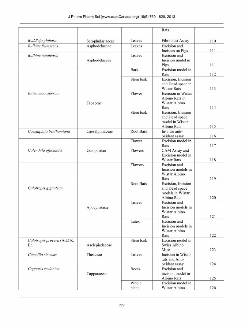
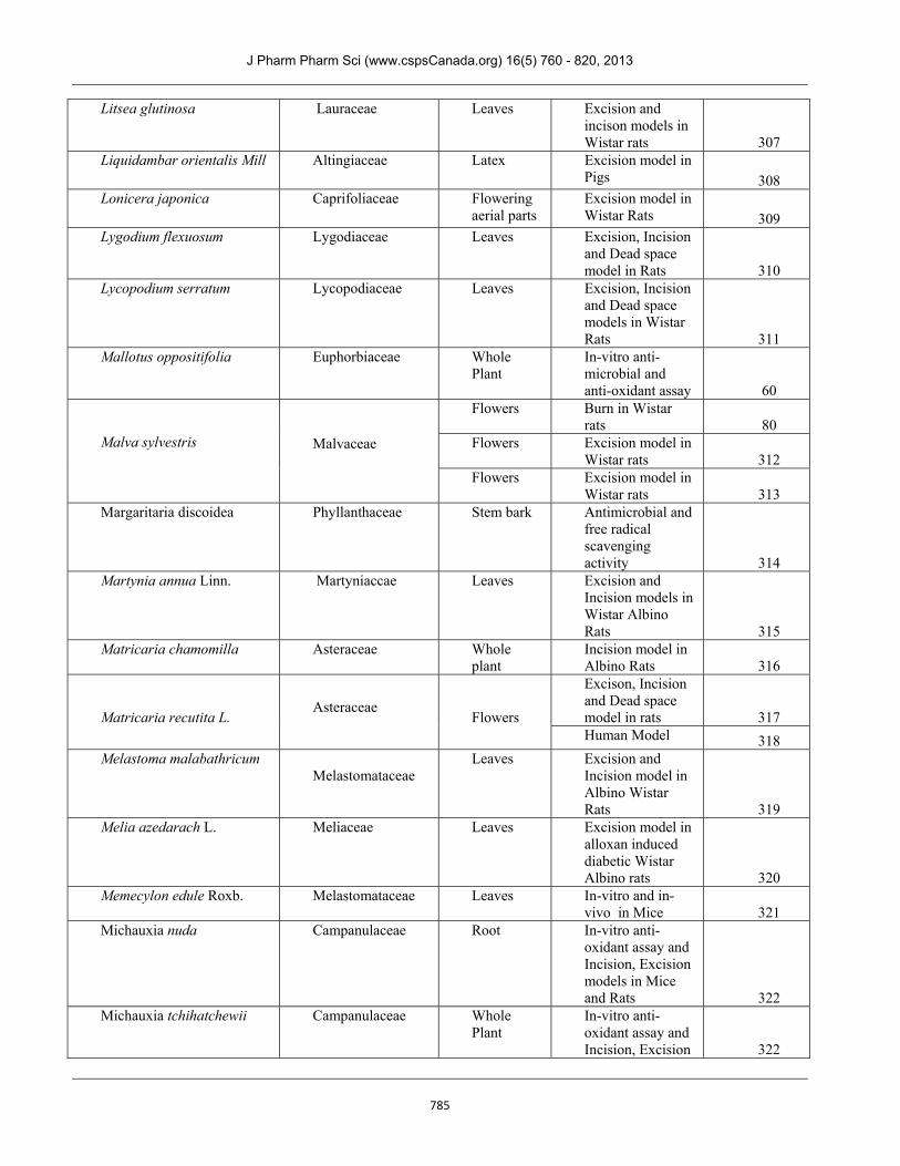
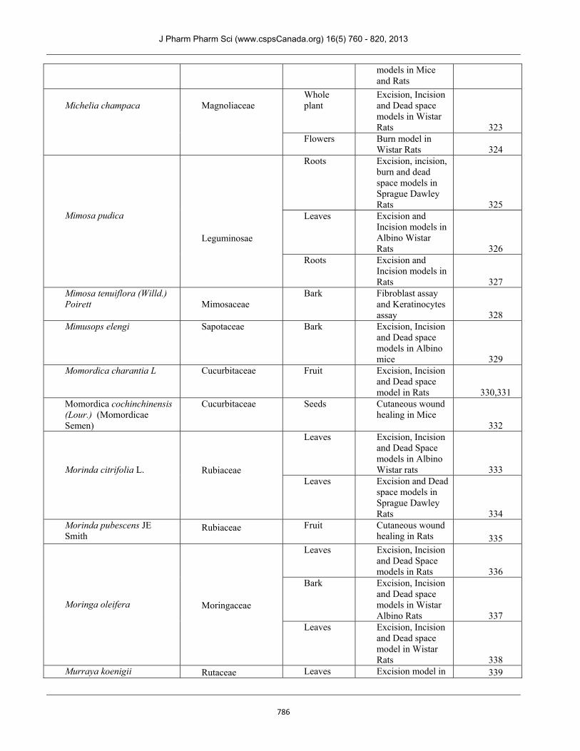
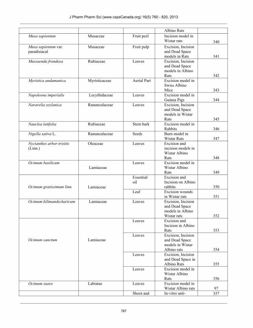
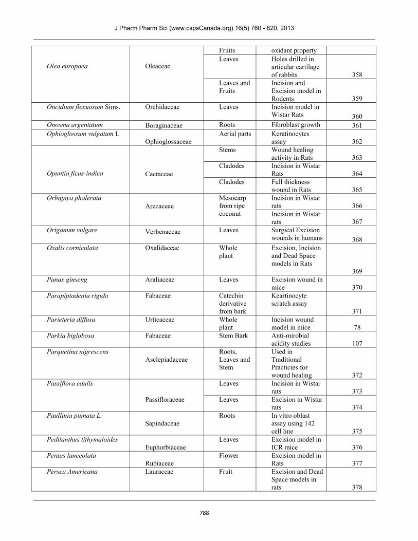
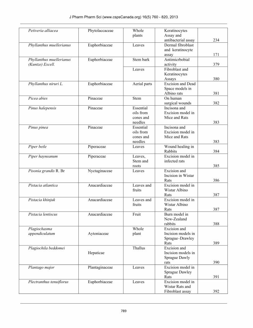
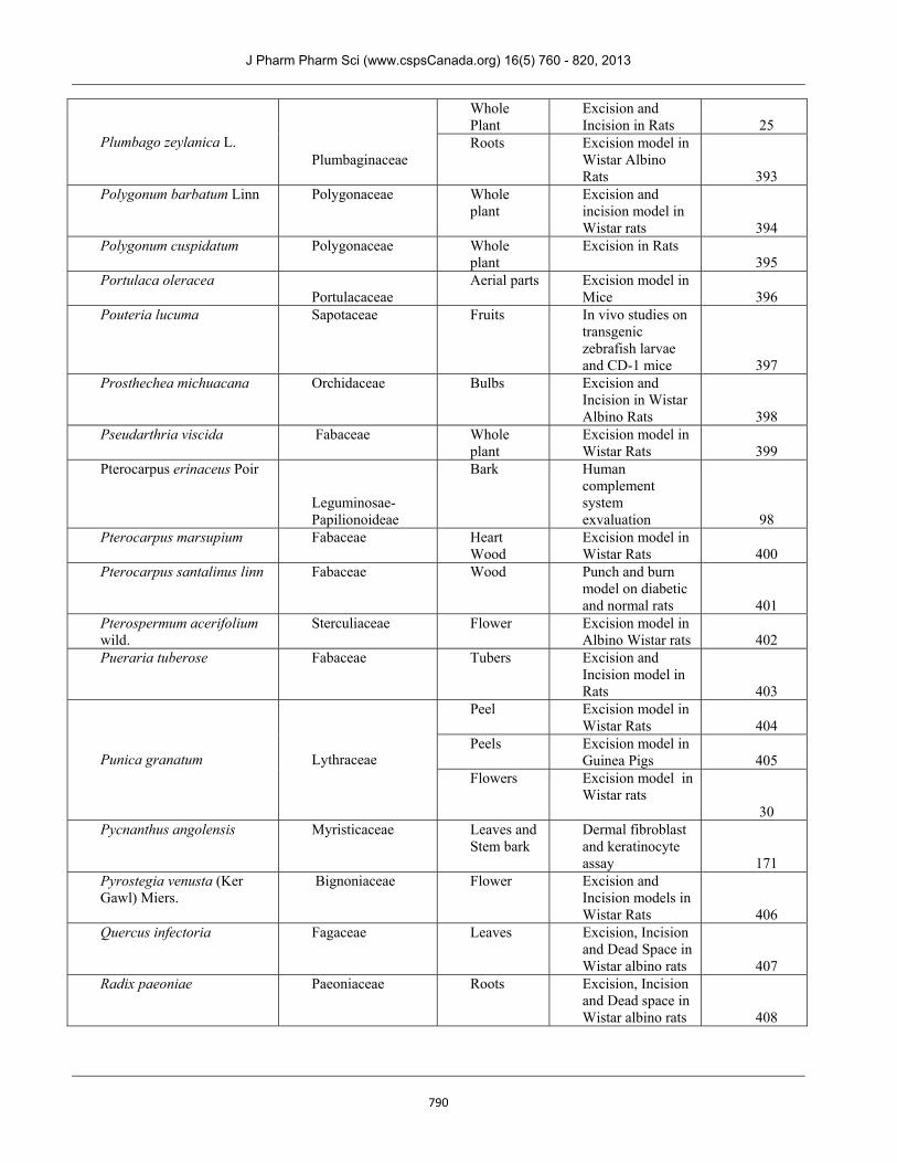
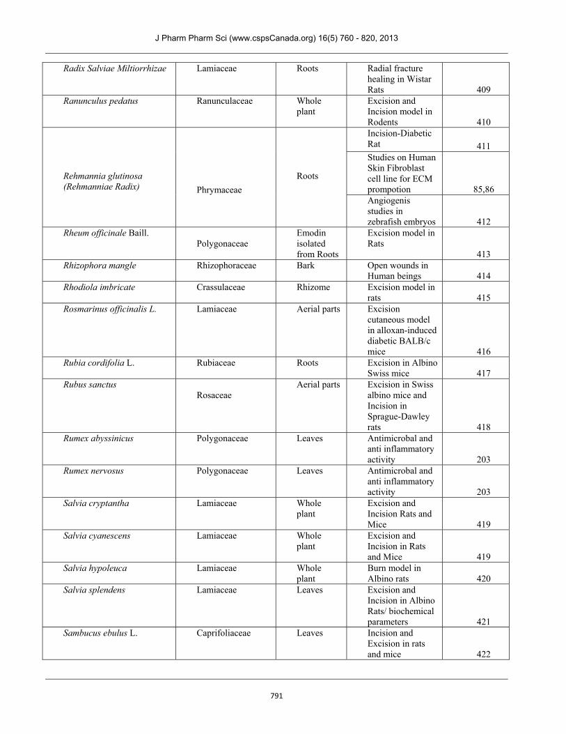
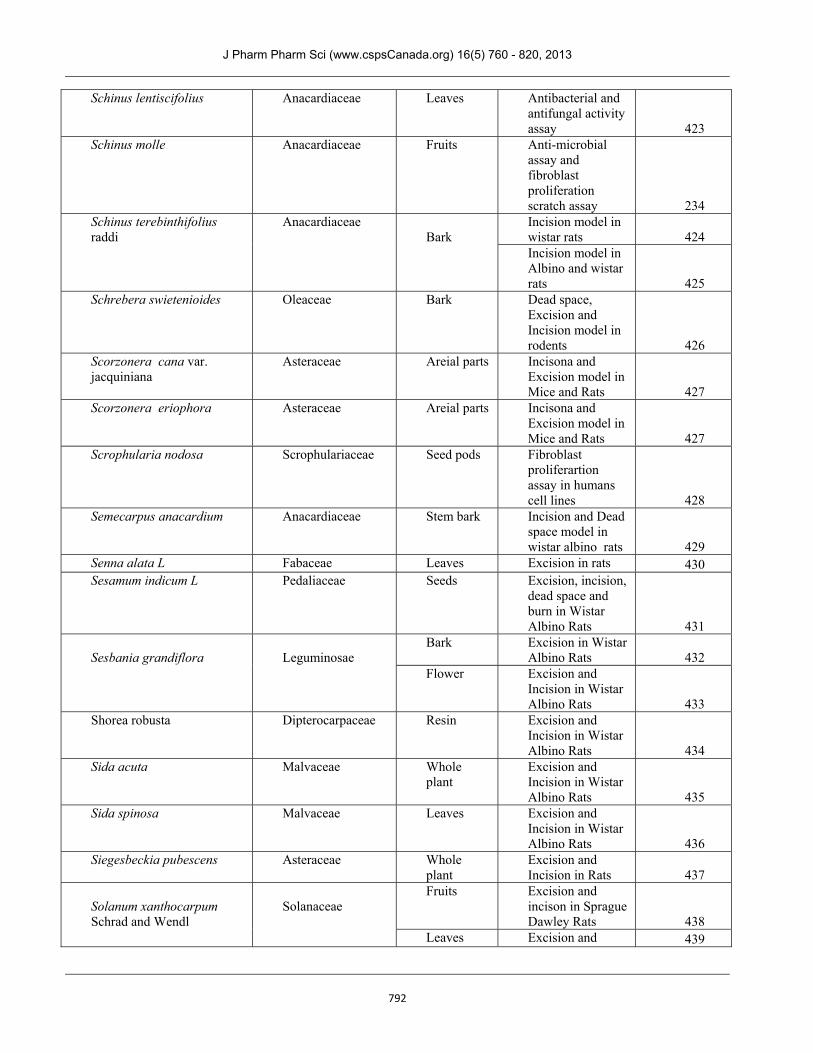
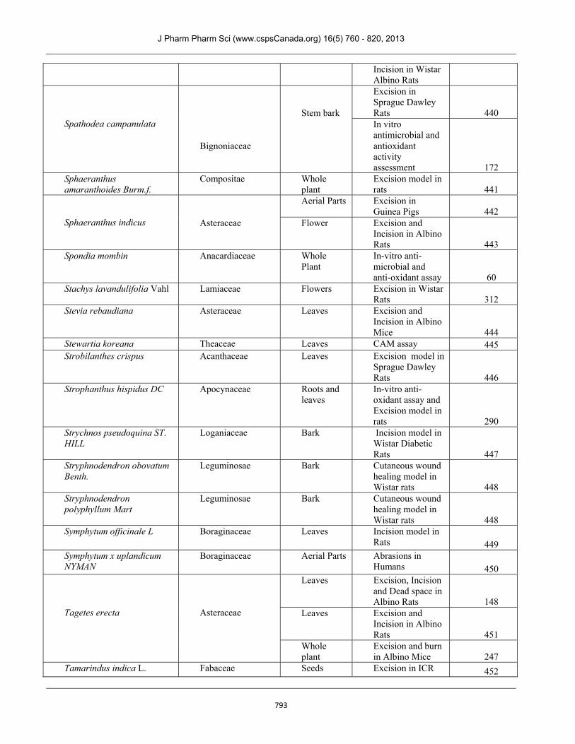
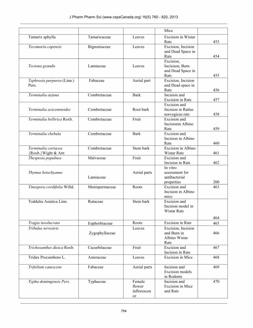
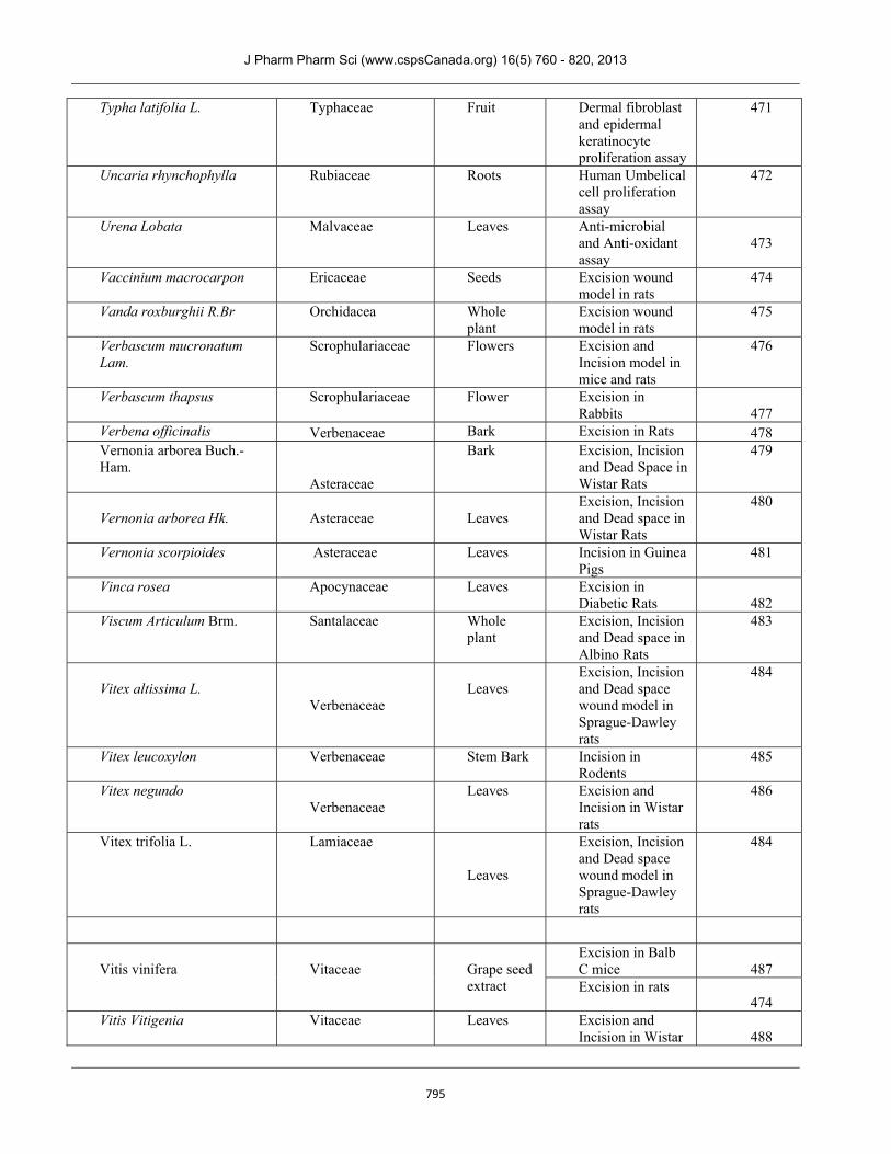
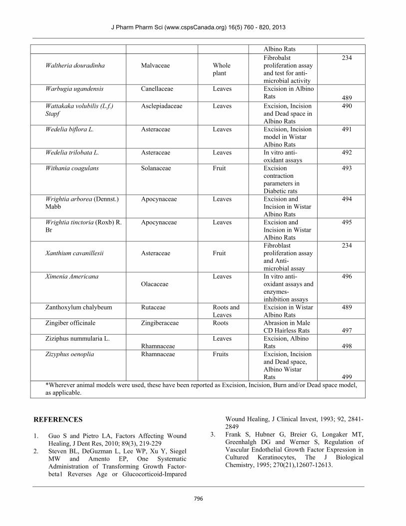
Table 1. (Plant name with Family and the plant parts used/studied for wound healing properties in different models)
Plant Name Plant Family Plant part used/ studied
Assessment Methods/Animal Wound Models where applicable *
Reference No.
Abrus precatorius L. Fabaceae Leaves Used in Folk medicine to treat Cuts and Wounds 22
Abutilon indicum Linn Malvaceae
Whole plant
Excision and Incision models in Wistar Albino Rats 23
Acalypha fruticosa Euphorbiaceae Aerial Part Excision and Dead space models in Rats 24
Acalypha indica Euphorbiaceae
Whole Plant
Excision and Incision models in Rats 25
Acalypha langiana Euphorbiaceae
Leaves On External Human Wounds 26
Acanthus ebracteatus Acanthaceae Stem Incision model in Balb/c mice 27
Achillea biebersteinii Afan.
Asteraceae
Essential Oil from Whole Plant
In-vitro anti-microbial assay
28 Roots Excision and
Incision in Sprague-Dawley rats and Albino mice 29
Achillea kellalensis Asteraceae
Flowers Excision model in Wistar Rats 30
Achillea millefolium L Asteraceae
Leaves Excision, Incision and Dead Space models in Wistar Albino Rats 31
Achyranthes aspera Amaranthaceae Leaves
Excision and Incision model in Albino Rats 32 Excision and incision models in Albino Rats 33
Acorus calamus Linn. Araceae
Leaves Excision and Incision model in Rats 34
Actinidia deliciosa Actinidiaceae
Fruit Burn model in Wistar Rats
35 Adhatoda vasica Acanthaceae
Leaves
Excision model in Wistar Rats 36
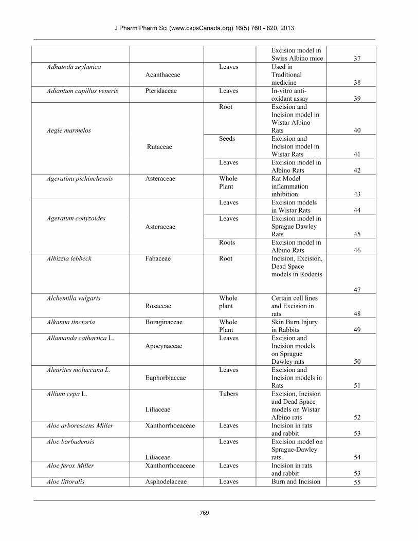
J Pharm Pharm Sci (www.cspsCanada.org) 16(5) 760 - 820, 2013
769
Excision model in Swiss Albino mice 37
Adhatoda zeylanica Acanthaceae
Leaves Used in Traditional medicine 38
Adiantum capillus veneris Pteridaceae
Leaves In-vitro anti-oxidant assay 39
Aegle marmelos
Rutaceae
Root Excision and Incision model in Wistar Albino Rats 40
Seeds Excision and Incision model in Wistar Rats 41
Leaves Excision model in Albino Rats 42
Ageratina pichinchensis Asteraceae
Whole Plant
Rat Model inflammation inhibition 43
Ageratum conyzoides
Asteraceae
Leaves Excision models in Wistar Rats 44
Leaves Excision model in Sprague Dawley Rats 45
Roots Excision model in Albino Rats 46
Albizzia lebbeck Fabaceae
Root Incision, Excision, Dead Space models in Rodents
47 Alchemilla vulgaris
Rosaceae
Whole plant
Certain cell lines and Excision in rats 48
Alkanna tinctoria Boraginaceae
Whole Plant
Skin Burn Injury in Rabbits 49
Allamanda cathartica L. Apocynaceae
Leaves Excision and Incision models on Sprague Dawley rats 50
Aleurites moluccana L. Euphorbiaceae
Leaves Excision and Incision models in Rats 51
Allium cepa L.
Liliaceae
Tubers Excision, Incision and Dead Space models on Wistar Albino rats 52
Aloe arborescens Miller Xanthorrhoeaceae
Leaves Incision in rats and rabbit 53
Aloe barbadensis
Liliaceae
Leaves Excision model on Sprague-Dawley rats 54
Aloe ferox Miller Xanthorrhoeaceae Leaves Incision in rats and rabbit 53
Aloe littoralis Asphodelaceae Leaves Burn and Incision 55
J Pharm Pharm Sci (www.cspsCanada.org) 16(5) 760 - 820, 2013
770
models on Wistar Rats
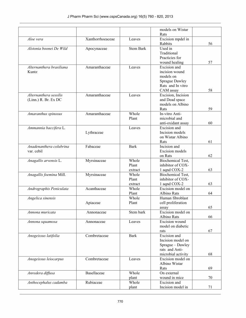
Aloe vera Xanthorrhoeaceae Leaves Excision mpdel in Rabbits 56
Alstonia boonei De Wild Apocynaceae Stem Bark Used in Traditional Practicies for wound healing 57
Alternanthera brasiliana Kuntz
Amaranthaceae Leaves Excision and incision wound models on Sprague Dawley Rats and In vitro CAM assay 58
Alternanthera sessilis (Linn.) R. Br. Ex DC
Amaranthaceae Leaves Excision, Incision and Dead space models on Albino Rats 59
Amaranthus spinosus Amaranthaceae
Whole Plant
In-vitro Anti-microbial and anti-oxidant assay 60
Ammannia baccifera L. Lythraceae
Leaves Excision and Incision models on Wistar Albino Rats 61
Anadenanthera colubrina var. cebil
Fabaceae Bark Incision and Excision models on Rats 62
Anagallis arvensis L. Myrsinaceae
Whole Plant extract
Biochemical Test, inhibitor of COX-1 aqnd COX-2 63
Anagallis foemina Mill. Myrsinaceae Whole Plant extract
Biochemical Test, inhibitor of COX-1 aqnd COX-2 63
Andrographis Peniculata Acanthaceae Whole Plant
Excision model on Albino Rats 64
Angelica sinensis Apiaceae
Whole Plant
Human fibroblast cell proliferation assay 65
Annona muricata Annonaceae
Stem bark Excision model on Albino Rats 66
Annona squamosa Annonaceae Leaves Excision wound model on diabetic rats 67
Anogeissus latifolia Combretaceae Bark Excision and Incision model on Sprague – Dawley rats and Anti-microbial activity 68
Anogeissus leiocarpus Combretaceae Leaves Excision model on Albino Wistar Rats 69
Anredera diffusa Basellaceae Whole plant
On external wound in mice 70
Anthocephalus cadamba Rubiaceae
Whole plant
Excision and Incision model in 71
J Pharm Pharm Sci (www.cspsCanada.org) 16(5) 760 - 820, 2013
771
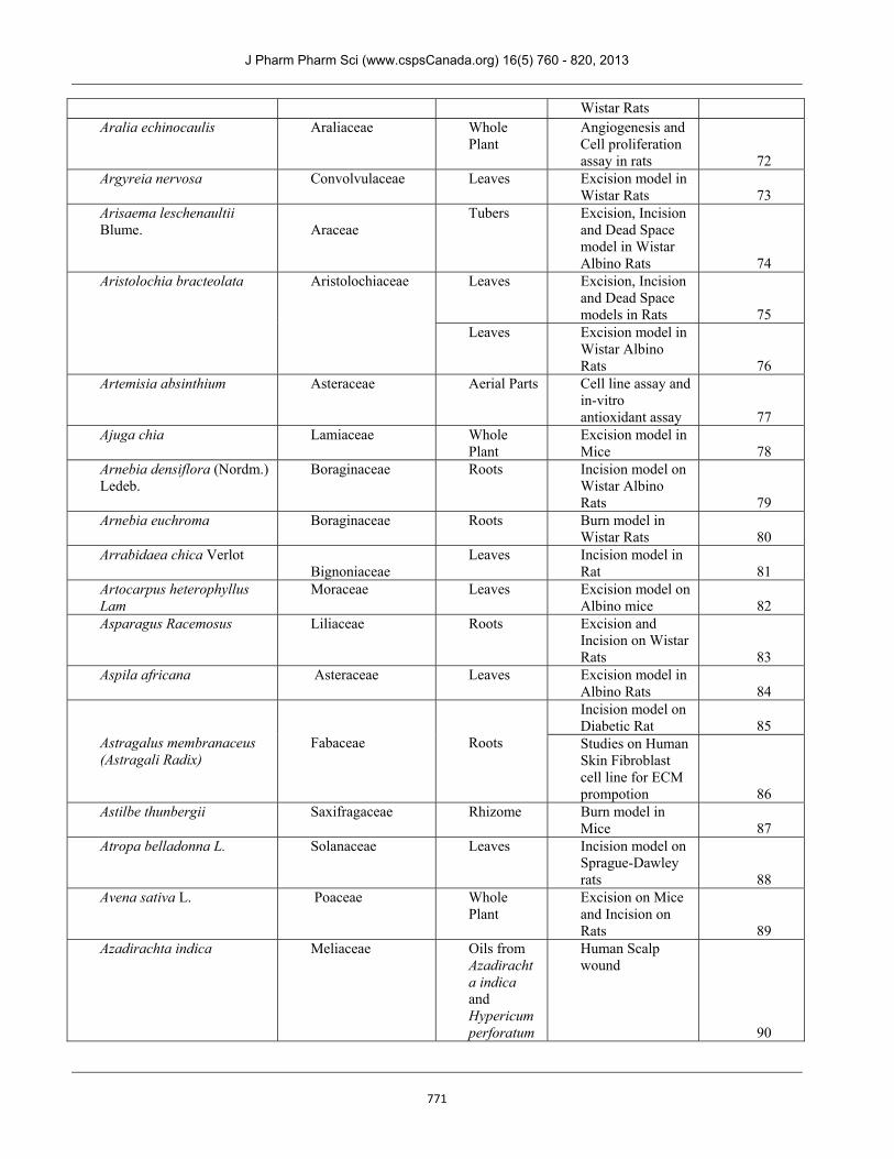
Wistar Rats Aralia echinocaulis Araliaceae
Whole Plant
Angiogenesis and Cell proliferation assay in rats 72
Argyreia nervosa Convolvulaceae
Leaves Excision model in Wistar Rats 73
Arisaema leschenaultii Blume. Araceae
Tubers Excision, Incision and Dead Space model in Wistar Albino Rats 74
Aristolochia bracteolata Aristolochiaceae
Leaves Excision, Incision and Dead Space models in Rats 75
Leaves Excision model in Wistar Albino Rats 76
Artemisia absinthium Asteraceae Aerial Parts Cell line assay and in-vitro antioxidant assay 77
Ajuga chia Lamiaceae Whole Plant
Excision model in Mice 78
Arnebia densiflora (Nordm.) Ledeb.
Boraginaceae Roots Incision model on Wistar Albino Rats 79
Arnebia euchroma Boraginaceae
Roots Burn model in Wistar Rats 80
Arrabidaea chica Verlot Bignoniaceae
Leaves Incision model in Rat 81
Artocarpus heterophyllus Lam
Moraceae Leaves Excision model on Albino mice 82
Asparagus Racemosus Liliaceae Roots Excision and Incision on Wistar Rats 83
Aspila africana Asteraceae Leaves Excision model in Albino Rats 84
Astragalus membranaceus (Astragali Radix)
Fabaceae
Roots
Incision model on Diabetic Rat 85 Studies on Human Skin Fibroblast cell line for ECM prompotion 86
Astilbe thunbergii Saxifragaceae Rhizome Burn model in Mice 87
Atropa belladonna L. Solanaceae Leaves Incision model on Sprague-Dawley rats 88
Avena sativa L. Poaceae Whole Plant
Excision on Mice and Incision on Rats 89
Azadirachta indica Meliaceae Oils from Azadirachta indica and Hypericum perforatum
Human Scalp wound
90
J Pharm Pharm Sci (www.cspsCanada.org) 16(5) 760 - 820, 2013
772
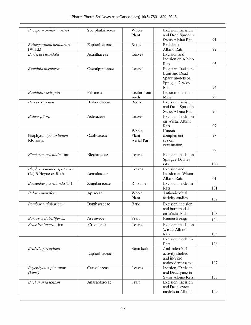
Bacopa monnieri wettest Scorphulariaceae Whole Plant
Excision, Incision and Dead Space in Swiss Albino Rat 91
Baliospermum montanum (Willd.)
Euphorbiaceae Roots Excision on Albino Rats 92
Barleria cuspidata Acanthaceae Leaves Excision and Incision on Albino Rats 93
Bauhinia purpurea Caesalpiniaceae Leaves Excision, Incision, Burn and Dead Space models on Sprague Dawley Rats 94
Bauhinia variegata Fabaceae Lectin from seeds
Incision model in Mice 95
Berberis lycium Berberidaceae Roots Excision, Incision and Dead Space in Swiss Albino Rat 96
Bidens pilosa Asteraceae Leaves Excision model on on Wistar Albino Rats 97
Biophytum petersianum Klotzsch.
Oxalidaceae
Whole Plant
Human complement system exvaluation
98 Aerial Part
99 Blechnum orientale Linn Blechnaceae Leaves Excision model on
Sprague-Dawley rats 100
Blepharis maderaspatensis (L.) B.Heyne ex Roth. Acanthaceae
Leaves Excision and Incision on Wistar Albino Rats 61
Boesenbergia rotunda (L.) Zingiberaceae Rhizome Excision model in Rats 101
Bolax gummifera Apiaceae Whole Plant
Anti-microbial activity studies 102
Bombax malabaricum Bombacaceae Bark Excision, incision and burn models on Wistar Rats 103
Borassus flabellifer L. Arecaceae Fruit Human Beings 104 Brassica juncea Linn Cruciferae Leaves Excision model on
Wistar Albino Rats 105
Bridelia ferruginea
Euphorbiaceae
Stem bark
Excision model in Rats 106 Anti-microbial activity studies and in-vitro antioxidant assay 107
Bryophyllum pinnatum (Lam.)
Crassulaceae Leaves Incision, Excision and Deadspace in Swiss Albino Rats 108
Buchanania lanzan Anacardiaceae Fruit Excision, Incision and Dead space models in Albino 109
J Pharm Pharm Sci (www.cspsCanada.org) 16(5) 760 - 820, 2013
773
Rats
Buddleja globosa Scrophulariaceae Leaves Fibroblast Assay 110 Bulbine frutescens Asphodelaceae
Leaves Excision and
Incision on Pigs 111 Bulbine natalensis
Asphodelaceae
Leaves Excision and Incision model in Pigs 111
Butea monosperma
Fabaceae
Bark Excision model in Rats 112
Stem bark Excision, Incision and Dead space in Wistar Rats 113
Flower Excision in Wistar Albino Rats in Wistar Albino Rats 114
Stem bark Excision, Incision and Dead space model in Wistar Albino Rats 115
Caesalpinia benthamiana Caesalpiniaceae
Root Bark In-vitro anti-oxidant assay 116
Calendula officinalis
Compositae
Flower Excision model in Rats 117
Flowers CAM Assay and Excision model in Wistar Rats 118
Calotropis gigantean
Apocynaceae
Flowers Excision and Incision models in Wistar Albino Rats 119
Root Bark Excision, Incision and Dead space models in Wistar Albino Rats 120
Leaves Excision and Incision models in Wistar Albino Rats 121
Latex Excision and Incision models in Wistar Albino Rats 122
Calotropis procera (Ait.) R. Br. Asclepiadaceae
Stem bark Excision model in Swiss Albino Mice 123
Camellia sinensis Theaceae
Leaves Incision in Wistar rats and Anti-oxidant assay 124
Capparis zeylanica Capparaceae
Roots Excision and incision model in Albino Rats 125
Whole plant
Excision model in Wistar Albino 126
J Pharm Pharm Sci (www.cspsCanada.org) 16(5) 760 - 820, 2013
774
Rats Carapa guianensis L.
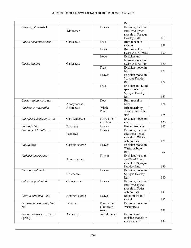
Meliaceae
Leaves Excision, Incision and Dead Space models in Sprague Dawley Rats 127
Carica candamarcensis Caricaceae Fruit Burn model in rodents 128
Carica papaya
Caricaceae
Latex Burn model in Swiss Albino mice 129
Roots Excision and Incision model in Swiss Albino Rats 130
Fruit Excision model in Mice 131
Leaves Excision model in Sprague Dawley Rats 132
Fruit Excision and Dead space models in Sprague Dawley Rats 133
Carissa spinarum Linn. Apocynaceae
Root Burn model in Mice 134
Carthamus oxycantha Asteraceae
Whole Plant
Irritant activity assessed on rabbit skin 135
Caryocar coriaceum Wittm. Caryocaraceae
Fixed oil of the plant
Excision model on mice 136
Cassia fistula Fabaceae Levaes Human wounds 137 Cassia occidentalis L.
Fabaceae
Leaves Excision, Incision and Dead Space models in Wistar Albino Rats 138
Cassia tora Caesulpinaceae Leaves Excision model in Wistar Albino Rats 76
Catharanthus roseus Apocynaceae
Flower Excision, Incision and Dead Space models in Sprague Dawley Rats 139
Cecropia peltata L. Urticaceae
Leaves Excision model in Sprague Dawley Rats 140
Celastrus paniculatus Celastraceae Leaves Excision, Incision and Dead space models in Swiss Albino Rats 141
Celosia argentea Linn. Amaranthaceae Leaves Rat burn wound model 142
Cenostigma macrophyllum Tul.
Fabaceae Fixed oil of plant from seeds
Excision model in Wistar Rats
143 Centaurea iberica Trev. Ex Spreng.
Asteraceae Aerial Parts Excision and Incision models in mice and rats 144
J Pharm Pharm Sci (www.cspsCanada.org) 16(5) 760 - 820, 2013
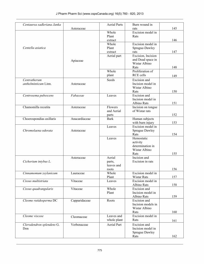
775
Centaurea sadleriana Janka Asteraceae
Aerial Parts Burn wound in rats 145
Centella asiatica
Apiaceae
Whole Plant extract
Excision model in Rats
146 Whole Plant extract
Excision model in Sprague-Dawley rats 147
Aerial part Excision, Incision and Dead space in Wistar Albino Rats 148
Whole plant
Proliferation of RCE cells 149
Centratherum anthelminticum Linn. Asteraceae
Seeds Excision and Incision model in Wistar Albino Rats 150
Centrosema pubescens Fabaceae Leaves Excision and Incision model in Albino Rats 151
Chamomilla recutita Asteraceae Flowers and Aerial parts
Incision on tongue of Wistar rats
152 Choerospondias axillaris Anacardiaceae Bark Human subjects
with burn injury 153 Chromolaena odorata
Asteraceae
Leaves Excision model in Sprague Dawley Rats 154
Leaves Hemostatic activity determination in Wistar Albino Rats 155
Cichorium intybus L.
Asteraceae Aerial parts, leaves and roots
Incision and Excision in rats
156 Cinnamomum zeylanicum
Lauraceae Whole Plant
Excision model in Wistar Rats 157
Cissus multistriata Vitaceae
Leaves Excision model in Albino Rats 158
Cissus quadrangularis Vitaceae Whole Plant
Excision and Incision model in Albino Rats 159
Cleome rutidosperma DC Capparidaceae Roots Excision and Incision models in Wistar Albino Rats 160
Cleome viscose Cleomaceae
Leaves and whole plant
Excision model in Rats 161
Clerodendron splendens G. Don
Verbenaceae Aerial Part Excision and Incision model in Sprague Dawley Rats 162
J Pharm Pharm Sci (www.cspsCanada.org) 16(5) 760 - 820, 2013
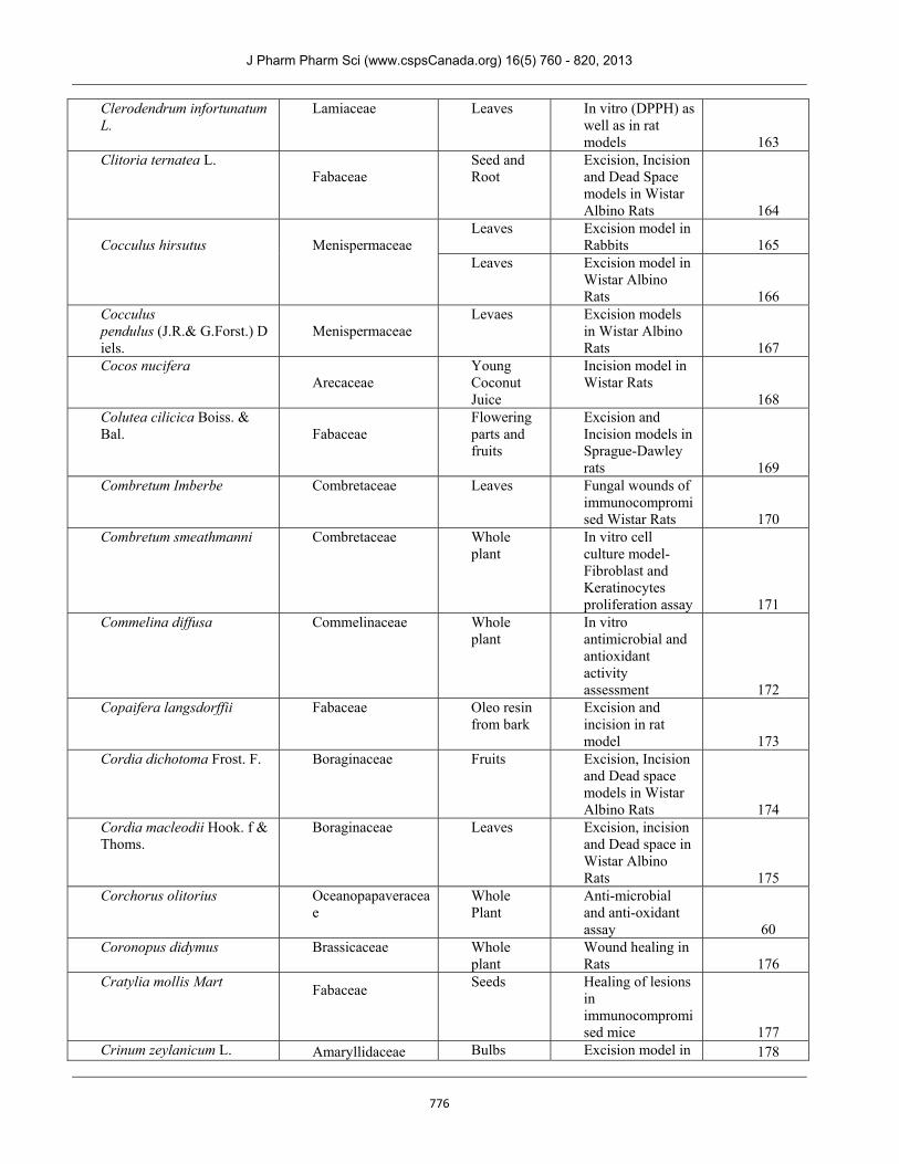
776
Clerodendrum infortunatum L.
Lamiaceae Leaves In vitro (DPPH) as well as in rat models 163
Clitoria ternatea L. Fabaceae
Seed and Root
Excision, Incision and Dead Space models in Wistar Albino Rats 164
Cocculus hirsutus
Menispermaceae
Leaves Excision model in Rabbits 165
Leaves Excision model in Wistar Albino Rats 166
Cocculus pendulus (J.R.& G.Forst.) Diels.
Menispermaceae
Levaes Excision models in Wistar Albino Rats 167
Cocos nucifera Arecaceae
Young Coconut Juice
Incision model in Wistar Rats
168 Colutea cilicica Boiss. & Bal. Fabaceae
Flowering parts and fruits
Excision and Incision models in Sprague-Dawley rats 169
Combretum Imberbe
Combretaceae Leaves Fungal wounds of immunocompromised Wistar Rats 170
Combretum smeathmanni Combretaceae Whole plant
In vitro cell culture model- Fibroblast and Keratinocytes proliferation assay 171
Commelina diffusa Commelinaceae Whole plant
In vitro antimicrobial and antioxidant activity assessment 172
Copaifera langsdorffii Fabaceae Oleo resin from bark
Excision and incision in rat model 173
Cordia dichotoma Frost. F. Boraginaceae Fruits Excision, Incision and Dead space models in Wistar Albino Rats 174
Cordia macleodii Hook. f & Thoms.
Boraginaceae Leaves Excision, incision and Dead space in Wistar Albino Rats 175
Corchorus olitorius Oceanopapaveraceae
Whole Plant
Anti-microbial and anti-oxidant assay 60
Coronopus didymus Brassicaceae
Whole plant
Wound healing in Rats 176
Cratylia mollis Mart
Fabaceae
Seeds Healing of lesions in immunocompromised mice 177
Crinum zeylanicum L. Amaryllidaceae Bulbs Excision model in 178
J Pharm Pharm Sci (www.cspsCanada.org) 16(5) 760 - 820, 2013
777
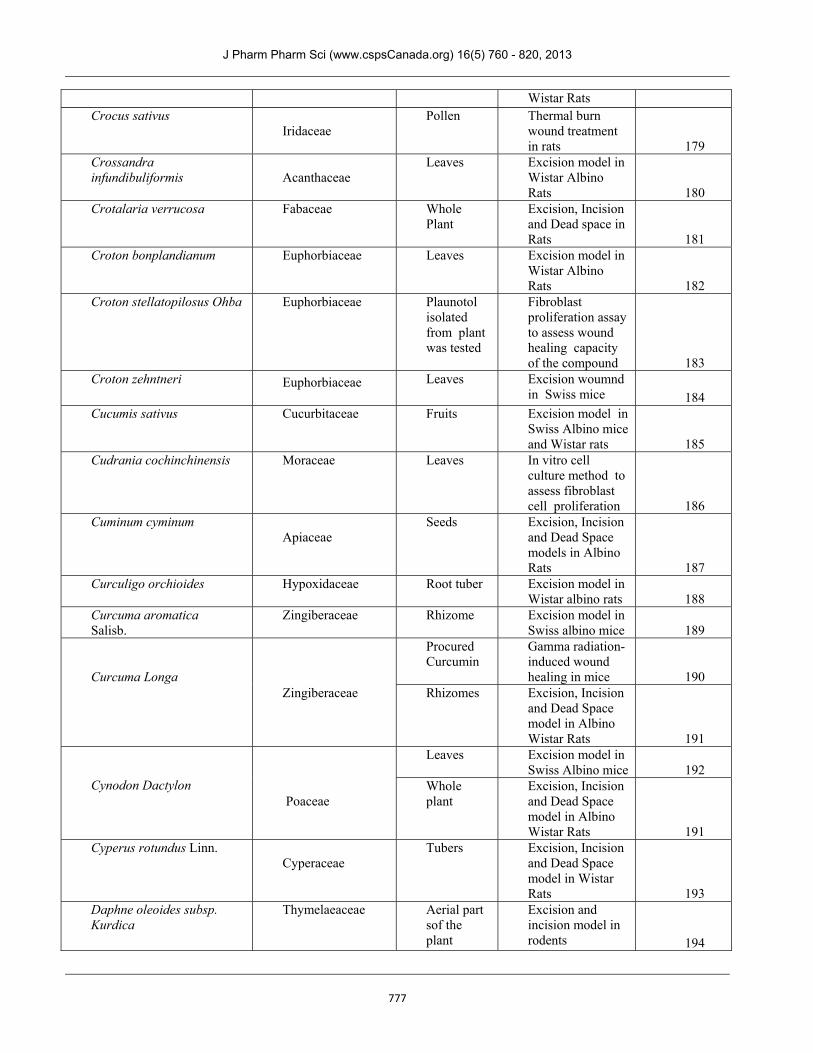
Wistar Rats Crocus sativus
Iridaceae
Pollen Thermal burn wound treatment in rats 179
Crossandra infundibuliformis Acanthaceae
Leaves Excision model in Wistar Albino Rats 180
Crotalaria verrucosa Fabaceae Whole Plant
Excision, Incision and Dead space in Rats 181
Croton bonplandianum Euphorbiaceae Leaves Excision model in Wistar Albino Rats 182
Croton stellatopilosus Ohba Euphorbiaceae
Plaunotol isolated from plant was tested
Fibroblast proliferation assay to assess wound healing capacity of the compound 183
Croton zehntneri Euphorbiaceae
Leaves Excision woumnd in Swiss mice 184
Cucumis sativus
Cucurbitaceae
Fruits Excision model in Swiss Albino mice and Wistar rats 185
Cudrania cochinchinensis
Moraceae
Leaves In vitro cell culture method to assess fibroblast cell proliferation 186
Cuminum cyminum Apiaceae
Seeds Excision, Incision and Dead Space models in Albino Rats 187
Curculigo orchioides Hypoxidaceae Root tuber Excision model in Wistar albino rats 188
Curcuma aromatica Salisb.
Zingiberaceae Rhizome Excision model in Swiss albino mice 189
Curcuma Longa
Zingiberaceae
Procured Curcumin
Gamma radiation-induced wound healing in mice 190
Rhizomes Excision, Incision and Dead Space model in Albino Wistar Rats 191
Cynodon Dactylon
Poaceae
Leaves Excision model in Swiss Albino mice 192
Whole plant
Excision, Incision and Dead Space model in Albino Wistar Rats 191
Cyperus rotundus Linn. Cyperaceae
Tubers Excision, Incision and Dead Space model in Wistar Rats 193
Daphne oleoides subsp. Kurdica
Thymelaeaceae Aerial part sof the plant
Excision and incision model in rodents 194
J Pharm Pharm Sci (www.cspsCanada.org) 16(5) 760 - 820, 2013
778
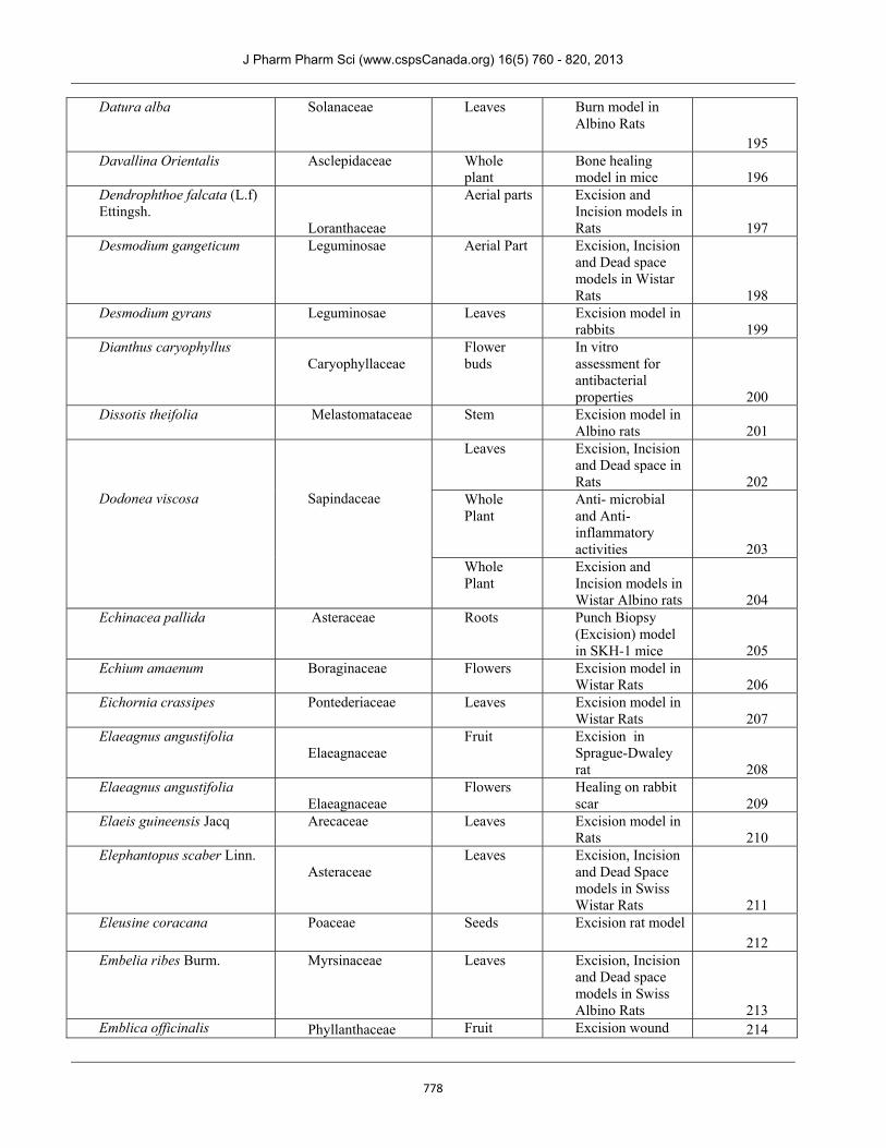
Datura alba
Solanaceae Leaves Burn model in Albino Rats 195
Davallina Orientalis Asclepidaceae
Whole plant
Bone healing model in mice 196
Dendrophthoe falcata (L.f) Ettingsh.
Loranthaceae
Aerial parts Excision and Incision models in Rats 197
Desmodium gangeticum Leguminosae Aerial Part Excision, Incision and Dead space models in Wistar Rats 198
Desmodium gyrans Leguminosae Leaves Excision model in rabbits 199
Dianthus caryophyllus Caryophyllaceae
Flower buds
In vitro assessment for antibacterial properties 200
Dissotis theifolia Melastomataceae
Stem Excision model in Albino rats 201
Dodonea viscosa
Sapindaceae
Leaves Excision, Incision and Dead space in Rats 202
Whole Plant
Anti- microbial and Anti- inflammatory activities 203
Whole Plant
Excision and Incision models in Wistar Albino rats 204
Echinacea pallida Asteraceae
Roots Punch Biopsy (Excision) model in SKH-1 mice 205
Echium amaenum Boraginaceae Flowers Excision model in Wistar Rats 206
Eichornia crassipes Pontederiaceae Leaves Excision model in Wistar Rats 207
Elaeagnus angustifolia Elaeagnaceae
Fruit Excision in Sprague-Dwaley rat 208
Elaeagnus angustifolia Elaeagnaceae
Flowers Healing on rabbit scar 209
Elaeis guineensis Jacq Arecaceae
Leaves Excision model in Rats 210
Elephantopus scaber Linn. Asteraceae
Leaves Excision, Incision and Dead Space models in Swiss Wistar Rats 211
Eleusine coracana Poaceae Seeds Excision rat model
212 Embelia ribes Burm. Myrsinaceae Leaves Excision, Incision
and Dead space models in Swiss Albino Rats 213
Emblica officinalis Phyllanthaceae Fruit Excision wound 214
J Pharm Pharm Sci (www.cspsCanada.org) 16(5) 760 - 820, 2013
779
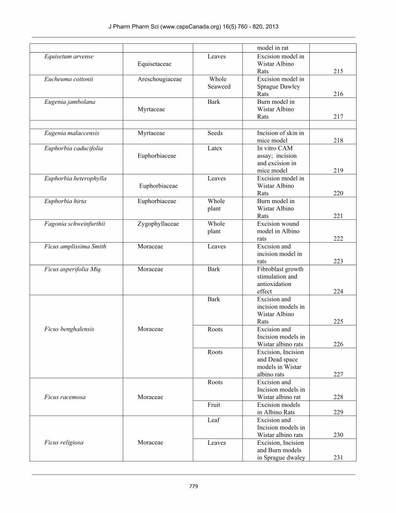
model in rat Equisetum arvense
Equisetaceae
Leaves Excision model in Wistar Albino Rats 215
Eucheuma cottonii Areschougiaceae Whole Seaweed
Excision model in Sprague Dawley Rats 216
Eugenia jambolana Myrtaceae
Bark Burn model in Wistar Albino Rats 217
Eugenia malaccensis Myrtaceae
Seeds Incision of skin in
mice model 218 Euphorbia caducifolia
Euphorbiaceae
Latex In vitro CAM assay; incision and excision in mice model 219
Euphorbia heterophylla Euphorbiaceae
Leaves Excision model in Wistar Albino Rats 220
Euphorbia hirta Euphorbiaceae Whole plant
Burn model in Wistar Albino Rats 221
Fagonia schweinfurthii Zygophyllaceae Whole plant
Excision wound model in Albino rats 222
Ficus amplissima Smith Moraceae Leaves Excision and incision model in rats 223
Ficus asperifolia Miq.
Moraceae Bark Fibroblast growth stimulation and antioxidation effect 224
Ficus benghalensis
Moraceae
Bark Excision and incision models in Wistar Albino Rats 225
Roots Excision and Incision models in Wistar albino rats 226
Roots Excision, Incision and Dead space models in Wistar albino rats 227
Ficus racemosa
Moraceae
Roots Excision and Incision models in Wistar albino rat 228
Fruit Excision models in Albino Rats 229
Ficus religiosa
Moraceae
Leaf Excision and Incision models in Wistar albino rats 230
Leaves Excision, Incision and Burn models in Sprague dwaley 231
J Pharm Pharm Sci (www.cspsCanada.org) 16(5) 760 - 820, 2013
780
rats
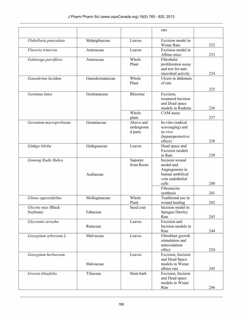
Flabellaria paniculata Malpighiaceae
Leaves Excision model in Wistar Rats 232
Flaveria trinervia Asteraceae
Leaves Excision model in Albino mice 233
Galinsoga parviflora Asteraceae Whole Plant
Fibrobalst proliferation assay and test for anti-microbial activity 234
Ganoderma lucidum Ganodermataceae Whole Plant
Ulcers in abdomen of rats
235 Gentiana lutea Gentianaceae Rhizome
Excision, resutured Incision and Dead space models in Rodents 236
Whole plant
CAM assay 237
Geranium macroprrhizum Geraniaceae Above and underground parts
In-vitro (radical scavenging) and in-vivo (hepatoprotective effect) 238
Ginkgo biloba Ginkgoaceae Leaves Dead space and Excision models in Rats 239
Ginseng Radix Rubra
Araliaceae
Saponin from Roots
Incision wound model and Angiogenesis in human umbilical vein endothelial cells 240 Fibronectin synthesis 241
Glinus oppositifolius Molluginaceae
Whole Plant
Traditional use in wound healing 242
Glycine max (Black Soybean) Fabaceae
Seed coat Incision model in Sprague Dawley Rats 243
Glycosmis arreabo Rutaceae
Leaves Excision and Incision models in Rats 244
Gossypium arboreum L. Malvaceae Leaves Fibroblast growth stimulation and antioxidation effect 224
Gossypium herbaceum
Malvaceae
Leaves Excision, Incision and Dead Space models in Wistar albino rats 245
Grewia tiliaefolia Tiliaceae Stem bark Excision, Incision and Dead space models in Wistar Rats 246
J Pharm Pharm Sci (www.cspsCanada.org) 16(5) 760 - 820, 2013
781
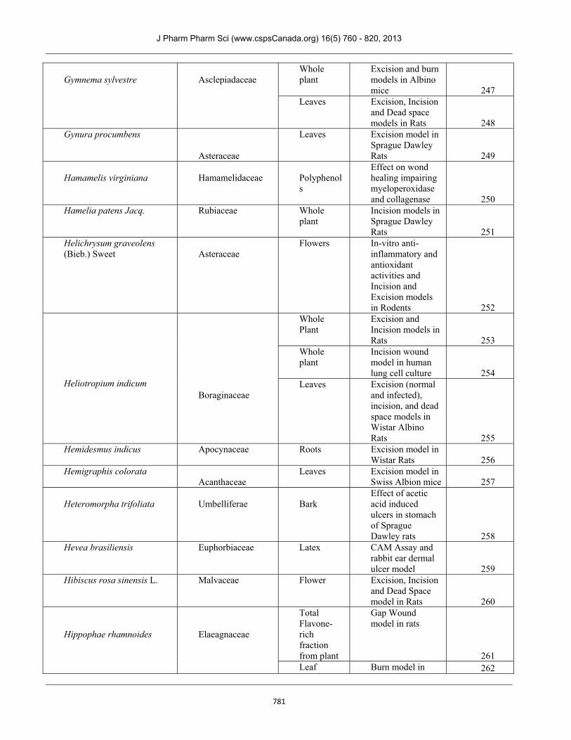
Gymnema sylvestre
Asclepiadaceae
Whole plant
Excision and burn models in Albino mice 247
Leaves Excision, Incision and Dead space models in Rats 248
Gynura procumbens
Asteraceae
Leaves Excision model in Sprague Dawley Rats 249
Hamamelis virginiana
Hamamelidaceae
Polyphenols
Effect on wond healing impairing myeloperoxidase and collagenase 250
Hamelia patens Jacq. Rubiaceae Whole plant
Incision models in Sprague Dawley Rats 251
Helichrysum graveolens (Bieb.) Sweet Asteraceae
Flowers In-vitro anti-inflammatory and antioxidant activities and Incision and Excision models in Rodents 252
Heliotropium indicum
Boraginaceae
Whole Plant
Excision and Incision models in Rats 253
Whole plant
Incision wound model in human lung cell culture 254
Leaves Excision (normal and infected), incision, and dead space models in Wistar Albino Rats 255
Hemidesmus indicus Apocynaceae Roots Excision model in Wistar Rats 256
Hemigraphis colorata Acanthaceae
Leaves Excision model in Swiss Albion mice 257
Heteromorpha trifoliata
Umbelliferae
Bark
Effect of acetic acid induced ulcers in stomach of Sprague Dawley rats 258
Hevea brasiliensis Euphorbiaceae Latex CAM Assay and rabbit ear dermal ulcer model 259
Hibiscus rosa sinensis L. Malvaceae
Flower Excision, Incision and Dead Space model in Rats 260
Hippophae rhamnoides
Elaeagnaceae
Total Flavone-rich fraction from plant
Gap Wound model in rats
261 Leaf Burn model in 262
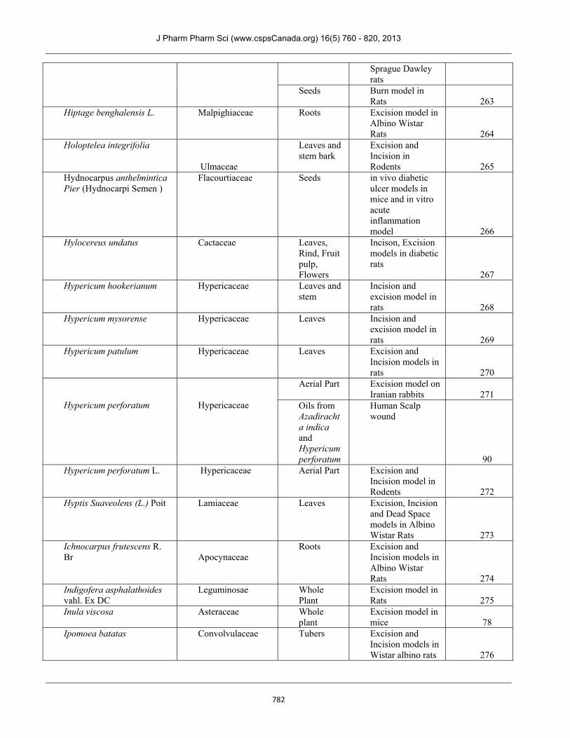
J Pharm Pharm Sci (www.cspsCanada.org) 16(5) 760 - 820, 2013
782
Sprague Dawley rats
Seeds Burn model in Rats 263
Hiptage benghalensis L. Malpighiaceae Roots Excision model in Albino Wistar Rats 264
Holoptelea integrifolia
Ulmaceae
Leaves and stem bark
Excision and Incision in Rodents 265
Hydnocarpus anthelmintica Pier (Hydnocarpi Semen )
Flacourtiaceae Seeds in vivo diabetic ulcer models in mice and in vitro acute inflammation model 266
Hylocereus undatus Cactaceae Leaves, Rind, Fruit pulp, Flowers
Incison, Excision models in diabetic rats
267 Hypericum hookerianum Hypericaceae Leaves and
stem Incision and excision model in rats 268
Hypericum mysorense Hypericaceae Leaves Incision and excision model in rats 269
Hypericum patulum Hypericaceae Leaves Excision and Incision models in rats 270
Hypericum perforatum
Hypericaceae
Aerial Part Excision model on Iranian rabbits 271
Oils from Azadirachta indica and Hypericum perforatum
Human Scalp wound
90 Hypericum perforatum L. Hypericaceae
Aerial Part Excision and Incision model in Rodents 272
Hyptis Suaveolens (L.) Poit Lamiaceae
Leaves Excision, Incision and Dead Space models in Albino Wistar Rats 273
Ichnocarpus frutescens R. Br Apocynaceae
Roots Excision and Incision models in Albino Wistar Rats 274
Indigofera asphalathoides vahl. Ex DC
Leguminosae
Whole Plant
Excision model in Rats 275
Inula viscosa Asteraceae Whole plant
Excision model in mice 78
Ipomoea batatas Convolvulaceae Tubers Excision and Incision models in Wistar albino rats 276
J Pharm Pharm Sci (www.cspsCanada.org) 16(5) 760 - 820, 2013
783
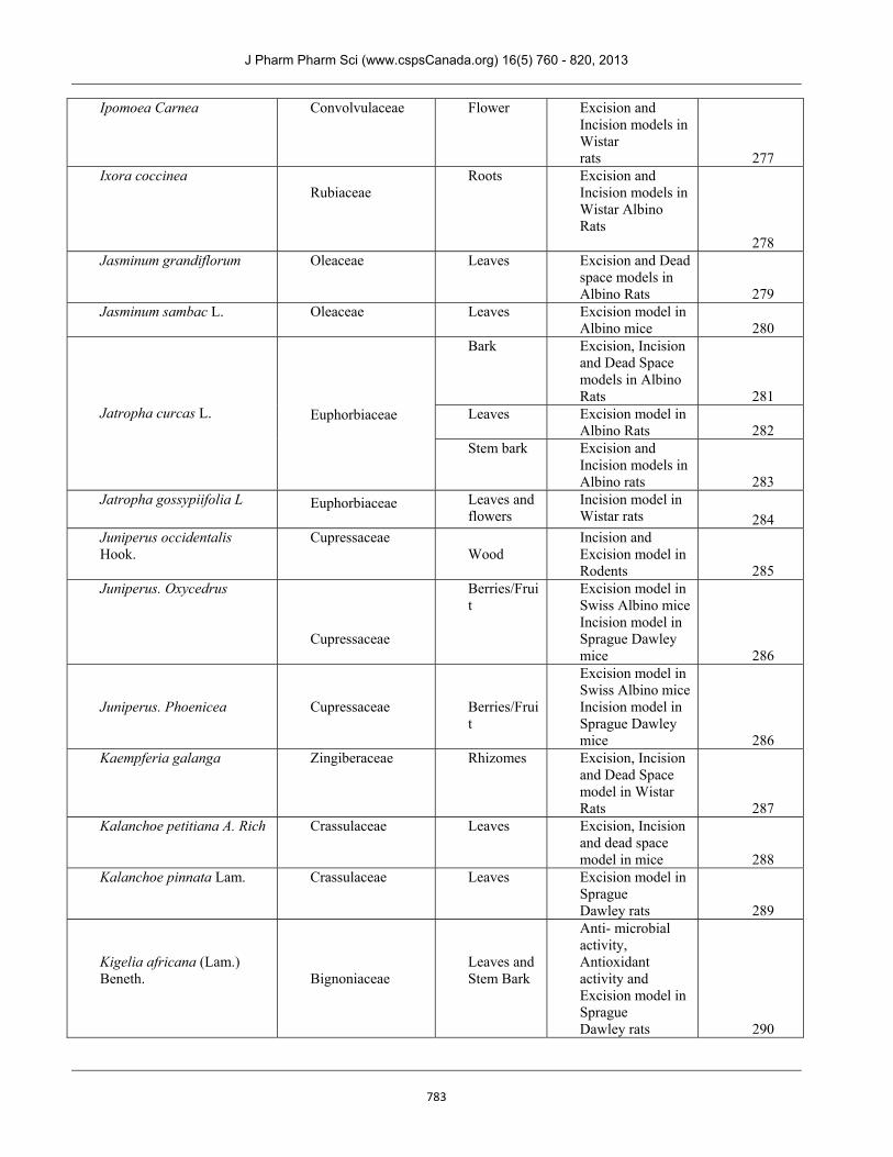
Ipomoea Carnea Convolvulaceae
Flower Excision and Incision models in Wistar rats 277
Ixora coccinea Rubiaceae
Roots Excision and Incision models in Wistar Albino Rats 278
Jasminum grandiflorum Oleaceae Leaves Excision and Dead space models in Albino Rats 279
Jasminum sambac L. Oleaceae Leaves Excision model in Albino mice 280
Jatropha curcas L.
Euphorbiaceae
Bark Excision, Incision and Dead Space models in Albino Rats 281
Leaves Excision model in Albino Rats 282
Stem bark Excision and Incision models in Albino rats 283
Jatropha gossypiifolia L Euphorbiaceae
Leaves and flowers
Incision model in Wistar rats 284
Juniperus occidentalis Hook.
Cupressaceae
Wood
Incision and Excision model in Rodents 285
Juniperus. Oxycedrus
Cupressaceae
Berries/Fruit
Excision model in Swiss Albino mice Incision model in Sprague Dawley mice 286
Juniperus. Phoenicea Cupressaceae
Berries/Fruit
Excision model in Swiss Albino mice Incision model in Sprague Dawley mice 286
Kaempferia galanga Zingiberaceae
Rhizomes Excision, Incision and Dead Space model in Wistar Rats 287
Kalanchoe petitiana A. Rich Crassulaceae
Leaves Excision, Incision and dead space model in mice 288
Kalanchoe pinnata Lam. Crassulaceae
Leaves Excision model in Sprague Dawley rats 289
Kigelia africana (Lam.) Beneth. Bignoniaceae
Leaves and Stem Bark
Anti- microbial activity, Antioxidant activity and Excision model in Sprague Dawley rats 290
J Pharm Pharm Sci (www.cspsCanada.org) 16(5) 760 - 820, 2013
784
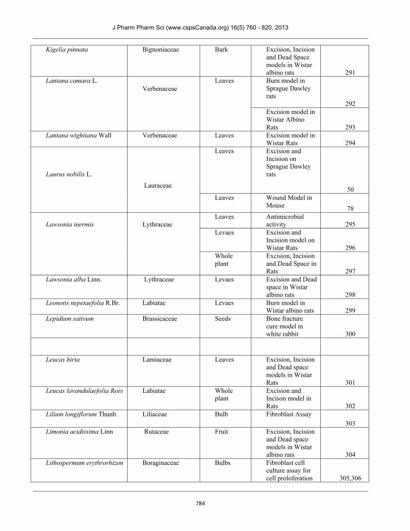
Kigelia pinnata Bignoniaceae
Bark Excision, Incision and Dead Space models in Wistar albino rats 291
Lantana camara L. Verbenaceae
Leaves Burn model in Sprague Dawley rats 292 Excision model in Wistar Albino Rats 293
Lantana wightiana Wall Verbenaceae
Leaves Excision model in Wistar Rats 294
Laurus nobilis L.
Lauraceae
Leaves Excision and Incision on Sprague Dawley rats 50
Leaves Wound Model in Mouse 78
Lawsonia inermis
Lythraceae
Leaves Antimicrobial activity 295
Levaes Excision and Incision model on Wistar Rats 296
Whole plant
Excision, Incision and Dead Space in Rats 297
Lawsonia alba Linn. Lythraceae
Levaes Excision and Dead space in Wistar albino rats 298
Leonotis nepetaefolia R.Br. Labiatae
Levaes Burn model in Wistar albino rats 299
Lepidium sativum Brassicaceae Seeds Bone fracture cure model in white rabbit 300
Leucas hirta Lamiaceae Leaves Excision, Incision
and Dead space models in Wistar Rats 301
Leucas lavandulaefolia Rees Labiatae Whole plant
Excision and Incison model in Rats 302
Lilium longiflorum Thunb. Liliaceae Bulb Fibroblast Assay
303 Limonia acidissima Linn Rutaceae Fruit Excision, Incision
and Dead space models in Wistar albino rats 304
Lithospermum erythrorhizon Boraginaceae Bulbs Fibroblast cell culture assay for cell proloferation 305,306
J Pharm Pharm Sci (www.cspsCanada.org) 16(5) 760 - 820, 2013
785
Litsea glutinosa Lauraceae Leaves Excision and incison models in Wistar rats 307
Liquidambar orientalis Mill Altingiaceae Latex Excision model in Pigs 308
Lonicera japonica Caprifoliaceae Flowering aerial parts
Excision model in Wistar Rats 309
Lygodium flexuosum Lygodiaceae Leaves Excision, Incision and Dead space model in Rats 310
Lycopodium serratum Lycopodiaceae Leaves Excision, Incision and Dead space models in Wistar Rats 311
Mallotus oppositifolia Euphorbiaceae
Whole Plant
In-vitro anti-microbial and anti-oxidant assay 60
Malva sylvestris
Malvaceae
Flowers Burn in Wistar rats 80
Flowers Excision model in Wistar rats 312
Flowers Excision model in Wistar rats 313
Margaritaria discoidea Phyllanthaceae Stem bark Antimicrobial and free radical scavenging activity 314
Martynia annua Linn. Martyniaccae Leaves Excision and Incision models in Wistar Albino Rats 315
Matricaria chamomilla Asteraceae Whole plant
Incision model in Albino Rats 316
Matricaria recutita L.
Asteraceae
Flowers
Excison, Incision and Dead space model in rats 317 Human Model 318
Melastoma malabathricum Melastomataceae
Leaves Excision and Incision model in Albino Wistar Rats 319
Melia azedarach L. Meliaceae
Leaves Excision model in alloxan induced diabetic Wistar Albino rats 320
Memecylon edule Roxb. Melastomataceae
Leaves In-vitro and in-vivo in Mice 321
Michauxia nuda Campanulaceae Root In-vitro anti-oxidant assay and Incision, Excision models in Mice and Rats 322
Michauxia tchihatchewii Campanulaceae Whole Plant
In-vitro anti-oxidant assay and Incision, Excision 322
J Pharm Pharm Sci (www.cspsCanada.org) 16(5) 760 - 820, 2013
786
models in Mice and Rats
Michelia champaca
Magnoliaceae
Whole plant
Excision, Incision and Dead space models in Wistar Rats 323
Flowers Burn model in Wistar Rats 324
Mimosa pudica
Leguminosae
Roots Excision, incision, burn and dead space models in Sprague Dawley Rats 325
Leaves Excision and Incision models in Albino Wistar Rats 326
Roots Excision and Incision models in Rats 327
Mimosa tenuiflora (Willd.) Poirett Mimosaceae
Bark Fibroblast assay and Keratinocytes assay 328
Mimusops elengi Sapotaceae Bark Excision, Incision and Dead space models in Albino mice 329
Momordica charantia L Cucurbitaceae
Fruit Excision, Incision and Dead space model in Rats 330,331
Momordica cochinchinensis (Lour.) (Momordicae Semen)
Cucurbitaceae
Seeds Cutaneous wound healing in Mice
332 Morinda citrifolia L. Rubiaceae
Leaves Excision, Incision and Dead Space models in Albino Wistar rats 333
Leaves Excision and Dead space models in Sprague Dawley Rats 334
Morinda pubescens JE Smith
Rubiaceae
Fruit Cutaneous wound healing in Rats 335
Moringa oleifera Moringaceae
Leaves Excision, Incision and Dead Space models in Rats 336
Bark Excision, Incision and Dead space models in Wistar Albino Rats 337
Leaves Excision, Incision and Dead space model in Wistar Rats 338
Murraya koenigii Rutaceae Leaves Excision model in 339
J Pharm Pharm Sci (www.cspsCanada.org) 16(5) 760 - 820, 2013
787
Albino Rats Musa sapientum Musaceae Fruit peel Incision model in
Wistar rats 340 Musa sapientum var. paradisiacal
Musaceae Fruit pulp Excision, Incision and Dead Space models in Rats 341
Mussaenda frondosa Rubiaceae Leaves Excision, Incision and Dead Space models in Albino Rats 342
Myristica andamanica Myristicaceae Aerial Part Excision model in Swiss Albino Mice 343
Napoleona imperialis Lecythidaceae Leaves Excision model in Guinea Pigs 344
Naravelia zeylanica Ranunculaceae
Leaves Excision, Incision and Dead Space models in Wistar Rats 345
Nauclea latifolia Rubiaceae
Stem bark Excision model in Rabbits 346
Nigella sativa L. Ranunculaceae
Seeds Burn model in Wistar Rats 347
Nyctanthes arbor-trisitis (Linn.)
Oleaceae Leaves Excision and incision models in Wistar Albino Rats 348
Ocimum basilicum Lamiaceae
Leaves Excision model in Wistar Albino Rats 349
Ocimum gratissimum linn. Lamiaceae
Essential oil
Excision and Incision on Albino rabbits 350
Leaf Excision wounds in Wistar rats 351
Ocimum kilimandscharicum Lamiaceae
Leaves Excision, Incision and Dead Space models in Albino Wistar rats 352
Ocimum sanctum
Lamiaceae
Leaves Excision and Incision in Albino Rats 353
Leaves Excision, Incision and Dead Space models in Wistar Albino rats 354
Leaves Excision, Incision and Dead Space in Albino Rats 355
Leaves Excision model in Wistar Albino Rats 356
Ocimum suave Labiatae Leaves Excision model in Wistar Albino rats 97
Shoot and In-vitro anti- 357
J Pharm Pharm Sci (www.cspsCanada.org) 16(5) 760 - 820, 2013
788
Olea europaea
Oleaceae
Fruits oxidant property Leaves Holes drilled in
articular cartilage of rabbits 358
Leaves and Fruits
Incision and Excision model in Rodents 359
Oncidium flexuosum Sims. Orchidaceae Leaves Incision model in Wistar Rats 360
Onosma argentatum Boraginaceae Roots Fibroblast growth 361 Ophioglossum vulgatum L
Ophioglossaceae Aerial parts Keratinocytes
assay 362 Opuntia ficus-indica Cactaceae
Stems Wound healing activity in Rats 363
Cladodes Incision in Wistar Rats 364
Cladodes Full thickness wound in Rats 365
Orbignya phalerata Arecaceae
Mesocarp from ripe coconut
Incision in Wistar rats 366 Incision in Wistar rats 367
Origanum vulgare Verbenaceae
Leaves Surgical Excision wounds in humans 368
Oxalis corniculata Oxalidaceae
Whole plant
Excision, Incision and Dead Space models in Rats
369 Panax ginseng Araliaceae
Leaves Excision wound in
mice 370 Parapiptadenia rigida Fabaceae
Catechin derivative from bark
Keartinocyte scratch assay
371 Parieteria diffusa Urticaceae
Whole plant
Incision wound model in mice 78
Parkia biglobosa Fabaceae
Stem Bark Anti-mirobial acidity studies 107
Parquetina nigrescens Asclepiadaceae
Roots, Leaves and Stem
Used in Traditional Practicies for wound healing 372
Passiflora edulis
Passifloraceae
Leaves Incision in Wistar rats 373
Leaves Excision in Wistar rats 374
Paullinia pinnata L. Sapindaceae
Roots In vitro oblast assay using 142 cell line 375
Pedilanthus tithymaloides Euphorbiaceae
Leaves Excision model in ICR mice 376
Pentas lanceolata Rubiaceae
Flower Excision model in Rats 377
Persea Americana Lauraceae Fruit Excision and Dead Space models in rats 378
J Pharm Pharm Sci (www.cspsCanada.org) 16(5) 760 - 820, 2013
789
Petiveria alliacea Phytolaccaceae Whole plants
Keratinocytes Assay and antibacterial assay 234
Phyllanthus muellerianus Euphorbiaceae Leaves Dermal fibroblast and keratinocyte assay 171
Phyllanthus muellerianus (Kuntze) Excell.
Euphorbiaceae Stem bark Antimicrbobial activity 379
Leaves Fibroblast and Keratinocytes Assays 380
Phyllanthus niruri L. Euphorbiaceae Aerial parts Excision and Dead Space models in Albino rats 381
Picea abies Pinaceae Stem On human surgical wounds 382
Pinus halepensis Pinaceae Essential oils from cones and needles
Incisona and Excision model in Mice and Rats
383 Pinus pinea Pinaceae Essential
oils from cones and needles
Incisona and Excision model in Mice and Rats
383 Piper betle Piperaceae Leaves Wound healing in
Rabbits 384 Piper hayneanum Piperaceae Leaves,
Stem and roots
Excision model in infected rats
385 Pisonia grandis R. Br Nyctaginaceae
Leaves Excision and Incision in Wistar Rats 386
Pistacia atlantica Anacardiaceae Leaves and fruits
Excision model in Wistar Albino Rats 387
Pistacia khinjuk Anacardiaceae Leaves and fruits
Excision model in Wistar Albino Rats 387
Pistacia lentiscus Anacardiaceae Fruit Burn model in New-Zealand rabbits 388
Plagiochasma appendiculatum Aytoniaceae
Whole plant
Excision and Incision models in Sprague–Drawley Rats 389
Plagiochila beddomei Hepaticae
Thallus Excision and Incision models in Sprague Dawly rats 390
Plantago major Plantaginaceae Leaves Excision model in Sprague Dawley Rats 391
Plectranthus tenuiflorus Euphorbiaceae Leaves Excision model in Wistar Rats and Fibroblast assay 392
J Pharm Pharm Sci (www.cspsCanada.org) 16(5) 760 - 820, 2013
790
Plumbago zeylanica L.
Plumbaginaceae
Whole Plant
Excision and Incision in Rats 25
Roots Excision model in Wistar Albino Rats 393
Polygonum barbatum Linn Polygonaceae Whole plant
Excision and incision model in Wistar rats 394
Polygonum cuspidatum Polygonaceae
Whole plant
Excision in Rats 395
Portulaca oleracea Portulacaceae
Aerial parts Excision model in Mice 396
Pouteria lucuma Sapotaceae Fruits In vivo studies on transgenic zebrafish larvae and CD-1 mice 397
Prosthechea michuacana Orchidaceae Bulbs Excision and Incision in Wistar Albino Rats 398
Pseudarthria viscida Fabaceae Whole plant
Excision model in Wistar Rats 399
Pterocarpus erinaceus Poir
Leguminosae-Papilionoideae
Bark Human complement system exvaluation 98
Pterocarpus marsupium Fabaceae
Heart Wood
Excision model in Wistar Rats 400
Pterocarpus santalinus linn Fabaceae
Wood Punch and burn model on diabetic and normal rats 401
Pterospermum acerifolium wild.
Sterculiaceae
Flower Excision model in Albino Wistar rats 402
Pueraria tuberose Fabaceae Tubers Excision and Incision model in Rats 403
Punica granatum
Lythraceae
Peel Excision model in Wistar Rats 404
Peels Excision model in Guinea Pigs 405
Flowers
Excision model in Wistar rats 30
Pycnanthus angolensis Myristicaceae
Leaves and Stem bark
Dermal fibroblast and keratinocyte assay 171
Pyrostegia venusta (Ker Gawl) Miers.
Bignoniaceae
Flower Excision and Incision models in Wistar Rats 406
Quercus infectoria Fagaceae
Leaves Excision, Incision and Dead Space in Wistar albino rats 407
Radix paeoniae Paeoniaceae Roots Excision, Incision and Dead space in Wistar albino rats 408
J Pharm Pharm Sci (www.cspsCanada.org) 16(5) 760 - 820, 2013
791
Radix Salviae Miltiorrhizae Lamiaceae
Roots Radial fracture healing in Wistar Rats 409
Ranunculus pedatus Ranunculaceae
Whole plant
Excision and Incision model in Rodents 410
Rehmannia glutinosa (Rehmanniae Radix)
Phrymaceae
Roots
Incision-Diabetic Rat 411 Studies on Human Skin Fibroblast cell line for ECM prompotion 85,86 Angiogenis studies in zebrafish embryos 412
Rheum officinale Baill. Polygonaceae
Emodin isolated from Roots
Excision model in Rats
413 Rhizophora mangle Rhizophoraceae Bark Open wounds in
Human beings 414 Rhodiola imbricate Crassulaceae Rhizome Excision model in
rats 415 Rosmarinus officinalis L. Lamiaceae Aerial parts Excision
cutaneous model in alloxan-induced diabetic BALB/c mice 416
Rubia cordifolia L. Rubiaceae Roots Excision in Albino Swiss mice 417
Rubus sanctus Rosaceae
Aerial parts Excision in Swiss albino mice and Incision in Sprague-Dawley rats 418
Rumex abyssinicus Polygonaceae
Leaves Antimicrobal and anti inflammatory activity 203
Rumex nervosus Polygonaceae
Leaves Antimicrobal and anti inflammatory activity 203
Salvia cryptantha Lamiaceae
Whole plant
Excision and Incision Rats and Mice 419
Salvia cyanescens Lamiaceae
Whole plant
Excision and Incision in Rats and Mice 419
Salvia hypoleuca Lamiaceae Whole plant
Burn model in Albino rats 420
Salvia splendens Lamiaceae
Leaves Excision and Incision in Albino Rats/ biochemical parameters 421
Sambucus ebulus L. Caprifoliaceae Leaves Incision and Excision in rats and mice 422
J Pharm Pharm Sci (www.cspsCanada.org) 16(5) 760 - 820, 2013
792
Schinus lentiscifolius Anacardiaceae Leaves Antibacterial and antifungal activity assay 423
Schinus molle Anacardiaceae Fruits Anti-microbial assay and fibroblast proliferation scratch assay 234
Schinus terebinthifolius raddi
Anacardiaceae Bark
Incision model in wistar rats 424 Incision model in Albino and wistar rats 425
Schrebera swietenioides Oleaceae Bark Dead space, Excision and Incision model in rodents 426
Scorzonera cana var. jacquiniana
Asteraceae Areial parts Incisona and Excision model in Mice and Rats 427
Scorzonera eriophora Asteraceae Areial parts Incisona and Excision model in Mice and Rats 427
Scrophularia nodosa Scrophulariaceae Seed pods Fibroblast proliferartion assay in humans cell lines 428
Semecarpus anacardium Anacardiaceae Stem bark Incision and Dead space model in wistar albino rats 429
Senna alata L Fabaceae Leaves Excision in rats 430 Sesamum indicum L Pedaliaceae Seeds Excision, incision,
dead space and burn in Wistar Albino Rats 431
Sesbania grandiflora
Leguminosae
Bark Excision in Wistar Albino Rats 432
Flower Excision and Incision in Wistar Albino Rats 433
Shorea robusta Dipterocarpaceae
Resin Excision and Incision in Wistar Albino Rats 434
Sida acuta Malvaceae Whole plant
Excision and Incision in Wistar Albino Rats 435
Sida spinosa Malvaceae Leaves Excision and Incision in Wistar Albino Rats 436
Siegesbeckia pubescens Asteraceae Whole plant
Excision and Incision in Rats 437
Solanum xanthocarpum Schrad and Wendl
Solanaceae
Fruits Excision and incison in Sprague Dawley Rats 438
Leaves Excision and 439
J Pharm Pharm Sci (www.cspsCanada.org) 16(5) 760 - 820, 2013
793
Incision in Wistar Albino Rats
Spathodea campanulata
Bignoniaceae
Stem bark
Excision in Sprague Dawley Rats 440 In vitro antimicrobial and antioxidant activity assessment 172
Sphaeranthus amaranthoides Burm.f.
Compositae
Whole plant
Excision model in rats 441
Sphaeranthus indicus
Asteraceae
Aerial Parts Excision in Guinea Pigs 442
Flower Excision and Incision in Albino Rats 443
Spondia mombin Anacardiaceae
Whole Plant
In-vitro anti-microbial and anti-oxidant assay 60
Stachys lavandulifolia Vahl Lamiaceae
Flowers Excision in Wistar Rats 312
Stevia rebaudiana Asteraceae Leaves Excision and Incision in Albino Mice 444
Stewartia koreana Theaceae Leaves CAM assay 445 Strobilanthes crispus Acanthaceae Leaves Excision model in
Sprague Dawley Rats 446
Strophanthus hispidus DC Apocynaceae Roots and leaves
In-vitro anti-oxidant assay and Excision model in rats 290
Strychnos pseudoquina ST. HILL
Loganiaceae Bark Incision model in Wistar Diabetic Rats 447
Stryphnodendron obovatum Benth.
Leguminosae Bark Cutaneous wound healing model in Wistar rats 448
Stryphnodendron polyphyllum Mart
Leguminosae Bark Cutaneous wound healing model in Wistar rats 448
Symphytum officinale L Boraginaceae Leaves Incision model in Rats 449
Symphytum x uplandicum NYMAN
Boraginaceae Aerial Parts Abrasions in Humans 450
Tagetes erecta
Asteraceae
Leaves Excision, Incision and Dead space in Albino Rats 148
Leaves Excision and Incision in Albino Rats 451
Whole plant
Excision and burn in Albino Mice 247
Tamarindus indica L. Fabaceae Seeds Excision in ICR 452
J Pharm Pharm Sci (www.cspsCanada.org) 16(5) 760 - 820, 2013
794
Mice
Tamarix aphylla Tamaricaceae
Leaves Excision in Wistar Rats 453
Tecomaria capensis Bignoniaceae
Leaves Excision, Incision and Dead Space in Rats 454
Tectona grandis
Lamiaceae
Leaves
Excision, Incisison, Burn and Dead Space in Rats 455
Tephrosia purpurea (Linn.) Pers.
Fabaceae Aerial part Excision, Incision and Dead space in Rats 456
Terminalia arjuna Combretaceae Bark Incision and Excision in Rats 457
Terminalia avicennioides
Combretaceae
Root bark
Excision and Incision in Rattus norvegicus rats 458
Terminalia bellirica Roxb. Combretaceae Fruit Excision and Incisionin Albino Rats 459
Terminalia chebula Combretaceae Bark Excision and Incision in Albino Rats 460
Terminalia coriacea {Roxb.}Wight & Arn
Combretaceae Stem bark Excision in Albino Wistar Rats 461
Thespesia populnea Malvaceae
Fruit Excision and Incision in Rats 462
Thymus kotschyanus
Lamiaceae
Aerial parts
In vitro assessment for antibacterial properties 200
Tinospora cordifolia Willd. Menispermaceae
Roots Excision and Incision in Albino mice
463
Toddalia Asiatica Linn. Rutaceae Stem bark Excision and Incision model in Wistar Rats
464 Tragia involucrata Euphorbiaceae Roots Excision in Rats 465 Tribulus terrestris
Zygophyllaceae
Leaves Excision, Incision and Burn in Albino Wistar Rats
466
Trichosanthes dioica Roxb. Cucurbitaceae
Fruit Excision and Incision in Rats
467
Tridax Procumbens L. Asteraceae
Leaves Excision in Mice 468
Trifolium canescens Fabaceae Aerial parts Incision and Excision models in Rodents
469
Typha domingensis Pers. Typhaceae Female flower inflorescence
Incision and Excision in Mice and Rats
470
J Pharm Pharm Sci (www.cspsCanada.org) 16(5) 760 - 820, 2013
795
Typha latifolia L. Typhaceae Fruit Dermal fibroblast and epidermal keratinocyte proliferation assay
471
Uncaria rhynchophylla Rubiaceae Roots Human Umbelical cell proliferation assay
472
Urena Lobata Malvaceae Leaves Anti-microbial and Anti-oxidant assay
473
Vaccinium macrocarpon Ericaceae Seeds Excision wound model in rats
474
Vanda roxburghii R.Br Orchidacea Whole plant
Excision wound model in rats
475
Verbascum mucronatum Lam.
Scrophulariaceae Flowers Excision and Incision model in mice and rats
476
Verbascum thapsus Scrophulariaceae Flower Excision in Rabbits 477
Verbena officinalis Verbenaceae Bark Excision in Rats 478 Vernonia arborea Buch.- Ham.
Asteraceae
Bark Excision, Incision and Dead Space in Wistar Rats
479
Vernonia arborea Hk.
Asteraceae
Leaves
Excision, Incision and Dead space in Wistar Rats
480
Vernonia scorpioides Asteraceae Leaves Incision in Guinea Pigs
481
Vinca rosea Apocynaceae
Leaves Excision in Diabetic Rats 482
Viscum Articulum Brm. Santalaceae Whole plant
Excision, Incision and Dead space in Albino Rats
483
Vitex altissima L.
Verbenaceae
Leaves
Excision, Incision and Dead space wound model in Sprague-Dawley rats
484
Vitex leucoxylon Verbenaceae
Stem Bark Incision in Rodents
485
Vitex negundo Verbenaceae
Leaves Excision and Incision in Wistar rats
486
Vitex trifolia L.
Lamiaceae
Leaves
Excision, Incision and Dead space wound model in Sprague-Dawley rats
484
Vitis vinifera
Vitaceae
Grape seed extract
Excision in Balb C mice 487 Excision in rats
474 Vitis Vitigenia Vitaceae Leaves Excision and
Incision in Wistar 488
J Pharm Pharm Sci (www.cspsCanada.org) 16(5) 760 - 820, 2013
796
Albino Rats Waltheria douradinha
Malvaceae
Whole plant
Fibrobalst proliferation assay and test for anti-microbial activity
234
Warbugia ugandensis Canellaceae Leaves Excision in Albino Rats 489
Wattakaka volubilis (L.f.) Stapf
Asclepiadaceae Leaves Excision, Incision and Dead space in Albino Rats
490
Wedelia biflora L. Asteraceae Leaves Excision, Incision model in Wistar Albino Rats
491
Wedelia trilobata L. Asteraceae Leaves In vitro anti-oxidant assays
492
Withania coagulans Solanaceae Fruit Excision contraction parameters in Diabetic rats
493
Wrightia arborea (Dennst.) Mabb
Apocynaceae Leaves Excision and Incision in Wistar Albino Rats
494
Wrightia tinctoria (Roxb) R. Br
Apocynaceae Leaves Excision and Incision in Wistar Albino Rats
495
Xanthium cavanillesii Asteraceae
Fruit
Fibroblast proliferation assay and Anti-microbial assay
234
Ximenia Americana Olacaceae
Leaves In vitro anti-oxidant assays and enzymes-inhibition assays
496
Zanthoxylum chalybeum Rutaceae
Roots and Leaves
Excision in Wistar Albino Rats
489
Zingiber officinale Zingiberaceae
Roots Abrasion in Male CD Hairless Rats 497
Ziziphus nummularia L. Rhamnaceae
Leaves Excision, Albino Rats 498
Zizyphus oenoplia Rhamnaceae Fruits Excision, Incision and Dead space, Albino Wistar Rats 499
*Wherever animal models were used, these have been reported as Excision, Incision, Burn and/or Dead space model, as applicable.
REFERENCES 1. Guo S and Pietro LA, Factors Affecting Wound
Healing, J Dent Res, 2010; 89(3), 219-229 2. Steven BL, DeGuzman L, Lee WP, Xu Y, Siegel
MW and Amento EP, One Systematic Administration of Transforming Growth Factor-beta1 Reverses Age or Glucocorticoid-Impared
Wound Healing, J Clinical Invest, 1993; 92, 2841-2849
3. Frank S, Hubner G, Breier G, Longaker MT, Greenhalgh DG and Werner S, Regulation of Vascular Endothelial Growth Factor Expression in Cultured Keratinocytes, The J Biological Chemistry, 1995; 270(21),12607-12613.
J Pharm Pharm Sci (www.cspsCanada.org) 16(5) 760 - 820, 2013
797
4. Frank S, Madlener M and Werner S, Transforming Growth Factors β1, β2 and β3 and their Receptors are differentially regulated during normal and impaired wound healing, J Biological Chemistry, 1996; 271(17),10188-10193
5. Palu A, Su C, Zhou BN, West B and Jensen J, Wound healing effects of noni (Morinda citrifolia L.) leaves: a mechanism involving its PDGF/A2A receptor ligand binding and promotion of wound closure, Pythother Res, 2010; 24(10), 1437-1441
6. Iyu D, Juttner M, Glen JR, White AE, Johnson AJ, Fox SC and Heptinstall S, PGE1 and PGE2 modify platelet function through different psotanoid receptors, Prostaglandins & other Lipid Mediators, 2011; 94, 9-16
7. Riccioti E and Fitzgerlad GA, Prostaglandins and Inflammation, Journal of the Amercian Heart Association, 2011; 31, 986-1000
8. Anitua E.,Andia I.,Ardanza B.,Nurden P. and Narden A.T. Autologus platelets as a source of proteins for healing and tissue regeneration, Thromb Haemost, 2004; 91,4-15
9. Borzini P and Mazzucco I, Platelet-rich plasma (PRP) and platelet derivatives for topical therapy. What is true from the biologic view point?, ISBT Science Series,2007; 2,272-281
10. Grinnell F., Billingham RE, Burgess L., Distribution of fibronectin during wound healing in vivo, J Invest Dermatol, 1981; 76,181-189
11. Noli C, Miolo A, The mast cell in wound healing, Vet Dermatol, 2001; 12, 303-313
12. Darby IA, Hewitson TD, Fibroblast differentiation in wound healing and fibrosis, Int Rev Cytol.,(2007),257, 143-179
13. Rangaraj A, Harding R and Leaper D, Role of collagen in wound management, Wounds UK, 2011; 7(2), 54-63
14. Dealey C. The Care of Wounds: A guide for nurses, Blackwell Publishing, 2005; 1-249
15. Lorenz HP and Longaker MT, Wounds: Biology, Pathology and Management, Stanford Univeristy Medical Centre, 2003; 77-88
16. Chronic wound. (2013, May 30). In Wikipedia, The Free Encyclopedia. Retrieved 07:55, November 21, 2013,http://en.wikipedia.org/w/index.php?title=Chronic_wound&oldid=557496238
17. Knighton DR, Hunt TK, Thakral KK, Goodson WH, Role of Platelets and Fibrin in the Healing Sequence, Ann of Surg, 1982; 196, 379-388
18. McClain SA, Simon M, Jones E, Nandi A, Gailit JO, Tonnesen MG, Newman D, Clark RA, Mesenchymal cell activation is the rate-limiting step of granulation tissue induction, Am J Pathol, 1996; 149, 1257-1270
19. Grelling D and Clark RAF, Fibronectin provides a conduit for fibroblast transmigration from
collagenous stroma into fibrin clot provisional matrix, J of Cell Sci, 1997; 110, 861-870
20. Li J, Chem J, Kirsner R, Pathophysiology of acute wound healing, Clin Dermatol, 2007; 25, 9-18
21. Hoffman M and Monroe DM, Coagulation 2006: A modern view of Hemostasis, Hematol Oncol Clin N Am, 2007; 21, 1-11
22. Kumar B, Vijayakumar M, Govindarajan R, Pushpangadan P, Ethnopharmacological approaches to wound healing--exploring medicinal plants of India, J Ethnopharmacol, 2007; 114,103-113
23. Suresh PG, Dharmalingam RGM., Baskar S, Senthil kumar P, Evaluation of Wound Healing Activity of “Abutilon Indicum” Linn In Wister Albino Rats, Int J Biol Med Res.,2011; 2,908-911
24. Gopalakrishnan S, Saroja K, Elizabeth JD, Wound healing activity of Acalypha fruticosa Forssk , J of Pharm Res, 2010; 3,2190
25. Reddy J.S., Rao P.R., Reddy M.S., Wound healing effects of Heliotropium indicum, Plumbago zeylanicum and Acalypha indica in rats, J of Ethnopharm, 2002; 79,249-251
26. Gutierrez RMP. and Vargas R, Evaluation of the wound healing properties of Acalypha langiana in diabetic rats, Fitoterapia, 2006; 77,286-289
27. Somchaichana J, Bunaprasert T, Patumraj S., Acanthus ebracteatus Vahl. Ethanol Extract Enhancement of the Efficacy of the Collagen Scaffold inWound Closure: A Study in a Full-Thickness-Wound Mouse Model, J of Biomed and Biotech, 2012:754527, 1-8
28. Baris O, Gulluce M, Sahin F, Ozer H, Kilic H, Ozkan H, Sokmen M and Ozbek T, Biological Activities of the Essential Oil and Methanol Extract of Achillea biebersteinii Afan. (Asteraceae), Turk J Biol, 2006; 30, 65-73
29. Akkol E.K., Koca U., Pesin I., Yilmazer D., Evaluation of the Wound Healing Potential of Achillea biebersteinii Afan. (Asteraceae) by In Vivo Excision and Incision Models, Evidence-Based Comp and Alt Med, 2011; 1-7
30. Pirbalouti A.G., Ayeh A.K., Karimi I., The wound healing activity of flower extracts of Punica granatum and Achillea kellalensis in Wistar rats, Acta Pol Pharm-Drug Res, 2010; 67,107-110
31. Nirmala S., Karthiyayini T., Wound Healing activity on the leaves of Achillea Millefolium L. By Excision, Incision, and Dead space model on adult wistar albino rats, Int Res J of Pharm,2010; 2,240-245
32. Ghosh P.K., Gupta V.B., Rathore M.S., Hussain I., Wound-healing potential of aqueous and ethanolic extracts of apamarga leaves, Int J of Green Pharm, 2011; 5, 12-15
J Pharm Pharm Sci (www.cspsCanada.org) 16(5) 760 - 820, 2013
798
33. Fikru A, Makonnen E, Eguale T, Debella A, Abie Mekonnen G. Evaluation of in vivo wound healing activity of methanol extract of Achyranthes aspera L., J Ethnopharmacol, 2012;143(2):469-74
34. Jain N, Jain R, Jain A, Jain DK, Chandel HS. Evaluation of wound-healing activity of Acorus calamus Linn., Nat Prod Res: Formerly Nat Prod Letters, 2010; 24,534-541
35. Kooshiar H, Abbaspour H, Motamed Al Shariati SM, Rakhshandeh H, Khajavi Rad A, Esmaily H and Vahdati Nia B., Topical effectiveness of kiwifruit versus fibrinolysin ointment on removal of necrotic tissue of full-thickness burns in male rats, Dermatol Ther, 2012; 25(6), 621-625
36. Vinothapooshan G. and Sundar K., Wound healing effect of various extracts of Adhatoda vasica, Int J of Pharm and Bio Sci, 2010; 1,530-536
37. Subhashini S. and Arunachalam K.D., Investigations on the phytochemical activities and wound healing properties of Adhatoda vasica leave in Swiss albino mice, Afr J of Plant Sci, 2011; 5,133-145
38. Bhardwaj S and Gakhar SK, Ethnomedicinal plants used by the tribals of Mizoram to cure cuts & wounds, Indian J of Trad Know, 2004; 4(1), 75-80
39. Rajurkar NS and Haikwad K, Evaluation of phytochemicals, antioxidant activity and elemental content of Adiantum capillus veneris leaves, J of Chem and Pharma Res, 2012; 4(1):365-374
40. Jaswanth A, Sathya S, Ramu S, Puratchikody A, Ruckmani K, Effect of root extract of Aegle marmelos on dermal wound healing in rats, Ancient Sci of Life, 2001; 20, 111-114
41. Sharma GN, Dubey SK, Sati N, Sanadya J, Evaluation of wound healing activity of Aegle marmelos seeds, Pharmacologyonline, 2011; 2,171-178
42. Solanki R, Mathur V, Mathur M, Purohit SK, Evaluation Of Wound Healing Activity Of Aegele Marmelos Leaves In Male Albino Rats, Amer J of Pharm Tech Res, 2012; 2,550-555
43. Romero-Cerecero O, Zamilpa A, González-Cortazar M, Alonso-Cortés D, Jiménez-Ferrer E, Nicasio-Torres P, Aguilar-Santamaría L, Tortoriello J, Pharmacological and chemical study to identify wound-healing active compounds in Ageratina pichinchensis, Planta Med, 2013; 79(8),622-627
44. Oladejo OW, Imosemi IO, Osuagwu FC, Oluwadara O, Aiku A, Adewoyin O, Ekpo OE, Oyedele OO, Akange EU, Enhancement Of Cutaneous Wound Healing By Methanolic Extracts Of Ageratum Conyzoides In The Wistar Rat, Afr J of Biomed Res, 2003; 6,27-31
45. Mustafa MR, Mahmood AA, Sidik K, Noor SM, Evaluation of wound healing potential of Ageratum
conyzoides leaf extract in combination with honey in rats as animal model, Int J of Mol Med and Adv Sci, 2005; 1,406-410
46. Jain S, Jain N, Tiwari A, Balekar N, Jain DK, Simple Evaluation of Wound Healing Activity of Polyherbal Formulation of Roots of Ageratum conyzoides Linn, Asian J Res Chem, 2009;2,135-138
47. Joshi A, Sengar N, Prasad SK, Goel RK, Singh A and Hemalatha S, Wound-healing potential of the root extract of Albizzia lebbeck, Planta Med, 2013; 79(9),737-743
48. Shrivastava R, Cucuat N, John GW, Effects of Alchemilla vulgaris and glycerine on epithelial and myofibroblast cell growth and cutaneous lesion healing in rats, Phytother Res, 2007; 21(4), 369-373
49. Ogurtan Z, Hatipoglu F, Ceylan C, The effect of Alkanna tinctoria Tausch on burn wound healing in rabbitss, Dtsch Tierarztl Wochenschr, 2002; 109(11), 481-485
50. Nayak S, Nalabothu P, Sandiford S, Bhogadi V, Adogwa A, Evaluation of wound healing activity of Allamanda cathartica. L. and Laurus nobilis. L. extracts on rats, 2006; BMC Comp and Alt Med ,6,1-6
51. Cesca TG, Faqueti LG, Rocha LW, Meira NA, Meyre-Silva C, de Souza MM, Quintão NL, Silva RM, Filho VC, Bresolin TM, Antinociceptive, anti-inflammatory and wound healing features in animal models treated with a semisolid herbal medicine based on Aleurites moluccana L. Willd. Euforbiaceae standardized leaf extract: semisolid herbal, J Ethnopharmacol, 2012; 143(1):355-362
52. Shenoy C, Patil MB, Kumar R, Patil S, Preliminary Phytochemical Investigation And Wound Healing Activity Of Allium Cepa Linn (Liliaceae), Int J of Pharmacy and Pharm Sci, 2009; 2,167-175
53. Jia Y, Zhao G, Jia J, Preliminary evaluation: the effects of Aloe ferox Miller and Aloe arborescens Miller on wound healing, J Ethnopharmacol, 2008; 120(2), 181-189
54. Oryan A, Naeini AT, Nikahval B, Gorjian E, Effect of aqueous extract of ffect of aqueous extract of Aloe vera loe vera on experimental cutaneous on experimental cutaneous wound healing in rat ound healing in rat, Veterinarski arhiv, 2010; 80,509-522
55. Hajhashemi V, Ghannadi A, Heidari AH, Anti-inflammatory and wound healing activities of Aloe littoralis in rats., Res in Pharma Sci, 2012; 7, 73-78
56. Subramanian S, Sathish kumar D., Arulselvan P, Wound Healing Potential of Aloe vera Leaf Gel
J Pharm Pharm Sci (www.cspsCanada.org) 16(5) 760 - 820, 2013
799
Studied in Experimental Rabbits, Asian J of Biochem, 2006; 1,178-185
57. Agyare C, Lechtenberg M and Hensel A, Composition of polysaccharides from aqueous extracts of some wound healing plants, Planta Med, 2011; DOI: 10.1055/s-0031-1282204
58. Barua CC, Talukdar A, Begum SA, Sarma DK, Pathak DC, Barua AG, Bora RS, Wound healing activity of methanolic extract of leaves of Alternanthera brasiliana Kuntz using in vivo and in vitro model, Indian J Expt Biol, 2009; 47, 1001-1005
59. Jalalpure SS, Agrawal N, Patil MB, Chimkode R, Tripathi A, Antimicrobial and wound healing activities of leaves of Alternanthera sessilis Linn, Int J of Green Pharmacy, 2008; 2, 141-144
60. Barku VYA, Opoku-Boahen Y,Owusu-Ansah E, NTKD. Dayie, Mensah, In-Vitro Assessment of Antioxidant and Antimicrobial Activities of Methanol Extracts of Six Wound Healing Medicinal Plants, J of Nat Sci Res, 2013; 3(1), 74-80
61. Rajasekaran A, Sivakumar V, Darlinquine S, Evaluation of wound healing activity of Ammannia baccifera and Blepharis maderaspatensis leaf extracts on rats, Revista Brasileira de Farmacognosia Brazilian J of Pharmacog, 2012; 22,418-427
62. Pessoa WS, Estevão LR, Simões RS, Barros ME, Mendonça Fde S, Baratella-Evêncio L, Evêncio-Neto J, Effects of angico extract (Anadenanthera colubrina var. cebil) in cutaneous wound healing in rats, Acta Cir Bras, 2012; 27(10),655-670
63. Lopez V, Jager AK, Akerreta S, Cavero RY, Calvo MI, Pharmacological properties of Anagallis arvensis L. ("scarlet pimpernel") and Anagallis foemina Mill. ("blue pimpernel") traditionally used as wound healing remedies in Navarra (Spain), J of Ethnopharmacology, 2011; 134, 1014-1017
64. Mohanty A, Pradhan DK, Mishra MR., Sahoo JK, Mishra A, Nandy BC, Meena K, Mokade L, Preliminary Pytochemical Screening and Wound Healing Activity of Andrographis Peniculata , J Chem Pharm Res, 2010; 2, 649-654
65. Zhao H, Mortezaei R, Wang Y, Sheng X, Aria F, Bojanowski K, SBD.4 stimulates regenerative processes in vitro, and wound healing in genetically diabetic mice and in human skin/severe-combined immunodeficiency mouse chimera, Wound Repair Regen, 2006; 14(5), 593-601
66. Paarakh PM, Chansouria JPN., Khosa RL, Wound Healing activity of Annona muricata extract, J of Pharmacy Res, 2009; 2,404-406
67. Ponrasu T, Suguna L, Efficacy of Annona squamosa on wound healing in streptozotocin-
induced diabetic rats., Int Wound J, 2012; 9(6), 613-623
68. Govindarajan R, Vijayakumar M, Rao CV, Shirwaikar A, Mehrotra S, Pushpangadan P,Healing potential of Anogeissus latifolia for dermal wounds in rats, Acta Pharm, 2004; 54(4), 331-338
69. Barku VVA, Boye A and Ayaba S, Phytochemical screening and assessment of wound healing activity of the leaves of Anogeissus leiocarpus, Pelagia Research Library,2013; 3(4), 18-25
70. Moura-Letts G, Villegas LF, Marçalo A, Vaisberg AJ, Hammond GB, In vivo wound-healing activity of oleanolic acid derived from the acid hydrolysis of Anredera diffusa, J Nat Prod, 2006; 69, 978-979
71. Umachigi SP, Kumar GS, Jayaveera KN, Kishore kumar D, Ashok kumar CK, Dhanapal R, Antimicrobial, Wound Healing And Antioxidant Activities Of Anthocephalus Cadamba, Afr J of Trad, Complem and Alt Med, 2007; 4,481-487
72. Yin X, Li L, Zheng LL, Zhang WQ, Zhu J, Pei LP, Dong FH, Influence of aqueous extract of Aralia echinocaulis Hand.-Mazz on the expression of fracture healing-related factor receptors, Zhongguo Gu Shang, 2011; 24(9), 761-765
73. Singhal AK, Gupta H, Vahti VS, Wound healing activity of Argyreia nervosa leaves extract, Int J of Appl and Basic Med Res, 2011; 1,36-39
74. Suruse P, Kale MK, Gunde M, Amnerkar N, Pathak AK, Evaluation of wound healing activity of arisaema leschenaultii blume. In rats, Der Pharmacia Lettre,2011; 3,200-206
75. Shirwaikar A, Somashekar AP, Udupa AL, Udupa SL, Somashekar S, Wound healing studies of Aristolochia bracteolata Lam. with supportive action of antioxidant enzymes, Phytomedicine, 2003; 10,558-562
76. Jayasutha J and Nithila SMJ., Evaluation of Wound healing activity of Ethanolic extract of Aristolochia bracteata and Cassia tora on Wistar Albino rats, Int J of Pharm Tech Res, 2011; 3, 1547-1550
77. Craciunescu O, Constantin D, Gaspar A, Toma L, Utoiu E, Moldovan L, Evaluation of antioxidant and cytoprotective activities of Arnica montana L. and Artemisia absinthium L. ethanolic extracts, Chem Cent J, 2012; 6(1), 1-11
78. Khalil EA, Afifi FU, Al-Hussaini M, Evaluation of the wound healing effect of some Jordanian traditional medicinal plants formulated in Pluronic F127 using mice (Mus musculus), J Ethnopharmacol, 2007; 109(1), 104-112
79. Kosger H.H., Ozturk M., Sokmen A., Bulut E., Wound Healing Effects Of Arnebia Densiflora
J Pharm Pharm Sci (www.cspsCanada.org) 16(5) 760 - 820, 2013
800
Root Extracts On Rat Palatal Mucosa, European J of Dent, 2009; 3,96-99
80. Pirbalouti AG, Yousefi M, Nazari H, Karimi I, Koohpayeh A, Evaluation of Burn Healing Properties of Arnebia euchroma and Malva sylvestris, Electronic J of Biol, 2009; 5,62-66
81. Jorge MP, Madjarof C, Gois Ruiz AL, Fernandes AT, Ferreira Rodrigues RA, de Oliveira Sousa IM, Foglio MA, de Carvalho JE, Evaluation of wound healing properties of Arrabidaea chica Verlot extract, J of Ethnopharmacol, 2008; 118, 361-366
82. Gupta N., Jain U.K., Pathak A.K., Wound healing properties of Artocarpus heterophyllus Lam, Anc Sci Life, 2009; 28, 36-37
83. Kodancha PG, Kumar SMC, Rajput R, Patil V, Udupa AL,Gupta S, Rathnakar U P, Rao S,Benegal D, Benegal A, Shubha HV.Wound Healing Profile of Asparagus Racemosus (Liliaceae) Wild, Curr Pharma Res, 2011; 1,111-114
84. Attama AA, Uzor PF, Nnadi CO, Okafor CG, Evaluation of the wound healing activity of gel formulations of leaf extract of Aspila africana Fam. Compositae , J Chem Pharm Res, 2011; 3, 718-724
85. Ren JW, Chan KM, Lai PK, Lau CB, Yu H, Leung PC, Fung KP, Yu WF, Cho CH, Extracts from Radix Astragali and Radix Rehmanniae promote keratinocyte proliferation by regulating expression of growth factor receptors, Phytother Res, 2012, 26(10), 1547-155
86. Zhang Q, Fong CC, Yu WK, Chen Y, Wei F, Koon CM, Lau KM, Leung PC, Lau CB, Fung KP, Yang M, Herbal formula Astragali Radix and Rehmanniae Radix exerted wound healing effect on human skin fibroblast cell line Hs27 via the activation of transformation growth factor (TGF-β) pathway and promoting extracellular matrix (ECM) deposition, Phytomedicine, 2012; 20(1),9-16
87. Kimura Y., Sumiyoshi M., Sakanaka M., Effects of Astilbe thunbergii rhizomes on wound healing, J of Ethnopharmacol, 2007; 109, 72-77
88. Gál P, Vasilenko T, Kováč I, Kostelníková M, Jakubčo J, Szabo P, Dvořánková B, Sabol F, Gabius HJ, Smetana K Jr.Atropa Belladonna L. Water Extract: Modulator of Extracellular Matrix Formation in Vitro and in Vivo, Physiol Res, 2012; 61(3), 241-250
89. Kupeli Akkol E., Suntar I., Erdogan Orhan I., Keles H., Kan A., Coksari G., Assessment of dermal wound healing and in vitro antioxidant properties
of Avena sativa L., J of Cereal Sci, 2011; 53,285-290
90. Läuchli S, Hafner J, Wehrmann C, French LE, Hunziker T, Post-surgical scalp wounds with exposed bone treated with a plant-derived wound therapeutic, J Wound Care, 2012; 21(5),228, 230, 232-233
91. Sharath R, Harish BG, Krishna V, Sathyanarayana BN, Swamy HM, Wound healing and protease inhibition activity of Bacoside-A, isolated from Bacopa monnieri wettest, Phytother Res, 2010; 24(8), 1217-1222
92. Kumar H.S., Jain K., Singh N., Dixit V., Singh P., Wound Healing Activity Of The Plant Of Baliospermum Montanum Willd, Int J of Pharmaceutical Sci and Res, 2011; 2,1073-1076
93. Mazumder P.M., Sasmal D., Choudhary R.K., Wound healing potential of the leaf extracts of Barleria cuspidata Heyne Ex Nees, Pharmacologyonline,2009; 1,357-362
94. Ananth K.V., Asad M., Kumar N.P., Asdaq S.M.B., Rao G.S., Evaluation of wound healing potential of Bauhinia purpurea leaf extracts in rats, Indian J Pharm Sci, 2010; 72 (1),122-127
95. Neto LG, Pinto Lda S, Bastos RM, Evaristo FF, Vasconcelos MA, Carneiro VA, Arruda FV, Porto AL, Leal RB, Júnior VA, Cavada BS, Teixeira EH,, Effect of the lectin of Bauhinia variegata and its recombinant isoform on surgically induced skin wounds in a murine model, Molecules, 2011; 16(11),9298-9315
96. Asif A, Kakub G, Mehmood S, Khunum R, Gulfraz M, , Wound healing activity of root extracts of Berberis lyceum royle in rats, Phytother Res, 2007; 21(6), 589-591
97. Hassan KA, Deogratius O, Nyafuono JF, Francis O, Engeu OP., Wound healing potential of the ethanolic extracts of Bidens pilosa and Ocimum suave, Afr J Pharm Pharacol, 2011; 5, 132-136.
98. Diallo D, Sogn C, Samaké FB, Paulsen BS, Michaelsen TE and Keita A, Wound Healing Plants in Mali, the Bamako Region. An Ethnobotanical Survey and Complement Fixation of Water Extracts from Selected Plants, Pharmaceutical Biol, 2002; 40(2), 117-128
99. Inngjerdingen KT, Coulibaly A, Diallo D, Michaelsen TE, Paulsen BS, A complement fixing polysaccharide from Biophytum petersianum

J Pharm Pharm Sci (www.cspsCanada.org) 16(5) 760 - 820, 2013
801
Klotzsch, a medicinal plant from Mali, West Africa, Biomacromolecules, 2006; 7(1):48-53
100. Lai H.Y., Lim Y.Y., Kim K.H., Potential dermal wound healing agent in Blechnum orientale Linn, BMC Comp and Alt Med, 2011; 11,1-9
101. Mahmood A.A., Mariod A.A., Abdelwahab S.I., Ismail S., Al-Bayaty., Potential activity of ethanolic extract of Boesenbergia rotunda (L.) rhizomes extract in accelerating wound healing in rats, J of Med Plants Res, 2010; 4,1570-1576
102. Mongelli E, Desmarchelier C, Coussio J, Ciccia G, Biological studies of Bolax gummifera, a plant of the Falkland Islands used as a treatment of wounds, J Ethnopharmacol, 1997; 56(2), 117-121
103. Chandrika P.U., Girija K., Lakshman K., Pruthvi N., Evaluation Of Wound Healing Activity Of Bark Of Bombax Malabaricum, Int J of Biol & Pharmaceutical Res, 2010; 1, 50-55
104. Keerthi A.P., W. Sunil J. Mendis, Jansz1 E.R., Ekanayake1 S., Perera M.S.A., A preliminary study on the effects of an antibacterial steroidal saponin from Borassus flabellifer L. fruit, on wound healing, J Natn Sci Foundation Sri Lanka, 2007; 35, 263-265
105. Malan R., Walia A., Saini V., Gupta S., Comparison of different extracts leaf of Brassica juncea Linn on wound healing activity, European J of Exp Biol, 2011; 1,33-40
106. Udegbunam R.I., Udegbunam S.O., Ugwuanyi S.C., The wound healing potential of the ethanol extract of Bridelia Ferruginea Benth in-vivo study, J of Pharmaceutical and Bio Sci, 2011; 6,1-4
107. Adetutu A, Morgan WA, Corcoran O, Ethnopharmacological survey and in vitro evaluation of wound-healing plants used in South-western Nigeria, J Ethnopharmacol, 2011; 137(1), 50-56
108. Khan M, Patil PA, Shobha JC, Influence of Bryophyllum pinnatum (Lam.) leaf extract on wound healing in albino rats, J of Nat Remedies, 2004; 4(1), 41-46
109. Chitra V, Dharani Prasad P, Pavan Kumar K, Narayana Rao Alla, Wound Healing Activity of Alcoholic Extract of Buchanania lanzan in Albino Rats, Int J of ChemTech Res, 2009; 1, 1026-1031
110. Mensah AY, Sampson J, Houghton PJ, Hylands PJ, Westbrook J., Dunn M, Hughes MA and Cherry GW, Effects of Buddleja globosa leaf and its
constituents relevant to wound healing, J Ethnopharmacol, 2001; 77(2-3), 219-226
111. Pather N., Viljoen A.M., Kramer B., A biochemical comparison of the in vivo effects of Bulbine frutescens and Bulbine natalensis on cutaneous wound healing, J of Ethnopharmacol, 2011; 133,364-370
112. Sumitra M., Manikandana P., Suguna L., Efficacy of Butea monosperma on Dermal Wound Healing in Rats, Int J Biochem Cell Biol, 2005; ,37,566-573
113. Muralidhar A., Babu K.S., Ravi sankar T., Reddanna P., Latha J., Evaluation of wound healing properties of bioactive fractions from the extract of Butea monosperma (lam) stem bark, Int J of Phytomedicine, 2011; 3,41-49
114. Sharma R, Chakraborthy G, Mazumder A, Evaluation Of Wound Healing Potential Of Methanol Extract Of Flower Of Butea Monosperma (Lam), Int J of Curr Pharmaceutical Res, 2012; 4,29-32
115. Gavimath C.C., Sudeep H.V., Sujan Ganapathy P.S., Padmalatha Rai S., Ramachandra Y.L., Evaluation of Wound healing activity of Butea monosperma lam. Extracts on rats, Pharmacologyonline, 2009; 2, 203-216
116. Dickson RA, Houghton PJ, Hylands PJ, Antibacterial and antioxidant cassane diterpenoids from Caesalpinia benthamiana, Phytochemistry, 2007; 68(10), 1436-1441
117. Preethi K.C. and Kuttan R., Wound healing activity of flower extract of Calendula officinalis, J Basic Clin Physiol Pharmacol, 2009; 20,73-79
118. Parente L.M.L., Junior R.S.L., Tresvenzol L.M.F., Paula J.R., Vinaud M.C., Paulo N.M., Healing and Anti-Inflammatory Effect in Animal Models of Calendula officinalis L. Growing in Brazil ,Evidence-Based Comp and Alt Med, 2012; 1, 1-7
119. Patil S.M. and Saini R., Investigation Of Wound Healing Activity Of Ethyl Acetate Extract Of Flowers Of Calotropis Gigantean, Int J of Pharmaceutical, Chem and Bio Sci, 2012; 2,134-137
120. Deshmukh P.T., Fernandes J., Atul A., Toppo E., Wound healing activity of Calotropis gigantea root bark in rats, J of Ethnopharmacol, 2009; 125, 178-181
121. Suresh babu A.R. and Karki S.S., Wound Healing Activity Of Calotropis Gigantea Leaves In Albino Wistar Rats, Int J Pharm, 2012; ,2,195-199
J Pharm Pharm Sci (www.cspsCanada.org) 16(5) 760 - 820, 2013
802
122. Nalwaya N., Pokharna G., Lokesh deb, Jain N.K., Wound Healing Activity Of Latex Of Calotropis Gigantea, Int J of Pharm and Pharmaceutical Sci, 2009; 1,176-181
123. Samy RP and Chow VTK, Pilot Study with regard to the Wound Healing Activity of Protein from Calotropis procera (Ait.) R. Br., Evidence-Based Comp and Alt Med, 2012; 1, 1-11
124. Asadi SY, Parsaei P, Karimi M, Ezzati S, Zamiri A, Mohammadizadeh F and Rafieian-Kopaei M, Effect of green tea (Camellia sinensis) extract on healing process of surgical wounds in rat ,Int J Surg, 2013; 11(4),332-337
125. Das C., Dash S., Sahoo DC, Sahu AK, Hota R and Rout D, Wound healing activity of Capparis zeylanica (root), J Chem Pharm, 2011; Res. 3, 15-19
126. Padhan A.R., Agrahari A.K., Meher A., Screening of Wound healing activity of capparis zeylanica linn. plant, Asian J of Pharmaceutical Sci and Res, 2011; 1,10-15
127. Nayak B.S., Pereira L.P., Kanhai J., Milne D.M., Swanston W.H., Experimental Evaluation of Ethanolic Extract of Carapa guianensis L. Leaf for Its Wound Healing Activity Using Three Wound Models, Evidence-Based Comp and Alt Med, 2011; 1,1-6
128. Gomes F.S.L., Spinola C.V., Ribeiro H.A., Lopes M.T.P., Cassali G.D., Salas C.E., Wound healing activity of a proteolytic fraction from Carica candamarcensis on experimentally induced burn, Burns, 2010; 36, 277-283
129. Gurung S. and Skalko-Basnet N., Wound healing properties of Carica papaya latex:In vivo evaluation in mice burn model, J of Ethnopharmacol, 2009; 121, 338-341
130. Tiwari P., Kumar K., Panik R., Pandey A., Pandey A., Sahu P.K., Evaluation of aqueous extract of Roots of Carica papaya on wound healing activity in albino Rats, J Chem Pharm Res, 2011; 3,291-295
131. Anuar N.S., Zahari S.S., Taib I.A., Rahman M.T., Effect of Green and ripe Carica papaya epicarp extracts on wound healing and during pregnancy, Food and Chem Toxicol, 2008; 46, 2384-2389
132. Mahmood A.A., Sidik K., Salmah I., Wound healing activity of Carica papaya L. aqueous leaf extract in rats, Int J of Mol Med and Adv Sci, 2005; 1, 398-401
133. Nayak B.S., Pereir L.P., Maharaj D., Wound healing activity of Carica papaya L. in experimentally induced diabetic rats, Indian J of Exp Biol, 2007; 45,739-743
134. Sanwal R, Chaudhary A.K., Wound healing and antimicrobial potential of Carissa spinarum Linn. in albino mice, J of Ethnopharmacol, 2011; 135,792-796
135. Saeed ul Hassan S, Waheed I, Khalil-ur Rehman M, Niaz U, Saeed MA, Counter irritant activity of Carthamus oxycantha, Pak J Pharm Sci, 2013; 26(4), 665-672
136. de Oliveira ML, Nunes-Pinheiro DC, Tomé AR, Mota EF, Lima-Verde IA, Pinheiro FG, Campello CC, de Morais SM, In vivo topical anti-inflammatory and wound healing activities of the fixed oil of Caryocar coriaceum Wittm. seeds, J Ethnopharmacol, 2010; 129(2),214-219
137. Senthil Kumar M., Sripriya, Raghavan H.V., Sehgal P.K., Wound healing potential of Cassia fistula on infected albino rat model, J of Surg Res, 2006; 131,283-289
138. Sheeba M., Emmanuel S., Revathi K., Ignacimuythu S., Wound healing activity of Cassia occidentalis L. in albino Wistar rats, Indian J of Integ Biol, 2009; 8,1-6
139. Nayak B.S. and Pereira L.M.P., Catharanthus roseus flower extract has wound-healing activity in Sprague Dawley rats, BMC Comp and Alt Med, 2006; 6,1-6
140. Nayak BS, Cecropia peltata L(Cecropiaceae) has wound-healing potential: a preclinical study in a Sprague Dawley rat model., Int J Low Extrem Wounds, 2006;5(1):20-6
141. Harish B.G., Krishna V, Santhosh Kumar H.S., Khadeer Ahamed B.M., Sharath R, Kumara Swamy H.M., Wound healing activity and docking of glycogen-synthase-kinase-3-beta-protein with isolated triterpenoid lupeol in rats, Phytomedicine, 2008; 15, 763-767
142. Priya KS, Arumugam G, Rathinam B, Wells A, Babu M, Celosia argentea Linn. leaf extract improves wound healing in a rat burn wound model, Wound Repair Regen, 2004; 12(6), 618-625
143. Coelho NP, Nogueira VC, Cardoso MA, Lopes Lda S, Nascimento PP, Rocha Edos S, Silva CL, Arisawa EÂ, Cenostigma macrophyllum Tul. on the healing of skin wounds in rats with Diabetes mellitus, Acta Cir Bras, 2013; 28(8), 594-600
J Pharm Pharm Sci (www.cspsCanada.org) 16(5) 760 - 820, 2013
803
144. Koca U., Süntar I.P., Keles H., Yesilada E., Akkol E.K., In vivo anti-inflammatory and wound healing activities of Centaurea iberica Trev. ex Spreng, J of Ethnopharmacol, 2009; 126, 551-556
145. Csupor D., Blazso G., Balogh Á., Hohmann J., The traditional Hungarian medicinal plant Centaurea sadleriana Janka accelerates wound healing in rats, J of Ethnopharmacol, 2010; 127, 193-195
146. Maquart FX, Chastang F, Simeon A, Birembaut P, Gillery P, Wegrowski Y, Triterpenes from Centella asiatica stimulate extracellular matrix accumulation in rat experimental wounds, Eur J Dermatol, 1999; 9(4), 289-96
147. Hong SS, Kim JH, Li H, Shim CK, Advanced formulation and pharmacological activity of hydrogel of the titrated extract of C. asiatica, Arch Pharm Res, 2005; 28(4), 502-508
148. Chatterjee S., Prakash T., Kotrsha D., Rama Rao N., Goli D., Comparative Efficacy of Tagetes erecta and Centella asiatica Extracts on Wound Healing in Albino Rats, Chinese Med, 2011; 2, 138-142
149. Ruszymah B.H.I., Chowdhury S.R., Abdul Manan N.A.B., Fong O.S., Adenan M.I., Aqueous extract of Centella asiatica promotes corneal epithelium wound healing in vitro,J of Ethnopharmacol, 2012; 140,333-338
150. Sahoo H.B., Sagar R., Patel V.K., Wound healing activity of Centratherum anthelminticum Linn., Mol & Clin Pharmacol, 2012; 3,1-7
151. Ekpo M., Mbagwu H., Jackson C., Mary Eno, Antimicrobial And Wound Healing Activities Of Centrosema Pubescens ( Leguminosae), JPCS, 2011; 1, 1-6
152. Duarte CM, Quirino MR, Patrocínio MC, Anbinder AL, Effects of Chamomilla recutita (L.) on oral wound healing in rats, Med Oral Patol Oral Cir Bucal, 2011; 16(6), 716-721
153. Nguyen DD, Nguyen NH, Nguyen TT, Phan TS, Nguyen VD, Grabe M, Johansson R, Lindgren G, Stjernström NE, Söderberg TA, The use of a water extract from the bark of Choerospondias axillaris in the treatment of second degree burns, Scand J Plast Reconstr Surg Hand Surg, 1996; 30(2), 139-144
154. Mahmood A.A., Sidik K., Suzainur K.A.R., Indran M., Salmah I., Evaluation of in-vivo wound healing activity of Chromolaena Odorata leaf extract on excision wounds model in rats, J of Food Tech, 2005; 3,126-129
155. Anyasor G.N., Aina D.A., Olushola M., Aniyikaye A.F., Phytochemical constituent, proximate analysis, antioxidant, antibacterial and wound healing properties of leaf extracts of Chromolaena Odorata, Annals of Biol Res, 2011; 2(2), 441-451
156. Süntar I, Küpeli Akkol E, Keles H, Yesilada E, Sarker SD, Baykal T, Comparative evaluation of traditional prescriptions from Cichorium intybus L. for wound healing: stepwise isolation of an active component by in vivo bioassay and its mode of activity, J Ethnopharmacol, 2012; 143(1), 299-309
157. Farahpour M.R. and Habibi M., Evaluation of the wound healing activity of an ethanolic extract of Ceylon cinnamon in mice, Vet Medicina, 2012; 57, 53-57
158. Omale James and Yunus Habiburahman Ogirima, Hypoglycemic and Wound Healing Properties of Cissus multistriata Leaf Extract in Rattus novergicus, European J of Med Plants, 2011; 1,50-59
159. Mohanty A., Sahu P.K., Das C., Wound healing activities of methanolic extract of Cissus quadrangularis on albino rat, Int J of Drug Form and Res, 2010; 1,176-184
160. Mondal S. and Suresh P., Wound healing activity of Cleome rutidosperma DC. Roots, Int Curr Pharmaceutical J, 2012; 1,151-154
161. Panduraju T., Parvathi B., Rammohan M., Reddy C.S., Wound healing properties of Cleome viscosa Linn, Hygeia J D Med, 2011; 3,41-45
162. Gbedema S.Y., Emelia K., Francis A., Kofi A., Eric W., Wound healing properties and kill kinetics of Clerodendron splendens G. Don, a Ghanaian wound healing plant, Pharmacognosy Res, 2010; 2, 63-68
163. Gouthamchandra K., Mahmood R., Manjunatha H., Free radical scavenging, antioxidant enzymes and wound healing activities of leaves extracts from Clerodendrum infortunatum L., Env Toxicol and Pharmacol, 2010; 30, 11-18
164. Solanki Y.B. and Jain S.M., Wound Healing Activity of Clitoria ternatea L. In Experimental Animal Models, Pharmacologia, 2012; 3,160-168
165. Kalirajan A., Savarimuthu Michael J., Ranjit singh A.J.A., Padmalatha C., Antimicrobial And Wound Healing Studies On The Extracts Of The Medicinal Plant Cocculus Hirsutus (Linn), Int J of Appl Biol and Pharmaceutical Tech, 2012; 3, 63-67
J Pharm Pharm Sci (www.cspsCanada.org) 16(5) 760 - 820, 2013
804
166. Nilani P., Pranavi A., Duraisamy B., Damodaran P., Subhashini V., Elango K., Formulation and evaluation of wound healing dermal patch, Afr J of Pharma and Pharmacol, 2011; 5,1252-1257
167. Rabari H., Pandya S., Vidyasagar G., Gajra B., Anti-inflammatory and Wound Healing activity of leaf extracts of cocculus pendulus (j.r. & g. forst.) diels., Int J of Pharma and Bio Sci, 2010; 1,1-11
168. Radenahmad N, Saleh F, Sayoh I, Sawangjaroen K, Subhadhirasakul P, Boonyoung P, Rundorn W and Mitranun W., Young coconut juice can accelerate the healing process of cutaneous wounds., BMC Comp Alt Med, 2012; 12:252, 1-10
169. Suntar I.P., Koca U., Akkol E.K., Yilmazer D., Alper M., Assessment of Wound Healing Activity of the Aqueous Extracts of Colutea cilicica Boiss. & Bal. Fruits and Leaves, Evidence-Based Comp and Alt Med, 2011; 1,1-7
170. Masoko P, Picard J, Howard RL, Mampuru LJ, Eloff JN, In vivo antifungal effect of Combretum and Terminalia species extracts on cutaneous wound healing in immunosuppressed rats, Pharm Biol, 2010; 48(6),621-632
171. Agyare C, Asase A, Lechtenberg M, Niehues M, Deters A, Hensel A, An ethnopharmacological survey and in vitro confirmation of ethnopharmacological use of medicinal plants used for wound healing in Bosomtwi-Atwima-Kwanwoma area, Ghana, J Ethnopharmacol, 2009; 125(3), 393-403
172. Mensah AY, Houghton PJ, Dickson RA, Fleischer TC, Heinrich M, Bremner P, In vitro evaluation of effects of two Ghanaian plants relevant to wound healing, Phytother Res, 2006; 20(11), 941-944
173. Paiva LA, de Alencar Cunha KM, Santos FA, Gramosa NV, Silveira ER, Rao VS, Investigation on the wound healing activity of oleo-resin from Copaifera langsdorffi in rats, Phytother Res, 2002; 16(8), 737-739
174. Kuppast I.J. and Vesudeva Nayak, Wound healing activity of Cordia dichotoma Forst. f. fruits, NatProd Rad, 2006; 5, 99-102
175. Bhide B., Ashok B.K., Acharya R.N., Ravishankar B., Anti-microbial and wound healing activities of Cordia macleodii Hook.f & Thoms. leaves, Indian J of Nat Prod and Reso, 2011; 1, 198-203
176. Prabhakar K.R., Srinivasan K.K., Rao P.G.M., Chemical Investigation, Anti-inflammatory and
Wound Healing Properties of Coronopus didymus, Pharmaceutical Biol, 2002; 40,490-493
177. de Melo CM, Porto CS, Melo-Júnior MR, Mendes CM, Cavalcanti CC, Coelho LC, Porto AL, Leão AM, Correia MT, Healing activity induced by Cramoll 1,4 lectin in healthy and immunocompromised mice, Int J Pharm, 2011; 408(1-2), 113-119
178. Yahaya T.A., Adeola S.O., Jaiyeoba G., Christian M.C., Adamu M.A., Christianah I.A., Ahmed C.B., Wound healing activity of Crinum zeylanicum L. (Amaryllidaceae), Phytopharmacol, 2012; 3,319-325
179. Khorasani G, Hosseinimehr SJ, Zamani P, Ghasemi M, Ahmadi A, The effect of saffron (Crocus sativus) extract for healing of second-degree burn wounds in rats, Keio J Med, 2008; 57(4), 190-195
180. Sumalatha K., Wound healing process and effect of petroleum ether extract of Crossandra infundibuliformis L. Leaf extract in excision wound model, International J of Preclin and Pharmaceutical Res, 2012; 3,42-49
181. Kumari M., Eesha B.R., Amberkar M., Sarath babu, Rajshekar, Neelesh Kumar, Wound healing activity of aqueous extract of Crotalaria verrucosa in Wistar albino rats, Asian Pacific J of Trop Med, 2010; 3,783-787
182. Divya S., Naveen Krishna K., Ramachandran S., Dhanaraju M.D., Wound Healing and In Vitro Antioxidant Activities of Croton bonplandianum Leaf Extract in Rats, Global J of Pharmacol, 2011; 5, 159-163
183. Khovidhunkit SO, Yingsaman N, Chairachvit K, Surarit R, Fuangtharnthip P, Petsom A, In vitro study of the effects of plaunotol on oral cell proliferation and wound healing, J Asian Nat Prod Res, 2011; 13(2), 149-159
184. Cavalcanti JM, Leal-Cardoso JH, Diniz LR, Portella VG, Costa CO, Linard CF, Alves K, Rocha MV, Lima CC, Cecatto VM, Coelho-de-Souza AN.The essential oil of Croton zehntneri and trans-anethole improves cutaneous wound healing, J Ethnopharmacol, 2012;144(2):240-7
185. Patil MVK, Kandhare AD, Bhise SD, Pharmacological evaluation of ameliorative effect of aqueous extract of Cucumis sativus L. fruit formulation on wound healing in Wistar rats, Chronicles of Young Scientists, 2011; 2(4), 207-213
J Pharm Pharm Sci (www.cspsCanada.org) 16(5) 760 - 820, 2013
805
186. van Hien T, Cherry GW, In vitro studies on the antioxidant and growth stimulatory activities of a polyphenolic extract from Cudrania cochinchinensis used in the treatment of wounds in Vietnam, Wound Repair Regen, 1997; 5(2), 159-167
187. Patil D.N., Kulkarni A.R., Shahapurkar A.A., Hatappakki B.C., Natural Cumin Seeds for Wound Healing Activity in Albino Rats, Int J of Biol Chem, 2009; 3,148-152
188. Agrahari A.K., Panda S.K., Meher A., Padhan A.R., Screening of Wound Healing Activity of Curculigo orchioides Gaertn Root Tubers’ Methanolic Extract, Int J of Pharmaceutical and Appl Sci, 2010; 1, 91-95
189. Kumar A., Chomwal R., Kumar P., Sawal R., Anti inflammatory and wound healing activity of Curcuma aromatica salisb extract and its formulation, J of Chem and Pharmaceutical Res, 2009; 1, 304-310
190. Jagetia GC, Rajanikant GK, Role of curcumin, a naturally occurring phenolic compound of turmeric in accelerating the repair of excision wound, in mice whole-body exposed to various doses of gamma-radiation, J Surg Res, 2004; 120(1),127-138
191. Thakare V.M., Chaudhari R.Y., Patil V.R., Wound Healing Evaluation of Some Herbal Formulations Containing Curcuma Longa and Cynodon Dactylon Extract International J of Phytomed, 2011; 3, 325.
192. Saroja M., Santhi R., Annapoorani S., Wound Healing Activity of Flavonoid fraction of Cynodon dactylon in swiss albino mice, Int Res J of Pharmacy, 2012; 3,230-231
193. Puratchikody A., Nithya Devi C., Nagalakhmi G., Wound Healing activity of Cyperus rotundus Linn., Indian J of Pharmaceutical Sci, 2006; 68,97-101
194. Süntar I, Küpeli Akkol E, Keles H, Yesilada E, Sarker SD, Arroo R, Baykal T, Efficacy of Daphne oleoides subsp. kurdica used for wound healing: identification of active compounds through bioassay guided isolation technique, J Ethnopharmacol, 2012; 141(3),1058-1070
195. Priya K.S. , Gnanamani A., Radhakrishnan N., Babu M., Healing potential of Datura alba on burn wounds in albino rats, J of Ethnopharmacol, 2002; 83, 193-199
196. Chow SP, Yeung HW, Law LK, Chan TM, Lau C, The effect of Davallina Orientalis on bone healing-
-a preliminary report, Am J Chin Med, 1982; 10(1-4), 101-106
197. Pattanayak S.P. and Sunita P., Wound healing, anti-microbial and antioxidant potential of Dendrophthoe falcata (L.f) Ettingsh, J of Ethnopharmacol, 2008; 120,241-247
198. Jain V, Prasad V, Pandey RS., Wound Healing Activity of Desmodium gangeticum in Different Wound Models, J of Plant Sci, 2006; 1, 247-253
199. Kalirajan A., Savarimuthu Michael J., Ranjit Singh A.J.A., A Preliminary Screening Of The Medicinal Plant Desmodium Gyrans (Linn.F) Dc For Its Antimicrobial, Phytochemical And Wound Healing Properties, Int J of Pharmaceutical Sci and Res, 2012; 3, 1726-1730
200. Mohammed MJ, Al-Bayati FA, Isolation and identification of antibacterial compounds from Thymus kotschyanus aerial parts and Dianthus caryophyllus flower buds, Phytomedicine, 2009; 16(6-7), 632-637
201. Odimegwu D.C., Ibezim E.C., Esimone C.O., Nworu C.S., Okoye F.B.C., Wound healing and antibacterial activities of the extract of Dissotis theifolia (Melastomataceae) stem formulated in a simple ointment base, J of Med Plants Res, 2008; 2,11-16
202. Joshi S.D., Aravind M.B., Ashok K., Veerapur V.P., Shastry C.S, Wound healing activity of Dodonaea viscosa leaves, Indian Drugs, 2003; 40, 549-552
203. Getie M, Gebre-Mariam T, Rietz R, Höhne C, Huschka C, Schmidtke M, Abate A, Neubert RH, Evaluation of the anti-microbial and anti-inflammatory activities of the medicinal plants Dodonaea viscosa, Rumex nervosus and Rumex abyssinicus, Fitoterapia, 2003; 74(1-2), 139-143
204. Ramya R., Anudeepa J., Senthilkumar C., Rajendran S.S., Sivasakthi R., Moorthy C.,Venkatnarayanan D.R., Wound Healing Activity Of Dodonea Viscosa Linn Ointment In Rats, Int J of Res in Pharmacy and Chem, 2011; 1,481-483
205. Zhai Z, Haney DM, Wu L, Solco AK, Murphy PA, Wurtele ES, Kohut ML, Cunnick JE, Alcohol extract of Echinacea pallida reverses stress-delayed wound healing in mice, Phytomedicine, 2009; 16,669-678
206. Farahpour M.R. and Mavaddati A.H., Effects of borage extract in rat skin wound healing model,
J Pharm Pharm Sci (www.cspsCanada.org) 16(5) 760 - 820, 2013
806
histopathological study, J of Med Plants Res, 2012; 6, 651-656
207. Ali H., Lata N., Janak Ahi, Ganesh N., Evaluation of wound-healing activity of Eichornia crassipes: A novel approach, Drug Inv Today, 2010; 2(3), 212-214
208. Natanzi M, Pasalar P, Kamalinejad M, Dehpour AR, Tavangar SM, Sharifi R, Ghanadian N, Rahimi-Balaei M, Gerayesh-Nejad S, Effect of aqueous extract of Elaeagnus angustifolia fruit on experimental cutaneous wound healing in rats, Acta Med Iran, 2012; 50(9), 589-596
209. Bucur L, Tarălungă G, Alexandrescu R, Negreanu T, Istudor V, Evaluation of biological activity of a dermatological preparation with elaeagnus angustifolia flowers soft extract, Rev Med Chir Soc Med Nat Iasi, 2008; 112(4), 1098-1103
210. Sasidharan S., Nilawatyi R., Xavier R., Latha L.Y., Amala R., Wound healing potential of Elaeis guineensis Jacq leaves in an infected albino rat model, Molecules, 2010; 15,3186-3199
211. Singh S.D.J., Krishna V., Mankani K.L., Manjunatha B.K., Vidya S.M., Manohara Y.N., Wound healing activity of the leaf extracts and deoxyelephantopin isolated from Elephantopus scaber Linn., Indian J Pharmacol, 2005; 37,238-242
212. Hegde PS, Anitha B, Chandra TS, In vivo effect of whole grain flour of finger millet (Eleusine coracana) and kodo millet (Paspalum scrobiculatum) on rat dermal wound healing, Indian J Exp Biol, 2005; 43(3), 254-258
213. Kumara Swamy H.M., Krishna V., Shankarmurthy K., Abdul Rahiman B., Mankani K.L., Mahadevan K.M., Harish B.G., Raja Naika H., Wound healing activity of embelin isolated from the ethanol extract of leaves of Embelia ribes Burm., J of Ethnopharmacol, 2007; 109, 529-534
214. Sumitra M, Manikandan P, Gayathri VS, Mahendran P, Suguna L, Emblica officinalis exerts wound healing action through up-regulation of collagen and extracellular signal-regulated kinases (ERK1/2), Wound Repair Regen, 2009; 17(1), 99-107
215. Ozay Y., Cimbiz A., Ozyurt S., Olgun E.G., Guzel S., Kasim Cayci M., Effects of Equisetum arvense Ointment on Dermal Wound Healing in Rats, Wounds, 2010; 22,261-267
216. Fard S.G., Roslan Tan R.T., Mohammed A.A., Meng G.Y., Muhamad S.K.S., AL-Jashamy K.A.,
Mohamed S., Wound healing properties of Eucheuma cottonii extracts in Sprague-Dawley rats, J of Med Plants Res, 2011; 5, 6373-6380
217. Palanimuthu P., Nandagopal S., Jalaludeen MD., Sankar ram S, Subramonian K, Saravana Ganthi A., Wound healing activities of eugenia jambolana lam. bark extracts in albino rats , Int J of Appl Biol and Pharmaceutical Tech, 2011; 2,112-116
218. Brustein VP, Souza-Araújo FV, Vaz AF, Araújo RV, Paiva PM, Coelho LC, Carneiro-Leão AM, Teixeira JA, Carneiro-da-Cunha MG, Correia MT, A novel antimicrobial lectin from Eugenia malaccensis that stimulates cutaneous healing in mice model, Inflammopharmacology, 2012; 20(6), 315-322
219. Goyal M, Nagori BP, Sasmal D, Wound healing activity of latex of Euphorbia caducifolia, J Ethnopharmacol, 2012; 144(3), 786-790
220. James O and Friday ET, Phytochemical composition, bioactivity and wound healing potential of Euphorbia heterophylla (euphorbiaceae) leaf extract, In J of Pharmaceutical and Biomed Res, 2010; 1,54-63
221. Jaiprakash B., Chandramohan, Reddy D.N., Burn wound healing activity of Euphorbia hirta, Ancient Sci of Life, 2006; 15, 16-18
222. Alqasoumi S.I., Yusufoglu HS, Alam A, Anti-inflammatory and wound healing activity of Fagonia schweinfurthii alcoholic extract herbal gel on albino rats. Afr J of Pharmacy and Pharmacol, 2011; 5(17), 1996-2001
223. Arunachalam K, Parimelazhagan T, Anti-inflammatory, wound healing and in-vivo antioxidant properties of the leaves of Ficus amplissima Smith, J Ethnopharmacol, 2013; 145(1), 139-145
224. Annan, K. and Houghton, P.J. Antibacterial, antioxidant and fibroblast growth stimulation of aqueous extracts of Ficus asperifolia Miq. and Gossypium arboreum L., wound-healing plants of Ghana, J of Ethnopharmacol, 2008; 119,141-144
225. Garg V.K. and Paliwal S.K., Wound-healing activity of ethanolic and aqueous extracts of Ficus benghalensis, J of Adv Pharmaceutical Tech, 2011; 2, 110-114
226. Murti K. and Kumar U., Reversal of Dexamethasone Depressant Action in Wound Healing by Ficus benghalensis L. Roots, Am J of Pharmacol and Toxicol, 2011; 6,68-75
J Pharm Pharm Sci (www.cspsCanada.org) 16(5) 760 - 820, 2013
807
227. Murti K., Kumar U., Panchal M., Healing promoting potentials of roots of Ficus benghalensis L. in albino rats, Asian Pac J Trop Med, 2011; 4, 921-924
228. Murti K. and Kumar U., Enhancement of wound healing with roots of Ficus racemosa L. in albino rat, Asian Pacific J of Trop Biomed, 2012; 2, 276-280
229. Rao K.M., Celestin baboo R.V., Jayachandran D.L., Jeganath S., Md Fareedullah.,Md Imtiaz Ahmed., Wound Healing Activity of Ficus racemosa Linn Fruit Extract, Int Journal of Pharmaceutical & Biol Archives, 2011; 2,1111-1113
230. Roy K., Shiva kumar H., Sarkar S., Wound Healing Potential of Leaf Extracts of Ficus religiosa on Wistar albino strain rats, Int J of Pharm Tech Res, 2009; 1,506-508
231. Nayeem N., Rohini R., Asdaq S.M.B., Das A.K., Wound healing activity of the hydro alcoholic extract of Ficus religiosa leaves in rats, Int J of Alt Med, 2009; 6,1-5
232. Olugbuyiro J.A., Abo K.A., Leigh O.O., Wound healing effect of Flabellaria paniculata leaf extracts, J of Ethnopharmacol, 2010; 127,786-788
233. Umadevi S., Mohanta G.P., Kalaichelvan V.K., Manavalan R., Studies on wound healing effect of Flaveria trinervia leaf in mice, Indian J of Pharmaceutical Sci, 2006; 68,106-108
234. Schmidt C, Fronza M, Goettert M, Geller F, Luik S, Flores EM, Bittencourt CF, Zanetti GD, Heinzmann BM, Laufer S, Merfort I, Biological studies on Brazilian plants used in wound healing, J Ethnopharmacol, 2009; 122(3), 523-532
235. Gao Y, Tang W, Gao H, Chan E, Lan J, Zhou S, Ganoderma lucidum polysaccharide fractions accelerate healing of acetic acid-induced ulcers in rats, J Med Food, 2004; 7(4), 417-421
236. Mathew A., Taranalli A.D., Torgal S.S., Evaluation of Anti-inflammatory and Wound Healing Activity of Gentiana lutea Rhizome Extracts in Animals, Pharmaceutical Biol (Formerly Int J of Pharmacognosy), 2004; 42,8-12
237. Oztürk N, Korkmaz S, Öztürk Y, Başer KAC, Effects of Gentiopicroside, Sweroside and Swertiamarine, Secoiridoids from Gentian (Gentiana lutea ssp. symphyandra), on Cultured Chicken Embryonic Fibroblasts, Pharmacology, 2006; 72(4), 289- 294
238. Radulović NS, Stojković MB, Mitić SS, Randjelović PJ, Ilić IR, Stojanović NM and Stojanović-Radić ZZ, Exploitation of the antioxidant potential of Geranium macrorrhizum (Geraniaceae): hepatoprotective and antimicrobial activities, Nat Prod Commun, 2012; 7(12),1609-1614
239. Bairy K.L. and Rao C.M., Wound healing profiles of Ginkgo biloba, J of Nat Remed, 2001; 1, 25-27
240. N. Morisaki, S. Watanabe, M. Tezuka, M. Zenibayashi, R. Shiina, N. Koyama, T. Kanzaki, and Y. Saito, Mechanism of angiogenic effects of saponin from Ginseng radix Rubra in human umbilical vein endothelial cells, Br J Pharmacol, 1995; 115(7), 1188–1193
241. Kanzaki T., Morisaki N., Shiina R., and Saito Y., Role of transforming growth factor-β pathway in the mechanism of wound healing by saponin from Ginseng radix Rubra, Br J Pharmacol, 1998; 125(2), 255–262
242. Inngjerdingen KT, Debes SC, Inngjerdingen M, Hokputsa S, Harding SE, Rolstad B, Michaelsen TE, Diallo D, Paulsen BS, Bioactive pectic polysaccharides from Glinus oppositifolius (L.) Aug. DC., a Malian medicinal plant, isolation and partial characterization, J Ethnopharmacol, 2005; 101(1-3), 204-214
243. Xu L, Choi TH, Kim S, Kim SH, Chang HW, Choe M, Kwon SY, Hur JA, Shin SC, Chung JI, Kang D, Zhang D, Anthocyanins From Black Soybean Seed Coat Enhance Wound Healing, Ann Plast Surg, 2013; 71(4):415-420
244. Silambujanaki P., Chandra C.B.T., Anil Kumar K., Chitra V., Wound healing activity of Glycosmis arborea leaf extract in rats, J of Ethnopharmacol, 2011; 134,198-201
245. Velmurugan C., Venkatesh S., Sandhya K., Bhagya Lakshmi S., Ramsila Vardhan R., Sravanthi B., Wound healing activity of methanolic extract of leaves of Gossypium herbaceum, Cent European J of Exp Biol, 2012; 1,7-10
246. Khadeer Ahamed B.M., Krishna V., Malleshappa K.H., In vivo wound healing activity of the methanolic extract and its isolated constituent, gulonic acid gamma-lactone, obtained from Grewia tiliaefolia, Planta Med, 2009; 75, 478-482
247. Kiranmai M., Kazim S.M., Ibrahim M., Combined Wound Healing Activity Of Gymnema Sylvestere
J Pharm Pharm Sci (www.cspsCanada.org) 16(5) 760 - 820, 2013
808
And Tagetes Erecta Linn, Int J of Pharmaceutical Appl, 2011; 2,135-140
248. Malik J.K., Manvi F.V., Nanjware B.R., Singh S., Wound healing properties of alcoholic extract of Gymnema sylvestre leaves in rats, J of Pharmacy Res, 2009; 2,1029-1030
249. Zahra A.A., Kadi F.A., Mahmood A.A., Al hadi A.A., Suzy A.A., Sabri S.Z., Ketuly K.A., Latif I.I., Acute toxicity study and wound healing potential of Gynura procumbens leaf extract in rats, J of Med Plants Res, 2011; 5,2551-2558
250. Antonio F, Guillem R, Sonia T, Clara M, Piergiorgio G, Valeria C, Gianluca C, Tzanov T, Cross-linked collagen sponges loaded with plant polyphenols with inhibitory activity towards chronic wound enzymes, Biotechnol J, 2011; (10), 1208-18
251. Gomez-Beloz A, Rucinski J.C., Balick M.J., Tipton C., Double incision wound healing bioassay using Hamelia patens from El Salvador, J of Ethnopharmacol, 2003; 88 ,169-173
252. Süntar I, Küpeli Akkol E, Keles H, Yesilada E, Sarker SD, Exploration of the wound healing potential of Helichrysum graveolens (Bieb.) Sweet: Isolation of apigenin as an active component, J Ethnopharmacol, 2013; 149(1, 103-110
253. Suresh Reddy J., Rao P.R., Reddy M.S., Wound healing effects of Heliotropium indicum, Plumbago zeylanicum and Acalypha indica in rats, J Ethnopharmacol, 2002; 79,249-251
254. Dodehe Y., Barthelemy A., Calixte B., Jean David N., Allico Joseph D., Nelly F., In Vitro Wound Healing Effect OF N-Butanol Fractions From Heliotropium Indicum, JITPS, 2011; 2, 1-7
255. Dash G.K. and Murthy P.N., Studies on Wound Healing activity of Heliotropium indicum Linn. Leaves on Rats, Int Scholarly Res Network, 2011; 1, 1-8
256. Ganesan S., Parasuraman S., Uma Maheswaran S., Gnanasekar N., Wound healing activity of Hemidesmus indicus formulation, J Pharmacol Pharmacother, 2012; 3, 66-67
257. Subramoniam A., Evans D.A., Rajasekharan S., Sreekandan Nair G., Effect of Hemigraphis colorata (Blume) H. G. Hallier leaf on wound healing and inflammation in mice, Indian J of Pharmacol, 2001; 33,283-285
258. Osim EE, Maredza T, Rao PV, Nhandara B, Adeyanju B, Duri ZJ, Heteromorpha trifoliata
(Dombwe) accelerates acetic acid-induced peptic ulcers: a preliminary study in the rat, Cent Afr J Med, 1999; 45(2), 35-40
259. Mendonca R.J., Mauricio V.B., Teixeira Lde B, Lachat J.J., Coutinho-Netto J., Increased vascular permeability, angiogenesis and wound healing induced by the serum of natural latex of the rubber tree Hevea brasiliensis, Phytother Res, 2010; 24, 764-768
260. Nayak SB., Raju SS., Orette F.A., Chalapathi Rao A.V., Effects of Hibiscus rosa sinensis L (Malvaceae) on Wound Healing Activity: A Preclinical Study in a Sprague Dawley Rat, Int J of Lower Extremity Wounds, 2007; 6,76-81
261. Fu SC, Hui CW, Li LC, Cheuk YC, Qin L, Gao J, Chan KM, Total flavones of Hippophae rhamnoides promotes early restoration of ultimate stress of healing patellar tendon in a rat model, Med Eng Phys, 2005; 27(4), 313-321
262. Upadhyay N.K., Kumar R., Siddiqui M.S., Gupta A., Mechanism of Wound-Healing Activity ofHippophae rhamnoides L. Leaf Extract in Experimental Burns, Evidence-Based Comp and Al Med, 2011; 1,1-9
263. Upadhyay N.K., Kumar R., Mandotra S.K., Meena R.N., Siddiqui M.S., Sawhney R.C., Gupta A., Safety and healing efficacy of Sea buckthorn (Hippophae rhamnoides L.) seed oil on burn wounds in rats, Food and Chem Toxicol, 2009; 47,1146-1153
264. Gandhimathi R., Nagamani A., Nagendrababu P., Ramyakrishna V., Sumalatha N., Evaluation of Wound Healing activity of root of Hiptage benghalensis (l.) on excision wound model in albino wistar rats, Int J of Pharmacol Res, 2012; 2, 56-60
265. Reddy B.S., Kiran Kumar Reddy R., Naidu V.G.M., Madhusudhana K., Agwane S.B., Ramakrishna S., Diwan P.V., Evaluation of antimicrobial, antioxidant and wound-healing potentials of Holoptelea integrifolia, J of Ethnopharmacol, 2008; 115, 249-256
266. Lee GS, Yim D, Cheong JH, Kang TJ, The n-Hexane, ethylacetate, and butanol fractions from Hydnocarpi Semen enhanced wound healing in a mice ulcer model, Immunopharmacol Immunotoxicol, 2012; 34(6), 968-974
J Pharm Pharm Sci (www.cspsCanada.org) 16(5) 760 - 820, 2013
809
267. Perez G RM, Vargas S R, Ortiz H YD, Wound healing properties of Hylocereus undatus on diabetic rats, Phytother Res, 2005; 19(8), 665-668
268. Mukherjee PK, Suresh B, The evaluation of wound-healing potential of Hypericum hookerianum leaf and stem extracts, J Alt Comp Med, 2000; 6(1), 61-69
269. Mukherjee PK, Suresh B, Studies on in-vivo wound healing activity of leaf extract of Hypericum mysorense with different wound model in rats, Nat Prod Sci, 2000; 6(2), 73-78
270. Mukherjee P.K., Verpoorte R., Suresh B., Evaluation of in-vivo wound healing activity of Hypericum patulum (Family: Hypericaceae) leaf extract on different wound model in rats, J of Ethnopharmacol, 2000; 70,315-321
271. Hemmati A.A., Rashidi I., Jafari M., Promotion Of Wound Healing By Hypericum Perforatum Extract In Rabbit, Jundishapur J of Nat Pharmaceutical Prod, 2007; 2, 78-86
272. Suntar I.P., Akkol E.K., Yılmazer D., Baykal T., Kırmızıbekmez H., Alper M., Yesilada E., Investigations on the in vivo wound healing potential of Hypericum perforatum L., J of Ethnopharmacol, 2010; 127,468-477
273. Shenoy C., Patil M.B., Ravi Kumar, Wound Healing Activity of Hyptis suaveolens (L.) Poit (Lamiaceae), Int J of Pharm Tech Res, 2009; 1,737-744
274. Pandurangan A., Khosa R.L., Hemalatha S., Evaluation of wound healing activity of Ichnocarpus frutescens root, Der Pharmacia Lettre, 2010; 2,444-449
275. Saritha B. and Brindha P., Evaluation of Wound Healing activity of Indigofera asphalathoides vahl. ex dc.- a traditional siddha drug, Int J of Pharmacy and Pharmaceutical Sci, 2012; 4,40-44
276. Panda V., Sonkamble M., Swati Patil, Wound healing activity of Ipomoea batatas tubers (sweet potato), Functional Foods in Health and Disease, 2011; 10,403-415
277. Ambiga S, Narayanan R, Gowri D, Sukumar D and madhavan S, Evaluation Of Wound Healing Activity Of Flavonoids From Ipomoea Carnea Jacq., Ancient Sci of Life, 2007; 26(3), 45-51
278. Selvaraj N., Lakshmanan B., Mazumder P.M., Karuppasamy M., Jena S.S., Pattnaik A.K., Evaluation of wound healing and antimicrobial
potentials of Ixora coccinea root extract, Asian Pacific J of Trop Med, 2011; 4,959-963
279. Mishra S.B., Mukerjee A., Vijayakumar M., Wound healing activity of the aqueous alcoholic extract of Jasminum grandiflorum Linn leaves, Pharmacologyonline, 2010; 3,35-40
280. Sabharwal S., Aggarwal S., Vats M., Sardana S., Preliminary Phytochemical Investigation and Wound Healing Activity of Jasminum sambac (linn) ait. (Oleaceae) Leaves, Int J of Pharmacognosy and Phytochemical Res, 2012; 4,146-150
281. Shetty S, Udupa SL, Udupa AL, Vollala VR. Wound healing activities of Bark Extract of Jatropha curcas Linn in albino rats, Saudi Med J, 2006; 27(10):1473-6
282. Esimone CO, Nworu CS, Jackson CL, Cutaneous wound healing activity of a herbal ointment containing the leaf extract of Jatropha curcas L. (Euphorbiaceae), Int J of Appl Res Nat Prod, 2009; 1, 1-4.
283. Sachdeva K., Garg P., Singhal M., Srivastava B., Pharmacological Evaluation Of Jatropha Curcas Linn (Stem Bark) For Wound Healing Potential In Rats, Pharmacologyonline, 2011; 3,251-264
284. Servin SC, Torres OJ, Matias JE, Agulham MA, de Carvalho FA, Lemos R, Soares EW, Soltoski PR, de Freitas AC, Effects of Jatropha gossypiifolia L. (bellyache bush) extract on the healing process of colonic anastomosis: experimental study in rats, Acta Cir Bras, 2006; 21(3), 89-96
285. Tumen I, Süntar I, Eller FJ, Keleş H, Akkol EK, Topical wound-healing effects and phytochemical composition of heartwood essential oils of Juniperus virginiana L., Juniperus occidentalis Hook., and Juniperus ashei J. Buchholz, J Med Food, 2013; 16(1),48-55
286. Tumen I, Süntar I, Keleş H, Küpeli Akkol E, A therapeutic approach for wound healing by using essential oils of Cupressus and Juniperus species growing in Turkey, Evid Based Comp Alt Med, 2012; (2012):728281, 1-7
287. Shanbhag TV, Sharma C, Sachidananda A, Kurady BL, Shenony S, Sheony G, Wound Healing Activity Of Alcoholic Extract Of Kaempferia Galanga In Wistar Rats, Indian J.Physiol.Pharmacol, 2006; 50(4),384-390
288. Mekonnen A, Sidamo T, Asres K, Engidawork E, In vivo wound healing activity and phytochemical
J Pharm Pharm Sci (www.cspsCanada.org) 16(5) 760 - 820, 2013
810
screening of the crude extract and various fractions of Kalanchoe petitiana A. Rich (Crassulaceae) leaves in mice, J Ethnopharmacol, 2013; 145(2),638-646
289. Nayak B., Marshall J.R., Isitor G., Wound healing potential of ethanolic extract of Kalanchoe pinnata Lam. leaf –a preliminary study, Indian J of Exp Biol, 2010; 48,572-576
290. Agyare C, Dwobeng AS, Agyepong N, Boakye YD, Mensah KB, Ayande PG, Adarkwa-Yiadom M, Antimicrobial, Antioxidant, and Wound Healing Properties of Kigelia africana (Lam.) Beneth. and Strophanthus hispidus DC., Adv Pharmacol Sci, 2013; 2013:692613
291. Sharma U.K., Singh A., Sharma U., Kumar M., Dhananjay rai, Agrahari P., Wound Healing Activity Of Kigelia Pinnata Bark Extract, Asian J Pharmaceutical and Clin Res, 2010; 3,73-75
292. Nayak B.S., Raju S.S., Ramsubhag A., Investigation of wound healing activity of Lantana camara L. in Sprague Dawley rats using a burn wound model, Int J of Appl Res Nat Prod, 2008; 1,15-19
293. Abdulla, M.A., Khalil, W.I., Rokik, R.H., Bashah, N.S.A., Mahmoud, F.H., Noor, S.M., Ali. H.M., Hassandarvish .P., Acceleration of wound healing potential by Lantana camara leaf extract in experimental rats, Res J Med Sci, 2009; 3 (2),75-79.
294. Reddy K.S., Sanjeeva Kumar A., Ganapathy S., Evaluation of Hypoglycemic and Wound Healing activities of Lantana Wightiana Wall. Ex Gamble Leaves, Int Res J Pharmacy, 2011; 2,264-266
295. Muhammad H.S. and Muhammad S., The use of Lawsonia inermis linn. (henna) in the management of burn wound infections, Afr J Biotech, 2005; 4,934-937
296. Sakarkar D.M., Sakarkar U.M., Shrikhande V.N., Vyas J.V., Mandavgade S., Jaiswal S.B., Purohit R.N., Wound healing properties of Henna leaves, Nat Prod Rad, 2004; 3,406-412
297. Nayak B.S., Isitor G., Davis E.M., Pillai G.K., The evidence based wound healing activity of Lawsonia inermis Linn., Phytother Res, 2007; 21,827-831
298. Nithya V. and Anusha basker, A Preclinical Study on Wound Healing Activity of Lawsonia ulba Linn., Res J Phytochem, 2011; 5,123-129
299. Nithya V., Brinda P., Anand K.V., Wound Healing activity of Leonotis nepetaefolia r.br., in wistar albino rats, Asian J Pharm Clin Res, 2011; 4,23-26
300. Juma Ab, The effects of Lepidium sativum seeds on fracture-induced healing in rabbits, MedGenMed, 2007; 9(2), 23
301. Manjunatha B.K., Vidhya S.M., Krishna V., Mankani K.L., Wound healing activity of Leucas hirta, Indian J of Pharmaceutical Sci, 2006; 68,380-384
302. Saha K, Mukherjee PK, Das J, Pal M, Saha BP, Wound healing activity of Leucas lavandulaefolia Rees, J Ethnopharmacol, 1997; 56(2), 139-144
303. Esposito D, Munafo JP Jr, Lucibello T, Baldeon M, Komarnytsky S, Gianfagna TJ, Steroidal glycosides from the bulbs of Easter lily (Lilium longiflorum Thunb.) promote dermal fibroblast migration in vitro, J Ethnopharmacol, 2013; 148(2),433-440
304. Ilango K. and Chitra V., Wound Healing and Anti-oxidant Activities of the Fruit Pulp of Limonia acidissima Linn (Rutaceae) in Rats, Trop J Pharmaceutical Res, 2010; 9, 223-230
305. Kim H, Kim J, Park J, Kim SH, Uchida Y, Holleran WM, Cho Y, Water extract of gromwell (Lithospermum erythrorhizon) enhances migration of human keratinocytes and dermal fibroblasts with increased lipid synthesis in an in vitro wound scratch model, Skin Pharmacol Physiol, 2012; 25(2),57-64
306. Fujita N, Sakaguchi I, Kobayashi H, Ikeda N, Kato Y, Minamino M, Ishii M, An extract of the root of Lithospermun erythrorhison accelerates wound healing in diabetic mice, Biol Pharm Bull, 2003; 26(3), 329-335
307. Devi, P. and Meera, R., Study of antioxdant, antiinflammatory and woundhealing activity of extracts of Litsea glutinosa, J Pharm Sci & Res, 2010; 2,155-163
308. Ocsel H, Teke Z, Sacar M, Kabay B, Duzcan SE, Kara IG, Effects of oriental sweet gum storax on porcine wound healing, J Invest Surg, 2012; 25(4), 262-270
309. Chen WC, Liou SS, Tzeng TF, Lee SL, Liu IM, Wound repair and anti-inflammatory potential of Lonicera japonica in excision wound-induced rats, BMC Comp Alt Med, 2012; 12:226,1-9
310. Chandra P, Yadav E, Mani M, Ghosh AK, Sachan N, Protective effect of Lygodium flexuosum (family: Lygodiaceae) against excision, incision
J Pharm Pharm Sci (www.cspsCanada.org) 16(5) 760 - 820, 2013
811
and dead space wounds models in experimental rats, Toxicol Ind Health, 2013; Jan 8
311. Manjunatha B.K., Krishna V., Vidhya S.M., Mankani K.L., Manohara Y.N., Wound Healing Activity of Lycopodium serratum, Indian J Pharmaceutical Sci, 2007; 69, 283-287
312. Pirbalouti A.G. and Koohpyeh A., Wound Healing Activity of Extracts of Malva sylvestris and Stachys lavandulifolia, Int J Biol, 2011; 3,174-179
313. Pirbalouti A.G., Azizi S., Koohpayeh, Hamedi B., Wound Healing Activity Of Malva Sylvestris And Punica Granatum In Alloxan-Induced Diabetic Rats,Acta Poloniae Pharmaceutica-Drug Res, 2010; 67,511-516
314. Ekuadzi E, Dickson R, Fleischer T, Annan K, Pistorius D, Oberer L, Gibbons S, Flavonoid glycosides from the stem bark of Margaritaria discoidea demonstrate antibacterial and free radical scavenging activities, Phytother Res, 2013; Aug 22
315. Lodhi S. and Singhai A.K., Preliminary pharmacological evaluation of Martynia annua Linn leaves for wound healing, Asian Pacific J Trop Biomed, 2011; 421-427
316. Jarrahi M., Vafaei A.A., Taherian A.A., Miladi H., Rashidi Pour A., Evaluation of topical Matricaria chamomilla extract activity on linear incisional wound healing in albino rats, Nat Prod Res, 2010; 24, 697-702
317. Nayak SB, Sivachandra R, Rao AVC, Wound healing activity of Matricaria recutita L., J Wound Care, 2007; 16(7), 298-302
318. Glowania HJ, Raulin C, Swoboda M, Effect of chamomile on wound healing--a clinical double-blind study, Z Hautkr, 1987; 62(17), 1267-1271
319. Sunilson J AJ, James J, Thomas J, Jayaraj P, Varatharajan R, Muthappan M, Antibacterial And Wound Healing Activities Of Melastoma Malabathricum Linn., Afr J Infect Diseases, 2008; 2(2), 68-73
320. Vidya T.V., Srinivasan D., Sengottuvelu S., Wound healing potential of Melia azedarach l. leaves in alloxan induced diabetic rats, Global J Res Med Plants and Ind Med, 2012; 1,265-271
321. Nualkaew S., Rattanamanee K., Thongpraditchote S., Wongkrajang Y., Nahrstedt A., Anti-inflammatory, analgesic and wound healing activities of the leaves of Memecylon edule Roxb., J Ethnopharmacol, 2009; 121,278-281
322. Güvenç A, Akkol EK, Hürkul MM, Süntar I, Keleş H, Wound healing and anti-inflammatory activities of the Michauxia L'Hérit (Campanulaceae) species native to Turkey, J Ethnopharmacol, 2012; 139(2), 401-408
323. Dwajani S, Shanbhag TV, Michelia champaca: Wound Healing Activity in Immunosuppressed Rats, Internet J Alt Med, 2009; 7, 1540-1545
324. Shanbhag T., Kodidela S., Shenoy S., Amuthan A., Kurra S., Effect of Michelia champaca linn flowers on burn wound healing in wistar rats, Int J Pharmaceutical Sci Rev and Res, 2011; 7,112-115
325. Paul J., Khan S., Asdaq S.M.B., Wound healing evaluation of chloroform and methanolic extracts of Mimosa Pudica roots in rats, Int J Biol Med Res, 2010; 1,223-227
326. Venkateshwarlu G., Vijayabhaskar K., Pavankumar G., Kirankumr P.,Harishbabu K., Ravi malothu, Wound healing activity of Mimosa Pudica in albino wistar rats, J Chem Pharm Res, 2011; 3,56-60
327. Kokane D.D., More R.Y., Kale M.B., Nehete M.N., Mehendale P.C., Gadgoli C.H., Evaluation of wound healing activity of root of Mimosa pudica, J Ethnopharmacol, 2009; 124, 311-315
328. Zippel J., Deters A.,Hensel A., Arabinogalactans from Mimosa tenuiflora (Willd.) Poiret bark as active principles for wound-healing properties: Specific enhancement of dermal fibroblast activity and minor influence on HaCaT keratinocytes, J Ethnopharmacol, 2009; 124, 391-396
329. Gupta N. and Jain U.K., Investigation Of Wound Healing Activity Of Methanolic Extract Of Stem Bark Of Mimusops Elengi Linn, Afr J Trad Comp Alt Med, 2011; 8, 98-103
330. Prasad V, Jain V, Girish D, Dorle AK, Wound-healing property of Momordica charantia L. fruit powder, J Herb Pharmacother, 2006; 6(3-4), 105-115
331. Teoh SL, Latiff AA, Das S, The effect of topical extract of Momordica charantia (bitter gourd) on wound healing in nondiabetic rats and in rats with diabetes induced by streptozotocin, Clin Exp Dermatol, 2009; 34(7), 815-822
332. Jung K, Chin YW, Chung YH, Park YH, Yoo H, Min DS, Lee B, Kim J, Anti-gastritis and wound healing effects of Momordicae Semen extract and its active component, Immunopharmacol Immunotoxicol, 2013; 35(1), 126-132
J Pharm Pharm Sci (www.cspsCanada.org) 16(5) 760 - 820, 2013
812
333. Rasal V.P., Sinnathambi A., Ashok P., Yeshmaina S., Wound Healing and Antioxidant Activities of Morinda citrifolia Leaf Extract in Rats, Iranian J Pharmacol Therapeutics, 2008; 7,49-52
334. Nayak B.S., Sandiford S., Maxwell A., Evaluation of the Wound-healing Activity of Ethanolic Extract of Morinda citrifolia L. Leaf, Evidence-Based Comp Alt Med, 2009; 6,351-356
335. Mathivanan N, Surendiran G, Srinivasan K, Malarvizhi K, Morinda pubescens JE Smith (Morinda tinctoria Roxb) fruit extract accelerates wound healing in rats, J Med Food, 2006; 9(4), 591-593
336. Rathi B.S., Bodhankar S.L., Baheti A.M., Evaluation of aqueous leaves extract of Moringa oleifera Linn for wound healing in albino rats, Indian J Exp Biol, 2006; 44,898-901
337. Vijay l. and Kumar U., Evauation of in vivo Wound Healing Activity of Moringa oleifera Bark Extracts on Different Wound Model in Rats, Pharmacologia, 2012; 3,637-640
338. Hukkeri V.I., Nagathan C.V., Karadi R.V., Patil B.S., Antipyretic and wound healing activities of moringa oleifera lam. in rats, Indian J Pharmaceutical Sci, 2006; 68, 124-126
339. Patidar D.K., Yadav N., Nakra V., Sharma P., Bagherwal A., Wound healing activity of Murraya koenigii leaf extract , Pharmacie Globale Int J Comprehensive Pharmacy, 2010; 4,1-2
340. Atzingen DA, Gragnani A, Veiga DF, Abla LE, Mendonça AR, Paula CA, Juliano Y, Correa JC, Faria MR, Ferreira LM,, Gel from unripe Musa sapientum peel to repair surgical wounds in rats, Acta Cir Bras, 2011; 26(5), 379-82
341. Agarwal P.K., Singh A., Gaurav K., Goel S., Khanna H.D., Goel RK., Evaluation of wound activity of extracts of plantain banana (Musa sapientum var. paradisiacal) in rats, Indian J Exp Biol, 2009; 47, 32-40
342. Patil SA and Joshi VG, Evaluation of antibacrerial and wound healing activity of leaves of Mussaenda frondosa linn, Int J Res in Pharmaceutical and Biomed Sci, 2011; 2,147-154
343. Arunachalam KD and Subhashini S., Preliminary phytochemical investigation wound healing activity of Myristica andamanica leaves in Swiss Albino mice albino mice, J Med Plants Res, 2011; 5, 1095-1106
344. Esimone C.O., Ibezim E.C., Chah K.F., The wound healing effect of herbal ointments formulated with Napoleona imperialis, J Pharmaceutical Allied Sci, 2005; 3, 294-299
345. Shenoy, M. A., Sridevi, G., Shastry, C. S., Evaluation of wound healing activity of Naravelia zeylanica leaves, Int J Chem Sci, 2009; 7,1857-1866
346. Udobre A.S., Usifoh C.O., Eseyin O.A., Udoh A.E., Awofisayo O.A., Akpan A.E., The wound healing activity of methanol extract of the stem bark of Nauclea latifolia, Int J Pharm Biomed Sci, 2012; 3, 136-139
347. Sarkhail P., Esmaily H., Baghaei A., Shafiee A., Abdollahi M., Khademi Y., Madandar M., Sarkheil P., Burn Healing Potential Of Nigella Sativa Seed Oil In Rats, Int J Pharmaceutical Sci Res, 2011; 2,34-40
348. Bharti M., Saxena R.C., Baghel O.S., Saxena R., Apte K.G., Wound Healing Activity Of Leaf Of Nyctanthes Arbor-Trisitis (Linn.), Int J Pharma Sci Res, 2011; 2, 2694-2698
349. Solanki R., Purohit S.K., Mathur V., Mathur M., Evaluation Of Wound Healing Activity Of Ethanolic Extract Of Ocimum Basilicum Leaves In Male Albino Rats, Int. J Drug Res Tech, 2012; ,2,208-211
350. Orafidiya LO, Agbani EO, Abereoje OA, Awe T, Abudu A, Fakoya FA, An investigation into the wound-healing properties of essential oil of Ocimum gratissimum linn., J Wound Care, 2003; 12(9), 331-334
351. Osuagwu FC, Oladejo OW, Imosemi IO, Adewoyin BA, Aiku A, Ekpo OE, Oluwadara OO, Ozegbe PC, Akang EE, Wound healing activities of methanolic extracts Ocimum gratissimum leaf in Wistar rats - a preliminary study, Afr J Med Med Sci, 2004; 33(1), 23-26
352. Paschapur M.S., Patil M.B., Ravi Kumar, Patil S.R., Evaluation of aqueous extract of leaves of Ocimum kilimandscharicum on wound healing activity in albino wistar rats, Int J Pharm Tech Res, 2009; 1,544-550
353. Asha B., Nagabhushan A., Shashikala G.H., Comparative Study of Wound Healing Activity of Topical and Oral Ocimum Sanctum Linn in Albino Rats, Al Ameen J Med Sci, 2011; 4, 309-314
354. Shetty S., Udupa S., Udupa L., Evaluation of Antioxidant and Wound Healing Effects of
J Pharm Pharm Sci (www.cspsCanada.org) 16(5) 760 - 820, 2013
813
Alcoholic and Aqueous Extract of Ocimum sanctum Linn in Rats, Evidence-Based Comp Alt Med, 2008; 5,95-101
355. Udupa S.L., Shetty S., Udupa A.L., Somayaji S.N., Effect of Ocimum sanctum Linn. on normal and dexamethasone suppressed wound healing, Indian J Exp Biol, 2006; 44,49-54
356. Goel A., Kumar S., Singh D.K., Bhatia A.K., Wound Healing potential of Ocimum sanctum Linn. With induction of tumor necrosis factor-α, Indian Journal of Experimental Biology, 2010; 48, 402-406
357. Marwah RG, Fatope MO, Mahrooqi RA, Varma GB,Hussain Al Abadi, Suad Khamis S. Al-Burtamani, Antioxidant capacity of some edible and wound healing plants in Oman, Food Chem, 2007; 101(2), 465- 470
358. Gong D, Geng C, Jiang L, Wang L, Yoshimuram H, Zhong L, Olive leaf extract facilitates healing of experimental cartilaginous injuries in rabbits, J Med Food, 2011; 14(3), 268-275
359. Koca U, Süntar I, Akkol EK, Yilmazer D, Alper M, Wound repair potential of Olea europaea L. leaf extracts revealed by in vivo experimental models and comparative evaluation of the extracts' antioxidant activity, J Med Food, 2011; 14(1-2), 140-146
360. de G de Gaspi FO, Foglio MA, de Carvalho JE, Santos GM, Testa M, Passarini JR Jr, de Moraes CP, Esquisatto MA, Mendonça JS, Mendonça FA, Effects of the Topical Application of Hydroalcoholic Leaf Extract of Oncidium flexuosum Sims. (Orchidaceae) and Microcurrent on the Healing of Wounds Surgically Induced inWistar Rats, Evidence-Based Comp Alt Med, 2011; 1, 1-9
361. Ozgen U, Ikbal M, Hacimuftuoglu A, Houghton PJ, Gocer F, Dogan H, Coskun M, Fibroblast growth stimulation by extracts and compounds of Onosma argentatum roots, J Ethnopharmacol, 2006; 104(1-2), 100-103
362. Clericuzio M, Tinello S, Burlando B, Ranzato E, Martinotti S, Cornara L, La Rocca A, Flavonoid oligoglycosides from Ophioglossum vulgatum L. having wound healing properties, Planta Med, 2012; 78(15), 1639-1644
363. Park E.H. and Chun M.J., Wound healing activity of Opuntia ficus indica,Fitoterapia, 2001; 72,165-167
364. Galati EM, Mondello MR, Monforte MT, Galluzzo M, Miceli N, Tripodo MM, Effect of Opuntia ficus-indica [L.] Mill. Cladodes in the Wound-Healing Process, J Professional Asso Cactus Dev, 2003; 5,1-16
365. Trombetta D, Puglia C, Perri D, Licata A, Pergolizzi S, Lauriano E.R., De Pasquale A, Sajja A, Bonina F.P., Effect of polysaccharides from Opuntia ficus-indica (L.) cladodes on the healing of dermal wounds in the rat,Phytomedicine, 2006; 13,352-358
366. Parada NML, Malafaia O, Ribas JMF, Heibel M, Baldez RN, Vasconcelos PRL, Moreira H, Mazza M, Nassif PAN, Walbach TZ, Comparative analysis of healing of the skind with the use of intraperitoneal aqueous extract of Orbignya phalerata (babassu). Controlled study in rats, Acta Cir Bras, 2006; 21 Suppl 3, 66-75
367. Brito Filho SB, Matias JE, Stahlke Júnior HJ, Torres OJ, Timi JR, Tenório SB, Tâmbara EM, Carstens AG, Campos RV, Myamoto M, Analysis of healing in the Alba Linea with the use of Orbignya phalerata (babassu) water extract. Controlled study in rats, Acta Cir Bras, 2006; 21 Suppl 3, 76-88
368. Ragi J, Pappert A, Rao B, Havkin-Frenkel D, Milgraum S, Oregano extract ointment for wound healing: a randomized, double-blind, petrolatum-controlled study evaluating efficacy, J Drugs Dermatol, 2011; 10(10), 1168-1172
369. Taranalli A.D., Tipare S.V., Shiv kumar, Torgal S.S., Wound Healing Activity Of Oxalis Corniculata Whole Plant Extract In Rats, Indian J Pharmaceutical Sci, 2004; 66,444-446
370. Kim WK, Song SY, Oh WK, Kaewsuwan S, Tran TL, Kim WS, Sung JH, Wound-healing effect of ginsenoside Rd from leaves of Panax ginseng via cyclic AMP-dependent protein kinase pathway, Eur J Pharmacol, 2013; 702(1-3), 285-293
371. Schmidt CA, Murillo R, Bruhn T, Bringmann G, Goettert M, Heinzmann B, Brecht V, Laufer SA, Merfort I, Catechin derivatives from Parapiptadenia rigida with in vitro wound-healing properties. J Nat Prod, 2010;73(12):2035-41
372. Odukoya OA, Sofidiya MO, Samuel AT, Ajose I, Onalo M and Shuaib B. Documentation of Wound Healing Plants in Lagos-Nigeria: Inhibition of Lipid Peroxidation as
J Pharm Pharm Sci (www.cspsCanada.org) 16(5) 760 - 820, 2013
814
In-vivo Prognostic Biomarkers of Activity, Annals of Biol Res, 2012; 3 (4):1683-1789
373. Gomes CS, Campos AC, Torres OJ, Vasconcelos PR, Moreira AT, Tenório SB, Tâmbara EM, Sakata K, Moraes Júnior H, Ferrer AL, Passiflora edulis extract and the healing of abdominal wall of rats: morphological and tensiometric study, Acta Cir Bras, 2006; 21 Suppl 2, 9-16
374. Garros Ide C, Campos AC, Tâmbara EM, Tenório SB, Torres OJ, Agulham MA, Araújo AC, Santis-Isolan PM, Oliveira RM, Arruda EC, Extract from Passiflora edulis on the healing of open wounds in rats: morphometric and histological study, Acta Cir Bras, 2006; 21 Suppl 3, 55-65
375. Annan K, Houghton PJ, Two novel lupane triterpenoids from Paullinia pinnata L. with fibroblast stimulatory activity, J Pharm Pharmacol, 2010; 62(5), 663-668
376. Sriwiroch W., Chungsamarnyart N., Chantakru S., Pongket P., Saengprapaitip K., Pongchairerk U., The Effect of Pedilanthus tithymaloides (L.) Poit Crude Extract on Wound Healing Stimulation in Mice, Kasetsart J Nat Sci, 2010; 44,1121-1127
377. Nayak B.S., Vinutha B., Geetha B., Sudha B., Experimental evaluation of Pentas lanceolata flowers for wound healing activity in rats, Fitoterapia, 2005; 76,671-675
378. Nayak BS, Raju SS, Chalapathi Rao AV, Wound healing activity of Persea americana (avocado) fruit: a preclinical study on rats, J Wound Care, 2008; 17(3), 123-126
379. Brusotti G, Cesari I, Frassà G, Grisoli P, Dacarro C, Caccialanza G, Antimicrobial properties of stem bark extracts from Phyllanthus muellerianus (Kuntze) Excell., J Ethnopharmacol, 2011; 135(3), 797-800
380. Agyare C, Lechtenberg M, Deters A, Petereit F, Hensel A, Ellagitannins from Phyllanthus muellerianus (Kuntze) Exell.: Geraniin and furosin stimulate cellular activity, differentiation and collagen synthesis of human skin keratinocytes and dermal fibroblasts, Phytomedicine, 2011; 18(7), 617-624
381. Okoli CO, Ezike AC, Akah PA, Udegbunam SO, Okoye TC, Mbanu TP, Egwu E, Studies on Wound Healing and Antiulcer Activities of Extract of Aerial Parts of Phyllanthus niruri L. (Euphorbiaceae), Am J Pharmacol Toxicol, 2009; 4(4), 118-126
382. Sipponen A, Kuokkanen O, Tiihonen R, Kauppinen H, Jokinen JJ, Natural coniferous resin salve used to treat complicated surgical wounds: pilot clinical trial on healing and costs, Int J Dermatol, 2012; 51(6), 726-732
383. Süntar I, Tumen I, Ustün O, Keleş H, Akkol EK, Appraisal on the wound healing and anti-inflammatory activities of the essential oils obtained from the cones and needles of Pinus species by in vivo and in vitro experimental models, J Ethnopharmacol, 2012; 139(2),533-540.
384. Santhanam G, Nagarajan S, Wound healing activity of Curcuma aromatica and Piper betle, Fitoperia, 1990; 61(5), 458-459
385. Bastos M.L.A., Houly R.L.S., Conserva L.M., Andrade V.S., Rocha E.M.M., Lemos R.P.L., Antimicrobial and wound healing activities of Piper hayneanum , J Chem Pharm Res, 2011; 3, 213-222
386. Prabu D., Nappinnai M., Ponnudurai K., Prabhu, Evaluation of Wound- Healing Potential of Pisonia grandis R.Br: A Preclinical Study in Wistar Rats, Int J Low Extrem Wounds, 2008; 7,21-27
387. Tohidi M., Khayami M., Nejati V., Meftahizade H., Evaluation of antibacterial activity and wound healing of Pistacia atlantica and Pistacia khinjuk, J Med Plants Res, 2011; 5,4310-4314
388. Djerrou Z., Maameri Z., Hamdi-Pacha Y., Serakta M., Riachi F., Djaalab H., Boukeloua A., Effect of Virgin Fatty Oil of Pistacia Lentiscus on Experimental Burn Wound's Healing in Rabbits, Afr J Trad Comp Alt Med, 2010; 7, 258-263
389. Singh M., Govindarajan R., Nath V., Rawat A.K., Mehrotra S., Antimicrobial, wound healing and antioxidant activity of Plagiochasma appendiculatum Lehm. et Lind, J Ethnopharmacol, 2006; 107,67-72
390. Manoj G.S. and Murugan K., Wound healing activity of methanolic and aqueous extracts of Plagiochila beddomei Steph. thallus in rat model, Indian J Exp Biol, 2012; 50,551-558
391. Mahmood A.A. and Phipps M.E., Wound healing activities of Plantago majorleaf extract in rats, Int J Trop Med, 2006; 1, 33-35
392. Khorshid F., Ali S.S., Alsofyani T., Albar H., Plectranthus tenuiflorus (Shara) promotes wound healing: In vitro and in vivo studies, Int J Bot, 2010; 6, 69-80
393. Kodati D.R., Burra S., Kumar G.P., Evaluation of wound healing activity of methanolic root extract
J Pharm Pharm Sci (www.cspsCanada.org) 16(5) 760 - 820, 2013
815
of Plumbago zeylanica L. in wistar albino rats, Asian J Plant Sci Res, 2001; 1, 26-34
394. Kinger H.K. and Gupta M.K., Wound Healing activity of Polygonum barbatum linn. (whole plant), World J Pharm and Pharmaceutical Sci, 2012; 1, 1084-1091
395. Wu XB, Luo XQ, Gu SY, Xu JH, The effects of Polygonum cuspidatum extract on wound healing in rats, J Ethnopharmacol, 2012; 141(3), 934-937
396. Rashed A.N., Afifi F.U., Disi A.M., Simple evaluation of the wound healing activity of a crude extract of Portulaca oleracea L. (growing in Jordan) in Mus musculus JVI-1, J Ethnopharmacol, 2003; 88,131-136
397. Rojo LE, Villano CM, Joseph G, Schmidt B, Shulaev V, Shuman JL, Lila MA, Raskin I, Wound-healing properties of nut oil from Pouteria lucuma, J Cosmet Dermatol, 2010; 9(3), 185-195
398. Gutierrez RMP, Solis RV. Anti-inflammatory and wound healing potential of Prosthechea michuacana in rats, Phcog Mag, 2009;5:219-25
399. Vijayabaskaran M., Sajeer P., Perumal P., Wound healing activity of ethanol extract of Pseudarthria viscida Linn., Int Res J Pharm, 2011; 2(4), 141
400. Singhal A.K., Gupta M., Edwin S., Soni R., Evaluation of wound healing potential of Pterocarpus marsupium heart wood extract in normal and diabetic rats, Chron Young Sci, 2012; 3,42-47
401. Biswas, T.K., Maity, L.N., Mukherjee, B., Wound healing potential of Pterocarpus santalinus Linn: a pharmacological evaluation, Int J Low Extrem Wounds, 2004; 3, 143–150
402. Senapati A.K., Giri R.K., Panda D.S., Satyanarayan S., Wound healing potential of Pterospermum acerifolium wild. With induction of tumor necrosis factor–α, J Basic and Clin Pharm, 2011; 2,203-206
403. Kambhoja S. and Murthy K. R. K., Wound healing and anti-inflammatory activity of Pueraria tuberosa (Roxb Ex wild) DC, Biomed, 2007; 2, 229-232
404. Murthy K.N.C., Reddy V.K., Veigas J.M., Murthy U.D., Study on wound healing activity of Punica granatum peel, J Med Food, 2004; 7,256-259
405. Hayouni E.A., Miledb K., Boubaker S., Bellasfar Z., Abedrabba M., Iwaski H., Okue H., Matsuie T., Limam F., Hamdi M., Hydroalcoholic extract based-ointment from Punica granatum L. peels with enhanced in vivo healing potential on dermal wounds , Phytomedicine, 2011; 18, 976-984
406. Roy P., Amdekar S., Kumar A., Singh R., Sharma P., Singh V., In vivo antioxidative property, antimicrobial and wound healing activity of flower extracts of Pyrostegia venusta (Ker Gawl) Miers., J Ethnopharmacol, 2012; 140,186-192
407. Umachigi S.P., Jayaveera K.N., Ashok Kumar C.K., Kumar G.S.,Vrushabendra Swamy B.M., Kishore Kumar D.V., Studies on Wound Healing Properties of Quercus infectoria, Trop J Pharmaceutical Res, 2008; 7,913-919
408. Malviya N. and Jain S., Wound Healing Activity Of Aqueous Extract Of Radix Paeoniae Root, Acta Poloniae Pharmaceutica-Drug Res, 2009; 66,543-547
409. Shi W, Fu S, Du N, Effect of effective fraction of Radix Salviae Miltiorrhizae on procollagen gene expression in fracture healing, Zhongguo Zhong Xi Yi Jie He Za Zhi, 2000; 20(4), 269-271
410. Akkol EK, Süntar I, Erdoğan TF, Keleş H, Gonenç TM, Kıvçak B, Wound healing and anti-inflammatory properties of Ranunculus pedatus and Ranunculus constantinapolitanus: a comparative study, J Ethnopharmacol, 2012; 139(2), 478-484
411. Lau TW, Lam FFY, Lau KM, Chan YW, Lee KM. Pharmacological investigation on the wound healing effects of Radix Rehmanniae in an animal model of diabetic foot ulcer. J Ethnopharmacol, 2009; 123 (1), 155- 162
412. Liu CL, Cheng L, Kwok HF, Ko CH, Lau TW, Koon CM, Zhao M, Lau CP, Lau KM, Wong CW, Leung PC, Fung KP, Lau CB, Bioassay-guided isolation of norviburtinal from the root of Rehmannia glutinosa, exhibited angiogenesis effect in zebrafish embryo model, J Ethnopharmacol, 2011; 137(3),1323-1327
413. Tang T., Yin L., Yang J., Shan G., Emodin, an anthraquinone derivative from Rheum officinale Baill, enhances cutaneous wound healing in rats, Eur J Pharmacol, 2007; 567,177-185
414. Fernandez O., Capdevila J.Z., Dalla G., Melchor G., Efficacy of Rhizophora mangle aqueous bark extract in the healing of open surgical wounds, Fitoterapia, 2002; 73, 564-568
415. Gupta A, Kumar R, Upadhyay NK, Pal K, Kumar R, Sawhney RC, Effects of Rhodiola imbricata on dermal wound healing, Planta Med, 2007; 73(8), 774-7
416. Abu-Al-Basal MA, Healing potential of Rosmarinus officinalis L. on full-thickness excision
J Pharm Pharm Sci (www.cspsCanada.org) 16(5) 760 - 820, 2013
816
cutaneous wounds in alloxan-induced-diabetic BALB/c mice, J Ethnopharmacol, 2010; 131(2), 443-450
417. Karodi R., Jadhav M., Rub R., Bafna A., Evaluation of the wound healing activity of a crude extract of Rubia cordifolia L. (Indian madder) in mice , Int J Appl Res Nat Prod, 2009; 2, 12-18
418. Suntar I., Koca U., Keles H., Akkol E.K., Wound Healing Activity of Rubus sanctus Schreber (Rosaceae):Preclinical Study in Animal Models, Evidence-Based Comp Alt Med, 2011; 1, 1-6
419. Suntar I., Akkol E.K., Senol F.S., Keles H., Orhan I.E., Investigating wound healing, tyrosinase inhibitory and antioxidant activities of the ethanol extracts of Salvia cryptantha and Salvia cyanescens using in vivo and in vitro experimental models, J Ethnopharmacol, 2011; 135,71-77
420. Estakhr J. and Javdan N., Evaluation of Wound Healing Activity of Salvia Hypoleuca Extract on Rats, Pharmacologyonline, 2011; 3, 622-625
421. Narayan S., Sasmal D., Mazumder P.M., Evaluation of the wound Healing effect of Herbal ointment formulated with Salvia splendens (Scarlet Sage), Int J Pharm Pharm Sci, 2011; 3,195-199
422. Suntar I.P., Akkol E.K., Yalcın F.N., Koca U., Keles H., Yesilada E., Wound healing potential of Sambucus ebulus L. leaves and isolation of an active component, quercetin 3-O-glucoside, J Ethnopharmacol, 2010; 129,106-114
423. Gehrke IT, Neto AT, Pedroso M, Mostardeiro CP, Da Cruz IB, Silva UF, Ilha V, Dalcol II, Morel AF, Antimicrobial activity of Schinus lentiscifolius (Anacardiaceae), J Ethnopharmacol, 2013; 148(2), 486-491
424. dos Santos OJ, Barros-Filho AK, Malafaia O, Ribas-Filho JM, Santos RH and Santos RA, Schinus terebinthifolius Raddi (Anacardiaceae) in the healing process of gastrorraphy in rats, Arq Bras Cir Dig, 2012; 25(3),140-146
425. Coutinho IH, Torres OJ, Matias JE, Coelho JC, Stahlke Júnior HJ, Agulham MA, Bachle E, Camargo PA, Pimentel SK, de Freitas AC, Schinus terebinthifolius Raddi and it's influence in the healing process of colonic anastomosis: experimental study in rats, Acta Cir Bras, 2006; 21 Suppl 3, 49-54
426. Rasal AS, Nayak PG, Baburao K, Shenoy RR, Mallikarjuna Rao C, Evaluation of the healing potential of Schrebera swietenioides in the
dexamethasone-suppressed wound healing in rodents, Int J Low Extrem Wounds, 2009; 8(3), 147-152
427. Suntar I, Acikara OB, Citoglu GS, Keles H, Ergene B and Akkol EK, In vivo and in vitro evaluation of the therapeutic potential of some Tukish Scorzonera species as wound healing agent, Curr Pharma Des, 2012; 18(10), 1421-1433
428. Stevenson PC, Simmonds MS, Sampson J, Houghton PJ, Grice P. Wound healing activity of acylated iridoid glycosides from Scrophularia nodosa. Phytother Res, 2002;16(1):33-5
429. Lingaraju GM, Krishna V, Joy Hoskeri H, Pradeepa K, Venkatesh, Babu PS, Wound healing promoting activity of stem bark extract of Semecarpus anacardium using rats, Nat Prod Res, 2012; 26(24), 2344-2347
430. Midawa S.M., Ali B.D., Mshelia B.Z., Johnson J., Cutaneous Wound Healing activity of the ethanolic extracts of the leaf of senna alata l. (fabaceae) , J Biol Sci Bioconserv, 2010; 2,63-68
431. Kiran K. and Asad M., Wound healing activity of Sesamum indicum L. seed and oil in rats, Indian J Exp Biol, 2008; 46, 777-782
432. Karthikeyan P, Suresh V, Suresh A, Aldrin bright J, Senthil velan S, Arunachalam G, Wound Healing activity of Sesbania grandiflora (l.) Poir. Bark, Int J Pharm Res Develop, 2011; 3, 87-93
433. Sheikh A.A., Sayyed Z., Siddiqui A.R., Pratapwar A.S., Sheakh S.S., Wound healing activity of Sesbania grandiflora Linn flower ethanolic extract using excision and incision wound model in wistar rats, Int J PharmTech Res, 2011; 3,895-898
434. Wani T.A., Chandra Shekara V., Kumar D., Prasad R., Gopal A., Sardar K.K., Tandan S.K., Kumar D., Wound healing activity of ethanolic extract of Shorea robusta Gaertn. f. resin, Indian J Exp Biol, 2012; 50,277-281
435. Akilandeswari S., Senthamarai R., Valarmathi R., Prema S., Wound Healing Actvity of Sida acuta in Rats, Int J PharmTech Res, 2010; 2, 585-587
436. Krishnan S.N., Kumar P.S., Shaji G., Mohideen S., Surendranath Y., Usha V., Evaluation Of Wound Healing Activity Of Sida Spinosa In Rats, Pharmacologyonline, 2011; 3,1017-1022
437. Wang J., Jin-lan Ruan, Ya-ling Cai, Qiong Luo, Hai-xing Xu, Yun-xia Wu, In vitro and in vivo evaluation of the wound healing properties of
J Pharm Pharm Sci (www.cspsCanada.org) 16(5) 760 - 820, 2013
817
Siegesbeckia pubescens, J Ethnopharmacol, 2011; 134,1033-1038
438. Kumar N., Prakash D., Kumar P., Wound healing activity of Solanum xanthocarpum Schrad. & Wendl. Fruits, Indian J Nat Prod Resources, 2010; 1,470-475
439. Dewangan H., Bais M., Jaiswal V., Verma V.K., Potential wound healing activity of the ethanolic extract of Solanum xanthocarpum schrad and wendl leaves, Pak J Pharm Sci, 2012; 25, 189-194
440. Ofori-Kwakye K., Kwapong A.A., Bayor M.T., Wound Healing Potential of Methanol Extract of Spathodea Campanulata Stem Bark Formulated into a Topical Preparation, Afr J Trad, Comp Alt Med, 2011; 8, 218-223
441. Geethalakshmi R, Sakravarthi C, Kritika T, Arul Kirubakaran M, Sarada DV, Evaluation of antioxidant and wound healing potentials of Sphaeranthus amaranthoides Burm.f., Biomed Res Int, 2013; 2013:607109
442. Sadaf F., Saleem R., Ahmed M., Ahmad S.I., Navaid-ul-Zafar., Healing potential of cream containing extract of Sphaeranthus indicus on dermal wounds in Guinea pigs, J Ethnopharmacol, 2006; 107,161-163
443. Jha RK, Bhandari A,Nema RK., Influence of Flower Head Aqueous Extract of Sphaeranthus indicus Linn. on Wound Healing in Albino Rats, American-Eurasian J Sci Res, 2011; 6, 13-18.
444. Das K., Investigation of wound healing potential of aqueous crude extract and ointment of Stevia rebaudiana Bert. in mice, Asian Pacific J Trop Biomed, 2012; 1, 1-6
445. Lee TH, Lee GW, Kim CW, Bang MH, Baek NI, Kim SH, Chung DK, Kim J, Stewartia koreana extract stimulates proliferation and migration of human endothelial cells and induces neovasculization in vivo, Phytother Res, 2010; 24(1), 20-25
446. Al-Henhena N, Mahmood AA, Al-magrami A, Nor Syuhada AB, Zahra AA, Mummaya MD, Suzi MS and Salmah I., Histological study of wound healing potential by ethanol leaf extract of Strobilanthes crispus in rats, J Med Plants Res, 2011; 5(16), 3660-3666
447. Honório-França AC, Marins CM, Boldrini F, França EL, Evaluation of hypoglicemic activity and healing of extract from amongst bark of "Quina do
Cerrado" (Strychnos pseudoquina ST. HILL), Acta Cir Bras, 2008; 23(6), 504-510
448. Lopes GC, Sanches AC, Nakamura CV, Dias Filho BP, Hernandes L, de Mello JC, Influence of extracts of Stryphnodendron polyphyllum Mart. and Stryphnodendron obovatum Benth. on the cicatrisation of cutaneous wounds in rats, J Ethnopharmacol, 2005; 99(2), 265-272
449. Araújo LU, Reis PG, Barbosa LC, Saúde-Guimarães DA, Grabe-Guimarães A, Mosqueira VC, Carneiro CM, Silva-Barcellos NM, In vivo wound healing effects of Symphytum officinale L. leaves extract in different topical formulations, Pharmazie, 2012; 67(4), 355-360
450. Barna M, Kucera A, Hladícova M, Kucera M, Wound healing effects of a Symphytum herb extract cream (Symphytum x uplandicum NYMAN: ): results of a randomized, controlled double-blind study, Wien Med Wochenschr, 2007; 157(21-22), 569-574
451. Ghosh T., Bose A., Dash G.K., Maity T. K., Wound healing activity of Tagetus erecta (L) leaves, Pharmainfo.net, 2004; 2, 1-3
452. Mohamad M.Y., Akram H.B., Bero D.N., Rahman M.T., Tamarind Seed Extract Enhances Epidermal Wound Healing, Int J Biol, 2012; 4,81-88
453. Yusufoglu H.S. and Alqasoumi S.I., Anti-inflammatory and Wound Healing Activities of Herbal Gel Containing an Antioxidant Tamarix aphylla Leaf Extract, Int J Pharmacol, 2011; 7,829-835
454. Saini N.K., Singhal M., Srivastava B., Evaluation of wound healing activity of Tecomaria capensis leaves, Chinese J Nat Med, 2012; 10,138-141
455. Majumdar M, Nayeem N, Kamath JV, Asad M, Evaluation of Tectona grandis leaves for wound healing activity, Pak J Pharm Sci, 2007; 20(2), 120-124
456. Lodhi S., Pawar R.S., Jain A.P., Singhai A.K., Wound healing potential of Tephrosia purpurea (Linn.) Pers. in rats, J Ethnopharmacol, 2006; 108,204-210
457. Chaudhari M and Mengi S., Evaluation of phytoconstituents of Terminilaia arjuna for wound healing activity in rats, Phythother Res, 2006; 20(9), 799-805
458. Mann A., Olabode SO Ajiboso, Ajeigbe S., Gbate M., Isalah S., Evaluation of the wound healing activity of ethanol extract of Terminalia
J Pharm Pharm Sci (www.cspsCanada.org) 16(5) 760 - 820, 2013
818
avicennioides root bark on two wound models in Rats, Int J Med Arom Plants, 2011; 1,95-100
459. Choudhary G.P., Wound healing activity of the ethanol extract of Terminalia bellirica Roxb. Fruits, Nat Prod Rad, 2008; 7, 19-21
460. Choudhary G.P., Wound Healing Activity Of The Ethanolic Extract Of Terminalia Chebula Retz, Int J Pharma Bio Sci, 2011; 2, 48-52
461. Khan M.S.A., Mat Jais A.M., Zakaria Z.A., Mohtarruddin N., Ranjbar M., Khan M., Jabeen A., Ahmad M., Khanam A., Yahya S.A., Ahmed N., Amjad M.S., Wound healing potential of Leathery Murdah, Terminalia coriacea (Roxb.) Wight & Arn., Phytopharmacology, 2012; 3, 158-168
462. Nagappa A.N. and Cheriyan B., Wound healing activity of the aqueous extract of Thespesia populnea fruit, Fitoterapia, 2001; 72,503-506
463. Nema A., Gupta N., Jain U.K., Evaluation of Wound healing activity of Tinospora cordifolia Willd, Der Pharmacia Sinica, 2012; 3,126-130
464. Kar D.M., Mohanty A., Sethi R.K., Dash G.K., Antimicrobial and wound healing properties of stem bark of Toddalia asiatica Linn., Indian J Pharm Sci, 2005; 67, 220-223
465. Perumal SR, Gopalakrishnakone P., Sarumathi M., Ignacimuthu S., Wound healing potential of Tragia involucrata extract in rats, Fitoterapia, 2006; 77,300-302
466. Wesley J.J., Christina A.J.M., Chidambaranathan N., Ravikumar K., Wound healing activity of the leaves of Tribulus terrestris (Linn.) aqueous extract in rats, J Pharma Res, 2009; 2,841-843
467. Shivhare Y., Singour P.K., Patil U.K., Pawar R.S., Wound healing potential of methanolic extract of Trichosanthes dioica Roxb (fruits) in rats, J Ethnopharmacol, 2010; 127,614-619
468. Yaduvanshi B., Mathur R., Mathur S.R., Velpandian T., Evaluation of wound healing potential of topical formulation of leaf juice of Tridax procumbens L. in mice, Indian J Pharm Sci, 2011; 73,303-306
469. Renda G, Yalçın FN, Nemutlu E, Akkol EK, Süntar I, Keleş H, Ina H, Çalış I, Ersöz T, Comparative assessment of dermal wound healing potentials of various Trifolium L. extracts and determination of their isoflavone contents as potential active ingredients, J Ethnopharmacol, 2013; 148(2), 423-432
470. Akkol E.K., Suntar I., Keles H., Yesilada E., The potential role of female flowers inflorescence of Typha domingensis Pers. in wound management, J Ethnopharmacol, 2011; 133(3), 1027-1032
471. Gescher K, Deters AM, Typha latifolia L. fruit polysaccharides induce the differentiation and stimulate the proliferation of human keratinocytes in vitro., J Ethnopharmacol, 2011; 137(1), 352-358
472. Choi DY, Huh JE, Lee JD, Cho EM, Baek YH, Yang HR, Cho YJ, Kim KI, Kim DY, Park DS, Uncaria rhynchophylla induces angiogenesis in vitro and in vivo, Biol Pharm Bull, 2005; 28(12), 2248-2252
473. Adeloye, OA, Akinpelu AD, Ogundaini OAbiodun and Obafemi AC, Studies on antimicrobial, antioxidant and phytochemical analysis of Urena lobata Leave extract, J Phy Nat Sci, 2007; 1(2), 1-9
474. Shivananda Nayak B, Dan Ramdath D, Marshall JR, Isitor G, Xue S, Shi J. Wound-healing properties of the oils of Vitis vinifera and Vaccinium macrocarpon. Phytother Res, 2011;25(8):1201-8
475. Nayak BS, Suresh R, Rao AV, Pillai GK, Davis EM, Ramkissoon V, McRae A, Evaluation of wound healing activity of Vanda roxburghii R.Br(Orchidacea): a preclinical study in a rat model, Int J Low Extrem Wounds, 2005; 4(4), 200-204
476. Akdemir Z, Kahraman C, Tatlı II, Küpeli Akkol E, Süntar I, Keles H, Bioassay-guided isolation of anti-inflammatory, antinociceptive and wound healer glycosides from the flowers of Verbascum mucronatum Lam., J Ethnopharmacol, 2011; 136(3), 436-443
477. Mehdinezhad B., Rezaei A., Mohajeri D., Safarmashaei S., Comparison of In vivo Wound Healing Activity of Verbascum thapsus Flower Extract with Zinc Oxide on Experimental Wound Model in Rabbits, Am-Eurasian J Toxicol Sci, 2012; 4, 24-30
478. Sanjay P., Umachigi, Jayaveera K.N., Ashok kumar C.K., Kumar G.S., Vrushabendra swamy B.M., Wound healing potential of Verbena Officinalis methanolic extract in rats, Int J Chem Sci, 2007; 5,1295-1300
479. Pradhan D., Panda P.K., Tripathy G., Wound healing activity of aqueous and methanolic bark extracts of Vernonia arborea Buch.-Ham in Wistar rats, Nat Prod Rad, 2009; 8,6-11
J Pharm Pharm Sci (www.cspsCanada.org) 16(5) 760 - 820, 2013
819
480. Manjunatha B.K., Vidhya S.M., Rashmi K.V., Mankani K.L., Shilpa H.J., Jagadeesh Singh S.D., Evaluation of wound-healing potency of Vernonia arborea Hk., Indian J Pharmacol, 2005; 37, 223-226
481. Leite S.N., Palhano G., Almeida S., Biavatti M.W., Wound healing activity and systemic effects of Vernonia scorpioides extract in guinea pig, Fitoterapia, 2002; 73,496-500
482. Nayak S., Influence of Ethanol Extract of Vinca rosea on Wound Healing in Diabetic Rats, Online J Biol Sci, 2006; 6,51-55
483. Garg S., Patil U.K., Shrivastava T.P., Wound Healing Potential of Viscum Articulum Brm., An Ethnomedical Plant of Sikkim on Rat, Int J Res Phytochem Pharmacol, 2012; 2, 138-142
484. Manjunatha BK, Vidya SM, Krishna V, Mankani KL, Singh SD, Manohara YN, Comparative evaluation of wound healing potency of Vitex trifolia L. and Vitex altissima L., Phytother Res, 2007; 21(5), 457-461
485. Sarma SP, Aithal KS, Srinivasan KK, Udupa AL, Vasanth K, Kulkarni DR, Rajagopal PK, Anti-inflammatory and wound healing activities of the crude alcoholic extract and flavonoids of Vitex leucoxylon, Fitoperia, 1990; 61(3), 263-265
486. Talekar Y.P., Biswadeep Das, Tania Paul, Talekar D.Y., Kishori G Apt, Pradeep B Parab, Wound Healing potential of Vitex negundo. linn in experimental animals, Int J Pharma Pharmaceutical Sci, 2012; 4,543-546
487. Khanna S., Venojarvi M, Roy S, Sharma N, Trikha P, Bagchi D, Bagchi M, Sen CK, Dermal Wound Healing Properties Of Redox-Active Grape Seed Proanthocyanidins, Free Rad Biol Med, 2002; 33,1089-1096
488. Murti K., Kaushik A., Kaushik M., Paliwal D., Antimicrobial and Wound Healing Potentials of Vitis Vitigenia Leaves, Am J Pharmacol Toxicol, 2011; 6,119-123
489. Ogwang PE, Nyafuono J, Agwaya M, Omujal F, Tumusiime HR, Kyakulaga AH, Preclinical efficacy and safety of herbal formulation for management of wounds, Afr Health Sci, 2011; 11(3),524-529
490. Ashoka Babu V.L., Goravanakolla A., Murali A., Madhavan V., Yoganarasimhan S.N., Wound healing activity of the leaves of Wattakaka
volubilis (L.f.) Stapf (Asclepiadaceae), Int J Appl Res Nat Prod, 2012; 5, 23-29
491. Biswas D,Yoganandam GP,Dey A and Deb L, Evaluation of antimicrobial and wound healing potentials of ethanol extract of wedelia biflora linn d.c. Leaves, Indian J. Pharma. Sci., (2013), 75(2), 156-161
492. Balekar N., Katkam N.G., Nakpheng T., Jehtae K., Srichana T., Evaluation of the wound healing potential of Wedelia trilobata (L.) leaves, J Ethnopharmacol, 2012; 141, 817-824
493. Prasad SK, Kumar R, Patel DK, Hemalatha S, Wound healing activity of Withania coagulans in streptozotocin-induced diabetic rats, Pharm Biol, 2010; 48(12), 1397-1404
494. Lakshmi Devi S. and Divakar M.C., Wound
healing activity studies of Wrightia arborea Phytosome in rats, Hygeia J Drugs and Med, 2012; 4,86-93
495. Divakar M.C. and Lakshmidevi S., Wound Healing activity of the leaves of Wrightia Tinctoria (roxb) R.Br, Indian Drugs, 2012; ,49, 40-46
496. Le NH, Malterud KE, Diallo D, Paulsen BS, Nergård CS and Wangensteen H, Bioactive polyphenols in Ximenia americana and the traditional use among Malian healers, J Ethnopharmacol, 2012; 139(3), 858-862
497. Bhagavathula N, Warner RL, DaSilva M, McClintock SD, Barron A, Aslam MN, Johnson KJ, Varani J, A combination of curcumin and ginger extract improves abrasion wound healing in corticosteroid-impaired hairless rat skin, Wound Repair Regen, 2009; 17(3), 360
498. Yusufoglu H.S., Topical Anti-inflammatory and Wound Healing Activities of Herbal Gel of Ziziphus nummularia L. (F. Rhamnaceae) Leaf Extract, Int J Pharmacol, 2011; 7,862-867
499. Kuppast I.J. and Satishkumar K.V., Wound Healing Activity Of Aqueous And Alcoholic Extrcts Of Fruits Of Zizyphus Oenoplia, Int J Chem Sci, 2012; 10, 1021-1027
500. Zambare MR, Bhosale UA, Somani RS, Yegnanarayan R, Talpate KA, Achyranthes aspera (Agadha): Herb That Improves Pancreatic function in Alloxan Induced Diabetic Rats, Asian J Pharm Biol Res, 2011; 1(2), 99-104
J Pharm Pharm Sci (www.cspsCanada.org) 16(5) 760 - 820, 2013
820
501. Tsala DE, Amadou D and Habtemariam S, Natural wound healing and bioactive natural products, Phytopharmacology, (2013), 4(3), 532-560
502. Inamdar PK,Yeole RD, Ghogare AB,de Souza NJ, Determination of biologically active constituents in Centella asiatica, J of Chromatography, 1996; 742, 127-130
503. Chung PY, Navaratnam P, Chung LY, Synergistic antimicrobial activity between pentacyclic triterpenoids and antibiotics against Staphylococcus aureus strains, Annals Clin Microbiol and Antimicrobial, 2011; 10(25), 1-6
504. Korkina LG, Mikhal Chik EV, Suprun MV, Pastore S and Toso RD, Molecular Mechanisms Underlying Wound Healing And Anti-Inflammatory Properties Of Naturally Occurring Biotechnologically Produced Phenylpropanoid Glycosides, Cell Mol Biol, 2007; 53, 84-91
505. Papageorgiou VP, Assimopoulou AN, Couladouros EA, Hepworth D and Nicolaou KC, The Chemistry and Biology of Alkannin, Shikonin, and Related Naphthazarin Natural Products, Angew Chem. Int, 1999; 38, 270-300
506. Unlü M, Daferera D, Dönmez E, Polissiou M, Tepe B, Sökmen A, Compositions and the in vitro antimicrobial activities of the essential oils of Achillea setacea and Achillea teretifolia (Compositae), J Ethnopharmacol, 2002; 83(1-2), 117-121
507. Song HS, Park TW, Sohn UD, Shin YK, Choi BC, Kim CJ and Sim SS, The effect of Caffeic acid on Wound Healing in Skin-Incised mice, Korean J. Physiol. Pharmacol, 2008; 12(6), 343-347
508. Chen WC, Liou SS, Tzeng TF, Lee SL and Liu IM, Effect of topical application of chlorogenic acid on excision wound healing in rats, Plants Med, 2013; 79(8), 616-621
509. Ghaisas MM, Kshirsagar SB and Sahane RS, Evaluation of wound healing activity of ferulic acid in diabetic rats, Int. Wound J, 2012; PMID: 23236955
510. Majewska I and Cendaszewska-Darmach E, Proangiogenic activity of plant extracts in accelerating wound healing- a new face of old phytomedicines, Acta Biochimica Polonica, 2011; 58(4), 449-460
511. Karamac M, Kosinska A and Pegg RB, Content Of Gallic Acid In Selected Plant Extracts, Pol J Food Nutr Sci, 2006; 15/56(1), 55-58
512. Shabbir A, Shahzad M, Arfat Y, Ali L, Aziz RS, Murtaza G, Waqar SA and Alamgeer, Berberis lycium Royle: A review of its traditional uses, phytochemistry and pharmacology, Afr J Pharma and Pharmacol, 2012; 6(31), 2346-2353
513. Besson T, Chosson E, Microwave-assisted synthesis of bioactive quinazolines and quinazolinones, Comb Chem High Throughput Screen, 2007; 10(10), 903-917
514. Patel NB, Patel JC, Patel SD and Barat GG, Synthesis, antibacterial and antifungal activity of pyrazolyl-quinazolin-4(3H)-one derivatives, Orbital, 2010; 2(3), 248-262
515. Selvam TP, Kumar PV, Quinazoline Marketed drugs – A Review, Res Pharma, 2011; 1(1), 1-21
516. Saundane AR and Walmik P, Syntheis, antimirobial and antioxidant activities of some indole analogues containing naphthridine and pyrimidonaphthyridine systems, Indian J Chem, 2012; 51B, 1593-1606
517. Nergard CS, Diallo D, Inngjerdingen K, Michaelsen TE, Matsumoto T, Kiyohara H, Yamada H, Paulsen BS, Medicinal use of Cochlospermum tinctorium in Mali Anti-ulcer-, radical scavenging- and immunomodulating activities of polymers in the aqueous extract of the roots, J Ethnopharmacol, 2005; 96(1-2), 255-269
518. Iordache F, Carmen I, Aneta P, Lupu M, Andrei E, Buzila C, H Maniu, Effects of plant lectin and extracts on adhesion molecules of endothelial progenitors, Central European Journal of Biology, (2011), 6(3), 330-341
519. Lau KM, Lai KK, Liu CL, Tam JC, To MH, Kwok HF, Lau CP, Ko CH, Leung PC, Fung KP, Poon SK, Lau CB, Synergistic interaction between Astragali Radix and Rehmanniae Radix in a Chinese herbal formula to promote diabetic wound healing, J Ethnopharmacol, 2012; 141(1), 250-25
Copyright © 2022 FDOKUMEN