a study of second degree burn wound healing with topical ...
76
A STUDY OF SECOND DEGREE BURN WOUND HEALING WITH TOPICAL HEPARIN COMPARED WITH TYPE I COLLAGEN Submitted to The Tamil Nadu Dr. M.G.R. Medical University For M.Ch Degree Examinations Branch-III - PLASTIC SURGERY THE TAMILNADU DR.M.G.R. MEDICAL UNIVERSITY CHENNAI , TAMIL NADU. AUGUST 2007
-
Upload
khangminh22 -
Category
Documents
-
view
1 -
download
0
Transcript of a study of second degree burn wound healing with topical ...

A STUDY OF SECOND DEGREE BURN WOUND HEALING WITH TOPICAL HEPARIN COMPARED
WITH TYPE I COLLAGEN
Submitted to The Tamil Nadu Dr. M.G.R. Medical University
For M.Ch Degree Examinations
Branch-III - PLASTIC SURGERY
THE TAMILNADU DR.M.G.R. MEDICAL UNIVERSITY CHENNAI , TAMIL NADU.
AUGUST 2007

CERTIFICATE
Certified that the dissertation entitled “A STUDY OF
SECOND DEGREE BURN WOUND HEALING WITH
TOPICAL HEPARIN COMPARED WITH TYPE I
COLLAGEN” is a bonafide work done by Dr. V. Dhamodara Raj,
Postgraduate, Department of Burns, Plastic and Reconstructive
Surgery, Kilpauk Medical College, Chennai under my guidance and
supervision in partial fulfillment of the regulation of the Tamil
Nadu Dr. M.G.R. Medical University for the award of M.Ch (Plastic
surgery) Branch-III during the academic period of August 2004 to
August 2007.
Dr. A. DHANIKACHALAM, M.S., M.Ch, CTBS (USA) Professor & Head of the Department, Department of Burns Plastic & Reconstructive Surgery, Kilpauk Medical College, Chennai – 600 010
Prof. Dr. M. DHANAPAL. MD., DM., DEAN Kilpauk Medical College and Hospital, Chennai – 600 010.

DECLARATION
I declare that the dissertation titled “A STUDY OF
SECOND DEGREE BURN WOUND HEALING WITH
TOPICAL HEPARIN COMPARED WITH TYPE I
COLLAGEN” submitted by me for the degree of M.Ch Plastic
Surgery is the record work carried out by me during the period of
August 2004 to August 2007 under the guidance of Professor
Dr. A. DHANIKACHALAM, M.S., M.Ch, CTBS (USA), Professor
& Head of the Department, Department of Burns, Plastic &
Reconstructive Surgery, Kilpauk Medical College, Chennai – 600
010 and as not formed basis of any Degree, Diploma, Associate
ship, fellowship titles in this or any other University or other
similar institution of higher learning
Signature of the Candidate
Place : Chennai Date :
(Dr. V. Dhamodara Raj)
Signature of the Guide Dr. A. DHANIKACHALAM, M.S., M.Ch CTBS (USA), Professor & Head of the Department, Department of Burns, Plastic & Reconstructive Surgery, Kilpauk Medical College, Chennai – 600 010

ACKNOWLEDGEMENT
My sincere thanks to Our DEAN Dr. M. DHANAPAL. MD..
DM., for permitting me to use the resources of this institution for
my study.
I wish to thank our Prof. Dr. A.DHANIKACHALAM, M.S.,
M.Ch, CTBS (USA), Professor & Head of the Department,
Department of Burns, Plastic & Reconstructive Surgery, Kilpauk
Medical College for the constant support encouragement and
guidance in my work.
I wish to thank Prof. Dr. K.V. ALALA SUNDARAM,
M.S.,M.ch Former Addl. Professor Govt. Royapettai Hospital,
Kilpauk Medical College for the constant support and
encouragement in my work.
I would like to thank Prof. Dr. T. MATHIVANAN, M.S.,
Mch, Reader Department of Burns, Plastic and Reconstructive
Surgery, Kilpauk Medical College and Hospital, Chennai for his
guidance and encouragement.
I would like to thank Prof. Dr. S.R. VIJAYALAKSHMI,
M.S., Mch, Addl. Professor Govt. Royapettah Hospital, Kilpauk
Medical College and Hospital, Chennai for her guidance and
encouragement.

I convey my thanks to Dr. R. Gopinath, Dr. V. Jayaraman,
Dr. Angeline Selvaraj, Dr. Nirmala Ponnambalam, Dr. Udesh
Ganapathy, Dr. C. Selvakumar, Dr. Saravanan, Dr. Sridevi,
Dr. Palanivel, Assistance Professors for the support through out
the study.
I thank Dr. Gunasekaran, Ph.d, of Encoll Corporation, USA
for supplying the Type 1 collagen sheet dressing to the hospital.
I would like to thank my departmental colleagues, technical
staff for their co-operation.
Finally I would like to thank My Wife Uma and daughter
Nila and friends for supporting me in all situations.

CONTENTS
S.NO. TITLE PAGE NO
1 INTRODUCTION 1
2 AIM OF THE STUDY 3
3 REVIEW OF LITERATURE 4
4 MATERIALS AND METHODS 35
5 RESULTS 42
6 DISCUSSION 48
7 CONCLUSION 53
8 BIBLIOGRAPHY 54
9 PROFORMA 58
10 MASTER CHART 60
1
INTRODUCTION
Ever since man discovered fire, burns are among the oldest
injuries man still suffers from. Since the days of Eber papyrus in
ancient Egypt, the treatment of burns has advanced to the present
day of skin substitutes. Autogenous skin remains the gold standard
for burn would cover. But the paucity of available donor skin
provided the impetus to look for materials that would provide
temporary wound closure. During the recent past the use of
biological and synthetic material in the temporary closure of the
wound has become common place. During healing, the
inflammatory phase is followed by the proliferative phase once the
fibroblasts arrive and produce collagens, other proteins,
glycosaminoglycans, the cells of tissue repair, and new capillaries
in the granulation tissue.
Thus, during the granulation tissue formation two major
extra-cellular matrix proteins – collagen and proteoglycans play an
important role.
Though the efficacy of collagen and the heparin (a
glycosaminoglycan) have been studied separately and found to be
effective as against standard care with topical SSD, comparative
study of burns treated with Heparin as a topical application and
collagen have not been reported.

2
In our 50 bedded critical burn centre at the Govt. Kilpauk
Medical College Hospital, Chennai, where about 7000 outpatients
of burns are treated with about 1600 in patients annually, the need
was felt to evaluate the efficacy of topical heparin as against Type I
collagen sheets in the management of partial thicken burns as a
temporary dressing modality.

3
AIM
To compare the outcome of partial thickness burns treated
with topical heparin as against with collagen Type I to evaluate in
each group the following.
1. Patient comfort with parameters like Pain, itching.
2. Healing time.
3. Length of stay in hospital.
4. The quality of the scar at the end of 6 months.
5. The cost benefit analysis.

4
REVIEW OF LITERATURE
Wound healing is a finely controlled biological process
involving a series of complex cellular interactions. Following
inflammation, the wound bed matrix is gradually replaced by
granulation tissue followed by the long slow process where collagen
accumulates and restores tensile strength. During healing, the
inflammatory phase is followed by the proliferative phase once the
fibroblasts arrive and produce collagen, other proteins,
glycosaminoglycans, the cells of tissue repair, and new capillaries
in the granulation tissue. Granulation tissue acts as a precursor to
the formation of scar tissue. The role played by the extracellular
matrix proteins from the initial days of healing, particularly during
granulation tissue formation, is of great importance. Increasing
evidence is available to show the necessity of appropriate
extracellular matrix for morphogenesis, cell differentiation and in
the maintenance of tissue – specific interactions. Along with these
the cytokines have been implicated during the process of healing.
During granulation tissue formation two major extracellular matrix
proteins – collagen and proteoglycans play an important role.
Collagen is involved from the very early stage to the final
remodeled tissue formation. The many vital processes of fibroblast
proliferation, matrix formation and remodeling during tissue repair
depend on a precise timing and controlled balance in collagen
synthesis and collagen turnover degradation.

5
The Anticoagulant effects of heparin and related
molecules form the rationale for using heparin in the treatment of
burns. Recent basic science literature suggests heparin may have a
biological role as an anti-inflammatory, anti-angiogenic, and anti-
metastatic agent. More importantly, at the molecular level, heparin
may be an enhancer of wound healing, which has enormous
implications for the treatment of acute and chronic burn wounds.
In the immediate post-burn setting, the benefits of heparin’s
postulated anti-inflammatory and enhanced wound healing
properties could include reduced pain (hence better compliance
with dressing changes or physiotherapy), infection, length of
hospital stay, and mortality. The long-term benefits of this
expanded range of uses of heparin in the treatment of burn injury
could include improved function and range of motion of extremities,
reduced scarring, and possibly decreased psychiatric or
psychosocial sequalae.
An expanded range of treatment options for burn injury is
desirable given that 1.25 million people on average are treated
annually for burns in the United States. Four percent of these
people will require hospitalization and specialized burn care.
Approximately 25 percent of people with severe burn injuries
(greater than 75 percent of total body surface area) will die even
after receiving advanced treatment at specialized burn centers. The
morbidity from burn injury is also great. Short term morbidity

6
includes the pain of the injury and subsequent surgical therapy.
Over the medium to long term, the psychosocial impact of
disfigurement, and the potential for post-traumatic stress disorder,
can have lasting ill effects on patients and patients’ loved ones. The
following question have to be looked into:
Does the method of application make a difference?
Do the outcomes vary by the type or degree of burn?
How do the outcomes of burn treatment with heparin
compare to current treatment without heparin?
Multiple roles for heparin in the treatment of burns were
examined. These roles included wound healing and pain control, as
well as the treatment of sepsis, inhalation injury, and venous
thrombosis. However, there was insufficient data available to
answer the key question. The research has been conducted in a
multitude of different countries with varying standards of burn
care. Thus, the available evidence was severely limited with respect
to its relevance and applicability to current treatment standards in
many locales. These deficiencies hampered the ability to judge the
reported effectiveness of heparin in burn treatment
These contraindications of the use of Heparin in burns
include bleeding diathesis, bleeding history, active bleeding or
associated trauma with potential bleeding, active intestinal ulcer,

7
thrombocytopenia, liver disease, renal disorders, or allergy to
heparin.
Saliba et al claimed that heparin had benefits for outcomes
such as pain, cosmesis, and wound healing. Articles had problems
regarding the use of invalid comparison groups or invalid outcomes.
The major issue with comparison groups was the use of controls
that were treated at earlier points in time, or at different hospitals,
than people who received heparin. In both cases, different
treatment protocols could have confounded the observed
associations between treatment and outcome.
Regarding outcomes examined using a validated
measurement instrument such as the McGill Pain Scale or other
methods like quantity of analgesics required were employed to
estimate the degree of pain relief in heparin versus control
patients. For cosmesis, pictures were used to demonstrate the
benefit associated with heparin use Confounding was not controlled
in any of the abstracted articles. Furthermore, confounding could
not be ruled out for the randomized controlled trials The evidence
from the abstracted articles was not applicable to all clinical
contexts. This was because the treatment protocols employed in the
articles did not demonstrate a common standard of burn care.
Reasons for the absence of commonality were temporal, i.e., the
research was done before current standards were adopted, or
contextual, i.e., the research was country specific and standards of

8
burn care differ between countries. If heparin is shown to promote
wound healing of the donor area, then the next study would involve
people (adults, adolescents, and children) with bilateral extremity
burns to the arms, hands, or legs. People would serve as their own
controls: topical heparin plus standard treatment would be applied
to one extremity and standard treatment alone would be applied to
the other extremity. Outcomes would be the same as in the first
study, plus there would be an evaluation of quality of life.
All these studies would have to be organized at multiple sites
to ensure that adequate numbers of patients are recruited to
achieve high statistical power.
A general list of potential outcomes includes:
Mortality
Incidence of medical procedures following initial treatment
with heparin or standard therapy (e.g., reintubation,
excision, grafting)
Functional performance (e.g., thumb opposition score,
fingertip-to-palm distance, prehensile score)
Pain (measured using the McGill Pain Scale)
Scarring (measured using the Vancouver Scar Scale)
Itching (measured via the amount of anti-pruritic
medications used [e.g., Benadryl®])

9
Quality of Life (measured using the Health Outcomes
Burn Questionnaire for children and

10
Heparin
Heparin belongs to a family of polyanionic polysaccharides
called glycosaminoglycans (GAGs). The structure of GAGs is
described in terms of their prevalent repeating disaccharide
sequences, which consist of alternating uronic acid and amino
sugar residues. Heparin is a highly sulfated polysaccharide
composed of hexuronic acid and D-glucosamine residues joined by
glycosidic linkages. Heparin is a polydisperse compound with a
molecular weight ranging from 3,000 to 30,000 Da (Daltons) (mean
weight, approximately 15,000 Da). Commercial heparin, or
unfractionated heparin (UFH), is isolated from mammalian tissues
rich in mast cells. Heparin acts as an anticoagulant by activating
antithrombin and accelerating the rate at which antithrombin
inactivates clotting enzymes, particularly thrombin (factor IIa) and
factor Xa. UFH also enhances the inhibition of factor IXa, factor
XIa, and factor VIIa bound to tissue factor by antithrombin.
Heparin binds to antithrombin through a high affinity
pentasaccharide, which is present on about one-third of heparin
molecules. Binding of heparin to antithrombin via its unique
pentasaccharide sequence causes a conformational change in the
reactive center loop of antithrombin that accelerates its interaction
with factor Xa, but not with thrombin. For inhibition of thrombin,
heparin must bind to both the coagulation enzyme and
antithrombin. This bridging effect requires a heparin chain that
contains at least 18 saccharides. By inactivating thrombin, heparin
11
not only prevents fibrin formation, but also inhibits thrombin-
induced activation of platelets and factors V and VIII.
Besides binding to antithrombin, heparin also binds to a wide
range of other proteins via electrostatic interactions. These proteins
include heparin cofactor II, receptors, and growth factors. The
relative strength of binding depends on the sulfation pattern,
charge density, and molecular weight
Low Molecular Weight Heparins
During the last decade, low molecular weight heparins (LMWHs)
have gradually replaced UFH for some clinical indications. LMWH
is prepared from UFH by controlled enzymatic or chemical
depolymerization. Like heparin, LMWHs are polydisperse and
comprise heparin chains from 1,000 to 10,000 Da. The mean
molecular weight of LMWHs is between 3,600 and 6,500 Da. About
15 to 20 percent of LMWH chains contain the antithrombin-binding
pentasaccharide sequence. At least half of the pentsaccharide-
containing chains of LMWH are too short to bridge thrombin to
antithrombin. For this reason, LMWHs have reduced ability to
inactivate thrombin. In contrast, the smaller molecular weight
chains retain their ability to inactivate factor Xa because bridging
between antithrombin and factor Xa is less critical. Compared to
UFH, LMWHs exhibit a better subcutaneous bioavailability, a more
predictable anticoagulant response, and a longer half-life.3 More

12
recently, synthetic analogs of the antithrombin-binding
pentasaccharide sequence have been developed.
Non-Anticoagulant Effects of Heparin
Heparin possesses both a flexible structure and a high
anionic charge that permits electrostatic interactions with a variety
of different molecules. While heparin has been used largely for its
anticoagulant effects, there is evidence that heparin and related
molecules also possess anti-inflammatory and antiangiogenic
properties, as well as a capacity for wound healing. These effects
are discussed separately below.
Anti-Inflammatory Effects
Although the mechanisms responsible for the anticoagulant
effects of heparin are well understood, the mechanisms underlying
heparin’s anti-inflammatory activity are not. The evidence that
heparin possess anti-inflammatory properties comes mainly from
cell culture and animal studies. The anti-inflammatory and
immunomodulating effects are far-reaching and include influencing
monocyte, T-cell and neutrophil activity, nitric oxide production,
chemokine and cytokine activity, complement activity, platelet
activation and aggregation, and smooth muscle cell proliferation.

13
Antiangiogenic and Antimetastatic Effects
There is increasing interest in a potential role for heparin
and related molecules in the management of cancer patients.
LMWHs have generated particular interest because they have been
validated in both the treatment and prevention of thromboembolic
disease in patients with malignancy. More interestingly, the
benefits of LMWH therapy appear to be independent of any
anticoagulant properties, which suggests that direct effects on
tumor cell biology can help to explain the mechanism. Possible
mechanisms include the inhibition of selectin-mediated cell-cell
interactions, heparanase inhibition, binding of proangiogenic
growth factors (e.g., basic fibroblast growth factor [bFGF] and
vascular endothelial growth factor [VEGF]), and stimulation of
tissue factor pathway inhibitor (TFPI) release.
Wound Healing Effects
A persistent inflammation with the accumulation of large
numbers of neutrophils is characteristic of chronic wounds.
Secretory products released from these cells, such as elastase,
cathepsin G, and proteinases, are detrimental to wound healing
because they degrade the extracellular matrix and growth factors
and further recruit neutrophils to the wound area. Heparin and
related molecules are thought to inhibit the action of these
secretary products via electrostatic interactions.

14
15
Clinical Uses of Heparin
Since its discovery in 1917, heparin preparations have been
used as an effective anticoagulant for thromboembolic prophylaxis
and treatment. With over half a century of use, other roles for
heparin have been elicited, including angiogenesis regulation,
lipoprotein lipase modulation, maintenance of endothelial
competence, and inhibition of vascular smooth muscle proliferation
after injury. This section will focus on clinically proven and
accepted applications of heparin. Heparin is the most widely used
parenteral antithrombotic in clinical medicine due to its ease of
administration and titration, availability, cost, known side-effect
profile, and demonstrated clinical efficacy. Other parenteral
antithrombotic agents available include heparinoids such as
fondaparinux or direct thrombin inhibitors such as hirudin and
bivalirudin. These drugs are more expensive, not as easily titrated
and reversed, and have been studied in fewer clinical applications
relative to heparin. Numerous guidelines define the role of heparin
in thrombosis prevention and treatment; the American College of
Chest Physicians (ACCP) guidelines are perhaps the most
frequently cited.
Collagen and Burns
Collagen is the most common mammalian protein, an
essential product of fibroblasts. Atleast 17 types of collagen have

16
been identified. The most known skin collagens are Type I, III, IV,
V, VI and VII. Type I, III, and V are fibrillar type and are the
normal components of dermis.
Fibrillar collagen type I and type III are the primary
fibroplastic molecules associated with granulation tissue and scars.
Type III is already forming with in 24 hours of burn injury,
whereas Type I and its precursor molecule can be seen in 4 to 7
days. Type III fibrillar collagen is the first to be produced and most
abundant. This is gradually reversed as the healing progresses and
Type I becomes more dominant. Mature dermal tissue has a normal
1:3 collagen ratio of 4:1, however this normal proportion is never
achieved by scar tissue.
Burn Injury
Approximately million people are treated annually for burn
injuries in India. Four percent of these people require
hospitalization and specialized burn care. High-risk populations for
burn injuries include children, elderly, physically or mentally
disabled.
Definition and Description of Burn Injury
Burn injuries are either partial thickness or full thickness in
nature. Partial thickness burns involve the epidermis and various
depths of the underlying dermis. These burns are diagnosed both
17
clinically and temporally. Partial thickness burns can be divided
into superficial or deep Superficial partial thickness burns appear
as an erythema (first degree) or blistering (second degree) on the
skin. Very superficial burns correlate with injury to the epithelial
layer of skin and usually heal without medical intervention or
scarring (except for possible hyperpigmentation, which is usually
temporary in nature [e.g., sunburn]). Superficial partial thickness
burns heal within 7 to 14 days. A superficial partial thickness burn
may also involve the superficial aspect of the dermis (second
degree), which can result in blistering and scarring of the skin. The
presence of varying shades of foci of pallor indicates deep partial
thickness burns that heal within six weeks. However, healing may
be incomplete. These burns scar the skin and frequently require
grafting Surgical Full thickness burns result in injury and loss of
the entire epithelium and dermis (third degree). A full thickness
burn may also involve injury to underlying structures such as
muscles, nerves, tendons, or bones (fourth degree). If left on their
own, without surgical intervention, these burns would take well in
excess of six weeks, or even months, to heal. These burns may
cause significant scarring and, if present around joints, may
severely limit the range of motion.
First degree - superficial (erythema) Second degree - deep
(blister, pallor) Third degree - white, tan, beige, red, etc. skin color
Fourth degree - involves tendon, bone, etc.

18
Burn injuries may also be classified according to the type of
noxious agent causing the burn (e.g., flame, scald, flash, contact,
smoke inhalation, electrical). Scald injuries, the most common burn
injury in civilian populations, are secondary to contact with hot
liquids. Hot water is the most common cause of scald injury, but
other agents can include coffee, tea, soup, sauces, hot grease, or oil.
Burns secondary to contact with tar and asphalt are also
considered scald injuries.
Flame burns are secondary to contact with a source of open
flame. House fires, careless smoking, automobile accidents,
inappropriate use of flammable materials, and ignition of clothing
are common factors associated with flame burn injury. Flame burns
are associated with a serious and potentially fatal condition known
as smoke inhalation injury. Inhalation injury is due to the exposure
of the respiratory tract to steam and toxic inhalants from the
smoke of a fire.
Flash burns are secondary to exposure to explosions of
combustible or flammable materials. Contact burns are secondary
to skin contact with hot items such as metal, glass, chemicals,
plastic, or coals. Electrical burns are thermal injuries that occur
when electrical energy is converted into heat upon contact with the
skin. Electrical burns can severely affect deeper structures such as
nerves or bones even when there is minimal damage to the
overlying skin.

19
Burn Care
In the past three decades, burn care has undergone
significant transformation, and this has led to markedly improved
survivability. The health care system has developed a sophisticated
approach to hospital burn care that is predicated on a network of
specialized burn treatment centers. These centers are well
equipped and professionally staffed to treat local injuries and to
handle the transfer and treatment of serious burn injuries from
more distant locales. This transformation of burn care reflects
advancements in multiple areas of medicine, including critical care,
wound infection control and antimicrobial therapy, surgical therapy
(e.g., early excision and grafting), specialized burn care research,
and coordinated methods of burn patient transfer (e.g., air
ambulance and accompanying medical support services). Early
excisional therapy of deep partial thickness or full thickness burns
is common Burns that heal within three weeks commonly do well
and are less likely to produce hypertrophic scarring or functional
impairment. Burns that require more than three weeks to heal are
commonly associated with hypertrophic scarring or functional
impairment. For patients with small to moderate burn injuries
where the healing time will exceed three weeks, early excision and

20
grafting is the recommended course of treatment. The benefits of
early excision and grafting include decreased hospitalization, early
return to work or school, enhanced functional status, and improved
physical appearance. However, properly estimating the time to
healing for a burn remains an important clinical challenge. Risk
factors associated with mortality in burn injury include total body
surface area (TBSA) greater than 40 percent, age over 60 years,
and inhalation injury. Temporary or permanent disabilities are
common in patients with significant burn injuries who are admitted
to specialized burn care facilities. Reconstructive surgery and long-
term rehabilitation are routine components of extended care for
disabled burn patients.
Psychosocial Aspects of Burn Injury
The morbidity associated with burn injury is not limited to
physical conditions such as pain or scarring. Psychiatric and
psychosocial morbidities form important and often overlooked
aspects of burn injury. Psychiatric and psychosocial morbidities are
classified into pre- and post injury conditions. Pre-injury
psychiatric conditions in adults may include depression, suicidality,
substance abuse, and personality disorders. In children, pre-injury
conditions may include behavioral disorders such as conduct
disorder or attention deficit hyperactivity disorder.
21
In the post-injury phase, hospitalization and acute burn care
can lead to psychiatric and psychosocial stresses for patients.
Common psychiatric conditions include delirium, acute stress
disorder (ASD), post-traumatic stress disorder (PTSD), and
depression. Psychological suffering (i.e., PTSD) may also be
manifest in the parents of children or adolescents with burn injury.
The first year post-burn injury may be particularly
psychologically stressful for patients, but most adult and pediatric
burn patients do not suffer long-term, burn-related, psychiatric
sequelae. For a minority of burn injured patients, altered patterns
of socialization may develop, especially for men with visible
disfigurement. In women, decreased levels of sexual satisfaction are
a frequent long-term result of burn injury.
Outcomes. Studies with the following outcomes could be included:
1. Need for surgical procedure (e.g., grafting, debridement,
fasciotomy, quality of graft take [percentage], re-grafting,
reconstructive surgery);
2. Pain;
3. Transfusion rate;
4. Mortality (prior to, or after, discharge from hospital);
5. Length of stay in hospital;
6. Scarring (size, hypertrophic scarring);
22
7. Decrease in range of motion, function, or activities of daily
living;
8. Respiratory measures (e.g., length of intubation);
9. Thrombosis and emboli;
10. Complications (e.g., bleeding, infection);
11. Rehabilitation;
12. Quality of life; and
13. Psychiatric adjustment (e.g., PTSD, anxiety, depression).
Three of the articles were randomized controlled trials
(RCTs) in adult and pediatric burn patients. The first, by
Srivastava et al., was a comparison of heparin and standard
therapy to standard therapy alone. Heparin use was found to
improve the following outcomes, mortality, infection rate, graft
healing, and eschar separation. For mortality, three out of 25
people died in the heparin group, while 11 out of 25 people died in
the control group. Infection rates were lower in the heparin group,
with 20 people having wound infection versus all 25 people in the
control group. Grafts healed 11 days faster on average in the
heparin group and eschar separation was a mean of 9 days faster in
the heparin group. The study had a clear monitoring protocol for
adverse effects and no increases in bleeding were found as a result
of heparin use. Lastly, the treatment regimen was a combination of
systemic and topical heparins, so any potential therapeutic benefits

23
could not be attributed to one route of administration over the
other.
Another RCT showed that topical heparin significantly
reduced primary scarring in 37 heparin-treated adults and
children. These people were compared to 27 controls who received
standard therapy. However, the method of treatment allocation
was not described in the publication and the outcome measures
were not validated in burned patients. Thus, it is difficult to
attribute the favorable outcome to heparin alone.
In a recent study by Venkatachalapathy et al., was conducted
to examine the effect of topical heparin on clinical outcomes in
people with second degree burns (age range:15 to 35 years). Control
patients received usual treatment, which included topical
antimicrobial cream, debridements, and skin graftings in the early
post-burn period. Outcomes included length of hospital stay,
mortality, and number of skin grafts. The authors found a
significantly (p<0.001) shorter length of hospital stay in the
heparin-treated patients (all 50 heparin-treated patients had
lengths of stay about 40 days, while 28 of 50 control patients had
stays of 40 to 50 days). There was also less mortality (0 heparin
versus 5 controls) and fewer skin grafts (4 heparin versus 10
controls) in the heparin group. Two articles contained
investigations of heparin’s use in adult-only burn populations. The
first, by Reyes et al., was a non-randomized, comparative (cohort)

24
study of nine patients who were injured in a thermal disaster. Four
patients received topical heparin immediately after hospital
admission and they were reported to have better pain relief, less
swelling, fewer fasciectomies, a shorter length of hospital stay, and
earlier burn revascularization than five control patients who did
not receive topical heparin until 5 days after hospitalization.
While the results were positive for heparin, they must be
interpreted. The other article about heparin use in adult burn
patients was written by Acharya, who compared the effects of three
therapies: 1) topical heparin, 2) topical heparin with topical steroid
and antibiotic, and 3) topical steroid and antibiotic alone. The study
showed no difference between treatment groups. Three studies
focused on the use of heparin to treat burns in pediatric
populations. Desai et al. conducted a non-randomized trial (cohort
study) to examine the effect of aerosolized heparin with
acetylcysteine for 7 days on inhalational burn injuries in children.
The heparin/acetylcysteine group (n = 47) had significantly less
reintubations, less atelectasis, and a lower mortality rate than the
standard therapy group (n = 43, p < 0.05). A pediatric article was
an unpublished cohort study to compare nine children undergoing
standard burn therapy in 1998 to 10 children undergoing standard
therapy plus heparin (intravenous followed by topical) in 1999. The
authors measured pain using subjective, observational criteria like
patient behavior (e.g., crying, struggling) and a decrease in the
25
“noisy din and distressing emotional ambience” of the hospital
ward. These observations were not measured in a systematic,
quantitative fashion and therefore should not be taken as
indicative of a treatment effect.
Given the above review, some of the abstracted studies
contain evidence that heparin has potential clinical benefits in the
areas of reducing mortality, reducing pain, improving cosmesis, and
alleviating lung injury in inhalational burns. However, these
studies suffer from some limitations. In light of these limitations,
the evidence supporting the use of heparin in burn injury cannot be
considered strong and has to be investigated by RCTS.
There are insufficient data available to determine if the
method of application of heparin in burn patients makes a
difference with respect to clinical outcomes.
The following gaps exist within the literature. Four published
studies and two unpublished manuscripts comparatively examined
(e.g., treatment versus control) clinical outcomes in the use of
heparin to treat burns. Another study had clinical outcomes, but
the effect of heparin could not be separated from concomitant
therapy. In these studies, no comparisons were made of systemic
heparin (intravenous or subcutaneous) or topical heparin
applications to the burn site.

26
There are insufficient data available to evaluate the outcome
of treatment with heparin in Burns that vary by type of degree of
Burn.

27
Outcomes of burn treatment with heparin compare to
current treatment without heparin
Multiple roles for heparin in the treatment of burns were
examined in the abstracted studies. These roles included wound
healing and pain control, as well as the treatment of sepsis,
inhalation injury, and venous thrombosis. However, there were
insufficient data available to answer the key question. This was
because the abstracted studies were conducted in eight different
countries with varying standards of burn care and published over a
time span of three decades. Thus, the studies simply did not
encompass any standard, current burn treatment.
In addition, nine abstracted studies were primarily
laboratory studies without clinical outcomes. Four of the abstracted
articles specifically addressed the issue of contraindications to the
use of heparin in burn patients. This was limited to listing
contraindications for subcutaneous or intravenous applications of
heparin such as bleeding diathesis, bleeding history, active
bleeding or associated trauma with potential bleeding, active
intestinal ulcer, thrombocytopenia, liver disease, renal disorders, or
allergy to heparin. The authors of two articles wrote that these
contraindications were exclusion criteria, while the authors of the
other two articles wrote that none of the patients in their studies
had any of these contraindications.

28
When using heparin in burn patients, it would be prudent to
apply the same precautions as would be applied to the use of
heparin in patients with thromboembolic disease. The most
common contraindication for heparin in patients with
thromboembolic disease is bleeding. The risk of bleeding increases
with higher heparin doses and is associated with patients’
anticoagulant responses, the method of heparin administration, the
co-administration of anti-platelet or fibrinolytic agents, and recent
trauma or surgery. Bleeding is as frequent with low molecular
weight heparins (LMWHs) as with unfractionated heparin (UFH).
In one study, bleeding was observed in 5.2 percent of patients who
were given continuous intravenous heparin and in 4.1 percent of
patients who were given subcutaneous heparin. Both groups
received approximately the same mean dose over 24 hours.
Heparin can cause thrombocytopenia and is therefore
contraindicated in patients who have had recent surgery (primarily
for venous problems) or pre-existing cardiovascular disease
primarily arterial). The incidence of thrombocytopenia was
reported to be 0.3 percent in patients treated with heparin
prophylaxis and 2.4 percent in patients treated with heparin
therapeutically. Heparin-induced thrombocytopenia is an antibody-
mediated process that can lead to arterial or venous thrombosis.
The estimated incidence of vertebral fractures in people receiving
long-term UFH therapy is three out of 100. Approximately 30 out of

29
100 people who receive therapeutic doses of heparin for longer than
one month will experience reduced bone density that can lead to
osteopenia or osteoporosis. The risk of osteoporosis was observed in
groups of patients who had received long-term heparin therapy (> 6
months) at doses greater than 15,000 anti-Xa units. Much of the
research on heparin and osteoporosis has been confined to pregnant
women, so prolonged heparin use is contraindicated in this group.
Osteoporosis is less common with LMWHs than with UFH.
Much of the available evidence regarding contraindications to
heparin concerns subcutaneous or intravenous applications of the
substance. In some of the abstracted articles, heparin was applied
topically and there is no information regarding the
contraindications of heparin when administered by this route.
Reported adverse effects of heparin in treating burns.
Srivastava et al. reported a clear monitoring protocol for
adverse effects (in their case, bleeding) and they did not find any
increases in bleeding secondary to heparin use. Two other articles
contained reports of bleeding in heparin-treated patients. The
incidence of bleeding was low when reported. One heparin-treated
patient in a pediatric study (n = 19) bled on the burn surface. In
another study (n = 9), three patients who received topical heparin
beginning on the fifth day of hospital admission developed bleeding
on day 8 of the study. However, the authors attribute the bleeding

30
to a treatment error: the dose of heparin was not reduced following
burn revascularization. The bleeding may have been avoided if
heparin was titrated properly.

31
HEPARIN TREATMENT REGIMENS AND RESULTS
Author Type of Heparin
Method of Herparin
Administration
Heparin Treatment Regimen
Outcomes Results Adverse Effects
Heparin Acharya Hirudodi
Anticoagulant (100 g)
equivalent to 25,000 units of
heparin)
Topical NR 1) Pain relief (relief within 5 minutes to 3 hours) 2) Healed (Reductino of the burned or inflamed surface by ≥50% within 3 days)
1) Hirudoid cream group : 27/36 pain relief and 19/36 healed 2) Anacal ointment group : 16/16 pain relief and 4/16 healed 3) Antibiotic group : 24/33 pain relief and 16/33 healed
NR
Curreri et al.
NR Subcutaneous 5,000 units Fibrin split – product concentration
No quantitative data reported in the published article
NR
Desai et al. NR Aerosolized 5,000 units of aerosolized heparin alternating with 3
ml of a 20% solution of acetylcystine, every 2 hours for the first 7 days
after injury
1) Reintubation 2) Atelactsis 3) Mortality
1) Reintubation : heparin group 3/47, control group 12/43 2) Atelactasis : heparin group 20/47, control group 30/43 3) Mortality : heparin group 2/47, control group 8/43
NR

32
Author Type of Heparin
Method of Herparin
Administration
Heparin Treatment Regimen
Outcomes Results Adverse Effects
Heparin lashvili et
al. NR Subcutaneous 6,000 units in the 3
groups treated with heparin
1) Changes in the gastyrointestinal mucosa (e.g., ulcers, erosions, and hemorrhages) 2) Separation of the burn eschar 3) Time between burning and development of he wound surface ready for auto grafting 4) The period of treatment between burning and complete healing
1) Changes in the gastrointestinal mucosa : control group 12/20, group 4 (complete therapeutic regiment) 7/20 2) Separation of the burn eschar : 7-9 days faster in group 4 3) Time between burning and development of the wound surface ready for auto grafting : 44% shorter in group IV 4) The period of treatment between burning and complete healing : REduced 30 days in group 4
NR
Khadzhiiski et al.
Heparin (cream and dressing)
Topical 5,000 IU Cicatrisation Significant reduction in primary cicatrisation in 37 treated children and adults compared to 27 controls
NR

33
Author Type of Heparin
Method of Herparin
Administration
Heparin Treatment Regimen
Outcomes Results Adverse Effects
Heparin Kuz’muk et
al. NR NR NR 1) Prothrombin
activity 2) Thrombotest value 3) plasma recalcification time 4) Plasma tolerance to heparin 5) Fibrinogen concentration
No quantitative data reported in the published article
NR
Loebl et al. NR Subcutaneous 20,000 units in four divided doses
Autologous half-life of erythrocytes
No quantitative data reported in the published article
NR
Mariano et al.
NR Continous infusion
Heparin + CPFA as renal replacement
therapy
1) Blood flow 2) Used Cartridges/session 3) Blood iCa 4) Blood pH and bicarbonates
No quantitative data reported in the published article
NR
Mims et al. Beef lung and intestinal mucosal
NR Heparin not used for treatment
(heparin was used as a reagent)
Platelet aggregation In contrast to controls, 15% of blood samples from burn patients demonstrated spontaneous aggregation, and 69% showed either first or second phase
NR

34
Author Type of Heparin
Method of Herparin
Administration
Heparin Treatment Regimen
Outcomes Results Adverse Effects
Heparin aggregation after exposure to heparin
Ono et al. NR Infusion 10,000 – 20,000 IU daily
1) Platelet counts 2) Finrinogen levels 3) Plasminogen levels 4) Fibrin degradation product levels
No quantitative data reported in the published article
NR
Peng et al. Heparin and low molecular
weight heparin
Intravenous 100 – 1,500 units 1) Median stay in ICU 2) Total days in hospital 3) Mortality
No quantitative data reported in the published articles
No heparin – related adverse effects
observed Reyes et al.
2001 NR Infusion,
subcutanesou, sprayed or dripped via
needle, aerosolized
1st applicaiton was 5,000 IU/ml dripped or sprayed on open
burn surfaces or injected into burn
blisters – retreatment at 5 –
10 minute intervals for 20 – 30 minutes
1) Mean doses of pain medication 2) Swelling 3) Fasciectomy 4) Burn revascularization
1) Mean doses of pain medicaiton: heparin group (received heparin day 1) = 4 doses, control group (received heparin day and 5 and later ) = 24 doses 2) Patients given heparin on day 1 had less burn swelling and body swelling, and no fasciectomies, compared to patients given heparin on day
Bleeding

35
Author Type of Heparin
Method of Herparin
Administration
Heparin Treatment Regimen
Outcomes Results Adverse Effects
Heparin 5 3) Burn revasularization was faster in patients given heparin on day 1
Srivastava et al.
NR Topical and systemic
1) Systemic route : 10,000 units 10% burn area, repeated every 4-6 hours; increased to maximum 300 – 400 units / 15% burn / kilogram body weight 2) Topical application ; 25,000 units / 10% burn
1) Mortality 2) Mean healing time 3) Full thickness Eschar separation 4) Raw area fit for grafting 5) Graft take
1) mortality : heparin group 3/25, control group 11/15 2) Mean healing time : heparin group 6 days (superficial) and 15 days (deep dermal), control group 10 days (superficial) and 28 days (deep dermal) 3) Eschar separation : heparin group 20 days, control group 36 days 5) Graft take : heparin group 95% control group 65%
No observed bleeding
Venkata-tachala –
pathy et al.
Heparin sodium solution (boving
Dripped onto buirn surfaces or
injected into burn blisters
200 IU/ml 1) Mortality 2) Days in hospital 3) Number of skin grafts
1) Mortality : heparin group 0/50, control group 5/50 2) Days in hospital :
NR

36
Author Type of Heparin
Method of Herparin
Administration
Heparin Treatment Regimen
Outcomes Results Adverse Effects
Heparin intesinal mucosa)
heparin group had 29 patients discharged in ≤ 10 days, control group had 3 patients discharged in ≤ 10 days 3) Number of skin grafts : heparin group 4/50, control group 10/50
Wahl et al . (a) (b)
Low molecular weight heparin
(enoxaparin)
Subcutaneous 40 units 4x/day Development of upper or lower extermity DVT or pulmonary embolism
7 patients had DVT (1 patient had upper extermity DVT and 2 patients had both upper and lower extermity DVT). 6 patient had superficial vein thrombosis (SVT) in the upper extremities.
NR

37
Study outcomes: A variety of clinical outcomes should be
considered for the next generation of studies on heparin and burns.
The outcomes would vary slightly depending on whether adult or
pediatric populations are studied. Some of these outcomes are:
1. Mortality
2. Incidence of medical procedures following initial treatment
with heparin or standard therapy (e.g., reintubation, excision,
grafting);
3. Pain (measured using the McGill Pain Scale)
4. Scarring (measured using the Vancouver Scar Scale)
5. Itching (measured via the amount of anti-pruritic
medications used [e.g., Pedichloryl®])
6. Quality of Life (measured using the Health Outcomes Burn
Questionnaire for children and the Burn-Specific Health
Scale100 for adults); and
7. Post-traumatic Stress Disorder (measured using the Child
Stress Disorders Checklist101 for children and a selected
range of measurement methodologies for adults).
Studies that are designed with the above precepts in mind
will overcome the pitfalls of the abstracted articles and provide the
clinical community with a clearer picture of the efficacy of the
various uses of heparin in the treatment of burns.

38
MATERIAL AND METHODS
Place of study
The study was done at Department of Burns and Plastic
surgery, Govt. Kilpauk Medical College, Chennai – 600 010.
Duration of Study
In the period between Jan 2005 to Jan 2006. Forty patients,
with adults and children with second degree burns and scalds with
TBSA burn 7% to 25% were recruited at random. They were
allotted in to 3 separate treatment groups. Group A patients
numbering 10 were treated with topical heparin in addition to the
standard protocol burns management. The group B patients
numbering 10 were treated with Healicoll™ (a Type I Collagen
sheet preparation).
Group C had 10 patients with symmetrical burns on either
side who served as their own control with Heparin on one side and
Collagen applied on other side.Thus making 20 burn wounds
treatment with heparin and the other 20 burn wound treated with
Type1 collagen.
Inclusion Criteria
1. TBSA burns size from 5% to 25%.
2. Partial Thickness (second degree) Burns.

39
3. Age from 1 year to 60 years.
4. Flame burns, scalds, steam burns were included.
Exclusion Criteria
1. Burns with TBSA more than 25%.
2. Patients aged more than 60 or less than 1 year .
3. Electric Burn.
4. I degree and full thickness burns.
General treatment of Group A & Group B
All the 40 patients enrolled were initially assessed at arrival
for the estimation of TBSA size. For calculating the TBSA, the
Lund and Browder chart was used. The degree of the burns was
assessed clinically. Rapid physical examination of vital signs,
mental status, adequacy of lung air entry were done.
The superficial second degree burn were diagnosed based on
clinical ground by the presence of
1. Red Appearance
2. Blisters
3. Blanching on finger pressure.
4. Intact tactile & pressure sensation
40
The second degree deep burns were recognized by the dry,
mottled wound appearance and failure to blanch under digital
pressure. All the patients had immediate intravenous lines secured
with 18G large bore canula. Emergency sedation with Pedichloyl
for paediatric patient and with Inj. Pentazocine with Promethazine
was administered to adult. Prophylactic tetanus toxoid was
routinely administered. Systemic antibiotics was given to all the
patients according to the antibiotic policy of the department, which
consisted rotation of empirically administered antibiotics based on
the monthly antibiogram audit report of the preceding month,
which included either single or combination of Ampicillin,
Gentamycin, Amikacin, Cefotaxime, Cerftriaxone and Metrogyl in
the standard dosages. These patients were neither catheterized nor
had Ryles tube insertion. Emergency base line investigation for
estimation of haemoglobin, hematocrit, RFT, and electrolytes were
done wound swab was not done on the day of admission.
Modified Parkland formula (3 x TBSA x wt in kg) was used to
calculate the IV fluid requirement for both adult and paediatric
subset with 10% TBSA burn on more.
To avoid any confounding effect due to intravenous heparin
on the outcome, both the group A & B & C were not administered
intravenous heparin, which otherwise is the standard protocol
followed in the department.

41
Local treatment of Group A
Under mild sedation, the local debridement of the wound was
done routinely in bed side. The blisters were routinely removed by
mechanical cleaning of skin. The wound washed with 5% povidone
iodine with saline. Heparin sodium IP in the concentration of
5000 IU/ml was sprayed over the cleaned wound with 10cc syringe
and 25G needle. The topical heparin dose was calculated with the
formula 5000 IU x TBSA / day. Half of the calculated dose was
sprayed over the wound and the remaining was diluted with saline
and gauze pieces soaked in this heparin saline were used to cover
the burn wound. Surgical pads were not used and heparin saline
gauze dressing covered with bandages. The dressing was changed
the next day and same amount of heparin was used till wound
epithelialised.
Local treatment of Group B
Under mild sedation, the local debridement of the wound was
done routinely bedside. The blisters were removed. Mechanical
cleaning of skin done in the same way, wound cleared with 5%
povidone and saline. A collagen based sterile wound dressing
HealicollTM (containing sterile reconstituted Type I collagen sheet,
a semi-occlusive and would adhesive dressing (Advanced biotech
production (P) Ltd., India) was applied to the wound. The collages
sheet was put in saline for a few seconds for easy application and

42
was not necessarily rinsed thoroughly as it did not contain any
irritant preservatives. The spread collagen sheets were allowed to
dry in room air and temperature for few hours till wound adherence
was complete.
Local treatment of Group C
These patients selected when they had symmetric
distribution of burns on either side of the body so that they served
as their own control on one side treated with Heparin and the other
side treated with Type I Collagen sheet (Healicoll TM)
Measurement of outcome
Complete epithelialisation was certified by a senior faculty.
The patients with Healicoll group B were discharged in a day and
were asked to come for review and alternate days to the OPD.
Group A patients on Heparin topical therapy were usually treated
as inpatients till complete epithelialisaiton since they needed daily
dressing.
(1) Healing time
Complete epithelialisation was certified by either the chief of
the department or a senior faculty and the same was noted.
(2) Patient comfort
43
(a) Pain : The pain was subjectively recorded as stated by the
patients in a visual analog pain scale of 1 – 10. The pain scale
reading was taken on the day 2 and day 4 and the average was
recorded.
(b) Itching : The itching was graded into mild moderate or severe
and the subjective description of the patient was recorded at the
time of complete would epithelialisation.
(3) Length of stay in hospital and mortality rates were recorded
from case records.
(4) Scar assessment
The patients scar was assessed at the time of complete
epithelialisation and at 2 months. The patients were communicated
by post at the end of 6 months to report to the burns unit for scar
assessment.
The scar assessment was objectively recorded using he
Vancouver scar scale.
Vancouver Scar Scale
I Pigmentation
grade 0 ( Normal colour)
grade 1 (Hypopigmentation)
grade 2 (Hyperpigmentation)

44
II Scar Thickness
grade 0 ( Normal)
grade1 (less than 2 mm)
grade2 ( 2 to 5 mm)
grade3 ( more than 5 mm)
III Itching
Grade1 (mild)
Grade2 (moderate)
Grade3 ( severe)
IV Scar texture
grade 0 ( Normal)
grade1 ( supple)
grade2 ( yielding)
grade3 ( firm)
grade4 ( banding but no limitation of ROM)
grade5 ( Contracture)
To avoid observer variation, the scar assessment with the
above criteria were done by same observer and recorded in the
proforma. The observer was in unaware of the primary treatment
modality of the burn wound to avoid bias.

45
46
RESULTS
I AGE
Children Adults
19 11
Superficial second degree burns for the study were
contributed by 66% paediatric patients defined as age below 12
years.
II PERCENTAGE OF BURNS
TBSA Children Adult
Upto 8% 8 2
9 – 16% 7 4
17 – 25% 4 5
Of the above patients wherever the patient had symmetrical
distribution on either side, the patients formed their own control
group by application of heparin on one side and Type II Collagen
Healicol on the other side. (n = 10).
III CAUSE OF BURNS
Scalds Flame burn
Children 16 3
Adults 6 5

47
Majority of the study group consisted of accidental scalds in
children (72% were scalds).
IV TIME TAKEN FOR EPITHELIALISATION
Heparin Collagen
7 – 8 days 1 -
9 – 10 days 10 6
11 – 12 days 5 9
13 – 14 days 3 5
15 – 16 days - -
17 – 18 days 1 -
It was noticed that heparin group epithelialised faster as the
collagen group took more time to get separated. Two patients on
topical heparin who developed mild wound infection took more time
to heal.
V PAIN, COMPLICATIONS AND MORTALITY
Heparin Collagen
Wound infection 2 -
Bleeding - -
Sensitivity reactions - -
Mortality 0 0
Pain estimate by analog visual scale 3.2 4.8

48
No case of bleeding was noted with 5000 IU/ml concentration
of topical heparin. Type I collagen sheet remarkably had no
infective complications. Heparin group patients were more
cooperative for mobilisation due to less pain as against the collagen
group who experienced pain and stiffness due to collagen drying up.
VI ITCHING ON COMPLETE EPITHELIALISATION
Heparin Collagen
Mild - 3
Moderate - 1
Severe - -
Collagen group needed mild doses of chlorpheniramine
maleate to alleviate mild pruritus. About 20% of collagen
applicated wounds experienced mild itching at some point of time.
VII SCAR COLOUR ASSESSMENT AT 2 MONTHS
Color of the Scar Heparin Collagen
Grade 0 2 0
Grade 1 (pink) 16 10
Grade 2 (Red) 2 9
Grade 3 (Purple) 0 1
Pink colour represented good scar. Red and purple scars were
adviced compression garments to avoid Hypertrophic scar.

49
VIII REQUIREMENT OF OTHER SURGICAL
PROCEDURES
Only superficial second degree burns were consciously
selected for the study. In both group complete epithelialisation was
observed sooner or later and none in the study group needed
grafting nor any other surgical procedures like escharatomy.
IX VANCOUVER SCAR SCALE ASSESSMENT AT 6
MONTHS
Pigmentation Heparin Collagen
Grade 0 near normal 11 1
Grade 1 (Hypo pigmentation) 9 18
Grade 2 (Hyper pigmentation) - 1
Collagen group healed with significant hypopigmentation
(59%), though not complained by the patients themselves.
B
Scar Thickness Heparin Collagen
Grade 0 16 8
Grade 1 < 2mm 3 8
Grade 2 - 2 – 5mm 1 3
Grade 3 > 5mm 0 1

50
63% applications of heparin healed with good scar
thickness defined as grade 0 or grade 1 as against 52.8% good scar
thickness in Type I collagen.
Scar Texture Heparin Collagen
Grade 0 9 5
Grade 1 supple 9 8
Grade 2 yielding 2 3
Grade 3 firm - 4
Grade 4 Binding / Blanching - -
Grade 5 contracture - -
Clinical palpation methods showed smoother and supple
scars with heparin group. Palpable roughness was noticed in the
collagen group healing, though no overt banding nor contractures
in either group.
D ITCHING AT 6 MONTHS
Heparin Collagen
Grade 0 20 17
Grade 1 - 3
Grade 2 - -
Grade 3 - -

51
9% of the wounds in collagen group had mild itching at 6
months which required seasonal medication. No itching noted with
heparin.
X SCAR ASSESSMENT BY THE PATIENT / OR /
RELATIVES
Heparin Collagen
Good 18 17
Satisfactory 2 3
Poor - -
Though there were perceptible minor differences in scar,
suppleness, texture and pigmentation, the patients, themselves
were happy about the outcome with 90% of heparin group and 88%
of collagen group reporting their scar as ‘good’.
52
DISCUSSION
Though both collagen as biological dressing and topical
heparin have been validated in the management of partial
thickness burns, no randomized control study have been done to
evaluate the supremacy one over the other. Comparisons have been
done with subjects taken during deficit times and of different age
groups with similar burns. However, the assessment of partial
thickness burn as superficial or deep is mostly by clinical methods
and are open to observer variation.
In this study topical heparin is used as once a day application
as partly as spray and partly as heparin impregnated gauze
dressing, after debriding the burn blisters. Saliba et al and few
other burn centers have used heparin in the concentration of 5000
IU/ml. In a study by Mohankumar et al from Pondicherry, Heparin
was used in the concentration of 200 IU/ml by adding 20.8 ml of
Heparin to 500ml of NS and same sued as a continuous drip
irrigation. The investigation have not debrided burn blisters but
have irrigated the blisters and allowed the blister skin to remain as
a desiring.
This study had more of scalds (73%) and 63% of the subject
were of pediatric age group. It was noted that 80% of pediatric burn
were due to accidental scalds.

53
On topical spraying of heparin sodium IP in the concentration
of 5000 IU/ml, at an average dosage of 5000 units per percent of
burnt area (1ml per 1% burn) the patients experienced a mild
burning sensation for 2 – 3 seconds which was followed by
immediate pain relief. The paediatric age group patients could get
rid of the apprehension are they could experience the pain relief.
For smaller area of burn less than 10% TBSA, though the
hospital policy is to treat on outpatient basis, the patients of the
heparin study group had to be admitted to make convenient the
daily dressing charges. Whereas the collagen group of patients were
routinely discharged after few hours after the collagen dried up or
the next day.
2 Cases of minor wound infection was noticed in the heparin
group as against no such infection in collage group. This is
understandable due to the large load of burn patients with varying
septic load being managed in the common ward and considering the
non occlusive nature of during with topical heparin. The infection
in the 2 cases leads to delayed wound epithelialisation in them by 5
days.
In the 10 patients in group C who was treated with either
form of dressing on either half served as their own controls. It was
well noticed that the ability and ease of functional mobilisation and
joint stretching manouvers by the physiotherapy team was easier

54
and had good patient co operation in the sides where heparin was
used. The collagen on drying up created a feeling of ‘catching up the
skin’ on attempted mobilisation and co operation for functional
mobilisation with collage was visibly less.
The change of dressing daily with the topical heparin was a
substantial discomfort to the patient on the heparin soaked gauze
drying up and stuck to wound and was painful while changing. The
daily dressing was an ordeal for the patient as it is not possible in a
large burn care centre as ours to give adequate sedation for daily
dressing changes and the time to be allotted to the patient for daily
dressing change by the surgeon was more. These two factors are a
definite disadvantage with topical heparin therapy.
The collagen stuck to the wound and even after
epithelialisation some fragments were skill stuck to the healed
wound and gave the impression of more time for epithelialisation.
A Substantial percentage of 20% with collagen experienced
pruritus at time of complete wound healing and for months after
complete wound healing. The reconstituted collagen might have
caused more aggregative of mast cells causing itching. But the
itching persisted for months and the reason for the prolonged
sensation of itching has to be studied further and remedied.
The Vancouver scar scale was specifically introduced for the
burn wound in 1991.

55
The parameters like color and pigmentation are observer
dependent and hence they were done by the same observer who was
a senior faculty member of the centre. The more recent methods of
scar assessment by volumetry or ultrasanography are not available
and not practical. The thickness of the scar was measured by
clinical means by palpation and no special techniques like
photothesiometry were used.
The scar outcome at 2 months and again at 6 months were
favourable with the heparin group, with no itching attack. There
were small difference in the texture of the collagen group and
heparin group. Though they were of academic importance the
patient was happy with the outcome on either group and they rated
than scar outcome as good. Most of the patients were not worried
about minor pigmentary variations of the healed skin, even when
in exposed parts of the body.
In superficial second degree burns (mostly scalds)the collagen
application notably had left a healed wound with marked
hypopigmentation at 6 months. The behaviour of this healed ulcer
beyond 6 months needs to be studied further.
The cost of dressing material needed for a 5 percent burn
wound in an average adult with Healicoll Type 1 collagen sheet
worked to Rs.1800. The cost of Heparin for treating similar percent
burns worked to Rs.450. But this may be an over simplification as

56
the repeated dressing time, the saline, the dressing gauze material
and technical man hours , the significant hospital stay with
heparin treatment are all to be taken in to account and thse
parameters can not be quantified. But it was commonly noticed
during the study that patients and their attenders wished to get
out of hospital sooner but had to be retained for the sake of daily
dressings, whereas the collagen group patients were discharged in
a day or 2 . Hence the cost of the total treatment can not be
confused with the cost of dressing material only. The hidden costs
and discomforts are far greater with the heparin treatment group.

57
CONCLUSION
1. Topical Heparin in concentration of 5000 IU/ml per percent
TBSA burns per day is safe and has no bleeding
complications and needs no monitoring by BT, CT or PTT.
2. The final scar outcome with parameters such as scar
itchiness, texture and pigmentation are favourable with
topical heparin therapy. However the patient’s satisfaction
level are same for both groups.
3. The wound infection rate is less with collagen sheet dressing
when compared to topical heparin.
4. Hospitalization and patient discomfort, technical labour
required are all significantly high with topical heparin.
May be concluded that Type I collagen sheet dressing is
preferable to heparin dressing in a large burn centre such as KMC
in view of faster patient turnover, lower infection rate, ease of
management and single application time, significantly reduced
hospital stay and comparable patient satisfaction level of final scar
outcome.

58
REFERENCES
1. Davidson BL, Geerts WH, Lensing AW. Lowdose heparin for
severe sepsis. N Engl J Med2002 Sep 26;347(13):1036-7.
2. Kent DM, Fendrick AM, Langa KM. New and dis-improved:
on the evaluation and use of less effective, less expensive
medical interventions. Med Decis Making 2004
May;24(3):281-6.
3. Ryan CM, Schoenfeld DA, Thorpe WP, et al. Objective
estimates of the probability of death from burn injuries. N
Engl J Med 1998 Feb 5;338(6):362-6.
4. Tarnowski KJ, Rasnake LK, Drabman RS. Behavioural
assessment and treatment of pediatric burn injuries: a
review. Behav Ther 1987;18:417-41.
5. Dans AL, Dans LF, Guyatt GH, et al. Users' guides to the
medical literature: XIV. How to decide on the applicability of
clinical trial results to your patient. Evidence-Based Medicine
Working Group. JAMA 1998 Feb 18;279(7):545-9.
6. Baryza MJ, Baryza GA. The Vancouver Scar Scale: an
administration tool and its interrater reliability. J Burn Care
Rehabil 1995 Sep;16(5):535-8.
7. Dantzer E, Queruel P, Salinier L, et al. Dermal regeneration
template for deep hand burns: clinical utility for both early
grafting and reconstructive surgery. Br J Plast Surg 2003
Dec;56(8):764-74.

59
8. Daltroy LH, Liang MH, Phillips CB, et al. American Burn
Association/Shriners Hospitals for Children burn outcomes
questionnaire: construction and psychometric properties. J
Burn Care Rehabil 2000 Jan;21(1 Pt 1):29-39.
9. Kazis LE, Liang MH, Lee A, et al. The development,
validation, and testing of a health outcomes burn
questionnaire for infants and children 5 years of age and
younger: American Burn Association/Shriners Hospitals for
Children. J Burn Care Rehabil 2002 May;23(3):196-207.
10. Munster AM, Horowitz GL, Tudahl LA. The abbreviated
Burn-Specific Health Scale. J Trauma 1987 Apr;27(4):425-8.
11. Saxe G, Chawla N, Stoddard F, et al. Child Stress Disorders
Checklist: a measure of ASD and PTSD in children. J Am
Acad Child Adolesc Psychiatry 2003 Aug;42(8):972-8.
12. Baur KM, Hardy PE, Van Dorsten B. Posttraumatic stress
disorder in burn populations: a critical review of the
literature. J Burn Care Rehabil 1998 May;19(3):230-40.
13. Saliba M.J.: Heparin in the treatment of burns: A review,
Burns, 27:348-57, 2001 (55 references). Summary of research
studies in burned subjects burns treated with anticoagulating
doses of heparin administered parenterally 1941 - 1960; and
studies of burns treated with larger doses of heparin
administered topically and parenterally 1967 – 2005.
14. Reyes A., AStiazaran J.A., Chavez C.C., Jaramilla F., Saliba
M.J. : Burns treated with and without heparin: Controlled

60
use in a thermal disaster. Annals of Burns and Fire
Disasters, 14: 183 – 191, 2001.
15. Ramakrishnan K.M.: Recent concepts in burn wound healing.
National C.M.E. in Burns and Plastic Surgery, 2002, Session
on Burns, March 2002, Kanchi Kamakoti Child Trust
Hospital, Chennai, India. Sponsored by National Academy of
Medical Sciences (India), New Delhi, 1: 1-4, 2002.
16. Saliba M.J.: The role of heparin in the management of burns.
National C.M.E in Burns and Plastic Surgery, 2002, Session
on Burns, March 2002, Kanchi Kamakoti Child Trust
Hospital, Chennai, India. Sponsored by National Academy of
Medical Sciences (India), New Delhi, 1:5-12, 2002.
17. International Burn Conference, Shanghai and Hefei City,
China, November 2003. Presentations by Saliba M.J.,
Escamillo A.R., Ramakrishnan K.M., Jayaraman V., Zhao-fan
Xia, Xu Lin Chen.
18. Ramakrishnan K.M. et al.: Beneficial effects of topical
heparin in burn wound healing. Syllabus of Proceedings:
Effects of Heparin in the Treatement of Burns: International
Meeting, February 1994, San Diego, CA, USA. Editor: Saliba
M.J., Saliba M.J.,n jr, et al., p. 123; and Proceedings, Heparin
Effects in Burns International Symposium, March 2000, Las
Vegas, Nevada, USA; and Heparin Effects in Burns
International Meeting II, June 2000, San Salvador, El
Salvador, p.17.

61
19. Bonilla M.A., Saravia M., Zayas G. : Introduction of heparin
treatment by parenteral and topical administration in burned
children in El Salvador. Proceedings, Heparin Effects In
Burns International Symposium, March 2000, Las Vegas,
Nevada, USA, Syllabus, 41-5.
20. Zapat – Sirvant R.L., Hansbrough J.F., Greenleaf G.E. et al.:
Reduction of bacterial translocation and intestinal structure
alterations by heparin in a murine burn injury model. J.
Trauma, 36: 1-6, 1994.
21. Ramakrishnan K.M., Babu M., Ramachandran K.: Effect of
heparin on collagen in burns: First study. Int. Trans. Plast.
Reconstr. Surg. Congress, Montreal, Canada, 1984.
22. Ramakrishnan K.M. et al.: Effect of heparin on collagen in
burns: Full study. Glycosaminoglycans in Burns Session,
Tenth International Society for Burn Injury Congress;
Jerusalem, November 1998.
23. McCarthy D.W., Downing M.T., Brigstock D.R. et al.:
Production of heparin-binding epidermal growth factor-like
growth factor at sites of thermal injury in paediatric patients.
J. Invest. Dermatol., 106:49 – 56, 1996.
24. A comparative study of Burns treated with Topical heparin
and without Heparin – Venkatachalapathy T.S., Mohan
Kumar et al. Indira Gandhi Medical Institute, Pondicherry,
Proceeding of International Heparin in Burns Symposium,
2005, Shangai.

62
PROFORMA
Burn wound healing heparin vs collagen
1. Name Age : Sex :
2. Address : IP No. :
DOA :
DOD :
3. TBSA % Burn
4. Occupation
5. Time of Accident Time of reporting for treatment
Date : Time : Date : Time :
6. Type of wound
Burn Scald
7. Wound care Collagen Type I
Heparin
8. Hospital stay duration
9. Pain score on visual scale of 1 – 10 Day 2 Day 4 Average

63
10. Time for epithelialisation 7 – 8 days
8 – 9 days
9 – 10 days
11. Itching severity on epithelialisation
Nil Mild Moderate Severe
12. Compliance for physiotherapy / mobilisation
Poor Satisfactory Good
13. Sear outcome at 6 months (Vancouver scar scale)
A) Pigmentation
B) Texture
C) Thickness
D) Itchiness
14. Complication of treatment
15. Cost of treatment
0 1 2
0 1 2 3 4 5
0 1 2 3
0 1 2

Dry type I Collagen HealicollTM and Heparin Sodium IP

How the wound responds immediate post burn differentially
→ DAY 1
DAY 3 → → DAY 5
DAY 8 →
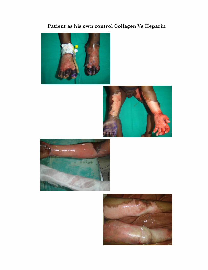
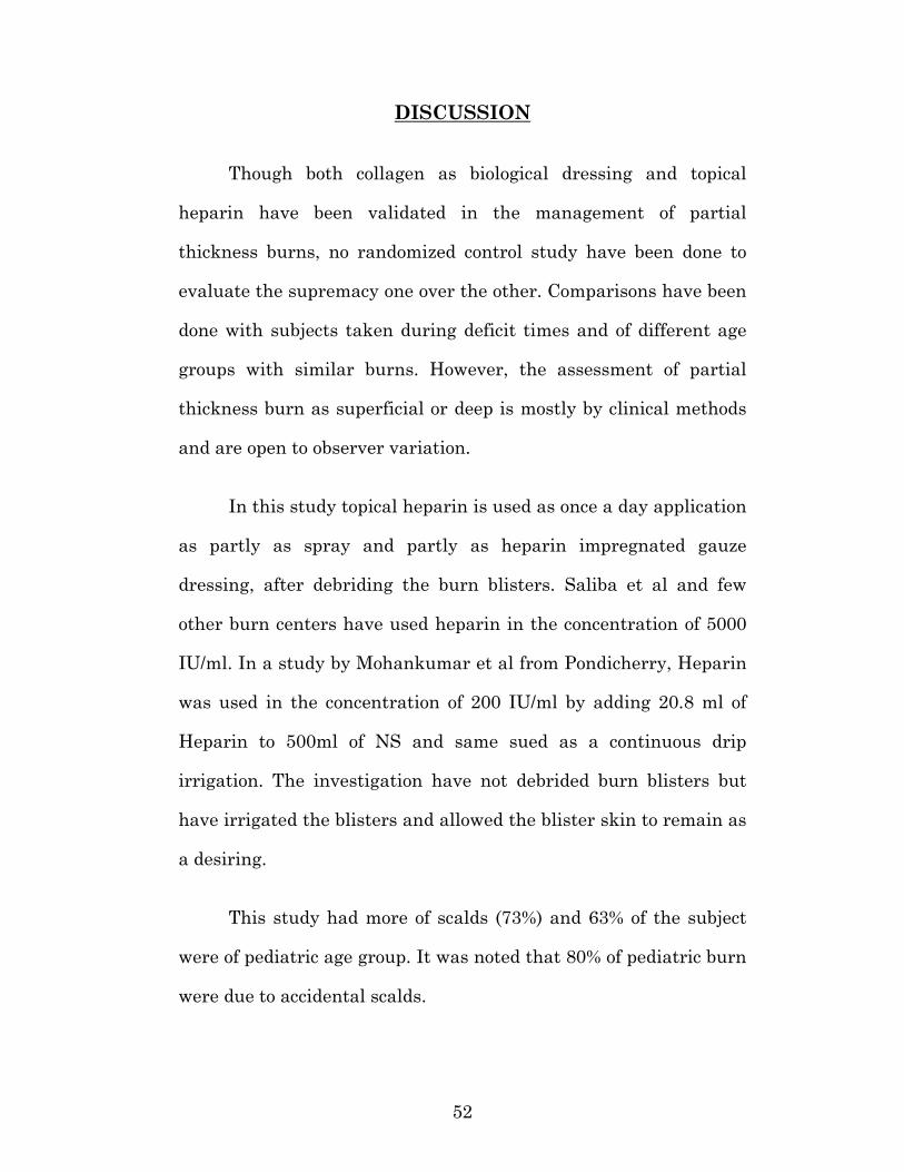
Patient as his own control Collagen Vs Heparin

Comparative healing in Face
Collagen Heparin
Collagen Heparin
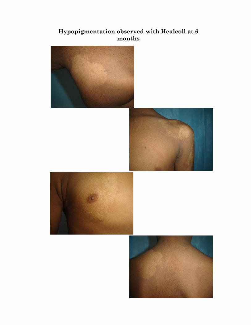
Hypopigmentation observed with Healcoll at 6 months

Comparative Healing Trunk
Heparin Collagen
Comparative Healing Forearm

Comparative Healing Forearm
Collagen
Heparin