Effect of aged garlic extract on wound healing: A new frontier in wound management
13
Drug and Chemical Toxicology, 2009; 32(3): 191–203 RESEARCH ARTICLE Effect of aged garlic extract on wound healing: A new frontier in wound management Sohail Ejaz 1,2,3 , Irina Chekarova 3 , Jae Woo Cho 3 , Seung Yeon Lee 3 , Shoaib Ashraf 2 , and Chae Woong Lim 3 1 Department of Clinical Neurosciences, Neurology Unit, Addenbrookes Hospital, University of Cambridge, Cambridge, UK, 2 Angiogenesis and Toxicology Research Laboratory, Department of Pharmacology and Toxicology, University of Veterinary and Animal Sciences, Lahore, Pakistan, and 3 Biosafety Research Institute and College of Veterinary Medicine, Chonbuk National University, Jeonju, 561–756, South Korea Address for Correspondence: S. Ejaz, Department of Clinical Neurosciences, Neurology Unit, Addenbrookes Hospital, University of Cambridge, UK. Tel: 00447903835750, Email: [email protected]. CW. Lim, Department of Pathology, College of Veterinary Medicine, Chonbuk National University, Jeonju, 561-756, South Korea, Tel: 82-63-270-3788, Fax: 82-63-270-3780, E-mail: [email protected] (Received 02 October 2008; accepted 28 October 2008) Introduction Skin is a vital organ that serves many important functions, including protection against infection, regulation of temperature and fluid loss, immu- nologic capacity, and sensory function (Bates and Jones, 2003). e entire skin surface overlies a vast network of capillary blood vessels. Beneath the epi- dermis, each cell exists not more than 200 µm from the nearest capillary, the diffusion distance of oxygen (Folkman, 2006). Most blood vessels are formed dur- ing fetal development, but adult tissues can induce angiogenesis in response to injury. is capability is governed by pro- and antiangiogenic factors present throughout the body. Cutaneous barrier injury opens the door to numerous complications, such as bacte- rial infections or fluid loss (Power et al., 2001). Wound healing involves dynamic reciprocity between cytokines, cells, and the extracellular matrix during three overlapping phases. e first phase, the ISSN 0148-0545 print/ISSN 1525-6014 online © 2009 Informa UK Ltd DOI: 10.1080/01480540902862236 Abstract Successful wound healing depends upon angiogenesis, and impaired angiogenesis is a hallmark of the chronic wounds encountered with diabetes and venous or arterial insufficiency. To intervene and improve wound closure, it is essential to investigate the effects of different natural remedies in wound healing. The chicken dorsum skin excisional wound assay was used to investigate the influence of different concentra- tions of aged garlic solution (AGS) on wound healing. Gross, histopathology, scanning electron microscopy (SEM) and computer-based three-dimensional (3D) image-probing techniques were utilized to determine the effects of AGS on wound closure, re-epithelialization, dermal matrix regeneration, and angiogenesis. Ninety chicks, aged 1 week and divided in 6 groups, were topically exposed to different concentrations of AGS for 6 days: control (group A), 1% (group B), 5% (group C), 10% (group D), 15% (group E), and skin lotion (group F). Different patterns, ranging from incomplete to almost complete wound closure, were observed among different groups with highly significant results (P < 0.001) in group E. Histological investigations revealed a positive augment in the re-epithelialization of all AGS exposed wounds. An increase in the number of new loosely packed collagen and maturation of collagen bundles was observed in all treated wounds at days 4 and 6 post-wounding, respectively. Similar results were achieved through SEM of treated wounds. Histological investigations revealed the profuse dose-dependent neovascularization among AGS-treated wounds. Abbott curve, angular spectrum, and different parameters of 3D surface roughness of wounds were also measured for the precise quantification of angiogenesis. A very highly significant (P < 0.001) increase in angiogenesis was observed among all treated groups. No significant change was observed among control and skin lotion–treated groups. These observations substantiate the beneficial use of AGS in the treatment of wounds. Additional studies are needed to study the specific wound-healing mechanisms of chemical, or group of chemicals, present in AGS. Keywords: Aged garlic; chicken; wound; angiogenesis http://www.informapharmascience.com/dct Drug and Chemical Toxicology Downloaded from informahealthcare.com by Mcgill University on 05/20/15 For personal use only.
-
Upload
independent -
Category
Documents
-
view
2 -
download
0
Transcript of Effect of aged garlic extract on wound healing: A new frontier in wound management

Drug and Chemical Toxicology, 2009; 32(3): 191–203
R E S E A R C H A R T I C L E
Effect of aged garlic extract on wound healing: A new frontier in wound management
Sohail Ejaz1,2,3, Irina Chekarova3, Jae Woo Cho3, Seung Yeon Lee3, Shoaib Ashraf2, and Chae Woong Lim3
1Department of Clinical Neurosciences, Neurology Unit, Addenbrookes Hospital, University of Cambridge, Cambridge, UK, 2Angiogenesis and Toxicology Research Laboratory, Department of Pharmacology and Toxicology, University of Veterinary and Animal Sciences, Lahore, Pakistan, and 3Biosafety Research Institute and College of Veterinary Medicine, Chonbuk National University, Jeonju, 561–756, South Korea
Address for Correspondence: S. Ejaz, Department of Clinical Neurosciences, Neurology Unit, Addenbrookes Hospital, University of Cambridge, UK. Tel: 00447903835750, Email: [email protected]. CW. Lim, Department of Pathology, College of Veterinary Medicine, Chonbuk National University, Jeonju, 561-756, South Korea, Tel: 82-63-270-3788, Fax: 82-63-270-3780, E-mail: [email protected]
(Received 02 October 2008; accepted 28 October 2008)
Introduction
Skin is a vital organ that serves many important functions, including protection against infection, regulation of temperature and fluid loss, immu-nologic capacity, and sensory function (Bates and Jones, 2003). The entire skin surface overlies a vast network of capillary blood vessels. Beneath the epi-dermis, each cell exists not more than 200 µm from the nearest capillary, the diffusion distance of oxygen
(Folkman, 2006). Most blood vessels are formed dur-ing fetal development, but adult tissues can induce angiogenesis in response to injury. This capability is governed by pro- and antiangiogenic factors present throughout the body. Cutaneous barrier injury opens the door to numerous complications, such as bacte-rial infections or fluid loss (Power et al., 2001).
Wound healing involves dynamic reciprocity between cytokines, cells, and the extracellular matrix during three overlapping phases. The first phase, the
ISSN 0148-0545 print/ISSN 1525-6014 online © 2009 Informa UK LtdDOI: 10.1080/01480540902862236
AbstractSuccessful wound healing depends upon angiogenesis, and impaired angiogenesis is a hallmark of the chronic wounds encountered with diabetes and venous or arterial insufficiency. To intervene and improve wound closure, it is essential to investigate the effects of different natural remedies in wound healing. The chicken dorsum skin excisional wound assay was used to investigate the influence of different concentra-tions of aged garlic solution (AGS) on wound healing. Gross, histopathology, scanning electron microscopy (SEM) and computer-based three-dimensional (3D) image-probing techniques were utilized to determine the effects of AGS on wound closure, re-epithelialization, dermal matrix regeneration, and angiogenesis. Ninety chicks, aged 1 week and divided in 6 groups, were topically exposed to different concentrations of AGS for 6 days: control (group A), 1% (group B), 5% (group C), 10% (group D), 15% (group E), and skin lotion (group F). Different patterns, ranging from incomplete to almost complete wound closure, were observed among different groups with highly significant results (P < 0.001) in group E. Histological investigations revealed a positive augment in the re-epithelialization of all AGS exposed wounds. An increase in the number of new loosely packed collagen and maturation of collagen bundles was observed in all treated wounds at days 4 and 6 post-wounding, respectively. Similar results were achieved through SEM of treated wounds. Histological investigations revealed the profuse dose-dependent neovascularization among AGS-treated wounds. Abbott curve, angular spectrum, and different parameters of 3D surface roughness of wounds were also measured for the precise quantification of angiogenesis. A very highly significant (P < 0.001) increase in angiogenesis was observed among all treated groups. No significant change was observed among control and skin lotion–treated groups. These observations substantiate the beneficial use of AGS in the treatment of wounds. Additional studies are needed to study the specific wound-healing mechanisms of chemical, or group of chemicals, present in AGS.
Keywords: Aged garlic; chicken; wound; angiogenesis
http://www.informapharmascience.com/dct
Dru
g an
d C
hem
ical
Tox
icol
ogy
Dow
nloa
ded
from
info
rmah
ealth
care
.com
by
Mcg
ill U
nive
rsity
on
05/2
0/15
For
pers
onal
use
onl
y.

192 Sohail Ejaz et al.
inflammatory phase, is distinguished by the infil-tration of inflammatory cells that secrete cytokines and growth factors, while assisting in the removal of apoptotic cells and debris. The next phase, the proliferative phase, involves the stage of tissue regen-eration that is initiated in response to stimulatory factors initially produced during the inflammatory phase. Angiogenesis is central to wound healing and involves the growth of new capillary blood vessels (Li and Li, 2008; Folkman, 2003, 2007; Rees et al., 1999). Finally, the remodeling phase encompasses the regression of capillaries, the reorganization of the extracellular matrix, and further maturation of the tissue that ensures the structural and mechanical stability of the dermoepidermal junction (Bernstein et al., 1994; Clark et al., 2004; Yamaguchi and Yoshikawa, 2001).
Successful wound healing depends upon angio-genesis and their appearance is synonymous with granulation, the creation of a provisional matrix comprised of proliferating blood vessels, migrating fibroblasts, and new collagen (Tonnesen et al., 2000). Impaired angiogenesis is a hallmark of the chronic wounds encountered with diabetes and venous or arterial insufficiency. Angiogenesis has, therefore, become a major focus of study for wound biologists and surgeons alike (Kubo et al., 2005).
Large surface wounds, such as those of extensively burned patients, need to be healed as fast as possible. To intervene and improve wound closure, it is essen-tial to investigate the effects of different natural rem-edies in wound healing. The medicinal use of garlic dates to at least 3,000 years BC, when Egyptian pyra-mid builders ate it to prevent illness (Ejaz et al., 2004). Hippocrates used garlic to treat infection, intestinal disorders, and chest pain. During the Middle Ages, it was consumed or worn by gravediggers and the clergy as prevention against the plague. In the mod-ern era, garlic was used to prevent infection by the Soviet army in World War II (Garty, 1993; Baruchin et al., 2001). More recently, the lipid-lowering and antihypertensive effects of garlic have been exam-ined scientifically (Lachter et al., 2003). Naturopathic practitioners recommend garlic ingestion to treat dia-betes, hyperlipidemia, hypertension, atherosclerotic plaque formation, candida infection, and other skin lesions (Burnham, 1995).
Recent investigations have brought forth ample data that support the significant protective activ-ity of garlic in different diseases (Balasenthil et al., 2000; Grudzinski et al., 2001; Ide and Lau, 1997, 2001; Wu et al., 2001). Among various preparations of garlic supplements, aged garlic solution (AGS) in particular has been given worthy attention in sound scientific experiments (Ho et al., 2001; Ide et al.,
1997, Ide and Lau, 1999a, 1999b; Ryu et al., 2001). Therefore, based on these data, the current study was undertaken to investigate the influence of AGS on different events of wound healing. The chicken dorsum skin assay was used in this study, which is extensively being used in wound-healing research (Feugate et al., 2002a, 2002b; Martins-Green, 2001; Martins-Green and Feugate, 1998). Chicken skin is cheaply and easily available and does not require the sacrifice of animals or extensive cell culture. It has many similarities to mammalian skin, including the epidermal and dermal histology and the signaling mechanisms between the epidermis and dermis that cause the formation of the various skin appendages (Harsum et al., 2001; Sengel, 1955). Although spe-cific details of structure or physiology may differ, the underlying cellular processes are comparable (Gallin and Hepperle, 1998), thus providing a useful model to study wound healing.
The effects of AGS on dermal wound healing were examined by assessing wound closure, re- epithelialization, dermal matrix regeneration, and angiogenesis. An additional aim of this research was to extend the use of computer-based image process-ing and analysis to quantify wound healing in order to overcome challenges in time-efficiency and staining differences between images.
Materials and methods
Animals
A total of 90 (1-day-old) Korean native mixed broilers were obtained from a local hatchery (Harim, Iksan, South Korea). The chicks were brooded on fresh lit-ter at 33°C from days 1 to 4, 29°C from days 5 to 10, 27°C from days 11 to 13, and 24°C from days 14 and 15. Lights were on for 24 hours a day until day 5 and for 23 hours a day thereafter. All birds were provided with normal tap water and commercial starter ration, ad libitum. At 1 week of age, all birds were weighed and divided in six groups containing 15 chicks in each group (Table 1). The chicks were wing-banded and randomly assigned to the front or back half of six
Table 1. Experimental groups with respective treatments of aged garlic solution.Group No. of chicks TreatmentA 15 ControlB 15 1% AGSC 15 5% AGSD 15 10% AGSE 15 15% AGSF 15 Skin lotion
Dru
g an
d C
hem
ical
Tox
icol
ogy
Dow
nloa
ded
from
info
rmah
ealth
care
.com
by
Mcg
ill U
nive
rsity
on
05/2
0/15
For
pers
onal
use
onl
y.

A new frontier in wound management 193
environmental chambers that had been divided by using a center divider.
Wounding and preparation of wound tissue
Chicks were anesthetized by an intraperitoneal (i.p.) injection of ketamine ( 10 mg/kg) (Mohammad et al., 1993) and xylazine (1. 5 mg/kg) (Raath et al., 1992). For the management of pain and suffering of animals, buprenorphine (an opioid analgesic), was given subcutaneously (s.c.) at a dose rate of 0. 04 mg/kg postoperatively, which produces analgesia up to 12 hours. Moreover, the buprenorphine injection was also repeated before each manipulation. The hairs on the dorsum of chicks were shaved with fine scissors, and the dorsum was subsequently wiped with 70% (v/v) ethanol. A wound was created, as described previously (Norfleet et al., 2000). Briefly, a 3-mm- diameter excision was created on the dorsum of chicks with a sterile biopsy punch (Kabasakal et al., 2005). Wounds were left undressed and chicks were housed separately after wounding.
Preparation and administration of garlic extract
In order to maintain the homogeneity of tested mate-rials, garlic cloves of similar origin, age, and grow-ing region were obtained from a botanical garden in our university. AGS was prepared by the method described elsewhere (Kasuga et al., 2001). Garlic cloves were sliced and soaked in a water-ethanol mix-ture and naturally extracted and aged for 6 months at room temperature. The extract was passed through a 0.22-µm acrodisc filter (Whatman, Maidstone, UK), and different preparations of AGS were prepared by mixing a sufficient amount of AGS in a commer-cially available skin lotion (SL) (Johnson & Son Inc., Evanston, Illinois, USA). All solutions were applied to the wound immediately after preparation and stored at 3–8°C until further use. The dorsum wounds of the chicks in groups B, C, D, and E were painted twice per day with 1, 5, 10, and 15% AGS, respectively. The animals of group F received the commercial SL, while the chicks of group A served as the control without any treatment (Table 1). Different AGS or control media were applied topically twice a day for 6 days post-wounding.
Macroscopic evaluation of woundsDuring 6 days of experiments, the wound-clo-sure activities were macroscopically recorded by photography. All wounds were visualized by using a high- resolution Lebeca camera (PanWest, Shanghai,
China) with a built-in CMOS image sensor (320,000-pixel) supporting high-quality VGA (640 × 480) reso-lution with 24-bit RGB color. The camera lens was made up of five glasses that contribute in picture improvement over regular lenses. The magnification was made adjustable by the manual placement of the camera from the object. To ensure an objective three-dimensional (3D) measurement of wound healing, serial images with respective x, y, and z dimensions were recorded at 30 frames per second, using a cam-era shutter speed of 1/2,000 second.
Image-processing system to quantify wound closureComputer elaboration and image processing were per-formed by an image-probing system (Figure 1) (IPS), using a 32-bit color quality under a Windows XP envi-ronment. As described earlier (Ejaz et al., 2005, 2006), the display screen (IBM-LG, Seoul, Korea), supported with SVGA graphics (NVIDIA GeForce 2 MX 100/200) and adjusted to a resolution of 800 × 600 pixels, pro-vided optimal optical representation.
After image acquisition, all images were imported to scan probing image-processing software (IBM, Lyngby, Denmark) that works on a specific algorithm (Ejaz et al., 2006). Respective x, y, and z dimensions of each image were loaded to determine different parameters to quantify wound healing. 3D surface roughness, which is one of the main parameters in 3D
Image Processor
CPU
Video Monitor
Wound site
Digital Camera
Data Evaluation
Figure 1. Schematic representation of the computerized system used to study macroscopic and 3D neovascularization at the sur-face of wound.
Dru
g an
d C
hem
ical
Tox
icol
ogy
Dow
nloa
ded
from
info
rmah
ealth
care
.com
by
Mcg
ill U
nive
rsity
on
05/2
0/15
For
pers
onal
use
onl
y.

194 Sohail Ejaz et al.
image analysis, was measured for the precise quan-tification of wound healing. Different, other impor-tant parameters (Sa, Sy, Sz, Sci) of 3D evaluation of micrometer and/or nanometer scale neovasculariza-tion were also quantified for the holistic quantifica-tion of wound healing. Sy is a vital parameter used to quantify the number of lowest valleys on the bear-ing surface, and Sz is an extreme parameter used to evaluate the average value of the absolute heights of the five highest peaks within the sampling area. Sz is a useful 3D parameter in characterizing the overall texture of microvasculature on bearing surface. Sci is the ratio of the void volume of the unit sampling area at the core zone over the root mean square deviation. This vital parameter of 3D quantitation is used for the determination of fluid retention in that surface, and a larger Sci value designates better neovascularization at that surface (Zeilinger et al., 2002).
Histological examination of wound healing
For the in-depth investigating of wound healing at different intervals, 5 chicks from each group were sacrificed at 2, 4, and 6 days post-wounding. The complete wound, including the scab and 3 mm of the epithelial margins, was excised at each time point, fixed in 10% formaldehyde solution, and proc-essed for histological examination. These tissues were then embedded in paraffin wax, sectioned at a 3-µm thickness, mounted on slides, and stained with hematoxylin and eosin (H&E) for routine light micro-scopy. Histological sections were also digitized with a spot camera, and different parameters of wound healing were quantified.
Scanning electron microscopyTopographical and ultrastructural evaluation of wound reactions to different AGS was performed by using scanning electron microscope (JSM-5900; Tokyo, Jeol). The samples from dorsum skin were prepared by fixing them with 2% glutaraldehyde solution, both on top and underneath the skin, and then processed for scanning electron microscopy (SEM), as described previously (Magers et al., 1995). Briefly, samples were cut out of the skin, washed in cacodylate buffer, and postfixed in 1% osmium tetroxide for 30–60 minutes. Samples were washed in distilled water, 3–5 minutes each, followed by dehydration in an ascending ethanol series from 30 to 100%. To ensure complete removal of water from the tissue, five changes of 100% ethanol were performed. Samples were critically point dried, mounted on alu-minum pedestals, and further processed by using standard electron microscopy techniques.
Statistics
Statistical analysis was carried out by using SPSS 13 software (SPSS Inc., Chicago, Illinois, USA). All data were expressed as the means ± standard error of the mean (SEM). Groups of data were compared with analysis of variance (ANOVA), followed by the Student’s t-test. Values of P < 0.05 were regarded as significant (James, 1999).
Results
Wound closure
Wound margins were easily recognized at day 1 postwounding by an abrupt interruption in the epi-thelial and dermal continuity (Figure 2). Different patterns of wound contraction, ranging from incom-plete to moderate wound closure, were observed among different groups at day 4 post-exposure. All of the AGS-treated groups showed significantly bet-ter results (P < 0.01) than the control group at day 4 post-exposure (Figure 3). Highly significant results (P < 0.001) were observed in group E with a 75% con-traction of the wound site at day 4 post-wounding (Figure 3). Variable levels of wound contractions were observed in all AGS-treated groups at day 6 post-wounding. Completely contracted wounds (P < 0.001) were prominently observed in group E
Figure 2. Macroscopic picture of skin wound at day 1 post-wounding. Note the easily recognizable wound margins by an abrupt interruption in the dermal continuity.
Dru
g an
d C
hem
ical
Tox
icol
ogy
Dow
nloa
ded
from
info
rmah
ealth
care
.com
by
Mcg
ill U
nive
rsity
on
05/2
0/15
For
pers
onal
use
onl
y.

A new frontier in wound management 195
at day 6 postwounding. No significant change was observed among control and SL-treated wounds.
Re-epithelialization at wound site
Different extents of re-epithelialization were observed macroscopically among all groups, and AGS-treated groups showed considerably better outcomes than control groups. Complete re-epithelialization of
wound sites were observed at day 6 post-wounding in group E (Figure 4). A comparative histological (H&E) analysis of the wound at different days post-wounding revealed both the neoepidermis and the neodermis progression toward the wound center after wounding. A scab covered the wound in close contact with the provisional matrix. We observed that re-epithelialization was dose dependent with potent re-epithelialization in group E. At day 4 post-wounding, a migratory tongue of epidermal cells penetrated into the wound bed of all treated groups, under the scab (Figure 5). The forward part of the tongue (tongue tip) was composed of a monolayer, progressively followed by a bilayer and a disorgan-ized, multilayered epithelium toward the wound margins (tongue tail). Hyperthickening and parak-eratosis was also observed in group E (Figure 6). At
A B
C
Figure 4. Macroscopic pictures at day 6 post-wounding, show-ing different patterns of re-epithelialization. Note the progres-sive re-epithelialization in the group treated with 10% AGS (B) and complete re-epithelialization in the group treated with 15% AGS (C).
S
Figure 5. Histological micrographs showing re-epithelialization in group E at day 4 post-wounding. Note the active epithelium (arrow) and a migratory tongue of epidermal cells penetrating into the wound bed, under the scab (S) (bar, 100 m).
S
Figure 6. Hyperthickening and parakeratosis in group E at day 4 post-wounding (bar, 50 m).
Wound contraction at different days post-wounding
0
50
100
150
2 4 6 Time in days
Wou
nd c
losu
re
(% a
ge) A B C D E
* ** *** *** *** ***
*** *** *** *** **
Figure 3. Graphical outline representing comparative wound closure at different periods of time. Note the highly significant wound closure in groups C, D, and E (A: control; B: 1%; C:5%; D: 10%; E: 15%) (*P < 0.05, **P < 0.01; ***P < 0.001).
Dru
g an
d C
hem
ical
Tox
icol
ogy
Dow
nloa
ded
from
info
rmah
ealth
care
.com
by
Mcg
ill U
nive
rsity
on
05/2
0/15
For
pers
onal
use
onl
y.

196 Sohail Ejaz et al.
day 6 post-wounding, different levels of stratified neoepithelium covered the center of wounds in AGS-treated groups. Completely covered wounds by stratified neoepithelium were observed in group E. An immature growth in both the neoepidermis and the neodermis was observed among control wounds with incomplete re- epithelialization and a thin granulation layer. Histological analysis thus revealed a nearly healed morphology of the AGS-treated wounds that was consistent with the wound-closure observations described above.
Dermal matrix regenerationThe composition of the dermal matrix underwent changes from an original fibrin clot to a granulation tissue and a collagenous dermis. All of the AGS-treated wounds, at day 2 post-wounding, were plugged by the
provisional matrix of variable levels, containing blood vessels and invading cells consistent with inflam-matory elements. At day 4 post-wounding, well-developed granulation tissues of variable intensities were established in all AGS-treated wounds, but still poor in collagen bundle (Figure 7A). At day 6 post-wounding, the density of collagen bundles increased in all AGS-treated groups, whereas the number of cells decreased. Maximum activities of dermal matrix regeneration and maturation were observed in group E. In contrast, the connective tissues of nonwounded areas of the skin showed a higher intensity of the collagen bundles acquired an “angel curl–shaped structure” (Figure 7B). SEM analysis of the wound matrix also revealed the similar results, which are consistent with the dermal matrix regeneration above (Figure 8).
Quantification of angiogenesisAbbott curve (graphical presentation of the presence of blood vessels at wound site), angular spectrum (graphical presentation of the angular distribu-tion of blood vessels at wound site), and 3D surface roughness of wounds were measured for the precise
S
A
B
Figure 7. Light micrographs showing dermal matrix regenera-tion at day 4 post-wounding. (A) Note the increase in number and activity of connective tissue cells resulting in the produc-tion of new, loosely packed collagen in group E (bar, 50 m). (B) Normal connective tissue, with dense collagen bundles and few cells (bar, 20 m).
A
B
Figure 8. Scanning electron micrographs showing dermal matrix regeneration during wound healing. (A) Note the increase in number of new, loosely packed collagen in group E at day 4 post-wounding (magnification, X3,000). (B) Maturation of colla-gen bundles in group E at day 6 postwounding (magnification, X3,000).
Dru
g an
d C
hem
ical
Tox
icol
ogy
Dow
nloa
ded
from
info
rmah
ealth
care
.com
by
Mcg
ill U
nive
rsity
on
05/2
0/15
For
pers
onal
use
onl
y.

A new frontier in wound management 197
quantification of angiogenesis at wound surfaces. Thus, blood vessels of micrometer and/or nanometer scale were also evaluated for the holistic quantifica-tion of angiogenesis. Considerable dose-dependent changes in the height of Abbott curve were recorded among all AGS-treated groups with highly significant changes (P < 0.01) in group E (Figure 9). It shows a substantial increase in the amount of blood vessels at wound sites of AGS-treated groups (Figure 10). Similarly, angular distribution of the blood vessels at wound sites revealed a uniform distribution of blood
20 40 60 80 100
2.0
4.0
6.0
8.0
10
12
Hei
ght [
µm]
A Bearing area [%] 20 40 60 80 100
2.0
4.0
6.0
8.0
10
12
14
Hei
ght [
µm]
B Bearing area[%]
20 40 60 80 100
2.0
4.0
6.0
8.0
10
12
14
Hei
ght [
µm]
C Bearing area [%]20 40 60 80 100
2.0
4.0
6.0
8.0
10
12
14
Hei
ght [
µm]
D Bearing area[%]
20 40 60 80 100
2.0
4.0
6.0
8.0
10
14
12
16
Hei
ght [
µm]
E Bearing area[%]
Figure 10. Graphical outline of Abbott curve measurements on wound sites of different groups at day 4 post-wounding. Note the expan-sion in height among different AGS-treated groups. (A) Control: 12 m; (B) 1%: 13 m; (C) 5%; 13.5 m; (D) 10%: 14 m; (E) 15%: 16.5 m.
Figure 9. Graphical outline representing comparative height of Abbott curve at different time periods. Note the highly significant difference in group E (A: control; B: 1%; C: 5%; D: 10%; E: 15%) (*P < 0.05; **P < 0.01).
Height of the abbott curve at different days post wounding
0
10
20
30
2 4 6 Time in days
Hei
ght
(Mic
ro m
eter
)
A B C D E
* * ** * ** ** * **
Dru
g an
d C
hem
ical
Tox
icol
ogy
Dow
nloa
ded
from
info
rmah
ealth
care
.com
by
Mcg
ill U
nive
rsity
on
05/2
0/15
For
pers
onal
use
onl
y.

198 Sohail Ejaz et al.
vessels among AGS-treated groups (Figure 11). A very highly significant (P < 0.001) increase in angular spectrum was observed in all AGS-treated groups, compared to the control group (Figure 12). 3D surface roughness (Figure 13) and different, other imperative parameters were also quantified. A very highly signifi-cant increase (P < 0.001) in average surface roughness (Sa) was observed among AGS-treated groups with a maximum increase in group E (Table 2). Almost similar results were recorded for the amount of low-est valleys (Sy) and maximum height of the wound surfaces (Sz) at different time periods (Table 2). A very highly significant increase in Sci values of all AGS-treated groups demonstrated ample cellu-lar activities at wound sites (Table 2). Histological
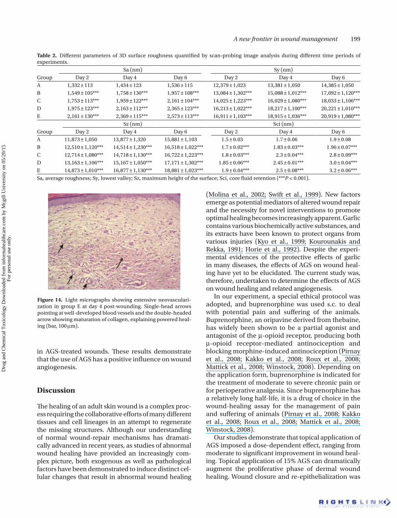
micrographs of wounds from AGS-treated samples exhibited profuse neovascularization, compared to wounds from control samples (Figure 14). About a 40–60% increase in neovascularization was observed
0.0 2500 5000 7500 10000100
150
120
90
60
30
0
A
0.0 2500 5000 7500 10000100
150
120
90
60
30
0
B
0.0 4000 8000 12000100
150
120
90 90
60
30
0
C
0.0 4000 8000 12000100
150
120 60
30
0
D
Figure 11. Graphical representation of angular spectrum of blood vessels on wound sites of different groups at day 4 post-wounding. Note the expansion of angular spread and distribution of blood vessels among AGS-treated groups. (A) Control: >10,000 points; (B) 1%: <10,000 points; (C) 10%: >12,000 points; (D) 15%: 12,000 points.
A B
C
Figure 13. Scan-probing images showing different echelons of wound-surface roughness. Arrows point at the ample increase of wound-surface roughness in group E at day 4 post-wounding (A: Control; B: 10%; C: 15%).
Figure 12. Graphical outline representing angular spectrum of blood vessels at different time periods. A very highly significant spread (P < 0.001) was observed in all AGS-treated groups (A: control; B: 1%; C: 5%; D: 10%; E: 15%).
Angular spectrum of blood vessels at different days post wounding
0 5000
10000 15000 20000
2 4 6 Time in days
Ang
ular
spe
ctru
m
(Poi
nts)
A B C D E
Dru
g an
d C
hem
ical
Tox
icol
ogy
Dow
nloa
ded
from
info
rmah
ealth
care
.com
by
Mcg
ill U
nive
rsity
on
05/2
0/15
For
pers
onal
use
onl
y.
A new frontier in wound management 199
in AGS-treated wounds. These results demonstrate that the use of AGS has a positive influence on wound angiogenesis.
Discussion
The healing of an adult skin wound is a complex proc-ess requiring the collaborative efforts of many different tissues and cell lineages in an attempt to regenerate the missing structures. Although our understanding of normal wound-repair mechanisms has dramati-cally advanced in recent years, as studies of abnormal wound healing have provided an increasingly com-plex picture, both exogenous as well as pathological factors have been demonstrated to induce distinct cel-lular changes that result in abnormal wound healing
(Molina et al., 2002; Swift et al., 1999). New factors emerge as potential mediators of altered wound repair and the necessity for novel interventions to promote optimal healing becomes increasingly apparent. Garlic contains various biochemically active substances, and its extracts have been known to protect organs from various injuries (Kyo et al., 1999; Kourounakis and Rekka, 1991; Horie et al., 1992). Despite the experi-mental evidences of the protective effects of garlic in many diseases, the effects of AGS on wound heal-ing have yet to be elucidated. The current study was, therefore, undertaken to determine the effects of AGS on wound healing and related angiogenesis.
In our experiment, a special ethical protocol was adopted, and buprenorphine was used s.c. to deal with potential pain and suffering of the animals. Buprenorphine, an oripavine derived from thebaine, has widely been shown to be a partial agonist and antagonist of the -opioid receptor, producing both -opioid receptor–mediated antinociception and blocking morphine-induced antinociception (Pirnay et al., 2008; Kakko et al., 2008; Roux et al., 2008; Mattick et al., 2008; Winstock, 2008). Depending on the application form, buprenorphine is indicated for the treatment of moderate to severe chronic pain or for perioperative analgesia. Since buprenorphine has a relatively long half-life, it is a drug of choice in the wound-healing assay for the management of pain and suffering of animals (Pirnay et al., 2008; Kakko et al., 2008; Roux et al., 2008; Mattick et al., 2008; Winstock, 2008).
Our studies demonstrate that topical application of AGS imposed a dose-dependent effect, ranging from moderate to significant improvement in wound heal-ing. Topical application of 15% AGS can dramatically augment the proliferative phase of dermal wound healing. Wound closure and re-epithelialization was
Table 2. Different parameters of 3D surface roughness quantified by scan-probing image analysis during different time periods of experiments.
GroupSa (nm) Sy (nm)
Day 2 Day 4 Day 6 Day 2 Day 4 Day 6A 1,332 ± 113 1,434 ± 123 1,536 ± 115 12,379 ± 1,023 13,381 ± 1,050 14,385 ± 1,050B 1,549 ± 105*** 1,758 ± 130*** 1,957 ± 108*** 13,084 ± 1,302*** 15,088 ± 1,012*** 17,092 ± 1,120***C 1,753 ± 113*** 1,959 ± 122*** 2,161 ± 104*** 14,025 ± 1,223*** 16,029 ± 1,080*** 18,033 ± 1,106***D 1,975 ± 123*** 2,163 ± 112*** 2,365 ± 123*** 16,213 ± 1,022*** 18,217 ± 1,100*** 20,221 ± 1,010***E 2,161 ± 130*** 2,369 ± 115*** 2,573 ± 113*** 16,911 ± 1,103*** 18,915 ± 1,036*** 20,919 ± 1,080***
GroupSz (nm) Sci (nm)
Day 2 Day 4 Day 6 Day 2 Day 4 Day 6A 11,873 ± 1,050 13,877 ± 1,320 15,881 ± 1,103 1.5 ± 0.03 1.7 ± 0.06 1.9 ± 0.08B 12,510 ± 1,120*** 14,514 ± 1,230*** 16,518 ± 1,022*** 1.7 ± 0.02*** 1.83 ± 0.03*** 1.96 ± 0.07***C 12,714 ± 1,080*** 14,718 ± 1,130*** 16,722 ± 1,223*** 1.8 ± 0.03*** 2.3 ± 0.04*** 2.8 ± 0.09***D 13,163 ± 1,106*** 15,167 ± 1,050*** 17,171 ± 1,302*** 1.85 ± 0.06*** 2.45 ± 0.01*** 3.0 ± 0.04***E 14,873 ± 1,010*** 16,877 ± 1,130*** 18,881 ± 1,023*** 1.9 ± 0.04*** 2.5 ± 0.08*** 3.2 ± 0.06***Sa, average roughness; Sy, lowest valley; Sz, maximum height of the surface; Sci, core fluid retention (***P < 0.001).
Figure 14. Light micrographs showing extensive neovasculari-zation in group E at day 4 post-wounding. Single-head arrows pointing at well-developed blood vessels and the double-headed arrow showing maturation of collagen, explaining powered heal-ing (bar, 100 m).
Dru
g an
d C
hem
ical
Tox
icol
ogy
Dow
nloa
ded
from
info
rmah
ealth
care
.com
by
Mcg
ill U
nive
rsity
on
05/2
0/15
For
pers
onal
use
onl
y.

200 Sohail Ejaz et al.
also significantly prompted in AGS-treated groups, which is consistent with a previous study demonstrat-ing prompt re-epithelialization of the buccal pouch by the topical application of garlic extract (Gosain and DiPietro, 2004).
An important component of tissue healing and wound contraction is the re-arrangement of ground collagen fibers, which can ultimately influence the quality of scars. The extracellular matrix not only functions as a reservoir for growth factors and sign-aling molecules, but also acts as a framework upon which endothelial cells can migrate during angiogen-esis (DiPietro, 1995; Li et al., 2003). The mechanism of tissue contraction during wound healing is not com-pletely understood. There are two theories proposed to explain this process. The first theory suggests that the contraction forces are generated by myofibroblasts, which are transmitted to other cells and surrounding connective tissues. This hypothesis suggests that the myofibroblasts would act as a multicellular unit to contract the tissue (Ozaki and Buchman, 1998). The second theory proposes that the contraction is caused by the re-arrangement of collagen fibers through the action of fibroblasts. The dislocation forces created by these cells within the connective tissue would lead to the re-orientation of collagen fibers into thicker bundles as well as their contraction (Ehrlich and Rajaratnam, 1990). Similar results were viewed in our study, and both histological and SEM investigations of wounds revealed a significant increase in collagen synthesis in wounds from AGS-treated groups. Berry et al. (1998) also observed that 88% of the healing of a sacral wound occurred due to contraction, and the other 22% occurred due to scar formation. Cystine (Barth et al. 2003; Ottl et al. 1996), methionine (Nelson et al., 1998; Squires et al., 2003; Dahlberg et al., 2000), and vitamin C (May and Qu, 2005; Senturk et al., 2004) are considered potent regulators to stimulate the production and re-organization of collagen fib-ers. Hence, the presence of cystine, methionine, and vitamin C in AGS (Shin et al., 2002; Rosen et al., 2001; Choi et al., 2006) might be involved to boost the proc-ess of wound healing in this experiment.
Histological evaluation of wounds from different AGS-treated groups revealed a considerable increase in neovascularization of wounds. Cytokines produced during the inflammatory phase of wound healing pro-mote the initiation of the proliferative phase, resulting in re-epithelialization of the wound bed, restoration of the extracellular matrix, and angiogenesis. Growth factors released by the migrating epithelium signal fibroblasts and endothelial cells to initiate collagen synthesis and angiogenesis, respectively. Wound ang-iogenesis is contingent upon chemokine secretion, extracellular matrix interactions, and equilibrium
between pro- and antiangiogenic factors (Kovacs and DiPietro, 1994; Nissen et al., 1996, 1998). Migration of endothelial cells into the extracellular matrix is dependent upon these cell-cell and cell-matrix inter-actions. The increase in wound vascularity could result from an increase in proangiogenic stimuli, increase in endothelial cell response, or a combina-tion of these two events.
Though the qualitative techniques (i.e., light micro-scopy and electron microscopy) used in our study have provided some insight of wound response to different AGS, they are limited in a way that they do not allow quantitative or statistical comparisons between dif-ferent groups (Pentcheff and Bolender, 1992). Though these techniques are relatively straightforward to perform, they can cause eyestrain from excessive use of the microscope, and they are susceptible to human counting error. With the increase in availability of affordable high-quality imaging hardware and com-puting equipment, possibly, human eyestrain and counting error could be reduced (Hansen et al., 1997; Sanders et al., 1999; Ozturk et al., 1995).
We used IPS in order to evaluate the holistic 3D quan-tification of angiogenesis. A dose-dependent increase in the height of Abbott curve, with highly significant changes (P < 0.01) in group E, revealed considerable increase in angiogenesis. Similarly, a very highly sig-nificant increase (P < 0.001) in angular spectrum of blood vessels was observed among all AGS-treated groups, which reflects excessive growth and uniformly distributed blood vessels at wound sites. Moreover, a considerable increase in the surface echelons and a very highly significant increase (P < 0.001) in Sa, Sy, Sz, and Sci of AGS-treated groups demonstrate a vital increase in angiogenesis, which cannot be measured by qualitative lab techniques. This productive increase in angiogenesis might be due to the presence of con-siderable amount of L-arginine, zinc, and copper in garlic, which are considered as angiogenic stimula-tors (Szende et al., 2001; Jacobi et al., 2005; Dai et al., 2004; Miyashita et al., 2004; Nasulewicz and Opolski, 2002; Goodman et al., 2004). Yamasaki and Lau have also explained that AGS protects vascular endothelial cells from H2
O2-induced oxidative damage by inhibit-
ing lipid peroxidation, resulting in stable angiogenesis (Yamasaki and Lau, 1997).
Conclusion
Our findings provide new evidence demonstrating that AGS exposure can influence multiple aspects of wound healing. Since AGS is a complex mixture with many different chemicals, it might contain a chemi-cal, or group of chemicals, that act a vital role in the
Dru
g an
d C
hem
ical
Tox
icol
ogy
Dow
nloa
ded
from
info
rmah
ealth
care
.com
by
Mcg
ill U
nive
rsity
on
05/2
0/15
For
pers
onal
use
onl
y.

A new frontier in wound management 201
stimulation of different important cascades of wound healing and angiogenesis. Additional studies are, therefore, needed to unveil the specific chemical, or group of chemicals, found in AGS and to elucidate the precise mechanistic cellular changes that are evoked by topical application of AGS.
Acknowledgment
This study was supported financially, in part, by Chonbuk National University funds for overseas research.
Declaration of interest: The authors report no financial conflicts of interest. The authors alone are responsible for the content and writing of this paper.
References
Balasenthil, S., Arivazhagan, S., Nagini, S. (2000). Garlic enhances circulatory antioxidants during 7,12-dimethylbenz[a]anthracene-induced hamster buccal pouch carcinogenesis. J Ethnopharmacol 72:429–433.
Barth, D., Musiol, H.J., Schutt, M., Fiori, S., Milbradt, A.G., Renner, C., Moroder, L. (2003). The role of cystine knots in collagen folding and stability, part I. Conformational properties of (Pro-Hyp-Gly)5 and (Pro-(4S)-FPro-Gly)5 model trimers with an artificial cystine knot. Chemistry 9:3692–3702.
Baruchin, A.M., Sagi, A., Yoffe, B., Ronen, M. (2001). Garlic burns. Burns 27:781–782.
Bates, D.O., Jones, R.O. (2003). The role of vascular endothelial growth factor in wound healing. Int J Low Extrem Wounds 2:107–120.
Bernstein, E.F., Chen, Y.Q., Tamai, K., Shepley, K.J., Resnik, K.S., Zhang, H., Tuan, R., Mauviel, A., Uitto, J. (1994). Enhanced elastin and fibrillin gene expression in chronically photodam-aged skin. J Invest Dermatol 103:182–186.
Berry, D.P., Harding, K.G., Stanton, M.R., Jasani, B., Ehrlich, H.P. (1998). Human wound contraction: collagen organiza-tion, fibroblasts, and myofibroblasts. Plast Reconstr Surg 102:124–131.
Burnham, B.E. (1995). Garlic as a possible risk for postoperative bleeding. Plast Reconstr Surg 95:213.
Choi, S.Y., Chung, M.J., Seo, W.D., Shin, J.H., Shon, M.Y., Sung, N.J. (2006). Inhibitory effects of Orostachys japonicus extracts on the formation of N-nitrosodimethylamine. J Agric Food Chem 54:6075–6078.
Clark, R.A., Lin, F., Greiling, D., An, J., Couchman, J.R. (2004). Fibroblast invasive migration into fibronectin/fibrin gels requires a previously uncharacterized dermatan sulfate-CD44 proteoglycan. J Invest Dermatol 122:266–277.
Dahlberg, L., Billinghurst, R.C., Manner, P., Nelson, F., Webb, G., Ionescu, M., et al. (2000). Selective enhancement of col-lagenase-mediated cleavage of resident type II collagen in cultured osteoarthritic cartilage and arrest with a synthetic inhibitor that spares collagenase 1 (matrix metalloprotein-ase 1). Arthritis Rheum 43:673–682.
Dai, Q., Huang, J., Klitzman, B., Dong, C., Goldschmidt-Clermont, P.J., March, K.L., et al. (2004). Engineered zinc finger-activat-ing vascular endothelial growth factor transcription factor plasmid DNA induces therapeutic angiogenesis in rabbits with hindlimb ischemia. Circulation 110:2467–2475.
DiPietro, L.A. (1995). Wound healing: the role of the macrophage and other immune cells. Shock 4:233–240.
Ehrlich, H.P., Rajaratnam, J.B. (1990). Cell locomotion forces versus cell contraction forces for collagen lattice contrac-tion: an in vitro model of wound contraction. Tissue Cell 22:407–417.
Ejaz, E., Shimada, A., Woong, L.C. (2005). Toxicological screening for the effects of short-term exposure of sidestream cigarette smoke on angiogenesis. Drug Chem Toxicol 28:447–465.
Ejaz, S., Akram, W., Lim, C.W., Lee, J.J., Hussain, I. (2004). Endocrine disrupting pesticides: a leading cause of cancer among rural people in Pakistan. Exp Oncol 26:98–105.
Ejaz, S., Chekarova, I., Ashraf, M., Lim, C.W. (2006). A novel 3-D model of chick chorioallantoic membrane for ameliorated studies in angiogenesis. Cancer Invest 24:567–575.
Feugate, J.E., Li, Q., Wong, L., Martins-Green, M. (2002a). The CXC chemokine, cCAF, stimulates differentiation of fibrob-lasts into myofibroblasts and accelerates wound closure. J Cell Biol 156:161–172.
Feugate, J.E., Wong, L., Li, Q.J., Martins-Green, M. (2002b). The CXC chemokine, cCAF, stimulates precocious deposition of ECM molecules by wound fibroblasts, accelerating develop-ment of granulation tissue. BMC Cell Biol 3:13.
Folkman, J. (2003). Angiogenesis and proteins of the hemostatic system. J Thromb Haemost 1:1681–1682.
Folkman, J. (2006). Angiogenesis. Annu Rev Med 57:1–18.Folkman, J. (2007). Is angiogenesis an organizing principle in
biology and medicine? J Pediatr Surg 42:1–11.Gallin, W.J., Hepperle, B. (1998). Burn healing in organ cul-
tures of embryonic chicken skin: a model system. Burns 24:613–620.
Garty, B.Z. (1993). Garlic burns. Pediatrics 91:658–659.Goodman, V.L., Brewer, G.J., Merajver, S.D. (2004). Copper
deficiency as an anticancer strategy. Endocr Relat Cancer 11:255–263.
Gosain, A., DiPietro, L.A. (2004) Aging and wound healing. World J.Surg 28:321–326.
Grudzinski, I.P., Frankiewicz-Jozko, A., Bany, J. (2001). Diallyl sulfide—a flavour component from garlic (Allium sati-vum) attenuates lipid peroxidation in mice infected with Trichinella spiralis. Phytomedicine 8:174–177.
Hansen, G.L., Sparrow, E.M., Kokate, J.Y., Leland, K.J., Iaizzo, P.A. (1997). Wound status evaluation using color image process-ing. IEEE Trans.Med.Imaging 16:78–86.
Harsum, S., Clarke, J.D., Martin, P. (2001). A reciprocal relation-ship between cutaneous nerves and repairing skin wounds in the developing chick embryo. Dev Biol 238:27–39.
Ho, S.E., Ide, N., Lau, B.H. (2001). S-allyl cysteine reduces oxi-dant load in cells involved in the atherogenic process. Phytomedicine 8:39–46.
Horie, T., Awazu, S., Itakura, Y., Fuwa, T. (1992). Identified dial-lyl polysulfides from an aged garlic extract which pro-tects the membranes from lipid peroxidation. Planta Med 58:468–469.
Ide, N., Lau, B.H. (1997). Garlic compounds protect vascular endothelial cells from oxidized low density lipoprotein-in-duced injury. J Pharm Pharmacol 49:908–911.
Ide, N., Lau, B.H. (1999a). Aged garlic extract attenuates intracel-lular oxidative stress. Phytomedicine 6:125–131.
Ide, N., Lau, B.H. (1999b). S-allylcysteine attenuates oxidative stress in endothelial cells. Drug Dev Ind Pharm 25:619–624.
Dru
g an
d C
hem
ical
Tox
icol
ogy
Dow
nloa
ded
from
info
rmah
ealth
care
.com
by
Mcg
ill U
nive
rsity
on
05/2
0/15
For
pers
onal
use
onl
y.

202 Sohail Ejaz et al.
Ide, N., Lau, B.H. (2001). Garlic compounds minimize intracel-lular oxidative stress and inhibit nuclear factor-kappa b acti-vation. J Nutr 131:1020S–1026S.
Jacobi, J., Sydow, K., von, Degenfeld G, Zhang, Y., Dayoub, H., Wang, B., et al. (2005). Overexpression of dimethyl-arginine dimethylaminohydrolase reduces tissue asym-metric dimethylarginine levels and enhances angiogenesis. Circulation 111:1431–1438.
James, J. (1999). Skin grafting in difficult situations. Trop Doct 29:41–42.
Kabasakal, L., Sehirli, O., Cetinel, S., Cikler, E., Gedik, N., Sener, G. (2005). Protective effect of aqueous garlic extract against renal ischemia/reperfusion injury in rats. J Med Food 8:319–326.
Kakko, J., Heilig, M., Sarman, I. (2008). Buprenorphine and meth-adone treatment of opiate dependence during pregnancy: comparison of fetal growth and neonatal outcomes in two consecutive case series. Drug Alcohol Depend 96:69–78.
Kasuga, S., Uda, N., Kyo, E., Ushijima, M., Morihara, N., Itakura, Y. (2001). Pharmacologic activities of aged garlic extract in comparison with other garlic preparations. J Nutr 131:1080S–1084S.
Kourounakis, P.N., Rekka, E.A. (1991). Effect on active oxygen species of alliin and Allium sativum (garlic) powder. Res Commun Chem Pathol Pharmacol 74:249–252.
Kovacs, E.J., DiPietro, L.A. (1994). Fibrogenic cytokines and con-nective tissue production. FASEB J 8:854–861.
Kubo, S., Inui, T., Hasegawa, H., Yoshimine, T. (2005). Repair of intractable cerebrospinal fluid rhinorrhea with mucosal flaps and recombinant human basic fibroblast growth fac-tor: technical case report. Neurosurgery 56:E627.
Kyo, E., Uda, N., Ushijima, M., Kasuga, S., Itakura, Y. (1999). Prevention of psychological stress-induced immune sup-pression by aged garlic extract. Phytomedicine 6:325–330.
Lachter, J., Babich, J.P., Brookman, J.C., Factor, A.Y. (2003). Garlic: a way out of work. Mil Med 168:499–500.
Li, J., Zhang, Y.P., Kirsner, R.S. (2003). Angiogenesis in wound repair: angiogenic growth factors and the extracellular matrix. Microsc Res Tech 60:107–114.
Li, V.W., Li, W.W. (2008). Antiangiogenesis in the treatment of skin cancer. J Drugs Dermatol 7:s17–s24.
Magers, T., Talbot, P., DiCarlantonio, G., Knoll, M., Demers, D., Tsai, I., et al. (1995). Cigarette smoke inhalation affects the reproductive system of female hamsters. Reprod Toxicol 9:513–525.
Martins-Green, M. (2001). The chicken chemotactic and ang-iogenic factor (cCAF), a CXC chemokine. Int J Biochem Cell Biol 33:427–432.
Martins-Green, M., Feugate, J.E. (1998). The 9E3/CEF4 gene prod-uct is a chemotactic and angiogenic factor that can initiate the wound-healing cascade in vivo. Cytokine 10:522–535.
Mattick, R.P., Kimber, J., Breen, C., Davoli, M. (2008) Buprenorphine maintenance versus placebo or methadone maintenance for opioid dependence. Cochrane Database Syst Rev CD002207.
May, J.M., Qu, Z.C. (2005). Transport and intracellular accumu-lation of vitamin C in endothelial cells: relevance to collagen synthesis. Arch Biochem Biophys 434:178–186.
Miyashita, H., Kanemura, M., Yamazaki, T., Abe, M., Sato, Y. (2004). Vascular endothelial zinc finger 1 is involved in the regulation of angiogenesis: possible contribution of stath-min/OP18 as a downstream target gene. Arterioscler Thromb Vasc Biol 24:878–884.
Mohammad, F.K., al, Badrany, M.S., al, Hasan, A.M. (1993). Detomidine-ketamine anaesthesia in chickens. Vet Rec 133:192.
Molina, P.E., McClain, C., Valla, D., Guidot, D., Diehl, A.M., Lang, C.H., et al. (2002). Molecular pathology and clinical aspects of alcohol-induced tissue injury. Alcohol Clin Exp Res 26:120–128.
Nasulewicz, A., Opolski, A. (2002). [The role of copper in tumor angiogenesis—clinical implications]. Postepy Hig Med Dosw 56:691–705.
Nelson, F., Dahlberg, L., Laverty, S., Reiner, A., Pidoux, I., Ionescu, M., et al. (1998). Evidence for altered synthesis of type II collagen in patients with osteoarthritis. J Clin Invest 102:2115–2125.
Nissen, N.N., Polverini, P.J., Gamelli, R.L., DiPietro, L.A. (1996). Basic fibroblast growth factor mediates angiogenic activity in early surgical wounds. Surgery 119:457–465.
Nissen, N.N., Polverini, P.J., Koch, A.E., Volin, M.V., Gamelli, R.L., DiPietro, L.A. (1998). Vascular endothelial growth factor mediates angiogenic activity during the prolifera-tive phase of wound healing. Am J Pathol 152:1445–1452.
Norfleet, A.M., Huang, Y., Sower, L.E., Redin, W.R., Fritz, R.R., Carney, D.H. (2000). Thrombin peptide TP508 accelerates closure of dermal excisions in animal tissue with surgically induced ischemia. Wound Repair Regen 8:517–529.
Ottl, J., Battistuta, R., Pieper, M., Tschesche, H., Bode, W., Kuhn, K., et al. (1996). Design and synthesis of heterotrimeric col-lagen peptides with a built-in cystine-knot. Models for col-lagen catabolism by matrix-metalloproteases. FEBS Lett 398:31–36.
Ozaki, W., Buchman, S.R. (1998). Use of scanning electron micro-scopy in the evaluation of craniosynostosis. J Craniofac Surg 9:30–38.
Ozturk, C., Nissannov, J., Dubin, S., Shi, W.Y., Nichols, J., Mark, R. (1995). Measurement of wound healing by image analysis. Biomed Sci Instrum 31:189–193.
Pentcheff, N.D., Bolender, R.P. (1992). Computer-assisted data collection for stereology: rationale and description of point counting stereology (PCS) software. Microsc Res Tech 21:347–354.
Pirnay, S., Megarbane, B., Decleves, X., Risede, P., Borron, S.W., Bouchonnet, S., et al. (2008). Buprenorphine alters des-methylflunitrazepam disposition and flunitrazepam toxicity in rats. Toxicol Sci 106:64–73.
Power, C., Wang, J.H., Sookhai, S., Street, J.T., Redmond, H.P. (2001). Bacterial wall products induce downregulation of vascular endothelial growth factor receptors on endothelial cells via a CD14-dependent mechanism: implications for surgical wound healing. J Surg Res 101:138–145.
Raath, J.P., Quandt, S.K., Malan, J.H. (1992). Ostrich (Struthio camelus) immobilisation using carfentanil and xylazine and reversal with yohimbine and naltrexone. J S Afr Vet Assoc 63:138–140.
Rees, M., Hague, S., Oehler, M.K., Bicknell, R. (1999). Regulation of endometrial angiogenesis. Climacteric 2:52–58.
Rosen, R.T., Hiserodt, R.D., Fukuda, E.K., Ruiz, R.J., Zhou, Z., Lech, J., et al. (2001). Determination of allicin, S-allylcysteine, and volatile metabolites of garlic in breath, plasma, or simulated gastric fluids. J Nutr 131:968S–971S.
Roux, P., Villes, V., Blanche, J., Bry, D., Spire, B., Feroni, I., et al. (2008). Buprenorphine in primary care: risk factors for treatment injection and implications for clinical manage-ment. Drug Alcohol Depend 97:105–113.
Ryu, K., Ide, N., Matsuura, H., Itakura, Y. (2001). N alpha-(1-de-oxy-D-fructos-1-yl)-L-arginine, an antioxidant compound identified in aged garlic extract. J Nutr 131:972S–976S.
Sanders, J.E., Goldstein, B.S., Leotta, D.F., Richards, K.A. (1999). Image processing techniques for quantitative analysis of skin structures. Comput.Meth Progr Biomed 59:167–180.
Dru
g an
d C
hem
ical
Tox
icol
ogy
Dow
nloa
ded
from
info
rmah
ealth
care
.com
by
Mcg
ill U
nive
rsity
on
05/2
0/15
For
pers
onal
use
onl
y.

A new frontier in wound management 203
Sengel, P. (1955). [Effect of tyrosine on pigmentation of feather germ of embryonic chick skin cultured in vitro.]. C RSeances SocBiol Fil 149:1479–1482.
Senturk, N., Keles, G.C., Kaymaz, F.F., Yildiz, L., Acikgoz, G., Turanli, A.Y. (2004). The role of ascorbic acid on collagen structure and levels of serum interleukin-6 and tumour necrosis factor-alpha in experimental lathyrism. Clin Exp Dermatol 29:168–175.
Shin, I.S., Rodgers, W.J., Gomaa, E.A., Strasburg, G.M., Gray, J.I. (2002). Inhibition of heterocyclic aromatic amine formation in fried ground beef patties by garlic and selected garlic-related sulfur compounds. J Food Prot 65:1766–1770.
Squires, G.R., Okouneff, S., Ionescu, M., Poole, A.R. (2003). The pathobiology of focal lesion development in aging human articular cartilage and molecular matrix changes characteristic of osteoarthritis. Arthritis Rheum 48:1261–1270.
Swift, M.E., Kleinman, H.K., DiPietro, L.A. (1999). Impaired wound repair and delayed angiogenesis in aged mice. Lab Invest 79:1479–1487.
Szende, B., Tyihak, E., Trezl, L. (2001). Role of arginine and its methylated derivatives in cancer biology and treatment. Cancer Cell Int 1:3.
Tonnesen, M.G., Feng, X., Clark, R.A. (2000). Angiogenesis in wound healing. J Invest Dermatol Symp Proc 5:40–46.
Winstock, A.R. (2008). Buprenorphine use by the smoking route in gaols in NSW. Drug Alcohol Rev 27:449–450.
Wu, C.C., Sheen, L.Y., Chen, H.W., Tsai, S.J., Lii, C.K. (2001). Effects of organosulfur compounds from garlic oil on the antioxidation system in rat liver and red blood cells. Food Chem Toxicol 39:563–569.
Yamaguchi, Y., Yoshikawa, K. (2001). Cutaneous wound healing: an update. J Dermatol 28:521–534.
Yamasaki, T., Lau, B.H. (1997). [Garlic compounds protect vascu-lar endothelial cells from oxidant injury]. Nippon Yakurigaku Zasshi 110(Suppl 1):138P–141P.
Zeilinger, K., Sauer, I.M., Pless, G., Strobel, C., Rudzitis, J., Wang, A., et al. (2002). Three-dimensional coculture of primary human liver cells in bioreactors for in vitro drug studies: effects of the initial cell quality on the long-term maintenance of hepato-cyte-specific functions. Altern Lab Anim 30:525–538.
Dru
g an
d C
hem
ical
Tox
icol
ogy
Dow
nloa
ded
from
info
rmah
ealth
care
.com
by
Mcg
ill U
nive
rsity
on
05/2
0/15
For
pers
onal
use
onl
y.