Corneal epithelial wound healing
13
http://ebm.sagepub.com/ Experimental Biology and Medicine http://ebm.sagepub.com/content/226/7/653 The online version of this article can be found at: 2001 226: 653 Exp Biol Med (Maywood) Luo Lu, Peter S. Reinach and Winston W.-Y. Kao Corneal Epithelial Wound Healing Published by: http://www.sagepublications.com On behalf of: Society for Experimental Biology and Medicine can be found at: Experimental Biology and Medicine Additional services and information for http://ebm.sagepub.com/cgi/alerts Email Alerts: http://ebm.sagepub.com/subscriptions Subscriptions: http://www.sagepub.com/journalsReprints.nav Reprints: http://www.sagepub.com/journalsPermissions.nav Permissions: What is This? - Jul 1, 2001 Version of Record >> by guest on June 6, 2013 ebm.sagepub.com Downloaded from
Transcript of Corneal epithelial wound healing

http://ebm.sagepub.com/Experimental Biology and Medicine
http://ebm.sagepub.com/content/226/7/653The online version of this article can be found at:
2001 226: 653Exp Biol Med (Maywood)
Luo Lu, Peter S. Reinach and Winston W.-Y. KaoCorneal Epithelial Wound Healing
Published by:
http://www.sagepublications.com
On behalf of:
Society for Experimental Biology and Medicine
can be found at:Experimental Biology and MedicineAdditional services and information for
http://ebm.sagepub.com/cgi/alertsEmail Alerts:
http://ebm.sagepub.com/subscriptionsSubscriptions:
http://www.sagepub.com/journalsReprints.navReprints:
http://www.sagepub.com/journalsPermissions.navPermissions:
What is This?
- Jul 1, 2001Version of Record >>
by guest on June 6, 2013ebm.sagepub.comDownloaded from

MINIREVIEW
Corneal Epithelial Wound HealingLUO LU,*1 PETER S. REINACH,† AND WINSTON W.-Y. KAO‡
*Department of Physiology and Biophysics, Wright State University School of Medicine, Dayton,Ohio; †Department of Biological Sciences, State University of New York College of Optometry,
New York, New York; ‡Department of Ophthalmology, University of Cincinnati College of Medicine,Cincinnati, Ohio
One of the important functions of the cornea is to maintainnormal vision by refracting light onto the lens and retina. Thisproperty is dependent in part on the ability of the cornealepithelium to undergo continuous renewal. Epithelial renewalis essential because it enables this tissue to act as a barrierthat protects the corneal interior from becoming infected bynoxious environmental agents. Furthermore, the smooth opti-cal properties of the corneal epithelial surface are sustainedthrough this renewal process. The rate of renewal is dependenton a highly integrated balance between the processes of cor-neal epithelial proliferation, differentiation, and cell death. Oneexperimental approach to characterize these three aspects ofthe renewal process has been to study the kinetics and dynam-ics of corneal re-epithelialization in a wound-healing model.This effort has employed in vivo and in vitro studies. From suchstudies it is evident that the appropriate integration and coor-dination of corneal epithelial proliferation, adhesion, migration,and cell demise is dependent on the actions of a myriad ofcytokines. Our goal here is to provide an overview into howthese mediators and environmental factors elicit control of cel-lular proliferation, adhesion, migration, and apoptosis. To thisend we review the pertinent literature dealing with the recep-tor and the cell signaling events that are responsible for medi-ating cytokine control of corneal epithelial renewal. It is ourhope that a better appreciation can be obtained about the com-plexity of the control processes that are responsible for assur-ing continuous corneal epithelial renewal in health and disease.
[Exp Biol Med Vol. 226(7):653–664, 2001]
Key words: apoptosis; cell proliferation; cytokines; growth factorreceptors; K+ channel
Normal vision depends on the ability of the cornea toremain transparent and, together with the ocularlens, to appropriately refract impinging light onto
the retina. Another function is that the outer corneal epithe-lial layer acts as a physical barrier in preventing noxiousagents from infecting this tissue. This protective function isthe result of the high resistance of the tight junctions be-tween its neighboring epithelial cells (1). The maintenanceof its epithelial tight functional integrity is essential for thecorneal optical properties when the tissue undergoes con-tinuous renewal. The tight junctional integrity is needed forthe corneal epithelial barrier function. This function helps inthe preservation of corneal deturgescence and transparency(2). The inner endothelial layer is essentially a nondividedmonolayer after birth in the human, yet it is able to maintaincorneal optical properties and its epithelial tight junctionalintegrity (3). It elicits net fluid transport outward from thestroma into the anterior chamber, which offsets the naturaltendency of the stromal ground substance, e.g., proteogly-cans to imbibe fluid and swell. By preventing stromal swell-ing, corneal clarity is preserved (3, 4). Therefore, cornealtransparency and appropriate optical refraction are depen-dent on the ability of the epithelial layer to undergo con-tinuous renewal and on the endothelial fluid transport ac-tivity to maintain stromal thinness.
The epithelium can also mediate some outward fluidtransport from the central stromal compartment into thetears. Epithelial fluid transport is vital for the maintenanceof normal corneal functions, although it only provides aminor contribution relative to that of the endothelium, evenunder maximally stimulated conditions (5). However, trans-port and barrier functions can persist even if renewal isimpaired. Like other epithelia, the corneal epithelium un-dergoes continuous renewal (6). This process can occur pro-vided that there is synchronized control of proliferation inthe basal cell layer coupled with differentiation of the
This study was supported by the National Institutes of Health (grant nos. EY12953and EY11653 to L.L., EY04795 to P.S.R., and EY10556 and EY11845 to W.W.-Y. K.).1 To whom requests for reprints should be addressed at Department of Physiology andBiophysics, Wright State University School of Medicine, Dayton, OH 45435.E-mail: [email protected]
0037-9727/01/2267-0653$15.00Copyright © 2001 by the Society for Experimental Biology and Medicine
CORNEAL EPITHELIAL RENEWAL 653
by guest on June 6, 2013ebm.sagepub.comDownloaded from

daughter cells as they move into the suprabasal layers andare ultimately sloughed off into the tears. Based on studiesperformed in cell culture, corneal epithelial cells do undergoapoptosis characterized by activation of specific signalingpathways and nuclear chromatin condensation (7). We haveshown in vivo that apoptosis can be induced by UV irradia-tion through the activation of cytokine receptor-linked cel-lular signaling pathways. Our focus here is to provide evi-dence that corneal epithelial renewal also depends on cyto-kine receptor-mediated control of its proliferative capacity,differentiation, and turnover (7–12). Therefore, determininghow cytokines mediate control of corneal epithelial growth,differentiation, and apoptosis is relevant to identifying po-tential strategies for promoting its essential function invision.
Our focus here is to provide an overview of the mecha-nisms involved in mediating control of corneal epithelialgrowth, differentiation, and turnover. We first review per-tinent issues regarding corneal homeostasis and we describethe roles of cell migration, adhesion, and proliferation inrenewal. The critical roles are considered of a number ofcytokines that induce growth, differentiation, and apoptosis.In addition, we discuss some of our emerging evidenceindicating how in vitro and in vivo exposure to environmen-tal levels of UV irradiation induces increases in the inci-dence of apoptosis.
Roles of Corneal Epithelium inTransparency Maintenance
It has been demonstrated that corneal epithelial renewalis a continuous process that is required to maintain theabove mentioned epithelial functions. Should wounding ofthe corneal epithelial surface occur, breakdown of tightjunctional integrity due to loss of the outer limiting epithe-lial layer may take place. This loss can lead to breakdown ofcell membrane permeability and selectivity, which is notrestored until after epithelial migration from the peripheryhas resulted in resurfacing of the denuded corneal surface. Apossible outcome of wounding and loss of tight junctionalintegrity is that the cornea interior becomes vulnerable toinfection by invasive pathogens. Such a scenario could alsolead to the development of corneal opacity as the result ofdecreases in endothelial fluid transport, which could in-crease stromal hydration.
One potential therapeutic approach to hasten recoveryof epithelial function after wounding is to stimulate cornealepithelial cell renewal. As wound closure studies haveshown that healing can be stimulated by various exogenouscytokines, numerous studies have been targeted towards ob-taining a better understanding of the intracellular signalingpathways mediating receptor control of growth, differentia-tion, and apoptosis. This endeavor could ultimately identifyintracellular targets for drug development. Drug modulationof such targets could bypass dysfunctional cytokine recep-tor-mediated control of growth, differentiation, and apopto-sis, which could overcome loss of cytokine-mediated con-
trol of these processes. With this outcome, the time neededfor recovery of normal corneal function would be decreased.
Importance of Corneal Epithelial Renewalto Function
The two major properties of the corneal epitheliumneeded for normal vision are to form a smooth refractivesurface via its interaction with the tear film and to form aprotective tight junctional barrier that prevents decreases innet fluid transport out from the stroma and prevents cornealpenetration by pathogens. To provide the epithelial barrierfunction, epithelial cells are specialized to form tight adher-ences between one another and to the basal lamellae. Thecells from the basal layer migrate into the suprabasal layersand undergo differentiation. The superficial cells possesstight junctions, zonulae occludens, which serve as a semi-permeable, highly resistive (12–16 kohms/cm2) membrane(13). As the superficial differentiated cells are lost, they arereplaced by the underlying proliferating basal cell layer.(There are two to three layers of wing cells—the suprabasalcells) Following their emergence, the newly formed cellsmigrate first from the basal layer into the upper differenti-ating wing layers and then into the superficial layers. It is ofparamount importance that the differentiating cells are ableto re-establish the tight junctional physical barrier that pro-tects the cornea interior from noxious agents, e.g., micro-organism infections, as the epithelial cell layers are inter-connected via desmosomes and communicate with one an-other via gap junctions, especially in the wing cell layer.Therefore, through the processes of mitosis, migration, andshedding, the cornea epithelium is well maintained.
The human corneal epithelial thickness is about 50 to52 �m and it is made up of five to seven layers of veryregularly arranged differentiating epithelial cells (14–16).As with other epithelia, the renewal of corneal epithelium ismaintained by the proliferation and differentiation of stemcells residing in the limbus. The limbal epithelium is thetransitional zone of the cornea and conjunctiva and is about10 to 12 cell layers thick, and contains melanocytes,Langerhan cells, and an underlying network of blood ves-sels in the limbal stroma. The limbal stroma with its over-lying epithelium is arranged in radial fibrovascular eleva-tions called the Palisades of Vogt, which alternate withepithelial rete ridges (17, 18). These palisades are present allaround the cornea, but are most defined inferiorly and su-periorly. The population density of basal cells is maximal inthe palisade region. The centripetal migration is character-istic of limbal basal epithelial cells that undergo cell divi-sion and migrate towards the central cornea and upwardtowards the corneal surface. Meanwhile, once the epithelialcells have left the limbal basal layer, they become differen-tiated and express keratin 12, the specific corneal epithelialdifferentiation marker (19). The basal corneal epithelialcells, transient amplifying cells, are capable of undergoing afew cell divisions prior to their upward migration, resultingin their terminal differentiation. Daughter cells of transient
654 CORNEAL EPITHELIAL RENEWAL
by guest on June 6, 2013ebm.sagepub.comDownloaded from

amplifying cells comigrate upward as a pair when they be-come committed to terminal differentiation. In addition tokeratin 12 expression, the differentiating corneal epithelialcells also express connexin 45 or 43, (20) and �6�4 integrin,which participates in the formation of desmosomes andhemidesmosomes via interaction with keratin intermediatefilaments (21). Injuries can alter connexin expression andcell communication in healing corneal epithelium.
The basal layer of columnar cells is tightly adherent toan underlying uniform 50-nm-thick basement membrane.The basement membrane is largely composed of type IVcollagen, laminin, heparin, proteoglycans, and lesseramounts of fibronectin (FN) and fibrillin. Immunohisto-chemical studies revealed that the epitope of �1 (IV) chainof type IV collagen is masked in the corneal epithelial base-ment membrane, whereas the anti-�1 (IV) chain antibodyreadily labels the limbal epithelial basement membrane(22).
These results are consistent with the notion that thetissue environments are important in the maintenance of thedifferentiation status of limbal basal and corneal basal epi-thelial cells. Each basement membrane is divisible into ananterior clear lamina lucida, which provides hemidesmo-somal attachments to basal epithelial cells and a posteriordark lamina densa. Anchoring fibrils composed of type VIIcollagen anchor the lamina densa to localized anchoringplaques located in the underlying stroma and Bowman’smembrane (16). Two or three layers of interdigitating, wing,or polygonal cells make up the intermediate layer followedby two layers of small flattened superficial cells. The ante-rior plasma membrane of the most superficial layer of cellsshows numerous microvilli and microplicae, which facili-tate transport of metabolites and tear film adhesion (14–16).
Wound Healing Models for CharacterizingEpithelial Renewal
To gain insight into how renewal occurs, wound clo-sure models have been employed by many investigators.They involve making a defined central epithelial wound andcharacterizing the kinetics of the healing response. Thehealing of epithelial wounds can be divided into severaldistinct but continuous phases: sliding of superficial cells tocover the denuded surface, cell proliferation, and stratifica-tion for re-establishment of multicellular layers. Prior to theonset of these events, there is a lag phase during which thecells alter their metabolic status. The size and depth of thewound and the nature of the injury affects the healing modeand outcome.
Cell Migration. Epithelial migration is initiated byforming lamellipodia, filopodia, rearranging actin filaments,and the complete resurfacing of defects. However, the eventthat initiates and controls healing is not fully understood.After about 5 hr, cells begin to migrate at a constant rate of60 to 80 �m/hr until wound closure has been completed(23–26). Spreading corneal epithelial cells normally elabo-rate cytoplasmic arrays of actin- rich stress fibers, which
insert onto the inner surface of the cell membrane at discreteadhesion complexes. It is known that blocking actin poly-merization inhibits epithelial cell migration. Proparacaine, atopical ocular anesthetic, inhibits corneal epithelial migra-tion and adhesion partly through alteration of the actin cyto-skeleton. Cell migration can be stimulated by cyclic aden-osine 3�, 5�–phosphate (cAMP), whose levels increase dur-ing migration (27). Of all tissues, the corneal epitheliumaccumulates the highest levels of acetylcholine. Cholineacetylase and cholinesterase activity have been detected.Cholinergic stimulation of the epithelium causes an increasein guanosine 3�, 5�-cyclic phosphate (cGMP) levels, sug-gesting a possible role of a cholinergic system in the regu-lation of cell division, cell growth, and wound healing (28–30). In cases of delayed healing, wound/healing promotionwith therapeutic agents has been explored. Agents that haveselective effects on epithelial growth, migration, adhesion,and differentiation have been identified. They include topi-cally applied growth factors such as epidermal growth fac-tor (EGF), fibroblast growth factor (FGF), transforminggrowth factor (TGF), keratinocyte growth factor (KGF), he-patocyte growth factor (HGF), platelet-derived growth fac-tor (PDGF), insulin-like growth factor (IGF), interleukins-(IL) 1 and -6, TNF-�, endothelin-1 (ET-1) FN, and retinoids(31–36).
During the lag phase between wounding and the initia-tion of cell migration, there is at first a great deal of cellularreorganization and protein synthesis. Migration and healingdepend on the synthesis of cytoskeletal proteins such asvinculin, actin, talin, and integrin and other cell surfacereceptors, e.g., CD44, the hyaluronan (HA) receptor. Syn-thesis of cell surface glycoproteins and glycolipids also in-crease during wound healing (21, 37–41). Upon epithelialdebridement, integrin �6�4 dissociates from desmosomesand hemidesmosomes and evenly distributes on the cellularsurface. It may serve as an adherens molecule to extracel-lular matrix (ECM) instead of a component of desmosomesand hemidesmosomes. The integrin receptor engagementand clustering leads to the formation of focal contacts inwhich integrins link to intracellular cytoskeletal complexesand bundles of actin filaments (42, 43). Although much isknown about the extracellular interactions that occur be-tween integrins and their ligands, very little is known aboutthe intracellular biochemical pathways and the subsequentcellular functions that are controlled and regulated byintegrins.
Another potential candidate for mediating dynamic celladhesion and migration in corneal epithelial wound healingis CD44. CD44 gene transcripts start to increase in epithe-lial cells surrounding the wound margin 3 hr after woundingand they peak at 18 hr in the basal epithelial cell layers,at which time the epithelia are actively migrating. As thecells begin proliferation after wounding, the density ofCD44 mRNA label declines, but is still significantly higherthan that in control specimens. The localization of CD44on cell surfaces during corneal re-epithelialization is con-
CORNEAL EPITHELIAL RENEWAL 655
by guest on June 6, 2013ebm.sagepub.comDownloaded from

sistent with its mRNA expression pattern. In corneas at 18hr after wounding, CD44 immunoreactivity increases overthe entire epithelium from its leading edge to the limbal-corneal border. As with mRNA, cell surface CD44 declinesas cells differentiate to reestablish the multilayered epithe-lium. The expression of CD44 correlates with corneal re-epithelialization, suggesting that CD44 may be involvedin cell-cell interactions that provide adhesive strength forthe much-stressed epithelial sheet and in the cell-substra-tum interactions that mediate cell migration duringre-epithelialization.
During the corneal epithelial wound healing process,other specific intracellular proteins such as vinculin, kerat-ins, and extracellular matrix proteins such as lumican, col-lagens, and metalloproteinates (MMPs) are upregulated.(44–49). At the early stage of wound healing, the localformation of fibrin functions as a provisional matrix andchemotractant to support migration and adhesion of cornealepithelial wound healing. Recent studies using plasmino-gen- and fibrinogen-deficient mice demonstrate thatcrosslinking of the expanding fibrin network is essential forcorneal wound healing (50). Subsequently, FN from the tearfilm also deposits on the denuded corneal surface and mayfacilitate cell adhesion and re-epithelialization duringhealing.
The intermediate filament components cytokeratins 3and 12 (i.e., K3 and K12, respectively) are regarded ashallmarks of the corneal epithelium since they are selec-tively expressed in many species. Both K3 and K12 playimportant roles in the maintenance of corneal epithelial in-tegrity. In K12-deficient animals, the corneal epitheliallayer becomes thinner and fragile. Corneal epithelial cellsare not able to adhere firmly onto the corneal surface (51).In addition, corneal epithelial wound healing is dependenton reorganization of the extracellular matrix. One contribu-tor to this process is thought to be amyloid precursor-likeprotein, APLP2. Recently, in cultured cells, isoforms ofAPLP2 and APP (amyloid precursor protein) were demon-strated to be post-translationally modified by the addition ofchondroitin sulfate (CS) (52, 53). CS proteoglycans(CSPGS) are important constituents of the extracellular ma-trix of mammalian cells and are involved in different as-pects of cell adhesion, cell migration, axonal guidance, andwound healing (53–57). Hence, alternatively spliced APLP2transcripts can give rise to at least four different isoformsand each of these isoforms may have a unique function inepithelial wound healing (48, 49). Furthermore, APLP2 ex-pression was observed in the denuded wound bed immedi-ately adjacent to the leading edge of migratory cells andunder the epithelial sheet after wound closure. The wound-induced, basal cell-specific APLP2 expression correlateswith epithelial cell migration, suggesting that APLP2 me-diates reorganization of the extracellular matrix and dy-namic cell-matrix adhesion during re-epithelialization. Re-modeling of basement membrane takes place during healingfollowing epithelial debridement in which the epitope of
�1(IV) chain can be detected underneath the migrating epi-thelium. It has been recently demonstrated that the remod-eling of basement membrane may also be associated withupregulated expression of MMPs that are proportionate tothe size of epithelial defects
FN and HA have been identified as important compo-nents of the extracellular matrix, both being absolutely re-quired in various biological phenomena, including woundhealing and morphogenesis. Only small amounts of each arepresent in the stroma of unwounded corneas. However, oncecorneal epithelium becomes injured, both FN and HA ex-pression appears to be rapidly stimulated in stromal fibro-blasts and the wounded area of the cornea, a hallmark forthe initiation, ultimately resulting in the activation of thecorneal epithelial wound healing mechanisms in vivo. Bind-ing of HA to FN may facilitate the induction of allostericchanges in the FN molecule that may stimulate the attach-ment of corneal epithelial cells to the FN-binding integrins(58). Increases in FN expression have been shown to bemodulated both in vitro and in vivo by various mediators,including cAMP, glucocorticoids, and growth factors suchas EGF, TGF�1, and PDGF (59–63).
Effects of Neural Factors on Epithelial WoundHealing. The cornea is more densely innervated with sen-sory nerve fibers than any other tissue in the body. In thecornea, dense networks of substance P- (SP) positive nervefibers have been reported with immunohistochemical tech-niques (64, 65). SP is a constituent of sensory nerve fibersand has been postulated to mediate various functions suchas plasma extravasation, vasodilatation, and mast cell his-tamine release (66–71). SP also has been assumed to play akey role in ocular and some other tissue neurogenic re-sponses to various stimuli (64, 72, 73). The SP level in thecornea of the adult mouse is reduced 40% by denervation ofthe trigeminal nerve to the eye (74). In fact, in various typesof corneal diseases such as herpetic keratitis, corneal sen-sitivity is reduced, which often associates with epithelialdefects in those anesthetized corneas (75, 76). It is reportedthat SP and IGF-1 have a permissive effect on the stimula-tion of rabbit corneal epithelial migration in an organ cul-ture. The addition of either SP or IGF-1 alone did not affectepithelial migration, while the combination of SP and IGF-Istimulated epithelial migration in a dose-dependent fashion.Their interactive effects were nulled by the addition of anSP antagonist or enkephalinase. Among neurotransmitters(vasoactive intestinal peptide, calcitonin gene-related pep-tide, acetylcholine chloride, norepinephrine, and serotonin)or tachykinins (neurokinin A, neurokinin B, kassinin, el-edoisin, and physalaemin), only SP had a permissive effectwith IGF-1 on cellular migration. The attachment of thecorneal epithelial cells to FN, collagen type IV, and lamininmatrices increased after treatment of the cells with SP andIGF-1, but SP or IGF-I by themselves did not affect theattachment of the cells to these extracellular matrix proteins.An NK-1 receptor agonist in place of SP also had a permis-sive effect in combination with IGF-1 on corneal epithelial
656 CORNEAL EPITHELIAL RENEWAL
by guest on June 6, 2013ebm.sagepub.comDownloaded from

migration, suggesting that this response may be through theNK-1 receptor system. These results suggest that the main-tenance of the normal integrity of the corneal epitheliummight be regulated by both humoral and neural factors.
Corneal epithelial wound healing is complete once thenewly regenerated stratified epithelium firmly anchors itselfto the underlying connective tissue. Thus, wound healing isin part dependent on integrins, which are receptors thatpromote interactions of corneal epithelial cells with theirextracellular matrix (77–80).
Glucose Utilization. Corneal epithelial cell migra-tion and proliferation depend on metabolic support providedby glucose in the aqueous humor and epithelial glycogenstores. In the rat cornea, it was reported that expression ofglucose transporter 1 (GLUT1) rapidly increased by halffold above its control at 4 hr post-debridement. GLUT1protein levels continued to increase even after epithelialwound closure (24 hr) and reached the peak of 5.8-foldhigher than the control at 2 days post-debridement. Theincrease in GLUT1 protein levels coincides with enhancedGLUT1 mRNA levels (81–85). The increases in GLUT1protein expression persist until 24 hr post-wounding. Theyare localized over the entire basal limbal and corneal epi-thelial cell membranes. However, after 24 hr, the increasedGLUT1 expression level remained elevated in the woundarea, whereas it declined to its control level in the limbalbasal cells. Interestingly, in addition to its ability to trans-port glucose, GLUT1 contains a water-filled channel thatspans the plasma membrane, which may serve as a waterchannel (86).
Cytokine-Mediated Control of Proliferationand Differentiation
Wound healing models are used to understand the rolesof cytokines in eliciting control of growth, differentiation,and apoptosis. Following epithelial wounding, there is apause in the natural process of exfoliation. Cells near thewound edge cease to divide for up to 1 day, while those atsome distance from the wound undergo an increased rate ofcell division. A wave of mitosis moves from the peripherytowards the wound and continues until the wound hashealed and normal thickness of the epithelium is restored.One approach to developing more appropriate treatmentsfor corneal epithelial damage has been to test the effects ofvarious growth factors on healing (87). Cytokines that havebeen found in human and rabbit tears include EGF, ET-1,HGF, TNF�, TGF�-1, TGF�-2, VEGF, PDGF-BB, IL-6,and bFGF (88–92). A number of them stimulate epithelialgrowth, whereas others can trigger epithelial cell apoptosis.Most recently, using a bovine corneal epithelium recon-struction (multilayer) culture system, EGF, HGF, andTGF�1 appear to effectively elicit full-thickness epithelialstructure (93). This effect of EGF is consistent with itseffectiveness as a mitogen in accelerating epithelial woundclosure in vivo and in vitro (94). As the tears contain theabove-mentioned cytokines, it is possible that there are in-
teractions between them that could affect their individualeffects on growth, differentiation, and apoptosis.
Effect of EGF on Corneal Epithelial Cell Prolif-eration. EGF is a 6-kDa polypeptide consisting of 53amino acids and it contains three intermolecular disulfidebonds that are required for its tertiary structure. Ligandbinding causes the EGF receptor to dimerize, which enablesthe two cytoplasmic domains to cross-phosphorylate onmultiple tyrosine autophosphorylation (95). Both EGF andits receptor are expressed in all three major corneal celltypes: i.e., epithelium, stroma, and endothelium, which sug-gests that EGF could have either autocrine- or paracrine-mediated effects (9, 96). It has been reported that EGFfacilitates corneal epithelial wound repair by promoting mi-gration and mitosis of the epithelial cells in both in vivo andin vitro model systems. The mitogenic effect of EGF wasinitially studied under a variety of conditions and in a va-riety of cells (36, 97). It is an especially potent mitogen fortissues of ectodermal and endodermal origin (98), and isinvolved in tissue repair and wound healing (99, 100). In thecorneal epithelium, EGF accelerates epithelial healingbased on increases in cell replication (proliferation) as mea-sured by rises in DNA content in the regenerating epithelium(101).
A simple organ culture model for assessing the ef-fects of growth factors on corneal re-epithelialization re-vealed that the application of exogenous EGF resulted incorneal re-epithelialization following wounding. Re-epithelialization occurred in a similar fashion to that ob-served in vivo, i.e., a lag phase followed by migration/proliferation and stratification (102). To examine differen-tial regulation of proliferation in cornea epithelium, acolony growth assay was developed. The presence of EGFenhanced the clonal growth of ocular surface epithelialcells. The cells gradually developed an increasing numberof colonies by Day 6. Elimination of EGF from the culturemedium abolished most of the colony growth, further indi-cating EGF’s role in cellular proliferation in cultured cor-neal epithelium (103). Indeed, in vitro studies have wellestablished EGF as a potent mitogen for corneal epithelialproliferation
EGF Receptor-Linked Signaling Pathways. InEGF receptor-mediated signaling, transduction can occur byvarious pathways to affect downstream targets (i.e., nucleartranscription factors). EGF binding to its receptor (EGFR)results in its dimerization, which leads to activation of thetyrosine kinase domains and autophosphorylation of the re-ceptor (104). Upon EGF binding to its receptor, phospho-lipase C (PLC; a membrane enzyme) binds to phosphoty-rosine domains on the EGF receptor and is activated bytyrosine phosphorylation via a G protein (105, 106). In rab-bit corneal epithelial cells, EGF receptor stimulation canresult in the stimulation of PLC and phospholipase D(PLD). (107). Activated PLC hydrolyzes phosphatidylino-sitol-4, 5-biphosphate (PIP2) to yield two active secondmessengers, diacylglycerol (DAG) and inositol-1,4,5,-
CORNEAL EPITHELIAL RENEWAL 657
by guest on June 6, 2013ebm.sagepub.comDownloaded from

triphosphate (IP3). (108, 109). Alternatively, PLC activationcan result in stimulation of PIP3 kinase (110). IP3 and DAGboth facilitate activation of protein kinase C (PKC) (108,111, 112). IP3 causes release of Ca2+ from intracellularstores, thereby activating Ca/calmodulin-dependent proteinkinase (CaM kinase) and also facilitating activation of PKC(112). DAG activates PKC. Activated PKC then promotesactivation of the mitogen-activated protein (MAP) kinasecascade leading to MAPK activation. (113–115).
Another pathway linked to EGFR receptor activation(i.e., tyrosine autophosphorylation) involves EGFR dockingwith Src homology domains of adapter proteins. This eventleads to activation of Ras, a GTP-binding protein, followedby activation of the MAP kinase pathway. In this sequentialchain, Raf is the entry point to the MAPK cascades. Theyare a superfamily of parallel-linked serine threonine kinasesthat elicit receptor control of nuclear events occurring inresponse to various stresses, growth, differentiation, andapoptosis-related cytokines. The limb of this superfamilythat primarily responds to growth factor stimulation is theextracellular signal response kinases 1 and 2 (Erk-1 andErk-2). Raf is also referred to as MAP kinase kinase kinase(MAPKKK) or MEKK and it ultimately phosphorylatesMAPK (116). There are two isoforms of MAP kinase, thep44 MAPK (Erk-1) and the p42 MAPK (Erk-2), which areexpressed in most cell types. The substrates of MAPK in-clude nuclear transcription factors and non-nuclear sub-strates such as the protein, serine/threonine kinase p90sk,cytoskeletal proteins. EGF-induced nuclear transcription ac-tivation stimulates cell proliferation by initiating G1 pro-gression to the S phase of the cell cycle. Another substrateof MAPK activation is phospholipase A2 (cPLA2) (117–120). cPLA2 catalyzes the release of arachidonic acid fromphospholipids in membranes and is one of the rate-limitingsteps in the synthesis of prostaglandins and other eicosa-noids. In cultured rabbit corneal endothelial cells, it wasshown that the prostaglandin PGE2 inhibits mitosis (121).
Feedback Effect of EGF-Induced PGE2 Produc-tion on MAP Kinase Activation. The EGF-induced mi-togenic response depends on the extent of stimulation oftwo different signaling pathways linked to EGFR. They arethe MAP kinase cascade and another one that elicits in-creases in PGE2 levels as a consequence of stimulation ofCOX-2 and cPLA2. Increases in PGE2 levels result instimulation of adenylate cyclase and enhanced levels ofcAMP and protein kinase A activity. This chain of eventselicits a negative feedback effect on EGF-induced MAPkinase activation at the level of Raf-1. Such feedback isconsistent with our findings that the mitogenic response toEGF is inversely related to cellular PGE2 levels and theamount of PGE2 in conditioned medium. There is also evi-dence in vascular endothelial cells, PC12 cells, fibroblasts,and renal mesangial cells that EGF-induced increases inPGE2 levels can inhibit at the level of Raf-1 EGF-linkedMAPK cascade activation and proliferation (122–125). Inaddition, the EGF-induced mitogenic response may also re-
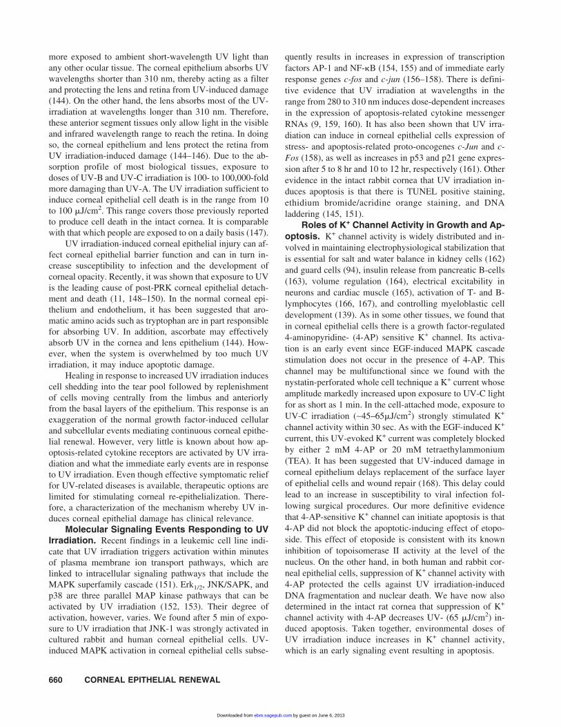
sult from induction of increases in protein kinase C (PKC)activity through activating PLC. Activation of PKC caninitially activate Raf-1 in many cell types and subsequentlyMAPKKK and Erks (i.e., MAPK) (113, 114, 126, 127). Assummarized in Figure 1, our data suggest that the level ofRaf-1 activation is determined by the balance between EGF-induced stimulation of Ras and cPLA2, which in turn affectsthe extent of Erk-2 activation and ultimately the size of themitogenic response (128).
Effect of ET-1 on Corneal Epithelial Cell Prolif-eration. ET-1 stimulates proliferation and wound healingin a variety of cells including human ovarian carcinomacells, Swiss 3T3 cells, vascular smooth muscle cells, andepithelial cells of the thymus (129–132). ET-1 has also beenshown to stimulate DNA synthesis and proliferation in vas-cular smooth muscle cells (129), OVCA 433 cells (131),Swiss 3T3 cells (130), and epithelial cells of the thymus(132).
Since ET-1 is also found in human tears and rabbitlacrimal gland, studies were performed examining the effectof ET-1 on corneal epithelial wound healing (90, 133). ET-1is a potent mitogen for corneal wound healing in rabbits andin cultured bovine corneal cells and it has a permissiveeffect on the mitogenic response to EGF (134, 135). Thiseffect of ET-1 occurs through ETA and ETB receptor-mediated increases in Ca2+ levels that are a consequence ofcalcium-induced calcium release from intracellular stores(136). In situ there is suggestive evidence that each of thesereceptor subtypes may elicit control over different aspectsof corneal epithelial renewal. This appears tenable becauseETA and ETB receptor distribution is not homogeneous. Theproliferating basal layer contains much higher levels of ETA
gene expression, whereas in the differentiating suprabasallayers, ETB expression predominates (134).
In some other tissues, the cell signaling componentsmediating the mitogenic response to EGF includes activa-
Figure 1. Summary of EGF-induced signaling pathways in RCEcells. Erk, MAP kinase; PGE2R, prostaglandin E2 receptor; PLA,phospholipase A; and Raf, MAPK kinase kinase.
658 CORNEAL EPITHELIAL RENEWAL
by guest on June 6, 2013ebm.sagepub.comDownloaded from
tion of membrane ion transport pathways. These events in-clude stimulation of various calcium channels and, in ML-1cells, a growth factor-mediated K+ channel (137–140). Wealso have preliminary data that in rabbit corneal epithelialcells EGF induces K+ channel activation. In addition toincreases in plasma membrane K+ channel activity, EGFincreases in corneal epithelial cells Na:K:2Cl cotransportactivity based on preliminary data obtained from measure-ments of its effects on bumetanide-sensitive 86Rb influx;intracellular Na+ concentration, and cell volume with con-focal microscopy (P. Reinack unpublished data).
Effect of Other Cytokines on Corneal EpithelialCell Proliferation. In primary epithelial cultures of hu-man cornea, there is expression of a myriad of cytokinereceptors and their ligands (141). Northern hybridizationwith oligonucleotide and cDNA probes to a total of 25cytokines and 12 of their receptors revealed cytokines thatcould be divided into four different expression patterns.Type I: TGF-�, IL-1 �, and PDGF-� are expressed exclu-sively by cornea epithelial cells, but their respective recep-tors EGFR and IL-1R are predominantly expressed by fi-broblasts. PDGFR-� is exclusively expressed by fibroblasts.Type II: IGF-1, TGF-�1, -�2, LIF, and bFGF and theirreceptors are expressed by both epithelial cells and fibro-blasts. Type III: KGF and HGF and their respective recep-tors are expressed exclusively by fibroblasts. Type IV: M-CSF, IL-6, and IL-8 are expressed by fibroblasts and/orepithelial cells, but their receptors are not expressed byeither epithelial or fibroblast cells. On the other hand, theyare expressed by immune or inflammatory cells. These andother cytokines have been found in both human and rabbittears (88, 133, 142). In preoperative and postoperativeanalysis of tear fluid following photoreactive keratectomy(PRK), cytokine release rates increased significantly duringthe first 2 days after PRK and returned to preoperative lev-els by Day 7 (133). These observations suggest that cornealwounding induces an increased release of several growthmodulating cytokines (e.g., PDGF-BD, HGF, TGF-� andTNF-�) that may be involved in wound healing. Many ofthese cytokines have been linked to the regulation of pro-liferation in many cell types. PDGF has been shown to havemitogenic effects via a receptor-mediated activation of aphospholipase C-IP3 pathway (143). TGF-� has beenshown to promote proliferation and lamellar differentiationof corneal epithelial cells through keratocyte-mediatedstimulation (93).
The distribution expression patterns of the above-mentioned cytokine receptors and their ligands by epithelialcells and stromal fibroblasts suggests that epithelial growthcontrol depends on epithelial stromal interactions. This isindicated since in a bovine corneal epithelium reconstruc-tion (multilayer) culture system, EGF, HGF, and TGF-�1appear to effectively elicit full-thickness structure for theepithelium through their control of proliferation and differ-entiation (93). EGF has been shown to cause proliferation of
embryonic cornea epithelium in organ culture and increasedepithelial resurfacing rates in adult rabbit tissue (32, 35, 36).
Corneal Epithelial Cell ApoptosisThe physiological balance between the ability of the
corneal epithelium to proliferate and turnover contributes tothe maintenance of corneal deturgescence, transparency,and normal vision (7–11). In a clinical setting, disturbancesin epithelial renewal can compromise corneal structure andfunction, which can in turn lead to infection, developmentof corneal opacity, and loss of vision. Therefore, there havebeen a number of studies directed towards understandingthe role of cytokines in eliciting corneal apoptosis.
Apoptosis-Related Cytokines in Corneal Epi-thelial Cell and Tears. Some of the cytokines expressedin corneal epithelial cells and in tears during the woundhealing process are apoptosis-related. Those found in hu-man and rabbit tears include HGF, TNF�, TGF�-1, TGF�-2, VEGF, PDGF-BB, IL-6, bFGF, EGF, and ET-1 (88–92,142). A number of them stimulate epithelial growth,whereas others can trigger epithelial cell apoptosis. BesidesEGF, the cell death-related cytokines include FGF, IL-1�,IL-6, IL-8, and TNF�. It will be of interest to determine ina reconstruction (multilayer) culture system whether thereare any dose-dependent interactions between EGF and theabove-mentioned cytokines that could modulate their indi-vidual effects on growth and apoptosis.
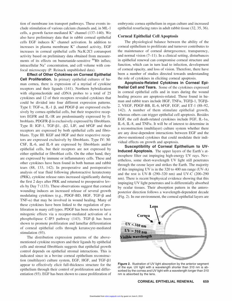
Susceptibility of Corneal Epithelium to UV-Induced Apoptosis. The upper layers of the Earth’s at-mosphere filter out impinging high-energy UV rays. Nev-ertheless, some short-wavelength UV light still penetratesthrough the ozone layer and strikes the Earth. The majorityof this impinging UV is in the 320 to 400 nm range (UV-A)and the rest is UV-B (290–320 nm) and UV-C (200–290nm). There is recent biophysical evidence showing that thisimpinging UV light penetrates and is differentially absorbedby ocular tissues. Their absorption pattern in the antero-posterior direction follows a wavelength-dependent decade(Fig. 2). In our environment, the corneal epithelial layers are
Figure 2. Illustration of UV light absorption by the anterior segmentof the eye. UV light with a wavelength shorter than 310 nm is ab-sorbed by the cornea and UV light with a wavelength longer than 310nm is absorbed by the lens.
CORNEAL EPITHELIAL RENEWAL 659
by guest on June 6, 2013ebm.sagepub.comDownloaded from
more exposed to ambient short-wavelength UV light thanany other ocular tissue. The corneal epithelium absorbs UVwavelengths shorter than 310 nm, thereby acting as a filterand protecting the lens and retina from UV-induced damage(144). On the other hand, the lens absorbs most of the UV-irradiation at wavelengths longer than 310 nm. Therefore,these anterior segment tissues only allow light in the visibleand infrared wavelength range to reach the retina. In doingso, the corneal epithelium and lens protect the retina fromUV irradiation-induced damage (144–146). Due to the ab-sorption profile of most biological tissues, exposure todoses of UV-B and UV-C irradiation is 100- to 100,000-foldmore damaging than UV-A. The UV irradiation sufficient toinduce corneal epithelial cell death is in the range from 10to 100 �J/cm2. This range covers those previously reportedto produce cell death in the intact cornea. It is comparablewith that which people are exposed to on a daily basis (147).
UV irradiation-induced corneal epithelial injury can af-fect corneal epithelial barrier function and can in turn in-crease susceptibility to infection and the development ofcorneal opacity. Recently, it was shown that exposure to UVis the leading cause of post-PRK corneal epithelial detach-ment and death (11, 148–150). In the normal corneal epi-thelium and endothelium, it has been suggested that aro-matic amino acids such as tryptophan are in part responsiblefor absorbing UV. In addition, ascorbate may effectivelyabsorb UV in the cornea and lens epithelium (144). How-ever, when the system is overwhelmed by too much UVirradiation, it may induce apoptotic damage.
Healing in response to increased UV irradiation inducescell shedding into the tear pool followed by replenishmentof cells moving centrally from the limbus and anteriorlyfrom the basal layers of the epithelium. This response is anexaggeration of the normal growth factor-induced cellularand subcellular events mediating continuous corneal epithe-lial renewal. However, very little is known about how ap-optosis-related cytokine receptors are activated by UV irra-diation and what the immediate early events are in responseto UV irradiation. Even though effective symptomatic relieffor UV-related diseases is available, therapeutic options arelimited for stimulating corneal re-epithelialization. There-fore, a characterization of the mechanism whereby UV in-duces corneal epithelial damage has clinical relevance.
Molecular Signaling Events Responding to UVIrradiation. Recent findings in a leukemic cell line indi-cate that UV irradiation triggers activation within minutesof plasma membrane ion transport pathways, which arelinked to intracellular signaling pathways that include theMAPK superfamily cascade (151). Erk1/2, JNK/SAPK, andp38 are three parallel MAP kinase pathways that can beactivated by UV irradiation (152, 153). Their degree ofactivation, however, varies. We found after 5 min of expo-sure to UV irradiation that JNK-1 was strongly activated incultured rabbit and human corneal epithelial cells. UV-induced MAPK activation in corneal epithelial cells subse-
quently results in increases in expression of transcriptionfactors AP-1 and NF-�B (154, 155) and of immediate earlyresponse genes c-fos and c-jun (156–158). There is defini-tive evidence that UV irradiation at wavelengths in therange from 280 to 310 nm induces dose-dependent increasesin the expression of apoptosis-related cytokine messengerRNAs (9, 159, 160). It has also been shown that UV irra-diation can induce in corneal epithelial cells expression ofstress- and apoptosis-related proto-oncogenes c-Jun and c-Fos (158), as well as increases in p53 and p21 gene expres-sion after 5 to 8 hr and 10 to 12 hr, respectively (161). Otherevidence in the intact rabbit cornea that UV irradiation in-duces apoptosis is that there is TUNEL positive staining,ethidium bromide/acridine orange staining, and DNAladdering (145, 151).
Roles of K+ Channel Activity in Growth and Ap-optosis. K+ channel activity is widely distributed and in-volved in maintaining electrophysiological stabilization thatis essential for salt and water balance in kidney cells (162)and guard cells (94), insulin release from pancreatic B-cells(163), volume regulation (164), electrical excitability inneurons and cardiac muscle (165), activation of T- and B-lymphocytes (166, 167), and controlling myeloblastic celldevelopment (139). As in some other tissues, we found thatin corneal epithelial cells there is a growth factor-regulated4-aminopyridine- (4-AP) sensitive K+ channel. Its activa-tion is an early event since EGF-induced MAPK cascadestimulation does not occur in the presence of 4-AP. Thischannel may be multifunctional since we found with thenystatin-perforated whole cell technique a K+ current whoseamplitude markedly increased upon exposure to UV-C lightfor as short as 1 min. In the cell-attached mode, exposure toUV-C irradiation (∼45–65�J/cm2) strongly stimulated K+
channel activity within 30 sec. As with the EGF-induced K+
current, this UV-evoked K+ current was completely blockedby either 2 mM 4-AP or 20 mM tetraethylammonium(TEA). It has been suggested that UV-induced damage incorneal epithelium delays replacement of the surface layerof epithelial cells and wound repair (168). This delay couldlead to an increase in susceptibility to viral infection fol-lowing surgical procedures. Our more definitive evidencethat 4-AP-sensitive K+ channel can initiate apoptosis is that4-AP did not block the apoptotic-inducing effect of etopo-side. This effect of etoposide is consistent with its knowninhibition of topoisomerase II activity at the level of thenucleus. On the other hand, in both human and rabbit cor-neal epithelial cells, suppression of K+ channel activity with4-AP protected the cells against UV irradiation-inducedDNA fragmentation and nuclear death. We have now alsodetermined in the intact rat cornea that suppression of K+
channel activity with 4-AP decreases UV- (65 �J/cm2) in-duced apoptosis. Taken together, environmental doses ofUV irradiation induce increases in K+ channel activity,which is an early signaling event resulting in apoptosis.
660 CORNEAL EPITHELIAL RENEWAL
by guest on June 6, 2013ebm.sagepub.comDownloaded from

ConclusionStudies of corneal wound healing have yielded useful
information for our understanding on the roles of cytokinein the maintenance of corneal epithelial functions. It shouldbe noted, however, that our knowledge about the roles ofcytokines in corneal wound healing and epithelial homeo-stasis are largely derived from the expression patterns ofligands and their respective receptors as determined by im-munohistochemistry and molecular biology techniques.However, little information is available regarding the pre-cise roles of individual cytokines in corneal morphogenesisduring development and homeostasis in adults. Recently,the use of mutant mice created via transgenic and gene-targeting techniques provides unique opportunities to eluci-date the significance of cytokines in modeling corneal mor-phogenesis during development. The mutant mice oftenmanifest mal development of ocular tissues including thecorneal epithelium. For example, transgenic mice overex-pressing FGF or KGF under the control of the alpha crys-talline promoter are characterized by the existence of exo-crine glandular epithelium overlying the cornea (169). Thedefects of mutant mice often occur early during develop-ment. Thus, it limits the usefulness of the mutant mice inelucidating the role of cytokines in the maintenance of cor-neal homeostasis in adult animals . The creation of inducibleexpression mouse lines using corneal specific promoters,e.g., keratocan (keratocyte-specific) and keratin 12 (cornealepithelium-specific) will allow us to circumvent the pitfallsof transgenic mice that constitutively express cytokines and/or their respective dominant negative mutant receptors.
1. Klyce S. Electrical profiles in the corneal epithelium. J Physiol226:407–429, 1972.
2. Klyce SD. Enhancing fluid secretion by the corneal epithelium. In-vest Ophthalmol Vis Sci 16:968–973, 1977.
3. Maurice DM. The location of the fluid pump in the cornea. J Physiol(Lond) 221:43–54, 1972.
4. Dikstein S, Maurice DM. The metabolic basis to the fluid pump in thecornea. J Physiol (Lond) 221:29–41, 1972.
5. Klyce SD, Wong RKS. Site and mode of adrenaline action on chlo-ride transport across the rabbit corneal epithelium. J Physiol (Lond)266:777–799, 1977.
6. Klyce SD, Crosson CE. Transport processes across the rabbit cornealepithelium: A review. Curr Eye Res 4:323–331, 1985.
7. Mohan RR, Liang Q, Kim WJ, Helena MC, Baerveldt F, Wilson SE.Apoptosis in the cornea: Further characterization of Fas/Fas ligandsystem. Exp Eye Res 65:575–589, 1997.
8. Pepose JS, Ubels JL. The cornea. In: Hart WM, Ed. Adler’s Physi-ology of the Eye (9th ed.). St. Louis: Mosby, 1992.
9. Wilson SE, He YG, Lloyd SA. EGF, EGF receptor, basic FGF, TGFbeta-1, and IL-1 alpha mRNA in human corneal epithelial cells andstromal fibroblasts. Invest Ophthalmol Vis Sci 33:1756–1765, 1992.
10. Wilson SE, Lloyd SA, He YG. EGF, basic FGF, and TGF beta-1messenger RNA production in rabbit corneal epithelial cells. InvestOphthalmol Vis Sci 33:1987–1995, 1992.
11. Wilson SE. Molecular cell biology for the refractive corneal surgeon:Programmed cell death and wound healing. J Refract Surg 13:171–175, 1997.
12. Wilson SE, Chen L, Mohan RR, Liang Q, Liu J. Expression of HGF,KGF, EGF and receptor messenger RNAs following corneal epithe-lial wounding. Exp Eye Res 68:377–397, 1999.
13. Klyce SD. Transport of Na, Cl, and water by the rabbit cornealepithelium at resting potential. Am J Physiol 228:1446–1452, 1975.
14. Kenyon KR. Anatomy and pathology of the ocular surface. Int Oph-thalmol Clin 19:3–35, 1979.
15. Maudgal PC, Missotten L. Superficial keratitis. Bull Soc Belge Oph-thalmol 187:1–192, 1980.
16. Arffa RC. Clinical applications of corneal topographic analysis. Se-min Ophthalmol 6:122–132, 1991.
17. Townsend B. Beyond our clinics: A vision for the future. Am J OccupTher 45:871–873, 1991.
18. Goldberg MF, Bron AJ. Limbal palisades of Vogt. Trans Am Oph-thalmol Soc 80:155–171, 1982.
19. Liu CY, Zhu G, Converse R, Kao CW, Nakamura H, Tseng SC, MuiMM, Seyer J, Justice MJ, Stech ME. Characterization and chromo-somal localization of the cornea-specific murine keratin geneKrt1.12. J Biol Chem 269:24627–24636, 1994.
20. Matic M, Petrov IN, Chen S, Wang C, Dimitrijevich SD, WolosinJM. Stem cells of the corneal epithelium lack connexins and metabo-lite transfer capacity. Differentiation 61:251–260, 1997.
21. Stepp MA, Zhu L, Cranfill R. Changes in beta 4 integrin expressionand localization in vivo in response to corneal epithelial injury. InvestOphthalmol Vis Sci 37:1593–1601, 1996.
22. Ishizaki M, Wakamatsu K, Matsunami T, Yamanaka N, Saiga T,Shimizu Y, Zhu G, Kao WW. Dynamics of the expression of cyto-skeleton components and adherens molecules by fibroblastic cells inalkali-burned and lacerated corneas. Exp Eye Res 59:537–549, 1994.
23. Pfister RR. The healing of corneal epithelial abrasions in the rabbit:A scanning electron microscope study. Invest Ophthalmol 14:648–661, 1975.
24. Kuwabara T, Perkins DG, Cogan DG. Sliding of the epithelium inexperimental corneal wounds. Invest Ophthalmol 15:4–14, 1976.
25. Matsuda M, Ubels JL, Edelhauser HF. A larger corneal epithelialwound closes at a faster rate. Invest Ophthalmol Vis Sci 26:897–900,1985.
26. Crosson CE, Klyce SD, Beuerman RW. Epithelial wound closure inthe rabbit cornea: A biphasic process. Invest Ophthalmol Vis Sci27:464–473, 1986.
27. Jumblatt MM, Neufeld AH. Characterization of cyclic AMP-mediated wound closure of the rabbit corneal epithelium. Curr EyeRes 1:189–195, 1981.
28. Cavanagh HD, Colley AM. Cholinergic, adrenergic, and PGE1 ef-fects on cyclic nucleotides and growth in cultured corneal epithelium.Metab Pediatr Syst Ophthalmol 6:63–74, 1982.
29. Jumblatt MM, Neufeld AH. Beta-adrenergic and serotonergic respon-siveness of rabbit corneal epithelial cells in culture. Invest Ophthal-mol Vis Sci 24:1139–1143, 1983.
30. Walkenbach RJ, Corwin JG, Ye GS. Corneal function after storage incommercial eye bank media. Invest Ophthalmol Vis Sci 32:1551–1557, 1991.
31. Imanishi J, Kamiyama K, Iguchi I, Kita M, Sotozono C, Kinoshita S.Growth factors: Importance in wound healing and maintenance oftransparency of the cornea. Prog Retin Eye Res 19:113–129, 2000.
32. Kawaba T, Nakayasu K, Kanai A. [Effect of human EGF and plasmafibronectin on corneal epithelial regeneration]. Nippon Ganka GakkaiZasshi 88:1237–1249, 1984.
33. Ubels JL, Edelhauser HF, Foley KM, Liao JC, Gressel P. The effi-cacy of retinoic acid ointment for treatment of xerophthalmia andcorneal epithelial wounds. Curr Eye Res 4:1049–1057, 1985.
34. Takagi C, King GL, Clermont AC, Cummins DR, Takagi H, BursellSE. Reversal of abnormal retinal hemodynamics in diabetic rats byacarbose, an alpha-glucosidase inhibitor. Curr Eye Res 14:741–749,1995.
35. Kandarakis AS, Page C, Kaufman HE. The effect of epidermalgrowth factor on epithelial healing after penetrating keratoplasty inhuman eyes. Am J Ophthalmol 98:411–415, 1984.
36. Elliott JH. Epidermal growth factor: In vivo ocular studies. Trans AmOphthalmol Soc 78:629–656, 1980.
37. Soong HK. Vinculin in focal cell-to-substrate attachments of spread-ing corneal epithelial cells. Arch Ophthalmol 105:1129–1132, 1987.
38. Gipson IK, Kiorpes TC, Brennan SJ. Epithelial sheet movement:effects of tunicamycin on migration and glycoprotein synthesis. DevBiol 101:212–220, 1984.
CORNEAL EPITHELIAL RENEWAL 661
by guest on June 6, 2013ebm.sagepub.comDownloaded from

39. Gipson IK, Kiorpes TC. Epithelial sheet movement: protein and gly-coprotein synthesis. Dev Biol 92:259–262, 1982.
40. Zieske JD, Gipson IK. Protein synthesis during corneal epithelialwound healing. Invest Ophthalmol Vis Sci 27:1–7, 1986.
41. Panjwani N, Michalopoulos G, Song J, Zaidi TS, Yogeeswaran G,Baum J. Neutral glycolipids of migrating and nonmigrating rabbitcorneal epithelium in organ and cell culture. Invest Ophthalmol VisSci 31:689–695, 1990.
42. Clark EA, Brugge JS. Integrins and signal transduction pathways:The road taken. Science 268:233–239, 1995.
43. Lida Y, Masuda T. Quantification analysis of translation initiationsignal sequences in vertebrate mRNAs. Nucleic Acids Symp Ser34:103–104, 1995.
44. Guo J, Thinakaran G, Guo Y, Sisodia SS, Yu FX. A role for amyloidprecursor-like protein 2 in corneal epithelial wound healing. InvestOphthalmol Vis Sci 39:292–300, 1998.
45. Zieske JD, Bukusoglu G, Gipson IK. Enhancement of vinculin syn-thesis by migrating stratified squamous epithelium. J Cell Biol109:571–576, 1989.
46. Yu FX, Gipson IK, Guo Y. Differential gene expression in healing ratcorneal epithelium. Invest Ophthalmol Vis Sci 36:1997–2007, 1995.
47. Wilson LK, Tyler JW, Besser TE, Parish SM, Gant R. Prediction ofserum IgG1 concentration in beef calves based on age and serumgamma-glutamyl-transferase activity. J Vet Intern Med 13:123–125,1999.
48. Sandbrink R, Masters CL, Beyreuther K. Complete nucleotide anddeduced amino acid sequence of rat amyloid protein precursor-likeprotein 2 (APLP2/APPH): Two amino acids length difference tohuman and murine homologues. Biochim Biophys Acta 1219:167–170, 1994.
49. Sandbrink R, Masters CL, Beyreuther K. APP gene family: Uniqueage-associated changes in splicing of Alzheimer’s betaA4-amyloidprotein precursor. Neurobiol Dis 1:13–24, 1994.
50. Kao WW, Kao CW, Kaufman AH, Kombrinck KW, Converse RL,Good WV, Bugge TH, Degen JL. Healing of corneal epithelial de-fects in plasminogen- and fibrinogen-deficient mice. Invest Ophthal-mol Vis Sci 39:502–508, 1998.
51. Kao WW, Liu CY, Converse RL, Shiraishi A, Kao CW, Ishizaki M,Doetschman T, Duffy J. Keratin 12-deficient mice have fragile cor-neal epithelia. Invest Ophthalmol Vis Sci 37:2572–2584, 1996.
52. Pangalos MN, Shioi J, Robakis NK. Expression of the chondroitinsulfate proteoglycans of amyloid precursor (appican) and amyloidprecursor-like protein 2. J Neurochem 65:762–769, 1995.
53. Thinakaran G, Slunt HH, Sisodia SS. Novel regulation of chondroitinsulfate glycosaminoglycan modification of amyloid precursor proteinand its homologue, APLP2. J Biol Chem 270:16522–16525, 1995.
54. Pettway Z, Domowicz M, Schwartz NB, Bronner-Fraser M. Age-dependent inhibition of neural crest migration by the notochord cor-relates with alterations in the S103L chondroitin sulfate proteoglycan.Exp Cell Res 225:195–206, 1996.
55. Milev P, Friedlander DR, Sakurai T, Karthikeyan L, Flad M, Mar-golis RK, Grumet M, Margolis RU. Interactions of the chondroitinsulfate proteoglycan phosphacan, the extracellular domain of a re-ceptor-type protein tyrosine phosphatase, with neurons, glia, and neu-ral cell adhesion molecules. J Cell Biol 127:1703–1715, 1994.
56. Jackson RL, Busch SJ, Cardin AD. Glycosaminoglycans: Molecularproperties, protein interactions, and role in physiological processes.Physiol Rev 71:481–539, 1991.
57. Henke CA, Roongta U, Mickelson DJ, Knutson JR, McCarthy JB.CD44-related chondroitin sulfate proteoglycan, a cell surface recep-tor implicated with tumor cell invasion, mediates endothelial cellmigration on fibrinogen and invasion into a fibrin matrix. J ClinInvest 97:2541–2552, 1996.
58. Nakamura K, Iwamoto R, Mekada E. Membrane-anchored heparin-binding EGF-like growth factor (HB-EGF) and diphtheria toxin re-ceptor-associated protein (DRAP27)/CD9 form a complex with inte-grin alpha 3 beta 1 at cell-cell contact sites. J Cell Biol 129:1691–1705, 1995.
59. Allen-Hoffmann BL, Schlosser SJ, Brondyk WH, Fahl WE. Fibro-nectin levels are enhanced in human fibroblasts overexpressing thec-sis protooncogene. J Biol Chem 265:521925, 1990.
60. Dean DC, Newby RF, Bourgeois S. Regulation of fibronectin bio-synthesis by dexamethasone, transforming growth factor beta, andcAMP in human cell lines. J Cell Biol 106:2159–2170, 1988.
61. Mathers WD, Sherman M, Fryczkowski A, Jester JV. Dose-dependent effects of epidermal growth factor on corneal wound heal-ing. Invest Ophthalmol Vis Sci 30:2403–2406, 1989.
62. Mustoe TA, Pierce GF, Thomason A, Gramates P, Sporn MB, DeuelTF. Accelerated healing of incisional wounds in rats induced bytransforming growth factor-beta. Science 237:1333–1336, 1987.
63. Maldonado BA, Furcht LT. Epidermal growth factor stimulates in-tegrin-mediated cell migration of cultured human corneal epithelialcells on fibronectin and arginine-glycine-aspartic acid peptide. InvestOphthalmol Vis Sci 36:2120–2126, 1995.
64. Shimizu Y. Localization of neuropeptides in the cornea and uvea ofthe rat: An immunohistochemical study. Cell Mol Biol 28:103–110,1982.
65. Tervo K, Tervo T, Eranko L, Eranko O. Substance P immunoreactivenerves in the rodent cornea. Neurosci Lett 25:95–97, 1981.
66. Hartschuh W, Weihe E, Reinecke M. Peptidergic (neurotensin, VIP,substance P) nerve fibres in the skin: Immunohistochemical evidenceof an involvement of neuropeptides in nociception, pruritus and in-flammation. Br J Dermatol 109(Suppl 25):14–17, 1983.
67. McGillis JP, Organist ML, Payan DG. Substance P and immunoreg-ulation. Fed Proc 46:196–199, 1987.
68. Pernow B. Substance P. Pharmacol Rev 35:85–141, 1983.69. Pernow B. Substance P: a putative mediator of antidromic vasodila-
tion. Gen Pharmacol 14:13–16, 1983.70. Wallengren J, Hakanson R. Effects of substance P, neurokinin A and
calcitonin gene-related peptide in human skin and their involvementin sensory nerve-mediated responses. Eur J Pharmacol 143:267–273,1987.
71. Payan DG, McGillis JP, Renold FK, Mitsuhashi M, Goetzl EJ. Neu-ropeptide modulation of leukocyte function. Ann N Y Acad Sci496:182–191, 1987.
72. Nishiyama A, Mochizuki M, Masuda K. Effects of substance P on theisolated iris sphincter muscle of the albino rabbit. Jpn J Ophthalmol26:29–36, 1982.
73. Unger WG, Butler JM, Cole DF, Bloom SR, McGregor GP. Sub-stance P, vasoactive intestinal polypeptide (VIP) and somatostatinlevels in ocular tissue of normal and sensorily denervated rabbit eyes.Exp Eye Res 32:797–801, 1981.
74. Keen P, Tullo AB, Blyth WA, Hill TJ. Substance P in the mousecornea: Effects of chemical and surgical denervation. Neurosci Lett29:231–235, 1982.
75. Dawson CR, Togni B. Herpes simplex eye infections: Clinical mani-festations, pathogenesis and management. Surv Ophthalmol 21:121–135, 1976.
76. Mackie IA. Role of the corneal nerves in destructive disease of thecornea. Trans Ophthalmol Soc U K 98:343–347, 1978.
77. Lauweryns B, van den Oord JJ, Volpes R, Foets B, Missotten L.Distribution of very late activation integrins in the human cornea: Animmunohistochemical study using monoclonal antibodies. InvestOphthalmol Vis Sci 32:2079–2085, 1991.
78. Tervo K, van Setten GB, Beuerman RW, Virtanen I, Tarkkanen A,Tervo T. Expression of tenascin and cellular fibronectin in the rabbitcornea after anterior keratectomy: Immunohistochemical study ofwound healing dynamics. Invest Ophthalmol Vis Sci 32:2912–2918,1991.
79. Stepp MA, Spurr-Michaud S, Gipson IK. Integrins in the woundedand unwounded stratified squamous epithelium of the cornea. InvestOphthalmol Vis Sci 34:1829–1844, 1993.
80. Stepp MA, Zhu L, Sheppard D, Cranfill RL. Localized distribution ofalpha 9 integrin in the cornea and changes in expression duringcorneal epithelial cell differentiation. J Histochem Cytochem43:353–362, 1995.
81. Harik SI, Kalaria RN, Whitney PM, Andersson L, Lundahl P, Led-better SR, Perry G. Glucose transporters are abundant in cells with“occluding” junctions at the blood-eye barriers. Proc Natl Acad SciU S A 87:4261–4264, 1990.
82. Gherzi R, Melioli G, De Luca M, D’Agostino A, Guastella M, Tra-verso CE, D’Anna F, Franzi AT, Cancedda R. High expression levelsof the “erythroid/brain” type glucose transporter (GLUT1) in thebasal cells of human eye conjunctiva and oral mucosa reconstituted inculture. Exp Cell Res 195:230–236, 1991.
83. Kaulen P, Kahle G, Keller K, Wollensak J. Autoradiographic map-ping of the glucose transporter with cytochalasin B in the mammalianeye. Invest Ophthalmol Vis Sci 32:1903–1911, 1991.
662 CORNEAL EPITHELIAL RENEWAL
by guest on June 6, 2013ebm.sagepub.comDownloaded from

84. Mantych GJ, Hageman GS, Devaskar SU. Characterization of glu-cose transporter isoforms in the adult and developing human eye.Endocrinology 133:600–607, 1993.
85. Kumagai AK, Glasgow BJ, Pardridge WM. GLUT1 glucose trans-porter expression in the diabetic and nondiabetic human eye. InvestOphthalmol Vis Sci 35:2887–2894, 1994.
86. Fischbarg J, Kuang KY, Vera JC, Arant S, Silverstein SC, Loike J,Rosen OM. Glucose transporters serve as water channels. Proc NatlAcad Sci U S A 87:3244–3247, 1990.
87. Tripathi RC, Raja SC, Tripathi BJ. Prospects for epidermal growthfactor in the management of corneal disorders. Surv Ophthalmol34:457–462, 1990.
88. Malecaze F, Simorre V, Chollet P, Tack JL, Muraine M, Le GuellecD, Vita N, Arne JL, Darbon JM. Interleukin-6 in tear fluid afterphotorefractive keratectomy and its effects on keratocytes in culture.Cornea 16:580–587, 1997.
89. Pang IH, Yorio T. Ocular actions of endothelins. Proc Soc Exp BiolMed 215:21–34, 1997.
90. Takashima Y, Takagi H, Takahashi M, Reinach PS, Mircheff AK,Warren DW, Yoshimura N. Endothelin protein expression in tearglands of the rabbit. Curr Eye Res 15:768–773, 1996.
91. Vesaluoma M, Teppo AM, Gronhagen-Riska C, Tervo T. Release ofTGF-beta 1 and VEGF in tears following photorefractive keratec-tomy. Curr Eye Res 16:19–25, 1997.
92. Vesaluoma M, Teppo AM, Gronhagen-Riska C, Tervo T. Platelet-derived growth factor-BB (PDGF-BB) in tear fluid: A potentialmodulator of corneal wound healing following photorefractive kera-tectomy. Curr Eye Res 16:825–831, 1997.
93. Nishimura T, Toda S, Mitsumoto T, Oono S, Sugihara H. Effects ofhepatocyte growth factor, transforming growth factor-beta1 and epi-dermal growth factor on bovine corneal epithelial cells under epithe-lial-keratocyte interaction in reconstruction culture. Exp Eye Res66:105–116, 1998.
94. Schroeder JI, Ward JM, Gassmann W. Perspectives on the physiol-ogy and structure of inward-rectifying K+ channels in higher plants:biophysical implications for K+ uptake. [Review]. Annu Rev Bio-phys Biomol Struct 23:441–471, 1994.
95. Boni-Schnetzler M, Scott W, Waugh SM, DiBella E, Pilch PF. Theinsulin receptor: Structural basis for high affinity ligand binding. JBiol Chem 262:8395–8401, 1987.
96. Wilson SE, He YG, Weng J, Zieske JD, Jester JV, Schultz GS. Effectof epidermal growth factor, hepatocyte growth factor, and keratino-cyte growth factor, on proliferation, motility and differentiation ofhuman corneal epithelial cells. Exp Eye Res 59:665–678, 1994.
97. Yeh YC, Burns ER, Yeh J, Yeh HW. Synergistic effects of endothe-lin-1 (ET-1) and transforming growth factor alpha (TGF-alpha) orepidermal growth factor (EGF) on DNA replication and G1 to Sphase transition. Biosci Rep 11:171–180, 1991.
98. Pimentel JL Jr., Wang S, Martinez-Maldonado M. Regulation of therenal angiotensin II receptor gene in acute unilateral ureteral obstruc-tion. Kidney Int 45:1614–1621, 1994.
99. Buckley A, Davidson JM, Kamerath CD, Wolt TB, Woodward SC.Sustained release of epidermal growth factor accelerates wound re-pair. Proc Natl Acad Sci U S A 82:7340–7344, 1985.
100. Podolsky DK. Healing the epithelium: Solving the problem from twosides. J Gastroenterol 32:122–126, 1997.
101. Kitazawa T, Kinoshita S, Fujita K, Araki K, Watanabe H, Ohashi Y,Manabe R. The mechanism of accelerated corneal epithelial healingby human epidermal growth factor. Invest Ophthalmol Vis Sci31:1773–1778, 1990.
102. Foreman DM, Pancholi S, Jarvis-Evans J, McLeod D, Boulton ME.A simple organ culture model for assessing the effects of growthfactors on corneal re-epithelialization. Exp Eye Res 62:555–564,1996.
103. Kruse FE, Tseng SC. A serum-free clonal growth assay for limbal,peripheral, and central corneal epithelium. Invest Ophthalmol Vis Sci32:2086–2095, 1991.
104. Ullrich A, Schlessinger J. Signal transduction by receptors with ty-rosine kinase activity. Cell 61:203–212, 1990.
105. Margolis B, Bellot F, Honegger AM, Ullrich A, Schlessinger J, Zil-berstein A. Tyrosine kinase activity is essential for the association ofphospholipase C-gamma with the epidermal growth factor receptor.Mol Cell Biol 10:435–441, 1990.
106. Noh DY, Shin SH, Rhee SG. Phosphoinositide-specific phospholi-
pase C and mitogenic signaling. Biochim Biophys Acta 1242:99–113, 1995.
107. Islam M, Akhtar RA. Epidermal growth factor stimulates phospho-lipase cgamma1 in cultured rabbit corneal epithelial cells. Exp EyeRes 70:261–269, 2000.
108. Asaoka Y, Oka M, Yoshida K, Sasaki Y, Nishizuka Y. Role oflysophosphatidylcholine in T-lymphocyte activation: involvement ofphospholipase A2 in signal transduction through protein kinase C.Proc Natl Acad Sci U S A 89:6447–6451, 1992.
109. Nishizuka Y. Intracellular signaling by hydrolysis of phospholipidsand activation of protein kinase C. Science 258:607–614, 1992.
110. Zhang Y, Akhtar RA. Effect of epidermal growth factor on phospha-tidylinositol 3-kinase activity in rabbit corneal epithelial cells. ExpEye Res 63:265–275, 1996.
111. Berridge MJ. Inositol trisphosphate and calcium signaling. Ann N YAcad Sci 766:31–43, 1995.
112. Newton AC. Regulation of protein kinase C. Curr Opin Cell Biol9:161–167, 1997.
113. Marquardt B, Frith D, Stabel S. Signalling from TPA to MAP kinaserequires protein kinase C, raf and MEK: Reconstitution of the sig-nalling pathway in vitro. Oncogene 9:3213–3218, 1994.
114. Marais R, Light Y, Mason C, Paterson H, Olson MF, Marshall CJ.Requirement of Ras-GTP-Raf complexes for activation of Raf-1 byprotein kinase C. Science 280:109–112, 1998.
115. Schonwasser DC, Marais RM, Marshall CJ, Parker PJ. Activation ofthe mitogen-activated protein kinase/extracellular signal- regulatedkinase pathway by conventional, novel, and atypical protein kinase Cisotypes. Mol Cell Biol 18:790–798, 1998.
116. Seger R, Krebs EG. The MAPK signaling cascade. FASEB J 9:726–735, 1995.
117. Davis S, Gale NW, Aldrich TH, Maisonpierre PC, Lhotak V, PawsonT, Goldfarb M, Yancopoulos GD. Ligands for EPH-related receptortyrosine kinases that require membrane attachment or clustering foractivity. Science 266:816–819, 1994.
118. Davis RJ. The mitogen-activated protein kinase signal transductionpathway. [Review]. J Biol Chem 268:14553–14556, 1993.
119. Treisman R. Regulation of transcription by MAP kinase cascades.Curr Opin Cell Biol 8:205–215, 1996.
120. Waskiewicz AJ, Cooper JA. Mitogen and stress response pathways:MAP kinase cascades and phosphatase regulation in mammals andyeast. [Review]. Curr Opin Cell Biol 7:798–805, 1995.
121. Jumblatt MM. Autocrine regulation of corneal endothelium by pros-taglandin E2. Invest Ophthalmol Vis Sci 35:2783–2790, 1994.
122. D’Angelo G, Lee H, Weiner RI. cAMP-dependent protein kinaseinhibits the mitogenic action of vascular endothelial growth factorand fibroblast growth factor in capillary endothelial cells by blockingRaf activation. J Cell Biochem 67:353–366, 1997.
123. Hsi LC, Eling TE. Inhibition of EGF-dependent mitogenesis by pros-taglandin E2 in Syrian hamster embryo fibroblasts. ProstaglandinsLeukot Essent Fatty Acids 58:271–281, 1998.
124. Li X, Zarinetchi F, Schrier RW, Nemenoff RA. Inhibition of MAPkinase by prostaglandin E2 and forskolin in rat renal mesangial cells.Am J Physiol 269:C986–C991, 1995.
125. Mark MD, Storm DR. Coupling of epidermal growth factor (EGF)with the antiproliferative activity of cAMP induces neuronal differ-entiation. J Biol Chem 272:17238–17244, 1997.
126. Ueda Y, Hirai S, Osada S, Suzuki A, Mizuno K, Ohno S. Proteinkinase C activates the MEK-ERK pathway in a manner independentof Ras and dependent on Raf. J Biol Chem 271:2352–2359, 1996.
127. Ueffing M, Lovric J, Philipp A, Mischak H, Kolch W. Protein kinaseC-epsilon associates with the Raf-1 kinase and induces the productionof growth factors that stimulate Raf-1 activity. Oncogene 15:2921–2927, 1997.
128. Kang SS, Li T, Xu D, Reinach PS, Lu L. Inhibitory effect of PGE2on EGF-induced MAP kinase activity and rabbit corneal epithelialproliferation. Invest Ophthalmol Vis Sci 41:2164–2169, 2000.
129. Breuiller-Fouche M, Heluy V, Fournier T, Dallot E, Vacher-LavenuMC, Ferre F. Role of endothelin-1 in regulating proliferation of cul-tured human uterine smooth muscle cells. Mol Hum Reprod 4:33–39,1998.
130. Brown KD, Littlewood CJ. Endothelin stimulates DNA synthesis inSwiss 3T3 cells: Synergy with polypeptide growth factors. BiochemJ 263:977–980, 1989.
131. Bagnato A, Tecce R, Moretti C, Di Castro V, Spergel D, Catt KJ.
CORNEAL EPITHELIAL RENEWAL 663
by guest on June 6, 2013ebm.sagepub.comDownloaded from

Autocrine actions of endothelin-1 as a growth factor in human ovar-ian carcinoma cells. Clin Cancer Res 1:1059–1066, 1995.
132. Malendowicz LK, Macchi V, Brelinska R, Trejer M, Gottardo G,Nussdorfer GG. Endothelin-1 enhances thymocyte proliferation inmonolaterally adrenalectomized rats with contralateral adrenocorticalregeneration. Histol Histopathol 13:721–725, 1998.
133. Vesaluoma MH, Tervo TT. Tenascin and cytokines in tear fluid afterphotorefractive keratectomy. J Refract Surg 14:447–454, 1998.
134. Tao W, Liou GI, Wu X, Abney TO, Reinach PS. ETB and epidermalgrowth factor receptor stimulation of wound closure in bovine cor-neal epithelial cells [published erratum appears in Invest OphthalmolVis Sci 1996 37(10):1937]. Invest Ophthalmol Vis Sci 36:2614–2622, 1995.
135. Takagi H, Reinach PS, Yoshimura N, Honda Y. Endothelin-1 pro-motes corneal epithelial wound healing in rabbits. Curr Eye Res13:625–628, 1994.
136. Tao W, Wu X, Liou GI, Abney TO, Reinach PS. Endothelin receptor-mediated Ca2+ signaling and isoform expression in bovine cornealepithelial cells. Invest Ophthalmol Vis Sci 38:130–141, 1997.
137. Wang L, Xu B, White ER, Lu L. Growth factor-mediated K+ channelactivity associated with human myeloblastic ML-1 cell proliferation.Am J Physiol 273:C1657–C1665, 1997.
138. Xu B, Wilson BA, Lu L. Induction of human myeloblastic ML-1 cellG1 arrest by suppression of K+ channel activity. Am J Physiol 271,1996.
139. Lu L, Yang T, Markakis D, Guggino WB, Craig RW. Alterations ina voltage-gated K+ current during the differentiation of ML-1 humanmyeloblastic leukemia cells. J Membr Biol 132:267–274, 1993.
140. Peppelenbosch MP, Tertoolen LG, de Laat SW. Epidermal growthfactor-activated calcium and potassium channels. J Biol Chem266:19938–19944, 1991.
141. Li DQ, Tseng SC. Three patterns of cytokine expression potentiallyinvolved in epithelial-fibroblast interactions of human ocular surface.J Cell Physiol 163:61–79, 1995.
142. Tervo T, Vesaluoma M, Bennett GL, Schwall R, Helena M, Liang Q,Wilson SE. Tear hepatocyte growth factor (HGF) availability in-creases markedly after excimer laser surface ablation. Exp Eye Res64:501–504, 1997.
143. Knorr M, Steuhl KP, Dartsch P, Thiel HJ. [Activation of phosphati-dylinositol metabolism of corneal epithelial cells by platelet derivedgrowth factor]. Fortschr Ophthalmol 87:649–652, 1990.
144. Ringvold A. Corneal epithelium and UV-protection of the eye. ActaOphthalmol Scand 76:149–153, 1998.
145. Podskochy A, Gan L, Fagerholm P. Apoptosis in UV-exposed rabbitcorneas. Cornea 19:99–103, 2000.
146. Taylor JM, Hildebrand JD, Mack CP, Cox ME, Parsons JT. Charac-terization of graf, the GTPase-activating protein for rho associatedwith focal adhesion kinase: Phosphorylation and possible regulationby mitogen-activated protein kinase. J Biol Chem 273:8063–8070,1998.
147. Tronnier H. [Hazards due to UV-light]. Strahlenschutz Forsch Prax20:36–49, 1980.
148. Johnson DG, Kezirian GM, George SP, Casebeer JC, Ashton J. Re-moval of corneal epithelium with phototherapeutic technique duringmultizone, multipass photorefractive keratectomy. J Refract Surg14:38–48, 1998.
149. Nagy ZZ, Hiscott P, Seitz B, Schlotzer-Schrehardt U, Suveges I,Naumann GO. Clinical and morphological response to UV-B irra-diation after excimer laser photorefractive keratectomy. Surv Oph-thalmol 42(Suppl 1):S64–S76, 1997.
150. Nagy ZZ, Seitz B, Maldonado MJ, Simon M. Changes of the corneal
endothelium after ultraviolet-B exposure in previously photokeratec-tomized eyes. Acta Chir Hung 35:25–332, 1995.
151. Wang L, Xu D, Dai W, Lu L. An ultraviolet-activated K+ channelmediates apoptosis of myeloblastic leukemia cells. J Biol Chem274:678–685, 1999.
152. Bender K, Blattner C, Knebel A, Iordanov M, Herrlich P, RahmsdorfHJ. UV-induced signal transduction. [Review]. J Photochem Photo-biol B Biol 37:1–17, 1997.
153. Derijard B, Hibi M, Wu IH, Barrett T, Su B, Deng T, Karin M, DavisRJ. JNK1: A protein kinase stimulated by UV light and Ha-Ras thatbinds and phosphorylates the c-Jun activation domain. Cell 76:1025–1037, 1994.
154. Herrlich P, Ponta H, Rahmsdorf HJ. DNA damage-induced geneexpression: Signal transduction and relation to growth factor signal-ing. [Review]. Rev Physiol Biochem Pharmacol 119:187–223, 1992.
155. Holbrook NJ, Liu Y, Fornace AJ Jr. Signaling events controlling themolecular response to genotoxic stress. [Review]. Exs 77:273–288,1996.
156. Buscher M, Rahmsdorf HJ, Litfin M, Karin M, Herrlich P. Activationof the c-fos gene by UV and phorbol ester: Different signal trans-duction pathways converge to the same enhancer element. Oncogene3:301–311, 1988.
157. Devary Y, Gottlieb RA, Lau LF, Karin M. Rapid and preferentialactivation of the c-jun gene during the mammalian UV response. MolCell Biol 11:2804–2811, 1991.
158. Gillardon F, Eschenfelder C, Uhlmann E, Hartschuh W, Zimmer-mann M. Differential regulation of c-fos, fosB, c-jun, junB, bcl-2 andbax expression in rat skin following single or chronic ultravioletirradiation and in vivo modulation by antisense oligodeoxynucleotidesuperfusion. Oncogene 9:3219–3225, 1994.
159. Kennedy M, Kim KH, Harten B, Brown J, Planck S, Meshul C,Edelhauser H, Rosenbaum JT, Armstrong CA, Ansel JC. Ultravioletirradiation induces the production of multiple cytokines by humancorneal cells. Invest Ophthalmol Vis Sci 38:2483–2491, 1997.
160. Cubitt CL, Tang Q, Monteiro CA, Lausch RN, Oakes JE. IL-8 geneexpression in cultures of human corneal epithelial cells and kerato-cytes. Invest Ophthalmol Vis Sci 34:3199–3206, 1993.
161. Hermeking H, Lengauer C, Polyak K, He TC, Zhang L, Thiagalin-gam S, Kinzler KW, Vogelstein B. 14-3-3 sigma is a p53-regulatedinhibitor of G2/M progression. Mol Cell 1:3–11, 1997.
162. Wang W, Sackin H, Giebisch G. Renal potassium channels and theirregulation. Annu Rev Physiol 54:81–96, 1992.
163. Dukes ID, Philipson LH. K+ channels: Generating excitement inpancreatic beta-cells. [Review]. Diabetes 45:845–853, 1996.
164. Deutsch C, Chen LQ. Heterologous expression of specific K+ chan-nels in T lymphocytes: Functional consequences for volume regula-tion. Proc Natl Acad Sci U S A 90:10036–10040, 1993.
165. Hille B. Ion Channels of Escitable Membranes. Sinauer AssociatesInc., Sunderland, MA. 1992.
166. DeCoursey TE, Chandy KG, Gupta S, Cahalan MD. Voltage-gatedK+ channels in human T lymphocytes: A role in mitogenesis? Nature307:465–468, 1984.
167. Amigorena S, Choquet D, Teillaud JL, Korn H, Fridman WH. Ionchannels and B cell mitogenesis. Mol Immunol 27:1259–1268, 1990.
168. Wickert H, Zaar K, Grauer A, John M, Zimmermann M, Gillardon F.Differential induction of proto-oncogene expression and cell death inocular tissues following ultraviolet irradiation of the rat eye. Br JOphthalmol 83:225–230, 1999.
169. Lovicu FJ, Kao WW, Overbeek PA. Ectopic gland induction bylens-specific expression of keratinocyte growth factor (FGF-7) intransgenic mice. Mech Dev 88:53, 1999.
664 CORNEAL EPITHELIAL RENEWAL
by guest on June 6, 2013ebm.sagepub.comDownloaded from