An ear punch model for studying the effect of radiation on wound healing

1 23
Lasers in Medical Science ISSN 0268-8921 Lasers Med SciDOI 10.1007/s10103-012-1161-9
Effect of laser and LED phototherapieson the healing of cutaneous wound onhealthy and iron-deficient Wistar rats andtheir impact on fibroblastic activity duringwound healingSusana C. P. Oliveira Sampaio, JulianaS. de C. Monteiro, Maria CristinaT. Cangussú, Gustavo M. Pires Santos,Marcos André Vannier dos Santos, et al.
1 23
Your article is protected by copyright and all
rights are held exclusively by Springer-Verlag
London Ltd. This e-offprint is for personal
use only and shall not be self-archived in
electronic repositories. If you wish to self-
archive your work, please use the accepted
author’s version for posting to your own
website or your institution’s repository. You
may further deposit the accepted author’s
version on a funder’s repository at a funder’s
request, provided it is not made publicly
available until 12 months after publication.

ORIGINAL ARTICLE
Effect of laser and LED phototherapies on the healingof cutaneous wound on healthy and iron-deficient Wistar ratsand their impact on fibroblastic activity during woundhealing
Susana C. P. Oliveira Sampaio & Juliana S. de C. Monteiro &
Maria Cristina T. Cangussú & Gustavo M. Pires Santos &
Marcos André Vannier dos Santos & Jean Nunes dos Santos &
Antonio L. B. Pinheiro
Received: 23 March 2012 /Accepted: 6 July 2012# Springer-Verlag London Ltd 2012
Abstract Iron deficiency impairs the formation of hemoglo-bin, red blood cells, as well the transport of oxygen. Thewound healing process involves numerous functions, manyof which are dependent on the presence of oxygen. Laser hasbeen shown to improve angiogenesis, increases blood supply,cell proliferation and function. We aimed to study the effect of
λ660 nm laser and λ700 nm light-emitting diode (LED) onfibroblastic proliferation on cutaneous wounds on iron-deficient rodents. Induction of iron anemia was carried outby feeding 105 newborn rats with a special iron-free diet. A1×1 cmwound was created on the dorsum of each animal thatwere randomly distributed into seven groups: I, control
S. C. P. Oliveira Sampaio : J. S. de C. Monteiro : J. N. dos Santos :A. L. B. Pinheiro (*)Center of Biophotonics, School of Dentistry,Federal University of Bahia,Av. Araújo Pinho, 62, Canela,Salvador, BA 40110-150, Brazile-mail: [email protected]
S. C. P. Oliveira Sampaioe-mail: [email protected]
J. S. de C. Monteiroe-mail: [email protected]
J. N. dos Santose-mail: [email protected]
M. C. T. CangussúOral Epidemiology and Public Health, School of Dentistry,Federal University of Bahia,Salvador, BA 40110-150, Brazile-mail: [email protected]
G. M. Pires Santos :M. A. V. dos SantosLaboratory of Parasitic Biomorphology,Institute Gonçalo Muniz–FIOCRUZ-BA,Salvador, BA, Brazil 40296-710
G. M. Pires Santose-mail: [email protected]
M. A. V. dos Santose-mail: [email protected]
J. N. dos SantosLaboratory of Surgical Pathology, School of Dentistry,Federal University of Bahia,Av. Araújo Pinho, 62, Canela,Salvador, BA 40110-150, Brazil
J. N. dos Santos :A. L. B. PinheiroNational Institute of Optics and Photonics,Physics Institute of São Carlos, University of São Paulo,São Carlos, SP, Brazil 13560-970
A. L. B. PinheiroInstitute of Biomedical Engineering, Unicastelo,São José dos Campos,São José, SP, Brazil 12245-230
Lasers Med SciDOI 10.1007/s10103-012-1161-9
Author's personal copy

anemic; II, anemic no treatment; III, anemic+L; IV, anemic+LED; V, healthy no treatment; VI, healthy+laser; VII, healthy+LED (n015 each). Phototherapy was carried out using either adiode laser (λ660 nm, 40 mW, 10 J/cm2) or a prototype LEDdevice (λ700±20 nm, 15 mW, 10 J/cm2). Treatment startedimmediately after surgery and was repeated at 48-h intervalduring 7, 14, and 21 days. After animal death, specimens weretaken, routinely processed, cut, stainedwith hematoxylin-eosin,and underwent histological analysis and fibroblast counting.Significant difference between healthy and anemic subjects onregards the number of fibroblast between treatments was seen(p<0.008, p<0.001). On healthy animals, significant highercount was seen when laser was used (p<0.008). Anemic sub-jects irradiated with LED showed significantly higher count(p<0.001). It is concluded that the use of LED light caused asignificant positive biomodulation of fibroblastic proliferationon anemic animals and laser was more effective on increasingproliferation on non-anemics.
Keywords Anemia . Phototherapy .Wound healing
Introduction
The prevalence of iron deficiencies is very high in developingcountries, iron deficiency anemia being the most common ofsuch disorders worldwide. This disease develops itself due toa low ingestion of iron. However, iron deficiency anemia co-exists with other conditions such as vitamin A deficiency,undernourishment, folate deficiency, and infections [1]. Thisanemia may be considered the last stage of a long period ofinsufficient iron intake impairing the formation of hemoglo-bin, which contains iron, by the organism making difficult theformation of red blood cells [1, 2].
Three stages of development of the disease are nec-essary for its clinical manifestation. On the first stage,there is the depletion of the iron that affects its depositon the body and represents the period of a more intensevulnerability on relation to the marginal balance of iron,and it may progress to a more severe deficiency. Thesecond stage is known as iron-deficient erythropoiesisthat is characterized by biochemical changes thatreflects the insufficient level of iron, making the normaland regular production of hemoglobin not possible. Thisis also seen on other iron composites found on theorganism, even when the concentration of hemoglobinis not reduced. The last stage is characterized by the reductionof the level of hemoglobin that causes a severe unbalance ofthe organism. Asmuch reduced, the level of hemoglobinmoresevere the anemia is [1, 2].
Wound healing is a complex process that involves theoccurrence of many phenomena. Most of these phenomenadepend upon the presence of oxygen. The development of
epithelial tissue depends on both the level of hydration andoxygenation the injured site. Angiogenesis is very importanton the healing process and tissue hypoxia has been point outas a critical trigger on the transcription of angiogenic fac-tors. Both production and maturation of the collagen matrixinfluence the tensile strength of the healing tissue and itis directly related to the partial pressure of the oxygenon the tissue [3, 4]. The healing deficiency observed onanemic subjects is due to the insufficient delivery ofoxygen to the tissue as well as to the reduction of thelevels of hemoglobin.
Laser phototherapy (LPT) is a non-invasive treatmentmodality that has been shown very helpful on the healingwounds being capable of increasing both the speed andquality of the healing process. Increasing use of LPT hasbeen shown efficacious on positively modulating woundhealing by stimulating mitotic activity, increased numberof fibroblasts, increased deposition of collagen matrix, in-creased angiogenesis, and others [5, 6].
Low-energy laser irradiation has documented benefits inpromoting the healing of hypoxic, ischemic, and infectedwounds. In comparison to lasers, light-emitting diode (LED)technology generates negligible amounts of heat. It is clini-cally proven to be safe and has achieved nonsignificant riskstatus for human trials by the Food and Drug Administration.LED technology has been shown to be an effective alternativefor the treatment of both cutaneous and mucosal wounds [7].
LEDs present a relatively narrow emission spectrum thatmay be optimally tuned to correspond to treatment’s require-ment and thus eliminate unnecessary wavelengths for thetherapy; they allow adjustment of light intensity and producehigh light levels with low radiant heat output maintaininguseful output for long periods of time. LED-based devicesmay provide a homogenous light dose in optimal intensity [8].
While lasers provide tissue stimulation, which increasecellular activity during wound healing, they have limitationin wavelength. It is difficult for lasers to produce the effi-cient wavelength combination optimal for wound healing.The size of wounds that may be treated by the small beamwidth of laser is also limited. In contrast, LEDs allow thecontrol of spectral composition and may be arranged in flatarrays of all sizes for the treatment of either small or largeareas. LEDs offer an effective alternative to the use of con-ventional light sources [8].
It is known that some wavelengths have the ability tostimulate cell proliferation, including the fibroblasts; andthese cells have the capacity to secrete collagen, a mainorganic component observed during repair [9, 10].
Wewere not able to find any previous report on the literatureon the effects of light sources, such as laser and LEDs, on thehealing process of surgical wounds on cases of iron deficiencyanemia. The aim of the present investigation was to study theeffect of two light sources, λ660 nm laser and λ700 nm LED
Lasers Med Sci
Author's personal copy

light, on the fibroblastic proliferation of cutaneous surgicalwounds on rodents suffering from iron deficiency anemia.
Materials and methods
Following approval by the Animal research Ethical Committeeof the School of Dentistry of the Federal University of Bahia,105 newborn male Wistar rats (21 days old, ±50 g) were usedon the present investigation [11]. The animals were kept undernatural conditions of light, humidity, and temperature at theAnimal House of Center of Biophotonics of the Federal Uni-versity of Bahia during the experimental period. Iron deficiencyanemia was induced by feeding the animals with a specialpelleted iron-free diet (no iron-AIN 93-G, Rhoster, SP, Brazil)during 15 days [12]. Anemia was confirmed by blood testing.The animals were kept in groups of three on individual metallicgages; kept at day/night light cycle and temperature controlledduring the experimental period.
Under intramuscular general anesthesia (0.2 ml/100 mg ofquetamine and 0.06 ml/100 g of xylazine), each animal had thedorsum shaven and the skin cleaned with 2 % chlorohexidinesolution. A 1×1 cm full-thickness excisional wound was creat-ed on the dorsum of each animal with a no15 scalpel blade(Fig. 1). The depth of the woundwas controlled by the cutting tothe depth of the bevel of the scalpel blade that was 1 mm [13].
The animals were randomly distributed into sevengroups: group I, control anemic (no wound, no treatment);group II, anemic no treatment (wound, no treatment); groupIII, anemic+laser; group IV, anemic+LED; group V, healthyno treatment; group VI, healthy+laser; group VII, healthy+LED, (n015 each group).
Phototherapy was carried out using either a diode laser(Twin Flex®, MMOptics, São Carlos, SP, Brazil) or a prototypeLED device (RED-LED, λ700±20 nm, Kondortech, SãoCarlos, SP, Brazil). When the laser was used, the energy wasdelivered to the tissue in four points around the wound (NSEW,4×2.5 J/cm2). Due to the size of the probe of the LED device anadaptor was used to reduce the illuminated area to 1 cm2, and
the energy was delivered by contact mode in a single point overthe wound. The probes of both devices were covered with asingle-use PVC film protection and placed directly in contactwith the tissue. As we used two different light sources, weopted to use the energy as a comparative parameter between thetwo treatments. The parameters of the treatments may be seenon Table 1. Treatment started immediately after surgery andwas repeated at 48-h interval during 7, 14, and 21 days [13].
At the end of each experimental time, the animals werekilled by an overdose of general anesthetics. The specimenswere taken and fixed during 24 h on a 10 % formalin solution,routinely processed to wax, cut and stained with hematoxylin-eosin, and underwent histological analysis by light microcopy(Axiolab®, ZEISS, Germany) at the Laboratory of SurgicalPathology of the School of Dentistry of the Federal Universityof Bahia. For descriptive analysis, five slides from each groupwere used [13]. Fibroblast counting was used as assessmentparameter. Each slide was divided into six size-standardizedfields located just below the healing epithelium and countingwas always carried out on the two central fields. The resultswere statistically analyzed using Minitab 15® software (Mini-tab, Belo Horizonte, MG, Brazil). Statistical analysis was car-ried out using Mann–Whitney test.
Results
Histological findings were as follows:Untreated non-anemic animals showed, on day 7,
wounds covered by keratinized squamous epithelium whosedermis showed, sometimes, spindle-shaped fibroblasts, col-lagen fibers arranged parallel to the surface, as well ascongested blood vessels and discrete chronic inflammation.On day 14, the wounds were histologically similar to thoseseen on day 7. However, inflammatory cells were rarelyseen. On day 21, the wounds were covered by keratinizedepithelium with flat interface; on dermis, angled, and
Fig. 1 Wound model used on the study
Table 1 Summary of the parameters used on the study
Parameters Groups
Red LED Laser
Wavelength (nm) 700±20 660
Mode CW
Spot of the probe (cm²) 1 0.04
Power output (W) 0.015 0.040
Exposure time (s, per session) 670 s 260
Power density (W/cm2) 0.015 1
Illuminated area (cm²) 1 0.04
Energy density per session (J) 10 10
Lasers Med Sci
Author's personal copy
spindle-shaped fibroblasts and collagen fibers arranged notparallel to the surface were also seen (Fig. 2a).
Untreated anemic animals showed, on day 7, woundscovered by keratinized-stratified squamous epithelium exhib-iting atrophy, crust, and fibrinoid necrosis. The dermis showedproliferating angular or oval-shaped fibroblasts, collagenfibers arranged parallel or not to the surface, and congestedblood vessels. At both 14 and 21 days, the specimens showedwounds covered by keratinized epithelium exhibiting atrophy.The dermis showed oval- and spindle-shaped fibroblastsamidst collagen fibers arranged parallel to the surface. Bloodvessels eventually congested were also seen (Fig. 2b).
LED-treated non-anemic animals showed, on day 7, themajority of surgical wounds covered by keratinized epithe-lium and, eventually, exhibiting ulceration and extensiveepithelium migration. The dermis showed proliferation ofoval and spindle fibroblasts and collagen fibers not parallelto the surface. Sometimes, congested blood vessels and mildchronic inflammatory infiltrate were seen scattered withinthe wound. On day 14, the majority of surgical wounds werecovered by keratinized epithelium. The dermis showedfibroblasts and collagen fibers parallel to the surface.Amidst dermis, there were small blood vessels, and a fewcongested and discrete chronic inflammatory infiltrate. Atthe end of the experimental period, the specimens coveredby epithelium of regular thickness whose dermis had dermalspindle-shaped fibroblasts and collagen fibers parallel tothe surface (Fig. 2c).
Laser-treated non-anemic animals showed, on day 7,wounds covered by keratinized epithelium whose dermisshowed oval- and spindle-shaped fibroblasts with collagenfibers arranged parallel to the surface, and discrete chronicinflammatory infiltrate. On day 14, the specimens showedthe wounds covered similarly as described on previousperiod. On day 21, the specimens showed similar aspect tothe specimens observed on day 7, but there was no inflam-mation (Fig. 2d).
Anemic animals treated with LED showed, on both days 7and 14, wounds covered by keratinized epithelium. Thedermis showed fibroblasts and collagen fibers arranged par-allel to the surface. In both periods of time, inflammationwas rarely seen, but congested blood vessels were present.At the end of the experimental period, the specimensshowed the wounds covered as described on previous peri-od. Angular and spindle-shaped fibroblasts with collagenfibers arranged parallel to the surface slightly were seenscattered on the dermis (Fig. 2e).
Anemic animals treated with laser, showed on day 7, thatmost specimens were covered by keratinized epitheliumexhibiting atypical reactive. The dermis showed spindle-shaped fibroblasts among collagen fibers parallel to thesurface. There were very congested blood vessels and mildto moderate chronic inflammatory infiltrate. On day 14, the
specimens showed the wound covered by keratinized epi-thelium regularly distributed with dermis similarly distrib-uted as described on the previous period. On day 21, thespecimens showed the wounds covered by keratinized epi-thelium that showed dermis, fibroblasts, and collagen fibersgenerally arranged parallel to the surface. In addition, therewere congested blood vessels (Fig. 2f).
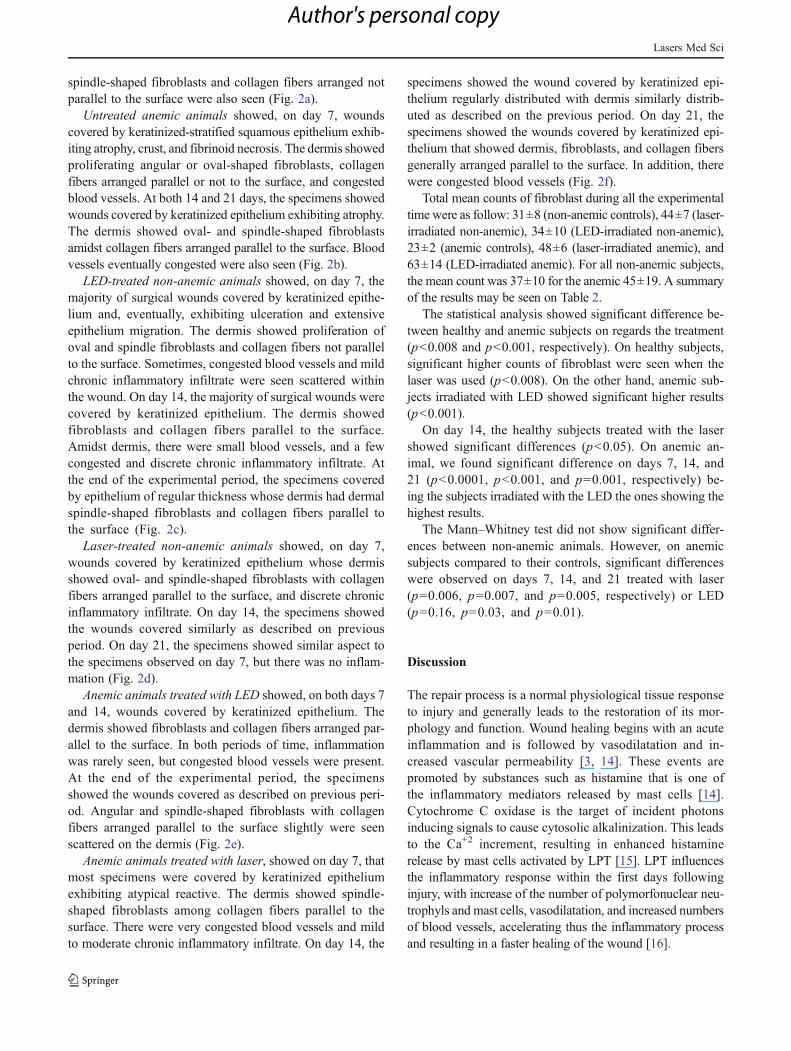
Total mean counts of fibroblast during all the experimentaltime were as follow: 31±8 (non-anemic controls), 44±7 (laser-irradiated non-anemic), 34±10 (LED-irradiated non-anemic),23±2 (anemic controls), 48±6 (laser-irradiated anemic), and63±14 (LED-irradiated anemic). For all non-anemic subjects,the mean count was 37±10 for the anemic 45±19. A summaryof the results may be seen on Table 2.
The statistical analysis showed significant difference be-tween healthy and anemic subjects on regards the treatment(p<0.008 and p<0.001, respectively). On healthy subjects,significant higher counts of fibroblast were seen when thelaser was used (p<0.008). On the other hand, anemic sub-jects irradiated with LED showed significant higher results(p<0.001).
On day 14, the healthy subjects treated with the lasershowed significant differences (p<0.05). On anemic an-imal, we found significant difference on days 7, 14, and21 (p<0.0001, p<0.001, and p00.001, respectively) be-ing the subjects irradiated with the LED the ones showing thehighest results.
The Mann–Whitney test did not show significant differ-ences between non-anemic animals. However, on anemicsubjects compared to their controls, significant differenceswere observed on days 7, 14, and 21 treated with laser(p00.006, p00.007, and p00.005, respectively) or LED(p00.16, p00.03, and p00.01).
Discussion
The repair process is a normal physiological tissue responseto injury and generally leads to the restoration of its mor-phology and function. Wound healing begins with an acuteinflammation and is followed by vasodilatation and in-creased vascular permeability [3, 14]. These events arepromoted by substances such as histamine that is one ofthe inflammatory mediators released by mast cells [14].Cytochrome C oxidase is the target of incident photonsinducing signals to cause cytosolic alkalinization. This leadsto the Ca+2 increment, resulting in enhanced histaminerelease by mast cells activated by LPT [15]. LPT influencesthe inflammatory response within the first days followinginjury, with increase of the number of polymorfonuclear neu-trophyls and mast cells, vasodilatation, and increased numbersof blood vessels, accelerating thus the inflammatory processand resulting in a faster healing of the wound [16].
Lasers Med Sci
Author's personal copy

Fig. 2 Histophatological aspects of the healing. a Untreated non-anemic animals: wound covered by keratinized stratified squa-mous epithelium exhibiting atrophy whose dermis shows an in-tense interstitial edema, tortuous and congested blood vessels anda relatively fewer collagen fibers. A mild to moderate amount ofmononuclear inflammatory cells were also seen. b Untreatedanemic animals: wound showing ulceration covered by fibrinousnecrosis and crust. The dermis showed a noncompletely organizedgranulation tissue in which fibroblasts were seen mostly parallelto the surface. Congested blood vessels were also seen. c LEDtreated non-anemic animals: wound covered by keratinized strat-ified squamous epithelium, whose dermis shows angular-shapedfibroblasts non-organized and congested and tortuous blood ves-sels amidst collagen fibers and rare mononuclear inflammatorycells. d Laser treated non-anemic animals: wound covered by
keratinized stratified squamous epithelium, whose dermis showsa high number of spindle-shaped fibroblasts organized parallel tothe surface. Extravased red blood cells were also seen on super-ficial dermis. e Anemic animals treated with LED: wound coveredpartially by keratinized stratified squamous epithelium exhibitingstrong atrophy and ulceration recovered by thick crust, whosedermis shows a high number of spindle-shaped fibroblasts orga-nized parallel to the surface amidst organized collagen fibers.Note absence of extravased red blood cells and rare inflammatorycells. f Anemic animals treated with laser: wound covered par-tially by keratinized stratified squamous epithelium exhibitingintense hyperplasia, whose dermis shows a moderate number ofspindle-shaped fibroblasts organized parallel to the surface amidstorganized collagen fibers. Note presence of tortuous blood redvessels eventually congested
Lasers Med Sci
Author's personal copy
Anemias are very common disorders worldwide and noprevious studies were found evaluating the effect of laser andLED on the fibroblastic proliferation during wound healing iniron deficiency anemia cases. The present model is new andmay provide a new option of studying the effects of photo-therapies on patients suffering from systemic diseases.
It has been shown that LPT affects most cells involved onthe healing wounds. A previous report showed that the use ofIR (λ980 nm) laser light improves cell growth [17]. Ourresults are aligned with this study as healthy animals treatedwith laser light showed better results on regards the healing.
Some studies have shown that the use of λ630–1,000 nmlaser or LED light, both in vivo and in vitro, causes apositive biomodulatory effect on the healing of wounds[10]. Our results indicate that the use of either laser orLED light causes an increased fibroblastic proliferation thusimproving the healing process.
The correct dosimetric determination of wavelength, flu-ence, and irradiance, as well as for each clinical condition andspecific cellular modulation, is of great relevance. It is knownthat, depending on the fluence delivered at the surface tissue,different tissue responses may be observed during the healingprocess [5, 18].
It seems possible that LED light presents beneficial effectssimilar to the ones observed when laser light is used as severalreports on the benefits of the use of LEDs operating at severalwavelengths have been published elsewhere both in vitro andin vivo on both normal and pathologic conditions [19]. It isalso possible that the mechanism involved being also similar.
Due to distinct light beams, both LED and laser protocolswere adjusted to provide the same energy for both kinds ofequipment allowing us the comparison of the therapeuticeffect of this different light source based upon a sameparameter. The wide absorption window in biological tis-sues exists and the therapeutic action of both light sourceshas been observed [20]. On the present investigation, thesame energy (10 J/cm2) was used for both treatments. Aprevious report [8] has indicated that studies involving theuse of different light sources might use similar energy den-sities and power densities. Despite this, many reports areindicative that the use of some types of light results onimprovement of healing [21]. Furthermore, LED light is asafe and low-cost option of treatment of wounds on skin andmucous membranes [7].
It is known that light coherence is lost in the first layers ofthe tissue [22]. In accordance with the positive stimulus to thefibroblastic proliferation found for both light sources in thisstudy, several studies confirm the positive biomodulation byincoherent light [7, 23]. Our results suggest that the fibroblasticproliferation is probably more dependent on wavelength andfluence than on the coherence factor. The LED emission spec-trum is broader than that of laser and, despite this featuredifferentiating the light sources a similar energy was used, butwith a distinct spectral distribution. Laser was characterized bya higher energy density concentration in a smaller spectralrange of 660 nm. LED energy density was distributed in abroader spectrum of 700 nm, possibly interacting with a highernumber of specific photoreceptors [21].
Our results demonstrated that the use of either λ700±20 nm LED or λ660 nm laser light caused stimulation of thefibroblastic proliferation on both anemic and healthy sub-jects. Many studies have shown that the use of LED light(λ630–1,000 nm) causes positive biomodulatory effects onmany biological processes occurring during wound healing,including on the genic expression [23], angiogenesis [21],on the mitochondrial oxidative metabolism [24], and on theprevention and treatment of oral mucosites [25].
On regards, the effects of LED light on the proliferationof fibroblasts, a previous in vitro study showed that the useof λ625–635 nm LED light was capable of keeping theviability of human fibroblasts as well as its synthesis ofcollagen [26]. A previous study demonstrated also an in-creased fibroblastic proliferation on mice, in vitro, followingthese of a combination of different LED’s (λ670, 728, and880 nm, 4 or 8 J/cm2, 50 mW/cm2) [7]. The study alsoshowed that the stimulation occurred only on the growthphase and not occurred on the stationary phase of the cellcycle, indicating that LED irradiation does not generate anexcessive growth or neoplastic transformation. We studiedthe effect of LED phototherapy of three distinct wavelengthson fibroblast on wound healing, on λ700 nm LED-irradiatedsubjects there was a marked amount of young fibroblaststhat were paralleled located to the surface, on λ530 nmmarked amount of fibroblasts was observed on 75 % ofthe cases and they were parallel to the wound surface andon λ460 nm illuminated subjects, marked amount of fibro-blast was seen on 50 % and these were discretely organizedon the dermis of the wound [27]. Our results are important
Table 2 Mean number of fibro-blast counts on both anemic andnon-anemic subjects treated ornot with laser or LED throughoutthe experimental time
aMean±SD
Group Non-anemic AnemictreatmentTime (days) Controla Lasera LEDa Controla Lasera LEDa
7 28±5 46±11 31±5 23±3 40±1 47±3
14 29±6 44±6 35±5 23±2 47±6 78±15
21 36 ±13 44±5 37±6 16±3 51±7 53±20
Lasers Med Sci
Author's personal copy

as they deal with the effect of two light sources on bothanemic and non-anemic animals. We were not able to findany previous reports on using this model in the literature.
Our results showed significant difference betweenhealthy and anemic subjects on regards the treatment used.We also found that on healthy subjects, significant higherresults on fibroblast proliferation were seen when the laserwas used. On the other hand, anemic subjects irradiated withLED showed significant higher results when compared tonon irradiated ones. On anemic animals, we found signifi-cant differences at days 7, 14, and 21, the subjects beingirradiated with the LED the ones showing the highestcounts. It is important to mention that we did not find signif-icant differences between non-anemic animals. However, onanemic subjects compared to their controls, significant differ-ences were observed at days 7, 14, and 21 treated with laser orLED. This is multistep study and further cellular and molec-ular assessment are planned in order to provide a more con-clusive evidence of the effects of light on this model.
There are still controversies on regards the effects ofsome characteristics of the laser light such as the coherence.It has been shown that the biological effects of the lightdepend mostly on the specific relationship between thewavelength of the light and the photoreceptor molecule.Monochromaticity has been considered the main factor re-sponsible for the positive stimulation of the tissues. Conse-quently, it has been proposed that the LED light, even with itspectral band, possesses similar effects to the laser light [7,26, 28]. Karu [29] has suggested that high monochromatic-ity is not essential and that it is important that the bandwidthto be within the absorption band of the photoreceptor. Wefound that the fibroblastic proliferation on anemic animalstreated with the red LED light showed better results thanwhen the laser was used. This may have been caused by thefact that hemoglobin, photoreceptor molecule that is defi-cient on anemic subjects, absorbs better the LED lightprobably due to greater bandwidth when compared to thelaser [22, 26, 28]. New ATP production occurs rapidly afterLED photomodulation, triggering subsequent metabolic activ-ity of fibroblasts [28].
This study showed positive biomodulatory effects of bothlaser and LED phototherapies when comparing irradiatedand non-irradiated animals. LED (λ700±20 nm) irradiationshowed better results on anemic animals when compared tothe use of laser light. On healthy animals, our results arealigned with previous reports when either laser [22, 30] orLED light were used [21, 31] on regards increased vasodi-lation and microcirculation. These phenomena are importantfor the nutritional support of the healing tissues. A previousreport [31] demonstrated the stimulation of the angiogenesisby the upregulation of both vascular endothelial growthfactor and nitric oxide synthase following the use of laserlight (λ804 nm). Another research [21], by a comparative
study using laser or LED, found no significant differ-ence between the two protocols and suggested that theeffect on angiogenesis could be attributed to the wave-length. This aspect may also be indicative that the sameoccurs on regards fibroblastic activity as found on the presentinvestigation.
Based upon our results, it is concluded that the useofλ700±20 nm LED Light caused a significant positivebiomodulation of the fibroblastic proliferation on anemic sub-jects and laser (λ660) irradiation was more effective on in-creasing proliferation on non-anemic animals.
References
1. Olivares M, Qalter T, Hertrampf E, Pizarro F (1999) Anaemia andiron deficiency disease in children. Br Med Bull 55:534–543
2. Almeida AP, Zandonade E, Abrantes MM, Lamounier JA (2004)Iron deficiency and anemia among children in Vitória, ES[Deficiência de ferro e anemia em crianças de Vitória, ES].Pediatrics 26:140–150
3. Jonshon K, Jensen A, Goodson WH, Scheuenstuhl H, West J, HopfHW, Hunt TK (1991) Tissue oxygenation, anemia, and perfusion inrelation to wound healing in surgical patients. Ann Surg 214:605–613
4. Shweiki D, Neeman M, Itin A, Keshet E (1992) Vascular endo-thelial growth factor induced by hypoxia may mediate hypoxia-initiated angiogenesis. Nature 359:843–845
5. Medrado AP, Pugliese LS, Reis SRA, Andrade ZA (2003)Influence of low level laser therapy on wound healing and itsbiological action upon myofibroblasts. Lasers Med Sci 32:239–244
6. Demir H, Menku P, Kirnap M, Calis M, Ikizceli I (2004)Comparison of the effects of laser, ultrasound, and combinedlaser+ultrasound treatments in experimental tendon healing.Lasers Surg Med 35:84–89
7. Whelan HT, Smits RL, Buchman EV, Whelan NT, Tuner SG,Margolis DA et al (2001) Effect of NASA light-emitting diodeirradiation on wound healing. J Clin Laser Med Surg 19:305–314
8. Barolet D (2008) Light-emitting diodes (LEDs) in dermatology.Semin Cutan Med Surg 27:227–238
9. Corazza AV, Jorge J, Kurachi C, Bagnato VS (2007)Photobiomodulation on the angiogenesis of skin wounds in ratsusing different light sources. Photomed Laser Surg 25:102–106
10. Smith KC (2005) Laser (and LED) therapy is phototherapy.Photomed Laser Surg 23:78–80
11. Murray-Kolb LE, Takaiwa F, Goto F, Yoshihara T, Theil EC, BeardJL (2002) Transgenic rice is a source of iron for iron-depleted rats.J Nutr 132(5):957–960
12. Lobo AR, Gaievski EHS, Colli C (2011) Hemoglobin regenerationefficiency in anemic rats: effects on bone mineral composition andbiomechanical properties. Biol Trace Elem Res 143:403–411
13. Pinheiro ALB Advances and perspectives on tissue repair andhealing. Photomed Laser Surg. doi:10.10890pho.2009.2716
14. Sawasaki I, Geraldo-Martins VR, Ribeiro MS, Marques MM(2009) Effect of low-intensity laser therapy on mast cell degranu-lation in human oral mucosa. Lasers Med Sci 24:113–116
15. Wu Z, Zhou Y, Chen J, Zhou L (2010) Mitochondrial signaling forhistamine releases in laser-irradiated RBL-2 H3 mast cells. LasersMed Sci 42:503–509
16. Pereira MCMC, Pinho CB, Medrado ARP, Andrade ZA, Reis SRA(2010) Influence of 670 nm low-level laser therapy on mast cells andvascular response of cutaneous injuries. J Photochem Photobiol98:188–192
Lasers Med Sci
Author's personal copy
17. Skopin MD, Molitor SC (2009) Effects of near-infrared laserexposure in a cellular model of wound healing. PhotodermatolPhotoimmunol Photomed 25:75–80
18. Al-Watban FA, Andres BL (2003) Polychromatic LED therapy inburn healing of non-diabetic and diabetic rats. J Clin Laser MedSurg 21:249–258
19. Vinck EM, Cagnie BJ, Cornelissen MJ, Declercq HA, Cambier DC(2003) Increased fibroblast proliferation induced by light emittingdiode and low power laser irradiation. Lasers Med Sci 18:95–99
20. Da Costa RS, Andersson H, Wilson BC (2003) Molecular fluores-cence excitation-emission matrices relevant to tissue spectroscopy.Photochem Photobiol 78:384–392
21. Tachiara R, Farinelli WA, Anderson R (2002) Low intensitylight-induced vasodilation in vivo. Lasers Surg Med 30:11
22. Whelan HT, Buchmann EV, Dhokalia A, Kane MP, Whelan NT,Wong-Riley MTT et al (2003) Effect of NASA light emitting diodeirradiation on molecular changes for wound healing in diabeticmice. J Clin Laser Med Surg 21:67–74
23. Desmet KD, Paz DA, Corry JJ (2006) Clinical and experimentalapplications of NIR-LED photobiomodulation. Photomed LaserSurg 24:121–128
24. Corti L, Chiarion-Sileni V, Aversa S, Ponzoni A, D’Arcais R,Pagnuttis S (2006) Treatment of chemotherapy-induced oral muco-sitis with light emitting diode. Photomed Laser Surg 24:207–213
25. Huang P, Huang Y, Su M, Yang TY, Huang JR, Jiang CP (2007) Invitro observations on the influence of copper peptide aids for theLED photoirradiation of fibroblast collagen synthesis. PhotomedLaser Surg 25:183–190
26. De Souza AP, Santos JN, Dos Reis JA Jr, de Souza J,Cangussú MC, Pinheiro ALB (2010) Effect of LED photo-therapy of three distinct wavelengths on fibroblasts on woundhealing: a histological study in a rodent model. PhotomedLaser Surg 28:547–552
27. Weiss RA, McDaniel DH, Geronemus RG, Weiss MA (2005)Clinical experience with light emitting diode (LED) photomodu-lation. Dermatol Surg 31:1199–1205
28. Karu TI (1989) Photobiology of low-power laser effects. HealthPhys 56:691–704
29. Ihsan FRM (2005) Low-level laser therapy accelerates collateralcirculation and enhances microcirculation. Photomed Laser Surg23:289–294
30. Lanzafame RJ, Stadler I, Whelan HT (2002) NASA LED photo-radiation influences nitric oxide and collagen production inwounded rats. Lasers Surg Med 30(Suppl):14
31. Tuby H, Maltz L, Oron U (2006) Modulations of VEGF and INOSin the rat heart by low level laser therapy are associated with cardioprotection and enhanced angiogenesis. Lasers Surg Med 38:682–688
Lasers Med Sci
Author's personal copy
Copyright © 2022 FDOKUMEN