Effects of angico extract (Anadenanthera colubrina var. cebil) in cutaneous wound healing in rats
Upload
khangminh22Category
view
1download
0
Journal of
Functional
Biomaterials
Review
Electrical Stimulation to Enhance Wound Healing
Saranya B. Rajendran 1, Kirsty Challen 2, Karen L. Wright 3,* and John G. Hardy 4,5,*
�����������������
Citation: Rajendran, S.B.; Challen, K.;
Wright, K.L.; Hardy, J.G. Electrical
Stimulation to Enhance Wound
Healing. J. Funct. Biomater. 2021, 12,
40. https://doi.org/10.3390/
jfb12020040
Academic Editor: Ebrahim Mostafavi
Received: 4 May 2021
Accepted: 9 June 2021
Published: 19 June 2021
Publisher’s Note: MDPI stays neutral
with regard to jurisdictional claims in
published maps and institutional affil-
iations.
Copyright: © 2021 by the authors.
Licensee MDPI, Basel, Switzerland.
This article is an open access article
distributed under the terms and
conditions of the Creative Commons
Attribution (CC BY) license (https://
creativecommons.org/licenses/by/
4.0/).
1 Lancaster Medical School, Faculty of Health and Medicine, Lancaster University, Lancaster,Lancashire LA1 4AT, UK; [email protected]
2 Emergency Department, Lancashire Teaching Hospitals NHS Trust, Royal Preston Hospital, Sharoe GreenLane, Preston, Lancashire PR2 9HT, UK; [email protected]
3 Division of Biomedical and Life Sciences, Faculty of Health and Medicine, Lancaster University, Lancaster,Lancashire LA1 4YG, UK
4 Department of Chemistry, Faculty of Science and Technology, Lancaster University, Lancaster,Lancashire LA1 4YB, UK
5 Materials Science Institute, Lancaster University, Lancaster, Lancashire LA1 4YB, UK* Correspondence: [email protected] (K.L.W.); [email protected] (J.G.H.)
Abstract: Electrical stimulation (ES) can serve as a therapeutic modality accelerating the healingof wounds, particularly chronic wounds which have impaired healing due to complications fromunderlying pathology. This review explores how ES affects the cellular mechanisms of woundhealing, and its effectiveness in treating acute and chronic wounds. Literature searches with nopublication date restrictions were conducted using the Cochrane Library, Medline, Web of Science,Google Scholar and PubMed databases, and 30 full-text articles met the inclusion criteria. In vitroand in vivo experiments investigating the effect of ES on the general mechanisms of healing demon-strated increased epithelialization, fibroblast migration, and vascularity around wounds. Six in vitrostudies demonstrated bactericidal effects upon exposure to alternating and pulsed current. Twelverandomized controlled trials (RCTs) investigated the effect of pulsed current on chronic woundhealing. All reviewed RCTs demonstrated a larger reduction in wound size and increased healingrate when compared to control groups. In conclusion, ES therapy can contribute to improved chronicwound healing and potentially reduce the financial burden associated with wound management.However, the variations in the wound characteristics, patient demographics, and ES parameters usedacross studies present opportunities for systematic RCT studies in the future.
Keywords: wound healing; chronic wounds; electrical stimulation; cell biology
1. Introduction
Our skin is our body’s largest external organ and it plays a crucial role in acting as thefirst line of defense against mechanical and pathogenic threats, and ideally, any injury tothis barrier will be repaired rapidly [1].
A wound is any injury causing a break in the structure of living tissue, which maygo beyond the skin’s epithelial layer to affect the underlying subcutaneous structuresdepending on the extent of damage [1]. Wound healing is a complex yet well-orchestratedphysiological process involving a variety of cells and chemical mediators. The series ofevents involved in wound healing can be broadly classified into three main phases: (a) Theinflammatory phase, (b) the proliferative phase, and (c) the remodeling phase [1]. Theevents occurring in these phases involve hemostasis to control bleeding, migration ofinflammatory cells to the wound site (chemotaxis), granulation tissue formation, collagenrepair, vascularization, and re-epithelialization [2]. These important events work througha signaling system coordinated by a myriad of mediators such as growth factors andcytokines [2]. Examples of these include transforming growth factor (TGF), insulin-likegrowth factor (IGF), fibroblast growth factor (FGF), vascular endothelial growth factor(VEGF), keratinocyte growth factor (KGF), and platelet derived growth factor (PDGF),
J. Funct. Biomater. 2021, 12, 40. https://doi.org/10.3390/jfb12020040 https://www.mdpi.com/journal/jfb

J. Funct. Biomater. 2021, 12, 40 2 of 17
that collectively help induce differentiation of immune cells to clear debris and fightinfection, stimulate growth, promote formation of new blood vessels, and release inflam-matory mediators [3]. Figure 1 depicts the stages of cutaneous wound healing (specificallyFigure 1A depicts a wound during the inflammatory phase of healing and Figure 1B de-picts a wound during the proliferative and remodeling phase of healing) and the respectivegrowth factors released to stimulate immune cells and cutaneous structures.
J. Funct. Biomater. 2021, 12, x FOR PEER REVIEW 2 of 18
(VEGF), keratinocyte growth factor (KGF), and platelet derived growth factor (PDGF),
that collectively help induce differentiation of immune cells to clear debris and fight in-
fection, stimulate growth, promote formation of new blood vessels, and release inflam-
matory mediators [3]. Figure 1 depicts the stages of cutaneous wound healing (specifically
Figure 1A depicts a wound during the inflammatory phase of healing and Figure 1B de-
picts a wound during the proliferative and remodeling phase of healing) and the respec-
tive growth factors released to stimulate immune cells and cutaneous structures.
Figure 1. (A) A cutaneous wound 3 days after injury. Growth factors thought to be necessary for
cell movement into the wound are shown. TGF-β1, TGF-β2, and TGF-β3 denote transforming
growth factor β1, β2, and β3, respectively; TGF-α transforming growth factor α; FGF fibroblast
growth factor; VEGF vascular endothelial growth factor; PDGF, PDGF AB, and PDGF BB platelet-
derived growth factor, platelet-derived growth factor AB, and platelet-derived growth factor BB,
respectively; IGF insulin-like growth factor; and KGF keratinocyte growth factor. (B) A cutaneous
wound 5 days after injury. Blood vessels are seen sprouting into the fibrin clot as epidermal cells
resurface the wound. Proteinases thought to be necessary for cell movement are shown. The abbre-
Figure 1. (A) A cutaneous wound 3 days after injury. Growth factors thought to be necessary for cellmovement into the wound are shown. TGF-β1, TGF-β2, and TGF-β3 denote transforming growthfactor β1, β2, and β3, respectively; TGF-α transforming growth factor α; FGF fibroblast growthfactor; VEGF vascular endothelial growth factor; PDGF, PDGF AB, and PDGF BB platelet-derivedgrowth factor, platelet-derived growth factor AB, and platelet-derived growth factor BB, respectively;IGF insulin-like growth factor; and KGF keratinocyte growth factor. (B) A cutaneous wound 5 daysafter injury. Blood vessels are seen sprouting into the fibrin clot as epidermal cells resurface thewound. Proteinases thought to be necessary for cell movement are shown. The abbreviation u-PAdenotes urokinase-type plasminogen activator; MMP-1, 2, 3, and 13 matrix metalloproteinases 1, 2, 3,and 13 (collagenase 1, gelatinase A, stromelysin 1, and collagenase 3, respectively); and t-PA tissueplasminogen activator. Images reproduced with permission from [4].
J. Funct. Biomater. 2021, 12, 40 3 of 17
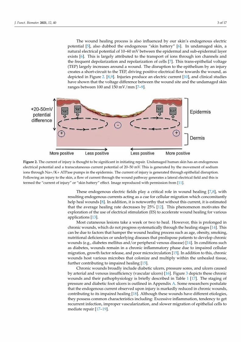
The wound healing process is also influenced by our skin’s endogenous electricpotential [5], also dubbed the endogenous “skin battery” [6]. In undamaged skin, anatural electrical potential of 10–60 mV between the epidermal and sub-epidermal layerexists [6]. This is largely attributed to the transport of ions through ion channels andthe frequent depolarization and repolarization of cells [7]. This trans-epithelial voltage(TEP) largely increases around a wound. The disruption to the epithelium by an injurycreates a short-circuit to the TEP, driving positive electrical flow towards the wound, asdepicted in Figure 2. [8,9]. Injuries produce an electric current [10], and clinical studieshave shown that the voltage difference between the wound site and the undamaged skinranges between 100 and 150 mV/mm [7–9].
J. Funct. Biomater. 2021, 12, x FOR PEER REVIEW 3 of 18
viation u-PA denotes urokinase-type plasminogen activator; MMP-1, 2, 3, and 13 matrix metallo-
proteinases 1, 2, 3, and 13 (collagenase 1, gelatinase A, stromelysin 1, and collagenase 3, respec-
tively); and t-PA tissue plasminogen activator. Images reproduced with permission from [4].
The wound healing process is also influenced by our skin’s endogenous electric po-
tential [5], also dubbed the endogenous “skin battery” [6]. In undamaged skin, a natural
electrical potential of 10–60 mV between the epidermal and sub-epidermal layer exists [6].
This is largely attributed to the transport of ions through ion channels and the frequent
depolarization and repolarization of cells [7]. This trans-epithelial voltage (TEP) largely
increases around a wound. The disruption to the epithelium by an injury creates a short-
circuit to the TEP, driving positive electrical flow towards the wound, as depicted in Fig-
ure 2. [8,9]. Injuries produce an electric current [10], and clinical studies have shown that
the voltage difference between the wound site and the undamaged skin ranges between
100 and 150 mV/mm [7–9].
Figure 2. The current of injury is thought to be significant in initiating repair. Undamaged human
skin has an endogenous electrical potential and a transcutaneous current potential of 20–50 mV.
This is generated by the movement of sodium ions through Na+/K+ ATPase pumps in the epidermis.
The current of injury is generated through epithelial disruption. Following an injury to the skin, a
flow of current through the wound pathway generates a lateral electrical field and this is termed the
“current of injury” or “skin battery” effect. Image reproduced with permission from [11].
These endogenous electric fields play a critical role in wound healing [7,8], with re-
sulting endogenous currents acting as a cue for cellular migration which concomitantly
help heal wounds [8]. In addition, it is noteworthy that without this current, it is estimated
that the average healing rate decreases by 25% [12]. This phenomenon motivates the ex-
ploration of the use of electrical stimulation (ES) to accelerate wound healing for various
applications [13].
Most cutaneous lesions take a week or two to heal. However, this is prolonged in
chronic wounds, which do not progress systematically through the healing stages [14].
This can be due to factors that hamper the wound healing process such as age, obesity,
smoking, nutritional deficiencies or underlying diseases that predispose patients to de-
velop chronic wounds (e.g., diabetes mellitus and/or peripheral venous disease) [14]. In
conditions such as diabetes, wounds remain in a chronic inflammatory phase due to im-
paired cellular migration, growth factor release, and poor microcirculation [15]. In addi-
tion to this, chronic wounds host various microbes that colonize and multiply within the
unhealed tissue, further contributing to impaired healing [15].
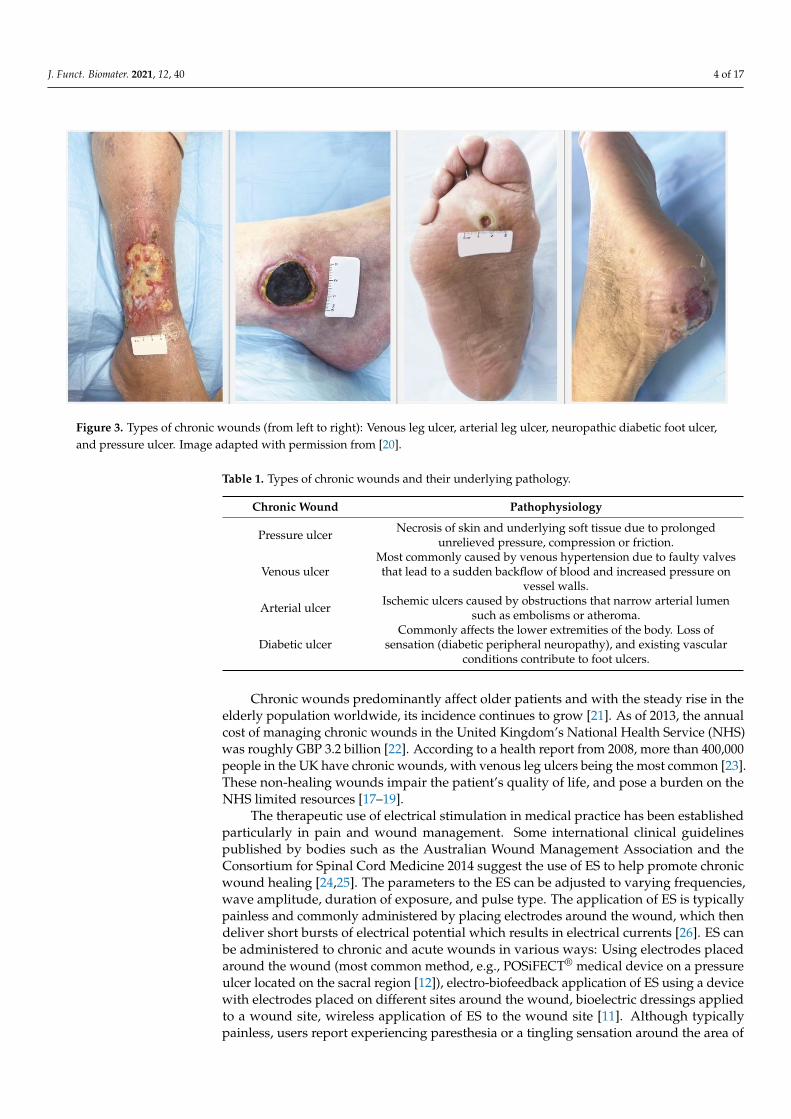
Chronic wounds broadly include diabetic ulcers, pressure sores, and ulcers caused
by arterial and venous insufficiency (vascular ulcers) [16]. Figure 3 depicts these chronic
wounds and their pathophysiology is briefly described in Table 1 [17]. The staging of pres-
sure and diabetic foot ulcers is outlined in Appendix A. Some researchers postulate that
the endogenous current observed upon injury is markedly reduced in chronic wounds,
contributing to its impaired healing [18]. Although these wounds have different etiolo-
gies, they possess common characteristics including: Excessive inflammation, tendency to
Figure 2. The current of injury is thought to be significant in initiating repair. Undamaged human skin has an endogenouselectrical potential and a transcutaneous current potential of 20–50 mV. This is generated by the movement of sodiumions through Na+/K+ ATPase pumps in the epidermis. The current of injury is generated through epithelial disruption.Following an injury to the skin, a flow of current through the wound pathway generates a lateral electrical field and this istermed the “current of injury” or “skin battery” effect. Image reproduced with permission from [11].
These endogenous electric fields play a critical role in wound healing [7,8], withresulting endogenous currents acting as a cue for cellular migration which concomitantlyhelp heal wounds [8]. In addition, it is noteworthy that without this current, it is estimatedthat the average healing rate decreases by 25% [12]. This phenomenon motivates theexploration of the use of electrical stimulation (ES) to accelerate wound healing for variousapplications [13].
Most cutaneous lesions take a week or two to heal. However, this is prolonged inchronic wounds, which do not progress systematically through the healing stages [14]. Thiscan be due to factors that hamper the wound healing process such as age, obesity, smoking,nutritional deficiencies or underlying diseases that predispose patients to develop chronicwounds (e.g., diabetes mellitus and/or peripheral venous disease) [14]. In conditions suchas diabetes, wounds remain in a chronic inflammatory phase due to impaired cellularmigration, growth factor release, and poor microcirculation [15]. In addition to this, chronicwounds host various microbes that colonize and multiply within the unhealed tissue,further contributing to impaired healing [15].
Chronic wounds broadly include diabetic ulcers, pressure sores, and ulcers causedby arterial and venous insufficiency (vascular ulcers) [16]. Figure 3 depicts these chronicwounds and their pathophysiology is briefly described in Table 1 [17]. The staging ofpressure and diabetic foot ulcers is outlined in Appendix A. Some researchers postulatethat the endogenous current observed upon injury is markedly reduced in chronic wounds,contributing to its impaired healing [18]. Although these wounds have different etiologies,they possess common characteristics including: Excessive inflammation, tendency to getrecurrent infection, improper vascularization, and slower migration of epithelial cells tomediate repair [17–19].
J. Funct. Biomater. 2021, 12, 40 4 of 17
J. Funct. Biomater. 2021, 12, x FOR PEER REVIEW 4 of 18
get recurrent infection, improper vascularization, and slower migration of epithelial cells
to mediate repair [17–19].
Figure 3. Types of chronic wounds (from left to right): Venous leg ulcer, arterial leg ulcer, neuropathic diabetic foot ulcer,
and pressure ulcer. Image adapted with permission from [20].
Table 1. Types of chronic wounds and their underlying pathology.
Chronic Wound Pathophysiology
Pressure ulcer Necrosis of skin and underlying soft tissue due to prolonged
unrelieved pressure, compression or friction.
Venous ulcer
Most commonly caused by venous hypertension due to
faulty valves that lead to a sudden backflow of blood and in-
creased pressure on vessel walls.
Arterial ulcer Ischemic ulcers caused by obstructions that narrow arterial
lumen such as embolisms or atheroma.
Diabetic ulcer
Commonly affects the lower extremities of the body. Loss of
sensation (diabetic peripheral neuropathy), and existing vas-
cular conditions contribute to foot ulcers.
Chronic wounds predominantly affect older patients and with the steady rise in the
elderly population worldwide, its incidence continues to grow [21]. As of 2013, the annual
cost of managing chronic wounds in the United Kingdom’s National Health Service
(NHS) was roughly GBP 3.2 billion [22]. According to a health report from 2008, more
than 400,000 people in the UK have chronic wounds, with venous leg ulcers being the
most common [23]. These non-healing wounds impair the patient’s quality of life, and
pose a burden on the NHS limited resources [17–19].
The therapeutic use of electrical stimulation in medical practice has been established
particularly in pain and wound management. Some international clinical guidelines pub-
lished by bodies such as the Australian Wound Management Association and the Consor-
tium for Spinal Cord Medicine 2014 suggest the use of ES to help promote chronic wound
healing [24,25]. The parameters to the ES can be adjusted to varying frequencies, wave
amplitude, duration of exposure, and pulse type. The application of ES is typically pain-
less and commonly administered by placing electrodes around the wound, which then
deliver short bursts of electrical potential which results in electrical currents [26]. ES can
be administered to chronic and acute wounds in various ways: Using electrodes placed
Figure 3. Types of chronic wounds (from left to right): Venous leg ulcer, arterial leg ulcer, neuropathic diabetic foot ulcer,and pressure ulcer. Image adapted with permission from [20].
Table 1. Types of chronic wounds and their underlying pathology.
Chronic Wound Pathophysiology
Pressure ulcer Necrosis of skin and underlying soft tissue due to prolongedunrelieved pressure, compression or friction.
Venous ulcerMost commonly caused by venous hypertension due to faulty valvesthat lead to a sudden backflow of blood and increased pressure on
vessel walls.
Arterial ulcer Ischemic ulcers caused by obstructions that narrow arterial lumensuch as embolisms or atheroma.
Diabetic ulcerCommonly affects the lower extremities of the body. Loss of
sensation (diabetic peripheral neuropathy), and existing vascularconditions contribute to foot ulcers.
Chronic wounds predominantly affect older patients and with the steady rise in theelderly population worldwide, its incidence continues to grow [21]. As of 2013, the annualcost of managing chronic wounds in the United Kingdom’s National Health Service (NHS)was roughly GBP 3.2 billion [22]. According to a health report from 2008, more than 400,000people in the UK have chronic wounds, with venous leg ulcers being the most common [23].These non-healing wounds impair the patient’s quality of life, and pose a burden on theNHS limited resources [17–19].
The therapeutic use of electrical stimulation in medical practice has been establishedparticularly in pain and wound management. Some international clinical guidelinespublished by bodies such as the Australian Wound Management Association and theConsortium for Spinal Cord Medicine 2014 suggest the use of ES to help promote chronicwound healing [24,25]. The parameters to the ES can be adjusted to varying frequencies,wave amplitude, duration of exposure, and pulse type. The application of ES is typicallypainless and commonly administered by placing electrodes around the wound, which thendeliver short bursts of electrical potential which results in electrical currents [26]. ES canbe administered to chronic and acute wounds in various ways: Using electrodes placedaround the wound (most common method, e.g., POSiFECT® medical device on a pressureulcer located on the sacral region [12]), electro-biofeedback application of ES using a devicewith electrodes placed on different sites around the wound, bioelectric dressings appliedto a wound site, wireless application of ES to the wound site [11]. Although typicallypainless, users report experiencing paresthesia or a tingling sensation around the area of

J. Funct. Biomater. 2021, 12, 40 5 of 17
application [26]. Commonly used ES in medical practice and their features are described inTable 2 [11,27].
Table 2. Characteristics of commonly used waveforms for electrical stimulation therapy.
Type of Exogenous ES Characteristics
Direct current (DC) Continuous flow of electric charge in a monophasic waveform (in onedirection). Currents of 20–200 µA can be supplied at a low voltage.
Alternating current (AC)Has a biphasic waveform, with two symmetrical electrical pulses
alternating one after the other. Voltages typically 50–150 V dependenton tissue hydration.
Pulsed current (PC)Intermittent flow of charged particles with gaps in current flow. This
can have a monophasic or biphasic waveform. Currents of1.2–1.5 mA can be supplied to the tissue at high voltage.
Degenerate wave (DW)A type of waveform used in certain biofeedback devices. A constant
current of 0.3 mA, which delivers an electric field of 10 mV/mmbetween the electrodes can be used.
This review focuses on our current understanding of how ES influences the cellularmechanisms involved in normal cutaneous wound healing, its antibacterial effects, and theclinical effectiveness of ES in accelerating chronic wound healing.
2. Literature Search Method
Literature searches with no publication date restriction were conducted in fourdatabases: The Cochrane Library, MedLine, PubMed, Web of Science and Google Scholar.The search strategy included the following terms: “Electric* stimulation”, “wound healing”,“cutaneous OR skin wound”, and “chronic wound”. “Randomized controlled trial” wasincluded for chronic wound studies. The terms were merged with the Boolean operator“AND”. Titles and abstracts of published studies were subsequently reviewed.
The inclusion criteria were: Full text records, in vitro experiments on human tissue,randomized controlled trials, primary research papers, and studies on cutaneous andchronic wounds only. We excluded duplicate studies, non-English papers, studies unrelatedto wound healing or chronic wound healing and unclear ES specifications. To assess theimpact of ES on the normal process of wound healing, in vitro studies and randomizedcontrolled trials (RCTs) on human skin are included. The second aim of this review focusedon the effects of ES on chronic wound healing, for which only in vivo RCTs on humanparticipants are included.
3. Results
The literature search yielded 30 studies meeting the inclusion criteria. Findings arecategorized into studies that investigate the effects of ES on bacteria colonizing wounds,the effects of ES on the stages of healing, and clinical trials that investigate its effect onchronic wounds.
3.1. Effects of ES on Bacteria Colonising Wounds
Six in vitro studies investigated the bacteriostatic effects of exogenous ES [28–33].Petrofsky et al. [28] exposed Staphylococcus aureus, Pseudomonas aeruginosa, and Escherichiacoli to alternating current (AC) or direct current (DC). These three bacteria commonlycolonize open wounds. The authors reported that 100 µA of DC had no inhibitory effecton any of the bacterial strain. In addition, 20 mA of AC significantly reduced the colony-forming unit (CFU) of only P. aeruginosa by 38% (p < 0.05). In contrast, Kincaid andLavoie [29] noted that the growth of all three bacteria were inhibited at the anode andcathode when exposed to HVPC for 2 h at 250 V or more. Focusing on chronic wounds,Gomes et al. [30] exposed these three strains obtained from chronic venous ulcers to fixeddiphasic (FD-B) current and high voltage monophasic current (HVMC) for 30 and 60 min,respectively. FD-B totally inhibited the colony forming unit (CFU) of all three strains in

J. Funct. Biomater. 2021, 12, 40 6 of 17
30 min (p < 0.05). However, HVMC led to an average 50% CFU reduction for all but the E.coli strain.
In an in vitro study, Daeschlein et al. [31] demonstrated the antibacterial effect ofelectric current on six bacteria that commonly colonize chronic wounds. They experimentedon three Gram-negative species (E. coli, P. aeruginosa, Klebsiella pneumonia) and three Gram-positive species (S. aureus, Staphylococcus epidermidis, Escherichia faecium). Exposure to42 mA of monophasic low voltage pulsed current (LVPC) for 30 min resulted in a significantreduction in CFU in all tested pathogens (p < 0.01), whereas no antibacterial effects werenoted in the control group. They also noted that applying positive polarity to the currenthad greater bacteriostatic effects than negative polarity. Barranco et al. [32] exposedS. aureus to DC using different electrodes (silver, platinum, gold, and steel) and varyingcurrents of 0.4, 4, 40, and 400 µA for 48 h, they reported that the positive silver anoderesulted in the highest reduction in bacterial growth at a lower current range (0.4 and 4 µA).Falcone and Spadaro [33] also reported that the electrically active silver anode showedpronounced bacteriostatic effects even at a low direct current (4 µA). Asadi and Torkamansummarize the antibacterial effects of ES as a combination of direct effects (disruption ofbacterial membranes and blocking the proliferation of bacterial cells) and indirect effects(pH changes, i.e. increased pH at the cathode and decreased pH at the anode; temperaturechanges; electrolysis products, e.g., toxic species, chlorine, H2O2, radicals; and galvanotaxis,increased migration of white blood cells such as macrophages and leukocytes to the infectedwound site) [34].
There is a lack of studies investigating the antibacterial effects of electrical stimulationin vivo. Further in vivo research may be required to ascertain that exogenous ES can helplimit the burden of infection accompanying dermal wounds clinically. However, currentevidence favors the likelihood that ES can be used as an adjunct for infection control inwound therapies.
3.2. Effects of ES on Cellular Migration and Tissue Perfusion
The inflammatory phase of wound healing involves control of bleeding, removalof pathogens, and increased tissue perfusion to recruit cells [1]. It has been observedthat cells involved in wound healing carry a charge and migrate towards an electric fieldwith the opposite polarity [35,36]. For instance, macrophages migrate towards the anode,fibroblasts migrate towards the cathode, and neutrophils migrate to both the anode andcathode [36], this phenomenon is known as Galvanotaxis [7,36]. This was demonstratedvia an experiment whereby participants’ skin was scarred and exposed to exogenous ES.An assessment of the cell composition in the skin exudate 6 h post-treatment showed thatthe neutrophil count was 24% higher in the exposed group than the control group due tothe increased electrotaxis of human cells in an electric field [37].
Four randomized controlled studies and one case series looked into the effects ofexogenous ES on tissue perfusion [38–42]. Cramp et al. [38] investigated the application oftranscutaneous electrical nerve stimulation (TENS) to understand its effect on skin bloodflow. After 15 min of exposure, skin perfusion was roughly four times higher using low-frequency TENS compared to high-frequency TENS. Clover et al. [39] noted an increasein capillary density by 25% and transcutaneous oxygen tension by 1.24-fold when treatedwith localized sub-contractile electrical stimulation for 6 weeks (whereas no significantincrease was observed in the control group). The immediate improvement in cutaneousperfusion was also demonstrated by Peters et al. [40] who noted a significant increasein transcutaneous partial pressure of oxygen (TcPO2) by 27% in patients with peripheralarterial disease using direct current, compared to no change in TcPO2 in the control group.The effects of improvement were noted within 5 min of ES exposure.
In an interesting case series by Goldman et al. [41], the investigators instructed sixpatients with critically ischemic limbs (defined as TcPO2 < 10 mmHg) to use the high voltagepulsed current (HVPC) (80–330 V) along with their standard wound dressing to treat theirwounds. They defined a TcPO2 below 20 mmHg as unfavorable for wound healing. Pre-

J. Funct. Biomater. 2021, 12, 40 7 of 17
exposure means that TcPO2 around wound edges were 2 ± 2 mmHg. Following 40 days ofdaily ES, the mean TcPO2 rose to 33 mmHg and wounds closed thereafter. In a subsequentRCT, Goldman et al. [42] continued to explore this by randomizing eight subjects withischemic limb wounds to receive active or sham HVPC for 14 weeks. After 4 weeks, woundstreated with active HVPC decreased in size (p < 0.05) while wounds in the control groupincreased in size. By week 8, HVPC treated wounds had an average TcPO2 measuring27 ± 12 mmHg, no longer at an ischemic range, whereas the control group had an averageof 2 ± 2 mmHg. Laser Doppler measures indicated a 3-fold increase in dermal capillaryperfusion in the treatment group (p < 0.01). Their trial demonstrated how electrotherapy(mainly HVPC) improves dermal microcirculation and wound healing. Table 3 summarizesthe key findings of papers investigating the effect of ES on this phase of wound healing.
Table 3. Key findings of studies investigating the effect of ES on skin perfusion.
StudyDesign Type of ES Exposure Duration Experimented on Key Outcome(s) Reference
RCT TENS 15 min
30 healthy subjectsHigh frequency (n = 10)Low frequency (n = 10)
Control (n = 10)
Increase in skinperfusion [38]
RCT Subcontractile ES 60 min/dayTotal 6 weeks
36 patients withischemic limbs
ES group (n = 24)Control (n = 12)
Increase in capillarydensity and skin
perfusion[39]
RCT HVPC Four 60-min periodsTotal 1 day
11 diabetic patients withand without PVD
Transient increase inTcPO2 in participants
with PVD within 5 min[40]
Case series HVPC 1 h/day, 7 days/weekfor 1–9 months
6 patients with ischemicleg ulcers
Increased TcPO2 andtotal healing of ulcers
post exposure[41]
RCT HVPC 1 h/day, 7 days/week,14 weeks
8 Ischemic limb woundsES group (n = 4)
Sham treatment (n = 4)
Decreased wound sizeand increased
peri-wound circulation[42]
3.3. Effects of ES on the Proliferative and Remodelling Phase of Healing
Events that occur in these phases include vascularization by angiogenesis, fibrob-last proliferation, granulation tissue formation, and re-epithelialization. Two in vivoRCTs [43,44] obtained punch biopsies from human volunteers and treated them withdegenerate wave current (DW) to understand which mechanisms of healing are induced.In a double-blind prospective trial by Sebastian et al. [43], volunteers had small biopsiestaken from each arm but DW was given to only one arm, allowing the participants to serveas their own control. The histological analysis showed that on average, DW-exposed armsexpressed vascular endothelial growth factor (VEGF) 42% more than unexposed skin. Onday 14, expression of endothelial cells was 6% higher, and RNA transcripts of type IVcollagen was 8% higher than the control. In a RCT reported by Ud-Din et al. [44] with a sim-ilar study design, treating biopsy wounds with DW showed a 38.9% reduction in woundvolume and a 78.6% increase in blood flow to the wound site compared to the control. Thehistological analysis indicated an increase in granulation tissue area and an average 35%rise in VEGF production. Both studies showed an upregulation of anti-inflammatory genes.Zhao et al. [45] also demonstrated how ES induces angiogenesis. Their in vitro experimentshowed that 1.5–2.9 mV DC-treated endothelial cells produced VEGF 2.6 times more thanthe control group.
With regards to the remodeling stage, Rouabhia et al. [46] found that fibroblastsexposed to 200 mV/mm of DC quickly migrated from both ends of a sample wound, andthe wound had fully closed in 24 h. However, over 30–40% of the wound area in a similar
J. Funct. Biomater. 2021, 12, 40 8 of 17
sample remained unhealed in the control group. There was also a significant increase inthe production of fibroblast growth factor (FGF-1 and FGF-2) (p < 0.01). Snyder et al. [47]similarly identified an increase in random migration of fibroblasts by DC stimulationin their study. Cheng and Goldman [48] demonstrated that electrical exposure induceshuman dermal fibroblasts to enter into the growth phase of the cellular cycle. This possiblyexplains the stimulation of fibroblast proliferation upon ES exposure.
Sebastian et al. [49] reported that ES-treated wounded skin had a 38% increase inkeratinocyte proliferation and an 18% increase in epithelium thickness compared to thecontrol (p = 0.002). In contrast, Bullock et al. [50] found no significant change to keratinocyteproliferation when subjecting in vitro skin wound models to the pulsed current. In sum-mary, the growth factors noted to be enhanced upon ES across the reviewed studies areFGF-1, FGF-2, and VEGF. These collectively aid in the proliferation of endothelial cells,fibroblasts, and expedites new vessel formation [51]. Table 4 summarizes key findings ofin vivo and in vitro studies that investigate the effect of ES on this phase of healing [43–49].
Table 4. In vitro and in vivo studies on the effects of exogenous ES on the proliferative and remodeling stages of wound healing.
Study Design Type of ES ExposureDuration Experimented on Key Outcome(s) Reference
RCT DW 14 days 20 healthy subjects, servedas own control
Increase in VEGF,collagen, epidermal cells,
and cell apoptotic markers[43]
RCT DW 14–20 days 40 healthy subjects, servedas own control
Reduced wound volume,increased perfusion and
vascularity[44]
In vitro Pulsed DC At 4, 8, and 24 h Human umbilical veinendothelial cell cultures
Increase in endothelial cellmigration and VEGF
production[45]
In vitro DC At 2, 4, and 6 h Human fibroblast cells ina wound model
Increase in FGF anddifferentiation of
fibroblasts[46]
In vitro DC 10 minFibronectin coated andnon-fibronectin coateddermal fibroblast cells
Increased randommigration of fibroblast
cells, no increase indermal fibroblast gene
expression
[47]
In vitro AC 12 h Dermal cell matrix
Dermal fibroblasts enteredinto the growth phase of
cell cycle with continuousES exposure
[48]
In vitro DC and DW 16 days Punch biopsies on samplehuman skin tissues
Increase in epidermisthickness and keratinocyte
proliferation[49]
3.4. Effects of ES on Chronic Wounds
This review evaluates 12 randomized controlled trials that assesses the effects ofelectrical stimulation (ES) on chronic wound healing [52–63]. The trials dated from 1988 to2018, and included 532 participants. The studies used different types of ES such as HVPC(n = 8), PC (n = 3), and AC (n = 1) with varied parameters, and worked on different chronicwounds (pressure ulcers n = 6, diabetic ulcers n = 4, and vascular ulcers n = 2). The mainchallenges in interpreting the data were variation in the duration of treatment and measureof outcome. The primary outcome of measure used by most studies (n = 10) in quantifyingthe healing rate is the change in wound surface area measured as a percentage of areareduction (PAR) before and after the treatment. One study by Baker et al. [60] used weekly

J. Funct. Biomater. 2021, 12, 40 9 of 17
healing rate as an outcome, and a study by Griffin et al. [54] used percentage of woundshealed. A study by Polak et al. [63] assessed the peri-wound blood flow alongside PAR astheir primary outcomes. Of the 12 studies, only four report both percentage of completewound healing and changes in wound surface area [53,56,61,62].
In all studies, participants with chronic wounds were randomized to receive electricalstimulation therapy or sham treatment. Given that most trials took place in a clinicalsetting, participants also received standard wound care alongside ES or sham therapy. Twostudies [59,62] used an electric stimulation device for patients to self-administer at homeafter receiving training. Of all trials, the study by Miller et al. [59] was the only studyfunded by a device manufacturer. Acknowledging the risk of bias, this was included in thereview with interest in the results after noting that investigators initiated the research andabided by RCT protocols. Overall, all studies report a degree of accelerated chronic woundhealing upon ES exposure.
Some trials included secondary measures of outcome such as changes in tissue perfusion,adverse effects of ES exposure, and the effect of participants’ compliance on wound healing.Lawson et al. [57] investigated whether blood flow to chronic wounds is increased withexogenous ES exposure. They report that tissue perfusion around the wounds increased by87% in patients with diabetes and only 6% in patients without diabetes. Three studies reportedadverse effects following ES exposure. In the study by Lawson et al. [57], two patients werehospitalized following unspecified complications and one dropped out after experiencingvertigo. Lundeberg et al. [61] found that 6% of ES group and 3% of sham experienced allergicsymptoms, and 9% of ES group and 6% of sham group experienced pain. Similarly, Peterset al. [62] reported that five subjects (10% of ES and 15% of placebo group) dropped outfollowing infection complications. It is uncertain whether the reported adverse effects canbe fully attributed to ES exposure. Confounding factors include the severity of the subjects’underlying disease. Of all studies, Peters et al. [62] were the only investigators who alsoreported whether compliance to the treatment affected the healing of wounds. Their resultsshowed that 71% of ulcers healed in patients who were compliant, whereas only 50% of ulcershealed in the non-complaint group. Patients were classed as compliant if the ES exposure was>20 h/week. Table 5 summarizes the key findings of all 12 RCTs.
Table 5. RCTs investigating the impact of electrical stimulation on chronic wound healing.
StudyDesign
Type ofES/Electrode
Placement
ExposureDuration
Type ofChronicWound
No. of Participants% Wound AreaReduction/% ofWounds Healed
Reference
RCTHVPC †/Treatment
electrode placedover wound
50 min/day,5 days/week for
6 weeks
Pressureulcers
63 patientsCathodal (n = 23)
Anodal-cathodal (n = 20)Sham treatment(n = 20)
PAR 1 82.34% and70.77% in ES
group, respectively,40.53% in
control/Wound healingnot specified
[52]
RCTHVPC/Treatmentelectrode placed
over wound
50 min/day,5 days/week for
6 weeks
Pressureulcers
77 patientsES (n = 24)
Sham (n = 28)US 1 (n = 25)
PAR 76.19% in ES group48.97% in control
group/52% of ulcershealed in ES group, 23%
healed in control
[53]
RCTHVPC/Treatmentelectrode placed
over wound
60 min/day for20 days
Pressureulcers
17 patientsES (n = 8)
Sham (n = 9)
Wound PAR notspecified, higher in ES
than control group/38%of ES wound healed vs.
22% in sham group(p > 0.05)
[54]
RCTHVPC/Treatmentelectrode placed
over wound
45–120 min dailyfor 5 weeks (45 minfor sham treatment)
Pressureulcers
60 patientsES (n = 45)
Sham (n = 15)
After 60 and 120 minexposure PAR 91% inES group vs. 25% insham group/Woundhealing not recorded
[55]

J. Funct. Biomater. 2021, 12, 40 10 of 17
Table 5. Cont.
StudyDesign
Type ofES/Electrode
Placement
ExposureDuration
Type ofChronicWound
No. of Participants% Wound AreaReduction/% ofWounds Healed
Reference
RCTHVPC/Treatmentelectrode placed
over wound
45 min/day,five days/week foraverage 7.4 weeks
Pressureulcers
16 patientsES (n = 9)
Sham (n = 7)
100% wound areareduction in treatment
group, 28.9% increase inwound area in control
group/Completehealing in ES group
[56]
RCT
Biphasiccurrent/Treatment
electrode placesacross wound on
intact skin
30 min/day,3 days/week,for 4 weeks
Mixed ulcers(diabetic and
vascular)
17 patientsDiabetic (n = 8)Non-diabetic
(n = 9)
PAR 70% in diabeticgroup, 38.4% in non-
diabetic group/Woundhealing not specified
[57]
RCTHVPC/Treatmentelectrode placed
over wound
45 min/day,3 days/weekfor 4 weeks
Mixed ulcers(diabetic and
venous)
27 patientsES (n = 14)
Sham (n = 13)
Wound PAR 44.3% in ESgroup and 16.6% in
control group/Woundhealing not specified
[58]
RCT
PC (using bodyflowdevice)/Treatmentelectrodes placedabove and belowthe wound site
20 min/day,4 days/week for
8 weeksVenous ulcers
23 patientsES (n = 14)
Sham (n = 9)
PAR 32.67% in ES,Sham ES
23.15%/Wound healingnot recorded
[59]
RCT
BiphasicPC/Treatment
electrodes placedover intact skinproximal to the
wound site
30 min of exposure Diabeticfootulcers
80 patientsAsymmetrical
PC (n = 21),SymmetricalPC (n = 20),
Low stimulation current(n = 19)
Sham (n = 20)
Healing rate per week:27% in asymmetrical
PC, 16% in symmetricalPC, ~9.7% in control
group/Wound healingnot recorded
[60]
RCT AC ‡/Not specified20 min twice daily
for 12 weeksDiabetic
footulcers
51 patientsES (n = 24)
Placebo (n = 27)
PAR 61% in ES group,41% in placebo
group/42% of ESexposed ulcers healed
vs. 15% in placebogroup
[61]
RCT
HVPC/Treatmentelectrodes
embedded instockings placed
around the wound
8 h nightly for12 weeks
Diabeticfootulcers
40 patientsES (n = 20)
Sham (n = 20)
PAR 86% in ES group,71% in sham
group/65% of ESwounds healed vs. 35%
of wounds in shamgroup
[62]
RCT
HVPC/Treatmentelectrodes placedon opposite edgesof the wound site
50 min/day,5 day/week for
8 weeks
Pressureulcers
61 patientsAnodal HVPC (n = 20)
Cathodal HVPC (n = 21)Sham (n = 20)
PAR 64.1% in anodalHVPC group, 74.06% incathodal HVPC group,
41.42% in shamgroup/Complete
wound healed notrecorded
[63]
† HVPC: High voltage pulsed current; ‡ AC: Alternating current; 1 PAR: Percentage area reduction; US: Ultrasound.
4. Discussion4.1. General Considerations
This review aims to understand how electrical stimulation (ES) influences the normalprocesses involved in wound healing, followed by its efficacy in healing chronic wounds.In vitro experiments on human cells and tissues were included in this review in orderto understand the mechanism of action of ES on a cellular level. In terms of the generalactivities of wound healing, reviewed in vivo and in vitro studies demonstrate that ESincreases tissue perfusion, promotes cellular migration, increases tissue vascularization,and causes a significant improvement in fibroblast proliferation.

J. Funct. Biomater. 2021, 12, 40 11 of 17
The reviewed experiments demonstrate increments in VEGF and FGF productionupon ES exposure. ES has been shown to have a promising ability to accelerate woundhealing by activating the angiogenesis signaling pathways and stimulating fibroblastproliferation. One in vitro study found that ES activates the mitogen-activated proteinkinase (MAPK) signaling pathway, which triggered angiogenesis in the process of woundhealing. Bai et al. [64] in a study on human endothelial cells found that ES inducesVEGF receptor signaling, consequently upregulating VEGF secretion and endothelial cellmigration. However, the underlying mechanism driving these molecular changes andincrease in growth factors remain largely unclear.
Although an in vitro experiment [50] showed no improvement in re-epithelializationby keratinocytes upon PC stimulation, two others found improved re-epithelializationupon DW and PC stimulation [45,49]. The difference in wound type and ES used couldaccount for that disparity. The selected studies reported p-values below 0.05, delineatingthe strong likelihood of ES contributing to enhanced healing. Findings from animalstudies [36,65,66] further support this. Gürgen et al. [65] demonstrated that exposing ratwounds to transcutaneous electrical nerve stimulation (TENS) resulted in a significantreduction in pro-inflammatory cytokines. They also reported an overall reduction in woundhealing time. Chu et al. [66] exposed guinea pig skin models to weak anodal DC (20–40 µA)to investigate its effect on wound healing. The authors report that DC treated wounds hadless granulation tissue and fibrosis compared to control wounds. Other animal studiesalso report increased fibroblast proliferation and collagen deposition in ES treated wounds,which favors the remodeling phase of wound healing [36].
Six in vitro studies investigated the antibacterial effect of ES. Bacterial infection hinderswounds from healing quickly due to the excessive release of toxins and inflammatorysignals. Chronic wounds have a larger risk of infection due to the prolonged healing time,making patients susceptible to septicemia [19]. Bacteria that commonly colonize acute andchronic wounds include S. aureus, P. aeruginosa, and E. coli. All six studies noted a reductionin the growth of at least one or all these bacterial strains when exposed to electricity. Threestudies interestingly noted how a positive polarity using the silver anode showed markedbacteriostatic effects when exposed to a weak DC compared to the other electrode materials(likely due to the formation of reactive oxygen species). The pulsed current lead to amarked colony forming unit (CFU) reduction across three studies [29–31]. One mechanismbehind this is that supplying electricity alters the pH of the bacteria’s environment. Thisconsequently damages the external membrane of bacteria, allowing an uncontrolled influxof solutes that ultimately kill them [30]. Whilst the antibacterial effects of ES (that can beachieved using disinfection techniques), is beneficial for wound healing, supporting thepotential use of ES in chronic wound treatment to minimize the clinical burden of infection.
Furthermore, the results of this review also indicate how ES, mainly HVPC, mayenhance chronic wound healing upon regular administration. Secondary outcomes suchas wound perfusion and complete wound healing were also positive upon ES exposure.Most trials utilized the pulsed current in their treatment (n = 11), with a majority ofthem using HVPC (n = 8). A meta-analysis of different ES on chronic wounds by Khouriet al. [67] reported that when comparing DC, LVPC, HVPC, and DW, HVPC showed thebest improvement in chronic wound size reduction. Most in vivo trials that were reviewedused the pulsed current and reported positive outcomes. Unlike continuous DC, which hasbeen noted to cause skin irritation due to pH changes, the monophasic pulsed current doesnot cause skin changes [27]. Furthermore, PC has polarity and is better able to mimic thephysiological current in comparison to sinusoidal AC, which lacks polarity. These factors,along with the ability of PC to penetrate deeper into the skin partially explains why thepulsed current has been commonly used in the trials reviewed. However, it is difficult toascertain a recommended method of ES application for chronic wounds as studies utilizedvarying parameters of ES and duration of exposure.

J. Funct. Biomater. 2021, 12, 40 12 of 17
4.2. Limitations on Chronic Wound Studies
There are external factors that influence chronic wound healing such as age, pre-existing medical conditions, wound care method, and nutrient deficiency. Only one RCTwas exclusively mentioned to have controlled these variables during their study [58]. Inaddition, it was the only study that followed up on their participants to assess full recovery.Only three reviewed studies assessed the adverse effects of treatment [57,61,62]. Long-termfollow up, users’ perspectives and implications of ES on the participant’s quality life arelacking in most studies, as their primary measure of outcome was wound size reduction.Furthermore, the authors did not assess any extra burden caused by ES exposure to thepatients, such as device-related complications and its acceptability. This limitation needsaddressing in future trials if ES becomes a widespread treatment.
Although all considered RCTs demonstrated that ES markedly reduces chronic woundsize, it is difficult to recommend an optimal plan using ES as a standard treatment. Al-though the pulsed current indicated significant success in accelerating chronic woundhealing across all the trials that used it, differences exist in the polarity, duration, andmethod of ES exposure used across the studies. In addition, the demographics and woundcharacteristics of chosen participants varied across trials. This brings questions such aswhat is the ideal anatomical location and the optimal method of delivering ES? The varietyin treatment protocols across trials makes it difficult to ascertain the most effective form ofES on a type of chronic wound. Another limitation is that in studies where participantshad multiple co-morbidities, the extent to which those diseases contributed to impairedwound healing were not outlined.
4.3. Potential Implications for Clinical Practice
Chronic wound care consumes almost 3% of healthcare expenditure in developedcountries [21]. In the United States, treating chronic venous leg ulcers cost USD 4000 permonth per patient and advance wound dressings incurred additional costs up to USD29,252 per treatment episode in 2012 [68]. Globally, the average cost of chronic woundcare was USD 2.8 billion in 2014, and it is predicted to rise to USD 3.5 billion in 2021 [69].A clinical study found that using ES therapy in adjunct with standard care reduces thecosts associated with managing chronic wounds in the UK by 16% when compared tousing standard procedures alone [70]. Another model indicated a potential reductionin NHS wound care costs by 15% if patients with chronic venous ulcers replaced theirregular treatment with ES therapy [71]. The results of this review indicate that electricalstimulation increases the rate of chronic wound healing. More studies need to be conductedto investigate the effect of ES with time to complete chronic wound closure.
The current management of chronic wounds in the NHS include using advancedwound dressings that control moisture levels, antimicrobial dressings, negative pressuretherapy, and offloading to reduce the pressure on wounds [72,73]. The National Institutefor Health and Care Excellence (NICE) provide evidence-based recommendations forhealth care in England. The management of each type of chronic wound differs but theNICE guidelines recommend using proper wound dressing techniques to optimize quickerhealing with minimal risk of infections [72]. Alternative therapies include skin grafting,growth factor therapy, hyperbaric oxygen therapy, and stem cell therapy. However, theirreliable efficiency in chronic wound management have yet to be proven significant [73].
At present, the NICE guidelines do not recommend using ES therapy on chronicwounds unless it is used in a clinical trial [74]. It is suggested that ES should not be givento patients who have a pacemaker inserted, who have skin conditions, who are pregnant orpatients with skin cancers and epilepsy [24]. The underlying health conditions of chronicwound patients should be taken into critical consideration. These limitations, followed bya lack of long-term therapeutic evidence on chronic wounds could suggest why ES is notused in regular practice in the UK. Contrastingly, physicians in Australia, New Zealand,and the United States are recommended to use ES as an adjunct therapy to treat chronicpressure ulcers [24,75]. The Pan Pacific Guideline for the Prevention and Management of

J. Funct. Biomater. 2021, 12, 40 13 of 17
Pressure Injury recommends the use of pulsed electrotherapy as an adjunct treatment toaccelerate the healing of pressure ulcers [24]. Its use in other forms of chronic wounds suchas diabetic ulcers is not specified.
5. Conclusions
Our bodies generate natural endogenous electrical potentials around a wound whichare known to accelerate the healing process by guiding cells to migrate to the site of in-jury [8]. Exogenous electrical stimulation is used to mimic this physiological occurrenceand it has been proven beneficial in accelerating wound healing. This review evidencesthat electrical stimulation limits inflammation, increases wound blood perfusion, controlsbacterial growth, increases fibroblast migration, induces angiogenesis, and encourages ker-atinocyte activity. This applies to both acute and chronic wounds. Additionally, electricalstimulation (notably pulsed current) significantly reduces the size of chronic wounds whencompared to control groups with no ES.
Current evidence supports the possibility of using ES as an adjunct therapy to chronicwound management. However, the most effective ES cannot be concluded from this reviewdue to variations in the studies’ experimental protocol. Additionally, trials on chronicwounds did not compare different ES to suggest an ideal type. Future trials can comparedifferent ES and monitor any long-term complications to recommend a specific type thatcontributes to the most effective healing.
We also foresee the potential of electroactive biomaterials to play a role in advancedwound healing technologies [76,77]. Recent advancements with nanogenerator technologyproducing self-sustainable ES as a wearable wound-healing device suggest the potentialfor exciting new opportunities in wound care management [78,79].
Author Contributions: Conceptualization, K.L.W. and J.G.H.; methodology, S.B.R.; formal analysis,all authors; investigation, S.B.R.; writing—original draft preparation, S.B.R.; writing—review andediting, all authors; supervision, K.L.W. and J.G.H.; project administration, K.L.W. and J.G.H.;funding acquisition, K.L.W. and J.G.H. The CRediT taxonomy directs the term explanation. Allauthors have read and agreed to the published version of the manuscript.
Funding: For funding supportive of research related to this article, we thank the Biotechnologyand Biological Sciences Research Council (BBSRC) for grant reference BB/L013819/1, the MedicalResearch Council (MRC) for grant reference MC_PC_17192, and the Royal Society of Chemistry (RSC)for grant reference RM1601-2703.
Institutional Review Board Statement: Not applicable.
Informed Consent Statement: Not applicable.
Data Availability Statement: Not applicable.
Acknowledgments: We acknowledge the reviewers and editors for their constructive critique.
Conflicts of Interest: The authors declare no conflict of interest. The funders had no role in the designof the study; in the collection, analyses, or interpretation of data; in the writing of the manuscript, orin the decision to publish the results. The views expressed are those of the authors and not necessarilythose of the National Health Service (NHS), the National Institute for Health Research (NIHR) or theDepartment of Health.
Appendix A
The stages in severity of diabetic and pressure ulcers are outlined in Table A1 [80] andTable A2 [81].

J. Funct. Biomater. 2021, 12, 40 14 of 17
Table A1. Stages of pressure ulcers to assess severity in clinical practice.
Stage Description of Wound
Stage ISkin is intact with no open tears. Reddened area with possible blanching and pain.
Area has a different texture compared tosurrounding skin.
Stage II Skin tears, forming an open wound with blisters.Painful with exudate release.
Stage III Painful deep wound. Dermis is lost, fully exposing the fattylayer below. No muscle or bone visible.
Stage IV Deeper, painful wound which exposes theunderlying muscle, tendon, or bone.
Table A2. The University of Texas Staging System for diabetic foot ulcers.
Stage Grade 0 Grade I Grade II Grade III
AUlcerative lesion
completelyepithelialized
Superficial ulcer, notpenetrating to
tendon or muscle
Ulcerpenetrating to
tendon orcapsule
Ulcerpenetrating to bone
or joint
B Infection Infection Infection InfectionC Ischemia Ischemia Ischemia Ischemia
D Infection andIschemia
Infection andIschemia
Infection andIschemia
Infection andIschemia
References1. Gerard, J.T. Principles of Anatomy and Physiology, 15th ed.; Wiley: Hoboken, NJ, USA, 2016.2. Slavin, J. The role of cytokines in wound healing. J. Pathol. 1996, 178, 5–10. [CrossRef]3. Pierce, G.F.; Mustoe, T.A. Pharmacologic enhancement of wound healing. Annu. Rev. Med. 1995, 46, 467–481. [CrossRef]4. Singer, A.J.; Clark, R.A.F. Cutaneous Wound Healing. N. Engl. J. Med. 1999, 341, 738–746. [CrossRef]5. Farber, P.L.; Hochman, B.; Furtado, F.; Ferreira, L.M. Electricity and colloidal stability: How charge distribution in the tissue can
affects wound healing. Med. Hypotheses 2014, 82, 199–204. [CrossRef]6. Jaffe, L.F.; Vanable, J.W., Jr. Electric fields and wound healing. Clin. Dermatol. 1984, 2, 34–44. [CrossRef]7. Sun, Y.-S. Electrical Stimulation for Wound-Healing: Simulation on the Effect of Electrode Configurations. BioMed. Res. Int. 2017,
2017, 5289041. [CrossRef] [PubMed]8. Zhao, M. Electrical fields in wound healing-An overriding signal that directs cell migration. Semin. Cell Dev. Biol. 2009, 20, 674–682.
[CrossRef] [PubMed]9. Nuccitelli, R. Endogenous electric fields in embryos during development, regeneration and wound healing. Radiat. Prot. Dosim.
2003, 106, 375–383. [CrossRef] [PubMed]10. Nuccitelli, R.; Nuccitelli, P.; Li, C.; Narsing, S.; Pariser, D.M.; Lui, K. The electric field near human skin wounds declines with age
and provides a noninvasive indicator of wound healing. Wound Repair Regen. Off. Publ. Wound Health Soc. Eur. Tissue Repair Soc.2011, 19, 645–655. [CrossRef] [PubMed]
11. Ud-Din, S.; Bayat, A. Electrical Stimulation and Cutaneous Wound Healing: A Review of Clinical Evidence. Healthcare 2014,2, 445–467. [CrossRef] [PubMed]
12. Hampton, S.; Collins, F. Treating a pressure ulcer with bio-electric stimulation therapy. Br. J. Nurs. 2006, 15, S14–S18. [CrossRef]13. Gardner, S.E.; Frantz, R.A.; Schmidt, F.L. Effect of electrical stimulation on chronic wound healing: A meta-analysis. Wound Repair
Regen. 1999, 7, 495–503. [CrossRef]14. Ashrafi, M.; Alonso-Rasgado, T.; Baguneid, M.; Bayat, A. The efficacy of electrical stimulation in lower extremity cutaneous
wound healing: A systematic review. Exp. Dermatol. 2017, 26, 171–178. [CrossRef]15. Rodrigues, M.; Kosaric, N.; Bonham, C.A.; Gurtner, G.C. Wound Healing: A Cellular Perspective. Physiol. Rev. 2019, 99, 665–706.
[CrossRef] [PubMed]16. Werdin, F.; Tennenhaus, M.; Schaller, H.E.; Rennekampff, H.O. Evidence-based management strategies for treatment of chronic
wounds. Eplasty 2009, 9, e19. [PubMed]17. Demidova-Rice, T.N.; Hamblin, M.R.; Herman, I.M. Acute and impaired wound healing: Pathophysiology and current methods
for drug delivery, part 1: Normal and chronic wounds: Biology, causes, and approaches to care. Adv. Skin. Wound Care 2012,25, 304–314. [CrossRef] [PubMed]
J. Funct. Biomater. 2021, 12, 40 15 of 17
18. Jünger, M.; Arnold, A.; Zuder, D.; Stahl, H.W.; Heising, S. Local therapy and treatment costs of chronic, venous leg ulcers withelectrical stimulation (Dermapulse): A prospective, placebo controlled, double blind trial. Wound Repair Regen. 2008, 16, 480–487.[CrossRef] [PubMed]
19. Frykberg, R.G.; Banks, J. Challenges in the Treatment of Chronic Wounds. Adv. Wound Care 2015, 4, 560–582. [CrossRef]20. Singer, A.J.; Tassiopoulos, A.; Kirsner, R.S. Evaluation and Management of Lower-Extremity Ulcers. N. Engl. J. Med. 2017,
377, 1559–1567. [CrossRef]21. Järbrink, K.; Ni, G.; Sönnergren, H.; Schmidtchen, A.; Pang, C.; Bajpai, R.; Car, J. The humanistic and economic burden of chronic
wounds: A protocol for a systematic review. Syst. Rev. 2017, 6, 15. [CrossRef]22. Guest, J.F.; Ayoub, N.; McIlwraith, T.; Uchegbu, I.; Gerrish, A.; Weidlich, D.; Vowden, K.; Vowden, P. Health economic burden
that wounds impose on the National Health Service in the UK. BMJ Open 2015, 5, e009283. [CrossRef]23. Posnett, J.; Franks, P.J. The burden of chronic wounds in the UK. Nurs. Times 2008, 104, 44–45.24. Pan Pacific Guideline for the Prevention and Management of Pressure Injury (Abridged Version); The Australian Wound Management
Association: Osborne Park, WA, Australia, 2012.25. Consortium for Spinal Cord Medicine. Pressure Ulcer Prevention and Treatment Following Spinal Cord Injury: A Clinical Practice
Guideline for Health-Care Professionals. Available online: https://pva-cdnendpoint.azureedge.net/prod/libraries/media/pva/library/publications/cpg_pressure-ulcer.pdf (accessed on 20 December 2020).
26. Arora, M.; Harvey, L.A.; Glinsky, J.V.; Nier, L.; Lavrencic, L.; Kifley, A.; Cameron, I.D. Electrical stimulation for treating pressureulcers. Cochrane. Database Syst. Rev. 2020, 1, Cd012196. [CrossRef]
27. Kloth, L.C. Electrical Stimulation Technologies for Wound Healing. Adv. Wound Care 2014, 3, 81–90. [CrossRef]28. Petrofsky, J.; Laymon, M.; Chung, W.; Collins, K.; Yang, T.-N. Effect of electrical stimulation on bacterial growth. J. Orthop.
Neurolsurg. 2008, 31, 43.29. Kincaid, C.B.; Lavoie, K.H. Inhibition of bacterial growth in vitro following stimulation with high voltage, monophasic, pulsed
current. Phys. Ther. 1989, 69, 651–655. [CrossRef]30. Gomes, R.C.; Brandino, H.E.; de Sousa, N.T.; Santos, M.F.; Martinez, R.; Guirro, R.R. Polarized currents inhibit in vitro growth of
bacteria colonizing cutaneous ulcers. Wound Repair Regen. 2015, 23, 403–411. [CrossRef]31. Daeschlein, G.; Assadian, O.; Kloth, L.C.; Meinl, C.; Ney, F.; Kramer, A. Antibacterial activity of positive and negative polarity
low-voltage pulsed current (LVPC) on six typical Gram-positive and Gram-negative bacterial pathogens of chronic wounds.Wound Repair Regen. 2007, 15, 399–403. [CrossRef] [PubMed]
32. Barranco, S.; Spadaro, J.; Berger, T.; Becker, R. In vitro effect of weak direct current on Staphylococcus aureus. Clin. Orthop. Relat.Res. 1974, 100, 250–255. [CrossRef]
33. Falcone, A.E.; Spadaro, J.A. Inhibitory effects of electrically activated silver material on cutaneous wound bacteria. Plast. Reconstr.Surg. 1986, 77, 455–459. [CrossRef] [PubMed]
34. Asadi, M.R.; Torkaman, G. Bacterial Inhibition by Electrical Stimulation. Adv. Wound Care 2014, 3, 91–97. [CrossRef]35. Orida, N.; Feldman, J.D. Directional protrusive pseudopodial activity and motility in macrophages induced by extracellular
electric fields. Cell Motil. 1982, 2, 243–255. [CrossRef]36. Kloth, L.C. Electrical stimulation for wound healing: A review of evidence from in vitro studies, animal experiments, and clinical
trials. Int. J. Low Extrem. Wounds 2005, 4, 23–44. [CrossRef] [PubMed]37. Eberhardt, A.; Szczypiorski, P.; Korytowski, G. Effect of transcutaneous electrostimulation on the cell composition of skin exudate.
Acta. Physiol. Pol. 1986, 37, 41–46. [PubMed]38. Cramp, A.F.; Gilsenan, C.; Lowe, A.S.; Walsh, D.M. The effect of high- and low-frequency transcutaneous electrical nerve
stimulation upon cutaneous blood flow and skin temperature in healthy subjects. Clin. Physiol. 2000, 20, 150–157. [CrossRef][PubMed]
39. Clover, A.J.; McCarthy, M.J.; Hodgkinson, K.; Bell, P.R.; Brindle, N.P. Noninvasive augmentation of microvessel number inpatients with peripheral vascular disease. J. Vasc. Surg. 2003, 38, 1309–1312. [CrossRef]
40. Peters, E.J.; Armstrong, D.G.; Wunderlich, R.P.; Bosma, J.; Stacpoole-Shea, S.; Lavery, L.A. The benefit of electrical stimulation toenhance perfusion in persons with diabetes mellitus. J. Foot. Ankle. Surg. 1998, 37, 396–400. [CrossRef]
41. Goldman, R.J.; Brewley, B.I.; Golden, M.A. Electrotherapy reoxygenates inframalleolar ischemic wounds on diabetic patients: Acase series. Adv. Skin. Wound Care 2002, 15, 112–120. [CrossRef]
42. Goldman, R.; Rosen, M.; Brewley, B.; Golden, M. Electrotherapy promotes healing and microcirculation of infrapopliteal ischemicwounds: A prospective pilot study. Adv. Skin. Wound Care 2004, 17, 284–294. [CrossRef]
43. Sebastian, A.; Syed, F.; Perry, D.; Balamurugan, V.; Colthurst, J.; Chaudhry, I.H.; Bayat, A. Acceleration of cutaneous healing byelectrical stimulation: Degenerate electrical waveform down-regulates inflammation, up-regulates angiogenesis and advancesremodeling in temporal punch biopsies in a human volunteer study. Wound Repair Regen. 2011, 19, 693–708. [CrossRef]
44. Ud-Din, S.; Sebastian, A.; Giddings, P.; Colthurst, J.; Whiteside, S.; Morris, J.; Nuccitelli, R.; Pullar, C.; Baguneid, M.; Bayat, A.Angiogenesis is induced and wound size is reduced by electrical stimulation in an acute wound healing model in human skin.PLoS ONE 2015, 10, e0124502. [CrossRef]
45. Zhao, M.; Bai, H.; Wang, E.; Forrester, J.V.; McCaig, C.D. Electrical stimulation directly induces pre-angiogenic responses invascular endothelial cells by signaling through VEGF receptors. J. Cell. Sci. 2004, 117, 397–405. [CrossRef]

J. Funct. Biomater. 2021, 12, 40 16 of 17
46. Rouabhia, M.; Park, H.; Meng, S.; Derbali, H.; Zhang, Z. Electrical stimulation promotes wound healing by enhancing dermalfibroblast activity and promoting myofibroblast transdifferentiation. PLoS ONE 2013, 8, e71660. [CrossRef] [PubMed]
47. Snyder, S.; DeJulius, C.; Willits, R.K. Electrical Stimulation Increases Random Migration of Human Dermal Fibroblasts. Ann.Biomed. Eng. 2017, 45, 2049–2060. [CrossRef] [PubMed]
48. Cheng, K.; Goldman, R.J. Electric fields and proliferation in a dermal wound model: Cell cycle kinetics. Bioelectromagnetics 1998,19, 68–74. [CrossRef]
49. Sebastian, A.; Iqbal, S.A.; Colthurst, J.; Volk, S.W.; Bayat, A. Electrical stimulation enhances epidermal proliferation in humancutaneous wounds by modulating p53-SIVA1 interaction. J. Investigative. Dermatol. 2015, 135, 1166–1174. [CrossRef] [PubMed]
50. Bullock, A.J.; Barker, A.T.; Coulton, L.; Macneil, S. The effect of induced biphasic pulsed currents on re-epithelialization of a novelwound healing model. Bioelectromagnetics 2007, 28, 31–41. [CrossRef] [PubMed]
51. Geng, K.; Wang, J.; Liu, P.; Tian, X.; Liu, H.; Wang, X.; Hu, C.; Yan, H. Electrical stimulation facilitates the angiogenesis of humanumbilical vein endothelial cells through MAPK/ERK signaling pathway by stimulating FGF2 secretion. Am. J. Physiol. CellPhysiol. 2019, 317, C277–C286. [CrossRef] [PubMed]
52. Polak, A.; Kloth, L.C.; Blaszczak, E.; Taradaj, J.; Nawrat-Szoltysik, A.; Ickowicz, T.; Hordynska, E.; Franek, A.; Kucio, C.The Efficacy of Pressure Ulcer Treatment With Cathodal and Cathodal-Anodal High-Voltage Monophasic Pulsed Current: AProspective, Randomized, Controlled Clinical Trial. Phys. Ther. 2017, 97, 777–789. [CrossRef]
53. Polak, A.; Taradaj, J.; Nawrat-Szoltysik, A.; Stania, M.; Dolibog, P.; Blaszczak, E.; Zarzeczny, R.; Juras, G.; Franek, A.; Kucio, C.Reduction of pressure ulcer size with high-voltage pulsed current and high-frequency ultrasound: A randomised trial. J. WoundCare 2016, 25, 742–754. [CrossRef]
54. Griffin, J.W.; Tooms, R.E.; Mendius, R.A.; Clifft, J.K.; Vander Zwaag, R.; el-Zeky, F. Efficacy of high voltage pulsed current forhealing of pressure ulcers in patients with spinal cord injury. Phys. Ther. 1991, 71, 433–442. [CrossRef]
55. Ahmad, E.T. High-voltage pulsed galvanic stimulation: Effect of treatment duration on healing of chronic pressure ulcers. Ann.Burn. Fire Disasters 2008, 21, 124–128.
56. Kloth, L.C.; Feedar, J.A. Acceleration of wound healing with high voltage, monophasic, pulsed current. Phys. Ther. 1988,68, 503–508. [CrossRef]
57. Lawson, D.; Petrofsky, J.S. A randomized control study on the effect of biphasic electrical stimulation in a warm room on skinblood flow and healing rates in chronic wounds of patients with and without diabetes. Med. Sci. Monit. 2007, 13, Cr258–Cr263.
58. Houghton, P.E.; Kincaid, C.B.; Lovell, M.; Campbell, K.E.; Keast, D.H.; Woodbury, M.G.; Harris, K.A. Effect of electrical stimulationon chronic leg ulcer size and appearance. Phys. Ther. 2003, 83, 17–28. [CrossRef]
59. Miller, C.; McGuiness, W.; Wilson, S.; Cooper, K.; Swanson, T.; Rooney, D.; Piller, N.; Woodward, M. Venous leg ulcer healing withelectric stimulation therapy: A pilot randomised controlled trial. J. Wound Care 2017, 26, 88–98. [CrossRef]
60. Baker, L.L.; Chambers, R.; DeMuth, S.K.; Villar, F. Effects of electrical stimulation on wound healing in patients with diabeticulcers. Diabetes Care 1997, 20, 405–412. [CrossRef] [PubMed]
61. Lundeberg, T.C.; Eriksson, S.V.; Malm, M. Electrical nerve stimulation improves healing of diabetic ulcers. Ann. Plast. Surg. 1992,29, 328–331. [CrossRef] [PubMed]
62. Peters, E.J.; Lavery, L.A.; Armstrong, D.G.; Fleischli, J.G. Electric stimulation as an adjunct to heal diabetic foot ulcers: Arandomized clinical trial. Arch. Phys. Med. Rehabil. 2001, 82, 721–725. [CrossRef] [PubMed]
63. Polak, A.; Kucio, C.; Kloth, L.C.; Paczula, M.; Hordynska, E.; Ickowicz, T.; Blaszczak, E.; Kucio, E.; Oleszczyk, K.; Ficek, K.; et al.A Randomized, Controlled Clinical Study to Assess the Effect of Anodal and Cathodal Electrical Stimulation on Periwound SkinBlood Flow and Pressure Ulcer Size Reduction in Persons with Neurological Injuries. Ostomy Wound Manag. 2018, 64, 10–29.[CrossRef]
64. Bai, H.; Forrester, J.V.; Zhao, M. DC electric stimulation upregulates angiogenic factors in endothelial cells through activation ofVEGF receptors. Cytokine 2011, 55, 110–115. [CrossRef]
65. Gürgen, S.G.; Sayın, O.; Cetin, F.; Tuç Yücel, A. Transcutaneous electrical nerve stimulation (TENS) accelerates cutaneous woundhealing and inhibits pro-inflammatory cytokines. Inflammation 2014, 37, 775–784. [CrossRef]
66. Chu, C.S.; McManus, A.T.; Mason, A.D., Jr.; Okerberg, C.V.; Pruitt, B.A., Jr. Multiple graft harvestings from deep partial-thicknessscald wounds healed under the influence of weak direct current. J. Trauma. 1990, 30, 1044–1049, discussion 1049–1050. [CrossRef][PubMed]
67. Khouri, C.; Kotzki, S.; Roustit, M.; Blaise, S.; Gueyffier, F.; Cracowski, J.L. Hierarchical evaluation of electrical stimulationprotocols for chronic wound healing: An effect size meta-analysis. Wound Repair Regen. 2017, 25, 883–891. [CrossRef]
68. Zhou, K.; Krug, K.; Brogan, M.S. Physical Therapy in Wound Care: A Cost-Effectiveness Analysis. Medicine 2015, 94, e2202.[CrossRef] [PubMed]
69. Sen, C.K. Human Wounds and Its Burden: An Updated Compendium of Estimates. Adv. Wound. Care 2019, 8, 39–48. [CrossRef]70. Clegg, J.P.; Guest, J.F. Modelling the cost-utility of bio-electric stimulation therapy compared to standard care in the treatment of
elderly patients with chronic non-healing wounds in the UK. Curr. Med. Res. Opin. 2007, 23, 871–883. [CrossRef] [PubMed]71. Taylor, R.R.; Sladkevicius, E.; Guest, J.F. Modelling the cost-effectiveness of electric stimulation therapy in non-healing venous leg
ulcers. J. Wound Care 2011, 20, 464, 466, 468–472. [CrossRef]72. NICE. Chronic Wounds: Advanced Wound Dressings and Antimicrobial Dressings. Available online: https://www.nice.org.uk/
advice/esmpb2/chapter/Key-points-from-the-evidence (accessed on 18 December 2020).

J. Funct. Biomater. 2021, 12, 40 17 of 17
73. Braddock, M.; Campbell, C.J.; Zuder, D. Current therapies for wound healing: Electrical stimulation, biological therapeutics, andthe potential for gene therapy. Int. J. Dermatol. 1999, 38, 808–817. [CrossRef]
74. NICE. Diabetic Foot Problems: Prevention and Management. Available online: https://www.nice.org.uk/guidance/ng19/chapter/Recommendations#diabetic-foot-infection-2. (accessed on 18 December 2020).
75. Qaseem, A.; Humphrey, L.L.; Forciea, M.A.; Starkey, M.; Denberg, T.D. Treatment of pressure ulcers: A clinical practice guidelinefrom the American College of Physicians. Ann. Intern. Med. 2015, 162, 370–379. [CrossRef] [PubMed]
76. Korupalli, C.; Li, H.; Nguyen, N.; Mi, F.-L.; Chang, Y.; Lin, Y.-J.; Sung, H.-W. Conductive Materials for Healing Wounds: TheirIncorporation in Electroactive Wound Dressings, Characterization, and Perspectives. Adv. Healthc. Mater. 2021, 10, 2001384.[CrossRef] [PubMed]
77. Talikowska, M.; Fu, X.; Lisak, G. Application of conducting polymers to wound care and skin tissue engineering: A review.Biosens. Bioelectron. 2019, 135, 50–63. [CrossRef] [PubMed]
78. Du, S.; Zhou, N.; Xie, G.; Chen, Y.; Suo, H.; Xu, J.; Tao, J.; Zhang, L.; Zhu, J. Surface-engineered triboelectric nanogeneratorpatches with drug loading and electrical stimulation capabilities: Toward promoting infected wounds healing. Nano. Energy 2021,85, 106004. [CrossRef]
79. Zhang, Y.; Zhou, Z.; Sun, L.; Liu, Z.; Xia, X.; Tao, T.H. “Genetically Engineered” Biofunctional Triboelectric Nanogenerators UsingRecombinant Spider Silk. Adv. Mater. 2018, 30, 1805722. [CrossRef] [PubMed]
80. Black, J.; Baharestani, M.; Cuddigan, J.; Dorner, B.; Edsberg, L.; Langemo, D.; Posthauer, M.E.; Ratliff, C.; Taler, G. NationalPressure Ulcer Advisory Panel–s updated pressure ulcer staging system. Dermatol. Nurs. 2007, 19, 343–349, quiz 350. [CrossRef][PubMed]
81. Understanding Diabetic Foot Ulcer Classification Systems. Available online: https://www.woundsource.com/blog/understanding-diabetic-foot-ulcer-classification-systems (accessed on 20 December 2020).
Copyright © 2022 FDOKUMEN