Wound Healing with Electrical Stimulation Technologies - MDPI
21
polymers Review Wound Healing with Electrical Stimulation Technologies: A Review Yt Jun Cheah 1 , Muhamad Ramdzan Buyong 2 and Mohd Heikal Mohd Yunus 1, * Citation: Cheah, Y.J.; Buyong, M.R.; Mohd Yunus, M.H. Wound Healing with Electrical Stimulation Technologies: A Review. Polymers 2021, 13, 3790. https://doi.org/ 10.3390/polym13213790 Academic Editors: Dimitrios Bikiaris and Barbara Vigani Received: 15 September 2021 Accepted: 29 October 2021 Published: 1 November 2021 Publisher’s Note: MDPI stays neutral with regard to jurisdictional claims in published maps and institutional affil- iations. Copyright: © 2021 by the authors. Licensee MDPI, Basel, Switzerland. This article is an open access article distributed under the terms and conditions of the Creative Commons Attribution (CC BY) license (https:// creativecommons.org/licenses/by/ 4.0/). 1 Department of Physiology, Universiti Kebangsaan Malaysia Medical Centre, Kuala Lumpur 56600, Malaysia; [email protected] 2 Institute of Microengineering and Nanoelectronics, Universiti Kebangsaan Malaysia, Bangi 43600, Selangor, Malaysia; [email protected] * Correspondence: [email protected] Abstract: Electrical stimulation (ES) is an attractive field among clinicians in the topic of wound healing, which is common yet complicated and requires multidisciplinary approaches. The con- ventional dressing and skin graft showed no promise on complete wound closure. These urge the need for the exploration of electrical stimulation to supplement current wound care management. This review aims to provide an overview of electrical stimulation in wound healing. The mecha- nism of galvanotaxis related to wound repair will be reviewed at the cellular and molecular levels. Meanwhile, different modalities of externally applied electricity mimicking a physiologic electric field will be discussed and compared in vitro, in vivo, and clinically. With the emerging of tissue engineering and regenerative medicine, the integration of electroconductive biomaterials into modern miniaturised dressing is of interest and has become possible with the advancing understanding of smart biomaterials. Keywords: electrical stimulation; skin; tissue engineering; wound healing 1. Introduction Chronic wounds are commonly encountered in patients with comorbidities [1] and not only physically, mentally, and financially impact the patient but also jeopardise the economy of a country [2]. In the UK’s National Health Service, an estimated £8.3 billion was spent on managing an estimated 3.8 million wound cases in 2017/2018 with a 71% increase in the prevalence from 2012/2013 [3]. Meanwhile, Medicare in the United States estimated a cost of $32 billion in wound care in 2014 [4]. In Germany, the prevalence of chronic wounds in 2012 was estimated to be 1.04% [5]. Hence, chronic wounds impose a profound economic burden, and the wound care market is predicted to reach $15–22 billion by 2024 [1]. Despite the increasing varieties of wound dressing materials available in the market, chronic wounds do not respond fast to conventional dressings. Although it is the gold standard treatment of chronic wounds, skin grafting is short of standardisation of the harvested graft thickness. It also possesses a high risk of graft failure, donor site infection, and pain over both surgical sites [6,7]. Meanwhile, skin substitutes such as Dermagraft, Apligraft, and Affinity are high cost and lack evidence [8]. It is crucial to reassess the pathology underlying a chronic wound when recovery is slower than expected. In the literature, ES is one of the recommended advanced wound-care modalities [9,10]. ES is an exogenous application of the electrical field to accelerate the process of wound healing by mimicking the natural current of an injury [11–13]. Directed cell migration under the influence of a naturally occurring electric field is known as galvanotaxis. This cue overrides other co-existing factors such as chemotaxis, wound void, population pressure, mechanical forces, injury stimulation, and contact inhibition release in the context of epithelial wound healing [14]. In view that chronic wounds have a weaker wound current Polymers 2021, 13, 3790. https://doi.org/10.3390/polym13213790 https://www.mdpi.com/journal/polymers
-
Upload
khangminh22 -
Category
Documents
-
view
2 -
download
0
Transcript of Wound Healing with Electrical Stimulation Technologies - MDPI

polymers
Review
Wound Healing with Electrical Stimulation Technologies:A Review
Yt Jun Cheah 1, Muhamad Ramdzan Buyong 2 and Mohd Heikal Mohd Yunus 1,*
�����������������
Citation: Cheah, Y.J.; Buyong, M.R.;
Mohd Yunus, M.H. Wound Healing
with Electrical Stimulation
Technologies: A Review. Polymers
2021, 13, 3790. https://doi.org/
10.3390/polym13213790
Academic Editors: Dimitrios Bikiaris
and Barbara Vigani
Received: 15 September 2021
Accepted: 29 October 2021
Published: 1 November 2021
Publisher’s Note: MDPI stays neutral
with regard to jurisdictional claims in
published maps and institutional affil-
iations.
Copyright: © 2021 by the authors.
Licensee MDPI, Basel, Switzerland.
This article is an open access article
distributed under the terms and
conditions of the Creative Commons
Attribution (CC BY) license (https://
creativecommons.org/licenses/by/
4.0/).
1 Department of Physiology, Universiti Kebangsaan Malaysia Medical Centre, Kuala Lumpur 56600, Malaysia;[email protected]
2 Institute of Microengineering and Nanoelectronics, Universiti Kebangsaan Malaysia,Bangi 43600, Selangor, Malaysia; [email protected]
* Correspondence: [email protected]
Abstract: Electrical stimulation (ES) is an attractive field among clinicians in the topic of woundhealing, which is common yet complicated and requires multidisciplinary approaches. The con-ventional dressing and skin graft showed no promise on complete wound closure. These urge theneed for the exploration of electrical stimulation to supplement current wound care management.This review aims to provide an overview of electrical stimulation in wound healing. The mecha-nism of galvanotaxis related to wound repair will be reviewed at the cellular and molecular levels.Meanwhile, different modalities of externally applied electricity mimicking a physiologic electricfield will be discussed and compared in vitro, in vivo, and clinically. With the emerging of tissueengineering and regenerative medicine, the integration of electroconductive biomaterials into modernminiaturised dressing is of interest and has become possible with the advancing understanding ofsmart biomaterials.
Keywords: electrical stimulation; skin; tissue engineering; wound healing
1. Introduction
Chronic wounds are commonly encountered in patients with comorbidities [1] andnot only physically, mentally, and financially impact the patient but also jeopardise theeconomy of a country [2]. In the UK’s National Health Service, an estimated £8.3 billionwas spent on managing an estimated 3.8 million wound cases in 2017/2018 with a 71%increase in the prevalence from 2012/2013 [3]. Meanwhile, Medicare in the United Statesestimated a cost of $32 billion in wound care in 2014 [4]. In Germany, the prevalence ofchronic wounds in 2012 was estimated to be 1.04% [5]. Hence, chronic wounds impose aprofound economic burden, and the wound care market is predicted to reach $15–22 billionby 2024 [1].
Despite the increasing varieties of wound dressing materials available in the market,chronic wounds do not respond fast to conventional dressings. Although it is the goldstandard treatment of chronic wounds, skin grafting is short of standardisation of theharvested graft thickness. It also possesses a high risk of graft failure, donor site infection,and pain over both surgical sites [6,7]. Meanwhile, skin substitutes such as Dermagraft,Apligraft, and Affinity are high cost and lack evidence [8]. It is crucial to reassess thepathology underlying a chronic wound when recovery is slower than expected. In theliterature, ES is one of the recommended advanced wound-care modalities [9,10].
ES is an exogenous application of the electrical field to accelerate the process of woundhealing by mimicking the natural current of an injury [11–13]. Directed cell migrationunder the influence of a naturally occurring electric field is known as galvanotaxis. This cueoverrides other co-existing factors such as chemotaxis, wound void, population pressure,mechanical forces, injury stimulation, and contact inhibition release in the context ofepithelial wound healing [14]. In view that chronic wounds have a weaker wound current
Polymers 2021, 13, 3790. https://doi.org/10.3390/polym13213790 https://www.mdpi.com/journal/polymers

Polymers 2021, 13, 3790 2 of 21
that slows down cell migration, proliferation, and differentiation [15], ES is expected torestore the current of injury and expedite wound recovery.
Posifect and Procellera are among the marketed ES. Posifect is a battery-driven devicewith two-electrodes placement, while Procellera is a wireless patch driven by the electro-chemical reaction between zinc and silver [16]. Procellera is approved by the FDA solely asan antimicrobial wound dressing. However, there is no ES device or dressing approved byFDA to promote wound healing.
ES for wound-care purposes is still in clinical trials and not widely practised, althoughpotential benefits have been proven. Therefore, ES shall be highly highlighted in a wound-related context and aims to replace painful surgical-based skin grafts in the future. In thispaper, skin battery and current of injury in wounded skin in addition to the mechanism ofthe wound-healing process related to ES are introduced. Different modalities of ES such asdirect current, pulsed current, and alternating current are discussed together with in vitro,in vivo, and clinical studies. Lastly, new technologies of ES such as electroconductivematerials, nanogenerator, and bioelectric plaster-like wound dressing are reviewed. Thisreview paper aims to provide an insight into the role of ES and the aspect of translationinto clinical application in wound care management.
1.1. Skin as an Endogenous Battery
In 1781, Luigi Galvani (1737–1798) found that a frog’s legs contracted when theexposed internal crural nerves were touched by a scalpel. Galvani stated that it was “animalelectricity” as shown in Figure 1. [17–19]; however, a study by Alessandro Volta (1745–1827)concluded that electricity was produced by the contact between different metals, and thefrog only acts as a passive conductor [20]. In 1843, DuBois-Reymond recognised the valuesin both Galvani’s and Volta’s experiments and constructed a galvanometer, which detectedthe current of injury of approximately 1 µA for the first time [21]. Their works led tothe current understanding of electrophysiology, electromagnetism, electrochemistry, andelectrical battery [20]. The human epidermis is found to be relatively negatively-chargedcompared to the dermis, and there is a negligible potential difference between any part ofthe dermis. This is known as skin battery or transepithelial potential (TEP) ranging between10 and 60 mV/mm in the human epidermis as shown in Figure 2a, with the hairless skinhaving a higher reading compared to the hairy skin [22,23]. TEP is generated by theasymmetric transport of charged ions in the epidermis by Na/K/ATPase pumps [24,25],and the main ions involved are sodium, potassium, calcium, and chloride [26].
Figure 1. Luigi Galvani and the prepared frog next to the spark experiment. A prepared frogconsisted of both the lower limbs with the internal crural nerves exposed from the spinal cord level,and a metal wire was inserted across the vertebral canal and positioned a distance away from acharged electrical machine. The frog’s legs contracted powerfully when an assistant of Galvani, mostlikely his wife Lucia Galeazzi Galvani, accidentally touched the internal crucal nerves with a lancet.In the meantime, a spark was observed from the nearby electrical machine. This observation ledGalvani to study animal electricity.

Polymers 2021, 13, 3790 3 of 21
Figure 2. TEP of a normal skin is generated by Na/K/ATPase pump where the Na+ ions arecontinuously pumped into skin. The TEP is measured to be 10-60 mV/mm in which the anucleatedstratum corneum is relatively negative compared to the stratum basale as shown in subfigure (a).Subfigure (b) shows the current of the injury when the skin is wounded. As the TEP is disturbedduring an injury, the center of a wound experiences a drop in potential, making it more negatively-charged comssspared to the wound edges. The potential difference between the edges and centerof a wound generates the current of injury of 150–200 mV/mm which directs the most skin cells tomigrate to the center of the wound in the healing process and closes up the wound.
1.2. Role of an Endogenous Electric Field in Wound Repair
When the epithelium is wounded, TEP will be disrupted, and the electric field willbe short-circuited as shown in Figure 2b. The centre of the wound is found to have adrop in potential compared to the surrounding [22,27]. This potential difference createsan endogenous lateral electrical field, which directs the current of injury from the woundedge towards the wound centre [26,28]. This trend of current of injury is also observed inanother study on rat and human skin wounds [27]. The endogenous current of the injuryin the wounded human epidermis was measured to be in the range of 150–200 mV/mm,and the value reaches zero when wound healing was completed [22,29–31]. In a rat skinmodel, approximately 3 µAcm−2 of the current of injury was measured, and the currentwas sustained for hours [32].
2. ES Approaches in Promoting Wound Healing
As the largest organ in humans, the skin serves mainly as a barrier protecting humansfrom external environmental insults [33]. A wound is formed when there is a break in thecontinuity of the skin affecting its physiological function. Disruption of the normal TEPgenerates a current of injury. This paper will review the mechanism of current of injury inthe sophisticated wound-healing process involving distinctive yet overlapping phases ofinflammation, proliferation, and remodelling [34,35]. Figure 3 shows the overall effects ofES on wound healing.
2.1. Inflammatory Phase
The inflammatory phase occurs immediately after a wound is formed and includesa coagulation cascade, inflammatory pathway, and activation of the immune system [36].ES promotes vasodilation and increases vascular permeability so that more cells includingleukocytes and platelets can be recruited to the wounded area [37,38]. Hoare et al. (2016)showed that macrophages migrated to the anode at a very sensitive level of 5 mV/mm, andthe speed of the directed migration was proportional to the field strength. Macrophagesexposed to physiological electric field strength (150 mV/mm for 2 h) are parallelly alignedto the applied external field vector with a polarised redistribution of polymerised actin,podosomes, and phagocytic receptors. This leads to an enhanced phagocytosis of car-boxylate microspheres, C albicans, and apoptotic neutrophils. At the molecular level, theelectric field activates ERK and P13K pathways that subsequently increase intracellular

Polymers 2021, 13, 3790 4 of 21
calcium influx such as TRPV2 -like calcium influx [39] in macrophages to enhance bacteriaphagocytosis efficiency [40].
Figure 3. The effects of ES on the major outcomes of wound healing.
Regarding bacteria issues in inflammation phase, there have been extensive studiesas early as the 19th century. DC of milliampere (0.2–140 mA) inhibited the growth of E.coli, while AC of similar intensity (current 15–30 mA) had a negligible outcome [41]. Inanother study, the lower intensity (0.4–400 microampere) of DC inhibited Staphylococcusaureus [42]. In recent years, there are only a few studies on bacteria using ES. A Bacterio-static effect was found on Staphylococcus epidermidis through the application of AC [43].A study on E. coli found that DC caused two-way leakage in the membrane that is largeenough to make way for protein leaking, thus killing the bacteria [44]. Another study usingAC (5 and 20 mA) inhibited growth of Pseudomonas aeruginosa, but no effect was seen onEscherichia coli and Staphylococcus aureus [45].
2.2. Proliferative Phase
The proliferative phase of wound healing involves re-epithelisation, fibroplasia, andangiogenesis. ES encourages keratinocyte proliferation and differentiation with moredeposition of keratin in addition to an elevated migration speed of keratinocytes, which isa prerequisite for re-epithelisation [46,47]. Another study demonstrated a similar findingin which electric field-exposed keratinocytes were directed to the cathode with increaseddirectedness that is proportional to the electric field strength, while control without ESshowed random migration [48]. Meanwhile, the ERK1/2 and p38 MAP kinase pathwaysare the upregulated pathways during the ES on keratinocytes, which is associated withthe reduction of proinflammatory cytokines IL-6 and IL-8 [46]. The downregulation ofproinflammatory cytokine expression suggest the effective transition from the inflammatoryphase to the proliferative phase [49]. An ex vivo study on the human wound with bothmonophasic and biphasic ES of the field strength of 100 mV/mm for 30 min daily for16 days accelerated the granulation tissue ingrowth into the wound centre. A significantlygreater staining of cytokeratin-10 in the immunohistochemical analysis and approximatelythree times higher cytokeratin-10 mRNA expression compared to control at day 16 withdouble the epidermal thickness was observed [50]. In this ex vivo wound model, ES was

Polymers 2021, 13, 3790 5 of 21
found to significantly upregulate PCNA, HDM2, and SIVA1 expression in the stratumbasale of epidermis at day 16 of treatment compared to the control.
Meanwhile, the proliferation of fibroblast increased with ES with more collagen de-position and faster migration [32,51–55]. ES within physiological strength is not onlynon-cytotoxic to the fibroblasts [56] but also promotes fibroblast proliferation and mi-gration that aid in wound closure [55]. Moreover, the secretion of FGF-1 and FGF-2 byfibroblasts is significantly upregulated by ES [55], promoting the regulation of cell migra-tion, proliferation, and differentiation [57,58]. As fibroplasia is important for granulationtissue formation, hence, fibroblasts must be able to migrate to cover the wound area. Thisability was enhanced by ES in which the directness of fibroblasts was synchronised withthe polarisation of integrin α2β1 and lamellipodia formation towards a cathode [59].
Furthermore, ES promotes angiogenesis, whereby endothelial cells migrate and pro-liferate faster to revascularize the wound [60–62]. A previous study with fibroblast andHUVEC illustrated that the ES is beneficial to the vessel formation, since the NOS pathwayis activated, upregulating the FGF2 and leading to the cascade activation of the MAPK/ERKpathway, thus promoting the expression of VEGF [61]. Meanwhile, the secretion of FGF-1and FGF-2 was found to be escalated post-ES on the fibroblasts [55]. It is important tonote that FGF-1, FGF-2, and VEGF are angiogenesis factors [63,64], which must be presentfor effective wound healing. In another study on HUVEC and HMEC, ES increased themigration speed of both cells to the cathode, and the mitosis cleavage plane for both cellswas found to be perpendicular to the field vector compared to the random orientation incontrol on top of the increased production of chemokine receptors CXCR4 and CXCR2 [60],which are crucial for endothelial cell migration [65].
2.3. Remodelling Phase
Lastly, ES enhances the remodelling phase by increasing the myofibroblast contractilityand collagen conversion from type III to type I on top of the reorganisation of collagenfibres to make scars stronger in terms of tensile strength [54,55]. Rouabhia et al. (2013)demonstrated that by using collagen gel assay, there was a greater contraction of thecollagen gel seeded with ES-exposed fibroblast compared to the control with both fieldstrength and exposure duration as the positive factors. Moreover, the expression of α-SMA fibres was only present in the ES-exposed fibroblast but not in the control. Thisfurther supports the notion that ES promotes the contractile capacity of fibroblast andtrans-differentiation into myofibroblast. In another study using pulsed ES, a similar resultwas obtained in which ES-activated fibroblast expressed a significantly higher level ofα-SMA mRNA in qRT-PCR [54]. A similar finding was found in a study on myofibroblasttrans-differentiation whereby ES-exposed HDF expressed a significantly higher level ofα-SMA mRNA and TGF-β1 expression with a significant area of contraction in collagengel contraction assay [66]. Hence, ES promotes the contractile capability of fibroblasts bytransdifferentiating into myofibroblasts.
2.4. Other Aspects of Wound Healing Promoted by ES
ES also was found to significantly upregulate the expression of Substance P (neuro-transmitter) and Protein Gene Product 9.5 (pan-neuronal marker) on day 14 post-injuryat 60- and 30-fold each compared to unwounded skin [67], indicating successful reinner-vation. The same study also identified that ES upregulated Class III β-tubulin (TUBB3)and its upstream molecule Factor-Induced Gene 4 with increased TUBB3+ melanocytes,nerve growth factor, and glycoprotein 100, which is a melanocyte-lineage specific antigenindicating the presence of melanogenesis. Hence, ES promotes wound healing with moreefficient physiological functions.
Although lymphatic drainage is not a major step in wound healing, lymphoedema isassociated with chronic wounds and impedes wound recovery [68]. Moreover, the lym-phatic system is part of the skin organ; therefore, lymphedema pathology must be treatedconcurrently, especially in the context of the chronic wound [69]. ES has been shown to

Polymers 2021, 13, 3790 6 of 21
stimulate lymphatic endothelial cells’ proliferation and migration via the FAK pathway [70].In a human study, ES increased transient lymphatic activity [71]. The potential therapeuticbenefits of ES in the treatment of lymphedema and associated skin ulcers in addition towound healing stimulation was reviewed and hence strongly recommended [72].
Overall, ES promotes all aspects of wound healing. Vasodilatation is enhanced with afaster delivery of more immune cells and elevated phagocytosis in the inflammatory phase.Meanwhile, ES speeds up the transition of a wound into the proliferative phase, which is akeynote to prevent chronic wounds. A more organised re-epithelisation, fibroplasia, andangiogenesis indicate the successful remodelling of a wound into a stronger and scarlesswound. Reinnervation, proper pigmentation, and reduction in lymphoedema improve thequality of a healed wound and boost the patient’s confidence upon recovery. These benefitsdirect our group to the application of ES in wound care.
3. Common Modes of ES
ES is a term used to describe the application of exogenous electric fields in clinicalmedicine. ES has been clinically practised since the 19th century when the electrocardio-gram (ECG) was introduced to detect heart electrical activity and has improvised to thecurrent 12-lead ECG [73], which is the gold standard investigation to detect myocardialinfarction [74]. Concurrently, cardiac pacing was introduced for cardiac resuscitationand technology advances, including cardiac pacemaker and defibrillator [75]. Other thancardiac devices, neuromuscular electrical stimulation (NMES) in sports medicine [76],transcutaneous nerve stimulation (TENS) for chronic pain [77], electroconvulsive therapyin psychiatric disorders [78,79], and deep brain stimulation in Parkinson’s disease [80]are included as ES. There are three common modes of ES, which are direct current (DC),pulsed current (PC), and alternating current (AC). The characteristics of each mode will bedescribed in the next section and shown in Figure 4.
Figure 4. The characteristics of waveform in DC (a), PC (b), HVMPC (c), and AC (d) with formulason basic measurements displayed below. PC with duty cycle of 100% has no pulse and is equivalentto DC.

Polymers 2021, 13, 3790 7 of 21
3.1. Direct Current (DC)
DC is also known as galvanic current. As shown in Figure 4a, DC is a unidirectionalcontinuous flow of the electric current of the same magnitude in the same direction with theduration of current flow longer than 1 s, giving the DC an absence of waveform [81]. Thereare two polarities, which are the negative pole named the cathode and the positive polenamed the anode. The current flows from the cathode to the anode [82]. In DC, polarityremains fixed unless manually manipulated. A continuous flow of current through an objectwill produce heat energy known as an electrothermal effect. The excessive electrothermaleffect can further cause burn injury to the living tissue and result in an electrophysicaleffect. Meanwhile, the electrochemical effect can result from a constant application ofcathode and anode whereby sodium and chloride ions are attracted, respectively, formingbasic sodium hydroxide and acidic hydrochloric acid that lead to chemical burn [83]. Theimportant parameters in the DC ES study are the voltage or current to represent fieldstrength, exposure duration, and polarity.
3.2. Pulsed Current (PC)
PC is a uni- or bidirectional pulsing flow of current that lasts shorter than 1 s with a rel-atively longer inter-pulse interval of no current as shown in Figure 4b [82]. There are manyways to categorise PC depending on the waveform, amplitude, duration, and frequency.PC is widely studied as it has lesser electrothermal, electrophysical, and electrochemicalside effects compared to DC due to the pulsing features of PC [48]. Most PC protocolsdeliver less than 20 mA, and high voltage monophasic pulsed current (HVMPC), as shownin Figure 4c, is the most common PC with twin spikes of high voltage (100–500 V) currentdelivered in a pair within 2–50 ms [84–87]. The field strength, exposure duration, and dutycycle affect the galvanotaxis of cells but not the frequency [48]. Hence, the frequency canbe adjusted, as PC with a high frequency of 4000 Hz and above is shorter than the nerverefractory period, and the skin senses it with no pain stimulus [52].
3.3. Alternating Current (AC)
AC is a continuous bidirectional flow of current that changes its direction and magni-tude periodically at least once every second [82]. AC can be delivered in various waveformsthat are either symmetrical or asymmetrical but not pulsed. The most common waveformof AC is sine-wave as shown in Figure 4d, in which both phases of the cycles are the mirrorimages of each other. The polarity of the electrodes in AC changes every cycle, and hence,there is no constant accumulation of heat and chemical by-products over the electrodes inAC. However, AC is not widely studied in wound healing [83].
Period (s): time for a complete cycle including Ton and Toff.Frequency (Hz): number of complete cycles in a second.Pulse duration or pulse width (s): time for pulse of current to be on.Duty cycle (%) = Ton
(Ton+Toff)× 100
3.4. Electrical Studies of In Vitro, In Vivo, and Clinical Trials
ES studies have been conducted in vitro, in vivo, and in clinical trials; these aretabulated in Table 1. In vitro studies mainly show the interaction of ES in wound healingat cellular and molecular levels as reviewed in the wound-healing section above. Thegalvanotactic response of cells is stronger with increasing field strength, and most cellswork effectively within the physiological electric field strength.
Cathodal stimulation is the preferred polarity in wound healing [84,88,89]. The reasonfor the cathodal preference is explained in Table 2 in which most skin cells migrate to thecathode, although there were studies that stated no difference in wound healing betweencathodal or anodal stimulation [85,86]. Oddly, Souza et al. (2018) found that anodalstimulation was better compared to cathodal to stimulate healing and integrate the skingraft [37].

Polymers 2021, 13, 3790 8 of 21
Table 1. Key outcomes of various ES studies.
StudyDesign
Type ofES
ES FieldStrength Exposure Duration Model Key Outcome Reference
In vitro PC 1, 3, and 5 V 15, 30, and 60 min HDF
• ES increased HDFproliferation withincreased expression ofPDGFA, FGF2,and TGF-β1.
[52]
In vitro PC200 µA
Duty cycle: 0%,10%, 50%, 90%
24 h HDF
• Duty cycle of 10% wasviable for HDF andpromotedtransdifferentiationinto myofibroblast.
[66]
In vivo PC 8 mA 30 min Rat• Angiogenesis and
fibroplasiawere promoted.
[90]
In vivo PC 50 µA 11 days Rat• Increased fibroplasia,
re-epithelisationand angiogenesis.
[91]
RCT DC 1.48 ± 0.98 mA An hour daily,3 d/wk for 4 wk
Type 2diabetic patients
• Reduced wound surfacearea with improvedblood flow to wound.
[88]
Caseseries PC 12 1 V
30 min each session,thrice daily, until
wound closed
Chronic wounds ofvarious aetiologies
• Reduced wound surfacearea and pain.
[92]
RCT HVMPC 0.25 A 30 min each session,4 sessions given Pressure ulcers
• Increased angiogenesisand reducedwound area.
[93]
RCT HVMPCsubjective dosage
(minimumvoltage of 100 V)
50 min each session,until detachment
of dressingBurn wounds
• ES promoted woundhealing and reducedpain in the donor site ofthe skin graft.
[89]
RCT PC 20 mA 30 min every otherday for 4 weeks Diabetic foot ulcers
• Reduced wound volumeand increasedblood flow.
[94]
Table 2. Galvanotaxis direction of cells and their optimal range of electric field.
Types of Cell Direction Optimal Range of Electric Field
Macrophage Anode DC/5–300 mV/mm [40]
Monocyte Cathode DC/150 mV/mm [40]
HUVEC Cathode DC/50–300 mV/mm [60]
HMEC Cathode DC/100–300 mV/mm [60]
Keratinocyte cell Cathode DC/50–200 mV/mm [95]
Fibroblast cell Cathode DC/50–200 mV/mm [55,96,97]
ES has been shown to promote wound healing by shortening the inflammatoryphase [98], increasing fibroplasia and collagen deposition [99] with more organised re-epithelisation [53] in addition to angiogenesis [37]. There were studies on diabetic fibrob-lasts and diabetic rats to simulate wound healing in a diabetic foot ulcer in which thestudies showed a promising result of the ES on wound recovery [100,101]. Nicotinic ratswere used to mimic arterial ulcers among smokers, and the application of high voltagepulsed current increased the levels of VEGF and FVIII, indicating increased vasodilatationand angiogenesis formation [37]. Although not all models of chronic wounds were studied,the current results provide a good insight into the stimulatory effects of ES on wound care.

Polymers 2021, 13, 3790 9 of 21
Clinical studies further support the application of ES in wound care. In diabetic footulcers, DC is found to increase angiogenesis and vasodilation, leading to increased peri-wound blood flow and reduction of the wound area [88]. The beneficial effect of ES wasalso seen in other diabetic-related wounds whereby a chronic wound at a post-amputationstump site of nearly 1-year duration closed completely after 45 sessions of ES [102]. Inpressure ulcers, amazing recovery was seen in HVMPC [84–86,103]. The outcome of ESwas comparable to the negative pressure wound therapy (NPWT) in a study of pressureulcers [104].
4. ES in Relation to Tissue Engineering and Regenerative Medicine
In recent decades, the three components of tissue engineering—namely, scaffold, cells,and growth factor—have gained popularity in regenerative medicine. The fabrication of anelectroconductive scaffold that can generate ES is gaining niches in skin tissue engineering.A new green source of energy empowered from nanogenerators and enzymatic biofuelcells can generate ES to promote wound healing. Stem cell (SC) therapy, which hashigh differentiation potency, will be reviewed, since ES can control SC proliferation anddifferentiation to regenerate new skin tissue. As a label-free form of cell manipulation,DEP is a new technology to sort cells and align them in a specific manner, making thewound-healing process organised and potentially could accelerate wound recovery.
4.1. Electroconductive Scaffold
The fabrication of biopolymers such as silk, gelatin, collagen, nanocellulose, chitosan,PVA, and PVP in the wound care context is well-established with high values of biodegrad-ability, renewability, environment-friendliness, non-cytotoxicity, good mechanical strength,low cost, and easy reproducibility [105–107]. On the one hand, natural polymers havebeen proven to be good candidates as skin scaffolds in wound healing since they mimicthe components of extracellular matrix [108]. However, they are electrically inert with anabsence of free electrons or ions, making them suitable as insulator polymers [109]. There,electroconductive materials play a role to make a scaffold electrically active. Generally,conductive materials can be categorised into organic and inorganic. Organic conductivematerials are commonly known as intrinsically conducting polymers (CP), while inorganicconductive materials are mainly carbon- and metal-based materials.
4.1.1. Conducting Polymers (CPs)
CPs were discovered in the 1960s when Hideki Shirakawa worked on polymersfrom acetylene using the Ziegler–Natta catalyst and accidentally produced a silvery poly-acetylene film after making by adding nearly 1000 times the initially planned catalystconcentration. However, the silvery polyacetylene film was not yet a conductor. A col-laboration among Shirakawa, Alan Heeger, and Alan MacDiarmid discovered the firstelectroconductive polymer named polyacetylene, and they were nominated for the Nobelprize in Chemistry 2020 [110]. The conductivity of the polyacetylene film increased by10 million times within a few minutes at room temperature after being exposed to halogenvapour [111]. Despite the astonishing discovery, polyacetylene was unstable in air andnot processable; hence, its further application was halted [112]. Nevertheless, this is aneye-opening finding and has prompted scientists to study other conjugated polymers thathave gained attention in biomedical science and tissue engineering [113,114].
The common commercialised CPs include polyaniline (PANi), polypyrrole (PPy), andpoly(3,4-ethylenedioxythiophene) (PEDOT). The presence of alternating single and doublecarbon–carbon bonds in CPs render the easy mobilisation of valence electrons, makingthem polarisable via doping or protonation. However, pure CPs are very stiff due totheir rigid backbones, making them very difficult to be tuned [115]. In addition, pureCP application in humans is not practical, as CPs are non-flexible, non-processable, andnon-biodegradable with the risk of chronic inflammation in long-term implantation [116].Nevertheless, the composite of CPs with other biodegradable polymers was shown to be
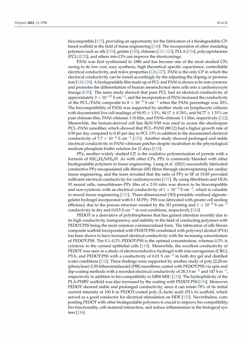
Polymers 2021, 13, 3790 10 of 21
biocompatible [117], providing an opportunity for the fabrication of a biodegradable CP-based scaffold in the field of tissue engineering [118]. The incorporation of other insulatingpolymers such as silk [119], gelatin [120], chitosan [121–123], PLLA [124], polycaprolactone(PCL) [125], and others into CPs can improve the shortcomings.
PANi was first synthesised in 1886 and has become one of the most studied CPsowing to its low cost, easy synthesis, high theoretical specific capacitance, controllableelectrical conductivity, and redox properties [126,127]. PANi is the only CP in which theelectrical conductivity can be tuned accordingly by the adjusting the doping or protona-tion [128,129]. A biodegradable film made up of PCL and PANi is shown to be non-cytotoxicand promotes the differentiation of human mesenchymal stem cells into a cardiomyocytelineage [130]. The same study showed that pure PCL had an electrical conductivity ofapproximately 3 × 10⁻¹² S cm−1, and the incorporation of PANi increased the conductivityof the PCL/PANi composite to 8 × 10⁻⁵ S cm−1 when the PANi percentage was 20%.The biocompatibility of PANi was supported by another study on lymphocyte cultureswith documented live cell readings of 94.93 ± 1.5%, 94.17 ± 0.76%, and 94.77 ± 1.51% onpure chitosan film, PANi–chitosan 1:10 film, and PANi–chitosan 1:1 film, respectively [122].Meanwhile, the human-derived cell line ReN-VM was used to access the electrospunPCL–PANi nanofiber, which showed that PCL–PANI (88:12) had a higher growth rate of0.50 per day compared to 0.45 per day in PCL 13% in addition to the documented electricalconductivity of 7.7 × 10⁻² S cm−1 [125]. Another study showed prolonged and stableelectrical conductivity in PANi–chitosan patches despite incubation in the physiologicalmedium phosphate buffer solution for 21 days [123].
PPy, another widely studied CP, is the oxidative polymerisation of pyrrole with aformula of H(C4H2NH)nH. As with other CPs, PPy is commonly blended with otherbiodegradable polymers in tissue engineering. Liang et al. (2021) successfully fabricatedconductive PPy-encapsulated silk fibroin (SF) fibres through electrospinning for cardiactissue engineering, and the team revealed that the ratio of PPy to SF at 15:85 providedsufficient electrical conductivity for cardiomyocytes [131]. By using fibroblasts and OLN-93 neural cells, nanochitosan–PPy film of a 2:10 ratio was shown to be biocompatibleand non-cytotoxic with an electrical conductivity of 1 × 10⁻³ S cm−1, which is valuablein neural tissue engineering [121]. Three-dimensional (3D)-printable oxidised alginate–gelatin hydrogel incorporated with 0.1 M PPy: PSS was fabricated with greater cell seedingefficiency due to the porous structure created by the 3D printing and 1 × 10⁻⁵ S cm−1
conductivity in dry and 0.015 S cm−1 in wet conditions, respectively [120].PEDOT is a derivative of polythiophenes that has gained attention recently due to
its high conductivity, transparency, and stability in the field of conducting polymers withPEDOT:PSS being the most common commercialised form. The fabrication of silk fibroincomposite scaffold incorporated with PEDOT:PSS crosslinked with polyvinyl alcohol (PVA)has been shown to have increased electrical conductivity with the increasing concentrationof PEDOT:PSS. The 0.1–0.2% PEDOT:PSS is the optimal concentration, whereas 0.3% iscytotoxic to the corneal epithelial cells [119]. Meanwhile, the excellent conductivity ofPEDOT was seen in a study of electroconductive hydrogel with iota-carrageenan (CRG),PVA, and PEDOT:PSS with a conductivity of 0.01 S cm−1 in both dry gel and distilledwater conditions [132]. These findings were supported by another study of poly [2,20-m-(phenylene)-5,50-bibenzimidazole] (PBI) nanofibres coated with PEDOT:PSS via spin anddip-coating methods with a recorded electrical conductivity of 28.3 S m−1 and 147 S m−1,respectively in addition to bio-compatibility to hBM-MSC [133]. The hydrophilicity of thePLA-PHBV scaffold was also increased by the coating with PEDOT:PSS [134]. Moreover,PEDOT showed stable and prolonged conductivity, since it can retain 78% of its initialcurrent intensity at 100 h in PEDOT-coated poly (L-lactic acid) (PLLA) scaffold, whichserved as a good conductor for electrical stimulation on HDF [135]. Nevertheless, com-positing PEDOT with other biodegradable polymers is crucial to improve bio-compatibility,bio-functionality, cell–material interaction, and reduce inflammation in the biological sys-tem [136].

Polymers 2021, 13, 3790 11 of 21
4.1.2. Inorganic Conducting Materials
Unlike CPs, inorganic conducting materials are those without carbon–hydrogen bonds.The examples include allotropes of carbon such as graphene, carbon nanotubes, and metal-lic compounds. Inorganic nanomaterials are on the rise in the platforms of wound healingand tissue engineering due to their outstanding intrinsic properties such as antimicrobialproperty in silver and silica, antioxidant effect in cerium oxide, reactive oxygen species(ROS) production to promote cell proliferation by zinc oxide, and the electrical conductivityin carbon nanotubes [137]. Next, this paper will review the inorganic conducting materialsthat can conduct electricity and their niches in skin tissue engineering.
Functional carbon-based nanomaterials such as graphene oxide, carbon nanotubes,and nanodiamond have been explored in the biomedical field due to their excellent elec-trical conductivity, high mechanical strength, and optical property [138]. Commerciallyavailable graphite or diamond nanoparticles were shown to disperse evenly in the PLAmatrix with an increase in both AC and DC conductivity of eight orders of magnitudescompared to the pure PLA [139]. Another study showed an even distribution of grapheneoxide (GO) nanosheets within methacryloyl-modified decellularised small intestine sub-mucosa hydrogel by coating the GO nanosheets with hydrophilic serum proteins prior tomixing, and the resulted GO-embedded hydrogel was biocompatible and non-cytotoxic tohuman adipose tissue-derived mesenchymal stem cells [140]. The same study successfullyreduced GO in situ by incubation with ascorbic acid at 37 ◦C for 3 days, and this resultedin a significantly improved electrical conductivity [140].
Carbon nanotubes are good conducting materials. Recently, enzymatic biofuel cellsemerged as a new fuel source to generate electrical energy through the enzymatic catalysisof biofuels such as sugar [141]. Carbon nanotubes were selected to conduct electricityproduced by enzymatic biofuel cells and recorded a voltage of 2.09 V [142]. By adoptingthe concept of biofuel cells, a bioelectric plaster was fabricated with a current intensityof 1 mA cm−2 that lasted for 12 h [143]. An in vivo study of the bioelectric plaster onthe full thickness of the rat wound model showed that ES generated a faster woundclosure, and the hydrogel provided a moist microenvironment to reduce wound contracture.Unfortunately, this bioelectric plaster is needed to be replaced every 12 h to ensure thecontinuous generation of electricity. This may cause secondary trauma to newly grownfragile granulation tissues in the wound, although the bioelectric plaster was shown to beadherent but not adhesive.
MXenes are the 2D transition metal carbides, nitrides, and carbonitrides that are gain-ing the attention in the field of tissue engineering, biomedicine, energy science, electronicdevices, and nanomaterials due to their excellent electrical conductivity, large surface areato volume ratio despite their nanoscale, and strong mechanical strength [144]. The availabil-ity of functional groups on the surface of MXenes allows for the surface modification andconjugation with other biocompatible polymers such as proteins and polysaccharides tofacilitate its application in the biological system [144]. Interestingly, MXenes have an excel-lent antibacterial property in which it is stronger compared to GO, and direct contact withMXenes can damage the bacterial cell membranes, giving a bactericidal effect [145]. A studyof composite hydrogel made up of bacterial nanocellulose and MXene-2% (rBC/MXene) ona full thickness of rat wound model found that electroconductive MXene significantly in-creased granulation tissue thickness with a more complete and organised re-epithelisationin addition to angiogenesis and lesser neutrophils infiltration [146].
The application of metal elements in wound dressing to generate ES was studied byYu et al. (2019) with the fabrication of microcurrent dressing using medical cotton cushionwith the wound facing one side being sprayed with silver and zinc particles in a dot matrix-arrayed method. This microcurrent dressing was able to generate 0.95 V substantially for3 days upon being moistened without external electric supply and closed up to 95% of thewound area on day 14 in vivo with a reduced level of inflammatory cytokines of TNF-αand IL-1β in addition to an increased level of growth factors VEGF and EGF [11].

Polymers 2021, 13, 3790 12 of 21
4.2. Nanogenerators (NG)
A nanogenerator is an electrical energy generator from mechanical energy [147].Piezoelectric (PENG) and triboelectric (TENG) nanogenerators are examples of nanogener-ators [148]. The utilisation of a nanogenerator to produce electricity is another idea otherthan biofuel cells in the modern ES.
A wearable NG was made by overlapping the electronegative layer of Cu/PTFEwith another electropositive layer of Cu to produce electrical energy when converting themechanical displacement between the two layers during the breathing movement of therat [149]. The pulsed ES generated approximately 2.2 V at a rate of 110 per min, whichpromoted nearly complete wound closure at 72 h in vivo compared to 50% wound closurein the control group. An in vitro study of the nanogenerator using NIH3T3 fibroblastsshowed excellent cell viability of 127% and increased proliferation in addition to elevatedexpression of VEGF, TGF-β, and EGF with a significantly reduced level of ROS.
Another wearable polyacrylamide-based gel containing lithium chloride salts coatedwith heptadecafluoro-1,1,2,2-tetrahydrodecyl trichlorosilane as TENG was fabricated [150].This fully stretchable and biocompatible ionic patch was able to generate approximately2 V when applied on an active rat, and the ES resulted in three times faster woundclosure in vivo, whereas an in vitro study on normal and diabetic HDF showed increasedproliferation, migration, and expression of FGF, VEGF, and EGF.
PVDF nanofibres is a piezoelectric material [151]. In another study, electrospun PVDFnanofibers, aluminium foils, and aluminium electrodes were used to make a piezoelec-tric nanogenerator, which was then attached to the centre of the adhesive hydrogel ofpolydopamine–polyacrylamide to be applied to the full thickness of the rat wound model.The result showed complete epithelisation on day 10 compared to 22.7% of the wound arearemaining open in the control group [152]. The piezoelectric nanogenerator generated anaverage of 0.1–0.5 V of ES, which leads to an increased expression of CD 31, VEGF-A, andTGF-β1 in the wound with no impact onto the blood, liver, and renal profile.
Nevertheless, the translation of nanogenerator application into human use requiresfurther refinement, as most experiments strip a nanogenerator on the chest of an animal,which is unethical to be applied on a patient. Furthermore, most patients with chronicwounds have comorbidities and are not as physically active as youngsters to generateadequate mechanical energy. Meanwhile, acute extensive burn patients may be haemody-namically unable to perform extensive physical activity in the early wound period, andlate treatment can result in contracture.
4.3. Stem Cell (SC) Therapy
SC therapy is a component in tissue engineering due to its self-renewal ability, andit restores the function of degenerative organs. In skin tissue engineering, SCs harvestedfrom bone marrow, adipose tissue, amniotic fluid, and the umbilical cord can be differenti-ated into the skin cell lineage and incorporated into the skin scaffold to promote woundhealing [153]. By the application of TENG, a pulsed ES of 30 µA was generated at 1.5 Hzthat successfully rejuvenated aged bone marrow mesenchymal SC (BMSC) with higherproliferation, pluripotency, and differentiation ability [154]. Meanwhile, ES generatedby enzymatic biofuel cells was found to be able to change the cell morphology and geneexpression of adipose mesenchymal SC (ASC) [155]. The combination of PC of 0.1 V/cmwith a pulse duration of 0.04 ms at 10 Hz for 30 min daily up to 21 days in a 3D nanofibrillarcellulose hydrogel resulted in increased osteogenic potential in ASC [156]. A biphasic PCof 1 V/cm at 0.5 Hz in combination with cyclic strain was found to well differentiatethe rat BMSC differentiation into neural cells [157]. ES using AC at 1.7 V and 20 Hz onASC significantly promoted cell proliferation with a 4.5-fold increase in cell numbers and2.7-fold increase of cellular surface coverage, and 50.5% of cells entered the proliferativephases of the cell cycle [158]. Hence, ES can assist the proliferation and differentiation ofSC into the skin cells of interest and promote wound healing by regenerative medicine.

Polymers 2021, 13, 3790 13 of 21
4.4. Dielectrophoresis (DEP)
AC is not commonly applied in wound healing, which is possibly due to the lack ofpolarity for galvanotaxis. Nevertheless, dielectrophoresis (DEP) is a form of electrokineticphenomenon requiring AC to generate a non-uniform electric field in order to manip-ulate the movement of neutral particles [159]. DEP has been well known to be cheap,fast, and label-free with high sensitivity and specificity for cell sorting and particle sep-aration [160,161]. The dielectrophoretic force can be manipulated by adjusting the inputfrequency of alternating current and conducting medium while the geometry and dielectricproperties of the cells are not fixed [162,163]. However, the interaction of DEP in mobilisinga particular keratinocyte or fibroblast and aligning them in a proper plane remains to beelucidated. The fabrication of tapered aluminium microelectrode arrays (TAMA) startedwith a silicon substrate as the base of the microelectrode, which was then topped withsilicon oxide followed by titanium/titanium nitrite and then aluminium/silicon/coppercomposite with photoresist and lastly the etching process [161]. Figure 5a shows the scan-ning electron microscope (SEM) image of the fabricated TAMA electrode. TAMA work ledto the identification of a tapered side wall between 60 and 70 degrees for the generationof a more uniform electric field from two spots at top and bottom edges of the taperedmicroelectrodes [161]. Refinement of the TAMA work concluded that 65 degrees was thebest reading in TAMA to generate dielectrophoretic force for lateral and vertical manipula-tion of particle manipulation as shown in Figure 5b [164]. A DEP study on keratinocytesusing “MyDEP” software showed that the DEP response of keratinocytes was successfullysimulated by using polystyrene particles [164]. This is of interest, as DEP targeted specificcells and hence, the mobilisation of keratinocytes in an appropriate direction, and theplane will make re-epithelisation possible, especially in the case of a chronic wound withsufficient granulation but slow or no opposing wound edges. Figure 6 shows successfulmanipulation of DEP on keratinocytes into area of interest Nevertheless, more studies ofDEP in wound healing need to be explored.
Figure 5. The SEM image of the fabricated microelectrode session of TAMA with measurements is clearly shown insubfigure (a). Subfigure (b) shows the finite-element method simulation analysis through COMSOL software in whichTAMA with a side wall profile of 65 degrees generated the best two spots of high electric field intensity, which subsequentlyproduce lateral and vertical dielectrophoretic forces for particles manipulation. Images are adapted with permission fromBuyong et al. (2017).

Polymers 2021, 13, 3790 14 of 21
Figure 6. Fluorescence microscopy observations of keratinocytes in DMEM/F-12 conductive mediumshowed that the keratinocytes experienced dielectrophorectic force and migrated to the area of lowestelectric field strength, which was the middle between electrodes at a final 10 VPP at 300 kHz (b),800 kHz (d), and 15 MHz (f) compared to the initial 0 VPP at 300 kHz (a), 800 kHz (c), and 15 MHz (e).Image is adapted with permission from Deivasigamani et al. (2021).
5. Conclusions
Wound care is a world burden, while clinically practised skin grafts produce a risk ofgraft failure, donor site infection, and other complications. The abilities of ES to restore theweakened endogenous current of injury in chronic wounds offer an astounding opportunityto expedite wound recovery. ES shortens the inflammatory phase, and it also enhancesfibroplasia, re-epithelisation, and angiogenesis together with remodelling to form a strongerscar in well-established in wound care.
HVMPC has overall good outcomes in chronic wounds; however, the presence ofelectrothermal, electrophysical, and electrochemical side effects, although lesser thanthe DC, must be taken into clinical consideration. Meanwhile, nanogenerators, a greenform of electric energy converted from mechanical energy, require an amount of physicalmovement, which can be difficult in patients with comorbidities in addition to discomfortbrought by stripping a nanogenerator to the human chest.
Undeniably, electroconductive materials are interesting to be incorporated as partof scaffolds to assist wound healing. However, pure conducting materials are not easilyprocessable or biodegradable with a potential risk of inducing inflammation. Hence,the incorporation of conducting materials into biodegradable polymers of both naturaland synthetic origin is widely practised and highly encouraged. Further fine-tuningof electroconductive scaffolds mimicking the targeted extracellular matrix shall directresearchers to a biodegradable, biocompatible, non-cytotoxic, non-immunogenic, easilysynthesised, and low-cost fabrication for popular application.
Bioelectric plaster-like wound dressings with electroconductive materials or enzy-matic biofuel cells are examples of smart wound dressings without an external powersupply. However, the frequent changing of wound dressing should be avoided to preventunnecessary pain and secondary trauma to the fragile growing granulation tissue.
Essentially, a successful wound closure within a short period in a painless procedureis the ultimate fundamental of research on wound healing. With the understanding of thecurrent of injury, ES is believed to accelerate wound healing and replace skin grafting inthe future either through electric devices or smart wound dressings.

Polymers 2021, 13, 3790 15 of 21
6. Future Direction
DEP is a label-free cell patterning that mobilises specific cells according to the dielec-trophoretic force exerted under a non-uniform electric field. The potential ability of DEP tomobilise keratinocytes and fibroblasts specifically into the top and bottom layers mimickingthe structure of epidermis and dermis is a field of interest in wound care. In addition, DEPapplies alternating current to run through electrodes, and hence, the accumulation of heatand chemicals at a particular electrode can be avoided. Nevertheless, the interaction of ESon wound microbial propagation remains to be elucidated.
Author Contributions: Conceptualization, Y.J.C., M.R.B. and M.H.M.Y.; methodology; software,Y.J.C. and M.R.B.; validation, M.R.B. and M.H.M.Y.; formal analysis, Y.J.C.; investigation; resources,Y.J.C. and M.R.B., M.H.M.Y.; data curation; writing—original draft preparation, Y.J.C.; writing—review and editing, M.R.B. and M.H.M.Y.; visualization, Y.J.C.; supervision, M.R.B. and M.H.M.Y.;project administration, M.H.M.Y.; funding acquisition All authors have read and agreed to thepublished version of the manuscript.
Funding: This research was funded by the Ministry of Education Malaysia, grant number FRGS/1/2020/SKK0/UKM/02/6.
Institutional Review Board Statement: Not applicable.
Informed Consent Statement: Informed consent is not applicable to this article.
Data Availability Statement: No new data were created or analyzed in this study. Data sharing isnot applicable to this article.
Conflicts of Interest: The authors declare no conflict of interest.
References1. Sen, C.K. Human wounds and its burden: An updated compendium of estimates. Adv. Wound Care 2019, 8, 39–48. [CrossRef]
[PubMed]2. Nicholas, M.N.; Yeung, J. Current status and future of skin substitutes for chronic wound healing. J. Cutan. Med. Surg. 2016,
21, 23–30. [CrossRef] [PubMed]3. Guest, J.F.; Fuller, G.W.; Vowden, P. Cohort study evaluating the burden of wounds to the UK’s National Health Service in
2017/2018: Update from 2012/2013. BMJ Open 2020, 10, e045253. [CrossRef] [PubMed]4. Nussbaum, S.R.; Carter, M.J.; Fife, C.E.; DaVanzo, J.; Haught, R.; Nusgart, M.; Cartwright, D. An economic evaluation of the
impact, cost, and medicare policy implications of chronic nonhealing wounds. Value Health 2018, 21, 27–32. [CrossRef]5. Heyer, K.; Herberger, K.; Protz, K.; Glaeske, G.; Augustin, M. Epidemiology of chronic wounds in Germany: Analysis of statutory
health insurance data. Wound Repair Regen. 2015, 24, 434–442. [CrossRef]6. Turissini, J.D.; Elmarsafi, T.; Evans, K.K.; Kim, P.J. Major risk factors contributing to split thickness skin graft failure.
Georget. Med. Rev. 2019, 3, 7755. [CrossRef]7. Guogiene, I.; Kievišas, M.; Varkalys, K.; Braziulis, K.; Rimdeika, R. Split-thickness skin grafting using grafts of different thickness.
Eur. J. Plast. Surg. 2018, 41, 583–590. [CrossRef]8. Jones, R.E.; Foster, D.S.; Longaker, M.T. Management of chronic wounds—2018. JAMA 2018, 320, 1481–1482. [CrossRef] [PubMed]9. Frykberg, R.G.; Banks, J. Challenges in the treatment of chronic wounds. Adv. Wound Care 2015, 4, 560–582. [CrossRef] [PubMed]10. Fujiwara, H.; Isogai, Z.; Irisawa, R.; Otsuka, M.; Kadono, T.; Koga, M.; Hirosaki, K.; Asai, J.; Asano, Y.; Abe, M.; et al. Wound,
pressure ulcer and burn guidelines—2: Guidelines for the diagnosis and treatment of pressure ulcers, second edition. J. Dermatol.2018, 47, 929–978. [CrossRef] [PubMed]
11. Yu, C.; Xu, Z.-X.; Hao, Y.-H.; Gao, Y.-B.; Yao, B.-W.; Zhang, J.; Wang, B.; Hu, Z.-Q.; Peng, R.-Y. A novel microcurrent dressing forwound healing in a rat skin defect model. Mil. Med. Res. 2019, 6, 1–9. [CrossRef]
12. You, D.; Li, K.; Guo, W.; Zhao, G.; Fu, C. Poly (lactic-co-glycolic acid)/graphene oxide composites combined with electricalstimulation in wound healing: Preparation and characterization. Int. J. Nanomed. 2019, 14, 7039–7052. [CrossRef] [PubMed]
13. Balakatounis, K.C.; Angoules, A. Low-intensity electrical stimulation in wound healing: Review of the efficacy of externallyapplied currents resembling the current of injury. Eplasty 2008, 8, 28.
14. Zhao, M. Electrical fields in wound healing—An overriding signal that directs cell migration. Semin. Cell Dev. Biol. 2009,20, 674–682. [CrossRef]
15. Shen, Y.; Pfluger, T.; Ferreira, F.; Liang, J.; Navedo, M.F.; Zeng, Q.; Reid, B.; Zhao, M. Diabetic cornea wounds produce significantlyweaker electric signals that may contribute to impaired healing. Sci. Rep. 2016, 6, 26525. [CrossRef]
16. Wahlsten, O.; Skiba, J.B.; Makin, I.R.S.; Apell, S.P. Electrical field landscape of two electroceuticals. J. Electr. Bioimpedance 2016,7, 13–19. [CrossRef]

Polymers 2021, 13, 3790 16 of 21
17. Galvani, L. De viribus electricitatis in motu musculari Commentarius. Bonoiensi Sci. Artium Intituo atque Acad. Comment.1791, 7, 363-418.Piccolino, M. Luigi Galvani and animal electricity: Two centuries after the foundation of electrophysiology.Trends Neurosci. 1997, 20, 443–448. [CrossRef]
18. Piccolino, M. Animal electricity and the birth of electrophysiology: The legacy of Luigi Galvani. Brain Res. Bull. 1998, 46, 381–407.[CrossRef]
19. Cajavilca, C.; Varon, J.; Sternbach, G.L. Luigi Galvani and the foundations of electrophysiology. Resuscitation 2009, 80, 159–162.[CrossRef] [PubMed]
20. Du Bois-Reymond, E.H. Vorläufiger abriss einer untersuchung uber den sogenannten froschstrom und die electomotorischenfische. Ann. Phys. Chem. 1843, 58, 1–30. [CrossRef]
21. Barker, A.T.; Jaffe, L.F.; Vanable, J.W. The glabrous epidermis of cavies contains a powerful battery. Am. J. Physiol. Integr.Comp. Physiol. 1982, 242, R358–R366. [CrossRef] [PubMed]
22. Foulds, I.S.; Barker, A.T. Human skin battery potentials and their possible role in wound healing. Br. J. Dermatol. 1983,109, 515–522. [CrossRef]
23. Zhao, M.; Song, B.; Pu, J.; Wada, T.; Reid, B.; Tai, G.; Wang, F.; Guo, A.; Walczysko, P.; Gu, Y.; et al. Electrical signals controlwound healing through phosphatidylinositol-3-OH kinase-γ and PTEN. Nature 2006, 442, 457–460. [CrossRef] [PubMed]
24. McGinnis, M.; Vanable, J.W., Jr. Electrical fields in Notophthalmus viridescens limb stumps. Dev. Biol. 1986, 116, 184–193. [CrossRef]25. Vieira, A.C.; Reid, B.; Cao, L.; Mannis, M.J.; Schwab, I.R.; Zhao, M. Ionic components of electric current at rat corneal wounds.
PLoS ONE 2011, 6, e17411. [CrossRef] [PubMed]26. Reid, B.R.; Nuccitelli, R.; Zhao, M. Non-invasive measurement of bioelectric currents with a vibrating probe. Nat. Protoc. 2007,
2, 661–669. [CrossRef] [PubMed]27. Reid, B.; Song, B.; McCaig, C.D.; Zhao, M. Wound healing in rat cornea: The role of electric currents. FASEB J. 2004, 19, 379–386.
[CrossRef]28. Nuccitelli, R.; Nuccitelli, P.; Ramlatchan, S.; Sanger, R.; Smith, P.J. Imaging the electric field associated with mouse and human
skin wounds. Wound Repair Regen. 2008, 16, 432–441. [CrossRef]29. Jaffe, L.F.; Vanable, J.W. Electric fields and wound healing. Clin. Dermatol. 1984, 2, 34–44. [CrossRef]30. Nuccitelli, R. Endogenous electric fields in embryos during development, regeneration and wound healing. Radiat. Prot. Dosim.
2003, 106, 375–383. [CrossRef]31. Guo, A.; Song, B.; Reid, B.; Gu, Y.; Forrester, J.V.; Jahoda, C.A.; Zhao, M. Effects of physiological electric fields on migration of
human dermal fibroblasts. J. Investig. Dermatol. 2010, 130, 2320–2327. [CrossRef] [PubMed]32. Wang, M.; Luo, Y.; Wang, T.; Wan, C.; Pan, L.; Pan, S.; He, K.; Neo, A.; Chen, X. Artificial skin perception. Adv. Mater. 2021,
33, 1–20. [CrossRef]33. Ashrafi, M.; Alonso-Rasgado, T.; Baguneid, M.; Bayat, A. The efficacy of electrical stimulation in experimentally induced
cutaneous wounds in animals. Vet. Dermatol. 2016, 27, 235-e57. [CrossRef]34. Gurtner, G.C.; Werner, S.; Barrandon, Y.; Longaker, M.T. Wound repair and regeneration. Nature 2008, 453, 314–321. [CrossRef]
[PubMed]35. Tottoli, E.M.; Dorati, R.; Genta, I.; Chiesa, E.; Pisani, S.; Conti, B. Skin wound healing process and new emerging technologies for
skin wound care and regeneration. Pharmaceutics 2020, 12, 735. [CrossRef]36. Souza, A.K.; Souza, T.R.; das Neves, L.M.S.; Ferreira Leite, G.d.P.M.; Garcia, S.B.; de Jesus Guirro, R.R.; Barbosa, R.I.; de Oliveira
Guirro, E.C. Effect of high voltage pulsed current on the integration of total skin grafts in rats submitted to nicotine action. J.Tissue Viability 2019, 28, 161–166. [CrossRef]
37. Wang, K.; Parekh, U.; Ting, J.K.; Yamamoto, N.A.D.; Zhu, J.; Costantini, T.; Arias, A.C.; Eliceiri, B.P.; Ng, T.N. A platform to studythe effects of electrical stimulation on immune cell activation during wound healing. Adv. Biosyst. 2019, 3, e1900106. [CrossRef][PubMed]
38. Lévêque, M.; Penna, A.; le Trionnaire, S.; Belleguic, C.; Desrues, B.; Brinchault, G.; Jouneau, S.; Lagadic-Gossmann, D.;Martin-Chouly, C. Phagocytosis depends on TRPV2-mediated calcium influx and requires TRPV2 in lipids rafts: Alteration inmacrophages from patients with cystic fibrosis. Sci. Rep. 2018, 8, 1–13. [CrossRef] [PubMed]
39. Hoare, J.I.; Rajnicek, A.M.; McCaig, C.D.; Barker, R.N.; Wilson, H.M. Electric fields are novel determinants of human macrophagefunctions. J. Leukoc. Biol. 2016, 99, 1141–1151. [CrossRef] [PubMed]
40. Rowley, B.A. Electrical current effects on E. coil growth rates. Exp. Biol. Med. 1972, 139, 929–934. [CrossRef]41. Barranco, S.D.; Spadaro, J.A.; Berger, T.J.; Becker, R.O. In vitro effect of weak direct current on Staphylococcus aureus. Clin. Orthop.
Relat. Res. 1974, 100, 250–255. [CrossRef]42. Dauben, T.J.; Ziebart, J.; Bender, T.; Zaatreh, S.; Kreikemeyer, B.; Bader, R. A novel in vitro system for comparative analyses of
bone cells and bacteria under electrical stimulation. BioMed Res. Int. 2016, 2016, 1–12. [CrossRef] [PubMed]43. Krishnamurthi, V.R.; Rogers, A.; Peifer, J.; Niyonshuti, I.I.; Chen, J.; Wang, Y. Microampere electric current causes bacterial
membrane damage and two-way leakage in a short period of time. Appl. Environ. Microbiol. 2020, 86, 01015–01020. [CrossRef][PubMed]
44. Petrofsky, J.; Laymon, M.; Chung, W.; Collins, K.; Yang, T.-N. Effect of Electrical Stimulation on Bacterial Growth. J. Orthop.Neurol. Surg. 2008, 31, 43.

Polymers 2021, 13, 3790 17 of 21
45. Rouabhia, M.; Park, H.J.; Abedin-Do, A.; Douville, Y.; Methot, M.; Zhang, Z. Electrical stimulation promotes the proliferation ofhuman keratinocytes, increases the production of keratin 5 and 14, and increases the phosphorylation of ERK1/2 and p38 MAPkinases. J. Tissue Eng. Regen. Med. 2020, 14, 909–919. [CrossRef] [PubMed]
46. Hart, F.X.; Laird, M.; Riding, A.; Pullar, C.E. Keratinocyte galvanotaxis in combined DC and AC electric fields supports anelectromechanical transduction sensing mechanism. Bioelectromagnetics 2013, 34, 85–94. [CrossRef] [PubMed]
47. Ren, X.; Sun, H.; Liu, J.; Guo, X.; Huang, J.; Jiang, X.; Zhang, Y.; Huang, Y.; Fan, D.; Zhang, J. Keratinocyte electrotaxis induced byphysiological pulsed direct current electric fields. Bioelectrochemistry 2019, 127, 113–124. [CrossRef]
48. Krzyszczyk, P.; Schloss, R.; Palmer, A.; Berthiaume, F. The role of macrophages in acute and chronic wound healing andinterventions to promote pro-wound healing phenotypes. Front. Physiol. 2018, 9, 419. [CrossRef]
49. Sebastian, A.; Iqbal, S.A.; Colthurst, J.; Volk, S.W.; Bayat, A. Electrical stimulation enhances epidermal proliferation in humancutaneous wounds by modulating p53–SIVA1 interaction. J. Investig. Dermatol. 2015, 135, 1166–1174. [CrossRef]
50. Casagrande, S.M.; Biondo-Simões, M.D.L.P.; Ioshii, S.; Robes, R.R.; Biondo-Simões, R.; Boeno, B.R.D.O. Histological evaluation ofthe effect of low-frequency electric stimulation on healing Achilles tendons in rats. Acta Cir. Bras. 2021, 36, e360103. [CrossRef]
51. Urabe, H.; Akimoto, R.; Kamiya, S.; Hosoki, K.; Ichikawa, H.; Nishiyama, T. Effects of pulsed electrical stimulation on growthfactor gene expression and proliferation in human dermal fibroblasts. Mol. Cell. Biochem. 2021, 476, 361–368. [CrossRef] [PubMed]
52. Rouabhia, M.; Park, H.J.; Zhang, Z. Electrically activated primary human fibroblasts improve in vitro and in vivo skin regenera-tion. J. Cell. Physiol. 2016, 231, 1814–1821. [CrossRef]
53. Wang, Y.; Rouabhia, M.; Zhang, Z. Pulsed electrical stimulation benefits wound healing by activating skin fibroblasts through theTGFβ1/ERK/NF-κB axis. Biochim. Biophys. Acta BBA—Gen. Subj. 2016, 1860, 1551–1559. [CrossRef]
54. Rouabhia, M.; Park, H.; Meng, S.; Derbali, H.; Zhang, Z. Electrical stimulation promotes wound healing by enhancing dermalfibroblast activity and promoting myofibroblast transdifferentiation. PLoS ONE 2013, 8, e71660. [CrossRef]
55. Snyder, S.; DeJulius, C.; Willits, R.K. Electrical stimulation increases random migration of human dermal fibroblasts.Ann. Biomed. Eng. 2017, 45, 2049–2060. [CrossRef] [PubMed]
56. Benington, L.; Rajan, G.; Locher, C.; Lim, L.Y. Fibroblast growth factor 2—A review of stabilisation approaches for clinicalapplications. Pharmaceutics 2020, 12, 508. [CrossRef]
57. Xie, Y.; Su, N.; Yang, J.; Tan, Q.; Huang, S.; Jin, M.; Ni, Z.; Zhang, B.; Zhang, D.; Luo, F.; et al. FGF/FGFR signaling in health anddisease. Signal Transduct. Target. Ther. 2020, 5, 1–38. [CrossRef] [PubMed]
58. Uemura, M.; Maeshige, N.; Koga, Y.; Ishikawa-Aoyama, M.; Miyoshi, M.; Sugimoto, M.; Terashi, H.; Usami, M. Monophasicpulsed 200-µA current promotes galvanotaxis with polarization of actin filament and integrin α2β1 in human dermal fibroblasts.Eplasty 2016, 16, 6.
59. Cunha, F.; Rajnicek, A.M.; McCaig, C.D. Electrical stimulation directs migration, enhances and orients cell division and upregulatesthe chemokine receptors CXCR4 and CXCR2 in endothelial cells. J. Vasc. Res. 2019, 56, 39–53. [CrossRef] [PubMed]
60. Geng, K.; Wang, J.; Liu, P.; Tian, X.; Liu, H.; Wang, X.; Hu, C.; Yan, H. Electrical stimulation facilitates the angiogenesisof human umbilical vein endothelial cells through MAPK/ERK signaling pathway by stimulating FGF2 secretion. Am. J.Physiol.—Cell Physiol. 2019, 317, C277–C286. [CrossRef] [PubMed]
61. Bai, H.; Forrester, J.V.; Zhao, M. DC electric stimulation upregulates angiogenic factors in endothelial cells through activation ofVEGF receptors. Cytokine 2011, 55, 110–115. [CrossRef]
62. Shoeibi, S.; Mozdziak, P.; Mohammadi, S. Important signals regulating coronary artery angiogenesis. Microvasc. Res. 2018,117, 1–9. [CrossRef]
63. Teleanu, R.I.; Chircov, C.; Grumezescu, A.M.; Teleanu, D.M. Tumor angiogenesis and anti-angiogenic strategies for cancertreatment. J. Clin. Med. 2019, 9, 84. [CrossRef] [PubMed]
64. Tan, Y.; Shu, L.; Xu, P.; Bai, S. Mesenchymal stem cells attract endothelial progenitor cells via a positive feedback loop betweenCXCR2 and CXCR4. Stem Cells Int. 2019, 2019, 1–9. [CrossRef] [PubMed]
65. Uemura, M.; Sugimoto, M.; Yoshikawa, Y.; Hiramatsu, T.; Inoue, T. Monophasic pulsed current stimulation of duty cycle 10%promotes differentiation of human dermal fibroblasts into myofibroblasts. Phys. Ther. Res. 2021, 24, 145–152. [CrossRef] [PubMed]
66. Sebastian, A.; Volk, S.W.; Halai, P.; Colthurst, J.; Paus, R.; Bayat, A. Enhanced neurogenic biomarker expression and reinnervationin human acute skin wounds treated by electrical stimulation. J. Investig. Dermatol. 2017, 137, 737–747. [CrossRef]
67. Keast, D.H.; Moffatt, C.; Janmohammad, A. Lymphedema impact and prevalence international study: The Canadian data.Lymphat. Res. Biol. 2019, 17, 178–186. [CrossRef] [PubMed]
68. Bjork, R.; Hettrick, H. Lymphedema: New concepts in diagnosis and treatment. Curr. Dermatol. Rep. 2019, 8, 190–198. [CrossRef]69. Kajiya, K.; Matsumoto-Okazaki, Y.; Sawane, M.; Fukada, K.; Takasugi, Y.; Akai, T.; Saito, N.; Mori, Y. Electric current-induced
lymphatic activation. Exp. Dermatol. 2014, 23, 936–938. [CrossRef] [PubMed]70. Wei, Y.; Yang, K.; Browne, M.; Bostan, L.; Worsley, P. Wearable electrical stimulation to improve lymphatic function. IEEE Sens. Lett.
2019, 3, 1–4. [CrossRef]71. Baglivo, M.; Martelli, F.; Paolacci, S.; Manara, E.; Michelini, S.; Bertelli, M. Electrical stimulation in the treatment of lymphedema
and associated skin ulcers. Lymphat. Res. Biol. 2020, 18, 270–276. [CrossRef] [PubMed]72. AlGhatrif, M.; Lindsay, J. A brief review: History to understand fundamentals of electrocardiography. J. Community Hosp. Intern.
Med. Perspect. 2012, 2, 14383. [CrossRef] [PubMed]

Polymers 2021, 13, 3790 18 of 21
73. Baloglu, U.B.; Talo, M.; Yildirim, O.; Tan, R.S.; Acharya, U.R. Classification of myocardial infarction with multi-lead ECG signalsand deep CNN. Pattern Recognit. Lett. 2019, 122, 23–30. [CrossRef]
74. Ball, C.M.; Featherstone, P.J. The early history of cardiac pacing. Anaesth. Intensiv. Care 2019, 47, 320–321. [CrossRef]75. Donnelly, C.; Stegmüller, J.; Blazevich, A.J.; von Roten, F.C.; Kayser, B.; Neyroud, D.; Place, N. Modulation of torque evoked by
wide-pulse, high-frequency neuromuscular electrical stimulation and the potential implications for rehabilitation and training.Sci. Rep. 2021, 11, 1–13. [CrossRef]
76. Gibson, W.; Wand, B.M.; Meads, C.; Catley, M.J.; O’Connell, N.E. Transcutaneous electrical nerve stimulation (TENS) for chronicpain—An overview of Cochrane reviews. Cochrane Database Syst. Rev. 2019, 1–27. [CrossRef]
77. Li, M.; Yao, X.; Sun, L.; Zhao, L.; Xu, W.; Zhao, H.; Zhao, F.; Zou, X.; Cheng, Z.; Li, B.; et al. Effects of electroconvulsive therapy ondepression and its potential mechanism. Front. Psychol. 2020, 11, 80. [CrossRef]
78. Weiner, R.D.; Reti, I.M. Key updates in the clinical application of electroconvulsive therapy. Int. Rev. Psychiatry 2017, 29, 54–62.[CrossRef]
79. Little, S.; Brown, P. Debugging adaptive deep brain stimulation for Parkinson’s disease. Mov. Disord. 2020, 35, 555–561. [CrossRef]80. Ryan, C.N.M.; Doulgkeroglou, M.N.; Zeugolis, D.I. Electric field stimulation for tissue engineering applications. BMC Biomed. Eng.
2021, 3, 1. [CrossRef]81. Ojingwa, J.C.; Isseroff, R.R. Electrical stimulation of wound healing. J. Investig. Dermatol. 2003, 121, 1–12. [CrossRef]82. Kloth, L.C. Electrical stimulation for wound healing: A review of evidence from in vitro studies, animal experiments, and clinical
trials. Int. J. Low. Extrem. Wounds 2005, 4, 23–44. [CrossRef] [PubMed]83. Polak, A.; Kloth, L.C.; Blaszczak, E.; Taradaj, J.; Nawrat-Szołtysik, A.; Walczak, A.; Bialek, L.; Paczula, M.; Franek, A.; Kucio, C.
Evaluation of the healing progress of pressure ulcers treated with cathodal high-voltage monophasic pulsed current: Results of aprospective, double-blind, randomized clinical trial. Adv. Skin Wound Care 2016, 29, 447–459. [CrossRef]
84. Polak, A.; Kucio, C.; Kloth, L.; Paczula, M.; Hordynska, E.; Ickowicz, T.; Blaszczak, E.; Kucio, E.; Oleszczyk, K.; Ficek, K.; et al. Arandomized, controlled clinical study to assess the effect of anodal and cathodal electrical stimulation on periwound skin bloodflow and pressure ulcer size reduction in persons with neurological injuries. Ostomy Wound Manag. 2018, 64, 10–29. [CrossRef]
85. Polak, A.; Kloth, L.C.; Blaszczak, E.; Taradaj, J.; Nawrat-Szoltysik, A.; Ickowicz, T.; Hordynska, E.; Franek, A.; Kucio, C. Theefficacy of pressure ulcer treatment with cathodal and cathodal-anodal high-voltage monophasic pulsed current: A prospective,randomized, controlled clinical trial. Phys. Ther. 2017, 97, 777–789. [CrossRef]
86. Wang, X.-F.; Li, M.-L.; Fang, Q.-Q.; Zhao, W.-Y.; Lou, D.; Hu, Y.-Y.; Chen, J.; Tan, W.-Q. Flexible electrical stimulation device withChitosan-Vaseline®dressing accelerates wound healing in diabetes. Bioact. Mater. 2021, 6, 230–243. [CrossRef] [PubMed]
87. Mohajeri-Tehrani, M.R.; Nasiripoor, F.; Torkaman, G.; Hedayati, M.; Annabestani, Z.; Asadi, M.R. Effect of low-intensity directcurrent on expression of vascular endothelial growth factor and nitric oxide in diabetic foot ulcers. J. Rehabil. Res. Dev. 2014,51, 815–824. [CrossRef] [PubMed]
88. Gomes, R.C.; Guirro, E.C.; Gonçalves, A.C.; Farina, J.A., Jr.; Murta, L.O., Jr.; Guirro, R.R. High-voltage electric stimulation of thedonor site of skin grafts accelerates the healing process. A randomized blinded clinical trial. Burns 2018, 44, 636–645. [CrossRef][PubMed]
89. Sugimoto, M.; Maeshige, N.; Honda, H.; Yoshikawa, Y.; Uemura, M.; Yamamoto, M.; Terashi, H. Optimum microcurrentstimulation intensity for galvanotaxis in human fibroblasts. J. Wound Care 2012, 21, 5–10. [CrossRef]
90. Kim, M.S.; Lee, M.H.; Kwon, B.-J.; Seo, H.J.; Koo, M.-A.; You, K.E.; Kim, D.; Park, J.-C. Control of neonatal human dermalfibroblast migration on poly (lactic-co-glycolic acid)-coated surfaces by electrotaxis. J. Tissue Eng. Regen. Med. 2017, 11, 862–868.[CrossRef]
91. Wang, L.; Mao, L.; Qi, F.; Li, X.; Ullah, M.W.; Zhao, M.; Shi, Z.; Yang, G. Synergistic effect of highly aligned bacterial cellu-lose/gelatin membranes and electrical stimulation on directional cell migration for accelerated wound healing. Chem. Eng. J.2021, 424, 130563. [CrossRef]
92. Oliveira, K.M.C.; Barker, J.H.; Berezikov, E.; Pindur, L.; Kynigopoulos, S.; Eischen-Loges, M.; Han, Z.; Bhavsar, M.B.; Henrich, D.;Leppik, L. Electrical stimulation shifts healing/scarring towards regeneration in a rat limb amputation model. Sci. Rep. 2019,9, 1–14. [CrossRef] [PubMed]
93. Asadi, M.R.; Torkaman, G.; Hedayati, M.; Mofid, M. Role of sensory and motor intensity of electrical stimulation on fibroblasticgrowth factor-2 expression, inflammation, vascularization, and mechanical strength of full-thickness wounds. J. Rehabil. Res. Dev.2013, 50, 489. [CrossRef] [PubMed]
94. Li, M.; Wang, X.; Rajagopalan, P.; Zhang, L.; Zhan, S.; Huang, S.; Li, W.; Zeng, X.; Ye, Q.; Liu, Y.; et al. Toward controlled electricalstimulation for wound healing based on a precision layered skin model. ACS Appl. Bio Mater. 2020, 3, 8901–8910. [CrossRef]
95. Abedin-Do, A.; Zhang, Z.; Douville, Y.; Méthot, M.; Rouabhia, M. Effect of electrical stimulation on diabetic human skin fibroblastgrowth and the secretion of cytokines and growth factors involved in wound healing. Biology 2021, 10, 641. [CrossRef] [PubMed]
96. Ramadhinara, A.; Poulas, K. Use of wireless microcurrent stimulation for the treatment of diabetes-related wounds: 2 case reports.Adv. Skin Wound Care 2013, 26, 1–4. [CrossRef] [PubMed]
97. Recio, A.C.; Felter, C.E.; Schneider, A.C.; McDonald, J.W. High-voltage electrical stimulation for the management of stage III andIV pressure ulcers among adults with spinal cord injury: Demonstration of its utility for recalcitrant wounds below the level ofinjury. J. Spinal Cord Med. 2012, 35, 58–63. [CrossRef] [PubMed]

Polymers 2021, 13, 3790 19 of 21
98. Ibrahim, Z.M.; Waked, I.S.; Ibrahim, O. Negative pressure wound therapy versus microcurrent electrical stimulation in woundhealing in burns. J. Wound Care 2019, 28, 214–219. [CrossRef]
99. Borba, G.C.; Hochman, B.; Liebano, R.E.; Enokihara, M.M.; Ferreira, L.M. Does preoperative electrical stimulation of the skin alterthe healing process? J. Surg. Res. 2011, 166, 324–329. [CrossRef] [PubMed]
100. Sari, Y.; Hartono; Sutrisna, E.; Saryono. The effect of short duration of electrical stimulation on wound healing in acute wound ina rat model. Wound Med. 2019, 24, 36–44. [CrossRef]
101. Fraccalvieri, M.; Salomone, M.; Zingarelli, E.M.; Rivarossa, F.; Bruschi, S. Electrical stimulation for difficult wounds: Only analternative procedure? Int. Wound J. 2014, 12, 669–673. [CrossRef]
102. Ud-Din, S.; Sebastian, A.; Giddings, P.; Colthurst, J.; Whiteside, S.; Morris, J.; Nuccitelli, R.; Pullar, C.; Baguneid, M.; Bayat, A.Angiogenesis is induced and wound size is reduced by electrical stimulation in an acute wound healing model in human skin.PLoS ONE 2015, 10, e0124502. [CrossRef]
103. Petrofsky, J.S.; Lawson, D.; Berk, L.; Suh, H. Enhanced healing of diabetic foot ulcers using local heat and electrical stimulationfor 30 min three times per week. J. Diabetes 2010, 2, 41–46. [CrossRef] [PubMed]
104. Arif, M.M.A.; Fauzi, M.B.; Nordin, A.; Hiraoka, Y.; Tabata, Y.; Yunus, M.H.M. Fabrication of bio-based gelatin sponge for potentialuse as a functional acellular skin substitute. Polymers 2020, 12, 2678. [CrossRef] [PubMed]
105. Ooi, K.S.; Haszman, S.; Wong, Y.N.; Soidin, E.; Hesham, N.; Mior, M.A.A.; Tabata, Y.; Ahmad, I.; Fauzi, M.B.; Yunus, M.H.Physicochemical characterization of bilayer hybrid nanocellulose-collagen as a potential wound dressing. Materials 2020, 13, 4352.[CrossRef]
106. Busra, F.M.; Rajab, N.F.; Tabata, Y.; Bin Saim, A.; Idrus, R.B.; Chowdhury, S.R. Rapid treatment of full-thickness skin loss usingovine tendon collagen type I scaffold with skin cells. J. Tissue Eng. Regen. Med. 2019, 13, 874–891. [CrossRef]
107. Park, S.-B.; Lih, E.; Park, K.-S.; Joung, Y.K.; Han, D.K. Biopolymer-based functional composites for medical applications. Prog.Polym. Sci. 2017, 68, 77–105. [CrossRef]
108. Zare, E.N.; Agarwal, T.; Zarepour, A.; Pinelli, F.; Zarrabi, A.; Rossi, F.; Ashrafizadeh, M.; Maleki, A.; Shahbazi, M.-A.;Maiti, T.K.; et al. Electroconductive multi-functional polypyrrole composites for biomedical applications. Appl. Mater. Today 2021,24, 101117. [CrossRef]
109. Levi, B.G. Nobel Prize in chemistry salutes the discovery of conducting polymers. Phys. Today 2000, 53, 19–22. [CrossRef]110. Shirakawa, H.; Louis, E.J.; MacDiarmid, A.G.; Chiang, C.K.; Heeger, A.J. Synthesis of electrically conducting organic polymers:
Halogen derivatives of polyacetylene, (CH) x. J. Chem. Soc. Chem. Commun. 1977, 16, 578–580. [CrossRef]111. Shirakawa, H.; McDiarmid, A.; Heeger, A. Focus article: Twenty-five years of conducting polymers. Chem. Commun. 2003, 1, 1–4.
[CrossRef]112. Tang, X.; Saveh-Shemshaki, N.; Kan, H.-M.; Khan, Y.; Laurencin, C.T. Biomimetic electroconductive nanofibrous matrices for
skeletal muscle regenerative engineering. Regen. Eng. Transl. Med. 2020, 6, 228–237. [CrossRef] [PubMed]113. Athukorala, S.S.; Tran, T.S.; Balu, R.; Truong, V.K.; Chapman, J.; Dutta, N.K.; Choudhury, R.N. 3D printable electrically conductive
hydrogel scaffolds for biomedical applications: A review. Polymers 2021, 13, 474. [CrossRef]114. Wijsboom, Y.H.; Patra, A.; Zade, S.S.; Sheynin, Y.; Li, M.; Shimon, L.J.W.; Bendikov, M. Controlling rigidity and planarity in
conjugated polymers: Poly (3,4-ethylenedithioselenophene). Angew. Chem. Int. Ed. 2009, 48, 5443–5447. [CrossRef] [PubMed]115. Zhang, Q.-S.; Yan, Y.-H.; Li, S.-P.; Feng, T. Synthesis of a novel biodegradable and electroactive polyphosphazene for biomedical
application. Biomed. Mater. 2009, 4, 035008. [CrossRef] [PubMed]116. Sedaghat, S. Synthesis and evaluation of chitosan-polyaniline copolymer in presence of ammonium persulfate as initiator. J. Appl.
Chem. Res. 2014, 8, 47–54.117. Hardy, J.G.; Lee, J.Y.; Schmidt, C.E. Biomimetic conducting polymer-based tissue scaffolds. Curr. Opin. Biotechnol. 2013,
24, 847–854. [CrossRef]118. Bhattacharjee, P.; Ahearne, M. Fabrication and biocompatibility of electroconductive silk fibroin/PEDOT: PSS composites for
corneal epithelial regeneration. Polymers 2020, 12, 3028. [CrossRef]119. Distler, T.; Polley, C.; Shi, F.; Schneidereit, D.; Ashton, M.D.; Friedrich, O.; Kolb, J.F.; Hardy, J.G.; Detsch, R.; Seitz, H.; et al. Electri-
cally conductive and 3D-printable oxidized alginate-gelatin polypyrrole: PSS hydrogels for tissue engineering. Adv. Health Mater.2021, 10, 2001876. [CrossRef]
120. Manzari-Tavakoli, A.; Tarasi, R.; Sedghi, R.; Moghimi, A.; Niknejad, H. Fabrication of nanochitosan incorporated polypyr-role/alginate conducting scaffold for neural tissue engineering. Sci. Rep. 2020, 10, 1–10. [CrossRef] [PubMed]
121. Pasela, B.R.; Castillo, A.P.; Simon, R.; Pulido, M.T.; Mana-Ay, H.; Abiquibil, M.R.; Montecillo, R.; Thumanu, K.; von Tumacder, D.;Taaca, K.L. Synthesis and characterization of acetic acid-doped polyaniline and polyaniline–chitosan composite. Biomimetics 2019,4, 15. [CrossRef] [PubMed]
122. Rahman, S.U.; Bilal, S.; Ali Shah, A.U.H. Synthesis and characterization of polyaniline-chitosan patches with enhanced stabilityin physiological conditions. Polymers 2020, 12, 2870. [CrossRef]
123. Ma, C.; Jiang, L.; Wang, Y.; Gang, F.; Xu, N.; Li, T.; Liu, Z.; Chi, Y.; Wang, X.; Zhao, L.; et al. 3D printing of conductive tissueengineering scaffolds containing polypyrrole nanoparticles with different morphologies and concentrations. Materials 2019,12, 2491. [CrossRef] [PubMed]

Polymers 2021, 13, 3790 20 of 21
124. Garrudo, F.; Chapman, C.A.; Hoffman, P.R.; Udangawa, R.; Silva, J.C.; Mikael, P.E.; Rodrigues, C.A.; Cabral, J.M.; Morgado, J.;Ferreira, F.C.; et al. Polyaniline-polycaprolactone blended nanofibers for neural cell culture. Eur. Polym. J. 2019, 117, 28–37.[CrossRef]
125. Korupalli, C.; Li, H.; Nguyen, N.; Mi, F.; Chang, Y.; Lin, Y.; Sung, H. Conductive materials for healing wounds: Their incorporationin electroactive wound dressings, characterization, and perspectives. Adv. Health Mater. 2021, 10, e2001384. [CrossRef] [PubMed]
126. Wang, X.; Wu, Y.; Song, X.; Dubal, D.P.; Zhao, X.; Zhang, D. Review on carbon/polyaniline hybrids: Design and synthesis forsupercapacitor. Molecules 2019, 24, 2263. [CrossRef]
127. MacDiarmid, A.G.; Chiang, J.; Halpern, M.; Huang, W.; Mu, S.; Nanaxakkara, L.; Wu, S.W.; Yaniger, S.I. “ Polyaniline ”:Interconversion of Metallic and Insulating Forms. Mol. Cryst. Liq. Cryst. 1985, 121, 173–180. [CrossRef]
128. Saberi, A.; Jabbari, F.; Zarrintaj, P.; Saeb, M.R.; Mozafari, M. Electrically conductive materials: Opportunities and challenges intissue engineering. Biomolecules 2019, 9, 448. [CrossRef]
129. Borriello, A.; Guarino, V.; Schiavo, L.; Alvarez-Perez, M.A.; Ambrosio, L. Optimizing PANi doped electroactive substrates aspatches for the regeneration of cardiac muscle. J. Mater. Sci. Mater. Electron. 2011, 22, 1053–1062. [CrossRef] [PubMed]
130. Liang, Y.; Mitriashkin, A.; Lim, T.T.; Goh, J.C.-H. Conductive polypyrrole-encapsulated silk fibroin fibers for cardiac tissueengineering. Biomaterials 2021, 276, 121008. [CrossRef]
131. Gotovtsev, P.M.; Badranova, G.U.; Zubavichus, Y.V.; Chumakov, N.K.; Antipova, C.G.; Kamyshinsky, R.; Presniakov, M.Y.;Tokaev, K.V.; Grigoriev, T. Electroconductive PEDOT: PSS-based hydrogel prepared by freezing-thawing method. Heliyon 2019, 5,e02498. [CrossRef] [PubMed]
132. Sordini, L.; Silva, J.C.; Garrudo, F.F.F.; Rodrigues, C.A.V.; Marques, A.C.; Linhardt, R.J.; Cabral, J.M.S.; Morgado, J.; Ferreira, F.C.PEDOT: PSS-coated polybenzimidazole electroconductive nanofibers for biomedical applications. Polymers 2021, 13, 2786.[CrossRef] [PubMed]
133. Chang, H.C.; Sun, T.; Sultana, N.; Lim, M.M.; Khan, T.H.; Ismail, A.F. Conductive PEDOT: PSS coated polylactide (PLA) and poly(3-hydroxybutyrate-co-3-hydroxyvalerate) (PHBV) electrospun membranes: Fabrication and characterization. Mater. Sci. Eng. C2016, 61, 396–410. [CrossRef]
134. Niu, X.; Rouabhia, M.; Chiffot, N.; King, M.W.; Zhang, Z. An electrically conductive 3D scaffold based on a nonwoven web ofpoly(l-lactic acid) and conductive poly(3,4-ethylenedioxythiophene). J. Biomed. Mater. Res. Part A 2015, 103, 2635–2644. [CrossRef]
135. Mantione, D.; del Agua, I.; Sanchez-Sanchez, A.; Mecerreyes, D. Poly(3,4-ethylenedioxythiophene) (PEDOT) derivatives:Innovative conductive polymers for bioelectronics. Polymers 2017, 9, 354. [CrossRef]
136. Nethi, S.K.; Das, S.; Patra, C.R.; Mukherjee, S. Recent advances in inorganic nanomaterials for wound-healing applications.Biomater. Sci. 2019, 7, 2652–2674. [CrossRef] [PubMed]
137. Cha, C.; Shin, S.R.; Annabi, N.; Dokmeci, M.R.; Khademhosseini, A. Carbon-based nanomaterials: Multifunctional materials forbiomedical engineering. ACS Nano 2013, 7, 2891–2897. [CrossRef] [PubMed]
138. Fal, J.; Bulanda, K.; Oleksy, M.; Sobczak, J.; Shi, J.; Liu, M.; Boncel, S.; Zyła, G. High AC and DC Electroconductivity of Scalableand Economic Graphite—Diamond Polylactide Nanocomposites. Materials 2021, 14, 2835. [CrossRef] [PubMed]
139. Rueda-Gensini, L.; Serna, J.A.; Cifuentes, J.; Cruz, J.C.; Muñoz-Camargo, C. Graphene oxide-embedded extracellular matrix-derived hydrogel as a multiresponsive platform for 3D bioprinting applications. Int. J. Bioprinting 2021, 7, 1–16. [CrossRef][PubMed]
140. Rasmussen, M.; Abdellaoui, S.; Minteer, S.D. Enzymatic biofuel cells: 30 years of critical advancements. Biosens. Bioelectron. 2016,76, 91–102. [CrossRef]
141. Miyake, T.; Haneda, K.; Yoshino, S.; Nishizawa, M. Flexible, layered biofuel cells. Biosens. Bioelectron. 2013, 40, 45–49. [CrossRef][PubMed]
142. Kai, H.; Yamauchi, T.; Ogawa, Y.; Tsubota, A.; Magome, T.; Miyake, T.; Yamasaki, K.; Nishizawa, M. Accelerated wound healingon skin by electrical stimulation with a bioelectric plaster. Adv. Health Mater. 2017, 6, 1–5. [CrossRef] [PubMed]
143. Wan, K.; Li, Y.; Wang, Y.; Wei, G. Recent advance in the fabrication of 2D and 3D metal carbides-based nanomaterials for energyand environmental applications. Nanomaterials 2021, 11, 246. [CrossRef] [PubMed]
144. Rasool, K.; Helal, M.; Ali, A.; Ren, C.E.; Gogotsi, Y.; Mahmoud, K.A. Antibacterial activity of Ti3C2Tx MXene. ACS Nano 2016,10, 3674–3684. [CrossRef] [PubMed]
145. Mao, L.; Hu, S.; Gao, Y.; Wang, L.; Zhao, W.; Fu, L.; Cheng, H.; Xia, L.; Xie, S.; Ye, W.; et al. Biodegradable and electroactiveregenerated bacterial cellulose/MXene (Ti3C2Tx) composite hydrogel as wound dressing for accelerating skin wound healingunder electrical stimulation. Adv. Health Mater. 2020, 9, 1–13. [CrossRef]
146. Sappati, K.K.; Bhadra, S. Piezoelectric polymer and paper substrates: A review. Sensors 2018, 18, 3605. [CrossRef]147. Mahapatra, B.; Patel, K.K.; Patel, P.K. A review on recent advancement in materials for piezoelectric/triboelectric nanogenerators.
Mater. Today Proc. 2021, 46, 5523–5529. [CrossRef]148. Long, Y.; Wei, H.; Li, J.; Yao, G.; Yu, B.; Ni, D.; Gibson, A.L.; Lan, X.; Jiang, Y.; Cai, W.; et al. Effective wound healing enabled by
discrete alternative electric fields from wearable nanogenerators. ACS Nano 2018, 12, 12533–12540. [CrossRef]149. Jeong, S.-H.; Lee, Y.; Lee, M.-G.; Song, W.J.; Park, J.-U.; Sun, J.-Y. Accelerated wound healing with an ionic patch assisted by a
triboelectric nanogenerator. Nano Energy 2021, 79, 105463. [CrossRef]150. Li, Q.; Xing, J.; Shang, D.; Wang, Y. A flow velocity measurement method based on a PVDF piezoelectric sensor. Sensors 2019,
19, 1657. [CrossRef]

Polymers 2021, 13, 3790 21 of 21
151. Du, S.; Zhou, N.; Gao, Y.; Xie, G.; Du, H.; Jiang, H.; Zhang, L.; Tao, J.; Zhu, J. Bioinspired hybrid patches with self-adhesivehydrogel and piezoelectric nanogenerator for promoting skin wound healing. Nano Res. 2020, 13, 2525–2533. [CrossRef]
152. Riha, S.; Maarof, M.; Fauzi, M. Synergistic effect of biomaterial and stem cell for skin tissue engineering in cutaneous woundhealing: A concise review. Polymers 2021, 13, 1546. [CrossRef] [PubMed]
153. Li, G.; Zhu, Q.; Wang, B.; Luo, R.; Xiao, X.; Zhang, Y.; Ma, L.; Feng, X.; Huang, J.; Sun, X.; et al. Rejuvenation of senescent bonemarrow mesenchymal stromal cells by pulsed triboelectric stimulation. Adv. Sci. 2021, 8, e2100964. [CrossRef] [PubMed]
154. Jeon, W.-Y.; Mun, S.; Ng, W.B.; Kang, K.; Han, K.; Hwang, S.; Kim, H.-H.; Lee, J.H. Modulation of human mesenchymal stem cellsby electrical stimulation using an enzymatic biofuel cell. Catalysts 2021, 11, 62. [CrossRef]
155. Bicer, M.; Sheard, J.; Iandolo, D.; Boateng, S.Y.; Cottrell, G.S.; Widera, D. Electrical stimulation of adipose-derived stem cells in 3Dnanofibrillar cellulose increases their osteogenic potential. Biomolecules 2020, 10, 1696. [CrossRef] [PubMed]
156. Cheng, H.; Huang, Y.; Chen, W.; Che, J.; Liu, T.; Na, J.; Wang, R.; Fan, Y. Cyclic strain and electrical co-stimulation improve neuraldifferentiation of marrow-derived mesenchymal stem cells. Front. Cell Dev. Biol. 2021, 9, 1–15. [CrossRef]
157. Kämmerer, P.W.; Engel, V.; Plocksties, F.; Jonitz-Heincke, A.; Timmermann, D.; Engel, N.; Frerich, B.; Bader, R.; Thiem, D.G.E.;Skorska, A.; et al. Continuous electrical stimulation affects initial growth and proliferation of adipose-derived stem cells.Biomedicines 2020, 8, 482. [CrossRef]
158. Sarno, B.; Heineck, D.; Heller, M.J.; Ibsen, S.D. Dielectrophoresis: Developments and applications from 2010 to 2020. Electrophoresis2021, 42, 539–564. [CrossRef] [PubMed]
159. Rahman, N.A.; Ibrahim, F.; Yafouz, B. Dielectrophoresis for biomedical sciences applications: A review. Sensors 2017, 17, 449.[CrossRef]
160. Buyong, M.R.; Larki, F.; Faiz, M.S.; Hamzah, A.A.; Yunas, J.; Majlis, B.Y. A tapered aluminium microelectrode array forimprovement of dielectrophoresis-based particle manipulation. Sensors 2015, 15, 10973–10990. [CrossRef]
161. Agarwal, T.; Maiti, T.K. Dielectrophoresis-Based Devices for Cell Patterning. In Bioelectronics and Medical Devices; Elservier Ltd.:Amsterdam, The Netherlands, 2019; pp. 493–511. ISBN 9780081024201.
162. Buyong, M.R.; Kayani, A.A.; Hamzah, A.A.; Majlis, B.Y. Dielectrophoresis manipulation: Versatile lateral and vertical mechanisms.Biosensors 2019, 9, 30. [CrossRef] [PubMed]
163. Buyong, M.R.; Larki, F.; Takamura, Y.; Majlis, B.Y. Tapered microelectrode array system for dielectrophoretically filtration:Fabrication, characterization, and simulation study. J. Micro/Nanolithogr. MEMS MOEMS 2017, 16, 044501. [CrossRef]
164. Deivasigamani, R.; Maidin, N.; Wee, M.; Mohamed, M.; Buyong, M. Dielectrophoresis prototypic polystyrene particle synchro-nization toward alive keratinocyte cells for rapid chronic wound healing. Sensors 2021, 21, 3007. [CrossRef] [PubMed]