Topical Brazilian propolis improves corneal wound healing and inflammation in rats following alkali...
7
RESEARCH ARTICLE Open Access Topical Brazilian propolis improves corneal wound healing and inflammation in rats following alkali burns Luiz Fernando Taranta Martin 1 , Eduardo Melani Rocha 1 , Sérgio Britto Garcia 2 and Jayter Silva Paula 1* Abstract Background: The objective of this study was to investigate the effects of the Brazilian Scaptotrigona sp propolis, a widely used folk medicine, in corneal wound healing and inflammation. Methods: Corneal epithelial defects of 1 mm in diameter were made in the right eyes of Wistar male adult rats by cauterization with silver nitrate sticks. Subsequently, they were divided in two groups (n = 40 rats/group): Brazilian propolis (BP) group was topically treated with a microemulsion containing 1% Brazilian propolis; vehicle (VH) group received the same formulation without propolis. The epithelial defect area was photographed and measured at t = 0 (wound induction), and after 12, 24, 48 and 120 h of treatment. The inflammatory response was evaluated based on counting of neutrophils. Epithelial regeneration rates were determined based on Ki-67 expression in basal epithelial cells. Comparisons were made using the Kruskal-Wallis and the Mann–Whitney U test. Results: The BP group presented both smaller epithelial defect areas at 12, 24 and 48 h and fewer corneal infiltrating neutrophils at 24 and 48 h (P < 0.01) than the VH group. These effects were associated with more pervasive Ki-67 staining in the BP group at 12 and 24 h (P < 0.05). Conclusions: Topically applied BP accelerated wound healing and reduced the inflammatory response to silver nitrate-induced corneal alkali burns in rats. Keywords: Brazilian propolis, Alkali burn, Cornea regeneration, Corneal inflammation, Ki-67 Background The corneal epithelium plays important roles in the main- tenance of corneal function and integrity. Corneal epithe- lial defects resulting from corneal injury such as a chemical burn may heal inappropriately and lead to corneal opacifi- cation, neovascularization, infection, and visual loss [1]. Al- though patients with these complications are frequently seen in eye clinics and the molecular mechanisms to revert them are known from translational studies, there are no commercially available products to effectively treat delayed corneal re-epithelization without inducing adverse side effects [1,2]. Herbal therapy constitutes the largest proportion of the anecdotal complementary and alternative medicines used in the United States of America, accounting for more than 15 million consumers [3]. Such increases in their usage necessitates that their safety and efficacy should be more extensively evaluated by employing accepted scientific criteria for this purpose. Propolis (honeybee glue) is a resinous mixture of botan- ical balsams and resin with digestive enzymes of bees used as a sealant in the hive. It is currently used as a dietary sup- plement for the treatment of various diseases [4-7]. Indeed, it has been shown to have a wide range of biological activ- ities, attributable to the presence of flavonoids and caffeic acid phenethyl ester [4]. Hence, the putative therapeutic properties of propolis could be related to its antibacterial [5,6], anti-inflammatory [7] and antioxidant activities [8,9]. A previous study has suggested that red propolis, a kind of propolis commonly used in Eastern Europe, reduces corneal inflammation [10]. * Correspondence: [email protected] 1 Department of Ophthalmology, Otorhinolaryngology and Head and Neck Surgery, School of Medicine of Ribeirão Preto - University of São Paulo, Av. Bandeirantes, 3900 – 12°. Andar, Ribeirão Preto, São Paulo, Brazil Full list of author information is available at the end of the article © 2013 Martin et al.; licensee BioMed Central Ltd. This is an open access article distributed under the terms of the Creative Commons Attribution License (http://creativecommons.org/licenses/by/2.0), which permits unrestricted use, distribution, and reproduction in any medium, provided the original work is properly cited. Martin et al. BMC Complementary and Alternative Medicine 2013, 13:337 http://www.biomedcentral.com/1472-6882/13/337
-
Upload
independent -
Category
Documents
-
view
1 -
download
0
Transcript of Topical Brazilian propolis improves corneal wound healing and inflammation in rats following alkali...

Martin et al. BMC Complementary and Alternative Medicine 2013, 13:337http://www.biomedcentral.com/1472-6882/13/337
RESEARCH ARTICLE Open Access
Topical Brazilian propolis improves cornealwound healing and inflammation in ratsfollowing alkali burnsLuiz Fernando Taranta Martin1, Eduardo Melani Rocha1, Sérgio Britto Garcia2 and Jayter Silva Paula1*
Abstract
Background: The objective of this study was to investigate the effects of the Brazilian Scaptotrigona sp propolis, awidely used folk medicine, in corneal wound healing and inflammation.
Methods: Corneal epithelial defects of 1 mm in diameter were made in the right eyes of Wistar male adult rats bycauterization with silver nitrate sticks. Subsequently, they were divided in two groups (n = 40 rats/group): Brazilianpropolis (BP) group was topically treated with a microemulsion containing 1% Brazilian propolis; vehicle (VH) groupreceived the same formulation without propolis. The epithelial defect area was photographed and measured at t = 0(wound induction), and after 12, 24, 48 and 120 h of treatment. The inflammatory response was evaluated based oncounting of neutrophils. Epithelial regeneration rates were determined based on Ki-67 expression in basal epithelialcells. Comparisons were made using the Kruskal-Wallis and the Mann–Whitney U test.
Results: The BP group presented both smaller epithelial defect areas at 12, 24 and 48 h and fewer corneal infiltratingneutrophils at 24 and 48 h (P < 0.01) than the VH group. These effects were associated with more pervasive Ki-67staining in the BP group at 12 and 24 h (P < 0.05).
Conclusions: Topically applied BP accelerated wound healing and reduced the inflammatory response to silvernitrate-induced corneal alkali burns in rats.
Keywords: Brazilian propolis, Alkali burn, Cornea regeneration, Corneal inflammation, Ki-67
BackgroundThe corneal epithelium plays important roles in the main-tenance of corneal function and integrity. Corneal epithe-lial defects resulting from corneal injury such as a chemicalburn may heal inappropriately and lead to corneal opacifi-cation, neovascularization, infection, and visual loss [1]. Al-though patients with these complications are frequentlyseen in eye clinics and the molecular mechanisms to revertthem are known from translational studies, there are nocommercially available products to effectively treat delayedcorneal re-epithelization without inducing adverse sideeffects [1,2].Herbal therapy constitutes the largest proportion of
the anecdotal complementary and alternative medicines
* Correspondence: [email protected] of Ophthalmology, Otorhinolaryngology and Head and NeckSurgery, School of Medicine of Ribeirão Preto - University of São Paulo,Av. Bandeirantes, 3900 – 12°. Andar, Ribeirão Preto, São Paulo, BrazilFull list of author information is available at the end of the article
© 2013 Martin et al.; licensee BioMed CentralCommons Attribution License (http://creativecreproduction in any medium, provided the or
used in the United States of America, accounting formore than 15 million consumers [3]. Such increases intheir usage necessitates that their safety and efficacyshould be more extensively evaluated by employingaccepted scientific criteria for this purpose.Propolis (honeybee glue) is a resinous mixture of botan-
ical balsams and resin with digestive enzymes of bees usedas a sealant in the hive. It is currently used as a dietary sup-plement for the treatment of various diseases [4-7]. Indeed,it has been shown to have a wide range of biological activ-ities, attributable to the presence of flavonoids and caffeicacid phenethyl ester [4]. Hence, the putative therapeuticproperties of propolis could be related to its antibacterial[5,6], anti-inflammatory [7] and antioxidant activities [8,9].A previous study has suggested that red propolis, a kind ofpropolis commonly used in Eastern Europe, reducescorneal inflammation [10].
Ltd. This is an open access article distributed under the terms of the Creativeommons.org/licenses/by/2.0), which permits unrestricted use, distribution, andiginal work is properly cited.

Martin et al. BMC Complementary and Alternative Medicine 2013, 13:337 Page 2 of 7http://www.biomedcentral.com/1472-6882/13/337
Propolis has a variety of botanical sources, and its chem-ical composition can also be variable. Several differentBrazilian propolis preparations are commonly used to sup-plement some foods and beverages. There are indicationsthat they maintain or even improve human health [11,12].Even though there are anecdotal indications that the top-ical application of native Brazilian stingless bee propolis(Scaptotrigona sp) to a penetrating corneal injury pro-motes corneal wound healing, and reduces inflammation,its effects have not been rigorously evaluated in any ex-perimental model. The current study evaluated its effecton corneal inflammation and wound healing in rats afteralkali injury.
MethodsAnimals and proceduresAll experimental procedures were approved by the Ethicsand Animal Experiments Committee at the University ofSão Paulo, Ribeirão Preto Medical School. Animals weretreated in accordance with guidelines provided in theARVO Statement for the Use of Animals in Ophthalmicand Vision Research.Wistar male rats 250–300 g (n = 80), housed in the Cen-
tral Bioterium of the University of São Paulo (RibeirãoPreto Campus), were anaesthetized with halothane (4%, inair), and had the centers of their right corneas cauterizedwith a silver nitrate applicator stick (75% silver nitrate, 25%potassium nitrate; Graham-Field Inc, Hauppauge, NY).The applicator was held in contact with the cornea for 2 s,producing a discrete grayish-white lesion of 1 mm indiameter. The cauterized eye was then rinsed several timeswith room temperature saline. Rats recovered fully fromanesthesia within a few minutes, and exhibited no outwardsigns of distress.After wounding, animals from a subset of each group
(n = 40 rats/group) were examined with a slit lamp. Adrop of fluorescein was used to stain the damaged areaand to measure the lesions. The rats were held in thefocus position by a fixation device commonly used forstereotaxic surgical procedures, attached to a plasticbase. Photographs were taken at five time points: imme-diately after lesion-induction (time 0), and 12, 24, 48and 120 h post-injury. Animals were labeled on the in-ferior eyelid, with a paper designating their group, num-ber and time after lesion. This paper also contained a1 mm ruler for posterior measurement of the cornealdefect areas and conversion from pixels to millimeters.In the slit lamp, cobalt blue light and a zoom of 16 times
were used (Carl Zeiss - Ltda, Germany). Photographs weretaken with a digital camera (DSC-W5, Sony Ltda, Japan)connected to the slit lamp by an optical system (D.F.Vasconcelos, Brazil). Images were downloaded to a desk-top computer and corneal wound areas were measured,using NIH Image J 1,33u software (NIH, USA).
Propolis preparation and useBrazilian propolis (BP) used is produced by Scaptotrigonasp. and was obtained from the Northeast region of Brazil(Barra do Corda, MA, Brazil). These bees are stingless,highly eusocial, less harmful to humans and more resistantto diseases than Apis mellifera [13].The propolis extract used in the present work was ob-
tained directly from beehives using water and ethanol 70%(7:3). A BP microemulsion was prepared containing 1% ofpropolis dry matter in the final product which was solubi-lized in a mixture of polyethylene glycol-6- caprylate/caprate, polyglyceryl-6-dioleate, glycerides caprylate/caprate(10,0%) (Mackaderm MicroExpress - McIntyre, USA),chloride benzalcone (0.01%) and deionized water 100% persystem MilliQ (Millipore, USA).The vehicle (VH) used as placebo (control group) was
also prepared using polyethylene glycol-6- caprylate/caprate, polyglyceryl-6-dioleate, glycerides caprylate/caprate(10,0%) (Mackaderm MicroExpress - McIntyre, USA),chloride benzalcone (0.01%) and water distilled deionized100% per system MilliQ (Millipore, USA).Subsequently, BP and VH were filtered separately using
a filtering membrane of 0.22 μm pore size (Milipore, MA,USA) and stored in eyedropper polyethylene bottles. Eyedrop manipulations were made in sterile conditions, usinga laminar flux camera irradiated with ultraviolet light(MiniFlow 1 – Marconi, São Paulo, Brazil). All materialwas autoclaved before use at 121°C, for 15 min.Both, the BP and VH groups received two 40 uL eye
drops, 10 seconds apart, immediately after cornealcauterization. Treatment was applied four times a dayuntil the rats were euthanized after 12, 24, 48 and 120 h(n = 10/subgroup per time point).
Tissue processing and data analysisAt each time point, animals were anesthetized and corneaswere examined with the slit lamp. After being photo-graphed, still under influence of anesthesia, the rats wereeuthanized in a carbon dioxide chamber. Corneas wereharvested, fixed with formalin, and blocked with paraffin.Histological cuts of 5 μm were performed, transferred toslides, and stained with Hematoxylin and Eosin (HE).Slides were analyzed with an optic microscope and at
400x magnification (Olympus BX40 light microscope,Olympus Corporation, Tokyo, Japan) and pictures weretaken with a digital camera (Olympus Q-color 5). Threefields were used for cell counting at 400X magnificationby an experienced pathologist (SBG), with posterior calcu-lation of the number of cells per mm2 (Figure 1). Theresults presented represent the sum of cells of all threecounting fields. The procedure was performed for twoslides from the center of paraffin block from each corneaharvested (per time point). The number of neutrophils in
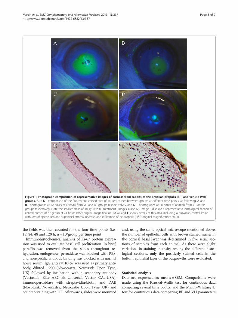
Figure 1 Photograph composition of representative images of corneas from rabbits of the Brazilian propolis (BP) and vehicle (VH)groups. A to D - comparison of the fluorescent-stained area of injured cornea between groups at different time points, as following: A andB - photographs at 12 hours of animals from VH and BP groups respectively; C and D – photographs at 48 hours of animals from VH an BPgroups respectively. Note the smaller areas of injury with BP treatment (images B and D). Image E displays a representative histological section ofcentral cornea of BP group at 24 hours (H&E; original magnification 100X), and F shows details of this area, including a brownish central lesionwith loss of epithelium and superficial stroma, necrosis and infiltration of neutrophils (H&E; original magnification: 400X).
Martin et al. BMC Complementary and Alternative Medicine 2013, 13:337 Page 3 of 7http://www.biomedcentral.com/1472-6882/13/337
the fields was then counted for the four time points (i.e.,12, 24, 48 and 120 h, n = 10/group per time point).Immunohistochemical analysis of Ki-67 protein expres-
sion was used to evaluate basal cell proliferation. In brief,paraffin was removed from the slides throughout re-hydration, endogenous peroxidase was blocked with PBS,and nonspecific antibody binding was blocked with normalhorse serum. IgG anti rat Ki-67 was used as primary anti-body, diluted 1:200 (Novocastra, Newcastle Upon Tyne,UK) followed by incubation with a secondary antibody(Vectastain Elite ABC kit Universal, Vector, CA, USA),immunoperoxidase with streptavidin/biotin, and DAB(NovoLink, Novocastra, Newcastle Upon Tyne, UK) andcounter-staining with HE. Afterwards, slides were mounted
and, using the same optical microscope mentioned above,the number of epithelial cells with brown stained nuclei inthe corneal basal layer was determined in five serial sec-tions of samples from each animal. As there were slightvariations in staining intensity among the different histo-logical sections, only the positively stained cells in thebottom epithelial layer of the outgrowths were evaluated.
Statistical analysisData are expressed as means ± SEM. Comparisons weremade using the Kruskal-Wallis test for continuous datacomparing several time points, and the Mann–Whitney Utest for continuous data comparing BP and VH parameters

Table 2 Neutrophils count in corneas at various timepoints after injury in groups BP (Brazilian propolis) andVH (vehicle) (mean ± SEM)
TIME (h) VH (cells/field) BP (cells/field) P
0 NA NA NA
12 150.4 ± 9.0 150.3 ± 10.8 1.0000
24 448.6 ± 21.7 354.7 ± 9.7 0.0006
48 408.0 ± 16.5 326.7 ± 13.3 0.0014
120 82.2 ± 20.0 56.9 ± 10.0 0.3752
NA: Not applicable.
Martin et al. BMC Complementary and Alternative Medicine 2013, 13:337 Page 4 of 7http://www.biomedcentral.com/1472-6882/13/337
at each time point (Graphpad 5.0 software, Prism, SanDiego, CA). The level of significance used was P < 0.05.
ResultsWound healingDigital analysis of the corneal injuries areas stained withfluorescein was used to calculate the size of the wounds(Figure 1). At 12, 24 and 48 h post-injury, the wounddefect areas was consistently smaller in the BP than in theVH treated groups (P < 0.01) (Table 1). On the other hand,there was no difference between the two groups immedi-ately after making the wounds. At 120 h after injury, inboth groups the corneal epithelial defects of all animalshad undergone complete re-epithelization (Figure 1).
Neutrophil infiltrationNeutrophil infiltration was used as an index to evaluatethe effects of propolis on inflammation. Mainly, in thecorneal central anterior stromal layer of both groups,necrosis was seen following injury (Figure 1). At 12 hafter injury, the number of neutrophils were the same inboth the BP and VH groups.However, at 24 and 48 h, fewer neutrophils were seen in
the group BP (P < 0.01). After 120 h, there was no longer adifference between the number of neutrophils between thetwo groups (Table 2).
Immunohistochemistry (Ki-67)There was a higher number of Ki-67 positive cells in thebasal epithelial layers in the BP than the VH group atboth 12 and 24 h (P < 0.0001 and P < 0.001, respectively)(Figure 2). However, 48 h after injury there no longerwas any difference between these groups (Table 3). Fur-thermore, wound closure occurred in the two groups120 h after injury, since Ki67 staining was absent, dueto complete re- epithelialization.
DiscussionCorneal surface injuries are among the most frequenttraumas of the eye [14]. The prompt recovery of such in-juries is critical to the maintenance of corneal transpar-ency. Although previous publications [15,16] addressed
Table 1 Area of corneal epithelial defect at various timepoints after injury on groups BP (Brazilian propolis) andVH (vehicle) (mean ± SEM)
TIME (h) VH (mm2) BP (mm2) P
0 0.0389 ± 0.0017 0.0341 ± 0.0014 0.1978
12 0.0291 ± 0.0012 0.0160 ± 0.0030 0.0032
24 0.0163 ± 0.0021 0.0076 ± 0.0022 0.0007
48 0.0075 ± 0.0009 0.0031 ± 0.0007 0.0019
120 0.00 0.00 N/A
NA: Not applicable.
therapeutic options, no commercial eye drops have beenapproved for this specific purpose, nor has BP been clinic-ally tested to the best of our knowledge.Aqueous extracts obtained from BP have antioxidative
properties and inhibitory actions on certain enzymes thatare greater than those obtained with ethanolic extracts.These extracts are rich in terpenoids and derivatives ofcoumaric, caffeoylquinic and cinnamic acids, and it isprobable that these components have synergistic effectson healing [15-17].It is known that red propolis causes damage to the cor-
neal epithelial cells of rats when used at concentrations of7.81 mg/mL [18]. Thus, although there are no specificstudies on corneal toxicity of BP, at a concentration of 1%(10 mg/mL) there was no indication in this study that ithad any toxic effects.The mechanisms by which propolis elicits healing effects
have been the focus of some previous studies [4-9]. Greenpropolis, a specific kind of Brazilian propolis, reducedboth neuronal necrosis and apoptosis of retinal ganglioncells [17,19]. Red propolis had a similar anti-inflammatoryeffect in another experimental model of a corneal chem-ical burn [10]. Furthermore, its enhancement of woundhealing is associated with a decline in free radical gener-ation, which provides a partial protection against oxidativestress induced lipid peroxidation [19].Mechanical deepithelization usually requires less time
for wound closure than those caused by an alkali burn[20,21]. Such a difference is due to the fact that a severealkali burn elicits stromal dysregulated inflammation andscar formation through inducing immune cell infiltrationand myofibroblast formation from keratocytes [22-24].This type of inflammatory response is not self-limitingand is associated with oxidative damage, causing degrad-ation of the stromal matrix [20,25]. These pathologicalfindings were the basis for our choice of the AgNO3 alkaliburn model to evaluate the effects of propolis on cornealwound healing.Complete re-epithelization occurred in different eyes
between 48 and 120 h although the exact mean re-epithelization time was not determined. It seemed closerto 48 than to 120 h, because at 48 h wound closure in the

Figure 2 Immunohistochemical Ki-67 analysis of cornea tissue cauterized with Silver Nitrate, and treated with VH (vehicle - A) or BP(Brazilian propolis - B) eye drops. Photographs were taken at time-point 12 hours with 400X (original magnification).
Martin et al. BMC Complementary and Alternative Medicine 2013, 13:337 Page 5 of 7http://www.biomedcentral.com/1472-6882/13/337
VH and BP groups was already 81% and 92%, completed,respectively. This time requirement for healing is consist-ent with other reports cited previously. Epithelial cellrenewal is dependent on basal cell mitotic activity. Theseproliferating cells are derived initially from quiescent stemcells in the limbal periphery, which are activated by injuryto become proliferative [26,27]. We only evaluated regen-erative capacity in the presence and absence of BP basedon Ki67 staining in the proliferating basal cell layer. In theBP group especially at 12 h there was more Ki67 stainingthan in the VH group.Acute inflammation induced by an alkali burn was evalu-
ated by counting the total number of infiltrating neutro-phils in the central injured region. At 12 h, in both groups,neutrophils counts increased. As the number of neutro-phils in the cornea of rats is small under normal conditions[28,29], the increase in neutrophil number during the first12 h is possibly due to neutrophil infiltration from the per-iphery. BP containing eye drops did not prevent or inhibittheir inward migration during this period. At 24 h, theirnumbers in both groups increased further by more than100%. However, in the VH group the neutrophil count
Table 3 Positive Ki-67 stained cells in the corneal basal layerin both groups (VH – vehicle and BP – Brazilian propolis)at the different time point studied (mean±SEM)
TIME (h) VH (No. of cells) BP (No. of cells) P
0 NA NA NA
12 46.7 ± 0,02 63.7 ± 0.02 <0.0001
24 64.2 ± 0.02 74.15 ± 0.0195 0.0067
48 61.7 ± 0.0130 58.05 ± 0.0209 0.1412
120 0.0 0.0 NA
NA: Not applicable.
values were 25% higher than those in the BP group. Thisdifference suggests that BP has significant anti-inflammatory effects in this model. Moreover, this dif-ference persists even after 48 h. At 120 h, the neutrophilcount had declined by about 80% from that at 48 h.There no longer was any difference between their num-bers in the BP and VH groups. These time-dependenttransient increases in neutrophil stromal counts are inaccord with those shown in other studies [30-32]. Thus,BP did not inhibit or change the time-course requiredfor completing wound closure even though acute in-flammation was partially suppressed during a portion ofthe healing response.In summary, the present work suggests that BP reduces
the inflammatory response to injury, but the overall effectof this suppression was not adequate to hasten epithelialwound healing. Although other preparations have hadsimilar effects in pre-clinical studies and randomized clin-ical trials, these therapeutic options require further explor-ation [31,33-35]. This need is indicated by the fact thateven though anti-inflammatory drugs such as corticoster-oid eye drop formulations reduce local inflammation, theycan contain vehicles and distinct stabilizing compounds,which may impair wound healing process.The proposal of BP as a topical treatment of chemical
burns, and potentially other aseptic keratitis, is based onboth its cicatrizing and anti-inflammatory effects. It isstill not clear which components of BP are responsiblefor these effects, but some studies have already identifiedeffective anti-inflammatory constituents [36,37]. Com-bining these BP actions, it has certain pro-healing attri-butes not found in other anti-inflammatory medications(e.g. corticosteroids) currently available for the treatmentof corneal injury.

Martin et al. BMC Complementary and Alternative Medicine 2013, 13:337 Page 6 of 7http://www.biomedcentral.com/1472-6882/13/337
ConclusionsTopically applied BP accelerated wound healing and re-duced inflammatory response after silver nitrate-inducedalkali burns in rats. The clinical usefulness of a treatmentwith topical BP in ocular surface diseases associated withcorneal epithelial defects should be explored further as analternative for the prevention of these potentially blindnessconditions.
Competing interestsFinancial support: São Paulo Research Foundation - FAPESP.Financial interest statement: The authors have no financial competinginterests in this study.
Authors’ contributionsLM carried out the experiments, participated in the statistical analysis anddrafted the manuscript. JP: participated in the design of the study, as well asthe statistical analysis and drafted the manuscript. ER participated in thedesign of the study and revision of the manuscript. SG carried out thehistology and immunohistochemistry analysis. All authors read and approvedthe final version of the manuscript.
Authors’ informationSG has been studying, in contribution with other authors, the effects ofBrazilian propolis in different tissues. As an associate professor and a formerchief of the Department of Pathology of the University of São Paulo, hecarried out the histology and immunohistochemistry analysis of multiplestudies, which correlated Brazilian propolis with inflammation and woundhealing in several tissues, including skin and bones.JP and ER are associate professors of ophthalmology in the Department ofOphthalmology, Otorhinolaringology, Head and Neck Surgery of theUniversity of São Paulo, and they both have been working with experimentalcorneal models, related to ocular surface disease in dry eye syndrome, andother ocular inflammation conditions. JP was a former chief of the Divisionof Ophthalmology of this Department, and ER is the current chief of thesame Division.LM is a young ophthalmologist who had been working with SG sincemedical school, before ophthalmology residency. After finishing residency ofophthalmology and a one-year fellowship in cornea, he was invited by thoseprofessors to develop the present study, as part of his PhD.
AcknowledgementsThe authors sincerely acknowledge Rosângela Orlandin Lopes, Mateus FreireLeite, Patrícia Modiano and Aline Turatti, of the Department of Pathology -Faculty of Medicine of Ribeirão Preto, University of São Paulo for their helpwith the histological preparations and photographs on this study. We alsoacknowledge the NIH Fellows Editorial Board for editorial assistance andPeter Sol Reinach for his careful assistance in the revision of the Englishgrammar and style.
Author details1Department of Ophthalmology, Otorhinolaryngology and Head and NeckSurgery, School of Medicine of Ribeirão Preto - University of São Paulo,Av. Bandeirantes, 3900 – 12°. Andar, Ribeirão Preto, São Paulo, Brazil.2Department of Pathology, School of Medicine of Ribeirão Preto - Universityof São Paulo, São Paulo, Brazil.
Received: 3 April 2013 Accepted: 22 November 2013Published: 27 November 2013
References1. Reinach PS, Pokorny KS: The corneal epithelium: clinical relevance of
cytokine-mediated responses to maintenance of corneal health. Arq BrasOftalmol 2008, 71:80–86.
2. Cade-Jorge F, Cade-Jorge I, Kusabara AA, Rocha EM: Perspectives in therapeuticinnovation in ocular surface disorders and dry eye syndrome. Recent PatEndocr Metab Immune Drug Discovery 2009, 3:194–199.
3. Eisenberg DM, Davis RB, Ettner SL, Appel S, Wilkey S, Van Rompay M, Kessler RC:Trends in alternative medicine use in the United States, 1990–1997: results ofa follow-up national survey. JAMA 1998, 280:1569–1575.
4. Natarajan K, Singh S, Burke TR Jr, Grunberger D, Aggarwal BB: Caffeic acidphenethyl ester is a potent and specific inhibitor of activation of nucleartranscription factor NF-kappa B. Proc Natl Acad Sci USA 1996, 93:9090–9095.
5. Bankova V, Marcucci MC, Simova S, Nikolova N, Kujumgiev A, Popov S:Antibacterial diterpenic acids from Brazilian propolis. Z Naturforsch C1996, 51:277–280.
6. Drago L, Mombelli B, De Vecchi E, Fassina MC, Tocalli L, Gismondo MR: Invitro antimicrobial activity of propolis dry extract. J Chemother 2000,12:390–395.
7. Mirzoeva OK, Calder PC: The effect of propolis and its components oneicosanoid production during the inflammatory response.Prostaglandins Leukot Essent Fatty Acids 1996, 55:441–449.
8. Krol W, Czuba Z, Scheller S, Gabrys J, Grabiec S, Shani J: Anti-oxidantproperty of ethanolic extract of propolis (EEP) as evaluated byinhibiting the chemiluminescence oxidation of luminol. Biochem Int1990, 21:593–597.
9. Scheller S, Wilczok T, Imielski S, Krol W, Gabrys J, Shani J: Free radicalscavenging by ethanol extract of propolis. Int J Radiat Biol 1990, 57:461–465.
10. Ozturk F, Kurt E, Cerci M, Emiroglu L, Inan U, Iker S: The effect of propolisextract in experimental chemical corneal injury. Ophthalmic Res 2000,32:13–18.
11. Trusheva B, Popova M, Bankova V, Simova S, Marcucci MC, Miorin PL, et al:Bioactive constituents of brazilian red propolis. Evid Based ComplementAlternat Med 2006, 3:249–254.
12. Pontin K, Da Silva Filho AA, Santos FF, Silva MLAE, Cunha WR, NanayakkaraNPD, et al: In vitro and in vivo antileishmanial activities of a Braziliangreen propolis extract. Parasitol Res 2008, 103:487–492.
13. Michener CD: The social behavior of bees - a comparative study. Cambridge,Massachusetts: Belknap Press of Harvard University Press; 1974:404.
14. Cecchetti DFA, de Cecchetti SAP, Nardy ACT, Carvalho SC, de RodriguesMLV, Rocha EM: [A clinical and epidemiological profile of ocularemergencies in a reference emergency center] Portuguese. Arq BrasOftalmol 2008, 71:635–638.
15. Keshavarz M, Mostafaie A, Mansouri K, Shakiba Y, Motlagh HRM: Inhibition ofcorneal neovascularization with propolis extract. Arch Med Res 2009, 40:59–61.
16. Vural A, Polat ZA, Topalkara A, Toker MI, Erdogan H, Arici MK, et al: Theeffect of propolis in experimental Acanthamoeba keratitis. Clin ExpOphthalmol 2007, 35:749–754.
17. Matsui T, Ebuchi S, Fujise T, Abesundara KJM, Doi S, Yamada H, et al: Strongantihyperglycemic effects of water-soluble fraction of Brazilian propolisand its bioactive constituent, 3,4,5-tri-O-caffeoylquinic acid. Biol PharmBull 2004, 27:1797–1803.
18. Miyataka H, Nishiki M, Matsumoto H, Fujimoto T, Matsuka M, Satoh T:Evaluation of propolis. I. Evaluation of Brazilian and Chinese propolis byenzymatic and physico-chemical methods. Biol Pharm Bull 1997, 20:496–501.
19. Nakajima Y, Shimazawa M, Mishima S, Hara H: Water extract of propolisand its main constituents, caffeoylquinic acid derivatives, exertneuroprotective effects via antioxidant actions. Life Sci 2007, 80:370–377.
20. Shimazawa M, Chikamatsu S, Morimoto N, Mishima S, Nagai H, Hara H:Neuroprotection by Brazilian Green Propolis against In vitro and In vivoIschemic Neuronal Damage. Evid Based Complement Alternat Med 2005,2:201–207.
21. Yamada J, Dana MR, Sotozono C, Kinoshita S: Local suppression of IL-1 byreceptor antagonist in the rat model of corneal alkali injury. Exp Eye Res2003, 76:161–167.
22. Li Z, Burns AR, Smith CW: Two waves of neutrophil emigration inresponse to corneal epithelial abrasion: distinct adhesion moleculerequirements. Invest Ophthalmol Vis Sci 2006, 47:1947–1955.
23. Zhao G-q, Ma Y-q, Liang T, Jiang T, Wang C-f, Zhang Y-x: Animal study onexpression of laminin and fibronectin in cornea during wound healingfollowing alkali burn. Chin J Traumatol 2003, 6:37–40.
24. Parks WC, Wilson CL, López-Boado YS: Matrix metalloproteinases as modulatorsof inflammation and innate immunity. Nat Rev Immunol 2004, 4:617–629.
25. Ishizaki M, Zhu G, Haseba T, Shafer SS, Kao WW: Expression of collagen I,smooth muscle alpha-actin, and vimentin during the healing of alkali-burned and lacerated corneas. Invest Ophthalmol Vis Sci 1993, 34:3320–3328.
26. Zhao M, Chen J, Yang P: [Immunologic experimental studies on the alkaliburn of cornea in rats] Chinese. Zhonghua Yan Ke Za Zhi 2000, 36:40–42. 4.

Martin et al. BMC Complementary and Alternative Medicine 2013, 13:337 Page 7 of 7http://www.biomedcentral.com/1472-6882/13/337
27. Sun TT, Green H: Cultured epithelial cells of cornea, conjunctiva and skin:absence of marked intrinsic divergence of their differentiated states.Nature 1977, 269:489–493.
28. Boulton M, Albon J: Stem cells in the eye. Int J Biochem Cell Biol 2004,36:643–657.
29. Brissette-Storkus CS, Reynolds SM, Lepisto AJ, Hendricks RL: Identification ofa novel macrophage population in the normal mouse corneal stroma.Invest Ophthalmol Vis Sci 2002, 43:2264–2271.
30. Hamrah P, Liu Y, Zhang Q, Dana MR: The corneal stroma is endowed witha significant number of resident dendritic cells. Invest Ophthalmol Vis Sci2003, 44:581–589.
31. Wenk HN, Honda CN: Silver nitrate cauterization: characterization of anew model of corneal inflammation and hyperalgesia in rat. Pain 2003,105:393–401.
32. Lin M, Jackson P, Tester AM, Diaconu E, Overall CM, Blalock JE, et al: Matrixmetalloproteinase-8 facilitates neutrophil migration through the cornealstromal matrix by collagen degradation and production of thechemotactic peptide Pro-Gly-Pro. Am J Pathol 2008, 173:144–153.
33. Patil K, Bellner L, Cullaro G, Gotlinger KH, Dunn MW, Schwartzman ML:Heme oxygenase-1 induction attenuates corneal inflammation andaccelerates wound healing after epithelial injury. Invest Ophthalmol Vis Sci2008, 49:3379–3386.
34. Lin M, Carlson E, Diaconu E, Pearlman E: CXCL1/KC and CXCL5/LIX areselectively produced by corneal fibroblasts and mediate neutrophilinfiltration to the corneal stroma in LPS keratitis. J Leukoc Biol 2007,81:786–792.
35. McInnis KA, Britain A, Lausch RN, Oakes JE: Human corneal epithelial cellssynthesize ELR(−)alpha-chemokines in response to proinflammatorymediators. Ocul Immunol Inflamm 2007, 15:295–302.
36. Sawaya ACHF, Cunha IBS, Marcucci MC, Rodrigues RFO, Eberlin MN:Brazilian Propolis of Tetragonisca angustula and Apis mellifera.Apidologie 2006, 37:398–407.
37. Sawaya ACHF, Calado JCP, Santos LC, Marcucci MC, Akatsu IP, Soares AEE,Abdelnur PV, Cunha IBS, Eberlin MN: Composition and antioxidant activityof propolis from three species of Scaptotrigona stingless bees. Jour ApiProduct and Api Medical Sci 2009, 1:37–42.
doi:10.1186/1472-6882-13-337Cite this article as: Martin et al.: Topical Brazilian propolis improvescorneal wound healing and inflammation in rats following alkali burns.BMC Complementary and Alternative Medicine 2013 13:337.
Submit your next manuscript to BioMed Centraland take full advantage of:
• Convenient online submission
• Thorough peer review
• No space constraints or color figure charges
• Immediate publication on acceptance
• Inclusion in PubMed, CAS, Scopus and Google Scholar
• Research which is freely available for redistribution
Submit your manuscript at www.biomedcentral.com/submit