Anti-Viral and Immunomodulatory Properties of Propolis - MDPI
47
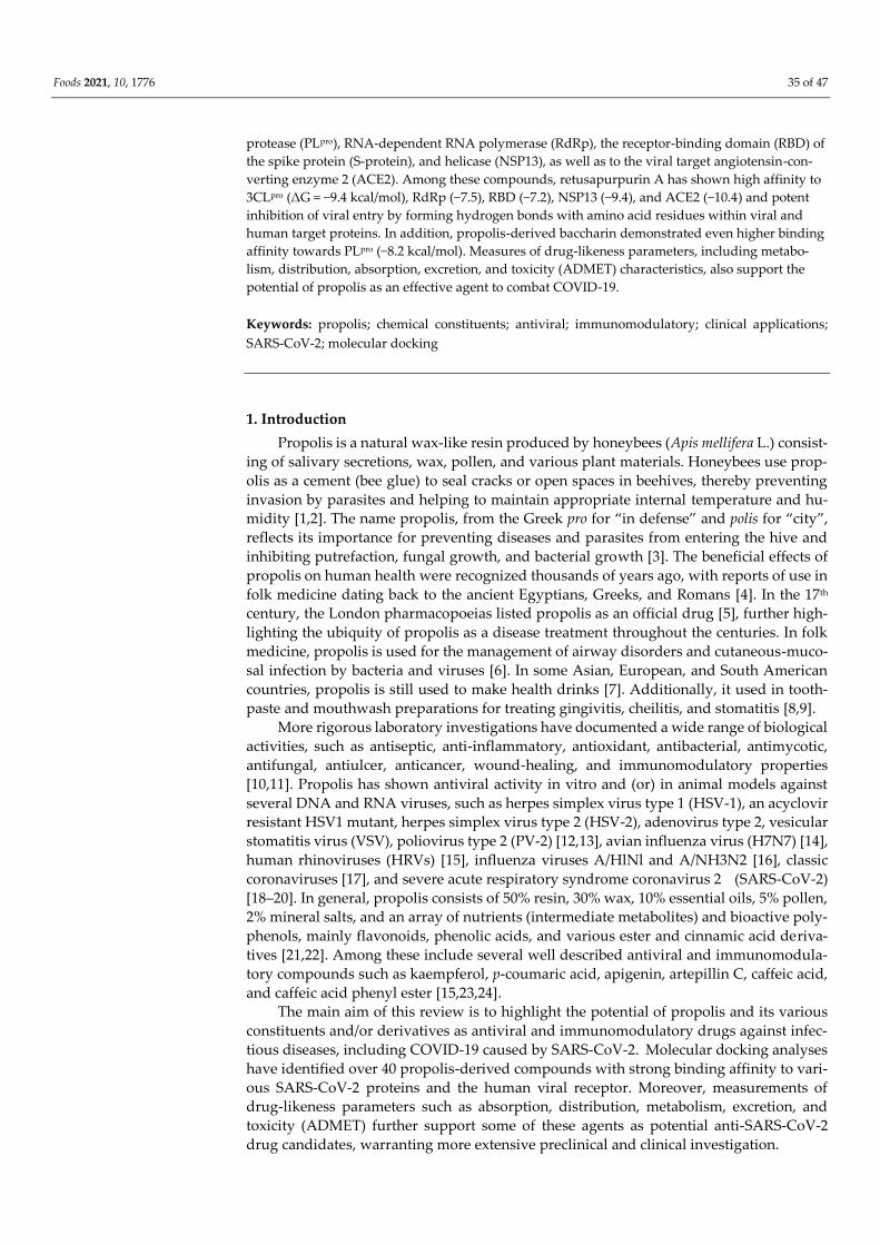
Foods 2021, 10, 1776. https://doi.org/10.3390/foods10081776 www.mdpi.com/journal/foods Review Anti-Viral and Immunomodulatory Properties of Propolis: Chemical Diversity, Pharmacological Properties, Preclinical and Clinical Applications, and In Silico Potential against SARS-CoV-2 Nermeen Yosri 1,2 , Aida A. Abd El-Wahed 3 , Reem Ghonaim 2 , Omar M. Khattab 2 , Aya Sabry 2 , Mahmoud A. A. Ibrahim 4 , Mahmoud F. Moustafa 5,6 , Zhiming Guo 1 , Xiaobo Zou 1 , Ahmed F. M. Algethami 7 , Saad H. D. Masry 8,9 , Mohamed F. AlAjmi 10 , Hanan S. Afifi 11 , Shaden A. M. Khalifa 12, * and Hesham R. El-Seedi 2,13,14, * 1 School of Food and Biological Engineering, Jiangsu University, Zhenjiang 212013, China; [email protected] (N.Y.); [email protected] (Z.G.); [email protected] (X.Z.) 2 Department of Chemistry, Faculty of Science, Menoufia University, Shebin El-Kom 32512, Egypt; [email protected] (R.G.); [email protected] (O.M.K.); [email protected] (A.S.) 3 Department of Bee Research, Plant Protection Research Institute, Agricultural Research Centre, Giza 12627, Egypt; [email protected] 4 Computational Chemistry Laboratory, Chemistry Department, Faculty of Science, Minia University, Minia 61519, Egypt; [email protected] 5 Department of Biology, College of Science, King Khalid University, Abha 9004, Saudi Arabia; hamdony@ya- hoo.com 6 Department of Botany & Microbiology, Faculty of Science, South Valley University, Qena 83523, Egypt 7 Alnahalaljwal Foundation Saudi Arabia, P.O. Box 617, Al Jumum 21926, Saudi Arabia; [email protected] 8 Department of Plant Protection and Biomolecular Diagnosis, Arid Lands Cultivation Research Institute (ALCRI), City of Scientific Research and Technological Applications, New Borg El-Arab City, Alexandria 21934, Egypt; [email protected] 9 Abu Dhabi Agriculture and Food Safety Authority (ADAFSA), Al Ain 52150, United Arab Emirates 10 Pharmacognosy Group, College of Pharmacy, King Saud University, Riyadh 11451, Saudi Arabia; [email protected] 11 Food Research Section, R&D Division, Abu Dhabi Agriculture and Food Safety Authority (ADAFSA), Abu Dhabi P.O. Box 52150, United Arab Emirates; [email protected] 12 Department of Molecular Biosciences, Stockholm University, The Wenner-Gren Institute, SE-106 91 Stockholm, Sweden 13 International Research Center for Food Nutrition and Safety, Jiangsu University, Zhenjiang 212013, China 14 Division of Pharmacognosy, Department of Pharmaceutical Biosciences, Uppsala University, Biomedical Centre, P.O. Box 591, SE 751 24 Uppsala, Sweden * Correspondence: Correspondence: [email protected] (S.A.M.K.); [email protected] (H.R.E.-S.); Tel.: +46-700-10-11-13 (S.A.M.K.); Tel.: +46-700-43-43-43 (H.R.E.-S.) Abstract: Propolis, a resin produced by honeybees, has long been used as a dietary supplement and folk remedy, and more recent preclinical investigations have demonstrated a large spectrum of potential therapeutic bioactivities, including antioxidant, antibacterial, anti-inflammatory, neu- roprotective, immunomodulatory, anticancer, and antiviral properties. As an antiviral agent, propolis and various constituents have shown promising preclinical efficacy against adenoviruses, influenza viruses, respiratory tract viruses, herpes simplex virus type 1 (HSV-1) and type 2 (HSV- 2), human immunodeficiency virus (HIV), and severe acute respiratory syndrome coronavirus 2 (SARS-CoV-2). Over 300 chemical components have been identified in propo- lis, including terpenes, flavonoids, and phenolic acids, with the specific constituent profile varying widely according to geographic origin and regional flora. Propolis and its constituents have demonstrated potential efficacy against SARS-CoV-2 by modulating multiple pathogenic and anti- viral pathways. Molecular docking studies have demonstrated high binding affinities of propolis derivatives to multiple SARS-CoV-2 proteins, including 3C-like protease (3CL pro ), papain-like Citation: Yosri, N.; Abd El-Wahed, A.A.; Ghonaim, R.; Khattab, O.M.; Sabry, A.; Ibrahim, M.A.A.; Moustafa, M.F.; Guo, Z.; Zou, X.; Algethami, A.A.F.; et al. Anti-Viral and Immunomodulatory Properties of Propolis: Chemical Diversity, Pharmacological Properties, Preclinical and Clinical Applications, and In Silico Potential against SARS- CoV-2. Foods 2021, 10, 1776. https:// doi.org/10.3390/foods10081776 Academic Editors: Luís Manuel Lopes Rodrigues da Silva and Gilberto Alves Received: 24 May 2021 Accepted: 28 July 2021 Published: 31 July 2021 Publisher’s Note: MDPI stays neu- tral with regard to jurisdictional claims in published maps and institu- tional affiliations. Copyright: © 2021 by the authors. Li- censee MDPI, Basel, Switzerland. This article is an open access article distributed under the terms and con- ditions of the Creative Commons At- tribution (CC BY) license (http://crea- tivecommons.org/licenses/by/4.0/).
-
Upload
khangminh22 -
Category
Documents
-
view
2 -
download
0
Transcript of Anti-Viral and Immunomodulatory Properties of Propolis - MDPI
Foods 2021, 10, 1776. https://doi.org/10.3390/foods10081776 www.mdpi.com/journal/foods
Review
Anti-Viral and Immunomodulatory Properties of Propolis:
Chemical Diversity, Pharmacological Properties, Preclinical
and Clinical Applications, and In Silico Potential against
SARS-CoV-2
Nermeen Yosri 1,2, Aida A. Abd El-Wahed 3, Reem Ghonaim 2, Omar M. Khattab 2, Aya Sabry 2,
Mahmoud A. A. Ibrahim 4, Mahmoud F. Moustafa 5,6, Zhiming Guo 1, Xiaobo Zou 1, Ahmed F. M. Algethami 7,
Saad H. D. Masry 8,9, Mohamed F. AlAjmi 10, Hanan S. Afifi 11, Shaden A. M. Khalifa 12,*
and Hesham R. El-Seedi 2,13,14,*
1 School of Food and Biological Engineering, Jiangsu University, Zhenjiang 212013, China;
[email protected] (N.Y.); [email protected] (Z.G.); [email protected] (X.Z.) 2 Department of Chemistry, Faculty of Science, Menoufia University, Shebin El-Kom 32512, Egypt;
[email protected] (R.G.); [email protected] (O.M.K.); [email protected] (A.S.) 3 Department of Bee Research, Plant Protection Research Institute, Agricultural Research Centre, Giza 12627,
Egypt; [email protected] 4 Computational Chemistry Laboratory, Chemistry Department, Faculty of Science, Minia University,
Minia 61519, Egypt; [email protected] 5 Department of Biology, College of Science, King Khalid University, Abha 9004, Saudi Arabia; hamdony@ya-
hoo.com 6 Department of Botany & Microbiology, Faculty of Science, South Valley University, Qena 83523, Egypt 7 Alnahalaljwal Foundation Saudi Arabia, P.O. Box 617, Al Jumum 21926, Saudi Arabia;
[email protected] 8 Department of Plant Protection and Biomolecular Diagnosis, Arid Lands Cultivation Research Institute
(ALCRI), City of Scientific Research and Technological Applications, New Borg El-Arab City,
Alexandria 21934, Egypt; [email protected] 9 Abu Dhabi Agriculture and Food Safety Authority (ADAFSA), Al Ain 52150, United Arab Emirates 10 Pharmacognosy Group, College of Pharmacy, King Saud University, Riyadh 11451, Saudi Arabia;
[email protected] 11 Food Research Section, R&D Division, Abu Dhabi Agriculture and Food Safety Authority (ADAFSA),
Abu Dhabi P.O. Box 52150, United Arab Emirates; [email protected] 12 Department of Molecular Biosciences, Stockholm University, The Wenner-Gren Institute,
SE-106 91 Stockholm, Sweden 13 International Research Center for Food Nutrition and Safety, Jiangsu University, Zhenjiang 212013, China 14 Division of Pharmacognosy, Department of Pharmaceutical Biosciences, Uppsala University,
Biomedical Centre, P.O. Box 591, SE 751 24 Uppsala, Sweden
* Correspondence: Correspondence: [email protected] (S.A.M.K.); [email protected]
(H.R.E.-S.); Tel.: +46-700-10-11-13 (S.A.M.K.); Tel.: +46-700-43-43-43 (H.R.E.-S.)
Abstract: Propolis, a resin produced by honeybees, has long been used as a dietary supplement
and folk remedy, and more recent preclinical investigations have demonstrated a large spectrum
of potential therapeutic bioactivities, including antioxidant, antibacterial, anti-inflammatory, neu-
roprotective, immunomodulatory, anticancer, and antiviral properties. As an antiviral agent,
propolis and various constituents have shown promising preclinical efficacy against adenoviruses,
influenza viruses, respiratory tract viruses, herpes simplex virus type 1 (HSV-1) and type 2 (HSV-
2), human immunodeficiency virus (HIV), and severe acute respiratory syndrome
coronavirus 2 (SARS-CoV-2). Over 300 chemical components have been identified in propo-
lis, including terpenes, flavonoids, and phenolic acids, with the specific constituent profile varying
widely according to geographic origin and regional flora. Propolis and its constituents have
demonstrated potential efficacy against SARS-CoV-2 by modulating multiple pathogenic and anti-
viral pathways. Molecular docking studies have demonstrated high binding affinities of propolis
derivatives to multiple SARS-CoV-2 proteins, including 3C-like protease (3CLpro), papain-like
Citation: Yosri, N.; Abd El-Wahed,
A.A.; Ghonaim, R.; Khattab, O.M.;
Sabry, A.; Ibrahim, M.A.A.;
Moustafa, M.F.; Guo, Z.; Zou, X.;
Algethami, A.A.F.; et al. Anti-Viral
and Immunomodulatory Properties
of Propolis: Chemical Diversity,
Pharmacological Properties,
Preclinical and Clinical Applications,
and In Silico Potential against SARS-
CoV-2. Foods 2021, 10, 1776. https://
doi.org/10.3390/foods10081776
Academic Editors: Luís Manuel
Lopes Rodrigues da Silva and
Gilberto Alves
Received: 24 May 2021
Accepted: 28 July 2021
Published: 31 July 2021
Publisher’s Note: MDPI stays neu-
tral with regard to jurisdictional
claims in published maps and institu-
tional affiliations.
Copyright: © 2021 by the authors. Li-
censee MDPI, Basel, Switzerland.
This article is an open access article
distributed under the terms and con-
ditions of the Creative Commons At-
tribution (CC BY) license (http://crea-
tivecommons.org/licenses/by/4.0/).
Foods 2021, 10, 1776 35 of 47
protease (PLpro), RNA-dependent RNA polymerase (RdRp), the receptor-binding domain (RBD) of
the spike protein (S-protein), and helicase (NSP13), as well as to the viral target angiotensin-con-
verting enzyme 2 (ACE2). Among these compounds, retusapurpurin A has shown high affinity to
3CLpro (ΔG = −9.4 kcal/mol), RdRp (−7.5), RBD (−7.2), NSP13 (−9.4), and ACE2 (−10.4) and potent
inhibition of viral entry by forming hydrogen bonds with amino acid residues within viral and
human target proteins. In addition, propolis-derived baccharin demonstrated even higher binding
affinity towards PLpro (−8.2 kcal/mol). Measures of drug-likeness parameters, including metabo-
lism, distribution, absorption, excretion, and toxicity (ADMET) characteristics, also support the
potential of propolis as an effective agent to combat COVID-19.
Keywords: propolis; chemical constituents; antiviral; immunomodulatory; clinical applications;
SARS-CoV-2; molecular docking
1. Introduction
Propolis is a natural wax-like resin produced by honeybees (Apis mellifera L.) consist-
ing of salivary secretions, wax, pollen, and various plant materials. Honeybees use prop-
olis as a cement (bee glue) to seal cracks or open spaces in beehives, thereby preventing
invasion by parasites and helping to maintain appropriate internal temperature and hu-
midity [1,2]. The name propolis, from the Greek pro for “in defense” and polis for “city”,
reflects its importance for preventing diseases and parasites from entering the hive and
inhibiting putrefaction, fungal growth, and bacterial growth [3]. The beneficial effects of
propolis on human health were recognized thousands of years ago, with reports of use in
folk medicine dating back to the ancient Egyptians, Greeks, and Romans [4]. In the 17th
century, the London pharmacopoeias listed propolis as an official drug [5], further high-
lighting the ubiquity of propolis as a disease treatment throughout the centuries. In folk
medicine, propolis is used for the management of airway disorders and cutaneous-muco-
sal infection by bacteria and viruses [6]. In some Asian, European, and South American
countries, propolis is still used to make health drinks [7]. Additionally, it used in tooth-
paste and mouthwash preparations for treating gingivitis, cheilitis, and stomatitis [8,9].
More rigorous laboratory investigations have documented a wide range of biological
activities, such as antiseptic, anti-inflammatory, antioxidant, antibacterial, antimycotic,
antifungal, antiulcer, anticancer, wound-healing, and immunomodulatory properties
[10,11]. Propolis has shown antiviral activity in vitro and (or) in animal models against
several DNA and RNA viruses, such as herpes simplex virus type 1 (HSV-1), an acyclovir
resistant HSV1 mutant, herpes simplex virus type 2 (HSV-2), adenovirus type 2, vesicular
stomatitis virus (VSV), poliovirus type 2 (PV-2) [12,13], avian influenza virus (H7N7) [14],
human rhinoviruses (HRVs) [15], influenza viruses A/HlNl and A/NH3N2 [16], classic
coronaviruses [17], and severe acute respiratory syndrome coronavirus 2 (SARS-CoV-2)
[18–20]. In general, propolis consists of 50% resin, 30% wax, 10% essential oils, 5% pollen,
2% mineral salts, and an array of nutrients (intermediate metabolites) and bioactive poly-
phenols, mainly flavonoids, phenolic acids, and various ester and cinnamic acid deriva-
tives [21,22]. Among these include several well described antiviral and immunomodula-
tory compounds such as kaempferol, p-coumaric acid, apigenin, artepillin C, caffeic acid,
and caffeic acid phenyl ester [15,23,24].
The main aim of this review is to highlight the potential of propolis and its various
constituents and/or derivatives as antiviral and immunomodulatory drugs against infec-
tious diseases, including COVID-19 caused by SARS-CoV-2. Molecular docking analyses
have identified over 40 propolis-derived compounds with strong binding affinity to vari-
ous SARS-CoV-2 proteins and the human viral receptor. Moreover, measurements of
drug-likeness parameters such as absorption, distribution, metabolism, excretion, and
toxicity (ADMET) further support some of these agents as potential anti-SARS-CoV-2
drug candidates, warranting more extensive preclinical and clinical investigation.
Foods 2021, 10, 1776 36 of 47
2. Ethnopharmacology
Since ancient times, propolis has been employed by many cultures as a dietary sup-
plement and folk remedy for improving health and managing disease [23,24]. The use of
propolis in folk medicine can be traced back to at least 300 BC [25]. The Egyptians are
considered the first peoples to use propolis for wound treatment and as an embalming
agent [26,27]. In addition, Greek physicians such as Hippocrates, Dioscorides, and Galen;
the Roman natural philosopher Pliny the Elder; and Inca healers utilized propolis as an
antiseptic, antipyretic disinfectant for cutaneous and buccal infections and wound treat-
ment. Propolis-based treatments were also in wide use in Europe during the 17th century
to treat colds, wounds, rheumatism, heart disease, and diabetes [21,28]. According to Hip-
pocrates, propolis may be used to improve health or prevent disease, including gastroin-
testinal disorders such as gastritis and gastric ulcer [29]. Arabs and Persians also used
propolis as a disease treatment and cleansing agent [30].
All over the world, propolis has been used in traditional and folk medicine to prevent
and treat many ailments, i.e., colds, wounds, rheumatism, heart disease, and diabetes [28].
Other documented uses include treatment of pharyngitis as well as wounds [31]. Brazilian
green propolis is used as an anti-inflammatory, antibacterial, and antiulcer treatment in
traditional medicine [32]. Administration for the treatment of abscesses and canker sores
as well as wounds has also been reported [33]. During the Anglo-Boer War and Second
World War, some physicians used propolis to promote tissue regeneration as well as for
wound healing and treatment of tuberculosis, lung inflammation, and malnutrition [30].
Propolis with Ashwagandha (Withania sominifera) is used in some traditional medicine
systems to boost immune function and prevent or cure various ailments [34].
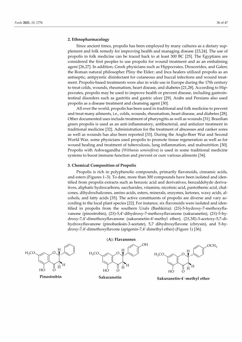
3. Chemical Composition of Propolis
Propolis is rich in polyphenolic compounds, primarily flavonoids, cinnamic acids,
and esters (Figures 1–3). To date, more than 300 compounds have been isolated and iden-
tified from propolis extracts such as benzoic acid and derivatives, benzaldehyde deriva-
tives, aliphatic hydrocarbons, saccharides, vitamins, nicotinic acid, pantothenic acid, chal-
cones, dihydrochalcones, amino acids, esters, minerals, enzymes, ketones, waxy acids, al-
cohols, and fatty acids [35]. The active constituents of propolis are diverse and vary ac-
cording to the local plant species [22]. For instance, six flavonoids were isolated and iden-
tified in propolis from the southern Urals (Bashkiria): (2S)-5-hydroxy-7-methoxyfla-
vanone (pinostrobin), (2S)-5,4′-dihydroxy-7-methoxyflavanone (sakuranetin), (2S)-5-hy-
droxy-7,4′-dimethoxyflavanone (sakuranetin-4′-methyl ether), (2S,3R)-3-acetoxy-5,7-di-
hydroxyflavanone (pinobanksin-3-acetate), 5,7 dihydroxyflavone (chrysin), and 5-hy-
droxy-7,4′-dimethoxyflavone (apigenin-7,4′ dimethyl ether) (Figure 1) [36].
(A): Flavanones

Foods 2021, 10, 1776 37 of 47
(B): Flavones
(C): Isoflavones
Foods 2021, 10, 1776 38 of 47
(D): Isoflavanones
(E): Chalcone (F): Flavanol (G): Isoflavan
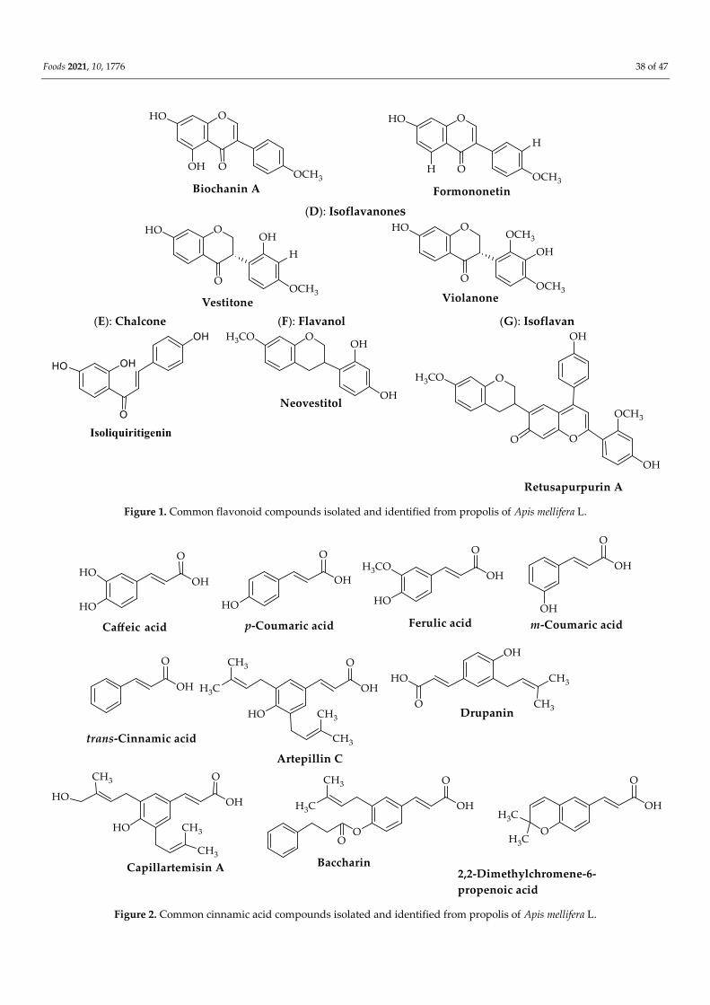
Figure 1. Common flavonoid compounds isolated and identified from propolis of Apis mellifera L.
Figure 2. Common cinnamic acid compounds isolated and identified from propolis of Apis mellifera L.

Foods 2021, 10, 1776 39 of 47
Figure 3. Common ester and acid compounds isolated and identified from propolis of Apis mellifera L.
Furthermore, two flavonoids (pinocembrin and chrysin) (Figure 1), trans-cinnamic
acid, and four phenolic cinnamic acid (caffeic acid, p-coumaric acid, ferulic acid, and m-
coumaric acid) (Figure 2), in addition to many volatile compounds, have been identified
by high-pressure liquid chromatography with UV detector (HPLC-UV), nuclear magnetic
resonance (NMR), and gas chromatography–mass spectrometry (GC–MS) from two prop-
olis samples from two apiaries with different geographical locations in Italy [1]. From 15
Brazilian green propolis samples of different geographically area and botanical source, 47
compounds were tentatively characterized using HPLC-DAD-MS/MS and NMR, includ-
ing prenylated phenylpropanoids (drupanin, capillartemisin A, 2,2-dimethylchromene-6-
propenoic acid, artepillin C, and baccharin) and (E)-2,3-dihydroconiferyl p-coumarate; fla-
vonoids and isoflavonoids (pratensein, violanone, formononetin, vestitone, and biochanin
A) (Figure 1); di- and triterpenoids, as well new compounds; and (E)-3-[4-hydroxy-3-(2-
hydroxy-3-methylbut-3-en-1-yl)-5-(3-methylbut-2-en-1-yl)phenyl] propenoic acid, among
other constituents (Figure 3) [2]. The constituent profile also varies according to extraction
method. Saito et al. compared conventional (Soxhlet) extraction using ethanol, methanol,
water, or hexane as the solvent to supercritical CO2 extraction for two different types of
green and red propolis from Brazil and found that methanolic extraction provided supe-
rior yield for both propolis types, while fractionation of red propolis ethanolic extract us-
ing supercritical CO2 yielded mixtures with much higher flavonoid content than the orig-
inal extract [37].
Dudoit et al. identified 12 distinct compounds (liquiritigenin, calycosin, calycosin
(isomer), luteolin, isoliquiritigenin, formononetin, (3S)-vestitol, (3S)-neovestitol, re-
tusapurpurin A, medicarpin, retusapurpurin A (isomer), and biochanin A) in a Brazilian
red propolis sample using HPLC-MS and TLC-bioautography (Figure 1). (3S)-Vestitol,
(3S)-neovestitol, and medicarpin were the major components, accounting for 45% of all
surface-based chromatographic peaks detected [38]. Picolotto et al. identified five addi-
tional flavonoid compounds (biochanin A, daidzein, formononetin, isoliquiritigenin, and
liquiritigenin) in red propolis samples collected from Alagoas State in northeastern Brazil
using ultrafast liquid chromatography-electrospray ionization-microTof mass spectrome-
try (UFLC-ESI-QTOF) [26]. An HPLC study of an Italian propolis sample found that the
main active constituents were galangin (42.25 mg/g dry propolis, retention time (RT)
=12.87 min), pinocembrin (27.30 mg/g dry propolis, RT = 10.61 min), and caffeic acid
phenethyl ester (CAPE) (11.21 mg/g dry propolis, RT = 11.86 min) [31].
Recently, a multi-dynamic extraction system (RP-HPLC–PDA–ESI–MSn) was used to
identify quercetin, pinobaskin, apigenin, chrysin, pinocembrin, and galangin in nine sam-
ples of propolis gathered separately from three different regions of Europe, America, and
Asia [22,39]. Various additional extraction methods such maceration, ultrasonic extrac-
tion, and microwave extraction have also shown variable efficacy for the isolation of prop-
olis components. For instance, ultrasonic extraction achieved a higher yield of p-coumaric
acid (271.65 mg/g propolis) than microwave extraction or maceration. Other compounds
identified by ultrasonic extraction included apigenin, caffeic acid, chrysin, galangin, iso-
rhamnetin, kaempferol, luteolin, myricetin, pinocembrin, rutin, and quercetin [40].
Foods 2021, 10, 1776 40 of 47
4. Anti-Viral Activity
Propolis has long been used to treat viral infections and more recently tested for effi-
cacy against SARS-CoV-2, the causative pathogen of COVID-19 [41]. Many disease-caus-
ing viruses are unresponsive to currently available antiviral drugs and may also evolve
into more drug- and vaccine-resistant strains. Thus, it is critical to identify novel candidate
antivirals, particularly from natural sources; as such compounds tend to have good safety
profiles.
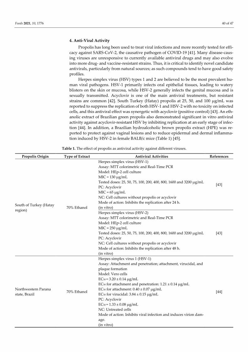
Herpes simplex virus (HSV) types 1 and 2 are believed to be the most prevalent hu-
man viral pathogens. HSV-1 primarily infects oral epithelial tissues, leading to watery
blisters on the skin or mucosa, while HSV-2 generally infects the genital mucosa and is
sexually transmitted. Acyclovir is one of the main antiviral treatments, but resistant
strains are common [42]. South Turkey (Hatay) propolis at 25, 50, and 100 µg/mL was
reported to suppress the replication of both HSV-1 and HSV-2 with no toxicity on infected
cells, and this antiviral effect was synergetic with acyclovir (positive control) [43]. An eth-
anolic extract of Brazilian green propolis also demonstrated significant in vitro antiviral
activity against acyclovir-resistant HSV by inhibiting replication at an early stage of infec-
tion [44]. In addition, a Brazilian hydroalcoholic brown propolis extract (HPE) was re-
ported to protect against vaginal lesions and to reduce epidermal and dermal inflamma-
tion induced by HSV-2 in female BALB/c mice (Table 1) [45].
Table 1. The effect of propolis as antiviral activity against different viruses.
Propolis Origin Type of Extract Antiviral Activities References
South of Turkey (Hatay
region) 70% Ethanol
Herpes simplex virus (HSV-1)
Assay: MTT colorimetric and Real-Time PCR
Model: HEp-2 cell culture
MIC = 130 μg/mL
Tested doses: 25, 50, 75, 100, 200, 400, 800, 1600 and 3200 μg/mL
PC: Acyclovir
MIC = 65 μg/mL
NC: Cell cultures without propolis or acyclovir
Mode of action: Inhibits the replication after 24 h.
(in vitro)
[43]
Herpes simplex virus (HSV-2)
Assay: MTT colorimetric and Real-Time PCR
Model: HEp-2 cell culture
MIC = 250 μg/mL
Tested doses: 25, 50, 75, 100, 200, 400, 800, 1600 and 3200 μg/mL
PC: Acyclovir
NC: Cell cultures without propolis or acyclovir
Mode of action: Inhibits the replication after 48 h.
(in vitro)
[43]
Northwestern Parana
state, Brazil 70% Ethanol
Herpes simplex virus 1 (HSV-1)
Assay: Attachment and penetration; attachment, virucidal, and
plaque formation
Model: Vero cells
EC50 = 3.20 ± 0.14 µg/mL
EC50 for attachment and penetration: 1.21 ± 0.14 µg/mL
EC50 for attachment: 0.40 ± 0.07 µg/mL
EC50 for virucidal: 3.84 ± 0.15 µg/mL
PC: Acyclovir
EC50 = 1.33 ± 0.08 µg/mL
NC: Untreated cells
Mode of action: Inhibits viral infection and induces virion dam-
age.
(in vitro)
[44]

Foods 2021, 10, 1776 41 of 47
Santa Flora City (RS-
Brazil) 70% Ethanol
Herpes simplex virus 2 (HSV-2)
Assay: Plaque reduction
Model: Female BALB/c mice
Dose of Pre-treatment: 50 mg/kg, once a day
Dose of Post-treatment: 50 mg/kg for 5 days more
PC: Not reported
NC: Untreated cells
Mode of action: Reduces extravaginal lesions and the histologi-
cal damage caused by HSV-2 infection in vaginal tissues of ani-
mals.
(in vivo and ex vivo)
[45]
Moravia, Czech
Republic
- Aqueous extract (15%
ethanol)
- 90% Ethanol
Herpes simplex virus 1 (HSV-1)
Assay: Plaque reduction
Model: RC-37 cells
Aqueous extract:
TC50 (%): 0.04; IC50 (%): 0.0004; SI: 100
Ethanol extract:
TC50 (%):0.0017; IC50 (%): 0.000035; SI: 485
PC: Heparin-Na and acyclovir
NC: Untreated cells
Mode of action: Mask viral compounds which are necessary for
adsorption or entry into host cells.
(in vitro)
[13]
Moravia, Czech Repub-
lic
- Aqueous extract
(15% ethanol)
- 90% Ethanol
Herpes simplex virus type 2 (HSV-2)
Assay: Plaque reduction
Model: RC-37 cells
IC50% for aqueous extract: 0.0005; SI: 80
IC50% for ethanolic extract: 0.0004; SI: 42.5
PC: Acyclovir
Inhibits replication of 98.8%
NC: Untreated cells without drugs
Mod of action:
- Suppresses HSV multiplication;
- Masks viral compounds which are necessary for adsorption
or entry into host cells.
(in vitro)
[46]
Canada 70% Ethanol
Herpes simplex virus 1 and 2
Assay: Virucidal assay
Model: MDBK cell
PPE marked effect: 3.2 mg/mL
PC: Acyclovir
NC: Not reported
Mode of action:
- Propolis had a pronounced virucidal effect against herpes
simplex viruses type 1 and type 2, and also interfered with
virus adsorption;
- Suppresses the adsorption of HSV-1 at a broad scope of the
viral inoculation.
(in vitro)
[47]
Botucatu, Brazil 70% Ethanol
Poliovirus type 1 (PV1)
Assay: Real-time PCR
Model: HEp-2 cells
- Pre-treatment: 10.9%
- Simultaneous treatment: 52.2%
- Post-treatment: 39.1%
[48]
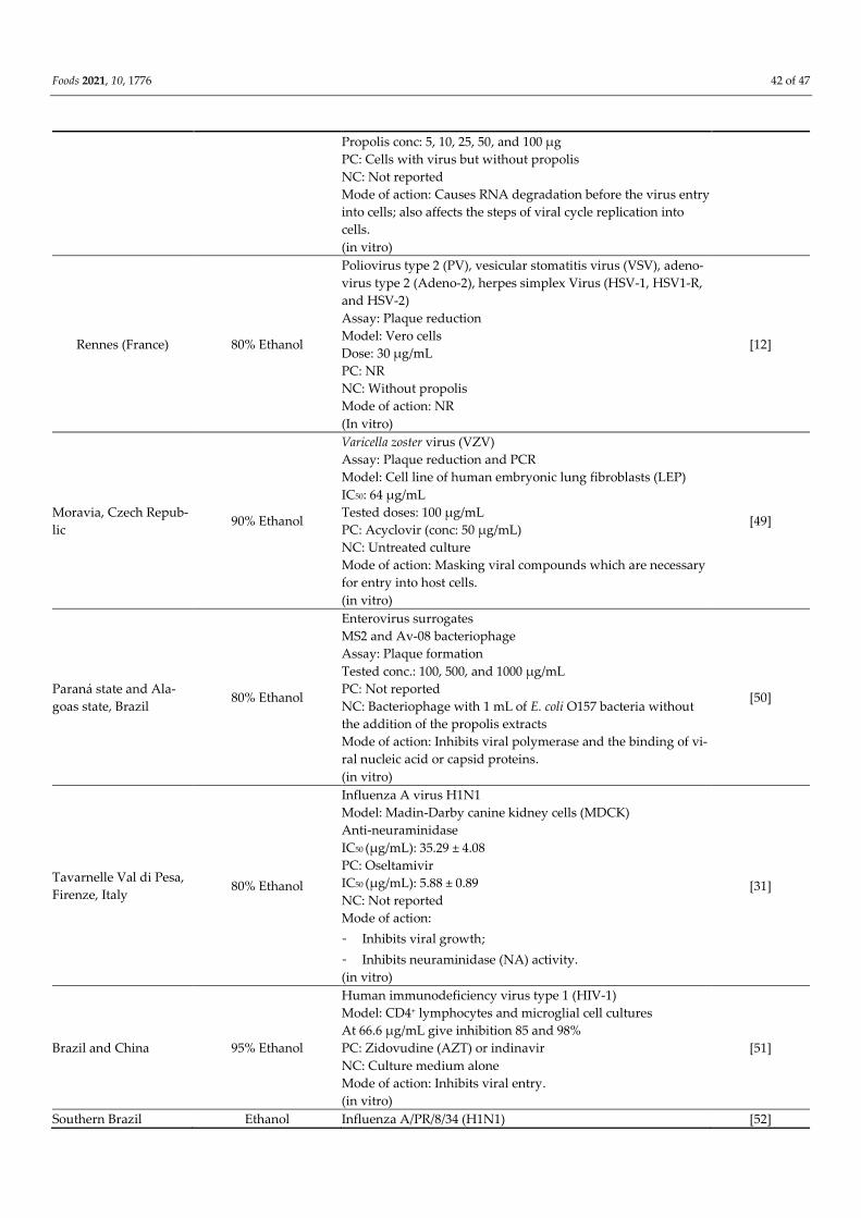
Foods 2021, 10, 1776 42 of 47
Propolis conc: 5, 10, 25, 50, and 100 µg
PC: Cells with virus but without propolis
NC: Not reported
Mode of action: Causes RNA degradation before the virus entry
into cells; also affects the steps of viral cycle replication into
cells.
(in vitro)
Rennes (France) 80% Ethanol
Poliovirus type 2 (PV), vesicular stomatitis virus (VSV), adeno-
virus type 2 (Adeno-2), herpes simplex Virus (HSV-1, HSV1-R,
and HSV-2)
Assay: Plaque reduction
Model: Vero cells
Dose: 30 µg/mL
PC: NR
NC: Without propolis
Mode of action: NR
(In vitro)
[12]
Moravia, Czech Repub-
lic 90% Ethanol
Varicella zoster virus (VZV)
Assay: Plaque reduction and PCR
Model: Cell line of human embryonic lung fibroblasts (LEP)
IC50: 64 μg/mL
Tested doses: 100 μg/mL
PC: Acyclovir (conc: 50 μg/mL)
NC: Untreated culture
Mode of action: Masking viral compounds which are necessary
for entry into host cells.
(in vitro)
[49]
Paraná state and Ala-
goas state, Brazil 80% Ethanol
Enterovirus surrogates
MS2 and Av-08 bacteriophage
Assay: Plaque formation
Tested conc.: 100, 500, and 1000 μg/mL
PC: Not reported
NC: Bacteriophage with 1 mL of E. coli O157 bacteria without
the addition of the propolis extracts
Mode of action: Inhibits viral polymerase and the binding of vi-
ral nucleic acid or capsid proteins.
(in vitro)
[50]
Tavarnelle Val di Pesa,
Firenze, Italy 80% Ethanol
Influenza A virus H1N1
Model: Madin-Darby canine kidney cells (MDCK)
Anti-neuraminidase
IC50 (µg/mL): 35.29 ± 4.08
PC: Oseltamivir
IC50 (µg/mL): 5.88 ± 0.89
NC: Not reported
Mode of action:
- Inhibits viral growth;
- Inhibits neuraminidase (NA) activity.
(in vitro)
[31]
Brazil and China 95% Ethanol
Human immunodeficiency virus type 1 (HIV-1)
Model: CD4+ lymphocytes and microglial cell cultures
At 66.6 µg/mL give inhibition 85 and 98%
PC: Zidovudine (AZT) or indinavir
NC: Culture medium alone
Mode of action: Inhibits viral entry.
(in vitro)
[51]
Southern Brazil Ethanol Influenza A/PR/8/34 (H1N1) [52]
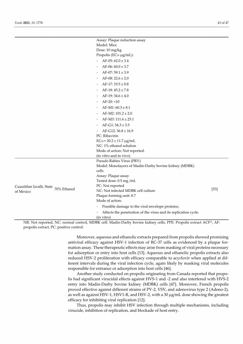
Foods 2021, 10, 1776 43 of 47
Assay: Plaque reduction assay
Model: Mice
Dose: 10 mg/kg
Propolis (EC50 µg/mL):
- AF-05: 62.0 ± 3.4
- AF-06: 60.0 ± 3.7
- AF-07: 59.1 ± 3.9
- AF-08: 22.6 ± 2.0
- AF-17: 19.5 ± 0.8
- AF-18: 45.2 ± 7.8
- AF-19: 34.6 ± 4.0
- AF-20: <10
- AF-M1: 60.3 ± 8.1
- AF-M2: 101.2 ± 2.0
- AF-M3: 111.6 ± 25.1
- AF-G1: 54.3 ± 3.5
- AF-G12: 36.8 ± 16.9
PC: Ribavirin
EC50 = 20.2 ± 11.7 µg/mL
NC: 1% ethanol solution
Mode of action: Not reported.
(in vitro and in vivo)
Cuautitlan Izcalli, State
of Mexico 70% Ethanol
Pseudo Rabies Virus (PRV)
Model: Monolayers of Madin-Darby bovine kidney (MDBK)
cells
Assay: Plaque assay
Tested dose: 0.5 mg /mL
PC: Not reported
NC: Not infected MDBK cell culture
Plaque forming unit: 8.7
Mode of action:
- Possible damage to the viral envelope proteins;
- Affects the penetration of the virus and its replication cycle.
(in vitro)
[53]
NR: Not reported, NC: normal control, MDBK cell: Madin-Darby bovine kidney cells, PPE: Propolis extract ACF® , AF:
propolis extract, PC: positive control.
Moreover, aqueous and ethanolic extracts prepared from propolis showed promising
antiviral efficacy against HSV-1 infection of RC-37 cells as evidenced by a plaque for-
mation assay. These therapeutic effects may arise from masking of viral proteins necessary
for adsorption or entry into host cells [13]. Aqueous and ethanolic propolis extracts also
reduced HSV-2 proliferation with efficacy comparable to acyclovir when applied at dif-
ferent intervals during the viral infection cycle, again likely by masking viral molecules
responsible for entrance or adsorption into host cells [46].
Another study conducted on propolis originating from Canada reported that propo-
lis had significant virucidal effects against HVS-1 and -2 and also interfered with HVS-2
entry into Madin-Darby bovine kidney (MDBK) cells [47]. Moreover, French propolis
proved effective against different strains of PV-2, VSV, and adenovirus type 2 (Adeno-2),
as well as against HSV-1, HSV1-R, and HSV-2, with a 30 µg/mL dose showing the greatest
efficacy for inhibiting viral replication [12].
Thus, propolis may inhibit HSV infection through multiple mechanisms, including
virucide, inhibition of replication, and blockade of host entry.
Foods 2021, 10, 1776 44 of 47
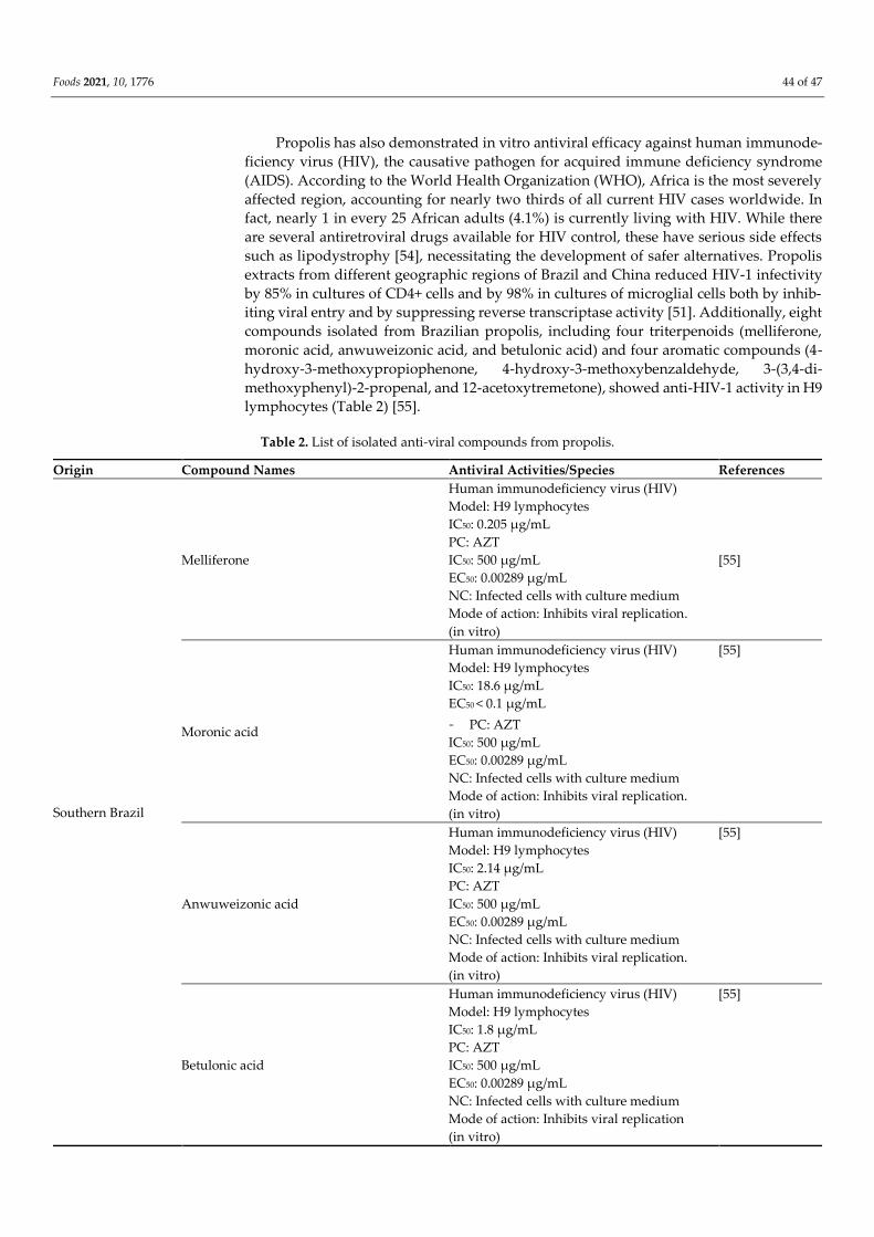
Propolis has also demonstrated in vitro antiviral efficacy against human immunode-
ficiency virus (HIV), the causative pathogen for acquired immune deficiency syndrome
(AIDS). According to the World Health Organization (WHO), Africa is the most severely
affected region, accounting for nearly two thirds of all current HIV cases worldwide. In
fact, nearly 1 in every 25 African adults (4.1%) is currently living with HIV. While there
are several antiretroviral drugs available for HIV control, these have serious side effects
such as lipodystrophy [54], necessitating the development of safer alternatives. Propolis
extracts from different geographic regions of Brazil and China reduced HIV-1 infectivity
by 85% in cultures of CD4+ cells and by 98% in cultures of microglial cells both by inhib-
iting viral entry and by suppressing reverse transcriptase activity [51]. Additionally, eight
compounds isolated from Brazilian propolis, including four triterpenoids (melliferone,
moronic acid, anwuweizonic acid, and betulonic acid) and four aromatic compounds (4-
hydroxy-3-methoxypropiophenone, 4-hydroxy-3-methoxybenzaldehyde, 3-(3,4-di-
methoxyphenyl)-2-propenal, and 12-acetoxytremetone), showed anti-HIV-1 activity in H9
lymphocytes (Table 2) [55].
Table 2. List of isolated anti-viral compounds from propolis.
Origin Compound Names Antiviral Activities/Species References
Southern Brazil
Melliferone
Human immunodeficiency virus (HIV)
Model: H9 lymphocytes
IC50: 0.205 µg/mL
PC: AZT
IC50: 500 µg/mL
EC50: 0.00289 µg/mL
NC: Infected cells with culture medium
Mode of action: Inhibits viral replication.
(in vitro)
[55]
Moronic acid
Human immunodeficiency virus (HIV)
Model: H9 lymphocytes
IC50: 18.6 µg/mL
EC50 < 0.1 µg/mL
- PC: AZT
IC50: 500 µg/mL
EC50: 0.00289 µg/mL
NC: Infected cells with culture medium
Mode of action: Inhibits viral replication.
(in vitro)
[55]
Anwuweizonic acid
Human immunodeficiency virus (HIV)
Model: H9 lymphocytes
IC50: 2.14 µg/mL
PC: AZT
IC50: 500 µg/mL
EC50: 0.00289 µg/mL
NC: Infected cells with culture medium
Mode of action: Inhibits viral replication.
(in vitro)
[55]
Betulonic acid
Human immunodeficiency virus (HIV)
Model: H9 lymphocytes
IC50: 1.8 µg/mL
PC: AZT
IC50: 500 µg/mL
EC50: 0.00289 µg/mL
NC: Infected cells with culture medium
Mode of action: Inhibits viral replication
(in vitro)
[55]
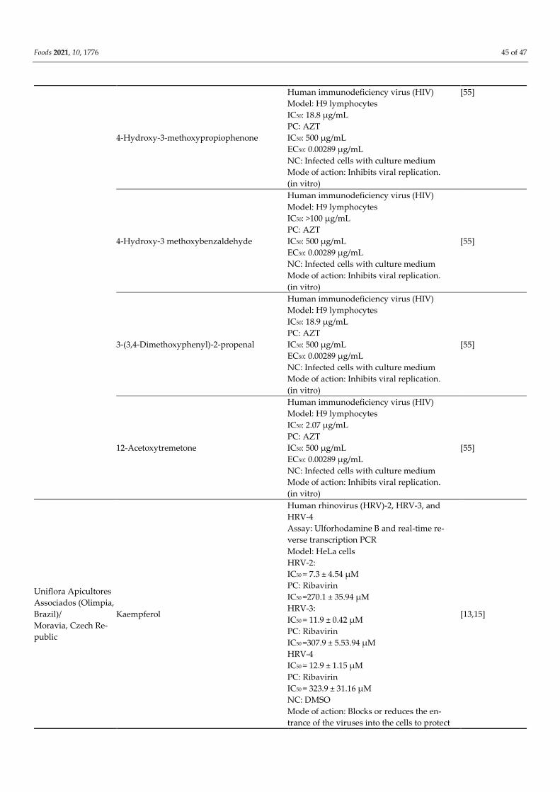
Foods 2021, 10, 1776 45 of 47
4-Hydroxy-3-methoxypropiophenone
Human immunodeficiency virus (HIV)
Model: H9 lymphocytes
IC50: 18.8 µg/mL
PC: AZT
IC50: 500 µg/mL
EC50: 0.00289 µg/mL
NC: Infected cells with culture medium
Mode of action: Inhibits viral replication.
(in vitro)
[55]
4-Hydroxy-3 methoxybenzaldehyde
Human immunodeficiency virus (HIV)
Model: H9 lymphocytes
IC50: >100 µg/mL
PC: AZT
IC50: 500 µg/mL
EC50: 0.00289 µg/mL
NC: Infected cells with culture medium
Mode of action: Inhibits viral replication.
(in vitro)
[55]
3-(3,4-Dimethoxyphenyl)-2-propenal
Human immunodeficiency virus (HIV)
Model: H9 lymphocytes
IC50: 18.9 µg/mL
PC: AZT
IC50: 500 µg/mL
EC50: 0.00289 µg/mL
NC: Infected cells with culture medium
Mode of action: Inhibits viral replication.
(in vitro)
[55]
12-Acetoxytremetone
Human immunodeficiency virus (HIV)
Model: H9 lymphocytes
IC50: 2.07 µg/mL
PC: AZT
IC50: 500 µg/mL
EC50: 0.00289 µg/mL
NC: Infected cells with culture medium
Mode of action: Inhibits viral replication.
(in vitro)
[55]
Uniflora Apicultores
Associados (Olimpia,
Brazil)/
Moravia, Czech Re-
public
Kaempferol
Human rhinovirus (HRV)-2, HRV-3, and
HRV-4
Assay: Ulforhodamine B and real-time re-
verse transcription PCR
Model: HeLa cells
HRV-2:
IC50 = 7.3 ± 4.54 µM
PC: Ribavirin
IC50 =270.1 ± 35.94 µM
HRV-3:
IC50 = 11.9 ± 0.42 µM
PC: Ribavirin
IC50 =307.9 ± 5.53.94 µM
HRV-4
IC50 = 12.9 ± 1.15 µM
PC: Ribavirin
IC50 = 323.9 ± 31.16 µM
NC: DMSO
Mode of action: Blocks or reduces the en-
trance of the viruses into the cells to protect
[13,15]

Foods 2021, 10, 1776 46 of 47
the cells from virus destruction and abate vi-
rus replication.
(in vitro)
p-Coumaric acid
Human rhinovirus (HRV)-2, HRV-3, and
HRV-4
Assay: Ulforhodamine B and real-time re-
verse transcription PCR
Model: HeLa cells
HRV-2
IC50 = 371.2 ± 7.74 µM
PC: Ribavirin
IC50 = 270.1 ± 35.94 µM
HRV-3
IC50 = 454.5 ± 3.16 µM
PC: Ribavirin:
IC50 = 307.9 ± 5.53.94 µM
HRV-4
IC50 = 604.3 ± 50.93 µM
PC: Ribavirin
IC50 = 323.9 ± 31.16 µM
NC: DMSO
Mode of action: Blocks or reduces the en-
trance of the viruses into the cells to protect
the cells from virus destruction and abate vi-
rus replication.
(in vitro)
[13,15]
Galangin
Human rhinovirus (HRV)-2, HRV-3, and
HRV-4
Assay: Ulforhodamine B and real-time re-
verse transcription PCR
Model: HeLa cells
HRV-2
IC50 = 20.0 ± 8.07 µM
PC: Ribavirin
IC50 =270.1 ±35.94 µM
HRV-3
IC50 = 116.2 ± 0.85 µM
PC: Ribavirin
IC50 = 307.9 ± 5.53.94 µM
HRV-4
IC50 = 88.1 ± 28.71 µM
PC: Ribavirin
IC50 = 323.9 ± 31.16 µM
NC: DMSO
Mode of action: Blocks or reduces the en-
trance of the viruses into the cells to protect
the cells from virus destruction and abate vi-
rus replication.
(in vitro)
[13,15]
Herpes simplex virus 1 (HSV-1)
Assay: Plaque reduction
Model: RC-37 cells
IC50 (%): 0.00045; SI: 3.3
PC: Heparin-Na and acyclovir
NC: Untreated cells
[13,15]

Foods 2021, 10, 1776 47 of 47
Mode of action: Masks viral compounds
which are necessary for adsorption or entry
into host cells.
(in vitro)
Quercetin
Human rhinovirus (HRV)-2, HRV-3, and
HRV-4
Assay: Ulforhodamine B and real-time re-
verse transcription PCR
Model: HeLa cells
HRV-2
IC50 = 34.1 ± 10.33 µM
PC: Ribavirin
IC50 =270.1 ± 35.94 µM
HRV-3
IC50 = 15.5 ± 2.29 µM
PC: Ribavirin:
IC50 =307.9 ± 5.53.94 µM
HRV-4
IC50 = 18.2 ± 4.14 µM
PC: Ribavirin
IC50 = 323.9 ± 31.16 µM
NC: DMSO
Mode of action: Blocks or reduces the en-
trance of the viruses into the cells to protect
the cells from virus destruction and abate vi-
rus replication.
(in vitro)
[13,15]
Fisetin
Human rhinovirus (HRV)-2, HRV-3, and
HRV-4
Assay: Ulforhodamine B and real-time re-
verse transcription PCR
Model: HeLa cells
HRV-2
IC50 = 40.9 ± 15.20 µM
PC: Ribavirin
IC50 =270.1 ± 35.94 µM
HRV-3
IC50 = 67.1 ± 3.50 µM
PC: Ribavirin
IC50 =307.9 ± 5.53.94 µM
HRV-4
IC50 = 66.4 ± 13.28 µM
PC: Ribavirin
IC50 = 323.9 ± 31.16 µM
NC: DMSO
Mode of action: Blocks or reduces the en-
trance of the viruses into the cells to protect
the cells from virus destruction and abate vi-
rus replication.
(in vitro)
[13,15]
Chrysin
Human rhinovirus (HRV)-2, HRV-3, and
HRV-4
Assay: Ulforhodamine B and real-time re-
verse transcription PCR
Model: HeLa cells
HRV-2
IC50 = 17.3 ± 9.83 µM
[13,15]
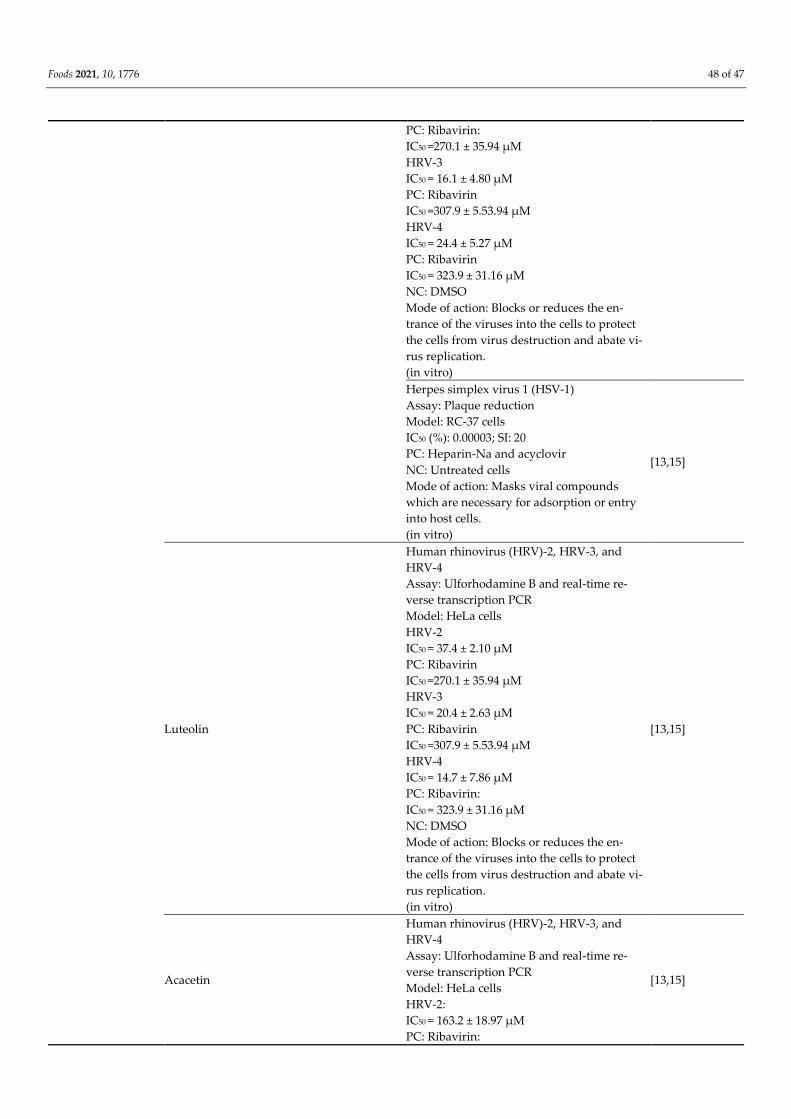
Foods 2021, 10, 1776 48 of 47
PC: Ribavirin:
IC50 =270.1 ± 35.94 µM
HRV-3
IC50 = 16.1 ± 4.80 µM
PC: Ribavirin
IC50 =307.9 ± 5.53.94 µM
HRV-4
IC50 = 24.4 ± 5.27 µM
PC: Ribavirin
IC50 = 323.9 ± 31.16 µM
NC: DMSO
Mode of action: Blocks or reduces the en-
trance of the viruses into the cells to protect
the cells from virus destruction and abate vi-
rus replication.
(in vitro)
Herpes simplex virus 1 (HSV-1)
Assay: Plaque reduction
Model: RC-37 cells
IC50 (%): 0.00003; SI: 20
PC: Heparin-Na and acyclovir
NC: Untreated cells
Mode of action: Masks viral compounds
which are necessary for adsorption or entry
into host cells.
(in vitro)
[13,15]
Luteolin
Human rhinovirus (HRV)-2, HRV-3, and
HRV-4
Assay: Ulforhodamine B and real-time re-
verse transcription PCR
Model: HeLa cells
HRV-2
IC50 = 37.4 ± 2.10 µM
PC: Ribavirin
IC50 =270.1 ± 35.94 µM
HRV-3
IC50 = 20.4 ± 2.63 µM
PC: Ribavirin
IC50 =307.9 ± 5.53.94 µM
HRV-4
IC50 = 14.7 ± 7.86 µM
PC: Ribavirin:
IC50 = 323.9 ± 31.16 µM
NC: DMSO
Mode of action: Blocks or reduces the en-
trance of the viruses into the cells to protect
the cells from virus destruction and abate vi-
rus replication.
(in vitro)
[13,15]
Acacetin
Human rhinovirus (HRV)-2, HRV-3, and
HRV-4
Assay: Ulforhodamine B and real-time re-
verse transcription PCR
Model: HeLa cells
HRV-2:
IC50 = 163.2 ± 18.97 µM
PC: Ribavirin:
[13,15]
Foods 2021, 10, 1776 49 of 47
IC50 = 270.1 ±35.94 µM
HRV-3:
IC50 = 107.6 ± 18.30 µM
PC: Ribavirin:
IC50 = 307.9 ± 5.53.94 µM
HRV-4:
IC50 = 102.3 ± 3.59 µM
PC: Ribavirin:
IC50 = 323.9 ± 31.16 µM
NC: DMSO
Mode of action: Blocks or reduces the en-
trance of the viruses into the cells to protect
the cells from virus destruction and abate vi-
rus replication.
(in vitro)
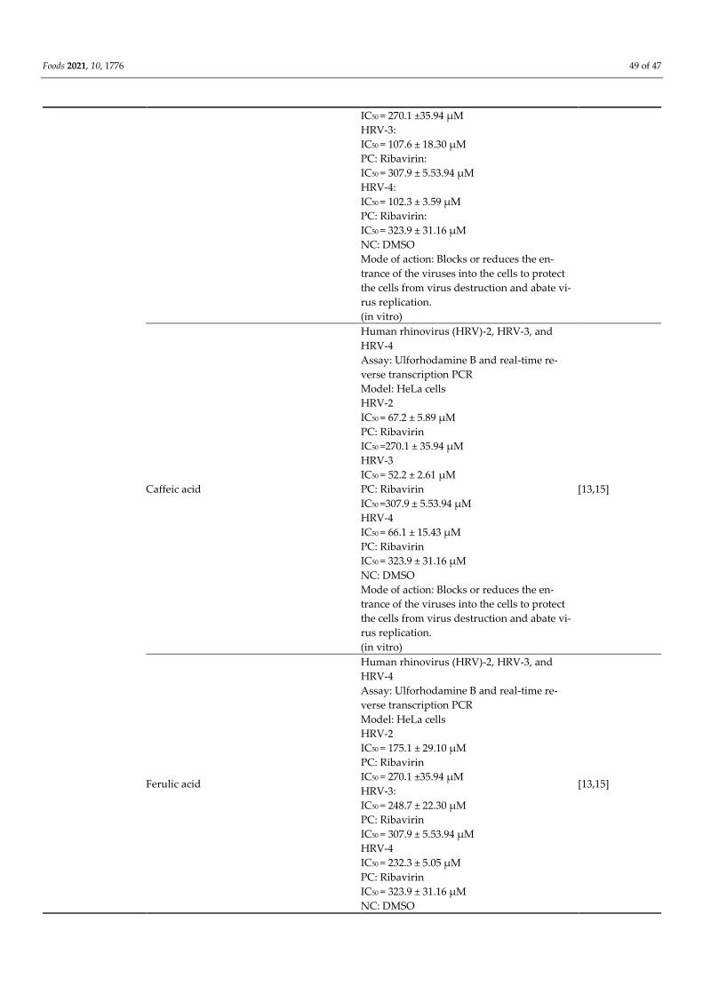
Caffeic acid
Human rhinovirus (HRV)-2, HRV-3, and
HRV-4
Assay: Ulforhodamine B and real-time re-
verse transcription PCR
Model: HeLa cells
HRV-2
IC50 = 67.2 ± 5.89 µM
PC: Ribavirin
IC50 =270.1 ± 35.94 µM
HRV-3
IC50 = 52.2 ± 2.61 µM
PC: Ribavirin
IC50 =307.9 ± 5.53.94 µM
HRV-4
IC50 = 66.1 ± 15.43 µM
PC: Ribavirin
IC50 = 323.9 ± 31.16 µM
NC: DMSO
Mode of action: Blocks or reduces the en-
trance of the viruses into the cells to protect
the cells from virus destruction and abate vi-
rus replication.
(in vitro)
[13,15]
Ferulic acid
Human rhinovirus (HRV)-2, HRV-3, and
HRV-4
Assay: Ulforhodamine B and real-time re-
verse transcription PCR
Model: HeLa cells
HRV-2
IC50 = 175.1 ± 29.10 µM
PC: Ribavirin
IC50 = 270.1 ±35.94 µM
HRV-3:
IC50 = 248.7 ± 22.30 µM
PC: Ribavirin
IC50 = 307.9 ± 5.53.94 µM
HRV-4
IC50 = 232.3 ± 5.05 µM
PC: Ribavirin
IC50 = 323.9 ± 31.16 µM
NC: DMSO
[13,15]

Foods 2021, 10, 1776 50 of 47
Mode of action: Blocks or reduces the en-
trance of the viruses into the cells to protect
the cells from virus destruction and abate vi-
rus replication.
(in vitro)
Brazil
Apigenin
Anti-influenza virus
Assay: Plaque reduction
Model: MDCK
A/PR/8/34(H1N1)
EC50 = 15.3 ± 3.0 µg/mL
A/Toyama/129/2011(H1N1)
EC50 = 17.8 ± 8.7 µg/mL
A/Toyama/26/2011(H1N1
EC50 = 8.1 ± 4.7 µg/mL
PC: Ribavirin
EC50 = 19.2 ± 7.5µg/mL
NC: Distilled water
Mode of action: Suppresses the stage of virus
replication after adsorption and/or invasion.
(in vitro)
[56]
Artepillin C
Anti-influenza virus
Assay: Plaque reduction
Model: MDCK
A/PR/8/34(H1N1)
EC50 ˃ 40 µg/mL
A/Toyama/129/2011(H1N1)
EC50 ˃ 40 µg/mL
A/Toyama/26/2011(H1N1
EC50 ˃ 40 µg/mL
PC: Ribavirin
EC50 = 19.2 ± 7.5µg/mL
NC: Distilled water
Mode of action: Suppresses the stage of virus
replication after adsorption and/or invasion.
(in vitro)
[56]
Kaempferol
Anti-influenza virus
Assay: Plaque reduction
Model: MDCK
A/PR/8/34(H1N1)
EC50 = 38.2 ± 17.1 µg/mL
A/Toyama/129/2011(H1N1)
EC50 = 21.7 ± 5.5 µg/mL
A/Toyama/26/2011(H1N1
EC50 = 24.8 ± 4.3 µg/mL
PC: Ribavirin
EC50 = 19.2 ± 7.5µg/mL
NC: Distilled water
Mode of action: Suppresses the stage of virus
replication after adsorption and/or invasion.
(in vitro)
[56]
Caffeic acid
Anti-influenza virus
Assay: Plaque reduction
Model: MDCK
A/PR/8/34(H1N1)
EC50 >100 µg/mL
A/Toyama/129/2011(H1N1)
EC50 = 49.7 ± 5.0 µg/mL
[56]
Foods 2021, 10, 1776 51 of 47
A/Toyama/26/2011(H1N1
EC50 > 100 µg/mL
PC: Ribavirin
EC50 = 19.2 ± 7.5µg/mL
NC: Distilled water
Mode of action: Suppresses the stage of virus
replication after adsorption and/or invasion.
(in vitro)
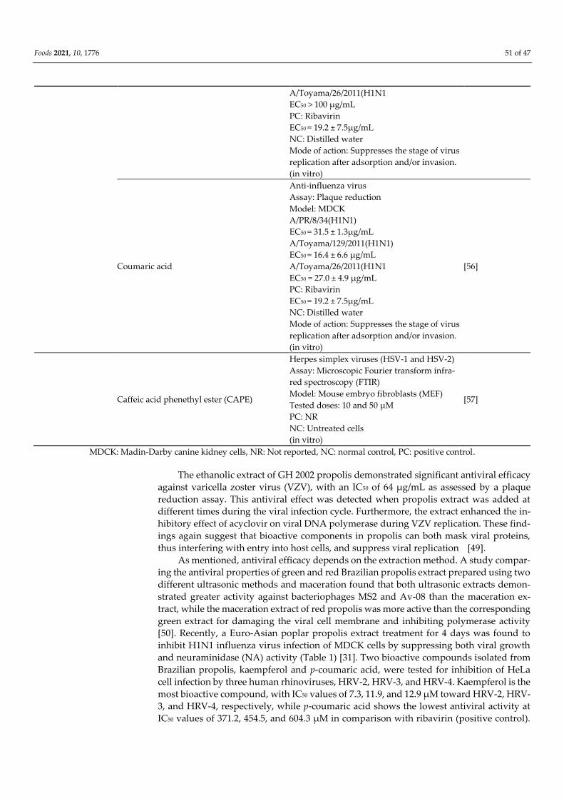
Coumaric acid
Anti-influenza virus
Assay: Plaque reduction
Model: MDCK
A/PR/8/34(H1N1)
EC50 = 31.5 ± 1.3µg/mL
A/Toyama/129/2011(H1N1)
EC50 = 16.4 ± 6.6 µg/mL
A/Toyama/26/2011(H1N1
EC50 = 27.0 ± 4.9 µg/mL
PC: Ribavirin
EC50 = 19.2 ± 7.5µg/mL
NC: Distilled water
Mode of action: Suppresses the stage of virus
replication after adsorption and/or invasion.
(in vitro)
[56]
Caffeic acid phenethyl ester (CAPE)
Herpes simplex viruses (HSV-1 and HSV-2)
Assay: Microscopic Fourier transform infra-
red spectroscopy (FTIR)
Model: Mouse embryo fibroblasts (MEF)
Tested doses: 10 and 50 µM
PC: NR
NC: Untreated cells
(in vitro)
[57]
MDCK: Madin-Darby canine kidney cells, NR: Not reported, NC: normal control, PC: positive control.
The ethanolic extract of GH 2002 propolis demonstrated significant antiviral efficacy
against varicella zoster virus (VZV), with an IC50 of 64 µg/mL as assessed by a plaque
reduction assay. This antiviral effect was detected when propolis extract was added at
different times during the viral infection cycle. Furthermore, the extract enhanced the in-
hibitory effect of acyclovir on viral DNA polymerase during VZV replication. These find-
ings again suggest that bioactive components in propolis can both mask viral proteins,
thus interfering with entry into host cells, and suppress viral replication [49].
As mentioned, antiviral efficacy depends on the extraction method. A study compar-
ing the antiviral properties of green and red Brazilian propolis extract prepared using two
different ultrasonic methods and maceration found that both ultrasonic extracts demon-
strated greater activity against bacteriophages MS2 and Av-08 than the maceration ex-
tract, while the maceration extract of red propolis was more active than the corresponding
green extract for damaging the viral cell membrane and inhibiting polymerase activity
[50]. Recently, a Euro-Asian poplar propolis extract treatment for 4 days was found to
inhibit H1N1 influenza virus infection of MDCK cells by suppressing both viral growth
and neuraminidase (NA) activity (Table 1) [31]. Two bioactive compounds isolated from
Brazilian propolis, kaempferol and p-coumaric acid, were tested for inhibition of HeLa
cell infection by three human rhinoviruses, HRV-2, HRV-3, and HRV-4. Kaempferol is the
most bioactive compound, with IC50 values of 7.3, 11.9, and 12.9 µM toward HRV-2, HRV-
3, and HRV-4, respectively, while p-coumaric acid shows the lowest antiviral activity at
IC50 values of 371.2, 454.5, and 604.3 µM in comparison with ribavirin (positive control).
Foods 2021, 10, 1776 52 of 47
Findings indicate that kaempferol and p-coumaric acid may block or reduce the entry of
the viruses into the host cells, in order to preserve the cells from virus replication [15].
Another study on the antiviral effect of Mexican propolis against MDBK cell mono-
layer infection by pseudo rabies virus (PRV) found that treated cells exhibited an electron-
dense layer on the cell membrane that prevented viral entry [53]. Another study of 13
ethanolic extracts from South Brazilian propolis identified four with significant anti-influ-
enza virus activity in vitro and subsequently found antiviral efficacy in vivo after oral
administration to infected mice (3 times daily/7 days), with 10 mg/kg showing the greatest
therapeutic effect [52].
Other work aimed to compare the effect of three samples, propolis, Baccharis dra-
cunculifolia (extract and essential oil), and some isolated compounds (caffeic and cinnamic
acids), on poliovirus type 1 (PV1). Three protocols (pre-, simultaneous, and post-treat-
ments) were used for evaluating the effects on the virus. For propolis, a high inhibition
percentage both in simultaneous and post-treatment was recorded. Propolis partially af-
fects both in viral cell entry and cell replication steps in the viral cycle or leads to RNA
degradation before the entry of virus to cells [48].
In summary, various bioactive compounds have been identified in Brazilian propolis
extract, including antiviral agents effective against different strains of the influenza virus.
Three compounds, apigenin, kaempferol, and coumaric acid, were shown to significantly
inhibit the infection of MDCK cells by suppressing the post adsorption and invasion
stages of viral replication [56]. In general, the antiviral activities of propolis are mediated
by flavonoids and other phenolic acids. These active constituents have different modes of
action, such as the formation of complexes with viral proteins required for infection
(masking), formation of an electron-dense layer on the cell membrane, directly damaging
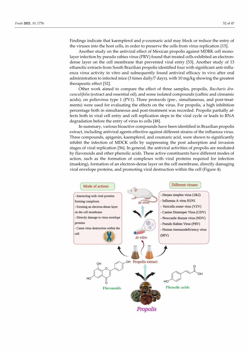
viral envelope proteins, and promoting viral destruction within the cell (Figure 4).
Foods 2021, 10, 1776 53 of 47
Figure 4. Anti-viral activity and the possible mode of action of propolis extracts/constituents against
different types of viruses.
5. Propolis as a Treatment for COVID-19
COVID-19 is a pandemic disease caused by the recently discovered SARS-CoV-2, the
seventh known member of the coronavirus family infectious to humans (after SARS coro-
navirus and Middle East respiratory syndrome (MERS) coronavirus) [58]. The epidemio-
logical burden of COVID-19 is currently a major healthcare challenge throughout the
world, as SARS-CoV-2 is readily transmitted from human to human via airborne micro-
droplets generated during coughing, talking, or sneezing. In addition, SARS-CoV-2 can
be transmitted by touching a contaminated surface and then touching the nose, mouth, or
eyes [59,60]. While many drugs have been screened for efficacy against SARS-CoV-2 in-
fection, no antiviral agent has yet proven broadly efficacious [61]. However, several nat-
ural product derivatives have shown promise as effective non-toxic antiviral agents [62].
Potential therapeutic agents may include honeybee products in addition to propolis, such
as honey, royal jelly, bee venom, wax, bee pollen, and bee bread, as all have demonstrated
antimicrobial, antifungal, anti-inflammatory, and (or) antiviral properties under certain
conditions [63]. Propolis has also shown promising broad spectrum antiviral effects in
vitro and in vivo against influenza virus, human respiratory syncytial and coronaviruses,
rotavirus, and human rhinovirus, among others, suggesting potential efficacy against
coronaviruses [16,64,65].
The potential efficacy of five propolis-derived flavonoids was recently evaluated in
vitro on different DNA and RNA viruses, including coronaviruses, using the viral plaque
reduction technique. Acacetin and galangin had no effect on either the infectivity or rep-
lication of any of the viruses tested, but chrysin and kaempferol were highly effective in
inhibiting replication, and quercetin was active against infectivity and replication at
higher concentrations [17]. Refaat et al. investigated the in vitro effects of crude Egyptian
propolis extract and a propolis liposome preparation on SARS-CoV-2 3CL-like protease,
S1 spike protein, and viral replication by RT-PCR. Liposomes inhibited SARS-CoV-2 3CL
protease activity with an IC50 of 1.183 ± 0.06 µg/mL, while the crude propolis extract in-
hibited 3CL protease activity with an IC50 of 2.452 ± 0.11 µg/mL, values comparable to
Remdesivir [18]. Sulawesi propolis and its components glyasperin A, broussoflavonol F,
and sulabiroins A also inhibited SARS-CoV-2 3C-like protease activity and interacted with
the protease catalytic sites His41 and Cys145, with docking scores of −7.8, −7.8, and −7.6
kcal/mol, respectively [19]. Similarly, Hashem et al. evaluated the in silico inhibitory ac-
tivity of six selected compounds present in propolis, 3-phenyllactic acid, CAPE, lumi-
chrome, galangin, chrysin, and caffeic acid, against SARS-CoV-2 3CLpro and found that all
six showed good docking scores, with the most potent being CAPE (−6.383 kcal/mol),
chrysin (−6.097), and galangin (−6.295) [20].
Other studies have found that propolis is able to inhibit the activity of P21 (RAC1)
Activated Kinase 1 (PAK1), a major “pathogenic” kinase in several diseases/disorders, in-
cluding inflammation, cancer, malaria, and pandemic viral infections such as HIV, influ-
enza, and COVID-19 [66]. Additionally, CAPE was found to bind and inhibit SARS-CoV-
2 transmembrane protease serine 2 to a degree comparable with Camostat mesylate as
evidenced by molecular docking and molecular dynamics (MD) simulations [34]. Moreo-
ver, quercetin alone and in conjunction with vitamin C was predicted to suppress SARS-
CoV-2 infection by binding to 3C-like protease (3CLpro) [67,68]. A pilot randomized clinical
study assessed the potential efficacy of Brazilian green propolis (400 or 800 mg/day orally
or via nasoenteral tube) against SARS-CoV-2 (NCT04480593). In addition, it supported the
idea that propolis may be an effective agent to combat coronavirus-induced fibrosis in the
lungs [65].
6. Immunomodulatory Activity
Foods 2021, 10, 1776 54 of 47
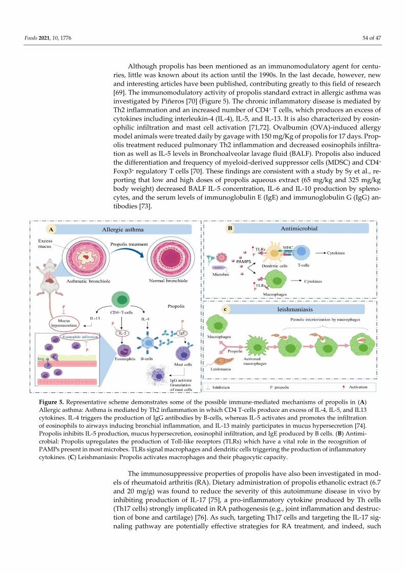
Although propolis has been mentioned as an immunomodulatory agent for centu-
ries, little was known about its action until the 1990s. In the last decade, however, new
and interesting articles have been published, contributing greatly to this field of research
[69]. The immunomodulatory activity of propolis standard extract in allergic asthma was
investigated by Piñeros [70] (Figure 5). The chronic inflammatory disease is mediated by
Th2 inflammation and an increased number of CD4+ T cells, which produces an excess of
cytokines including interleukin-4 (IL-4), IL-5, and IL-13. It is also characterized by eosin-
ophilic infiltration and mast cell activation [71,72]. Ovalbumin (OVA)-induced allergy
model animals were treated daily by gavage with 150 mg/Kg of propolis for 17 days. Prop-
olis treatment reduced pulmonary Th2 inflammation and decreased eosinophils infiltra-
tion as well as IL-5 levels in Bronchoalveolar lavage fluid (BALF). Propolis also induced
the differentiation and frequency of myeloid-derived suppressor cells (MDSC) and CD4+
Foxp3+ regulatory T cells [70]. These findings are consistent with a study by Sy et al., re-
porting that low and high doses of propolis aqueous extract (65 mg/kg and 325 mg/kg
body weight) decreased BALF IL-5 concentration, IL-6 and IL-10 production by spleno-
cytes, and the serum levels of immunoglobulin E (IgE) and immunoglobulin G (IgG) an-
tibodies [73].
Figure 5. Representative scheme demonstrates some of the possible immune-mediated mechanisms of propolis in (A)
Allergic asthma: Asthma is mediated by Th2 inflammation in which CD4 T-cells produce an excess of IL-4, IL-5, and IL13
cytokines. IL-4 triggers the production of IgG antibodies by B-cells, whereas IL-5 activates and promotes the infiltration
of eosinophils to airways inducing bronchial inflammation, and IL-13 mainly participates in mucus hypersecretion [74].
Propolis inhibits IL-5 production, mucus hypersecretion, eosinophil infiltration, and IgE produced by B cells. (B) Antimi-
crobial: Propolis upregulates the production of Toll-like receptors (TLRs) which have a vital role in the recognition of
PAMPs present in most microbes. TLRs signal macrophages and dendritic cells triggering the production of inflammatory
cytokines. (C) Leishmaniasis: Propolis activates macrophages and their phagocytic capacity.
The immunosuppressive properties of propolis have also been investigated in mod-
els of rheumatoid arthritis (RA). Dietary administration of propolis ethanolic extract (6.7
and 20 mg/g) was found to reduce the severity of this autoimmune disease in vivo by
inhibiting production of IL-17 [75], a pro-inflammatory cytokine produced by Th cells
(Th17 cells) strongly implicated in RA pathogenesis (e.g., joint inflammation and destruc-
tion of bone and cartilage) [76]. As such, targeting Th17 cells and targeting the IL-17 sig-
naling pathway are potentially effective strategies for RA treatment, and indeed, such
Foods 2021, 10, 1776 55 of 47
treatments are currently under investigation [77]. Okamoto et al. reported that Brazilian
propolis suppressed Th17 cell activity in vitro at 12.48 µg/mL by inhibiting the IL-6-in-
duced phosphorylation of signal transducer and activator of transcription 3 (STAT3), a
key transcription factor driving Th17 cell differentiation. In addition, Th17 cell differenti-
ation induced by transforming growth factor-β (TGF-β) plus IL-16 was downregulated by
propolis in RA model animals, while propolis induced no detectable cellular toxicity at
concentrations up to 96 µg/mL [78]. The propolis-derived compound CAPE has also been
reported to suppress autoimmune uveoretinitis. CAPE was found to hinder T cell-de-
pendent production of chemokines and cytokines as well as of antibodies induced by in-
terphotoreceptor retinoid binding protein (IRBP). Treatment with 200 µL CAPE also re-
duced serum concentrations of TNF-α, IL-6, interferon-γ (IFN-γ), and TNF-α in the retina
and inhibited the transcriptional activity of nuclear factor-kappa B (NF-kB) and phospho-
IkBα. Hence, it was concluded that the immunosuppressive activity of CAPE in uveitis is
mediated by suppression of the pro-inflammatory NF-kB–cytokine pathway [79].
Propolis extracts and derivatives may also augment microbe-induced immune re-
sponses by modulating Toll-like receptor (TLR) signaling. Toll-like receptors recognize
pathogen-associated molecular patterns (PAMPs), conserved molecules expressed by
many microorganisms [80]. Toll-like receptor 2 (TLR2), for instance, recognizes lipo-
teichoic acid on Gram-positive bacteria and fungi, while TLR-4 recognizes lipopolysac-
charide on Gram-negative bacteria [81]. Toll-like receptors are mainly expressed by anti-
gen-presenting cells (APCs), including monocytes, macrophages, B cells, and dendritic
cells (DCs) [82]. These cells also express human leukocyte antigen-DR isotype (HLA-DR)
and cluster of differentiation 80 (CD80) molecules that present peptides to T cells, result-
ing in T cell activation. Additionally, TLR signal transduction may activate transcription
factors controlling the expression of genes encoding chemokines, cytokines, and antimi-
crobial peptides [83].
Treatment of BALB/c mice with 200 mg/kg of 30% propolis ethanolic extract for three
consecutive days increased expression of TLR-2 and TLR-4 by peritoneal macrophages
and spleen cells and elevated the production of IL-1β and IL-6 [84]. In another study,
propolis treatment of mice prevented the inhibition of TLR-2 and TLR-4 induced by 14
days of restraint stress. Additionally, real-time polymerase chain reaction (RT-PCR) re-
vealed a significant increase in TLR gene expression in mice receiving propolis treatment
without stress [81]. Propolis treatment (10, 20, and 40 µg/mL) also increased the expres-
sion of TLR-4 and CD8 by human DCs through a mechanism involving has-miR-155, re-
sulting in enhanced bactericidal activity against Streptococcus mutans, and promoted the
production of NF-kB, TNF-α, IL-6, and IL-10 [85]. Conversely, cinnamic acid (5–100
μg/mL) downregulated the expression levels of TLR-2, HLA-DR, and CD80 by human
monocytes, although this treatment upregulated TLR-4. High concentrations of cinnamic
acid also inhibited expression of TNF-α and IL-10. As TNF-α is known to activate mono-
cytes and macrophages, while IL-10 inhibits these cells, Conti et al. concluded that the
increase in fungicidal activity could be due to mechanisms involving other cytokines [83].
Following a similar protocol, Búfalo et al. found that caffeic acid inhibited the expression
of TLR-2 and HLA-DR, while CD80 and TLR-4 were not affected. The fungicidal activity
of monocytes increased, however, despite the decrease in TNF-α and IL-10 [86].
The in vivo antileishmanial effect of Brazilian propolis was reported for the first time
by Pontin et al., that is, an administration of the hydroalcoholic extract at a dose of 1.5
mg/kg/day reduced the lesion diameter in leishmania braziliensis infected albino mice by
90% after 90 days of treatment. Pontin et al. pointed out that the reduction could be a
result of the activation of macrophages and their phagocytic capacity [87]. Consistent with
this explanation, da Silva et al. found that 5 and 10 µg/mL propolis activated macrophage
phagocytic activity and in turn increased parasite interiorization. This upregulation of
macrophage activity was attributed to increased TNF-α and reduced IL-12 signaling. Mor-
phological changes in promastigote forms of Leishmania were also observed by scanning
electron microscopy upon treatment [88]. Additionally, propolis was found to regulate
Foods 2021, 10, 1776 56 of 47
the expression of CCL5 and IFN-γ, factors involved in the development of Th1 cells in
leishmaniasis patients. Leishmaniasis is usually associated with the development of a
strong Th1 response that impairs the wound healing process [89]. In another study by dos
Santos Thomazelli et al., the hydroalcoholic extract showed an immunomodulatory effect
on both healthy donors and American tegumentar leishmaniasis patients’ human-derived
peripheral blood mononuclear cells (PBMC) in leishmaniasis models. This impact was ex-
plained by the increase in IL-4 and IL-17 and a decrease in IL-10 in a dose-dependent
manner. On the other hand, nitric oxide (NO) levels remained constant [90].
Numerous studies have also suggested that propolis extracts can suppress tumor
growth or promote immune-mediated tumor destruction. Benkovic et al. examined the
possible synergistic effect of a water-soluble derivative of propolis (WSDP) and ethanolic
extract of propolis (EEP) with the anticancer drug irinotecan in Swiss albino mice inocu-
lated with Ehrlich ascites tumor (EAT) cells. Intraperitoneal injection of WSDP and EEP
at 100 mg/kg for three days prior to 50 mg/kg irinotecan injection enhanced the antitumor
efficacy and reduced the non-target cytotoxicity of irinotecan compared to irinotecan
alone or the combination of irinotecan with the phenolic compounds quercetin and nar-
ingin [91]. Further investigation revealed that the decrease in irinotecan-induced non-tar-
get cytotoxicity was due to the immunomodulatory properties of propolis. Pretreatment
with WSDP+EEP activated macrophages and increased the number of neutrophils in the
peritoneal cavity [92]. Oršolić et al. [93] also reported that WSDP at 50 or 150 mg/kg sup-
pressed metastasis and tumor development in mice transplanted with mammary carci-
noma cells. This antimetastatic effect was associated with macrophage activation and en-
suing nonspecific tumor resistance. Additionally, high levels of lymphocyte activating fac-
tor (LAF) produced by these activated macrophages increased tumor cell killing effi-
ciency. Furthermore, WSDP significantly increased the expression of CD4+ and CD8+ by
splenocytes [93]. It was concluded that the antitumor activity of WSDP is likely due to the
synergistic effects of constituent polyphenolic compounds such as caffeic acid, quercetin,
chrysin, and naringenin, and it was further proposed that these compounds interfere with
tumor growth by enhancing apoptosis, macrophage activation, and production of pro-
inflammatory cytokines such as IL-1, IL-6, IL-8, TNF-α, and NO, some of which can di-
rectly damage tumor cells, whereas others act indirectly by enhancing the activities of
natural killer (NK) cells and cytotoxic T lymphocytes. Furthermore, these factors stimulate
the production of complement factor C3 production and C-reactive protein, which partic-
ipate in the opsonization of tumor cells [94–96].
Propolis was also shown to reduce the severity of Aujeszky disease when used as a
vaccine adjuvant. Mice treated with 5 mg propolis extract, aluminum hydroxide Al(OH)3,
and inactivated Suid herpesvirus type 1 (SuHV-1) demonstrated significantly higher neu-
tralizing antibody titers than mice receiving vaccine without propolis, indicating that the
adjuvant properties of propolis are associated with enhanced humoral and cellular im-
munity related to increased IFN-ɣ mRNA production. Moreover, expression of mRNA
IFN-ɣ was even higher when propolis was conjugated with antigen [97]. Although nu-
merous preclinical studies have shown the potential efficacy of propolis against immuno-
logical diseases, standardized quality controls and well-designed clinical trials are needed
before propolis or its components can be adopted as therapeutics (Table 3) [98].
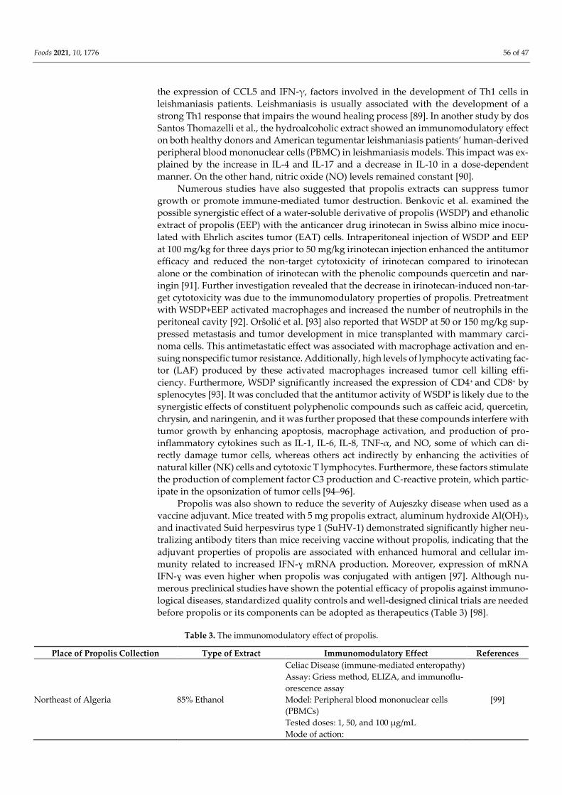
Table 3. The immunomodulatory effect of propolis.
Place of Propolis Collection Type of Extract Immunomodulatory Effect References
Northeast of Algeria 85% Ethanol
Celiac Disease (immune-mediated enteropathy)
Assay: Griess method, ELIZA, and immunoflu-
orescence assay
Model: Peripheral blood mononuclear cells
(PBMCs)
Tested doses: 1, 50, and 100 µg/mL
Mode of action:
[99]
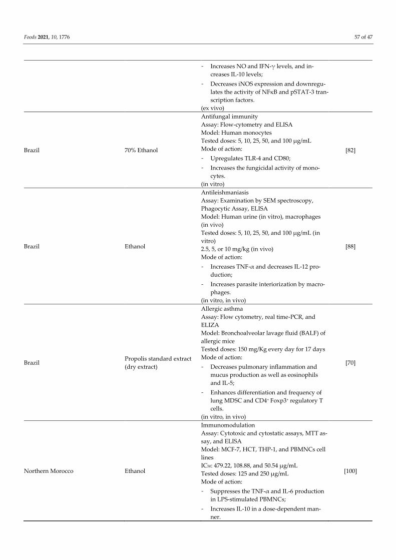
Foods 2021, 10, 1776 57 of 47
- Increases NO and IFN-γ levels, and in-
creases IL-10 levels;
- Decreases iNOS expression and downregu-
lates the activity of NFκB and pSTAT-3 tran-
scription factors.
(ex vivo)
Brazil 70% Ethanol
Antifungal immunity
Assay: Flow-cytometry and ELISA
Model: Human monocytes
Tested doses: 5, 10, 25, 50, and 100 μg/mL
Mode of action:
- Upregulates TLR-4 and CD80;
- Increases the fungicidal activity of mono-
cytes.
(in vitro)
[82]
Brazil Ethanol
Antileishmaniasis
Assay: Examination by SEM spectroscopy,
Phagocytic Assay, ELISA
Model: Human urine (in vitro), macrophages
(in vivo)
Tested doses: 5, 10, 25, 50, and 100 μg/mL (in
vitro)
2.5, 5, or 10 mg/kg (in vivo)
Mode of action:
- Increases TNF-α and decreases IL-12 pro-
duction;
- Increases parasite interiorization by macro-
phages.
(in vitro, in vivo)
[88]
Brazil Propolis standard extract
(dry extract)
Allergic asthma
Assay: Flow cytometry, real time-PCR, and
ELIZA
Model: Bronchoalveolar lavage fluid (BALF) of
allergic mice
Tested doses: 150 mg/Kg every day for 17 days
Mode of action:
- Decreases pulmonary inflammation and
mucus production as well as eosinophils
and IL-5;
- Enhances differentiation and frequency of
lung MDSC and CD4+ Foxp3+ regulatory T
cells.
(in vitro, in vivo)
[70]
Northern Morocco Ethanol
Immunomodulation
Assay: Cytotoxic and cytostatic assays, MTT as-
say, and ELISA
Model: MCF-7, HCT, THP-1, and PBMNCs cell
lines
IC50: 479.22, 108.88, and 50.54 μg/mL
Tested doses: 125 and 250 μg/mL
Mode of action:
- Suppresses the TNF-α and IL-6 production
in LPS-stimulated PBMNCs;
- Increases IL-10 in a dose-dependent man-
ner.
[100]
Foods 2021, 10, 1776 58 of 47
(in vitro)
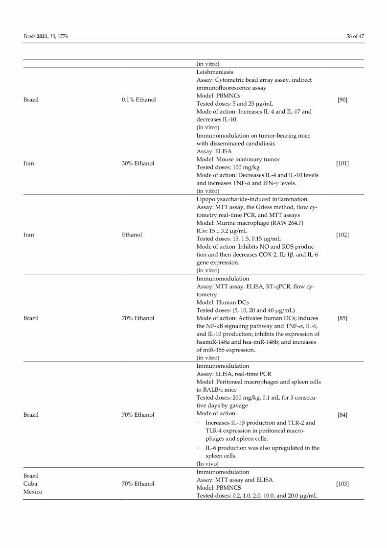
Brazil 0.1% Ethanol
Leishmaniasis
Assay: Cytometric bead array assay, indirect
immunofluorescence assay
Model: PBMNCs
Tested doses: 5 and 25 µg/mL
Mode of action: Increases IL-4 and IL-17 and
decreases IL-10.
(in vitro)
[90]
Iran 30% Ethanol
Immunomodulation on tumor-bearing mice
with disseminated candidiasis
Assay: ELISA
Model: Mouse mammary tumor
Tested doses: 100 mg/kg
Mode of action: Decreases IL-4 and IL-10 levels
and increases TNF-α and IFN-γ levels.
(in vitro)
[101]
Iran Ethanol
Lipopolysaccharide-induced inflammation
Assay: MTT assay, the Griess method, flow cy-
tometry real-time PCR, and MTT assays
Model: Murine macrophage (RAW 264.7)
IC50: 15 ± 3.2 µg/mL
Tested doses: 15, 1.5, 0.15 µg/mL
Mode of action: Inhibits NO and ROS produc-
tion and then decreases COX-2, IL-1β, and IL-6
gene expression.
(in vitro)
[102]
Brazil 70% Ethanol
Immunomodulation
Assay: MTT assay, ELISA, RT-qPCR, flow cy-
tometry
Model: Human DCs
Tested doses: (5, 10, 20 and 40 μg/mL)
Mode of action: Activates human DCs; induces
the NF-kB signaling pathway and TNF-α, IL-6,
and IL-10 production; inhibits the expression of
hsamiR-148a and hsa-miR-148b; and increases
of miR-155 expression.
(in vitro)
[85]
Brazil 70% Ethanol
Immunomodulation
Assay: ELISA, real-time PCR
Model: Peritoneal macrophages and spleen cells
in BALB/c mice
Tested doses: 200 mg/kg, 0.1 mL for 3 consecu-
tive days by gavage
Mode of action:
- Increases IL-1β production and TLR-2 and
TLR-4 expression in peritoneal macro-
phages and spleen cells;
- IL-6 production was also upregulated in the
spleen cells.
(In vivo)
[84]
Brazil
Cuba
Mexico
70% Ethanol
Immunomodulation
Assay: MTT assay and ELISA
Model: PBMNCS
Tested doses: 0.2, 1.0, 2.0, 10.0, and 20.0 μg/mL
[103]
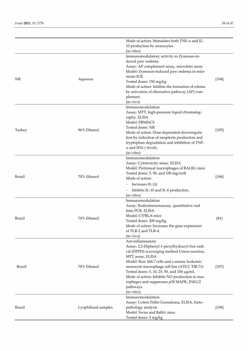
Foods 2021, 10, 1776 59 of 47
Mode of action: Stimulates both TNF-α and IL-
10 production by monocytes.
(in vitro)
NR Aqueous
Immunomodulatory activity in Zymosan-in-
duced paw oedema
Assay: AP complement assay, microtiter assay
Model: Zymosan-induced paw oedema in mice
strain ICR
Tested doses: 150 mg/kg
Mode of action: Inhibits the formation of edema
by activation of alternative pathway (AP) com-
plement.
(in vivo)
[104]
Turkey 96% Ethanol
Immunomodulation
Assay: MTT, high-pressure liquid chromatog-
raphy, ELISA
Model: PBMNCS
Tested doses: NR
Mode of action: Dose-dependent downregula-
tion by induction of neopterin production and
tryptophan degradation and inhibition of TNF-
α and IFN-γ levels.
(in vitro)
[105]
Brazil 70% Ethanol
Immunomodulation
Assay: Cytotoxicity assay, ELISA
Model: Peritoneal macrophages of BALB/c mice
Tested doses: 5, 50, and 100 mg/well
Mode of action:
- Increases IL-1β;
- Inhibits IL-10 and IL-6 production.
(in vitro)
[106]
Brazil 70% Ethanol
Immunomodulation
Assay: Radioimmunoassay, quantitative real
time PCR, ELISA
Model: C57BL/6 mice
Tested doses: 200 mg/kg
Mode of action: Increases the gene expression
of TLR-2 and TLR-4.
(in vivo)
[81]
Brazil 70% Ethanol
Ant-inflammatory
Assay: 2,2-Diphenyl-1-picrylhydrazyl free radi-
cal (DPPH) scavenging method Griess reaction,
MTT assay, ELISA
Model: Raw 264.7 cells and a mouse leukemic
monocyte macrophage cell line (ATCC TIB-71)
Tested doses: 5, 10, 25, 50, and 100 μg/mL
Mode of action: Inhibits NO production in mac-
rophages and suppresses p38 MAPK, JNK1/2
pathways.
(in vitro)
[107]
Brazil Lyophilized samples
Immunomodulation
Assay: Cotton Pellet Granuloma, ELISA, histo-
pathology analysis
Model: Swiss and Balb/c mice
Tested doses: 5 mg/kg
[108]
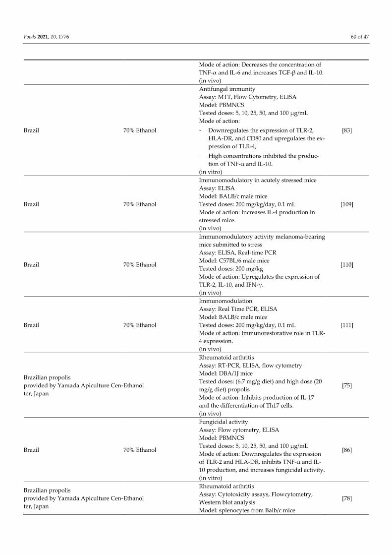
Foods 2021, 10, 1776 60 of 47
Mode of action: Decreases the concentration of
TNF-α and IL-6 and increases TGF-β and IL-10.
(in vivo)
Brazil 70% Ethanol
Antifungal immunity
Assay: MTT, Flow Cytometry, ELISA
Model: PBMNCS
Tested doses: 5, 10, 25, 50, and 100 μg/mL
Mode of action:
- Downregulates the expression of TLR-2,
HLA-DR, and CD80 and upregulates the ex-
pression of TLR-4;
- High concentrations inhibited the produc-
tion of TNF-α and IL-10.
(in vitro)
[83]
Brazil 70% Ethanol
Immunomodulatory in acutely stressed mice
Assay: ELISA
Model: BALB/c male mice
Tested doses: 200 mg/kg/day, 0.1 mL
Mode of action: Increases IL-4 production in
stressed mice.
(in vivo)
[109]
Brazil 70% Ethanol
Immunomodulatory activity melanoma-bearing
mice submitted to stress
Assay: ELISA, Real-time PCR
Model: C57BL/6 male mice
Tested doses: 200 mg/kg
Mode of action: Upregulates the expression of
TLR-2, IL-10, and IFN-γ.
(in vivo)
[110]
Brazil 70% Ethanol
Immunomodulation
Assay: Real Time PCR, ELISA
Model: BALB/c male mice
Tested doses: 200 mg/kg/day, 0.1 mL
Mode of action: Immunorestorative role in TLR-
4 expression.
(in vivo)
[111]
Brazilian propolis
provided by Yamada Apiculture Cen-
ter, Japan
Ethanol
Rheumatoid arthritis
Assay: RT-PCR, ELISA, flow cytometry
Model: DBA/1J mice
Tested doses: (6.7 mg/g diet) and high dose (20
mg/g diet) propolis
Mode of action: Inhibits production of IL-17
and the differentiation of Th17 cells.
(in vivo)
[75]
Brazil 70% Ethanol
Fungicidal activity
Assay: Flow cytometry, ELISA
Model: PBMNCS
Tested doses: 5, 10, 25, 50, and 100 μg/mL
Mode of action: Downregulates the expression
of TLR-2 and HLA-DR, inhibits TNF-α and IL-
10 production, and increases fungicidal activity.
(in vitro)
[86]
Brazilian propolis
provided by Yamada Apiculture Cen-
ter, Japan
Ethanol
Rheumatoid arthritis
Assay: Cytotoxicity assays, Flowcytometry,
Western blot analysis
Model: splenocytes from Balb/c mice
[78]

Foods 2021, 10, 1776 61 of 47
Tested doses: 12, 48 μg/mL
Mode of action:
- Inhibits IL-6 plus TGF-β-induced Th17 dif-
ferentiation;
- Suppresses IL-6-induced phosphorylation of
STAT3.
(in vitro)
Beekeeping Section, UNESP 70% Ethanol
Immunomodulation
Assay: Real-time PCR, ELISA
Model: Male BALB/c mice
Tested doses: 200 mg/kg, 0.1 mL
Mode of action: Inhibits the production of IFN-
γ.
(in vivo)
[112]
Bulgaria Ethanol
Prophylactic activity against Gram-negative
bacteria
Assay: Negative Limulus amoebocyte lysate as-
say
Model: Strain ICR mice
Tested doses: 150 mg g-1
Mode of action: Production of Clq Macro-
phages, and change in the alternative comple-
ment pathway hemolysis.
(in vivo)
[113]
Croatia
- Water-soluble derivative
of propolis (WSDP) was
prepared by freeze-dry-
ing ethanolic propolis
extract
- Ethanolic propolis ex-
tract was prepared by
80% (V/V) ethanol
Immunomodulatory effect against irinotecan-
induced toxicity and genotoxicity
Assay: Hematological analysis, peripheral
blood micronucleus (MN) assay
Model: Male albino mice of the Swiss strains
Tested doses: 100 mg/kg
Mode of action: Inhibits the growth of Ehrlich
ascites tumors (EAT) by activation of macro-
phages and neutrophils, which inhibits Iri-
notecan induced toxicity.
(in vivo)
[92]
Croatia WSDP
Antimetastatic effect against lung cancer
Assay: Flow cytometry, colorimetric Griess re-
action
Model: CBA inbred mice
Tested doses: 50 or 150 mg/kg
Mode of action: Suppression of metastasis by
activation of macrophages and production of
TNF-α, H2O2, and nitric oxide NO.
(in vivo)
[114]
Brazil 70% Ethanol
Immunomodulation
Assay: ELISA, spleen cells proliferation assay
Model: Spleen cells of BALB/c male
Tested doses: 2.5, 5, and 10 mg/kg, for 3 days
Mode of action: Decreases splenocyte prolifera-
tion and stimulates IFN-γ production.
(in vitro)
[115]
Brazil
Croatia WSDP
Antimetastatic effect against lung cancer
Assay: Flow cytometry, Plaque-forming cells
assay, hematological analysis
Model: Male and female CBA inbred mice
Tested doses: 50 or 150 mg/kg and 50 mg/mL
[93]

Foods 2021, 10, 1776 62 of 47
Mode of action: Suppression of metastasis and
increased level of LAF produced macrophages.
(in vitro, in vivo)
NR WSDP
Transplantable mammary carcinoma
Assay: Colorimetric Griess reaction, hematolog-
ical analysis
Model: Male and female CBA inbred mice
Tested doses: 50 or 150 µg/kg
Mode of action: Inhibits tumor by macrophages
activation, induces NO production, increases,
the response of splenocytes to monoclonal anti-
bodies, and inhibits [3 H]TdR incorporation into
HeLa cells.
(in vitro)
[116]
Brazil 70% Ethanol
Antileishmanial activity
Assay: MTT assay, real-time PCR
Model: PBMNCs
Tested doses: 5, 10, 25 µg/mL
Mode of action: Reduces the expression of
CCL5 and IFN-γ.
(in vitro)
[89]
Brazil Hydroalcoholic extract
Antileishmanial activity
Assay: Neutral red method
Model: Male Balb/C albino mice (lineage of Mus
musculus),
Tested doses: 1.5 mg/kg/day
Mode of action: NR
(in vitro)
[87]
Brazil 70% Ethanol
Immunomodulatory activity in chronically
stressed mice
Assay: Griess Reaction, histopathological analy-
sis
Model: Male BALB/c mice
Tested doses: 200 mg/kg
Mode of action: Increases production of H2O2
and inhibits NO.
(in vivo)
[117]
Bulgaria WSDP
Immunomodulatory activity
Assay: Popliteal lymph node assay
Model: Macrophage
Tested doses: 50 and 150 mg/kg
Mode of action: Activation of macrophages,
which induces the production of IL-1.
(in vitro)
[118]
Croatia
Brazil WSDP
Immunomodulatory activity against Ehrlich as-
cites tumor
Assay: Differential cell counts
Model: Male albino mice of the Swiss strain
Tested doses: 50 mg/kg
Mode of action: Increases macrophage spread-
ing activity.
(in vivo)
[94]
Croatia WSDP
Ehrlich ascites tumor
Assay: Macrophage spreading assay, colorimet-
ric Griess reaction, plaque-forming cells (PFC)
assay, biuret method
[95]
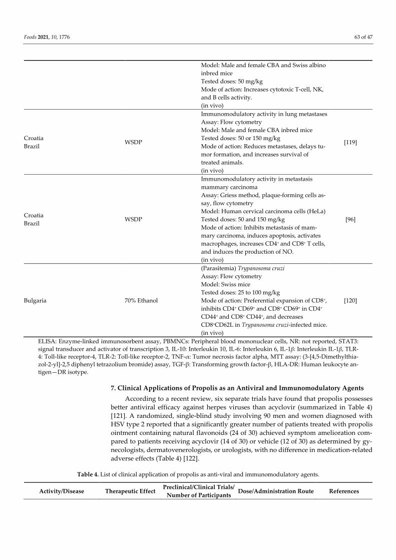
Foods 2021, 10, 1776 63 of 47
Model: Male and female CBA and Swiss albino
inbred mice
Tested doses: 50 mg/kg
Mode of action: Increases cytotoxic T-cell, NK,
and B cells activity.
(in vivo)
Croatia
Brazil WSDP
Immunomodulatory activity in lung metastases
Assay: Flow cytometry
Model: Male and female CBA inbred mice
Tested doses: 50 or 150 mg/kg
Mode of action: Reduces metastases, delays tu-
mor formation, and increases survival of
treated animals.
(in vivo)
[119]
Croatia
Brazil WSDP
Immunomodulatory activity in metastasis
mammary carcinoma
Assay: Griess method, plaque-forming cells as-
say, flow cytometry
Model: Human cervical carcinoma cells (HeLa)
Tested doses: 50 and 150 mg/kg
Mode of action: Inhibits metastasis of mam-
mary carcinoma, induces apoptosis, activates
macrophages, increases CD4+ and CD8+ T cells,
and induces the production of NO.
(in vivo)
[96]
Bulgaria 70% Ethanol
(Parasitemia) Trypanosoma cruzi
Assay: Flow cytometry
Model: Swiss mice
Tested doses: 25 to 100 mg/kg
Mode of action: Preferential expansion of CD8+,
inhibits CD4+ CD69+ and CD8+ CD69+ in CD4+
CD44+ and CD8+ CD44+, and decreases
CD8+CD62L in Trypanosoma cruzi-infected mice.
(in vivo)
[120]
ELISA: Enzyme-linked immunosorbent assay, PBMNCs: Peripheral blood mononuclear cells, NR: not reported, STAT3:
signal transducer and activator of transcription 3, IL-10: Interleukin 10, IL-6: Interleukin 6, IL-1β: Interleukin IL-1β, TLR-
4: Toll-like receptor-4, TLR-2: Toll-like receptor-2, TNF-α: Tumor necrosis factor alpha, MTT assay: (3-[4,5-Dimethylthia-
zol-2-yl]-2,5 diphenyl tetrazolium bromide) assay, TGF-β: Transforming growth factor-β, HLA-DR: Human leukocyte an-
tigen—DR isotype.
7. Clinical Applications of Propolis as an Antiviral and Immunomodulatory Agents
According to a recent review, six separate trials have found that propolis possesses
better antiviral efficacy against herpes viruses than acyclovir (summarized in Table 4)
[121]. A randomized, single-blind study involving 90 men and women diagnosed with
HSV type 2 reported that a significantly greater number of patients treated with propolis
ointment containing natural flavonoids (24 of 30) achieved symptom amelioration com-
pared to patients receiving acyclovir (14 of 30) or vehicle (12 of 30) as determined by gy-
necologists, dermatovenerologists, or urologists, with no difference in medication-related
adverse effects (Table 4) [122].
Table 4. List of clinical application of propolis as anti-viral and immunomodulatory agents.
Activity/Disease Therapeutic Effect Preclinical/Clinical Trials/
Number of Participants Dose/Administration Route References

Foods 2021, 10, 1776 64 of 47
Anti-viral/genital herpes
(HSV-2)
Heal genital herpetic
lesions and reduce lo-
cal symptoms
Randomized controlled tri-
als/90 p
Four times daily for 10
days/topical [122]
Anti-viral/HSV-1 Treat herpetic skin le-
sions NR Propolis 3%/topical [121]
Anti-viral/Herpes zoster Heal skin lesion and
reduce pain Clinical trial/60 p
Propolis lotion (3 times/day
topical) + Acyclovir (400 or
800 mg oral) for 28 days
[123,124]
Anti-viral/Herpes labialis
Reduce pain, short
healing time with
painless incrustation
Randomized, double-blind
trial/150 p
Propolis (0.1%, 0.5% and
1%)/topical [125]
Anti-viral/Herpes labialis
Reduce pain, burning,
itching, tension, and
swelling
Randomized, single-blind
trial/379 p
Propolis 0.5%/5 times per 5
days/topical [126]
Anti-viral/Herpes labialis Treat episodes of her-
pes labialis
Randomized, controlled
double-blind study/397 p
Propolis 0.5%/(0.2 g) 5 times
per 5 days/topical [127]
Anti-viral/dengue hemor-
rhagic fever virus
Reduce TNF-α levels
and improve platelet
counts
Randomized, double-blind,
placebo-controlled trial/63 p
Propolis 200 mg three times a
day for 7 days/oral [3]
Similarly, a propolis lotion produced significantly greater healing rates than a prop-
olis-free lotion (placebo control) following Herpes zoster virus infection (p < 0.001 for pain
reduction at all visits, reduced new vesicles on day 7 of treatment, and greater global effi-
cacy on the last (28th) day of treatment) with excellent skin tolerability and no allergic re-
actions, skin irritations, or other adverse events [123]. Holcová et al. utilized three differ-
ent concentrations (0.1%, 0.5%, and 1%) of propolis special extract GH 2002 in a lip balm
through a double-blind, randomized dermatological study involving 150 patients infected
with Herpes labialis, and all three concentrations of propolis proved to be effective against
Herpes labialis (p < 0.0005) for painless incrustation and local pains, but good tolerability
was observed with the 0.5% concentration [125]. Another study conducted by Arenberger
et al. showed the ability of propolis special extract (GH 2002) at 0.5% to treat episodes of
herpes labialis virus versus 5% Acyclovir cream (p < 0.0001), and no allergic reactions,
local irritations, or other adverse effects were observed (Table 4) [126].
Furthermore, propolis extract was evaluated in another double-blind, randomized,
placebo-controlled trial, which reported the ability of propolis (200 mg three times daily
for 7 days) to treat patients with dengue hemorrhagic fever virus, a faster recovery in
platelet counts (p = 0.006), a greater decline in circulating TNF level (p = 0.018), and a
shorter hospitalization period compared with placebo-treated patients (p = 0.012) (Table
4) [3].
Although propolis has plenty of biological and pharmacological properties, there is
a lack of clinical reports on the effectiveness of propolis. Furthermore, some research has
indicated that propolis is unsafe because it induces hypersensitivity and might induce
adverse reactions such as allergic cheilitis and oral ulceration (Table 4) [128].
8. In Silico Drug Discovery
In seeking potential natural products to combat COVID-19 disease, the molecular
docking technique was applied to portend the binding modes and affinities of 40 propolis
derivatives towards five viral targets and one human target, namely 3CLpro, RNA-depend-
ent RNA polymerase (RdRp), papain-like protease (PLpro), receptor-binding domain
(RBD) of the spike protein (S-protein), helicase (NSP13), and human angiotensin-convert-
ing enzyme 2 (ACE2). The technical details of the employed molecular docking calcula-
tions are described in References [129–136]. In brief, the crystal structures of SARS-CoV-2
3CLpro (PDB code: 6LU7) [137], RdRp (PDB code: 6M71) [138], PLpro (PDB code: 6W9C)
[139], RBD (PDB code: 6M0J) [140], and NSP13 (PDB code: 5RMM) [141] were opted for as
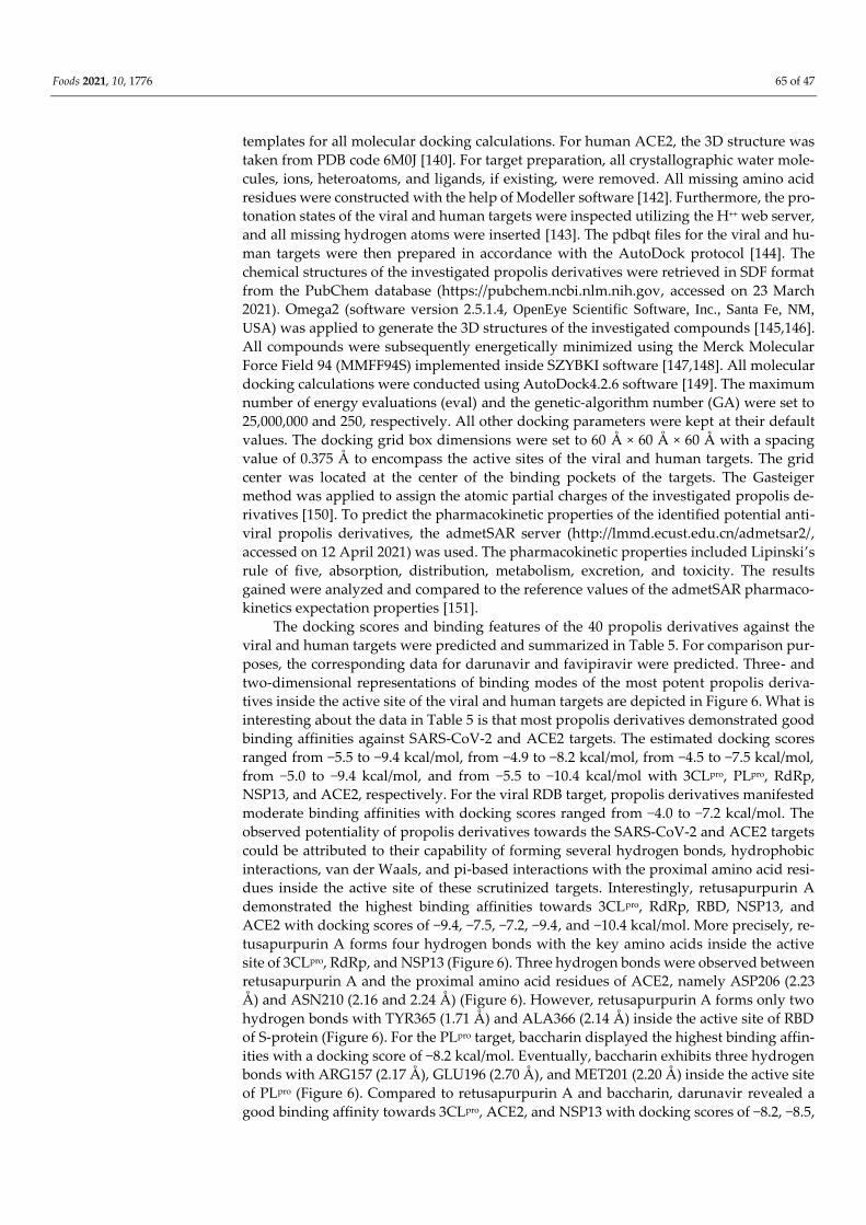
Foods 2021, 10, 1776 65 of 47
templates for all molecular docking calculations. For human ACE2, the 3D structure was
taken from PDB code 6M0J [140]. For target preparation, all crystallographic water mole-
cules, ions, heteroatoms, and ligands, if existing, were removed. All missing amino acid
residues were constructed with the help of Modeller software [142]. Furthermore, the pro-
tonation states of the viral and human targets were inspected utilizing the H++ web server,
and all missing hydrogen atoms were inserted [143]. The pdbqt files for the viral and hu-
man targets were then prepared in accordance with the AutoDock protocol [144]. The
chemical structures of the investigated propolis derivatives were retrieved in SDF format
from the PubChem database (https://pubchem.ncbi.nlm.nih.gov, accessed on 23 March
2021). Omega2 (software version 2.5.1.4, OpenEye Scientific Software, Inc., Santa Fe, NM,
USA) was applied to generate the 3D structures of the investigated compounds [145,146].
All compounds were subsequently energetically minimized using the Merck Molecular
Force Field 94 (MMFF94S) implemented inside SZYBKI software [147,148]. All molecular
docking calculations were conducted using AutoDock4.2.6 software [149]. The maximum
number of energy evaluations (eval) and the genetic-algorithm number (GA) were set to
25,000,000 and 250, respectively. All other docking parameters were kept at their default
values. The docking grid box dimensions were set to 60 Å × 60 Å × 60 Å with a spacing
value of 0.375 Å to encompass the active sites of the viral and human targets. The grid
center was located at the center of the binding pockets of the targets. The Gasteiger
method was applied to assign the atomic partial charges of the investigated propolis de-
rivatives [150]. To predict the pharmacokinetic properties of the identified potential anti-
viral propolis derivatives, the admetSAR server (http://lmmd.ecust.edu.cn/admetsar2/,
accessed on 12 April 2021) was used. The pharmacokinetic properties included Lipinski’s
rule of five, absorption, distribution, metabolism, excretion, and toxicity. The results
gained were analyzed and compared to the reference values of the admetSAR pharmaco-
kinetics expectation properties [151].
The docking scores and binding features of the 40 propolis derivatives against the
viral and human targets were predicted and summarized in Table 5. For comparison pur-
poses, the corresponding data for darunavir and favipiravir were predicted. Three- and
two-dimensional representations of binding modes of the most potent propolis deriva-
tives inside the active site of the viral and human targets are depicted in Figure 6. What is
interesting about the data in Table 5 is that most propolis derivatives demonstrated good
binding affinities against SARS-CoV-2 and ACE2 targets. The estimated docking scores
ranged from −5.5 to −9.4 kcal/mol, from −4.9 to −8.2 kcal/mol, from −4.5 to −7.5 kcal/mol,
from −5.0 to −9.4 kcal/mol, and from −5.5 to −10.4 kcal/mol with 3CLpro, PLpro, RdRp,
NSP13, and ACE2, respectively. For the viral RDB target, propolis derivatives manifested
moderate binding affinities with docking scores ranged from −4.0 to −7.2 kcal/mol. The
observed potentiality of propolis derivatives towards the SARS-CoV-2 and ACE2 targets
could be attributed to their capability of forming several hydrogen bonds, hydrophobic
interactions, van der Waals, and pi-based interactions with the proximal amino acid resi-
dues inside the active site of these scrutinized targets. Interestingly, retusapurpurin A
demonstrated the highest binding affinities towards 3CLpro, RdRp, RBD, NSP13, and
ACE2 with docking scores of −9.4, −7.5, −7.2, −9.4, and −10.4 kcal/mol. More precisely, re-
tusapurpurin A forms four hydrogen bonds with the key amino acids inside the active
site of 3CLpro, RdRp, and NSP13 (Figure 6). Three hydrogen bonds were observed between
retusapurpurin A and the proximal amino acid residues of ACE2, namely ASP206 (2.23
Å ) and ASN210 (2.16 and 2.24 Å ) (Figure 6). However, retusapurpurin A forms only two
hydrogen bonds with TYR365 (1.71 Å ) and ALA366 (2.14 Å ) inside the active site of RBD
of S-protein (Figure 6). For the PLpro target, baccharin displayed the highest binding affin-
ities with a docking score of −8.2 kcal/mol. Eventually, baccharin exhibits three hydrogen
bonds with ARG157 (2.17 Å ), GLU196 (2.70 Å ), and MET201 (2.20 Å ) inside the active site
of PLpro (Figure 6). Compared to retusapurpurin A and baccharin, darunavir revealed a
good binding affinity towards 3CLpro, ACE2, and NSP13 with docking scores of −8.2, −8.5,
Foods 2021, 10, 1776 66 of 47
and −7.1 kcal/mol, respectively. On the other hand, darunavir revealed a low binding af-
finity of −4.4, −3.8, and −3.3 kcal/mol against RdRp, PLpro, and RBD of S-protein, respec-
tively. In contrast, favipiravir manifested poor binding affinities with docking scores of
−4.3, −4.0, −4.8, −4.4, −4.2, and −4.3 kcal/mol against 3CLpro, PLpro, RdRp, ACE2, RBD, and
NSP13, respectively. The current data shed new light on the significance of retusapurpu-
rin A and baccharin as promising SARS-CoV-2 and ACE2 inhibitors.
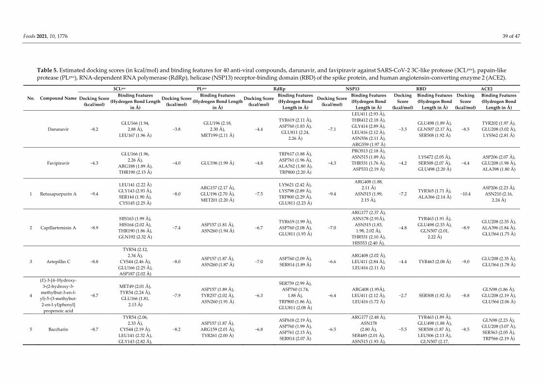
Foods 2021, 10, 1776 39 of 47
Table 5. Estimated docking scores (in kcal/mol) and binding features for 40 anti-viral compounds, darunavir, and favipiravir against SARS-CoV-2 3C-like protease (3CLpro), papain-like
protease (PLpro), RNA-dependent RNA polymerase (RdRp), helicase (NSP13) receptor-binding domain (RBD) of the spike protein, and human angiotensin-converting enzyme 2 (ACE2).
No. Compound Name
3CLpro PLpro RdRp NSP13 RBD ACE2
Docking Score
(kcal/mol)
Binding Features
(Hydrogen Bond Length
in Å)
Docking Score
(kcal/mol)
Binding Features
(Hydrogen Bond Length
in Å)
Docking Score
(kcal/mol)
Binding Features
(Hydrogen Bond
Length in Å)
Docking Score
(kcal/mol)
Binding Features
(Hydrogen Bond
Length in Å)
Docking
Score
(kcal/mol)
Binding Features
(Hydrogen Bond
Length in Å)
Docking
Score
(kcal/mol)
Binding Features
(Hydrogen Bond
Length in Å)
Darunavir −8.2
GLU166 (1.94,
2.88 Å ),
LEU167 (1.96 Å )
−3.8
GLU196 (2.18,
2.30 Å ),
MET199 (2.11 Å )
−4.4
TYR619 (2.11 Å ),
ASP760 (1.83 Å ),
GLU811 (2.24,
2.26 Å )
−7.1
LEU411 (2.93 Å ),
THR412 (2.18 Å ),
GLY414 (2.89 Å ),
LEU416 (2.12 Å ),
ASN556 (2.11 Å ),
ARG559 (1.97 Å )
−3.3
GLU498 (1.89 Å ),
GLN507 (2.17 Å ),
SER508 (1.92 Å )
−8.5
TYR202 (1.97 Å ),
GLU208 (3.02 Å ),
LYS562 (2.81 Å )
Favipiravir −4.3
GLU166 (1.96,
2.26 Å ),
ARG188 (1.89 Å ),
THR190 (2.15 Å )
−4.0 GLU196 (1.99 Å ) −4.8
TRP617 (1.88 Å ),
ASP761 (1.96 Å ),
ALA762 (1.80 Å ),
TRP800 (2.20 Å )
−4.3
PRO513 (2.18 Å ),
ASN515 (1.89 Å ),
THR531 (1.76 Å ),
ASP533 (2.19 Å )
−4.2
LYS472 (2.05 Å ),
SER508 (2.07 Å ),
GLU498 (2.20 Å )
−4.4
ASP206 (2.07 Å ),
GLU208 (1.98 Å ),
ALA398 (1.80 Å )
1 Retusapurpurin A −9.4
LEU141 (2.22 Å )
GLY143 (2.93 Å ),
SER144 (1.90 Å ),
CYS145 (2.25 Å )
−8.0
ARG157 (2.17 Å ),
GLU196 (2.70 Å ),
MET201 (2.20 Å )
−7.5
LYS621 (2.42 Å ),
LYS798 (2.89 Å ),
TRP800 (2.29 Å ),
GLU811 (2.23 Å )
−9.4
ARG408 (1.88,
2.11 Å )
ASN515 (1.99,
2.15 Å ),
−7.2 TYR365 (1.71 Å ),
ALA366 (2.14 Å ) −10.4
ASP206 (2.23 Å ),
ASN210 (2.16,
2.24 Å )
2 Capillartemisin A −8.9
HIS163 (1.99 Å ),
HIS164 (2.02 Å ),
THR190 (1.86 Å ),
GLN192 (2.32 Å )
−7.4 ASP157 (1.81 Å ),
ASN260 (1.94 Å ) −6.7
TYR619 (1.99 Å ),
ASP760 (2.08 Å ),
GLU811 (1.93 Å )
−7.0
ARG177 (2.37 Å ),
ASN178 (2.91Å ),
ASN515 (1.83,
1.98, 2.02 Å ),
THR531 (2.10 Å ),
HIS553 (2.40 Å ),
−4.8
TYR463 (1.91 Å ),
GLU498 (2.33 Å ),
GLN507 (2.01,
2.22 Å )
−8.9
GLU208 (2.35 Å ),
ALA396 (1.84 Å ),
GLU564 (1.75 Å )
3 Artepillin C −8.8
TYR54 (2.12,
2.34 Å ),
CYS44 (2.46 Å ),
GLU166 (2.25 Å ),
ASP187 (2.02 Å )
−8.0 ASP157 (1.87 Å ),
ASN260 (1.87 Å ) −7.0
ASP760 (2.09 Å ),
SER814 (1.89 Å ) −6.6
ARG408 (2.02 Å ),
LEU411 (2.84 Å ),
LEU416 (2.11 Å )
−4.4 TYR463 (2.08 Å ) −9.0 GLU208 (2.35 Å ),
GLU564 (1.78 Å )
4
(E)-3-[4−Hydroxy-
3-(2-hydroxy-3-
methylbut-3-en-l-
yl)-5-(3-methybut-
2-en-l-yl)phenyl]
propenoic acid
−8.7
MET49 (2.01 Å ),
TYR54 (2.24 Å ),
GLU166 (1.81,
2.13 Å )
−7.9
ASP157 (1.89 Å ),
TYR257 (2.02 Å ),
ASN260 (1.91 Å )
−6.3
SER759 (2.99 Å ),
ASP760 (1.74,
1.88 Å ),
TRP800 (1.86 Å ),
GLU811 (2.08 Å )
−6.4
ARG408 (1.95Å ),
LEU411 (2.12 Å ),
LEU416 (1.72 Å )
−2.7 SER508 (1.92 Å ) −8.8
GLN98 (1.86 Å ),
GLU208 (2.19 Å ),
GLU564 (2.06 Å )
5 Baccharin −8.7
TYR54 (2.06,
2.33 Å ),
CYS44 (2.19 Å ),
LEU141 (2.32 Å ),
GLY143 (2.82 Å ),
−8.2
ASP157 (1.87 Å ),
ARG159 (2.01 Å ),
TYR261 (2.00 Å )
−6.8
ASP618 (2.19 Å ),
ASP760 (1.99 Å ),
ASP761 (2.15 Å ),
SER814 (2.07 Å )
−6.5
ARG177 (2.48 Å ),
ASN178
(2.80 Å ),
SER485 (2.01 Å ),
ASN515 (1.93 Å ),
−5.5
TYR463 (1.89 Å ),
GLU498 (1.88 Å ),
SER508 (1.87 Å ),
LEU506 (2.13 Å ),
GLN507 (2.17,
−8.5
GLN98 (2.23 Å ),
GLU208 (3.07 Å ),
SER563 (2.05 Å ),
TRP566 (2.19 Å )

Foods 2021, 10, 1776 40 of 47
ASP187 (2.45 Å ),
GLN189 (1.90 Å )
THR531 (1.92 Å ) 2.60 Å )
6
(E)-2,3-Dihydro-
coniferyl p-couma-
rate
−8.6
LEU141 (1.96 Å ),
GLY143 (3.06 Å ),
SER144 (2.14 Å ),
CYS145 (2.96 Å ),
THR190 (2.05 Å )
−7.6
LYS150 (1.97 Å ),
LEU155 (2.01 Å ),
ASP259 (1.94 Å )
−5.7
ASP760 (2.14 Å ),
CYS813 (2.63 Å ),
SER814 (1.96 Å )
−6.8 ASN515 (2.52 Å ),
ASP533 (2.07Å ) −3.0
GLU498 (1.83 Å ),
GLN507 (2.38 Å ) −8.0
LEU95 (1.91 Å ),
GLN98 (1.97 Å ),
ASN210 (1.94 Å ),
TRP566 (2.23 Å )
7 Quercetin −8.5
HIS164 (2.23 Å ),
ASP187 (1.91 Å ),
THR190 (2.08,
2.08 Å ),
GLN192 (1.51 Å )
−6.8
LYS150 (2.36 Å ),
GLU160 (2.28 Å ),
ASN260 (1.79 Å ),
TYR266 (2.98 Å ),
THR294 (2.09 Å ),
ALA239 (3.01 Å )
−6.6
TYR619 (1.99,
2.03 Å ),
ASP761 (2.10 Å ),
GLU811 (2.33 Å )
−6.9 ASN178 (2.25 Å ),
LEU416 (1.94 Å ) −4.7
PHE361 (2.00,
2.08 Å ),
TYR365 (2.89 Å ),
TYR463 (2.16 Å )
−9.1
GLU208 (1.88 Å ),
ASN210 (2.06, 2.16,
2.85 Å ),
SER563 (2.00 Å ),
GLU564 (2.92 Å )
8 Sakuranetin −8.5 HIS164 (2.22 Å ),
THR190 (2.01 Å ) −6.9 ASP295 (1.65 Å ), −6.7
TYR619 (1.86 Å ),
ASP761 (2.43 Å ) −6.9
PRO405
(1.84 Å ),
ASN515 (1.96 Å )
−4.9
SER363 (1.93 Å ),
TYR365 (1.77 Å ),
ASN364 (1.99,
2.73 Å )
−8.8
ASP206 (1.99 Å ),
ASN210 (1.87,
2.00 Å )
9 Kaempferol −8.4
HIS164 (2.04 Å ),
ASP187 (1.99 Å ),
THR190 (1.81 Å )
−7.2
LEU255 (1.91 Å ),
ASN260 (1.96 Å ),
THR294 (1.83 Å )
−6.4
TRP617 (2.45 Å ),
TYR619 (1.84 Å ),
ASP761 (2.27 Å ),
TRP800 (2.12 Å )
−6.9
ASN178 (2.03 Å ),
LEU416 (1.89 Å ),
ASN556 (1.87 Å ),
ARG559 (2.70 Å )
−4.9
SER363 (2.08 Å ),
TYR365 (1.69 Å ),
ASN464 (1.92,
2.67 Å )
−8.9
ASP206 (1.86 Å ),
GLU208 (1.89 Å ),
ASN210 (2.02,
2.15, 2.83 Å )
SER563 (2.02 Å ),
GLU564 (2.99 Å )
10 Isorhamnetin −8.3
HIS164 (2.36 Å ),
ASP187 (1.98 Å ),
THR190 (2.07 Å )
−6.8
GU160 (1.99 Å ),
ASN260 (1.89 Å ),
TYR266 (2.16 Å ),
THR294 (1.84 Å )
−6.5
LYS621 (2.32 Å ),
CYS622 (2.19 Å ),
ASP760 (1.77 Å ),
ASP761 (1.90 Å ),
GLU811 (2.27 Å )
−6.6
ASN178 (2.15 Å ),
ARG408 (2.18 Å ),
PRO513 (1.83 Å ),
THR531 (1.76 Å ),
ASN515 (2.89 Å )
−4.9
SER363 (2.05 Å ),
TYR365 (1.75 Å ),
ASN464 (2.02,
2.63 Å )
−9.1
ASP206 (2.23 Å ),
GLU208 (1.83 Å ),
ASN210 (2.01,
2.09, 3.03 Å ),
LYS562 (2.25 Å ),
SER563 (1.95 Å ),
GLU564 (2.98 Å )
11 Sakuranetin-
4’-methylether −8.2
HIS164 (2.25 Å ),
GLN192 (2.21 Å ) −7.0
ARG159 (1.93 Å ),
THR294 (1.87 Å ) −6.5 ASP761 (2.40 Å ) −7.0
ARG408 (2.13 Å ),
ASN515 (2.12,
2.30 Å )
−4.9
TYR365 (2.21 Å ),
ASN464 (2.22,
2.26 Å )
−8.8 ASN210 (1.96,
1.98 Å )
12 Pinobanksin-
3-acetate −8.1
HIS164 (2.21 Å ),
GLU166 (1.96 Å ),
ASP187 (1.87 Å )
−6.8 TYR266 (2.92 Å ),
ASP295 (2.08 Å ) −6.0
LYS621 (2.45,
2.77 Å ),
CYS622 (1.89 Å ),
LYS798 (2.21 Å )
−6.7
ASN178 (2.22 Å ),
THR409 (2.00 Å ),
ARG559 (2.29 Å )
−5.4
PHE361 (1.71 Å ),
TYR365 (2.09 Å ),
ASN464 (2.14 Å )
−8.8
ASN210 (2.05,
2.16, 2.92 Å )
SER563 (2.21 Å )
13 Calycosin −8.1 ASP187 (2.0 Å ),
THR190 (2.15 Å ) −7.4
LYS150 (2.42 Å ),
GLU160 (2.29 Å ),
THR294 (1.92 Å )
−6.2
LYS619 (2.59 Å ),
LYS621 (2.22 Å ),
ASP623 (2.08 Å ),
LYS798 (2.33 Å )
-7.0
ASN176 (1.97,
2.90 Å ),
TYR197 (1.78 Å ),
ASP533 (1.84 Å )
-5.1 PHE361 (2.10 Å ),
TYR365 (1.65 Å ) −8.7
ASP206 (2.10 Å ),
ASN210 (1.73 Å ),
LYS562 (2.10 Å ),
SER563 (2.08 Å ),
GLU564 (3.04 Å )
14 Myricetin −8.0
HIS164 (2.23 Å ),
ASP187 (1.91 Å ),
THR190 (2.02,
2.09 Å ),
−7.0
ARG159 (3.01,
3.02 Å ),
ALA239 (2.84 Å ),
TYR257 (2.79 Å ),
−6.8
TYR619 (2.22 Å ),
LYS621 (2.83 Å ),
ASP760 (1.92 Å ),
ASP761 (1.95 Å ),
−7.0
ASN178 (2.09 Å ),
PRO405 (1.80 Å ),
LEU416 (1.85 Å ),
ASN556 (1.91 Å ),
−4.8
ARG360 (2.95 Å ),
PHE361 (1.98,
2.32 Å ),
TYR363 (2.17 Å ),
−8.6
ASP206 (1.88 Å ),
ASN210 (1.99,2.16
ALA396 (1.82,
1.84 Å ),
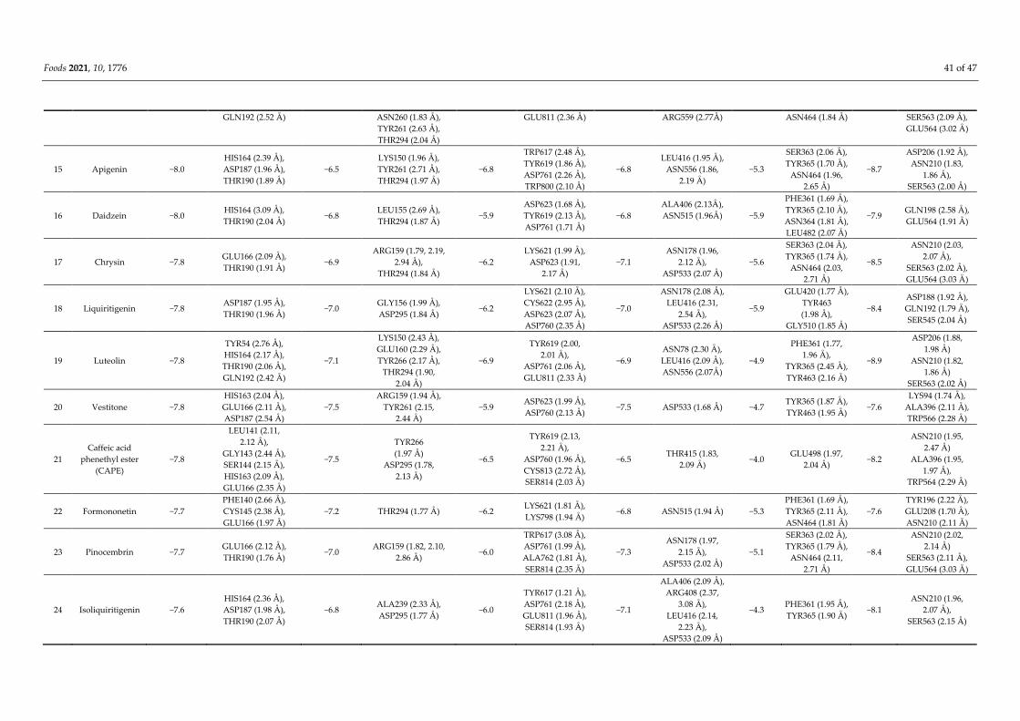
Foods 2021, 10, 1776 41 of 47
GLN192 (2.52 Å ) ASN260 (1.83 Å ),
TYR261 (2.63 Å ),
THR294 (2.04 Å )
GLU811 (2.36 Å ) ARG559 (2.77Å ) ASN464 (1.84 Å ) SER563 (2.09 Å ),
GLU564 (3.02 Å )
15 Apigenin −8.0
HIS164 (2.39 Å ),
ASP187 (1.96 Å ),
THR190 (1.89 Å )
−6.5
LYS150 (1.96 Å ),
TYR261 (2.71 Å ),
THR294 (1.97 Å )
−6.8
TRP617 (2.48 Å ),
TYR619 (1.86 Å ),
ASP761 (2.26 Å ),
TRP800 (2.10 Å )
−6.8
LEU416 (1.95 Å ),
ASN556 (1.86,
2.19 Å )
−5.3
SER363 (2.06 Å ),
TYR365 (1.70 Å ),
ASN464 (1.96,
2.65 Å )
−8.7
ASP206 (1.92 Å ),
ASN210 (1.83,
1.86 Å ),
SER563 (2.00 Å )
16 Daidzein −8.0 HIS164 (3.09 Å ),
THR190 (2.04 Å ) −6.8
LEU155 (2.69 Å ),
THR294 (1.87 Å ) −5.9
ASP623 (1.68 Å ),
TYR619 (2.13 Å ),
ASP761 (1.71 Å )
−6.8
ALA406 (2.13Å ),
ASN515 (1.96Å )
−5.9
PHE361 (1.69 Å ),
TYR365 (2.10 Å ),
ASN364 (1.81 Å ),
LEU482 (2.07 Å )
−7.9 GLN198 (2.58 Å ),
GLU564 (1.91 Å )
17 Chrysin −7.8 GLU166 (2.09 Å ),
THR190 (1.91 Å ) −6.9
ARG159 (1.79, 2.19,
2.94 Å ),
THR294 (1.84 Å )
−6.2
LYS621 (1.99 Å ),
ASP623 (1.91,
2.17 Å )
−7.1
ASN178 (1.96,
2.12 Å ),
ASP533 (2.07 Å )
−5.6
SER363 (2.04 Å ),
TYR365 (1.74 Å ),
ASN464 (2.03,
2.71 Å )
−8.5
ASN210 (2.03,
2.07 Å ),
SER563 (2.02 Å ),
GLU564 (3.03 Å )
18 Liquiritigenin −7.8 ASP187 (1.95 Å ),
THR190 (1.96 Å ) −7.0
GLY156 (1.99 Å ),
ASP295 (1.84 Å ) −6.2
LYS621 (2.10 Å ),
CYS622 (2.95 Å ),
ASP623 (2.07 Å ),
ASP760 (2.35 Å )
−7.0
ASN178 (2.08 Å ),
LEU416 (2.31,
2.54 Å ),
ASP533 (2.26 Å )
−5.9
GLU420 (1.77 Å ),
TYR463
(1.98 Å ),
GLY510 (1.85 Å )
−8.4
ASP188 (1.92 Å ),
GLN192 (1.79 Å ),
SER545 (2.04 Å )
19 Luteolin −7.8
TYR54 (2.76 Å ),
HIS164 (2.17 Å ),
THR190 (2.06 Å ),
GLN192 (2.42 Å )
−7.1
LYS150 (2.43 Å ),
GLU160 (2.29 Å ),
TYR266 (2.17 Å ),
THR294 (1.90,
2.04 Å )
−6.9
TYR619 (2.00,
2.01 Å ),
ASP761 (2.06 Å ),
GLU811 (2.33 Å )
−6.9
ASN78 (2.30 Å ),
LEU416 (2.09 Å ),
ASN556 (2.07Å )
−4.9
PHE361 (1.77,
1.96 Å ),
TYR365 (2.45 Å ),
TYR463 (2.16 Å )
−8.9
ASP206 (1.88,
1.98 Å )
ASN210 (1.82,
1.86 Å )
SER563 (2.02 Å )
20 Vestitone −7.8
HIS163 (2.04 Å ),
GLU166 (2.11 Å ),
ASP187 (2.54 Å )
−7.5
ARG159 (1.94 Å ),
TYR261 (2.15,
2.44 Å )
−5.9 ASP623 (1.99 Å ),
ASP760 (2.13 Å ) −7.5 ASP533 (1.68 Å ) −4.7
TYR365 (1.87 Å ),
TYR463 (1.95 Å ) −7.6
LYS94 (1.74 Å ),
ALA396 (2.11 Å ),
TRP566 (2.28 Å )
21
Caffeic acid
phenethyl ester
(CAPE)
−7.8
LEU141 (2.11,
2.12 Å ),
GLY143 (2.44 Å ),
SER144 (2.15 Å ),
HIS163 (2.09 Å ),
GLU166 (2.35 Å )
−7.5
TYR266
(1.97 Å )
ASP295 (1.78,
2.13 Å )
−6.5
TYR619 (2.13,
2.21 Å ),
ASP760 (1.96 Å ),
CYS813 (2.72 Å ),
SER814 (2.03 Å )
−6.5 THR415 (1.83,
2.09 Å ) −4.0
GLU498 (1.97,
2.04 Å ) −8.2
ASN210 (1.95,
2.47 Å )
ALA396 (1.95,
1.97 Å ),
TRP564 (2.29 Å )
22 Formononetin −7.7
PHE140 (2.66 Å ),
CYS145 (2.38 Å ),
GLU166 (1.97 Å )
−7.2 THR294 (1.77 Å ) −6.2 LYS621 (1.81 Å ),
LYS798 (1.94 Å ) −6.8 ASN515 (1.94 Å ) −5.3
PHE361 (1.69 Å ),
TYR365 (2.11 Å ),
ASN464 (1.81 Å )
−7.6
TYR196 (2.22 Å ),
GLU208 (1.70 Å ),
ASN210 (2.11 Å )
23 Pinocembrin −7.7 GLU166 (2.12 Å ),
THR190 (1.76 Å ) −7.0
ARG159 (1.82, 2.10,
2.86 Å ) −6.0
TRP617 (3.08 Å ),
ASP761 (1.99 Å ),
ALA762 (1.81 Å ),
SER814 (2.35 Å )
−7.3
ASN178 (1.97,
2.15 Å ),
ASP533 (2.02 Å )
−5.1
SER363 (2.02 Å ),
TYR365 (1.79 Å ),
ASN464 (2.11,
2.71 Å )
−8.4
ASN210 (2.02,
2.14 Å )
SER563 (2.11 Å ),
GLU564 (3.03 Å )
24 Isoliquiritigenin −7.6
HIS164 (2.36 Å ),
ASP187 (1.98 Å ),
THR190 (2.07 Å )
−6.8 ALA239 (2.33 Å ),
ASP295 (1.77 Å ) −6.0
TYR617 (1.21 Å ),
ASP761 (2.18 Å ),
GLU811 (1.96 Å ),
SER814 (1.93 Å )
−7.1
ALA406 (2.09 Å ),
ARG408 (2.37,
3.08 Å ),
LEU416 (2.14,
2.23 Å ),
ASP533 (2.09 Å )
−4.3 PHE361 (1.95 Å ),
TYR365 (1.90 Å ) −8.1
ASN210 (1.96,
2.07 Å ),
SER563 (2.15 Å )

Foods 2021, 10, 1776 42 of 47
25 Violanone −7.6 CYS44 (1.91 Å ) −7.3 LEU155 (2.04 Å ),
THR294 (2.02 Å ) −6.1
ASP164 (1.89 Å ),
LYS621 (2.31,
2.13 Å ),
TYR619 (2.30 Å ),
LYS798 (2.07 Å )
−7.3 ARG408 (2.12 Å ),
ASN515 (2.09 Å ) −4.9
SER363 (1.88, 1.96,
2.32 Å ),
ALA366 (2.13 Å ),
TYR463 (1.96 Å )
−8.1
LYS94 (2.08 Å ),
GLN98 (2.50 Å )
ASN210 (2.20,
2.49 Å )
ALA396 (1.90 Å ),
TRP566 (2.24 Å )
26 Drupanin −7.5
HIS164 (1.96 Å ),
THR190 (1.83 Å ),
GLN192 (2.06 Å )
−7.0 ASP157 (1.87 Å ),
ASN260 (1.94 Å ) −6.3 ASP760 (1.85 Å ) −6.1
ASN178 (1.90 Å ),
TYR179 (1.72 Å ),
SER485 (2.08 Å ),
ASN515 (1.91 Å )
−4.0 GLU498 (1.68 Å ),
SER508 (2.01 Å ) −7.8
ASN210 (2.10 Å ),
GLU564 (1.70 Å )
27 Galangin −7.5 HIS164 (2.21 Å ),
ASP187 (1.92 Å ) −6.8
ARG159 (1.85 Å ),
THR294 (1.73 Å ) −5.8
TRP617 (2.40 Å ),
ASP761 (2.33 Å ),
TRP800 (2.15 Å ),
SER814 (3.04 Å )
−6.6
LEU416 (1.91 Å ),
ASN556 (1.85 Å ),
ARG559 (2.71 Å )
−5.3
SER363 (2.06 Å ),
TYR365 (1.74 Å ),
ASN464 (2.01 Å )
−8.7
GLU208 (1.88 Å ),
ASN210 (2.04, 2.16,
2.86 Å ),
SER563 (1.99 Å ),
GLU564 (2.98 Å )
28 Pinobaskin −7.5 HIS164 (2.23 Å ),
ASP187 (1.90 Å ) −6.7
ARG159 (1.83 Å ),
THR294 (1.73 Å ) −5.6
TRP617 (2.39 Å ),
ASP761 (2.30 Å ),
TRP800 (2.17 Å ),
SER814 (3.04 Å )
−6.3
LEU416 (1.91 Å ),
ASN556 (1.86 Å ),
ARG559 (2.70 Å )
−5.0
SER363 (2.04 Å ),
TYR365 (1.76 Å ),
ASN464 (2.01 Å )
−8.6
GLU208 (1.86 Å ),
ASN210 (2.04, 2.18,
2.85 Å ),
SER563 (1.99 Å ),
GLU564 (2.98 Å )
29 Pinostrobin −7.4 SER144 (2.92 Å ),
HIS163 (1.98 Å ) −6.9 ARG159 (1.80 Å ) −6.1 ASP761 (2.23 Å ) −6.7
LEU416 (2.05,
2.30 Å ),
ARG559 (1.93 Å )
−5.1
TYR365 (2.20 Å ),
ASN464 (2.23,
2.27 Å )
−8.6 ASN210 (1.80,
1.98 Å )
30 Biochanin A −7.4 THR190 (1.78 Å ),
GLN192 (3.06 Å ) −7.5
ARG159 (1.93 Å ),
TYR261 (2.13 Å )
ALA239 (2.92 Å )
−6.3
TYR619 (3.05 Å ),
LYS621 (2.06 Å ),
ASP760 (1.82 Å ),
SER795 (2.79 Å )
−7.0 ASN178 (2.14 Å ),
ASN515 (1.96 Å ) −5.1
PHE361 (1.75 Å ),
TYR365 (2.11 Å ),
ASN464 (1.78 Å )
−7.9
TYR196 (2.22 Å ),
GLU208 (1.71,
2.01 Å ),
ASN210 (2.18 Å )
31 Pratensein −7.4
LEU141 (2.17 Å ),
GLY143 (2.16 Å ),
ASP187 (2.85 Å )
−7.6 LEU155 (2.09 Å ),
THR294 (1.90 Å ) −6.9
LYS621 (2.13 Å ),
SER795 (2,11,
2.69 Å )
−7.2 ASN178 (2.16 Å ),
ASN515 (1.96 Å ) −4.9
GLU498 (1.83 Å ),
GLN507 (2.38 Å ) −7.8
GLN98 (2.72 Å ),
ASN210 (2.17 Å ),
GLU564 (1.85 Å )
32
2,2-Dimethyl
chromene-6-prope-
noic acid
−7.3 THR190 (1.76 Å ) −7.3 ASP157 (2.07 Å ) −6.4 TRP617 (2.79 Å ),
ASP761 (1.88 Å ) −6.4
ASN178 (2.24 Å ),
THR531 (2.40 Å ),
ASN515 (2.03,
2.22 Å ),
ARG559 (2.89 Å )
−5.6 TYR467 (1.84 Å ) −7.2 GLU564 (1.95 Å )
33 Neovestitol −7.2 GLU166 (2.03 Å ) −7.1 ARG159 (2.01 Å ),
TYR261 (2.11 Å ) −5.9 LYS798 (2.13 Å ) −6.8
ALA416 (2.16 Å ),
ARG408 (2.27 Å ) −5.5 SER508 (2.09 Å ) −7.4
ASN210 (1.80,
2.04 Å )
34 Rutin −7.2
ASP187 (1.90 Å ),
HIS164 (2.25 Å ),
GLU166 (2.05 Å ),
GLN192 (2.86 Å ),
THR190 (2.19,
1.92 Å )
−5.2
GLU160 (1.95,
1.92 Å ),
SER163 (2.11 Å )
GLU196 (2.12 Å ),
ARG176 (2.84 Å ),
MET201 (2.13 Å )
−4.5 ASP760 (2.23 Å ) −6.7
THR415 (1.84 Å ),
THR551 (2.51,
3.01 Å )
−3.3
SER363 (2.07 Å ),
TYR365 (1.80 Å ),
TYR463 (2.27 Å ),
ASN464 (1.98,
2.63, 3.05 Å )
−9.3
TYR196 (2.60 Å ),
ASP206 (2.01,
2.02 Å ),
GLU208 (2.20,
2.24 Å ),
ASN210 (2.33,
2.49 Å ),
TRP566 (2.32 Å )
35 Medicarpin −6.5 GLY143 (2.18 Å ), −7.0 TYR266 (1.76 Å ), −6.5 ASP761 (2.23 Å ), −7.3 ARG408 (2.10 Å ), −5.6 ARG417 (2.14 Å ), −7.7 LYS94 (2.55 Å )
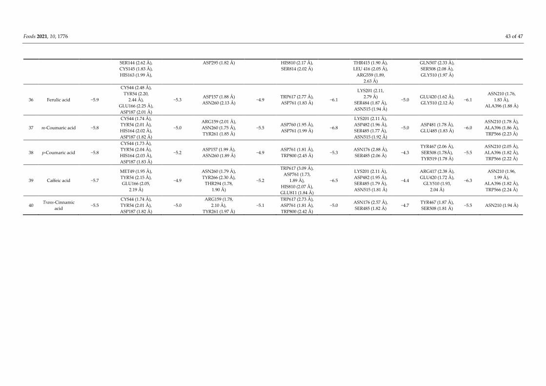
Foods 2021, 10, 1776 43 of 47
SER144 (2.62 Å ),
CYS145 (1.83 Å ),
HIS163 (1.99 Å ),
ASP295 (1.82 Å ) HIS810 (2.17 Å ),
SER814 (2.02 Å )
THR415 (1.90 Å ),
LEU 416 (2.05 Å ),
ARG559 (1.89,
2.63 Å )
GLN507 (2.33 Å ),
SER508 (2.08 Å ),
GLY510 (1.97 Å )
36 Ferulic acid −5.9
CYS44 (2.48 Å ),
TYR54 (2.20,
2.44 Å ),
GLU166 (2.25 Å ),
ASP187 (2.01 Å )
−5.3 ASP157 (1.88 Å )
ASN260 (2.13 Å ) −4.9
TRP617 (2.77 Å ),
ASP761 (1.83 Å ) −6.1
LYS201 (2.11,
2.79 Å )
SER484 (1.87 Å ),
ASN515 (1.94 Å )
−5.0 GLU420 (1.62 Å ),
GLY510 (2.12 Å ) −6.1
ASN210 (1.76,
1.83 Å ),
ALA396 (1.88 Å )
37 m-Coumaric acid −5.8
CYS44 (1.74 Å ),
TYR54 (2.01 Å ),
HIS164 (2.02 Å ),
ASP187 (1.82 Å )
−5.0
ARG159 (2.01 Å ),
ASN260 (1.75 Å ),
TYR261 (1.85 Å )
−5.5 ASP760 (1.95 Å ),
ASP761 (1.99 Å ) −6.8
LYS201 (2.11 Å ),
ASP482 (1.96 Å ),
SER485 (1.77 Å ),
ASN515 (1.92 Å )
−5.0 ASP481 (1.78 Å ),
GLU485 (1.83 Å ) −6.0
ASN210 (1.78 Å ),
ALA396 (1.86 Å ),
TRP566 (2.23 Å )
38 p-Coumaric acid −5.8
CYS44 (1.73 Å ),
TYR54 (2.04 Å ),
HIS164 (2.03 Å ),
ASP187 (1.83 Å )
−5.2 ASP157 (1.99 Å ),
ASN260 (1.89 Å ) −4.9
ASP761 (1.81 Å ),
TRP800 (2.45 Å ) −5.3
ASN176 (2.88 Å ),
SER485 (2.06 Å ) −4.3
TYR467 (2.06 Å ),
SER508 (1.78Å ),
TYR519 (1.78 Å )
−5.5
ASN210 (2.05 Å ),
ALA396 (1.82 Å ),
TRP566 (2.22 Å )
39 Caffeic acid −5.7
MET49 (1.95 Å ),
TYR54 (2.15 Å ),
GLU166 (2.05,
2.19 Å )
−4.9
ASN260 (1.79 Å ),
TYR266 (2.30 Å ),
THR294 (1.78,
1.90 Å )
−5.2
TRP617 (3.09 Å ),
ASP761 (1.73,
1.89 Å ),
HIS810 (2.07 Å ),
GLU811 (1.84 Å )
−6.5
LYS201 (2.11 Å ),
ASP482 (1.95 Å ),
SER485 (1.79 Å ),
ASN515 (1.81 Å )
−4.4
ARG417 (2.38 Å ),
GLU420 (1.72 Å ),
GLY510 (1.93,
2.04 Å )
−6.3
ASN210 (1.96,
1.99 Å ),
ALA396 (1.82 Å ),
TRP566 (2.24 Å )
40 Trans-Cinnamic
acid −5.5
CYS44 (1.74 Å ),
TYR54 (2.01 Å ),
ASP187 (1.82 Å )
−5.0
ARG159 (1.78,
2.10 Å ),
TYR261 (1.97 Å )
−5.1
TRP617 (2.73 Å ),
ASP761 (1.81 Å ),
TRP800 (2.42 Å )
−5.0 ASN176 (2.57 Å ),
SER485 (1.82 Å ) −4.7
TYR467 (1.87 Å ),
SER508 (1.81 Å ) −5.5 ASN210 (1.94 Å )
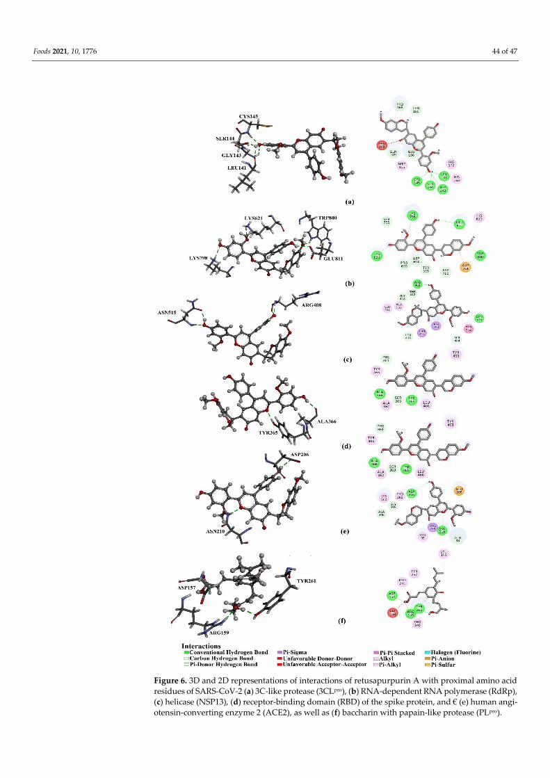
Foods 2021, 10, 1776 44 of 47
Figure 6. 3D and 2D representations of interactions of retusapurpurin A with proximal amino acid
residues of SARS-CoV-2 (a) 3C-like protease (3CLpro), (b) RNA-dependent RNA polymerase (RdRp),
(c) helicase (NSP13), (d) receptor-binding domain (RBD) of the spike protein, and € (e) human angi-
otensin-converting enzyme 2 (ACE2), as well as (f) baccharin with papain-like protease (PLpro).
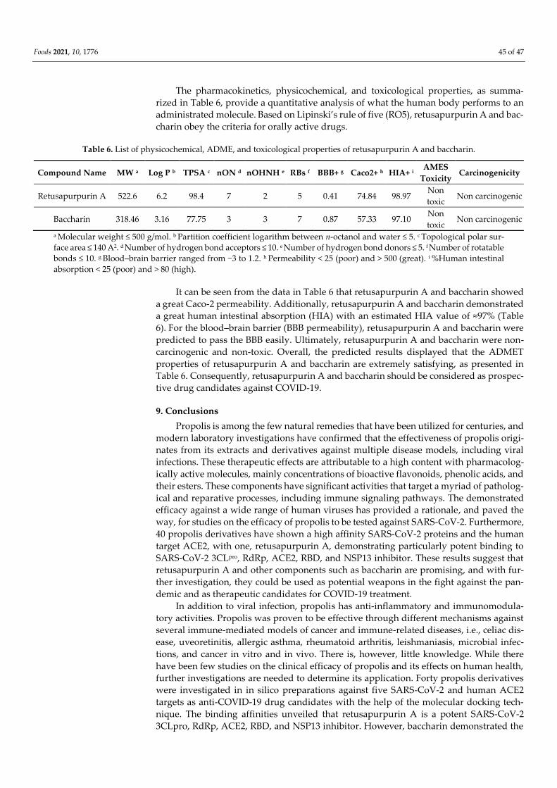
Foods 2021, 10, 1776 45 of 47
The pharmacokinetics, physicochemical, and toxicological properties, as summa-
rized in Table 6, provide a quantitative analysis of what the human body performs to an
administrated molecule. Based on Lipinski’s rule of five (RO5), retusapurpurin A and bac-
charin obey the criteria for orally active drugs.
Table 6. List of physicochemical, ADME, and toxicological properties of retusapurpurin A and baccharin.
Compound Name MW a Log P b TPSA c nON d nOHNH e RBs f BBB+ g Caco2+ h HIA+ i AMES
Toxicity Carcinogenicity
Retusapurpurin A 522.6 6.2 98.4 7 2 5 0.41 74.84 98.97 Non
toxic Non carcinogenic
Baccharin 318.46 3.16 77.75 3 3 7 0.87 57.33 97.10 Non
toxic Non carcinogenic
a Molecular weight ≤ 500 g/mol. b Partition coefficient logarithm between n-octanol and water ≤ 5. c Topological polar sur-
face area ≤ 140 A2. d Number of hydrogen bond acceptors ≤ 10. e Number of hydrogen bond donors ≤ 5. f Number of rotatable
bonds ≤ 10. g Blood–brain barrier ranged from −3 to 1.2. h Permeability < 25 (poor) and > 500 (great). i %Human intestinal
absorption < 25 (poor) and > 80 (high).
It can be seen from the data in Table 6 that retusapurpurin A and baccharin showed
a great Caco-2 permeability. Additionally, retusapurpurin A and baccharin demonstrated
a great human intestinal absorption (HIA) with an estimated HIA value of ≈97% (Table
6). For the blood–brain barrier (BBB permeability), retusapurpurin A and baccharin were
predicted to pass the BBB easily. Ultimately, retusapurpurin A and baccharin were non-
carcinogenic and non-toxic. Overall, the predicted results displayed that the ADMET
properties of retusapurpurin A and baccharin are extremely satisfying, as presented in
Table 6. Consequently, retusapurpurin A and baccharin should be considered as prospec-
tive drug candidates against COVID-19.
9. Conclusions
Propolis is among the few natural remedies that have been utilized for centuries, and
modern laboratory investigations have confirmed that the effectiveness of propolis origi-
nates from its extracts and derivatives against multiple disease models, including viral
infections. These therapeutic effects are attributable to a high content with pharmacolog-
ically active molecules, mainly concentrations of bioactive flavonoids, phenolic acids, and
their esters. These components have significant activities that target a myriad of patholog-
ical and reparative processes, including immune signaling pathways. The demonstrated
efficacy against a wide range of human viruses has provided a rationale, and paved the
way, for studies on the efficacy of propolis to be tested against SARS-CoV-2. Furthermore,
40 propolis derivatives have shown a high affinity SARS-CoV-2 proteins and the human
target ACE2, with one, retusapurpurin A, demonstrating particularly potent binding to
SARS-CoV-2 3CLpro, RdRp, ACE2, RBD, and NSP13 inhibitor. These results suggest that
retusapurpurin A and other components such as baccharin are promising, and with fur-
ther investigation, they could be used as potential weapons in the fight against the pan-
demic and as therapeutic candidates for COVID-19 treatment.
In addition to viral infection, propolis has anti-inflammatory and immunomodula-
tory activities. Propolis was proven to be effective through different mechanisms against
several immune-mediated models of cancer and immune-related diseases, i.e., celiac dis-
ease, uveoretinitis, allergic asthma, rheumatoid arthritis, leishmaniasis, microbial infec-
tions, and cancer in vitro and in vivo. There is, however, little knowledge. While there
have been few studies on the clinical efficacy of propolis and its effects on human health,
further investigations are needed to determine its application. Forty propolis derivatives
were investigated in in silico preparations against five SARS-CoV-2 and human ACE2
targets as anti-COVID-19 drug candidates with the help of the molecular docking tech-
nique. The binding affinities unveiled that retusapurpurin A is a potent SARS-CoV-2
3CLpro, RdRp, ACE2, RBD, and NSP13 inhibitor. However, baccharin demonstrated the
Foods 2021, 10, 1776 46 of 47
highest binding affinity against SARS-CoV-2 PLpro. Moreover, drug-likeness and AD-
MET properties were predicted for the most potent compounds and demonstrated satis-
factory pharmacokinetics, physicochemical parameters, and toxicological properties.
These results suggest that retusapurpurin A and baccharin can be further investigated as
convenient therapeutic treatments for COVID-19 disease, and clinical trials are feasible
due to the generally good safety profiles of propolis derivatives.
Author Contributions: Writing—original draft preparation N.Y., A.A.A.E.-W., R.G., O.M.K., and
A.S.; Molecular docking, M.A.A.I. and M.F.M. Writing—review and editing, Z.G., X.Z., A.F.M.A.,
S.H.D.M., M.F.A., H.S.A., S.A.M.K. and H.R.E.-S.; Project administration, H.R.E.-S.; Supervision,
S.A.M.K. and H.R.E.-S. All authors have read and agreed to the published version of the manuscript.
Funding: This work was supported by the Swedish Research Council Vetenskapsrådet (VR grant
2016–05885).
Institutional Review Board Statement: Not applicable.
Informed Consent Statement: Not applicable.
Data Availability Statement: Not applicable.
Acknowledgments: Authors are very grateful to the Swedish Research links Grant VR 2016–05885
and the Department of Molecular Biosciences, Wenner-Grens Institute, Stockholm University, Swe-
den, for the financial support.
Conflicts of Interest: The authors declare that they have no conflict of interest.
References
1. Pavlovic, R.; Borgonovo, G.; Leoni, V.; Giupponi, L.; Ceciliani, G.; Sala, S.; Bassoli, A.; Giorgi, A. Effectiveness of different
analytical methods for the characterization of propolis: A case of study in Northern Italy. Molecules 2020, 25, 504–529.
2. Costa Jr, A.G.; Yoshida, N.C.; Garcez, W.S.; Perdomo, R.T.; Matos, M. de F.C.; Garcez, F.R. Metabolomics approach expands the
classification of propolis samples from midwest Brazil. J. Nat. Prod. 2020, 83, 333–343.
3. Soroy, L.; Bagus, S.; Yongkie, I.P.; Djoko, W. The effect of a unique propolis compound (PropoelixTM) on clinical outcomes in
patients with dengue hemorrhagic fever. Infect. Drug Resist. 2014, 7, 323–329.
4. Jasprica, I.; Mornar, A.; Debeljak, Ž.; Smolčić-Bubalo, A.; Medić-Šarić, M.; Mayer, L.; Romić, Ž.; Bućan, K.; Balog, T.; Sobočanec,
S. In vivo study of propolis supplementation effects on antioxidative status and red blood cells. J. Ethnopharmacol. 2007, 110,
548–554.
5. Sforcin, J.M.; Bankova, V. Propolis: Is there a potential for the development of new drugs? J. Ethnopharmacol. 2011, 133, 253–260.
6. Paulino, N.; Dantas, A.P.; Bankova, V.; Longhi, D.T.; Scremin, A.; de Castro, S.L.; Calixto, J.B. Bulgarian propolis induces
analgesic and anti-inflammatory effects in mice and inhibits in vitro contraction of airway smooth muscle. J. Pharmacol. Sci. 2003,
93, 307–313.
7. Yuan, J.; Liu, J.; Hu, Y.; Fan, Y.; Wang, D.; Guo, L.; Nguyen, T.L.; Zhao, X.; Liu, X.; Liu, C. The immunological activity of propolis
flavonoids liposome on the immune response against ND vaccine. Int. J. Biol. Macromol. 2012, 51, 400–405.
8. Burdock, G.A. Review of the biological properties and toxicity of bee propolis (propolis). Food Chem. Toxicol. 1998, 36, 347–363,
doi:https://doi.org/10.1016/S0278-6915(97)00145-2.
9. Skaba, D.; Morawiec, T.; Tanasiewicz, M.; Mertas, A.; Bobela, E.; Szliszka, E.; Skucha-Nowak, M.; Dawiec, M.; Yamamoto, R.;
Ishiai, S.; et al. Influence of the toothpaste with Brazilian ethanol extract propolis on the oral cavity health. Evidence-Based
Complement. Altern. Med. 2013, 2013, 215391, doi:10.1155/2013/215391.
10. Sforcin, J.M. Biological properties and therapeutic applications of propolis. Phyther. Res. 2016, 30, 894–905.
11. Pasupuleti, V.R.; Sammugam, L.; Ramesh, N.; Gan, S.H. Honey, propolis, and royal jelly: A comprehensive review of their
biological actions and health benefits. Oxid. Med. Cell. Longev. 2017, 2017, doi:org/10.1155/2017/1259510.
12. Amoros, M.; Sauvager, F.; Girre, L.; Cormier, M. In vitro antiviral activity of propolis. Apidologie 1992, 23, 231–240.
13. Schnitzler, P.; Neuner, A.; Nolkemper, S.; Zundel, C.; Nowack, H.; Sensch, K.H.; Reichling, J. Antiviral activity and mode of
action of propolis extracts and selected compounds. Phyther. Res. 2010, 24, S20–S28.
14. Kujumgiev, A.; Tsvetkova, I.; Serkedjieva, Y.; Bankova, V.; Christov, R.; Popov, S. Antibacterial, antifungal and antiviral activity
of propolis of different geographic origin. J. Ethnopharmacol. 1999, 64, 235–240, doi:https://doi.org/10.1016/S0378-8741(98)00131-
7.
15. Kwon, M.J.; Shin, H.M.; Perumalsamy, H.; Wang, X.; Ahn, Y.-J. Antiviral effects and possible mechanisms of action of
constituents from Brazilian propolis and related compounds. J. Apic. Res. 2020, 59, 413–425.
16. Serkedjieva, J.; Manolova, N.; Bankova, V. Anti-influenza virus effect of some propolis constituents and their analogues (esters
of substituted cinnamic acids). J. Nat. Prod. 1992, 55, 294–297, doi:10.1021/np50081a003.
17. Debiaggi, M.; Tateo, F.; Pagani, L.; Luini, M.; Romero, E. Effects of propolis flavonoids on virus infectivity and replication.
Foods 2021, 10, 1776 47 of 47
Microbiologica 1990, 13, 207–213.
18. Refaat, H.; Mady, F.M.; Sarhan, H.A.; Rateb, H.S.; Alaaeldin, E. Optimization and evaluation of propolis liposomes as a
promising therapeutic approach for COVID-19. Int. J. Pharm. 2020, 120028, doi:https://doi.org/10.1016/j.ijpharm.2020.120028.
19. Sahlan, M.; Irdiani, R.; Flamandita, D.; Aditama, R.; Alfarraj, S.; Javed Ansari, M.; Cahya Khayrani, A.; Kartika Pratami, D.;
Lischer, K. Molecular interaction analysis of Sulawesi propolis compounds with SARS-CoV-2 main protease as preliminary
study for COVID-19 drug discovery. J. King Saud Univ. - Sci. 2020, 101234, doi:https://doi.org/10.1016/j.jksus.2020.101234.
20. Hashem, H. In silico approach of some selected honey constituents as SARS-CoV-2 main protease (COVID-19) inhibitors.
ChemRxiv. 2020, doi:10.26434/chemrxiv.12115359.v2.
21. Zabaiou, N.; Fouache, A.; Trousson, A.; Baron, S.; Zellagui, A.; Lahouel, M.; Lobaccaro, J.-M.A. Biological properties of propolis
extracts: Something new from an ancient product. Chem. Phys. Lipids 2017, 207, 214–222.
22. Zaccaria, V.; Garzarella, E.U.; Di Giovanni, C.; Galeotti, F.; Gisone, L.; Campoccia, D.; Volpi, N.; Arciola, C.R.; Daglia, M. Multi
dynamic extraction: An innovative method to obtain a standardized chemically and biologically reproducible polyphenol
extract from poplar-type propolis to be used for its anti-infective properties. Materials (Basel). 2019, 12, 3746–3761.
23. Popova, M.; Trusheva, B.; Bankova, V. Propolis of stingless bees: A phytochemist’s guide through the jungle of tropical
biodiversity. Phytomedicine 2019, 86, 153098, doi:https://doi.org/10.1016/j.phymed.2019.153098.
24. Tolba, M.F.; Azab, S.S.; Khalifa, A.E.; Abdel-Rahman, S.Z.; Abdel-Naim, A.B. Caffeic acid phenethyl ester, a promising
component of propolis with a plethora of biological activities: A review on its anti-inflammatory, neuroprotective,
hepatoprotective, and cardioprotective effects. IUBMB Life 2013, 65, 699–709.
25. Banskota, A.H.; Tezuka, Y.; Kadota, S. Recent progress in pharmacological research of propolis. Phyther. Res. 2001, 15, 561–571.
26. Picolotto, A.; Pergher, D.; Pereira, G.P.; Machado, K.G.; da Silva Barud, H.; Roesch-Ely, M.; Gonzalez, M.H.; Tasso, L.;
Figueiredo, J.G.; Moura, S. Bacterial cellulose membrane associated with red propolis as phytomodulator: Improved healing
effects in experimental models of diabetes mellitus. Biomed. Pharmacother. 2019, 112, 108640.
27. Siheri, W.; Alenezi, S.; Tusiimire, J.; Watson, D.G. The chemical and biological properties of propolis. In In bee products - chemical
and biological properties; Alvarez-Suarez, J.M., Ed.; Springer International Publishing: Cham, 2017; pp. 137–178 ISBN 978-3-319-
59689-1.
28. Granados-Pineda, J.; Uribe-Uribe, N.; García-López, P.; Ramos-Godinez, M.D.P.; Rivero-Cruz, J.F.; Pérez-Rojas, J.M. Effect of
pinocembrin isolated from Mexican brown propolis on diabetic nephropathy. Molecules 2018, 23, 852–870.
29. Costa, P.; Almeida, M.O.; Lemos, M.; Arruda, C.; Casoti, R.; Somensi, L.B.; Boeing, T.; Mariott, M.; Stein, B.D.P.; de Souza, P.
Artepillin C, drupanin, aromadendrin-4′-O-methyl-ether and kaempferide from Brazilian green propolis promote
gastroprotective action by diversified mode of action. J. Ethnopharmacol. 2018, 226, 82–89.
30. Rojczyk, E.; Klama-Baryła, A.; Łabuś, W.; Wilemska-Kucharzewska, K.; Kucharzewski, M. Historical and modern research on
propolis and its application in wound healing and other fields of medicine and contributions by Polish studies. J. Ethnopharmacol.
2020, 262, 113159, doi:https://doi.org/10.1016/j.jep.2020.113159.
31. Governa, P.; Cusi, M.G.; Borgonetti, V.; Sforcin, J.M.; Terrosi, C.; Baini, G.; Miraldi, E.; Biagi, M. Beyond the biological effect of
a chemically characterized poplar propolis: Antibacterial and antiviral activity and comparison with flurbiprofen in cytokines
release by lps-stimulated human mononuclear cells. Biomedicines 2019, 7, 73–82.
32. Costa, P.; Somensi, L.B.; Mariano, L.N.B.; Boeing, T.; Longo, B.; Perfoll, E.; de Souza, P.; Gushiken, L.F.S.; Pellizzon, C.H.;
Rodrigues, D.M. Role of the antioxidant properties in the gastroprotective and gastric healing activity promoted by Brazilian
green propolis and the healing efficacy of Artepillin C. Inflammopharmacology 2019, 28, 109–1025.
33. Tolba, M.F.; Omar, H.A.; Azab, S.S.; Khalifa, A.E.; Abdel-Naim, A.B.; Abdel-Rahman, S.Z. Caffeic acid phenethyl ester: A review
of its antioxidant activity, protective effects against ischemia-reperfusion injury and drug adverse reactions. Crit. Rev. Food Sci.
Nutr. 2016, 56, 2183–2190.
34. Kumar, V.; Dhanjal, J.K.; Bhargava, P.; Kaul, A.; Wang, J.; Zhang, H.; Kaul, S.C.; Wadhwa, R.; Sundar, D. Withanone and
Withaferin-A are predicted to interact with transmembrane protease serine 2 (TMPRSS2) and block entry of SARS-CoV-2 into
cells. J. Biomol. Struct. Dyn. 2020, doi:10.1080/07391102.2020.1775704.
35. Anjum, S.I.; Ullah, A.; Khan, K.A.; Attaullah, M.; Khan, H.; Ali, H.; Bashir, M.A.; Tahir, M.; Ansari, M.J.; Ghramh, H.A.
Composition and functional properties of propolis (bee glue): A review. Saudi J. Biol. Sci. 2019, 26, 1695–1703.
36. Zainullin, R.A.; Kunakova, R. V; Gareev, V.F.; Galyautdinov, I. V; Sadretdinova, Z.R.; Muslimov, Z.S.; Odinokov, V.N.
flavanones and flavones from bashkir propolis. Chem. Nat. Compd. 2018, 54, 975–977.
37. Saito, É.; Sacoda, P.; Paviani, L.C.; Paula, J.T.; Cabral, F.A. Conventional and supercritical extraction of phenolic compounds
from Brazilian red and green propolis. Sep. Sci. Technol. 2020, doi:10.1080/01496395.2020.1731755.
38. Dudoit, A.; Mertz, C.; Chillet, M.; Cardinault, N.; Brat, P. Antifungal activity of Brazilian red propolis extract and isolation of
bioactive fractions by thin-layer chromatography-bioautography. Food Chem. 2020, 327, 127060,
doi:org/10.1016/j.foodchem.2020.127060.
39. Farooqui, Akhlaq, F. Beneficial effects of propolis on human health and neurological diseases Tahira. Front. Biosci. 2012, 4, 779–
793.
40. Oroian, M.; Dranca, F.; Ursachi, F. Comparative evaluation of maceration, microwave and ultrasonic-assisted extraction of
phenolic compounds from propolis. J. Food Sci. Technol. 2020, 57, 70–78.
41. Berretta, A.A.; Silveira, M.A.D.; Cóndor Capcha, J.M.; De Jong, D. Propolis and its potential against SARS-CoV-2 infection
mechanisms and COVID-19 disease: Running title: Propolis against SARS-CoV-2 infection and COVID-19. Biomed. Pharmacother.
Foods 2021, 10, 1776 48 of 47
2020, 131, 110622, doi:https://doi.org/10.1016/j.biopha.2020.110622.
42. Chu, Y.; Lv, X.; Zhang, L.; Fu, X.; Song, S.; Su, A.; Chen, D.; Xu, L.; Wang, Y.; Wu, Z.; et al. Wogonin inhibits in vitro herpes
simplex virus type 1 and 2 infection by modulating cellular NF-κB and MAPK pathways. BMC Microbiol. 2020, 20, 227,
doi:10.1186/s12866-020-01916-2.
43. Yildirim, A.; Duran, G.G.; Duran, N.; Jenedi, K.; Bolgul, B.S.; Miraloglu, M.; Muz, M. Antiviral activity of hatay propolis against
replication of herpes simplex virus type 1 and type 2. Med. Sci. Monit. Int. Med. J. Exp. Clin. Res. 2016, 22, 422–430.
44. Mazia, R.S.; de Araújo Pereira, R.R.; de Francisco, L.M.B.; Natali, M.R.M.; Dias Filho, B.P.; Nakamura, C.V.; Bruschi, M.L.; Ueda-
Nakamura, T. Formulation and evaluation of a mucoadhesive thermoresponsive system containing Brazilian green propolis for
the treatment of lesions caused by herpes simplex type I. J. Pharm. Sci. 2016, 105, 113–121.
45. Sartori, G.; Pesarico, A.P.; Pinton, S.; Dobrachinski, F.; Roman, S.S.; Pauletto, F.; Rodrigues, L.C.; Prigol, M. Protective effect of
brown Brazilian propolis against acute vaginal lesions caused by herpes simplex virus type 2 in mice: Involvement of
antioxidant and anti-inflammatory mechanisms. Cell Biochem. Funct. 2012, 30, 1–10.
46. Nolkemper, S.; Reichling, J.; Sensch, K.H.; Schnitzler, P. Mechanism of herpes simplex virus type 2 suppression by propolis
extracts. Phytomedicine 2010, 17, 132–138.
47. Bankova, V.; Galabov, A.S.; Antonova, D.; Vilhelmova, N.; Di Perri, B. Chemical composition of propolis extract ACF® and
activity against herpes simplex virus. Phytomedicine 2014, 21, 1432–1438.
48. Búfalo, M.C.; Figueiredo, A.S.; De Sousa, J.P.B.; Candeias, J.M.G.; Bastos, J.K.; Sforcin, J.M. Anti-poliovirus activity of Baccharis
dracunculifolia and propolis by cell viability determination and real-time PCR. J. Appl. Microbiol. 2009, 107, 1669–1680.
49. Labská, K.; Plodková, H.; Pumannová, M.; Sensch, K.H. Antiviral activity of propolis special extract GH 2002 against Varicella
zoster virus in vitro. Die Pharm. Int. J. Pharm. Sci. 2018, 73, 733–736.
50. Silva-Beltrán, N.P.; Balderrama-Carmona, A.P.; Umsza-Guez, M.A.; Souza Machado, B.A. Antiviral effects of Brazilian green
and red propolis extracts on Enterovirus surrogates. Environ. Sci. Pollut. Res. 2020, 27, 28510–28517, doi:10.1007/s11356-019-
07458-z.
51. Gekker, G.; Hu, S.; Spivak, M.; Lokensgard, J.R.; Peterson, P.K. Anti-HIV-1 activity of propolis in CD4+ lymphocyte and
microglial cell cultures. J. Ethnopharmacol. 2005, 102, 158–163.
52. Shimizu, T.; Hino, A.; Tsutsumi, A.; Park, Y.K.; Watanabe, W.; Kurokawa, M. Anti-influenza virus activity of propolis in vitro
and its efficacy against influenza infection in mice. Antivir. Chem. Chemother. 2008, 19, 7–13.
53. Búrquez, M. de J.G.; Mosqueda, M. de L.J.; Mendoza, H.R.; Zárate, C.I.S.; Miranda, L.C.; Sánchez, T.A.C. Protective effect of a
Mexican propolis on MDBK cells exposed to aujeszky’s disease virus (pseudorabies virus). African J. Tradit. Complement. Altern.
Med. 2015, 12, 106–111.
54. Laila, U.; Akram, M.; Shariati, M.A.; Hashmi, A.M.; Akhtar, N.; Tahir, I.M.; Ghauri, A.O.; Munir, N.; Riaz, M.; Akhter, N.; et al.
Role of medicinal plants in HIV/AIDS therapy. Clin. Exp. Pharmacol. Physiol. 2019, 46, 1063–1073,
doi:https://doi.org/10.1111/1440-1681.13151.
55. Ito, J.; Chang, F.-R.; Wang, H.-K.; Park, Y.K.; Ikegaki, M.; Kilgore, N.; Lee, K.-H. Anti-AIDS agents. 48. Anti-HIV activity of
moronic acid derivatives and the new melliferone-related triterpenoid isolated from Brazilian propolis. J. Nat. Prod. 2001, 64,
1278–1281.
56. Kai, H.; Obuchi, M.; Yoshida, H.; Watanabe, W.; Tsutsumi, S.; Park, Y.K.; Matsuno, K.; Yasukawa, K.; Kurokawa, M. In vitro
and in vivo anti-influenza virus activities of flavonoids and related compounds as components of Brazilian propolis (AF-08). J.
Funct. Foods 2014, 8, 214–223, doi:https://doi.org/10.1016/j.jff.2014.03.019.
57. Erukhimovitch, V.; Talyshinsky, M.; Souprun, Y.; Huleihel, M. Spectroscopic investigation of herpes simplex viruses infected
cells and their response to antiviral therapy. J. Mol. Struct. 2006, 792–793, 99–103,
doi:https://doi.org/10.1016/j.molstruc.2005.12.051.
58. Bachevski, D.; Damevska, K.; Simeonovski, V.; Dimova, M. Back to the basics: Propolis and COVID-19. Dermatol. Ther. 2020, 33,
e13780, doi:https://doi.org/10.1111/dth.13780.
59. Keni, R.; Alexander, A.; Nayak, P.G.; Mudgal, J.; Nandakumar, K. COVID-19: Emergence, spread, possible treatments, and
global burden. Front. Public Heal. 2020, 8, 216–228.
60. Khalifa, S.A.M.; Mohamed, B.S.; Elashal, M.H.; Du, M.; Guo, Z.; Zhao, C.; Musharraf, S.G.; Boskabady, M.H.; El-Seedi, H.H.R.;
Efferth, T.; et al. Comprehensive overview on multiple strategies fighting COVID-19. Int. J. Environ. Res. Public Health 2020, 17,
5813–5825.
61. Beigel, J.H.; Tomashek, K.M.; Dodd, L.E.; Mehta, A.K.; Zingman, B.S.; Kalil, A.C.; Hohmann, E.; Chu, H.Y.; Luetkemeyer, A.;
Kline, S.; et al. Remdesivir for the treatment of Covid-19 - final report. N. Engl. J. Med. 2020, 383, 1813–1826,
doi:10.1056/NEJMoa2007764.
62. Khalifa, S.A.M.; Yosri, N.; El-Mallah, M.F.; Ghonaim, R.; Guo, Z.; Musharraf, S.G.; Du, M.; Khatib, A.; Xiao, J.; Saeed, A.; et al.
Screening for natural and derived bio-active compounds in preclinical and clinical studies: One of the frontlines of fighting the
coronaviruses pandemic. Phytomedicine 2020, 85, 153311–153333, doi:https://doi.org/10.1016/j.phymed.2020.153311.
63. Shaldam, M.A.; Yahya, G.; Mohamed, N.H.; Abdel-Daim, M.M.; Al Naggar, Y. In silico screening of potent bioactive compounds
from honey bee products against COVID-19 target enzymes. ChemRxiv 2020, 12644102, 1–18.
64. Cheng, P.C.; Wong, G. Honey bee propolis: Prospects in medicine. Bee World 1996, 77, 8–15, doi:10.1080/0005772X.1996.11099278.
65. Lima, W.G.; Brito, J.C.M.; da Cruz Nizer, W.S. Bee products as a source of promising therapeutic and chemoprophylaxis
strategies against COVID-19 (SARS-CoV-2). Phyther. Res. 2020, 35, 743–750, doi:https://doi.org/10.1002/ptr.6872.
Foods 2021, 10, 1776 49 of 47
66. Maruta, H.; He, H. PAK1-blockers: Potential therapeutics against COVID-19. Med. Drug Discov. 2020, 6, 100039,
doi:https://doi.org/10.1016/j.medidd.2020.100039.
67. Chiow, K.H.; Phoon, M.C.; Putti, T.; Tan, B.K.H.; Chow, V.T. Evaluation of antiviral activities of Houttuynia cordata Thunb.
extract, quercetin, quercetrin and cinanserin on murine coronavirus and dengue virus infection. Asian Pac. J. Trop. Med. 2016, 9,
1–7, doi:https://doi.org/10.1016/j.apjtm.2015.12.002.
68. Colunga Biancatelli, R.M.L.; Berrill, M.; Catravas, J.D.; Marik, P.E. Quercetin and vitamin c: An experimental, synergistic
therapy for the prevention and treatment of sars-cov-2 related disease (COVID-19). Front. Immunol. 2020, 11, 1451.
69. Sforcin, J.M. Propolis and the immune system: a review. J. Ethnopharmacol. 2007, 113, 1–14,
doi:https://doi.org/10.1016/j.jep.2007.05.012.
70. Piñeros, A.R.; de Lima, M.H.F.; Rodrigues, T.; Gembre, A.F.; Bertolini, T.B.; Fonseca, M.D.; Berretta, A.A.; Ramalho, L.N.Z.;
Cunha, F.Q.; Hori, J.I.; et al. Green propolis increases myeloid suppressor cells and CD4+Foxp3+ cells and reduces Th2
inflammation in the lungs after allergen exposure. J. Ethnopharmacol. 2020, 252, 112496,
doi:https://doi.org/10.1016/j.jep.2019.112496.
71. Lambrecht, B.N.; Hammad, H. Asthma: The importance of dysregulated barrier immunity. Eur. J. Immunol. 2013, 43, 3125–3137,
doi:10.1002/eji.201343730.
72. Robinson, D.S.; Hamid, Q.; Ying, S.; Tsicopoulos, A.; Barkans, J.; Bentley, A.M.; Corrigan, C.; Durham, S.R.; Kay, A.B.
Predominant TH2-like bronchoalveolar T-lymphocyte population in atopic asthma. N. Engl. J. Med. 1992, 326, 298–304,
doi:10.1056/NEJM199201303260504.
73. Sy, L.B.; Wu, Y.-L.; Chiang, B.-L.; Wang, Y.-H.; Wu, W.-M. Propolis extracts exhibit an immunoregulatory activity in an OVA-
sensitized airway inflammatory animal model. Int. Immunopharmacol. 2006, 6, 1053–1060,
doi:https://doi.org/10.1016/j.intimp.2006.01.015.
74. Athari, S.S. Targeting cell signaling in allergic asthma. Signal Transduct. Target. Ther. 2019, 4, 45, doi:10.1038/s41392-019-0079-0.
75. Tanaka, M.; Okamoto, Y.; Fukui, T.; Masuzawa, T. Suppression of interleukin 17 production by Brazilian propolis in mice with
collagen-induced arthritis. Inflammopharmacology 2012, 20, 19–26, doi:10.1007/s10787-011-0088-2.
76. van den Berg, W.B.; Miossec, P. IL-17 as a future therapeutic target for rheumatoid arthritis. Nat. Rev. Rheumatol. 2009, 5, 549–
553, doi:10.1038/nrrheum.2009.179.
77. Roeleveld, D.M.; Koenders, M.I. The role of the Th17 cytokines IL-17 and IL-22 in Rheumatoid Arthritis pathogenesis and
developments in cytokine immunotherapy. Cytokine 2015, 74, 101–107, doi:https://doi.org/10.1016/j.cyto.2014.10.006.
78. Okamoto, Y.; Tanaka, M.; Fukui, T.; Masuzawa, T. Brazilian propolis inhibits the differentiation of Th17 cells by inhibition of
interleukin-6-induced phosphorylation of signal transducer and activator of transcription 3. Immunopharmacol. Immunotoxicol.
2012, 34, 803–809, doi:10.3109/08923973.2012.657304.
79. Choi, J.-H.; Roh, K.-H.; Oh, H.; Park, S.-J.; Ha, S.-M.; Kang, M.S.; Lee, J.-H.; Jung, S.Y.; Song, H.; Yang, J.W.; et al. Caffeic acid
phenethyl ester lessens disease symptoms in an experimental autoimmune uveoretinitis mouse model. Exp. Eye Res. 2015, 134,
53–62, doi:https://doi.org/10.1016/j.exer.2015.03.014.
80. Akira, S.; Takeda, K.; Kaisho, T. Toll-like receptors: Critical proteins linking innate and acquired immunity. Nat. Immunol. 2001,
2, 675–680, doi:10.1038/90609.
81. Orsatti, C.L.; Sforcin, J.M. Propolis immunomodulatory activity on TLR-2 and TLR-4 expression by chronically stressed mice.
Nat. Prod. Res. 2012, 26, 446–453, doi:10.1080/14786419.2010.482049.
82. Búfalo, M.C.; Bordon-Graciani, A.P.; Conti, B.J.; de Assis Golim, M.; Sforcin, J.M. The immunomodulatory effect of propolis on
receptors expression, cytokine production and fungicidal activity of human monocytes. J. Pharm. Pharmacol. 2014, 66, 1497–1504,
doi:10.1111/jphp.12279.
83. Conti, B.J.; Búfalo, M.C.; Golim, M. de A.; Bankova, V.; Sforcin, J.M. Cinnamic acid is partially involved in propolis
immunomodulatory action on human monocytes. Evidence-Based Complement. Altern. Med. 2013, 2013, 109864,
doi:10.1155/2013/109864.
84. Orsatti, C.L.; Missima, F.; Pagliarone, A.C.; Bachiega, T.F.; Búfalo, M.C.; Araújo Jr, J.P.; Sforcin, J.M. Propolis
immunomodulatory action in vivo on Toll-like receptors 2 and 4 expression and on pro-inflammatory cytokines production in
mice. Phyther. Res. 2010, 24, 1141–1146, doi:10.1002/ptr.3086.
85. Conti, B.J.; Santiago, K.B.; Cardoso, E.O.; Freire, P.P.; Carvalho, R.F.; Golim, M.A.; Sforcin, J.M. Propolis modulates miRNAs
involved in TLR-4 pathway, NF-κB activation, cytokine production and in the bactericidal activity of human dendritic cells.
J. Pharm. Pharmacol. 2016, 68, 1604–1612, doi:10.1111/jphp.12628.
86. Búfalo, M.C.; Sforcin, J.M. The modulatory effects of caffeic acid on human monocytes and its involvement in propolis action.
J. Pharm. Pharmacol. 2015, 67, 740–745, doi:10.1111/jphp.12364.
87. Pontin, K.; Da Silva Filho, A.A.; Santos, F.F.; Silva, M.L.A. e.; Cunha, W.R.; Nanayakkara, N.P.D.; Bastos, J.K.; de Albuquerque,
S. In vitro and in vivo antileishmanial activities of a Brazilian green propolis extract. Parasitol. Res. 2008, 103, 487–492,
doi:10.1007/s00436-008-0970-z.
88. da Silva, S.S.; Thomé, G. da S.; Cataneo, A.H.D.; Miranda, M.M.; Felipe, I.; Andrade, C.G.T. de J.; Watanabe, M.A.E.; Piana, G.M.;
Sforcin, J.M.; Pavanelli, W.R.; et al. Brazilian propolis antileishmanial and immunomodulatory effects. Evidence-Based
Complement. Altern. Med. 2013, 2013, 673058, doi:10.1155/2013/673058.
89. Amarante, M.K.; Watanabe, M.A.E.; Conchon-Costa, I.; Fiori, L.L.; Oda, J.M.M.; Búfalo, M.C.; Sforcin, J.M. The effect of propolis
on CCL5 and IFN-γ expression by peripheral blood mononuclear cells from leishmaniasis patients. J. Pharm. Pharmacol. 2012,
Foods 2021, 10, 1776 50 of 47
64, 154–160, doi:10.1111/j.2042-7158.2011.01385.x.
90. dos Santos Thomazelli, A.P.F.; Tomiotto-Pellissier, F.; da Silva, S.S.; Panis, C.; Orsini, T.M.; Cataneo, A.H.D.; Miranda-Sapla,
M.M.; Custódio, L.A.; Tatakihara, V.L.H.; Bordignon, J.; et al. Brazilian propolis promotes immunomodulation on human cells
from American Tegumentar Leishmaniasis patients and healthy donors infected with L. braziliensis. Cell. Immunol. 2017, 311,
22–27, doi:https://doi.org/10.1016/j.cellimm.2016.09.014.
91. Benkovic, V.; Horvat Knezevic, A.; Brozovic, G.; Knezevic, F.; Đikic, D.; Bevanda, M.; Basic, I.; Orsolic, N. Enhanced antitumor
activity of irinotecan combined with propolis and its polyphenolic compounds on Ehrlich ascites tumor in mice. Biomed.
Pharmacother. 2007, 61, 292–297, doi:https://doi.org/10.1016/j.biopha.2007.02.012.
92. Oršolić, N.; Benković, V.; Lisičić, D.; Đikić, D.; Erhardt, J.; Horvat Knežević, A. Protective effects of propolis and related
polyphenolic/flavonoid compounds against toxicity induced by irinotecan. Med. Oncol. 2010, 27, 1346–1358, doi:10.1007/s12032-
009-9387-5.
93. Oršolić, N.; Bašić, I. Immunomodulation by water-soluble derivative of propolis: A factor of antitumor reactivity. J.
Ethnopharmacol. 2003, 84, 265–273, doi:https://doi.org/10.1016/S0378-8741(02)00329-X.
94. Oršolić, N.; Bašić, I. Water-soluble derivative of propolis and its polyphenolic compounds enhance tumoricidal activity of
macrophages. J. Ethnopharmacol. 2005, 102, 37–45, doi:https://doi.org/10.1016/j.jep.2005.05.036.
95. Oršolic, N.; Terzic, S.; Šver, L.; Bašic, I. Polyphenolic compounds from propolis modulate immune responses and increase host
resistance to tumour cells. Food Agric. Immunol. 2005, 16, 165–179, doi:10.1080/09540100500258484.
96. Oršolic, N.; Šver, L.; Terzić, S.; Bašić, I. Peroral application of water-soluble derivative of propolis (WADP) and its related
polyphenolic compounds and their influence on immunological and antitumour activity. Vet. Res. Commun. 2005, 29, 575–593,
doi:10.1007/s11259-005-3303-z.
97. Fischer, G.; Conceição, F.R.; Leite, F.P.L.; Dummer, L.A.; Vargas, G.D.; Hübner, S. de O.; Dellagostin, O.A.; Paulino, N.; Paulino,
A.S.; Vidor, T. Immunomodulation produced by a green propolis extract on humoral and cellular responses of mice immunized
with SuHV-1. Vaccine 2007, 25, 1250–1256, doi:https://doi.org/10.1016/j.vaccine.2006.10.005.
98. Chan, G.C.-F.; Cheung, K.-W.; Sze, D.M.-Y. The immunomodulatory and anticancer properties of propolis. Clin. Rev. Allergy
Immunol. 2013, 44, 262–273, doi:10.1007/s12016-012-8322-2.
99. Medjeber, O.; Touri, K.; Rafa, H.; Djeraba, Z.; Belkhelfa, M.; Boutaleb, A.F.; Arroul-Lammali, A.; Belguendouz, H.; Touil-
Boukoffa, C. Ex vivo immunomodulatory effect of ethanolic extract of propolis during Celiac Disease: Involvement of nitric
oxide pathway. Inflammopharmacology 2018, 26, 1469–1481, doi:10.1007/s10787-018-0460-6.
100. Touzani, S.; Embaslat, W.; Imtara, H.; Kmail, A.; Kadan, S.; Zaid, H.; ElArabi, I.; Badiaa, L.; Saad, B. In vitro evaluation of the
potential use of propolis as a multitarget therapeutic product: Physicochemical properties, chemical composition, and
immunomodulatory, antibacterial, and anticancer properties. Biomed Res. Int. 2019, 2019, 4836378, doi:10.1155/2019/4836378.
101. Khosravi, A.R.; Shokri, H.; Darvishi, S.; Taghavi, M. Immunomodulatory efficacy of ethanol extract of propolis on tumor-
bearing mice with disseminated candidiasis. J. Mycol. Med. 2014, 24, e143–e148,
doi:https://doi.org/10.1016/j.mycmed.2014.05.002.
102. Asgharpour, F.; Moghadamnia, A.A.; Motallebnejad, M.; Nouri, H.R. Propolis attenuates lipopolysaccharide-induced
inflammatory responses through intracellular ROS and NO levels along with downregulation of IL-1β and IL-6 expressions in
murine RAW 264.7 macrophages. J. Food Biochem. 2019, 43, e12926, doi:10.1111/jfbc.12926.
103. Conti, B.J.; Santiago, K.B.; Búfalo, M.C.; Herrera, Y.F.; Alday, E.; Velazquez, C.; Hernandez, J.; Sforcin, J.M. Modulatory effects
of propolis samples from Latin America (Brazil, Cuba and Mexico) on cytokine production by human monocytes. J. Pharm.
Pharmacol. 2015, 67, 1431–1438, doi:10.1111/jphp.12431.
104. Ivanovska, N.D.; Dimov, V.B.; Bankova, V.S.; Popov, S.S. Immunomodulatory action of propolis. VI. Influence of a water soluble
derivative on complement activity in vivo. J. Ethnopharmacol. 1995, 47, 145–147, doi:https://doi.org/10.1016/0378-8741(95)01272-
F.
105. Girgin, G.; Baydar, T.; Ledochowski, M.; Schennach, H.; Bolukbasi, D.N.; Sorkun, K.; Salih, B.; Sahin, G.; Fuchs, D.
Immunomodulatory effects of Turkish propolis: Changes in neopterin release and tryptophan degradation. Immunobiology 2009,
214, 129–134, doi:https://doi.org/10.1016/j.imbio.2008.07.002.
106. Bachiega, T.F.; Orsatti, C.L.; Pagliarone, A.C.; Sforcin, J.M. The effects of propolis and its isolated compounds on cytokine
production by murine macrophages. Phyther. Res. 2012, 26, 1308–1313, doi:10.1002/ptr.3731.
107. Búfalo, M.C.; Ferreira, I.; Costa, G.; Francisco, V.; Liberal, J.; Cruz, M.T.; Lopes, M.C.; Batista, M.T.; Sforcin, J.M. Propolis and its
constituent caffeic acid suppress LPS-stimulated pro-inflammatory response by blocking NF-κB and MAPK activation in
macrophages. J. Ethnopharmacol. 2013, 149, 84–92, doi:https://doi.org/10.1016/j.jep.2013.06.004.
108. Machado, J.L.; Assunção, A.K.M.; da Silva, M.C.P.; Reis, A.S. dos; Costa, G.C.; Arruda, D. de S.; Rocha, B.A.; Vaz, M.M. de
O.L.L.; Paes, A.M. de A.; Guerra, R.N.M.; et al. Brazilian green propolis: anti-inflammatory property by an immunomodulatory
activity. Evidence-Based Complement. Altern. Med. 2012, 2012, 157652, doi:10.1155/2012/157652.
109. Pagliarone, A.C.; Missima, F.; Orsatti, C.L.; Bachiega, T.F.; Sforcin, J.M. Propolis effect on Th1/Th2 cytokines production by
acutely stressed mice. J. Ethnopharmacol. 2009, 125, 230–233, doi:https://doi.org/10.1016/j.jep.2009.07.005.
110. Missima, F.; Pagliarone, A.C.; Orsatti, C.L.; Araújo Jr, J.P.; Sforcin, J.M. The Effect of propolis on Th1/Th2 cytokine expression
and production by melanoma-bearing mice submitted to stress. Phyther. Res. 2010, 24, 1501–1507, doi:10.1002/ptr.3142.
111. Pagliarone, A.C.; Orsatti, C.L.; Búfalo, M.C.; Missima, F.; Bachiega, T.F.; Júnior, J.P.A.; Sforcin, J.M. Propolis effects on pro-
inflammatory cytokine production and Toll-like receptor 2 and 4 expression in stressed mice. Int. Immunopharmacol. 2009, 9,

Foods 2021, 10, 1776 51 of 47
1352–1356, doi:https://doi.org/10.1016/j.intimp.2009.08.005.
112. Orsatti, C.L.; Missima, F.; Pagliarone, A.C.; Sforcin, J.M. Th1/Th2 cytokines’ expression and production by propolis-treated mice.
J. Ethnopharmacol. 2010, 129, 314–318, doi:https://doi.org/10.1016/j.jep.2010.03.030.
113. Dimov, V.; Ivanovska, N.; Bankova, V.; Popov, S. Immunomodulatory action of propolis: IV. Prophylactic activity against Gram-
negative infections and adjuvant effect of the water-soluble derivative. Vaccine 1992, 10, 817–823,
doi:https://doi.org/10.1016/0264-410X(92)90043-J.
114. Oršolić, N.; Knežević, A.H.; Šver, L.; Terzić, S.; Bašić, I. Immunomodulatory and antimetastatic action of propolis and related
polyphenolic compounds. J. Ethnopharmacol. 2004, 94, 307–315, doi:https://doi.org/10.1016/j.jep.2004.06.006.
115. Sá-Nunes, A.; Faccioli, L.H.; Sforcin, J.M. Propolis: Lymphocyte proliferation and IFN-γ production. J. Ethnopharmacol. 2003, 87,
93–97, doi:https://doi.org/10.1016/S0378-8741(03)00121-1.
116. Oršolić, N.; Šaranović, A.B.; Bašić, I. Direct and indirect mechanism(s) of antitumour activity of propolis and its polyphenolic
compounds. Planta Med 2006, 72, 20–27, doi:10.1055/s-2005-873167.
117. Missima, F.; Sforcin, J.M. Green brazilian propolis action on macrophages and lymphoid organs of chronically stressed mice.
Evidence-Based Complement. Altern. Med. 2008, 5, 973715, doi:10.1093/ecam/nel112.
118. Dimov, V.; Ivanovska, N.; Manolova, N.; Bankova, V.; Nikolov, N.; Popov, S. Immunomodulatory action of propolis. Influence
on anti-infectious protection and macrophage function. Apidologie 1991, 22, 155–162.
119. Orsolic, N.; Sver, L.; Terzic, S.; Tadic, Z.; Basic, I. Inhibitory effect of water-soluble derivative of propolis and its polyphenolic
compounds on tumor growth and metastasizing ability: A possible mode of antitumor action. Nutr. Cancer 2003, 47, 156–163,
doi:10.1207/s15327914nc4702_8.
120. Dantas, A.P.; Olivieri, B.P.; Gomes, F.H.M.; De Castro, S.L. Treatment of Trypanosoma cruzi-infected mice with propolis
promotes changes in the immune response. J. Ethnopharmacol. 2006, 103, 187–193, doi:https://doi.org/10.1016/j.jep.2005.07.018.
121. Münstedt, K. Bee products and the treatment of blister-like lesions around the mouth, skin and genitalia caused by herpes
viruses—A systematic review. Complement. Ther. Med. 2019, 43, 81–84.
122. Vynograd, N.; Vynograd, I.; Sosnowski, Z. A comparative multi-centre study of the efficacy of propolis, acyclovir and placebo
in the treatment of genital herpes (HSV). Phytomedicine 2000, 7, 1–6.
123. Tomanova, D.; Holcova, S.; Hladikova, M. Clinical study: Lotion containing propolis special extract GH 2002 0.5% vs. placebo
as on-top treatment of herpes zoster. Health (Irvine. Calif). 2017, 9, 1337–1347.
124. Adis Insight. GH 2002; Propolis extract; Propolis special extract. Avaiiable online: https://adisinsight.springer.com. (accessed on 20
June 2021).
125. Holcová, S.; Hladiková, M. Efficacy and tolerability of propolis special extract GH 2002 as a lip balm against herpes labialis: A
randomized, double-blind three-arm dose finding study. Health (Irvine. Calif). 2011, 3, 49–55.
126. Arenberger, P.; Arenbergerova, M.; Hladíková, M.; Holcova, S.; Ottillinger, B. Comparative study with a lip balm containing
0.5% Propolis special extract GH 2002 versus 5% aciclovir cream in patients with herpes labialis in the papular/erythematous
stage: a single-blind, randomized, two-arm study. Curr. Ther. Res. 2018, 88, 1–7.
127. Jautová, J.; Zelenková, H.; Drotarová, K.; Nejdková, A.; Grünwaldová, B.; Hladiková, M. Lip creams with propolis special
extract GH 2002 0.5% versus aciclovir 5.0% for herpes labialis (vesicular stage). Wiener Medizinische Wochenschrift 2019, 169, 193–
201.
128. Scully, C. Propolis: a background. Br. Dent. J. 2006, 200, 359–360.
129. Kandeel, M.; Abdelrahman, A.H.M.; Oh-Hashi, K.; Ibrahim, A.; Venugopala, K.N.; Morsy, M.A.; Ibrahim, M.A.A. Repurposing
of FDA-approved antivirals, antibiotics, anthelmintics, antioxidants, and cell protectives against SARS-CoV-2 papain-like
protease. J. Biomol. Struct. Dyn. 2020, 1–8, doi:10.1080/07391102.2020.1784291.
130. Mohamed, T.A.; Elshamy, A.I.; Ibrahim, M.A.A.; Zellagui, A.; Moustafa, M.F.; Abdelrahman, A.H.M.; Ohta, S.; Pare, P.W.;
Hegazy, M.-E.F. Carotane sesquiterpenes from Ferula vesceritensis: In silico analysis as SARS-CoV-2 binding inhibitors. RSC
Adv. 2020, 10, 34541–34548, doi:10.1039/D0RA06901A.
131. Ibrahim, M.A.A.; Mohamed, E.A.R.; Abdelrahman, A.H.M.; Allemailem, K.S.; Moustafa, M.F.; Shawky, A.M.; Mahzari, A.;
Hakami, A.R.; Abdeljawaad, K.A.A.; Atia, M.A.M. Rutin and flavone analogs as prospective SARS-CoV-2 main protease
inhibitors: In silico drug discovery study. J. Mol. Graph. Model. 2021, 105, 107904, doi:https://doi.org/10.1016/j.jmgm.2021.107904.
132. Ibrahim, M.A.A.; Abdelrahman, A.H.M.; Hussien, T.A.; Badr, E.A.A.; Mohamed, T.A.; El-Seedi, H.R.; Pare, P.W.; Efferth, T.;
Hegazy, M.-E.F. In silico drug discovery of major metabolites from spices as SARS-CoV-2 main protease inhibitors. Comput.
Biol. Med. 2020, 126, 104046, doi:https://doi.org/10.1016/j.compbiomed.2020.104046.
133. Ibrahim, M.A.A.; Abdelrahman, A.H.M.; Hegazy, M.-E.F. In-silico Drug Repurposing and Molecular Dynamics Puzzled out
Potential SARS-CoV-2 Main Protease Inhibitors. Available online:
https://www.tandfonline.com/doi/full/10.1080/07391102.2020.1791958 (accessed on 29 July 2021).
134. Ibrahim, M.A.A.; Abdelrahman, A.H.M.; Allemailem, K.S.; Almatroudi, A.; Moustafa, M.F.; Hegazy, M.-E.F. In silico evaluation
of prospective anti-Covid-19 drug candidates as potential SARS-COV-2 main protease inhibito. Protein J. 2021, 40, 296–309,
doi:10.1007/s10930-020-09945-6.
135. Ibrahim, M.A.A.; Abdeljawaad, K.A.A.; Abdelrahman, A.H.M.; Hegazy, M.-E.F. Natural-like products as potential SARS-CoV-
2 Mpro inhibitors: in-silico drug discovery. J. Biomol. Struct. Dyn. 2020, 1–13, doi:10.1080/07391102.2020.1790037.
136. Ibrahim, M.A.A.; Abdelrahman, A.H.M.; Mohamed, T.A.; Atia, M.A.M.; Al-Hammady, M.A.M.; Abdeljawaad, K.A.A.; Elkady,
E.M.; Moustafa, M.F.; Alrumaihi, F.; Allemailem, K.S.; et al. in silico mining of terpenes from red-sea invertebrates for sars-

Foods 2021, 10, 1776 52 of 47
COV-2 main protease (Mpro) inhibitors. Molecules 2021, 26, 2082–2121.
137. Jin, Z.; Du, X.; Xu, Y.; Deng, Y.; Liu, M.; Zhao, Y.; Zhang, B.; Li, X.; Zhang, L.; Peng, C.; et al. Structure of Mpro from SARS-CoV-
2 and discovery of its inhibitors. Nature 2020, 582, 289–293, doi:10.1038/s41586-020-2223-y.
138. Gao, Y.; Yan, L.; Huang, Y.; Liu, F.; Zhao, Y.; Cao, L.; Wang, T.; Sun, Q.; Ming, Z.; Zhang, L.; et al. Structure of the RNA-
dependent RNA polymerase from COVID-19 virus. Science (80-. ). 2020, 368, 779–782, doi:10.1126/science.abb7498.
139. Osipiuk, J.; Azizi, S.-A.; Dvorkin, S.; Endres, M.; Jedrzejczak, R.; Jones, K.A.; Kang, S.; Kathayat, R.S.; Kim, Y.; Lisnyak, V.G.; et
al. Structure of papain-like protease from SARS-CoV-2 and its complexes with non-covalent inhibitors. Nat. Commun. 2021, 12,
743, doi:10.1038/s41467-021-21060-3.
140. Lan, J.; Ge, J.; Yu, J.; Shan, S.; Zhou, H.; Fan, S.; Zhang, Q.; Shi, X.; Wang, Q.; Zhang, L.; et al. Structure of the SARS-CoV-2 spike
receptor-binding domain bound to the ACE2 receptor. Nature 2020, 581, 215–220, doi:10.1038/s41586-020-2180-5.
141. Newman, J.A.; Douangamath, A.; Yazdani, S.; Yosaatmadja, Y.; Aimon, A.; Brandão-Neto, J.; Dunnett, L.; Gorrie-stone, T.;
Skyner, R.; Fearon, D.; et al. Structure, Mechanism and Crystallographic Fragment Screening of the SARS-COV-2 NSP13
Helicase. bioRxiv 2021. Available online: https://www.biorxiv.org/content/10.1101/2021.03.15.435326v1.abstract (accessed on 29
July 2021).
142. Martí-Renom, M.A.; Stuart, A.C.; Fiser, A.; Sánchez, R.; Melo, F.; Šali, A. Comparative protein structure modeling of genes and
genomes. Annu. Rev. Biophys. Biomol. Struct. 2000, 29, 291–325, doi:10.1146/annurev.biophys.29.1.291.
143. Gordon, J.C.; Myers, J.B.; Folta, T.; Shoja, V.; Heath, L.S.; Onufriev, A. H++: A server for estimating p Ka s and adding missing
hydrogens to macromolecules. Nucleic Acids Res. 2005, 33, W368–W371, doi:10.1093/nar/gki464.
144. Forli, S.; Huey, R.; Pique, M.E.; Sanner, M.F.; Goodsell, D.S.; Olson, A.J. Computational protein–ligand docking and virtual drug
screening with the AutoDock suite. Nat. Protoc. 2016, 11, 905–919, doi:10.1038/nprot.2016.051.
145. Hawkins, P.C.D.; Skillman, A.G.; Warren, G.L.; Ellingson, B.A.; Stahl, M.T. Conformer generation with OMEGA: Algorithm
and validation using high quality structures from the protein databank and cambridge structural database. J. Chem. Inf. Model.
2010, 50, 572–584, doi:10.1021/ci100031x.
146. OMEGA 2.5.1.4 OpenEye Scientific Software. Available online:
https://www.yumpu.com/en/document/view/19633413/omega-release-2514-openeye-scientific-software-inc (accessed on 29
July 2021).
147. Halgren, T.A. MMFF VI. MMFF94s option for energy minimization studies. J. Comput. Chem. 1999, 20, 720–729,
doi:https://doi.org/10.1002/(SICI)1096-987X(199905)20:7<720::AID-JCC7>3.0.CO;2-X.
148. SZYBKI 1.9.0.3 OpenEye Scientific Software: Santa Fe, NM, USA 2016.
149. Morris, G.M.; Huey, R.; Lindstrom, W.; Sanner, M.F.; Belew, R.K.; Goodsell, D.S.; Olson, A.J. AutoDock4 and AutoDockTools4:
Automated docking with selective receptor flexibility. J. Comput. Chem. 2009, 30, 2785–2791, doi:https://doi.org/10.1002/jcc.21256.
150. Gasteiger, J.; Marsili, M. Iterative partial equalization of orbital electronegativity—a rapid access to atomic charges. Tetrahedron
1980, 36, 3219–3228, doi:https://doi.org/10.1016/0040-4020(80)80168-2.
151. Cheng, F.; Li, W.; Zhou, Y.; Shen, J.; Wu, Z.; Liu, G.; Lee, P.W.; Tang, Y. AdmetSAR: A comprehensive source and free tool for
assessment of chemical ADMET properties. J. Chem. Inf. Model. 2012, 52, 3099–3105, doi:10.1021/ci300367a.