Comparative Evaluation of Formocresol, Propolis and Growth ...
Upload
independentCategory
view
0download
0
© 2008 ISZS, Blackwell Publishing and IOZ/CAS
ORIGINAL ARTICLE
Hepatic endogenous defense potential of propolis after mercury
intoxication
Monika BHADAURIA,1 Sangeeta SHUKLA,1 Ramesh MATHUR,1 Om Prakash AGRAWAL,2 Sadhana
SHRIVASTAVA,1 Sonia JOHRI,3 Deepmala JOSHI,1 Varsha SINGH,1 Deepak MITTAL1 and Satendra
Kumar NIRALA1
1Reproductive Biology and Toxicology Laboratory and 2Insect Physiology and Biochemistry, School of Studies in Zoology, Jiwaji
University, Gwalior, India, and 3Boston College for Professional Studies, Gwalior, India
Abstract
Exposure to mercuric chloride (HgCl2; 5 mg kg–1 body weight; i.p.) induced oxidative stress in mice and substantially
increased lipid peroxidation (LPO) and oxidized glutathione (GSSG) levels, decreased the level of reduced glu-
tathione (GSH) and various antioxidant enzymes in liver and also increased the activities of liver marker enzymes in
serum. Therapy with propolis extract, a resinous wax-like beehive product (200 mg kg–1 orally, after mercury
administration), for 3 days inhibited LPO and the formation of GSSG and increased the level of GSH in the liver.
Release of serum transaminases, alkaline phosphatase, lactate dehydrogenase and ã-glutamyl transpeptidase were
significantly restored after propolis treatment. The activities of antioxidant enzymes, that is, superoxide dismutase,
catalase, glutathione-S-transferase and glucose-6-phosphate dehydrogenase, were also concomitantly restored
towards normal levels after propolis administration. These observations clearly demonstrate that propolis treatment
augments antioxidant defense against mercury-induced toxicity and provide evidence that propolis has therapeutic
potential as a hepatoprotective agent.
Key words: antioxidant enzymes, glutathione, hepatoprotection, mercuric chloride, propolis.
Correspondence: Monika Bhadauria, Reproductive Biology and
Toxicology Laboratory, School of Studies in Zoology, Jiwaji
University, Gwalior 474011, India.
Email: [email protected]
INTRODUCTION
Over the past century, there has been an increasing
awareness of the health and developmental risks associ-
ated with environmental exposure to toxic metals, such as
lead (Pb), mercury (Hg), cadmium (Cd) and arsenic (As).
Although exposure to toxic levels of any of these envi-
ronmental contaminants can impair the health of adults,
the toxicological effects of these metals are often more
devastating in the developing central nervous system and
general physiological systems of children. Although Pb is
perhaps the most publicized and well known of the pediat-
ric metal intoxicants, Hg is at least equally toxic (Counter
& Buchanan 2004). The element Hg is classified as a heavy
metal (atomic weight 200.59) and exists in three forms: el-
emental Hg, inorganic Hg compounds (primarily mercuric
chloride) and organic Hg (primarily methylmercury). Mer-
cury is presented in different industrial settings, in the air,
as well as in drinking water and food, resulting in continu-
ous exposure to the entire human population and other
organisms (World Health Organization 1991).
Integrative Zoology 2008; 4: 311–321 doi: 10.1111/j.1749-4877.2008.00103.x
© 2008 ISZS, Blackwell Publishing and IOZ/CAS
The use of elemental Hg or inorganic Hg salts in vari-
ous industrial procedures (Kark 1994) as well as organic
(alkyl and aryl) Hg compounds applied as disinfectants
and fungicides can be the source of its occupational
exposure. On a population level, Hg released into the en-
vironment and dental amalgam are major exposure sources
(Papp et al. 2005). The Hg++ form of inorganic Hg has a
great affinity for sulphydril (SH) groups of endogenous
biomolecules (Clarkson 1997) and is invariably found in
cells and tissues attached to thiol-containing proteins and
small-molecular-weight thiols, such as cysteine and re-
duced glutathione (GSH). In addition, Hg can also give
rise to free radicals that induce lipid, protein and DNA
oxidation (Lund et al. 1993; Clarkson 1997; Perottoni et al.
2004). Thus, inorganic Hg has been considered in the
present study. Therapy using natural products that might
suppress the formation of free radicals or neutralize/re-
move them from the body would be useful in reducing Hg
poisoning.
Apitherapy or therapy with bee products (e.g. honey,
pollen, propolis, fortified honey, herb honey) is an old
tradition that has been revived by recent researchers.
These products, which are used as health foods and
medicines, are receiving renewed focus on their beneficial
effects in a general “back to nature” trend (Dobrowolski
et al. 1991). Propolis is a resinous material collected by
honey bees from plant exudates, which is used for the
construction and repair of honey comb. It has a pleasant
aromatic odor and is yellow-green to dark brown in color,
depending on its source and age (Ghisalberti 1979). Pro-
polis has attracted much attention in recent years as a
useful substance for medicines and cosmetics, although
it has been used in folk medicine since ancient times
(Sforcin et al. 2000). Several empirical and clinical findings
point to the fact that propolis may be more effective against
pathogenic microorganisms than conventional
medications, with the added advantage that it causes mi-
nor side-effects (Miyaves et al. 1988), which is true of
many natural prescriptions (Higashi & Castro 1994). Pro-
polis has a long history of being used in traditional medi-
cine dating back to at least 300 BC (Ghisalberti 1979) and
has been reported to have a broad spectrum of biological
activities, including arthritis (Park & Kahng 1999; Hu et
al. 2005) and as an hepatoprotective agent against galac-
tosamine (Sugimoto et al. 1999), econazole (Liu et al. 2004),
tert-butyl hydroperoxide (Wang et al. 2006), paracetamol
(Seo et al. 2003; Nirala & Bhadauria in press), ethanol
(Sharma et al. 1997) and carbon tetrachloride (Bhadauria
et al. 2007a). Synergism between propolis and antibiotics
(Krol et al. 1993) and antibacterial agents (Stepanovic et
Figure 1 Therapeutic influence of propolis on mercury-induced alterations in (A) aspartate aminotransferase and (B) alanine
aminotransferase. P-value Hg versus control taking +P ≤ 0.05 and *P ≤ 0.01; P-value treatments versus Hg taking ++P ≤ 0.05 and**P ≤ 0.01. F = 62.9 and F = 57.8 for aspartate aminotransferase and alanine aminotransferase, respectively. C, control; P per
se, propolis per se; Hg, mercury; Hg+P, mercury + propolis; Hg+S, mercury + silymarin.
M. Bhadauria et al.

© 2008 ISZS, Blackwell Publishing and IOZ/CAS
al. 2003) and with chelators against light-metal (Nirala et
al. 2008) and heavy-metal intoxication (Geckil et al. 2005)
has also been observed. In the present study, an attempt
has been made to assess the therapeutic potential of pro-
polis to improve the health status against Hg-induced tox-
icity through various marker enzymes, oxidative stress and
antioxidant status.
MATERIALS AND METHODS
Animals and chemicals
Female swiss albino mice (25 ± 5 g body weight, 8–10
weeks old) were maintained in the institutional animal fa-
Figure 2 Therapeutic influence of propolis on mercury-induced alterations in liver marker enzymes (A) serum alkaline phosphatase
(SALP), (B) lactate dehydrogenase (LDH) and (C) g-glutamyl transpeptidase ( -GT). P-value Hg versus control taking +P ≤ 0.05 and*P ≤ 0.01; P-value treatments versus Hg taking ++P ≤ 0.05 and **P ≤ 0.01. F = 176.3, F = 209.3 and F = 153.8 for SALP, LDH and -
GT, respectively. C, control; P per se, propolis per se; Hg, mercury; Hg+P, mercury + propolis; Hg+S, mercury + silymarin.
Propolis reverses mercury-induced toxicity

© 2008 ISZS, Blackwell Publishing and IOZ/CAS
cility under standard husbandry conditions of light (14 h)
and dark (10 h) at a temperature of 25 ± 2°C and a relative
humidity of 60–70%. The animals were fed dry pellets con-
sisting of a standard animal diet (provided by the animal
facility) and given drinking water ad libitum. The experi-
mental protocols were approved and carried out accord-
ing to the guidelines set by the Institutional Animal Ethics
Committee.
Mercury, in the form of mercury chloride [HgCl2] and
silymarin was purchased from Sigma-Aldrich., St. Louis,
USA and crude propolis was collected from the hive of
Apis mellifera. All other chemicals used in the present
Figure 3 Therapeutic influence of propolis on mercury-induced alterations in (A) lipid peroxidation (LPO), (B) reduced glutathione
(GSH) and (C) oxidized glutathione (GSSG) in the liver. P-value Hg versus control taking +P ≤ 0.05 and *P ≤ 0.01; P-value treatments
versus Hg taking ++P ≤ 0.05 and **P ≤ 0.01. F = 124.2, F = 41.3 and F =135 for LPO, GSH and GSSG, respectively. C, control; P per
se, propolis per se; Hg, mercury; Hg+P, mercury + propolis; Hg+S, mercury + silymarin.
M. Bhadauria et al.
© 2008 ISZS, Blackwell Publishing and IOZ/CAS
study were of the highest purity and analytical reagent
grade.
Preparation and administration of the doses
Mercuric chloride was dissolved in triple-distilled wa-
ter (5 mg kg–1) and administered intraperitoneally (i.p.)
(Sener et al. 2007). A series of extractions was carried out
to yield an ethanolic extract of propolis (62.8% w/w) as
described previously and the propolis was kept at 4°C
until use (Shukla et al. 2005). Aqueous suspensions of
propolis (200 mg kg–1, p.o.) and silymarin (50 mg kg–1, p.o.)
were prepared in 1% gum acacia suspension (GAS)
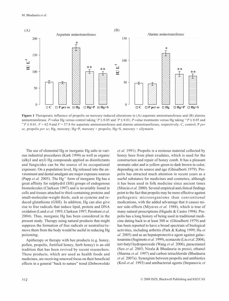
Figure 4 Therapeutic influence of propolis on mercury-induced alterations in antioxidant enzymatic status. (A) Glutathione-S-
transferase (GST), (B) glucose-6-phosphatase dehydrogenase (G6PDH), (C) super oxide dismutase (SOD) and (D) catalase (CAT).
P-value Hg versus control taking +P ≤ 0.05 and *P ≤ 0.01; P-value treatments versus Hg taking ++P ≤ 0.05 and **P ≤ 0.01. F = 15.6,
F = 17.6, F = 21.2 and F = 34.3 for GST, G6PDH, SOD and CAT, respectively. C, control; P per se, propolis per se; Hg, mercury;
Hg+P, mercury + propolis; Hg+S, mercury + silymarin.
Propolis reverses mercury-induced toxicity

© 2008 ISZS, Blackwell Publishing and IOZ/CAS
(Bhadauria et al. 2007b) and silymarin was given as a posi-
tive control. The dose of propolis was selected on the
basis of our previous study (Shukla et al. 2005).
Experimental protocol
Thirty mice were assigned into five groups of six ani-
mals each. Group 1: received saline (i.p.; 0.9% NaCl) fol-
Figure 5 (A) Photomicrograph of a con-
trol liver showing well-arranged hepato-
cytes with well-formed intact nucleus,
obvious sinusoidal space (arrow) and
well-formed central vein (CV) (100×). (B)
Propolis per se treated liver showing
well-formed sinusoidal space (arrow)
with central vein (CV) (100×). (C) He-
patic injury induced by mercury show-
ing congestion in the sinusoidal space
with damaged central vein (CV) (100×).
(D) Propolis recovered the hepatocytes
with better cord arrangement and nor-
mal sinusoidal space (arrows) and well-
formed central vein (CV) (100×); (E)
Silymarin treatment shows normal hepa-
tocytes with prominent nucleus and
proper sinusoidal space (arrow) and nor-
mal central vein (CV) (100×).
M. Bhadauria et al.

© 2008 ISZS, Blackwell Publishing and IOZ/CAS
lowed by GAS (5 mL kg–1, p.o.) for 3 days. Group 2: re-
ceived saline (as in group 1) followed by propolis per se
(200 mg kg–1, p.o.) for 3 days. Groups 3–5: received mercu-
ric chloride (5 mg kg–1 i.p., single dose). Group 3: served as
the experimental control and received GAS (5 mL kg–1, p.
o.) for 3 days after toxicant administration. Groups 4 and 5:
treated with propolis and silymarin, respectively, for 3 con-
secutive days after Hg intoxication.
Blood biochemical analysis
Twenty-four hours after the final administration the
animals were killed under a mild ether anesthesia and blood
was drawn immediately by puncturing the retro-orbital
venous sinus for isolation of serum and used to determine
the leakage of aspartate aminotransferase (AST) and ala-
nine aminotransferase (ALT) (Reitman & Frankel 1957),
lactate dehydrogenase (LDH) (Wroblewski & La Due 1955)
and serum alkaline phosphatase (SALP) (Halk et al. 1954).
ã-Glutamyl transpeptidase (ã-GT) was measured using the
Merck kit (Merck Specialties Private Limited, Mumbai,
India) as per the manufacturer’s instructions.
Oxidative stress and antioxidant status in the
liver
Tissue samples of the liver were homogenized with ice-
cold 1.15% KCl for the determination of thiobarbituric acid
reactive substances (TBARS) and the activities of glu-
cose-6-phosphate dehydrogenase (G6PDH), glutathione-
S-transferase (GST) and catalase (CAT). For the estima-
tion of GSH and superoxide dismutase (SOD) activity, the
tissues were homogenized in 5% sulfo-salicylic acid and
normal saline solution, respectively.
Hepatic LPO was measured by estimating TBARS as
described by Sharma and Krishna Murti (1968). Reduced
glutathione and oxidized glutathione (GSSG) were mea-
sured using the method of Roberts and Francetic (1993).
Total GST activity was measured according to the method
of Habig et al. (1974). Total SOD activity was estimated by
measuring the inhibition of auto oxidation of epinephrine
as per Mishra and Fridovich (1972), and CAT activity was
estimated using the method of Aebi (1984). The activity of
G6PDH was measured using the method of Askar et al.
(1996). Total protein was estimated using the method of
Lowry et al. (1951) with bovine serum albumin as standard.
Histopathological analysis
For photomicroscopic observations, liver samples were
fixed in Bouin’s fixative and routinely embedded in
paraffin. Tissue sections (5-ìm thickness) were stained with
hematoxylin–eosin (HE) and examined under a
photomicroscope.
Statistical analysis
The results are expressed as the mean ± standard error
(SE) of the six animals in each group. The data were sub-
jected to statistical analysis using a one-way ANOVA and
statistical significance was set a priori at P = 0.05. The
data were also subjected to a Student’s t-test with statis-
tical significance set a priori at P = 0.01 and P = 0.05; P-
value Hg versus control taking +=0.05 and *£0.01; P-value
treatments versus Hg taking ++=0.05 and **=0.01 for
Student’s t-test (Snedecor & Cochran 1994).
RESULTS
Blood biochemical analysis
Figure 1 (a,b) and Fig. 2 (a–c) represent the effect of
propolis extract on Hg-induced blood biochemical
alterations. The AST, ALT, SALP, g-GT and LDH activi-
ties were significantly increased after Hg intoxication (P =
0.01). Propolis therapy significantly recovered all the en-
zymes back toward the control level (P = 0.01) and showed
>70% protection in all parameters except g-GT, which
showed more than 60% protection in its release. Mercury
administration significantly increased serum bilirubin con-
tents (P = 0.01), which were significantly diminished by
propolis (P = 0.01) and showed more than 80 % protection.
Oxidative stress and antioxidant status
The protective effect of propolis on the Hg-induced
experimentally oxidative stress and antioxidant status are
shown in Figs 3 (a–c) and 4 (a–d). Hepatic LPO increased
with Hg administration (P = 0.01). The administration of
propolis and silymarin significantly inhibited LPO (P £ 0.
01) and reduced hepatic peroxidative stress by more than
80% (Fig. 3a). Acute administration of Hg caused a signifi-
cant decrease in hepatic GSH contents and increased GSSG
contents (P = 0.01). Propolis therapy recovered the GSH
contents towards the control (P = 0.01) and showed better
protection compared with the positive control (Fig. 3b).
Three days of treatment with propolis diminished the con-
tents of GSSG (P = 0.01) and the ANOVA was significant at
P = 0.05 (Fig. 3c). Mercury diminished the antioxidative
status of the organs after its administration and decreased
the activities of GST (Fig. 4a), G6PDH (Fig. 4b), SOD (Fig.
4c) and CAT (Fig. 4d) (P = 0.01). Treatment with propolis
and silymarin recovered the activities of all these enzymes
towards normal levels (P = 0.01). Recovery of 60% was
found in SOD and CAT, whereas more than 70% improve-
ment was observed in the activities of GST and G6PDH
Propolis reverses mercury-induced toxicity

© 2008 ISZS, Blackwell Publishing and IOZ/CAS
with propolis treatment.
Histopathological observations
Figure 5 (A–E) presents the histology of the liver after
the different treatments. Light microscopic evaluation
showed regular morphology of the liver parenchyma with
well-designed hepatic cells and sinusoids in the control
group and with propolis per se (Fig. 5A,B). The Hg-treated
group showed severe liver injury with prominent conges-
tion in the sinusoidal space and a damaged central vein
(Fig. 5C). The hepatocytes were found with normal
appearance, arranged in cord in most of the areas having
prominent nucleus and granular cytoplasm after propolis
(Fig. 5D) and silymarin treatment (Fig. 5E).
DISCUSSION
Acute administration of Hg caused toxic effects in the
liver and this damage was observed to be associated with
increases in LPO and GSSG, indicating oxidative tissue
damage, and with decreases in antioxidant enzymatic ac-
tivities and a significant reduction in GSH levels. Treat-
ment of the animals with propolis extract and silymarin
appeared to afford protection against this noxious stimulus.
Because of the high affinity of Hg to thiol groups, Hg is
known to affect living organisms by damaging thiol pro-
teins and enzymes (Clarkson 1997). Divalent ions of tran-
sition metals, especially Pb, Hg and copper, can promote
LPO and much attention is currently focused on LPO in
the pathogenesis of metal toxicity. The LPO is an auto-
catalytic free-radical process whereby polyunsaturated
fatty acids in cell membranes undergo degradation by a
chain reaction to yield lipid hydroperoxides, which subse-
quently decompose to form a variety of products, includ-
ing malondialdehyde (MDA).
The LPO induction by Hg has been demonstrated in
the present study and cell membrane permeability might
be affected by this process (Nava et al. 2000; Mahboob et
al. 2001). Propolis treatment was found to protect the liver
against Hg toxicity and suppressed the process of LPO.
Antioxidants have a protective effect against tissue inju-
ries in the pathogenesis of which LPO may be involved
and previous studies have demonstrated that quercetin (a
major component of propolis) inhibits LPO effectively by
scavenging free radicals and/or transition metal ions (Terao
& Piskula 1999). Flavonoids and phenolics are a group of
naturally occurring compounds that are widely distributed
as secondary metabolites in the plant kingdom and pre-
vent oxidative injury and cell death by protecting against
LPO (Molina et al. 2004) and by chelating the metals
(Afanasev et al. 1989).
The present study suggests that Hg administration
depletes GSH content and increases GSSG in the liver,
which makes hepatocytes more susceptible to oxidative
damage, particularly during increased free radical
production. The GSH is an important intracellular antioxi-
dant (hydrogen-donating compound) that spontaneously
neutralizes several electrophiles and reactive oxygen spe-
cies (Lu 1999); and the contents of GSH/GSSG play a key
role in cells and maintain the redox status of the cell (Rana
et al. 2002). Concomitant restoration of the GSH pool after
propolis treatment was consistent with some other reports
(Nirala et al. 2008). GSH is a substrate of the enzyme GST,
a phase II enzyme that plays a key role in the cellular
detoxification of xenobiotics, electrophiles and reactive
oxygen species through their conjugation to GSH (Mates
2000). In the present study, reduction in GST activity after
Hg exposure might decrease the conjugation reaction and
increase the toxic events. GSSG is reduced back to GSH
by nicotinamide adinine dinucleotide phosphate
(NADPH)-dependent reaction. G6PDH, the first enzyme
of the pentose phosphate pathway, is an important site of
NADPH production. Reduction in the activity of G6PDH
enzyme after Hg might affect the rate of generation of
NADPH and, thus, affect the redox status of the cell. Pro-
polis therapy maintained the normal status of GST and
G6PDH activities and, thus, improved the antioxidant sta-
tus of the hepatocytes in terms of increased GSH and de-
creased GSSG contents. Previous studies have shown that
propolis prevents hepatic disorders induced by acetami-
nophen (Nirala & Bhadauria 2008) and CCl4 (Bhadauria et
al. 2007a; b) as well as light-metal-induced lipid
peroxidation in liver homogenates (Nirala et al. 2008) ow-
ing to the presence of flavonoids and phenolics.
Mercury-induced toxic effects arise from alterations in
the structural integrity of the mitochondrial inner
membrane, resulting in a loss of normal cation selectivity,
which allows the membrane to participate effectively in
oxidative metabolism (Lund et al. 1993). Mercury ions per-
turb the mitochondrial inner membrane function, which
results in depletion of mitochondrial GSH content and in-
creased formation of H2O2 by the mitochondrial electron
transport chain (Lund et al. 1991). Increased H2O2 forma-
tion might be accompanied by increased peroxidation of
mitochondrial lipids, which is consistent with an oxidative
stress-like condition (Lund et al. 1993). Cells have a num-
ber of mechanisms to protect themselves from the toxic
effects of reactive oxygen species. The SOD removes su-
per oxide (O2) by converting it to H
2O2, which can be rap-
idly converted to water by CAT. The activity of SOD is a
M. Bhadauria et al.

© 2008 ISZS, Blackwell Publishing and IOZ/CAS
sensitive index to hepatic damage because it scavenges
the superoxide anion to form hydrogen peroxide, dimin-
ishing the toxic effect. CAT is an enzymatic antioxidant
widely distributed in all animal tissues that decomposes
hydrogen peroxide and protects the tissue from highly
reactive hydroxyl radicals. Therefore, alteration in the ac-
tivity of these two enzymes might result in a number of
deleterious effects because of the accumulation of super-
oxide radicals and hydrogen peroxide. In the present study,
propolis extract significantly restored SOD and CAT
activity, which indicated that propolis extract could scav-
enge reactive free radicals and eventually lessen oxida-
tive damage to the tissues and subsequently improve the
activities of these antioxidant enzymes. It is proposed that
the beneficial effects of a pharmacological dose of propo-
lis might result from its antioxidant properties by reviving
the endogenous antioxidant defense system.
Mercury intoxication resulted in significant hepatic
damage and oxidative stress as evidenced by substantial
increases in the serum activities of AST, ALT, SALP, LDH
and g-GT into the circulation as a result of hepatocellular
nacrosis, which causes an increase in the permeability of
the cell membrane (Sharma et al. 2002). A reduction in the
levels of these parameters toward the respective normal
values by propolis therapy is an indication of the stabili-
zation of the plasma membranes and reparation of the he-
patic tissue damage caused by Hg. This effect is in agree-
ment with the commonly accepted view that serum levels
of transaminases would return to normal after healing of
the hepatic parenchyma and the regeneration of
hepatocytes. Histopathological observations of the im-
provement in the histoarchitecture of the liver after pro-
polis treatment are also consistent with the biochemical
findings of the present study.
In conclusion, our findings demonstrate that propolis
extract is able to reverse pathological parameters, such as
necrotic histological features, and serum levels of AST,
ALT, SALP, LDH and g-GT of acute liver damage induced
by Hg. This protective ability of propolis resulted from its
modulatory effects on detoxification (GST and G6PDH)
and antioxidative enzymes (SOD and CAT), which, in turn,
suppress the production of free radicals and reduce sub-
sequent liver damage. Furthermore, the high content of
polyphenols and flavonoids in propolis contributes to free
radical scavenging and antioxidation activities. Therefore,
dietary propolis administration might be useful as a
hepatoprotective agent against Hg-induced acute liver
damage.
ACKNOWLEDGMENTS
The authors wish to thank the Indian Council of Medi-
cal Research, New Delhi, India (45/15/2002/PHA/BMS) for
financial assistance.
REFERENCES
Aebi HL (1984). Catalase in vitro. Method of Enzymol-
ogy 105, 121–6.
Afanasev IB, Dorozhko AI, Brodskii AV, Kostyuk VA,
Potapovitch AI (1989). Chelating and free radical
scavenging mechanisms of inhibitory action of rutin
and quercetin in lipid peroxidation. Biochemical
Pharmacology 38, 1763–9.
Askar MA, Sumathy K, Baquer NJ (1996). Regulation
and properties of glucose-6-phosphate dehydroge-
nase from rat brain. Indian Journal of Biochemistry
and Biophysics 33, 512–18.
Bhadauria M, Nirala SK, Shukla S (2007a). Propolis pro-
tects CYP2E1 enzymatic activities and oxidative stress
induced by carbon tetrachloride. Molecular and
Cellular Biochemistry 302, 215–24.
Bhadauria M, Nirala SK, Shukla S (2007b). Duration
dependent hepatoprotective effect of propolis extract
against carbon tetrachloride induced acute damage
in rats. Advances in Therapy 24, 1134–43.
Clarkson TW (1997). The toxicology of mercury. Criti-
cal Reviews in Clinical Laboratory Sciences 34,
369–403.
Countera SA, Buchanan LH (2004). Mercury exposure
in children: a review. Toxicology and Applied Phar-
macology 198, 209–30.
Dobrowolski JW, Vohora SB, Sharma K, Shah SA, Naqvi
SAH, Dandiya PC (1991). Antibacterial, antifungal,
antiamoebic, anti-inflammatory and antipyretic stud-
i e s on p ropo l i s bee p roduc t s . Journa l o f
Ethnopharmacology 35, 77–82.
Geckil I, Ates B, Durmaz G, Erdogan S, Yilmaz I (2005).
Antioxidant, free radical scavenging and metal
characterictics of propolis. American Journal of Bio-
chemistry and Biotechnology 1, 27–31.
Ghisalberti E (1979). Propolis: a review. Bee World 60,
59–84.
Halk PB, Oster BL, Summerson WH (1954). The Practi-
cal Physiological Chemistry, 14th edn. McGraw Hill
Book Company, New York.
Habig WH, Pabst MT, Jakoby WB (1974). Glutathione–
Stransferases. The first enzymatic step in mercaptu-
Propolis reverses mercury-induced toxicity

© 2008 ISZS, Blackwell Publishing and IOZ/CAS
ric acid formation. Journal of Biological Chemistry
249, 7130–39.
Higashi KO, de Castro SL (1994). Propolis extracts are
effective against Trypanosoma curzi and have an
impact on its interaction with host cells. Journal of
Ethnopharmacology 43, 149–55.
Hu F, Hepburn HR, Li Y, Chen M, Radloff SE, Daya S
(2005). Effects of ethanol and water extracts of pro-
polis (bee glue) on acute inflammatory animal models.
Journal of Ethnopharmacology. 100, 276–83.
Kark RAP (1994). Clinical and neurochemical aspects of
inorganic mercury intoxication. In: de Wolff FA, ed.
Handbook of Clinical Neurology, 20. Elsevier,
Amsterdam, pp. 367–411.
Krol W, Scheller S, Shani J, Pietsz G, Czuba Z (1993).
Synergistic effect of ethanolic extract of propolis and
antibiotics on the growth of Staphylococcus aureus.
Arzneim-Forsch. 43: 607–9.
Liu CF, Lin CH, Lin CC, Lin YH, Chen CF, Lin CK, Lin SC
(2004). Antioxidative natural product protect against
econazole induced liver injuries. Toxicology 196, 87–
93.
Lowry OH, Rosenbrough NJ, Farr AL, Randall RJ (1951).
Protein measurement with Folin’s phenol reagent.
Journal of Biological Chemistry 193, 265–75.
Lu SC (1999). Regulation of hepatic glutathione
synthesis: current concepts and controversies. The
FASEB Journal 13, 1169–83.
Lund BO, Miller DM, Woods JS (1991). Mercury-in-
duced H2O2 production and lipid peroxidation and
lipid oxidation in vitro in rat kidney mitochondria.
Biochemical Pharmacology 42, 181–7.
Lund BO, Miller DM, Woods JS (1993). Studies on Hg
(II)-induced H2O2 formation and oxidative stress in
vivo and in vitro in rat kidney mitochondria. Bio-
chemical Pharmacology 45, 2017–24.
Mahboob M, Shireen KF, Atkinson A, Khan AT (2001).
Lipid peroxidation and antioxidant enzyme activity
indifferent organs of mice exposed to low level of
mercury. Journal of Environmental Science and
Health B 36, 687–97.
Mates JM (2000). Effects of antioxidant enzymes in the
molecular control of reactive oxygen species
toxicology. Toxicology 153, 83–104.
Mishra P, Fridovich I (1972). The role of superoxide
anion in the autooxidation of epinephrine and a
simple assay for superoxide dismutase. Journal of
Biological Chemistry 247, 3170–75.
Miyares C, Hollands I, Castaneda C, Gonzales T, Fragoso
T, Curras R, Soria C (1988). Ensayoterapeutico con
un preparado a base de propoleo propolisina en la
giardiasis del humano. Acta Gastroenterologia
Latina-Americana 18, 195–201.
Molina MF, Sanchez-Reus I, Iglesias I, Benedi J (2003).
Quercitin, a flavonoids antioxidant, prevents and
protects against ethanol induced oxidative stress in
mouse liver. Biological and Pharmaceutical Bulle-
tin 26, 1398–1402.
Nava M, Romero F, Quiroz Y, Parra G, Bonet L,
Rodriquez-Iturbe B (2000). Melatonin attenuation of
acute renal failure and oxidative stress induced by
mercuric chloride in rats. American Journal of Physi-
ology for Renal Physiology 279, F910–18.
Nirala SK, Bhadauria M (2008). Propolis reverses ac-
etaminophen induced hepatorenal biochemical and
histopathological alterations. Archives of Pharmacal
Research 31, 451-461.
Nirala SK, Bhadauria M, Mathur R, Mathur A (2008).
Influence of á-tocopherol, propolis and piperine on
therapeutic potential of tiferron against beryllium
induced toxic manifestations. Journal of Applied
Toxicology 28, 44–54.
Papp A, Nagymajtenyi L, Vezer T (2005). Subchronic
mercury treatment of rats in different phases of
ontogenesis: functional effects on the central and
peripheral nervous system. Food Chemistry and
Toxicology 43, 77–85.
Park EH, Kahng JH (1999). Suppressive effects of pro-
polis in rat adjuvant arthritis. Archives of Pharamcal
Research 22, 554–8.
Perottoni J, Lobato LP, Silveira A, Rocha JBT, Emanuelli
T (2004). Effects of mercury and selenite on d-
aminolevulinate dehydratase activity and on selected
oxidative stress parameters in rats. Environmental
Research 95, 166–73.
Rana SV, Allen T, Singh R (2002). Inevitable glutathione,
then and now, Indian Journal of Experimental Bi-
ology 40, 706–16.
Reitman S, Frankel S (1957). A colorimetric method for
determination of serum glutamic oxaloacetic and
glutamic pyruvic transaminases. American Journal
of Clinical Pathology 28, 56–63.
Roberts JC, Francetic DJ (1993). The importance of
sample preparation and storage of glutathione
analysis. Analytical Biochemistry 211, 183–7.
Sener G, Sehirli O, Tozan A, Ovunc AV, Gedik N, Omurtag
M. Bhadauria et al.

© 2008 ISZS, Blackwell Publishing and IOZ/CAS
GZ (2007). Ginkgo biloba extract protects against
mercury(II)-induced oxidative tissue damage in rats.
Food and Chemical Toxicology 45, 543–50.
Seo KW, Park M, Song YJ, Kim SJ, Yoon KR (2003). The
protective effects of propolis on hepatic injury and
its mechanism. Phytotherapy Research 17, 250–53.
Sforcin JM, Fernandes JA, Lopes CAM, Bankova V,
Funari SRC (2000). Seasonal effect on Brazalian pro-
p o l i s a n t i b a c t e r i a l a c t i v i t y . J o u r n a l o f
Ethnopharmacology 73, 243–9.
Sharma M, Pillai KK, Husain SZ, Giri DK (1997). Protec-
tive role of propolis against alcohol-carbon tetrachlo-
ride-induced hepatotoxicity in rats. Indian Journal
of Pharmacology 29, 76–81.
Sharma MK, Kumar M, Kumar A (2002). Ocimum sanc-
tum aqueous leaf extract provides protection against
mercury induced toxicity in Swiss albino mice. In-
dian Journal of Experimental Biology 40, 1079–82.
Sharma SK, Krishna Murti CR (1968). Production of lipid
peroxides by brain. Journal of Neurochemistry 15,
147–9.
Shukla S, Bhadauria M, Jadon A (2005). Evaluation of
hepatoprotective potential of propolis extract in car-
bon tetrachloride induced liver injury in rats. Indian
Journal of Biochemistry and Biophysics 42, 321–5.
Snedecor GW, Cochran WG (1994). Statistical Method,
8th edn. Iowa State University Press, Ames.
Stepanovic S, Antic N, Dakic I, Svabic-Vlahovic M
(2003). In vitro antimicrobial activity of propolis and
synergism between propolis and antimicrobial drugs.
Microbiology Research 158, 353–7.
Sugimoto Y, Tarumi T, Kaneko Y et al. (1999). Effect of
propolis extract on D-galactosamine-induced hepatic
injury in rats. Biological and Pharmaceutical Bul-
letin 22, 1237–9.
Terao J, Piskula MK (1999). Flavonoids and membrane
lipid peroxidatiotn inhibition. Nutrition 15, 790–91.
Wang BJ, Lien YH, Su CL, Wu CP, Yu ZR (2006). Frac-
tionation using supercritical CO2 influences and
hepatoprotective activity of propolis against induced
by tert-butyl hydroperoxide. International Journal
of Food Science and Technology 41, 68–75.
World Health Organization (1991). Inorganic mercury.
Environmental Health Criteria 118. World Health
Organization, Geneva, pp. 1–168.
Wroblewski F, La Due JS (1955). Lactic dehydrogenase
activity in blood. Proceedings of the Society for Ex-
perimental Biology and Medicine 90, 210–13.
Propolis reverses mercury-induced toxicity
Copyright © 2022 FDOKUMEN