Antiatherogenic and anti-angiogenic activities of polyphenols from propolis
Upload
khangminh22Category
view
0download
0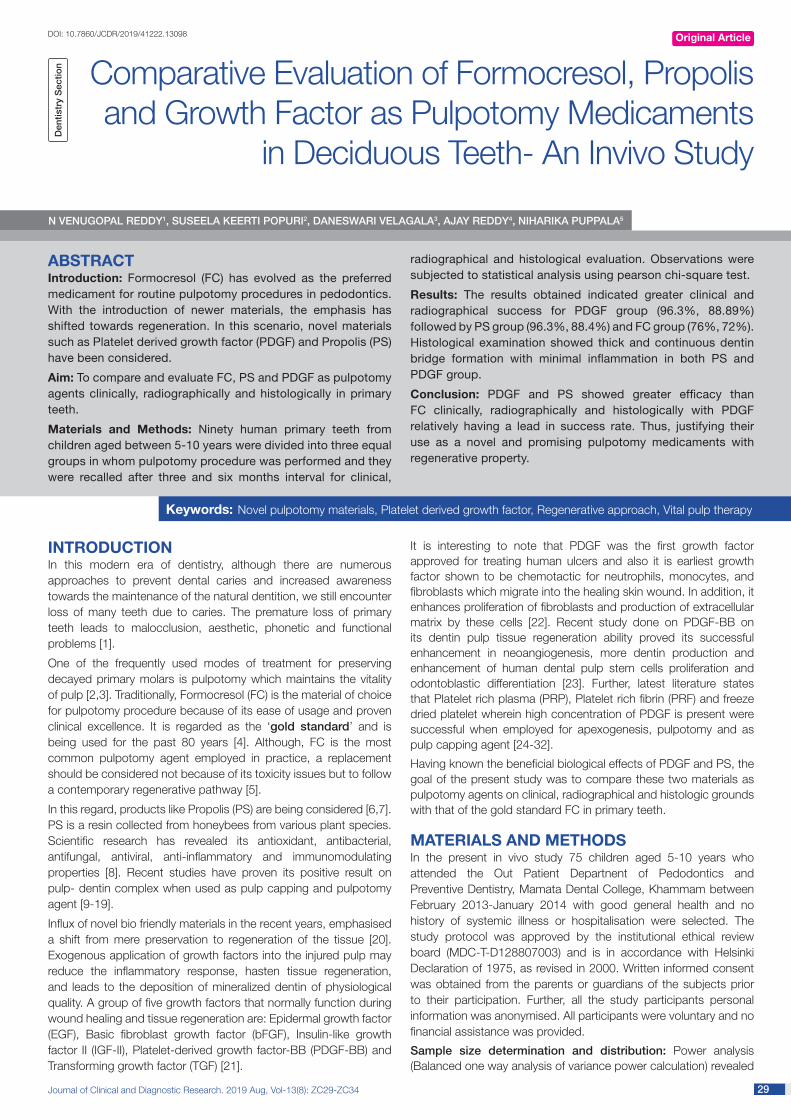
Journal of Clinical and Diagnostic Research. 2019 Aug, Vol-13(8): ZC29-ZC34 2929
DOI: 10.7860/JCDR/2019/41222.13098 Original Article
Den
tistr
y S
ectio
n Comparative Evaluation of Formocresol, Propolis and Growth Factor as Pulpotomy Medicaments
in Deciduous Teeth- An Invivo Study
IntrOductIOnIn this modern era of dentistry, although there are numerous approaches to prevent dental caries and increased awareness towards the maintenance of the natural dentition, we still encounter loss of many teeth due to caries. The premature loss of primary teeth leads to malocclusion, aesthetic, phonetic and functional problems [1].
One of the frequently used modes of treatment for preserving decayed primary molars is pulpotomy which maintains the vitality of pulp [2,3]. Traditionally, Formocresol (FC) is the material of choice for pulpotomy procedure because of its ease of usage and proven clinical excellence. It is regarded as the ‘gold standard’ and is being used for the past 80 years [4]. Although, FC is the most common pulpotomy agent employed in practice, a replacement should be considered not because of its toxicity issues but to follow a contemporary regenerative pathway [5].
In this regard, products like Propolis (PS) are being considered [6,7]. PS is a resin collected from honeybees from various plant species. Scientific research has revealed its antioxidant, antibacterial, antifungal, antiviral, anti-inflammatory and immunomodulating properties [8]. Recent studies have proven its positive result on pulp- dentin complex when used as pulp capping and pulpotomy agent [9-19].
Influx of novel bio friendly materials in the recent years, emphasised a shift from mere preservation to regeneration of the tissue [20]. Exogenous application of growth factors into the injured pulp may reduce the inflammatory response, hasten tissue regeneration, and leads to the deposition of mineralized dentin of physiological quality. A group of five growth factors that normally function during wound healing and tissue regeneration are: Epidermal growth factor (EGF), Basic fibroblast growth factor (bFGF), Insulin-like growth factor II (IGF-II), Platelet-derived growth factor-BB (PDGF-BB) and Transforming growth factor (TGF) [21].
It is interesting to note that PDGF was the first growth factor approved for treating human ulcers and also it is earliest growth factor shown to be chemotactic for neutrophils, monocytes, and fibroblasts which migrate into the healing skin wound. In addition, it enhances proliferation of fibroblasts and production of extracellular matrix by these cells [22]. Recent study done on PDGF-BB on its dentin pulp tissue regeneration ability proved its successful enhancement in neoangiogenesis, more dentin production and enhancement of human dental pulp stem cells proliferation and odontoblastic differentiation [23]. Further, latest literature states that Platelet rich plasma (PRP), Platelet rich fibrin (PRF) and freeze dried platelet wherein high concentration of PDGF is present were successful when employed for apexogenesis, pulpotomy and as pulp capping agent [24-32].
Having known the beneficial biological effects of PDGF and PS, the goal of the present study was to compare these two materials as pulpotomy agents on clinical, radiographical and histologic grounds with that of the gold standard FC in primary teeth.
MAterIAls And MethOdsIn the present in vivo study 75 children aged 5-10 years who attended the Out Patient Departnent of Pedodontics and Preventive Dentistry, Mamata Dental College, Khammam between February 2013-January 2014 with good general health and no history of systemic illness or hospitalisation were selected. The study protocol was approved by the institutional ethical review board (MDC-T-D128807003) and is in accordance with Helsinki Declaration of 1975, as revised in 2000. Written informed consent was obtained from the parents or guardians of the subjects prior to their participation. Further, all the study participants personal information was anonymised. All participants were voluntary and no financial assistance was provided.
Sample size determination and distribution: Power analysis (Balanced one way analysis of variance power calculation) revealed
N VeNugopal ReDDy1, SuSeela KeeRti popuRi2, DaNeSwaRi Velagala3, ajay ReDDy4, NihaRiKa puppala5
Keywords: Novel pulpotomy materials, Platelet derived growth factor, Regenerative approach, Vital pulp therapy
ABstrActIntroduction: Formocresol (FC) has evolved as the preferred medicament for routine pulpotomy procedures in pedodontics. With the introduction of newer materials, the emphasis has shifted towards regeneration. In this scenario, novel materials such as Platelet derived growth factor (PDGF) and Propolis (PS) have been considered.
Aim: To compare and evaluate FC, PS and PDGF as pulpotomy agents clinically, radiographically and histologically in primary teeth.
Materials and Methods: Ninety human primary teeth from children aged between 5-10 years were divided into three equal groups in whom pulpotomy procedure was performed and they were recalled after three and six months interval for clinical,
radiographical and histological evaluation. Observations were subjected to statistical analysis using pearson chi-square test.
results: The results obtained indicated greater clinical and radiographical success for PDGF group (96.3%, 88.89%) followed by PS group (96.3%, 88.4%) and FC group (76%, 72%). Histological examination showed thick and continuous dentin bridge formation with minimal inflammation in both PS and PDGF group.
conclusion: PDGF and PS showed greater efficacy than FC clinically, radiographically and histologically with PDGF relatively having a lead in success rate. Thus, justifying their use as a novel and promising pulpotomy medicaments with regenerative property.
N Venugopal Reddy et al., Clinical, Radiological and Histological Evaluation of Three Different Pulpotomy Medicaments in Deciduous Teeth www.jcdr.net
Journal of Clinical and Diagnostic Research. 2019 Aug, Vol-13(8): ZC29-ZC343030
an adequate sample size of 30 teeth in each group would be required to show power=0.8 and significance level of 0.05. Thus the total sample size was calculated to be 90. Sample size calculation was performed using R studio programming language software. Hence 90 teeth from 75 children requiring pulpotomy treatment who met clinical and radiographic criteria were selected and were divided equally into three groups: FC, PS and PDGF group using sealed envelope randomisation method. Further, from each group, 5 teeth were subjected to histological evaluation (3 teeth at the end of 3 months and 2 teeth at the end of 6 months from each group).
The clinical and radiographic criteria for tooth selection were as follows. Teeth with deep carious lesion (radiographically caries approximating pulp) restorable after completion of the procedure. Teeth with lack of progressed irreversible pulpitis symptoms like history of night pain, spontaneous pain and nonvital tooth with draining sinus and soft tissue swelling. Teeth with lack of clinical and radiographic signs of pulpal necrosis which includes furcation involvement, periapical pathology, internal resorption, calcification in the canal. Lastly, with the application of sterile pledget of moist cotton haemorrhage from the amputated pulp stumps should stop within five minutes [33, 34].
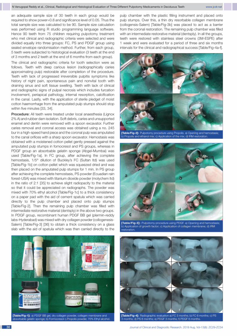
procedure: All teeth were treated under local anaesthesia (Lignox 2% A) and rubber dam isolation. Soft debris, caries and unsupported enamel and dentin were removed with a spoon excavator. Further caries removal and coronal access was obtained using a no. 245 bur in a high-speed hand piece and the coronal pulp was amputated to the canal orifices with a sharp spoon excavator. Hemostasis was obtained with a moistened cotton pellet gently pressed against the amputated pulp stumps in fomocresol and PS groups, whereas in PDGF group an absorbable gelatin sponge (Abgel-Mumbai) was used [Table/Fig-1a]. In FC group, after achieving the complete hemostasis, 1/5th dilution of Buckley’s FC (Sultan ltd) was used [Table/Fig-1b] on cotton pellet which was squeezed dried and was then placed on the amputated pulp stumps for 1 min. In PS group after achieving the complete hemostasis, PS powder (Ecuadian rain forest-USA) was mixed with titanium dioxide powder (molychem ltd) in the ratio of 2:1 [35] to achieve slight radiopacity to the material so that it could be appreciated on radiographs. The powder was mixed with 70% ethyl alcohol [Table/Fig-1c] to a thick consistency on a paper pad with the aid of cement spatula which was carried directly to the pulp chamber and placed onto pulp stumps [Table/Fig-2]. Then the remaining pulp chamber was filled with intermediate restorative material (dentsply) in the above two groups. In PDGF group, recombinant human PDGF BB gel (plermin-reddy labs-Hyderabad) was mixed with dry collagen powder (collogenesis-Salem) [Table/Fig-3] [36] to obtain a thick consistency on a glass slab with the aid of spatula which was then carried directly to the
[table/Fig-1]: a) PDGF BB gel, dry collagen powder, collagen membrane and absorbable gelatin sponge; b) Formocresol c.Propolis powder, 70% Ethyl alcohol.
[table/Fig-2]: Pulpotomy procedure using Propolis. a) Opening and hemostasis; b) Propolis and ethanol mix; c) Application of the mix; d) IRM restoration.
pulp chamber with the plastic filling instrument and placed onto pulp stumps. Over this, a thin dry resorbable collagen membrane (collogensis-Salem) [Table/Fig-3b] was placed to act as a barrier from the coronal restoration. The remaining pulp chamber was filled with an intermediate restorative material (dentsply). In all the groups, teeth were restored with stainless steel crowns (3M-ESPE) after 1 week and were evaluated for a period of three and six months intervals for the clinical and radiographical success [Table/Fig-4a-f].
[table/Fig-3]: Pulpotomy procedure using PDGF. a) Opening and hemostasis; b) Application of growth factor; c) Application of collagen membrane; d) IRM restoration.
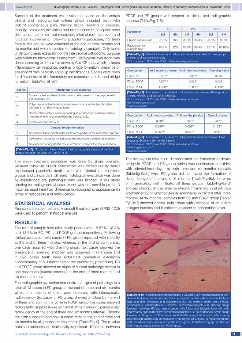
[table/Fig-4]: Radiographic evaluation a) FC 3 months; b) FC 6 months; c) PS 3 months; d) PS 6 months; e) PDGF 3 months; f) PDGF 6 months.
www.jcdr.net N Venugopal Reddy et al., Clinical, Radiological and Histological Evaluation of Three Different Pulpotomy Medicaments in Deciduous Teeth
Journal of Clinical and Diagnostic Research. 2019 Aug, Vol-13(8): ZC29-ZC34 3131
Success of the treatment was evaluated based on the certain clinical and radiographical criteria which included teeth with lack of spontaneous pain, draining fistula, swelling or abscess, mobility, premature exfoliation and no presence of periapical bone destruction, abnormal root resorption, internal root resorption and furcation involvement, Following pulpotomy procedure, 15 teeth from all the groups were extracted at the end of three months and six months and were subjected to histological analysis. Only teeth, undergoing serial extraction for the interceptive orthodontic purpose were taken for histological assessment. Histological evaluation was done according to criteria laid down by Cox CF et al., which includes inflammatory cell response, dentinal bridge formation, presence or absence of pulp necrosis and pulp calcifications. Scores were given for different levels of inflammatory cell response and dentinal bridge formation [Table/Fig-5] [37].
PDGF and PS groups with respect to clinical and radiographic success [Table/Fig-7,8].
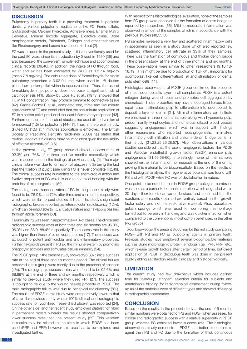
[table/Fig-9]: Histological photomicrograph (H&E Stain). a) Photomicrograph at 40X-dentinal bridge formation between PDGF and pulp chamber with intact odontoblastic layer, abundant fibroblasts and collagen bundles and minimal inflammatory infiltrate composed of lymphocytes at 3 months; b) Photomicrograph 40X- dentinal bridge formation between PS and pulp chamber with intact odontoblastic layer with mild inflammatory cells at 3 months; c) Photomicrograph at 40x- No evidence of dentin bridge formation in FC group; d) Photomicrograph at 40X- area of mild chronic inflammatory cell infiltrate predominantly composed of lymphocytes in FC group; e) Photomicrograph 40X- no inflammatory cells at 6 months in PS group; f) Photomicrograph at 10X- no inflammatory cells at 6months in PDGF group.
Scores inflammatory cell response
1None or a few scattered inflammatory cells present in the pulp beneath the exposure site
2Polymorphonuclear leukocytes (acute) or mononuclear lymphocytes (chronic) in an inflammatory lesion
3Severe inflammatory lesion appearing as an abscess or dense infiltrate involving one third or more than the coronal pulp.
4 Completely necrotic pulp
Dentinal bridge formation
1 New barrier tissue directly adjacent to some portion of the restorative material
2 New dentin bridge formation some distance from the material interface
3 No evidence of any dentin tissue formation in any of the tissue sections
[table/Fig-5]: Scores for different levels of inflammatory response and dentinal bridge formation as given by Cox CF et al.
The entire treatment procedure was done by single operator, whereas follow-up clinical assessment was carried out by senior experienced paediatric dentist who was blinded on treatment groups and clinical data. Similarly histological evaluation was done by experienced oral pathologist who was blinded. In our study blinding for radiographical assessment was not possible as the 3 materials used here had difference in radiographic appearance (in terms of radiopacity and radiolucency).
stAtIstIcAl AnAlysIsPearson chi-square test and Microsoft Excel software (SPSS-17.0) were used to perform statistical analysis.
resultsThe rate of sample loss after study period was 16.67%, 13.3% and 13.3% in FC, PS and PDGF groups respectively. Following clinical evaluation two cases in FC group reported with mobility at the end of three months, whereas at the end of six months, one case reported with draining sinus, two cases showed the presence of swelling, mobility was observed in one case and in two cases teeth were exfoliated (premature exfoliation approximately at 5.3 months after the pulpotomy procedure). PS and PDGF group showed no signs of clinical pathology except in one case each (buccal abscess) at the end of three months and six months interval.
The radiographic evaluation demonstrated signs of pathology in a total of 12 cases in FC group at the end of three and six months where the majority of them were observed with interradicular radiolucency. Six cases in PS group showed a failure by the end of three and six months while in PDGF group five cases showed radiographic signs of failure with most of them showing periradicular radiolucency at the end of three and six months interval. Thereby the clinical and radiographic success rates at the end of three and six months for all groups are depicted in [Table/Fig-6]. The p-value obtained indicates no statistically significant difference between
parameterFC pS pDgF
3M 6M 3M 6M 3M 6M
Clinical success rate 92.3% 76% 96.3% 96.3% 96.3% 96.3%
Radiographical success rate
76.9% 72% 88.9% 88.4% 92.6% 88.89%
[table/Fig-6]: Clinical success and radiographical success rates of three groups at three and six months interval.FC: Formocresol; PS: Propolis; PDGF: Platelet derived growth factor
Comparison at 3 months p-value at 6 months p-value overall p-value
FC vs. PS 0.507NS 0.032* 0.032*
FC vs. PDGF 0.507NS 0.032* 0.032*
PS vs. PDGF 1.000NS 1.000NS 1.000NS
[table/Fig-7]: Comparison of p-values for clinical success between three groups at three months and six months interval.FC: Formocresol; PS: Propolis; PDGF: Platelet derived growth factorNS: Not significant p>0.05*Significant p<0.05
Comparison at 3 months p-value at 6 months p-value overall p-value
FC vs. PS 0.88NS 0.62NS 0.38NS
FC vs. PDGF 0.098NS 0.098NS 0.123NS
PS vs. PDGF 0.552NS 0.552NS 0.299NS
[table/Fig-8]: Comparison of p-value for radiographical success between three groups at three and six months interval.FC: Formocresol; PS: Propolis; PDGF: Platelet derived growth factorNS-Not significant p>0.05*Significant p<0.05
The histological evaluation demonstrated the formation of dentin bridge in PDGF and PS group which was continuous and thick with odontoblastic layer, at both three and six months intervals [Table/fig-9a,b] while FC group did not reveal the formation of dentin bridge at the end of 6 months [Table/Fig-9c]. In terms of inflammatory cell infiltrate, all three groups [Table/Fig-9a-d] showed chronic, diffuse, minimal chronic inflammatory cell infiltrate predominantly of lymphocytes in specimens extracted after three months. At six months, samples from PS and PDGF group [Table/Fig-9e,f] showed normal pulp tissue with presence of abundant collagen bundles and fibroblasts adjacent to odontoblast layer.
N Venugopal Reddy et al., Clinical, Radiological and Histological Evaluation of Three Different Pulpotomy Medicaments in Deciduous Teeth www.jcdr.net
Journal of Clinical and Diagnostic Research. 2019 Aug, Vol-13(8): ZC29-ZC343232
dIscussIOnPulpotomy in primary teeth is a prevailing treatment in pediatric dentistry. Various pulpotomy medicaments like FC, Ferric sulfate, Glutaraldehyde, Calcium hydroxide, Adhesive liners, Enamel Matrix Derivative, Mineral Trioxide Aggregate, Bioactive glass, Bone morphogenic protein, Pulpotech, Collagen and other techniques like Electrosurgery and Lasers have been tried out [2].
FC was included in the present study as it is conventionally used for the past 80 years since its introduction by Sweet in 1930 [38] and also because of the convenient, simple technique and accomplished clinical records [39,40]. In addition, the intake of FC through food, water and air has been estimated by WHO as 1.5-14 mg/day (mean 7.8 mg/day). The calculated dose of formaldehyde for single pulpotomy procedure is 0.02-0.1 mg, when used in 1:5 dilution placed on cotton pellet which is squeeze dried. Thus, the use of formaldehyde in pulpotomy does not pose a significant risk of carcinogenesis [41]. Study by Loos PJ et al., (1973) showed that FC in full concentration, may produce damage to connective tissue [42]. Garcia-Godoy F et al., compared one, three and five minute applications of FC and concluded that the one-minute application of FC in a cotton pellet produced the least inflammatory response [43]. Furthermore, some of the latest studies also used diluted version of formocresol (1:5) for pulpotomy [44-47]. Thus, in the present study, diluted FC (1:5) at 1 minutes application is employed. The British Society of Paediatric Dentistry guidelines (2006) has stated that routine usage of 1:5 dilution “may be imprudent given the availability of effective alternatives” [48].
In the present study, FC group showed clinical success rates of 92.3% and 76% after three and six months respectively which was in accordance to the findings of previous study [5]. The major clinical failure was due to formation of abscess (8%) being the fact that the fixation of pulp tissue using FC is never complete [42,49]. The clinical success rate is credited to the antimicrobial action and fixative properties of FC which is due to chemical bonding with the proteins of microorganisms [50].
The radiographic success rates of FC in the present study were found to be 76.9% and 72% after three and six months respectively which were similar to past studies [51,52]. The study’s significant radiographic failures reported as interradicular radiolucency (12%), which can be imputable to FC’s fixative nature and its vapors passing through apical foramen [53].
Failure with PS was seen in approximately 4% of cases. The clinical and radiographic success rates at both three and six months are 96.3%, 96.3% and 88.9, 88.4% respectively. The success rate in this study was higher than those of other recent studies [17]. The success was attributed to potent antimicrobial and anti-inflammatory properties. Further flavonoids present in PS aid the immune system by promoting phagocytic activities and stimulates cellular immunity [54].
The PDGF group in the present study showed 96.3% clinical success rate at the end of three and six months period. The clinical failures observed in this group were mostly due to the presence of abscess (4%). The radiographic success rates were found to be 92.6% and 88.89% at the end of three and six months respectively which is similar to previous study where they used PRF [27]. The success is thought to be due to the wound healing property of PDGF. The main radiographic failure was due to periapical radiolucency (8%). The results of PDGF in this study were comparatively lower to that of a similar previous study where 100% clinical and radiographic success rate for lyophilized freeze-dried platelet was reported [24]. On the other side, another recent study employed platelet rich fibrin in permanent molars wherein the results showed comparatively lower success rates than the present study [29]. This variation in results may be related to the form in which PDGF has been used (PRF and PRP) however this area has to be explored and investigated further.
With respect to the histopathological evaluation, none of the samples from FC group were observed for the formation of dentin bridge as reported by researchers [55]. Mild to moderate inflammation was observed in almost all the samples which is in accordance with the previous studies [48,55,56].
PS samples revealed a very few and scattered inflammatory cells in specimens as seen in a study done which also reported few scattered inflammatory cell infiltrate in 33% of their samples. There was the formation of continuous and thicker dentin bridge in the present study, at the end of three months and six months. These observations were similar to other researchers [9,10,13-16,19]. This might be due to production of TGF-β1, (important for odontoblast like cell differentiation) [9] and stimulation of dental pulp stem cells [11].
Histological observations of PDGF group confirmed the presence of intact odontoblastic layer in all samples as PDGF is a potent mitogen during wound repair and promotes cell aggregation by chemotaxis. These properties may have encouraged fibrous tissue repair, also it stimulates pulp to differentiate into odontoblast to deposit a layer of dentin [21]. Minimal inflammatory cell infiltrate were noticed in three months sample along with hyperemic pulp, predominantly lymphocytes and numerous dilated blood vessels suggesting angiogenesis which was in support with findings other researchers who reported neoangiogenesis, minimal/no inflammatory cell infiltration and uniform dentin bridge barrier in their study [21,23,25,26,28,57]. Also, observations in various studies considered that the use of angiogenic factors like PDGF and Vascular endothelial growth factor (VEGF) enhance pulp angiogenesis [31,56,58-60]. Interestingly, none of the samples showed neither inflammation nor necrosis at the end of 6 months, proving this material to be biocompatible. Based on the results of the histological analysis, the regenerative potential was found with PS and with PDGF while FC was of devitalization in nature.
One point to be noted is that in PDGF group collagen membrane was used as a barrier to coronal restoration which degraded within 20 days, therefore it can be understood and confirmed that the reactions and results obtained are entirely based on the growth factor solely and not the restorative material. Also, absorbable gelatin sponge which was used for controlling haemorrhage turned out to be easy in handling and was quicker in action when compared to the conventional moist cotton pellet used in the other two groups.
To our knowledge, the present study may be the first study comparing PDGF with PS and FC as pulpotomy agents in primary teeth. Previous studies have employed several biocompatible materials such as Bone morphogenic protein, emdogain gel, PRF, PRP etc., which release growth factors after certain period of time, but direct application of PDGF in deciduous teeth was done in the present study yielding satisfactory results clinically and histopathlogically.
lIMItAtIOnThe current study had few drawbacks which includes defined time for follow-up, stringent selection criteria for subjects and unattainable blinding for radiographical assessment during follow-up as all the materials were of different types and showed difference in radiographic appearance.
cOnclusIOnBased on the results, in the present study at the end of 6 months similar numbers were obtained for PS and PDGF when assessed for clinical and radiographic success with a relative superiority in PDGF group whereas FC exhibited lower success rate. The histological observations clearly demonstrate PDGF as a better biocompatible agent than PS and FC due to the formation of thick continuous

www.jcdr.net N Venugopal Reddy et al., Clinical, Radiological and Histological Evaluation of Three Different Pulpotomy Medicaments in Deciduous Teeth
Journal of Clinical and Diagnostic Research. 2019 Aug, Vol-13(8): ZC29-ZC34 3333
dentin bridge with no inflammatory infiltrate after six months of time interval. Therefore PDGF and PS may be used as a suitable pulpotomy materials due to its regenerative potential.
reFerences Pinkham JR. Pediatric Dentistry Infancy through adolescence. Fourth ed. [1]
St.Louis. W.B. Saunders, 2005. Chapter 22: Pulp therapy for primary dentition;375-76.
Guideline on Pulp Therapy for Primary and Immature Permanent Teeth. AAPD [2]Reference Manual. 2014;40(6):343-51.
Golpayegani MV, Ansari G, Tadayon N, Shams Sh, Mir M. Low-Level Laser [3]therapy for pulpotomy treatment of primary molars. J Dent. 2009;6(4):168-74.
Ranly DM. Pulpotomy therapy in primary teeth: New modalities for old rationales. [4]Pediatr Dent. 1994;16(6):403-09.
Erdem AP, Guven Y, Balli B, IIhan B, Sepet E, Ulukapi I, et al. Success Rates [5]of Mineral Trioxide Aggregate, Ferric Sulfate, and FC Pulpotomies: A 24-month Study. Pediatr Dent. 2011;33(2):165-70.
Ajay M, Shanthala BM, Praveen VS, Rao VVN, Chandru TP. Histological evaluation [6]of diode laser pulpotomy in dogs. J Oral Laser Applications. 2010;10:07-16.
Saltzman B, Sigal M, Clokie C, Rukavina J, Titley K, Kulkarni GV. Assessment [7]of a novel alternative to conventional FC-zinc oxide eugenol pulpotomy for the treatment of pulpally involved human primary teeth-diode laser-mineral trioxide aggregate pulpotomy. Int J Paediatr Dent. 2005;15:437-47.
Prasanna N, Nitya, Nazar N. Ethnopharmacological approach in endodontic [8]treatment. A focussed review. Int J Drug Dev Res. 2011;3(4):68-77.
Sabir A, Charles R, Agustiono P, Sosroseno W. Histological analysis of rat dental [9]pulp tissue capped with PS. J Oral Sci. 2005;47(3):135-38.
Abhishek P, Thomas MS, Kundabala M, Mohan M. A comparative histological [10]analysis of human pulp following direct pulp capping with PS, mineral trioxide aggregate and Dycal. Aust Dent J. 2010;55:59-64.
Ahangari Z, Naseri M, Jalili M, Mansouri Y, Mashhadiabbas F, Torkaman A. Effect [11]of PS on dentin regeneration and the potential role of dental pulp stem Cell in Guinea Pigs. Cell J. 2012;13(4):223-28.
Esmeraldo MR, Carvalho MG, Carvalho RA, Lima Rde F, Costa EM. [12]Inflammatory effect of green propolis on dental pulp in rats. Braz Oral Res. 2013;27(5):417-22.
Likitpongpipat N, Sangmaneedet S, Klanrit P, Noisombut R, Krisanaprakornkit [13]S, Chailertvanitkul P. Promotion of Dental Pulp Wound Healing in New Zealand White Rabbits’ Teeth by Thai Propolis Product. J Vet Dent. 2019;36(1):17-24.
Lima RVE, Esmeraldo MRA, Carvalho MGF, Oliveira PTD, Carvalho RAD, Silva [14]FLD, et al. Pulp repair after pulpotomy using different pulp capping agents: A comparative histologic analysis. Pediatr Dent. 2011;33:14-18.
Ozorio JEV, Carvalho LF, de Oliveira DA, de Sousa-Neto MD, da Cruz Perez DE. [15]Standardized PS extract and calcium hydroxide as pulpotomy agents in primary pig teeth. J Dent Child. 2012;79(2):53-58.
Meto A, Bimbari B, Shytaj K, Ozcan M. Anti-inflammatory and regenerative [16]effects of albanian propolis in experimental vital amputations. Eur J Prosthodont Restor Dent. 2016;24(3):145-51.
Alolofi H, El-Sayed M, Taha S. Clinical and radiographical evaluation of propolis [17]and thymus vulgaris extracts compared with formocresol pulpotomy in human primary molars. BDJ Open. 2016;2:16005.
Hugar SM, Kukreja P, Hugar SS, Gokhale N, Assudani H. Comparative Evaluation [18]of clinical and radiographic success of formocresol, propolis, turmeric gel, and calcium hydroxide on pulpotomized primary molars: A preliminary study. Int J Clin Pediatr Dent. 2017;10(1):18-23.
Balata GF, Abdelhady MIS, Mahmoud GM, Matar MA, Abd El-Latif AN. Formulation [19]of Saudi Propolis into biodegradable chitosan chips for vital pulpotomy. Curr Drug Deliv. 2018;15(1):97-109.
Lovschall H, Fejerskov O, Flyvbjerg A. Pulp capping with recombinant human [20]insulin like growth factor I (rh IGF-I) in rat molars. Adv Dent Res. 2001; 15(108):01-12.
Hu CC, Zhang C, Qian Q, Tatum NB. Reparative Dentin formation in rat molars [21]after direct pulp capping with growth factors. J Endod. 1998;24(11):744-51.
Werner S, Grose R. Regulation of wound healing by growth factors and cytokines. [22]Physiol Rev. 2003;83:835-70.
Zhang M, Jiang F, Zhang X, Wang S, Jin Y, Zhang W, et al. The Effects of platelet- [23]derived growth factor-BB on human dental pulp stem cells mediated dentin pulp complex regeneration. Stem Cells Transl Med. 2017;6(12):2126-34.
Kalaskar RR, Damle SG. Comparative evaluation of lyophilized freeze dried [24]platelet derived preparation with calcium hydroxide as pulpotomy agents in primary molars. J Indian Soc Pedod Prev Dent. 2004;24(1):24-29.
Danilovic V, Petrovic V, Markovic D, Aleksic Z. Histological evaluation of platelet [25]rich plasma and hydroxyapatite in apexogenesis: study on experimental animals. Vojnosanit Pregl. 2008;65(2):128-34.
Petrovic V, Pejcic N, Cakic S. The influence of different therapeutic modalities and [26]platelet rich plasma on apexogenesis: a preliminary study in monkeys. Adv Clin Exp Med. 2013;22(4):469-79.
Keswani D, Pandey RK, Ansari A, Gupta S. Comparative evaluation of platelet-[27]rich fibrin and mineral trioxide aggregate as pulpotomy agents in permanent teeth with incomplete root development: a randomized controlled trial. J Endod. 2014;40(5):599-605.
Tabatabayi MH, Tavakoli A, Bahareh, Ameghani BA. Regenerative property of [28]PRF used as capping material in pulpotomy in dogs. Biomedical Research. 2017;28(10):4634-39.
Varun K, Ruchi J, Jigyasa D, Pankaj S, Sanjay T. Comparative evaluation [29]of platelet-rich fibrin, mineral trioxide aggregate, and calcium hydroxide as pulpotomy agents in permanent molars with irreversible pulpitis: A randomized controlled trial. Contemp Clin Dent. 2016; 7(4):512-18.
Chetna B, Poonam B, Singh SV, Gupta S, Makhija C. PRF in vital pulp therapy: [30]case report. Baba Farid University Dental Journal. 2017;7(1):61-66.
Patidar S, Kalra N, Khatri A, Tyagi R. Clinical and radiographic comparison of [31]platelet-rich fibrin and mineral trioxide aggregate as pulpotomy agents in primary molars. J Indian Soc Pedod Prev Dent. 2017;35(4):367-73.
Jun H, Lei D, Qifang Y, Yuan X, Dequin Y. Effects of concentrated growth factors [32]on the angiogenic properties of dental pulp cells and endothelial cells: an invitro study. Braz Oral Res. 2018;32(48):01-09.
Waterhouse PJ, Nunn JH, Whitworth JM. An investigation of the relative efficacy [33]of Buckley’s FC and calcium hydroxide in primary molar vital pulp therapy. Br Dent J. 2000;188:32-36.
Caicedo R, Abbott PV, Alongi DJ, Alarcon MY. Clinical, radiographic and [34]histological analysis of the effects of mineral trioxide aggregate used in direct pulp capping and pulpotomies of primary teeth. Aust Dent J. 2006;51:297-305.
Ramos S, Biz MT, Paulino N, Scremin A, Dellabona A, Barletta FB, et al. [35]Histopathological analysis of corticosteroid antibiotic preparation and PS paste formulation as intracanal medication after pulpectomy: an in vivo study. J Appl Oral Sci. 2010;15:50-56.
Li Z, Lim VS. Comparison of acidic fibroblast growth factor on collagen carrier [36]with calcium hydroxide as pulp capping agents in monkeys. Dent Traumatol. 2007;23:278-86.
Cox CF, Su bay RK, Suzuki S, Suzuki SH, Ostro E. Biocompatibility of various [37]dental materials: pulp healing with a surface seal. Int J Periodontics Restorative Dent. 1996;16:241-51.
Sweet CA: Procedure for treatment of exposed and pulpless deciduous teeth. J [38]Am Dent Assoc. 1930;17:1150-53.
Fuks AB. Vital Pulp therapy with new materials for primary teeth new directions [39]and treatment perspectives. J Endod. 2008;34(7):18-24.
Godhi B, Sood PB, Sharma A. Effects of mineral trioxide aggregate and FC on [40]vital pulp after pulpotomy of primary molars: An in vivo study. Contemp Clin Dent. 2011;2(4):296-301.
[41] Walker LA, Sanders BJ, Jones JE, Dean JA, Legan JJ, Maupome G, et al. Current trends in pulp therapy: a survey analyzing pulpotomy techniques taught in pediatric dental residency programs. J Dent Child. 2013;80(1):31-35.
[42] Loos PJ, Straffon LH, Han SS. Biological effects of formocresol. ASDC J Dent Child. 1973;40(3):193-37.
[43] Garcia-Godoy F, Novakovic DP, Carvajal IN. Pulpal response to different application times of formocresol. J Pedod. 1982;6(2):176-93.
[44] Sushynski JM, Zealand CM, Botero TM, Boynton JR, Majewski RF, Shelburne CE, et al. Comparison of gray mineral trioxide aggregate and diluted formocresol in pulpotomized primary molars: a 6- to 24-month observation. Pediatr Dent. 2012;34(5):120-28.
Mettlach SE, Zealand CM, Botero TM, Boynton JR, Majewski RF, Hu JC. [45]Comparison of mineral trioxide aggregate and diluted formocresol in pulpotomized human primary molars: 42-month follow-up and survival analysis. Pediatr Dent. 2013;35(3):87-94.
Goyal S, Abuwala T, Joshi K, Mehta J, Indushekar KR, Hallikerimath S. [46]The Clinical, Radiographic and Histological evaluation of three different concentrations of Formocresol as a pulpotomy agent. J Int Oral Health. 2014;6(2):118-25.
Ghoniem N, Vaidyanathan V, Zealand CM, Sushynski JM, Mettlach SM, Botero [47]TM, et al. Mineral Trioxide Aggregate and Diluted Formocresol Pulpotomy: Prospective and Retrospective Study Outcomes. J Mich Dent Assoc. 2018;100(4):40-65.
Rodd H D, Waterhouse PJ, Fuks AB, Fayle SA, Moffat MA. UK national clinical [48]guidelines in paediatric dentistry, Pulp therapy for primary molars. Int J Paediatr Dent. 2006;16:15-23.
Berger JE. A review of the erroneously labeled “mummification” techniques of [49]pulp therapy. Oral Surg Oral Med Oral Pathol. 1972;34:131-44.
Shivayogi MH, Shobha DD. Comparative investigation of clinical and [50]radiographical signs of mineral trioxide aggregate and FC on pulpotomized primary molars. Contemp Clin Dent. 2010;1(3):146-51.
Cameron MZ, Daniel MB, Botero MT, Boynton JR, Hu CC. Comparing Gray [51]mineral trioxide aggregate and diluted FC in pulpotomized human primary molars. Pediatr Dent. 2010;32:393-99.
Vargas KG, Packham B. Radiographic success of ferric sulfate and FC [52]pulpotomies in relation to early exfoliation. Pediatr Dent. 2005;27(3):233-37.
Kurji ZA, Michael JS, Keith T. A retrospective study of a modified 1-minute FC [53]pulpotomy technique part 1: clinical and radiographic findings. Pediatr Dent. 2011;33(2):131-38.
Abhishek P, Thomas MS, Kundabala M, Mohan M. PS and its potential uses in [54]oral health. Int J Med. Med Sci. 2010;2(7):210-15.
Karami B, Khayat A, Moazami F, Soheil P, Paul A. Histological evaluation of [55]the effect of three medicaments: trichloracetic acid, FC and mineral trioxide aggregate on pulpotomised teeth of dogs. Aust Endod J. 2009;31:18-28.
Sheridan MH, Shea LD, Peters MC, Mooney DJ. Bioabsorbable polymer [56]scaffolds for tissue engineering capable of sustained growth factor delivery. J Control Release. 2000;64:91-102.
Denholm A, Moule AJ, Bartold PM. The behaviour and proliferation of human [57]dental pulp cell strains in vitro, and their response to the application of platelet
N Venugopal Reddy et al., Clinical, Radiological and Histological Evaluation of Three Different Pulpotomy Medicaments in Deciduous Teeth www.jcdr.net
Journal of Clinical and Diagnostic Research. 2019 Aug, Vol-13(8): ZC29-ZC343434
paRtiCulaRS oF CoNtRiButoRS:1. Professor and Head, Department of Pedodontics, Mamata Dental College, Khammam, Telangana, India.2. Private Practitioner, Pedodontics, Bangalore, Karnataka, India.3. Professor, Department of Pedodontics, Mamata Dental College, Khammam, Telangana, India.4. Reader, Department of Pedodontics, Mamata Dental College, Khammam, Telangana, India.5. Reader, Department of Pedodontics, Mamata Dental College, Khammam, Telangana, India.
NaMe, aDDReSS, e-Mail iD oF the CoRReSpoNDiNg authoR:Dr. Suseela Keerti Popuri,D-14/09, DRDO Complex, Phase-1, C. V. Raman Nagar, Bangalore, Karnataka, Telangana, India.E-mail: [email protected]
FiNaNCial oR otheR CoMpetiNg iNteReStS: None.
Date of Submission: apr 26, 2019Date of Peer Review: jun 01, 2019Date of Acceptance: jul 15, 2019Date of Publishing: aug 01, 2019
derived growth factor-BB and insulin-like growth factor-1. Int Endod J. 1998;31:251-58.
Peters MC, Polverini PJ, Mooney DJ. Engineering vascular networks in porous [58]polymer matrices. J Biomed Mater Res. 2002;60:668-78.
Sun Q, Chen RR, Shen Y, Mooney DJ, Rajagopalan S, Grossman PM. Sustained [59]
vascular endothelial growth factor delivery enhances angiogenesis and perfusion in ischemic hind limb. Pharm Res. 2005;22:1110-16.
Stiver SI, Tan X, Brown LF, Hedley-Whyte ET, Dvorak HF. VEGF-A angiogenesis [60]induces a stable neovasculature in adult murine brain. J Neuropathol Exp Neurol. 2004;63:841-55.
Copyright © 2022 FDOKUMEN