Myofibroblast activation in colorectal cancer lymph node metastases

Corneal stroma PDGF blockade and myofibroblast development
Harmeet Kaur1, Shyam S. Chaurasia1, Fabricio W. de Medeiros1,2, Vandana Agrawal1,Marcella Q. Salomao1, Nirbhai Singh3, Balamurali K. Ambati3, and Steven E. Wilson1,*
1Cole Eye Institute, The Cleveland Clinic, Cleveland, OH
2Department of Ophthalmology, University of Sao Paulo, Sao Paulo, Brazil
3Moran Eye Center, University of Utah, Salt Lake City, UT
AbstractMyofibroblast development and haze generation in the corneal stroma is mediated by cytokines,including transforming growth factor beta (TGF β), and possibly other cytokines. This studyexamined the effects of stromal PDGF-β blockade on the development of myofibroblasts in responseto -9.0 diopter photorefractive keratectomy in the rabbit. Rabbits that had haze generatingphotorefractive keratectomy (PRK, for 9 diopters of myopia) in one eye were divided into threedifferent groups: stromal application of plasmid pCMV.PDGFRB.23KDEL expressing a subunit ofPDGF receptor b (domains 2-3, which bind PDGF-B), stromal application of empty plasmid pCMV,or stromal application of balanced salt solution (BSS). The plasmids (at a concentration 1000 ng/μl) or BSS was applied to the exposed stroma immediately after surgery and every 24 hours for 4-5days until the epithelium healed. The group treated with pCMV.PDGFRB.23KDEL showed lowerαSMA+ myofibroblast density in the anterior stroma compared to either control group (P≤ 0.001).Although there was also lower corneal haze at the slit lamp at one month after surgery, the differencein haze after PDGF-B blockade was not statistically significant compared to either control group.Stromal PDGF-B blockade during the early postoperative period following PRK decreases stromalαSMA+ myofibroblast generation. PDGF is an important modulator of myofibroblast developmentin the cornea.
KeywordsCorneal stroma; myofibroblasts; platelet derived growth factor; platelet derived growth factor betareceptor (PDGFR-β); wound healing; soluble cytokine receptors
1. IntroductionMyofibroblasts participate in corneal responses to injury produced by corneal surgery, traumaand infection (Masur et al., 1996; Jester et al., 1999b; Jester et al., 2002; Funderburgh et al.,2001). Extracellular matrix and other components produced by these cells, in addition to thecells themselves, alter corneal transparency and may lead to severe opacity, termed haze, aftersome injuries—including photorefractive keratectomy (PRK) when it is used to correct high
*Address for correspondence and reprint request to: Dr. Steven E. Wilson, M.D., Professor of Ophthalmology, Director of CornealResearch, Cole Eye Institute, Cleveland Clinic, 9500 Euclid Ave., Cleveland, OH 44195. Email: E-mail: [email protected], Telephone:216-444-5887, Fax: 216-445-3670.Publisher's Disclaimer: This is a PDF file of an unedited manuscript that has been accepted for publication. As a service to our customerswe are providing this early version of the manuscript. The manuscript will undergo copyediting, typesetting, and review of the resultingproof before it is published in its final citable form. Please note that during the production process errors may be discovered which couldaffect the content, and all legal disclaimers that apply to the journal pertain.
NIH Public AccessAuthor ManuscriptExp Eye Res. Author manuscript; available in PMC 2010 May 1.
Published in final edited form as:Exp Eye Res. 2009 May ; 88(5): 960–965. doi:10.1016/j.exer.2008.12.006.
NIH
-PA Author Manuscript
NIH
-PA Author Manuscript
NIH
-PA Author Manuscript
levels of myopia (Jester, et al., 1999a; Moeller-Pedersen, et al., 1998; Mohan, et al., 2003).Recent studies have suggested that epithelial-stromal interactions are critical determinates ofmyofibroblast generation (Netto, et al., 2006).
Transforming growth factor-beta (TGF-β) has long been appreciated as one of the criticalmodulators of corneal myofibroblast development in vitro (Masur, et al., 1996; Jester, et al.,1999a, Jester, et al., 1999b; Fini, 1999; Petridou, et al., 2000; Funderburgh, et al., 2001; Jester,et al., 2002) and in vivo (Jester, et al., 1997). The likely in vivo sources for the TGF-β thatmodulates myofibroblast development are the corneal epithelium and tears (Wilson, et al.,1994; Vesaluoma, et al., 1997a; Netto, et al., 2006). The epithelial basement membrane limitsthe access of TGF-β to the stroma in the normal unwounded cornea, but once injury occurs,structural and functional defects in the basement membrane allow prolonged penetration of thecytokine into the stroma (Netto, et al., 2006).
Platelet-derived growth factor (PDGF) is another cytokine that has been shown to be animportant modulator of myofibroblast development in several non-ocular tissues (Tang, et al.,1996; Boström, et al., 1996; Powell, et al., 1999; Nedeau, et al., 2008). Although the role ofPDGF in the development of myofibroblasts in the cornea has not been well characterized,studies have suggested a role for this cytokine in corneal myofibroblast generation (Jester, etal., 2002; Stramer and Fini, 2004).
PDGF is expressed in the corneal epithelium (Kim, et al., 1999) and has been detected in tears(Vesaluoma et al., 1997b). PDGF binds with high affinity to the intact basement membraneand, therefore, its penetration into the corneal stroma is probably limited in the normalunwounded cornea (Kim, et al., 1999). After wounding and damage to the epithelial basementmembrane, however, large quantities of PDGF likely gain access to the corneal stroma, wherethey can bind to PDGF receptors expressed on stromal cells to regulate functions such as cellproliferation and migration (Kamiyama, et al., 1998).
The present study was designed to examine the effects of blockade of PDGF-B in the stromaon the development of myofibroblasts after PRK corneal injury in rabbits through theintroduction of a plasmid vector (pCMV.PDGFRB.23KDEL) that expresses a subunit of PDGFreceptor β (domains 2-3, which bind PDGF-B), along with KDEL, which is an endoplasmicreticulum retention signal. The expressed PDGFRb23-kdel recombinant protein is retained inthe endoplasmic reticulum and sequesters PDGF-B within cells and reduces signaling (Ambatiet al., 2007).
2. Methods2.1 Animals and surgery
The Animal Control Committee at the Cleveland Clinic Foundation approved the animalstudies included in this work. All animals were treated in accordance with the tenets of theARVO statement for the Use of Animals in Ophthalmic and Vision Research. A total ofeighteen 12-to15 week old female New Zealand white rabbits weighing 2.5-3.0 kg each wereincluded in this study. Anesthesia was achieved by intramuscular injection of ketaminehydrochloride (30mg/kg) and xylazine hydrochloride (5mg/kg). In addition, topicalproparacaine hydrochloride 1% (Alcon, Ft. Worth, TX, USA) was applied to each eye justbefore surgery. One eye of each rabbit, selected at random, had PRK with a 6.0 mm ablationzone using an Apex Summit Laser (Alcon, Fort Worth, TX, USA). Euthanasia was performedafter 4 weeks with an intravenous injection of 100 mg/kg pentobarbital while the animal wasunder general anesthesia.
Kaur et al. Page 2
Exp Eye Res. Author manuscript; available in PMC 2010 May 1.
NIH
-PA Author Manuscript
NIH
-PA Author Manuscript
NIH
-PA Author Manuscript

2.2. Plasmids vector designDomains 2 and 3 of platelet-derived growth factor receptor beta (PDGFR-β), that areresponsible for binding platelet-derived growth factor (PDGF-B), with endoplasmic reticulumretention signal KDEL tag sequence on 3′ end, were amplified using PCR, as previouslyreported (Ambati, et al., 2007). A human PDGF-β cDNA clone was used as the template DNAfor the PCR reactions (Open Biosystems, Huntsville, AL). Domains 2 and 3 of PDGF-β withKDEL were amplified with flanking EcoR1 and Hind III restriction sites on 5′ and 3′ endrespectively using the primers 5′- TCA GAA TTC ATG GTG GGC TTC CTC CCT AAT GATGCC GA -3′ and 5′- TCG GCA TCA TTA GGG AGG AAG CCC ACC ATG AAT TCT GA-3′. The amplified PCR products were ligated into CMV Script (invitrogen Carlsbad, CA)vector using EcoR1 and Hind III restriction sites. The pCMV.PDGFRB.23KDEL wastransfected into competent Escherichia coli (DH5-α) cells and selected by using kanamycin.Colonies were screened for the presence of the insert using PCR with T3 and T7 primers.Finally, the orientation of the insert was verified by DNA sequencing (3730 XL 96-capillarysequencer; Applied Biosystems, Foster City, CA). The clone was cultured in Luria's broth(containing kanamycin) and maxiplasmid preparations were made (Eppendorf, Westbury,NY). Puc19 was used as a positive control throughout the transformations.
2.3 In vitro confirmation of expression of PDGF receptor fragment in corneal cellsPrimary rabbit corneal fibroblasts were transfected with either empty pCMV vector or pCMVcontaining the PDGF β receptor-expressing sequence (PCMV.PDGFRB23K) using theLipofectamine PLUS (Invitrogen, Carlsbad, CA) system according to the manufacturer'srecommendations. Briefly, cells were grown in fibronectin coated glass plates to 50-70%confluency and transfected with 2 μg of plasmid DNA in serum-free media for 3 hours.Immediately after incubation, cells were supplemented with media containing 1% FBS for 24hours. The following day, cells were fixed in Histochoice MB Fixative (Electron MicroscopyServices, Hatfield, PA) for 30 minutes and stained with goat polyclonal to PDGF receptor β(ab10848, Abcam, Cambridge, MA) at 1:20 concentration for 90 minutes at room temperature.Alexa Fluor 594 donkey anti-goat IgG (H+L) (A11058, Invitrogen) at 1:100 was used assecondary antibody.
2.4 Animal treatment groupsAnimals were divided into three treatment groups, with six animals in each group: Group 1animals received pCMV.PDGFRB.23KDEL plasmid, Group 2 animals received empty pCMVplasmid and Group 3 animals had only BSS. Only one eye was treated in each animal. Theepithelium was removed by scraping over a 7 mm diameter zone of the central cornea and the6.0 mm diameter excimer laser PRK ablation was performed on the exposed stromal surface.Then 100 μl of treatment solution (plasmid at a concentration 1000 ng/μl or BSS) was appliedand spread over the exposed stroma. The application for each group was repeated every 24hours for 4-5 days, until the epithelium was completely healed.
2.5 Biomicroscopic grading of corneal hazeThe level of opacity (haze) in the cornea was measured with a slit lamp at four weeks afterPRK using the scale reported by Fantes and coworkers (1990). Briefly, with animals underanesthesia the opacity in the central cornea was graded. Grade 0 was a completely clear cornea;grade 0.5 had trace haze seen with careful oblique illumination; grade 1 was more prominenthaze, but not interfering with visibility of fine iris details; grade 2 was mild obscuration of theiris details and the lens; and grade 4 was complete opacification of the stroma in the area ofthe ablation. Haze grading was performed in a masked manner by two independent observersand the results for each eye were averaged to obtain the score for that animal.
Kaur et al. Page 3
Exp Eye Res. Author manuscript; available in PMC 2010 May 1.
NIH
-PA Author Manuscript
NIH
-PA Author Manuscript
NIH
-PA Author Manuscript

2.6 Cornea collection, fixation and sectioningRabbits were euthanized and the corneoscleral rims of ablated and unablated contralateralcontrol eyes were removed with 0.12 forceps and sharp Westcott scissors. For histologicalanalyses, the corneas were embedded in liquid OCT compound (Sakura Finetek, Torrance,CA, USA) within a 24-mm X 24mm X 5mm mould (Fisher, Pittsburgh, PA, USA). Corneaspecimens were centered within mould so that the block could be bisected and transversesections cut from the center of the cornea. The frozen tissue blocks were stored at -80° C untilsectioning was performed. Central corneal sections (7μm thick) were cut with a cryostat (HM505M, Micron GmbH, Walldorf, Germany). Sections were placed on 25-mm X 75 mm X 1mmmicroscope slides (Superfrost Plus, Fisher) and maintained frozen at -80° C until staining wasperformed.
2.7 ImmunocytochemistryImmunofluorescent staining for α-smooth muscle actin (α-SMA), a marker for myofibroblasts,was performed using a mouse monoclonal anti-human smooth muscle actin clone1A4 (Dako,M0851, Carpinteria, CA). Tissue sections (7-μm) were incubated for 90 minutes at roomtemperature with the anti-α-SMA monoclonal antibody at 1:50 dilution in 1% BSA. This wasfollowed by incubation at room temperature with secondary antibody, Alexa Fluor 488(Invitrogen, A11001, Carlsbad, California) goat anti-mouse IgG (H+L) (green) at a dilution of1:100 in 1% BSA for 1 hour. Coverslips were mounted with Vectashield containing DAPI(Vector Laboratories Inc., Burlingame, CA) to allow visualization of all nuclei in the tissuesections. Irrelevant isotype–matched primary antibody, secondary antibody alone and tissuesections from naïve eyes were used for negative controls for each immunocytochemistryexperiment. The sections were viewed and photographed with a Leica DM5000 microscopeequipped with Q-Imaging Retiga 4000RV (Surrey, BC, Canada) camera and ImageProsoftware.
2.8 Quantification of α-SMA positive cellsSix corneas for each group were used to quantify α-SMA-positive cells in the tissues. The α-SMA-positive cells in the six randomly selected, non-overlapping, full-thickness centralcorneal columns extending from the anterior stromal surface to the posterior stromal surfacewere counted following a method reported previously (Mohan et al, 2003). The diameter ofeach column was a 400X microscopic field.
2.9 Statistical analysisStatistical comparisons between the groups were performed using one-way or two-way analysisof variance (ANOVA) with Student-Newman-Keuls method test where applicable (Sigma Statsoftware 3.5).
3. RESULTS3.1 In vitro expression of the PDGF β receptor fragment
Primary corneal fibroblast cells transfected with the plasmid expressing the PDGF β receptorfragment expressed the fragment in membrane bound organelles in the cytoplasm (Fig. 1 A).Cells transfected with empty plasmid vector did not express detectible PDGF receptor b (Fig.1B).
3.2 Biomicroscopic evaluation of hazeThe PRK treated corneas of the different groups had different levels of corneal haze at 4 weeksafter PRK (Fig. 2). Haze was less in rabbit corneas treated with plasmid pCMV.PDGFRB.23KDEL compared to control group treated with the empty plasmid vector pCMV or the
Kaur et al. Page 4
Exp Eye Res. Author manuscript; available in PMC 2010 May 1.
NIH
-PA Author Manuscript
NIH
-PA Author Manuscript
NIH
-PA Author Manuscript

control group treated with BSS alone. However, the differences did not reach statisticalsignificance (Kruskal-Wallis One Way Analysis of Variance on Ranks, P = 0.32).
3.3 Immunofluorescence detection of myofibroblastsThe generation of myofibroblasts in the rabbit corneas at 4 weeks after PRK was also confirmedby immunocytochemical detection of the marker α-SMA. This was previously found to be thetime point at which myofibroblast density is greatest after PRK in the rabbit (Mohan et al.,2003). The expression of α-SMA was negligible in most animals in the soluble PDGF-βreceptor plasmid treated group, although two corneas had limited α-SMA expression (Fig. 3).Cells expressing α-SMA were present at much higher density in each control group (Fig. 4).The number of α-SMA-positive cells/400X column were determined for each cornea (Fig. 5)and the number of α-SMA-positive cells was significantly less (ANOVA, P=<0.001) in thePDGF-β receptor fragment plasmid treated group compared to either control group (PDGF-βreceptor fragment plasmid 13±5.5; empty plasmid 57±7.7; BSS control 49±5.9). The differencebetween the control groups was not statistically significant.
4. DiscussionThe results of this study in rabbits provide evidence that platelet-derived growth factor in thecornea contributes to the development of myofibroblasts in the stroma associated with hazeafter an injury stimulus that triggers stromal opacification. Interestingly, there is uncouplingbetween the generation of anterior stromal haze and the appearance of α-smooth muscle actin(α-SMA)-positive myofibroblasts in this model, with the effect of PDGF-B blockade beinggreater on the generation of α-SMA+ myofibroblasts (Fig. 5) than it is on stromal opacity (Fig.2).
PDGF is a mitogenic and chemotactic factor for many cells types, including fibroblasts,vascular smooth muscle cells, and glial cells (Kamiyana, et al., 1998; Alvarez, et al., 2006;Kim, et al., 1999). PDGF functions as a homodimer (AA or BB) or heterodimer (A,B) of tworelated polypeptides coded by distinct genes (Hart, et al., 1995; Heldin and Westermark,1999). PDGF mediates its effects through tyrosine kinase receptors that are expressed ashomodimers or heterodimers (Hart, et al., 1995). The two PDGF receptors, PDGFR-α andPDGFR-β, dimerize after binding ligand. The α-receptor binds both A and B chains of PDGF,whereas the β-receptor binds preferentially to the B chain of PDGF (Hart, et al., 1995). Theplasmid vector used in this study expressed a subunit of PDGF receptor β (domains 2-3, whichbind PDGF-B), along with KDEL, which is an endoplasmic reticulum retention signal. Theexpressed PDGFRb23-kdel recombinant protein is retained in the endoplasmic reticulum andsequesters PDGF-B within cells and reduces signaling (Ambati et al., 2007).
PDGF and PDGF receptors are expressed in the cornea, and a role for PDGF in the modulationof corneal wound healing functions, such as stromal cell proliferation and motility, isestablished (Imanishi et al., 2000; Kim et al., 1999; Jester et al., 2002). Analogous totransforming growth factor-β (TGFβ) (Wilson, et al., 1994), PDGF that modulatesmyofibroblast development and other stromal cellular functions could be derived from severalsources. One source is the overlying corneal epithelium (Kim, et al., 1999). In the unwoundedcornea, the intact basement membrane serves as a barrier to penetration of both TGFβ andPDGF into the corneal stroma (Kim, et al., 1999). After injury to the epithelium and itsbasement membrane, these cytokines penetrate into the corneal stroma and initiatedevelopment of myofibroblasts from precursor cells. Recent studies have shown that persistentstructural and functional defects in the epithelial basement membrane are associated with thedevelopment of myofibroblasts in the corneal stroma (Netto, et al., 2006). These defects in thebasement membrane may be attributable to stromal surface irregularity, genetic abnormalities,or other unidentified factors. We hypothesize that sustained stromal levels of TGFβ, PDGF,
Kaur et al. Page 5
Exp Eye Res. Author manuscript; available in PMC 2010 May 1.
NIH
-PA Author Manuscript
NIH
-PA Author Manuscript
NIH
-PA Author Manuscript

and possibly other cytokines, adequate to drive myofibroblast development and maintainmyofibroblast viability are achieved when such defects in the basement membrane persist fora prolonged period of time measured in months or years. Repair of the basement membranelikely leads to a drop in the concentration of these cytokines and apoptosis of myofibroblasts(Wilson, Chaurasia and Medeiros, 2007). Another potential source for PDGF is autocrineproduction of the cytokine by keratocytes when they are stimulated by TGFβ derived fromepithelium or other sources (Jester et al., 2002). Stimulation of autocrine PDGF production byTGFβ has been demonstrated in fibroblastic cells from other tissues (Soma and Grotendorst,1989). Presumably, once TGFβ levels in the corneal stroma drop, autocrine PDGF productionin the keratocytes would also diminish.
Keratocytes that subsequently invade the anterior stroma likely reabsorb and reorder matrixmaterials such as collagen secreted by the myofibroblasts, reestablishing corneal transparencyassociated with the clinical disappearance of haze (Zieske, 2001). In some eyes, these cellularprocesses may take years to clear haze spontaneously. In some cases the stromal opacitygenerated by PRK has been permanent.
In this study, a slit lamp based clinical grading scale for haze was used to compare differencesin corneal transparency between the groups treated with the plasmid vector expressing thePDGF β receptor fragment and the empty plasmid. No difference was noted. It is possible thata small, but statistically significant, difference in the transparency between corneas in the twogroups could have been detected if a more quantitative method such as clinical confocalmicroscopy with image analysis had been used. However, such a small difference would nothave altered the overall conclusions of this study and would likely not have beeen clinicallymeaningful.
At present we cannot provide a definitive explanation for why a decrease in stromal haze doesn'tparallel a decrease in α-SMA+ myofibroblast density in the stroma after PDGF-B blockade. Itis possible, however, that this observation provides a clue to the myofibroblast differentiationprocess. Recently, we have noted that α-SMA+ myofibroblasts that appear in the stroma aftercorneal injury are preceded by Vimentin+, α-SMA-precursor cells (Chaurasia, Medeiros, Kaur,and Wilson. Invest. Ophthalmol. Vis. Sci. 2008 E-Abstract 2942, ARVO annual meeting,2008). Nothing is known about regulation of the transition from vimentin+, α-SMA- tovimentin+, α-SMA+ myofibroblasts or about the extracellular matrix production of thesespecific cell types. Based on the results of the present in situ study, in vitro experiments areunderway to examine the possibility that PDGF regulates the vimentin+, α-SMA- to vimentin+, α-SMA+ transition and, therefore, when PDGF B function is inhibited in the stroma, themajority of cells in the myofibroblast lineage are α-SMA-, but still competent to produceextracellular matrix. Alternatively, perhaps other cell types such as corneal fibroblastscontribute to extracellular matrix production associated with haze.
PDGF, along with other cytokines such as TGFβ, participates in stromal-epithelial interactionsinvolved in the generation of myofibroblasts associated with corneal stromal opacity. Theprocess of unraveling these complex cytokine-mediated interactions is likely to provideimportant insights into the biology of myofibroblasts in the cornea.
AcknowledgementsThis study was supported by EY10056, EY015638, and Research to Prevent Blindness, New York, NY. Steven E.Wilson is a recipient of the RPB Physician-Scientist Award.
Kaur et al. Page 6
Exp Eye Res. Author manuscript; available in PMC 2010 May 1.
NIH
-PA Author Manuscript
NIH
-PA Author Manuscript
NIH
-PA Author Manuscript

ReferencesAlvarez RH, Kantarjian HM, Cortes JE. Biology of platelet derived growth factor and its involvement in
disease. Mayo Clin Proc 2006;81:1241–1257. [PubMed: 16970222]Ambati BK, Patterson A, Jani P, Jenkins C, Higgins E, Singh N, Suthar T, Vira N, Smith K, Caldwell R.
Soluble vascular endothelial growth factor receptor-1 contributes to the corneal antiangiogenic barrier.Br J Ophthalmol 2007;91:505–508. [PubMed: 17151056]
Boström H, Willetts K, Pekny M, Levéen P, Lindahl P, Hedstrand H, Pekna M, Hellström M, Gebre-Medhin S, Schalling M, Nilsson M, Kurland S, Törnell J, Heath JK, Betsholtz C. PDGF-A signalingis a critical event in lung alveolar myofibroblast development and alveogenesis. Cell 1996;85:863–73. [PubMed: 8681381]
Fantes FE, Hanna KD, Waring GO, Pouliquen Y, Thompson KP, Savoldelli M. Wound healing afterexcimer laser keratomileusis (photorefractive keratectomy) in monkeys. Arch Ophthalmol1990;108:665–675. [PubMed: 2334323]
Fini ME. Keratocyte and fibroblast phenotypes in the repairing cornea. Prog Retin Eye Res 1999;18:529–551. [PubMed: 10217482]
Funderburgh JL, Funderburgh ML, Mann MM, Corpuz L, Roth MR. Proteoglycan expression duringtransforming growth factor beta induced keratocyte-myofibroblast transdifferentiation. J Biol Chem2001;276:44173–44178. [PubMed: 11555658]
Hart KC, Galvin BD, Donoghue DJ. Structure and function of the platelet derived growth factor familyand their receptors. Genet Eng 1995;17:181–208.
Heldin CH, Westermark B. Mechanism of action and in vivo role of platelet-derived growth factor. PhysioRev 1999;79:1283–1316.
Imanishi J, Kamiyama K, Iguchi I, Kita M, Sotozono C, Kinoshita S. Growth factors: importance inwound healing and maintenance of transparency of the cornea. Prog Retin Eye Res 2000;19:113–129.[PubMed: 10614683]
Jester JV, Barry-Lane PA, Petroll WM, Olsen DR, Cavanagh HD. Inhibition of corneal fibrosis by topicalapplication of blocking antibodies to TGF beta in the rabbit. Cornea 1997;16:177–187. [PubMed:9071531]
Jester JV, Huang J, Barry-Lane PA, Kao WW, Petroll WM, Cavanagh HD. Transforming growth factor(beta)- mediated corneal myofibroblast differentiation requires actin and fibronectin assembly. InvestOphthalmol Vis Sci 1999b;40:1959–1967. [PubMed: 10440249]
Jester JV, Huang J, Petroll WM, Cavanagh HD. TGF beta induced myofibroblast differentiation of rabbitkeratocytes requires synergistic TGF beta, PDGF and integrin signaling. Exp Eye Res 2002;75:645–657. [PubMed: 12470966]
Jester JV, Moller-Pedersen T, Huang J, Sax CM, Kays WT, Cavangh HD, Petroll WM, Piatigorsky J.The cellular basis of corneal transparency: evidence for ‘corneal crystallins’. J Cell Sci 1999a;112:613–622. [PubMed: 9973596]
Kamiyama K, Iguchi I, Wang X, Imanishi J. Effects of PDGF on the migration of rabbit corneal fibroblastsand epithelial cells. Cornea 1998;17:315–325. [PubMed: 9603389]
Kim WJ, Mohan RR, Mohan RR, Wilson SE. Effect of PDGF, IL-1 alpha, and BMP2/4 on cornealfibroblast chemotaxis : expression of the platelet-derived growth factor system in the cornea. InvestOphthalmol Vis Sci 1999;40:1364–1372. [PubMed: 10359318]
Masur SK, Dewal HS, Dinh TT, Erenberg I, Petridou S. Myofibroblasts differentiate from fibroblastswhen plated at low density. Proc Natl Acad Sci USA 1996;93:4219–4223. [PubMed: 8633044]
Mohan RR, Hutcheon AE, Choi R, Hong J, Lee J, Mohan RR, Ambrosio R Jr, Zeiske JD, Wilson SE.Apoptosis, necrosis, proliferation, and myofibroblast generation in the stroma following LASIK andPRK. Exp Eye Res 2003;76:71–87. [PubMed: 12589777]
Moller-Pedersen T, Cavanagh HD, Petroll WM, Jester JV. Corneal haze development after PRK isregulated by volume of stromal tissue removal. Cornea 1998;17:627–639. [PubMed: 9820944]
Nedeau AE, Bauer RJ, Gallagher K, Chen H, Liu ZJ, Velazquez OC. A CXCL5- and bFGF-dependenteffect of PDGF-B-activated fibroblasts in promoting trafficking and differentiation of bone marrow-derived mesenchymal stem cells. Exp Cell Res 2008;314:2176–86. [PubMed: 18570917]
Kaur et al. Page 7
Exp Eye Res. Author manuscript; available in PMC 2010 May 1.
NIH
-PA Author Manuscript
NIH
-PA Author Manuscript
NIH
-PA Author Manuscript

Netto MV, Mohan RR, Sinha S, Sharma A, Dupps W, Wilson SE. Stromal haze, myofibroblasts, andsurface irregularity after PRK. Exp Eye Res 2006;82:788–97. [PubMed: 16303127]
Petridou S, Maltseva O, Spanakis S, Masur SK. TGF-beta receptor expression and smad2 localizationare cell density dependent in fibroblasts. Invest Ophthalmol Vis Sci 2000;41:89–95. [PubMed:10634606]
Powell DW, Mifflin RC, Valentich JD, Crowe SE, Saada JI, West AB. Myofibroblasts. I. Paracrine cellsimportant in health and disease. Am J Physiol 277:C1–C9. [PubMed: 10409103]
Soma Y, Grotendorst GR. TGF-beta stimulates primary human skin fibroblast DNA synthesis via anautocrine production of PDGF-related peptides. J Cell Physiol 1989;140:246–53. [PubMed:2745561]
Stramer BM, Fini ME. Uncoupling keratocyte loss of corneal crystallin from markers of fibrotic repair.Invest Ophthalmol Vis Sci 2004;45:4010–5. [PubMed: 15505050]
Tang WW, Ulich TR, Lacey DL, Hill DC, Qi M, Kaufman SA, Van GY, Tarpley JE, Yee JS. Platlet-derived growth factor-BB induces renal tubulointerstitial myofibroblast formation andtubulointerstitial fibrosis. Am J Pathol 1996;148:1169–80. [PubMed: 8644858]
Vesaluoma M, Teppo AM, Gronhagen-Riska C, Tervo T. Release of TGF-β 1 and VEGF in tearsfollowing photorefractive keratectomy. Curr Eye Res 1997a;16:19–25. [PubMed: 9043819]
Vesaluoma M, Teppo AM, Gronhagen-Riska C, Tervo T. Platelet derived growth factor-BB (PDGF-BB)in tear fluid: a potential modulator of corneal wound healing following photorefractive keratectomy.Curr Eye Res 1997b;16:825–831. [PubMed: 9255512]
Wilson SE, Chaurasia SS, Medeiros FW. Apoptosis in the initiation, modulation and termination of thecorneal wound healing response, Exp. Eye Res 2007;85:305–11.
Wilson SE, Schultz GS, Chegini N, Weng J, He YG. Epidermal growth factor, transforming growth factoralpha, transforming growth factor beta, acidic fibroblast growth factor, basic fibroblast growth factor,and interleukin-1 proteins in the cornea. Exp Eye Res 1994;59:63–71. [PubMed: 7530663]
Zieske JD. Extracellular matrix and wound healing. Curr Opin Ophthalmol 2001;12:237–241. [PubMed:11507335]
Kaur et al. Page 8
Exp Eye Res. Author manuscript; available in PMC 2010 May 1.
NIH
-PA Author Manuscript
NIH
-PA Author Manuscript
NIH
-PA Author Manuscript

Fig.1.Plasmid driven expression of PDGF receptor fragment in primary corneal fibroblasts in vitro.Corneal fibroblasts were transfected with pCMV.PDGFRB.23KDEL (A) or the empty plasmidvector (B). After 24 hours the cells were fixed and probed for the PDGF receptor fragmentusing immunocytochemistry. Note that in cells in A express the PDGF receptor fragment (redstain) and it is present in the cytoplasm in membrane bound organelles, as would be expectedfor a protein product containing the KDEL domain that is a endoplasmic reticulum retentionsignal. No expression was detected in control cells transfected with empty plasmid vector (B).Magnification 500X
Kaur et al. Page 9
Exp Eye Res. Author manuscript; available in PMC 2010 May 1.
NIH
-PA Author Manuscript
NIH
-PA Author Manuscript
NIH
-PA Author Manuscript
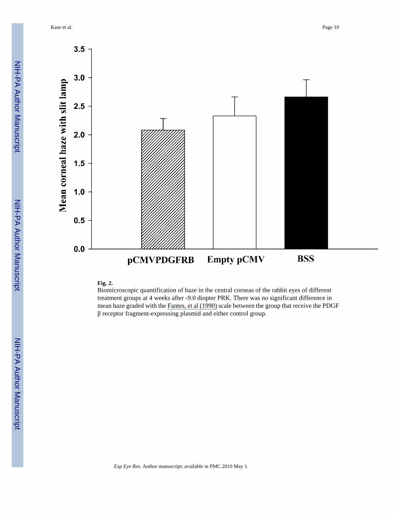
Fig. 2.Biomicroscopic quantification of haze in the central corneas of the rabbit eyes of differenttreatment groups at 4 weeks after -9.0 diopter PRK. There was no significant difference inmean haze graded with the Fantes, et al (1990) scale between the group that receive the PDGFβ receptor fragment-expressing plasmid and either control group.
Kaur et al. Page 10
Exp Eye Res. Author manuscript; available in PMC 2010 May 1.
NIH
-PA Author Manuscript
NIH
-PA Author Manuscript
NIH
-PA Author Manuscript

Fig.3.α-SMA muscle actin staining in rabbit corneas in the experimental group treated withpCMVPDGFRB23KDEL plasmid collected at 4 weeks after -9.0D PRK. A, B: Corneas withno α-SMA staining; C: A cornea with a few α-SMA staining myofibroblasts. Cell nuclei werestained blue with DAPI and α-SMA-positive cells were stained green. Magnification 400X.The bar in each panel is 30 μm.
Kaur et al. Page 11
Exp Eye Res. Author manuscript; available in PMC 2010 May 1.
NIH
-PA Author Manuscript
NIH
-PA Author Manuscript
NIH
-PA Author Manuscript

Fig.4.α-SMA muscle actin staining in rabbit corneas in the control groups collected at 4 weeks after-9.0D PRK. A,B: Corneas treated with empty pCMV plasmid; C: Corneas treated with BSS.Cell nuclei were stained blue with DAPI and α-SMA-positive cells were stained green.Magnification 400X. The bar in each panel is 30 μm.
Kaur et al. Page 12
Exp Eye Res. Author manuscript; available in PMC 2010 May 1.
NIH
-PA Author Manuscript
NIH
-PA Author Manuscript
NIH
-PA Author Manuscript

Fig.5.Quantification of α-SMA-positive cells/400X in the rabbit corneas of the different groups at 4weeks after -9.0D PRK. pCMVPDGFRB: Corneas of rabbits treated withpCMV.PDGFRB23KDEL plasmid; Empty pCMV: Corneas of rabbits treated with the emptypCMV plasmid; BSS: Corneas of rabbits treated with balanced salt solution. The error barsrepresents the mean ± standard error. **, P=0.001 compared to BSS; * P<0.001 compared toempty pCMV.
Kaur et al. Page 13
Exp Eye Res. Author manuscript; available in PMC 2010 May 1.
NIH
-PA Author Manuscript
NIH
-PA Author Manuscript
NIH
-PA Author Manuscript
Copyright © 2022 FDOKUMEN