Lack of MK2 Inhibits Myofibroblast Formation and Exacerbates Pulmonary Fibrosis
11
Lack of MK2 Inhibits Myofibroblast Formation and Exacerbates Pulmonary Fibrosis Tiegang Liu 1 , Rod R. Warburton 1 , Oscar E. Guevara 1 , Nicholas S. Hill 1 , Barry L. Fanburg 1 , Matthias Gaestel 2 , and Usamah S. Kayyali 1 1 Pulmonary and Critical Care Division, Department of Medicine/Tupper Research Institute, Tufts-New England Medical Center, and Tufts University School of Medicine, Boston, Massachusetts; and 2 Institute of Biochemistry, Medical School Hannover, Hannover, Germany Fibroblasts play a major role in tissue repair and remodeling. Their differentiation into myofibroblasts, marked by increased expression of smooth muscle–specific a-actin (a-SMA), is believed to be impor- tant in wound healing and fibrosis. We have recently described a role for MK2 in this phenotypic differentiation in culture. In this article, we demonstrate that MK2 also regulates myofibroblasts in vivo. Disruption of MK2 in mice prevented myofibroblast formation in a model of pulmonary fibrosis. However, MK2 disruption and consequent lack of myofibroblast formation exacerbated fibrosis rather than ameliorated it as previously postulated. When mice lacking MK2 (MK2 2/2 ) were exposed to bleomycin, more collagen accumulated and more fibroblasts populated fibrotic regions in their lungs than in similarly treated wild-type mice. While there were many vimentin-positive cells in the bleomycin-treated MK2 2/2 mouse lungs, few a-SMA–positive cells were observed in these lungs compared with wild-type mouse lungs. siRNA against MK2 reduced a-SMA expression in wild-type mouse embryonic fibroblasts (MEF), consistent with its suppression in MK2 2/2 MEF. On the other hand expressing constitutively active MK2 in MK2 2/2 MEF significantly increased a-SMA expression. MK2 2/2 MEF proliferated at a faster rate and produced more collagen; however, they migrated at a slower rate than wild-type MEF. Overexpressing phosphomimick- ing HSP27, a target of MK2, did not reverse the effect of MK2 disruption on fibroblast migration. MK2 disruption did not affect Smad2 activation by transforming growth factor-b. Thus, MK2 appears to mediate myofibroblast differentiation, and inhibiting that differentiation might contribute to fibrosis rather than protect against it. Keywords: actin; cytoskeleton; fibroblast; p38; idiopathic pulmonary fibrosis Pulmonary fibrosis as a major component of interstitial lung disease is characterized by abnormal fibroblast proliferation and deposition of extracellular matrix proteins that remodel the normal pulmonary tissue structure and compromise its function. In some forms of interstitial lung disease, for example, idio- pathic pulmonary fibrosis (IPF), the mechanism by which fi- brosis arises remains poorly understood (1, 2). Differentiation of fibroblasts into myofibroblasts has long been believed to be an important event in many conditions such as wound repair and fibrosis. For example, it has been reported that myofibro- blasts occur in areas of active fibrosis and are responsible for production and deposition of extracellular matrix (ECM) pro- teins in pulmonary fibrosis (3). We have recently described a key role for MK2 (mitogen-activated protein kinase–activated pro- tein kinase 2 or MAPKAPK2) in the differentiation of mouse embryonic fibroblasts into myofibroblasts (4). We now demon- strate that MK2 is also necessary for myofibroblast formation in vivo in a model of pulmonary fibrosis. Furthermore, our results indicate that MK2 disruption leads to more severe pulmonary fibrosis in response to bleomycin treatment, suggesting that it might be a target in pulmonary and other types of fibrosis. Fibrosis arises in several tissues in response to inflammation and chemical injury, or idiopathically, leading to accumulation of fibroblasts that deposit ECM proteins. Such processes are part of normal wound healing, and are often reversible by apoptosis of accumulating fibroblasts and resorption of ECM proteins. However, in fibrosis the proliferation of fibroblasts and ECM deposits appear excessive and dysregulated, resulting in the gradual displacement of normal tissue by fibrotic foci that disrupt tissue function. Studies aimed at elucidating the mech- anisms of fibrosis implicated certain cytokines in animal models as well as in human patients. In the lungs of patients with IPF, transforming growth factor (TGF)-b (5), IL-1b, and TNF-a (6, 7) were reported to be elevated. Indeed TGF-b is believed to play a pivotal role in fibrosis, and its overepxression in mice is sufficient to cause pulmonary fibrosis independent of injury and inflammation (8, 9). TGF-b is a cytokine that can exert many actions and has been implicated in diseases ranging from cancer to pulmonary hypertension. One action of TGF-b that has been of interest in fibrosis research is its causing fibroblasts to differentiate into myofibroblasts, which are known to accumu- late in sites of fibrosis (3, 10–13). Myofibroblasts, which are believed to play a role in wound contraction, are more contractile than fibroblasts due to in- creased expression of the smooth muscle–specific proteins, such as a-SMA (for review, see Ref. 14). Studies into the mechanism by which myofibroblast differentiation occurs focused on sig- naling pathways activated by TGF-b in fibroblasts. TGF-b is known to signal through Smad proteins (for review, see Ref. 15), but can also activate Ras and Erk (16, 17), Rho GTPase and JNK (18), as well as p38 MAP kinase (19). Inhibition of p38 has been reported to reduce pulmonary (20, 21) and renal (22) fibrosis in animal models. We have recently reported that MK2, a kinase which is a substrate for p38, plays an important role in the differentiation of fibroblasts into myofibroblasts (4). Our findings demonstrated that disrupting the expression of MK2 in CLINICAL RELEVANCE Our findings implicate MK2 in the pathogenesis of pulmo- nary fibrosis. They further suggest that myofibroblasts might play a protective role in pulmonary fibrosis, in contrast to the commonly held view that they contribute to the pathogenesis of fibrosis. (Received in original form March 6, 2007 and in final form June 12, 2007) This work was supported by NIH Grant HL-79320 (to U.S.K.). Correspondence and requests for reprints should be addressed to Usamah S. Kayyali, Ph.D., Pulmonary & Critical Care Division, Tufts-New England Medical Center, 750 Washington Street # 257, Boston, MA 02111. E-mail: ukayyali@ tufts-nemc.org This article has an online supplement, which is accessible from this issue’s table of contents at www.atsjournals.org. Am J Respir Cell Mol Biol Vol 37. pp 507–517, 2007 Originally Published in Press as DOI: 10.1165/rcmb.2007-0077OC on June 28, 2007 Internet address: www.atsjournals.org
-
Upload
independent -
Category
Documents
-
view
5 -
download
0
Transcript of Lack of MK2 Inhibits Myofibroblast Formation and Exacerbates Pulmonary Fibrosis

Lack of MK2 Inhibits Myofibroblast Formation andExacerbates Pulmonary Fibrosis
Tiegang Liu1, Rod R. Warburton1, Oscar E. Guevara1, Nicholas S. Hill1, Barry L. Fanburg1, Matthias Gaestel2,and Usamah S. Kayyali1
1Pulmonary and Critical Care Division, Department of Medicine/Tupper Research Institute, Tufts-New England Medical Center, and Tufts
University School of Medicine, Boston, Massachusetts; and 2Institute of Biochemistry, Medical School Hannover, Hannover, Germany
Fibroblasts play a major role in tissue repair and remodeling. Theirdifferentiation into myofibroblasts, marked by increased expressionof smooth muscle–specific a-actin (a-SMA), is believed to be impor-tant in wound healing and fibrosis. We have recently described a rolefor MK2 in this phenotypic differentiation in culture. In this article,we demonstrate that MK2 also regulates myofibroblasts in vivo.Disruption of MK2 in mice prevented myofibroblast formation ina model of pulmonary fibrosis. However, MK2 disruption andconsequent lack of myofibroblast formation exacerbated fibrosisrather than ameliorated it as previously postulated. When micelacking MK2 (MK22/2) were exposed to bleomycin, more collagenaccumulated and more fibroblasts populated fibrotic regions in theirlungs than in similarly treated wild-type mice. While there weremany vimentin-positive cells in the bleomycin-treated MK22/2
mouse lungs, few a-SMA–positive cells were observed in these lungscompared with wild-type mouse lungs. siRNA against MK2 reduceda-SMA expression in wild-type mouse embryonic fibroblasts (MEF),consistent with its suppression in MK22/2 MEF. On the other handexpressing constitutively active MK2 in MK22/2 MEF significantlyincreased a-SMA expression. MK22/2MEF proliferated at a fasterrate and produced more collagen; however, they migrated ata slower rate than wild-type MEF. Overexpressing phosphomimick-ing HSP27, a target of MK2, did not reverse the effect of MK2disruption on fibroblast migration. MK2 disruption did not affectSmad2 activation by transforming growth factor-b. Thus, MK2appears to mediate myofibroblast differentiation, and inhibitingthat differentiation might contribute to fibrosis rather than protectagainst it.
Keywords: actin; cytoskeleton; fibroblast; p38; idiopathic pulmonaryfibrosis
Pulmonary fibrosis as a major component of interstitial lungdisease is characterized by abnormal fibroblast proliferation anddeposition of extracellular matrix proteins that remodel thenormal pulmonary tissue structure and compromise its function.In some forms of interstitial lung disease, for example, idio-pathic pulmonary fibrosis (IPF), the mechanism by which fi-brosis arises remains poorly understood (1, 2). Differentiationof fibroblasts into myofibroblasts has long been believed to bean important event in many conditions such as wound repairand fibrosis. For example, it has been reported that myofibro-blasts occur in areas of active fibrosis and are responsible for
production and deposition of extracellular matrix (ECM) pro-teins in pulmonary fibrosis (3). We have recently described a keyrole for MK2 (mitogen-activated protein kinase–activated pro-tein kinase 2 or MAPKAPK2) in the differentiation of mouseembryonic fibroblasts into myofibroblasts (4). We now demon-strate that MK2 is also necessary for myofibroblast formationin vivo in a model of pulmonary fibrosis. Furthermore, our resultsindicate that MK2 disruption leads to more severe pulmonaryfibrosis in response to bleomycin treatment, suggesting that itmight be a target in pulmonary and other types of fibrosis.
Fibrosis arises in several tissues in response to inflammationand chemical injury, or idiopathically, leading to accumulationof fibroblasts that deposit ECM proteins. Such processes arepart of normal wound healing, and are often reversible byapoptosis of accumulating fibroblasts and resorption of ECMproteins. However, in fibrosis the proliferation of fibroblasts andECM deposits appear excessive and dysregulated, resulting inthe gradual displacement of normal tissue by fibrotic foci thatdisrupt tissue function. Studies aimed at elucidating the mech-anisms of fibrosis implicated certain cytokines in animal modelsas well as in human patients. In the lungs of patients with IPF,transforming growth factor (TGF)-b (5), IL-1b, and TNF-a (6,7) were reported to be elevated. Indeed TGF-b is believed toplay a pivotal role in fibrosis, and its overepxression in mice issufficient to cause pulmonary fibrosis independent of injury andinflammation (8, 9). TGF-b is a cytokine that can exert manyactions and has been implicated in diseases ranging from cancerto pulmonary hypertension. One action of TGF-b that has beenof interest in fibrosis research is its causing fibroblasts todifferentiate into myofibroblasts, which are known to accumu-late in sites of fibrosis (3, 10–13).
Myofibroblasts, which are believed to play a role in woundcontraction, are more contractile than fibroblasts due to in-creased expression of the smooth muscle–specific proteins, suchas a-SMA (for review, see Ref. 14). Studies into the mechanismby which myofibroblast differentiation occurs focused on sig-naling pathways activated by TGF-b in fibroblasts. TGF-b isknown to signal through Smad proteins (for review, see Ref. 15),but can also activate Ras and Erk (16, 17), Rho GTPase andJNK (18), as well as p38 MAP kinase (19). Inhibition of p38 hasbeen reported to reduce pulmonary (20, 21) and renal (22)fibrosis in animal models. We have recently reported that MK2,a kinase which is a substrate for p38, plays an important role inthe differentiation of fibroblasts into myofibroblasts (4). Ourfindings demonstrated that disrupting the expression of MK2 in
CLINICAL RELEVANCE
Our findings implicate MK2 in the pathogenesis of pulmo-nary fibrosis. They further suggest that myofibroblastsmight play a protective role in pulmonary fibrosis, incontrast to the commonly held view that they contributeto the pathogenesis of fibrosis.
(Received in original form March 6, 2007 and in final form June 12, 2007)
This work was supported by NIH Grant HL-79320 (to U.S.K.).
Correspondence and requests for reprints should be addressed to Usamah
S. Kayyali, Ph.D., Pulmonary & Critical Care Division, Tufts-New England Medical
Center, 750 Washington Street # 257, Boston, MA 02111. E-mail: ukayyali@
tufts-nemc.org
This article has an online supplement, which is accessible from this issue’s table of
contents at www.atsjournals.org.
Am J Respir Cell Mol Biol Vol 37. pp 507–517, 2007
Originally Published in Press as DOI: 10.1165/rcmb.2007-0077OC on June 28, 2007
Internet address: www.atsjournals.org
mouse embryonic fibroblasts causes these cells to express sig-nificantly less a-SMA. Furthermore, cells that lacked MK2 didnot increase a-SMA expression in response to treatment withTGF-b as did wild-type fibroblasts (4). Thus MK2 appeared tobe necessary for myofibroblast differentiation.
We launched this current study to examine whether thedisruption of MK2 in knockout mice will also reduce myofibro-blast differentiation in vivo and whether it will affect thedevelopment of fibrosis. Using bleomycin to model pulmonaryfibrosis, we demonstrate that MK2 is indeed important formyofibroblast formation in vivo. Furthermore, absence of MK2and lack of myofibroblast formation resulted in more severefibrosis. These findings suggest that myofibroblast formationmight be part of the repair phase of fibrosis rather than of activedamage, and that MK2 activity is necessary for that process tooccur.
MATERIALS AND METHODS
Materials and Reagents
Media and supplements (Dulbecco’s modified Eagle’s medium[DMEM]), fetal bovine serum (FBS), penicillin G potassium, strepto-mycin, fungizone, and glutamine were purchased from Invitrogen(Carlsbad, CA). TGF-b was purchased from R&D Systems (Minneapolis,MN) and activated before use according to manufacturer’s instructions.SB431542 and all other reagents and drugs were obtained from Sigma(St. Louis, MO). On the day of the experiments, all drugs were dilutedin serum-free medium.
Animals
Mice that lack MK2 (MK22/2) were generated as described earlier(23). In brief, a neo selection cassette, which contains translational stopcodons in all three reading frames, was inserted into the Sac I sitelocated in the exon that encodes subdomains V and VI of MK2. MK22/2
mice were then backcrossed into C57BL/6J mice, and lack of expres-sion of MK2 was verified by PCR and immunoblotting as describedearlier (4). Wild-type C57BL/6J from Charles River Laboratories(Wilmington, MA) were used as controls for the described experi-ments. These mice were anesthetized with xylozine-ketamine, and thensaline or bleomycin (3 U/kg in 50 ml) were introduced into their lungsby insufflation.
Cell Culture
Immortalized mouse embryonic fibroblasts (MEF) from wild-type andMK22/2 knockout mice were prepared as described earlier (23).Primary embryonic fibroblasts from C57BL/6J wild-type or MK22/2
mice were co-transfected with pSV40Tag encoding the SV40 large Tantigen and the pREP8 plasmid, followed by selection and expansionof histidinol-resistant colonies. MEF cells were maintained in DMEMcontaining 10% FBS, penicillin, streptomycin, fungizone, and gluta-mine at 378C in humidified air containing 5% CO2. MEF were passagedin 0.25% trypsin–0.02% EDTA solution, and 1 day before the experi-ments cells were maintained in serum-free or 1% serum–containingmedia.
For rescue experiments, MK22/2 MEF were transfected withpcDNA3-MK2EE, in which the residues T205E and T317E weremutated to act as constitutively active MK2 (caMK2) as we describedearlier (24). The pcDNA3-MK2EE vector was co-transfected withpMEpuro (a selection vector that confers resistance to puromycin toeukaryotic cells). For studying the effects of HSP27 phosphorylation,wild-type and MK22/2 MEF were transfected with the pcDNA3-HSP27PM (25) to express HSP27 in which the residues, S15D, S78D,and S82D were mutated to mimic phosphorylated HSP27 (pmHSP27).The pmHSP27 vector was introduced alone in the case of wild-typeMEF, and co-transfected with pMEpuro in the case of MK22/2 MEF.The vectors were introduced (5 mg of pmHSP27 or caMK2, and 0.5 mgpMEpuro) into MEF using lipofectamine. Stable transfected wild-typeMEF cell lines were obtained by selection with geneticin, and resistantcolonies were isolated, expanded, and then screened for pmHSP27 or
MK2 by immunoblotting of cell lysates with anti-HSP27 or anti MK2antibodies. As MK22/2 MEF were already resistant to geneticin, stabletransfectants produced by co-transfection with pMEpuro were selectedwith puromycin, and resistant clones were isolated and expanded asabove.
For proliferation assays, cells were seeded in 12-well dishes andcultured for 24, 48, and 72 hours. Counting was performed after tryp-sinizing the cells using a Coulter counter apparatus according to manu-facturer’s instructions.
For migration assays, cells were plated in 35-mm dishes. After24 hours the monolayer was scratched with a sterile tip to makethe wounding gap (time 0 h). Cells were cultured for another 24 hoursin 10% serum media and then fixed with 4% formaldehyde for10 minutes. Four pictures were randomly taken from duplicate dishesand wound closure was compared with time 0 h to assess cell migration.
Collagen Assay
After 14 days of exposure to bleomycin or saline, lungs were removedand snap frozen. Next, lungs were assayed for acid-soluble collagenusing the Sircol Assay (Biocolor Ltd, Newtownabbey, UK). In brief,lungs were minced into small cubes and incubated overnight in 10 volsof 0.5 M acetic acid. Next, homogenates were incubated with an aliquotof Sircol Dye reagent (Sirus Red in picric acid) for 30 minutes. Thecollagen-dye complex was then pelleted by centrifugation at 10,000 3 gfor 10 minutes. The dye was next extracted with 0.5 M NaOH, and theabsorbance of samples along with known collagen standards wasmeasured at 540 nm in a Tecan Spectrafluor plate reader. The sameassay was used to assay collagen in conditioned media from MEF cells(without the acid step) and in cell lysates (scraped into 0.5 M aceticacid).
Histochemistry and Immunohistochemistry
Lungs were instilled intratracheally with 4% formaldehyde at 21 cmH2O pressure. The tissue blocks were then embedded in paraffin, and4-mm sections were cut for staining. Gomori’s Trichrome staining wasused to highlight collagen deposition. The Gomori’s Trichrome stain-ing was used to assign a fibrosis score as follows. Three investigatorswere asked to blindly score three sections from each animal (sevensaline-treated wild-type, eight bleomycin-treated wild-type, four saline-treated MK22/2, and eight bleomycin-treated MK22/2 mice). Eachslide was given a value as 0, 1, or 2 according to the area and degree oftarget staining excluding airways and blood vessels, and the scores werepooled and averaged. Other sections were immunostained with anti-bodies against a-SMA (1A4; Biogenex, San Ramon, CA) and vimentin(VIM 3B4; Ventana, Tucson, AZ) using an automated immunostainer(Ventana) according to manufacturer’s instructions. An IgG control(mouse IgG; Ventana) was included to control for nonspecific staining.The immunostainer uses biotinylated secondary antibodies and horse-radish peroxidase complexed by avidin, followed by diaminobenzidinefor colorimetric detection (Ventana). Three sections from two animals/treatment group were processed for immunolabeling simultaneously tocontrol for staining variability. The area and degree of labeling,excluding airways and blood vessels, were used to rank the sectionsblindly by three investigators. The assigned scores were pooled andaveraged as with the Trichrome staining.
siRNA Transfection
Eighty to ninety percent confluent cells were transfected with MK2siRNA (Cat # 16704; Ambion, Austin, TX) and negative controlsiRNA (Cat # 4611; Ambion) with Lipofectamine 2000 (Invitrogen)at indicated concentrations for 30 hours according to the manufac-turer’s instructions. Western blotting was then performed to test thesilencing effect of siRNA on the expression levels of target proteins.
SDS-PAGE and Immunoblotting
Cell lysates were assayed for protein using the Bradford protein assay(26) and then diluted with 23 Laemmli loading buffer for SDS-PAGE(27). Equal amounts of protein were then loaded in 4 to 20% Tris/glycine gels, and electrophoresed for 90 minutes at 125 V constantvoltage. Next, the gel was blotted onto an Immobilon-P membraneby electrophoretic transfer at 25 V constant voltage overnight. The
508 AMERICAN JOURNAL OF RESPIRATORY CELL AND MOLECULAR BIOLOGY VOL 37 2007
membrane was then washed, blocked with 5% milk, and probed withantibodies against MK2, p38, and phospho p38 (Cell Signaling,Beverly, MA), a-SMA (1A4; Sigma), pan-actin (C-2; Santa Cruz,Santa Cruz, CA), smad2/3(18, BD-Transduction Laboratories, SanJose, CA), and phospho-smad2 (138D4; Cell Signaling). Appropriatesecondary antibodies conjugated to horseradish peroxidase (Pierce,Rockford, IL), and a chemiluminescent substrate (SuperSignal; Pierce)were used according to the manufacturer’s instructions to visualizeimmunoreactive bands.
Statistical Analysis
Data are presented as means 6 SD. Statistical differences weredetermined by either Student’s t test for comparison of two samplemeans or ANOVA for comparison of more than two sample meansfollowed by Holm-Sidak post hoc testing for multiple comparisonsbetween two sample means. Holm-Sidak test considers the rank of theP values of each comparison and the number of comparisons incalculating statistical significance. Statistical analysis was performedusing SigmaStat (Systat, San Jose, CA). P , 0.05 was consideredstatistically significant.
RESULTS
MK22/2 Mice Are More Susceptible to Fibrosis than
Wild-Type Mice
We have recently described that embryonic fibroblasts (MEF)isolated from MK22/2 mice expressed less a-SMA than theirwild-type counterparts either at baseline or in response to TGF-b (4). In continuation of this project we attempted to ascertainthe relevance of these findings to the whole animal. Thus weassessed the development of pulmonary fibrosis in MK22/2
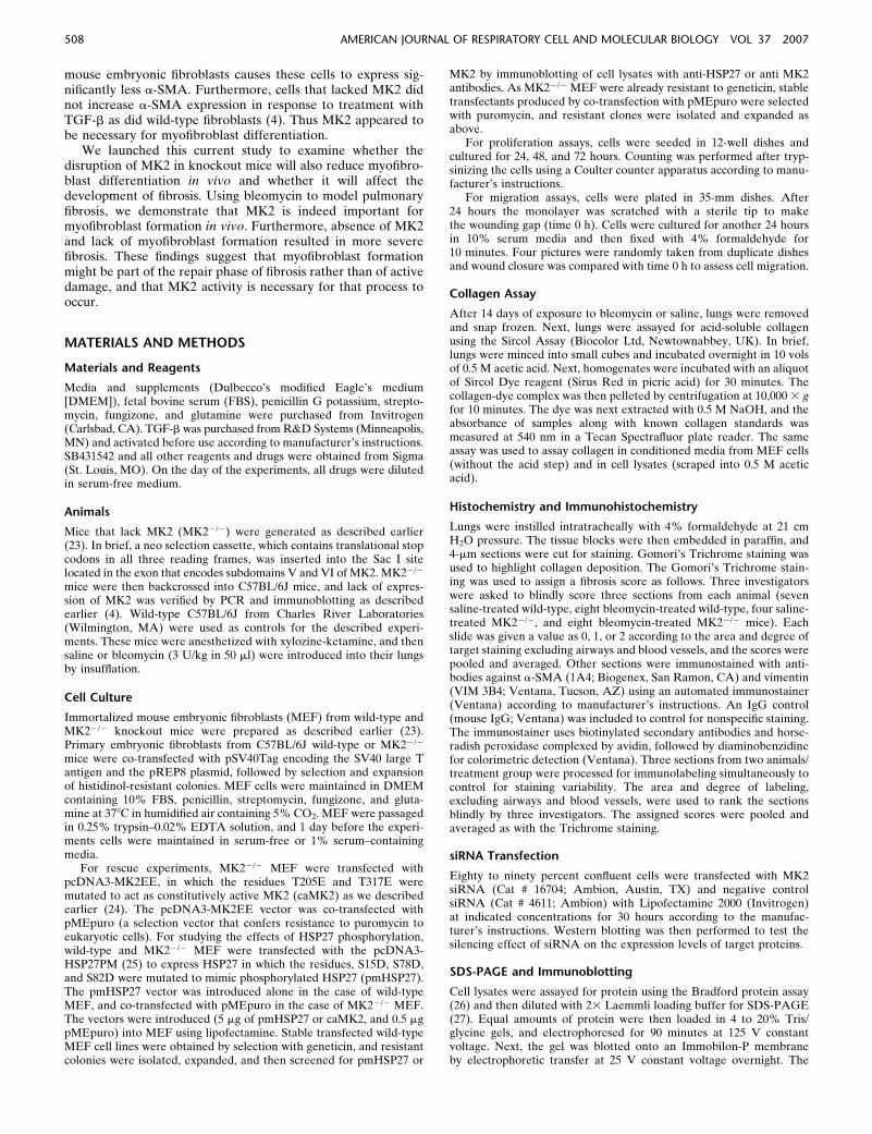
mice compared with wild-type mice. Animals from each groupwere exposed to either saline or bleomycin, a chemotherapeuticassociated with pulmonary fibrosis in human patients that hasbeen used extensively to model pulmonary fibrosis in animals.After 2 weeks of exposure to a single dose of saline orbleomycin, mice were evaluated for the amount of acid-solublecollagen in their lungs. An increase in collagen deposition isassociated with fibrosis. As shown in Figure 1, treatment with
bleomycin caused a significant increase in acid-soluble lungcollagen content in both wild-type and MK22/2 mice. However,the increase in collagen in response to bleomycin was signifi-cantly higher in MK22/2 mice (180%) than in wild-type mice(77%). These data suggested that MK22/2 mice might be moresusceptible to fibrosis than wild-type mice.
To confirm the findings of the collagen assay, we examinedlungs from the mice treated as described above histochemically.After perfusion and fixation, lung sections were stained withGomori’s Trichrome stain, which stains collagen. As shown inFigures 2A–2D, the bleomycin-treated animals had significantcellularity in regions that should correspond to airspaces. TheTrichrome stain also revealed increased collagen deposition(blue) in interstitial spaces of bleomycin-treated mice comparedwith saline-treated mice (Figures 2A–2D). Furthermore, thebleomycin-treated MK22/2 mice developed more severe fibro-sis compared with the wild-type mice. To quantify the fibrosis inthese mice, we ranked several Trichrome-stained sections fromdifferently treated mice as described in MATERIALS AND METHODS.Figure 2E shows the average and standard deviations of thepooled fibrosis scores. Both wild-type and MK22/2 bleomycin-treated mice had significantly higher fibrosis scores than thesaline-treated corresponding mice. Moreover, there was a statis-tically significant difference between the wild-type and MK22/2
bleomycin-treated mice, indicating that MK22/2 mice developedmore severe pulmonary fibrosis than wild-type mice.
Disruption of MK2 Leads to a-SMA Suppression, and
Overexpression of MK2 Rescues Expression of a-SMA in
MK22/2 Fibroblasts
We have recently reported that MEF from MK22/2 miceexpress much less a-SMA than MEF from wild-type mice (4).Since different populations of fibroblasts have been reported toproduce different amounts of a-SMA, we wished to eliminatethe possibility that our findings were due to isolation of differentpopulations of MEF from MK22/2 and wild-type mice. Fur-thermore, we wanted to test if the observed reduction in a-SMAin MK22/2 MEF was due to a developmental effect in MK22/2
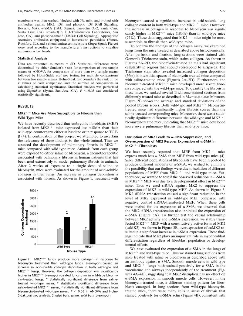
mice. Thus we used siRNA against MK2 to suppress theexpression of MK2 in wild-type MEF. As shown in Figure 3,MK2 siRNA transfection caused a significant reduction in thelevel of MK2 expressed in wild-type MEF compared withnegative control siRNA-transfected MEF. When these cellswere probed for the expression of a-SMA, we observed thatthe MK2 siRNA transfecteion also inhibited the expression ofa-SMA (Figure 3A). To further test the causal relationshipbetween MK2 activity and a-SMA expression, we stably trans-fected MK22/2 MEF with a constitutively active form of MK2(caMK2). As shown in Figure 3B, overexpression of caMK2 re-sulted in a significant increase in a-SMA expression. These find-ings indicate that MK2 plays an important role in myofibroblastdifferentiation regardless of fibroblast population or develop-mental effects.
We next evaluated the expression of a-SMA in the lungs ofMK22/2 and wild-type mice. Thus we stained lung sections frommice treated with saline or bleomycin as described above withan antibody against a-SMA. Smooth muscle cells in wild-typeand MK22/2 lungs both stained positively for a-SMA in thevasculature and airways independently of the treatment (Fig-ures 4A–4E), suggesting that MK2 disruption has no effect ona-SMA expression in smooth muscle cells. However, in thebleomycin-treated mice, a different staining pattern for fibro-blasts emerged. In lung sections from wild-type bleomycin-treated mice, there were many cells in areas of fibrosis thatstained positively for a-SMA actin (Figure 4B), consistent with
Figure 1. MK22/2 lungs produce more collagen in response to
bleomycin treatment than wild-type lungs. Bleomycin caused an
increase in acid-soluble collagen deposition in both wild-type and
MK22/2 lungs. However, the collagen deposition was significantlyhigher in MK22/2 bleomycin-treated lungs than in wild-type bleomy-
cin-treated lungs. * Statistically significant difference from saline-
treated wild-type mean, # statistically significant difference from
saline-treated MK22/2 mean, ^ statistically significant difference frombleomycin-treated wild-type mean. P , 0.05 in ANOVA and Holm-
Sidak post hoc analysis. Shaded bars, saline; solid bars, bleomycin.
Liu, Warburton, Guevara, et al.: MK2 Inhibition Exacerbates Fibrosis 509

the presence of myofibroblasts in these lesions. However, areasof extensive fibrosis in the MK22/2 bleomycin-treated micecontained minimal staining for a-SMA (Figure 4D). Weattempted to quantify differences in a-SMA staining by havingthree investigators blindly rank several slides for the degree ofa-SMA labeling in fibrotic areas. As shown in Figure 4E,MK22/2 sections contained significantly less a-SMA–positivefibroblasts than wild-type sections. Furthermore, the number ofa-SMA–positive cells populating areas of fibrosis in bleomycin-treated MK22/2 sections were not significantly different fromeither wild-type or MK22/2 saline-treated sections. We con-firmed the presence of fibroblasts in these areas using an
antibody against vimentin, which revealed many vimentin-positive cells in areas of fibrosis in both wild-type and MK22/2
belomycin-treated mice (Figure 4F-I). Indeed scoring of vimentinslides in the manner we followed with a-SMA staining revealedmore vimentin staining in bleomycin-treated MK22/2 sectionsthan in bleomycin-treated wild-type sections (Figure 4J). Thesefindings indicate that extensive pulmonary fibrosis can occur ata more severe degree without correlating with a higher numberof a-SMA–positive fibroblasts. Since a-SMA is one of the mostcommonly used markers for myofibroblasts, a reexamination ofits role in defining myofibroblasts or the role of myofibroblastsin fibrosis might be warranted.
Figure 2. MK22/2 mice develop more severe fibrosis in response to bleomycin than wild-type mice. Treatment of wild-type mice with bleomycin
(B) caused significant cellularity and collagen deposition (blue staining) relative to saline-treated wild-type mice (A), as revealed by Gomori’sTrichrome stain. MK22/2 mice exhibited more severe cellularity and collagen deposition in response to bleomycin (D), compared with saline-treated
MK22/2 mice (C) and bleomycin-treated wild-type mice (B). Several slides were scored as described in MATERIALS AND METHODS. Average scores
demonstrate that bleomycin-treated MK22/2 mice develop more severe fibrosis than bleomycin-treated wild-type mice (E). Shaded bars, saline; solid
bars, bleomycin. * Statistically significant difference from saline-treated wild-type mean, # statistically significant difference from saline-treatedMK22/2 mean, ^ statistically significant difference from bleomycin-treated wild-type mean. P , 0.05 in ANOVA and Holm-Sidak post hoc analysis.
510 AMERICAN JOURNAL OF RESPIRATORY CELL AND MOLECULAR BIOLOGY VOL 37 2007
Embryonic Fibroblasts from MK22/2 Mice Exhibit a Higher
Proliferative Rate and Secrete More Collagen than
Wild-Type Fibroblasts
Since both MEF and whole animals exhibit the same charac-teristic inhibition of a-SMA in response to MK2 disruption, weinvestigated other characteristics of fibroblasts in culture thatmight explain the observations we made in bleomycin-treatedmice. When seeded at equal density, MEF from MK22/2 micegrew at a faster rate than their wild-type counterparts. Thiseffect was observed at 10% serum and in the absence of serum,such that MK22/2 were more tolerant to serum deprivation. Asshown in Figure 5A, MK22/2 MEF cell count increases fasterthan wild-type MEF. Such increased proliferation rate mightexplain the excessive cellularity observed in fibrotic foci inbleomycin-treated MK22/2 mouse lungs.
We next assayed collagen in wild-type and MK22/2 MEFafter seeding at equal density and maintaining in 1% serum for24 hours. As shown in Figure 5B, MK22/2 fibroblasts producedmore collagen than their corresponding wild-type fibroblasts.These data are consistent with the increased collagen depositionwe observed in the lungs of bleomycin-treated MK22/2 micerelative to wild-type mice (Figure 1).
MK22/2 Fibroblasts Are Defective in Migratory Activity and
the Defect Is Not Due to Lack of Phosphorylated HSP27
We have already established that MEF from MK2 mice lack a-SMA and linked that causally to MK2 expression. MK22/2
MEF have been described to migrate more slowly than their
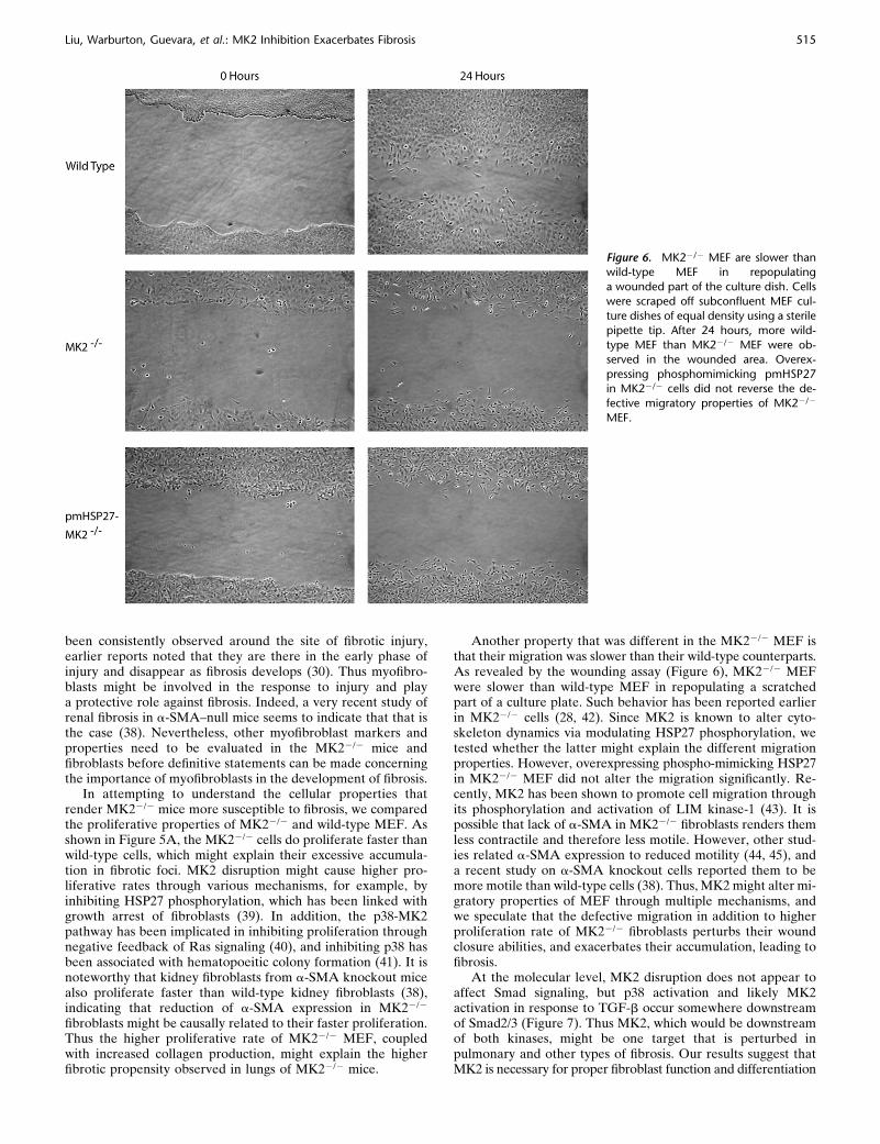
wild-type counterparts (28). We have assayed the ability ofMEF to repopulate a wound made by a sterile pipette in a tissueculture dish. As seen in Figure 6, wild-type MEF repopulatedthe wounded area much faster than the MK22/2 cells. As MK2is known to alter the actin cytoskeleton through its direct actionon the small heat shock protein HSP27, we also generatedstably transfected phosphomimicking (pm) HSP27 MK22/2
MEF. We have previously demonstrated that expressing thisconstruct stably in endothelial cells causes an increase in actinstress fibers, which is believed to represent a more contractilephenotype (24). While transfected MK22/2 MEF expressed pmHSP27, they were still defective in migration compared withwild-type cells (Figure 6). These results indicate that MK2affects migration of fibroblasts, not through its effect on HSP27phosphorylation, but possibly through a direct effect on a-SMAexpression.
Disruption of MK2 Does Not Affect Smad Signaling, Which
Appears to Be Upstream of p38
Fibroblast proliferation and a-SMA expression have beenlinked to factors such as TGF-b, which is known to exert manyof its effects through Smad signaling. Thus we wished to ex-amine this signaling pathway in MK22/2 versus wild-type MEF.After treatment with TGF-b (1 ng/ml) for different times, weassayed Smad2 phosphorylation. As shown in Figure 7A, Smad2phosphorylation is activated by TGF-b similarly in wild-type orMK22/2 MEF, indicating that MK2 is not upstream of Smad2 inTGF-b–activated signaling. We also examined the activation ofp38, which is an upstream activator of MK2, by TGF-b in bothcell types, and demonstrated that it is indeed activated, albeit ata later time-course than Smad2 (Figure 7A). The fact that p38becomes activated in MK22/2 cells is consistent with its beingupstream of MK2. When wild-type or MK22/2 fibroblasts werepreincubated with an inhibitor of TGF-b Type I receptor kinaseSB431542 (10 mM), Smad2 activation by TGF-b peaking at 60minutes was inhibited in both cell types. Furthermore, inhibitingSmad2 activation by SB431542 also inhibited p38 activation(Figure 7B). These findings suggest that Smad2 signals upstreamof MK2 and that the effects of MK2 on fibroblast physiologyand fibrosis are not due to altered downstream Smad signaling.
DISCUSSION
Fibrosis occurs when fibroblasts and the extracellular matrixproteins they produce, such as collagen, accumulate in tissues,replacing normal tissue that is required for proper organfunction. In pulmonary fibrosis, which can arise in response todrugs, asbestos, physical injury, inflammation, or unknown agents,the replacement of alveolar epithelium by fibrotic tissue resultsin inadequate gas exchange. Pulmonary fibrosis, particularlywhen due to unknown etiology (i.e., IPF), is usually progressiveand fatal. Research on the role played by fibroblasts in fibrosishas increasingly focused on a differentiated form of fibroblasts,the myofibroblast. Because of their expression of smooth muscle–specific proteins, such as a-SMA, their known role in woundcontraction and cytokine production, as well as their localiza-tion near fibrotic foci, myofibroblasts were suspected to pro-mote fibrosis. Thus significant research has been conducted intothe mechanism by which fibroblasts differentiate into myofibro-blasts. In this article we describe an important role for thekinase MK2 in fibroblast differentiation into myofibroblasts inculture and in mice with pulmonary fibrosis. Furthermore, ourresults suggest that MK2 and myofibroblast formation mightplay a role in protection against rather than in causing fibrosis.
Figure 3. Knocking down MK2 reduces a-SMA expression. Expressionof constitutively active MK2 in MK22/2 MEF restores a-SMA expression.
Transfection of wild-type MEF with siRNA against MK2 reduced the
level of MK2 and a-SMA relative to negative control siRNA (C siRNA)
transfection in a dose-dependent manner (A). MK2 siRNA transfectionhad no effect on total actin expression (A). On the other hand,
transfection of MK22/2 MEF with a constitutively active form of MK2
(caMK2) caused a significant increase in a-SMA expression (B).
Liu, Warburton, Guevara, et al.: MK2 Inhibition Exacerbates Fibrosis 511
Myofibroblasts are believed to be derived from fibroblasts orepithelial cells through differentiation into a muscle cell–likephenotype, which is identified by expression of smooth muscle–specific proteins such as a-SMA. The classical profibrotic cyto-kine TGF-b, which is elevated in IPF lungs (5), causes fibroblasts
to differentiate into myofibroblasts (10–13). As TGF-b has beencentral to pulmonary fibrosis pathogenesis (5, 29, 30), investigat-ing its effect on fibroblasts was of particular interest. TGF-bbelongs to a large family of proteins, including members such asbone morphogenetic proteins, that are implicated in a variety of
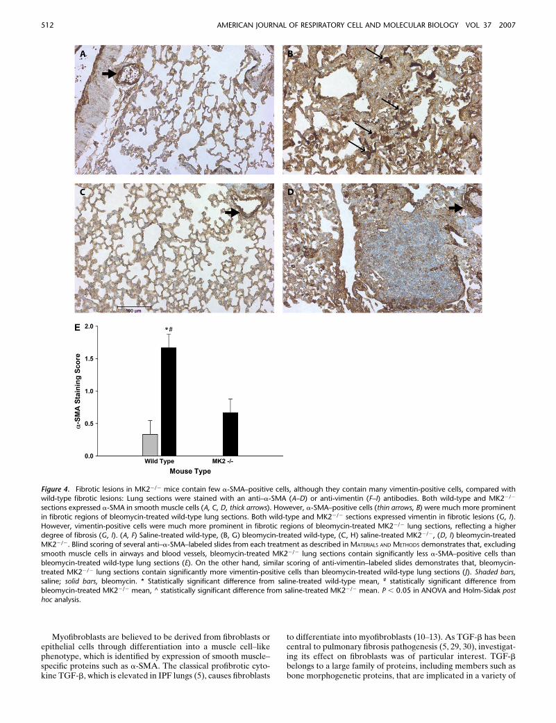
Figure 4. Fibrotic lesions in MK22/2 mice contain few a-SMA–positive cells, although they contain many vimentin-positive cells, compared withwild-type fibrotic lesions: Lung sections were stained with an anti–a-SMA (A–D) or anti-vimentin (F–I) antibodies. Both wild-type and MK22/2
sections expressed a-SMA in smooth muscle cells (A, C, D, thick arrows). However, a-SMA–positive cells (thin arrows, B) were much more prominent
in fibrotic regions of bleomycin-treated wild-type lung sections. Both wild-type and MK22/2 sections expressed vimentin in fibrotic lesions (G, I).
However, vimentin-positive cells were much more prominent in fibrotic regions of bleomycin-treated MK22/2 lung sections, reflecting a higherdegree of fibrosis (G, I). (A, F) Saline-treated wild-type, (B, G) bleomycin-treated wild-type, (C, H) saline-treated MK22/2, (D, I) bleomycin-treated
MK22/2. Blind scoring of several anti–a-SMA–labeled slides from each treatment as described in MATERIALS AND METHODS demonstrates that, excluding
smooth muscle cells in airways and blood vessels, bleomycin-treated MK22/2 lung sections contain significantly less a-SMA–positive cells than
bleomycin-treated wild-type lung sections (E). On the other hand, similar scoring of anti-vimentin–labeled slides demonstrates that, bleomycin-treated MK22/2 lung sections contain significantly more vimentin-positive cells than bleomycin-treated wild-type lung sections (J). Shaded bars,
saline; solid bars, bleomycin. * Statistically significant difference from saline-treated wild-type mean, # statistically significant difference from
bleomycin-treated MK22/2 mean, ^ statistically significant difference from saline-treated MK22/2 mean. P , 0.05 in ANOVA and Holm-Sidak posthoc analysis.
512 AMERICAN JOURNAL OF RESPIRATORY CELL AND MOLECULAR BIOLOGY VOL 37 2007

diseases, such as cancer and pulmonary hypertension. It exerts itsactions primarily through cell surface receptors that activateSmad proteins (for review, see Ref. 15). TGF-b has also beenreported to activate Ras and Erk (16, 17), Rho GTPase and JNK(18), and p38 MAP kinase (19). Myofibroblast differentiation inresponse to TGF-b has been suggested to be mediated by p38,and inhibition of p38 has been reported to reduce pulmonary (20,21) and renal (22) fibrosis in animal models. Yet some research-ers reported no effect of inhibiting p38 on differentiation of lungfibroblasts into myofibroblasts (31).
p38 is activated by stimuli such as ultraviolet radiation andhyperosmolarity and a variety of cytokines and growth factorsthat lead to its phosphorylation. Activated p38 phosphorylatesa variety of substrates, including the kinase MK2, leading to itsactivation. MK2 in turn phosphorylates additional substrates,such as the small heat shock protein HSP27. The latter eventleads to actin stress fiber formation because phosphorylated
HSP27 no longer blocks the polymerization of actin (32). Wehave investigated the p38-MK2-HSP27 pathway in endothelialcells and demonstrated that it mediates actin stress fiberformation in response to hypoxia (24, 33). Our laboratory hasalso recently demonstrated that fibroblasts that lack MK2contain less actin stress fibers than wild-type fibroblasts (4).These fibroblasts also contained significantly less a-SMA thantheir wild-type counterparts. Formation of actin stress fibers,which is expected to be reduced in MK22/2 cells that cannotphosphorylate HSP27, has been associated with increased cellcontractility, which in turn can induce a-SMA expression (34,35). This effect, however, is not likely to explain the reduced a-SMA expression in MK22/2 MEF, because overexpressingphospho-mimicking HSP27 in these cells did not significantlyincrease a-SMA expression (data not shown).
Our recent manuscript also showed that the MK22/2 fibro-blasts did not differ from wild-type cells in the activation of
Figure 4. (Continued ).
Liu, Warburton, Guevara, et al.: MK2 Inhibition Exacerbates Fibrosis 513

serum response element, which is one of the main pathways fora-SMA induction. However, MK2 disruption significantly reduceda-SMA mRNA stability (4), which is consistent with the knownaction of MK2 on cytokine expression (28, 36, 37). As we haveperformed these experiments in MEF, and since populationsof fibroblasts are known to be heterogeneous in the level ofa-SMA being expressed, we further tested the relation of a-SMAexpression to MK2 by transfecting wild-type MEF with MK2siRNA. The latter approach also caused a reduction in a-SMAexpression (Figure 3A). On the other hand, when we overex-pressed constitutively active MK2 in MK22/2 fibroblasts, a-SMAexpression increased significantly. These results coupled withthe observations made in mouse lungs argue against the ob-served difference in a-SMA level being due to variation in iso-lated MEF populations, or due to developmental effects ona-SMA expression in MK22/2 mice from which the MEF wereisolated. Indeed, the effect of MK2 appears to be specific tofibroblasts in the sense that we have seen intense a-SMAstaining in smooth muscle cells in lung airways and vasculatureof MK22/2 mice.
As myofibroblasts are believed to play an important role inwound repair and fibrosis, we evaluated whether the MK22/2
mice differed in their susceptibility to pulmonary fibrosis in-duced by bleomycin treatment. The MK22/2 mice developedmore severe fibrosis in response to belomycin than wild-typemice, as judged by the amount of collagen deposited in theirlungs, as well as by immunohistochemical evidence includingincreased cellularity and Trichrome staining. While the accu-mulated cells in fibrotic foci of MK22/2 mice were stainedpositively for vimentin, a fibroblast marker, they were notsignificantly stained for a-SMA. On the other hand, fibroticfoci of wild-type mice contained cells that stained positively forboth vimentin and a-SMA. Our findings suggest that MK2 isneeded for myofibroblast differentiation in fibrotic injury, yet itsabsence and lack of myofibroblasts formation appear to exac-erbate fibrosis instead of ameliorating it. While this conclusionis contrary to commonly expressed views on the role ofmyofibroblasts in pulmonary fibrosis, the evidence for myofi-broblast involvement in pathogenesis has been more circum-stantial than direct. For instance, while myofibroblasts have
Figure 5. MK22/2 MEF proliferate faster and produce more collagen than wild-type MEF. When wild-type and MK22/2 MEF were seeded at equal
density, and counted after 24, 48, and 72 h, MK22/2 MEF cell count increased faster than wild-type MEF cell count (A). MEF from wild-type and
MK22/2 mice were seeded at equal density. After 24 hours, collagen was assayed as described in MATERIALS AND METHODS in conditioned media, as wellas in attached cells. MK22/2 MEF produced more collagen than their wild-type counterparts (B). *Statistically significant difference from wild-type
mean, P , 0.05 in t test.
514 AMERICAN JOURNAL OF RESPIRATORY CELL AND MOLECULAR BIOLOGY VOL 37 2007
been consistently observed around the site of fibrotic injury,earlier reports noted that they are there in the early phase ofinjury and disappear as fibrosis develops (30). Thus myofibro-blasts might be involved in the response to injury and playa protective role against fibrosis. Indeed, a very recent study ofrenal fibrosis in a-SMA–null mice seems to indicate that that isthe case (38). Nevertheless, other myofibroblast markers andproperties need to be evaluated in the MK22/2 mice andfibroblasts before definitive statements can be made concerningthe importance of myofibroblasts in the development of fibrosis.
In attempting to understand the cellular properties thatrender MK22/2 mice more susceptible to fibrosis, we comparedthe proliferative properties of MK22/2 and wild-type MEF. Asshown in Figure 5A, the MK22/2 cells do proliferate faster thanwild-type cells, which might explain their excessive accumula-tion in fibrotic foci. MK2 disruption might cause higher pro-liferative rates through various mechanisms, for example, byinhibiting HSP27 phosphorylation, which has been linked withgrowth arrest of fibroblasts (39). In addition, the p38-MK2pathway has been implicated in inhibiting proliferation throughnegative feedback of Ras signaling (40), and inhibiting p38 hasbeen associated with hematopoeitic colony formation (41). It isnoteworthy that kidney fibroblasts from a-SMA knockout micealso proliferate faster than wild-type kidney fibroblasts (38),indicating that reduction of a-SMA expression in MK22/2
fibroblasts might be causally related to their faster proliferation.Thus the higher proliferative rate of MK22/2 MEF, coupledwith increased collagen production, might explain the higherfibrotic propensity observed in lungs of MK22/2 mice.
Another property that was different in the MK22/2 MEF isthat their migration was slower than their wild-type counterparts.As revealed by the wounding assay (Figure 6), MK22/2 MEFwere slower than wild-type MEF in repopulating a scratchedpart of a culture plate. Such behavior has been reported earlierin MK22/2 cells (28, 42). Since MK2 is known to alter cyto-skeleton dynamics via modulating HSP27 phosphorylation, wetested whether the latter might explain the different migrationproperties. However, overexpressing phospho-mimicking HSP27in MK22/2 MEF did not alter the migration significantly. Re-cently, MK2 has been shown to promote cell migration throughits phosphorylation and activation of LIM kinase-1 (43). It ispossible that lack of a-SMA in MK22/2 fibroblasts renders themless contractile and therefore less motile. However, other stud-ies related a-SMA expression to reduced motility (44, 45), anda recent study on a-SMA knockout cells reported them to bemore motile than wild-type cells (38). Thus, MK2 might alter mi-gratory properties of MEF through multiple mechanisms, andwe speculate that the defective migration in addition to higherproliferation rate of MK22/2 fibroblasts perturbs their woundclosure abilities, and exacerbates their accumulation, leading tofibrosis.
At the molecular level, MK2 disruption does not appear toaffect Smad signaling, but p38 activation and likely MK2activation in response to TGF-b occur somewhere downstreamof Smad2/3 (Figure 7). Thus MK2, which would be downstreamof both kinases, might be one target that is perturbed inpulmonary and other types of fibrosis. Our results suggest thatMK2 is necessary for proper fibroblast function and differentiation
Figure 6. MK22/2 MEF are slower thanwild-type MEF in repopulating
a wounded part of the culture dish. Cells
were scraped off subconfluent MEF cul-
ture dishes of equal density using a sterilepipette tip. After 24 hours, more wild-
type MEF than MK22/2 MEF were ob-
served in the wounded area. Overex-
pressing phosphomimicking pmHSP27in MK22/2 cells did not reverse the de-
fective migratory properties of MK22/2
MEF.
Liu, Warburton, Guevara, et al.: MK2 Inhibition Exacerbates Fibrosis 515

into myofibroblasts. Restoring that activity or increasing it throughdifferent approaches might be of therapeutic value in wound heal-ing and fibrosis.
Conflict of Interest Statement: None of the authors has a financial relationshipwith a commercial entity that has an interest in the subject of this manuscript.
Acknowledgments: The authors are grateful to Dr. Howard Surks (Tufts-NewEngland Medical Center), and Dr. Kazuo Maruyama (Tokyo Medical and DentalUniversity) for providing the pMEpuro vector.
References
1. American Thoracic Society/European Respiratory Society. Idiopathicpulmonary fibrosis: diagnosis and treatment. International consensusstatement. Am J Respir Crit Care Med 2000;161:646–664.
2. Cherniack RM, Crystal RG, Kalica AR. NHLBI workshop summary.Current concepts in idiopathic pulmonary fibrosis: a road map for thefuture. Am Rev Respir Dis 1991;143:680–683.
3. Vyalov SL, Gabbiani G, Kapanci Y. Rat alveolar myofibroblasts acquirealpha-smooth muscle actin expression during bleomycin-inducedpulmonary fibrosis. Am J Pathol 1993;143:1754–1765.
4. Sousa AM, Liu T, Guevara O, Stevens J, Fanburg BL, Gaestel M,Toksoz D, Kayyali US. Smooth muscle alpha-actin expression andmyofibroblast differentiation by tgfbeta are dependent upon mk2.J Cell Biochem 2007;100:1581–1592.
5. Khalil N, O’Connor RN, Unruh HW, Warren PW, Flanders KC, KempA, Bereznay OH, Greenberg AH. Increased production and immu-nohistochemical localization of transforming growth factor-beta in idio-pathic pulmonary fibrosis. Am J Respir Cell Mol Biol 1991;5:155–162.
6. Pan LH, Ohtani H, Yamauchi K, Nagura H. Co-expression of tnf alphaand il-1 beta in human acute pulmonary fibrotic diseases: animmunohistochemical analysis. Pathol Int 1996;46:91–99.
7. Piguet PF, Ribaux C, Karpuz V, Grau GE, Kapanci Y. Expression andlocalization of tumor necrosis factor-alpha and its mrna in idiopathicpulmonary fibrosis. Am J Pathol 1993;143:651–655.
8. Lee CG, Cho SJ, Kang MJ, Chapoval SP, Lee PJ, Noble PW,Yehualaeshet T, Lu B, Flavell RA, Milbrandt J, et al. Early growthresponse gene 1-mediated apoptosis is essential for transforminggrowth factor fbetag1-induced pulmonary fibrosis. J Exp Med 2004;200:377–389.
9. Sime PJ, Xing Z, Graham FL, Csaky KG, Gauldie J. Adenovector-mediated gene transfer of active transforming growth factor-beta 1induces prolonged severe fibrosis in rat lung. J Clin Invest 1997;100:768–776.
10. Desmouliere A, Geinoz A, Gabbiani F, Gabbiani G. Transforminggrowth factor-beta 1 induces alpha-smooth muscle actin expression ingranulation tissue myofibroblasts and in quiescent and growingcultured fibroblasts. J Cell Biol 1993;122:103–111.
11. Ronnov-Jessen L, Petersen OW. Induction of alpha-smooth muscle actinby transforming growth factor-beta 1 in quiescent human breast glandfibroblasts. Implications for myofibroblast generation in breast neo-plasia. Lab Invest 1993;68:696–707.
12. Roy SG, Nozaki Y, Phan SH. Regulation of [alpha]-smooth muscle actingene expression in myofibroblast differentiation from rat lung fibro-blasts. Int J Biochem Cell Biol 2001;33:723.
13. Yokozeki M, Moriyama K, Shimokawa H, Kuroda T. Transforminggrowth factor-beta 1 modulates myofibroblastic phenotype of ratpalatal fibroblasts in vitro. Exp Cell Res 1997;231:328–336.
14. Hinz B, Gabbiani G. Mechanisms of force generation and transmissionby myofibroblasts. Curr Opin Biotechnol 2003;14:538–546.
15. Attisano L, Wrana JL. Signal transduction by the tgf-beta superfamily.Science 2002;296:1646–1647.
16. Hartsough MT, Frey RS, Zipfel PA, Buard A, Cook SJ, McCormick F,Mulder KM. Altered transforming growth factor signaling in epithe-lial cells when ras activation is blocked. J Biol Chem 1996;271:22368–22375.
17. Hartsough MT, Mulder KM. Transforming growth factor beta activationof p44[image] in proliferating cultures of epithelial cells. J Biol Chem1995;270:7117–7124.
18. Atfi A, Djelloul S, Chastre E, Davis R, Gespach C. Evidence for a roleof rho-like gtpases and stress-activated protein kinase/c-jun n-terminal
Figure 7. MK2 is not upstream of Smad
signaling in response to TGF-b. (A) TGF-b
causes phosphorylation of both Smad2/3and p38 in wild-type and MK22/2 MEF.
Smad2/3 phosphorylation is observed
before p38 phosphorylation. (B) Inhibit-
ing the TGF-b Type I receptor kinase bySB431542 blocked Smad2/3 phosphory-
lation in both wild-type and MK22/2
MEF. SB431542 also blocked the activa-tion of p38 by TGF-b.
516 AMERICAN JOURNAL OF RESPIRATORY CELL AND MOLECULAR BIOLOGY VOL 37 2007

kinase (sapk/jnk) in transforming growth factor beta -mediated signal-ing. J Biol Chem 1997;272:1429–1432.
19. Hanafusa H, Ninomiya-Tsuji J, Masuyama N, Nishita M, Fujisawa J-i,Shibuya H, Matsumoto K, Nishida E. Involvement of the p38 mitogen-activated protein kinase pathway in transforming growth factor-beta -induced gene expression. J Biol Chem 1999;274:27161–27167.
20. Matsuoka H, Arai T, Mori M, Goya S, Kida H, Morishita H, Fujiwara H,Tachibana I, Osaki T, Hayashi S. A p38 mapk inhibitor, fr-167653,ameliorates murine bleomycin-induced pulmonary fibrosis. Am JPhysiol Lung Cell Mol Physiol 2002;283:L103–L112.
21. Underwood DC, Osborn RR, Bochnowicz S, Webb EF, Rieman DJ, LeeJC, Romanic AM, Adams JL, Hay DW, Griswold DE. Sb 239063,a p38 mapk inhibitor, reduces neutrophilia, inflammatory cytokines,mmp-9, and fibrosis in lung. Am J Physiol Lung Cell Mol Physiol2000;279:L895–L902.
22. Stambe C, Atkins RC, Tesch GH, Masaki T, Schreiner GF, Nikolic-Paterson DJ. The role of p38alpha mitogen-activated protein kinaseactivation in renal fibrosis. J Am Soc Nephrol 2004;15:370–379.
23. Kotlyarov A, Neininger A, Schubert C, Eckert R, Birchmeier C, VolkHD, Gaestel M. Mapkap kinase 2 is essential for lps-induced tnf-alpha biosynthesis. Nat Cell Biol 1999;1:94–97.
24. Kayyali US, Pennella CM, Trujillo C, Villa O, Gaestel M, Hassoun PM.Cytoskeletal changes in hypoxic pulmonary endothelial cells aredependent on the map kinase-associated protein kinase mk2. J BiolChem 2002;28:28.
25. Rogalla T, Ehrnsperger M, Preville X, Kotlyarov A, Lutsch G, DucasseC, Paul C, Wieske M, Arrigo AP, Buchner J, et al. Regulation ofhsp27 oligomerization, chaperone function, and protective activityagainst oxidative stress/tumor necrosis factor alpha by phosphoryla-tion. J Biol Chem 1999;274:18947–18956.
26. Bradford MM. A rapid and sensitive method for the quantitation ofmicrogram quantities of protein utilizing the principle of protein-dyebinding. Anal Biochem 1976;72:248–254.
27. Laemmli UK. Cleavage of structural proteins during the assembly of thehead of bacteriophage t4. Nature 1970;227:680–685.
28. Kotlyarov A, Gaestel M. Is mk2 (mitogen-activated protein kinase-activated protein kinase 2) the key for understanding post-transcrip-tional regulation of gene expression? Biochem Soc Trans 2002;30:959–963.
29. Broekelmann TJ, Limper AH, Colby TV, McDonald JA. Transforminggrowth factor fbetag1 is present at sites of extracellular matrix geneexpression in human pulmonary fibrosis. Proc Natl Acad Sci USA1991;88:6642–6646.
30. Kapanci Y, Desmouliere A, Pache JC, Redard M, Gabbiani G.Cytoskeletal protein modulation in pulmonary alveolar myofibro-blasts during idiopathic pulmonary fibrosis: possible role of trans-forming growth factor beta and tumor necrosis factor alpha. Am JRespir Crit Care Med 1995;152:2163–2169.
31. Hashimoto S, Gon Y, Takeshita I, Matsumoto K, Maruoka S, Horie T.Transforming growth factor-beta1 induces phenotypic modulation ofhuman lung fibroblasts to myofibroblast through a c-jun-nh2-terminalkinase-dependent pathway. Am J Respir Crit Care Med 2001;163:152–157.
32. Benndorf R, Hayess K, Ryazantsev S, Wieske M, Behlke J, Lutsch G.Phosphorylation and supramolecular organization of murine smallheat shock protein hsp25 abolish its actin polymerization-inhibitingactivity. J Biol Chem 1994;269:20780–20784.
33. An SS, Pennella CM, Gonnabathula A, Chen J, Wang N, Gaestel M,Hassoun PM, Fredberg JJ, Kayyali US. Hypoxia alters biophysicalproperties of endothelial cells via p38 mapk- and rho kinase-dependentpathways. Am J Physiol Cell Physiol 2005;289:C521–C530.
34. Hinz B, Mastrangelo D, Iselin CE, Chaponnier C, Gabbiani G.Mechanical tension controls granulation tissue contractile activityand myofibroblast differentiation. Am J Pathol 2001;159:1009–1020.
35. Arora PD, Narani N, McCulloch CAG. The compliance of collagen gelsregulates transforming growth factor-b induction of falphag-smoothmuscle actin in fibroblasts. Am J Pathol 1999;154:871–882.
36. Neininger A, Kontoyiannis D, Kotlyarov A, Winzen R, Eckert R, VolkH-D, Holtmann H, Kollias G, Gaestel M. Mk2 targets au-richelements and regulates biosynthesis of tumor necrosis factor andinterleukin-6 independently at different post-transcriptional levels.J Biol Chem 2002;277:3065–3068.
37. Hitti E, Iakovleva T, Brook M, Deppenmeier S, Gruber AD, RadziochD, Clark AR, Blackshear PJ, Kotlyarov A, Gaestel M. Mitogen-activated protein kinase-activated protein kinase 2 regulates tumornecrosis factor mrna stability and translation mainly by alteringtristetraprolin expression, stability, and binding to adenine/uridine-rich element. Mol Cell Biol 2006;26:2399–2407.
38. Takeji M, Moriyama T, Oseto S, Kawada N, Hori M, Imai E, Miwa T.Smooth muscle falphag-actin deficiency in myofibroblasts leads toenhanced renal tissue fibrosis. J Biol Chem 2006;281:40193–40200.
39. Arata S, Hamaguchi S, Nose K. Inhibition of colony formation of nih 3t3cells by the expression of the small molecular weight heat shockprotein hsp27: Involvement of its phosphorylation and aggregation atthe c-terminal region. J Cell Physiol 1997;170:19–26.
40. Chen G, Hitomi M, Han J, Stacey DW. The p38 pathway providesnegative feedback for ras proliferative signaling. J Biol Chem 2000;275:38973–38980.
41. Navas TA, Mohindru M, Estes M, Ma JY, Sokol L, Pahanish P, ParmarS, Haghnazari E, Zhou L, Collins R, et al. Inhibition of overactivatedp38 mapk can restore hematopoiesis in myelodysplastic syndromeprogenitors. Blood 2006;108:4170–4177.
42. Hannigan MO, Zhan L, Ai Y, Kotlyarov A, Gaestel M, Huang CK.Abnormal migration phenotype of mitogen-activated protein kinase-activated protein kinase 22/2 neutrophils in zigmond chamberscontaining formyl-methionyl-leucyl-phenylalanine gradients. J Immunol2001;167:3953–3961.
43. Kobayashi M, Nishita M, Mishima T, Ohashi K, Mizuno K. Mapkapk-2-mediated lim-kinase activation is critical for vegf-induced actin remod-eling and cell migration. EMBO J 2006;25:713–726.
44. Ronnov-Jessen L, Petersen OW. A function for filamentous alpha-smooth muscle actin: Retardation of motility in fibroblasts. J Cell Biol1996;134:67–80.
45. Okamoto-Inoue M, Kamada S, Kimura G, Taniguchi S. The induction ofsmooth muscle alpha actin in a transformed rat cell line suppressesmalignant properties in vitro and in vivo. Cancer Lett 1999;142:173–178.
Liu, Warburton, Guevara, et al.: MK2 Inhibition Exacerbates Fibrosis 517