STAT3-Mediated Signaling Dysregulates Lung Fibroblast-Myofibroblast Activation and Differentiation...
15
Cardiovascular, Pulmonary, and Renal Pathology STAT3-Mediated Signaling Dysregulates Lung Fibroblast-Myofibroblast Activation and Differentiation in UIP/IPF Dmitri V. Pechkovsky,* † Cecilia M. Prêle, ‡ John Wong,* Cory M. Hogaboam, § Robin J. McAnulty, ¶ Geoffrey J. Laurent, ¶ Samuel S.-M. Zhang, Moisés Selman,** Steven E. Mutsaers, ‡ and Darryl A. Knight* † From the UBC James Hogg Research Centre,* Institute for Heart Lung Health, St. Paul’s Hospital, Vancouver, British Columbia, Canada; the Department of Anesthesiology, Pharmacology and Therapeutics, † University of British Columbia, Vancouver, British Columbia, Canada; the Lung Institute of Western Australia, ‡ Centre for Asthma, Allergy and Respiratory Research, Department of Medicine, University of Western Australia, Perth, Australia; the Immunology Program, § Department of Pathology, University of Michigan Medical School, Ann Arbor, Michigan; the Centre for Respiratory Research, ¶ Division of Medicine, University College London, London, United Kingdom; the Department of Neural and Behavioral Sciences, Penn State University College of Medicine, Hershey, Pennsylvania; and the National Institute of Respiratory Diseases, Mexico City, Mexico STAT3 is a latent transcription factor that plays a role in regulating fibroblast function in fibrotic lung dis- eases. To further understand the role of STAT3 in the phenotypic divergence and function of human lung fibroblasts (LFs), we investigated the effect of basal and cytokine-induced STAT3 activity on indices of LF differentiation and activation, including expression of -smooth muscle actin (-SMA), collagen, and ad- hesion molecules Thy-1/CD90 and v 3 and 5 integ- rins. We identified a population of fibroblasts from usual interstitial pneumonia (UIP)/idiopathic pulmo- nary fibrosis (IPF) lungs characterized by constitu- tively phosphorylated STAT3, lower proliferation rates, and diminished expression of -SMA, Thy-1/ CD90, and 3 integrins compared with control LFs. Staining of UIP lung biopsy specimens demonstrated that phosphorylated STAT3 was not present in -SMA–positive fibroblastic foci but was observed in the nuclei of cells located in the areas of dense fibro- sis. STAT3 activation in LFs did not significantly influ- ence basal or transforming growth factor 1 –induced collagen I expression but inhibited expression of -SMA, Thy-1/CD90, and v 3 integrins. Suppression of STAT3 signaling diminished resistance of IPF LFs to staurosporine-induced apoptosis and responsiveness to transforming growth factor 1 but increased basal -SMA and restored 3 integrin expression in LFs via an ALK-5– dependent, SMAD3/7-independent mecha- nism. These data suggest that STAT3 activation regulates several pathways in human LFs associated with normal wound healing, whereas aberrant STAT3 signaling plays a critical role in UIP/IPF pathogenesis. (Am J Pathol 2012, 180:1398 –1412; DOI: 10.1016/j.ajpath.2011.12.022) Usual interstitial pneumonia (UIP)/idiopathic pulmonary fibrosis (IPF) is a disease characterized by abnormal lung tissue remodeling, increased collagen deposition, aber- rant reepithelialization, and angiogenesis, with a conse- quent progressive destruction of normal lung tissue, rapid loss of lung function, and premature death. 1–5 The poor prognosis of IPF is intimately associated with an increased number of specific regions in the lung charac- terized by accumulation of -smooth muscle actin (- SMA)–positive fibroblasts called fibroblastic foci. 4–6 Fi- broblasts exhibit phenotypic divergence within the normal human lung, 7 and this heterogeneity might be significantly greater in the IPF lung. 8,9 Numerous reports have demonstrated that fibroblasts isolated from IPF pa- tients display a range of abnormalities, including mor- Supported by Canadian Institutes for Health Research (MOP-97993), the British Columbia Lung Association, the Wolfe and Gita Churg Foundation, the Rashpal Dhillon endowment for IPF Research, and the National Health and Medical Research Council of Australia (project grant 303137). D.A.K. is the Canada Research Chair in Airway Disease and is a Career Inves- tigator at the Michael Smith Foundation for Health Research. Accepted for publication December 6, 2011. Supplemental material for this article can be found at http://ajp. amjpathol.org and at doi: 10.1016/j.ajpath.2011.12.022. Address reprint requests to Dmitri V. Pechovsky, M.D., Ph.D. or Darryl A. Knight, Ph.D., UBC James, Hogg iCAPTURE Centre, Room 166, St. Paul’s Hospital, 1081 Burrard St., Vancouver, BC V6Z 1Y6, Canada. E-mail: [email protected] or [email protected]. The American Journal of Pathology, Vol. 180, No. 4, April 2012 Copyright © 2012 American Society for Investigative Pathology. Published by Elsevier Inc. All rights reserved. DOI: 10.1016/j.ajpath.2011.12.022 1398
-
Upload
newcastle-au -
Category
Documents
-
view
1 -
download
0
Transcript of STAT3-Mediated Signaling Dysregulates Lung Fibroblast-Myofibroblast Activation and Differentiation...

The American Journal of Pathology, Vol. 180, No. 4, April 2012
Copyright © 2012 American Society for Investigative Pathology.
Published by Elsevier Inc. All rights reserved.
DOI: 10.1016/j.ajpath.2011.12.022
Cardiovascular, Pulmonary, and Renal Pathology
STAT3-Mediated Signaling Dysregulates LungFibroblast-Myofibroblast Activation and
Differentiation in UIP/IPFDmitri V. Pechkovsky,*† Cecilia M. Prêle,‡
John Wong,* Cory M. Hogaboam,§
Robin J. McAnulty,¶ Geoffrey J. Laurent,¶
Samuel S.-M. Zhang,� Moisés Selman,**Steven E. Mutsaers,‡ and Darryl A. Knight*†
From the UBC James Hogg Research Centre,* Institute for Heart �
Lung Health, St. Paul’s Hospital, Vancouver, British Columbia,
Canada; the Department of Anesthesiology, Pharmacology and
Therapeutics,† University of British Columbia, Vancouver, British
Columbia, Canada; the Lung Institute of Western Australia,‡
Centre for Asthma, Allergy and Respiratory Research, Department
of Medicine, University of Western Australia, Perth, Australia; the
Immunology Program,§ Department of Pathology, University of
Michigan Medical School, Ann Arbor, Michigan; the Centre for
Respiratory Research,¶ Division of Medicine, University College
London, London, United Kingdom; the Department of Neural
and Behavioral Sciences,� Penn State University College of
Medicine, Hershey, Pennsylvania; and the National Institute of
Respiratory Diseases,�� Mexico City, Mexico
STAT3 is a latent transcription factor that plays a rolein regulating fibroblast function in fibrotic lung dis-eases. To further understand the role of STAT3 in thephenotypic divergence and function of human lungfibroblasts (LFs), we investigated the effect of basaland cytokine-induced STAT3 activity on indices of LFdifferentiation and activation, including expressionof �-smooth muscle actin (�-SMA), collagen, and ad-hesion molecules Thy-1/CD90 and �v �3 and �5 integ-rins. We identified a population of fibroblasts fromusual interstitial pneumonia (UIP)/idiopathic pulmo-nary fibrosis (IPF) lungs characterized by constitu-tively phosphorylated STAT3, lower proliferationrates, and diminished expression of �-SMA, Thy-1/CD90, and �3 integrins compared with control LFs.Staining of UIP lung biopsy specimens demonstratedthat phosphorylated STAT3 was not present in�-SMA–positive fibroblastic foci but was observed inthe nuclei of cells located in the areas of dense fibro-sis. STAT3 activation in LFs did not significantly influ-
ence basal or transforming growth factor �1–induced1398
collagen I expression but inhibited expression of�-SMA, Thy-1/CD90, and �v �3 integrins. Suppressionof STAT3 signaling diminished resistance of IPF LFs tostaurosporine-induced apoptosis and responsivenessto transforming growth factor �1 but increased basal�-SMA and restored �3 integrin expression in LFs viaan ALK-5–dependent, SMAD3/7-independent mecha-nism. These data suggest that STAT3 activation regulatesseveral pathways in human LFs associated with normalwound healing, whereas aberrant STAT3 signaling playsa critical role in UIP/IPF pathogenesis. (Am J Pathol
2012, 180:1398–1412; DOI: 10.1016/j.ajpath.2011.12.022)
Usual interstitial pneumonia (UIP)/idiopathic pulmonaryfibrosis (IPF) is a disease characterized by abnormal lungtissue remodeling, increased collagen deposition, aber-rant reepithelialization, and angiogenesis, with a conse-quent progressive destruction of normal lung tissue,rapid loss of lung function, and premature death.1–5 Thepoor prognosis of IPF is intimately associated with anincreased number of specific regions in the lung charac-terized by accumulation of �-smooth muscle actin (�-SMA)–positive fibroblasts called fibroblastic foci.4–6 Fi-broblasts exhibit phenotypic divergence within thenormal human lung,7 and this heterogeneity might besignificantly greater in the IPF lung.8,9 Numerous reportshave demonstrated that fibroblasts isolated from IPF pa-tients display a range of abnormalities, including mor-
Supported by Canadian Institutes for Health Research (MOP-97993), theBritish Columbia Lung Association, the Wolfe and Gita Churg Foundation,the Rashpal Dhillon endowment for IPF Research, and the National Healthand Medical Research Council of Australia (project grant 303137). D.A.K.is the Canada Research Chair in Airway Disease and is a Career Inves-tigator at the Michael Smith Foundation for Health Research.
Accepted for publication December 6, 2011.
Supplemental material for this article can be found at http://ajp.amjpathol.org and at doi: 10.1016/j.ajpath.2011.12.022.
Address reprint requests to Dmitri V. Pechovsky, M.D., Ph.D. or DarrylA. Knight, Ph.D., UBC James, Hogg iCAPTURE Centre, Room 166, St.Paul’s Hospital, 1081 Burrard St., Vancouver, BC V6Z 1Y6, Canada.
E-mail: [email protected] or [email protected].Original text:
givenname
Original text:
surname
Original text:
givenname
Original text:
surname
Original text:
givenname
Original text:
givenname
Original text:
surname
Original text:
surname
Original text:
givenname
Original text:
surname
Original text:
givenname
Original text:
givenname
Original text:
surname
Original text:
surname
Original text:
givenname
Original text:
surname
Original text:
givenname
Original text:
givenname
Original text:
surname
Original text:
surname
Original text:
givenname
Original text:
surname
Original text:
givenname
Original text:
givenname
Original text:
surname
Original text:
surname
Original text:
givenname
Original text:
surname
Dima
Text Box
Manuscript #1 Appendix

STAT3 and Fibroblast Phenotypes in IPF 1399AJP April 2012, Vol. 180, No. 4
phologic characteristics, proliferative potential, re-sponse to inflammatory mediators and extracellularmatrix (ECM), biosynthetic capacity, and reduced cellsurface expression of Thy-1/CD90 and caveolin-1.6,10 –15 However, the molecular mechanisms andtranscription factors regulating activation, differentia-tion, persistence, and clearance of human lung myofi-broblasts, either in normal wound repair process or inIPF, remain largely unknown.
Current data suggest that myofibroblasts are the pri-mary cell type responsible for the excessive synthesisand deposition of collagen and other ECM proteins andthe resultant tissue remodeling in a multitude of fibroticdiseases of the lung, including UIP/IPF.16–18 �-SMA ex-pression is the most well-known characteristic and widelyused marker of myofibroblasts. Although several mole-cules have been shown to induce �-SMA and promotethe appearance of myofibroblasts in vitro and in vivo, thebest studied pathway is SMAD-dependent signaling in-duced by transforming growth factor (TGF)-�.16,19,20
However, recent findings suggest that knocking out�-SMA expression surprisingly worsens experimental fi-brosis in mouse kidney and lung.21,22 Moreover, we andothers have recently reported that a normal wound heal-ing process of human lung parenchyma is associatedwith activation of TGF-� signaling and persistence ofmyofibroblasts without an accompanying fibrogenic re-sponse.7,23,24 Thus, these observations strongly supportthe idea that disruption of the intrinsic program of myofi-broblast differentiation and activation in injured lungs in-duces aberrant wound healing, which ultimately leads tofibrosis.
STAT3 is a ubiquitously expressed latent cytoplasmicprotein that is transiently activated by a large number ofdifferent ligands, including the IL-6 family cytokines, sev-eral growth factors, and nonreceptor (Src-family) tyrosinekinases.25 Activation of STAT3 has been shown to playcritical roles in numerous biological activities, includingcell proliferation, migration, survival, and oncogenesis ina tissue- and cell-specific manner.26–28 In this context,skin fibroblasts derived from keloids display constitutiveactivation of STAT3 signaling, and its inhibition reversedthe enhanced collagen production and proliferative ac-tivity of the cells.29 In our previous studies, we demon-strated that a specific population of IPF-derived fibroblastsdisplayed the opposite response to IL-6 stimulation, dimin-ished apoptosis and increased proliferative activity com-pared with control fibroblasts, and this response was asso-ciated with a relative inability to activate STAT3.13,30 Theseconflicting results further support the idea that multiplefibroblast phenotypes might be associated with fibroticdisease initiation and progression. However, the preciserole of STAT3 in regulating mesenchymal cell phenotypesduring the evolutionary phases of the lung fibrosis isunknown.
To address this, we used several approaches to ex-amine the effects of STAT3 activation on key elements offibroblast-myofibroblast differentiation and fibrogenesis.We identified a unique subpopulation of lung fibroblasts(LFs) from patients with IPF characterized by constitu-
tively active STAT3 expression, which display a differen-tial gene expression profile. In tissue sections of IPFlungs, we found cells with high levels of phosphorylatedSTAT3 (pSTAT3) in the areas of dense fibrosis but not infibroblastic foci. We then generated several in vitromodels using primary normal LFs and demonstratedthat sustained activation of STAT3 markedly decreasedthe expression of several key surface receptors and�-SMA, recapitulating the pathological phenotype of theIPF-derived cells. In contrast, inhibition of high basalSTAT3 signaling by a dominant-negative STAT3 mutant(STAT3DN) or the STAT3 inhibitor STA-21 enhanced ex-pression of �3 integrin and �-SMA in normal LFs anddecreased resistance of IPF LFs to staurosporine-in-duced apoptosis and TGF-�1 responsiveness. Thesefindings suggest that STAT3 is a key transcription factorregulating human LF-myofibroblast differentiation andhomeostasis and as such likely plays a role in the patho-genesis of UIP/IPF.
Materials and Methods
Human Lung Tissue
This study was approved by the ethics committees of allinstitutions involved. The IPF lung tissues were obtainedfrom diagnostic lung biopsy specimens or explanted lungduring lung transplantation. The IPF patients underwent ahistory, physical examination, high-resolution computedtomography, pulmonary function tests, and diagnosticlung biopsy. In all cases the pathological diagnosis wasUIP and the consensus clinical diagnosis was IPF. Nor-mal human lung tissue was obtained from macroscopi-cally tumor-free lung resections of patients with lung can-cer and from healthy transplant donor’s lungs not usedfor transplantation obtained through the International In-stitute for the Advancement of Medicine (Edison, NJ).
LF Isolation and Culture
Primary cultures of LFs were derived from distal paren-chyma of healthy, nonsmoking transplant donors, pa-tients with lung cancer (controls), and patients with IPF aspreviously described.7,31,32 In the present study, weidentified and characterized LFs isolated from 15 patientswith UIP/IPF. Because of the abnormal growth character-istics of IPF LFs, it was impossible to synchronize allthese fibroblast lines with the control LF cultures andanalyze them simultaneously. Some of the IPF LF culturesgrew at such a slow growth rate that we were able toevaluate them only in one independent experiment. Nor-mal human diploid fetal lung fibroblasts (HFL-1) wereobtained from the American Type Culture Collection (Ma-nassas, VA). Cells were cultured in Dulbecco’s modifiedEagle’s medium with high levels of glucose and L-glu-tamine (Invitrogen, Burlington, Ontario, Canada) supple-mented with 10% fetal bovine serum (FBS; Invitrogen)and 1% antibiotics/antimycotics (Invitrogen) [completemedium (CM)]. All primary fibroblast cultures from IPFpatients and control groups were used at passages 3 to6 under the same cell culture conditions. Before experi-
ments, HFL-1 cells and primary LFs were placed in 6-well
1400 Pechkovsky et alAJP April 2012, Vol. 180, No. 4
plates (BD Bioscience, Mississauga, Ontario, Canada) inCM at a density of 105 cells/well and cultured overnight.For STAT3 activation, the recombinant human cytokinesIL-6, IL-11, leukemia inhibitory factor, oncostatin M (OSM)(R&D Systems, Minneapolis, MN), and IL-10 (PeproTech,Rocky Hill, NJ) were used. Recombinant, human, biolog-ically active TGF-�1 (PeproTech) was used for inductionof �-SMA and collagen I expression. Cell-permeable andhighly selective STAT3 inhibitory peptide (PpYLKTK-mts,S3IP) was purchased from EMD Chemicals (Gibbstown,NJ) and added to primary LF cultures for 16 to 24 hours.The new STAT3 inhibitor STA-21 was obtained from EnzoLife Sciences International (Plymouth Meeting, PA).HFL-1 cells and primary LFs were exposed to STA-21 for24 to 72 hours at a concentration of 20 �mol/L.33 Apop-tosis was induced by exposure of primary LFs to stauro-sporine (0.5 �mol/L) (Sigma-Aldrich, Oakville, Ontario,Canada) for 16 hours in serum-free medium.
Antibodies
Antibodies against STAT3, Smad2/3, and �3 integrinwere purchased from BD Biosciences. pSTAT3 (Tyr705)and cleaved caspase-3 antibodies were purchased fromCell Signaling Technology (Danvers, MA). �-Tubulin, �v
�3, and Thy-1/CD90 antibodies were purchased fromMillipore (Billerica, MA). Human �-SMA and collagen Iantibodies were purchased from Sigma-Aldrich and Ab-cam (Cambridge, MA), respectively. Antibodies againsthuman phospho-Smad3, �v �5, �5 integrin, and pro–caspase-3 were obtained from Epitomics (Burlingame,CA), R&D Systems, Abcam, and Santa Cruz Biotechnol-ogy (Santa Cruz, CA), respectively.
DNA Constructs, Lentivirus Preparation, andGeneration of Stably STAT3-Transduced LFs
The STAT3C-expressing vector was obtained from thenonprofit plasmid repository Addgene (Cambridge, MA)according to a material transfer agreement with Dr.James Darnell (Rockefeller University, New York, NY).26
The vectors pLenti6/V5-GW/STAT3C was constructed us-ing the ViraPower Lentiviral Expression System (Invitro-gen) and the pLenti6/V5 Directional TOPO Cloning Kit(Invitrogen) according to the manufacturer’s protocols.pLenti6/V5-GW/LacZ vector was provided by Invitrogenand used as an infection control. The control LFs wereinfected with lentivirus at an multiplicity of infection of 5,and stably transfected cells were generated using theselective antibiotic blasticidin (Invitrogen) according tothe manufacturer’s recommendations. Stably express-ing pLenti6/V5-GW/STAT3C and pLenti6/V5-GW/LacZclones of the control LFs were maintained in blasticidin-free media for 72 hours before experimental setup. TheORF clone of human STAT3 DNA expressing in the vectorpCMV6Entry was purchased from OriGene (Rockville,MD). HFL-1 cells were transiently transfected with a mixof pCMV6Entry-STAT3 and pcDNA3–green fluorescentprotein (GFP) (1:1) plasmids using jetPRIME transfection
reagent (Polyplus-Transfection Inc., New York, NY) ac-cording to the manufacturer’s protocol. Cells transfectedwith pcDNA3-GFP vector alone were used as an irrele-vant vector/transfection control. Efficiency of the transfec-tion protocol was evaluated by counting of GFP-positivecells, and it was �50%. Characteristics of the trans-fected cells are provided in Supplemental Figure S1,A-C (available at http://ajp.amjpathol.org), demonstrat-ing that approximately 45% of cells are strongly posi-tive for GFP fluorescence in plane cell culture 48 hoursafter transfection.
Overexpression of Wild-Type and Dominant-Negative STAT3 in LFs
Replication-deficient adenoviruses encoding wild-typeSTAT3 (STAT3WT) and STAT3DN (STAT3 Y705F mutant)were generated as described elsewhere.34 In brief, Ad-STAT3WT or Ad-STAT3DN was produced by homolo-gous recombination of adenoviral backbone vectorpAdEasy-1 and bicistronic vectors, which contains twogenes: STAT3WT or STAT3DN and GFP. The latter iscontrolled by a separate cytomegalovirus (CMV) pro-moter. A GFP-only recombinant adenovirus (Ad-GFP)was also made and used as an infection control. Viruseswere amplified in HEK293 cells, harvested, and purifiedusing a ViraBind Adenovirus Purification Kit (Cell Biolabs,San Diego, CA). Small-volume aliquots of cleared andconcentrated viruses were kept at �80°C. In the prelim-inary experiments with HFL-1 cells and primary LFs, theoptimal concentration of viruses for infection was deter-mined (150 to 300 ng of the viral DNA in 1 mL of CM).HFL-1 or LFs were placed in six-well plates and culturedovernight in CM. HFL-1 or LFs were exposed to virusesfor 24 hours. After infection, cells were washed with freshCM and further incubated for additional 24 to 48 hours.Efficiency of the infection was monitored by a fluorescentmicroscopy detecting GFP-positive cells. Approximately80% of cultured HFL-1 or LFs were infected after 24 hoursof exposure and expressed GFP-STAT3DN or GFP aloneas detected by flow cytometry using this infection proto-col. Characteristics of the infected LFs are provided inSupplemental Figure S2, A and B (available at http://ajp.amjpathol.org), showing that infection of human LFswith Ad-STAT3WT induced significant activation ofSTAT3 signaling by enhanced phosphorylation of theoverexpressed STAT3 protein. Ad-STAT3DN and Ad-GFP or mock-infected HFL-1 was stimulated with TGF-�1
(5 ng/mL, 48 hours) in the presence of the ALK-5 inhibitorSB505124,7,35 and �-SMA mRNA and protein levels weredetermined by quantitative RT-PCR and immunoblotting,respectively.
Immunoblotting and ProteinExpression/Phosphorylation
The expression of STAT3, pSTAT3, Smad 2/3, phospho-Smad3, �-tubulin, �-SMA, collagen I, and �3 and �5
integrins was assessed by immunoblotting as previouslydescribed.7,36 Briefly, total cell protein extracts were re-
solved by SDS-PAGE and transferred to nitrocellulose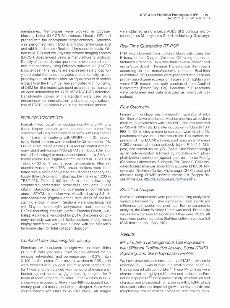
STAT3 and Fibroblast Phenotypes in IPF 1401AJP April 2012, Vol. 180, No. 4
membranes. Membranes were blocked in Odysseyblocking buffer (LI-COR Biosciences, Lincoln, NE) andprobed with the appropriate target antibody. Detectionwas performed with IR700 and IR800 anti-mouse andanti-rabbit antibodies (Rockland Immunochemicals, Gil-bertsville, PA) and the Odyssey Infrared Imaging System(LI-COR Biosciences) using a manufacturer’s protocol.Density of the bands was quantified in two infrared chan-nels independently using Odyssey software 2.1 (LI-CORBiosciences). The results are expressed as a phosphor-ylated protein/nonphosphorylated protein density ratio orprotein/�-tubulin density ratio. An equal amount of proteinextract from the HFL-1 cell line stimulated with 10 ng/mLof OSM for 15 minutes was used as an internal standardon each immunoblot for Y705-pSTAT3/STAT3 detection.Densitometry values of this standard were used as adenominator for normalization and percentage calcula-tion of STAT3 activation level in the individual probes.
Immunohistochemistry
Formalin-fixed, paraffin-embedded non-IPF and IPF lungtissue biopsy samples were obtained from tumor-freespecimens of lung resections of patients with lung cancer(n � 4) and from patients with UIP/IPF (n � 3), respec-tively. Serial sections were blocked for 30 minutes in 10%FBS in Tris-buffered saline (TBS) and incubated with pri-mary rabbit anti-human Y705-pSTAT3 antibody (Cell Sig-naling Technology) or mouse monoclonal anti–�-SMA an-tibody (clone 1A4, Sigma-Aldrich) diluted in TBS/0.05%Triton X-100 for 1 hour at room temperature. After se-quential washing with TBS, tissue sections were incu-bated with a biotin-conjugated anti-rabbit secondary an-tibody (DakoCytomation, Glostrup, Denmark) at 1:200 inTBS/0.05% Triton X-100 for 45 minutes, followed bystreptavidin–horseradish peroxidase conjugate (1:200dilution, DakoCytomation) for 30 minutes at room temper-ature. pSTAT3 expression was visualized using 3,3=-di-aminobenzidine (Sigma-Aldrich), with areas of positivestaining shown in brown. Sections were counterstainedwith Mayer’s hematoxylin, dehydrated, and mounted inDePeX mounting medium (Merck, Frenchs Forest, Aus-tralia). As a negative control for pSTAT3 expression, pri-mary antibody was omitted. Serial sections of lung tissuebiopsy specimens were also stained with the Masson’strichrome stain for total collagen detection.
Confocal Laser Scanning Microscopy
Fibroblasts were cultured on eight-well chamber slides(1 � 104 cells per well), fixed in cold ethanol for 15minutes, rehydrated, and permeabilized in 0.2% TritonX-100 for 3 minutes. After several washes in PBS, cellswere blocked with 5% goat serum at room temperaturefor 1 hour and then stained with monoclonal mouse anti-bodies against human �v �3 and �v �5 integrins for 2hours at room temperature. After further washes in PBS,slides were exposed to Alexa Fluor-488–conjugated sec-ondary goat anti-mouse antibody (Invitrogen). Cells were
counterstained with DAPI to visualize nuclei. All imageswere obtained using a Leica AOBS SP2 confocal micro-scope (Leica Microsystems GmbH, Heidelberg, Germany).
Real-Time Quantitative RT-PCR
RNA was obtained from cultured fibroblasts using theRNeasy kit from Qiagen (Valencia, CA) using the manu-facturer’s protocols. RNA was then reverse transcribedusing SuperScript II Reverse Transcriptase (Invitrogen)according to the manufacturer’s protocol. Real-timequantitative PCR reactions were prepared with TaqManprobe–based gene expression assays and TaqMan uni-versal PCR master mix, both purchased from AppliedBiosystems (Foster City, CA). Real-time PCR reactionswere performed and data analyzed as previously de-scribed.7
Flow Cytometry
Primary LF monolayer was immersed in trypsin/EDTA solu-tion; then cells were collected, washed one time with culturemedium (supplemented with 10% FBS), and resuspendedin PBS with 10% FBS. LFs after incubation in PBS with 10%FBS for 30 minutes at room temperature were fixed in 2%paraformaldehyde for 10 minutes on ice. Cell surface ex-pression of Thy-1/CD90 was analyzed using an anti-humanCD90 monoclonal mouse antibody (clone F15-42-1; Milli-pore) and normal mouse IgG1 (Santa Cruz Biotechnology)as an isotype control, followed by incubation with phos-phatidylethanolamine-conjugated goat–anti-mouse F(ab’)2(Cedarlane Laboratories, Burlington, ON, Canada). Cell-asso-ciated fluorescence was acquired by a Coulter EPICS XL flowcytometer (Beckman Coulter, Mississauga, ON, Canada) andanalyzed using WinMDI software version 2.8 (Scripps Re-search Institute; http://facs.scripps.edu/software.html).
Statistical Analysis
Statistical comparisons were performed using analysis ofvariance followed by Fisher’s protected least significantdifference test performed post hoc. For nonparametricanalysis, the Mann-Whitney U-test was used. Probabilityvalues were considered significant if they were �0.05. Alltests were performed using StatView software version 5.0(SAS Institute Inc., Cary, NC).
Results
IPF LFs Are a Heterogeneous Cell Populationwith Different Proliferative Activity, Basal STAT3Signaling, and Gene Expression Profiles
We have previously demonstrated that STAT3 activation inresponse to IL-6 was blunted in a small number of IPF LFlines compared with control LFs.13 These IPF LF lines werecharacterized as highly proliferative and resistant to Fas-induced apoptosis.30 In the present study, we identified andcharacterized LFs isolated from patients with UIP/IPF, whichdisplayed noticeably impaired growth activity and distinct
morphologic characteristics compared with control cells.
1402 Pechkovsky et alAJP April 2012, Vol. 180, No. 4
Doubling time of IPF LF lines was significantly longer com-pared with control LFs cultured in the same cell cultureconditions (Figure 1A). In addition, the IPF LF lines weresubstantially larger compared with control LFs and dis-played stellate morphologic characteristics (Figure 1B). Weexamined constitutive levels of Y705-pSTAT3 in IPF LF linesand found higher basal STAT3 signaling activity comparedwith control LFs (Figure 1, C and D). Further analysis re-vealed that IPF LF lines also expressed significantly less�-SMA (ACTA2) and �3 integrin (ITGB3) but more fibronec-tin-1 (FN1) mRNA at baseline (Figure 1E). There was a trendtoward increased basal expression levels of vimentin andcollagen 1�1 (COL1A1) in IPF LFs compared with controlLFs, but this difference did not reach statistical significance(Figure 1E). Thus, the increased basal pSTAT3 expressionlevel in IPF LFs was associated with a specific phenotypecharacterized by a markedly decreased expression of�-SMA and �3 integrin but increased expression of FN1 andslower growth rate compared with control cells.
Localization of pSTAT3 Immunoreactive Cells inthe Lungs of Patients with UIP/IPF
We next sought to determine the localization of cells with
activated STAT3 signaling in UIP lungs. Figure 2, A–C,shows areas of a dense fibrosis with numerous cellsstrongly positive for pSTAT3. Regions within fibroblasticfoci and surrounding vasculature showed strong stainingfor �-SMA, but pSTAT3-labeled cells were not observed(Figure 2, D and E, respectively). Some cells within thealveolar septa of the lungs from non-IPF controls stainedpositive for pSTAT3 but not as extensively as in IPF lungs(Figure 2, G and E, respectively) and on closer examina-tion appeared to be restricted to alveolar epithelial cells(Figure 2H).
Modulation of Basal STAT3 Signaling ModifiesCollagen I Secretion but Not Gene Expression inIPF LFs
Thus far, our findings show that IPF LFs express highbasal levels of pSTAT3 and FN1 in vitro and are adjacentto areas of dense fibrosis in vivo. We next tested whetheractivation of STAT3 induced by gp130 agonists is in-volved in the fibrogenic function of LFs. First, we deter-mined the responsiveness of IPF LFs to gp130-mediatedSTAT3 signaling. These experiments revealed that bothcontrols LFs and IPF LFs normally respond to IL-10, IL-11, and OSM stimulation; however, several IPF LF cul-
Figure 1. Fibroblasts isolated from the lungs ofIPF patients spontaneously express several dis-tinctive and stable phenotypes in in vitro cul-tures. A: Doubling time of IPF LFs displaying lowgrowth activity in comparison with control LFs.Graph data are mean � SE. *P � 0.05. B: Imagesof phase-contrast microscopy of control LFs andIPF LFs in routine cultures (original magnifica-tion, �20). C: Expression of Y705 pSTAT3 andtotal STAT3 proteins was determined by immu-noblotting of protein extracts from IPF LF andcontrol LF lines (one representative immunoblotof two independent experiments is shown).Densitometry and STAT3 activity measuringwere performed as described in Materials andMethods. D: Levels of STAT3 activation atbaseline in IPF LFs were significantly highercompared with control LFs (*P � 0.05; Mann-Whitney U-test). The data presented in box-and-whisker plots are from 8 IPF LFs and 10 controlLFs; horizontal lines are median, bars are quar-tiles, and dots are range of STAT3 activation. E:Levels of baseline �-SMA (ACTA2), COL1A1,FN1, vimentin (VIM), and �3 integrin (ITGB3)mRNA expression in IPF LFs (n � 6) and controlLFs (n � 7). Data are mean � SE. *P � 0.05compared with control LFs.
tures displayed diminished responsiveness to IL-6 and

STAT3 and Fibroblast Phenotypes in IPF 1403AJP April 2012, Vol. 180, No. 4
leukemia inhibitory factor compared with control LFs(Figure 3A). However, stimulation with IL-6 or OSM aloneand in the presence of a cell-permeable STAT3 inhibitorpeptide did not significantly alter COL1A1 and COL1A2secretion by control LFs (Figure 3B). Figure 3C shows atrend toward higher basal levels of COL1A1 mRNA in IPFLFs compared with control LFs, although this differencedid not reach statistical significance. As expected,TGF-�1 dose dependently increased COL1A1 mRNA inboth control LFs and IPF LFs (Figure 3C). In contrast, IL-6and OSM did not significantly change COL1A1 mRNAexpression levels either alone or in combination withTGF-�1 in both IPF LF and control LF cultures (Figure3C). We also transiently transfected HFL-1 cells with aplasmid-expressing human STAT3 and determinedCOL1A1 and COL1A2 secretion at baseline and after IL-6or OSM stimulation. Cells overexpressing STAT3 displayed90 times higher SOCS3 mRNA expression levels after stim-ulation, with IL-6 confirming functionality of the STAT3 con-struct; however, they did not secrete more COL1A1 andCOL1A2 either at rest or after cytokine stimulation com-pared with GFP-transfected controls (see SupplementalFigures S1B and S3 at http://ajp.amjpathol.org). In con-trast to normal LFs (Figure 3D), infection of IPF LFs withAd-STAT3DN had a strong inhibitory effect on basal andTGF-�1–induced COL1A1 and COL1A2 secretion com-pared with Ad-GFP–infected controls (Figure 3E). Themost prominent effect of STAT3 inhibition was seen in50% of IPF-derived lines predominantly affecting secre-tion of COL1A2 chain (two of four IPF LFs examined are
shown in Figure 3E). Taken together, these findingssuggest that STAT3 signaling has little effect on colla-gen I expression and secretion in normal LFs, but itmight be involved in the regulation of collagen I secre-tion by lung fibroblasts in UIP/IPF.
IL-6 and OSM Down-Regulate TGF-�1–Induced�-SMA Protein in LFs by STAT3-DependentMechanism
The observation that IPF LFs showed reduced basal�-SMA gene expression prompted us to ask whetherelevated STAT3 signaling was involved. Thus, we stimu-lated control LFs with IL-6, OSM, or TGF-�1 alone and incombination for 8 hours and measured production of�-SMA by immunoblotting. As expected, TGF-�1 was themost potent inducer of �-SMA expression (12-fold induc-tion over baseline) (Figure 4, A and B). Although IL-6modestly up-regulated �-SMA production alone, it signif-icantly diminished �-SMA induced by TGF-�1. Exposureto OSM, the most potent activator of STAT3 signaling,had no effect on �-SMA expression alone but had aneven more pronounced inhibitory effect on TGF-�1–in-duced �-SMA expression (Figure 4, A and B). Interest-ingly, the inhibitory effect of STAT3 activation was notassociated with modulation of Smad3 phosphorylationlevels either at baseline or induced by TGF-�1 (Figure4B). To examine this further, we infected LFs derived fromnormal lung parenchyma with either Ad-STAT3DN or Ad-GFP and measured �-SMA expression after OSM stimu-
Figure 2. Y705 pSTAT3, �-SMA, and total col-lagen in the lung tissue sections from patientswith UIP/IPF. A–C: Serial lung tissue sectionsfrom one IPF patient stained for total collagenwith Masson’s trichrome stain (blue bundles, A)and for pSTAT3 (brown dots, B; C: magnificationof boxed area in B; scale bar � 100 �m). D andE: Serial lung tissue sections from IPF patientstained for �-SMA (D, brown bundles; arrow-heads show fibroblastic foci, and arrows showvasculature) and for Tyr705-pSTAT3 (E, browndots). Scale bar � 200 �m. F–H: Serial sectionsfrom control non-IPF lung tissue stained for col-lagen with Masson’s trichrome stain (F) andpSTAT3 (G; H is magnification of boxed area inG). Scale bar � 100 �m. Representative imagesof lung tissue sections from three patients withUIP/IPF and four patients with lung cancer (usedas non-IPF controls) are shown.
lation. We have previously shown that these cells consti-

1404 Pechkovsky et alAJP April 2012, Vol. 180, No. 4
tutively express enhanced TGF-�1 signaling and �-SMAexpression.7 Figure 4C shows that OSM inhibited basal�-SMA expression in normal LF cultures after 24 hours,whereas blockade of STAT3 signaling by STAT3DN com-
Figure 4. IL-6– and OSM-induced STAT3 signaling modulates �-SMA expreng/mL), or TGF-�1 (10 ng/mL) and their combinations for 8 hours. A: Expressare mean � SE percentage of �-SMA expression in nonstimulated cells (n �pSTAT3, and Smad3 were determined in the protein extracts from LFs trexperiments is shown. C: LFs isolated from three healthy donor’s lungs werinfection treated with recombinant human OSM (10 ng/mL) or vehicle co
immunoblotting. One representative immunoblot of three independent experimentsAd-STAT3DN and Ad-GFP was determined by quantitative RT-RCR (data are meanpletely abrogated this inhibitory effect, confirming a crit-ical role for STAT3 signaling in OSM-induced inhibition of�-SMA. Moreover, inhibition of basal STAT3 signaling byoverexpression of STAT3DN mutant protein significantly
Figure 3. Effect of IL-6, OSM, and STAT3-medi-ated signaling on collagen I expression and se-cretion by normal LFs and LFs obtained fromUIP/IPF patients. A: Levels of STAT3 activationinduced by IL-6 family cytokines and IL-10 after15 minutes of stimulation of IPF LFs and controlLFs. Each dot represents an individual value foreach LF line and cytokine. B: Primary normal LFswere stimulated for 24 hours with IL-6 and OSMwith and without cell-permeable STAT3 inhibi-tory peptide (S3IP, 100 �mol/L). COL1A1 andCOL1A2 in cell culture supernatants were deter-mined by immunoblotting. One representativeimmunoblot of two independent experiments isshown. C: COL1A1 gene expression by IPF LFs(n � 4) and control LFs (n � 4) in the presenceof IL-6, OSM, and TGF-�1 at different concen-trations. Cells were stimulated for 48 hours. Dataare mean � SE. *P � 0.05 compared with non-stimulated (NS) controls. D: Primary normal LFswere infected with Ad-STAT3DN and Ad-GFP asan infection control and after 72 hours of culturecell-free supernatants were collected andCOL1A1 and COL1A2 determined by immuno-blotting. Two representative immunoblots ofthree experiments with primary normal LFs per-formed in triplicates are shown. E: Four ran-domly selected IPF LF lines were infected withAd-STAT3DN and Ad-GFP and 24 hours afterinfection treated with TGF-�1 (5 ng/mL) for 48hours. COL1A1 and COL1A2 in cell-free condi-tional culture medium was determined by im-munoblotting. In two IPF LF lines of four linesanalyzed, inhibition of STAT3 signaling stronglydiminished both basal and TGF-�1–inducedCOL1A1 and COL1A2 secretion. Two immuno-blots from two independent experiments areshown.
y primary human LFs. LFs were stimulated with IL-6 (100 ng/mL), OSM (10-SMA was analyzed by immunoblotting of the total cell protein extracts. Data
0.05 compared with TGF-�1 alone; †P � 0.05. B: �-SMA, total STAT3 ands described above. One representative immunoblot of three independentd with Ad-STAT3DN and Ad-GFP as an infection control and 24 hours afterr 24 hours. �-SMA and �-tubulin as a loading control were detected by
ssion bion of �3). *P �
eated ae infectentrol fo
is shown. D: Basal expression of �-SMA gene (ACTA2) in LFs infected with� SE, n � 3). *P � 0.05.

STAT3 and Fibroblast Phenotypes in IPF 1405AJP April 2012, Vol. 180, No. 4
up-regulated constitutive �-SMA mRNA expression com-pared with Ad-GFP–infected control cells (Figure 4D).
Inhibition of STAT3 Signaling Increases�-SMA Expression in Fibroblasts via an ALK-5–Dependent but SMAD7-IndependentMechanism
The results from the previous experiments suggest thatSTAT3 has a suppressive effect on basal �-SMA expres-sion of normal LFs that does not appear to involve anyinhibitory effects on Smad3. We next investigated themechanisms underlying this inhibitory effect further. Asshown in Figure 5, A and B, inhibition of basal STAT3signaling by Ad-STAT3DN significantly increased basaland TGF-�1–induced �-SMA gene expression and pro-tein production compared with Ad-GFP–infected ormock-infected cells. ALK-5 inhibition completely abro-gated �-SMA expression induced by exogenously addedTGF-�1 in Ad-STAT3DN–infected cells, indicating thatTGF-� signaling was functional in this model. However,the presence of the ALK-5 inhibitor did not completelyabrogate STAT3DN-induced �-SMA expression in thepresence or absence of TGF-�1, suggesting TGF-�–in-dependent mechanisms are also involved (Figure 5, Aand B). Expression of TGF-�1 mRNA was strongly up-regulated by exogenously added TGF-�1 and was notsignificantly modified by Ad-STAT3DN, excluding thepossibility of STAT3DN increasing autocrine expressionof TGF-�1 (Figure 5C). Moreover, we found that in con-trast with �-SMA, SMAD7 gene expression level at base-line was not affected after infection with Ad-STAT3DN(Figure 5D). Importantly, Ad-STAT3DN depressedSMAD7 mRNA expression induced by the exogenouslyadded TGF-�1, and this inhibitory effect was completelyabrogated by SB505124 (Figure 5D), suggesting a keyrole of ALK-5 signaling in the SMAD7-mediated feed-back loop. We reproduced these findings using theSTAT3 small molecule inhibitor STA-21 (see Supplemen-tal Figure S4 at http://ajp.amjpathol.org). Collectively,these results suggest that basal STAT3 signaling is arepressor of �-SMA expression in human normal lungfibroblasts, and blockade of ALK-5 activation, but notSMAD3-mediated signaling, is involved.
STAT3 Activity Reduces �v �3 IntegrinExpression
Having established that STAT3 suppresses �-SMA ex-pression, we next determined whether the modifyingeffects of STAT3 extended to other fibroblast-myofibro-blast differentiation markers. In initial experiments, wescreened 16 primary LF cultures for basal STAT3 activa-tion levels and found several lines that expressed consti-tutively a high basal level of pSTAT3 over several re-peated passages, similar to that seen in the IPF LF lines.Interestingly, these cells, designated LF-pSTAT3high,were characterized by the absence of �v �3 integrinexpression on the cell surface (Figure 6A), as well as low
proliferation rates when compared with LF-pSTAT3low(data not shown). In contrast, surface expression of �v �5
integrins in LF-pSTAT3high was slightly higher comparedwith LF-pSTAT3low (Figure 6A). To determine a causalrole for STAT3 in this integrin expression profile, cellswere infected with Ad-STAT3DN or STAT3C or treatedwith the small molecule inhibitor STA-21. As shown inFigure 6B, the higher basal level of � � -integrin expres-
Figure 5. Inhibition of STAT3 signaling increases �-SMA expression infibroblasts via TGF�RI/ALK-5–dependent but SMAD7-independent mecha-nism. A: �-SMA protein in HFL-1 cells infected with Ad-STAT3DN andAd-GFP, mock infected as controls, and stimulated with TGF-�1 (5 ng/mL) inthe presence of the ALK-5 inhibitor SB505124 (10 �mol/L) was determinedby immunoblotting. One representative immunoblot of two independentexperiments is shown. �-SMA (ACTA2) (B), TGF-�1 (C), and SMAD7 (D)mRNA expression index of HFL-1 cells treated as described in A. Data aremean � SE of the gene expression indices determined in two independentexperiments by quantitative RT-PCR performed in triplicate (*P � 0.05 com-pared with cells treated with dimethyl sulfoxide or SB505124; **P � 0.05compared with dimethyl sulfoxide controls; †P � 0.05).
v 3
sion on the cell surface of LF-pSTAT3low was mirrored by

1406 Pechkovsky et alAJP April 2012, Vol. 180, No. 4
an enhanced �3-integrin mRNA expression, suggest-ing a transcriptional effect of STAT3. Furthermore, Ad-STAT3DN significantly up-regulated basal �3-integringene expression in both LF-pSTAT3high and LF-pSTAT3low cells. This outcome was confirmed using theselective STAT3 inhibitor STA-21 (see Supplemental Fig-ures S4 and S5 at http://ajp.amjpathol.org). In contrast,neither Ad-STAT3DN (Figure 6C) nor STA-21 treatment(data not shown) significantly altered basal �5-integrinmRNA expression. However, LF-pSTAT3high constitu-tively expressed higher �5-integrin mRNA levels com-pared with LF-pSTAT3low (Figure 6C). To further confirmthese findings, we forced activation of STAT3 using aSTAT3C construct and found decreased expression of �3
integrin in STAT3C-transduced cells when comparedwith parental noninfected and LacZ-transduced controls(Figure 6D). The increased �3-integrin gene expressionof LFs infected with Ad-STAT3DN was paralleled by asignificantly elevated �3-integrin protein level determined
by immunoblotting (Figure 6E). Thus, these findings sug-gest that basal STAT3 activity in LFs represses �3 integrinvia a transcription-dependent mechanism. In contrast,STAT3 does not appear to be significantly involved inregulating �5-integrin expression.
Expression of Thy-1/CD90 on the Cell Surfaceof LFs Is Negatively Correlated with STAT3Activation at Baseline
Thy-1/CD90 expression is associated with distinctive fi-broblast phenotypes, and reduction of this cell surfacemolecule has been found in UIP/IPF-derived LFs.6,12 Fur-thermore, interaction of �v �3 and �v �5 integrins withThy-1 is an important mechanism, mediating cell adhe-sion, communication, and TGF-� signaling.37,38 There-fore, we investigated whether STAT3 activation influ-enced expression of Thy-1/CD90 on LFs. We screened14 primary LF cultures for Thy-1/CD90 expression and
Figure 6. Constitutively high level of STAT3 ac-tivity is associated with decreased expression of�v �3 integrins in primary human lung fibro-blasts. A: Immunostaining of LF-pSTAT3high andLF-pSTAT3low for �v �3 and �v �5 integrins(green). Nuclei were counterstained with DAPI(blue). Images were acquired by confocal laserscanning microscopy (Scale bar: 47 �m). LF-pSTAT3low express higher levels of �v �3 andlower levels of �v �5 detected in the focal adhe-sion sites (intensive green dots on the cell sur-face) compared with LF-pSTAT3high. �3 integrin(ITGB3) (B) and �5 integrin (ITGB5) (C) mRNAexpression was determined in LF-pSTAT3high
and LF-pSTAT3low infected with Ad-STAT3DNand Ad-GFP as an infection control (*P � 0.05compared with control, n � 3; †P � 0.05; ‡P �0.001). D: Expression of �3 and �5 integrins wasanalyzed by immunoblotting using total proteinextracts from parental nontransfected, STAT3C-transfected, and LacZ-transfected control LFs.�-Tubulin was used as a loading control. Twoimmunoblots from three independent experi-ments with two different control LF lines areshown. E: Using the same LFs as shown in B andC, production of �3 and �5 integrins in LFs in-fected with Ad-STAT3DN and Ad-GFP was de-termined by immunoblotting. Data are mean �SE of �3/�5-integrin/�-tubulin ratios determinedby a quantitative densitometry. *P � 0.05 comparedwith corresponding Ad-GFP controls (n � 3).
STAT3 activation and observed a statistically significant

STAT3 and Fibroblast Phenotypes in IPF 1407AJP April 2012, Vol. 180, No. 4
negative correlation (Figure 7A), suggesting a role forSTAT3 signaling in the regulation of this molecule. Similarto that seen for �3-integrin expression, cells infected withSTAT3C displayed a markedly reduced Thy-1/CD90 cellsurface expression compared with noninfected or LacZLF controls (Figure 7B). However, inhibition of STAT3activity by Ad-STAT3DN infection did not significantlychange Thy-1 mRNA expression levels in both TGF-�1–
Figure 7. Basal levels of STAT3 activation is negatively correlated withThy-1/CD90 cell surface expression on primary human LFs. A: Screening of14 individual lines of LFs isolated from different compartments of the non-IPFlungs for Thy-1/CD90 cell surface expression determined by flow cytometryand basal levels of STAT3 activation revealed a negative correlation betweenthe percentage of Thy-1/CD90 positive cells and the basal levels of Y705-pSTAT3 in the given cell population (P � 0.05). B: Expression of Thy-1/CD90antigen on the cell surface of parental nontransfected control, stably STAT3C-transfected, and LacZ-transfected control LFs was determined by flow cytom-etry. Representative flow cytometry histograms from three independent ex-periments are shown. C: Effect of the STAT3 signaling inhibition by Ad-STAT3DN on basal and TGF-�1–mediated expression of Thy-1 mRNA inHFL-1 cells. Data are mean � SE of the THY1 gene expression indexdetermined in two independent experiments by quantitative RT-PCR per-formed in triplicates.
stimulated and control cultures (Figure 7C), suggesting
that unlike �3 integrin, the inhibitory effect of STAT3 onThy-1/CD90 expression is not transcriptionally regulated.
Inhibition of STAT3 Signaling in IPF LFsDiminishes Responsiveness to Exogenous TGF-�1 and Reverses Resistance to Apoptosis
To more definitively determine the role of STAT3 signalingin IPF-derived fibroblasts, we infected IPF LFs with Ad-STATDN and analyzed cell responsiveness to recombi-nant human TGF-�1. Figure 8A demonstrates that in con-trast to normal LFs, IPF LFs infected with Ad-STAT3DN
Figure 8. Blockade of STAT3 signaling in IPF LFs diminished cell respon-siveness to exogenous TGF-�1 and apoptotic resistance. IPF LFs and controlLFs were infected with Ad-STAT3DN and Ad-GFP as an infection control, and24 hours later TGF-�1 was added to the cultures for 48 hours. �-SMA in totalprotein extracts was detected by immunoblotting. A: One representativeimmunoblot of four independent experiments is shown; total STAT3 proteinis significantly elevated in Ad-STATDN IPF LFs and control LFs, confirmingexpression of the mutant protein in both cell lines. �-Tubulin was used as aloading control. B: Basal and TGF-�1–induced �-SMA protein in IPF LFsinfected with Ad-STAT3DN and Ad-GFP as a control. Data are mean � SE ofpercent changes of �-SMA/�-tubulin ratios. *P � 0.05 compared with Ad-GFP and Ad-STAT3DN without TGF-�1 (n � 4). C: �-SMA gene expressionin the same IPF LFs described in B; data are mean � SE of the �-SMA geneexpression index determined in two different primary IPF LF cultures byquantitative RT-PCR performed in triplicate. *P � 0.05 compared with Ad-GFP and Ad-STAT3DN with and without TGF-�1 stimulation. D: IPF LFs andcontrol LFs were infected with Ad-STAT3DN and Ad-GFP; 24 hours afterinfection all cells were treated with staurosporine in serum-free medium toinduce apoptosis. Total procaspase-3 and cleaved fragments of activatedcaspase-3 were detected by immunoblotting. Densitometry ratios of cleaved/total procaspase-3 were calculated and considered as a caspase-3 activationindex. Graph data are mean � SE of percent changes of caspase-3 activation
calculated independently for control LFs and IPF LFs. *P � 0.05 comparedwith Ad-GFP and Ad-STAT3WT (n � 3).
1408 Pechkovsky et alAJP April 2012, Vol. 180, No. 4
displayed diminished responsiveness to TGF-�1, ex-pressing significantly lower levels of �-SMA protein (Fig-ure 8B) and mRNA (Figure 8C) after TGF-�1 stimulationcompared with Ad-GFP infected controls. In contrast tonormal LFs (Figures 4 and 5), inhibition of basal STAT3signaling in IPF LFs did not significantly change consti-tutive �-SMA expression levels (Figure 8, B and C).
We have previously shown that IPF LFs are resistant toFas-induced apoptosis in a STAT3-dependent manner.30
In the present study, we investigated the effect of STAT3inhibition on IPF LF apoptosis induced by staurosporine,a broad-spectrum kinase inhibitor with a strong apoptoticcapability mediated via the caspase-3–dependent path-way.39 As shown in Figure 8D, STAT3DN significantlyenhanced staurosporine-induced caspase-3 activation inIPF LFs as detected by the increase of cleaved fragmentsof procaspase-3. Moreover, infection of LFs with Ad-STAT3WT, which drastically increased basal STAT3 ac-tivity in LFs (see Supplemental Figure S2 at http://ajp.amjpathol.org), inhibited caspase-3 activation induced bythe staurosporine exposure in IPF LFs but not in normalLFs (Figure 8D), suggesting a different role for STAT3signaling in apoptotic resistance in normal LFs and IPF-derived fibroblasts.
Discussion
We performed this study to examine whether STAT3-mediated signaling plays an important role in the patho-genesis of UIP/IPF by inducing aberrant activation anddifferentiation of fibroblasts and whether manipulation ofthis signaling pathway is a potential therapeutic target inIPF. We isolated a population of fibroblasts from the lungsof patients with UIP/IPF, which were characterized byconstitutively high levels of pSTAT3. These cells dis-played persistent morphologic and functional abnormal-ities in vitro, including reduced proliferation and expres-sion of �-SMA, Thy-1/CD90, and �v �3 integrins butincreased expression of fibronectin. Furthermore, we ob-served a large number of cells expressing pSTAT3 inUIP/IPF lungs, particularly in areas of dense fibrosis, butstaining was not seen in the �-SMA–positive fibroblasticfoci. Although these observations suggest STAT3 is in-volved in the development of this fibroblast phenotype,they do not show causality. To investigate this, we gen-erated several in vitro models that allowed us to manipu-late STAT3 signaling in primary LFs.
We show that forced induction of STAT3 signaling inLFs drives these cells toward an abnormal phenotype,which bears a striking resemblance with fibroblasts weisolated from UIP/IPF lung tissue. We observed an inhib-itory effect of the STAT3-activating cytokines IL-6 andOSM on basal and TGF-�1–induced �-SMA expression,suggesting a regulatory effect on the differentiation ofmyofibroblasts. Going further, LFs infected withSTAT3DN or treated with the STAT3 inhibitor STA-21showed increased �-SMA expression and production of�3 integrin, both of which were dependent on TGF-�receptor I (TGF�RI)/ALK-5 signaling. Although neither
constitutive nor cytokine-induced activation of STAT3 inLFs had any effect on collagen I expression or secretion,inhibition of STAT3 signaling in IPF LFs diminished bothbasal and TGF-�1–induced secretion of collagen I. More-over, we found that blockade of STAT3-mediated signalingin IPF LFs significantly attenuated sensitivity to exogenousTGF-�1 and, in contrast to normal LFs, augmented stauro-sporine-induced apoptosis. Considering that STAT3 is anessential transcription factor for several facets of celldifferentiation, proliferation, and apoptosis, these find-ings suggest that sustained and enhanced activation ofSTAT3 signaling is an important mechanism dysregulat-ing normal reparative functions of resident fibroblastsand myofibroblasts in the human lung.
We have previously shown that a population of fibro-blasts isolated from IPF lungs responds to IL-6 stimula-tion with blunted activation of STAT3. Although most ofthese cells were �-SMA positive, they displayed en-hanced proliferative activity and resistance to Fas-in-duced apoptosis compared with non-IPF control fibro-blasts.13,30 In the present study, we describe anotherpopulation of fibroblasts isolated from a larger number ofIPF lungs, characterized by significantly lower prolifera-tive activity but normal STAT3 activation in response toIL-10 and gp130 cytokine stimulation. These cells arelarger than LFs derived from non-IPF lungs and displaydiminished expression of �-SMA and two key adhesionmolecules �v �3 integrin and CD90/Thy-1 but increasedexpression of fibronectin. Similar to our findings, Ramosand associates reported on a population of fibroblastsfrom IPF lungs that exhibited slower growth rate and anincreased rate of spontaneous apoptosis compared withcontrol cells.40 Interestingly, Lim and colleagues re-ported enhanced levels of pSTAT3 in fibroblasts derivedfrom keloids, but in contrast to our findings, these cellsdemonstrated significantly increased proliferative andsynthetic activity compared with normal dermal fibro-blasts.29 We and others have recently shown that normalprimary human lung parenchyma–derived fibroblastsdisplay classic myofibroblastic phenotype characterizedby constitutively active TGF-�/Smad signaling and highlevels of �-SMA expression, contractility, and prolifera-tion.7,23,24 However, somewhat surprisingly, these myofi-broblasts displayed decreased ECM synthetic capacityand lower responsiveness to exogenously added biolog-ically active TGF-�1 compared with paired airway-de-rived cells,7,23 suggesting some self-limiting mechanismsfor ECM synthesis in highly contractile �-SMA–express-ing myofibroblasts. In support of this, ablation of �-SMAexpression worsens the course of fibrosis in mouse kid-ney and lung.21,22 Alternatively, activation of STAT3 sig-naling might induce differentiation of resident LFs towarda stable and functionally distinct population of cells withinIPF lung. Adding to this paradigm of fibroblast heteroge-neity, the data in this article suggest that LFs might alsobe separated into two distinct subpopulations based onthe level of STAT3 activation, with LF-pSTAT3high andLF-pSTAT3low cells consistently demonstrating the oppo-site phenotypic and functional responses. Thus, theseobservations, together with the findings that absence ofThy-1/CD90 expression confers a profibrotic phenotype
to IPF LFs41 with enhanced responsiveness to TGF-�1,38
STAT3 and Fibroblast Phenotypes in IPF 1409AJP April 2012, Vol. 180, No. 4
support our hypothesis that STAT3-mediated suppres-sion of constitutive �-SMA, �v �3 integrin, and Thy-1/CD90 expression is critically important for pathologicalphenotype of mesenchymal cells in UIP/IPF lung.
To explore this concept further, we infected normal LFswith constructs encoding wild-type STAT3 protein,STAT3C,26 or STAT3DN42 mutants and examined expres-sion of several key parameters of activation and fibro-blast/myofibroblast differentiation. We found that expres-sion of STAT3C in normal LFs significantly reducedsurface presentation of Thy-1/CD90. Furthermore, exam-ination of a large number of different human primaryfibroblast lines isolated from non-IPF lungs also revealeda negative correlation between the levels of pSTAT3 andsurface expression of Thy-1/CD90. Interestingly, STAT3modified Thy-1 on the cell surface without affecting thegene transcription. Thy-1 is a 25- to 37-kDa cell membra-ne–anchored protein implicated in many aspects ofwound healing and fibrosis, although its precise roles arepoorly defined.6,37,43,44 Differential expression of Thy-1/CD90 is suggested to be a reliable marker of the fibrosisbecause cells within fibroblastic foci are Thy-1(�),whereas normal lung fibroblasts are predominantly Thy-1(�).6,12,45 More recently, Zhou and colleagues demon-strated that the interaction of �v �5 integrin with Thy-1 isa critical element of myofibroblast contraction and latentTGF-� activation.38 Thus, the reduced expression of Thy-1/CD90 and �v �3 but enhanced �v �5 integrin expres-sion mediated by STAT3 may disrupt normal integrin-mediated signaling, which in turn is likely responsible forthe higher responsiveness of IPF LFs to TGF-� and otherprofibrotic factors.32
In line with previous observations, we also found thatIPF LFs were deficient in �3 integrin and �-SMA expres-sion, two critical elements of fibroblast/myofibroblast ac-tivation, differentiation, and TGF-� signaling.46,47 Wehave previously shown that �v �3 plays an important rolein regulating myofibroblast functions, including cell re-sponsiveness to TGF-�1 and communication with theECM.7,36,48 Accordingly, we investigated whether STAT3was involved in regulating �v �3 expression and foundthat STAT3 activation is associated with diminished �3-integrin gene expression and surface expression of �v �3
but not �v �5 integrins in LFs. In contrast, inhibition ofendogenous STAT3 activity restored �3-integrin expres-sion most drastically in LF-pSTAT3high. These two piecesof evidence strongly suggest a direct suppressive role ofSTAT3 on �3 integrin and, most likely, indirectly on latentTGF-� activation and, therefore, myofibroblast differenti-ation.36,38,48 Previously, �v �3 and �v �5 integrins havebeen demonstrated to display a different functional role inprocesses of cell adhesion and migration49 and bindingcapacity to TGF�RII.48 Both of these processes are rel-evant to the enhanced fibrogenic activity of IPF LFs. It hasbeen shown that wound healing in �3-integrin knockoutmice was accompanied by enhanced TGF-� signaling.50
Similar observations were made in transgenic mice with akinase-deficient TGF�RII51 and in aortic tissues from pa-tients with the Loeys-Dietz syndrome associated withR495X mutation of TGF�RII.52 In addition, Pannu and
associates have reported that enhanced collagen pro-duction and responsiveness of SSc-derived fibroblasts toTGF-� were associated with diminished cell surface ex-pression of TGF�RII.53 Taking into consideration that �v
�3 but not �v �5 is a functional partner of TGF�RII48 andin this study IPF LFs were characterized by a decreased�3-integrin expression and enhanced responsiveness toTGF-�1 stimulation, we speculate that exaggerated acti-vation of STAT3 signaling in IPF fibroblasts is responsiblefor that phenomenon. Currently, a role for �v �3 and �v �5
integrins in UIP/IPF pathogenesis is unknown.54
Experiments with Ad-STAT3DN–infected or STA-21–exposed LFs show that even in fibroblasts with undetect-able levels of pSTAT3 there is basal activity of this intra-cellular signaling pathway. Both of these inhibitorymanipulations led to the increase of �-SMA and �3-in-tergin expression in normal LFs. Currently, we do notknow which cytokines and growth factors are responsiblefor basal STAT3 activation in control LF and IPF LF cells;however, it is not likely confined to gp130 activationbecause we have shown that epidermal growth factoris a potent repressor of basal and TGF-�1–induced �v
�3 expression in fibroblasts.36 Platelet-derived growthfactor55 and other epithelium-derived growth factorstogether with IL-10,56 abundantly secreted by alterna-tively activated alveolar macrophages and signalingvia STAT3-dependent pathway,57–59 are also potentialcandidates.
Cross talk between cytokines and growth factors isvital for the transition from wound closure to normal tissuefunction restoration or, if left uncontrolled, fibrosis.20,60
Emerging data describe a complex interaction betweengp130/STAT3 and TGF-�/Smad in various pathologicalsituations and suggest that although these moleculescooperate in the progression of some disease states, inothers they are antagonistic.61–64 We tested the interac-tion between STAT3 and SMAD3 signaling on productionof collagen I and �-SMA. As expected, TGF-�1 inducedSmad3 phosphorylation and collagen I expression in LFs.Addition of IL-6 or OSM had no effect on collagen ex-pression and secretion despite robust activation ofSTAT3 signaling, suggesting that STAT3 activity does notexert a significant effect on collagen I production in nor-mal LFs. However, we observed robust inhibitory effect ofSTAT3DN on collagen I secretion in some IPF LF lines.Interestingly, this effect was only evident in IPF LF cul-tures at baseline and after TGF-�1 stimulation, suggest-ing a different and yet unknown mechanism of a STAT3signaling inhibition on collagen I secretion in IPF-derivedfibroblasts. Surprisingly, we found that activation ofSTAT3 signaling in normal LFs is associated with depres-sion of TGF-�1–induced �-SMA expression. Taking intoconsideration a steady-state level of pSTAT3 induced byOSM in LFs, it is likely that STAT3 signaling was involvedin the observed antagonistic effect of two cytokines on�-SMA expression. Indeed, blockade of STAT3 signalingnoticeably attenuated the inhibitory effect of OSM on�-SMA expression. These results are in line with the re-cent findings by Stangou et al,65 who demonstrated thatin rats with an experimental kidney disease, treatmentwith human IL-11 depressed TGF-� signaling and im-
paired expression of �-SMA. However, although the in-
1410 Pechkovsky et alAJP April 2012, Vol. 180, No. 4
hibitory effect of OSM appears to involve signalingthrough ALK-5, SMAD3 phosphorylation was not reducedby the OSM-induced STAT3 activation. The inhibition ofALK-5–mediated signaling, which is upstream of bothSMAD-dependent and SMAD-independent pathways,strongly attenuated the increased �-SMA and �3-integrinexpression after STAT3 inhibition. In keeping with aSMAD3-independent effect, SMAD7 expression was notmodified by Ad-STAT3DN. In a previous study, we dem-onstrated that �3-integrin expression induced by TGF-�1was insensitive to overexpression of SMAD7.36 There-fore, STAT3 inhibition is affecting �3-integrin and �-SMAexpression in LFs by ALK-5–dependent but SMAD-inde-pendent mechanisms. The most likely candidate isp38MAPK-mediated signaling, which we and others havedemonstrated plays a key role in TGF-�1–induced �3-integrin and �-SMA expression and ultimately for con-tractile function of myofibroblasts36,46,66 and which hasbeen shown engaged in negative feedback inhibition ofSTAT3 activity.67,68 In contrast to normal LFs, STAT3DNsignificantly attenuated TGF-�1–induced �-SMA expres-sion in IPF LFs, further supporting our hypothesis of animportant role for the aberrant STAT3 signaling in a shap-ing of fibroblast phenotypes in UIP/IPF.
Altogether, these findings clearly suggest that fibro-blasts with high STAT3 activity, which we found in abun-dance within the dense fibrotic areas of IPF lungs, haveimpaired expression of �-SMA at baseline, which itseems is paralleled by an elevated capacity to expressECM proteins and up-regulate TGF-�–mediated signal-ing, down-regulation of several key adhesion molecules,and growth inhibition. The reasons for the appearance ofthis fibroblast phenotype in IPF are unknown, although itis worth noting that active STAT3 signaling mediates sev-eral antiapoptotic mechanisms in fibroblasts, includingpremature senescence.27,69,70 However, the precisemechanism of STAT3-mediated resistance of human lungfibroblasts to apoptosis and its role in the pathogenesis ofUIP/IPF is largely unknown.
We have previously identified several significant differ-ences between non-IPF and IPF-derived fibroblasts interms of apoptosis.30 Those IPF LF lines had lower levelsof apoptosis at baseline and after combined gp130/FASreceptor activation in a STAT3-dependent manner. Sim-ilarly, modulation of STAT3 activity in embryonic mouseand human skin fibroblasts has been shown to signifi-cantly change their resistance to apoptosis through mod-ulation of both extrinsic and intrinsic pathways of pro-grammed cell death.27,69,70 Data from the current studysupport these earlier findings. Indeed, inhibition of basalSTAT3 activity in IPF-derived fibroblasts sensitizes cellsto staurosporine-induced apoptosis via caspase-3–de-pendent pathway, whereas constitutively active STAT3signaling led to inhibition of caspase-3 activation. In con-trast, modulating STAT3 activity had little effect on thestaurosporine-induced caspase-3 activation in normalLFs. These data support our hypothesis that enhancedSTAT3 activity in IPF fibroblasts might be responsible fortheir persistence and fibrogenic activity in the areas ofdense fibrosis in UIP via inhibition of several proapoptotic
pathways.In summary, we describe for the first time a role forSTAT3 in regulating of fibroblast phenotype and functionin UIP/IPF. Taking into consideration that STAT3 is themain signaling pathway activated by several cytokinesassociated with pulmonary fibrosis, including the IL-6family, IL-10, and receptor tyrosine kinase activatorssuch as epidermal growth factor and platelet-derivedgrowth factor, it is likely that by regulating myofibroblastfunction, persistent STAT3 activation may be one of thecritical mechanisms in the pathogenesis of UIP/IPF. Webelieve that assessment of STAT3 signaling pathway ac-tivation in cells or tissue derived from pathological re-gions of the UIP/IPF lungs might serve as a phenotypicand prognostic marker of the disease.
Acknowledgments
We thank David Lane, Ralph McWhinnie, and JulianaVandergugten for excellent technical assistance.
References
1. Katzenstein AL, Myers JL: Nonspecific interstitial pneumonia and theother idiopathic interstitial pneumonias: classification and diagnosticcriteria. Am J Surg Pathol 2000, 24:1–3
2. Gross TJ, Hunninghake GW: Idiopathic pulmonary fibrosis. N EnglJ Med 2001, 345:517–525
3. Selman M, King TE, Pardo A: Idiopathic pulmonary fibrosis: prevailingand evolving hypotheses about its pathogenesis and implications fortherapy. Ann Intern Med 2001, 134:136–151
4. King TE, Jr., Schwarz MI, Brown K, Tooze JA, Colby TV, Waldron JA,Jr., Flint A, Thurlbeck W, Cherniack RM: Idiopathic pulmonaryfibrosis: relationship between histopathologic features and mortality.Am J Respir Crit Care Med 2001, 164:1025–1032
5. Monaghan H, Wells AU, Colby TV, du Bois RM, Hansell DM, Nichol-son AG: Prognostic implications of histologic patterns in multiplesurgical lung biopsies from patients with idiopathic interstitial pneu-monias. Chest 2004, 125:522–526
6. Hagood JS, Prabhakaran P, Kumbla P, Salazar L, MacEwen MW,Barker TH, Ortiz LA, Schoeb T, Siegal GP, Alexander CB, Pardo A,Selman M: Loss of fibroblast Thy-1 expression correlates with lungfibrogenesis. Am J Pathol 2005, 167:365–379
7. Pechkovsky DV, Hackett TL, An SS, Shaheen F, Murray LA, KnightDA: Human lung parenchyma but not proximal bronchi producesfibroblasts with enhanced TGF-beta signaling and alpha-SMA ex-pression. Am J Respir Cell Mol Biol 2010, 43:641–651
8. Larsson O, Diebold D, Fan D, Peterson M, Nho RS, Bitterman PB,Henke CA: Fibrotic myofibroblasts manifest genome-wide derange-ments of translational control. PLoS One 2008, 3:e3220
9. Emblom-Callahan MC, Chhina MK, Shlobin OA, Ahmad S, Reese ES,Iyer EP, Cox DN, Brenner R, Burton NA, Grant GM, Nathan SD:Genomic phenotype of non-cultured pulmonary fibroblasts in idio-pathic pulmonary fibrosis. Genomics 2010, 96:134–145
10. Raghu G, Chen YY, Rusch V, Rabinovitch PS: Differential proliferationof fibroblasts cultured from normal and fibrotic human lungs. Am RevRespir Dis 1988, 138:703–708
11. Jordana M, Schulman J, McSharry C, Irving L, Newhouse M, JordanaG, Gauldie J: Heterogenous proliferative characteristics of humanadult lung fibroblast lines and clonally derived fibroblasts from controland fibrotic tissue. Am Rev Respir Dis 1988, 137:579–584
12. Koumas L, Smith TJ, Feldon S, Blumberg N, Phipps RP: Thy-1 ex-pression in human fibroblast subsets defines myofibroblastic or lipo-fibroblastic phenotypes. Am J Pathol 2003, 163:1291–1300
13. Moodley YP, Scaffidi AK, Misso NL, Keerthisingam C, McAnulty RJ,Laurent GJ, Mutsaers SE, Thompson PJ, Knight DA: Fibroblastsisolated from normal lungs and those with idiopathic pulmonary fibro-
sis differ in interleukin-6/gp130-mediated cell signaling and prolifer-ation. Am J Pathol 2003, 163:345–354
STAT3 and Fibroblast Phenotypes in IPF 1411AJP April 2012, Vol. 180, No. 4
14. Wang XM, Zhang Y, Kim HP, Zhou Z, Feghali-Bostwick CA, Liu F,Ifedigbo E, Xu X, Oury TD, Kaminski N, Choi AM: Caveolin-1: a criticalregulator of lung fibrosis in idiopathic pulmonary fibrosis. J Exp Med2006, 203:2895–2906
15. Xia H, Diebold D, Nho R, Perlman D, Kleidon J, Kahm J, Avdulov S,Peterson M, Nerva J, Bitterman P, Henke C: Pathological integrinsignaling enhances proliferation of primary lung fibroblasts from pa-tients with idiopathic pulmonary fibrosis. J Exp Med 2008, 205:1659–1672
16. Desmouliere A, Geinoz A, Gabbiani F, Gabbiani G: Transforminggrowth factor-beta 1 induces alpha-smooth muscle actin expressionin granulation tissue myofibroblasts and in quiescent and growingcultured fibroblasts. J Cell Biol 1993, 122:103–111
17. Zhang HY, Gharaee-Kermani M, Zhang K, Karmiol S, Phan SH: Lungfibroblast alpha-smooth muscle actin expression and contractile phe-notype in bleomycin-induced pulmonary fibrosis. Am J Pathol 1996,148:527–537
18. Tomasek JJ, Gabbiani G, Hinz B, Chaponnier C, Brown RA: Myofi-broblasts and mechano-regulation of connective tissue remodelling.Nat Rev Mol Cell Biol 2002, 3:349–363
19. Leask A, Abraham DJ: TGF-beta signaling and the fibrotic response.FASEB J 2004, 18:816–827
20. Wynn TA: Integrating mechanisms of pulmonary fibrosis. J Exp Med2011, 208:1339–1350
21. Takeji M, Moriyama T, Oseto S, Kawada N, Hori M, Imai E, Miwa T:Smooth muscle alpha-actin deficiency in myofibroblasts leads toenhanced renal tissue fibrosis. J Biol Chem 2006, 281:40193–40200
22. Liu T, Warburton RR, Guevara OE, Hill NS, Fanburg BL, Gaestel M,Kayyali US: Lack of MK2 inhibits myofibroblast formation and exac-erbates pulmonary fibrosis. Am J Respir Cell Mol Biol 2007, 37:507–517
23. Kotaru C, Schoonover KJ, Trudeau JB, Huynh ML, Zhou X, Hu H,Wenzel SE: Regional fibroblast heterogeneity in the lung: implicationsfor remodeling. Am J Respir Crit Care Med 2006, 173:1208–1215
24. Nihlberg K, Andersson-Sjoland A, Tufvesson E, Erjefalt JS, Bjermer L,Westergren-Thorsson G: Altered matrix production in the distal air-ways of individuals with asthma. Thorax 2010, 65:670–676
25. Levy DE, Darnell JE, Jr.: Stats: transcriptional control and biologicalimpact. Nat Rev Mol Cell Biol 2002, 3:651–662
26. Bromberg JF, Wrzeszczynska MH, Devgan G, Zhao Y, Pestell RG,Albanese C, Darnell JE, Jr.: Stat3 as an oncogene, Cell 1999, 98:295–303
27. Shen Y, Devgan G, Darnell JE, Jr., Bromberg JF: Constitutively acti-vated Stat3 protects fibroblasts from serum withdrawal and UV-in-duced apoptosis and antagonizes the proapoptotic effects of acti-vated Stat1. Proc Natl Acad Sci U S A 2001, 98:1543–1548
28. Huang HF, Murphy TF, Shu P, Barton AB, Barton BE: Stable expres-sion of constitutively-activated STAT3 in benign prostatic epithelialcells changes their phenotype to that resembling malignant cells. MolCancer 2005, 4:2
29. Lim CP, Phan TT, Lim IJ, Cao X: Stat3 contributes to keloid patho-genesis via promoting collagen production, cell proliferation andmigration. Oncogene 2006, 25:5416–5425
30. Moodley YP, Misso NL, Scaffidi AK, Fogel-Petrovic M, McAnulty RJ,Laurent GJ, Thompson PJ, Knight DA: Inverse effects of interleukin-6on apoptosis of fibroblasts from pulmonary fibrosis and normal lungs.Am J Respir Cell Mol Biol 2003, 29:490–498
31. Hogaboam CM, Carpenter KJ, Evanoff H, Kunkel SL: Approaches toevaluation of fibrogenic pathways in surgical lung biopsy specimens.Methods Mol Med 2005, 117:209–221
32. Murray LA, Argentieri RL, Farrell FX, Bracht M, Sheng H, Whitaker B,Beck H, Tsui P, Cochlin K, Evanoff HL, Hogaboam CM, Das AM:Hyper-responsiveness of IPF/UIP fibroblasts: interplay between TGF-beta1, IL-13 and CCL2. Int J Biochem Cell Biol 2008, 40:2174–2182
33. Song H, Wang R, Wang S, Lin J: A low-molecular-weight compounddiscovered through virtual database screening inhibits Stat3 functionin breast cancer cells. Proc Natl Acad Sci U S A 2005, 102:4700–4705
34. Zhang SS, Liu MG, Kano A, Zhang C, Fu XY, Barnstable CJ: STAT3activation in response to growth factors or cytokines participates inretina precursor proliferation. Exp Eye Res 2005, 81:103–115
35. DaCosta Byfield S, Major C, Laping NJ, Roberts AB: SB-505124 is a
selective inhibitor of transforming growth factor-beta type I receptorsALK4. ALK5, and ALK7. Mol Pharmacol 2004, 65:744–75236. Pechkovsky DV, Scaffidi AK, Hackett TL, Ballard J, Shaheen F,Thompson PJ, Thannickal VJ, Knight DA: Transforming growth factorbeta1 induces alphavbeta3 integrin expression in human lung fibro-blasts via a beta3 integrin-, c-Src-, and p38 MAPK-dependent path-way. J Biol Chem 2008, 283:12898–12908
37. Hermosilla T, Munoz D, Herrera-Molina R, Valdivia A, Munoz N, NhamSU, Schneider P, Burridge K, Quest AF, Leyton L: Direct Thy-1/alphaVbeta3 integrin interaction mediates neuron to astrocyte com-munication. Biochim Biophys Acta 2008, 1783:1111–1120
38. Zhou Y, Hagood JS, Lu B, Merryman WD, Murphy-Ullrich JE: Thy-1-integrin alphav beta5 interactions inhibit lung fibroblast contraction-induced latent transforming growth factor-beta1 activation and myo-fibroblast differentiation. J Biol Chem 2010, 285:22382–22393
39. Johansson AC, Steen H, Ollinger K, Roberg K: Cathepsin D mediatescytochrome c release and caspase activation in human fibroblastapoptosis induced by staurosporine. Cell Death Differ 2003, 10:1253–1259
40. Ramos C, Montano M, Garcia-Alvarez J, Ruiz V, Uhal BD, Selman M,Pardo A: Fibroblasts from idiopathic pulmonary fibrosis and normallungs differ in growth rate, apoptosis, and tissue inhibitor of metallo-proteinases expression. Am J Respir Cell Mol Biol 2001, 24:591–598
41. Ramirez G, Hagood JS, Sanders Y, Ramirez R, Becerril C, Segura L,Barrera L, Selman M, Pardo A: Absence of Thy-1 results in TGF-betainduced MMP-9 expression and confers a profibrotic phenotype tohuman lung fibroblasts. Lab Invest 2011, 91:1206–1218
42. Kaptein A, Paillard V, Saunders M: Dominant negative stat3 mutantinhibits interleukin-6-induced Jak-STAT signal transduction. J BiolChem 1996, 271:5961–5964
43. Zhou Y, Hagood JS, Murphy-Ullrich JE: Thy-1 expression regulatesthe ability of rat lung fibroblasts to activate transforming growthfactor-beta in response to fibrogenic stimuli. Am J Pathol 2004, 165:659–669
44. Rege TA, Hagood JS: Thy-1, a versatile modulator of signaling affect-ing cellular adhesion, proliferation, survival, and cytokine/growth fac-tor responses. Biochim Biophysi Acta 2006, 1763:991–999
45. Derdak S, Penney DP, Keng P, Felch ME, Brown D, Phipps RP:Differential collagen and fibronectin production by Thy 1� and Thy 1-lung fibroblast subpopulations. Am J Physiol 1992, 263:L283–L290
46. Wang J, Fan J, Laschinger C, Arora PD, Kapus A, Seth A, McCullochCA: Smooth muscle actin determines mechanical force-induced p38activation. J Biol Chem 2005, 280:7273–7284
47. Wipff PJ, Rifkin DB, Meister JJ, Hinz B: Myofibroblast contractionactivates latent TGF-beta1 from the extracellular matrix. J Cell Biol2007, 179:1311–1323
48. Scaffidi AK, Petrovic N, Moodley YP, Fogel-Petrovic M, Kroeger KM,Seeber RM, Eidne KA, Thompson PJ, Knight DA: alpha(v)beta(3)Integrin interacts with the transforming growth factor beta (TGFbeta)type II receptor to potentiate the proliferative effects of TGFbeta1 inliving human lung fibroblasts. J Biol Chem 2004, 279:37726–37733
49. Clyman RI, Mauray F, Kramer RH: Beta 1 and beta 3 integrins havedifferent roles in the adhesion and migration of vascular smoothmuscle cells on extracellular matrix. Exp Cell Res 1992, 200:272–284
50. Reynolds LE, Conti FJ, Lucas M, Grose R, Robinson S, Stone M,Saunders G, Dickson C, Hynes RO, Lacy-Hulbert A, Hodivala-Dilke K:Accelerated re-epithelialization in beta3-integrin-deficient- mice isassociated with enhanced TGF-beta1 signaling. Nat Med 2005, 11:167–174
51. Denton CP, Zheng B, Evans LA, Shi-wen X, Ong VH, Fisher I, Laza-ridis K, Abraham DJ, Black CM, de Crombrugghe B: Fibroblast-specific expression of a kinase-deficient type II transforming growthfactor beta (TGFbeta) receptor leads to paradoxical activation ofTGFbeta signaling pathways with fibrosis in transgenic mice. J BiolChem 2003, 278:25109–25119
52. Loeys BL, Schwarze U, Holm T, Callewaert BL, Thomas GH, Pannu H,DE Backer JF, Oswald GL, Symoens S, Manouvrier S, Roberts AE,Faravelli F, Greco MA, Pyeritz RE, Milewicz DM, Coucke PJ, CameronDE, Braverman AC, Byers PH, DE Paepe AM, Dietz HC: Aneurysmsyndromes caused by mutations in the TGF-beta receptor. N EnglJ Med 2006, 355:788–798
53. Pannu J, Gore-Hyer E, Yamanaka M, Smith EA, Rubinchik S, Dong JY,Jablonska S, Blaszczyk M, Trojanowska M: An increased transform-
ing growth factor beta receptor type I: type II ratio contributes toelevated collagen protein synthesis that is resistant to inhibition via a
1412 Pechkovsky et alAJP April 2012, Vol. 180, No. 4
kinase-deficient transforming growth factor beta receptor type II inscleroderma. Arthritis Rheum 2004, 50:1566–1577
54. Luzina IG, Todd NW, Nacu N, Lockatell V, Choi J, Hummers LK,Atamas SP: Regulation of pulmonary inflammation and fibrosisthrough expression of integrins alphaVbeta3 and alphaVbeta5 onpulmonary T lymphocytes. Arthritis Rheum 2009, 60:1530–1539
55. Hecker L, Jagirdar R, Jin T, Thannickal VJ: Reversible differentiationof myofibroblasts by MyoD. Exp Cell Res 2011, 317:1914–1921
56. Sun L, Louie MC, Vannella KM, Wilke CA, LeVine AM, Moore BB,Shanley TP: New concepts of IL-10-induced lung fibrosis: fibrocyterecruitment and M2 activation in a CCL2/CCR2 axis. Am J PhysiolLung Cell Mol Physiol 2011, 300:L341–353
57. Prasse A, Pechkovsky DV, Toews GB, Jungraithmayr W, Kollert F,Goldmann T, Vollmer E, Muller-Quernheim J, Zissel G: A vicious circleof alveolar macrophages and fibroblasts perpetuates pulmonary fi-brosis via CCL18. Am J Respir Crit Care Med 2006, 173:781–792
58. Pechkovsky DV, Prasse A, Kollert F, Engel KM, Dentler J, LuttmannW, Friedrich K, Muller-Quernheim J, Zissel G: Alternatively activatedalveolar macrophages in pulmonary fibrosis-mediator production andintracellular signal transduction. Clin Immunol 2010, 137:89–101
59. Bonner JC: Mesenchymal cell survival in airway and interstitial pul-monary fibrosis. Fibrogenesis Tissue Repair 2010, 3:15
60. Mutsaers SE, Bishop JE, McGrouther G, Laurent GJ: Mechanisms oftissue repair: from wound healing to fibrosis. Int J Biochem Cell Biol1997, 29:5–17
61. Becker C, Fantini MC, Schramm C, Lehr HA, Wirtz S, Nikolaev A, BurgJ, Strand S, Kiesslich R, Huber S, Ito H, Nishimoto N, Yoshizaki K,Kishimoto T, Galle PR, Blessing M, Rose-John S, Neurath MF: TGF-beta suppresses tumor progression in colon cancer by inhibition ofIL-6 trans-signaling. Immunity 2004, 21:491–501
62. Jenkins BJ, Grail D, Nheu T, Najdovska M, Wang B, Waring P, IngleseM, McLoughlin RM, Jones SA, Topley N, Baumann H, Judd LM,Giraud AS, Boussioutas A, Zhu HJ, Ernst M: Hyperactivation of Stat3
in gp130 mutant mice promotes gastric hyperproliferation and de-sensitizes TGF-beta signaling. Nat Med 2005, 11:845–85263. Jenkins BJ, Roberts AW, Greenhill CJ, Najdovska M, Lundgren-MayT, Robb L, Grail D, Ernst M: Pathologic consequences of STAT3hyperactivation by IL-6 and IL-11 during hematopoiesis and lympho-poiesis. Blood 2007, 109:2380–2388
64. Ogata H, Chinen T, Yoshida T, Kinjyo I, Takaesu G, Shiraishi H, IidaM, Kobayashi T, Yoshimura A: Loss of SOCS3 in the liver promotesfibrosis by enhancing STAT3-mediated TGF-beta1 production. Onco-gene 2006, 25:2520–2530
65. Stangou M, Bhangal G, Lai PC, Smith J, Keith JC, Jr., Boyle JJ, PuseyCD, Cook T, Tam FW: Effect of IL-11 on glomerular expression ofTGF-beta and extracellular matrix in nephrotoxic nephritis in WistarKyoto rats. J Nephrol 2011, 24:106–111
66. Ding Q, Gladson CL, Wu H, Hayasaka H, Olman MA: Focal adhesionkinase (FAK)-related non-kinase inhibits myofibroblast differentiationthrough differential MAPK activation in a FAK-dependent manner.J Biol Chem 2008, 283:26839–26849
67. Ahmed ST, Ivashkiv LB: Inhibition of IL-6 and IL-10 signaling and Statactivation by inflammatory and stress pathways. J Immunol 2000,165:5227–5237
68. Radtke S, Wuller S, Yang XP, Lippok BE, Mutze B, Mais C, de LeurHS, Bode JG, Gaestel M, Heinrich PC, Behrmann I, Schaper F,Hermanns HM: Cross-regulation of cytokine signalling: pro-inflamma-tory cytokines restrict IL-6 signalling through receptor internalisationand degradation. J Cell Sci 2010, 123:947–959
69. Demaria M, Giorgi C, Lebiedzinska M, Esposito G, D’Angeli L, BartoliA, Gough DJ, Turkson J, Levy DE, Watson CJ, Wieckowski MR,Provero P, Pinton P, Poli V: A STAT3-mediated metabolic switch isinvolved in tumor transformation and STAT3 addiction. Aging (AlbanyNY) 2010, 2:823–842
70. Ivanov VN, Ghandhi SA, Zhou H, Huang SX, Chai Y, Amundson SA,Hei TK: Radiation response and regulation of apoptosis induced by acombination of TRAIL and CHX in cells lacking mitochondrial DNA: arole for NF-kappaB-STAT3-directed gene expression. Exp Cell Res
2011, 317:1548–1566