IgE autoantibodies and autoreactive T cells and their role in ...
Upload
mh-hannoverCategory
view
0download
0
Immune deficiencies, infection, and systemic immune disorders
Mutations in STAT3 and diagnostic guidelines for hyper-IgEsyndrome
Cristina Woellner, MSc,a E. Michael Gertz, PhD,b Alejandro A. Schaffer, PhD,b Macarena Lagos, MD,d Mario Perro, MSc,a
Erik-Oliver Glocker, MD,a Maria C. Pietrogrande, MD,e Fausto Cossu, MD,f Jose L. Franco, MD, PhD,g Nuria Matamoros,
MD,h Barbara Pietrucha, MD, PhD,i Edyta Heropolitanska-Pliszka, MD,i Mehdi Yeganeh, MD,j Mostafa Moin, MD,j
Teresa Espanol, MD, PhD,k Stephan Ehl, MD,l Andrew R. Gennery, MD,m Mario Abinun, MD, PhD,m Anna Breborowicz,
MD,n Tim Niehues, MD,o Sara Sebnem Kilic, MD,p Anne Junker, MD,q Stuart E. Turvey, MD, PhD,q Alessandro Plebani,
MD,r Berta Sanchez, PhD,s Ben-Zion Garty, MD,t Claudio Pignata, MD,u Caterina Cancrini, MD,v Jiri Litzman, MD,w
Ozden Sanal, MD,x Ulrich Baumann, MD,y Rosa Bacchetta, MD,z Amy P. Hsu, BA,c Joie N. Davis, CRNP,c
Lennart Hammarstr€om, MD,aa E. Graham Davies, MD,bb Efrem Eren, MD,cc Peter D. Arkwright, MD, PhD,dd
Jukka S. Moilanen, MD, PhD,ee Dorothee Viemann, MD,ff Sujoy Khan, MRCP,gg Laszlo Marodi, MD,hh Andrew J. Cant,
MD,m Alexandra F. Freeman, MD,c Jennifer M. Puck, MD,ii Steven M. Holland, MD,c and Bodo Grimbacher, MDa London,
Newcastle upon Tyne, Southampton, Manchester, and Scunthorpe, United Kingdom, Bethesda, Md, Valparaıso, Chile, Milan, Cagliari,
Brescia, Naples, and Rome, Italy, Medellın, Colombia, Palma de Mallorca, Barcelona, and Seville, Spain, Warsaw and Poznan, Poland,
Tehran, Iran, Freiburg, D€usseldorf, Hannover, and M€unster, Germany, Bursa and Ankara, Turkey, Vancouver, British Columbia, Canada,
Petah Tiqva, Israel, Brno, Czech Republic, Stockholm, Sweden, Tampere, Finland, Debrecen, Hungary, and San Francisco, Calif
From athe Department of Immunology and Molecular Pathology, Royal Free Hospital,
University College London; bthe National Center for Biotechnology Information
and cthe Laboratory of Clinical Infectious Diseases, National Institute of Allergy
and Infectious Diseases, National Institutes of Health, Department of Health and Hu-
man Services, Bethesda; dCatedra de Inmunologıa Escuela de Medicina, Universidad
de Valparaıso; ethe Department of Pediatrics, University of Milan, Fondazione Policli-
nico IRCCS; fthe Bone Marrow Transplant Unit, Ospedale Microcitemico, Cagliari;gthe Group of Primary Immunodeficiencies, University of Antioquia, Medellın; hthe
Immunology Service, Son Dureta Hospital, Palma de Mallorca; ithe Gastroenterology,
Hepatology and Immunology Clinic, Children’s Memorial Health Institute, Warsaw;jImmunology, Asthma and Allergy Research Institute, Children Medical Centre,
Tehran University of Medical Sciences; kthe Immunology Unit, Hospital Vall d’He-
bron, School of Medicine, Barcelona; lthe Department of Pediatrics and Adolescent
Medicine, University Hospital Freiburg; mthe Children’s Bone Marrow Transplant
Unit, University of Newcastle upon Tyne; nthe Department of Pediatric Pulmonology,
Allergy and Clinical Immunology, 3rd Department of Pediatrics, Poznan University of
Medical Sciences; othe Immunodeficiency and Pediatric Rheumatology Centre, HE-
LIOS Klinikum Krefeld, Heinrich Heine University of D€usseldorf; pthe Department
of Pediatric Immunology, Faculty of Medicine, Uludag University, Bursa; qthe Depart-
ment of Pediatrics, British Columbia’s Children’s Hospital and University of British
Columbia; rthe Department of Pediatrics and Institute of Molecular Medicine A. Nov-
icelli, University of Brescia; sthe Immunology Service, University Hospital, Virgen del
Rocıo, Sevilla; tthe Department of Pediatrics, Schneider Children’s Medical Center,
Petah Tiqva; uthe Department of Pediatrics, ‘‘Federico II’’ University of Naples; vthe
Division of Immunology and Infectious Disease, Bambino Gesu Children’s Hospital,
University of Rome Tor Vergata; wthe Department of Clinical Immunology and Aller-
gology, Faculty of Medicine, Masaryk University, St Anne’s University Hospital,
Brno; xthe Immunology Division, Hacettepe University Children’s Hospital, Ankara;ythe Department of Pediatric Pulmonology and Neonatology, Medical School Hann-
over; zthe San Raffaele Telethon Institute for Gene Therapy (HSR-TIGET), Milan;aathe Division of Clinical Immunology, Department of Laboratory Medicine, Karolin-
ska Institute at the Karolinska University Hospital, Stockholm; bbthe Department of
Immunology, Great Ormond Street Hospital, London; ccthe Immunology Department,
Southampton General Hospital; ddPaediatric Immunology, University of Manchester;eethe Department of Clinical Genetics, University of Oulu and Medical School, Uni-
versity of Tampere; ffthe Institute of Immunology and Department of Pediatrics, Uni-
versity of M€unster; ggPath Links Immunology, Scunthorpe General Hospital; hhthe
Department of Infectious and Pediatric Immunology, Medical and Health Science
Center, University of Debrecen; and iithe Department of Pediatrics, University of
California, San Francisco.
Supported by in part by the European grant MEXT-CT-2006-042316 to B.G., a grant of
the Primary Immunodeficiency Association provided by GSK, the European consor-
tium grant EURO-PADnet HEALTH-F2-2008-201549, the grant OTKA49017 to
L.M., the foundation C. Golgi from Brescia to A.P., the Intramural Research Program
of the National Institutes of Health, NLM (E.M.G., A.A.S.), the National Institute of
Allergy and Infectious Diseases (A.P.H., A.F.F., J.N.D., S.M.H.), and federal funds
from the National Cancer Institute, National Institutes of Health, under contract N01-
CO-12400. The content of this publication does not necessarily reflect the views or pol-
icies of the Department of Health and Human Services, nor does mention of trade
names, commercial products, or organizations imply endorsement by the US
Government.
Disclosure of potential conflict of interest: B. Grimbacher has received research support
from the European Union and the Primary Immunodeficiency Association, and is
secretary of the European Society for Immunodeficiencies. J. L. Franco is vice presi-
dent of Fundacion Diana Garcıa de Olarte. T. Niehues has received consulting fees
from Talecris Biotherapeutics, Octapharma, Behring ZLB, and Baxter. S. S. Kilic
has received research support from Uludag University. A. Junker has received research
support from the BS Children’s Hospital Foundation, the Michael Smith Foundation,
and the Canadian Institutes of Health Research. L. Hammarstr€om has received re-
search support from the National Institutes of Health, the European Union, and the
Swedish Research Council. J. S. Moilanen has received research support from the
Academy of Finland. A. J. Cant has received research support from the Bubble Foun-
dation, UK, and the Medical Research Council, and has served as an expert witness on
cases of bacterial meningitis and herpes simplex encephalitis. J. M. Puck has received
research support from the National Institutes of Health, NICHD, and the Jeffrey Mod-
ell Foundation, and is on the Steering Committee of the Immune Deficiency Founda-
tion and the Expert Committee on Primary Immunodeficiencies for IUIS. The rest of
the authors have declared that they have no conflict of interest.
Received for publication November 13, 2008; revised October 2, 2009; accepted for pub-
lication October 8, 2009.
Reprint requests: Bodo Grimbacher, MD, Dept of Immunology and Molecular Pathology,
Royal Free Hospital and University College London, Pond Street, London NW3 2QG,
United Kingdom. E-mail: [email protected].
0091-6749/$36.00
� 2010 American Academy of Allergy, Asthma & Immunology
doi:10.1016/j.jaci.2009.10.059
424

Abbreviations usedHIES: Hyper-IgE syndrome
NIH: National Institutes of Health
SEB: Staphylococcus enterotoxin B
SH2: Src homology 2
STAT3: Signal transducer and activator of transcription 3
SVM: Support vector machine
UPN: Unique patient number
J ALLERGY CLIN IMMUNOL
VOLUME 125, NUMBER 2
WOELLNER ET AL 425
Background: The hyper-IgE syndrome (HIES) is a primaryimmunodeficiency characterized by infections of the lung and skin,elevated serum IgE, and involvement of the soft and bony tissues.Recently, HIES has been associated with heterozygous dominant-negative mutations in the signal transducer and activator oftranscription 3 (STAT3) and severe reductions of TH17 cells.Objective: To determine whether there is a correlation betweenthe genotype and the phenotype of patients with HIES and toestablish diagnostic criteria to distinguish between STAT3mutated and STAT3 wild-type patients.Methods: We collected clinical data, determined TH17 cellnumbers, and sequenced STAT3 in 100 patients with a strongclinical suspicion of HIES and serum IgE >1000 IU/mL. Weexplored diagnostic criteria by using a machine-learningapproach to identify which features best predict a STAT3mutation.Results: In 64 patients, we identified 31 different STAT3mutations, 18 of which were novel. These included mutations atsplice sites and outside the previously implicated DNA-bindingand Src homology 2 domains. A combination of 5 clinicalfeatures predicted STAT3 mutations with 85% accuracy. TH17cells were profoundly reduced in patients harboring STAT3mutations, whereas 10 of 13 patients without mutations had low(<1%) TH17 cells but were distinct by markedly reducedIFN-g–producing CD41T cells.Conclusion: We propose the following diagnostic guidelines forSTAT3-deficient HIES. Possible: IgE >1000IU/mL plus aweighted score of clinical features >30 based on recurrentpneumonia, newborn rash, pathologic bone fractures,characteristic face, and high palate. Probable: Thesecharacteristics plus lack of TH17 cells or a family history fordefinitive HIES. Definitive: These characteristics plus adominant-negative heterozygous mutation in STAT3. (J AllergyClin Immunol 2010;125:424-32.)
Key words: Hyper-IgE syndrome, HIES, Job syndrome, TH17 cells,STAT3 mutations, diagnostic guidelines
The hyper-IgE syndrome (HIES) is a multisystem disordercharacterized by eczema, skin abscesses, recurrent staphylococ-cal infections of the skin and lungs, pneumatocele formation,candidiasis, eosinophilia, and elevated serum levels of IgE.Nonimmunologic features of HIES include characteristic facialappearance, scoliosis, retained primary teeth, joint hyperextensi-bility, bone fractures after minimal trauma, and craniosynosto-sis.1-3 In 1999, the National Institutes of Health (NIH) clinicalHIES scoring system based on 19 clinical and laboratory findingswas introduced.4 A point scale was developed: more specific andobjective findings were assigned more points. Scores of at least 40points suggested HIES, whereas a score below 20 made the diag-nosis unlikely. For intermediate values, no firm conclusion couldbe reached.
In 2007, mutations in signal transducer and activator oftranscription 3 (STAT3) were shown to be a cause of HIES.5,6
STAT3 plays a key role in the signal transduction of a broad rangeof cytokines.7,8 After cytokine binding and Janus kinase activa-tion, STAT3 is phosphorylated, dimerizes, and translocates tothe nucleus, where it controls transcription of its target genes.9
STAT3 is crucial for the IL-6–mediated regulation of TH17 cellsthat are significant producers of IL-17, a proinflammatory
cytokine involved in the host defense of Staphylococcus aureusand Candida.10-12
There are 7 publications on STAT3 mutations reporting on 155patients with HIES. In 141 of these patients, heterozygous muta-tions in STAT3 were identified.5,6,13-17 Therefore, we addressedthe following question: how common is a diagnosis of HIES with-out a STAT3 mutation? We also asked the following question: dosome features of the HIES phenotype make it more likely that apatient with HIES has a STAT3 mutation, and can any featuresof the HIES phenotype predict the location of mutations withinSTAT3—that is, is there any phenotype-genotype correlation?
Because STAT3 has 24 exons and 3 splice variants, predictingwhich patients are likely to have a STAT3 mutation could savesequencing resources.
In a multicenter cohort of 100 patients with suspected HIES,we evaluated 17 of the clinical and laboratory features used inthe original scoring method,4 the reported laboratory feature ofa very low TH17 CD41 T-cell count, and the genetic diagnosisto develop a new scoring system aimed to discern those patientswith HIES with STAT3 mutations from those without muta-tions.14-16
On the basis of our analysis of 100 unrelated patients evaluatedworldwide, we propose guidelines for a clinical assessmentbefore a confirmation of the suspected diagnosis by laboratoryand molecular analysis.
METHODS
Patients and controlsOver the period of the last 8 years, we have collected a cohort of 228
patients with the suspected diagnosis of HIES in a worldwide collaboration; 55
of these patients have been published elsewhere.6,13,17,18 Of the remaining pa-
tients, 100 unrelated patients fulfilled inclusion criteria for this study: signed
consent form, complete NIH scoring sheet, a strong clinical suspicion of HIES
according to the referring immunologist, an IgE >1000 IU/mL, and an avail-
able sample of genomic DNA or RNA. To promote uniformity of documenta-
tion across the 33 different participating centers, a scoring sheet listing the
original NIH clinical symptoms was used.4
Of the 100 patients with the clinical suspicion of HIES, 61 were male and
39 female; the age of the patients at the time of clinical evaluation ranged
between 1 and 58 years. Seventy-two patients came from Europe, 20 from the
Middle East, 7 from South America, and 1 from North America.
Eighty patients had HIES scores �40, suggesting that these patients
probably had HIES, whereas 20 patients had scores below 40, suggesting a
diagnostic uncertainty or a variant of HIES.19 Only 2 of these patients (unique
patient numbers [UPNs] 133 and 134) have been described in published
STAT3 mutation reports.17 Detailed information on patients, including clinical
scores and detected STAT3 mutations, are summarized in this article’s Table
E1 in the Online Repository at www.jacionline.org and in Moin et al.20
We applied the diagnostic guidelines, developed by using the clinical
scores of the cohort of 100 patients to a replication set of 50 unrelated patients
all scored by a consistent team of clinicians at the NIH. Of these 50, 33 had a

J ALLERGY CLIN IMMUNOL
FEBRUARY 2010
426 WOELLNER ET AL
mutation in STAT3 and 17 did not; the 33 patients with a STAT3 mutation were
from a previously published cohort.6 In addition, 28 patients with severe atopic
dermatitis and an IgE level >1000 IU/mL were scored.
Control DNA was isolated from 100 healthy Caucasian subjects according
to approved protocols. STAT3 was sequenced in all controls to verify that the
sequence changes seen in patients were not frequent polymorphisms. In addi-
tion, 12 controls were studied for their lymphocyte phenotype. All patients and
controls or their parental or legal guardians provided written consent for the
conducted studies following local ethics committee requirements.
PCR and sequencingGenomic DNA and RNA of controls and patients was isolated from either
whole blood or PBMCs by using the RNeasy Kit (Qiagen, United Kingdom)
according to the manufacturer’s instructions. RNA was reverse-transcribed by
using Omniscript reverse transcriptase (Qiagen).
Coding genomic sequences and cDNA of STAT3 were amplified and puri-
fied by using the QIAquick PCR purification kit (Qiagen). Primer sequences
are available on request. Purified PCR products were sequenced with the
ABI PRISM BigDye Terminator cycle ready reaction kit V3.1 (Applied Bio-
systems, Foster City, Calif) by using the PCR primers as sequencing primers.
The sequencing was performed on a 3130xl Applied Biosystems Genetic An-
alyzer, and the data were analyzed with DNA Sequencing Analysis software,
version 5.2 (Applied Biosystems) and Sequencher version 4.8 (Gene Codes
Corp, Ann Arbor, Mich).
Cell culture and TNF-a ELISAMonocyte-derived macrophages were generated as previously described,21
kept in Opti-Mem I serum free medium (Invitrogen, United Kingdom), and
differentiated by adding 50 ng/mL M-CSF (PeproTech, United Kingdom)
per day. After 5 days of culture, the monocytes/macrophages were preincu-
bated with 25 ng/mL IL-10 (R&D, United Kingdom) for 1 hour and then
stimulated overnight with 50 ng/mL Escherichia coli LPS (Sigma, United
Kingdom). TNF-a release was measured by ELISA (PeproTech, United
Kingdom) according to manufacturer’s instructions.
Lymphocyte stimulation and flow cytometryBlood was collected from 30 patients and 12 healthy donors. PBMCs were
isolated by using Lymphoprep (Axis-Shield, United Kingdom) and viably fro-
zen. Freshly thawed cells resuspended at a concentration of 106/mL in RPMI
(Lonza, United Kingdom), supplemented with 10% FCS (Gibco, United King-
dom), were exposed to Staphylococcus enterotoxin B (SEB; Sigma) 1mg/mL
and brefeldin A (Sigma) 2.5 mg/mL 16 hours at 378C and 5% CO2. The cells
were then surface-stained for CD4 (perinidin chlorophyll protein) and
CD45RO (phycoerythrin-Cy7), followed by an intracellular staining for IL-
17 (Alexa Fluor 647) and IFN-g (fluorescein isothiocyanate). Intracellular
staining was performed by using the BD Cytofix/Cytoperm Fixation/Permea-
bilization Kit (BD Biosciences). All antibodies were from BD Biosciences,
except for IL-17, which was from eBioscience. Data were acquired on an
LSR II flow cytometer and analyzed by using FACSDiva software (BD Biosci-
ences). Wilcoxon rank-sum tests were performed with Prism 5.01 software
(Prism); P values are 2-sided.
Statistical analysisUnivariate statistical analyses were done by using GNU R.22 For tests for
which we had a previous hypothesis (eg, higher clinical score is associated
with a STAT3 mutation), P values were 1-sided; otherwise, P values were
2-sided. A Bonferroni correction was applied to each series of univariate tests.
The 100 patients in the cohort form our data set for univariate analysis and
our training set for the machine learning methods described in this subsection.
The gold standard for the diagnosis of STAT3-deficient HIES was the discov-
ery of a heterozygous mutation (most mutations proven to be dominant-neg-
ative) in STAT3 by using the DNA sequencing techniques described under
PCR and sequencing.
Table I shows the prevalence of each feature in our HIES cohort. On the ba-
sis of preliminary experiments, we found that support vector machines
(SVMs) are an effective way to classify this data set. To compare feature
sets,23 we took the scores for 17 of the 19 features4 and calculated the
leave-1-out accuracy for SVMs generated from each subset of size at least 2
and no more than 7. Because there are 41,208 such subsets, we used the soft-
ware package OOQP (http://pages.cs.wisc.edu/;swright/ooqp),24 which con-
tains specialized code to generate linear SVMs. We used a prerelease version
of OOQP (available from E.M.G.) that provides leave-1-out estimation and
that has been optimized for speed.
We chose as candidate feature sets those 344 sets for which OOQP reported
leave-1-out accuracy of better than 80%. Leave-1-out testing was performed
with OOQP and SVMlight (http://svmlight.joachims.org),25 as described in
the subsection Statistical analyses in the Online Repository. For several feature
sets, a classifier was trained by using SVMlight on the scores from our cohort
of 100 patients and then applied to predict whether each of the patients in the
replication set had a STAT3 mutation.
RESULTS
STAT3 mutationsOf the 100 unrelated patients with suspected HIES, 64 carried
heterozygous mutations within STAT3. Thirty-six patients did notshow any mutation in the coding regions of STAT3 or their flank-ing intronic sequences. Overall, we found 31 distinct mutations:46 patients carried previously described mutations,5,6,13-17 and18 patients harbored distinct, novel mutations. Of these 18 muta-tions, 8 affected the DNA-binding domain, 5 the Src homology 2(SH2) domain, 4 the transactivation domain, and 1 the coiled-coildomain (Table II).
Five mutations, seen in 6 patients, were heterozygous muta-tions at the intron-exon boundaries of exons 12 or 22, affectingthe DNA binding and transactivation domains, respectively. Eachof these intronic mutations led to an incorrect splicing of itsneighboring exon (data not shown). Three patients had 2 distinctheterozygous mutations at the 39 splice site before exon 12, and 2patients had distinct mutations in at the 59 splice site after exon12. Two of these 4 mutations were described by Renner et al,13
and the other 2 are novel. All 4 mutations cause exon 12 to beskipped, suggesting an in-frame deletion of amino acids 371 to380.
None of 100 healthy controls carried any of the mutations listedin Table II.
To analyze the effects of the newly observed STAT3 mutationin vitro, we used a TNF-a release assay as described previouslyby Minegishi et al.5 We preincubated monocytes/macrophagesof 10 patients and 11 healthy controls with IL-10 for 1 hourand then stimulated with LPS. Eight of the 10 patients testedhad novel mutations, 1 was wild-type for STAT3, and 1 hadthe previously reported mutation R382Q. In healthy controlsand a STAT3 wild-type patient, IL-10 reduced TNF-a releaseby approximately 90%, whereas in the R382Q patient, TNF-arelease was reduced by only 40%. All 8 novel mutations showeda reduction in IL-10–mediated downregulation of TNF-a secre-tion. We compared the effect of the 8 novel mutations collec-tively with 11 healthy controls by a rank-sum test and reachedsignificance (P < .0001). However, not all mutations had thesame effect, suggesting that some mutations may lead toSTAT3 proteins with a hypomorphic function and possibly to amilder phenotype (Fig 1).
A substantial portion (36 patients) of our cohort did not havemutations in the coding or splice regions of STAT3. To exclude

TABLE I. Number and percentage of patients positive for the 17 evaluated features
all HIES HIES STAT3 wild-type HIES STAT3 mutated
No. % No. % No. %
Recurrent pneumonia 85/100 85 24/36 66.7 61/64 95.3Eczema 90/100 90 32/36 88.9 58/64 90.6Recurrent skin abscesses 86/100 86 28/36 77.8 58/64 90.6Characteristic face 82/99 82.8 24/35 68.6 58/64 90.6Failure to shed deciduous teeth 60/86 68.9 16/31 51.6 44/55 80.0Lung cyst formation 61/97 62.9 14/34 41.2 47/63 74.6Eosinophilia 68/94 72.3 27/36 75.0 41/58 70.7Newborn rash 52/86 60.5 15/29 51.7 37/57 64.9Other unusual infections 47/94 50 13/34 38.2 34/60 56.7Increased interalar distance 37/83 44.6 10/31 32.3 27/52 51.9Cathedral palate 41/84 48.8 12/31 38.7 29/53 54.7Hyperextensibility 37/87 42.5 8/32 25.0 29/55 52.7Pathologic bone fractures 32/94 34.0 5/35 14.3 27/59 45.8Recurrent upper respiratory infections 41/92 44.6 14/33 42.4 27/59 45.8Candidiasis 37/91 40.6 12/33 36.4 25/58 43.1Scoliosis 20/83 24.1 7/33 21.2 13/50 26.0Midline anomaly 12/86 14.0 5/34 14.7 7/52 13.5
Features were considered as present if more than 1 point of the NIH score4 was achieved. The 5 cardinal clinical features (recurrent pneumonia, newborn rash, pathologic bone
fractures, characteristic face, and cathedral palate) are shaded.
TABLE II. Among the 100 patients studied, 64 patients had 31 distinct mutations
No. of patients Protein domain Site of mutation DNA sequence change Predicted amino acid change
1 Coiled-coil Exon 3 c.172C>T H58Y1 DNA-binding Exon 10 c.982_990dupTGCATGCCC C328_P330dup1 DNA-binding Exon 10 c.1025G>A G342D2 DNA-binding Intron 11 c.1110–2A>G D371_G380del1 DNA-binding Intron 11 c.1110–1G>T D371_G380del1 DNA-binding Intron 12 c.11391 1G>T D371_G380del1 DNA-binding Intron 12 c.11391 2insT D371_G380del14 DNA-binding Exon 13 c.1144C>T R382W2 DNA-binding Exon 13 c.1145G>T R382L9 DNA-binding Exon 13 c.1145G>A R382Q2 DNA-binding Exon 13 c.1150T>C F384L1 DNA-binding Exon 13 c.1166C>T T389I1 DNA-binding Exon 14 c.1268G>A R423Q1 DNA-binding Exon 16 c.1387_1389delGTG V463del1 DNA-binding Exon 16 c. 1396 A>G N466D1 DNA-binding Exon 16 c.1397A>G N466S1 DNA-binding Exon 16 c.1397A>C N466T1 DNA-binding Exon 16 c.1398C>G N466K1 DNA-binding Exon 16 c.1407G>T Q469H1 SH2 Exon 20 c.1771A>G K591E1 SH2 Exon 20 c.1865C>T T622I1 SH2 Exon 21 c.1907C>A S636Y10 SH2 Exon 21 c.1909G>A V637M1 SH2 Exon 21 c.1910T>C V637A1 SH2 Exon 21 c.1915T>C P639S1 SH2 Exon 21 c.1970A>G Y657C1 SH2 Exon 21 c.2003C>T S668F1 Transactivation Exon 22 c.2124C>G T708S1 Transactivation Exon 22 c.2129T>C F710C1 Transactivation Exon 22 c.2141C>G T714A1 Transactivation Intron 22 c.21441 1G>A p.?
p.?, Unknown effect on protein level.
Thirteen of these mutations were reported previously.5,6,13-17 The other 18 distinct mutations, which are shaded, have not been previously reported. The cDNA sequence positions
reported are from isoform NM_139276.2. The amino acid positions are from the isoform NP_644805.1.
J ALLERGY CLIN IMMUNOL
VOLUME 125, NUMBER 2
WOELLNER ET AL 427

FIG 1. Inhibition of TNF-a release in LPS-activated macrophages by IL-10. Macrophages of nine STAT3-
mutated patients, 8 of whom had novel mutations, one STAT3 wild-type (wt) patient, and 11 healthy
controls were pretreated with IL-10 and then stimulated with LPS. Supernatants were examined for the
presence of TNF-a. To ensure better comparability of data from different healthy donors, the impact of
IL-10 on TNF-a release is shown as percentage of maximum TNF-a release on LPS stimulation.
J ALLERGY CLIN IMMUNOL
FEBRUARY 2010
428 WOELLNER ET AL
undetected splice site variants not described in healthy individ-uals, we sequenced the cDNA from 25 of these patients, noneof whom revealed any mutation or exon skipping. Because ofthe limited availability of samples, the cDNA of 11 patients couldnot be tested (UPNs 21, 28, 35, 38, 58, 78, 80, 82, 88, 105, and112). Although some patients lacking STAT3 mutations alsohad NIH-HIES scores below the likely diagnostic cutoff of 40points, 20 (56%) had scores �40 (Table E1).
Evaluating the value of the NIH-HIES clinical scoring
systemTo evaluate the NIH-HIES scoring system,4 we performed 3
Wilcoxon rank-sum tests to calculate the significance of the asso-ciation of total clinical HIES score with mutation status. High to-tal HIES score was strongly associated with having a mutation inSTAT3 (P value 3.9e-07). Among those patients with a STAT3mutation, a high total HIES score may carry some informationabout whether the mutation is located in the DNA binding domain(P 5 .094) but was not significantly associated with havinga mutation in the SH2 domain rather than elsewhere in STAT3(see this article’s Table E2 in the Online Repository atwww.jacionline.org).
We evaluated whether any cutoff value for the NIH-HIES scorewould be useful for predicting whether a patient has a STAT3 mu-tation. Using the rule ‘‘choose the largest threshold T that givesthe smallest error,’’ leave-1-out testing gave 23% error, 75% sen-sitivity, and 80.1% selectivity. The optimal cutoff was T 5 49.5.On the replication set of 50 patients, the cutoff T 5 49.5 has a
sensitivity of 97.0% but a selectivity of 58.8%. Therefore wesought a smaller and more precise set of features than the NIH-HIES score. These seemingly competing objectives could beachieved with a linear support vector machine.
Set of clinical features best predicting the genotypeIn search of a minimal set of clinical features associated
with STAT3 mutation-positive HIES, we prospectively scoredthe 100 patients according to the NIH-HIES method4 and per-formed leave-1-out error estimates for all feature sets tested.
There were 12 sets with a leave-1-out accuracy of 85% (see thisarticle’s Table E3 in the Online Repository at www.jacionli-ne.org). By logistic regression of the individual features occurringin at least 2 feature sets, pneumonia (P 5 .002), lung cysts (P 5
.001), and characteristic face (P 5 .002) were positively associ-ated with a STAT3 mutation. Of the 12 sets, 1 had 5 features,and all other sets had more than 5 features. These 5 features are(1) pneumonia, (2) newborn rash, (3) pathologic fractures, (4)characteristic face of Job syndrome, and (5) cathedral palate. No-tably, a patient with HIES does not need to have severe scores forall 5 features to get a total score above the threshold of 30 pointsfor predicting a STAT3 mutation (see this article’s Table E4 in theOnline Repository at www.jacionline.org). More informationabout the 5 features chosen for the STAT3 score is provided ina subsection of the same name in the Online Repository.
Although some clinical features may take years to develop, themisclassification rate on young children (5/37 for age 11 yearsand younger; 2/17 for age 7 years and younger) was comparable

TABLE III. Percentages of IL-17 and IFN-g expressing CD41 memory T cells in 30 analyzed patients
UPN Sex
Age of
scoring Origin
DNA sequence
change
Predicted
amino acid change Domain NIH score*
STAT3
score†
% of
CD45RO+
IL171IFN-γ -
cells
% of
CD45RO+
IL17-IFN-γ +
cells Candidiasis
110 F 28 EU c.1025G>A G342D DNA-binding 79 63.33 0.27 11.44 Yes114 M 22 EU c.11102A>G D371_G380del DNA-binding 70 50.00 0.32 5.57 Yes86 M 13 EU c.11101G>T D371_G380del DNA-binding 83 76.67 0.13 5.80 Yes17 F 25 EU c.11391 1G>T D371_G380del DNA-binding 65 41.66 0.33 1.43 Yes
108 M 14 EU c.11191 2insT D371_G380del DNA-binding 54 31.67 0.22 3.51 Yes15 M 29 EU c.1144C>T R382W DNA-binding 76 63.33 0.21 15.76 Yes19 M 15 EU c.1144C>T R382W DNA-binding 63 48.33 0.71 5.88 Yes
126 F 11 EU c.1144C>T R382W DNA-binding 58 40 0.12 7.17 Yes128 F 3 EU c.1144C>T R382W DNA-binding 29 38.33 0.53 1.73 Yes60 M 29 ME c.1150T>C F384L DNA-binding 71 55 0.68 1.33 Yes
118 F 32 EU c.1268G>A R423Q DNA-binding 59 40 0.19 5.43 No71 M 24 EU c.1907C>A S636Y SH2 71 40 0.31 2.36 Yes16 F 39 EU c.1909G>A V637M SH2 68 55 0.18 8.9 Yes
115 M 19 EU c.1909G>A V637M SH2 56 68.33 0.72 5.72 ND125 M 16 EU c.1909G>A V637M SH2 58 45 0.22 7.79 No18 M 23 EU c.2129T>C F710C Transactivation 58 40 0.46 11.41 Yes
116 M 8 EU c.2141C>G T714A Transactivation 63 50 0.4 5.59 Yes1 M 19 EU No mutation 72 63.33 0.25 2.45 Yes4 M 17 EU No mutation 60 35 0.79 10.46 Yes9 M 30 EU No mutation 49 25 0.69 1.49 Yes
10 M 59 EU No mutation 29 25 0.18 0.65 No14 F 34 EU No mutation 47 21.67 0.28 1.8 No20 F 9 EU No mutation 32 21.67 1 3.28 No22 F 18 EU No mutation 43 25 0.54 2.53 Yes54 M 21 EU No mutation 29 20 0.83 1.01 Yes69 M 25 EU No mutation 61 48.33 1.03 4.06 Yes97 M 5 EU No mutation 39 20 0.81 3.56 Yes
113 F 5 EU No mutation 39 20 0.73 0.67 No124 M 1 EU No mutation 22 5 1.21 0.47 ND127 F 7 EU No mutation 49 26.67 0.77 3.8 Yes
EU, Europe; F, female; M, male; ME, Middle East; ND, not determined.
Patients with less than 0.5% CD45RO1 IL171 IFN-g- or 5% CD45RO1 IL17- IFN-g1 cells are shaded.
* Scoring system described in Grimbacher et al.4
� Weighted score based on the 5 cardinal features (recurrent pneumonia, newborn rash, pathologic bone fractures, a characteristic face, and a cathedral palate) shown in Table E4. A
total number of scaled points greater than 30.0 predicts a STAT3 mutation.
FIG 2. Percentage of IL-17 (A) and IFN-g (B) expressing CD41 memory T cells, determined by intracellular
cytokine expression after overnight stimulation with SEB. Each symbol represents the value from an indi-
vidual donor or patient. Statistical significance was determined with a Wilcoxon rank-sum test. P values are
2-sided. Median values are shown as horizontal bars. wt, Wild-type.
J ALLERGY CLIN IMMUNOL
VOLUME 125, NUMBER 2
WOELLNER ET AL 429

J ALLERGY CLIN IMMUNOL
FEBRUARY 2010
430 WOELLNER ET AL
to those of the entire cohort (see this article’s Table E5 in the On-line Repository at www.jacionline.org). Therefore, this set of di-agnostic features can also be applied to children suspected ofmutations in STAT3.
We applied the score on a control cohort of 28 patients withatopic dermatitis and IgE levels of >1000 IU/mL, and all had aweighted STAT3 score of <30 points.
In leave-1-out testing, this set of clinical features in our cohortof patients with suspected HIES had a sensitivity of 87.5% forpredicting presence of a STAT3 mutation, a specificity of 80.6%,and an error rate of 15%. On the replication set of 50 patients, thissame set of features had a sensitivity of 93.9%, a specificity of70.6%, and an error rate of 14%. Increasing the number of fea-tures did not improve the quality of our model.
None of the 11 features shown in Table E3 was significantlyassociated with the domain in which the STAT3 mutation was lo-cated, hence no phenotype-genotype correlation can be reported.
Using TH17 cells in HIES as a diagnostic marker:
IL-17 and IFN-g production by CD41T cells in HIESThe induction of TH17 cells after stimulation with either the su-
perantigen SEB or the mitogen phorbol 12-myristate 13-acetatehas been reported to be impaired in patients with HIES withSTAT3 mutations.13-17 We analyzed the level of TH17 cells amongthe PBMCs in 30 of our 100 patients with HIES and in 12 controlsubjects. Seventeen of these patients had mutations in STAT3, and13 did not. Thirteen of the 17 patients harboring mutations inSTAT3 had less than 0.5% of IL-17–producing CD41 T cells. Incontrast, all but 1 of our healthy donors had a frequency of morethan 1%. The 13 patients without mutations in STAT3 had
FIG 3. Schematic structure of STAT3. Every black dot r
who carry STAT3 mutations and who were described
part of the figure.5,6,13-17 The 64 patients identified in t
the lower part. The patient carrying a splice site mutati
change because the effect on the protein is not known
significantly more IL-17–producing T cells than the STAT3-defi-cient patients (P 5 .003), but significantly fewer than the healthycontrols (P 5 .0001); only 3 of these 13 patients had an extremelylow frequency of IL-17–producing T cells, less than 0.5% (Fig 2).No correlation among the lack of TH17 cells, the incidence of Can-dida infections, or the sex of the patients has been observed (TableIII).
We measured the frequency of SEB-induced IFN-g–producingCD41 T cells from PBMCs of the same set of subjects (Fig 2). In-terestingly, the patients without a STAT3 mutation had signifi-cantly fewer IFN-g–producing CD41 T cells than did healthycontrols (P 5 .0001). Of the 13 patients lacking STAT3 mutations,only 1 had >5% IFN-g–producing CD41 T cells. Patients withHIES carrying STAT3 mutations also had significantly lower fre-quencies of IFN-g–producing CD41 T cells than healthy controls(P 5 .02) but significantly higher frequencies than patients with-out STAT3 mutation (P 5 .007).
We conclude that the lack of TH17 cells is a useful predictivemarker for heterozygous mutations in STAT3.
DISCUSSIONIn our cohort of 100 patients with suspected HIES, we found 18
novel mutations in STAT3. Because these were spread over thewhole gene, it appears necessary to sequence the entire STAT3gene to exclude a possible mutation, an expensive process be-cause of the size of the gene (Fig 3). However, the relative fre-quencies of mutations in Table II suggest that a cost-consciousstrategy would be to sequence either cDNA or genomic DNA ofSTAT3 in the following order: (1) the exons and intron junctionsof the DNA-binding domain, (2) the exons and intron junctions of
epresents 1 patient carrying the mutation. Patients
in previous publications are shown in the upper
his study who had STAT3 mutations are shown in
on after exon 22 is shown as having DNA sequence
. p.?, Unknown effect on protein level.
J ALLERGY CLIN IMMUNOL
VOLUME 125, NUMBER 2
WOELLNER ET AL 431
the SH2 domain, (3) the exons and intron junctions of the trans-activation domain, and (4) the rest of the coding sequences. Inall patients with HIES with detected STAT3 mutations, only 1 al-lele was affected, consistent with the observation that completeloss of STAT3 leads to early embryonic death in STAT3 knockoutmice.26
Mutations in the promoter region are possible, but are unlikelyto cause HIES when present in heterozygosity because they willnot exert a dominant-negative effect, which seems necessary forthe HIES phenotype. 5
Although more than 90% of the mutations we found arelocated in the exons of STAT3, we found 5 mutations at the in-tron-exon boundaries. Four of these mutations caused an in-frame deletion of the 10 amino acids encoded by exon 12. Theeffect of the splice site mutation on exon 22 is more complex.In healthy individuals, alternative splicing of exon 23 results in2 major isoforms of STAT3: STAT3a and STAT3b. WhereasSTAT3a contains the entire exon 23, STAT3b lacks the first 50bp of the same exon.27 The splice site introduced by the mutation1 nucleotide after exon 22 causes the 43-bp exon to be skipped.In the wild-type STAT3a transcript, this deletion would result ina frame shift and possibly nonsense mediated decay. However, inconjunction with the shortened exon 23 of STAT3b, this deletionis predicted to produce a transcript coding for STAT3a with anin-frame deletion of amino acids 701 to 732, which would en-compass both the tyrosine and serine phosphorylation sites ofSTAT3a (see this article’s Figure E1 in the Online Repositoryat www.jacionline.org).
All 8 novel mutations that we tested deteriorated the suppres-sion of LPS-induced production of TNF-a by the IL-10/Januskinase/STAT3 pathway in the patients’ macrophages. Comparedwith the most prevalent mutation affecting the DNA-bindingdomain of STAT3 (R382Q), this effect was less prominent in someof the novel mutations. This result could reflect the fact that somemutations have a different impact on the retained functionality ofthe STAT3 dimerization, nuclear translocation, or transcriptionalactivation.
We confirm that patients with HIES carrying STAT3 mutationshave significantly reduced numbers of IL-17–producing CD41 Tcells. On the basis of these data, we suggest that TH17 cells maybe used as an additional marker to distinguish between patientswith HIES with or without STAT3 mutations.
Patients without STAT3 mutations had a striking reduction ofIFN-g–producing CD41 T cells. This cytokine imbalance hasbeen previously described in HIES and may be explained byan intrinsic T-cell defect.28 In our HIES cohort of STAT3 wild-type patients, only 3 of 13 had less than 0.5% IL-17–producingT cells. These 3 patients may have defects in other proteins in-volved in STAT3 signaling or in the differentiation of TH17 cells.
Proposed set of diagnostic features for STAT3-
mutant HIESHyper-IgE syndrome with mutations in STAT3 is transmitted as
an autosomal-dominant trait. Therefore, STAT3-mutated patientswith HIES have a 50% risk of transmitting the disease to each oftheir offspring.4,6 Thus, a positive family history contributes tothe risk of having HIES. In addition clinical features have beenobserved to accumulate over time as affected children get older.1
Hence, any diagnostic algorithm is likely to underdiagnose young
patients. However, adding an age feature did not improve ourSVM classifier.
On the basis of data from our multicenter cohort, we proposethe following diagnostic guidelines for STAT3-mutant HIES:
d Possible: IgE �1000 IU/mL plus a weighted score of clin-ical features >30 based on recurrent pneumonia, newbornrash, pathologic bone fractures, characteristic face, andhigh palate (Table E4).
d Probable: These characteristics plus lack of TH17 cells or afamily history for definitive HIES.
d Definitive: These characteristics plus a dominant-negativeheterozygous mutation in STAT3.
We caution, however, that none of the scores should be used tokeep physicians from pursuing a molecular diagnosis in aparticular patient. Patients with HIES accrue findings over time.Aggressive treatment with antibiotics can forestall infectiouscomplications that would be diagnostic if allowed to occur.Moreover, the unique feature of HIES that abscesses or otherinfections are ‘‘cold,’’ or not accompanied by a normal intensity ofpain or inflammation, has not been captured by any score, butshould increase clinical suspicion. HIES scoring will not replacegood clinical judgment but may help the diagnostic process.
The aim of the proposed guidelines is to discern patients withHIES carrying mutations in STAT3 to facilitate time-saving andresource-saving diagnosis of patients, thereby leading to an earlyand effective treatment. This screening tool will need to be eval-uated and updated as new knowledge is revealed and new muta-tions causing HIES are identified. We acknowledge that mostclinicians will continue to diagnose HIES with its key clinical fea-tures such as recurrent pneumonia, lung cysts, typical facies, re-tention of primary teeth, and pathological fractures. When itcomes to the question whether to evaluate STAT3, however, thisscoring should be used.
We thank the patients and their families, and we acknowledge the help of J.
Milner in improving our TH17-cell staining.
Clinical implications: These diagnostic guidelines for STAT3-deficient HIES will facilitate time-saving and resource-savingdiagnosis of the patients, thereby leading to an early and effec-tive treatment.
REFERENCES
1. Grimbacher B, Holland SM, Gallin JI, Greenberg F, Hill SC, Malech HL, et al. Hy-
per-IgE syndrome with recurrent infections—an autosomal dominant multisystem
disorder. N Engl J Med 1999;340:692-702.
2. Davis SD, Schaller J, Wedgwood RJ. Job’s syndrome: recurrent, ‘‘cold’’, staphylo-
coccal abscesses. Lancet 1966;287:1013-5.
3. Buckley RH, Wray BB, Belmaker EZ. Extreme hyperimmunoglobulinemia E and
undue susceptibility to infection. Pediatrics 1972;49:59-70.
4. Grimbacher B, Schaffer AA, Holland SM, Davis J, Gallin JI, Malech HL, et al. Ge-
netic linkage of hyper-IgE syndrome to chromosome 4. Am J Hum Genet 1999;65:
735-44.
5. Minegishi Y, Saito M, Tsuchiya S, Tsuge I, Takada H, Hara T, et al. Dominant-neg-
ative mutations in the DNA-binding domain of STAT3 cause hyper-IgE syndrome.
Nature 2007;448:1058-62.
6. Holland SM, DeLeo FR, Elloumi HZ, Hsu AP, Uzel G, Brodsky N, et al. STAT3
mutations in the hyper-IgE syndrome. N Engl J Med 2007;357:1608-19.
7. Murray PJ. The JAK-STAT signaling pathway: input and output integration.
J Immunol 2007;178:2623-9.
8. Reich NC, Liu L. Tracking STAT nuclear traffic. Nat Rev Immunol 2006;6:602-12.
9. Levy DE, Lee CK. What does Stat3 do? J Clin Invest 2002;109:1143-8.
J ALLERGY CLIN IMMUNOL
FEBRUARY 2010
432 WOELLNER ET AL
10. Dong C. TH17 cells in development: an updated view of their molecular identity
and genetic programming. Nat Rev Immunol 2008;5:337-48.
11. Huang W, Na L, Fidel PL, Schwarzenberger P. Requirement of interleukin-17A for
systemic anti-Candida albicans host defense in mice. J Infect Dis 2004;190:624-31.
12. Happel KI, Dubin PJ, Zheng M, Ghilardi N, Lockhart C, Quinton LJ, et al. Diver-
gent roles of IL-23 and IL-12 in host defense against Klebsiella pneumoniae. J Exp
Med 2005;202:761-9.
13. Renner ED, Rylaarsdam S, Anover-Sombke S, Rack AL, Reichenbach J, Carey JC,
et al. Novel signal transducer and activator of transcription 3 (STAT3) mutations,
reduced TH17 cell numbers, and variably defective STAT3 phosphorylation in
hyper-IgE syndrome. Allergy Clin Immunol 2008;122:181-7.
14. Milner JD, Brenchley JM, Laurence A, Freeman AF, Hill BJ, Elias KM, et al. Im-
paired TH17 cell differentiation in subjects with autosomal dominant hyper-IgE
syndrome. Nature 2008;452:773-6.
15. Ma CS, Chew GYJ, Simpson N, Priyadarshi A, Wong M, Grimbacher B, et al. De-
ficiency of Th17 cells in hyper IgE syndrome due to mutations in STAT3. J Exp
Med 2008;205:1551-7.
16. de Beaucoudrey L, Puel A, Filipe-Santos O, Cobat A, Ghandil P, Chrabieh M, et al.
Mutations in STAT3 and IL12RB1 impair the development of human IL-17-produc-
ing T cells. J Exp Med 2008;205:1543-50.
17. Jiao H, Toth B, Erd}os M, Fransson I, Rakoczi E, Balogh I, et al. Novel and recur-
rent STAT3 mutations in hyper-IgE syndrome patients from different ethnic groups.
Mol Immunol 2008;46:202-6.
18. Renner ED, Puck JM, Holland SM, Schmitt M, Weiss M, Frosch M, et al. Autoso-
mal recessive hyperimmunoglobulin E syndrome: a distinct disease entity. J Pediatr
2004;144:93-9.
19. Grimbacher B, Holland SM, Puck JM. Hyper-IgE syndromes. Immunol Rev 2005;
203:244-50.
20. Moin M, Farhoudi A, Movahedi M, Rezaei N, Pourpak Z, Yeganeh M, et al. The
clinical and laboratory survey of Iranian patients with hyper-IgE syndrome. Scand
J Infect Dis 2006;38:898-903.
21. Buettner M, Meinken C, Bastian M, Bhat R, St€ossel E, Faller G, et al. Inverse cor-
relation of maturity and antibacterial activity in human dendritic cells. J Immunol
2005;174:4203-9.
22. R Development Core Team. R: A language and environment for statistical comput-
ing. Vienna, Austria: R Foundation for Statistical Computing; 2006.
23. Vapnik V. The nature of statistical learning theory. New York: Springer-Verlag; 1996.
24. Gertz EM, Wright SJ. Object-oriented software for quadratic programming. ACM
TOMS 2003;29:58-81.
25. Joachims T. Making large-scale SVM learning practical. In: Sch€olkopf B, Burges
C, Smola A, editors. Advances in kernel methods—support vector learning. Cam-
bridge: MIT Press; 1999. p. 41-56.
26. Takeda K, Noguchi K, Shi W, Tanaka T, Matsumoto M, Yoshida N, et al. Targeted
disruption of the mouse Stat3 gene leads to early embryonic lethality. Proc Natl
Acad Sci U S A 1997;94:3801-4.
27. Maritano D, Sugrue ML, Tininini S, Dewilde S, Strobl B, Fu XP, et al. The
STAT3 isoforms a and b have unique and specific functions. Nat Immunol
2004;5:401-9.
28. Netea MG, Kullberg BJ, van der Meer JWM. Severely impaired IL-12/IL-18/
IFNg axis in patients with hyper IgE syndrome. Eur J Clin Invest 2005;35:
718-21.

J ALLERGY CLIN IMMUNOL
VOLUME 125, NUMBER 2
WOELLNER ET AL 432.e1
METHODS
Statistical analysisTo perform leave-1-out testing, OOQP first trains an SVM by using the
full set of observations and a relatively stringent termination tolerance of
1025. If an observation is not a support vector of the SVM trained on the
full set of observations, then omitting that observation and retraining the
SVM produces the same linear classifier. Thus, we needed only to retrain
the SVM using those observations that were support vectors—that is, those
observations with an approximate Lagrange multiplier of at least 1024. We
retrained the SVM to the more relaxed tolerance of 1023 when computing
individual leave-1-out trials.
We chose as candidate feature sets those 344 sets for which OOQP
reported leave-1-out accuracy of better than 80%. We repeated leave-1-out
testing on these sets by using SVMlight (http://svmlight.joachims.org),24
another package for generating SVMs, because OOQP and SVMlight use
different termination criteria that are not entirely comparable. We pro-
grammed scripts to perform leave-1-out testing by calling SVMlight
repeatedly.
For several feature sets, we tested the accuracy of the classifier generated by
SVMlight on a replication set of 50 patients from NIH. In each of these tests, a
classifier was trained by using the scores from our cohort of 100 patients and
then applied to predict whether each of the 50 patients in the replication set had
a STAT3 mutation.
Both OOQP and SVMlight were run with a penalty of 1.0. Otherwise
default values were used for SVMlight. By default, SVMlight uses a
termination tolerance of 1023.
Set of clinical features best predicting the genotypeWe found 12 feature sets with a leave-1-out accuracy of 85%. Of these, 1 had
5 features, 4 had 6 features each, and 7 consisted of 7 features. Table E3 shows
the features that occurred more than once in the top feature sets. Pathological
fractures and characteristic face occurred in all 12 sets. Moreover, at least 1 of
the 2 related features, lung cysts or pneumonia, also occurred in each set. By lo-
gistic regression, pneumonia (P 5 .002), lung cysts (P 5 .001), and characteristic
face (P 5 .002) were significantly positively associated with a STAT3 mutation.
The SVM weights for these 4 features were always positive in the sets in which
they occurred. Midline congenital anomalies and upper respiratory tract infec-
tions were assigned negative weights by the SVM each time they occurred.
Neither of these features achieved statistical significance in logistic regression.
Two of the 5 features in the best all-positive set were immunologic, and 3
were not. Importantly, criteria for these features were strict. Pneumonia was
positive only if proven by radiograph; a newborn rash had to occur in the first 3
weeks of life; pathologic fractures were radiograph-proven fractures after un-
recognized or minor trauma, defined as, for example, a fall from less than
twice the patient’s height; the characteristic facial features were assessed by
an immunologist familiar with HIES; and a cathedral palate was defined as
a palate higher than twice the height of a molar tooth.

FIG E1. Two alternative splicing sites, 1 between exons 21 and 22 and 1 between exons 22 and 23, result in 3
alternative wild-type transcripts: 2 STAT3a transcripts (NM_139276, NM_003150) and 1 STAT3b
(NM_213662) transcript. The splice site introduced by the mutation 1 nucleotide after exon 22 causes the
43-bp exon 22 to be skipped. In mutant variants of NM_139276 and NM_003150, this deletion would result
in a frame shift and a premature stop-codon. In conjunction with the 50-bp shorter exon 23 of STAT3b, the
deletion of exon 22 is predicted to produce a transcript that codes for a variant of STAT3a with an in-frame
deletion of 31 amino acids. The amino acid sequences of the STAT3a transcripts shared with the mutated
STAT3b transcript are highlighted in pink.
J ALLERGY CLIN IMMUNOL
FEBRUARY 2010
432.e2 WOELLNER ET AL

TABLE E1. Clinical findings and STAT3 mutations of the 100 analyzed patients
J ALLERGY CLIN IMMUNOL
VOLUME 125, NUMBER 2
WOELLNER ET AL 432.e3
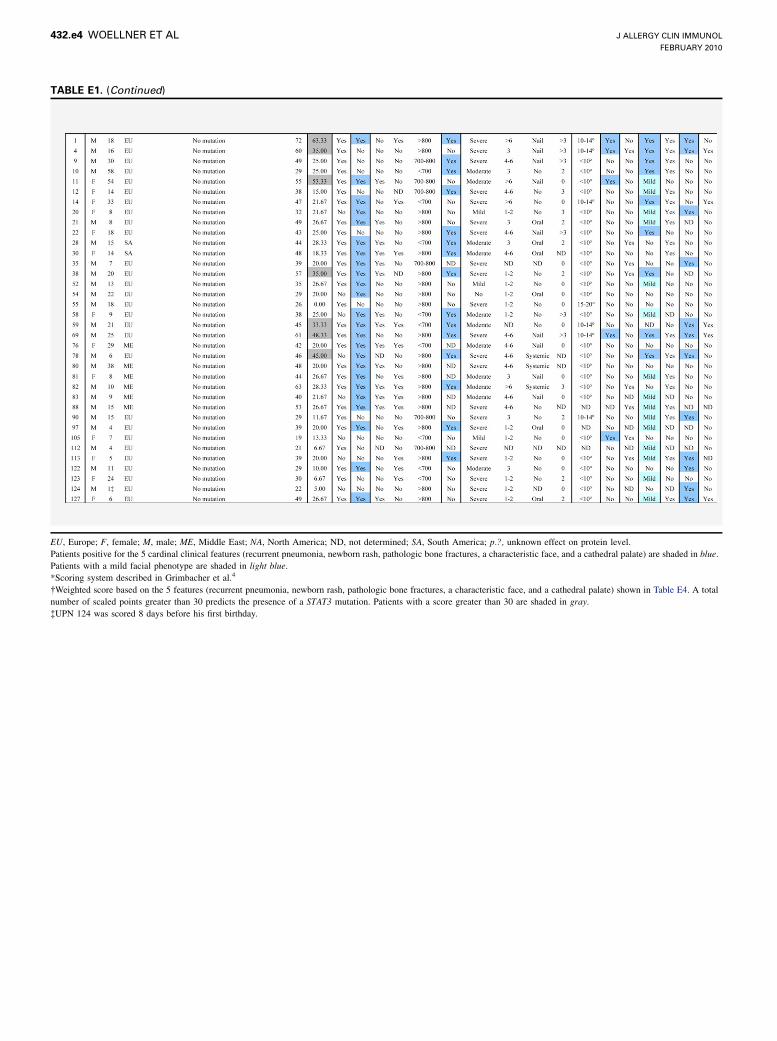
TABLE E1. (Continued)
EU, Europe; F, female; M, male; ME, Middle East; NA, North America; ND, not determined; SA, South America; p.?, unknown effect on protein level.
Patients positive for the 5 cardinal clinical features (recurrent pneumonia, newborn rash, pathologic bone fractures, a characteristic face, and a cathedral palate) are shaded in blue.
Patients with a mild facial phenotype are shaded in light blue.
*Scoring system described in Grimbacher et al.4
�Weighted score based on the 5 features (recurrent pneumonia, newborn rash, pathologic bone fractures, a characteristic face, and a cathedral palate) shown in Table E4. A total
number of scaled points greater than 30 predicts the presence of a STAT3 mutation. Patients with a score greater than 30 are shaded in gray.
�UPN 124 was scored 8 days before his first birthday.
J ALLERGY CLIN IMMUNOL
FEBRUARY 2010
432.e4 WOELLNER ET AL

TABLE E2. Association of NIH-HIES score with mutation status
Kind of mutation Patients positive Patients negative Rank-sum P value
Any mutation in STAT3 64 36 3.9e-7
DNA binding domain 42 22 .094
SH2 domain 17 47 NS
The first column indicates the kind of mutations considered, the second column shows the number of patients positive for that mutation, and the third column shows the number of
patients negative for that mutation. The fourth column is the P value of a Wilcoxon rank-sum test against the hypothesis that both groups have the same distribution of score. The
rank-sum test for the first row includes all 100 patients in our cohort; the other 2 rows include only the 64 mutation-positive patients.
J ALLERGY CLIN IMMUNOL
VOLUME 125, NUMBER 2
WOELLNER ET AL 432.e5

TABLE E3. The 11 features that occur more than once in the 12 feature sets with best leave-1-out accuracy
Feature Times included Weight signs P value
Recurrent pneumonia 11 Positive .002
Lung cyst formation 3 Positive .001
Other unusual infections 3 Mixed NS
Newborn rash 10 Positive NS
Upper respiratory tract infections 4 Negative NS
Scoliosis 3 Positive NS
Pathologic bone fractures 12 Positive NS
Characteristic face for Job syndrome 12 Positive .002
Increased interalar distance 5 Mixed NS
Cathedral palate 10 Positive NS
Midline anomaly 2 Negative NS
The second column shows the number of sets that include the feature. The third column indicates whether the weight assigned the feature is positive or negative in each of 12 SVM
classifiers in which that feature occurs. The features ‘‘increased interalar distance’’ and ‘‘other unusual infections’’ were sometimes assigned positive and sometimes assigned
negative weights. The fourth column is the P value of the null hypothesis that the slope of the logistic regression function for the indicated feature is 0. The sign of the logistic
regression function was positive for the features recurrent pneumonia, lung cyst formation, and characteristic face for Job syndrome.
J ALLERGY CLIN IMMUNOL
FEBRUARY 2010
432.e6 WOELLNER ET AL

TABLE E4. HIES STAT3 score
J ALLERGY CLIN IMMUNOL
VOLUME 125, NUMBER 2
WOELLNER ET AL 432.e7

TABLE E5. Effectiveness of the best SVM classifier by age range in our cohort of 100 patients
Error Sensitivity Specificity Positive Negative
Age 0-8 y 10 88.9 90.9 9 11
Age 9-16 y 13.2 85.2 90.3 27 11
Age 17-30 y 21.2 87.0 60.0 23 10
Age 311 y 11.1 100.0 75.0 5 4
The first column shows an age range for each row, where age is determined at the date of scoring. The second, third, and fourth columns show the error rate, the sensitivity, and the
specificity of the SVM classifier when applied to the subset of 100 patients in the given age range. The fifth and sixth columns list the number of individuals in the given age range
that are either positive or negative for a STAT3 mutation.
J ALLERGY CLIN IMMUNOL
FEBRUARY 2010
432.e8 WOELLNER ET AL
Copyright © 2022 FDOKUMEN