The EGFR-STAT3 Oncogenic Pathway Up-regulates the Eme1 Endonuclease to Reduce DNA Damage after...
12
The EGFR-STAT3 Oncogenic Pathway Up-regulates the Eme1 Endonuclease to Reduce DNA Damage after Topoisomerase I Inhibition Arnaud Vigneron, Erick Gamelin, and Olivier Coqueret Cancer Center Paul Papin, Institut National de la Sante et de la Recherche Medicale U892, Universite´ d’Angers, Angers, France Abstract The epidermal growth factor receptor (EGFR)-src-signal transducers and activators of transcription 3 (STAT3) onco- genic pathway plays a central role in tumorigenesis and is involved not only in cell transformation but also in tumor escape to genotoxic treatments. Despite its importance, the molecular mechanisms by which this signaling pathway induces resistance to DNA damage remain most of the time to be characterized. In this study, we show that the EGFR-src pathway is activated in response to topoisomerase I inhibi- tion. After treatment, this signaling cascade induced the activation of STAT3 and the binding of the transcription factor to the promoter of the Eme1 gene. Eme1 is an endonuclease involved in the processing of DNA damage after topoisomer- ase I inhibition. These results suggest a model by which the STAT3-mediated activation of Eme1 prevents DNA damage and enhances cell survival in response to topoisomerase inhibition. This survival pathway was inhibited by a combined treatment with a src inhibitor, SKI, and with cetuximab, a monoclonal antibody directed against the EGFR that is widely used in the treatment of colorectal cancers. We therefore propose that the benefit of anti-EGFR therapy relies on an increase of DNA damage generated by topoisomerase I inhibition. [Cancer Res 2008;68(3):815–25] Introduction The epidermal growth factor receptor (EGFR) family consists of the EGFR, ErbB2/Neu, ErbB3, and ErbB4 proteins, which are activated after binding of several growth factors, such as EGF, transforming growth factor-a, or amphiregulin. Several studies have shown that activated forms of the receptor can induce cell transformation, and many epithelial cancers exhibit amplification, mutation, or autocrine activation of the EGFR, which in many cases, correlates with disease progression and poor prognosis. Targeted therapies interfering with EGFR signaling are now used as efficient strategies to increase the efficiency of conventional genotoxic treatments (1). Interfering with the EGFR oncogenic pathway is achieved through the use of either monoclonal antibodies or small molecule inhibiting its tyrosine kinase activity. Cetuximab (Erbitux; C225), a monoclonal antibody that binds to the EGFR, has been shown to inhibit the growth of several cancer cells in vitro and in nude mice (2). In addition, cetuximab potentiates the effect of genotoxic drugs such as the topoisomerase I inhibitor irinotecan (3–6). Most importantly, these preclinical results have been confirmed by clinical trials showing that the inhibition of the EGFR pathway through the use of cetuximab can reverse drug resistance, induce response, and prolong the survival of patients with irinotecan-refractory colorectal carcinoma (7, 8). One essential question of cancer treatment is why tumors fail to respond to chemotherapy and which oncogenic signaling is predictive of tumor resistance. Unfortunately, the molecular mechanisms underlying the role of the EGFR pathway in refractory colorectal tumors remain to be characterized. The main pathways stimulated by the receptor are the mitogen-activated protein kinases (MAPK), the phosphatidylinositol 3-kinase–AKT, and signal transducer and activator of transcription proteins (STAT), such as STAT3 (9). In addition, the activation of STAT3 also relies on the recruitment of the src kinase to the EGFR (9–11). Upon nuclear translocation, the STAT3 transcription factor up-regulates several genes involved in cell cycle progression (12, 13), cell survival (14–18), and metastasis (19). Therefore, through the activation of src-STAT3, the EGFR signaling induces the expression of survival proteins such as Bcl-Xl or mcl1. These proteins are well-known to play an important role in drug resistance and tumor escape by inhibiting the cell death pathways associated with genotoxic treatments. Besides apoptosis, we have recently shown that the src-STAT3 oncogenes prevent senescence induction in response to genotoxic drugs through the up-regulation of Myc and the inactivation of the p53-p21 pathway (20). For these reasons, we have recently proposed that the STAT3 oncogenic signaling pathway plays an important role in drug resistance through the inactivation of tumor suppressor networks (21). We now extend these observations and show in this study that the EGFR-src-STAT3 pathway is activated in response to top- oisomerase I inhibition. After inhibition, DNA damage induced the activation of the src kinase, the activation of STAT3, and the binding of the transcription factor to the promoter of the Eme1 gene. Eme is an endonuclease identified as a member of the XPF family of endonucleases (22). This protein plays an important role in the processing of DNA repair after topoisomerase I inhibition, and its up-regulation confers resistance to several DNA-damaging agents. These results indicate that upon cell transformation, the EGFR-src-STAT3 oncogenic pathway induces the up-regulation of Eme1, thereby leading to an intrinsic drug resistance program. Consequently, the inactivation of the EGFR pathway by cetuximab prevents the expression of Eme1 and enhanced DNA damage and cell death in response to topoisomerase I inhibition. We propose that the benefit of cetuximab and anti-EGFR therapy relies on the Note: Supplementary data for this article are available at Cancer Research Online (http://cancerres.aacrjournals.org/). Current address for A. Vigneron: Beatson Institute for Cancer Research, Glasgow, United Kingdom. Requests for reprints: Olivier Coqueret, Paul Papin Cancer Center, Institut National de la Sante et de laRecherche Medicale U892, Universite´ d’Angers, 2 rue Moll, 49033 Angers, France. Phone: 33-2-41-35-27-00, ext. 2564; Fax: 33-2-41-48-31-90; E-mail: [email protected]. I2008 American Association for Cancer Research. doi:10.1158/0008-5472.CAN-07-5115 www.aacrjournals.org 815 Cancer Res 2008; 68: (3). February 1, 2008 Research Article Research. on January 22, 2015. © 2008 American Association for Cancer cancerres.aacrjournals.org Downloaded from
-
Upload
independent -
Category
Documents
-
view
0 -
download
0
Transcript of The EGFR-STAT3 Oncogenic Pathway Up-regulates the Eme1 Endonuclease to Reduce DNA Damage after...

The EGFR-STAT3 Oncogenic Pathway Up-regulates
the Eme1 Endonuclease to Reduce DNA Damage after
Topoisomerase I Inhibition
Arnaud Vigneron, Erick Gamelin, and Olivier Coqueret
Cancer Center Paul Papin, Institut National de la Sante et de la Recherche Medicale U892, Universite d’Angers, Angers, France
Abstract
The epidermal growth factor receptor (EGFR)-src-signaltransducers and activators of transcription 3 (STAT3) onco-genic pathway plays a central role in tumorigenesis and isinvolved not only in cell transformation but also in tumorescape to genotoxic treatments. Despite its importance, themolecular mechanisms by which this signaling pathwayinduces resistance to DNA damage remain most of the timeto be characterized. In this study, we show that the EGFR-srcpathway is activated in response to topoisomerase I inhibi-tion. After treatment, this signaling cascade induced theactivation of STAT3 and the binding of the transcription factorto the promoter of the Eme1 gene. Eme1 is an endonucleaseinvolved in the processing of DNA damage after topoisomer-ase I inhibition. These results suggest a model by which theSTAT3-mediated activation of Eme1 prevents DNA damageand enhances cell survival in response to topoisomeraseinhibition. This survival pathway was inhibited by a combinedtreatment with a src inhibitor, SKI, and with cetuximab, amonoclonal antibody directed against the EGFR that is widelyused in the treatment of colorectal cancers. We thereforepropose that the benefit of anti-EGFR therapy relies on anincrease of DNA damage generated by topoisomerase Iinhibition. [Cancer Res 2008;68(3):815–25]
Introduction
The epidermal growth factor receptor (EGFR) family consists ofthe EGFR, ErbB2/Neu, ErbB3, and ErbB4 proteins, which areactivated after binding of several growth factors, such as EGF,transforming growth factor-a, or amphiregulin. Several studieshave shown that activated forms of the receptor can induce celltransformation, and many epithelial cancers exhibit amplification,mutation, or autocrine activation of the EGFR, which in manycases, correlates with disease progression and poor prognosis.Targeted therapies interfering with EGFR signaling are now used asefficient strategies to increase the efficiency of conventionalgenotoxic treatments (1). Interfering with the EGFR oncogenicpathway is achieved through the use of either monoclonalantibodies or small molecule inhibiting its tyrosine kinase activity.
Cetuximab (Erbitux; C225), a monoclonal antibody that bindsto the EGFR, has been shown to inhibit the growth of severalcancer cells in vitro and in nude mice (2). In addition, cetuximabpotentiates the effect of genotoxic drugs such as the topoisomeraseI inhibitor irinotecan (3–6). Most importantly, these preclinicalresults have been confirmed by clinical trials showing thatthe inhibition of the EGFR pathway through the use of cetuximabcan reverse drug resistance, induce response, and prolong thesurvival of patients with irinotecan-refractory colorectal carcinoma(7, 8).One essential question of cancer treatment is why tumors fail to
respond to chemotherapy and which oncogenic signaling ispredictive of tumor resistance. Unfortunately, the molecularmechanisms underlying the role of the EGFR pathway in refractorycolorectal tumors remain to be characterized. The main pathwaysstimulated by the receptor are the mitogen-activated proteinkinases (MAPK), the phosphatidylinositol 3-kinase–AKT, and signaltransducer and activator of transcription proteins (STAT), such asSTAT3 (9). In addition, the activation of STAT3 also relies on therecruitment of the src kinase to the EGFR (9–11). Upon nucleartranslocation, the STAT3 transcription factor up-regulates severalgenes involved in cell cycle progression (12, 13), cell survival(14–18), and metastasis (19). Therefore, through the activation ofsrc-STAT3, the EGFR signaling induces the expression of survivalproteins such as Bcl-Xl or mcl1. These proteins are well-known toplay an important role in drug resistance and tumor escape byinhibiting the cell death pathways associated with genotoxictreatments. Besides apoptosis, we have recently shown that thesrc-STAT3 oncogenes prevent senescence induction in responseto genotoxic drugs through the up-regulation of Myc and theinactivation of the p53-p21 pathway (20). For these reasons, wehave recently proposed that the STAT3 oncogenic signalingpathway plays an important role in drug resistance through theinactivation of tumor suppressor networks (21).We now extend these observations and show in this study that
the EGFR-src-STAT3 pathway is activated in response to top-oisomerase I inhibition. After inhibition, DNA damage induced theactivation of the src kinase, the activation of STAT3, and thebinding of the transcription factor to the promoter of the Eme1gene. Eme is an endonuclease identified as a member of the XPFfamily of endonucleases (22). This protein plays an important rolein the processing of DNA repair after topoisomerase I inhibition,and its up-regulation confers resistance to several DNA-damagingagents. These results indicate that upon cell transformation, theEGFR-src-STAT3 oncogenic pathway induces the up-regulation ofEme1, thereby leading to an intrinsic drug resistance program.Consequently, the inactivation of the EGFR pathway by cetuximabprevents the expression of Eme1 and enhanced DNA damage andcell death in response to topoisomerase I inhibition. We proposethat the benefit of cetuximab and anti-EGFR therapy relies on the
Note: Supplementary data for this article are available at Cancer Research Online(http://cancerres.aacrjournals.org/).
Current address for A. Vigneron: Beatson Institute for Cancer Research, Glasgow,United Kingdom.
Requests for reprints: Olivier Coqueret, Paul Papin Cancer Center, InstitutNational de la Sante et de la Recherche Medicale U892, Universite d’Angers, 2 rue Moll,49033 Angers, France. Phone: 33-2-41-35-27-00, ext. 2564; Fax: 33-2-41-48-31-90; E-mail:[email protected].
I2008 American Association for Cancer Research.doi:10.1158/0008-5472.CAN-07-5115
www.aacrjournals.org 815 Cancer Res 2008; 68: (3). February 1, 2008
Research Article
Research. on January 22, 2015. © 2008 American Association for Cancercancerres.aacrjournals.org Downloaded from

down-regulation of Eme1 expression and on the consequentincrease of DNA damage generated by anticancer drugs, such asirinotecan, that induce an inhibition of topoisomerase I.Altogether, these results reveal a complex network of inter-
actions indicating that the EGFR-src-STAT3 oncogenic pathwayinduces the expression of the Eme1 endonuclease to induce celltransformation and drug resistance.
Materials and Methods
Materials. Antibodies were obtained from Santa Cruz Biotechnology.Anti-atubulin (T9026) was obtained from Sigma, and Anti-Eme1 was
obtained from Immuquest (IQ284). SKI was obtained from Calbiochem
(#567805). The src siRNA was obtained from Santa Cruz (sc-29228), andSN38 comes from Pfitzer and Cetuximab from Merck. All cells were
maintained in RPMI supplemented with 10% serum and were not used
beyond 25 to 30 passages.
Chromatin immunoprecipitation assay. Attached cells were washedand cross-linked with 1% formaldehyde at room temperature for 10 min.
Cells were washed sequentially twice with 1 mL ice-cold PBS, centrifuged,
and then resuspended in 0.5 mL of lysis buffer [1% SDS, 10 mmol/L EDTA,
50 mmol/L Tris-HCl (pH 8.1), 1 mmol/L phenylmethylsulfonyl fluoride,1 Ag/mL leupeptin, and 1 Ag/mL aprotinin] and sonicated thrice for 15 s
each at the maximum setting. Supernatants were then recovered
by centrifugation at 12,000 rpm for 10 min at 4jC, diluted twice in dilution
buffer [1% Triton X-100, 2 mmol/L EDTA, 150 mmol/L NaCl, and 20 mmol/LTris-HCl (pH 8.1)], and subjected to one round of immunoclearing for 2 h at
4jC with 2 Ag sheared salmon sperm DNA, 2,5 Ag preimmune serum, and
20 AL of protein A sepharose (of 50% slurry). Immunoprecipitation wasperformed overnight with specific antibodies, then 2 Ag sheared salmon
sperm DNA and 20 AL of protein A sepharose (of 50% slurry) were further
added for 1 h at 4jC. Immunoprecipitates were washed sequentially for
10min each in TSE I [0.1% SDS, 1% Triton X-100, 2 mmol/L EDTA, 20 mmol/LTris-HCl (pH 8.1), and 150mmol/LNaCl], TSE II [0.1% SDS, 1% Triton X-100, 2
mmol/L EDTA, 20 mmol/L Tris-HCl (pH 8.1), and 500 mmol/L NaCl], and
buffer III [0.25 mol/LLiCl, 1% NP40, 1% deoxycholate, 1 mmol/L EDTA, and
10 mmol/L Tris-HCl (pH 8.1)]. Beads precipitates were then washed thricewith TE buffer and eluted twice with 1% SDS and 0.1 mol/L NaHCO3. Eluates
were pooled, heated at 65jC for 6 h to reverse the formaldehyde cross-linking,
and DNA was precipitated using classic procedures. For PCR, 10 AL from a100 AL DNA preparation were used for 25 to 30 cycles of amplifications. The
amplified regions are presented on Fig. 5, and primers are available upon
request.
Real-time PCR. For RNA quantification, PCR was performed with 5 AL ofDNA and 5 pmol/L primers diluted in a final volume of 5 AL Reaction Mix
lightcycler (Roche Diagnostics GmbH) and 4 mmol/L of MgCl2. Accumu-
lation of fluorescent products was monitored by real-time PCR using a
lightcycler (Roche Diagnostics GmbH). The relative quantification of geneexpression was performed using the comparative CT method, with
normalization of the target gene to the endogenous housekeeping gene
glyceraldehyde-3-phosphate dehydrogenase (GAPDH).Flow cytometry analysis. For DNA content analysis, 1 � 106 cells
were washed twice with PBS and fixed in 70% ethanol. Cells were treated
with 100 units/mL RNase A for 20 min at 37jC, resuspended in PBS
containing 50 Ag/mL propidium iodide, and immediately analyzed by flowcytometry (Becton Dickinson). For protein expression, 1 � 106 cells were
washed twice with PBS, fixed in 70% ethanol, and further incubated for
45 min at 37jC in PBS containing 5% bovine serum albumin, 1 mg/mL
RNase, and 2 Ag/mL anti-H2AX (Upstate) or a control IgG. After threewashes of PBS 0.1% Tween, cells were further incubated for 20 min with a
FITC antibody at room temperature. After washes, samples were analyzed
by flow cytometry. To quantify H2AX phosphorylation, result are presented
as a ratio between the fluorescence with the H2Ax antibody and a controlIgG: Fluo IgG H2AX/Fluo Control IgG.
Colony formation assay. For colony formation assay, 1.5 � 106 cells
were plated per 10-cm plate and treated for the indicated times. Cells were
then washed, trypsinized, and replated in drug-free medium in 6-well plates(diluted from 100,000 cells per well to 100 cells per well) and allowed to
form colonies for 7 to 10 days. This corresponds to a hundred cells present
in average in each colony. In parallel experiments, the percentage of
senescent cells was determined by staining SA-h-gal activity using X-gal atpH 6.0. Cells were fixed in 0.25% glutaraldehyde in PBS for 15 min, washed
thrice with PBS, and stained using fresh buffer [1 mg of 5-bromo-4-chloro-3-
indolyl-h-D-galactoside/mL (stock 40 mg/mL DMSO), 5 mmol/L potassium
ferrocyanide, 5 mmol/L potassium ferricyanide, 40 mmol/L citric acidsodium phosphate (pH 6.0), 150 mmol/L NaCl, 2 mmol/L MgCl2, and PBS]
at 37jC, for 16 to 20 h and in the absence of carbon dioxide. Positive cells
were then counted by bright-field microscopy after scoring 100 to 1,000 cells
for each sample.Comet assay. A single-cell gel electrophoresis (Comet Assay) kit was
used for evaluating DNA damage following the manufacturer’s instructions
(Trevigen, Inc.). After treatment of cells with DNA damage agents (i.e., SN38,cetuximab, or SKI), cells in 1� PBS (Ca2+ and Mg2+ free) at 1 � 105/mL
were combined with molten LMAgarose (at 37jC) at a ratio of 1:10 (v/v) andimmediately pipetted 75 AL onto CometSlideTM. After a gentle cell lysis,
samples were treated with alkali to unwind and denature DNA. The sampleswere subjected to electrophoresis, stained with SYBR Green, and visualized
by microscopy.
Results
The combined inhibition of src and EGFR sensitizes cell totopoisomerase I inhibition. We first tested the hypothesis thatEGF-src signaling could act as a survival pathway in response totopoisomerase I inhibition. To this end, clonogenic assays wereperformed after exposure of colorectal cell lines to moderate dosesof SN38, the active metabolite of irinotecan, which is widely used inthe treatment of colorectal cancers. These experiments wereperformed in the presence or absence of the src inhibitor SKI andof cetuximab, a monoclonal antibody directed against the EGFRused in irinotecan-refractory colorectal carcinoma. Results pre-sented in Fig. 1A showed that SN38 treatment resulted in 59% and52% clonogenicity relative to untreated HCT116 and HT29 controlcells, respectively. In the presence of cetuximab and SKI, asignificant decrease of cell viability was observed compared withSN38 (Fig. 1A ; compare lanes 1 and 5, lanes 6 and 10). Importantly,no effect of cetuximab or SKI was observed when added alone tothe cell culture, and a nonrelated IgG did not affect cellproliferation. We also observed that the inhibition of the EGFR-src pathway decreased the concentration of SN38 required toreduce cell viability (Fig. 1B). The IC50 values were 1.6 ng/mL forSN38-treated HCT116 cells and 0.9 ng/mL in the presence ofcetuximab-SKI, and 3.2 ng/mL for SN38-treated HT29 cells and2.2 ng/mL in the absence of cetuximab-SKI.To characterize the enhanced sensitivity to SN38, we then
examined changes in senescence and apoptosis. The percentage ofcells entering apoptosis was characterized by fluorescence-activated cell sorting (FACS) analysis (sub-G1 fraction) and visualexamination of condensed chromatin. The induction of thesenescence program was detected by h-galactosidase stainingand visual examination of the flattened phenotype. Micronuclea-tion was characterized by micronuclei examination and detectionof noncondensed chromatin in the main nuclei. In HCT116 cells,and as previously described (23), SN38 treatment results in theappearance of cells with multiple micronuclei, a hallmark ofmitotic catastrophe. As a consequence, around 60% of the attachedcells became senescent after 4 days of treatment (Fig. 1C, left).In the presence of cetuximab and SKI, a slight increase inSA-h-galactosidase–positive cells was noticed compared with the
Cancer Research
Cancer Res 2008; 68: (3). February 1, 2008 816 www.aacrjournals.org
Research. on January 22, 2015. © 2008 American Association for Cancercancerres.aacrjournals.org Downloaded from

Figure 1. The combined inhibition of src and EGFR sensitizes cell to topoisomerase I inhibition. A, HCT116 or HT29 cells were treated with SN38 for 2 d in thepresence or absence of cetuximab (cetux ) and SKI as indicated, washed, replated, and further grown for 14 d [HCT116 (sn38: 2.5 nmol/L), HT29 (sn38: 3.7 nmol/L)].Colony formation was then counted using an inverted microscope, and for each cell line, growth of nontreated cells was set up at 100%. Clonogenic survival wasthen plotted as a fraction relative to these untreated cells (n = 3 F SD). B, colony formation was investigated as described in A in the presence of increasingconcentrations of SN38 (n = 3 F SD). C, growing HCT116 cells were treated with SN38 (2.5 nmol/L) for 48 h, in the presence or absence of cetuximab (2.5 Ag/mL) andSKI (10 Amol/L) as indicated. The percentage of senescent cells was evaluated as the number of cells expressing SA-h-gal activity or by flow cytometry analysisusing polyclonal antibodies directed against ICAM-1. In parallel, total cell extracts were prepared and analyzed by Western blot (n = 3 F SD). SSC , side scatter.D, growing HT29 cells were treated with SN38 (3.7 nmol/L) for 48 h, in the presence or absence of cetuximab (2.5 Ag/mL) and SKI (10 Amol/L) as indicated. After 36 h,the percentage of apoptotic cells was evaluated by flow cytometry using either polyclonal antibodies directed against the active form of caspase 3 or by FACS analysisof sub-G1 cells. In parallel, total cell extracts were prepared and analyzed by Western blot (n = 3 F SD). PARP, poly(ADP)ribose polymerase.
STAT3 Activates the Expression of Eme1 Gene
www.aacrjournals.org 817 Cancer Res 2008; 68: (3). February 1, 2008
Research. on January 22, 2015. © 2008 American Association for Cancercancerres.aacrjournals.org Downloaded from

drug alone (Fig. 1C, left). To confirm this result, we evaluated theexpression of the intercellular adhesion molecule-1 (ICAM-1), oneof the markers overexpressed in senescent cells (24, 25). Asexpected, ICAM-1 was effectively induced by SN38 in HCT116 cells,and a significant up-regulation was noticed upon down-regulationof EGFR signaling (Fig. 1C, middle). After checkpoint arrest,senescence is also induced with the participation of the cell cycleinhibitor p21waf1. As expected, the SN38-mediated expression ofp21waf1 was enhanced in the presence of cetuximab and SKI(Fig. 1C, right), indicating that EGFR-src inhibition up-regulates theinduction of senescence.We then determined if this effect was only related to the
senescence pathway or also to other forms of cell death. To thisend, we used HT29 cells because this cell line responds differentlyto genotoxic treatment and has a mutated form of p53, so that anyeffect would be independent of the tumor suppressor. In response
to SN38, <10% of HT29 cells enter the senescence program orpresent micronuclei (data not shown; see also ref. 23). By contrast,apoptosis occurred in 35% to 40% of these cells in response toSN38, and this was enhanced upon inhibition of EGFR-src signaling(Fig. 1D, left). An increased in the percentage of sub-G1 cells and ofthe cleavage of the poly(ADP)ribose polymerase protein was alsoobserved in these conditions (Fig. 1D, middle and right). No effectwas observed when cetuximab and SKI were added alone to the cellculture (data not shown).Altogether, these results indicate that the inhibition of EGFR-src
signaling potentiates the induction of senescence and apoptosis inresponse to topoisomerase I inhibition.The inhibition of the EGFR-src signaling pathway induces
DNA damage. SN38 induces topoisomerase I cleavage complexesthat inhibit replication forks and induce DNA double strand breaks(26). Based on these observations, we made the hypothesis that
Figure 2. The combined inhibition of src and EGFRpotentiates DNA damage induced by topoisomeraseI (topo1 ) inhibition. A, HCT116 or HT29 growingcells were treated with SN38 for 24 h (HCT116,2.5 nmol/L; HT29, 3.7 nmol/L), in the presenceor absence of cetuximab (2.5 Ag/mL) and SKI(10 Amol/L) as indicated. Cells were then washedand further treated for 12 h by cetuximab-SKI. Theamount of DNA strand breaks was then determinedusing the alkaline comet assay and quantifiedusing the Comet Assay IV software (n = 3 F SD).B, HCT116 or HT29 growing cells were treated asdescribed in A , and the generation of DNA doublestrand breaks was quantified by FACS analysisusing polyclonal antibodies directed against theSer139 phosphorylated form of g-histone H2Ax(n = 3 F SD). Phospho-H2Ax , histone H2Axphosphorylation. C, cells were treated as describedin A , and total cell extracts were prepared andanalyzed by Western blot (n = 3).
Cancer Research
Cancer Res 2008; 68: (3). February 1, 2008 818 www.aacrjournals.org
Research. on January 22, 2015. © 2008 American Association for Cancercancerres.aacrjournals.org Downloaded from
EGFR-src inhibition might potentiate the effect of SN38 on DNAdamage. To verify this, genotoxicity analysis were performed usingeither the alkaline comet assay (Fig. 2A) or Ser139 phosphorylationof histone H2Ax as a marker of DNA double strand breaks (Fig. 2B).As expected, SN38 treatment led to a significant increase of the tailmoment in HT29 and HCT116 cells. Accordingly, FACS analysisalso showed an increase in g-H2Ax phosphorylation, confirmingthat DNA double strand breaks occurred in SN38-treated cells(Fig. 2B). Importantly, when cetuximab and SKI were added to thecell cultures, we observed that the inhibition of the EGFR-srcsignaling pathway further enhanced DNA damage. As shown Fig. 2A ,an increased tail momentwas observed in the presence of cetuximaband SKI both in HT29 cells (tail moment, 6.3 F 1.4 for treated cells;compared with SN38 alone tail moment, 2.2F 0.6) and HCT116 cells(tail moment, 6.9 F 1.6 for treated cells; compared with SN38 tailmoment, 3.7 F 0.4). Accordingly, FACS analysis indicated that theSer139 phosphorylation of H2Ax was also enhanced after EGFR-srcinhibition (Fig. 2B). To confirm these results, we then analyzedthe activation of DNA damage checkpoints in drug-treated cells(Fig. 2C). After genotoxic treatment of HCT116 and HT29 cells, weobserved that the chk1 kinase was phosphorylated on its Ser345
residue. In addition, SN38 also induced the phosphorylation of p53on Ser15 in both cell lines. Confirming the above results, theactivation of chk1 and p53 was enhanced when the EGFR-srcpathway was inhibited (Fig. 2C, lanes 3, 6, 9 , and 12). Importantly,kinetic experiments indicated that EGFR inhibition had nosignificant effect on DNA damage after 24 h of treatment (data notshown). Looking at tail moments or g-H2Ax phosphorylation, DNAdamage induction became detectable only when cells were allowed
to proliferate for a further 12 h in the presence of cetuximab and SKI(Fig. 2). In addition, we have also observed that the lengthof S phase is not modified after cetuximab-SKI treatment. Thismight indicate that EGFR inhibition affects late S phase or G2 eventsthat occur after topoisomerase I inhibition, such as the processingof collapsed replication forks and/or the processing of the D loopgenerated during the recombination process (27).Taken together, these results suggest that the inhibition of the
EGFR-src signaling pathway induced cell death through enhanced
DNA damage.The EGFR-src signaling pathway regulates the expression of
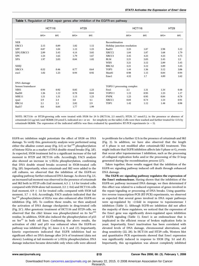
the Eme1 endonuclease. Having shown that the inhibition of theEGFR-src pathway increased DNA damage, we then determined ifthis effect was related to a reduced expression of genes involved inthe repair/signaling or processing of DNA breaks. Using quantita-tive reverse transcription-PCR (RT-PCR) experiments, we observedas expected that several genes involved in DNA repair pathwayswere up-regulated by >2-fold in response to topoisomerase Iinhibition (Table 1). Although EGFR-src inhibition did not affectthe majority of these regulators, we noticed that the expression ofthe Eme1 gene was significantly down-regulated upon inhibitionof EGFR signaling (Table 1). Eme1 is an endonuclease that isimplicated in the efficient rescue of broken replication forks inyeast. Importantly, Eme1 inactivation has been correlated withelevated levels of DNA damage, chromosomal aberrations, anddrug sensitivity (22, 28). In HCT116 and HT29 cells, Western blotand quantitative PCR experiments showed that Eme1 expressionwas significantly induced in response to SN38 (Fig. 3A and B).Importantly, this up-regulation was almost completely inhibited
Table 1. Regulation of DNA repair genes after inhibition of the EGFR-src pathway
HCT116 HT29 HCT116 HT29
src+ src� src+ src� src+ src� src+ src�
NER Recombination
ERCC1 2.15 0.89 1.82 1.12 Holiday junction resolution
XPF 0.87 1.04 1.12 1.23 Rad51 2.23 1.87 2.98 3.12
XPG/ERCC5 2.89 3.45 4.14 3.85 XRCC2 1.84 1.87 1.68 1.79XPC 1.54 1.65 1.79 1.67 XRCC3 3.45 4.23 4.23 4.75
XPA 1.97 2.03 0.84 1.02 BLM 2.21 2.03 2.45 2.2
WRN 3.21 3.12 2.89 3.45NHEJ BRCA2 3.21 3.12 2.89 3.45
XRCC4 0.52 0.46 0.77 0.64 TOP3 1.44 1.56 3.12 3.06
exo1 1.02 1.23 0.94 0.92 Mus81 0.98 1.15 0.84 0.94
Eme1 4.52 1.7 4.89 1.62
DNA damage
Sensor/transducer TOPO I processing complex
NBS1 0.94 0.92 0.85 1.23 Fen1 2.1 1.31 1.34 0.48Rad50 1.26 1.12 0.78 0.84 TOPO I 1.12 0.95 1.23 1.17
MRE11 1.51 1.31 1.13 1.23 PNPK 1.23 0.95 0.84 0.78
rpa2 2.9 2.4 3.8 4.1 XRCC1 0.84 0.74 1.54 0.91BRCA1 2.1 2.3 2.82 2.9 TDP1 1.42 1.12 1.48 0.98
Rad17 0.8 0.84 1.77 1.99
NOTE: HCT116- or HT29-growing cells were treated with SN38 for 24 h (HCT116, 2.5 nmol/L; HT29, 3.7 nmol/L), in the presence or absence of
cetuximab (2.5 Ag/mL) and SKI606 (10 Amol/L; indicated src+ or src� for simplicity on the table). Cells were then washed and further treated for 12 h bycetuximab-SKI606. The expression of the indicated mRNAs was then evaluated by quantitative RT-PCR experiments (n = 5).
STAT3 Activates the Expression of Eme1 Gene
www.aacrjournals.org 819 Cancer Res 2008; 68: (3). February 1, 2008
Research. on January 22, 2015. © 2008 American Association for Cancercancerres.aacrjournals.org Downloaded from

upon EGFR-src down-regulation (Fig. 3A, lanes 3 and 6). As acontrol, no effect was noticed on the expression of other DNArepair genes, such as xrrc1 or fen1, and a nonrelated IgG had noeffect on Eme1 expression (data not shown).Altogether, these results indicate that the Eme1 endonuclease is
up-regulated by the EGFR pathway upon topoisomerase I
inhibition.DNA damage induced the phosphorylation of the EGF
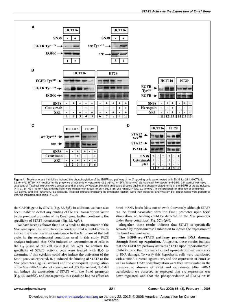
receptor. During the course of these experiments, we observedthat the EGFR pathway was surprisingly activated upontopoisomerase I inhibition. As depicted in Fig. 4A , an increasedphosphorylation of the intracellular domain of the EGFR wasobserved after drug exposure of HCT116 cells (Fig. 4A, lanes 1–2).In addition, topoisomerase I inhibition also induced the phos-phorylation of the src kinase on its Tyr419-activating residue(Fig. 4A, lanes 3–4). Importantly, this EGFR phosphorylation wasinhibited by pretreatment exposure with SKI and with cetuximabin HCT116 and HT29 cells (Fig. 4B ; compare lanes 2 and 5, lanes 7and 10). Under these conditions, a significant inhibition of srcphosphorylation was also observed (Fig. 4C, lanes 3 and 6). Again,when these two inhibitors were used alone, no significant effectwas observed on the SN38-mediated activation of the EGFR. As acontrol, herceptin, a monoclonal antibody directed against theHerII receptor, did not affect the phosphorylation of the receptor(Fig. 4B, lanes 11–16). Because the EGFR-scr pathway activates
STAT3, we then asked if the transcription factor was activatedunder the same conditions. Western blot experiments showed thatSN38 induced the phosphorylation of STAT3 on its Ser727 residue inHCT116 and HT29 cells (Fig. 4D, lanes 3 and 6). As expected, thephosphorylation of STAT3 was inhibited in the presence of SKI andcetuximab, whereas the phosphorylation of the Akt kinase was notaffected.Altogether, these results indicate that the EGFR-src-STAT3
signaling pathway is activated upon topoisomerase I inhibition.The STAT3 transcription factor is activated by SN38 to bind
to the Eme1 promoter. In light of these results, we speculatedthat the EGFR-src cascade regulates the expression of Eme1through STAT3 activation. Transcription factor recognition siteanalysis of the Eme1 promoter (MatInspector; Genomatix) revealedthe presence of a potential STAT3 binding site in the proximalpromoter of the gene (Fig. 5A, left). To determine if STAT3 isassociated with this promoter in response to SN38, chromatinimmunoprecipitation (ChIP) experiments were then performedusing STAT3 antibodies. Ets1 antibodies were used as controlsbecause a potential ets1 binding site was also found in this region.ChIP experiments indicated that STAT3 was significantly recruitedto the proximal Eme1 promoter upon SN38 stimulation (Fig. 5A,right). Importantly, the association of the transcription factor withDNA was significantly prevented after EGFR-src inhibition. As acontrol, PCR analysis did not detect any occupancy of the exon 2 of
Figure 3. The inhibition of the EGFR-srcpathway prevents the expression of the Eme1endonuclease in response to topoisomerase Iinhibition. A, HCT116- or HT29-growing cells weretreated with SN38 for 36 h (HCT116, 2.5 nmol/L;HT29, 3.7 nmol/L), in the presence or absenceof cetuximab (2.5 Ag/mL) and SKI (10 Amol/L) asindicated. Total cell extracts were prepared andanalyzed by Western blot (n = 3). B, cells (HCT116,left ; HT29, right ) were treated as described abovefor 24 h, and the expression of the indicated mRNAwas analyzed by quantitative RT-PCR experiments(n = 3 F SD).
Cancer Research
Cancer Res 2008; 68: (3). February 1, 2008 820 www.aacrjournals.org
Research. on January 22, 2015. © 2008 American Association for Cancercancerres.aacrjournals.org Downloaded from
the GAPDH gene by STAT3 (Fig. 5B, left). In addition, we have alsobeen unable to detect any binding of the ets1 transcription factorto the proximal promoter of the Eme1 gene, further confirming thespecificity of STAT3 recruitment (Fig. 5B, right).We have recently shown that STAT3 binds to the promoter of the
Myc gene upon IL-6 stimulation, a condition that is well-known toinduce the transition from quiescence to the G1 phase of the cellcycle. In the experimental conditions used in this study, FACSanalysis indicated that SN38 induced an accumulation of cells inthe G2 phase of the cell cycle (Fig. 5C, left). To confirm thespecificity of STAT3 activity, cells were treated with IL-6 todetermine if this cytokine could also induce the activation of theEme1 gene. As expected, IL-6 induced the binding of STAT3 to theMyc promoter (Fig. 5C, middle) and the consequent up-regulationof the Myc mRNA (data not shown; see ref. 12). By contrast, IL-6 didnot induce the association of STAT3 with the Eme1 promoter(Fig. 5C, middle), and consequently, this cytokine had no effect on
Eme1 mRNA levels (data not shown). Conversely, although STAT3can be found associated with the Eme1 promoter upon SN38stimulation, no binding could be detected on the Myc promoterunder these conditions (Fig. 5C, right).Altogether, these results indicate that STAT3 is specifically
activated by topoisomerase I inhibition to induce the expression ofthe Eme1 endonuclease.The EGFR-src-STAT3 pathway prevents DNA damage
through Eme1 up-regulation. Altogether, these results indicatethat the EGFR-src pathway activates STAT3 upon topoisomerase Iinhibition, and that this leads to Eme1 up-regulation and resistanceto DNA damage. To verify this hypothesis, cells were transfectedwith a siRNA directed against src, and the expression of Eme1 aswell as histone H2Ax phosphorylation were then investigated in thepresence or absence of SN38 and cetuximab. After siRNAtransfection, we observed as expected that src expression wasdown-regulated, and that the phosphorylation of STAT3 on its
Figure 4. Topoisomerase I inhibition induced the phosphorylation of the EGFR-src pathway. A to C, growing cells were treated with SN38 for 24 h (HCT116,2.5 nmol/L; HT29, 3.7 nmol/L), in the presence or absence of cetuximab (2.5 Ag/mL) or SKI (10 Amol/L) as indicated. Herceptin (anti-Erb2, 2.5 Ag/mL) was usedas a control. Total cell extracts were prepared and analyzed by Western blot with antibodies directed against the phosphorylated forms of the EGFR or src as indicated(n = 3). D, HCT116 or HT29 growing cells were treated with SN38 for 36 h (HCT116, 2.5 nmol/L; HT29, 3.7 nmol/L), in the presence or absence of cetuximab(2.5 Ag/mL) and SKI (10 Amol/L) as indicated. Total cell extracts (including the chromatin fraction) were then prepared, and Western blot experiments were performedwith the indicated antibodies (n = 3).
STAT3 Activates the Expression of Eme1 Gene
www.aacrjournals.org 821 Cancer Res 2008; 68: (3). February 1, 2008
Research. on January 22, 2015. © 2008 American Association for Cancercancerres.aacrjournals.org Downloaded from

Figure 5. The STAT3 transcription factor is activated in response to topoisomerase I inhibition to induce the expression of the Eme1 gene. A, representation ofthe STAT3 and ets1 binding site on the Eme1 promoter (left ). HCT116 or HT29 growing cells were treated as described above, and soluble chromatin was prepared fromthe indicated cells and immunoprecipitated with antibodies directed against the nonphosphorylated form of STAT3. DNA was amplified using one pair ofprimers that covers the STAT3 binding site of the Eme1 promoter. ChIP assays were analyzed by RT-PCR, and the percentage of immunoprecipitated DNA wascalculated by comparison to input signals (n = 3; middle part of the figure). A representative agarose gel of the STAT3 ChIP on the Eme1 promoter is shown onthe right part of the figure. B, ChIP experiments were performed as decribed above and analyzed using a pair of primers that covers the GAPDH gene as a control.In parallel, the association of the ets1 transcription factor with the Eme1 promoter was analyzed by ChIP in growing cells or in cells that were treated as decribedabove (n = 3). C, HT29 cells were treated or not with SN38 for 36 h. FACS analysis was then performed on the indicated attached cells after propidium iodidestaining. In parallel, cells were serum starved for 2 d and stimulated with IL-6 (10 ng/mL) for 30 min as indicated. ChIP experiments were then performed to detectthe association of STAT3 with the Eme1 gene or with the Myc proxymal promoter (region �333/�44; relative to the P2 promoter). In addition, growing cells weretreated with SN38 for 36 h, and the association of STAT3 with the Myc promoter was analyzed by ChIP experiments (right ; n = 3).
Cancer Research
Cancer Res 2008; 68: (3). February 1, 2008 822 www.aacrjournals.org
Research. on January 22, 2015. © 2008 American Association for Cancercancerres.aacrjournals.org Downloaded from
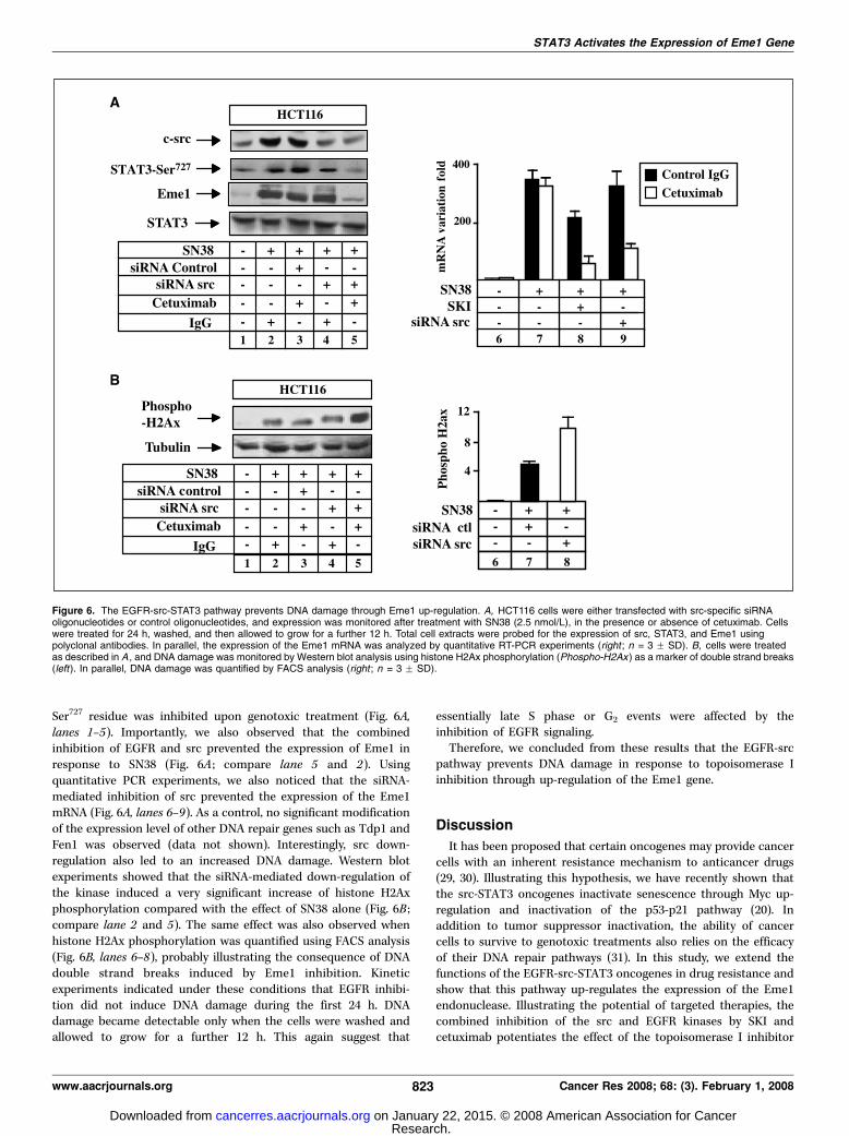
Ser727 residue was inhibited upon genotoxic treatment (Fig. 6A,lanes 1–5). Importantly, we also observed that the combinedinhibition of EGFR and src prevented the expression of Eme1 inresponse to SN38 (Fig. 6A ; compare lane 5 and 2). Usingquantitative PCR experiments, we also noticed that the siRNA-mediated inhibition of src prevented the expression of the Eme1mRNA (Fig. 6A, lanes 6–9). As a control, no significant modificationof the expression level of other DNA repair genes such as Tdp1 andFen1 was observed (data not shown). Interestingly, src down-regulation also led to an increased DNA damage. Western blotexperiments showed that the siRNA-mediated down-regulation ofthe kinase induced a very significant increase of histone H2Axphosphorylation compared with the effect of SN38 alone (Fig. 6B ;compare lane 2 and 5). The same effect was also observed whenhistone H2Ax phosphorylation was quantified using FACS analysis(Fig. 6B, lanes 6–8), probably illustrating the consequence of DNAdouble strand breaks induced by Eme1 inhibition. Kineticexperiments indicated under these conditions that EGFR inhibi-tion did not induce DNA damage during the first 24 h. DNAdamage became detectable only when the cells were washed andallowed to grow for a further 12 h. This again suggest that
essentially late S phase or G2 events were affected by theinhibition of EGFR signaling.Therefore, we concluded from these results that the EGFR-src
pathway prevents DNA damage in response to topoisomerase Iinhibition through up-regulation of the Eme1 gene.
Discussion
It has been proposed that certain oncogenes may provide cancercells with an inherent resistance mechanism to anticancer drugs(29, 30). Illustrating this hypothesis, we have recently shown thatthe src-STAT3 oncogenes inactivate senescence through Myc up-regulation and inactivation of the p53-p21 pathway (20). Inaddition to tumor suppressor inactivation, the ability of cancercells to survive to genotoxic treatments also relies on the efficacyof their DNA repair pathways (31). In this study, we extend thefunctions of the EGFR-src-STAT3 oncogenes in drug resistance andshow that this pathway up-regulates the expression of the Eme1endonuclease. Illustrating the potential of targeted therapies, thecombined inhibition of the src and EGFR kinases by SKI andcetuximab potentiates the effect of the topoisomerase I inhibitor
Figure 6. The EGFR-src-STAT3 pathway prevents DNA damage through Eme1 up-regulation. A, HCT116 cells were either transfected with src-specific siRNAoligonucleotides or control oligonucleotides, and expression was monitored after treatment with SN38 (2.5 nmol/L), in the presence or absence of cetuximab. Cellswere treated for 24 h, washed, and then allowed to grow for a further 12 h. Total cell extracts were probed for the expression of src, STAT3, and Eme1 usingpolyclonal antibodies. In parallel, the expression of the Eme1 mRNA was analyzed by quantitative RT-PCR experiments (right ; n = 3 F SD). B, cells were treatedas described in A , and DNA damage was monitored by Western blot analysis using histone H2Ax phosphorylation (Phospho-H2Ax ) as a marker of double strand breaks(left). In parallel, DNA damage was quantified by FACS analysis (right ; n = 3 F SD).
STAT3 Activates the Expression of Eme1 Gene
www.aacrjournals.org 823 Cancer Res 2008; 68: (3). February 1, 2008
Research. on January 22, 2015. © 2008 American Association for Cancercancerres.aacrjournals.org Downloaded from

SN38 on cell death. This effect was shown to result from Eme1down-regulation and enhanced DNA damage.The Mus81-Eme1 complex was initially characterized in yeast as
a member of the XPF family of endonuclease complexes involved inthe repair of replication and meiotic defects. By analogy to XPF-ERCC1, Eme1 needs to associate with its Mus81 partner to form anactive heterodimer complex that plays an important role in therepair and/or processing of postreplication damage. Furtherexperiments need to be performed to determine if the EGFRsignaling is involved in parallel in the regulation of the Mus81 gene.After genotoxic treatment and replication blockade, Mus81-Eme1removes the collapsed replication forks and activates thehomologous recombination process (27). As a consequence, theoverexpression of the Mus81-Eme1 complex confers resistance toagents that block replication forks such as hydroxyurea andtopoisomerase I inhibitors. In addition, its inactivation has beenshown to induce genomic instability and enhance the sensitivity toDNA cross-linking agents (28, 32). We therefore propose that a highexpression of Eme1 due to STAT3 activation represents an intrinsicdrug resistance program that allows EGFR-scr–expressing tumorsto respond to topoisomerase I inhibitors. Importantly, thecombined inhibition of src and EGFR with SKI-cetuximabinactivates STAT3 and down-regulates the expression of Eme1.Although this remains to be shown, this treatment should preventthe processing of topoisomerase I cleavage complexes during G2
arrest. Because colorectal cancer cells are able to adapt to G2 DNAdamage checkpoints (33, 34), we speculate that drug adaptation inresponse to SKI-cetuximab allows the progression of SN38-treatedcells toward mitosis. The detection of broken forks and damagedchromosomes by the spindle checkpoint would then induce celldeath through the up-regulation of control proteins such as BubR1,Mad1, or Mad2 (35). By contrast, in the absence of cetuximab-SKI,a normal up-regulation of Eme1 in SN38-treated cells would allow amore efficient processing of cleavage complexes. As a consequence,a higher percentage of cells would resume proliferation during theG2 phase of the cell cycle, allowing tumor escape to cell death andmitotic checkpoint.Further experiments need also to be performed to determine
how DNA damage and topoisomerase I inhibition induce theactivation of the EGFR-src pathway. Previous studies have
already suggested that the EGFR is activated by osmotic andoxidative stresses through metalloprotease cleavage of EGF-likeligand precursors (36). This EGFR signal resulted from stress-induced activation of the ADAM family of metalloproteases andis mediated by the MAPK p38. Interestingly, DNA damageinduced by cisplatin also leads to the phosphorylation of theEGFR and this effect is also mediated by p38 MAPK (37).Altogether, these observations lead to the interesting hypothesisthat the up-regulation of Eme1 by STAT3 could result from theactivation of the p38-ADAM pathway by topoisomerase Iinhibition. In addition, it remains also to be determined if theEGFR pathway could also up-regulate the expression of Eme1through mRNA or protein stabilization.Altogether, these results uncover new functions for the STAT3
oncogenic pathway in the regulation of DNA repair. Althoughthis remains to be firmly established, we therefore propose thatthe EGFR-src-STAT3 oncogenic pathway up-regulates Eme1expression to allow a more efficient processing of cleavagecomplexes and to induce drug resistance (Supplementary Fig.S1). Because an important correlation exists between EGFR, src,STAT3, and colorectal cancers, our results suggest that tumorsexpressing the EGFR-src-STAT3 pathway together with a highexpression of Eme1 might be resistant to DNA–topoisomerase Iinhibitors such as irinotecan. For these reasons, the detection ofthese proteins should help to determine the drug resistance profileof individual tumors to define in advance the subsets of tumors thatwill fail to respond to chemotherapy. Provided that this combina-tion does not lead to serious side effects, we propose that cetuximaband src inhibitors should be used as targeted therapies withtopoisomerase I inhibitors to sensitize drug-resistant colorectaltumors to cell death.
Acknowledgments
Received 8/31/2007; revised 11/20/2007; accepted 11/28/2007.Grant support: A fellowship (A. Vigneron), a grant from the Ligue contre le Cancer
(Equipe labelisee 2007–2010; O. Coqueret), and a grant from Assocation pour laRecherche sur le Cancer (ARC 2005–2007, #3133).
The costs of publication of this article were defrayed in part by the payment of pagecharges. This article must therefore be hereby marked advertisement in accordancewith 18 U.S.C. Section 1734 solely to indicate this fact.
We thank Philippe Juin and Neil Perkins for a critical reading of the manuscript.
References1. Hynes NE, Lane HA. ERBB receptors and cancer: thecomplexity of targeted inhibitors. Nat Rev Cancer 2005;5:341–4.2. Galizia G, Lieto E, De Vita F, et al. Cetuximab, achimeric human mouse anti-epidermal growth factorreceptor monoclonal antibody, in the treatment ofhuman colorectal cancer. Oncogene 2007;26:3654–60.3. Kim S, Prichard CN, Younes MN, et al. Cetuximab andirinotecan interact synergistically to inhibit the growthof orthotopic anaplastic thyroid carcinoma xenograftsin nude mice. Clin Cancer Res 2006;12:600–7.4. Ciardiello F, Bianco R, Damiano V, et al. Antitumoractivity of sequential treatment with topotecan andanti-epidermal growth factor receptor monoclonalantibody C225. Clin Cancer Res 1999;5:909–16.5. Huang SM, Bock JM, Harari PM. Epidermal growthfactor receptor blockade with C225 modulates prolifera-tion, apoptosis, and radiosensitivity in squamous cellcarcinomas of the head and neck. Cancer Res 1999;59:1935–40.6. Prewett MC, Hooper AT, Bassi R, et al. Enhanced
antitumor activity of anti-epidermal growth factorreceptor monoclonal antibody IMC-C225 in combina-tion with irinotecan (CPT-11) against human colorectaltumor xenografts. Clin Cancer Res 2002;8:994–1003.7. Cunningham D, Humblet Y, Siena S, et al. Cetuximabmonotherapy and cetuximab plus irinotecan in irinote-can-refractory metastatic colorectal cancer. N Engl JMed 2004;351:337–45.8. Saltz LB, Meropol NJ, Loehrer PJ, et al. Phase II trial ofcetuximab in patients with refractory colorectal cancerthat expresses the epidermal growth factor receptor.J Clin Oncol 2004;22:1201–8.9. Grandis JR, Drenning SD, Chakraborty A, et al.Requirement of Stat3 but not Stat1 activation forepidermal growth factor receptor- mediated cell growthin vitro . J Clin Invest 1998;102:1385–92.10. Olayioye MA, Beuvink I, Horsch K, Daly JM, HynesNE. ErbB receptor-induced activation of stat transcrip-tion factors is mediated by Src tyrosine kinases. J BiolChem 1999;274:17209–18.11. Biscardi JS, Maa MC, Tice DA, et al. c-Src-mediatedphosphorylation of the epidermal growth factorreceptor on Tyr845 and Tyr1101 is associated with
modulation of receptor function. J Biol Chem 1999;274:8335–43.12. Barre B, Vigneron A, Coqueret O. The STAT3transcription factor is a target for the Myc and retino-blastoma proteins on the Cdc25A promoter. J Biol Chem2005;280:15673–81.13. Leslie K, Lang C, Devgan G, et al. Cyclin D1 istranscriptionally regulated by and required for transfor-mation by activated signal transducer and activator oftranscription 3. Cancer Res 2006;66:2544–52.14. Kiuchi N, Nakajima K, Ichiba M, et al. STAT3 isrequired for the gp130-mediated full activation of thec-myc gene. J Exp Med 1999;189:63–73.15. Shirogane T, Fukada T, Muller JM, et al. Synergisticroles for Pim-1 and c-Myc in STAT3-mediated cellcycle progression and antiapoptosis. Immunity 1999;11:709–19.16. Bromberg J, Wrzeszczynska M, Devgan G, et al. Stat3as an oncogene. Cell 1999;98:295–303.17. Ivanov VN, Bhoumik A, Krasilnikov M, et al.Cooperation between STAT3 and c-jun suppresses Fastranscription. Mol Cell 2001;7:517–28.18. Catlett-Falcone R, Landowski TH, Oshiro M, et al.
Cancer Research
Cancer Res 2008; 68: (3). February 1, 2008 824 www.aacrjournals.org
Research. on January 22, 2015. © 2008 American Association for Cancercancerres.aacrjournals.org Downloaded from
Constitutive activation of Stat3 signaling confersresistance to apoptosis in human U266 myeloma cells.Immunity 1999;10:105–15.19. Dechow TN, Pedranzini L, Leitch A, et al. Require-ment of matrix metalloproteinase-9 for the transforma-tion of human mammary epithelial cells by Stat3-C.Proc Natl Acad Sci U S A 2004;101:10602–7.20. Vigneron A, Roninson IB, Gamelin E, Coqueret O. Srcinhibits adriamycin-induced senescence and G2 check-point arrest by blocking the induction of p21waf1.Cancer Res 2005;65:8927–35.21. Barre B, Vigneron A, Perkins N, et al. The STAT3oncogene as a predictive marker of drug resistance.Trends Mol Med 2007;13:4–11.22. Osman F, Whitby MC. Exploring the roles ofMus81-1/Mms4 at perturbed replication forks. DNARepair (Amst) 2007;6:1004–17.23. Bhonde MR, Hanski ML, Notter M, et al. Equivalenteffect of DNA damage-induced apoptotic cell death orlong-term cell cycle arrest on colon carcinoma cellproliferation and tumour growth. Oncogene 2006;25:165–75.24. Gorgoulis VG, Pratsinis H, Zacharatos P, et al. p53-dependent ICAM-1 overexpression in senescent human
cells identified in atherosclerotic lesions. Lab Invest2005;85:502–11.25. Gorgoulis VG, Zacharatos P, Kotsinas A, et al. p53activates ICAM-1 (CD54) expression in an NF-nB-independent manner. EMBO J 2003;22:1567–78.26. Pommier Y, Redon C, Rao VA, et al. Repair of andcheckpoint response to topoisomerase I-mediated DNAdamage. Mutat Res 2003;532:173–203.27. Whitby MC. Junctions on the road to cancer. NatStruct Mol Biol 2004;11:693–5.28. Dendouga N, Gao H, Moechars D, et al. Disruption ofmurine Mus81 increases genomic instability and DNAdamage sensitivity but does not promote tumorigenesis.Mol Cell Biol 2005;25:7569–79.29. Johnstone RW, Ruefli AA, Lowe SW. Apoptosis: a linkbetween cancer genetics and chemotherapy. Cell 2002;108:153–64.30. Lee S, Schmitt CA. Chemotherapy response andresistance. Curr Opin Genet Dev 2003;13:90–6.31. Masters JR, Koberle B. Curing metastatic cancer:lessons from testicular germ-cell tumours. Nat RevCancer 2003;3:517–25.32. Hiyama T, Katsura M, Yoshihara T, et al. Haploinsuffi-ciency of theMus81-1 endonuclease activates the intra-S-
phase and G2-M checkpoints and promotes rereplicationin human cells. Nucleic Acids Res 2006;34:880–92.33. TaoW, South VJ, Zhang Y, et al. Induction of apoptosisby an inhibitor of the mitotic kinesin KSP requires bothactivation of the spindle assembly checkpoint andmitotic slippage. Cancer Cell 2005;8:49–59.34. Andreassen PR, Lacroix FB, Lohez OD, Margolis RL.Neither p21WAF1 nor 14-3-3j prevents G2 progressionto mitotic catastrophe in human colon carcinoma cellsafter DNA damage, but p21WAF1 induces stable G1
arrest in resulting tetraploid cells. Cancer Res 2001;61:7660–8.35. Weaver BA, Cleveland DW. Decoding the linksbetween mitosis, cancer, and chemotherapy: the mitoticcheckpoint, adaptation, and cell death. Cancer Cell 2005;8:7–12.36. Fischer OM, Hart S, Gschwind A, Prenzel N, UllrichA. Oxidative and osmotic stress signaling in tumorcells is mediated by ADAM proteases and heparin-binding epidermal growth factor. Mol Cell Biol 2004;24:5172–83.37. Winograd-Katz SE, Levitzki A. Cisplatin inducesPKB/Akt activation and p38(MAPK) phosphorylationof the EGF receptor. Oncogene 2006;25:7381–90.
STAT3 Activates the Expression of Eme1 Gene
www.aacrjournals.org 825 Cancer Res 2008; 68: (3). February 1, 2008
Research. on January 22, 2015. © 2008 American Association for Cancercancerres.aacrjournals.org Downloaded from
2008;68:815-825. Cancer Res Arnaud Vigneron, Erick Gamelin and Olivier Coqueret Topoisomerase I InhibitionEme1 Endonuclease to Reduce DNA Damage after The EGFR-STAT3 Oncogenic Pathway Up-regulates the
Updated version
http://cancerres.aacrjournals.org/content/68/3/815
Access the most recent version of this article at:
Material
Supplementary
http://cancerres.aacrjournals.org/content/suppl/2008/01/29/68.3.815.DC1.html
Access the most recent supplemental material at:
Cited Articles
http://cancerres.aacrjournals.org/content/68/3/815.full.html#ref-list-1
This article cites by 37 articles, 17 of which you can access for free at:
Citing articles
http://cancerres.aacrjournals.org/content/68/3/815.full.html#related-urls
This article has been cited by 8 HighWire-hosted articles. Access the articles at:
E-mail alerts related to this article or journal.Sign up to receive free email-alerts
Subscriptions
Reprints and
To order reprints of this article or to subscribe to the journal, contact the AACR Publications
Permissions
To request permission to re-use all or part of this article, contact the AACR Publications
Research. on January 22, 2015. © 2008 American Association for Cancercancerres.aacrjournals.org Downloaded from


![Substituted dibenzo[ c,h]cinnolines: topoisomerase I-targeting anticancer agents](https://static.fdokumen.com/doc/165x107/631871c065e4a6af370f5e52/substituted-dibenzo-chcinnolines-topoisomerase-i-targeting-anticancer-agents.jpg)