Mutations in STAT3 and diagnostic guidelines for hyper-IgE syndrome
Upload
independentCategory
view
1download
0
STAT1 and STAT3 in tumorigenesisA matter of balance
Lidia Avalle,1† Sara Pensa,2,† Gabriella Regis,1 Francesco Novelli3 and Valeria Poli1,*
1Molecular Biotechnology Center and Department of Genetics, Biology and Biochemistry; University of Turin; Turin, Italy; 2Department of Pathology; University of Cambridge;
Cambridge, UK; 3Center for Experimental Research and Medical Studies (CERMS); San Giovanni Battista Hospital; and Department of Medicine and Experimental Oncology;
University of Turin; Turin, Italy
†These authors contributed equally to this work.
Keywords: STAT1, STAT3, tumorigenesis, oncogene, tumor suppressor, inflammation, apoptosis, proliferation, survival, metastasis,tumor invasivity, anti-tumor immune response
Abbreviations: IFNs, interferons; GAS, gamma-activated sequences; CSCs, cancer stem cells; HNSCC, head and neck squamous cellcarcinoma; HSCs, hematopoietic stem cells; EMT, epithelial-mesenchymal transition; NK, natural killer; NSCLC, non small cell lungcancer; TAMs, tumor-associated macrophages; APCs, antigen presenting cells; DC, dendritic cells; MDSC, myeloid-derived suppressor
cells; IECs, intestinal epithelial cells
The transcription factors STAT1 and STAT3 appear to playopposite roles in tumorigenesis. While STAT3 promotes cellsurvival/proliferation, motility and immune tolerance and isconsidered as an oncogene, STAT1 mostly triggers anti-proliferative and pro-apoptotic responses while enhancinganti-tumor immunity. Despite being activated downstream ofcommon cytokine and growth factor receptors, their activationis reciprocally regulated and perturbation in their balancedexpression or phosphorylation levels may re-direct cytokine/growth factor signals from proliferative to apoptotic, orfrom inflammatory to anti-inflammatory. Here we review thefunctional canonical and non-canonical effects of STAT1 andSTAT3 activation in tumorigenesis and their potential cross-regulation mechanisms.
Introduction
Cross-talk between signaling pathways determines how a cellintegrates the environmental signals received, ultimately translat-ing them in transcriptional regulation of specific sets of genes.Transcription factors belonging to the signal transducer andactivator of transcription family (STATs), being able to detect avariety of signals at the inner cell membrane and to transducethem to the nucleus directly affecting gene regulation, are ideallysuited to play a central role in orchestrating the outcome of thiscross-talk.1 Indeed, despite an intriguing convergence of distinctcytokine and growth factor receptors on STAT1 and STAT3signaling [e.g., type I and II Interferons (IFNs), IL-6-typecytokines, growth factors such as EGF and PDGF], these solublemediators elicit specific patterns and duration of STATs
activation, resulting in distinct and often opposing effects ontarget cells.2 While STATs activation is typically fast but transient,due to specific negative feedback mechanisms, aberrant activationoften leads to pathological conditions such as chronic inflam-mation, defective immune responses or cancer. Here, we presentwhat is known about the actions of STAT1 and STAT3 inoncogenesis and finally discuss data suggesting reciprocal cross-regulation, likely to impact on different stages of tumor biology.
STAT1 is a central mediator of both type I (alpha and beta) andtype II (gamma) IFNs,3 which are involved in cell growthregulation and antiviral and immune defense. IFNcmainly triggersprolonged STAT1 activation that induces gene expression bybinding to gamma-activated sequences (GAS). In contrast, type IIFNs activate both STAT1 and STAT2 triggering the formationof the ISGF3 transcriptional complex. Both IFN types can inaddition activate STAT3, albeit to a lesser extent/more transiently.2
The IL-6 family of cytokines acts instead through homo- orhetero-dimerization of a common signal transduction subunit,gp130, with other specific receptors such as the LIFR. Thisactivated receptor complex triggers tyrosine phosphorylation ofSTAT1/STAT3, which is either prolonged (STAT3) or transient(STAT1).4 Once phosphorylated, these two factors can bind tosimilar cognate sites both as homo- and as hetero-dimers, at leastin vitro. However, the repertoire of target genes is mostly distinct,suggesting the need for specific co-factors for in vivo binding.2
STAT1 and STAT3 are thought to play opposite roles intumorigenesis. STAT1 exerts a complex array of functions onboth tumor cells and the immune system and is usually consideredas a tumor suppressor.5 In contrast, STAT3 is considered as anoncogene, being constitutively active in nearly 70% of solid andhematological tumors, which often become dependent on itsactivity for their growth and survival.6 Accordingly, STAT3activation can be elicited by a number of oncogenes (e.g., v-Src,v-Fps, v-Sis, Met and polyoma middle T antigen) in addition tothe above listed cytokines and growth factors.
*Correspondence to: Valeria Poli; Email: [email protected]: 01/20/12; Revised: 03/16/12; Accepted: 03/16/12http://dx.doi.org/10.4161/jkst.20045
REVIEW
JAK-STAT 1:2, 65–72; April/May/June 2012; G 2012 Landes Bioscience
www.landesbioscience.com JAK-STAT 65
Moreover, overexpression of its constitutively active form,STAT3C, is sufficient to transform already immortalizedfibroblasts and other non-malignant cell types such as breastand prostate epithelial cell lines (reviewed in ref. 6). Of note, thecontinued activity of the STAT3C artificial mutant was shown tobe at least partly due to slower dephosphorylation rates andincreased DNA binding affinity, resulting in prolonged activationand elevated expression of STAT3 target genes.7,8
Both STAT1 and STAT3 can exert their opposing effects ontumorigenesis either directly, through transcriptional regulation oftarget genes in the neoplastic cell, or indirectly, by modulating tumorangiogenesis, tissue invasion or the anti-tumor immune response.
Direct Effects of STAT1 and STAT3 Activityon Tumor Growth
STAT1 and STAT3 can directly activate targets with functions onproliferation and apoptosis. STAT1 typically promotes apoptosisby inducing the expression of members of the cell surface deathreceptor family and their ligands, in addition to caspases andiNOS; it can also repress the p53-inhibitor Mdm2 and act as ap53 co-activator.6,9,10 Additionally, STAT1 negatively regulatesthe cell cycle inducing the expression of IFITM1, the CDKinhibitors p21waf/cip1 and p27Kip1 and the G1/S phase blockerKLF4.6 Conversely, STAT1 activation inhibits the expression ofseveral cyclins and of the oncogenes c-Myc and HER-2.6,11
In contrast, STAT3 constitutive activity is essential forproliferation and/or survival of many established or primarytumor cells, and its inhibition impairs tumor growth.6 STAT3expression signature, although different in distinct tumorcontexts, is indeed consistent with tumor cell growth and survival.Among STAT3 target genes are anti-apoptotic proteins, such assurvivin and members of the Bcl family [e.g., Bcl-XL, Bcl-2 andmyeloid cell leukemia sequence 1 (Mcl1)] and proteins involvedin proliferation and cell cycle progression such as cyclin D1,c-Myc and pim-1/2.6,12,13 STAT3 can also inhibit apoptosis bysuppressing Fas transcription and, in the mouse, p53 expression.It must be noted, however, that under physiological conditionsSTAT3 may act as an inducer of cell death. In particular, STAT3is required for lysosome-mediated epithelial cell death duringmammary gland involution.14
Consistently with its predominant anti-apoptotic role in cancercells, STAT3 can confer resistance to chemotherapeutic drugs inseveral tumors. For example, doxorubicin activates STAT3 in ametastatic subline of breast cancer cells, suggesting that STAT3-mediated anti-apoptotic effects may represent one of theprotection mechanisms activated in response to chemotherapeuticdrugs.15 Indeed, interference with STAT3 activity sensitizes cellsto doxorubicin-, taxol- or adriamycine-induced apoptosis inhighly metastatic breast cancer cell lines,15 suggesting that STAT3inhibition coupled to chemotherapeutic treatment might be avalid approach to cancer therapy.
In contrast with its prevalent pro-apoptotic role, STAT1 wasrecently shown to correlate also with improved resistance tochemotherapeutic drugs, as in the case of ovarian cancer and ofcutaneous T-cell lymphoma.16,17
STAT3 can enhance survival and proliferation by acting ontumor glucose metabolism. STAT3 is essential for tumortransformation downstream of several oncogenes including Src18
and Ras.19 Interestingly, while Src induces STAT3 tyrosinephosphorylation and transcriptional activity, Ras elicits phos-phorylation of STAT3 on serine 727, recently shown to berequired for its localization to mitochondria. In turn, mitochon-drial STAT3 enhances the respiratory chain activity under normalor Ras-transformed conditions, thus supporting Ras oncogenictransformation.19,20 In contrast, we have recently shown thatconstitutively active STAT3 is part of the complex signalingnetwork that shapes the metabolic phenotype of tumor cells andthat includes, among others, the PI3K pathway and thetranscription factors hypoxia-inducible factor (HIF), p53, Mycand NFkB.21,22 Indeed, constitutively active STAT3 acts as amaster regulator of cell metabolism, inducing aerobic glycolysisvia HIF-1a transcriptional induction and downregulating mito-chondrial activity in a HIF-1a-independent way both in primaryfibroblasts and in STAT3-dependent tumor cell lines.23 Cells arethus protected from apoptosis and senescence while becominghighly sensitive to glucose deprivation. This metabolic switch,known as the Warburg effect, is shared by most cancer cells and isbelieved to lend a metabolic advantage to highly proliferating cellswhen nutrient supply is not limiting, as it favors the synthesis ofessential cellular components required for fast cell duplication.24
In addition, glycolysis generates ATP less efficiently at a higherrate than oxidative phosphorylation and this is an advantageousfeature for tumor cells as long as glucose supplies are not limited.Aerobic glycolysis and reduced mitochondrial respiration areimportant components of STAT3-mediated oncogenesis, asSTAT3 inhibition in tumor cell lines downregulates glycolysisand upregulates mitochondrial activity prior to leading to growtharrest and cell death.23 This novel metabolic role is likely at thecore of the addiction to STAT3 shown by so many biologicallydifferent tumors, and may provide further selective advantages inthe hypoxic environment of solid tumors and of the stem cellniche. Of note, IFNc-induced STAT1 activation was recentlyshown to negatively regulate HIF-1a-dependent transcription inhuman glioblastoma cells lines, once more highlighting theopposite effects of STAT1 and STAT3 in tumors.25
Thus, two distinct forms of “active” STAT3 exist: (1) tyrosinephosphorylated STAT3, which is nuclear and transcriptionallyactive and (2) serine phosphorylated STAT3, which is localized tomitochondria and transcriptionally inactive. Although regulateddownstream of different signals under distinct physiological orpathological conditions, and eliciting different effects on cellularrespiration, both forms finally converge to enhance cell survivaland protection from apoptosis via specific regulation ofmitochondrial functions.26
Both STAT1 and STAT3 can support the expansion ofcancer stem cells. Cancer stem cells (CSCs), or tumor initiatingcells, are a small population of slowly, asymmetrically dividingcells, which are resistant to chemotherapy and thought to beresponsible for recurrence. STAT3, prominently involved inmaintaining undifferentiated mouse embryonic stem cells, alsoplays a role in the maintenance of CSCs. Indeed, in head and neck
66 JAK-STAT Volume 1 Issue 2
squamous cell carcinoma (HNSCC) cells STAT3 tyrosinephosphorylation levels correlate with high tumorigenicity andthe expression of stem cell markers.27 STAT3 activity is alsocrucial for neurosphere formation in glioblastoma,28 and fortumorsphere formation in human colon cell lines.29 STAT3inhibition in glioblastoma and HNSCC cells leads to sensitizationto chemotherapeutic treatment,27,28 a potential strategy for CSCseradication. Additionally, several data sets point to a role forSTAT3 in breast CSCs. STAT3 is required for the viability of thestem-like side population of MCF-7 cells.30 Moreover, autocrineIL-6 signaling sustains the aggressiveness of hypoxia-selectedMCF-7 cells and enhances breast CSCs malignancy in severalmodels.31 Finally, the stem cell-like subpopulation from humanbreast tumors shows high STAT3 activation and this drivesSTAT3 phosphorylation in other tumor cells via secretion ofIL-6,32 and IL-6 drives the conversion of non-stem cancer cellsinto CSCs in human breast tumors and in a prostate cell line.33
The data correlating CSCs with STAT1 are more conflicting.In a model of oncogene-driven leukemia, the expansion potentialof leukemia initiating cells is severely reduced in the absence ofSTAT1,34 and STAT1-deficient mice are partially protected fromleukemia development.35 In contrast, IFNa treatment triggersproliferative reactivation of dormant hematopoietic stem cells(HSCs).36 This process requires STAT1 activity and sensitizesHSCs to the chemotherapeutic agent 5-fluoro-uracil, providing apotential mechanism for the so far unexplained clinical effects ofIFNa on leukemic cells.
STAT3 Enhances Epithelialto Mesenchymal Transition and Cell Migration
Epithelial-mesenchymal transition (EMT), during which epithe-lial cells lose cell-cell adherence and acquire mesenchymalproperties leading to migration, tissue invasion and metastasis,has been linked to the progression of epithelial tumors.37 STAT3activity is required for EMT during gastrulation in zebrafish viathe regulation of the breast-cancer-associated zinc transporterLIV1.38 More recent data suggest a direct link between STAT3and EMT. Twist1, a major player in EMT, is a direct STAT3target, and STAT3-mediated Twist-1 induction is involved inEGF-triggered EMT of cancer cells.6,39 Moreover, IL-6 inducesEMT in the human breast cancer cell line MCF-7, andconstitutive Twist1 expression triggers aberrant IL-6 productionand STAT3 activation, suggesting a positive autocrine loop.40
Finally, we have recently shown that the tensin family memberCten, known to play a role in invasive cancer and to mediateEGF-induced migration, is a novel STAT3 target that contributesto disruption of cell-cell contacts and enhances migration andinvasion in a model of HER2-mediated breast cancer.41
STAT3 can regulate cell movement also independently ofEMT, as suggested by the observation that STAT3 conditionaldisruption in keratinocytes results in impaired migration andwound healing in response to EGF, TGF-a, HGF and IL-6.Moreover, STAT3 was shown to contribute to the disruption ofepithelial adhesion and polarity downstream of ErbB2-Integrin β4signaling (reviewed in ref. 6).
STAT3 may regulate cell motility also via non-nuclearfunctions. For example, phosphorylated STAT3 was reported tolocalize to focal adhesions in ovarian carcinoma cells and tointeract with active focal adhesion kinase and paxillin, correlatingwith cell motility and aggressiveness.42 Interestingly, we couldobserve a similar localization in cells derived from MMTV-NeuTtumors expressing constitutively active STAT3.41 In addition,non-phosphorylated STAT3 can interact with the microtubule-destabilizing protein stathmin, resulting in enhanced polymeriza-tion and cell migration.43
Effects of STAT1 and STAT3 Activityon Tumor Angiogenesis and Metastasis
Both STAT1 and STAT3 can also indirectly control tumorgrowth by regulating angiogenesis in opposite ways. STAT1 actsas a negative regulator of tumor angiogenesis and, hence, tumorgrowth and metastasis by suppressing VEGF biological activityand the expression of pro-angiogenic FGF-β, while inducing theantiangiogenic chemokine IP-10 (CXCL10).6 The IFNc/STAT1pathway also counteracts pancreatic tumor growth by inhibitingthe pro-tumoral fibrosis due to overproduction of extra-cellular matrix proteins by pancreatic stellate cells.44 Similarly,STAT1-mediated inhibition of the urokinase-type plasminogenactivator (uPA) gene expression in breast cancer plays anantimetastatic role.45
In contrast, STAT3 enhances angiogenesis by mediatingboth VEGF expression and signaling, either directly or viaHIF-1a.6 In addition, STAT3 can also enhance tissue invasionand metastasis by inducing members of the matrix-metallo-proteinases family of proteins including MMP-9 and MMP-2,both of which are instead downregulated by STAT1.6,46
Moreover, STAT3 contributes to initiation and progressionof pancreatic ductal adenocarcinoma at least partly by upre-gulating MMP7, whose serum levels are predictive of metastaticdisease.47
Both STAT1 and STAT3 are Important Regulatorsof Anti-Tumor Immune Responses
Despite some contrasting observations, most data point to STAT1as an important check-point to control tumor development viaactivation of the immune system, acting on both tumor andimmune cells. Both type I and type II IFNs are crucial in thephenomenon formerly known as immunosurveillance and nowdefined by the broader concept of cancer immunoediting.48
Indeed, several human melanomas and squamous-cell carcinomasare able to bypass the immune system control by downregulatingSTAT1 expression.6
Activation of the IFNc/STAT1/IRF1 axis favors processing andpresentation of tumor antigens, in association with MHC class Ior class II molecules.6 This function is mediated by directregulation of MHC class I antigens expression, of the antigenprocessing-associated transporters (TAP) 1 and 2, and of LMP2and LMP7.6,49,50 Moreover, the expression of the MHC class IItransactivator CIITA, a coactivator essential for MHC class II
www.landesbioscience.com JAK-STAT 67
transcription that is reduced in many tumors, is STAT1-dependent.6 Defective antigen presentation due to impairedIFNc/STAT1 signaling, including STAT1 gene promotermethylation or de-acetylation, is a frequently observed strategyto escape immunosurveillance.6 These tumors become resistant tothe direct anti-proliferative/pro-apoptotic effect of IFNc releasedby T and natural killer (NK) cells and fail to overexpress MHCclass I in response to IFNc, thus becoming unable to displaytumor associated antigens to effector CD8+ T cells. Of note, intra-tumoral T cells, nuclear STAT1 and strong MHC class Iexpression correlate with improved survival and identify patientsthat may benefit from immunotherapy in colorectal cancer.49 Inthe same vein, STAT1 is one out of five genes predictive ofrelapse-free and overall survival in non-small cell lung cancer(NSCLC),51 and its activation in immune cells predicts a favorableoutcome correlating with activation of the immune response inmelanoma.52 Indeed, downregulation of the IFN signalingpathway can also occur in the immune cells, an alternativestrategy to escape immunosurveillance observed in T lymphocytesfrom patients with metastatic melanoma.53 Thus, defects in IFNc/STAT1 signaling represent novel, dominant mechanisms ofimmune dysfunction in cancer. These findings might be exploitedto design therapies to counteract immune dysfunction andimprove cancer immunotherapy. It must however be borne inmind that STAT1 activation can sometimes inhibit rather thanfavor anti-tumor immune responses. In tumor-associated macro-phages (TAMs), for example, STAT1 regulates the expression ofarginase and NO, which in turn suppress T cell-mediatedimmune responses and induce T cell apoptosis, respectively,correlating with an adverse outcome.54,55 In addition, STAT1 caninduce indoleamine 2,3-dioxygenase, an enzyme overexpressed inmany cancers that blocks T lymphocytes activation,56 it cansuppress the IL-12-mediated anti-tumor CTL activity,57 and itsexpression increases in late stage melanoma.58
STAT1 activity has also an ambiguous role in mammarycarcinoma, where it represents a positive prognostic factor whenexpressed alone,59 but not when associated with phospho-STAT3.60 In agreement with the first observation, three recentpapers report both enhanced and spontaneous mammarycarcinogenesis in the absence of STAT1.80-82 In contrast,STAT1 co-expression with CD74 and Mx1 or with MUC1,respectively, identifies most aggressive triple negative breasttumors,61 correlating with poor prognosis.62
In contrast to STAT1, STAT3 has a key role in mediatingtumor immune-evasion. STAT3 constitutive activity in tumorcells is indeed able to inhibit the maturation of antigen presentingcells (APCs), including dendritic cells (DC) and macrophages, atleast partly through the production of soluble anergyzing factorssuch as VEGF and IL-10 and the reduced secretion of pro-inflammatory mediators.13 This inhibitory effect occurs viaSTAT3 activation in APCs, which is in turn responsible forimpaired DC-mediated induction of T cell responses. Recently,tumor-derived exosome-associated Hsp72 was shown to induceSTAT3 activity in myeloid-derived suppressor cells (MDSC)in a TLR2/MyD88-dependent manner.63 STAT3 activation inTAMs triggers a switch between the production of the anti- and
pro-tumorigenic cytokines IL-12 or IL-23, respectively, byblocking NFkB-dependent IL-12 production while inducingIL-23 transcription.64 IL-23 in turn triggers the expansion of pro-inflammatory Th17 cells, which require STAT3 for theirdifferentiation and activity, and stimulates the production ofIL-10 by regulatory T cells, further inhibiting the immuneresponse. Recently, constitutively active STAT3 in growingtumors and tumor-infiltrating immune cells was shown tofacilitate NFkB binding to genes that are important for tumorgrowth, which contain both NFkB and STAT3 DNA-bindingsite(s), while inhibiting its binding to Th1 immunostimulatorygenes, only carrying NFkB responsive elements.65
Indeed, STAT3 blockade in tumor cells increases theproduction of chemoattractants and the infiltration of immunecells, resulting in macrophage-mediated cytostatic activity.13 Onthe other hand, inhibition of STAT3 in macrophages couldinduce an anti-tumor immune response in several cancermodels,13,64 and in vivo deletion of STAT3 in hematopoieticprecursor cells resulted in enhanced anti-tumor activity triggeredby DC, T cell, NK cells and neutrophils, correlating with areduction of regulatory T cells.13
STAT3 and Tumor-Associated Inflammation
Tumor-associated inflammation has a crucial role in bothinitiation and progression of many malignancies.66 Indeed,chronic inflammatory conditions such as inflammatory boweldisease, hepatitis or pancreatitis are well known cancer predis-posing factors. Infiltrating immune cells promote chronicinflammation and sustain growth and survival of pre-malignantcells by triggering continuous production of pro-inflammatorycytokines like IL-6. High IL-6 levels are often found in the serumof human patients with colon, breast, prostate, lung, ovary andliver cancer.67 Indeed, STAT3 has emerged as a key player intumor-associated inflammation. Active STAT3 is often found atthe invasive edge of tumors, adjacent to inflammatory cells,suggesting a role in the crosstalk between immune and tumorcells.68 Moreover, mice lacking STAT3 in the intestinal epithelialcells (IECs) are more resistant to the onset of AOM + DSS-induced colon carcinomas, a model of colitis-associated tumor-igenesis.69,70 IL-6 produced by myeloid cells infiltrating thepre-cancerous tissue activates STAT3 in IECs, thus triggeringsurvival and proliferation of pre-malignant cells and supporting apersistent inflammatory microenvironment that later will alsosustain STAT3-dependent tumor growth. However, we haverecently observed that ablation of STAT3 in IECs, althoughreducing early adenomas multiplicity, promotes tumor progres-sion at later stages, leading to invasive carcinomas and significantlyshortened lifespan.71 These data suggest specific functionsaccording to different tumoral contexts, which need to becarefully assessed before resorting to anti-STAT3 therapeuticapproaches.
On the other hand, STAT3 has been shown to be essential forthe initiation and progression of pancreatic ductal adenocarcino-mas.47,72 STAT3 deletion in pancreatic epithelial cells severelyaffects tumor formation in the KrasG12D mouse model of
68 JAK-STAT Volume 1 Issue 2
pancreatic intraepithelial neoplasia.47 In this context, STAT3mediates the conversion of pancreatic epithelial cells toprogenitor-like cells, more susceptible to Kras-mediated trans-formation, upon cerulein-induced pancreatitis. Furthermore,STAT3 orchestrates tumor-associated inflammation by regulatingthe production of chemokines able to attract IL-6 and IL-11-producing immune cells, which in turn sustain STAT3 activity.
STAT1:STAT3 Cross-Regulation
As discussed above and depicted in Figure 1, with someexceptions STAT1 and STAT3 play opposing roles in prolifera-tion, apoptotic death, inflammatory and anti-tumor immuneresponses. In addition, studies on STAT-deficient cells/animalshave revealed the existence of reciprocal STAT1:STAT3
regulatory mechanisms (reviewed in ref. 2). Indeed, STAT3 hasrecently emerged as a key player in holding the balance not onlybetween the reciprocal STAT3 vs. STAT1 activation levels butalso between the STAT vs. the MAPK branches of cytokinesignaling. STAT32/2 cells display prolonged STAT1 and MAPKactivation downstream of several IL-6 family cytokines both invitro and in vivo.73,74 As a consequence, IL-6 induces in these cellsan IFNc-like response74 and triggers prolonged induction ofimmediate early genes such as cFos and Egr1.75 Interestingly, wehave recently shown that phosphorylated STAT1 binds to thepromoters of the IFN-responsive IRF-1 as well as of the Fos andEgr1 immediate early genes, mediating their aberrant transcrip-tional activation in the absence of STAT3.75 Increased andprolonged phosphorylation of STAT1 in response to gp130cytokines occurs in several systems upon STAT3 gene inactivation
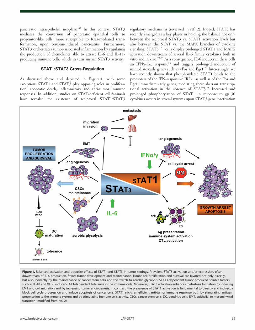
Figure 1. Balanced activation and opposite effects of STAT1 and STAT3 in tumor settings. Prevalent STAT3 activation and/or expression, oftendownstream of IL-6 production, favors tumor development and maintenance. Tumor cell proliferation and survival are favored not only directly,but also indirectly by the maintenance of cancer stem cells and the switch to aerobic glycolysis. STAT3-dependent tumor-produced soluble factorssuch as IL-10 and VEGF induce STAT3-dependent tolerance in the immune cells. Moreover, STAT3 activation enhances metastasis formation by inducingEMT and cell migration and by increasing tumor angiogenesis. In contrast, the prevalence of STAT1 activation is fundamental to directly and indirectlyblock cell cycle progression and induce apoptosis of cancer cells. STAT1 elicits an efficient anti-tumor immune response both by stimulating antigenpresentation to the immune system and by stimulating immune cells activity. CSCs, cancer stem cells; DC, dendritic cells; EMT, epithelial to mesenchymaltransition (modified from ref. 2).
www.landesbioscience.com JAK-STAT 69
(reviewed in ref. 2), suggesting that in normal cells one of thefunctions of STAT3 in response to IL-6 is to downregulateSTAT1 activity. In this vein, STAT1 expression was recentlyshown to be required for cell death induced by treatment with theJAK1 inhibitor AG490, believed to act by abolishing STAT3activation76 and by a STAT3-decoy oligonucleotide.77 Of notehowever, Lui et al. have shown STAT1-independent effects of thesame decoy.78 Similarly, IFNc and IFNa trigger proliferativeresponses correlating with STAT3-mediated transcription inSTAT1-deficient bone-marrow-derived macrophages, T lympho-cytes or MEFs.6 Thus, the relative abundance of STAT3 orSTAT1 may play a role in determining their activation levels inresponse to activating stimuli. This may be relevant for thedevelopment and growth of tumors in the presence of specifictumor microenvironments, where different cytokine/growthfactor combinations can modulate the relative levels of STAT1and STAT3, resulting in their differential activation. Interestingly,many of the inflammatory mediators produced by cancer cellsupon STAT3 inactivation are typical STAT1 targets (e.g.,CXCL10, CCL5 and ICAM1).
It is thus tempting to speculate that interfering with theactivation of either factor in tumors may result in activation orre-activation of the other. Thus, care should be taken to plantherapeutic intervention using compounds that could unbalancefinely tuned equilibria between STAT1 and STAT3. At the sametime, the possibility to activate a specific STAT pathway byinterfering with the other may under specific conditions provideunique therapeutic opportunities. For example, we have recentlyshown that interference with STAT3 activity in IFNc-resistanthuman T lymphoma cells enhances pro-apoptotic responses toIFNc while making cells sensitive to IL-6, which under theseconditions triggers prolonged activation of STAT1, apoptosis andimpaired in vivo growth.79 This strategy might be suitable in otherconditions characterized by impaired STAT1 activation.
Acknowledgments
Work in the author’s laboratories was supported by the ItalianMinistry of Research (MIUR, PRIN and FIRB), Italian Ministryof Health (Progetto Integrato Oncologia), Piedmont Region andby the Italian Association for Cancer Research (AIRC IG 9272).
References1. Schindler C, Levy DE, Decker T. JAK-STAT signaling:
from interferons to cytokines. J Biol Chem 2007;282:20059-63; PMID:17502367; http://dx.doi.org/10.1074/jbc.R700016200
2. Regis G, Pensa S, Boselli D, Novelli F, Poli V. Ups anddowns: the STAT1:STAT3 seesaw of Interferon andgp130 receptor signalling. Semin Cell Dev Biol 2008;19:351-9; PMID:18620071; http://dx.doi.org/10.1016/j.semcdb.2008.06.004
3. Schindler C, Brutsaert S. Interferons as a paradigm forcytokine signal transduction. Cell Mol Life Sci 1999;55:1509-22; PMID:10526569; http://dx.doi.org/10.1007/s000180050391
4. Heinrich PC, Behrmann I, Müller-Newen G, SchaperF, Graeve L. Interleukin-6-type cytokine signallingthrough the gp130/Jak/STAT pathway. Biochem J1998; 334:297-314; PMID:9716487
5. Stephanou A, Latchman DS. STAT-1: a novel regulatorof apoptosis. Int J Exp Pathol 2003; 84:239-44; PMID:14748743; http://dx.doi.org/10.1111/j.0959-9673.2003.00363.x
6. Pensa S, Regis G, Boselli D, Novelli F, Poli V. STAT1and STAT3 in tumorigenesis: two sides of the samecoin. In: Stephanou A, ed. JAK-STAT Pathway inDisease. Austin: Landes Bioscience, 2008: 100-121.
7. Li L, Shaw PE. Elevated activity of STAT3C due tohigher DNA binding affinity of phosphotyrosine dimerrather than covalent dimer formation. J Biol Chem2006; 281:33172-81; PMID:16956893; http://dx.doi.org/10.1074/jbc.M606940200
8. Liddle FJ, Alvarez JV, Poli V, Frank DA. Tyrosinephosphorylation is required for functional activation ofdisulfide-containing constitutively active STATmutants. Biochemistry 2006; 45:5599-605; PMID:16634641; http://dx.doi.org/10.1021/bi0525674
9. Allione A, Bernabei P, Bosticardo M, Ariotti S, Forni G,Novelli F. Nitric oxide suppresses human T lymphocyteproliferation through IFN-gamma-dependent and IFN-gamma-independent induction of apoptosis. J Immunol1999; 163:4182-91; PMID:10510354
10. Chin YE, Kitagawa M, Kuida K, Flavell RA, Fu XY.Activation of the STAT signaling pathway can causeexpression of caspase 1 and apoptosis. Mol Cell Biol1997; 17:5328-37; PMID:9271410
11. Kominsky SL, Hobeika AC, Lake FA, Torres BA,Johnson HM. Downregulation of neu/HER-2 byinterferon-gamma in prostate cancer cells. Cancer Res2000; 60:3904-8; PMID:10919667
12. Barré B, Avril S, Coqueret O. Opposite regulation ofmyc and p21waf1 transcription by STAT3 proteins. JBiol Chem 2003; 278:2990-6; PMID:12438313;http://dx.doi.org/10.1074/jbc.M210422200
13. Yu H, Kortylewski M, Pardoll D. Crosstalk betweencancer and immune cells: role of STAT3 in the tumourmicroenvironment. Nat Rev Immunol 2007; 7:41-51;PMID:17186030; http://dx.doi.org/10.1038/nri1995
14. Kreuzaler PA, Staniszewska AD, Li W, Omidvar N,Kedjouar B, Turkson J, et al. Stat3 controls lysosomal-mediated cell death in vivo. Nat Cell Biol 2011;13:303-9; PMID:21336304; http://dx.doi.org/10.1038/ncb2171
15. Gariboldi MB, Ravizza R, Molteni R, Osella D,Gabano E, Monti E. Inhibition of Stat3 increasesdoxorubicin sensitivity in a human metastatic breastcancer cell line. Cancer Lett 2007; 258:181-8; PMID:17920763; http://dx.doi.org/10.1016/j.canlet.2007.08.019
16. Fantin VR, Loboda A, Paweletz CP, Hendrickson RC,Pierce JW, Roth JA, et al. Constitutive activation ofsignal transducers and activators of transcriptionpredicts vorinostat resistance in cutaneous T-celllymphoma. Cancer Res 2008; 68:3785-94; PMID:18483262; http://dx.doi.org/10.1158/0008-5472.CAN-07-6091
17. Stronach EA, Alfraidi A, Rama N, Datler C, Studd JB,Agarwal R, et al. HDAC4-regulated STAT1 activationmediates platinum resistance in ovarian cancer. CancerRes 2011; 71:4412-22; PMID:21571862; http://dx.doi.org/10.1158/0008-5472.CAN-10-4111
18. Bromberg JF, Horvath CM, Besser D, Lathem WW,Darnell JE, Jr.. Stat3 activation is required for cellulartransformation by v-src. Mol Cell Biol 1998; 18:2553-8; PMID:9566875
19. Gough DJ, Corlett A, Schlessinger K, Wegrzyn J,Larner AC, Levy DE. Mitochondrial STAT3 supportsRas-dependent oncogenic transformation. Science2009; 324:1713-6; PMID:19556508; http://dx.doi.org/10.1126/science.1171721
20. Wegrzyn J, Potla R, Chwae YJ, Sepuri NB, Zhang Q,Koeck T, et al. Function of mitochondrial Stat3 incellular respiration. Science 2009; 323:793-7; PMID:19131594; http://dx.doi.org/10.1126/science.1164551
21. Cairns RA, Harris IS, Mak TW. Regulation of cancercell metabolism. Nat Rev Cancer 2011; 11:85-95;PMID:21258394; http://dx.doi.org/10.1038/nrc2981
22. Mauro C, Leow SC, Anso E, Rocha S, Thotakura AK,Tornatore L, et al. NF-kB controls energy homeostasisand metabolic adaptation by upregulating mitochon-drial respiration. Nat Cell Biol 2011; 13:1272-9;PMID:21968997; http://dx.doi.org/10.1038/ncb2324
23. Demaria M, Giorgi C, Lebiedzinska M, Esposito G,D’Angeli L, Bartoli A, et al. A STAT3-mediatedmetabolic switch is involved in tumour transformationand STAT3 addiction. Aging (Albany NY) 2010;2:823-42; PMID:21084727
24. Vander Heiden MG, Cantley LC, Thompson CB.Understanding the Warburg effect: the metabolicrequirements of cell proliferation. Science 2009;324:1029-33; PMID:19460998; http://dx.doi.org/10.1126/science.1160809
25. Hiroi M, Mori K, Sakaeda Y, Shimada J, Ohmori Y.STAT1 represses hypoxia-inducible factor-1-mediatedtranscription. Biochem Biophys Res Commun 2009;387:806-10; PMID:19646959; http://dx.doi.org/10.1016/j.bbrc.2009.07.138
26. Demaria M, Poli V. From the nucleus to themitochondria and back: the odyssey of a multitaskSTAT3. Cell Cycle 2011; 10:3221-2; PMID:21926478; http://dx.doi.org/10.4161/cc.10.19.17379
27. Chen YW, Chen KH, Huang PI, Chen YC, Chiou GY,Lo WL, et al. Cucurbitacin I suppressed stem-likeproperty and enhanced radiation-induced apoptosis inhead and neck squamous carcinoma–derived CD44(+)ALDH1(+) cells. Mol Cancer Ther 2010; 9:2879-92;PMID:21062915; http://dx.doi.org/10.1158/1535-7163.MCT-10-0504
28. Sherry MM, Reeves A, Wu JK, Cochran BH. STAT3 isrequired for proliferation and maintenance of multi-potency in glioblastoma stem cells. Stem Cells 2009;27:2383-92; PMID:19658181; http://dx.doi.org/10.1002/stem.185
70 JAK-STAT Volume 1 Issue 2
29. Lin L, Fuchs J, Li C, Olson V, Bekaii-Saab T, Lin J.STAT3 signaling pathway is necessary for cell survivaland tumorsphere forming capacity in ALDH⁺/CD133⁺stem cell-like human colon cancer cells. BiochemBiophys Res Commun 2011; 416:246-51; PMID:22074823; http://dx.doi.org/10.1016/j.bbrc.2011.10.112
30. Zhou J, Wulfkuhle J, Zhang H, Gu P, Yang Y, Deng J,et al. Activation of the PTEN/mTOR/STAT3 pathwayin breast cancer stem-like cells is required for viabilityand maintenance. Proc Natl Acad Sci U S A 2007;104:16158-63; PMID:17911267; http://dx.doi.org/10.1073/pnas.0702596104
31. Sansone P, Storci G, Tavolari S, Guarnieri T,Giovannini C, Taffurelli M, et al. IL-6 triggersmalignant features in mammospheres from humanductal breast carcinoma and normal mammary gland. JClin Invest 2007; 117:3988-4002; PMID:18060036;http://dx.doi.org/10.1172/JCI32533
32. Marotta LL, Almendro V, Marusyk A, Shipitsin M,Schemme J, Walker SR, et al. The JAK2/STAT3signaling pathway is required for growth ofCD44⁺CD24⁻ stem cell-like breast cancer cells in humantumors. J Clin Invest 2011; 121:2723-35; PMID:21633165; http://dx.doi.org/10.1172/JCI44745
33. Iliopoulos D, Hirsch HA, Wang G, Struhl K. Inducibleformation of breast cancer stem cells and their dynamicequilibrium with non-stem cancer cells via IL6secretion. Proc Natl Acad Sci U S A 2011; 108:1397-402; PMID:21220315; http://dx.doi.org/10.1073/pnas.1018898108
34. Heuser M, Sly LM, Argiropoulos B, Kuchenbauer F,Lai C, Weng A, et al. Modeling the functionalheterogeneity of leukemia stem cells: role of STAT5in leukemia stem cell self-renewal. Blood 2009;114:3983-93; PMID:19667399; http://dx.doi.org/10.1182/blood-2009-06-227603
35. Kovacic B, Stoiber D, Moriggl R, Weisz E, Ott RG,Kreibich R, et al. STAT1 acts as a tumor promoter forleukemia development. Cancer Cell 2006; 10:77-87;PMID:16843267; http://dx.doi.org/10.1016/j.ccr.2006.05.025
36. Essers MAG, Offner S, Blanco-Bose WE, Waibler Z,Kalinke U, Duchosal MA, et al. IFNalpha activatesdormant haematopoietic stem cells in vivo. Nature2009; 458:904-8; PMID:19212321; http://dx.doi.org/10.1038/nature07815
37. Christiansen JJ, Rajasekaran AK. Reassessing epithelialto mesenchymal transition as a prerequisite forcarcinoma invasion and metastasis. Cancer Res 2006;66:8319-26; PMID:16951136; http://dx.doi.org/10.1158/0008-5472.CAN-06-0410
38. Yamashita S, Miyagi C, Fukada T, Kagara N, Che YS,Hirano T. Zinc transporter LIVI controls epithelial-mesenchymal transition in zebrafish gastrula organizer.Nature 2004; 429:298-302; PMID:15129296; http://dx.doi.org/10.1038/nature02545
39. Cheng GZ, Zhang WZ, Sun M, Wang Q, Coppola D,Mansour M, et al. Twist is transcriptionally induced byactivation of STAT3 and mediates STAT3 oncogenicfunction. J Biol Chem 2008; 283:14665-73; PMID:18353781; http://dx.doi.org/10.1074/jbc.M707429200
40. Sullivan NJ, Sasser AK, Axel AE, Vesuna F, Raman V,Ramirez N, et al. Interleukin-6 induces an epithelial-mesenchymal transition phenotype in human breastcancer cells. Oncogene 2009; 28:2940-7; PMID:19581928; http://dx.doi.org/10.1038/onc.2009.180
41. Barbieri I, Pensa S, Pannellini T, Quaglino E, MaritanoD, Demaria M, et al. Constitutively active Stat3enhances neu-mediated migration and metastasis inmammary tumors via upregulation of Cten. Cancer Res2010; 70:2558-67; PMID:20215508; http://dx.doi.org/10.1158/0008-5472.CAN-09-2840
42. Silver DL, Naora H, Liu J, Cheng W, Montell DJ.Activated signal transducer and activator of transcrip-tion (STAT) 3: localization in focal adhesions andfunction in ovarian cancer cell motility. Cancer Res2004; 64:3550-8; PMID:15150111; http://dx.doi.org/10.1158/0008-5472.CAN-03-3959
43. Ng DC, Lin BH, Lim CP, Huang G, Zhang T, Poli V,et al. Stat3 regulates microtubules by antagonizing thedepolymerization activity of stathmin. J Cell Biol 2006;172:245-57; PMID:16401721; http://dx.doi.org/10.1083/jcb.200503021
44. Fitzner B, Brock P, Nechutova H, Glass A, Karopka T,Koczan D, et al. Inhibitory effects of interferon-gammaon activation of rat pancreatic stellate cells are mediatedby STAT1 and involve down-regulation of CTGFexpression. Cell Signal 2007; 19:782-90; PMID:17116388; http://dx.doi.org/10.1016/j.cellsig.2006.10.002
45. Ilkovitch D, Handel-Fernandez ME, Herbert LM,Lopez DM. Antitumor effects of Mucin 1/sec involvesthe modulation of urokinase-type plasminogen activatorand signal transducer and activator of transcription 1expression in tumor cells. Cancer Res 2008; 68:2427-35; PMID:18381451; http://dx.doi.org/10.1158/0008-5472.CAN-07-5651
46. Ugarte-Berzal E, Redondo-Muñoz J, Eroles P, DelCerro MH, García-Marco JA, Terol MJ, et al. VEGF/VEGFR2 interaction down-regulates matrix metallo-proteinase-9 via STAT1 activation and inhibits Bchronic lymphocytic leukemia cell migration. Blood2010; 115:846-9; PMID:19965686; http://dx.doi.org/10.1182/blood-2009-08-239426
47. Fukuda A, Wang SC, Morris JP, 4th, Folias AE, LiouA, Kim GE, et al. Stat3 and MMP7 contribute topancreatic ductal adenocarcinoma initiation and progres-sion. Cancer Cell 2011; 19:441-55; PMID:21481787;http://dx.doi.org/10.1016/j.ccr.2011.03.002
48. Schreiber RD, Old LJ, Smyth MJ. Cancer immuno-editing: integrating immunity’s roles in cancer suppres-sion and promotion. Science 2011; 331:1565-70;PMID:21436444; http://dx.doi.org/10.1126/science.1203486
49. Simpson JAD, Al-Attar A, Watson NFS, ScholefieldJH, Ilyas M, Durrant LG. Intratumoral T cellinfiltration, MHC class I and STAT1 as biomarkersof good prognosis in colorectal cancer. Gut 2010;59:926-33; PMID:20581241; http://dx.doi.org/10.1136/gut.2009.194472
50. Leibowitz MS, Andrade Filho PA, Ferrone S, Ferris RL.Deficiency of activated STAT1 in head and neck cancercells mediates TAP1-dependent escape from cytotoxicT lymphocytes. Cancer Immunol Immunother 2011;60:525-35; PMID:21207025; http://dx.doi.org/10.1007/s00262-010-0961-7
51. Chen H-Y, Yu S-L, Chen C-H, Chang G-C, ChenC-Y, Yuan A, et al. A five-gene signature and clinicaloutcome in non-small-cell lung cancer. N Engl J Med2007; 356:11-20; PMID:17202451; http://dx.doi.org/10.1056/NEJMoa060096
52. Simons DL, Lee G, Kirkwood JM, Lee PP. Interferonsignaling patterns in peripheral blood lymphocytes maypredict clinical outcome after high-dose interferontherapy in melanoma patients. J Transl Med 2011;9:52; PMID:21545749; http://dx.doi.org/10.1186/1479-5876-9-52
53. Critchley-Thorne RJ, Yan N, Nacu S, Weber J,Holmes SP, Lee PP. Down-regulation of the interferonsignaling pathway in T lymphocytes from patients withmetastatic melanoma. PLoS Med 2007; 4:e176; PMID:17488182; http://dx.doi.org/10.1371/journal.pmed.0040176
54. Álvaro T, Lejeune M, Camacho FI, Salvadó MT,Sánchez L, García JF, et al. The presence of STAT1-positive tumor-associated macrophages and their rela-tion to outcome in patients with follicular lymphoma.Haematologica 2006; 91:1605-12; PMID:17145596
55. Kusmartsev S, Gabrilovich DI. STAT1 signalingregulates tumor-associated macrophage-mediated T celldeletion. J Immunol 2005; 174:4880-91; PMID:15814715
56. Uyttenhove C, Pilotte L, Théate I, Stroobant V, ColauD, Parmentier N, et al. Evidence for a tumoral immuneresistance mechanism based on tryptophan degradationby indoleamine 2,3-dioxygenase. Nat Med 2003;9:1269-74; PMID:14502282; http://dx.doi.org/10.1038/nm934
57. Torrero MN, Xia X, Henk W, Yu S, Li S. Stat1deficiency in the host enhances interleukin-12-mediated tumor regression. Cancer Res 2006;66:4461-7; PMID:16618773; http://dx.doi.org/10.1158/0008-5472.CAN-05-3554
58. Schultz J, Koczan D, Schmitz U, Ibrahim SM, Pilch D,Landsberg J, et al. Tumor-promoting role of signaltransducer and activator of transcription (Stat)1 in late-stage melanoma growth. Clin Exp Metastasis 2010;27:133-40; PMID:20180146; http://dx.doi.org/10.1007/s10585-010-9310-7
59. Widschwendter A, Tonko-Geymayer S, Welte T,Daxenbichler G, Marth C, Doppler W. Prognosticsignificance of signal transducer and activator oftranscription 1 activation in breast cancer. ClinCancer Res 2002; 8:3065-74; PMID:12374673
60. Charpin C, Secq V, Giusiano S, Carpentier S, AndracL, Lavaut M-N, et al. A signature predictive of diseaseoutcome in breast carcinomas, identified by quantita-tive immunocytochemical assays. Int J Cancer 2009;124:2124-34; PMID:19142869; http://dx.doi.org/10.1002/ijc.24177
61. Greenwood C, Metodieva G, Al-Janabi K, Lausen B,Alldridge L, Leng L, et al. Stat1 and CD74 over-expression is co-dependent and linked to increasedinvasion and lymph node metastasis in triple-negativebreast cancer. J Proteomics 2012; 75:3031-41; PMID:22178447; http://dx.doi.org/10.1016/j.jprot.2011.11.033
62. Khodarev N, Ahmad R, Rajabi H, Pitroda S, Kufe T,McClary C, et al. Cooperativity of the MUC1oncoprotein and STAT1 pathway in poor prognosishuman breast cancer. Oncogene 2010; 29:920-9;PMID:19915608; http://dx.doi.org/10.1038/onc.2009.391
63. Chalmin F, Ladoire S, Mignot G, Vincent J, BruchardM, Remy-Martin JP, et al. Membrane-associatedHsp72 from tumor-derived exosomes mediatesSTAT3-dependent immunosuppressive function ofmouse and human myeloid-derived suppressor cells. JClin Invest 2010; 120:457-71; PMID:20093776
64. Kortylewski M, Xin H, Kujawski M, Lee H, Liu Y,Harris T, et al. Regulation of the IL-23 and IL-12balance by Stat3 signaling in the tumor microenviron-ment. Cancer Cell 2009; 15:114-23; PMID:19185846;http://dx.doi.org/10.1016/j.ccr.2008.12.018
65. Lee H, Deng J, Xin H, Liu Y, Pardoll D, Yu H. Arequirement of STAT3 DNA binding precludes Th-1immunostimulatory gene expression by NF-kB intumors. Cancer Res 2011; 71:3772-80; PMID:21502401; http://dx.doi.org/10.1158/0008-5472.CAN-10-3304
66. Hanahan D, Weinberg RA. Hallmarks of cancer: thenext generation. Cell 2011; 144:646-74; PMID:21376230; http://dx.doi.org/10.1016/j.cell.2011.02.013
www.landesbioscience.com JAK-STAT 71
67. Heikkilä K, Ebrahim S, Lawlor DA. Systematic reviewof the association between circulating interleukin-6(IL-6) and cancer. Eur J Cancer 2008; 44:937-45;PMID:18387296; http://dx.doi.org/10.1016/j.ejca.2008.02.047
68. Bromberg J, Wang TC. Inflammation and cancer: IL-6and STAT3 complete the link. Cancer Cell 2009;15:79-80; PMID:19185839; http://dx.doi.org/10.1016/j.ccr.2009.01.009
69. Bollrath J, Phesse TJ, von Burstin VA, Putoczki T,Bennecke M, Bateman T, et al. gp130-mediated Stat3activation in enterocytes regulates cell survival and cell-cycle progression during colitis-associated tumorigen-esis. Cancer Cell 2009; 15:91-102; PMID:19185844;http://dx.doi.org/10.1016/j.ccr.2009.01.002
70. Grivennikov S, Karin E, Terzic J, Mucida D, Yu GY,Vallabhapurapu S, et al. IL-6 and Stat3 are required forsurvival of intestinal epithelial cells and development ofcolitis-associated cancer. Cancer Cell 2009; 15:103-13;PMID:19185845; http://dx.doi.org/10.1016/j.ccr.2009.01.001
71. Musteanu M, Blaas L, Mair M, Schlederer M, BilbanM, Tauber S, et al. Stat3 is a negative regulator ofintestinal tumor progression in Apc(Min) mice.Gastroenterology 2010; 138:1003-11, e1-5; PMID:19962983; http://dx.doi.org/10.1053/j.gastro.2009.11.049
72. Lesina M, Kurkowski MU, Ludes K, Rose-John S,Treiber M, Klöppel G, et al. Stat3/Socs3 activationby IL-6 transsignaling promotes progression ofpancreatic intraepithelial neoplasia and developmentof pancreatic cancer. Cancer Cell 2011; 19:456-69;PMID:21481788; http://dx.doi.org/10.1016/j.ccr.2011.03.009
73. Maritano D, Sugrue ML, Tininini S, Dewilde S, StroblB, Fu X, et al. The STAT3 isoforms alpha and betahave unique and specific functions. Nat Immunol2004; 5:401-9; PMID:15021879; http://dx.doi.org/10.1038/ni1052
74. Costa-Pereira AP, Tininini S, Strobl B, Alonzi T,Schlaak JF, Is’harc H, et al. Mutational switch of anIL-6 response to an interferon-gamma-like response.Proc Natl Acad Sci U S A 2002; 99:8043-7; PMID:12060750; http://dx.doi.org/10.1073/pnas.122236099
75. Schiavone D, Avalle L, Dewilde S, Poli V. Theimmediate early genes Fos and Egr1 become STAT1transcriptional targets in the absence of STAT3. FEBSLett 2011; 585:2455-60; PMID:21723864; http://dx.doi.org/10.1016/j.febslet.2011.06.020
76. Shim SH, Sung MW, Park SW, Heo DS. Absence ofSTAT1 disturbs the anticancer effect induced bySTAT3 inhibition in head and neck carcinoma celllines. Int J Mol Med 2009; 23:805-10; PMID:19424608
77. Souissi I, Najjar I, Ah-Koon L, Schischmanoff PO,Lesage D, Le Coquil S, et al. A STAT3-decoyoligonucleotide induces cell death in a human colorectalcarcinoma cell line by blocking nuclear transfer ofSTAT3 and STAT3-bound NF-kB. BMC Cell Biol2011; 12:14; PMID:21486470; http://dx.doi.org/10.1186/1471-2121-12-14
78. Lui VWY, Boehm AL, Koppikar P, Leeman RJ,Johnson D, Ogagan M, et al. Antiproliferativemechanisms of a transcription factor decoy targetingsignal transducer and activator of transcription (STAT)3: the role of STAT1. Mol Pharmacol 2007; 71:1435-43; PMID:17325127; http://dx.doi.org/10.1124/mol.106.032284
79. Regis G, Icardi L, Conti L, Chiarle R, Piva R, GiovarelliM, et al. IL-6, but not IFN-gamma, triggers apoptosisand inhibits in vivo growth of human malignant T cellson STAT3 silencing. Leukemia 2009; 23:2102-8;PMID:19626047; http://dx.doi.org/10.1038/leu.2009.139
80. Raven JF, Williams V, Wang S, Tremblay ML, MullerWJ, Durbin JE, et al. Stat1 is a suppressor of ErbB2/Neu-mediated cellular transformation and mousemammary gland tumor formation. Cell Cycle 2011;10:794-804; PMID:21311224; http://dx.doi.org/10.4161/cc.10.5.14956
81. Schneckeleithner C, Bago-Horvath Z, Dolzing H,Neugebauer N, Kollmann, Kolbe T, et al. Puttingbrakes on mammary tumorigenesis: Loss of STAT1predisposes to intraepithelial neoplasias. Oncotarget2011; 2:1043-1054; PMID:22185785
82. Chan SR, Vermi W, Luo J, Lucini L, Rickert C,Fowler AM, et al. STAT1-deficient mice spontaneouslydevelop estrogen receptor alpha-positive luminal mam-mary carcinomas. Breast Cancer Res 2012; 14:R16;PMID:22264274; http://dx.doi.org/10.1186/bcr3100
72 JAK-STAT Volume 1 Issue 2
Copyright © 2022 FDOKUMEN