Prognostic impact of Skp2, ER and PGR in male and female patients with soft tissue sarcomas
Upload
independentCategory
view
8download
0
The American Journal of Pathology, Vol. 182, No. 5, May 2013
ajp.amjpathol.org
TUMORIGENESIS AND NEOPLASTIC PROGRESSION
Skp2 Deficiency Inhibits Chemical Skin TumorigenesisIndependent of p27Kip1 AccumulationChristopher Sistrunk,* Sun Hye Kim,* Xian Wang,* Sung Hyun Lee,* Yongbaek Kim,y Everardo Macias,z andMarcelo L. Rodriguez-Puebla*
From the Department of Molecular Biomedical Sciences and the Center for Comparative Medicine and Translational Research,* College of VeterinaryMedicine, North Carolina State University, Raleigh, North Carolina; the Laboratory of Veterinary Clinical Pathology,y College of Veterinary Medicine, SeoulNational University, Seoul, South Korea; and the Dell Pediatric Research Institute,z College of Pharmacy, The University of Texas, Austin, Texas
Accepted for publication
C
P
h
January 14, 2013.
Address correspondence toMarcelo L. Rodriguez-Puebla,Ph.D., Department of MolecularBiomedical Sciences andCenter for ComparativeMedicine and TranslationalResearch, College of VeterinaryMedicine, North Carolina StateUniversity, 1060WilliamMooreDr, Raleigh, NC 27606. E-mail:[email protected].
opyright ª 2013 American Society for Inve
ublished by Elsevier Inc. All rights reserved
ttp://dx.doi.org/10.1016/j.ajpath.2013.01.016
S-phase kinase-associated protein 2 (Skp2) functions as the receptor component of the SkpeCullineF-boxcomplex and is implicated in the degradation of several cell cycle regulators, such as p21Cip1, p27Kip1,p57Kip2, and cyclin E. Numerous studies in human and experimental tumors have demonstrated low p27Kip1
levels and elevated Skp2 expression. However, a direct association between the inverse correlation of Skp2and p27Kip1 with tumorigenesis has not been demonstrated. Herein, we provide evidence that skintumorigenesis is inhibited in Skp2�/� mice. An analysis of mouse keratinocytes indicates that increasedp27Kip1 levels in Skp2�/� epidermis cause reduced cell proliferation that is alleviated in the epidermis fromSkp2�/�/p27�/� compound mice. In contrast, we establish that a p27Kip1 deficiency does not overturn thereduced skin tumorigenesis experienced by Skp2�/� mice. In addition, Skp2�/� epidermis exhibits anaccumulation of p53-cofactor CBP/p300 that is associated with elevated apoptosis in hair follicles anddecreased skin tumorigenesis. We conclude that p27Kip1 accumulation is responsible for the hypoplasiaobserved in normal tissues of Skp2�/� mice but does not have a preponderant function in reducing skintumorigenesis. (Am J Pathol 2013, 182: 1854e1864; http://dx.doi.org/10.1016/j.ajpath.2013.01.016)
Supported by National Cancer Institute/NIH grant RO1 CA116328(M.L.R.-P.).C.S. and S.H.K. contributed equally to this work.
The proteasome pathway involves ubiquitin modificationand degradation of substrates by the proteasome complex.The S-phase kinase-associated protein (Skp)eCullineF-boxcomplex is a ubiquitin ligase that typically contains foursubunits, termed Skp1, Cullin, Ring-finger protein, anda member of the large family of F-box adaptor proteinsinvolved in specific substrate recognition.1 Skp2 is an F-boxprotein that targets several cell cycle regulators for ubiq-uitination and subsequent degradation.2,3 The specificsubstrates of Skp2 include p21Cip1, p27Kip1, p57Kip2, p130,Tob1, FOXO1, and c-Myc.4e10 Because most of thesesubstrates are tumor suppressor proteins, Skp2 has beenclassified as an oncogene. Moreover, Skp2 also suppressesthe p53-dependent apoptosis pathway by antagonizing theinteraction between CBP/p300 and p53.11 Althoughmultiple substrates for Skp2 have been established, the bestdescribed is p27Kip1.12,13 Accordingly, Skp2 knockout (KO)mice exhibit high p27Kip1 levels; these mice grow slowerthan littermate controls and have smaller organs, withhypoplastic tissues.14 The ablation of p27Kip1 abolishes all
stigative Pathology.
.
of the phenotypes observed in the Skp2�/� mouse, whichsuggests that p27Kip1 is the main target of Skp2.15
Skp2 induces cell proliferation in various experimentalassays, has transforming activity, is found overexpressed indiverse human cancers, and is, therefore, classified as anoncogene.16e18 Presumably, Skp2 expression or impropertemporal expression confers a growth advantage by increasingp27Kip1 degradation. Moreover, experimental and humantumors have shown a strong correlation between increasedlevels of the Skp2 oncoprotein and diminished p27Kip1 levels,which suggests that reduced p27Kip1 levels have a prepon-derant function in tumorigenesis.19,20 Thus, decreased p27Kip1
protein levels are commonly observed in many humancancers, including epithelial cancers and brain tumors.21
Consequently, high Skp2 and low p27Kip1 levels are indica-tors for shorter disease-free survival or unfavorable melanoma,
Skp2/p27 Axis in Skin Tumorigenesis
breast, prostate, and lung cancer prognoses.18,22,23 However,whether the inverse correlation between Skp2 and p27Kip1
protein levels and the association between Skp2 levels andtumor grade are directly responsible for Skp2 oncogenicactivity has never been addressed.
Our laboratory has previously demonstrated that Myc-induced keratinocyte proliferation was abolished by the lossof Skp2 and presumably by the increased level of p27Kip1.24
However, we also observed that Skp2 ablation did not affectMyc-driven oral tumorigenesis. These finding suggestedthat Skp2 and p27Kip1 are critical for Myc-driven prolifer-ation, although Myc-mediated tumorigenesis in the oralepithelium is independent of the Skp2-p27Kip1 axis.24 In thisstudy, we seek to determine the effect of an Skp2 deficiencyon p27Kip1 levels and the rates of keratinocyte proliferationand skin tumorigenesis. Herein, we show that an Skp2deficiency diminishes ras-mediated skin tumorigenesis thatcorrelates with p27Kip1 accumulation. Furthermore, wedetermined that a p27Kip1 deficiency reverses Skp2�/�
epidermal hypoplasia, but surprisingly, this deficiency doesnot overturn the reduced skin tumorigenesis that is experi-enced by the Skp2�/� mice. These data provide directgenetic evidence that p27Kip1 accumulation is responsiblefor the reduced keratinocyte proliferation and epidermalhypoplasia, but not for the reduced number of tumorsobserved in the Skp2�/� mice. Our data also suggest thatSkp2-mediated apoptosis in the bulge region of hair follicles(HFs) plays a preponderant role by blocking an early stageof mouse skin tumorigenesis.
Materials and Methods
Generation of Transgenic Mice
Skp2�/� animals were developed as previously described byNakayama et al.14 Mice heterozygous for Skp2 (Skp2þ/�) ona C57BL/6 background were bred to generate mice that werehomozygous, heterozygous, or nullizygous for Skp2. Thep27Kip1 heterozygous mice were purchased from The JacksonLaboratory (Bar Harbor, ME; strain B6.129S4-Cdkn1btm1Mlf)and crossed back to the genetic background SENCAR for twogenerations. Skp2�/�/p27�/� compound mice, p27�/�,Skp2�/�, and control wild-type (WT) mice were generated bycrossing p27Kip1 heterozygous mice with Skp2 heterozygousmice. The p53�/� mice on the C57BL/6 background were agift from Dr. Robert Smart at North Carolina State University,Raleigh (TSG-p53; Taconic, Hudson, NY). Skp2�/�/p53þ/�
mice and control littermates were generated by breedingSkp2þ/� with p53þ/� mice.25
Mouse Experiments
For the two-stage carcinogenesis experiment, 3-week-oldSkp2�/� mice and WT siblings were initiated with a topicalapplication of 200 nmol 7,12-dimethylbenz(a)anthracene(DMBA) in 200 mL of acetone on their dorsal surface. Two
The American Journal of Pathology - ajp.amjpathol.org
weeks later, themicewere dosed topically twiceweeklywith 4mg of 12-O-tetradecanoylphorbol-13-acetate (TPA) in 200 mLof acetone for 30weeks. The tumors were countedweekly andrecorded to determine multiplicity, latency, and incidence.
In the Skp2�/�/p27�/� two-stage carcinogenesis experi-ments, newborn mice were initiated at day 1 after birth withan application of 50 mg of DMBA in 50 mL of acetone ontheir dorsal surface. At day 21, the mice were dosed twiceweekly with 2.5 mg of TPA in 200 mL of acetone for 25weeks. The skin tumors were counted weekly until the endof the experiment at 30 weeks. Malignant progression tosquamous cell carcinomas (SCCs) was determined bymacroscopic observation and further confirmed by thehistopathological analysis of paraffin-embedded H&E-stained cross sections.
Western Blot Analyses and Kinase Assays
For immunoblotting, the epidermal tissue was scraped offwith a razor blade, placed into homogenization buffer [150mmol/L NaCl, 1.0% polyoxyethylene nonylphenol, 0.5%deoxycholic acid, 0.1% SDS, and 50 mmol/L Tris (pH 8.0)],and homogenized using a manual homogenizer.26 Forimmunoblot analysis of skin tumors, the papillomas weresnap frozen in liquid nitrogen and crushed with a pestle andmortar. The homogenates were sonicated and centrifuged at11,000 � g at 4�C. The supernatants were boiled in 2 mL ofLaemmli sample buffer for Western blot analysis or stored at�80�C. The protein concentration was measured with a Bio-Rad protein assay system (Bio-Rad Laboratories, Richmond,CA). The protein lysates (25 mg from each sample) wereelectrophoresed through 12% acrylamide gels and electro-phoretically transferred onto nitrocellulose membranes. Afterbeing blocked with 5% nonfat powdered milk in Dulbecco’sPBS, themembranes were incubated with 1 mg/mL of specificantibodies. The following antibodies were used: polyclonalantibodies against CDK4 (C22), CDK2 (M2), p27Kip1
(M197), p21Cip1 (H164), Puma (G3), p300 (N15), actin (H6),cyclin A (C19), and cyclin E (C19) (Santa Cruz Biotech-nology, Santa Cruz, CA), p53 (1C12), and acetylated p53(Cys379) (Cell Signaling Technology Inc., Boston, MA).
To assess the CDK2 kinase activity, proteins wereextracted and immunoprecipitated in NP40 lysis buffer [Tris(pH 7.5), 150 mmol/L NaCl, 0.5% NP40, 50 mmol/L NaF,1 mmol/L Na3VO4, 1 mmol/L dithiothreitol, and 1 mmol/Lphenylmethylsulfonyl fluoride]. To assess the CDK4 kinaseactivity, proteins were extracted and immunoprecipitated inTween 20 buffer (50 mmol/L HEPES, 150 mmol/L NaCl, 1mmol/L EDTA, 2.5 mmol/L EGTA, 10% glycerol, 0.1%Tween 20, 1 mmol/L NaF, 1 mmol/L Na3VO4, and 1 mmol/Ldithiothreitol). Briefly, 250 mg of protein lysates wasimmunoprecipitated with 2.5 mg of antibodies againstCDK2 (M-20) or CDK4 (C-22) (Santa Cruz Biotechnology)for 2 hours at 4�C and then incubated with 35 mL of proteinAeagarose beads. The beads were twice washed in animmunoprecipitation buffer and a kinase buffer [50 mmol/L
1855

Sistrunk et al
HEPES (pH 7), 10 mmol/L MgCl2, and 5 mmol/L MnCl2].Subsequently, 30 mL of kinase buffer, 1 mg of pRb peptideor histone H1 (EMD Millipore, Billerica, MA) substrate,5 mCi of [g-32P]ATP (6000 Ci/mmol), 1 mmol/L dithio-threitol, and 5 mmol/L ATP were added to the bead pelletand incubated for 30 minutes at 30�C. SDS sample bufferwas added. Each sample was boiled for 3 minutes to stop thereaction and electrophoresed through a polyacrylamide gel.The Western blot images and kinase assay bands werequantified using UN-SCAN-IT gel software version 6.1 forWindows (Silk Scientific, Inc., Orem, UT).
Tunnel Assay
Apoptotic cells were determined using TUNEL assays withthe FragEL DNA Fragmentation Detection kit (Colori-metric-TdT enzyme; Calbiochem, EMB Biosciences Inc.),following the manufacturer’s instructions. To quantify thenormal and apoptotic cells, the cells were counterstainedwith methyl green. Apoptotic keratinocytes in the inter-follicular and follicular epidermis were quantified insections (1 cm thick). To determine the incidence ofapoptosis in the HFs, HFs that contained at least oneapoptotic cell in the bulge were counted as positive. In allcases, 12 fields were counted per section in a total of 10paraffin-embedded sections, which represented five miceper genotype.
Statistical Analysis
Statistical analysis was performed using GraphPad PrismSoftware version 4 (GraphPad Software, San Diego, CA).
Results
Skp2 Deficiency Reduces Skin Tumorigenesis
To investigate the function of Skp2 in skin carcinogenesis, weassessed the response of Skp2�/� mice to the two-stagecarcinogenesis protocol. This is a well-suited model forunderstanding the multistage nature of tumor progression, inwhich tumor initiation is accomplished through a singletopical application of a carcinogen, typically DMBA. Thistreatment produces a somatic mutation in the Ha-ras onco-gene, and tumor promotion occurs when the initiated cells areexpanded through multiple applications of a tumor promoter,usually TPA, leading to skin papilloma development.Therefore, Skp2�/� and WT littermates were subjected to theDMBA/TPA regimen for up to 30 weeks, and the incidenceand multiplicity of papillomas were scored weekly. Theincidence of papilloma formation reached a plateau atapproximately 11 weeks, in which 70% to 80% of the WTmice developed skin tumors. In contrast, a strong reductionwas observed in the Skp2�/�mice, which reached a plateau ofapproximately 20% at 14 weeks of promotion (Figure 1A).Skp2 deficiency also caused a robust decline in the number of
1856
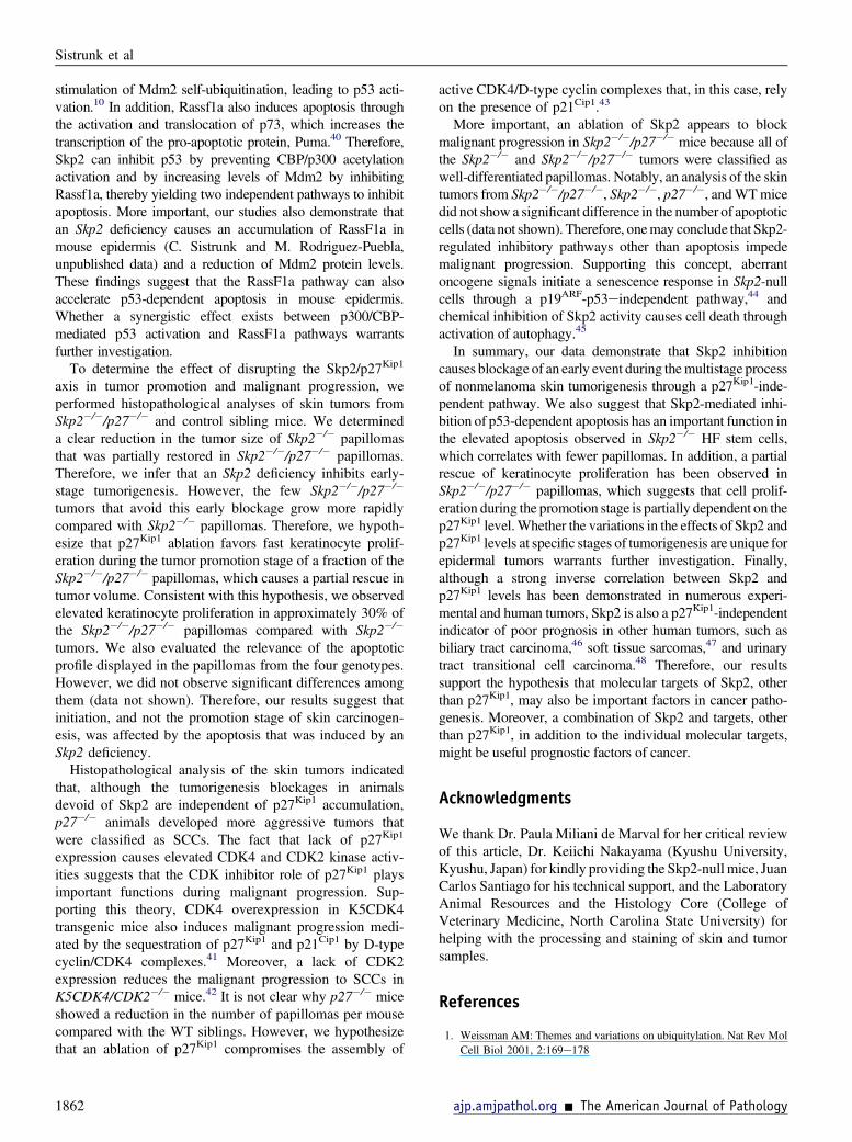
tumors per mouse (multiplicity). Thirty weeks after the firstTPA application, the Skp2�/� group exhibited an average of0.37 papillomas per mouse, whereas the WT mice developedfive papillomas per mouse (P < 0.001, U-test) (Figure 1B).The size of the tumors also varied among the genotypes. TheWTpapillomaswere the largest; 85%of tumors exceeded 100mm3. Conversely, the Skp2�/� papillomas were smaller; 16%of these tumors failed to reach a volume of 30 mm3, and 65%of the tumors did not surpass 100 mm3 (Figure 1C). Histo-pathological analysis of the skin tumors collected at 30 weeksof promotion indicated that all Skp2�/� tumors were well-differentiated papillomas with no atypia in the basal layers.In brief, 25% of theWT tumors were classified as moderatelydifferentiated squamous cell carcinomas (approximately50% of differentiating cells), and 75% of these tumors werewell-differentiated papillomas (Figure 1, D and E). Compa-rable to the Skp2�/� mouse epidermis,24 a biochemicalanalysis of papillomas presented a fourfold increase inp27Kip1 protein levels, whereas other putative Skp-Cullin-F-boxSkp2 substrates, such as cyclin E, exhibited no differencebetween the Skp2�/� and WT papillomas. A mild increase,although not significant, was observed in the level of p21Cip1
in Skp2�/� papillomas (Figure 2A). Similar to mouseepidermis,24 CDK2 kinase activity in Skp2�/� papillomaswas comparable to that of WT mice. Moreover, we did notobserve significant differences in the CDK4 kinase activity ofWT and Skp2�/� tumors (Figure 2B). Collectively, theseobservations suggest that a lack of Skp2 expression inmouseepidermis leads to a relevant increase in p27Kip1 stability inskin papillomas but has either a minimal or a null effect onCDK2 and CDK4 kinase activities. Therefore, we concludethat a lack of Skp2 expression severely affects the onset ofskin tumorigenesis and the proliferative/antiproliferativebalance, which leads to a reduced tumor size. However,these effects are unlikely to be mediated by the inhibition ofCDK2/CDK4 activities.
Inhibition of Skin Tumorigenesis in Skp2-Null Mice IsIndependent of p27 Accumulation
To evaluate the hypothesis that elevated p27Kip1 protein levelsare associated with reduced tumorigenesis in Skp2�/� mice,we developed Skp2�/�/p27�/� compound mice and chal-lenged this mouse model to the classic DMBA/TPA regimen.The C57BL/6 mouse genetic background, used in theprevious experiment, is refractory to the initiation-promotionprotocol when TPA is used as a promoter.27 Therefore, theSkp2�/�/p27�/� compound mice were generated on an FVB/Sencar hybrid background that exhibits higher sensitivity tothe two-stage carcinogenesis protocol.27 As previously re-ported byNakayama et al,15 the body size of p27�/�micewaslarger than that of WT siblings, and Skp2�/� mice weresmaller than the WT controls, whereas the body size of theSkp2�/�/p27�/� double mutant was similar to that of the WTcontrols (Figure 3A). More important, thymus measurementsfor each of the four genotypes analyzed confirmed that
ajp.amjpathol.org - The American Journal of Pathology
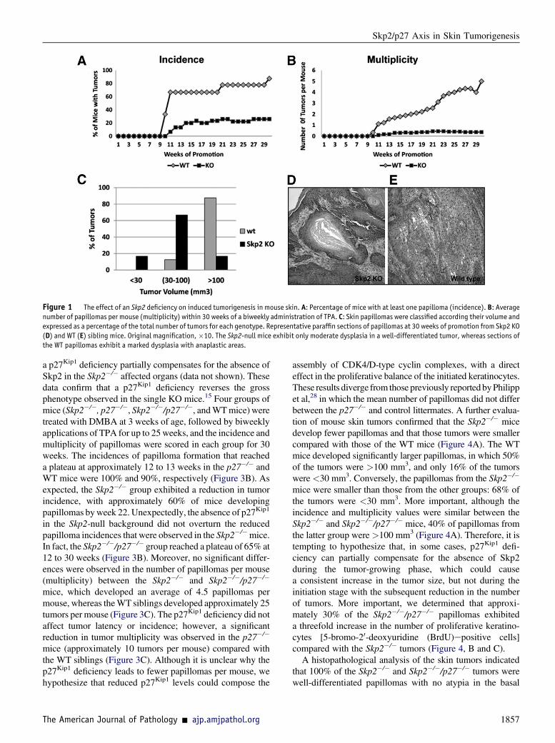
Figure 1 The effect of an Skp2 deficiency on induced tumorigenesis in mouse skin. A: Percentage of mice with at least one papilloma (incidence). B: Averagenumber of papillomas per mouse (multiplicity) within 30 weeks of a biweekly administration of TPA. C: Skin papillomas were classified according their volume andexpressed as a percentage of the total number of tumors for each genotype. Representative paraffin sections of papillomas at 30 weeks of promotion from Skp2 KO(D) and WT (E) sibling mice. Original magnification,�10. The Skp2-null mice exhibit only moderate dysplasia in a well-differentiated tumor, whereas sections ofthe WT papillomas exhibit a marked dysplasia with anaplastic areas.
Skp2/p27 Axis in Skin Tumorigenesis
a p27Kip1 deficiency partially compensates for the absence ofSkp2 in the Skp2�/� affected organs (data not shown). Thesedata confirm that a p27Kip1 deficiency reverses the grossphenotype observed in the single KO mice.15 Four groups ofmice (Skp2�/�, p27�/�, Skp2�/�/p27�/�, andWTmice) weretreated with DMBA at 3 weeks of age, followed by biweeklyapplications of TPA for up to 25weeks, and the incidence andmultiplicity of papillomas were scored in each group for 30weeks. The incidences of papilloma formation that reacheda plateau at approximately 12 to 13 weeks in the p27�/� andWT mice were 100% and 90%, respectively (Figure 3B). Asexpected, the Skp2�/� group exhibited a reduction in tumorincidence, with approximately 60% of mice developingpapillomas byweek 22. Unexpectedly, the absence of p27Kip1
in the Skp2-null background did not overturn the reducedpapilloma incidences that were observed in the Skp2�/�mice.In fact, the Skp2�/�/p27�/� group reached a plateau of 65% at12 to 30 weeks (Figure 3B). Moreover, no significant differ-ences were observed in the number of papillomas per mouse(multiplicity) between the Skp2�/� and Skp2�/�/p27�/�
mice, which developed an average of 4.5 papillomas permouse, whereas theWT siblings developed approximately 25tumors per mouse (Figure 3C). The p27Kip1 deficiency did notaffect tumor latency or incidence; however, a significantreduction in tumor multiplicity was observed in the p27�/�
mice (approximately 10 tumors per mouse) compared withthe WT siblings (Figure 3C). Although it is unclear why thep27Kip1 deficiency leads to fewer papillomas per mouse, wehypothesize that reduced p27Kip1 levels could compose the
The American Journal of Pathology - ajp.amjpathol.org
assembly of CDK4/D-type cyclin complexes, with a directeffect in the proliferative balance of the initiated keratinocytes.These results diverge from those previously reportedbyPhilippet al,28 in which the mean number of papillomas did not differbetween the p27�/� and control littermates. A further evalua-tion of mouse skin tumors confirmed that the Skp2�/� micedevelop fewer papillomas and that those tumors were smallercompared with those of the WT mice (Figure 4A). The WTmice developed significantly larger papillomas, in which 50%of the tumors were >100 mm3, and only 16% of the tumorswere <30 mm3. Conversely, the papillomas from the Skp2�/�
mice were smaller than those from the other groups: 68% ofthe tumors were <30 mm3. More important, although theincidence and multiplicity values were similar between theSkp2�/� and Skp2�/�/p27�/� mice, 40% of papillomas fromthe latter group were >100 mm3 (Figure 4A). Therefore, it istempting to hypothesize that, in some cases, p27Kip1 defi-ciency can partially compensate for the absence of Skp2during the tumor-growing phase, which could causea consistent increase in the tumor size, but not during theinitiation stage with the subsequent reduction in the numberof tumors. More important, we determined that approxi-mately 30% of the Skp2�/�/p27�/� papillomas exhibiteda threefold increase in the number of proliferative keratino-cytes [5-bromo-20-deoxyuridine (BrdU)epositive cells]compared with the Skp2�/� tumors (Figure 4, B and C).A histopathological analysis of the skin tumors indicated
that 100% of the Skp2�/� and Skp2�/�/p27�/� tumors werewell-differentiated papillomas with no atypia in the basal
1857
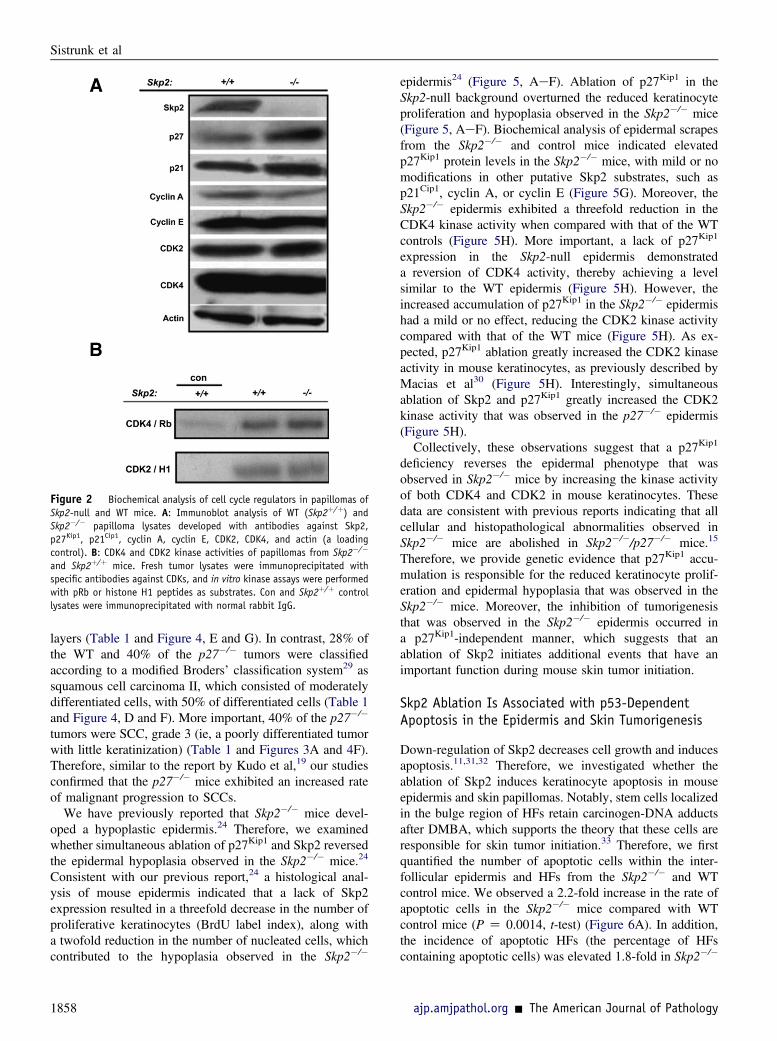
Figure 2 Biochemical analysis of cell cycle regulators in papillomas ofSkp2-null and WT mice. A: Immunoblot analysis of WT (Skp2þ/þ) andSkp2�/� papilloma lysates developed with antibodies against Skp2,p27Kip1, p21Cip1, cyclin A, cyclin E, CDK2, CDK4, and actin (a loadingcontrol). B: CDK4 and CDK2 kinase activities of papillomas from Skp2�/�
and Skp2þ/þ mice. Fresh tumor lysates were immunoprecipitated withspecific antibodies against CDKs, and in vitro kinase assays were performedwith pRb or histone H1 peptides as substrates. Con and Skp2þ/þ controllysates were immunoprecipitated with normal rabbit IgG.
Sistrunk et al
layers (Table 1 and Figure 4, E and G). In contrast, 28% ofthe WT and 40% of the p27�/� tumors were classifiedaccording to a modified Broders’ classification system29 assquamous cell carcinoma II, which consisted of moderatelydifferentiated cells, with 50% of differentiated cells (Table 1and Figure 4, D and F). More important, 40% of the p27�/�
tumors were SCC, grade 3 (ie, a poorly differentiated tumorwith little keratinization) (Table 1 and Figures 3A and 4F).Therefore, similar to the report by Kudo et al,19 our studiesconfirmed that the p27�/� mice exhibited an increased rateof malignant progression to SCCs.
We have previously reported that Skp2�/� mice devel-oped a hypoplastic epidermis.24 Therefore, we examinedwhether simultaneous ablation of p27Kip1 and Skp2 reversedthe epidermal hypoplasia observed in the Skp2�/� mice.24
Consistent with our previous report,24 a histological anal-ysis of mouse epidermis indicated that a lack of Skp2expression resulted in a threefold decrease in the number ofproliferative keratinocytes (BrdU label index), along witha twofold reduction in the number of nucleated cells, whichcontributed to the hypoplasia observed in the Skp2�/�
1858
epidermis24 (Figure 5, AeF). Ablation of p27Kip1 in theSkp2-null background overturned the reduced keratinocyteproliferation and hypoplasia observed in the Skp2�/� mice(Figure 5, AeF). Biochemical analysis of epidermal scrapesfrom the Skp2�/� and control mice indicated elevatedp27Kip1 protein levels in the Skp2�/� mice, with mild or nomodifications in other putative Skp2 substrates, such asp21Cip1, cyclin A, or cyclin E (Figure 5G). Moreover, theSkp2�/� epidermis exhibited a threefold reduction in theCDK4 kinase activity when compared with that of the WTcontrols (Figure 5H). More important, a lack of p27Kip1
expression in the Skp2-null epidermis demonstrateda reversion of CDK4 activity, thereby achieving a levelsimilar to the WT epidermis (Figure 5H). However, theincreased accumulation of p27Kip1 in the Skp2�/� epidermishad a mild or no effect, reducing the CDK2 kinase activitycompared with that of the WT mice (Figure 5H). As ex-pected, p27Kip1 ablation greatly increased the CDK2 kinaseactivity in mouse keratinocytes, as previously described byMacias et al30 (Figure 5H). Interestingly, simultaneousablation of Skp2 and p27Kip1 greatly increased the CDK2kinase activity that was observed in the p27�/� epidermis(Figure 5H).Collectively, these observations suggest that a p27Kip1
deficiency reverses the epidermal phenotype that wasobserved in Skp2�/� mice by increasing the kinase activityof both CDK4 and CDK2 in mouse keratinocytes. Thesedata are consistent with previous reports indicating that allcellular and histopathological abnormalities observed inSkp2�/� mice are abolished in Skp2�/�/p27�/� mice.15
Therefore, we provide genetic evidence that p27Kip1 accu-mulation is responsible for the reduced keratinocyte prolif-eration and epidermal hypoplasia that was observed in theSkp2�/� mice. Moreover, the inhibition of tumorigenesisthat was observed in the Skp2�/� epidermis occurred ina p27Kip1-independent manner, which suggests that anablation of Skp2 initiates additional events that have animportant function during mouse skin tumor initiation.
Skp2 Ablation Is Associated with p53-DependentApoptosis in the Epidermis and Skin Tumorigenesis
Down-regulation of Skp2 decreases cell growth and inducesapoptosis.11,31,32 Therefore, we investigated whether theablation of Skp2 induces keratinocyte apoptosis in mouseepidermis and skin papillomas. Notably, stem cells localizedin the bulge region of HFs retain carcinogen-DNA adductsafter DMBA, which supports the theory that these cells areresponsible for skin tumor initiation.33 Therefore, we firstquantified the number of apoptotic cells within the inter-follicular epidermis and HFs from the Skp2�/� and WTcontrol mice. We observed a 2.2-fold increase in the rate ofapoptotic cells in the Skp2�/� mice compared with WTcontrol mice (P Z 0.0014, t-test) (Figure 6A). In addition,the incidence of apoptotic HFs (the percentage of HFscontaining apoptotic cells) was elevated 1.8-fold in Skp2�/�
ajp.amjpathol.org - The American Journal of Pathology
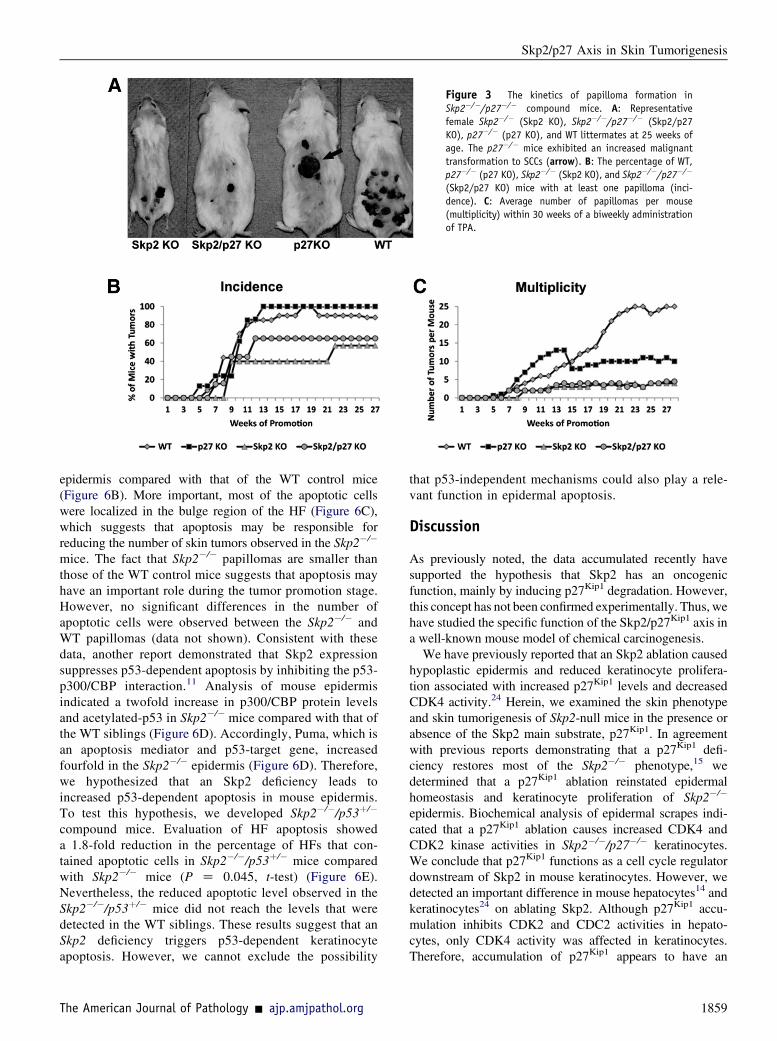
Figure 3 The kinetics of papilloma formation inSkp2�/�/p27�/� compound mice. A: Representativefemale Skp2�/� (Skp2 KO), Skp2�/�/p27�/� (Skp2/p27KO), p27�/� (p27 KO), and WT littermates at 25 weeks ofage. The p27�/� mice exhibited an increased malignanttransformation to SCCs (arrow). B: The percentage of WT,p27�/� (p27 KO), Skp2�/� (Skp2 KO), and Skp2�/�/p27�/�
(Skp2/p27 KO) mice with at least one papilloma (inci-dence). C: Average number of papillomas per mouse(multiplicity) within 30 weeks of a biweekly administrationof TPA.
Skp2/p27 Axis in Skin Tumorigenesis
epidermis compared with that of the WT control mice(Figure 6B). More important, most of the apoptotic cellswere localized in the bulge region of the HF (Figure 6C),which suggests that apoptosis may be responsible forreducing the number of skin tumors observed in the Skp2�/�
mice. The fact that Skp2�/� papillomas are smaller thanthose of the WT control mice suggests that apoptosis mayhave an important role during the tumor promotion stage.However, no significant differences in the number ofapoptotic cells were observed between the Skp2�/� andWT papillomas (data not shown). Consistent with thesedata, another report demonstrated that Skp2 expressionsuppresses p53-dependent apoptosis by inhibiting the p53-p300/CBP interaction.11 Analysis of mouse epidermisindicated a twofold increase in p300/CBP protein levelsand acetylated-p53 in Skp2�/� mice compared with that ofthe WT siblings (Figure 6D). Accordingly, Puma, which isan apoptosis mediator and p53-target gene, increasedfourfold in the Skp2�/� epidermis (Figure 6D). Therefore,we hypothesized that an Skp2 deficiency leads toincreased p53-dependent apoptosis in mouse epidermis.To test this hypothesis, we developed Skp2�/�/p53þ/�
compound mice. Evaluation of HF apoptosis showeda 1.8-fold reduction in the percentage of HFs that con-tained apoptotic cells in Skp2�/�/p53þ/� mice comparedwith Skp2�/� mice (P Z 0.045, t-test) (Figure 6E).Nevertheless, the reduced apoptotic level observed in theSkp2�/�/p53þ/� mice did not reach the levels that weredetected in the WT siblings. These results suggest that anSkp2 deficiency triggers p53-dependent keratinocyteapoptosis. However, we cannot exclude the possibility
The American Journal of Pathology - ajp.amjpathol.org
that p53-independent mechanisms could also play a rele-vant function in epidermal apoptosis.
Discussion
As previously noted, the data accumulated recently havesupported the hypothesis that Skp2 has an oncogenicfunction, mainly by inducing p27Kip1 degradation. However,this concept has not been confirmed experimentally. Thus, wehave studied the specific function of the Skp2/p27Kip1 axis ina well-known mouse model of chemical carcinogenesis.
We have previously reported that an Skp2 ablation causedhypoplastic epidermis and reduced keratinocyte prolifera-tion associated with increased p27Kip1 levels and decreasedCDK4 activity.24 Herein, we examined the skin phenotypeand skin tumorigenesis of Skp2-null mice in the presence orabsence of the Skp2 main substrate, p27Kip1. In agreementwith previous reports demonstrating that a p27Kip1 defi-ciency restores most of the Skp2�/� phenotype,15 wedetermined that a p27Kip1 ablation reinstated epidermalhomeostasis and keratinocyte proliferation of Skp2�/�
epidermis. Biochemical analysis of epidermal scrapes indi-cated that a p27Kip1 ablation causes increased CDK4 andCDK2 kinase activities in Skp2�/�/p27�/� keratinocytes.We conclude that p27Kip1 functions as a cell cycle regulatordownstream of Skp2 in mouse keratinocytes. However, wedetected an important difference in mouse hepatocytes14 andkeratinocytes24 on ablating Skp2. Although p27Kip1 accu-mulation inhibits CDK2 and CDC2 activities in hepato-cytes, only CDK4 activity was affected in keratinocytes.Therefore, accumulation of p27Kip1 appears to have an
1859
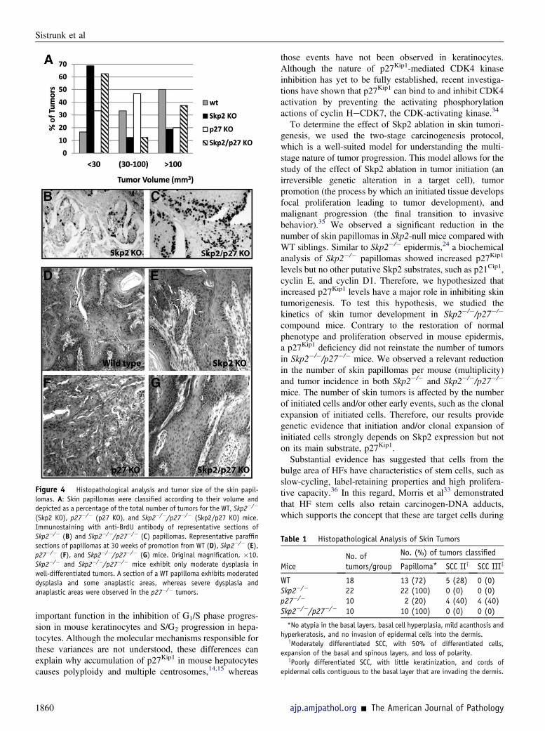
Figure 4 Histopathological analysis and tumor size of the skin papil-lomas. A: Skin papillomas were classified according to their volume anddepicted as a percentage of the total number of tumors for the WT, Skp2�/�
(Skp2 KO), p27�/� (p27 KO), and Skp2�/�/p27�/� (Skp2/p27 KO) mice.Immunostaining with anti-BrdU antibody of representative sections ofSkp2�/� (B) and Skp2�/�/p27�/� (C) papillomas. Representative paraffinsections of papillomas at 30 weeks of promotion from WT (D), Skp2�/� (E),p27�/� (F), and Skp2�/�/p27�/� (G) mice. Original magnification, �10.Skp2�/� and Skp2�/�/p27�/� mice exhibit only moderate dysplasia inwell-differentiated tumors. A section of a WT papilloma exhibits moderateddysplasia and some anaplastic areas, whereas severe dysplasia andanaplastic areas were observed in the p27�/� tumors.
Table 1 Histopathological Analysis of Skin Tumors
MiceNo. oftumors/group
No. (%) of tumors classified
Papilloma* SCC IIy SCC IIIz
WT 18 13 (72) 5 (28) 0 (0)Skp2�/� 22 22 (100) 0 (0) 0 (0)p27�/� 10 2 (20) 4 (40) 4 (40)Skp2�/�/p27�/� 10 10 (100) 0 (0) 0 (0)
*No atypia in the basal layers, basal cell hyperplasia, mild acanthosis andhyperkeratosis, and no invasion of epidermal cells into the dermis.
yModerately differentiated SCC, with 50% of differentiated cells,expansion of the basal and spinous layers, and loss of polarity.
zPoorly differentiated SCC, with little keratinization, and cords ofepidermal cells contiguous to the basal layer that are invading the dermis.
Sistrunk et al
important function in the inhibition of G1/S phase progres-sion in mouse keratinocytes and S/G2 progression in hepa-tocytes. Although the molecular mechanisms responsible forthese variances are not understood, these differences canexplain why accumulation of p27Kip1 in mouse hepatocytescauses polyploidy and multiple centrosomes,14,15 whereas
1860
those events have not been observed in keratinocytes.Although the nature of p27Kip1-mediated CDK4 kinaseinhibition has yet to be fully established, recent investiga-tions have shown that p27Kip1 can bind to and inhibit CDK4activation by preventing the activating phosphorylationactions of cyclin HeCDK7, the CDK-activating kinase.34
To determine the effect of Skp2 ablation in skin tumori-genesis, we used the two-stage carcinogenesis protocol,which is a well-suited model for understanding the multi-stage nature of tumor progression. This model allows for thestudy of the effect of Skp2 ablation in tumor initiation (anirreversible genetic alteration in a target cell), tumorpromotion (the process by which an initiated tissue developsfocal proliferation leading to tumor development), andmalignant progression (the final transition to invasivebehavior).35 We observed a significant reduction in thenumber of skin papillomas in Skp2-null mice compared withWT siblings. Similar to Skp2�/� epidermis,24 a biochemicalanalysis of Skp2�/� papillomas showed increased p27Kip1
levels but no other putative Skp2 substrates, such as p21Cip1,cyclin E, and cyclin D1. Therefore, we hypothesized thatincreased p27Kip1 levels have a major role in inhibiting skintumorigenesis. To test this hypothesis, we studied thekinetics of skin tumor development in Skp2�/�/p27�/�
compound mice. Contrary to the restoration of normalphenotype and proliferation observed in mouse epidermis,a p27Kip1 deficiency did not reinstate the number of tumorsin Skp2�/�/p27�/� mice. We observed a relevant reductionin the number of skin papillomas per mouse (multiplicity)and tumor incidence in both Skp2�/� and Skp2�/�/p27�/�
mice. The number of skin tumors is affected by the numberof initiated cells and/or other early events, such as the clonalexpansion of initiated cells. Therefore, our results providegenetic evidence that initiation and/or clonal expansion ofinitiated cells strongly depends on Skp2 expression but noton its main substrate, p27Kip1.Substantial evidence has suggested that cells from the
bulge area of HFs have characteristics of stem cells, such asslow-cycling, label-retaining properties and high prolifera-tive capacity.36 In this regard, Morris et al33 demonstratedthat HF stem cells also retain carcinogen-DNA adducts,which supports the concept that these are target cells during
ajp.amjpathol.org - The American Journal of Pathology
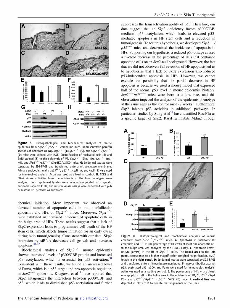
Figure 5 Histopathological and biochemical analyses of mouseepidermis from Skp2�/�/p27�/� compound mice. Representative paraffinsections of skin from WT (A), Skp2�/� (B), p27�/� (C), and Skp2�/�/p27�/�
(D) mice were stained with H&E. Quantification of nucleated cells (E) andBrdU stained (F) in the epidermis of WT, Skp2�/� (Skp2 KO), p27�/� (p27KO), and Skp2�/�/p27�/� (Skp2KO/p27KO) mice. G: Epidermal lysates wereseparated by SDS-PAGE and transferred onto a nitrocellulose membrane.Primary antibodies against p27Kip1, p21Cip1, cyclin A, and cyclin E were usedfor immunoblot analysis. Actin was used as a loading control. H: CDK2 andCDK4 kinase activities from the epidermis of the four genotypes wereanalyzed. Fresh epidermal lysates were immunoprecipitated with specificantibodies against CDKs, and in vitro kinase assays were performed with pRbor histone H1 peptides as substrates.
Figure 6 Histopathological and biochemical analyses of mouseepidermis from Skp2�/�/p53þ/� mice. A: Apoptosis in interfollicularepidermis and HF. B: The percentage of HFs with at least one apoptotic cellin the bulge area was analyzed by the TUNEL assay. C: Apoptotic kerati-nocyte (arrow) in the HF of Skp2�/� mice. The boxed area in the leftpanel corresponds to a higher-magnification (original magnification, �20)image in the right panel. D: Epidermal lysates were separated by SDS-PAGEand transferred onto a nitrocellulose membrane. Primary antibodies againstp53, acetylated p53, p300, and Puma were used for immunoblot analysis.Actin was used as a loading control. E: The percentage of HFs with at leastone apoptotic cell in the bulge area in the epidermis of WT, Skp2�/� (Skp2KO), and p53þ/�/Skp2�/� (p53þ/� SKP2 KO) mice. A vertical line wasdepicted in blots of D to denote rearrangements of the lines.
Skp2/p27 Axis in Skin Tumorigenesis
chemical initiation. More important, we observed anelevated number of apoptotic cells in the interfollicularepidermis and HFs of Skp2�/� mice. Moreover, Skp2�/�
mice exhibited an increased incidence of apoptotic cells inthe bulge area of HFs. These results suggest that a lack ofSkp2 expression leads to programmed cell death of the HFstem cells, which affects tumor initiation (or an early eventduring skin tumorigenesis). Consistent with our data, Skp2inhibition by siRNA decreases cell growth and increasesapoptosis.31,37
Biochemical analysis of Skp2�/� mouse epidermisshowed increased levels of p300/CBP protein and increasedp53 acetylation, which is essential for p53 activation.38
Consistent with these results, we found an increased levelof Puma, which is a p53 target and pro-apoptotic regulator,in Skp2�/� epidermis. Kitagawa et al11 have reported thatSkp2 antagonizes the interaction between p300/CBP andp53, which leads to diminished p53 acetylation and further
The American Journal of Pathology - ajp.amjpathol.org
suppresses the transactivation ability of p53. Therefore, ourdata suggest that an Skp2 deficiency favors p300/CBP-mediated p53 acetylation, which leads to elevated p53-mediated apoptosis in HF stem cells and a reduction intumorigenesis. To test this hypothesis, we developed Skp2�/�/p53þ/� mice and determined the incidence of apoptosis inHFs. Supporting our hypothesis, a reduced p53 dosage causeda twofold decrease in the percentage of HFs that containedapoptotic cells on an Skp2-null background. However, the factthat we did not observe a full reversion of HF apoptosis led usto hypothesize that a lack of Skp2 expression also inducedp53-independent apoptosis in HFs. However, we cannotexclude the possibility that the partial decrease in HFapoptosis is because we used a mouse model that expressedhalf of the normal p53 level in mouse epidermis. Notably,Skp2�/�/p53�/� mice were born at a low rate, and thisobservation impeded the analysis of the epidermis phenotypeat the same ages as the control mice (7 weeks). Furthermore,Skp2 inhibits p53 activities in additional pathways. Inparticular, studies by Song et al39 have identified RassF1a asa specific target of Skp2. RassF1a inhibits Mdm2 through
1861
Sistrunk et al
stimulation of Mdm2 self-ubiquitination, leading to p53 acti-vation.10 In addition, Rassf1a also induces apoptosis throughthe activation and translocation of p73, which increases thetranscription of the pro-apoptotic protein, Puma.40 Therefore,Skp2 can inhibit p53 by preventing CBP/p300 acetylationactivation and by increasing levels of Mdm2 by inhibitingRassf1a, thereby yielding two independent pathways to inhibitapoptosis. More important, our studies also demonstrate thatan Skp2 deficiency causes an accumulation of RassF1a inmouse epidermis (C. Sistrunk and M. Rodriguez-Puebla,unpublished data) and a reduction of Mdm2 protein levels.These findings suggest that the RassF1a pathway can alsoaccelerate p53-dependent apoptosis in mouse epidermis.Whether a synergistic effect exists between p300/CBP-mediated p53 activation and RassF1a pathways warrantsfurther investigation.
To determine the effect of disrupting the Skp2/p27Kip1
axis in tumor promotion and malignant progression, weperformed histopathological analyses of skin tumors fromSkp2�/�/p27�/� and control sibling mice. We determineda clear reduction in the tumor size of Skp2�/� papillomasthat was partially restored in Skp2�/�/p27�/� papillomas.Therefore, we infer that an Skp2 deficiency inhibits early-stage tumorigenesis. However, the few Skp2�/�/p27�/�
tumors that avoid this early blockage grow more rapidlycompared with Skp2�/� papillomas. Therefore, we hypoth-esize that p27Kip1 ablation favors fast keratinocyte prolif-eration during the tumor promotion stage of a fraction of theSkp2�/�/p27�/� papillomas, which causes a partial rescue intumor volume. Consistent with this hypothesis, we observedelevated keratinocyte proliferation in approximately 30% ofthe Skp2�/�/p27�/� papillomas compared with Skp2�/�
tumors. We also evaluated the relevance of the apoptoticprofile displayed in the papillomas from the four genotypes.However, we did not observe significant differences amongthem (data not shown). Therefore, our results suggest thatinitiation, and not the promotion stage of skin carcinogen-esis, was affected by the apoptosis that was induced by anSkp2 deficiency.
Histopathological analysis of the skin tumors indicatedthat, although the tumorigenesis blockages in animalsdevoid of Skp2 are independent of p27Kip1 accumulation,p27�/� animals developed more aggressive tumors thatwere classified as SCCs. The fact that lack of p27Kip1
expression causes elevated CDK4 and CDK2 kinase activ-ities suggests that the CDK inhibitor role of p27Kip1 playsimportant functions during malignant progression. Sup-porting this theory, CDK4 overexpression in K5CDK4transgenic mice also induces malignant progression medi-ated by the sequestration of p27Kip1 and p21Cip1 by D-typecyclin/CDK4 complexes.41 Moreover, a lack of CDK2expression reduces the malignant progression to SCCs inK5CDK4/CDK2�/� mice.42 It is not clear why p27�/� miceshowed a reduction in the number of papillomas per mousecompared with the WT siblings. However, we hypothesizethat an ablation of p27Kip1 compromises the assembly of
1862
active CDK4/D-type cyclin complexes that, in this case, relyon the presence of p21Cip1.43
More important, an ablation of Skp2 appears to blockmalignant progression in Skp2�/�/p27�/� mice because all ofthe Skp2�/� and Skp2�/�/p27�/� tumors were classified aswell-differentiated papillomas. Notably, an analysis of the skintumors from Skp2�/�/p27�/�, Skp2�/�, p27�/�, andWTmicedid not show a significant difference in the number of apoptoticcells (data not shown). Therefore, onemay conclude that Skp2-regulated inhibitory pathways other than apoptosis impedemalignant progression. Supporting this concept, aberrantoncogene signals initiate a senescence response in Skp2-nullcells through a p19ARF-p53eindependent pathway,44 andchemical inhibition of Skp2 activity causes cell death throughactivation of autophagy.45
In summary, our data demonstrate that Skp2 inhibitioncauses blockage of an early event during themultistage processof nonmelanoma skin tumorigenesis through a p27Kip1-inde-pendent pathway. We also suggest that Skp2-mediated inhi-bition of p53-dependent apoptosis has an important function inthe elevated apoptosis observed in Skp2�/� HF stem cells,which correlates with fewer papillomas. In addition, a partialrescue of keratinocyte proliferation has been observed inSkp2�/�/p27�/� papillomas, which suggests that cell prolif-eration during the promotion stage is partially dependent on thep27Kip1 level.Whether the variations in the effects of Skp2 andp27Kip1 levels at specific stages of tumorigenesis are unique forepidermal tumors warrants further investigation. Finally,although a strong inverse correlation between Skp2 andp27Kip1 levels has been demonstrated in numerous experi-mental and human tumors, Skp2 is also a p27Kip1-independentindicator of poor prognosis in other human tumors, such asbiliary tract carcinoma,46 soft tissue sarcomas,47 and urinarytract transitional cell carcinoma.48 Therefore, our resultssupport the hypothesis that molecular targets of Skp2, otherthan p27Kip1, may also be important factors in cancer patho-genesis. Moreover, a combination of Skp2 and targets, otherthan p27Kip1, in addition to the individual molecular targets,might be useful prognostic factors of cancer.
Acknowledgments
We thank Dr. Paula Miliani de Marval for her critical reviewof this article, Dr. Keiichi Nakayama (Kyushu University,Kyushu, Japan) for kindly providing the Skp2-null mice, JuanCarlos Santiago for his technical support, and the LaboratoryAnimal Resources and the Histology Core (College ofVeterinary Medicine, North Carolina State University) forhelping with the processing and staining of skin and tumorsamples.
References
1. Weissman AM: Themes and variations on ubiquitylation. Nat Rev MolCell Biol 2001, 2:169e178
ajp.amjpathol.org - The American Journal of Pathology
Skp2/p27 Axis in Skin Tumorigenesis
2. Gregory MA, Hann SR: c-Myc proteolysis by the ubiquitin-proteasome pathway: stabilization of c-Myc in Burkitt’s lymphomacells. Mol Cell Biol 2000, 20:2423e2435
3. Nakayama KI, Nakayama K: Ubiquitin ligases: cell-cycle control andcancer. Nat Rev Cancer 2006, 6:369e381
4. Bahram F, von der Lehr N, Cetinkaya C, Larsson LG: c-Myc hot spotmutations in lymphomas result in inefficient ubiquitination anddecreased proteasome-mediated turnover. Blood 2000, 95:2104e2110
5. Yu ZK, Gervais JL, Zhang H: Human CUL-1 associates with theSKP1/SKP2 complex and regulates p21(CIP1/WAF1) and cyclin Dproteins. Proc Natl Acad Sci U S A 1998, 95:11324e11329
6. Kamura T, Hara T, Kotoshiba S, Yada M, Ishida N, Imaki H,Hatakeyama S, Nakayama K, Nakayama KI: Degradation of p57Kip2mediated by SCFSkp2-dependent ubiquitylation. Proc Natl Acad Sci US A 2003, 100:10231e10236
7. Tedesco D, Lukas J, Reed SI: The pRb-related protein p130 is regu-lated by phosphorylation-dependent proteolysis via the protein-ubiquitin ligase SCF(Skp2). Genes Dev 2002, 16:2946e2957
8. Hiramatsu Y, Kitagawa K, Suzuki T, Uchida C, Hattori T, Kikuchi H,Oda T, Hatakeyama S, Nakayama KI, Yamamoto T, Konno H,Kitagawa M: Degradation of Tob1 mediated by SCFSkp2-dependentubiquitination. Cancer Res 2006, 66:8477e8483
9. Huang H, Regan KM, Wang F, Wang D, Smith DI, van Deursen JM,Tindall DJ: Skp2 inhibits FOXO1 in tumor suppression throughubiquitin-mediated degradation. Proc Natl Acad Sci U S A 2005, 102:1649e1654
10. Song MS, Song SJ, Kim SJ, Nakayama K, Nakayama KI, Lim DS:Skp2 regulates the antiproliferative function of the tumor suppressorRASSF1A via ubiquitin-mediated degradation at the G1-S transition.Oncogene 2008, 27:3176e3185
11. Kitagawa M, Lee SH, McCormick F: Skp2 suppresses p53-dependentapoptosis by inhibiting p300. Mol Cell 2008, 29:217e231
12. Bai C, Sen P, Hofmann K, Ma L, Goebl M, Harper JW, Elledge SJ:SKP1 connects cell cycle regulators to the ubiquitin proteolysismachinery through a novel motif, the F-box. Cell 1996, 86:263e274
13. Carrano AC, Eytan E, Hershko A, Pagano M: SKP2 is required forubiquitin-mediated degradation of the CDK inhibitor p27. Nat CellBiol 1999, 1:193e199
14. Nakayama K, Nagahama H, Minamishima YA, Matsumoto M,Nakamichi I, Kitagawa K, Shirane M, Tsunematsu R, Tsukiyama T,IshidaN,KitagawaM,NakayamaK,HatakeyamaS: Targeted disruptionof Skp2 results in accumulation of cyclin E and p27(Kip1), polyploidyand centrosome overeduplication. EMBO J 2000, 19:2069e2081
15. Nakayama K, Nagahama H, Minamishima YA, Miyake S, Ishida N,Hatakeyama S, Kitagawa M, Iemura S, Natsume T, Nakayama KI:Skp2-mediated degradation of p27 regulates progression into mitosis.Dev Cell 2004, 6:661e672
16. Nakayama KI, Nakayama K: Regulation of the cell cycle by SCF-typeubiquitin ligases. Semin Cell Dev Biol 2005, 16:323e333
17. Latres E, Chiarle R, Schulman BA, Pavletich NP, Pellicer A,Inghirami G, Pagano M: Role of the F-box protein Skp2 in lympho-magenesis. Proc Natl Acad Sci U S A 2001, 98:2515e2520
18. Carrano AC, Pagano M: Role of the F-box protein Skp2 in adhesion-dependent cell cycle progression. J Cell Biol 2001, 153:1381e1390
19. Kudo Y, Kitajima S, Sato S, Miyauchi M, Ogawa I, Takata T: Highexpression of S-phase kinase-interacting protein 2, human F-boxprotein, correlates with poor prognosis in oral squamous cell carci-nomas. Cancer Res 2001, 61:7044e7047
20. Shim EH, Johnson L, Noh HL, Kim YJ, Sun H, Zeiss C, Zhang H:Expression of the F-box protein SKP2 induces hyperplasia, dysplasia,and low-grade carcinoma in the mouse prostate. Cancer Res 2003, 63:1583e1588
21. Slingerland J, Pagano M: Regulation of the cdk inhibitor p27 and itsderegulation in cancer. J Cell Physiol 2000, 183:10e17
22. Osoegawa A, Yoshino I, Tanaka S, Sugio K, Kameyama T,Yamaguchi M, Maehara Y: Regulation of p27 by S-phase kinase-
The American Journal of Pathology - ajp.amjpathol.org
associated protein 2 is associated with aggressiveness in non-small-cell lung cancer. J Clin Oncol 2004, 22:4165e4173
23. Li Q, Murphy M, Ross J, Sheehan C, Carlson JA: Skp2 and p27kip1expression in melanocytic nevi and melanoma: an inverse relationship.J Cutan Pathol 2004, 31:633e642
24. Sistrunk C, Macias E, Nakayama K, Kim Y, Rodriguez-Puebla ML:Skp2 is necessary for myc-induced keratinocyte proliferation butdispensable for myc oncogenic activity in the oral epithelium. Am JPathol 2011, 178:2470e2477
25. Donehower LA,HarveyM, Slagle BL,McArthurMJ,MontgomeryCAJr,Butel JS, Bradley A: Mice deficient for p53 are developmentallynormal but susceptible to spontaneous tumours. Nature 1992, 356:215e221
26. Rodriguez-Puebla ML, Robles AI, Johnson DG, LaCava M, Conti CJ:Synchronized proliferation induced by 12-O-tetradecanoylphorbol-13-acetate treatment of mouse skin: an in vivo model for cell cycleregulation. Cell Growth Differ 1998, 9:31e39
27. Naito M, DiGiovanni J: Genetic background and development of skintumors. Edited by Conti CJ, Slaga TJ, Klein-Szanto AJ. CarcinogenesisA Comprehensive Survey. New York, Raven Press, 1989, pp 187e212
28. Philipp J, Vo K, Gurley KE, Seidel K, Kemp CJ: Tumor suppressionby p27Kip1 and p21Cip1 during chemically induced skin carcino-genesis. Oncogene 1999, 18:4689e4698
29. Klein-Szanto A: Melanotic and non-melanotic tumours of the rodentskin. Pathology of Neoplasia and Preneoplasia in Rodents. Schattauer,European Late Effects Project, 1997, pp 1e18
30. Macias E, de Marval PL, Senderowicz A, Cullen J, Rodriguez-Puebla ML: Expression of CDK4 or CDK2 in mouse oral cavity isretained in adult pituitary with distinct effects on tumorigenesis.Cancer Res 2008, 68:162e171
31. Harada K, Supriatno, Kawashima Y, Itashiki Y, Yoshida H, Sato M:Down-regulation of S-phase kinase associated protein 2 (Skp2) inducesapoptosis in oral cancer cells. Oral Oncol 2005, 41:623e630
32. Jiang F, Caraway NP, Li R, Katz RL: RNA silencing of S-phasekinase-interacting protein 2 inhibits proliferation and centrosomeamplification in lung cancer cells. Oncogene 2005, 24:3409e3418
33. Morris RJ, Fischer SM, Slaga TJ: Evidence that a slowly cyclingsubpopulation of adult murine epidermal cells retains carcinogen.Cancer Res 1986, 46:3061e3066
34. Ray A, James MK, Larochelle S, Fisher RP, Blain SW: p27Kip1inhibits cyclin D-cyclin-dependent kinase 4 by two independentmodes. Mol Cell Biol 2009, 29:986e999
35. Slaga T: Mechanism involved in two-stage carcinogenesis in mouseskin. Edited by Slaga T. Mechanism of Tumor Promotion. Boca Raton,CRC Press, 1984, pp 1e16
36. Morris RJ: Keratinocyte stem cells: targets for cutaneous carcinogens.J Clin Invest 2000, 106:3e8
37. Lee SH, McCormick F: Downregulation of Skp2 and p27/Kip1synergistically induces apoptosis in T98G glioblastoma cells. J MolMed 2005, 83:296e307
38. Grossman SR: p300/CBP/p53 interaction and regulation of the p53response. Eur J Biochem 2001, 268:2773e2778
39. Song MS, Song SJ, Kim SY, Oh HJ, Lim DS: The tumour suppressorRASSF1A promotes MDM2 self-ubiquitination by disrupting theMDM2-DAXX-HAUSP complex. EMBO J 2008, 27:1863e1874
40. Matallanas D, Romano D, Yee K, Meissl K, Kucerova L, Piazzolla D,Baccarini M, Vass JK, Kolch W, O’Neill E: RASSF1A elicitsapoptosis through an MST2 pathway directing proapoptotic tran-scription by the p73 tumor suppressor protein. Mol Cell 2007, 27:962e975
41. Miliani de Marval PL, Macias E, Conti CJ, Rodriguez-Puebla ML:Enhanced malignant tumorigenesis in Cdk4 transgenic mice. Onco-gene 2004, 23:1863e1873
42. Macias E, Kim Y, Miliani de Marval PL, Klein-Szanto A, Rodriguez-Puebla ML: Cdk2 deficiency decreases ras/CDK4-dependent malignantprogression, but not myc-induced tumorigenesis. Cancer Res 2007, 67:9713e9720
1863

Sistrunk et al
43. Cheng M, Olivier P, Diehl JA, Fero M, Roussel MF, Roberts JM,Sherr CJ: The p21(Cip1) and p27(Kip1) CDK “inhibitors” are essentialactivators of cyclin D-dependent kinases in murine fibroblasts. EMBOJ 1999, 18:1571e1583
44. Lin HK, Chen Z, Wang G, Nardella C, Lee SW, Chan CH, Yang WL,Wang J, Egia A, Nakayama KI, Cordon-Cardo C, Teruya-Feldstein J,Pandolfi PP: Skp2 targeting suppresses tumorigenesis by Arf-p53-independent cellular senescence. Nature 2010, 464:374e379
45. Chen Q, Xie W, Kuhn DJ, Voorhees PM, Lopez-Girona A, Mendy D,Corral LG, Krenitsky VP, Xu W, Moutouhede Parseval L, Webb DR,Mercurio F, Nakayama KI, Nakayama K, Orlowski RZ: Targeting the
1864
p27 E3 ligase SCF(Skp2) results in p27- and Skp2-mediated cell-cyclearrest and activation of autophagy. Blood 2008, 111:4690e4699
46. Sanada T, Yokoi S, Arii S, Yasui K, Imoto I, Inazawa J: Skp2 over-expression is a p27Kip1-independent predictor of poor prognosis inpatients with biliary tract cancers. Cancer Sci 2004, 95:969e976
47. Oliveira AM, Okuno SH, Nascimento AG, Lloyd RV: Skp2 proteinexpression in soft tissue sarcomas. J Clin Oncol 2003, 21:722e727
48. Langner C, von Wasielewski R, Ratschek M, Rehak P, Zigeuner R:Expression of p27 and its ubiquitin ligase subunit Skp2 in upperurinary tract transitional cell carcinoma. Urology 2004, 64:611e616
ajp.amjpathol.org - The American Journal of Pathology
Copyright © 2022 FDOKUMEN