Direct induction of cyclin D2 by Myc contributes to cell cycle progression and sequestration of p27
13
The EMBO Journal Vol.18 No.19 pp.5321–5333, 1999 Direct induction of cyclin D2 by Myc contributes to cell cycle progression and sequestration of p27 Caroline Bouchard, Katja Thieke, Antje Maier, Rainer Saffrich 1 , Joan Hanley-Hyde 2 , Wilhelm Ansorge 1 , Steve Reed 2 , Piotr Sicinski 3 , Jiri Bartek 4 and Martin Eilers 5 Institute of Molecular Biology and Tumour Research, Phillips- Universita ¨t Marburg, Emil-Mannkopff-Strasse 2, 35033 Marburg, 1 Biological Instrumentation Programme, EMBL, Meyerhofstrasse 1, 69112 Heidelberg, Germany, 2 Department of Molecular Biology, The Scripps Research Institute, La Jolla, CA 92037, 3 Dana Farber Cancer Institute, 44 Binney Street, Boston, MA 02115, USA and 4 Institute of Cancer Biology, Strandboulevarden 49, 2100 Copenhagen, Denmark 5 Corresponding author e-mail: [email protected] Ectopic expression of Myc induces Cdk2 kinase activity in quiescent cells and antagonizes association of p27 kip1 with Cdk2. The target gene(s) by which Myc mediates this effect is largely unknown. We now show that p27 is rapidly and transiently sequestered by cyclin D2– Cdk4 complexes upon activation of Myc and that cyclin D2 is a direct target gene of Myc. The cyclin D2 promoter is repressed by Mad–Max complexes and de- repressed by Myc via a single highly conserved E-box element. Addition of trichostatin A to quiescent cells mimics activation of Myc and induces cyclin D2 expres- sion, suggesting that cyclin D2 is repressed in a histone deacetylase-dependent manner in quiescent cells. Inhibition of cyclin D2 function in established cell lines, either by ectopic expression of p16 or by antibody injection, inhibits Myc-dependent dissociation of p27 from Cdk2 and Myc-induced cell cycle entry. Primary mouse fibroblasts that are cyclin D2-deficient undergo accelerated senescence in culture and are not immortal- ized by Myc; induction of apoptosis by Myc is unim- paired in such cells. Our data identify a downstream effector pathway that links Myc directly to cell cycle progression. Keywords: Cdk2/cyclin D2/Mad-1/Myc/p27 Introduction The proto-oncogene c-myc encodes a transcription factor of the helix–loop–helix/leucine zipper family of proteins (for review, see Henriksson and Lu ¨scher, 1996). The encoded protein, Myc, binds to specific sequences on DNA [so-called E-boxes with a central CAC(G/A)TG motif] as part of a heterodimeric complex with a partner protein, Max, and activates transcription. Max also forms heterodimers with a second group of related proteins, termed Mad-1, Mxi-1, Mad-3, Mad-4 and Mnt/Rox (Ayer et al., 1993; Zervos et al., 1993; Hurlin et al., 1996, 1997; Meroni et al., 1997). These complexes bind to identical © European Molecular Biology Organization 5321 E-box elements, but repress transcription, at least in part, via recruitment of histone deacetylase activity (Alland et al., 1997; Heinzel et al., 1997; Laherty et al., 1997; Sommer et al., 1997). In mammalian cells, the relative levels of Myc and Mad proteins can determine the rate of cell proliferation and apoptosis (for review, see Bouchard et al., 1998). For example, ectopic expression of Myc or activation of conditional alleles of Myc (MycER) (Eilers et al., 1989; Littlewood et al., 1995) can induce quiescent cells to enter the cell cycle and undergo apoptosis (Eilers et al., 1991; Evan et al., 1992). Conversely, ectopic expression of Mad-1 prevents growth factor-induced pro- liferation of fibroblasts (Roussel et al., 1996). It is most likely, therefore, that this network of interacting proteins regulates a set of target genes important in controlling cell proliferation. One downstream target of Myc activity is the cell cycle inhibitor p27 kip1 and the related p21 waf-1 protein. Several lines of evidence support this notion. First, activation of conditional alleles of Myc in quiescent fibroblasts rapidly induces cyclin E–Cdk2 kinase activity and loss of p27 from Cdk2 complexes (Steiner et al., 1995; Mu ¨ller et al., 1997). Myc also up-regulates Cdk2 kinase activity at any point during the cell cycle of exponentially growing fibroblasts (Pusch et al., 1997), demonstrating that the effects of Myc on Cdk2 are independent of its ability to stimulate quiescent cells to enter the cycle. Activation of Cdk2 is required for Myc to promote G 1 progression (Rudolph et al., 1996). Secondly, expression of dominant- negative alleles of Myc arrests cell proliferation and suppresses cyclin E–Cdk2 activity (Berns et al., 1997). Expression of cyclin E and Cdk2 is unimpaired in the arrested cells, suggesting that Myc function is required at a step after cyclin E synthesis. In fibroblasts carrying a homozygous null mutation of Myc, levels of p27 are elevated and cyclin E-dependent kinase activity is reduced (Mateyak et al., 1999). Thirdly, ectopic expression of Myc partially antagonizes the growth suppressive activities of p27 (Vlach et al., 1996); however, p27 is dominant when expressed at high concentration, demonstrating that Myc does not bypass p27 function (Rudolph et al., 1996; Mu ¨ller et al., 1997). Ectopic expression of Myc also suppresses the p21-dependent growth arrest of temperature-sensitive alleles of p53 (Wagner et al., 1994; Hermeking et al., 1995). In both cases, it was suggested that Myc induces the ‘sequestration’ of the inhibitor into a heat-labile complex. The identity of this complex, however, remained unknown. In order to identify Myc-induced p27-binding proteins, we began to purify p27-containing complexes from RAT1 fibroblasts that express a MycER protein. During the course of purification, we noticed that activation of Myc induces a rapid increase in the amount of cyclin D2– Cdk4–p27 complexes. We now provide evidence that
-
Upload
independent -
Category
Documents
-
view
4 -
download
0
Transcript of Direct induction of cyclin D2 by Myc contributes to cell cycle progression and sequestration of p27
The EMBO Journal Vol.18 No.19 pp.5321–5333, 1999
Direct induction of cyclin D2 by Myc contributes tocell cycle progression and sequestration of p27
Caroline Bouchard, Katja Thieke,Antje Maier, Rainer Saffrich1,Joan Hanley-Hyde2, Wilhelm Ansorge1,Steve Reed2, Piotr Sicinski3, Jiri Bartek4
and Martin Eilers5
Institute of Molecular Biology and Tumour Research, Phillips-Universitat Marburg, Emil-Mannkopff-Strasse 2, 35033 Marburg,1Biological Instrumentation Programme, EMBL, Meyerhofstrasse 1,69112 Heidelberg, Germany,2Department of Molecular Biology, TheScripps Research Institute, La Jolla, CA 92037,3Dana Farber CancerInstitute, 44 Binney Street, Boston, MA 02115, USA and4Institute ofCancer Biology, Strandboulevarden 49, 2100 Copenhagen, Denmark5Corresponding authore-mail: [email protected]
Ectopic expression of Myc induces Cdk2 kinase activityin quiescent cells and antagonizes association of p27kip1
with Cdk2. The target gene(s) by which Myc mediatesthis effect is largely unknown. We now show that p27is rapidly and transiently sequestered by cyclin D2–Cdk4 complexes upon activation of Myc and thatcyclin D2 is a direct target gene of Myc. The cyclin D2promoter is repressed by Mad–Max complexes and de-repressed by Myc via a single highly conserved E-boxelement. Addition of trichostatin A to quiescent cellsmimics activation of Myc and induces cyclin D2 expres-sion, suggesting that cyclin D2 is repressed in a histonedeacetylase-dependent manner in quiescent cells.Inhibition of cyclin D2 function in established cell lines,either by ectopic expression of p16 or by antibodyinjection, inhibits Myc-dependent dissociation of p27from Cdk2 and Myc-induced cell cycle entry. Primarymouse fibroblasts that are cyclin D2-deficient undergoaccelerated senescence in culture and are not immortal-ized by Myc; induction of apoptosis by Myc is unim-paired in such cells. Our data identify a downstreameffector pathway that links Myc directly to cell cycleprogression.Keywords: Cdk2/cyclin D2/Mad-1/Myc/p27
Introduction
The proto-oncogenec-mycencodes a transcription factorof the helix–loop–helix/leucine zipper family of proteins(for review, see Henriksson and Lu¨scher, 1996). Theencoded protein, Myc, binds to specific sequences onDNA [so-called E-boxes with a central CAC(G/A)TGmotif] as part of a heterodimeric complex with a partnerprotein, Max, and activates transcription. Max also formsheterodimers with a second group of related proteins,termed Mad-1, Mxi-1, Mad-3, Mad-4 and Mnt/Rox (Ayeret al., 1993; Zervoset al., 1993; Hurlinet al., 1996, 1997;Meroni et al., 1997). These complexes bind to identical
© European Molecular Biology Organization 5321
E-box elements, but repress transcription, at least in part,via recruitment of histone deacetylase activity (Allandet al., 1997; Heinzelet al., 1997; Lahertyet al., 1997;Sommeret al., 1997). In mammalian cells, the relativelevels of Myc and Mad proteins can determine the rate ofcell proliferation and apoptosis (for review, see Bouchardet al., 1998). For example, ectopic expression of Myc oractivation of conditional alleles of Myc (MycER) (Eilerset al., 1989; Littlewoodet al., 1995) can induce quiescentcells to enter the cell cycle and undergo apoptosis (Eilerset al., 1991; Evanet al., 1992). Conversely, ectopicexpression of Mad-1 prevents growth factor-induced pro-liferation of fibroblasts (Rousselet al., 1996). It is mostlikely, therefore, that this network of interacting proteinsregulates a set of target genes important in controllingcell proliferation.
One downstream target of Myc activity is the cell cycleinhibitor p27kip1 and the related p21waf-1 protein. Severallines of evidence support this notion. First, activation ofconditional alleles of Myc in quiescent fibroblasts rapidlyinduces cyclin E–Cdk2 kinase activity and loss of p27from Cdk2 complexes (Steineret al., 1995; Muller et al.,1997). Myc also up-regulates Cdk2 kinase activity at anypoint during the cell cycle of exponentially growingfibroblasts (Puschet al., 1997), demonstrating that theeffects of Myc on Cdk2 are independent of its ability tostimulate quiescent cells to enter the cycle. Activation ofCdk2 is required for Myc to promote G1 progression(Rudolphet al., 1996). Secondly, expression of dominant-negative alleles of Myc arrests cell proliferation andsuppresses cyclin E–Cdk2 activity (Bernset al., 1997).Expression of cyclin E and Cdk2 is unimpaired in thearrested cells, suggesting that Myc function is required ata step after cyclin E synthesis. In fibroblasts carrying ahomozygous null mutation of Myc, levels of p27 areelevated and cyclin E-dependent kinase activity is reduced(Mateyaket al., 1999). Thirdly, ectopic expression of Mycpartially antagonizes the growth suppressive activities ofp27 (Vlachet al., 1996); however, p27 is dominant whenexpressed at high concentration, demonstrating that Mycdoes not bypass p27 function (Rudolphet al., 1996; Mulleret al., 1997). Ectopic expression of Myc also suppressesthe p21-dependent growth arrest of temperature-sensitivealleles of p53 (Wagneret al., 1994; Hermekinget al.,1995). In both cases, it was suggested that Myc inducesthe ‘sequestration’ of the inhibitor into a heat-labilecomplex. The identity of this complex, however, remainedunknown.
In order to identify Myc-induced p27-binding proteins,we began to purify p27-containing complexes from RAT1fibroblasts that express a MycER protein. During thecourse of purification, we noticed that activation of Mycinduces a rapid increase in the amount of cyclin D2–Cdk4–p27 complexes. We now provide evidence that
C.Bouchard et al.
cyclin D2 is a direct target gene of Myc and thataccumulation of cyclin D2 contributes to the sequestrationof p27 that occurs in Myc-transformed cells.
Results
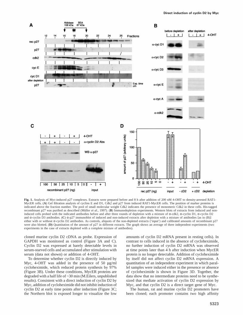
In order to purify Myc-induced p27-containing complexes,density-arrested RAT1-MycER cells were stimulated bythe addition of 200 nM 4-hydroxy-tamoxifen (4-OHT)and extracts were prepared 8 h later. At this time point,p27 is largely dissociated from Cdk2 complexes but notyet degraded (Mu¨ller et al., 1997). Upon gel filtration ofeither non-induced or induced extracts, p27 eluted as asingle peak with a molecular weight of ~160 kDa, co-migrating with both cyclins E and D1 and with part ofCdk2 (shown for induced extracts in Figure 1A). Incontrast, recombinant p27 eluted with a size of 60 kDa,suggesting that it forms a dimer. Also, a brief heat-treatment of cellular extracts caused p27 to migrate withthe same molecular weight (data not shown). Thus, inRAT1 cells p27 is sequestered in heat-labile complexesbefore and after activation of Myc, suggesting that activa-tion of Myc induces a redistribution of p27 among suchcomplexes (Vlachet al., 1996).
An immunodepletion procedure was used to removeknown binding partners of p27. Extracts were depletedthree times by a mixture ofα-Cdk2, α-cyclin D1,α-cyclin D2 andα-cyclin D3 antibodies that had beencovalently coupled to protein A–Sepharose. Controlsshowed that cyclin E, Cdk2, cyclins D1, D2 and D3 wereeach reduced by 90–95% in the depleted extracts relativeto input (Figure 1B). Cyclin A was reduced by 50%,presumably because it can also form complexes with Cdc2(Draettaet al., 1989). As p27 interacts only weakly withCdc2 complexes, we assume that this will not affect theresults (Polyaket al., 1994; Toyoshima and Hunter, 1994).
Depleted and non-depleted extracts were probed for theamount of p27 by Western blotting together with calibratedamounts of recombinant p27. The blots were developedby an enhanced chemiluminescence protocol and differentexposures were scanned and quantitated. A representativeWestern blot is shown in Figure 1C, and the quantitationof three independent experiments is depicted in Figure 1D.Depletion of the known binding partners of p27 depleted.90% of cellular p27 both from induced and non-inducedextracts. Fractionation revealed that the remaining p27co-migrated with the bulk of p27 from non-depletedextracts (Figure 1A). Therefore, we did not obtain anyevidence for an as yet unknown Myc-induced complexcontaining p27. While we cannot exclude the possibilitythat the remaining 10% of non-depleted p27 containsnovel Myc-induced p27-binding proteins, the low levelsof cyclin proteins remaining after depletion can alsoaccount for this amount of p27.
Since p27 dissociates from Cdk2 and we did not observeaccumulation of cyclin D1- or cyclin D3-containing com-plexes (see Figure 2C) (Mu¨ller et al., 1997), we repeatedthe depletion experiment omitting theα-cyclin D2 anti-bodies from the depletion mixture (Figure 1C and D).Under these conditions, 90% of p27 was depleted fromthe non-induced, but only 50% from the induced extracts,suggesting the presence of significant amounts ofcyclin D2–p27 complexes in the induced extracts. We
5322
concluded that activation of Myc promotes the formationof cyclin D2–p27 complexes.
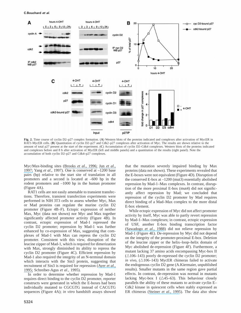
Upon activation of Myc, p27 dissociates from Cdk2within 4–6 h (Muller et al., 1997). To assess whether theformation of cyclin D2–p27 complexes could account forthis rapid dissociation, time course experiments wereperformed. Extracts were prepared from quiescent RAT1-MycER cells before, or at different time points afteractivation of Myc. At each time point, the amount ofcyclin D2, cyclin A, Cdk2, p27, p27–Cdk2 and p27–cyclin D2 complexes was determined (Figure 2A).
Consistent with previous results, activation of Mycinduced a rapid dissociation of p27 from Cdk2, whichpreceded overt degradation of p27 by several hours. Inresponse to activation of Myc, levels of cyclin D2 andcyclin D2–p27 complexes strongly increased within 3 hand peaked between 6–9 h after addition of 4-OHT.Quantitation of the results showed that up to 20% ofcellular p27 complexed with cyclin D2 after activation ofMyc in this experiment and that the maximal amount ofp27 bound to cyclin D2 exceeded the amount of p27initially bound to Cdk2 (Figure 2B). We concluded thatcomplex formation with cyclin D2 could account for thedissociation of p27 from Cdk2 after activation of Myc.Induction of cyclin D2 was transient and protein levelsdropped in late G1; a similar decrease has been observedpreviously in exponentially growing human cells (see alsoFigure 8) (Lukaset al., 1995).
In several time course experiments, we noted a smallbut significant delay between the accumulation of thecyclin D2 protein and loss of p27 from Cdk2 (see timepoint at 3 h and also Figure 6). It is possible, therefore,that the apparent shift of p27 from Cdk2 to cyclin D2results from binding of newly synthesized p27 to enhancedlevels of cyclin D2 and loss of p27 from Cdk2 by turnoveror degradation rather than from direct transfer of pre-existing p27 molecules from Cdk2 to cyclin D2. This isconsistent with previous observations that mutants of p27that cannot be phosphorylated by Cdk2, and therefore arenot degraded, dissociate more slowly than wild-type p27from Cdk2 after induction of Myc (Mu¨ller et al., 1997).
In parallel experiments, we determined the change ineach D-type cyclin–p27 and Cdk4–p27 complex(Figure 2C) and the amount of Cdk4 bound to differentD-type cyclins before and after activation of Myc (datanot shown). The amount of both cyclin D2 and Cdk4bound to p27 increased in response to induction of Myc;in contrast, the amount of p27 bound to either cyclin D1or D3 remained constant (Figure 2C; see also Mu¨lleret al., 1997). Consistent with these findings, we observeda 3- to 4-fold increase in cyclin D2 bound to Cdk4 (datanot shown); 30% of total Cdk4 was bound to cyclin D2after induction of Myc. The remaining Cdk4 was boundto cyclin D1 (30% of total Cdk4) and to cyclin D3 (40%);the absolute amounts of these latter complexes remainedconstant during induction (data not shown). We concludethat the formation of ternary p27 cyclin D2–p27–Cdk4complexes upon activation of Myc significantly contributesto sequestration of p27.
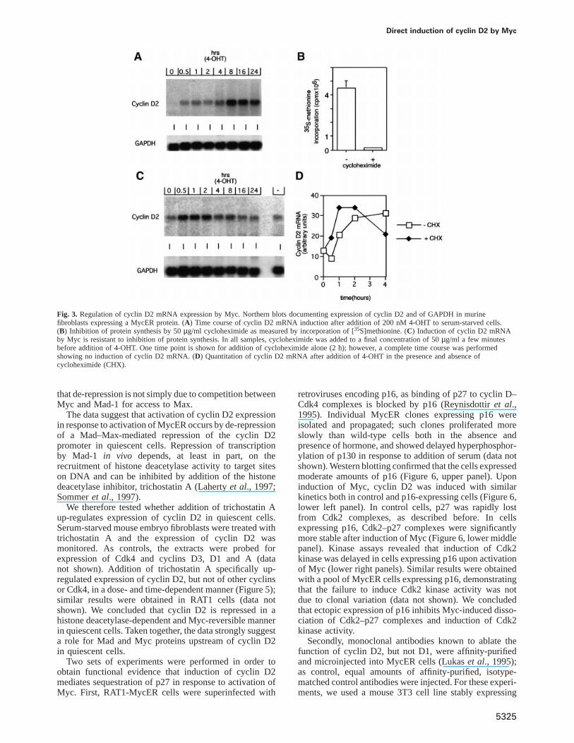
To assess whether induction of cyclin D2 by Mycoccurs at the RNA level, total RNA was isolated fromserum-starved murine fibroblasts that express a MycERprotein and subjected to Northern blot analysis using a
Direct induction of cyclin D2 by Myc
Fig. 1. Analysis of Myc-induced p27 complexes. Extracts were prepared before and 8 h after addition of 200 nM 4-OHT to density-arrested RAT1-MycER cells. (A) Gel filtration analysis of cyclins E and D1, Cdk2 and p27 from induced RAT1-MycER cells. The position of marker proteins isindicated above the fraction number. The pool of small molecular weight Cdk2 indicates the presence of monomeric Cdk2 in these cells. His-taggedrecombinant p27 was purified as described (Mu¨ller et al., 1997). (B) Immunodepletion experiments. Western blots of extracts from induced and non-induced cells probed with the indicated antibodies before and after three rounds of depletion with a mixture ofα-cdk2, α-cyclin D1, α-cyclin D2andα-cyclin D3 antibodies. (C) α-p27 immunoblot of induced and non-induced extracts after depletion with a mixture of antibodies [as in (B)]either with or withoutα-cyclin D2 antibodies. As controls, aliquots of the non-depleted extracts (‘input’) and calibrated amounts of recombinant p27were also blotted. (D) Quantitation of the amount of p27 in different extracts. The graph shows an average of three independent experiments (twoexperiments in the case of extracts depleted with a complete mixture of antibodies).
cloned murine cyclin D2 cDNA as probe. Expression ofGAPDH was monitored as control (Figure 3A and C).Cyclin D2 was expressed at barely detectable levels inserum-starved cells and accumulated after stimulation withserum (data not shown) or addition of 4-OHT.
To determine whether cyclin D2 is directly induced byMyc, 4-OHT was added in the presence of 50µg/mlcycloheximide, which reduced protein synthesis by 97%(Figure 3B). Under these conditions, MycER proteins aredegraded with a half life of ~30 min (M.Eilers, unpublishedresults). Consistent with a direct induction of cyclin D2 byMyc, addition of cycloheximide did not inhibit induction ofcyclin D2 at early time points after induction (Figure 3C;the Northern blot is exposed longer to visualize the low
5323
amounts of cyclin D2 mRNA present in resting cells). Incontrast to cells induced in the absence of cycloheximide,no further induction of cyclin D2 mRNA was observedat time points later than 4 h after induction when MycERprotein is no longer detectable. Addition of cycloheximideby itself did not affect cyclin D2 mRNA expression. Aquantitation of an independent experiment in which paral-lel samples were induced either in the presence or absenceof cycloheximide is shown in Figure 3D. Together, thedata show that no intermediate proteins need to be synthe-sized that mediate activation of cyclin D2 expression byMyc, and that cyclin D2 is a direct target gene of Myc.
The human, rat and murine cyclin D2 promoters havebeen cloned; each promoter contains two high affinity
C.Bouchard et al.
Fig. 2. Time course of cyclin D2–p27 complex formation. (A) Western blots of the proteins indicated and complexes after activation of MycER inRAT1-MycER cells. (B) Quantitation of cyclin D2–p27 and Cdk2–p27 complexes after activation of Myc. The results are shown relative to theamount of total p27 present at the start of the experiment. (C) Accumulation of cyclin D2–Cdk4 complexes. Western blots of the proteins indicatedand complexes before and 8 h after activation of MycER (left and middle panels) and a quantitation of the results (right panel). Note theaccumulation of both cyclin D2–p27 and Cdk4–p27 complexes.
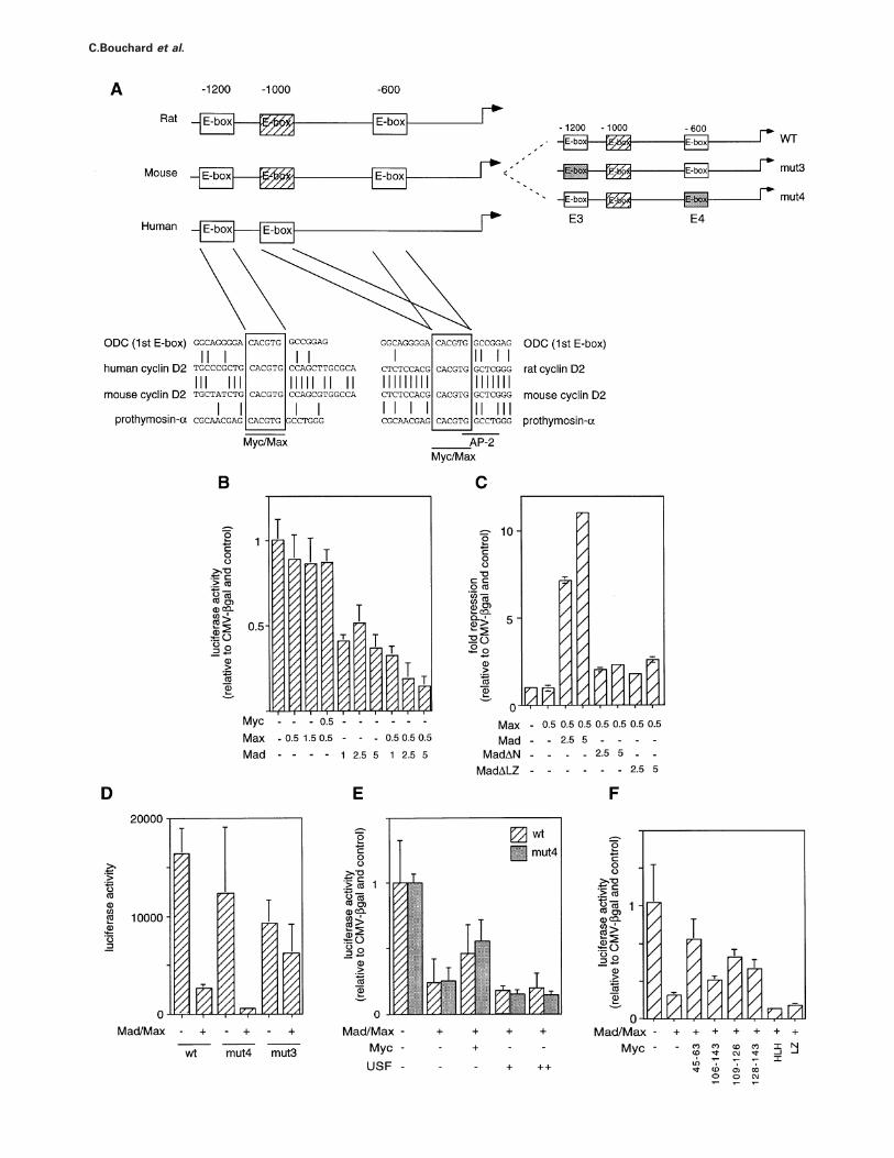
Myc/Max-binding sites (Brookset al., 1996; Junet al.,1997; Yanget al., 1997). One is conserved at –1200 basepairs (bp) relative to the start site of translation in allpromoters and a second is located at –600 bp in therodent promoters and –1000 bp in the human promoter(Figure 4A).
RAT1 cells are not easily amenable to transient transfec-tions. Therefore, transient transfection experiments wereperformed in NIH 3T3 cells to assess whether Myc, Maxor Mad proteins can regulate the murine cyclin D2promoter (Figure 4B–F). Ectopic expression of neitherMax, Myc (data not shown) nor Myc and Max togethersignificantly affected promoter activity (Figure 4B). Incontrast, ectopic expression of Mad-1 repressed thecyclin D2 promoter; repression by Mad-1 was furtherenhanced by co-expression of Max, suggesting that com-plexes of Mad-1 with Max can repress the cyclin D2promoter. Consistent with this view, disruption of theleucine zipper of Mad-1, which is required for dimerizationwith Max, strongly diminished its ability to repress thecyclin D2 promoter (Figure 4C). Efficient repression byMad-1 also required the integrity of an N-terminal domainwhich interacts with the Sin3 protein, suggesting thatrecruitment of Sin3 is required for repression (Ayeret al.,1995; Schreiber-Aguset al., 1995).
In order to determine whether repression by Mad-1requires direct binding to the cyclin D2 promoter, reporterconstructs were generated in which the E-boxes had beenindividually mutated to CGCGTG instead of CACGTGsequences (Figure 4A);in vitro bandshift assays showed
5324
that the mutation severely impaired binding by Maxproteins (data not shown). These experiments revealed thatthe E-boxes were not equivalent (Figure 4D). Disruption ofthe conserved E-box at –1200 (mut3) essentially abolishedrepression by Mad-1–Max complexes. In contrast, disrup-tion of the more proximal E-box (mut4) did not signific-antly affect repression by Mad; we concluded thatrepression of the cyclin D2 promoter by Mad requiresdirect binding of a Mad–Max complex to the more distalE-box element.
While ectopic expression of Myc did not affect promoteractivity by itself, Myc was able to partly revert repressionby Mad-1–Max complexes; in contrast, ectopic expressionof USF, another E-box binding transcription factor(Sawadogoet al., 1988) did not relieve repression byMad-1 (Figure 4E). De-repression by Myc did not dependon the integrity of the promoter-proximal E-box. Deletionof the leucine zipper or the helix–loop–helix domain ofMyc abolished de-repression (Figure 4F). Furthermore, amutant lacking 37 amino acids encompassing Myc-box II(n106–143) poorly de-repressed the cyclin D2 promoter;in vivo, (n106–143) MycER chimeras failed to activatethe endogenous cyclin D2 gene (A.Kiermaier, unpublishedresults). Smaller mutants in the same region gave partialeffects. In contrast, de-repression was normal in mutantslacking Myc-box I (n45–63). This behaviour closelyparallels the ability of these mutants to activate cyclin E–Cdk2 kinase in quiescent cells when stably expressed asER chimeras (Steineret al., 1995). The data also show
Direct induction of cyclin D2 by Myc
Fig. 3. Regulation of cyclin D2 mRNA expression by Myc. Northern blots documenting expression of cyclin D2 and of GAPDH in murinefibroblasts expressing a MycER protein. (A) Time course of cyclin D2 mRNA induction after addition of 200 nM 4-OHT to serum-starved cells.(B) Inhibition of protein synthesis by 50µg/ml cycloheximide as measured by incorporation of [35S]methionine. (C) Induction of cyclin D2 mRNAby Myc is resistant to inhibition of protein synthesis. In all samples, cycloheximide was added to a final concentration of 50µg/ml a few minutesbefore addition of 4-OHT. One time point is shown for addition of cycloheximide alone (2 h); however, a complete time course was performedshowing no induction of cyclin D2 mRNA. (D) Quantitation of cyclin D2 mRNA after addition of 4-OHT in the presence and absence ofcycloheximide (CHX).
that de-repression is not simply due to competition betweenMyc and Mad-1 for access to Max.
The data suggest that activation of cyclin D2 expressionin response to activation of MycER occurs by de-repressionof a Mad–Max-mediated repression of the cyclin D2promoter in quiescent cells. Repression of transcriptionby Mad-1 in vivo depends, at least in part, on therecruitment of histone deacetylase activity to target siteson DNA and can be inhibited by addition of the histonedeacetylase inhibitor, trichostatin A (Lahertyet al., 1997;Sommeret al., 1997).
We therefore tested whether addition of trichostatin Aup-regulates expression of cyclin D2 in quiescent cells.Serum-starved mouse embryo fibroblasts were treated withtrichostatin A and the expression of cyclin D2 wasmonitored. As controls, the extracts were probed forexpression of Cdk4 and cyclins D3, D1 and A (datanot shown). Addition of trichostatin A specifically up-regulated expression of cyclin D2, but not of other cyclinsor Cdk4, in a dose- and time-dependent manner (Figure 5);similar results were obtained in RAT1 cells (data notshown). We concluded that cyclin D2 is repressed in ahistone deacetylase-dependent and Myc-reversible mannerin quiescent cells. Taken together, the data strongly suggesta role for Mad and Myc proteins upstream of cyclin D2in quiescent cells.
Two sets of experiments were performed in order toobtain functional evidence that induction of cyclin D2mediates sequestration of p27 in response to activation ofMyc. First, RAT1-MycER cells were superinfected with
5325
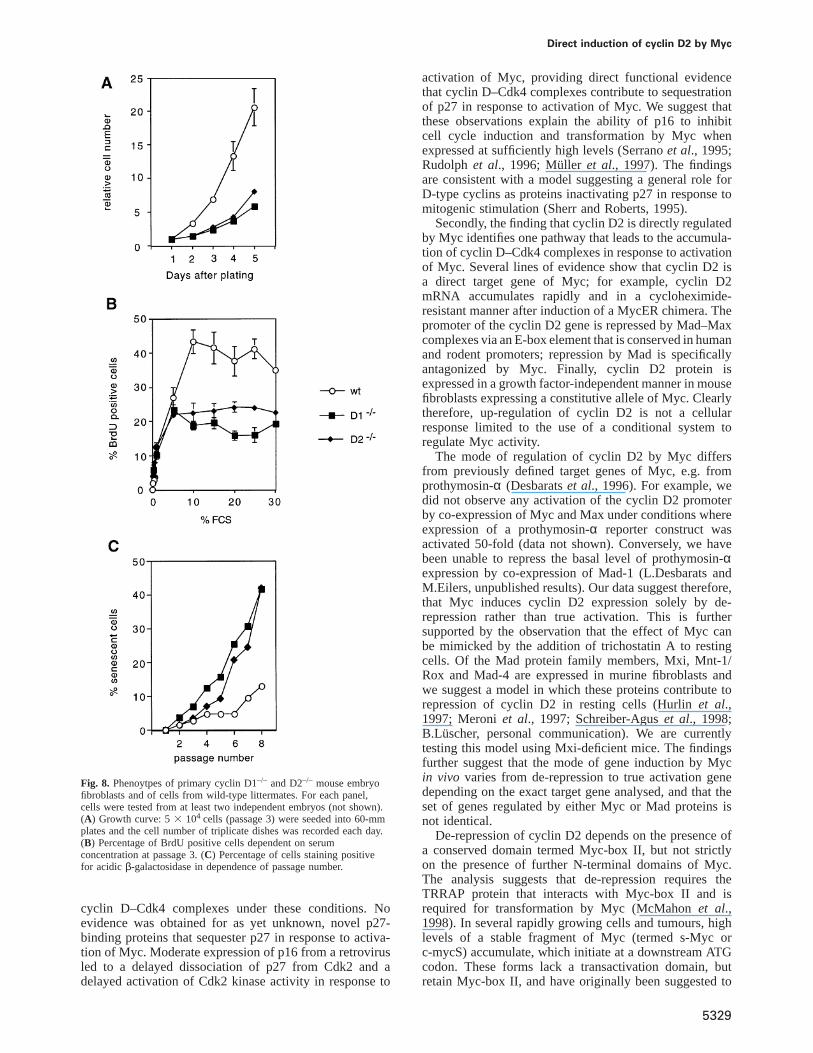
retroviruses encoding p16, as binding of p27 to cyclin D–Cdk4 complexes is blocked by p16 (Reynisdottiret al.,1995). Individual MycER clones expressing p16 wereisolated and propagated; such clones proliferated moreslowly than wild-type cells both in the absence andpresence of hormone, and showed delayed hyperphosphor-ylation of p130 in response to addition of serum (data notshown). Western blotting confirmed that the cells expressedmoderate amounts of p16 (Figure 6, upper panel). Uponinduction of Myc, cyclin D2 was induced with similarkinetics both in control and p16-expressing cells (Figure 6,lower left panel). In control cells, p27 was rapidly lostfrom Cdk2 complexes, as described before. In cellsexpressing p16, Cdk2–p27 complexes were significantlymore stable after induction of Myc (Figure 6, lower middlepanel). Kinase assays revealed that induction of Cdk2kinase was delayed in cells expressing p16 upon activationof Myc (lower right panels). Similar results were obtainedwith a pool of MycER cells expressing p16, demonstratingthat the failure to induce Cdk2 kinase activity was notdue to clonal variation (data not shown). We concludedthat ectopic expression of p16 inhibits Myc-induced disso-ciation of Cdk2–p27 complexes and induction of Cdk2kinase activity.
Secondly, monoclonal antibodies known to ablate thefunction of cyclin D2, but not D1, were affinity-purifiedand microinjected into MycER cells (Lukaset al., 1995);as control, equal amounts of affinity-purified, isotype-matched control antibodies were injected. For these experi-ments, we used a mouse 3T3 cell line stably expressing
C.Bouchard et al.
Direct induction of cyclin D2 by Myc
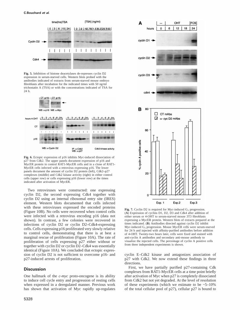
a MycER chimera, as the available antibodies recognizemouse cyclin D2 more easily than rat cyclin D2(Figure 7A). Similarly to RAT1 cells, all three D-typecyclins were induced upon addition of growth factors toserum-starved cells, but only cyclin D2 was induced uponactivation of Myc. Cell cycle progression was monitoredby staining with antibodies directed against cyclin A. Threeindependent experiments were performed (Figure 7B). Ineach experiment, we observed a significant inhibition ofcyclin A induction by Myc when antibodies directedagainst cyclin D2, but not control antibodies, were injected.We concluded that cyclin D2 function is required forMyc to promote G1 progression efficiently in establishedcell lines.
Previous work has shown that Myc is essential for theproliferation of primary mouse fibroblasts; in contrast, thephenotype of cyclin D2-deficient fibroblasts has not beencharacterized (Daviset al., 1993; Sicinskiet al., 1996).Therefore, primary fibroblasts were isolated fromcyclin D2-deficient mice and wild-type littermates; ascontrols, we used primary fibroblasts from cyclin D1–/–
mice. In culture, both cyclin D1–/– and cyclin D2–/– cellsshowed a reduced rate of proliferation relative to wild-type cells (Figure 8A); this phenotype was observed withfibroblasts from different embryos, demonstrating that itwas not due to variation between individual litters (datanot shown). In order to identify the reason for this defectmore precisely, we measured the growth factor-dependenceof DNA replication by determining the percentage of cellsincorporating BrdU dependent on the concentration offetal calf serum in the culture medium (Figure 8B). Inboth cyclin D–/– and wild-type cells, the percentage ofreplicating cells was strictly dependent on the concentra-tion of fetal calf serum; half-maximal incorporation wasobserved at ~2% fetal calf serum in all cell types. However,even at the highest concentration of serum, a significantlylower percentage of cyclin D2–/– and cyclin D1–/– cellsincorporated BrdU relative to wild-type cells. The datasuggest that the lower rate of proliferation of cyclin D–/–
cells is not due to a reduced sensitivity to serum growthfactors.
Primary cells have a finite lifespan in culture andundergo senescence after a number of passages. In orderto assess whether cyclin D2–/– cells undergo prematuresenescence, we determined the percentage of senescentcells at each passage for wild-type and cyclin D–/– cellsby staining for acidicβ-galactosidase, an establishedmarker for senescent cells (Figure 8C) (Dimriet al.,1995). Both cyclin D1–/– and cyclin D2–/– showed amarkedly accelerated start of senescence, suggesting thataccelerated senescence causes their lower rate of prolif-eration.
Ectopic expression of Myc prevents senescence and
Fig. 4. Regulation of the cyclin D2 promoter by Mad and Myc proteins. (A) The left drawing shows the murine, rat and human cyclin D2 promotersindicating the location and conservation of the two consensus E-box elements. The dashed E-box element located at –1000 in the rat and mousepromoter has the core sequence CACATG, which is a low-affinity Myc/Max binding site (Blackwellet al., 1993). On the right, a scheme of themouse promoter indicating which E-boxes are disrupted in the reporter constructs mut3 and mut4. (B) Co-expression of Mad and Max represses themurine cyclin D2 promoter. Results of transient transfection assays in NIH 3T3 with the indicated amounts (µg) of CMV-driven expression plasmidsand a murine cyclin D2 promoter reporter construct. (C) Repression by Mad–Max complexes requires the integrity of the Mad leucine zipper andN-terminal Sin3 interaction domain. MadnLZ refers to a mutant lacking the leucine zipper, MadnN to a mutant lacking the N-terminal Sin3-interaction domain (Sommeret al., 1997). (D) Repression requires the integrity of E-box 3, but not E-box 4 in the murine cyclin D2 promoter.(E) Expression of Myc, but not USF, antagonizes repression of the cyclin D2 promoter by Mad. (F) Domains of Myc required for de-repression ofthe cyclin D2 promoter. The numbers indicate the amino acids deleted in each construct.
5327
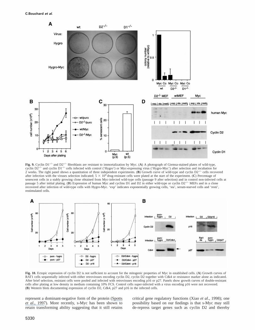
immortalizes primary rodent fibroblasts. In order to testwhether expression of Myc can bypass the acceleratedsenescence observed in cyclin D-deficient cells, weinfected wild-type and cyclin D-deficient cells with eithercontrol viruses or retroviruses expressing human Myc.Controls using a GFP-expressing retrovirus showed thatboth cell types could be infected with comparable effici-ency (data not shown). Resistant cells were selected andthe plates inspected for several weeks. After 10–14 days,individual colonies emerged on wild-type cells infectedwith retroviruses expressing Myc, but few or no colonieswere recovered in any of the other plates (Figure 9A, leftpanel; a quantitation of the results of three independentexperiments is shown on the right). Similarly, plating ofan equal number of resistant cells recovered after infectionshowed that both cyclin D2–/– and cyclin D1–/– cells wereunable to proliferate after infection by Myc (data shownfor cyclin D2–/– cells in Figure 9B). We concluded thatimmortalization of primary cells by Myc requires thepresence of both cyclins D1 and D2. All cell types showedsigns of apoptosis when infected with retroviruses thatexpress both human Myc and H2B-GFP (data not shown),suggesting that the ability of Myc to induce apoptosisdoes not require D-type cyclins.
We established cell lines from colonies emerging fromwild-type cells infected with Myc; these lines were essen-tially immortal, as they could be cultured for manypassages without signs of senescence (Figure 9C). Westernblots using a human specific anti-Myc antibody (9E10;Evan et al., 1985) documented that these cell linesexpressed human Myc protein (Figure 9D). Cells immor-talized by Myc, expressed elevated levels of cyclin D2protein in a growth factor-independent manner, whereasthe expression of cyclin D2 was lower and dependent onthe presence of external growth factors in the primarycells before immortalization. The expression of cyclin D1was identical in primary and Myc-immortalized cells. Thedata show that ectopic expression of Myc is sufficientto up-regulate cyclin D2 expression in primary mousefibroblasts; induction of cyclin D2 is, therefore, not anartificial property of MycER chimeras. Similar observationshave previously been published for established Balb/c-3T3 and RAT1 cells (Jansen-Du¨rr et al., 1993; Vlachet al., 1996).
Finally, we were curious whether up-regulation ofcyclin D2 is sufficient to account for the mitogenicproperties of Myc in established cell lines. To assess this,we used a retroviral infection protocol in which ectopicexpression of Myc has been shown to confer enhancedresistance to the cell cycle inhibitors, p16 and p27 (Vlachet al., 1996; Alevizopouloset al., 1997). Therefore, weasked whether cells ectopically expressing cyclin D2 showenhanced resistance to ectopic expression of p27 and p16.
C.Bouchard et al.
Fig. 5. Inhibition of histone deacetylases de-represses cyclin D2expression in serum-starved cells. Western blots probed with theantibodies indicated of extracts from serum-starved mouse embryofibroblasts after incubation for the indicated times with 50 ng/mltrichostatin A (TSA) or with the concentrations indicated of TSA for24 h.
Fig. 6. Ectopic expression of p16 inhibits Myc-induced dissociation ofp27 from Cdk2. The upper panels document expression of p16 andMycER protein in control RAT1-MycER cells and in a clone of RAT1-MycER cells infected with a retrovirus expressing p16. The lowerpanels document the amount of cyclin D2 protein (left), Cdk2–p27complexes (middle) and Cdk2 kinase activity (right) in either controlcells (upper row) or cells expressing p16 (lower row) at the timesindicated after activation of MycER.
Two retroviruses were constructed: one expressingcyclin D2, the second expressing Cdk4 together withcyclin D2 using an internal ribosomal entry site (IRES)element. Western blots documented that cells infectedwith these retroviruses expressed the encoded proteins(Figure 10B). No cells were recovered when control cellswere infected with a retrovirus encoding p16 (data notshown). In contrast, a few colonies were recovered ininfections of cyclin D2 or cyclin D2–Cdk4-expressingcells. Cells expressing p16 proliferated very slowly relativeto control cells, demonstrating that there is at best amarginal rescue of proliferation (Figure 10A). The rate ofproliferation of cells expressing p27 either without ortogether with cyclin D2 or cyclin D2–Cdk4 was essentiallyidentical (Figure 10A). We concluded that ectopic expres-sion of cyclin D2 is not sufficient to overcome p16- andp27-induced arrests of proliferation.
Discussion
One hallmark of thec-mycproto-oncogene is its abilityto induce cell cycle entry and progression of resting cellswhen expressed in a deregulated manner. Previous workhas shown that activation of Myc rapidly up-regulates
5328
Fig. 7. Cyclin D2 is required for Myc-induced G1 progression.(A) Expression of cyclins D1, D2, D3 and Cdk4 after addition ofeither serum or 4-OHT to serum-starved mouse 3T3 fibroblastsexpressing a MycER protein. Western blots of extracts prepared at thetimes indicated. (B) Antibodies directed against cyclin D2 inhibitMyc-induced G1 progression. Mouse MycER cells were serum-starvedfor 24 h and injected with affinity-purified antibodies before additionof 4-OHT. Twenty-two hours later, cells were fixed and stained withanti-cyclin A antibodies and secondary anti-mouse antibody tovisualize the injected cells. The percentage of cyclin A positive cellsfrom three independent experiments is shown.
cyclin E–Cdk2 kinase and antagonizes association ofp27 with Cdk2. We now extend these findings in threedirections.
First, we have partially purified p27-containing Cdkcomplexes from RAT1-MycER cells at a time point brieflyafter activation of Myc when p27 is completely dissociatedfrom Cdk2 but not yet degraded. At the level of resolutionof these experiments (which we estimate to be ~5–10%of the total cellular pool of p27), cellular p27 is bound to
Direct induction of cyclin D2 by Myc
Fig. 8. Phenoytpes of primary cyclin D1–/– and D2–/– mouse embryofibroblasts and of cells from wild-type littermates. For each panel,cells were tested from at least two independent embryos (not shown).(A) Growth curve: 53 104 cells (passage 3) were seeded into 60-mmplates and the cell number of triplicate dishes was recorded each day.(B) Percentage of BrdU positive cells dependent on serumconcentration at passage 3. (C) Percentage of cells staining positivefor acidic β-galactosidase in dependence of passage number.
cyclin D–Cdk4 complexes under these conditions. Noevidence was obtained for as yet unknown, novel p27-binding proteins that sequester p27 in response to activa-tion of Myc. Moderate expression of p16 from a retrovirusled to a delayed dissociation of p27 from Cdk2 and adelayed activation of Cdk2 kinase activity in response to
5329
activation of Myc, providing direct functional evidencethat cyclin D–Cdk4 complexes contribute to sequestrationof p27 in response to activation of Myc. We suggest thatthese observations explain the ability of p16 to inhibitcell cycle induction and transformation by Myc whenexpressed at sufficiently high levels (Serranoet al., 1995;Rudolphet al., 1996; Muller et al., 1997). The findingsare consistent with a model suggesting a general role forD-type cyclins as proteins inactivating p27 in response tomitogenic stimulation (Sherr and Roberts, 1995).
Secondly, the finding that cyclin D2 is directly regulatedby Myc identifies one pathway that leads to the accumula-tion of cyclin D–Cdk4 complexes in response to activationof Myc. Several lines of evidence show that cyclin D2 isa direct target gene of Myc; for example, cyclin D2mRNA accumulates rapidly and in a cycloheximide-resistant manner after induction of a MycER chimera. Thepromoter of the cyclin D2 gene is repressed by Mad–Maxcomplexes via an E-box element that is conserved in humanand rodent promoters; repression by Mad is specificallyantagonized by Myc. Finally, cyclin D2 protein isexpressed in a growth factor-independent manner in mousefibroblasts expressing a constitutive allele of Myc. Clearlytherefore, up-regulation of cyclin D2 is not a cellularresponse limited to the use of a conditional system toregulate Myc activity.
The mode of regulation of cyclin D2 by Myc differsfrom previously defined target genes of Myc, e.g. fromprothymosin-α (Desbaratset al., 1996). For example, wedid not observe any activation of the cyclin D2 promoterby co-expression of Myc and Max under conditions whereexpression of a prothymosin-α reporter construct wasactivated 50-fold (data not shown). Conversely, we havebeen unable to repress the basal level of prothymosin-αexpression by co-expression of Mad-1 (L.Desbarats andM.Eilers, unpublished results). Our data suggest therefore,that Myc induces cyclin D2 expression solely by de-repression rather than true activation. This is furthersupported by the observation that the effect of Myc canbe mimicked by the addition of trichostatin A to restingcells. Of the Mad protein family members, Mxi, Mnt-1/Rox and Mad-4 are expressed in murine fibroblasts andwe suggest a model in which these proteins contribute torepression of cyclin D2 in resting cells (Hurlinet al.,1997; Meroniet al., 1997; Schreiber-Aguset al., 1998;B.Luscher, personal communication). We are currentlytesting this model using Mxi-deficient mice. The findingsfurther suggest that the mode of gene induction by Mycin vivo varies from de-repression to true activation genedepending on the exact target gene analysed, and that theset of genes regulated by either Myc or Mad proteins isnot identical.
De-repression of cyclin D2 depends on the presence ofa conserved domain termed Myc-box II, but not strictlyon the presence of further N-terminal domains of Myc.The analysis suggests that de-repression requires theTRRAP protein that interacts with Myc-box II and isrequired for transformation by Myc (McMahonet al.,1998). In several rapidly growing cells and tumours, highlevels of a stable fragment of Myc (termed s-Myc orc-mycS) accumulate, which initiate at a downstream ATGcodon. These forms lack a transactivation domain, butretain Myc-box II, and have originally been suggested to
C.Bouchard et al.
Fig. 9. Cyclin D1–/– and D2–/– fibroblasts are resistant to immortalization by Myc. (A) A photograph of Giemsa-stained plates of wild-type,cyclin D2–/– and cyclin D1–/– cells infected with control (‘Hygro’) or Myc-expressing virus (‘Hygro-Myc’) after selection and incubation for2 weeks. The right panel shows a quantitation of three independent experiments. (B) Growth curve of wild-type and cyclin D2–/– cells recoveredafter infection with the viruses selection indicated; 53 104 drug-resistant cells were plated at the start of the experiment. (C) Percentage ofsenescent cells in a stably growing clone obtained from Myc-infected wild-type cells (passage 9 after selection) and in control non-infected cells atpassage 5 after initial plating. (D) Expression of human Myc and cyclins D1 and D2 in either wild-type or cyclin D2–/– MEFs and in a clonerecovered after infection of wild-type cells with Hygro-Myc. ‘exp’ indicates exponentially growing cells, ‘sta’, serum-starved cells and ‘resti’,restimulated cells.
Fig. 10. Ectopic expression of cyclin D2 is not sufficient to account for the mitogenic properties of Myc in established cells. (A) Growth curves ofRAT1 cells sequentially infected with either retroviruses encoding cyclin D2, cyclin D2 together with Cdk4 or resistance marker alone as indicated.After brief selection, resistant cells were pooled and infected with retroviruses encoding p16 or p27. Panels show growth curves of double-resistantcells after plating at low density in medium containing 10% FCS. Control cells super-infected with a virus encoding p16 were not recovered.(B) Western blots documenting expression of cyclin D2, Cdk4, p27 and p16 in the infected cells.
represent a dominant-negative form of the protein (Spottset al., 1997). More recently, s-Myc has been shown toretain transforming ability suggesting that it still retains
5330
critical gene regulatory functions (Xiaoet al., 1998); onepossibility based on our findings is that s-Myc may stillde-repress target genes such as cyclin D2 and thereby
Direct induction of cyclin D2 by Myc
promote proliferation even in the absence of a transactivat-ing domain.
Thirdly, our results indicate that cyclin D2 function isrequired for two gain-of-function phenotypes of Myc.Microinjection of monoclonal antibodies strongly indicatesa role for cyclin D2 in Myc-induced cell cycle entry inan established murine fibroblast cell line. Similarly, ectopicexpression of p16 blocked Myc-induced sequestration ofp27 in RAT1 fibroblasts suggesting a role for cyclin D2in cell cycle entry in response to activation of Myc in thiscell line too.
Using fibroblasts from cyclin D2–/– animals, we foundthat cyclin D2 is required for Myc to immortalize primarymouse embryo fibroblasts. Cyclin D2–/– cells could easilybe infected with retroviruses expressing either H2B–GFP or human Myc together with H2B–GFP; however,cyclin D2–/– failed to proliferate after expression of Myc,and stable cell lines could only be established from wild-type, not from cyclin D2–/– cells after infection withMyc. Cyclin D2–/– primary cells underwent acceleratedsenescence in culture, suggesting that failure to up-regulateCyclin D2 can account, at least in part, for the loss-of-function phenotype of Myc in such cells. It remains to betested whether cyclin D2 is required for tumorigenesis byMyc in vivo; such experiments are under way.
Similarly, antibodies against cyclin D1 ablate cell cycleprogression in RAT1 cells and mouse fibroblasts down-stream of Myc (Rousselet al., 1995; Steineret al., 1995),and cyclin D1–/– fibroblasts fail to be immortalized byMyc. Therefore, parallel pathways may exist that lead toup-regulation of cyclin D1 protein expression by Myc. Thishas been suggested from the finding that the eukaryotictranslation factor, eIF4, is up-regulated by Myc via anE-box in the promoter of the gene, and from the observationthat enhanced levels of eIF-4 may up-regulate translationof D-type cyclins (e.g. Johnstonet al., 1998). Alternatively,induction of cyclin D1 (or cyclin D3) expression by Myc-independent signalling pathways contributes to deregula-tion of cell cycle progression by Myc. This might explainobservations that induction of Myc is not sufficient toinduce proliferation in a number of cell types. For example,activation of both Myc and Ras signalling is requiredfor efficient cell cycle entry of REF52 cells (Leoneet al., 1997).
Loss of cyclin D2 did not significantly affect the abilityof Myc to induce apoptosis in primary mouse fibroblasts,demonstrating that Myc has other functions in these cellsin addition to up-regulation of cyclin D2. At least someof these functions also affect cell proliferation infibroblasts. This can be concluded from a number ofexperiments in which the growth-promoting properties ofMyc and cyclin D proteins have been analysed using avariety of proliferation assays. The results are often verysimilar for both proteins, for example, ectopic expressionof either D-type cyclins or Myc rescues mitogenic signal-ling by a defective CSF-1 receptor and complementstransformation-deficient alleles of bcr-abl (Rousselet al.,1991, 1995; Afaret al., 1994, 1995). Also, cyclin D1mimics the effect of Myc on activation of cyclin E–Cdk2kinase in breast cancer cells (Prallet al., 1998). However,Myc and D-type cyclins do not always act in an identicalfashion; for example, Myc-transformed cells show a par-tial, but not complete resistance against elevated levels of
5331
p27 and p16 (Vlachet al., 1996; Alevizopouloset al.,1997), and ectopic expression of cyclin D2 does notsubstitute for Myc in this function (Figure 10;Alevizopouloset al., 1997). Similarly, ectopic expressionof cyclin D does not rescue the growth defect of fibroblastscarrying a null mutation of c-myc (Mateyaket al., 1999).Clearly therefore, Myc controls cell proliferation and alsop27 metabolism by pathways other than up-regulation ofcyclin D2. Also, accumulation of cyclin D2, in contrastto Myc, can occur in stably arrested cells (Meyyappanet al., 1998).
Finally, loss of cyclin D2 expression does not accountfor the loss-of-function phenotype of Myc during embryo-genesis, as the phenotype of cyclin D2-deficient mice isless severe than that of c-Myc-deficient mice (Daviset al.,1993; Sicinskiet al., 1996). Again, these findings pointto a function of Myc unrelated to its ability to up-regulatecyclin D2.
Materials and methods
Tissue cultureRAT1 cells, primary mouse embryo fibroblasts and NIH 3T3 cells weregrown in DMEM supplemented with 10% heat-inactivated fetal calfserum (FCS), 2 mML-glutamine, 100 U/ml penicillin and 100µg/mlstreptomycin. Culture of RAT1-MycER™ cells has been describedpreviously (Littlewoodet al., 1995). The detailed description of mouseembryo fibroblasts expressing MycER™ proteins will be publishedelsewhere. Staining for senescent cells was carried out as described(Dimri et al., 1995).
Transient transfections into NIH 3T3 cells were performed as describedpreviously for HeLa cells (Desbaratset al., 1996). The expressionvectors used, CMV-Myc, CMV-Max, CMV-Mad, CMV-Mad∆N,CMV-Mad∆LZ and CMV-USF, have been described previously(Desbaratset al., 1996; Sommeret al., 1997). The cyclin D2 reporterconstructs were generated by inserting a 2.3 kb fragment from themurine cyclin D2 promoter into theSacI site of pGL2 (Promega).Mutants were generated using the ExSite kit (Stratagene) and verifiedby sequencing. In each transfection, 10 ng of CMV-lacZ were transfectedto normalize different transfection efficiencies. The total amount of DNAwas kept constant in each transfection by adding equal amounts ofexpression plasmids.
Retroviral supernatants were generated by transient transfections ofBOSC23 and Phoenix cells and used to infect RAT1 cells and primarymouse embryo fibroblasts as published (Pearet al., 1993; Grignaniet al.,1998). Infected cells were selected with 5µg/ml puromycin or 150µg/ml hygromycin as appropriate.
To generate a retrovirus expressing cyclin D2, rat cyclin D2 cDNAwas amplified with the following primers: 59-primer, CGGGAT-CCACCATGGAGCTGCTGTGCTGTGAGG; and 39-primer, CGG-AATTCCTAGAGAGAGAGAGAGAAGGGGCTAGC and cloned intopbabe-puro and -hygro (Morgenstern and Land, 1990). To generate avirus expressing both cyclin D2 and Cdk4, a cDNA encoding humancyclin D2 was fused to an internal ribosomal entry site and cloneddownstream of human Cdk4 into pbabe-puro. Viruses expressing p27and p16 have been described (Mu¨ller et al., 1997). To generate pbabe-H2BGFP, the puromycin gene was excised from pbabe-puro and replacedwith an open reading frame encoding an H2B–GFP chimeric protein(Kandaet al., 1998).
RNA extraction and Northern blottingTotal RNA was isolated with the RNeasy Mini Kit (Qiagen) accordingto the manufacturer’s instructions. Twenty micrograms per lane ofdenaturated RNA were size fractionated in 1% agarose–6% formaldehydegel, hydrolyzed in 50 mM NaOH for 20 min then transferred onto nylonmembranes (Zeta-Probe, Bio-Rad). Blots were submitted to UV light(GS Gene Linker, Bio-Rad) and pre-hybridized in 50% deionizedformamide, 53 standard saline citrate (SSC), 53 Denhardt’s, 50 mMNaPO4 pH 6.5, 0.1% SDS containing 100µg/ml denaturated salmonsperm DNA for 2–8 h at 42°C. Hybridization was performed at 42°Cfor 18 h in 50% deionized formamide, 53 SSC, 13 Denhardt’s, 50 mMNaPO4 pH 6.5, 0.1% SDS, 10% dextran sulfate containing 100µg/ml
C.Bouchard et al.
denaturated salmon sperm DNA and 23 106 c.p.m./ml of32P-labelledmouse cyclin D2 or rat GAPDH cDNAs as probes. Membranes werewashed, dried and then exposed to Biomax MR films (Kodak).
Immunoblotting and immunoprecipitationImmunoblotting, immunoprecipitation and gel filtration conditions havebeen described previously (Steineret al., 1995). For depletion experi-ments, three sequential immunoprecipitations were performed. Theα-D-type cyclins andα-Cdk2 antibodies were chemically cross-linkedto protein A-Sepharose beads using dimethylpimelimidate (Harlow andLane, 1988), since they showed a high rate of leakage from the protein Abeads in the absence of cross-linking (data not shown).
The following antibodies were used. Cyclin D1: DCS-6, DCS-11;cyclin D2: DCS-3, DCS-5 (Lukaset al., 1995), M-20 (Santa Cruz);cyclin D3: DCS-22, DCS-28 (Bartkovaet al., 1998); p27: K25020 (Trans-duction Laboratories), C-19 (Santa Cruz); cyclin A: C-19 (Westernblotting), H-432 (immunofluorescence); cyclin E: M-20; Cdk2: M2;Cdk4: C-22 (all Santa Cruz Biotechnology). Where indicated, linearexposures of the blots were scanned using an Arcus II scanner (Agfa)and quantitated using NIH Image software.
MicroinjectionsMEF-MycER cells were plated at a density of 60% on coverslips in6-well plates. After overnight incubation, cells were serum-starved for2 days in DMEM containing 0.1% FCS. Cells were microinjected intothe nucleus with protein A affinity-purified mouse monoclonal anti-cyclin D2 DCS-3 at a concentration of 3.4 mg/ml in phosphate-bufferedsaline using a Zeiss AIS microinjection system and an Axiovert invertedmicroscope. As control, protein A affinity-purified isotypic control IgG2aantibodies (Ascites, M-7769, Sigma; 8 mg/ml) were injected. Cells wereinduced by the addition of 200 nM of 4-OHT for 22 h, washed oncewith PBS, fixed in methanol/acetone (1:1) for 5 min and then subjectedto indirect immunofluorescence as described previously (Rudolphet al.,1996). Microinjected cells were detected by Cy™3-conjugated anti-mouse secondary antibodies.
Acknowledgements
We thank Eugen Kerkhoff and Tarik Mo¨roy for the gift of cells, BernardLuscher for Mad-expression constructs, Bruno Amati and Hartmut Landfor communication of results prior to publication and Kunio Nakashimafor rat cyclin D2 cDNA and promoter constructs. We also thank themembers of the laboratory for critically reading the manuscript. C.B.acknowledges a long term EMBO fellowship and a stipend from theEuropean Community; M.E. acknowledges support from the DeutscheForschungsgemeinschaft and the Human Frontiers of Science Organ-isation.
References
Afar,D.E.H., Goga,A., McLaughlin,J., Witte,O.N. and Sawyers,C.L.(1994) Differential complementation of Bcr-Abl point mutants withc-Myc. Science, 264, 424–426.
Afar,D.E.H., McLaughlin,J., Sherr,C.J., Witte,O.N. and Roussel,M.F.(1995) Signaling by ABL oncogenes through cyclin D1.Proc. NatlAcad. Sci. USA, 92, 9540–9544.
Alevizopoulos,K., Vlach,J., Hennecke,S. and Amati,B. (1997) Cyclin Eand c-Myc promote cell proliferation in the presence of p16INK4aand of hypophosphorylated retinoblastoma family proteins.EMBO J.,16, 5322–5333.
Alland,L., Muhle,R., Hou,H., Potes,J., Chin,L., Schreiber-Agus,N. andPinho,R.D. (1997) Role for N-CoR and histone deacetylase in Sin3-mediated transcriptional repression. Nature, 387, 49–55.
Ayer,D.E., Kretzner,L. and Eisenman,R.N. (1993) Mad: a heterodimericpartner for Max that antagonizes Myc transcriptional activity. Cell,72, 211–222.
Ayer,D.E., Lawrence,Q.A. and Eisenman,R.N. (1995) Mad-Myctranscriptional repression is mediated by ternary complex formationwith mammalian homologs of yeast repressor Sin3.Cell, 80, 767–776.
Bartkova,J., Lukas,J., Strauss,M. and Bartek,J. (1998) Cyclin D3:requirement for G1/S transition and high abundance in quiescent tissuessuggest a dual role in proliferation and differentiation.Oncogene, 17,1027–1037.
Berns,K., Hijmans,E.M. and Bernards,R. (1997) Repression of c-Mycresponsive genes in cycling cells causes G1 arrest through reductionof cyclin E/CDK2 kinase activity.Oncogene, 15, 1347–1356.
5332
Bouchard,C., Staller,P. and Eilers,M. (1998) Control of cell proliferationby Myc. Trends Cell Biol., 8, 202–206.
Brooks,A.R., Shiffman,D., Chan,C.S., Brooks,E.E. and Milner,P.G.(1996) Functional analysis of the human cyclin D2 and cyclin D3promoters.J. Biol. Chem., 271, 9090–9099.
Davis,A.C., Wims,M., Spotts,G.D., Hann,S.R. and Bradley,A. (1993) Anull c-myc mutation causes lethality before 10.5 days of gestation inhomozygotes and reduced fertility in heterozygous female mice.GenesDev., 7, 671–682.
Desbarats,L., Gaubatz,S. and Eilers,M. (1996) Discrimination betweendifferent E-box binding proteins at an endogenous target gene of Myc.Genes Dev., 10, 447–460.
Dimri,G.P. et al. (1995) A biomarker that identifies senescent humancells in culture and in aging skinin vivo. Proc. Natl Acad. Sci. USA,92, 9363–9367.
Draetta,G., Luca,F., Westendorf,J., Brizuela,L., Ruderman,J. andBeach,D. (1989) Cdc2 protein kinase is complexed with both cyclinA and B: evidence for proteolytic inactivation of MPF.Cell, 56,829–838.
Eilers,M., Picard,D., Yamamoto,K. and Bishop,J.M. (1989) Chimaerasbetween the MYC oncoprotein and steroid receptors cause hormone-dependent transformation of cells. Nature, 340, 66–68.
Eilers,M., Schirm,S. and Bishop,J.M. (1991) The MYC protein activatestranscription of theα-prothymosin gene.EMBO J., 10, 133–141.
Evan,G.I., Lewis,G.K., Ramsay,G. and Bishop,J.M. (1985) Isolation ofmonoclonal antibodies specific for human c-myc proto-oncogeneproduct.Mol. Cell. Biol., 5, 3610–3616.
Evan,G.I., Wyllie,A.H., Gilbert,C.S., Littlewood,T.D., Land,H.,Brooks,M., Waters,C.M., Penn,L.Z. and Hancock,D.C. (1992)Induction of apoptosis in fibroblasts by c-myc protein.Cell, 69,119–128.
Grignani,F., Kinsella,T., Mencarelli,A., Valtieri,M., Riganelli,D.,Lanfrancone,L., Peschle,C., Nolan,G.P. and Pelicci,P.G. (1998) High-efficiency gene transfer and selection of human hematopoieticprogenitor cells with a hybrid EBV/retroviral vector expressing thegreen fluorescence protein.Cancer Res., 58, 14–19.
Harlow,E. and Lane,D. (1988)Antibodies: A Laboratory Manual. ColdSpring Harbor Laboratory Press, Cold Spring Harbor, NY.
Heinzel,T. et al. (1997) A complex containing N-CoR, mSin3 andhistone deacetylase mediates transcriptional repression.Nature, 387,43–48.
Henriksson,M. and Lu¨scher, B. (1996) Proteins of the Myc network:essential regulators of cell growth and differentiation.Adv. CancerRes., 68, 109–182.
Hermeking,H., Funk,J.O., Reichert,M., Ellwart,J.W. and Eick,D. (1995)Abrogation of p53-induced cell cycle arrest by c-Myc: evidence foran inhibitor of p21WAF1/CIP1/SDI1.Oncogene, 11, 1409–1415.
Hurlin,P.J., Queva,C., Koskinen,P.J., Steingrimsson,E., Ayer,D.E.,Copeland,N.G., Jenkins,N.A. and Eisenman,R.N. (1996) Mad3 andMad4: novel Max-interacting transcriptional repressors that suppressc-myc dependent transformation and are expressed during neural andepidermal differentiation.EMBO J., 15, 2030–2038.
Hurlin,P.J., Queva,C. and Eisenman,R.N. (1997) Mnt, a novel Max-interacting protein is coexpressed with Myc in proliferating cells andmediates repression at Myc binding sites.Genes Dev., 11, 44–58.
Jansen-Du¨rr,P., Meichle,A., Steiner,P., Pagano,M., Finke,K., Botz,J.,Wessbecher,J., Draetta,G. and Eilers,M. (1993) Differential modulationof cyclin gene expression by MYC.Proc. Natl Acad. Sci. USA, 90,3685–3689.
Johnston,K.A., Polymenis,M., Wang,S., Branda,J. and Schmidt,E.V.(1998) Novel regulatory factors interacting with the promoter of thegene encoding the mRNA cap binding protein (eIF4E) and theirfunction in growth regulation.Mol. Cell. Biol., 18, 5621–5633.
Jun,D.Y., Kim,M.K., Kim,I.G. and Kim,Y.H. (1997) Characterization ofthe murine cyclin D2 gene: exon/intron organization and promoteractivity. Mol. Cell, 7, 537–543.
Kanda,T., Sullivan,K.F. and Wahl,G.M. (1998) Histone–GFP fusionprotein enables sensitive analysis of chromosome dynamics in livingmammalian cells.Curr. Biol., 8, 377–385.
Laherty,C.D., Yang,W.-M., Sun,J.-M., Davie,J.R., Seto,E. andEisenman,R.N. (1997) Histone deacetylases associated with themSin3 corepressor mediate mad transcriptional repression. Cell, 89,349–356.
Leone,G., DeGregori,J., Sears,R., Jakoi,L. and Nevins,J.R. (1997) Mycand Ras collaborate in inducing accumulation of active cyclin E/Cdk2and E2F.Nature, 387, 422–426.
Direct induction of cyclin D2 by Myc
Littlewood,T.D., Hancock,D.C., Danielian,P.S., Parker,M.G. andEvan,G.I. (1995) A modified oestrogen receptor ligand binding domainas an improved switch for the regulation of heterologous proteins.Nucleic Acids Res., 23, 1686–1690.
Lukas,J., Bartkova,J., Welcker,M., Petersen,O.W., Peters,G., Strauss,M.and Bartek,J. (1995) Cyclin D2 is a moderately oscillatingnucleoprotein required for G1 phase progression in specific cell types.Oncogene, 10, 2125–2134.
Mateyak,M.K., Obaya,A.J. and Sedivy,J.M. (1999) c-Myc regulatescyclin D-cdk4 and -cdk6 activity but affects cell cycle progression atmultiple independent points.Mol. Cell. Biol., 19, 4672–4683.
McMahon,S.B., Van Buskirk,H.A., Dugan,K.A., Copeland,T.D. andCole,M.D. (1998) The novel ATM-related protein TRRAP is anessential cofactor for the c-Myc and E2F oncoproteins. Cell, 94,363–374.
Meroni,G. et al. (1997) Rox, a novel bHLHZip protein expressed inquiescent cells that heterodimerizes with Max, binds a non-canonicalE box and acts as a transcriptional repressor.EMBO J., 16, 2892–2906.
Meyyappan,M., Wong,H., Hull,C. and Riabowol,K.T. (1998) Increasedexpression of cyclin D2 during multiple states of growth arrest inprimary and established cells.Mol. Cell. Biol., 18, 3163–3172.
Morgenstern,J.P. and Land,H. (1990) Advanced mammalian genetransfer: high titre retroviral vectors with multiple drug selectionmarkers and a complementary helper-free packaging cell line.NucleicAcids Res., 18, 3587–3596.
Muller,D., Bouchard,C., Rudolph,B., Steiner,P., Stuckmann,I.,Saffrich,R., Ansorge,W., Huttner,W. and Eilers,M. (1997) Cdk2-dependent phosphorylation of p27 facilitates its Myc-induced releasefrom cyclin E/cdk2 complexes.Oncogene, 15, 2561–2576.
Pear,W.S., Nolan,G.P., Scott,M.L. and Baltimore,D. (1993) Productionof high-titer helper-free retroviruses by transient transfection.Proc.Natl Acad. Sci. USA, 90, 8392–8396.
Polyak,K., Lee,M.-H., Erdjument-Bromage,H., Koff,A., Roberts,J.M.,Tempst,P. and Massague,J. (1994) Cloning of p27Kip1, a cyclin-dependent kinase inhibitor and a potential mediator of extracellularantimitogenic signals. Cell, 78, 59–66.
Prall,O.W.J., Rogan,E.M., Musgrove,E.A., Watts,C.K.W. andSutherland,R.L. (1998) c-Myc or cyclin D1 mimics estrogen effectson cyclin E-cdk2 activation and cell cycle reentry.Mol. Cell. Biol.,18, 4499–4508.
Pusch,O., Bernaschek,G., Eilers,M. and Hengstschlager,M. (1997)Activation of c-Myc uncouples DNA replication from activation ofG1-cyclin-dependent kinases.Oncogene, 15, 649–656.
Reynisdottir,I., Polyak,K., Iavarone,A. and Massague,J. (1995) Kip/Cipand Ink4 Cdk inhibitors cooperate to induce cell cycle arrest inresponse to TGF-β. Genes Dev., 9, 1831–1845.
Roussel,M.F., Cleveland,J.L., Shurtleff,S.A. and Sherr,C.J. (1991) Mycrescue of a mutant CSF-1 receptor impaired in mitogenic signalling.Nature, 353, 361–363.
Roussel,M.F., Theodoras,A.M., Pagano,M. and Sherr,C.J. (1995) Rescueof defective mitogenic signaling by D-type cyclins.Proc. Natl Acad.Sci. USA, 92, 6837–6841.
Roussel,M.F., Ashmun,R.A., Sherr,C.J., Eisenman,R.N. and Ayer,D.E.(1996) Inhibition of cell proliferation by the Mad1 transcriptionalrepressor.Mol. Cell. Biol., 16, 2796–2801.
Rudolph,B., Zwicker,J., Saffrich,R., Henglein,B., Mu¨ller,R., Ansorge,W.and Eilers,M. (1996) Activation of cyclin dependent kinases by Mycmediates transcriptional activation of cyclin A, but not apoptosis.EMBO J., 15, 3065–3076.
Sawadogo,M., Van Dyke,M.W., Gregor,P.D. and Roeder,R.G. (1988)Multiple forms of the human gene-specific transcription factor USF.I. Complete purification and identification of USF from HeLa cellnuclei.J. Biol. Chem., 263, 11985–11993.
Schreiber-Agus,N., Chin,L., Chen,K., Torres,R., Rao,G., Guida,P.,Skoultchi,A.I. and DePinho,R.A. (1995) An amino-terminal domainof Mxi1 mediates anti-Myc oncogenic activity and interacts with ahomolog of the yeast transcriptional repressor SIN3.Cell, 80, 777–786.
Schreiber-Agus,N., Meng,Y., Hoang,T., Hou,H.,Jr, Chen,K.,Greenberg,R., Cordon-Cardo,C., Lee,H.W. and DePinho,R.A. (1998)Role of Mxi1 in ageing organ systems and the regulation of normaland neoplastic growth. Nature, 393, 483–487.
Serrano,M., Gomez Lahoz,E., DePinho,R.A., Beach,D. and Bar Sagi,D.(1995) Inhibition of ras-induced proliferation and cellulartransformation by p16INK4.Science, 267, 249–252.
Sherr,C.J. and Roberts,J.M. (1995) Inhibitors of mammalian G1 cyclin-dependent kinases.Genes Dev., 9, 1149–1163.
5333
Sicinski,P.et al. (1996) Cyclin D2 is an FSH-responsive gene involvedin gonadal cell proliferation and oncogenesis. Nature, 384, 470–474.
Sommer,A., Hilfenhaus,S., Menkel,A., Kremmer,E., Seiser,C., Loidl,P.and Luscher,B. (1997) Cell growth inhibiiton by the Mad/Max complexthrough recruitment of histone deacetylase activity.Curr. Biol., 7,357–365.
Spotts,G.D., Patel,S.V., Xiao,Q. and Hann,S.R. (1997) Identification ofdownstream-initiated c-Myc proteins which are dominant-negativeinhibitors of transactivation by full-length c-Myc proteins.Mol. Cell.Biol., 17, 1459–1468.
Steiner,P., Philipp,A., Lukas,J., Godden-Kent,D., Pagano,M.,Mittnacht,S., Bartek,J. and Eilers,M. (1995) Identification of a Myc-dependent step during the formation of active G1 cyclin/cdkcomplexes.EMBO J., 14, 4814–4826.
Toyoshima,H. and Hunter,T. (1994) p27, a novel inhibitor of G1 cyclin-Cdk protein kinase activity, is related to p21.Cell, 78, 67–74.
Vlach,J., Hennecke,S., Alevizopoulos,K., Conti,D. and Amati,B. (1996)Growth arrest by the cyclin-dependent kinase inhibitor p27Kip1 isabrogated by c-Myc.EMBO J., 15, 6595–6604.
Wagner,A.J., Kokontis,J.M. and Hay,N. (1994) Myc-mediated apoptosisrequires wild-type p53 in a manner independent of cell cycle arrestand the ability of p53 to induce p21waf1/cip1. Genes Dev., 8, 2817–2830.
Xiao,Q., Claassen,G., Shi,J., Adachi,S., Sedivy,J. and Hann,S.R. (1998)Transactivation-defective c-MycS retains the ability to regulateproliferation and apoptosis.Genes Dev., 12, 3803–3808.
Yang,M., Hosokawa,Y., Hu,Y., Kaneko,S., Kaneko,H., Tanaka,M. andNakashima,K. (1997) Cloning and functional analysis of rat cyclin D2promoter: multiple prolactin-responsive elements.Biochem. Mol. Biol.Int., 43, 749–754.
Zervos,J., Gyuris,A.S. and Brent,R. (1993) Mxi1, a protein thatspecifically interacts with Max to bind Myc-Max recognition sites.Cell, 72, 223–232.
Received March 4, 1999; revised and accepted July 23, 1999