c-myc Regulates Cell Proliferation during Lens Development
12
c-myc Regulates Cell Proliferation during Lens Development Gabriel R. Cavalheiro . , Gabriel E. Matos-Rodrigues . , Anielle L. Gomes, Paulo M. G. Rodrigues, Rodrigo A. P. Martins* Programa de Biologia Celular e do Desenvolvimento, Instituto de Cie ˆ ncias Biome ´ dicas, Universidade Federal do Rio de Janeiro, Rio de Janeiro, Rio de Janeiro, Brazil Abstract Myc protooncogenes play important roles in the regulation of cell proliferation, growth, differentiation and survival during development. In various developing organs, c-myc has been shown to control the expression of cell cycle regulators and its misregulated expression is detected in many human tumors. Here, we show that c-myc gene (Myc) is highly expressed in developing mouse lens. Targeted deletion of c-myc gene from head surface ectoderm dramatically impaired ocular organogenesis, resulting in severe microphtalmia, defective anterior segment development, formation of a lens stalk and/or aphakia. In particular, lenses lacking c-myc presented thinner epithelial cell layer and growth impairment that was detectable soon after its inactivation. Defective development of c-myc-null lens was not caused by increased cell death of lens progenitor cells. Instead, c-myc loss reduced cell proliferation, what was associated with an ectopic expression of Prox1 and p27 Kip1 proteins within epithelial cells. Interestingly, a sharp decrease in the expression of the forkhead box transcription factor Foxe3 was also observed following c-myc inactivation. These data represent the first description of the physiological roles played by a Myc family member in mouse lens development. Our findings support the conclusion that c- myc regulates the proliferation of lens epithelial cells in vivo and may, directly or indirectly, modulate the expression of classical cell cycle regulators in developing mouse lens. Citation: Cavalheiro GR, Matos-Rodrigues GE, Gomes AL, Rodrigues PMG, Martins RAP (2014) c-myc Regulates Cell Proliferation during Lens Development. PLoS ONE 9(2): e87182. doi:10.1371/journal.pone.0087182 Editor: Amit Singh, University of Dayton, United States of America Received April 19, 2013; Accepted December 20, 2013; Published February 4, 2014 Copyright: ß 2014 Cavalheiro et al. This is an open-access article distributed under the terms of the Creative Commons Attribution License, which permits unrestricted use, distribution, and reproduction in any medium, provided the original author and source are credited. Funding: International Brain Research Organization (IBRO); International Retinal Research Foundation (IRRF); Fundac ¸a ˜o Carlos Chagas Filho de Amparo a ` Pesquisa do Estado do Rio de Janeiro (FAPERJ); Conselho Nacional de Desenvolvimento Cientı ´fico e Tecnolo ´ gico (CNPq). GRC, ALG and PMRG received undergrad student fellowships from the PIBIC program (CNPq/UFRJ) or from FAPERJ. The funders had no role in study design, data collection and analysis, decision to publish, or preparation of the manuscript. Competing Interests: The authors have declared that no competing interests exist. * E-mail: [email protected] . These authors contributed equally to this work. Introduction Complex developmental processes must be carefully orches- trated for the correct formation of the vertebrate eye. Even though optic-cup morphogenesis was recently reproduced in vitro by the use of three-dimensional stem cell culture systems [1], the molecular mechanisms driving eye organogenesis in vivo are still a major question in developmental biology. Concomitant with the coordinated growth of the developing lens and retina, the formation of a functionally mature eye depends on the precise coordination of cell proliferation, cell cycle exit and cell differentiation within these structures. In the past several years, a lot has been learned about the mechanisms that regulate these events, including the cell-extrinsic cues, such as growth factors, and cell-intrinsic factors, including cell cycle proteins and transcriptional regulators. Importantly, several homeodomain- containing proteins that act as transcription factors were identified and characterized as regulators of cell proliferation and cell differentiation in the developing lens. In contrast, few studies described the roles of basic-helix-loop-helix (bHLH) transcription factors in lens development [2–5]. Some recent studies addressed how these transcriptional networks functionally interact in vivo to regulate cell proliferation during lens ontogenesis [6,7]. The refined architecture and well characterized stages of the developing vertebrate lens makes it an excellent model to study how these basic cellular processes are regulated in coordination. During embryonic development of the mouse, the contact between the optic vesicle and the head surface ectoderm occurs around embryonic day 9 (E9) and triggers the thickening and invagination of the ectoderm, forming the lens vesicle. Then, cells located on the posterior end of the vesicle exit cell cycle and elongate to terminally differentiate into primary lens fiber cells, while anterior cells continue proliferating and form the lens epithelia. At later stages, cell proliferation continues in the germinative zone from where cells migrate towards lens equator, exit cell cycle and start the secondary differentiation process. Through adulthood, epithe- lial cells exit the cell cycle and differentiate into fiber cells at the equatorial region of the lens. The adult lens is composed of post mitotic terminally differentiated fiber cells and a layer of cuboidal epithelial cells in its anterior region. Some of these epithelial cells remain as proliferative stem/progenitor cells [8]. Proper lens organogenesis requires the maintenance of the epithelial identity and cell proliferation by lens epithelial cells in precise coordination with cell cycle exit and cell differentiation. Many secreted growth factors, such as FGF, BMP and others [9–12], are known regulators of these events. PLOS ONE | www.plosone.org 1 February 2014 | Volume 9 | Issue 2 | e87182
Transcript of c-myc Regulates Cell Proliferation during Lens Development
c-myc Regulates Cell Proliferation during LensDevelopmentGabriel R. Cavalheiro., Gabriel E. Matos-Rodrigues., Anielle L. Gomes, Paulo M. G. Rodrigues,
Rodrigo A. P. Martins*
Programa de Biologia Celular e do Desenvolvimento, Instituto de Ciencias Biomedicas, Universidade Federal do Rio de Janeiro, Rio de Janeiro, Rio de Janeiro, Brazil
Abstract
Myc protooncogenes play important roles in the regulation of cell proliferation, growth, differentiation and survival duringdevelopment. In various developing organs, c-myc has been shown to control the expression of cell cycle regulators and itsmisregulated expression is detected in many human tumors. Here, we show that c-myc gene (Myc) is highly expressed indeveloping mouse lens. Targeted deletion of c-myc gene from head surface ectoderm dramatically impaired ocularorganogenesis, resulting in severe microphtalmia, defective anterior segment development, formation of a lens stalk and/oraphakia. In particular, lenses lacking c-myc presented thinner epithelial cell layer and growth impairment that wasdetectable soon after its inactivation. Defective development of c-myc-null lens was not caused by increased cell death oflens progenitor cells. Instead, c-myc loss reduced cell proliferation, what was associated with an ectopic expression of Prox1and p27Kip1 proteins within epithelial cells. Interestingly, a sharp decrease in the expression of the forkhead boxtranscription factor Foxe3 was also observed following c-myc inactivation. These data represent the first description of thephysiological roles played by a Myc family member in mouse lens development. Our findings support the conclusion that c-myc regulates the proliferation of lens epithelial cells in vivo and may, directly or indirectly, modulate the expression ofclassical cell cycle regulators in developing mouse lens.
Citation: Cavalheiro GR, Matos-Rodrigues GE, Gomes AL, Rodrigues PMG, Martins RAP (2014) c-myc Regulates Cell Proliferation during Lens Development. PLoSONE 9(2): e87182. doi:10.1371/journal.pone.0087182
Editor: Amit Singh, University of Dayton, United States of America
Received April 19, 2013; Accepted December 20, 2013; Published February 4, 2014
Copyright: � 2014 Cavalheiro et al. This is an open-access article distributed under the terms of the Creative Commons Attribution License, which permitsunrestricted use, distribution, and reproduction in any medium, provided the original author and source are credited.
Funding: International Brain Research Organization (IBRO); International Retinal Research Foundation (IRRF); Fundacao Carlos Chagas Filho de Amparo aPesquisa do Estado do Rio de Janeiro (FAPERJ); Conselho Nacional de Desenvolvimento Cientıfico e Tecnologico (CNPq). GRC, ALG and PMRG received undergradstudent fellowships from the PIBIC program (CNPq/UFRJ) or from FAPERJ. The funders had no role in study design, data collection and analysis, decision topublish, or preparation of the manuscript.
Competing Interests: The authors have declared that no competing interests exist.
* E-mail: [email protected]
. These authors contributed equally to this work.
Introduction
Complex developmental processes must be carefully orches-
trated for the correct formation of the vertebrate eye. Even
though optic-cup morphogenesis was recently reproduced in vitro
by the use of three-dimensional stem cell culture systems [1], the
molecular mechanisms driving eye organogenesis in vivo are still a
major question in developmental biology. Concomitant with the
coordinated growth of the developing lens and retina, the
formation of a functionally mature eye depends on the precise
coordination of cell proliferation, cell cycle exit and cell
differentiation within these structures. In the past several years,
a lot has been learned about the mechanisms that regulate these
events, including the cell-extrinsic cues, such as growth factors,
and cell-intrinsic factors, including cell cycle proteins and
transcriptional regulators. Importantly, several homeodomain-
containing proteins that act as transcription factors were
identified and characterized as regulators of cell proliferation
and cell differentiation in the developing lens. In contrast, few
studies described the roles of basic-helix-loop-helix (bHLH)
transcription factors in lens development [2–5]. Some recent
studies addressed how these transcriptional networks functionally
interact in vivo to regulate cell proliferation during lens
ontogenesis [6,7].
The refined architecture and well characterized stages of the
developing vertebrate lens makes it an excellent model to study
how these basic cellular processes are regulated in coordination.
During embryonic development of the mouse, the contact between
the optic vesicle and the head surface ectoderm occurs around
embryonic day 9 (E9) and triggers the thickening and invagination
of the ectoderm, forming the lens vesicle. Then, cells located on
the posterior end of the vesicle exit cell cycle and elongate to
terminally differentiate into primary lens fiber cells, while anterior
cells continue proliferating and form the lens epithelia. At later
stages, cell proliferation continues in the germinative zone from
where cells migrate towards lens equator, exit cell cycle and start
the secondary differentiation process. Through adulthood, epithe-
lial cells exit the cell cycle and differentiate into fiber cells at the
equatorial region of the lens. The adult lens is composed of post
mitotic terminally differentiated fiber cells and a layer of cuboidal
epithelial cells in its anterior region. Some of these epithelial cells
remain as proliferative stem/progenitor cells [8]. Proper lens
organogenesis requires the maintenance of the epithelial identity
and cell proliferation by lens epithelial cells in precise coordination
with cell cycle exit and cell differentiation. Many secreted growth
factors, such as FGF, BMP and others [9–12], are known
regulators of these events.
PLOS ONE | www.plosone.org 1 February 2014 | Volume 9 | Issue 2 | e87182

The molecular mechanisms driving cell proliferation in
developing lens have been extensively studied [13]. Proper cell
cycle exit and terminal differentiation of fiber cells critically
depends on the Rb pathway. In Rb-null lens, cells in the transition
zone fail to exit cell cycle [14]. A similar phenotype of
hyperproliferation, followed by apoptotic cell death, was observed
when CDK inhibitors p27Kip1 and p57Kip2 were both inactivated,
suggesting that these CKIs functionally cooperate as upstream
regulators of Rb pathway during lens terminal differentiation.
These CDK inhibitors p27Kip1 and p57Kip2 are expressed in the
fibers and their upregulation in the context of cell cycle exit
depends on the transcription factor Prox1 (prospero-related
homeobox 1) [15]. Regulation of cell cycle exit by Prox1 was
previously shown in several developing tissues and in cancer [16–
18] and evidence that Prox1 may regulate p27Kip1 transcription by
directly binding to its promoter was also observed [19]. Little is
known about how Prox1 expression is regulated during lens
development [17–18], specially how its expression gets restricted to
and maintained in early differentiating cells following terminal
differentiation [20–22].
Previous studies have described that Myc transcription factors
are expressed in developing lens of various vertebrates [23,24].
The Myc family of proto-oncogenes includes c-myc (Myc), N-myc
(Mycn) and L-myc (Myc1) that encode transcription factors
containing basic-helix-loop-helix leucine zipper (bHLHZ) motifs
and are known to regulate gene expression through a variety of
mechanisms, including transcriptional activation through the
formation of a heterodimer with Max as well as Max-independent
mechanisms of c-myc-mediated transcriptional repression [25–28].
Myc proto-oncogenes have been shown to regulate cell survival,
size, differentiation and specially cell proliferation in several
developing organs, explaining why these transcription factors are
absolutely crucial for life during development [27]. While N-myc
plays a role in eye development by regulating retinal progenitor
cells proliferation [29], previous studies, based in overexpression
approaches, suggested that other Myc transcription factors may
play a role in lens development [23,30]. For instance, Ishibashi
and colleagues reported the enlargement of the ocular globe as a
result of c-myc overexpression driven by the Mx-promoter.
However, the reported findings were either the result of
overexpression of truncated c-myc in differentiating fiber cells
[23] or overexpression was driven by promoters that increased c-
myc expression in the cornea, iris, lens, and retina [30]. These
nonspecific gain-of-function approaches made it impossible to
clearly determine the tissue-specific roles of c-myc in eye
development.
In the present study, we investigated whether c-myc regulates
lens development in vivo. c-myc knockout mice die in uterus [31],
so to determine the roles of c-myc in developing lens, we analyzed
eyes and lenses of Le-Cre; c-mycflox/flox (c-mycLe-Cre) mice [32,33]. The
Le-Cre transgene is expressed in the surface ectoderm leading to
inactivation of targeted alleles in both the lens and in the corneal
epithelium [33]. Inactivation of c-myc by Le-Cre resulted in severe
eye and lens growth impairment and anterior chamber malfor-
mation. In the absence of c-myc, no increase in cell death was
detected and even though crystallin expression was normal,
degeneration of fiber cells was observed at postnatal ages. For the
first time we provide evidence that a sharp decrease in cell
proliferation occurred after inactivation of c-myc. Consistently,
ectopic expression of cell cycle exit proteins p27Kip1 and Prox1 was
observed within epithelial cells and gene expression of Foxe3 and
p27Kip1 was misregulated in c-myc-deficient lens.
Materials and Methods
MiceExperimental procedures with animals were approved by the
Committee of Ethics in Animal Use (CEUA) of the Health
Science Center (CCS) based on the currently accepted interna-
tional rules.
The c-myc floxed [32] (Myctm2Fwa, MGI id:2178233) and the
Lens-Cre [33] (Tg(Pax6-cre,GFP)1Pgr, MGI id:3045749) mice
were previously generated and kindly shared. The control group
(c-mycCtrl) was composed of c-myc+/+, c-myc+/F and c-mycF/F. Mice
with homozygous inactivation of c-myc specifically in the lens were
identified as c-mycLe-Cre = c-mycF/F; Le-Cre+/2 and mice with
heterozygosis of c-myc in the lens were identified as c-mycHet =
c-myc+/F; Le-Cre+/2. To ensure that the offspring would inherit
only one copy of the Cre transgene, Cre-positive animals
(Le-Cre+/2; c-mycF/F) were always mated to Cre-negative animals
(Le-Cre2/2; c-mycF/F or Le-Cre2/2; c-myc+/F).
RNA extraction, cDNA synthesis, and real-time RT-PCRanalysis
Dissected lenses were obtained from staged embryonic (E12.5,
E14.5, E17.5) and postnatal (P0, P3, P11, adult) C57BL/6 mice.
RNA extraction and cDNA synthesis were performed as
previously described [29,34]. Real time RT-PCR reactions were
performed in an ABI7500 machine (Applied Biosystems) using
TaqManH probes synthesized with 59-FAM and 39-BHQ for c-
myc (Myc), Foxe3, p27Kip1 (Cdkn1b), actin (Actb) and GAPDH
(Gapd). Primers used were listed in Table S1. Data analysis and
normalization were performed as previously described [29,34].
Volume measurements of the eye and lensThe volume of postnatal (P0, P15) and adult (P30) eyes were
measured as previously described [29]. After enucleation, eyes
were fixed in phosphate-buffered saline (PBS)-buffered parafor-
maldehyde 4%. The axial length and two coronal axes (dorso-
ventral and medial-lateral) of each eye were measured with a
digital paquimeter and the eye volume was calculated after
applying the formula (4/36PI) 6 (eye axial length in mm) 6 (eye
coronal length in mm) 6 (eye dorsal-ventral length in mm). After
dissection of the retina and the lens, the same procedure was used
to calculate lens volume.
Histology and H&E stainingEmbryos or eyes were collected in 4% PBS-buffered parafor-
maldehyde for 24 hours at 4uC and cryoprotected in 10, 20 and
30% (overnight) PBS-buffered sucrose solutions, respectively.
Sections of 10 mm were obtained using a Leica 1850 cryostat.
Hematoxilin (Proquimios) and Eosin (Merck) staining followed
standard protocol.
ImmunohistochemistryAn antigen retrieval with citrate buffer was performed prior the
antibodies incubation. The following antibodies were used: anti-
PCNA (1:500, Santa Cruz Biotechnology, cat#: sc-56), anti-Ser10
pH3 (1:50, Cell Signaling, cat#: 9701), anti-active caspase-3
(1:100, BD Biosciences, cat#: 559565), anti-Prox1 (1:500,
Covance, cat#: PR-238C), anti-p27Kip1 (1:50, BD Biosciences,
cat#: 610241), anti-cyclin D1 (1:50, Cell Signaling, cat#: 2926),
anti-aH2AX (1:300, Millipore, cat# 05-636), anti-E-cadherin
(1:100, Cell Signaling, cat#3195), anti-phopho-Erk1/2 (thr202/
tyr204) (1:500, Cell Signaling cat#: 4370). The a-crystallin (1:50),
a-crystallin (1:300) and a-crystallin (1:50) antibodies were obtained
c-myc Drives Cell Proliferation in Developing Lens
PLOS ONE | www.plosone.org 2 February 2014 | Volume 9 | Issue 2 | e87182

from Dr. J. Samuel Zigler (Wilmer Eye Institute). The Foxe3
antibody (1:150) was a gift from Dr. Peter Carlsson.
Immunohistochemistry reactions were developed with biotini-
lated secondary antibody (1:400, Vector labs, cat#: BA2000 or
BA1000) followed by ABC complex (Vector labs, cat#: PK6100)
and DAB substrate kit (Vector labs, cat#: SK4100). Nuclear
staining with methylgreen (Sigma Aldrich, cat#: 323829).
Immunofluorescence reactions were developed by 2 alternative
methods: biotinilated secondary antibody followed by ABC
complex and Cy3-tyramide kit (Perkin Elmer, cat#: FP1046) or
an Alexa 488 secondary antibody (1:500, Life, cat#: A11001).
Fluorescent nuclear counter staining were performed with Sytox
Green (1/15000, Invitrogen, cat#: S7020) or with DAPI (Lonza,
cat#: PA3013), respectively.
TUNEL analysis was performed following manufacturer
instructions (Promega, cat#: G7362). Images were captured with
a Leica TCS-SP5 with an AOBS system.
Western blotProtein extraction and blotting procedures were performed as
previously reported [34]. Primary antibodies were as follows: c-
myc (1:1000, Cell Signaling, cat#: 5605), a-tubulin (1:10000,
Santa Cruz, cat #: sc32293). HRP-conjugated secondary
antibodies from Cell Signaling (anti-mouse IgG, cat #: 7076,
anti-rabbit IgG, cat #: 7074) were used at a 1:1000 dilution. The
ECL system (cat #: RPN2132) was used according to the
manufacturer’s instructions.
Statistical analysist-test, one or two-way ANOVA were performed as indicated in
the figure legends. p-values are based on two-sided tests. Tests
were performed using Graphpad Prism software.
Results
Inactivation of c-myc in the surface ectoderm severelyimpairs lens and eye growth
Previous studies described the expression of some Myc family
transcription factors, including c-myc, in developing lens of
different species [23,24,35]. To determine whether the relative
amount of c-myc gene (Myc) expression would vary during mouse
developing lens, we initially performed real time RT-PCR using c-
myc-specific primers previously characterized [28] in various
stages of lens development. c-myc mRNA expression was found as
early as embryonic day 12.5 (E12.5), the earliest stage analyzed.
Interestingly, c-myc gene expression sharply decreased thereafter
remaining ,8 to 100-fold smaller at all the older developmental
stages analyzed (E14.5, E17.5, P0, P3, P11) (Figure 1A). We also
measured c-myc protein expression in developing lens. In
agreement with the gene expression data, western blot analysis
of protein extracts from E15.5 and E18.5 lens showed that the
amount of c-myc protein decreases during embryonic lens
development (Figure 1B).
To analyze whether c-myc plays a role in mouse lens
development, we generated c-mycLe-Cre (c-mycF/F; Le-Cre+/2) mice
in which c-myc was inactivated in the surface ectoderm. The Le-
Cre transgenic mice present Cre recombinase activity in the lens
placode as early as E9.5 [33]. Loss of c-myc protein in c-mycLe-Cre
lenses was confirmed by western blot (Figure 1B). First, we asked
whether inactivation of c-myc in the mouse lens would affect lens
growth. At postnatal day 30 (P30), we observed a reduction of
approximately 80% in lens volume (Figure 1C, D, E). The lens of
c-mycLe-Cre mice were significantly smaller than c-mycCtrl ones
(21.0160.42 mm3 vs. 4.5861.44 mm3; p,0, 0001) (Figure 1F).
In one of the c-mycLe-Cre mice no lens was formed at all, a
phenotype known as aphakia. In the majority of c-mycLe-Cre mice we
observed either a mild (up to 50% reduction in lens volume) or an
aggressive lens volume reduction (reduction of more than 50% in
lens volume) (Table 1). The severe lens growth defect observed did
not allow us to perform precise measurements of the c-myc-
deficient lenses at earlier stages of development.
Previous studies have demonstrated that eye growth during
development depends on the correct development of the lens
[9,36]. As shown in figure 1 and in table 1, the impairment of
lens development following c-myc inactivation also affected the
growth of the whole eye. The eyes of c-mycLe-Cre mice were
approximately 65% smaller during postnatal development (P15)
and adulthood (P30) (Figure 1G). Notably, the eyes of mice with
heterozygous lens (c-mycHet) were smaller then wild types, but
significantly different from the homozygous (c-mycLe-Cre) P30 eyes
(Figure S1). These findings suggest that the amount of c-myc
protein within lens cells may be of functional relevance, since the
phenotype observed may be correlated with c-myc content.
Additionally, we observed that, at birth (P0), the eyes of c-mycLe-Cre
mice were already smaller than the eyes of control littermates,
suggesting that c-myc function is required during embryonic lens
development (Figure 1G).
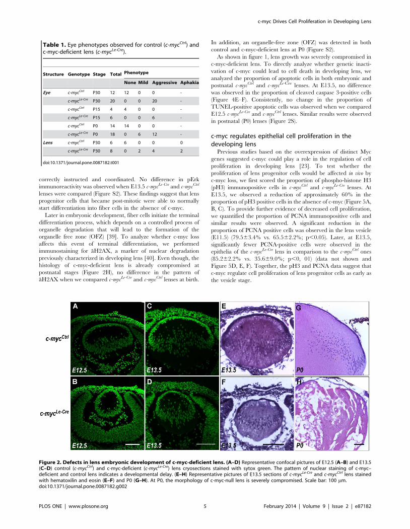
Defective embryogenesis of the lens and anteriorchamber
To characterize how c-myc-deficiency affects embryonic
development of the lens, we first compared the morphology of
c-mycLe-Cre and c-mycCtrl lenses. At E12.5, the distribution of cell
nuclei in control lens was characteristic of primary fibers cells
that elongated from the posterior region of the lens vesicle. In
contrast, c-myc-deficient lens displayed a vesicle-like morphology
(Figure 2A–B). To determine whether the observed phenotype
resulted from a developmental delay or from malformation we
analyzed lens morphology at later stages. At E13.5, c-mycLe-Cre
lens presented a distribution of cell nuclei (Figure 2C–D) and
overall morphology (Figure 2E–F) characteristic of primary fibers
cells elongating from posterior region of vesicle. At P0, a highly
vacuolized fiber mass was observed in the lens of c-mycLe-Cre mice
(Figure 2G–H). Altogether, these findings suggest that, in the
absence of c-myc, a slight developmental delay occurs and that
initial steps of fiber cell differentiation were not affected.
To evaluate whether the c-myc-deficiency affected earlier stages
of lens development (e.g. lens vesicle formation), we performed
H&E staining in sections of E11 eyes and counted the number of
cells in the lens at this stage of development. No difference
between c-mycCtrl (62.6765.510; n = 3) and c-mycLe-Cre
(61.7567.364; n = 4) was observed. However, a connection
between the lens epithelium and the cornea (lens stalk) was
observed in all animals analyzed at this stage (E11) and in more
than 50% of the animals analyzed at E13.5 (Figure 3 A, B, C, D
and Table 2).
In addition, we observed that inactivation of c-myc in the
surface ectoderm led to severe defects in the development of the
anterior segment of c-mycLe-Cre mice. At P0, defects included
corneal stroma loosening and absence of corneal endothelium.
Furthermore, at P30, the c-mycLe-Cre mice presented thinner corneal
epithelia. Other anterior segment structures were also affected. As
shown in figure 3 (Figure 3E, F, G, H, I, J), it was not possible to
distinguish the stroma of the ciliary body, the stroma of the iris or
the chamber angle. In addition, we observed pigmented cells along
the anterior segment (arrowheads in Figure 3).
c-myc Drives Cell Proliferation in Developing Lens
PLOS ONE | www.plosone.org 3 February 2014 | Volume 9 | Issue 2 | e87182

Inactivation of c-myc does not affect cell survival or earlysteps of lens fiber differentiation
To test whether c-myc deficiency would lead to defects in cell
differentiation during embryonic lens development, we analyzed
classical aspects of fiber cells differentiation. First, we analyzed the
pattern of a-, a- and a-crystallins expression in c-myc-null lens,
given the fact that their expression is a hallmark of appropriate
fiber cell differentiation [9,36]. At E13.5, no difference in the
expression pattern of a- crystallin, a- crystallin or a-crystallin was
observed between c-mycCtrl and c-mycLe-Cre (Figure 4A, B, C, D, and
data not shown).
Several studies have demonstrated that signaling pathways
activated by cell-extrinsic growth factors, such as FGF, lead to
phosphorylation of ERK (pERK) and trigger fiber cell differen-
tiation [37,38]. Therefore, the pattern of Erk phosphorylation in
embryonic lens may indicate whether fiber cells differentiation was
Figure 1. c-myc is highly expressed in developing mouse lens and genetic inactivation of c-myc in the surface ectoderm severelyimpairs lens and eye growth. (A) Real-time RT-PCR analysis of c-myc mRNA (Myc) expression at 6 stages of mouse lens development. Real-timeRT-PCR data was obtained using TaqMan probes and datasets were normalized to actin (Actb). Normalized relative expression shows that Mycexpression is highest in earliest stage analyzed (E12.5) and sharply decrease as development proceeds. (B) Western blot analysis of c-myc proteinExpression (C–E) Representative pictures of c-mycLe-Cre and c-mycCtrl mice (P60) (C), eyes (P15) (D) and lens (P30) at the indicated ages (E). (F)Measurement of lens volume at P30 shows that inactivation of c-myc in developing lens leads to a severe reduction of the lens volume (c-mycLe-Cre;n = 8; c-mycCtrl; n = 6). (G) Inactivation of c-myc in the developing lens dramatically impairs eye growth as observed by the reduction of eye volumealready at birth (P0; c-mycLe-Cre; n = 18; c-mycCtrl; n = 14). Eye growth impairment was observed throughout postnatal development (P15; c-mycLe-Cre;n = 6; c-mycCtrl; n = 4) and adulthood (P30; c-mycLe-Cre; n = 20; c-mycCtrl; n = 12). A t-test was performed for (F) and a two-way ANOVA test wasperformed for (G). Error bars indicate SEM; *** p,0, 0001.doi:10.1371/journal.pone.0087182.g001
c-myc Drives Cell Proliferation in Developing Lens
PLOS ONE | www.plosone.org 4 February 2014 | Volume 9 | Issue 2 | e87182
correctly instructed and coordinated. No difference in pErk
immunoreactivity was observed when E13.5 c-mycLe-Cre and c-mycCtrl
lenses were compared (Figure S2). These findings suggest that lens
progenitor cells that became post-mitotic were able to normally
start differentiation into fiber cells in the absence of c-myc.
Later in embryonic development, fiber cells initiate the terminal
differentiation process, which depends on a controlled process of
organelle degradation that will lead to the formation of the
organelle free zone (OFZ) [39]. To analyze whether c-myc loss
affects this event of terminal differentiation, we performed
immunostaining for aH2AX, a marker of nuclear degradation
previously characterized in developing lens [40]. Even though, the
histology of c-myc-deficient lens is already compromised at
postnatal stages (Figure 2H), no difference in the pattern of
aH2AX when we compared c-mycLe-Cre and c-mycCtrl lenses at birth.
In addition, an organelle-free zone (OFZ) was detected in both
control and c-myc-deficient lens at P0 (Figure S2).
As shown in figure 1, lens growth was severely compromised in
c-myc-deficient lens. To directly analyze whether genetic inacti-
vation of c-myc could lead to cell death in developing lens, we
analyzed the proportion of apoptotic cells in both embryonic and
postnatal c-mycCtrl and c-mycLe-Cre lenses. At E13.5, no difference
was observed in the proportion of cleaved caspase 3-positive cells
(Figure 4E–F). Consistently, no change in the proportion of
TUNEL-positive apoptotic cells was observed when we compared
E12.5 c-mycLe-Cre and c-mycCtrl lenses. Similar results were observed
in postnatal (P0) lenses (Figure 2S).
c-myc regulates epithelial cell proliferation in thedeveloping lens
Previous studies based on the overexpression of distinct Myc
genes suggested c-myc could play a role in the regulation of cell
proliferation in developing lens [23]. To test whether the
proliferation of lens progenitor cells would be affected in vivo by
c-myc loss, we first scored the proportion of phospho-histone H3
(pH3) immunopositive cells in c-mycCtrl and c-mycLe-Cre lenses. At
E13.5, we observed a reduction of approximately 60% in the
proportion of pH3 positive cells in the absence of c-myc (Figure 5A,
B, C). To provide further evidence of decreased cell proliferation,
we quantified the proportion of PCNA immunopositive cells and
similar results were observed. A significant reduction in the
proportion of PCNA positive cells was observed in the lens vesicle
(E11.5) (79.563.4% vs. 65.562.2%; p,0.05). Later, at E13.5,
significantly fewer PCNA-positive cells were observed in the
epithelia of the c-mycLe-Cre lens in comparison to the c-mycCtrl ones
(85.262.2% vs. 35.669.0%; p,0, 01) (data not shown and
Figure 5D, E, F). Together, the pH3 and PCNA data suggest that
c-myc regulate cell proliferation of lens progenitor cells as early as
the vesicle stage.
Table 1. Eye phenotypes observed for control (c-mycCtrl) andc-myc-deficient lens (c-mycLe-Cre).
Structure Genotype Stage Total Phenotype
None Mild Aggressive Aphakia
Eye c-mycCtrl P30 12 12 0 0 -
c-mycLe-Cre P30 20 0 0 20 -
c-mycCtrl P15 4 4 0 0 -
c-mycLe-Cre P15 6 0 0 6 -
c-mycCtrl P0 14 14 0 0 -
c-mycLe-Cre P0 18 0 6 12 -
Lens c-mycCtrl P30 6 6 0 0 0
c-mycLe-Cre P30 8 0 2 4 2
doi:10.1371/journal.pone.0087182.t001
Figure 2. Defects in lens embryonic development of c-myc-deficient lens. (A–D) Representative confocal pictures of E12.5 (A–B) and E13.5(C–D) control (c-mycCtrl) and c-myc-deficient (c-mycLe-Cre) lens cryosections stained with sytox green. The pattern of nuclear staining of c-myc–deficient and control lens indicates a developmental delay. (E–H) Representative pictures of E13.5 sections of c-mycLe-Cre and c-mycCtrl lens stainedwith hematoxilin and eosin (E–F) and P0 (G–H). At P0, the morphology of c-myc-null lens is severely compromised. Scale bar: 100 mm.doi:10.1371/journal.pone.0087182.g002
c-myc Drives Cell Proliferation in Developing Lens
PLOS ONE | www.plosone.org 5 February 2014 | Volume 9 | Issue 2 | e87182

Misregulated expression of Prox1 and p27Kip1 proteins inc-myc-deficient lens
Appropriate timing of cell cycle exit is essential for proper cell
differentiation during lens embryogenesis [13]. The reduction in
cell proliferation following c-myc loss led us to test whether known
regulators of lens cell cycle exit, such as Prox1 and p27Kip1 [15,41]
would be misregulated in the lens of c-mycLe-Cre mice. It’s well
characterized that the behavior of proliferating cells depends on
the region of the epithelia occupied by these lens progenitors.
Therefore, after staining sections of E13.5 c-mycCtrl and c-mycLe-Cre
lenses for p27Kip1 and Prox1, we performed a careful regionalized
quantification of the immunopositive cells [42] to characterize
whether alterations in the expression of these proteins would be
specific for different zones of the lens epithelia (Figure 6A–B). As
demonstrated in figures 6C, D, E, in the absence of c-myc, the
proportion of p27Kip1–positive cells significantly increased in the
prospective germinative zone (GZ), prospective transition zone
(TZ) and in the central epithelia (CE) of the c-mycLe-Cre lenses
(Figure 6E). Similar analysis was performed for Prox1 expression
(Figure 6F, G, H). As observed for p27Kip1, the total proportion of
Prox1-positive cells significantly increased in c-myc-deficient lens.
Interestingly, the regions of Prox1 upregulation were slightly
different from p27Kip1, since more Prox1-positive cells were
detected in the prospective GZ and the prospective TZ, but not in
the CE (Figure 6F).
Toprovide furtherevidence thatc-mycinactivationcausedectopic
expression of p27Kip1 protein within lens epithelial cells, we
performed a double immunostaining for E-cadherin, a classical
marker of lens epithelial cells, and p27Kip1 in c-mycCtrl and c-mycLe-Cre
cryosections (Figure 7A, B, C, D, E, F, G, H, I). As observed in
figures 7F and 7I, in the c-mycLe-Cre mice, E-cadherin positive cells
located in the central epithelium were also expressing p27Kip1
protein. We quantified the proportion of double positive cells and
verified that the proportion of epithelial cells expressing p27Kip1
protein increased ,6 fold in c-myc-deficient lens. To quantify the
mRNA expression of p27Kip1, we performed realtime RT-PCR in
E17.5 lenses. A subtle increase (,1.7 fold) in p27Kip1 gene expression
was observed in c-myc-null lenses (Figure 7K).
The observed phenotypes: (1) ectopic expression of cycle exit
regulators within epithelial cells and (2) defective morphogenesis of
the anterior chamber have some similarities with the ones
observed in Foxe3-null mice [43]. To test whether the expression
of Foxe3 would be misregulated in c-myc-deficient lens, we
performed immunofluorescence to Foxe3. Interestingly, the
expression of Foxe3 protein in the anterior epithelia of E13.5 c-
mycLe-Cre mice was drastically reduced (Figure 8A–B). In addition,
realtime RT-PCR analysis of E17.5 lens also demonstrated a
decrease in Foxe3 gene expression (Figure 8C), suggesting that c-
myc may regulate the expression of Foxe3 in developing lens.
Altogether, our findings indicate, for the first time, that in vivo
inactivation of c-myc in the surface ectoderm leads to defective
development of the anterior segment and the lens, resulting in
severe microphtalmia. We propose that misregulation of cell
proliferation in developing lens contributes to the described
phenotypes.
Discussion
In this study, we found several lines of evidence that the proto-
oncogene c-myc is required for proper development of the lens in
vivo. First, we performed gene expression studies to confirm that
this member of the bHLHZ transcription factor family is expressed
during embryonic and postnatal development of the mouse lens.
Importantly, we show that the amount of c-myc (Myc) transcripts
sharply decreases during lens embryonic development and remain
at lower levels from E17.5 through adulthood. Using genetic
assays, we inactivated c-myc expression in a tissue-specific manner
and demonstrated that the loss of c-myc, starting at the surface
Figure 3. Loss of c-myc leads to development defects in theanterior chamber. Representative pictures of hematoxylin & eosin(H&E) staining at E11.0 (A, B) and immunofluorescence for E-cadherin atE13.5 (C, D) illustrates the defective lens vesicle formation, which leadsto the formation of the lens stalk. (E–J) H&E staining of anterior segmentat P0 (E–H) and P30 (I–J) demonstrates that c-mycLe-Cre eyes presentedcorneal stroma loosening, absence of corneal endothelium andpresence of pigmented cells along the anterior chamber (at P0 andP30, arrowheads in F, H and J). Scale bar: 100 mm.doi:10.1371/journal.pone.0087182.g003
Table 2. Phenotypes of control (c-mycCtrl) and c-myc-deficient embryonic lens (c-mycLe-Cre).
Structure Genotype Stage Total Aphakia Lens Stalk
Lens c-mycCtrl E13.5 12 0 0
c-mycLe-Cre E13.5 9 0 5
c-mycCtrl E11.0 5 0 0
c-mycLe-Cre E11.0 4 0 4
doi:10.1371/journal.pone.0087182.t002
c-myc Drives Cell Proliferation in Developing Lens
PLOS ONE | www.plosone.org 6 February 2014 | Volume 9 | Issue 2 | e87182

ectoderm stage, severely impairs lens and eye organogenesis.
Consistent with previous studies that used alternative genetic
approaches to alter c-myc expression in developing lens [23], we
found no evidence for a role of c-myc in the regulation of cell
survival in developing mouse lens. Even though we did not observe
alterations in the expression pattern of crystallins and phosphor-
ylated Erk during embryonic development or aH2AX at early
postnatal stages, degeneration of fiber cells in c-myc-deficient lens
was observed at later stages of development. More importantly, we
found that, in the absence of c-myc, cell proliferation was greatly
reduced during embryonic development of the lens. In addition,
we provided some evidences of the mechanisms of cell cycle
control by c-myc in developing lens. Prox1 and p27Kip1 proteins
are ectopically expressed in lens epithelial cells and a slight
increase in p27Kip1 mRNA expression was observed in c-myc-null
lens. Based on these findings, we propose that c-myc plays an
important role in lens development through the regulation of the
cell cycle in the lens progenitor cells. These data led us to propose
the hypothetical model in which c-myc negatively regulates the
expression of Prox1 and p27Kip1 in lens epithelial cells preventing
lens progenitor cell cycle exit (Figure 8D).
Regulation of cell proliferation, but not cell death, in lensdevelopment
Germ line inactivation of c-myc made it clear that c-myc was
essential to life, because homozygous mice did not survive beyond
Figure 4. Inactivation of c-myc does not affect cell survival or the expression of crystallins. Representative confocal pictures of theimmunofluorescence for a- crystallin (A–B) or a-crystallin (C–D) performed in control (c-mycCtrl) and c-myc-deficient (c-mycLe-Cre) lens sections atE13.5. Sytox nuclear counterstaining is shown in green. Immunostaining patterns are indistinguishable between c-myc–deficient and control lens. (E–F) Representative pictures of cleaved caspase-3 staining of control (E) and c-myc–deficient (F) lens at E13.5. c-myc loss did not lead to misregulationof apoptosis during embryonic lens development. Scale bar: 100 mm.doi:10.1371/journal.pone.0087182.g004
c-myc Drives Cell Proliferation in Developing Lens
PLOS ONE | www.plosone.org 7 February 2014 | Volume 9 | Issue 2 | e87182

E10.5 days of gestation [31]. In this study, no detailed description
about the defects in eye development was provided. Even though it
would have been possible to detect specific abnormalities in the
formation of the lens vesicle, it was only briefly mentioned that
optic development scored poorly in c-myc homozygous mice. In
this context, our work contributes as the first example of a loss-of-
function approach that clearly demonstrates a physiological role of
a Myc family member in eye development.
Our data provide genetic evidence that c-myc is required for
lens and eye development. As demonstrated in Figure 1 and
Figure S1, eye and lens growth were severely compromised in c-
mycLe-Cre mice. Notably, the reduction in adult eye volume was
smaller for mice with one functional copy of c-myc. This
observation that c-myc heterozygosis resulted in an intermediary
phenotype suggests that the amount of this Myc protein may be of
relevance for c-myc-mediated functions in developing lens, as well
as undermines the possibility of a toxic role of Cre recombinase in
the generation of the observed lens phenotypes. Importantly, eye
volume was significantly smaller at birth, suggesting that c-myc
function is required early in lens embryogenesis. Developmental
defects in c-myc-deficient lens were observed as soon as E11.5
(Figure 3), approximately, 2 days after Cre mediated recombina-
tion is detected in the surface ectoderm [33]. The vesicle
morphology of E12.5 c-myc-deficient lens showed that primary
fiber cell differentiation was not yet initiated at this stage. Genetic
inactivation of c-Maf proto-oncogene [44,45] arrests lens devel-
opment at the vesicle stage. To distinguish between such a severe
malformation or a developmental delay in embryonic develop-
ment, we analyzed the morphology of E13.5 lens and found
evidence that primary fiber cell differentiation was normally
initiated in the absence of c-myc (Figure 2). These findings
suggested that the impaired organogenesis of c-mycLe-Cre eye and
lens and eye were not caused by a complete impairment of lens
vesicle formation or defects in fiber cell differentiation.
Inactivation of c-myc in embryonic lens tissue reduced the
proportion of classical proliferation markers. The proportion of
mitotic cells, stained for the phosphorylated form of histone H3
(pH3) was reduced in 60% (Figure 5). Since the mitotic cells are
inevitably the smallest population of proliferating cells, we
expanded the characterization of cell proliferation in c-myc-null
lens analyzing another marker of this event (PCNA). Consistently,
a ,2.5-fold reduction in the proportion of PCNA-positive cells
was detected within the lens tissue at E13.5. A similar, but less
pronounced, decrease in the proportion of PCNA positive cells
was detected at the lens vesicle stage (E11.5). These results suggest
that the loss of c-myc function resulted in misregulation of the cell
cycle of lens progenitor cells few days after its genetic inactivation.
Figure 5. c-myc regulates lens epithelial cells proliferation. Immunohistochemistry for pH3 (A–B) and PCNA (D–E) were performed in control(c-mycCtrl) and c-myc-deficient (c-mycLe-Cre) lens sections at E13.5 followed by methylgreen nuclear counterstaining. Proportions of pH3immunopositive cells (arrows) (C) and PCNA immunopositive cells (F) were scored within lens epithelial cells (c-mycLe-Cre; n = 5; c-mycCtrl; n = 4).Inactivation of c-myc reduced the proportion of proliferating cells within lens tissue in vivo. Error bars indicate SEM. A t-test resulted in * p,0, 05;** p,0, 01.doi:10.1371/journal.pone.0087182.g005
c-myc Drives Cell Proliferation in Developing Lens
PLOS ONE | www.plosone.org 8 February 2014 | Volume 9 | Issue 2 | e87182

In accordance with our findings, overexpression of c-myc in
(driven by aA crystallin promoter) induced lens cells to enter the
S-phase of the cell cycle [23]. Since, forced expression of c-myc
resulted in cell cycle progression, it was suggested that c-myc
may be sufficient to induce cells to re-enter cell cycle. Our work,
add to the previous findings by showing that lens progenitor cells
are able to proliferate in the absence of c-myc. It remains to be
determined whether redundant or compensatory expression of
another Myc gene in any of the proliferative cell populations of
the lens is related with the maintenance of proliferation after c-
myc loss. The possibility that c-myc expression and function is
heterogeneous within subpopulations of lens progenitor may not
be discarded.
Altogether, these findings strongly suggest that the impairment
of lens and eye development here described is mainly caused by
misregulation of cell proliferation following c-myc loss in
developing lens.
As mentioned, c-myc inactivation in developing lens severely
impaired eye and lens development (Figures 1 and 2). It has
become clear that the decision of a cell to undergo apoptosis and
the participation of c-myc in this process are specific for the cell
type and biological context [46]. Therefore, we analyzed whether
an increase in cell death could contribute to the phenotype
discovered. The lack of c-myc protein in developing lens did not
affected cell death in any of the stages studied, as verified by both
TUNEL assay and staining for activated caspase 3 during
embryonic and postnatal development (Figure 4 and Figure S2).
It’s still not determined whether c-myc-deficiency may lead to cell
death at later stages of lens postnatal development (after birth).
Consistent with our observations that c-myc genetic inactivation
did not altered cell death in developing lens, when c-myc was
overexpressed no increase in apoptosis was reported [23].
Even though, we favor the hypothesis that c-myc does not
regulate cell death in developing lens, a role for c-myc in cell death
during early steps of lens embryogenesis may not be completely
discarded, since other Myc family members could compensate for
c-myc loss.
Molecular mechanisms regulated by c-myc in developinglens
In developing lens, Prox1 protein is first detected around E10.5
in cells of the anterior and posterior compartments of the lens
vesicle. Following primary differentiation, at E12.5, Prox1 levels
are still high in the nucleus of elongating fiber cells and decrease in
lens epithelium. Afterwards, Prox1 protein expression becomes
restricted to early differentiating cells [22]. To our knowledge c-
myc has not been previously shown to regulate Prox1 expression.
Ectopic expression of Prox1 was demonstrated in Foxe3-null
lens [43]. As shown in figure 8, we observed that the expression of
Foxe3 protein is downregulated in c-myc-deficient lens epithelia.
This effect is likely caused by a decrease in Foxe3 gene expression,
since Foxe3 mRNA content was also reduced in c-mycLe-Cre lens
(Figure 8). Therefore, it is reasonable that the ectopic expression of
Prox1 we observed in c-myc-deficient lens is a consequence of
Foxe3 downregulation. Alternatively, Prox1 transcription may
directly or indirectly regulated by c-myc in a Foxe3-independent
manner. It has been shown that the Myc-associated zinc finger
protein (MAZ) may regulate the gene expression of Prox1 in
hepatocellular carcinoma [47]. In addition, Pitx3 has been shown
to play a role in the cell cycle of lens epithelial cells and fiber cell
differentiation by positively regulating Foxe3 expression and
negatively regulating Prox1 in the anterior lens epithelium. These
effects culminated in prevention of p27Kip1 and p57Kip2 activation
and maintenance of lens epithelial cells in cell cycle [20].
Figure 6. Misregulation of p27Kip1 and Prox1 proteins in c-myc-deficient lens. (A–B) Schematic illustration of the division of the lens cells in4 regions that were independently quantified in stained sections: central epithelia (CE), prospective germinative zone (pGZ), prospective transitionzone (pTZ) and lens fibers (LF). (C–D) Immunofluorescence for p27Kip1 and (F–G) immunohistochemistry for Prox1 were performed in control (c-mycCtrl) and c-myc-deficient (c-mycLe-Cre) lens sections at E13.5 followed by nuclear either counterstaining with DAPI (C–D) or methylgreen (F–G),respectively. Proportions of p27Kip1 immunopositive cells (white arrows) and Prox1 immunopositive cells (black arrows) were scored in each of theregions CE, GZ, TZ and LF (c-mycLe-Cre; n = 3; c-mycCtrl; n = 4). Inactivation of c-myc increased the proportion of cells expressing p27Kip1 and Prox1within lens tissue in vivo. Error bars indicate SEM; * p,0, 05; ** p,0, 01; *** p,0, 001. Scale bar: 100 mm.doi:10.1371/journal.pone.0087182.g006
c-myc Drives Cell Proliferation in Developing Lens
PLOS ONE | www.plosone.org 9 February 2014 | Volume 9 | Issue 2 | e87182

Therefore, it is possible that the regulation of p27Kip1 and/or
Prox1 expression by c-myc depends on interactions with Foxe3
(Figure 8D) or Pitx3.
Binding of c-myc to initiator (Inr) elements can abolish Miz-1-
mediated transcriptional activation. Repression of p27Kip1 expres-
sion following c-myc binding to Inr elements located in p27Kip1
promoter has been previously demonstrated [48]. It’s known,
however, that Prox1 regulates p27Kip1 in the lens [15], so it is
possible that regulation of p27Kip1 expression by c-myc may be
indirectly mediated by Prox1. Interestingly, our data supports
previous findings [11,22] that Prox1 and p27Kip1 are not always
expressed in the same cells at the same developmental stage.
Therefore, the c-myc-Prox1-p27Kip1 axis proposed in Figure 8D
may not be found in every differentiating lens cell. Future studies
are necessary to better determine the transcriptional network
regulated by c-myc in developing lens.
c-myc and anterior segment morphogenesisThe Le-Cre transgene leads to genetic inactivation in the
surface ectoderm, that will give rise to the lens, and in the ocular
surface epithelia (corneal, conjunctival and eyelid epithelia)
[33,57]. In addition to the lens phenotypes described above, we
observed several defects in other structures of the anterior
chamber following inactivation of c-myc from the surface
ectoderm. In addition, we observed a remnant connection
between the lens and the surface ectoderm – usually referred as
lens stalk – as early as E11.5. Published studies report the
degeneration of this connection between developing lens and
cornea at slightly different stages. While some have described the
presence of lens stalk in wild-type mice as late as E12–E12.5
[5,49,50], several other studies reported that this structure is
already absent at E11.5 [51–53]. In our hands, no lens stalk was
observed in control mice at ,E11, but, in c-mycLe-Cre mice, the lens
stalk was observed in all animals we analyzed (n = 4, Table 2). Few
days later, (E13.5), more than 50% of the c-myc-deficient lens had
remnant connection. These findings indicate that c-myc is
necessary for proper lens vesicle separation from the surface
ectoderm.
Anterior segment dysgenesis has been described in several
human diseases (OMIM 107250), but relatively few transcription
factors were shown to be critical for the development of the
anterior segment of the eye in both human and mice [54]. Here,
we reported that c-myc function is necessary for proper anterior
segment morphogenesis. Around birth, a reduction in corneal
Figure 7. c-myc loss leads to ectopic expression of p27kip1 in the lens anterior epithelium. Representative confocal pictures of a doubleimmunofluorescence for E-cadherin (A, D, G) (green), p27kip1 (B, E, H) (red) in control (c-mycCtrl) and two different c-myc-deficient (c-mycLe-Cre) lens atE13.5. DAPI nuclear counterstaining is shown in blue. The insets show the IHC for p27kip1 and E-cadherin specifically in the lens anterior epithelium. (J)The proportion of p27kip1+ and E-cadherin+ cells increased in the c-mycLe-Cre. (K) Real-time RT-PCR analysis of p27kip1 mRNA (Cdkn2b) expression incontrol (c-mycCtrl) and c-myc-deficient (c-mycLe-Cre) lens at E17.5. Gapdh (Gapd) was used to normalize p27kip1 mRNA (Cdkn1b) expression. Error barsindicate SEM; ** p,0, 01; Scale bar: 100 mm.doi:10.1371/journal.pone.0087182.g007
c-myc Drives Cell Proliferation in Developing Lens
PLOS ONE | www.plosone.org 10 February 2014 | Volume 9 | Issue 2 | e87182

thickness and corneal stroma loosening were detected in c-mycLe-Cre
mice. In addition, it was not possible to distinguish the stroma of the
ciliary body and of the iris and pigmented cells were detected along the
anterior segment (Figure 3). It’s clear that signals from the lens
epithelium are required for proper differentiation of the cells that form
the corneal endothelium, iris stroma and anterior chamber angle. For
example, repositioning of the lens, in way that it does not face the
anterior chamber, leads to defective anterior segment development
[55]. It’s also established that neural crest-derived mesenchymal cells
contribute to the proper development of iris, ciliary process, corneal
stroma and endothelium. Therefore, it’s possible that the phenotypes
here described are caused by defective cell differentiation in the iris/
anterior segment and of these migrating mesenchymal cells that do not
differentiate and end up located in the anterior chamber of the c-mycLe-
Cre mice eyes.
Similar phenotypes have been shown in other transgenic mice that
show defects in the lens epithelium [43,56,57]. In particular, anterior
segment malformation is also found in dyl mice (Foxe3 mutated) [58].
As mentioned, we observed that the expression of Foxe3 mRNA and
protein are downregulated in c-myc-deficient lens (Figure 8). It’s
reasonable to suggest that the anterior segment defects caused by c-myc
loss are due to a decrease in the expression of Foxe3 in the lens
epithelia. However, the possibility that c-myc may have cell-
autonomous roles in the survival, proliferation and/or differentiation
of iris, ciliary body and cornea cells may not be discarded.
Complex integration and communication between cell popula-
tions derived from the neuroectoderm or neural crest are crucial
for proper eye organogenesis. We believe our findings contribute
by adding another transcriptional regulator to the already complex
set of events required for the coordinated development of multiple
eye tissues. Challenges for the future include determining which of
the cell populations affected require c-myc function autonomously
and understanding the network of transcriptional regulators that
regulate or are regulated by c-myc.
Supporting Information
Figure S1 Heterozygous inactivation of c-myc in thedeveloping lens partially impairs eye growth. Representative
pictures of hematoxylin & eosin staining in P0 and P30 eyes sections of
control (c-mycCtrl) (A), c-myc heterozygous (c-mycHet) (B, C) and c-myc
deficient-lens (c-mycLe-Cre) (D, E). Measurement of eye volume at P30
shows that inactivation of c-myc in developing lens leads to a severe
reduction of the eye volume and that the reduction observed is
dependent of c-myc dosage (c-mycLe-Cre; n = 20; c-mycHet; n = 8; c-mycCtrl;
n = 12). Error bars indicate SEM. ANOVA test resulted in p,0, 0001
for all comparisons performed.
(TIF)
Figure S2 (A–B) Representative pictures of p-Erk staining of control
(A) and c-myc–deficient (B) lens at E13.5. (C–D) Representative
pictures of aH2AX staining in control (C) and c-myc deficient (D) lens
at P0. (E–F) Representative pictures of TUNEL staining of control (E)
and c-myc–deficient (F) lens at E12.5. (G–H) Representative pictures of
TUNEL staining of control (G) and c-myc–deficient (H) lens at P0.
Loss of c-myc did not increase apoptotic cell death during embryonic
or postnatal lens development. Scale bar: 100 mm. OFZ = organelle-
free zone.
(TIF)
Table S1 Primers and Probes Used for Real-Time RT-PCR Analysis.
(TIF)
Acknowledgments
We thank Julio Muniz, Marinara Oliveira, Jose Nılson dos Santos and Dr.
Fabio J. M. da Silva for technical assistance, Dr. Graziela Ventura for
assistance in image acquisition in the confocal microscopy facility of the
Instituto de Ciencias Biomedicas (ICB, UFRJ), Dr. Peter Carlsson for
sharing Foxe3 antibody, Dr. J. Samuel Zigler for sharing crystallins
antibodies, Dr. Frederick W. Alt for sharing c-mycflox mice and Dr. Ruth
Ashery-Padan for sharing Le-Cre mice.
Author Contributions
Conceived and designed the experiments: GRC GEM ALG RM.
Performed the experiments: GRC GEM ALG PMGR. Analyzed the data:
GRC GEM ALG RM. Wrote the paper: RM.
References
1. Eiraku M, Takata N, Ishibashi H, Kawada M, Sakakura E, et al. (2011) Self-
organizing optic-cup morphogenesis in three-dimensional culture. Nature 472:
51–56.
2. Young H, Wroblewski E, Philips GT, Stair CN, Conley K, et al. (2005) Multiple
requirements for Hes1 during early eye formation. Dev Biol 284: 464–478.
Figure 8. Misregulation of Foxe3 expression in c-myc-deficientlens. (A–B) Representative confocal pictures of an immunofluores-cence for Foxe3 performed in control (c-mycCtrl) (A) and c-myc-deficient(c-mycLe-Cre) (B) lens sections at E13.5 followed by sytox green nuclearcounterstaining. (C) Real-time RT-PCR analysis of Foxe3 mRNAexpression in control (c-mycCtrl) and c-myc-deficient (c-mycLe-Cre) lensat E17.5. Gapdh (Gapd) was used to normalize Foxe3 expression. Asharp decrease in Foxe3 mRNA and protein expression was detectedafter c-myc inactivation (D) Schematic illustration of the proposed rolesof c-myc in developing mouse lens. The ectopic expression of Prox1 andp27Kip1 proteins in lens epithelial cells of the c-mycLe-Cre mice suggeststhat the expression of these cell-cycle exit inducers may be regulated byc-myc in the embryonic lens. The expression of Foxe3, a knownregulator of Prox1, was also misregulated in c-myc-deficient lens.doi:10.1371/journal.pone.0087182.g008
c-myc Drives Cell Proliferation in Developing Lens
PLOS ONE | www.plosone.org 11 February 2014 | Volume 9 | Issue 2 | e87182

3. Dickmeis T, Rastegar S, Lam CS, Aanstad P, Clark M, et al. (2002) Expression
of the helix-loop-helix gene id3 in the zebrafish embryo. Mech Dev 113: 99–102.4. Liu S, Piatigorsky J (2011) Regulation of mouse small heat shock protein ab-
crystallin gene by aryl hydrocarbon receptor. PLoS One 6: e17904.
5. Saravanamuthu SS, Le TT, Gao CY, Cojocaru RI, Pandiyan P, et al. (2012)Conditional ablation of the Notch2 receptor in the ocular lens. Dev Biol 362:
219–229.6. Xie Q, Cvekl A (2011) The orchestration of mammalian tissue morphogenesis
through a series of coherent feed-forward loops. J Biol Chem 286: 43259–43271.
7. Xie Q, Yang Y, Huang J, Ninkovic J, Walcher T, et al. (2013) Pax6 interactionswith chromatin and identification of its novel direct target genes in lens and
forebrain. PLoS One 8: e54507.8. Kuzsac JR, Costello MJ (2004) The strucuture of the vertebrate lens, Chapter 4:
71–118. In ‘‘Development of the Ocular Lens’’. Lovicu FJ, Robinson ML,editors New York, NY: Cambridge University Press.
9. Rowan S, Conley KW, Le TT, Donner AL, Maas RL, et al. (2008) Notch
signaling regulates growth and differentiation in the mammalian lens. Dev Biol321: 111–122.
10. Cain S, Martinez G, Kokkinos MI, Turner K, Richardson RJ, et al. (2008)Differential requirement for beta-catenin in epithelial and fiber cells during lens
development. Dev Biol 321: 420–433.
11. Zhao H, Yang T, Madakashira BP, Thiels CA, Bechtle CA, et al. (2008)Fibroblast growth factor receptor signaling is essential for lens fiber cell
differentiation. Dev Biol 318: 276–288.12. Belecky-Adams TL, Adler R, Beebe DC (2002) Bone morphogenetic protein
signaling and the initiation of lens fiber cell differentiation. Development 129:3795–3802.
13. Griep AE (2006) Cell cycle regulation in the developing lens. Semin Cell Dev
Biol 17: 686–697.14. Morgenbesser SD (1994) p53-dependent apoptosis by Rb-deficiency in the
developing mouse lens. Nature 371: 72–74.15. Wigle JT, Chowdhury K, Gruss P, Oliver G (1999) Prox1 function is crucial for
mouse lens-fibre elongation. Nat Genet 21: 318–322.
16. Sosa-Pineda B, Wigle JT, Oliver G (2000) Hepatocyte migration during liverdevelopment requires Prox1. Nat Genet 25: 254–255.
17. Kaltezioti V, Kouroupi G, Oikonomaki M, Mantouvalou E, Stergiopoulos A, etal. (2010) Prox1 regulates the notch1-mediated inhibition of neurogenesis. PLoS
Biol 8: e1000565.18. Shimoda M, Takahashi M, Yoshimoto T, Kono T, Ikai I, et al. (2006) A
homeobox protein, prox1, is involved in the differentiation, proliferation, and
prognosis in hepatocellular carcinoma. Clin Cancer Res 12: 6005–6011.19. Foskolou IP, Stellas D, Rozani I, Lavigne MD, Politis PK (2013) Prox1
suppresses the proliferation of neuroblastoma cells via a dual action in p27-Kip1and Cdc25A. Oncogene 32: 947–960.
20. Medina-Martinez O, Shah R, Jamrich M (2009) Pitx3 controls multiple aspects
of lens development. Dev Dyn 238: 2193–2201.21. Karalay O, Doberauer K, Vadodaria KC, Knobloch M, Berti L, et al. (2011)
Prospero-related homeobox 1 gene (Prox1) is regulated by canonical Wntsignaling and has a stage-specific role in adult hippocampal neurogenesis. Proc
Natl Acad Sci U S A 108: 5807–5812.22. Duncan MK, Cui W, Oh DJ, Tomarev SI (2002) Prox1 is differentially localized
during lens development. Mech Dev 112: 195–198.
23. Morgenbesser SD, Schreiber-Agus N, Bidder M, Mahon KA, Overbeek PA, etal. (1995) Contrasting roles for c-Myc and L-Myc in the regulation of cellular
growth and differentiation in vivo. EMBO J 14: 743–756.24. Hourdry J, Brulfert A, Gusse M, Schoevaert D, Taylor MV, et al. (1988)
Localization of c-myc expression during oogenesis and embryonic development
in Xenopus laevis. Development 104: 631–641.25. Eisenman RN (2001) Deconstructing Myc. Genes Dev 15: 2023–2030.
26. Gallant P, Steiger D (2009) Myc’s secret life without Max. Cell Cycle 8: 3848–3853.
27. Meyer N, Penn LZ (2008) Reflecting on 25 years with MYC. Nat Rev Cancer 8:
976–990.28. Gartel AL, Shchors K (2003) Mechanisms of c-myc-mediated transcriptional
repression of growth arrest genes. Exp Cell Res 283: 17–21.29. Martins RAP, Zindy F, Donovan S, Zhang J, Pounds S, et al. (2008) N-myc
coordinates retinal growth with eye size during mouse development. Genes Dev22: 179–193.
30. Ishibashi K, Yamamoto H, Hatano M, Koizumi T, Yamamoto M, et al. (1999)
Enlargement of the globe with ocular malformations in c-Myc transgenic mice.Jpn J Ophthalmol 43: 201–208.
31. Davis AC, Wims M, Spotts GD, Hann SR, Bradley A (1993) A null c-mycmutation causes lethality before 10.5 days of gestation in homozygotes and
reduced fertility in heterozygous female mice. Genes Dev 7: 671–682.
32. de Alboran IM, O’Hagan RC, Gartner F, Malynn B, Davidson L, et al. (2001)Analysis of C-MYC function in normal cells via conditional gene-targeted
mutation. Immunity 14: 45–55.
33. Ashery-Padan R, Marquardt T, Zhou X, Gruss P (2000) Pax6 activity in the lens
primordium is required for lens formation and for correct placement of a single
retina in the eye. Genes Dev: 2701–2711.
34. Martins RAP, Linden R, Dyer MA (2006) Glutamate regulates retinal
progenitors cells proliferation during development. Eur J Neurosci 24: 969–980.
35. Harris LL, Talian JC, Zelenka PS (1992) Contrasting patterns of c-myc and N-
myc expression in proliferating, quiescent, and differentiating cells of the
embryonic chicken lens. Development 115: 813–820.
36. Robinson ML, MacMillan-Crow L A, Thompson JA, Overbeek PA (1995)
Expression of a truncated FGF receptor results in defective lens development in
transgenic mice. Development 121: 3959–3967.
37. Lovicu FJ, McAvoy JW, de Iongh RU (2011) Understanding the role of growth
factors in embryonic development: insights from the lens. Philos Trans R Soc
Lond B Biol Sci 366: 1204–1218.
38. Lovicu FJ, Mcavoy JW (2001) FGF-induced lens cell proliferation and
differentiation is dependent on MAPK (ERK1/2) signalling. Development
128: 5075–5084.
39. Wride MA (2011) Lens fibre cell differentiation and organelle loss: many paths
lead to clarity. Philos Trans R Soc Lond B Biol Sci 366: 1219–1233.
40. Wang W, Li Q, Xu J (2010) Lens Fiber Cell Differentiation and Denucleation
Are Disrupted through Expression of the N-Terminal Nuclear Receptor Box of
Ncoa6 and Result in p53-dependent and p53-independent Apoptosis. Mol Biol
Cell 21: 2453–2468.
41. Zhang P, Wong C, DePinho RA, Harper JW, Elledge SJ (1998) Cooperation
between the Cdk inhibitors p27KIP1 and p57KIP2 in the control of tissue
growth and development. Genes Dev 12: 3162–3167.
42. Rajagopal R, Dattilo LK, Kaartinen V, Deng C, Umans L, et al. (2008)
Functions of the type 1 BMP receptor Acvr1 (Alk2) in lens development: cell
proliferation, terminal differentiation, and survival. Invest Ophthalmol Vis Sci
49: 4953–4960.
43. Medina-Martinez O, Brownell I, Hu Q, Behringer RR and Jamrich M (2005)
Severe Defects in Proliferation and Differentiation of Lens Cells in Foxe3 Null
Mice. MCB 25(20): 8854–8863.
44. Kim JI, Li T, Ho IC, Grusby MJ and Glimcher LH (1999) Requirement for the
c-Maf transcription factor in crystallin gene. Proc Natl Acad Sci U S A 96:
3781–3785.
45. Ring BZ, Cordes SP, Overbeek PA, Barsh GS (2000) Regulation of mouse lens
fiber cell development and differentiation by the Maf gene. Development 127:
307–317.
46. Hoffman B, Liebermann DA (2008) Apoptotic signaling by c-MYC. Oncogene
27: 6462–6472.
47. Dudas J, Mansuroglu T, Moriconi F, Haller F, Wilting J, et al. (2008) Altered
regulation of Prox1-gene-expression in liver tumors. BMC Cancer 8: 92.
48. Yang W, Shen J, Wu M, Arsura M, Fitzgerald M, et al. (2001) Repression of
transcription of the p27(Kip1) cyclin-dependent kinase inhibitor gene by c-Myc.
Oncogene 20: 1688–1702.
49. Kuracha MR, Burgess D, Siefker E, Cooper JT, Licht JD, et al. (2011) Spry1
and Spry2 are necessary for lens vesicle separation and corneal differentiation.
Invest Ophthalmol Vis Sci 52: 6887–6897.
50. Ozeki H, Ogura Y, Hirabayashi Y, Shimada S (2001). Suppression of lens stalk
cell apoptosis by hyaluronic acid leads to faulty separation of the lens vesicle. Exp
Eye Res. 72: 63–70.
51. Faber SC, Dimanlig P, Makarenkova HP, Shirke S, Ko K, et al. (2001) Fgf
receptor signaling plays a role in lens induction. Development 128: 4425–4438.
52. Cain S, Martinez G, Kokkinos MI, Turner K, Richardson RJ, et al. (2008)
Differential requirement for beta-catenin in epithelial and fiber cells during lens
development. Dev Biol 321: 420–433.
53. Zhao H, Yang T, Madakashira BP, Thiels CA, Bechtle CA, et al. (2008)
Fibroblast growth factor receptor signaling is essential for lens fiber cell
differentiation. Dev Biol 318: 276–288.
54. Cvekl A, Tamm ER (2008) Anterior eye development and ocular mesenchyme:
new insights from mouse models and human diseases. Bioessays 26: 374–386.
55. Beebe DC, Coats JM (2000) The lens organizes the anterior segment:
specification of neural crest cell differentiation in the avian eye. Dev Biol 220:
424–431.
56. Beebe DC, Garcia C, Wang X, Rajagopal R, Feldmeier M, et al. (2004)
Contributions by members of the TGFbeta superfamily to lens development.
Int J Dev Biol 48: 845–856.
57. Garcia CM, Yu K, Zhao H, Ashery-Padan R, Ornitz DM, et al. (2005)
Signaling through FGF receptor-2 is required for lens cell survival and for
withdrawal from the cell cycle during lens fiber cell differentiation. Dev Dyn
233: 516–527.
58. Blixt A, Mahlapuu M, Aitola M, Pelto-Huikko M, Enerba S, et al. (2000) A
forkhead gene, FoxE3, is essential for lens epithelial proliferation and closure of
the lens vesicle. Genes Dev: 245–254.
c-myc Drives Cell Proliferation in Developing Lens
PLOS ONE | www.plosone.org 12 February 2014 | Volume 9 | Issue 2 | e87182