Cooperative roles for E-cadherin and N-cadherin during lens vesicle separation and lens epithelial...
26
Co-operative roles for E-cadherin and N-cadherin during lens vesicle separation and lens epithelial cell survival Giuseppe F. Pontoriero 1,¶ , April N. Smith 2,¶ , Leigh-Anne D. Miller 2 , Glenn L. Radice 3 , Judith A. West-Mays 1,* , and Richard A. Lang 2,* 1 Deptartment of Pathology and Molecular Medicine, McMaster University, Hamilton, ON, Canada 2 Divisions of Pediatric Ophthalmology and Developmental Biology, Children’s Hospital Research Foundation, Cincinnati, OH, USA. 3 Center for Research on Reproduction and Women’s Health, University of Pennsylvania School of Medicine, Philadelphia, PA, USA. Abstract The classical cadherins are known to have both adhesive and signaling functions. It has also been proposed that localized regulation of cadherin activity may be important in cell assortment during development. In the context of eye development, it has been suggested that cadherins are important for separation of the invaginated lens vesicle from the surface ectoderm. To test this hypothesis, we conditionally deleted N-cadherin or E-cadherin from the presumptive lens ectoderm of the mouse. Conditional deletion of either cadherin alone did not produce a lens vesicle separation defect. However, these conditional mutants did exhibit common structural deficits, including microphthalmia, severe iris hyperplasia, persistent vacuolization within the fibre cell region, and eventual lens epithelial cell deterioration. To assess the co-operative roles of E-cadherin and N-cadherin within the developing lens, double conditional knockout embryos were generated. These mice displayed distinct defects in lens vesicle separation and persistent expression of another classical cadherin, P-cadherin, within the cells of the persistent lens stalk. Double mutant lenses also exhibited severe defects in lens epithelial cell adhesion and survival. Finally, the severity of the lens phenotype was shown to be sensitive to the number of wild-type E- and N-cadherin alleles. These data suggest that the co-operative expression of both E- and N- cadherin during lens development is essential for normal cell sorting and subsequent lens vesicle separation. INTRODUCTION The formation of tissues during embryogenesis depends largely upon close interactions between neighboring cells. The ocular lens is no exception, as this tissue relies on the © 2008 Elsevier Inc. All rights reserved. To whom correspondence should be addressed: Judith A. West-Mays, Ph.D. Department of Pathology and Molecular Medicine McMaster University, Health Sciences Centre, Room 1R10 Hamilton, ON Canada L8N 3Z5 [email protected] Tel.: 905-525-9140 ext 26237 Fax: 905-525-7400 Richard A. Lang, Ph.D. Divisions of Pediatric Ophthalmology and Developmental Biology Children’s Hospital Research Foundation Cincinnati, OH USA 45229-3039 [email protected] Tel.: 513-636-7030 Fax: 513-803-0740. ¶ Equal contribution was made by both authors * Equal contribution was made by both senior authors. Publisher's Disclaimer: This is a PDF file of an unedited manuscript that has been accepted for publication. As a service to our customers we are providing this early version of the manuscript. The manuscript will undergo copyediting, typesetting, and review of the resulting proof before it is published in its final citable form. Please note that during the production process errors may be discovered which could affect the content, and all legal disclaimers that apply to the journal pertain. NIH Public Access Author Manuscript Dev Biol. Author manuscript; available in PMC 2012 July 30. Published in final edited form as: Dev Biol. 2009 February 15; 326(2): 403–417. doi:10.1016/j.ydbio.2008.10.011. NIH-PA Author Manuscript NIH-PA Author Manuscript NIH-PA Author Manuscript
-
Upload
southalabama -
Category
Documents
-
view
3 -
download
0
Transcript of Cooperative roles for E-cadherin and N-cadherin during lens vesicle separation and lens epithelial...

Co-operative roles for E-cadherin and N-cadherin during lensvesicle separation and lens epithelial cell survival
Giuseppe F. Pontoriero1,¶, April N. Smith2,¶, Leigh-Anne D. Miller2, Glenn L. Radice3, JudithA. West-Mays1,*, and Richard A. Lang2,*
1Deptartment of Pathology and Molecular Medicine, McMaster University, Hamilton, ON, Canada2Divisions of Pediatric Ophthalmology and Developmental Biology, Children’s Hospital ResearchFoundation, Cincinnati, OH, USA.3Center for Research on Reproduction and Women’s Health, University of Pennsylvania Schoolof Medicine, Philadelphia, PA, USA.
AbstractThe classical cadherins are known to have both adhesive and signaling functions. It has also beenproposed that localized regulation of cadherin activity may be important in cell assortment duringdevelopment. In the context of eye development, it has been suggested that cadherins areimportant for separation of the invaginated lens vesicle from the surface ectoderm. To test thishypothesis, we conditionally deleted N-cadherin or E-cadherin from the presumptive lensectoderm of the mouse. Conditional deletion of either cadherin alone did not produce a lensvesicle separation defect. However, these conditional mutants did exhibit common structuraldeficits, including microphthalmia, severe iris hyperplasia, persistent vacuolization within thefibre cell region, and eventual lens epithelial cell deterioration. To assess the co-operative roles ofE-cadherin and N-cadherin within the developing lens, double conditional knockout embryos weregenerated. These mice displayed distinct defects in lens vesicle separation and persistentexpression of another classical cadherin, P-cadherin, within the cells of the persistent lens stalk.Double mutant lenses also exhibited severe defects in lens epithelial cell adhesion and survival.Finally, the severity of the lens phenotype was shown to be sensitive to the number of wild-typeE- and N-cadherin alleles. These data suggest that the co-operative expression of both E- and N-cadherin during lens development is essential for normal cell sorting and subsequent lens vesicleseparation.
INTRODUCTIONThe formation of tissues during embryogenesis depends largely upon close interactionsbetween neighboring cells. The ocular lens is no exception, as this tissue relies on the
© 2008 Elsevier Inc. All rights reserved.
To whom correspondence should be addressed: Judith A. West-Mays, Ph.D. Department of Pathology and Molecular MedicineMcMaster University, Health Sciences Centre, Room 1R10 Hamilton, ON Canada L8N 3Z5 [email protected] Tel.:905-525-9140 ext 26237 Fax: 905-525-7400 Richard A. Lang, Ph.D. Divisions of Pediatric Ophthalmology and DevelopmentalBiology Children’s Hospital Research Foundation Cincinnati, OH USA 45229-3039 [email protected] Tel.: 513-636-7030 Fax:513-803-0740.¶Equal contribution was made by both authors*Equal contribution was made by both senior authors.
Publisher's Disclaimer: This is a PDF file of an unedited manuscript that has been accepted for publication. As a service to ourcustomers we are providing this early version of the manuscript. The manuscript will undergo copyediting, typesetting, and review ofthe resulting proof before it is published in its final citable form. Please note that during the production process errors may bediscovered which could affect the content, and all legal disclaimers that apply to the journal pertain.
NIH Public AccessAuthor ManuscriptDev Biol. Author manuscript; available in PMC 2012 July 30.
Published in final edited form as:Dev Biol. 2009 February 15; 326(2): 403–417. doi:10.1016/j.ydbio.2008.10.011.
NIH
-PA Author Manuscript
NIH
-PA Author Manuscript
NIH
-PA Author Manuscript

maintenance of epithelial cell-cell contacts throughout the morphogenetic process (Zelenka,2004). The vertebrate lens arises from a series of interactions and inductive signals betweenthe surface ectoderm, and the underlying mesenchyme and neuroectoderm (Lang, 2004).The surface ectoderm, a thin epithelial sheet, thickens to give rise to the lens placode. Theplacodal epithelial cells invaginate and separate from the overlying ectoderm to give rise tothe lens vesicle. As the lens vesicle matures, the posteriorly positioned epithelial cellselongate and differentiate giving rise to primary fibre cells. Thus, all of the cells comprisingthe mature lens are derived from a common ancestor that is epithelial in origin and it is theadhesive properties of these epithelial cells that maintain the general lens architecture duringand following lens morphogenesis. Furthermore, cell adhesion molecules that maintain thisadhesiveness are not only required for maintaining the structural/architectural properties ofthe cell, but are also critical to the regulation of cell differentiation and development(Fagotto and Gumbiner, 1996).
One of the most influential adhesive interactions necessary for the development andmaintenance of tissues is that mediated by the cadherins. Cadherins comprise a family ofcalcium-dependent cell adhesion molecules which undergo homophilic interactions in orderto maintain cell-cell contacts at adherens junctions (Perez-Moreno et al., 2003). Theclassical cadherins, including E-, N-, P- and R-cadherin, are the most well studied membersof the cadherin family and share a great degree of structural similarity (Goodwin and Yap,2004). They are single-pass transmembrane glycoproteins that mediate cell-cell adhesionthrough their extracellular amino terminus, consisting of tandem repeats of domains carryingnegatively charged amino acids. The cytoplasmic tail, in turn, associates with variousintracellular proteins, including the catenins (β-catenin, α-catenin, p120-catenin). Thiscomplex of proteins, comprise the “core” cadherin-catenin complex and serves as a scaffoldto which other proteins bind and link the cadherin molecule to the actin cytoskeleton (Abeand Takeichi, 2008; Kemler, 1993). Thus, the cadherin-catenin adhesion complex isessential for the polarization and function of epithelial cells and for the integrity of variouscell strata (Tepass et al., 2000).
Cadherins are differentially expressed during development, and their expression is oftenrestricted to specific cell types within a tissue (Edelman, 1985; Edelman and Crossin, 1991;Takeichi, 1988; Takeichi, 1991; Takeichi et al., 1990). In the lens these classical cadherinmolecules (E, N, P) possess distinct expression patterns throughout development asevidenced by in situ hybridization and immunofluorescent analyses (Pontoriero et al., 2008;Takeichi, 1988; Xu et al., 2002). At early stages, both E- and P-cadherin are highlyexpressed in the surface ectoderm and developing lens placode. As the lens vesicle separatesfrom the overlying surface ectoderm, P-cadherin expression is gradually lost while E-cadherin expression is maintained in the lens vesicle. At the same time, this event isaccompanied by increased expression of N-cadherin within the developing lens vesicle. Asthe lens matures, E-cadherin expression is preserved within the lens epithelium while N-cadherin is conserved in both lens epithelial and fibre cell compartments. The implicationsof this differential expression are not known, but one proposed function is to mediate cellsorting, whereby the switching in expression of different cadherins results in the aggregationof cells into separate populations that express the same cadherin (Friedlander et al., 1989;Nose et al., 1988; Steinberg and Takeichi, 1994; Takeichi, 1988; Takeichi et al., 1981). Ithas been recently proposed that cadherin switching at this stage contributes to the eventsleading to lens vesicle separation (Pontoriero et al., 2008; Wheelock et al., 2008). However,this hypothesis has yet to be directly tested, as loss of function germline mutation of E-cadherin and N-cadherin results in embryonic lethality that precedes lens vesicle separation(Larue et al., 1994; Radice et al., 1997).
Pontoriero et al. Page 2
Dev Biol. Author manuscript; available in PMC 2012 July 30.
NIH
-PA Author Manuscript
NIH
-PA Author Manuscript
NIH
-PA Author Manuscript

Although E-cadherin and N-cadherin are highly expressed in the lens during development,little is known about the intrinsic role(s) for E-cadherin- and N-cadherin-mediated cellularadhesion and signaling during lens morphogenesis. Thus, the aim of this study was toeliminate E-cadherin and N-cadherin expression specifically from the developing lensutilizing Cre-recombinase-mediated conditional deletion. Lens-Cre-mediated deletion(Ashery-Padan et al., 2000) of either E- or N-cadherin resulted in severe microphthalmia andiris hyperplasia along with lens epithelial and fibre cell defects. However, no defects in lensseparation were observed. Cadherin molecules have been previously shown to functionallycompensate for one another. Ectopic expression of E-cadherin within the myocardium of N-cadherin-null mice restored myocyte adhesion and cardiac looping (Luo et al., 2001).Considering E- and N-cadherin possess an overlapping expression pattern within thedeveloping lens, the possibility of cadherin compensation could thus not be ruled out. Assuch, simultaneous conditional deletion of E- and N-cadherin was performed and resulted incomplete lens deterioration during postnatal development. Interestingly, the lens phenotypeobserved within these mice varied depending on the number of genetic copies of either E- orN-cadherin. Analysis of embryonic stages of double mutant mice revealed residual adhesionof the lens vesicle with the surface ectoderm, suggesting that E- and N-cadherin play animportant role during lens vesicle separation.
MATERIALS AND METHODSAnimal Maintenance and Use
Animals were housed in a pathogen-free vivarium in accordance with institutional policies.All animal studies were carried out in accordance with the Association for Research inVision and Ophthalmology (ARVO) Statement for the Use of Animals in Ophthalmic andVision Research and the Canadian Council on Animal Care guidelines. Gestational age wasdetermined through detection of a vaginal plug. Day 0.5 of embryogenesis was defined asnoon of the day of the appearance of the vaginal plug (E0.5). At specific gestational ages,fetuses were removed by hysterectomy after the dams had been anesthetized with isoflurane.
Generation of Mouse LinesIn order to generate a strains of mice possessing either single or double conditionalknockouts of cadherin-1 (Cdh1), the gene encoding E-cadherin, and/or cadherin-2 (Cdh2),the gene encoding N-cadherin, specifically within the developing lens placode the followingtransgenic mouse lines were generated previously and employed: heterozygous Lens-Cremice (Ashery-Padan et al., 2000), heterozygous E-cadherin knockout mice (Larue et al.,1994), homozygous E-cadherin floxed mice (Boussadia et al., 2002), heterozygous N-cadherin mutant mice (Radice et al. 1997), and homozygous N-cadherin floxed mice(Kostetskii et al., 2005). Resultant mutant progeny were compared with control littermateslacking expression of the Cre transgene.
GenotypingDNA from embryonic yolk sac or adult mouse ear tissue was extracted using the DNeasytissue kit (Qiagen). Mice genotypes were determined with PCR using protocols that havebeen previously described. For the detection of the Cre transgene, forward primer, Cre1 (5′-GCT GGT TAG CAC CGC AGG TGT AGA G-3′), and reverse primer, Cre3 (5′-CGCCAT CTT CCA GCA GGC GCA CC-3′), were used. PCRs ran for 35 cycles (45 seconds at95°C, 45 seconds at 67°C, and 1.5 minutes at 72°C) generating a 420 bp fragment. For thedetection of the E-cadherin floxed allele, forward primer, pE10.2 (5′-CTT ATA CCG CTCGAG AGC CGG A-3′), and reverse primer, pE11as.2 (5′-GTG TCC CTC CAA ATC CGATA-3′), were used. PCRs ran for 35 cycles (45 seconds at 94°C, 45 seconds at 65°C, and 1.5minutes at 72°C) producing products of 900 and 980 bp for the wild-type and floxed alleles,
Pontoriero et al. Page 3
Dev Biol. Author manuscript; available in PMC 2012 July 30.
NIH
-PA Author Manuscript
NIH
-PA Author Manuscript
NIH
-PA Author Manuscript

respectively. To detect the Cdh1 knockout allele, forward primer, NeoF (5′-AGG TGAGAT GAC AGG AGA TC-3′), and reverse primer, NeoR (5′-CTT GGG TGG AGA GGCTAT TC-3′), detecting the neor cassette were used. PCRs ran for 35 cycles (30 seconds at94°C, 45 seconds at 62°C, and 1 minute at 72°C) generating a 280 bp fragment. To detectthe Cdh2 mutant allele, the primers, 5′-CGT GTT CCG GCT GTC AGC GCA GG-3′ and5′-CAA CGC TAT GTC CTG ATA GCG GTC C-3′, detecting the neor cassette were used.PCRs ran for 30 cycles (1 minute at 94°C, 2 minutes at 65°C, and 3 minutes at 72°C). Todetect the Cdh2 floxed allele, forward primer, L07 (5′-TGC TGG TAG CAT TCC TATGG-3′), and reverse primer L08 (5′-TAC AAG TTT GGG TGA CAA GC-3′), wereutilized. PCRs ran for 35 cycles (30 seconds at 94°C, 45 seconds at 65°C, and 1 minute at72°C) generating 640 bp and 600 bp fragments for the floxed and wild-type alleles,respectively.
HistologySpecimens were collected from mutant and control mice at various embryonic and postnataltimepoints. Tissue was either fixed in 10% neutral buffered formalin overnight at 4°C,processed and embedded in paraffin or fixed in 4% paraformaldehyde overnight at 4°C,cryo-preserved in a 30% sucrose/PBS solution and embedded in Tissue-Tek® OCTcompound. Serial sections were cut at 5 μm in thickness and stained with hematoxylin andeosin (H&E), or used for subsequent experiments.
ImmunohistochemistryIndirect immunofluorescence was used to detect protein expression in wild-type and mutantembryonic mouse eyes at various stages. 5 μm thick frozen tissue sections were washed inPBS. Sections were blocked with normal serum and incubated with either mousemonoclonal anti-E-cadherin (1:200, BD Transduction Laboratories, Franklin Lakes, NJ),rabbit polyclonal anti-N-cadherin (1:500, Abcam Inc., Cambridge, MA), mouse monoclonalanti-ZO-1 (1:500, Invitrogen), mouse monoclonal anti-β-catenin (1:200, BD TransductionLaboratories), rabbit polyclonal anti-MIP26 (1:200, kindly provided by Dr. Joseph Horwitz(Jules Stein Eye Institute, Los Angeles, CA)), mouse anti-βB1-crystallin (1:200, kindlyprovided by Dr. Joseph Horwitz), rabbit polyclonal anti-Pax6 (Covance, Princeton, NJ,1:250), goat polyclonal anti-calretinin (Santa Cruz Biotechnology, Santa Cruz, CA, 1:800),mouse anti-syntaxin-1 (Sigma-Aldrich, Oakville, ON, 1:2000), mouse monoclonal anti-AP-2α (3B5; University of Iowa, used neat), or rabbit anti-Foxe3 (1:2000, kindly providedby Dr. Peter Carlsson (University of Gothenburg, Gothenburg, Sweden)) primary antibodiesovernight at 4°C. The locations of these antigens were then revealed using Alexa Fluor-labelled fluorescent secondary antibodies (Invitrogen – Molecular Probes, Burlington, ON).After repeated washing, sections were either counterstained with Alexa-Fluor-labelledphalloidin (Invitrogen – Molecular Probes) and Hoechst 33342 (Sigma-Aldrich) or mountedwith Vectashield mounting medium containing 4′,6-diamidino-2-phenylindole (DAPI)(Vector Laboratories, Burlingame, CA).
Direct immunofluorescence was used to detect α-smooth muscle actin protein expression. Amonoclonal anti-α-smooth muscle actin antibody conjugated to fluorescein isothiocyanate(FITC) (Sigma-Aldrich, 1:100) was incubated with tissue sections for 1 hour at roomtemperature. Slides were then mounted with Vectashield containing DAPI (VectorLaboratories).
Terminal Uridine Deoxynucleotidyl Transferase dUTP Nick End LabelingThe ApopTag® In Situ Apoptosis Fluorescein Detection Kit (Chemicon International,Temecula, CA) was used to detect apoptosis in wild-type and E-cadherin conditionalknockout mutant embryos. Firstly, frozen tissue sections were air dried and rinsed with PBS.
Pontoriero et al. Page 4
Dev Biol. Author manuscript; available in PMC 2012 July 30.
NIH
-PA Author Manuscript
NIH
-PA Author Manuscript
NIH
-PA Author Manuscript

The tissue was then pretreated with proteinase K (20 μg/ml) for 15 minutes at roomtemperature. The sections were then incubated in equilibration buffer for 10 seconds at roomtemperature and incubated with TdT enzyme for 1 hour at 37°C in a humidity chamber.Following washing, slides were incubated in anti-digoxigenin-fluorescein conjugate for 30minutes at room temperature, washed and mounted with Vectashield mounting mediumcontaining DAPI (Vector Laboratories).
RESULTSLens-Cre-mediated deletion of Cdh1 or Cdh2 results in conditional loss of E-cadherin andN-cadherin protein expression during lens development
Genomic wide elimination of either E-cadherin or N-cadherin results in early embryoniclethality (Larue et al., 1994; Radice et al., 1997). In order to bypass the requirement for E-cadherin and N-cadherin in the early embryo and address the roles that E-cadherin and N-cadherin possess in vertebrate lens development, conditional knockout (cKO) mouse strainsof both E-and N-cadherin in the lens placode were created using the Cre/loxP recombinationapproach (Gu et al., 1994). The Lens-Cre mouse line, which directs expression of Cre-recombinase within the early lens placode (embryonic day 9 (E9)) was used to induce lens-specific deletion of E- and N-cadherin (Ashery-Padan et al., 2000). Lens-cre mice were bredwith those possessing either the Cdh1 or Cdh2 genes flanked by two loxP sites that havebeen used previously for deletion of E- and N-cadherin in the embryonic mammary glandand myocardium (Boussadia et al., 2002; Kostetskii et al., 2005).
Compared to wild-type littermates at E9.5 (Fig. 1A), in which normal expression of E-cadherin was observed in the overlying surface ectoderm, reduced E-cadherin proteinexpression was present within the lens placode of E-cadherin cKO embryos (Fig. 1B).Similarly, at E10.5, little E-cadherin expression was observed within the lens pit or headectoderm of E-cadherin cKOs (Fig. 1D) compared with control embryos (Fig. 1C). Whilenormal expression of E-cadherin was observed within the anterior lens epithelium andsurface ectoderm of wild-type embryos at E12.5 (Fig. 1E) and E14.5 (Fig. 1G), noobservable E-cadherin expression was evident in these regions in E-cadherin cKO embryosat both E12.5 (Fig. 1F) and E14.5 (Fig. 1H), indicating successful lens-specific deletion ofthe protein.
Examination of N-cadherin protein expression in both wild-type (Fig. 1I) and N-cadherincKO (Fig. 1J) embryos at E9.5 revealed normal expression of N-cadherin within theunderlying optic vesicle. At E10.5, N-cadherin was observed within the invaginating lensplacode and optic cup of control embryos (Fig. 1K). In N-cadherin cKO embryos, N-cadherin protein expression was eliminated from lens pit but was maintained within theinvaginating optic cup (Fig. 1L). Similarly, successful deletion of N-cadherin proteinexpression was observed within the developing lenses of N-cadherin mutant embryos atE12.5 (Fig. 1N) and E14.5 (Fig. 1P), while its expression was maintained in the underlyingoptic cup.
Microphthalmia and iris hyperplasia in either single E-cadherin or N-cadherin cKO miceMacroscopic examination of three-week old E-cadherin cKO mice revealed significantdefects in eye development as compared with wild-type counterparts. E-cadherin cKO micedisplayed severe microphthalmia, a phenotype that was consistently observed bilaterally andin all mutants examined at this stage of postnatal development (Fig. 2A). Additionally, thesemutant mice also exhibited severe iris hyperplasia and lacked an observable pupil (Fig.2A,E). Histological examination of E-cadherin cKO mice at P21 indicated that thesemutants possessed significantly smaller lenses than their wild-type counterparts (Fig. 2E).
Pontoriero et al. Page 5
Dev Biol. Author manuscript; available in PMC 2012 July 30.
NIH
-PA Author Manuscript
NIH
-PA Author Manuscript
NIH
-PA Author Manuscript
Furthermore, these E-cadherin mutant lenses appeared to be severely vacuolated and lackeda distinct lens epithelial cell layer (Fig. 2F). N-cadherin cKO embryos at E14.5 alsoexhibited microphthalmia but no other overt ocular phenotypes at this stage (Fig. 2B).Histological examination of N-cadherin cKO mice at P21 also revealed smaller lenses ascompared with control mice, along with severe iris hyperplasia and no pupil (Fig. 2G).Additionally, N-cadherin cKO mice exhibited severe defects in both lens epithelial and fibrecell integrity (Fig. 2H).
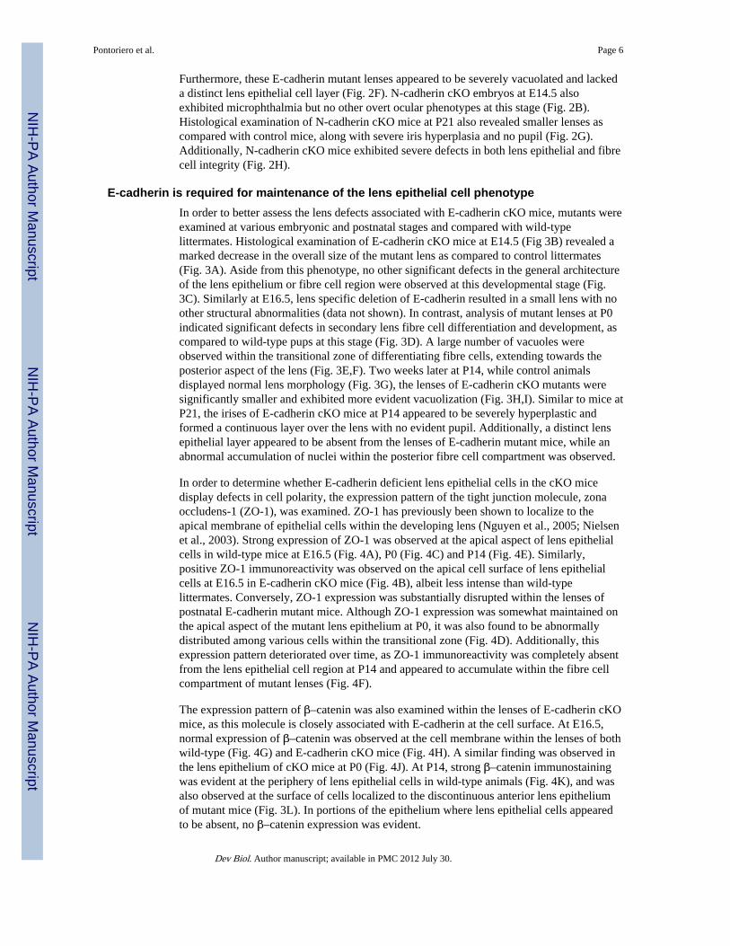
E-cadherin is required for maintenance of the lens epithelial cell phenotypeIn order to better assess the lens defects associated with E-cadherin cKO mice, mutants wereexamined at various embryonic and postnatal stages and compared with wild-typelittermates. Histological examination of E-cadherin cKO mice at E14.5 (Fig 3B) revealed amarked decrease in the overall size of the mutant lens as compared to control littermates(Fig. 3A). Aside from this phenotype, no other significant defects in the general architectureof the lens epithelium or fibre cell region were observed at this developmental stage (Fig.3C). Similarly at E16.5, lens specific deletion of E-cadherin resulted in a small lens with noother structural abnormalities (data not shown). In contrast, analysis of mutant lenses at P0indicated significant defects in secondary lens fibre cell differentiation and development, ascompared to wild-type pups at this stage (Fig. 3D). A large number of vacuoles wereobserved within the transitional zone of differentiating fibre cells, extending towards theposterior aspect of the lens (Fig. 3E,F). Two weeks later at P14, while control animalsdisplayed normal lens morphology (Fig. 3G), the lenses of E-cadherin cKO mutants weresignificantly smaller and exhibited more evident vacuolization (Fig. 3H,I). Similar to mice atP21, the irises of E-cadherin cKO mice at P14 appeared to be severely hyperplastic andformed a continuous layer over the lens with no evident pupil. Additionally, a distinct lensepithelial layer appeared to be absent from the lenses of E-cadherin mutant mice, while anabnormal accumulation of nuclei within the posterior fibre cell compartment was observed.
In order to determine whether E-cadherin deficient lens epithelial cells in the cKO micedisplay defects in cell polarity, the expression pattern of the tight junction molecule, zonaoccludens-1 (ZO-1), was examined. ZO-1 has previously been shown to localize to theapical membrane of epithelial cells within the developing lens (Nguyen et al., 2005; Nielsenet al., 2003). Strong expression of ZO-1 was observed at the apical aspect of lens epithelialcells in wild-type mice at E16.5 (Fig. 4A), P0 (Fig. 4C) and P14 (Fig. 4E). Similarly,positive ZO-1 immunoreactivity was observed on the apical cell surface of lens epithelialcells at E16.5 in E-cadherin cKO mice (Fig. 4B), albeit less intense than wild-typelittermates. Conversely, ZO-1 expression was substantially disrupted within the lenses ofpostnatal E-cadherin mutant mice. Although ZO-1 expression was somewhat maintained onthe apical aspect of the mutant lens epithelium at P0, it was also found to be abnormallydistributed among various cells within the transitional zone (Fig. 4D). Additionally, thisexpression pattern deteriorated over time, as ZO-1 immunoreactivity was completely absentfrom the lens epithelial cell region at P14 and appeared to accumulate within the fibre cellcompartment of mutant lenses (Fig. 4F).
The expression pattern of β–catenin was also examined within the lenses of E-cadherin cKOmice, as this molecule is closely associated with E-cadherin at the cell surface. At E16.5,normal expression of β–catenin was observed at the cell membrane within the lenses of bothwild-type (Fig. 4G) and E-cadherin cKO mice (Fig. 4H). A similar finding was observed inthe lens epithelium of cKO mice at P0 (Fig. 4J). At P14, strong β–catenin immunostainingwas evident at the periphery of lens epithelial cells in wild-type animals (Fig. 4K), and wasalso observed at the surface of cells localized to the discontinuous anterior lens epitheliumof mutant mice (Fig. 3L). In portions of the epithelium where lens epithelial cells appearedto be absent, no β–catenin expression was evident.
Pontoriero et al. Page 6
Dev Biol. Author manuscript; available in PMC 2012 July 30.
NIH
-PA Author Manuscript
NIH
-PA Author Manuscript
NIH
-PA Author Manuscript

A loss in E-cadherin expression has been intimately linked with the phenomenon known asepithelial to mesenchymal transition (EMT), in which epithelial cells transform anddifferentiate to express a mesenchymal cell phenotype (Huber et al., 2005). Cells undergoingthis process generally lose their cell polarity and one hallmark feature of this transformationis the presence of the contractile element, alpha-smooth muscle actin (α-SMA) (Zavadil andBottinger, 2005). As such, α-SMA protein expression was examined within the lenses of E-cadherin cKO mice and wild-type littermates. While wild-type animals express α-SMAnormally within the overlying iris tissue (Fig. 4M), an accumulation of α-SMA wasobserved within the lens epithelium of E-cadherin cKO animals beginning at E16.5 (Fig.4N) and continuing to P0 (Fig. 4P). At P14, a substantial up-regulation of α-SMAexpression was observed within the lenses of E-cadherin cKO mutants (Fig. 4R) ascompared to control mice (Fig. 4Q). Positive staining for α-SMA protein expression withinthe anterior region of the mutant lens extended across the entire lens epithelial surface.Surprisingly, positive α-SMA immunoreactivity was also present along the posterior aspectof the lens suggesting abnormal migration of cells to the posterior lens fibre cell region (datanot shown).
N-cadherin is required for lens fibre morphogenesis and elongation, but is not required fordifferentiation
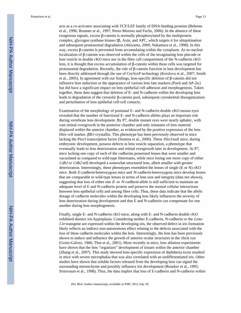
N-cadherin is maintained within the lens fibre cell compartment throughout morphogenesis.Histological examination of N-cadherin cKO mice during embryogenesis revealed no overtdefects in lens morphogenesis as compared with wild-type littermates. At E14.5, N-cadherincKO lenses appeared to be smaller in size but still developed distinct epithelial and fibre cellcompartments (Fig. 5B). Similarly, at E17.5, N-cadherin mutant lenses (Fig. 5D) weresubstantially smaller than the lenses of wild-type littermates (Fig. 5C) yet sill appeared todevelop normally. Conversely, postnatal examination of N-cadherin cKO lenses revealedgross defects in fibre cell and iris development, phenotypes which varied in severity fromone mutant to another. At P14, N-cadherin cKO mice exhibited distinct iris hyperplasia inthe absence of a pupil (Fig. 5F). Additionally, the lenses of N-cadherin mutant miceexhibited extensive vacuolization throughout the entire fibre cell compartment (Fig. 5F,G).Furthermore, N-cadherin mutant lenses also possessed an abnormal accumulation of nucleiwithin the transitional zone, also suggesting defects in fibre cell development (Fig. 5G).
In order to further examine the changes in lens fibre cell differentiation and architecturewithin N-cadherin cKO mice, a number of markers for fibre cell development were assessed.Lens epithelial cells begin to differentiate as early as embryonic stage E10.5, where β-crystallin, one of the first detectable proteins involved in fibre cell differentiation, isexpressed. At E12.5, β-crystallin expression is retained in both wild-type (Fig. 6A) and N-cadherin cKO lenses (Fig. 6B), revealing that lens fiber differentiation had occurred. Asimilar finding was observed at E14.5, in which β-crystallin immunoreactivity wasmaintained in the lenses of both N-cadherin mutant (Fig. 6C) and control littermates (Fig.6D). Additionally, two major fibre cell membrane components, lens major intrinsic protein(MIP26) and ZO-1, were also examined and shown to be equally expressed in both wild-type (Fig. 6E,I) and N-cadherin cKO embryos (Fig. 6F,J) at E12.5. Similarly at E14.5, theintensity of MIP26 and ZO-1 expression was equivalent between the lenses of N-cadherinmutant (Fig. 6H’,L’) and control embryos (Fig. 6G’,K’). However, the distribution andarrangement of lens fibre cells in N-cadherin cKO lenses appeared to be disrupted whencompared to wild-type counterparts at this stage. Wild-type lens fibre cells were wellorganized and appeared to form “Y” sutures as they joined and elongated through the lenscapsule (Fig. 6G’,K’). In contrast, the fibre cells of N-cadherin cKO lenses failed to properlyelongate as evidenced by the disrupted expression patterns of both ZO-1 (arrowhead in Fig.6H’) and MIP26 (Fig. 6L) and altered phalloidin distribution.
Pontoriero et al. Page 7
Dev Biol. Author manuscript; available in PMC 2012 July 30.
NIH
-PA Author Manuscript
NIH
-PA Author Manuscript
NIH
-PA Author Manuscript

β-catenin was also examined as this molecule is required for cell adhesion during lensdevelopment and has shown to play an important part in the coordination of morphogenesis(Smith et al., 2005). Junctional β-catenin expression was observed within the lens epitheliaand fibre cell compartments of wild-type embryos at both E12.5 (Fig. 6M) and E14.5 (Fig.6O’). While the expression of β-catenin was maintained within the lenses of N-cadherincKO mice at E12.5 (Fig. 6N), distinct β-catenin expression was absent, specifically from thefibre cell region, of N-cadherin mutant embryos at E14.5 (Fig. 6P’). Interestingly, β-cateninexpression was maintained within the lens epithelial layer of N-cadherin cKO mice at allstages examined. Thus, in the absence of N-cadherin, β-catenin expression is eventually lostat fibre cell membrane junctions, which in turn may account for the observed disruption anddisorganization of the fibre cell region at this stage.
E- and N-cadherin dosage has a significant impact on lens developmentN-cadherin expression was maintained within the lenses of E-cadherin cKO mice and E-cadherin was maintained within N-cadherin cKO lenses (data not shown). Thus, in order toexamine the possibility of cadherin compensation in either the E-cadherin conditional or N-cadherin cKO lens, both E- and N-cadherin were simultaneously eliminated from thepresumptive lens ectoderm using the Lens-Cre transgenic mouse line. Analysis of mutantprogeny and control littermates revealed that at P7, double conditional E- and N-cadherincKO mice exhibited ocular defects more severe than either single cKO mouse model (Fig.7C,E). In addition to microphthalmia, double cKO mice lacked a distinct lens (Fig. 7D).However, some double mutants possessed remnants of lens material positioned eitherdirectly beneath the iris (LR in Fig. 7F) or distributed throughout the anterior compartmentas evidenced by distinct βB1-crystallin immunoreactivity (Fig. 7H). The retinas of doublecKO mice appeared to be overgrown, occupying the majority of the ocular space (Fig. 7D).Furthermore, these retinas also appeared to be completely laminated as evidenced bypositive immunoreactivity for the retinal cell markers, Pax6, calretinin, and syntaxin (datanot shown). Double cKO animals also lacked an observable pupil, possessing iris tissue thathad abnormally migrated and infiltrated across the entire anterior chamber, in a manneranalogous to either the E- or N-cadherin single cKO mouse models. Additionally, the irisappeared to fuse with the overlying cornea which in turn lacked an endothelium andstratified epithelium.
The nature of the breeding scheme employed to generate the double cKO mice allowed us todetermine the allelic dosage effect through the elimination of various combinations of E-and N-cadherin during lens development. Mice deficient for only one copy of each of thesecadherin molecules (Fig. 7I,J) did not lack a lens or pupil, but lenses were reduced in size.These lenses also exhibited distinct vacuolization, a phenotype that was previously observedin either of the single E- or N-cadherin cKO mouse models. Mice possessing only onefunctional copy of N-cadherin but lacking both E-cadherin alleles (Fig. 7K,L) or micepossessing one functional E-cadherin allele but lacking both copies of N-cadherin within thelens (Fig. 7M,N) were also microphthalmic and possessed lenses that were much smaller ascompared with mice either lacking only one copy of each cadherin or wild-type littermates.These data clearly demonstrate that the combined loss of E-cadherin and N-cadherin withinthe lens results in gradual aphakia and that the allelic dosage of E-and N-cadherin has asignificant impact on the regulation of lens growth.
E-cadherin and N-cadherin are required for lens vesicle separation and survival of the lensepithelium
Histological examination of E- and N-cadherin double cKO mice was next performed atvarious embryonic and postnatal stages to assess the co-operative roles for these cadherinsduring lens morphogenesis. At E10.5, the lens placode of wild-type littermates invaginated
Pontoriero et al. Page 8
Dev Biol. Author manuscript; available in PMC 2012 July 30.
NIH
-PA Author Manuscript
NIH
-PA Author Manuscript
NIH
-PA Author Manuscript

along with the optic vesicle to give rise to the lens pit and optic cup, respectively (Fig. 8A).Similarly, in E- and N-cadherin double cKO, the lens placode appeared to develop andinvaginate towards the optic vesicle (Fig. 8B). At E11.5, normal development of the lensvesicle was observed in wild-type embryos (Fig. 8C). Conversely, significant defects in lensvesicle development were consistently observed in double cKO mice (Fig. 8D). Ahomogeneous population of cells infiltrated and occupied the normally vacant lumen of thelens vesicle. Additionally, the epithelium comprising the anterior region of the lens vesiclewas substantially thinner than its respective littermates and appeared to possess a lens stalkremnant, suggesting defects in lens vesicle separation. At E13.5, distinct defects in lensvesicle separation became more evident. While wild-type littermates (Fig. 8E) possessednormal lenses which separated from the surface ectoderm, the lens vesicle of double cKOembryos at E13.5 (Fig. 8F) remained continuous with the overlying surface ectoderm,exhibiting the formation of a persistent lens stalk. H&E staining of E15.5 embryos revealedthat unlike control littermates, which possessed a defined lens epithelium and underlyingfibre cell region (Fig. 8G), the lenses of double cKO animals lacked a distinct lens epitheliallayer (Fig. 8H). Instead, double cKO mice possessed a nucleated fibre cell mass. Thisvacuolated lentoid structure also appeared to migrate anteriorly into the periocular space,suggesting defects in lens capsule formation. Additionally, cells from the periocularmesenchyme also abnormally migrated behind the lens, into the vitreal space. Similarly atP0, as compared to wild-type lenses (Fig. 8I), double cKO mice were severelymicrophthalmic and possessed nucleated fibre cell-like material which had infiltrated theanterior chamber (Fig. 8J). Not surprising, double cKO lenses at P0 also lacked anobservable lens epithelium. Furthermore, double cKO eyes at this stage exhibited overtretinal overgrowth in the absence of a distinct lens.
Histological examination of double cKO embryos revealed severe defects in lens vesicleformation. A large population of cells had infiltrated the lumen of the developing lensvesicle. As such, in order to determine the origin of these abnormally displaced cells,markers of lens epithelial cell development were immunolocalized in the double cKOembryo at E11.5. As shown in Fig. 9A, localization of Pax6 protein within wild-typeembryos at E11.5 revealed the characteristic broad expression pattern of Pax6 throughoutsurface ectoderm, lens vesicle and optic cup. Assessment of Pax6 protein expression indouble cKO embryos at E11.5 revealed that in addition to the characteristic Pax6 expressionprofile within the cells comprising the lens vesicle, distinct Pax6 immunostaining was alsoevident in the cells displaced within the developing lens vesicle (Fig. 9B). The expressionpattern of the transcription factor, AP-2α, was also localized within the lenses of doublecKO and control embryos as this protein has been shown to localize to lens epithelial cellspositioned more anteriorly than Pax6 (Makhani et al., 2007). In wild-type embryos at E11.5,expression of AP-2α was observed within the anterior portion of the lens vesicle and theoverlying surface ectoderm (Fig. 9C). In double cKO embryos, expression of AP-2α wasalso evident with the abnormally displaced lens epithelial cells within the lumen of the lensvesicle (Fig. 9D). Finally, the expression of Foxe3, a lens epithelial-specific cell marker,was characterized within the lenses of both wild-type (Fig. 9E) and double cKO embryos(Fig. 9F). Distinct Foxe3 immunoreactivity was also observed within the cells situatedwithin the lumen of the double cKO lens vesicle (Fig. 9F). These findings demonstrate thatthe cells in the lumen of double mutant lenses are of lens epithelial origin and that in theabsence of E- and N-cadherin, the anteriorly positioned cells are likely those that are lost.Corroborating this hypothesis, the anterior region of the lens vesicle in double cKO miceappeared to be significantly thinner than control embryos at this stage.
When epithelial cells are prohibited from maintaining cellular adhesion with theextracellular matrix or surrounding cells for extended periods of time, they can undergoapoptosis by a phenomenon known as anoikis (Fouquet et al., 2004; Shanmugathasan and
Pontoriero et al. Page 9
Dev Biol. Author manuscript; available in PMC 2012 July 30.
NIH
-PA Author Manuscript
NIH
-PA Author Manuscript
NIH
-PA Author Manuscript

Jothy, 2000). In order to address the hypothesis that loss of epithelial cells into the lumen ofthe lens vesicle leads to subsequent apoptosis, a TUNEL analysis was performed at E11.5.As shown in Fig. 9G, very few epithelial cells located within the lens vesicle of wild-typeembryos appeared to undergo programmed cell death, as evidenced by the lack of TUNEL-positive cells. In contrast, a substantial increase in TUNEL-labeling was observed with thelens vesicles of embryos harboring various genetic copies of either E- or N-cadherin (datanot shown). Similarly, a large number of TUNEL-positive cells were observed within thelens vesicle of double cKO embryos, with the greatest proportion located within the lumen(Fig. 9H). These data suggest that E- and N-cadherin are required for lens epithelial cellsurvival and for the prevention of detachment-mediated apoptosis within the lens vesicle andthat the combined loss of various cadherin molecules within the lens results in a greaterdegree of apoptosis.
The expression profile of β-catenin was also examined within the developing lens of E- andN-cadherin double cKO mice. At E10.5, wild-type embryos exhibited strongimmunoreactivity for β-catenin at cell-cell junctions within the developing lens pit (Fig. 9I).In double cKO lenses, while normal expression of β-catenin was maintained within thedeveloping optic cup, β-catenin expression was substantially down-regulated within theinvaginating lens pit as compared with control littermates (Fig. 9J). Normal expression of β-catenin was observed within the developing lens at both E11.5 (Fig. 9K) and E13.5 (Fig.9M). In contrast to littermate controls, expression of β-catenin was completely lost withinthe lens vesicle of double mutant embryos at E11.5 (Fig. 9L) and E13.5 (Fig. 9N).Interestingly, no nuclear localization of β-catenin was observed at either stage examined,suggesting that liberated β-catenin was likely targeted for proteasomal degradation in doublemutant mice.
The alteration in the distribution of cadherin molecules has been hypothesized to play animportant role during the separation of various tissues (Wheelock et al., 2008). P-cadherin,another cadherin family member is also expressed within the presumptive lens ectodermduring embryonic development and is usually lost within the lens vesicle as the lensseparates. Analysis of P-cadherin expression was performed in the E-cadherin and N-cadherin double cKO mice and revealed that normal expression of P-cadherin was observedwithin the developing surface ectoderm and retinal pigmented epithelium of both normalcontrol (Fig. 9O) and double cKO embryos (Fig. 9P) at E13.5. However, distinct P-cadherinimmunoreactivity was also observed at the junctions of cells comprising the residual lensstalk of double cKO mice (Fig. 9P). Taken together, these findings suggest that both E- andN-cadherin are required for normal lens vesicle separation and that the maintenance of P-cadherin expression within the residual stalk cells may also play an important role inpreventing normal lens separation.
DISCUSSIONThe classical cadherins are important mediators of cell adhesion that maintain cell-cellcontacts throughout the life of an organism. The importance of these molecules duringembryogenesis is demonstrated from studies of germline knockout mice. For example,homozygous N-cadherin-null embryos die by E10 of gestation (Radice et al., 1997). Themost dramatic cell adhesion defect in these mice is observed in the primitive heart, wheremyocardial tissue initially develops but rapidly deteriorates as myocytes lose adhesion anddissociate. Homozygous negative E-cadherin embryos die around the time of implantationand fail to form a trophoectodermal epithelium or a blastocyst cavity (Larue et al., 1994).These results clearly demonstrate the pivotal roles of these adhesion molecules in thedevelopment of multicellular vertebrate organisms. However, the early embryonic lethality
Pontoriero et al. Page 10
Dev Biol. Author manuscript; available in PMC 2012 July 30.
NIH
-PA Author Manuscript
NIH
-PA Author Manuscript
NIH
-PA Author Manuscript

associated with these mouse models prevents their use for examining the roles cadherinsplay in lens development.
Previous studies have shown a correlation between loss in the expression of cadherinmolecules during lens development and failure in lens vesicle separation (Pontoriero et al.,2008). To directly address the hypothesis that loss of cadherin expression within the lensresults in defects in lens separation, the Lens-Cre transgene was employed to induce a lensspecific knockout of either E-cadherin or N-cadherin within the developing lens. Lens-Cre-mediated recombination of either Cdh1 or Cdh2 floxed alleles resulted in successfulelimination of E-cadherin and N-cadherin expression within the developing lens by E10.5.In both mutant models, defects in lens vesicle separation were not observed. Both the E-cadherin and N-cadherin cKO mice did however exhibit severe microphthalmia with smallervacuolated lenses, along with a hyperplastic iris. The vacuolization observed in E-cadherincKO lenses originated within the transitional zone and extended throughout the cortical fibrecell region during postnatal development. Additionally, the epithelial layer of E-cadherincKO mice appeared to lose polarity over time. A disruption in ZO-1 expression becameapparent along with the accumulation of α-SMA within the anterior and posterior lensregions. Thus, these findings show that E-cadherin is essential for maintaining the integrityof the lens epithelial cell layer and maintenance of the lens epithelial cell phenotype.Examination of N-cadherin cKO lenses also revealed extensive vacuolization within thefibre cell region. Defects in normal fibre cell elongation were also observed and correlatedwith a loss of β-catenin expression within the fibre cell compartment of N-cadherin cKOembryos. Thus, the E-cadherin and N-cadherin single cKO mice exhibited both overlappingand distinct lens phenotypes. This may be partly due to the fact that these proteins have bothsimilar and unique expression patterns in the developing lens, with E- and N-cadherinexpressed in the lens epithelium and only N-cadherin expressed in the fibre cellcompartment. However, it may also be that E- and N-cadherin have both similar and discreteroles during lens development. Supporting this hypothesis, Kan and colleagues expressed N-cadherin from the E-cadherin locus (Kan et al., 2007) and demonstrated that while N-cadherin cDNA was able to rescue the loss of E-cadherin for morula compaction, it was notable to functionally compensate for E-cadherin during the ensuing formation of thetrophoectoderm.
In order to address the possibility of cadherin compensation and to determine the co-operative roles for these cadherins in lens development, a double E and N-cadherin lens-specific knockout mouse was generated. Notably, double cKO embryos exhibited a defect inlens vesicle formation, as compared to their wild-type littermates. This defect appeared toinvolve a failure in separation of the lens from that overlying ectoderm, which wascorrelated with sustained expression of P-cadherin within the remaining lens stalk region.Interestingly, cKO mice lacking the transcription factor, AP-2α, from the developing lens,exhibit a persistent lens stalk phenotype that is also correlated with a loss of E- and N-cadherin and sustained expression of P-cadherin within the cells comprising the lens stalkregion (Pontoriero et al., 2008). From the morphological data alone, however, it cannot bedetermined whether the defect in the double cadherin cKO mice was due to failure of thelens vesicle, once closed, to separate from the overlying ectoderm or was caused by failurein proper closure of the vesicle, perhaps due to inadequate adhesion of cells lining eitherside of the lens pit. Furthermore, whether the sustained expression of P-cadherin within thecorneal-lenticular adhesion of these mutant mice is simply a consequence/remnant or acontributing factor to lens stalk formation remains to be elucidated. One could, however,envision that the persistent expression of P-cadherin within the lens stalk cells preventedproper cell sorting as these cells are now more phenotypically similar to the epithelial cellsof the surface ectoderm. Future studies aimed at either conditionally inactivating oroverexpressing P-cadherin specifically within the presumptive lens ectoderm will help to
Pontoriero et al. Page 11
Dev Biol. Author manuscript; available in PMC 2012 July 30.
NIH
-PA Author Manuscript
NIH
-PA Author Manuscript
NIH
-PA Author Manuscript

further determine its contribution to the formation of this developmental defect. A numberof other mouse mutants exhibit failure in lens vesicle separation with a persistent lens stalk(Blixt et al., 2000; Collinson et al., 2001; Favor et al., 1997; Hill et al., 1991; Rieger et al.,2001; Wurm et al., 2008; Yoshimoto et al., 2005). It may therefore be useful to examine theexpression profiles of E-, N-, and P-cadherin within the lenses of these mutant mice in orderto further determine the contribution of cadherins to lens vesicle separation.
Histological analysis of double cKO embryos at E11.5 revealed an accumulation of cellswith the lumen of the developing lens vesicle. These abnormally displaced cells expressedcharacteristics of the anterior lens epithelium, including the expression of the lens epithelialcell markers, Pax6 and AP-2α, and were also labeled positively in TUNEL analyses.Importantly, the displaced cells were also found to express FoxE3, an additional lensepithelial cell marker, which unlike AP-2α and Pax6, is not expressed in the overlyingectoderm following lens separation. This further suggests that these cells were derived fromthe lens epithelium rather than being derived from the overlying surface ectoderm duringvesicle closure. Thus, these data suggest that there is a co-requirement for E-cadherin and N-cadherin in maintenance of the developing anterior lens epithelium. In the absence of bothE- and N-cadherin, these cells appear to lose their ability to maintain cell-cell and/or cell-matrix contacts and are lost to the lumen of the developing lens vesicle where they undergoapoptosis. Interestingly, lenses lacking other combinations of either E-cadherin or N-cadherin displayed increased TUNEL reactivity, as compared to wild-type embryos, but didnot appear to lose epithelial cells to the lens vesicle lumen, suggesting that at least one copyof either cadherin, E or N, can prevent the loss of anterior lens epithelial cells during earlylens development. The biological process, in which cells lose their contact with theunderlying extracellular matrix and undergo programmed cell death, has been termeddetachment-induced apoptosis or anoikis (Frisch and Francis, 1994; Grossmann, 2002). Theimportance of cadherins in the maintenance of cell-cell adhesion and prevention ofdetachment-induced apoptosis has been demonstrated elsewhere. For example, treatment ofintestinal epithelial cells with a blocking anti-E-cadherin antibody increased the rate ofanoikis, whereas the activation of E-cadherin using a dimeric E-cadherin-IgG Fc chimeraprotein reduced anoikis (Fouquet et al., 2004). More recently, activation of Src familykinases in a chick lens model of cortical cataracts resulted in the disassembly of N-cadherinjunctions, followed by subsequent apoptosis (Zhou et al., 2007). Cadherins have also beenshown to directly interact with molecules, such as the integrins, which are important forlinking cells to the basement membrane. For example, the fifth cadherin ectodomain hasbeen shown to be essential for heterophilic contact with integrins (Shiraishi et al., 2005), andcadherins and integrins are also thought to crosstalk and mediate differentiation viaepidermal growth factor signaling and downstream Akt kinase activity (Muller et al., 2008).Thus, the deletion of cadherins within the developing lens in the cKO mice may haveperturbed common downstream signaling targets of integrins that are important formediating cell anchorage to the basement membrane and the prevention of apoptosis.
Cadherins, at adherens junctions, link the extracellular environment to the actin cytoskeletonvia their interactions with the armadillo repeat protein, β-catenin (Perez-Moreno and Fuchs,2006; Perez-Moreno et al., 2003). As such, β-catenin protein expression was examinedwithin the developing lenses of cKO mice. Normal expression of β-catenin was observed atthe cell membranes of lens epithelial and fibre cells in E-cadherin cKO mice during earlydevelopment. This finding is not surprising given that normal expression of N-cadherin wasalso observed in E-cadherin cKO lenses at various developmental timepoints (data notshown). Interestingly, β-catenin expression was lost within the developing lens vesicle ofdouble cKO mice as early as E10.5, while in N-cadherin cKO mice, β-catenin expressionwas only notably down-regulated within the fibre cell compartment. In the presence of anextracellular Wnt signal, β-catenin has the potential to translocate to the nucleus where it
Pontoriero et al. Page 12
Dev Biol. Author manuscript; available in PMC 2012 July 30.
NIH
-PA Author Manuscript
NIH
-PA Author Manuscript
NIH
-PA Author Manuscript
acts as a co-activator associating with TCF/LEF family of DNA-binding proteins (Behrenset al., 1996; Brunner et al., 1997; Perez-Moreno and Fuchs, 2006). In the absence of theseexogenous signals, excess β-catenin is normally phosphorylated by the multiproteincomplex, glycogen synthase kinase-3β, Axin, and APC, which targets it for ubiquitinationand subsequent proteasomal degradation (Akiyama, 2000; Nakamura et al., 1998). In thisway, excess β-catenin is prevented from accumulating within the cytoplasm. As no nuclearlocalization of β-catenin was observed within the cells of the invaginating lens placode orlens vesicle in double cKO mice nor in the fibre cell compartment of the N-cadherin cKOlens, it is thought that excess accumulation of β-catenin within these cells was targeted forproteasomal degradation. Recently, the role of β-catenin function in lens development hasbeen directly addressed through the use of Cre/loxP technology (Kreslova et al., 2007; Smithet al., 2005). In agreement with our findings, lens-specific deletion of β-catenin did notinfluence lens induction or the appearance of various lens fate markers (Pax6 and AP-2α)but did have a significant impact on lens epithelial cell adhesion and morphogenesis. Takentogether, these data suggest that deletion of E- and N-cadherin within the developing lensleads to degradation of the cytosolic β-catenin pool, subsequent cytoskeletal disorganizationand perturbation of lens epithelial cell-cell contacts.
Examination of the morphology of postnatal E- and N-cadherin double cKO mouse eyesrevealed that the number of functional E- and N-cadherin alleles plays an important roleduring vertebrate lens development. By P7, double mutant eyes were nearly aphakic, withvast retinal overgrowth in the posterior chamber and only remnants of lens materialdisplaced within the anterior chamber, as evidenced by the positive expression of the lensfibre cell marker, βB1-crystallin. This phenotype has been previously observed in micelacking the Pitx3 transcription factor (Semina et al., 2000). These Pitx3-null mice, duringembryonic development, possess defects in lens vesicle separation, a phenotype thateventually leads to lens deterioration and retinal overgrowth later in development. At P7,mice lacking one copy of each of the cadherins possessed lenses that were smaller andvacuolated as compared to wild-type littermates, while mice losing one more copy of eitherCdh1 or Cdh2 still developed a somewhat structured lens, albeit smaller with greaterdeterioration. Interestingly, these phenotypes resembled the lenses of single (E or N) cKOmice. Both E-cadherin heterozygous mice and N-cadherin heterozygous mice develop lensesthat are comparable to wild-type lenses in terms of lens size and integrity (data not shown),suggesting that loss of either one E- or N-cadherin allele is still sufficient to maintain anadequate level of E and N-cadherin protein and preserve the normal cellular interactionsbetween lens epithelial cells and among fibre cells. Thus, these data indicate that the allelicdosage of cadherin molecules within the developing lens likely influences the severity oflens deterioration during development and that E and N-cadherin can compensate for oneanother during lens morphogenesis.
Finally, single E- and N-cadherin cKO mice, along with E- and N-cadherin double cKOexhibited distinct iris hyperplasia. Considering neither E-cadherin, N-cadherin or the Lens-Cre transgene are expressed within the developing iris, the observed defect in iris formationlikely reflects an indirect non-autonomous effect relating to the defects associated with theloss of these cadherin molecules within the lens. Interestingly, the lens has been previouslyshown to induce and influence the growth of anterior ocular structures in the chick eye(Genis-Galvez, 1966; Thut et al., 2001). More recently in mice, lens ablation experimentshave shown that the lens “organizes” development of tissues within the anterior chamber(Zhang et al., 2007). This study showed lens-specific expression of diphtheria toxin resultedin mice with severe microphakia that was also correlated with an undifferentiated iris. Otherstudies have shown that soluble factors released from the developing lens can signal thesurrounding mesenchyme and possibly influence iris development (Reneker et al., 1995;Srinivasan et al., 1998). Thus, the data implies that loss of E-cadherin and N-cadherin within
Pontoriero et al. Page 13
Dev Biol. Author manuscript; available in PMC 2012 July 30.
NIH
-PA Author Manuscript
NIH
-PA Author Manuscript
NIH
-PA Author Manuscript

the developing lens leads to the abnormal modulation of lens-secreted signaling moleculeswhich likely play pivotal roles in regulation normal iris migration. A number of additionalknockout mouse models have been created which result in defects in iris development(hypoplasia or hyperplasia), yet their relationship to E-cadherin and N-cadherin or otherfactors that regulate iris development is not yet known (Ramaesh et al., 2003; Swamynathanet al., 2007; Zaki et al., 2006).
In summary, the data suggest that E- and N-cadherin regulate vertebrate lens vesicleseparation from the overlying surface ectoderm. Dual elimination of these cadherinmolecules within the presumptive lens ectoderm results in loss of cell-cell contacts withinthe anterior region of the lens vesicle and subsequent detachment-induced apoptosis(anoikis). Thus, E- and N-cadherin function as survival factors within developing lensepithelial cells and their absence results in complete deterioration of the anterior lensepithelium during mid-embryogenesis. Furthermore, loss of various combinations ofcadherin molecules within the lens resulted in less severe lens defects, suggesting that notonly can E- and N-cadherin can compensate for one another during lens morphogenesis butthat the dosage of cadherin molecules within the developing lens likely dictates the severityof the lenticular defects.
AcknowledgmentsThe following individuals are acknowledged for their outstanding technical assistance: Ms. Lauren Kirkby, Mr.Paul Speeg, and Mrs. Paula Deschamps. We would also like to thank Dr. Ruth Ashery-Padan (Tel Aviv University,Ramat Aviv, Israel) for providing the Lens-Cre transgenic mouse line, Dr. Joseph Horwitz (Jules Stein EyeInstitute, Los Angeles, CA) for providing the rabbit polyclonal anti-MIP26 and mouse anti-βB1-crystallinantibodies and Dr. Peter Carlsson (University of Gothenburg, Gothenburg, Sweden) for providing the rabbitpolyclonal anti-Foxe3 antibody. This work was supported by the following: NIH R01 EY11910 (J.W.M.), NIHR01’s EY15766 (R.A.L.), EY16241 (R.A.L.), EY17848 (R.A.L.) and CA131270 (R.A.L.), by funds from theAbrahamson Pediatric Eye Institute Endowment at Children’s Hospital Medical Center of Cincinnati (R.A.L.) and aNatural Sciences and Engineering Research Council of Canada – Postgraduate Scholarship (G.F.P.).
REFERENCESAbe K, Takeichi M. EPLIN mediates linkage of the cadherin catenin complex to F-actin and stabilizes
the circumferential actin belt. Proc Natl Acad Sci U S A. 2008; 105:13–9. [PubMed: 18093941]
Akiyama T. Wnt/beta-catenin signaling. Cytokine Growth Factor Rev. 2000; 11:273–82. [PubMed:10959075]
Ashery-Padan R, Marquardt T, Zhou X, Gruss P. Pax6 activity in the lens primordium is required forlens formation and for correct placement of a single retina in the eye. Genes Dev. 2000; 14:2701–11. [PubMed: 11069887]
Behrens J, von Kries JP, Kuhl M, Bruhn L, Wedlich D, Grosschedl R, Birchmeier W. Functionalinteraction of beta-catenin with the transcription factor LEF-1. Nature. 1996; 382:638–42.[PubMed: 8757136]
Blixt A, Mahlapuu M, Aitola M, Pelto-Huikko M, Enerback S, Carlsson P. A forkhead gene, FoxE3, isessential for lens epithelial proliferation and closure of the lens vesicle. Genes Dev. 2000; 14:245–54. [PubMed: 10652278]
Boussadia O, Kutsch S, Hierholzer A, Delmas V, Kemler R. E-cadherin is a survival factor for thelactating mouse mammary gland. Mech Dev. 2002; 115:53–62. [PubMed: 12049767]
Brunner E, Peter O, Schweizer L, Basler K. pangolin encodes a Lef-1 homologue that acts downstreamof Armadillo to transduce the Wingless signal in Drosophila. Nature. 1997; 385:829–33. [PubMed:9039917]
Collinson JM, Quinn JC, Buchanan MA, Kaufman MH, Wedden SE, West JD, Hill RE. Primarydefects in the lens underlie complex anterior segment abnormalities of the Pax6 heterozygous eye.Proc Natl Acad Sci U S A. 2001; 98:9688–93. [PubMed: 11481423]
Pontoriero et al. Page 14
Dev Biol. Author manuscript; available in PMC 2012 July 30.
NIH
-PA Author Manuscript
NIH
-PA Author Manuscript
NIH
-PA Author Manuscript

Edelman GM. Expression of cell adhesion molecules during embryogenesis and regeneration. Exp CellRes. 1985; 161:1–16. [PubMed: 3902488]
Edelman GM, Crossin KL. Cell adhesion molecules: implications for a molecular histology. Annu RevBiochem. 1991; 60:155–90. [PubMed: 1883195]
Fagotto F, Gumbiner BM. Cell contact-dependent signaling. Dev Biol. 1996; 180:445–54. [PubMed:8954717]
Favor J, Grimes P, Neuhauser-Klaus A, Pretsch W, Stambolian D. The mouse Cat4 locus maps tochromosome 8 and mutants express lens-corneal adhesion. Mamm Genome. 1997; 8:403–6.[PubMed: 9166583]
Fouquet S, Lugo-Martinez VH, Faussat AM, Renaud F, Cardot P, Chambaz J, Pincon-Raymond M,Thenet S. Early loss of E-cadherin from cell-cell contacts is involved in the onset of Anoikis inenterocytes. J Biol Chem. 2004; 279:43061–9. [PubMed: 15292248]
Friedlander DR, Mege RM, Cunningham BA, Edelman GM. Cell sorting-out is modulated by both thespecificity and amount of different cell adhesion molecules (CAMs) expressed on cell surfaces.Proc Natl Acad Sci U S A. 1989; 86:7043–7. [PubMed: 2780560]
Frisch SM, Francis H. Disruption of epithelial cell-matrix interactions induces apoptosis. J Cell Biol.1994; 124:619–26. [PubMed: 8106557]
Genis-Galvez JM. Role of the lens in the morphogenesis of the iris and cornea. Nature. 1966;210:209–10. [PubMed: 5962091]
Goodwin M, Yap AS. Classical cadherin adhesion molecules: coordinating cell adhesion, signalingand the cytoskeleton. J Mol Histol. 2004; 35:839–44. [PubMed: 15609097]
Grossmann J. Molecular mechanisms of “detachment-induced apoptosis--Anoikis”. Apoptosis. 2002;7:247–60. [PubMed: 11997669]
Gu H, Marth JD, Orban PC, Mossmann H, Rajewsky K. Deletion of a DNA polymerase beta genesegment in T cells using cell type-specific gene targeting. Science. 1994; 265:103–6. [PubMed:8016642]
Hill RE, Favor J, Hogan BL, Ton CC, Saunders GF, Hanson IM, Prosser J, Jordan T, Hastie ND, vanHeyningen V. Mouse small eye results from mutations in a paired-like homeobox-containing gene.Nature. 1991; 354:522–5. [PubMed: 1684639]
Huber MA, Kraut N, Beug H. Molecular requirements for epithelial-mesenchymal transition duringtumor progression. Curr Opin Cell Biol. 2005; 17:548–58. [PubMed: 16098727]
Kan NG, Stemmler MP, Junghans D, Kanzler B, de Vries WN, Dominis M, Kemler R. Genereplacement reveals a specific role for E-cadherin in the formation of a functional trophectoderm.Development. 2007; 134:31–41. [PubMed: 17138661]
Kemler R. From cadherins to catenins: cytoplasmic protein interactions and regulation of celladhesion. Trends Genet. 1993; 9:317–21. [PubMed: 8236461]
Kostetskii I, Li J, Xiong Y, Zhou R, Ferrari VA, Patel VV, Molkentin JD, Radice GL. Induceddeletion of the N-cadherin gene in the heart leads to dissolution of the intercalated disc structure.Circ Res. 2005; 96:346–54. [PubMed: 15662031]
Kreslova J, Machon O, Ruzickova J, Lachova J, Wawrousek EF, Kemler R, Krauss S, Piatigorsky J,Kozmik Z. Abnormal lens morphogenesis and ectopic lens formation in the absence of beta-catenin function. Genesis. 2007; 45:157–68. [PubMed: 17410548]
Lang RA. Pathways regulating lens induction in the mouse. Int J Dev Biol. 2004; 48:783–91.[PubMed: 15558471]
Larue L, Ohsugi M, Hirchenhain J, Kemler R. E-cadherin null mutant embryos fail to form atrophectoderm epithelium. Proc Natl Acad Sci U S A. 1994; 91:8263–7. [PubMed: 8058792]
Luo Y, Ferreira-Cornwell M, Baldwin H, Kostetskii I, Lenox J, Lieberman M, Radice G. Rescuing theN-cadherin knockout by cardiac-specific expression of N- or E-cadherin. Development. 2001;128:459–69. [PubMed: 11171330]
Makhani LF, Williams T, West-Mays JA. Genetic analysis indicates that transcription factorsAP-2alpha and Pax6 cooperate in the normal patterning and morphogenesis of the lens. Mol Vis.2007; 13:1215–25. [PubMed: 17679940]
Pontoriero et al. Page 15
Dev Biol. Author manuscript; available in PMC 2012 July 30.
NIH
-PA Author Manuscript
NIH
-PA Author Manuscript
NIH
-PA Author Manuscript

Muller EJ, Williamson L, Kolly C, Suter MM. Outside-in signaling through integrins and cadherins: acentral mechanism to control epidermal growth and differentiation? J Invest Dermatol. 2008;128:501–16. [PubMed: 18268536]
Nakamura T, Hamada F, Ishidate T, Anai K, Kawahara K, Toyoshima K, Akiyama T. Axin, aninhibitor of the Wnt signalling pathway, interacts with beta-catenin, GSK-3beta and APC andreduces the beta-catenin level. Genes Cells. 1998; 3:395–403. [PubMed: 9734785]
Nguyen MM, Rivera C, Griep AE. Localization of PDZ domain containing proteins Discs Large-1 andScribble in the mouse eye. Mol Vis. 2005; 11:1183–99. [PubMed: 16402019]
Nielsen PA, Baruch A, Shestopalov VI, Giepmans BN, Dunia I, Benedetti EL, Kumar NM. Lensconnexins alpha3Cx46 and alpha8Cx50 interact with zonula occludens protein-1 (ZO-1). Mol BiolCell. 2003; 14:2470–81. [PubMed: 12808044]
Nose A, Nagafuchi A, Takeichi M. Expressed recombinant cadherins mediate cell sorting in modelsystems. Cell. 1988; 54:993–1001. [PubMed: 3416359]
Perez-Moreno M, Fuchs E. Catenins: keeping cells from getting their signals crossed. Dev Cell. 2006;11:601–12. [PubMed: 17084354]
Perez-Moreno M, Jamora C, Fuchs E. Sticky business: orchestrating cellular signals at adherensjunctions. Cell. 2003; 112:535–48. [PubMed: 12600316]
Pontoriero GF, Deschamps P, Ashery-Padan R, Wong R, Yang Y, Zavadil J, Cvekl A, Sullivan S,Williams T, West-Mays JA. Cell autonomous roles for AP-2alpha in lens vesicle separation andmaintenance of the lens epithelial cell phenotype. Dev Dyn. 2008; 237:602–17. [PubMed:18224708]
Radice GL, Rayburn H, Matsunami H, Knudsen KA, Takeichi M, Hynes RO. Developmental defectsin mouse embryos lacking N-cadherin. Dev Biol. 1997; 181:64–78. [PubMed: 9015265]
Ramaesh T, Collinson JM, Ramaesh K, Kaufman MH, West JD, Dhillon B. Corneal abnormalities inPax6+/− small eye mice mimic human aniridia-related keratopathy. Invest Ophthalmol Vis Sci.2003; 44:1871–8. [PubMed: 12714618]
Reneker LW, Silversides DW, Patel K, Overbeek PA. TGF alpha can act as a chemoattractant toperioptic mesenchymal cells in developing mouse eyes. Development. 1995; 121:1669–80.[PubMed: 7600984]
Rieger DK, Reichenberger E, McLean W, Sidow A, Olsen BR. A double-deletion mutation in thePitx3 gene causes arrested lens development in aphakia mice. Genomics. 2001; 72:61–72.[PubMed: 11247667]
Semina EV, Murray JC, Reiter R, Hrstka RF, Graw J. Deletion in the promoter region and alteredexpression of Pitx3 homeobox gene in aphakia mice. Hum Mol Genet. 2000; 9:1575–85.[PubMed: 10861284]
Shanmugathasan M, Jothy S. Apoptosis, anoikis and their relevance to the pathobiology of coloncancer. Pathol Int. 2000; 50:273–9. [PubMed: 10849312]
Shiraishi K, Tsuzaka K, Yoshimoto K, Kumazawa C, Nozaki K, Abe T, Tsubota K, Takeuchi T.Critical role of the fifth domain of E-cadherin for heterophilic adhesion with alpha E beta 7, butnot for homophilic adhesion. J Immunol. 2005; 175:1014–21. [PubMed: 16002701]
Smith AN, Miller LA, Song N, Taketo MM, Lang RA. The duality of beta-catenin function: arequirement in lens morphogenesis and signaling suppression of lens fate in periocular ectoderm.Dev Biol. 2005; 285:477–89. [PubMed: 16102745]
Srinivasan Y, Lovicu FJ, Overbeek PA. Lens-specific expression of transforming growth factor beta1in transgenic mice causes anterior subcapsular cataracts. J Clin Invest. 1998; 101:625–34.[PubMed: 9449696]
Steinberg MS, Takeichi M. Experimental specification of cell sorting, tissue spreading, and specificspatial patterning by quantitative differences in cadherin expression. Proc Natl Acad Sci U S A.1994; 91:206–9. [PubMed: 8278366]
Swamynathan SK, Katz JP, Kaestner KH, Ashery-Padan R, Crawford MA, Piatigorsky J. Conditionaldeletion of the mouse Klf4 gene results in corneal epithelial fragility, stromal edema, and loss ofconjunctival goblet cells. Mol Cell Biol. 2007; 27:182–94. [PubMed: 17060454]
Takeichi M. The cadherins: cell-cell adhesion molecules controlling animal morphogenesis.Development. 1988; 102:639–55. [PubMed: 3048970]
Pontoriero et al. Page 16
Dev Biol. Author manuscript; available in PMC 2012 July 30.
NIH
-PA Author Manuscript
NIH
-PA Author Manuscript
NIH
-PA Author Manuscript

Takeichi M. Cadherin cell adhesion receptors as a morphogenetic regulator. Science. 1991; 251:1451–5. [PubMed: 2006419]
Takeichi M, Atsumi T, Yoshida C, Uno K, Okada TS. Selective adhesion of embryonal carcinomacells and differentiated cells by Ca2+-dependent sites. Dev Biol. 1981; 87:340–50. [PubMed:6793433]
Takeichi M, Inuzuka H, Shimamura K, Fujimori T, Nagafuchi A. Cadherin subclasses: differentialexpression and their roles in neural morphogenesis. Cold Spring Harb Symp Quant Biol. 1990;55:319–25. [PubMed: 2132824]
Tepass U, Truong K, Godt D, Ikura M, Peifer M. Cadherins in embryonic and neural morphogenesis.Nat Rev Mol Cell Biol. 2000; 1:91–100. [PubMed: 11253370]
Thut CJ, Rountree RB, Hwa M, Kingsley DM. A large-scale in situ screen provides molecularevidence for the induction of eye anterior segment structures by the developing lens. Dev Biol.2001; 231:63–76. [PubMed: 11180952]
Wheelock MJ, Shintani Y, Maeda M, Fukumoto Y, Johnson KR. Cadherin switching. J Cell Sci. 2008;121:727–35. [PubMed: 18322269]
Wurm A, Sock E, Fuchshofer R, Wegner M, Tamm ER. Anterior segment dysgenesis in the eyes ofmice deficient for the high-mobility-group transcription factor Sox11. Exp Eye Res. 2008; 86:895–907. [PubMed: 18423449]
Xu L, Overbeek PA, Reneker LW. Systematic analysis of E-, N- and P-cadherin expression in mouseeye development. Exp Eye Res. 2002; 74:753–60. [PubMed: 12126948]
Yoshimoto A, Saigou Y, Higashi Y, Kondoh H. Regulation of ocular lens development by Smad-interacting protein 1 involving Foxe3 activation. Development. 2005; 132:4437–48. [PubMed:16162653]
Zaki PA, Collinson JM, Toraiwa J, Simpson TI, Price DJ, Quinn JC. Penetrance of eye defects in miceheterozygous for mutation of Gli3 is enhanced by heterozygous mutation of Pax6. BMC Dev Biol.2006; 6:46. [PubMed: 17029624]
Zavadil J, Bottinger EP. TGF-beta and epithelial-to-mesenchymal transitions. Oncogene. 2005;24:5764–74. [PubMed: 16123809]
Zelenka PS. Regulation of cell adhesion and migration in lens development. Int J Dev Biol. 2004;48:857–65. [PubMed: 15558477]
Zhang Y, Overbeek PA, Govindarajan V. Perinatal ablation of the mouse lens causes multiple anteriorchamber defects. Mol Vis. 2007; 13:2289–300. [PubMed: 18199970]
Zhou J, Leonard M, Van Bockstaele E, Menko AS. Mechanism of Src kinase induction of corticalcataract following exposure to stress: destabilization of cell-cell junctions. Mol Vis. 2007;13:1298–310. [PubMed: 17679932]
Pontoriero et al. Page 17
Dev Biol. Author manuscript; available in PMC 2012 July 30.
NIH
-PA Author Manuscript
NIH
-PA Author Manuscript
NIH
-PA Author Manuscript

Figure 1.Loss of E-cadherin and N-cadherin in lens ectoderm derivatives. Normal expression ofEcadherin (red) in wild-type (WT) mice is evident within the lens placode at E9.5 (A) andinvaginating lens pit at E10.5 (C). E-cadherin is maintained within the lens epithelium andoverlying surface ectoderm at both E12.5 (E) and E14.5 (G). Reduced E-cadherin expressionis observed within the lens placode and lens pit of E-cadherin cKO mice (Lens-Cre+,Cdh1flox/KO) at E9.5 (B) and E10.5 (D), respectively. E-cadherin expression is lostwithin the lens epithelium and overlying surface ectoderm of E-cadherin cKO mice at bothE12.5 (F) and E14.5 (H). Normal expression of N-cadherin (red) is evident within the opticvesicle of wild-type mice at E9.5 (I) and the invaginating lens pit and optic cup at E10.5 (K).N-cadherin expression is maintained within the lens epithelial and primary fibre cells ofcontrol mice at both E12.5 (M) and E14.5 (O). In N-cadherin cKO mice (Lens-Cre+,Cdh1flox/neo), N-cadherin expression is reduced within the lens pit at E10.5 (L) and lostthroughout the lens epithelium and fibre cell region as development progressed (N,P).Nuclei are counterstained with either DAPI (blue) or Hoechst (blue) staining.
Pontoriero et al. Page 18
Dev Biol. Author manuscript; available in PMC 2012 July 30.
NIH
-PA Author Manuscript
NIH
-PA Author Manuscript
NIH
-PA Author Manuscript
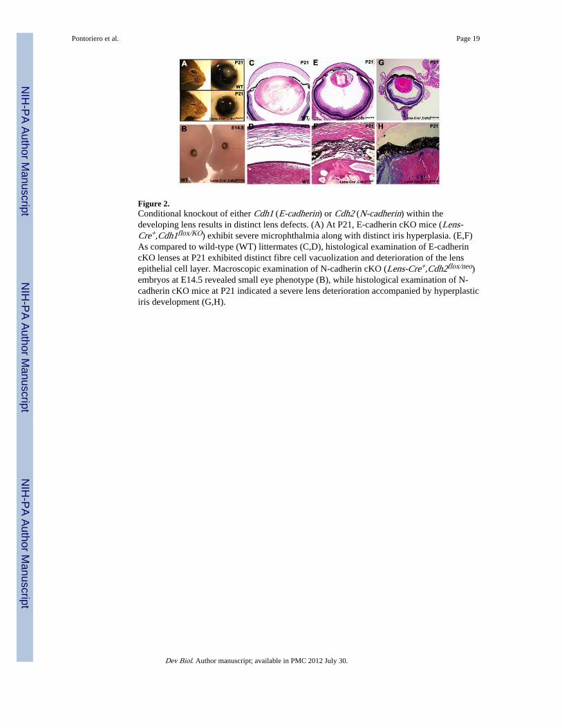
Figure 2.Conditional knockout of either Cdh1 (E-cadherin) or Cdh2 (N-cadherin) within thedeveloping lens results in distinct lens defects. (A) At P21, E-cadherin cKO mice (Lens-Cre+,Cdh1flox/KO) exhibit severe microphthalmia along with distinct iris hyperplasia. (E,F)As compared to wild-type (WT) littermates (C,D), histological examination of E-cadherincKO lenses at P21 exhibited distinct fibre cell vacuolization and deterioration of the lensepithelial cell layer. Macroscopic examination of N-cadherin cKO (Lens-Cre+,Cdh2flox/neo)embryos at E14.5 revealed small eye phenotype (B), while histological examination of N-cadherin cKO mice at P21 indicated a severe lens deterioration accompanied by hyperplasticiris development (G,H).
Pontoriero et al. Page 19
Dev Biol. Author manuscript; available in PMC 2012 July 30.
NIH
-PA Author Manuscript
NIH
-PA Author Manuscript
NIH
-PA Author Manuscript

Figure 3.Inactivation of E-cadherin within the developing lens leads to defects in lens integrity.Although the lenses of E-cadherin cKO (Lens-Cre+,Cdh1flox/KO) mice (B,C) appearedsmaller, no apparent alterations in lens structure were observed at E14.5 as compared withwild-type (WT) littermates (A). (E,F) P0 E-cadherin cKO lenses exhibited fibre cell defectsas evidenced by the presence of numerous vacuoles within the transitional zone. (H,I) AtP14, E-cadherin cKO mouse lenses possessed discontinuous lens epithelia, with a severelyvacuolized lens fibre cell region. Furthermore mutant mice developed hyperplastic irises thatextended across the entire anterior face of the lens. Scale bars are 100 μm.
Pontoriero et al. Page 20
Dev Biol. Author manuscript; available in PMC 2012 July 30.
NIH
-PA Author Manuscript
NIH
-PA Author Manuscript
NIH
-PA Author Manuscript

Figure 4.E-cadherin is essential for maintenance of the lens epithelial cell phenotype. ZO-1 islocalized on the apical aspect of the lens epithelial cell layer (red) in wild-type (WT) animalsat E16.5 (A), P0 (C) and P14 (E). Expression of ZO-1 is somewhat maintained at the apicallens epithelial membrane in E-cadherin cKO embryos (Lens-Cre+,Cdh1flox/KO) at E16.5 (B).In contrast, E-cadherin cKO lenses at P0 (D) and P14 (F) exhibit disrupted ZO-1 lensexpression at apical lens epithelial cell junctions. Expression of β-catenin (green) wasobserved within the developing lenses of control mice at E14.5 (G), P0 (I), and P14 (K). β-catenin expression was maintained within the lens epithelium and fibre cell region of E-cadherin cKO mice at E16.5 (H). As the lens deteriorated at P0 (J) and continuing to P14(L), β-catenin expression was maintained at the cell surface of lens epithelial cells only inplaces in which the epithelium had not yet deteriorated. Positive α-SMA immunoreactivity(green) was observed within the irises of wild-type embryos at E16.5 (M), P0 (O), P14 (Q).An accumulation of α-SMA expression within the lens epithelium of E-cadherin cKO micewas observed at E16.5 (N). α-SMA immunoreactivity was further up-regulated within theanterior lens epithelium of P0 (P) and P14 (R).Dotted lines outline the lens capsule. Nucleiare counterstained with DAPI (blue). Abbreviations: LE, lens epithelium; IR, iris.
Pontoriero et al. Page 21
Dev Biol. Author manuscript; available in PMC 2012 July 30.
NIH
-PA Author Manuscript
NIH
-PA Author Manuscript
NIH
-PA Author Manuscript

Figure 5.Conditional deletion of N-cadherin within the developing lens is associated with defects inlens development. The lenses of N-cadherin cKO (Lens-Cre+,Cdh2flox/neo) embryos at E15.5(B) and E17.5 (D) were substantially smaller than their wild-type (WT) counterparts (A,C)yet did not exhibit any other overt defects in lens development as they developed distinctlens epithelial and fibre cell compartments. At P14, N-cadherin cKO mice (F,G) possessvacuolated lenses that had accumulated nuclei within the fibre cell region. Additionally,mutant mice also exhibited defects in iris development, as mutant irises displayed significanthyperplasia.
Pontoriero et al. Page 22
Dev Biol. Author manuscript; available in PMC 2012 July 30.
NIH
-PA Author Manuscript
NIH
-PA Author Manuscript
NIH
-PA Author Manuscript
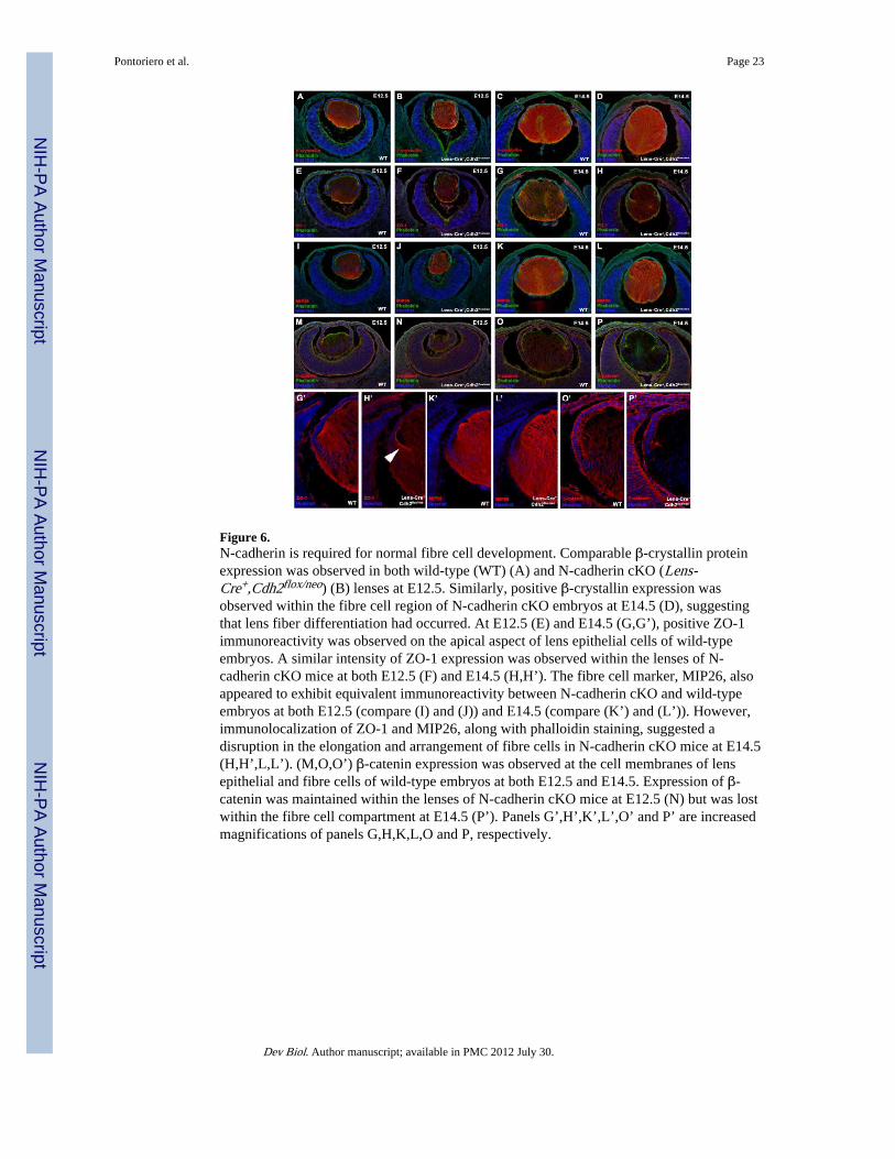
Figure 6.N-cadherin is required for normal fibre cell development. Comparable β-crystallin proteinexpression was observed in both wild-type (WT) (A) and N-cadherin cKO (Lens-Cre+,Cdh2flox/neo) (B) lenses at E12.5. Similarly, positive β-crystallin expression wasobserved within the fibre cell region of N-cadherin cKO embryos at E14.5 (D), suggestingthat lens fiber differentiation had occurred. At E12.5 (E) and E14.5 (G,G’), positive ZO-1immunoreactivity was observed on the apical aspect of lens epithelial cells of wild-typeembryos. A similar intensity of ZO-1 expression was observed within the lenses of N-cadherin cKO mice at both E12.5 (F) and E14.5 (H,H’). The fibre cell marker, MIP26, alsoappeared to exhibit equivalent immunoreactivity between N-cadherin cKO and wild-typeembryos at both E12.5 (compare (I) and (J)) and E14.5 (compare (K’) and (L’)). However,immunolocalization of ZO-1 and MIP26, along with phalloidin staining, suggested adisruption in the elongation and arrangement of fibre cells in N-cadherin cKO mice at E14.5(H,H’,L,L’). (M,O,O’) β-catenin expression was observed at the cell membranes of lensepithelial and fibre cells of wild-type embryos at both E12.5 and E14.5. Expression of β-catenin was maintained within the lenses of N-cadherin cKO mice at E12.5 (N) but was lostwithin the fibre cell compartment at E14.5 (P’). Panels G’,H’,K’,L’,O’ and P’ are increasedmagnifications of panels G,H,K,L,O and P, respectively.
Pontoriero et al. Page 23
Dev Biol. Author manuscript; available in PMC 2012 July 30.
NIH
-PA Author Manuscript
NIH
-PA Author Manuscript
NIH
-PA Author Manuscript

Figure 7.Lens growth and integrity is sensitive to the number of E- and N-cadherin alleles. (A,B)Histological examination of wild-type (WT) animals at P7 revealed normal lens, cornea andretinal morphology. (C,D,E,F) E- and N-cadherin double cKO mice (Lens-Cre+,Cdh1flox/KO,Cdh2flox/flox) at P7 were severely microphthalmic as compared with alllittermates examined. In most cases, these mice lacked normal lens formation (aphakia) butdid exhibit remnants of lens material within the eye (LR in panel F). Instead E- and N-cadherin double cKO mice possessed retinas which exhibited severe overgrowth and foldingwithin the orbit of the eye (D). (G) Normal expression of βB1-crystallin (red) in wild-typemice was observed within the lens fibre cell region at P7. (H) Abnormal accumulation ofβB1-crystallin-positive lens material was observed within the anterior ocular compartmentof E- and N-cadherin double cKO mice. Nuclei are counterstained with DAPI (blue). (I,J)Conditional knockout of one copy of both E- and N-cadherin (Lens-Cre+,Cdh1+/flox,Cdh2+/flox) in P7 mice resulted in microphthalmia, a smaller lens, extensivevacuolization within the lens fibre cell region. (K,L) Conditional elimination of both copiesof E-cadherin and one N-cadherin allele (Lens-Cre+,Cdh1flox/flox,Cdh2+/flox) also inducedmicrophthalmia, a decrease in lens size, substantial lens vacuolization and iris hyperplasia.(M,N) Mice lacking one copy of E-cadherin and both N-cadherin alleles (Lens-Cre+,Cdh1+/flox,Cdh2flox/neo) also exhibited microphthalmia, with smaller lenses, extensivelenticular vacuolization and iris hyperplasia. Scale bars are 100 μm.
Pontoriero et al. Page 24
Dev Biol. Author manuscript; available in PMC 2012 July 30.
NIH
-PA Author Manuscript
NIH
-PA Author Manuscript
NIH
-PA Author Manuscript

Figure 8.Conditional inactivation of E- and N-cadherin within the developing lens leads to a failure inlens vesicle separation. At E10.5, both wild-type (WT) (A) and E- and N-cadherin doublecKO (Lens-Cre+,Cdh1flox/KO,Cdh2flox/flox) mice (B) exhibited normal lens placodeinvagination and lens pit formation. While wild-type mice (C) developed normally doublecKO embryos (D) at E11.5 exhibited a distinct lens stalk remnant that appeared continuouswith the overlying surface ectoderm (arrowhead), along with an accumulation of cells withinthe lumen of the lens vesicle. (F) Similarly at E12.5, E- and N-cadherin double cKOembryos exhibited persistent lens stalk formation suggesting a failure in lens vesicleseparation (arrowhead). While wild-type embryos at E15.5 (G) developed a distinctorganized lens epithelial layer and fibre cell region, the lenses of E- and N-cadherin doublecKO mice (H) lacked an epithelial layer. Moreover, the lens material abnormally infiltratedthe anterior chamber of the eye, which was correlated with the abnormal migration of theperioptic mesenchyme (arrowhead in (H)). (J) At P0, double cKO mice lacked a cuboidalepithelial layer and possessed lens material mispositioned within the anterior chamber.
Pontoriero et al. Page 25
Dev Biol. Author manuscript; available in PMC 2012 July 30.
NIH
-PA Author Manuscript
NIH
-PA Author Manuscript
NIH
-PA Author Manuscript

Figure 9.Expression of lens epithelial markers in E- and N-cadherin double cKO embryos. Wild-type(WT) embryos (A) at E11.5 exhibited normal expression of Pax6 (red) in all the cellscomprising the lens vesicle, surface ectoderm and neural retina. Immunolocalization of Pax6within E- and N-cadherin cKO (Lens-Cre+,Cdh1flox/KO,Cdh2flox/flox) embryos (B) was alsoobserved within the abnormally displaced cells positioned in the lumen of the lens vesicle.AP-2α (green) protein expression was evident within the anterior lens epithelium of the lensvesicle in wild-type embryos (C), while it was also immunolocalized within the cells of thelumen of double cKO lens vesicles (D). (E) Lens-specific expression of Foxe3 (green) wasobserved within wild-type embryos at E11.5. (F) Distinct Foxe3 immunoreactivity wasevident within the cells of the abnormal lens vesicle of E- and N-cadherin double cKOmutant embryos. In wild-type embryos (G) at E11.5, very few TUNEL (green) positive lensepithelial cells were observed within the lens vesicle. Conversely in double cKO embryos(H), TUNEL positive cells were present within the abnormally displaced cells of the lensvesicle. Normal expression of β-catenin (green) was observed at cell-cell junctions withinthe developing lens and neural retina of wild-type (WT) mice at E10.5 (I), E11.5 (K) andE13.5 (M). Expression of β-catenin was reduced within the lens pit of double cKO (Lens-Cre+,Cdh1flox/KO,Cdh2flox/flox) mice at E10.5 (J) and was eventually lost within the lensesof double cKO embryos by E11.5 (L) and E13.5 (N). (O) Wild-type (WT) embryos at E13.5exhibit positive expression of P-cadherin (red) within the developing surface ectoderm andretinal pigmented epithelium. (P) E- and N-cadherin double cKO (Lens-Cre+,Cdh1flox/KO,Cdh2flox/flox) embryos at E13.5 exhibit a failure in lens vesicle separationthat correlated with positive immunoreactivity for P-cadherin (red) within the cells of thepersistent lens stalk. Abbreviations: RPE, retinal pigmented epithelium; LS, lens stalk.Nuclei are counterstained with DAPI (blue).
Pontoriero et al. Page 26
Dev Biol. Author manuscript; available in PMC 2012 July 30.
NIH
-PA Author Manuscript
NIH
-PA Author Manuscript
NIH
-PA Author Manuscript