T-cadherin structures reveal a novel adhesive binding mechanism
10
NATURE STRUCTURAL & MOLECULAR BIOLOGY VOLUME 17 NUMBER 3 MARCH 2010 339 ARTICLES Cadherins are a large family of cell-surface transmembrane proteins that mediate intercellular adhesion in vertebrates and invertebrates 1–3 . Sequence analysis has revealed numerous cadherin subfamilies 4,5 , including the classical cadherins, whose biological roles in cell recog- nition, development and tissue homeostasis have been well character- ized: 19 classical cadherins (6 members of the type I and 13 members of the type II subfamilies) are conserved in vertebrate genomes 4,5 . Type I cadherins are typically expressed broadly in epithelia, whereas type II cadherins have more finely grained expression patterns often restricted to the nervous system and vasculature 6–8 . Numerous nonclassical cadherins have been characterized, including the gene-clustered protocadherins 9 , flamingo-like cadherins with receptor-like seven- helix transmembrane regions 10 and very large cadherins such as cadherin-23, which forms ‘rope-like’ helical structures between adja- cent stereocilia of acousticolateral hair cells 4,5,11 . Insight into the structure and function of cadherins has been acquired through studies of classical cadherins, which are all single- pass class I transmembrane proteins with adhesive ectodomains. Their mature ectodomains are composed of five tandem β-sandwich fold ‘extracellular cadherin’ domains 12 , termed EC1 to EC5. Three Ca 2+ ions bind at each of the linker regions between successive EC domains 13,14 and rigidify the interdomain connections 15 . Cell adhesion mediated by classical cadherins depends on the binding between cadherin extracellular domains presented on the surfaces of apposing cells and is regulated through intracellular association with β- and α-catenins, which affects the dynamics of the actin- based cytoskeleton 16,17 . Structural aspects of the homophilic adhesive interactions of clas- sical cadherins have been revealed in crystallographic studies of ecto- domain regions from both type I 13,14,18–21 and type II 22 subfamilies. These crystal structures show two-fold symmetrical dimers whose interfaces involve residues belonging exclusively to the N-terminal membrane-distal EC1 domains. In each classical cadherin adhesive interface, the N-terminal β-strand (the A*-strand) of each protomer juts out and inserts one (type I) or two (type II) conserved tryptophan side chains into the hydrophobic core of the partner EC1 domain. The formation of an interface based on strand-swapping is an exam- ple of the more general phenomenon of ‘three-dimensional domain swapping’, an oligomerization mechanism that results in low-affinity binding, even for protein-protein interfaces with large surface area 23 . Although both type I and type II classical cadherins use a strand- swap binding mechanism, type I interfaces are formed exclusively by swapped elements, whereas type II cadherin interfaces also include large regions of interaction that are not swapped 22 . Sequence analy- sis suggests that desmosomal cadherins also uses a strand-swapped interface 5 . Strikingly, however, the sequence determinants of strand- swapping are absent from most other cadherins, including all inver- tebrate cadherins, the gene-clustered protocadherins, flamingos and other nonclassical cadherins, suggesting that they use different bind- ing mechanisms. Classical cadherins of both subfamilies are often coexpressed with a variant nonclassical cadherin family member, truncated (T-) cadherin 24 , for which a single gene is present in each vertebrate genome 8,25–28 . T-cadherin is unusual among cadherins in that it lacks 1 Department of Biochemistry and Molecular Biophysics, 2 Howard Hughes Medical Institute and 3 Department of Neuroscience, Columbia University, New York, New York, USA. 4 Burnham Institute for Medical Research, La Jolla, California, USA. 5 Center for Computational Biology and Bioinformatics and 6 Edward S. Harkness Eye Institute, Columbia University, New York, New York, USA. Correspondence should be addressed to L.S. ([email protected]). Received 15 July 2009; accepted 24 November 2009; published online 28 February 2010; doi:10.1038/nsmb.1781 T-cadherin structures reveal a novel adhesive binding mechanism Carlo Ciatto 1 , Fabiana Bahna 1,2 , Niccolò Zampieri 1,3 , Harper C VanSteenhouse 4 , Phini S Katsamba 1,2 , Goran Ahlsen 1 , Oliver J Harrison 1,2 , Julia Brasch 1 , Xiangshu Jin 1,2 , Shoshana Posy 1,2,5 , Jeremie Vendome 1,2,5 , Barbara Ranscht 4 , Thomas M Jessell 1–3 , Barry Honig 1,2,5 & Lawrence Shapiro 1,6 Vertebrate genomes encode 19 classical cadherins and about 100 nonclassical cadherins. Adhesion by classical cadherins depends on binding interactions in their N-terminal EC1 domains, which swap N-terminal -strands between partner molecules from apposing cells. However, strand-swapping sequence signatures are absent from nonclassical cadherins, raising the question of how these proteins function in adhesion. Here, we show that T-cadherin, a glycosylphosphatidylinositol (GPI)-anchored cadherin, forms dimers through an alternative nonswapped interface near the EC1-EC2 calcium-binding sites. Mutations within this interface ablate the adhesive capacity of T-cadherin. These nonadhesive T-cadherin mutants also lose the ability to regulate neurite outgrowth from T-cadherin–expressing neurons. Our findings reveal the likely molecular architecture of the T-cadherin homophilic interface and its requirement for axon outgrowth regulation. The adhesive binding mode used by T-cadherin may also be used by other nonclassical cadherins. © 2010 Nature America, Inc. All rights reserved.
Transcript of T-cadherin structures reveal a novel adhesive binding mechanism

nature structural & molecular biology VOLUME 17 NUMBER 3 MARCH 2010 339
a r t i c l e s
Cadherins are a large family of cell-surface transmembrane proteins that mediate intercellular adhesion in vertebrates and invertebrates1–3. Sequence analysis has revealed numerous cadherin subfamilies4,5, including the classical cadherins, whose biological roles in cell recog-nition, development and tissue homeostasis have been well character-ized: 19 classical cadherins (6 members of the type I and 13 members of the type II subfamilies) are conserved in vertebrate genomes4,5. Type I cadherins are typically expressed broadly in epithelia, whereas type II cadherins have more finely grained expression patterns often restricted to the nervous system and vasculature6–8. Numerous nonclassical cadherins have been characterized, including the gene-clustered protocadherins9, flamingo-like cadherins with receptor-like seven-helix transmembrane regions10 and very large cadherins such as cadherin-23, which forms ‘rope-like’ helical structures between adja-cent stereocilia of acousticolateral hair cells4,5,11.
Insight into the structure and function of cadherins has been acquired through studies of classical cadherins, which are all single-pass class I transmembrane proteins with adhesive ectodomains. Their mature ectodomains are composed of five tandem β-sandwich fold ‘extracellular cadherin’ domains12, termed EC1 to EC5. Three Ca2+ ions bind at each of the linker regions between successive EC domains13,14 and rigidify the interdomain connections15. Cell adhesion mediated by classical cadherins depends on the binding between cadherin extracellular domains presented on the surfaces of apposing cells and is regulated through intracellular association with β- and α-catenins, which affects the dynamics of the actin-based cytoskeleton16,17.
Structural aspects of the homophilic adhesive interactions of clas-sical cadherins have been revealed in crystallographic studies of ecto-domain regions from both type I13,14,18–21 and type II22 subfamilies. These crystal structures show two-fold symmetrical dimers whose interfaces involve residues belonging exclusively to the N-terminal membrane-distal EC1 domains. In each classical cadherin adhesive interface, the N-terminal β-strand (the A*-strand) of each protomer juts out and inserts one (type I) or two (type II) conserved tryptophan side chains into the hydrophobic core of the partner EC1 domain. The formation of an interface based on strand-swapping is an exam-ple of the more general phenomenon of ‘three-dimensional domain swapping’, an oligomerization mechanism that results in low-affinity binding, even for protein-protein interfaces with large surface area23. Although both type I and type II classical cadherins use a strand-swap binding mechanism, type I interfaces are formed exclusively by swapped elements, whereas type II cadherin interfaces also include large regions of interaction that are not swapped22. Sequence analy-sis suggests that desmosomal cadherins also uses a strand-swapped interface5. Strikingly, however, the sequence determinants of strand-swapping are absent from most other cadherins, including all inver-tebrate cadherins, the gene-clustered protocadherins, flamingos and other nonclassical cadherins, suggesting that they use different bind-ing mechanisms.
Classical cadherins of both subfamilies are often coexpressed with a variant nonclassical cadherin family member, truncated (T-) cadherin24, for which a single gene is present in each vertebrate genome8,25–28. T-cadherin is unusual among cadherins in that it lacks
1Department of Biochemistry and Molecular Biophysics, 2Howard Hughes Medical Institute and 3Department of Neuroscience, Columbia University, New York, New York, USA. 4Burnham Institute for Medical Research, La Jolla, California, USA. 5Center for Computational Biology and Bioinformatics and 6Edward S. Harkness Eye Institute, Columbia University, New York, New York, USA. Correspondence should be addressed to L.S. ([email protected]).
Received 15 July 2009; accepted 24 November 2009; published online 28 February 2010; doi:10.1038/nsmb.1781
T-cadherin structures reveal a novel adhesive binding mechanismCarlo Ciatto1, Fabiana Bahna1,2, Niccolò Zampieri1,3, Harper C VanSteenhouse4, Phini S Katsamba1,2, Goran Ahlsen1, Oliver J Harrison1,2, Julia Brasch1, Xiangshu Jin1,2, Shoshana Posy1,2,5, Jeremie Vendome1,2,5, Barbara Ranscht4, Thomas M Jessell1–3, Barry Honig1,2,5 & Lawrence Shapiro1,6
Vertebrate genomes encode 19 classical cadherins and about 100 nonclassical cadherins. Adhesion by classical cadherins depends on binding interactions in their N-terminal EC1 domains, which swap N-terminal -strands between partner molecules from apposing cells. However, strand-swapping sequence signatures are absent from nonclassical cadherins, raising the question of how these proteins function in adhesion. Here, we show that T-cadherin, a glycosylphosphatidylinositol (GPI)-anchored cadherin, forms dimers through an alternative nonswapped interface near the EC1-EC2 calcium-binding sites. Mutations within this interface ablate the adhesive capacity of T-cadherin. These nonadhesive T-cadherin mutants also lose the ability to regulate neurite outgrowth from T-cadherin–expressing neurons. Our findings reveal the likely molecular architecture of the T-cadherin homophilic interface and its requirement for axon outgrowth regulation. The adhesive binding mode used by T-cadherin may also be used by other nonclassical cadherins.
© 2
010
Nat
ure
Am
eric
a, In
c. A
ll ri
gh
ts r
eser
ved
.
340 VOLUME 17 NUMBER 3 MARCH 2010 nature structural & molecular biology
a r t i c l e s
RESULTSSingle-domain EC1 fragments from T-cadherin are monomericWe determined the crystal structures of EC1 domain fragments of T-cadherins from three vertebrate species: chicken, mouse and Xenopus laevis. Each of these molecules crystallized in distinct crystal forms (Table 1a,b), but they revealed highly similar molecular structures, comprising a typical cadherin domain topology consisting of seven β-strands forming two sheets, with two of the strands connected by a ‘pseudo–β-helix’, a structural element present in type I but not type II cadherins (Fig. 1a).
Unlike crystal structures of EC1 domains from classical cadherins, which form dimers with symmetrically exchanged A*-strands between partner molecules, chicken T-cadherin EC1 appears mono-meric, with the entire A*-strand integrated into the main body of its own protomer. Ile2, which aligns with the critical anchor residue, Trp2, of classical cadherins, is accommodated in a hydrophobic pocket lined by aliphatic (Val24, Val77 and Val79) and aromatic (Phe35) side chains (Fig. 1b). This pocket is similar to the Trp2 acceptor pocket of classical cadherins, but in the T-cadherin structure, contacts are intramolecular rather than intermolecular. Similar conformations are observed in the crystal structures of mouse and X. laevis T-cadherin EC1, each determined at 1.8-Å resolution (Fig. 1a). We analyzed the buried surface area as well as hydrophobic and polar contacts in these three T-cadherin structures to look for assemblies that could represent functional dimers. No significant interfaces were found between molecules related by crystallographic or noncrystallographic symmetry, nor was a conserved pattern of interaction discovered among the structures from the different species. Moreover, analytical ultracentrifugation (AUC) results showed that each T-cadherin EC1
transmembrane and cytoplasmic regions and instead is attached to the plasma membrane via a glycosylphosphatidylinositol (GPI) moiety. T-cadherin is nevertheless a close phylogenetic relative of classical cad-herins, sharing a common ectodomain organization and high sequence similarity24 (mouse T-cadherin is 46% identical to mouse N-cadherin over the mature ectodomain). Yet T-cadherin differs in that it lacks a tryptophan-containing A*-strand, which appears to be a common element in all cadherin domains that strand-swap5. Instead, T-cadherin has an isoleucine residue in place of the key strand-swap anchor resi-due Trp2. Despite this notable difference, expression of T-cadherin in Chinese hamster ovary (CHO) and mouse fibroblast L-cells confers calcium-dependent homophilic adhesion25,29. NMR structural studies of a T-cadherin EC1 protein revealed a cadherin fold, but this protein was monomeric and thus could not provide insight into the mecha-nism of adhesion30.
Understanding of the functional roles of T-cadherin is still incom-plete. In vivo, axons that express T-cadherin avoid regions demarcated by T-cadherin expression31. In vitro, T-cadherin–positive neurons extend stunted neurites with a convoluted morphology on monolayers of cells transfected with T-cadherin, contrasting with more extended axon patterns shown on naive cell substrates32. A recent study using ectopic expression and RNA interference in developing chick embryos provides direct evidence for T-cadherin’s role as a negative guidance cue for extending motor axons (H.C.V. and B.R., unpublished data). In contrast to the inhibitory effects of T-cadherin, classical cadherins typ-ically promote axon outgrowth, although the mechanisms underlying this phenomenon also remain undetermined3. T-cadherin is expressed on vascular endothelial cells33 and affects pathology-induced neovas-cularization in vivo34. In this latter context, however, T-cadherin seems to act as a binding protein for the adipocyte-derived hormone adiponectin35.
To gain insight into the divergent biologi-cal properties of nonclassical cadherins we undertook a structural, biophysical and func-tional analysis of T-cadherin. Crystallographic results show that the T-cadherin dimer interface, unlike that of classical cadherins, does not involve strand-swapping. Rather, T-cadherin pairs dimerize at a site between the EC1 and EC2 domains, close to the Ca2+-binding region. This configuration, which we now refer to as an ‘X dimer’ (or ‘X interface’) because the dimeric assembly of elongated molecules resembles the shape of an X, is topologically identical to dimers found for E-cadherin proteins containing mutations that prevent strand-swapping (see the accom-panying manuscript36). Mutations designed to disrupt the X interface in T-cadherin ablate molecular dimerization, homophilic cell adhesive capacity and the effects of T-cadherin on neurite extension. By con-trast, mutations of T-cadherin regions that correspond to the strand-swapping elements of classical cadherins have no effect on T-cadherin binding. These findings reveal a novel molecular mechanism for homophilic adhesion and show that the recognition of T-cadherin molecules on one cell by those from neighboring cells is critical for effects on neurite outgrowth.
Table 1a Data collection and refinement statistics (molecular replacement structures)Chicken T-cadherin EC1-EC2
Mouse T-cadherin EC1
Chicken T-cadherin EC1
X. laevis T-cadherin EC1
Data collection
Space group P 212121 P 62 P 212121 I4122
Cell dimensions
a, b, c (Å) 55.8, 79.2, 105.2 50.4, 50.4, 122.5 44.2, 44.9, 56.7 78.9, 78.9,
a, b, g (°) 90, 90, 90 90, 90, 120 90, 90, 90 90, 90, 90
Resolution (Å) 30–2.9 20–1.8 19–1.1 28–1.8
Rmerge 12.1 (30.5) 4.8 (12.5) 11.7 (18.6) 3.9 (20.9)
I / σI 5.2 (2.3) 45.2 (12.5) 11.7 (6.4) 11.0 (3.6)
Completeness (%) 99.9 (99.9) 99.3 (99.9) 99.8 (97.3) 99.9 (100.0)
Redundancy 6.3 (5.9) 10.8 (10.1) 5.7 (5.2) 13.4 (14.0)
Refinement
Resolution (Å) 30.0–2.9 19.4–1.8 18.7–1.1 27.9–1.8
No. reflections 19,930 15,825 40,658 17,486
Rwork / Rfree 0.213 / 0.294 0.168 / 0.226 0.142 / 0.158 0.227 / 0.284
No. atoms
Protein 3,330 1,538 760 760
Ligand/ion 6 0 2 1
Water 27 221 354 102
B-factors
Protein 33.5 21.0 7.6 30.8
Ligand/ion 14.7 6.4 24.9
Water 18.7 31.0 26.6 33.6
R.m.s. deviations
Bond lengths (Å) 0.007 0.009 0.015 0.017
Bond angles (°) 1.2 1.4 2.0 1.9
All data sets were collected from a single crystal. Values in parentheses are for highest-resolution shell.
© 2
010
Nat
ure
Am
eric
a, In
c. A
ll ri
gh
ts r
eser
ved
.
nature structural & molecular biology VOLUME 17 NUMBER 3 MARCH 2010 341
a r t i c l e s
domain was monomeric in solution (data not shown), differing from classical cadherin EC1 domains, which behave as dimers. We con-clude that all three T-cadherin EC1 structures represent monomers that, unlike equivalent fragments of classical cadherins, do not form strand-swapped dimers.
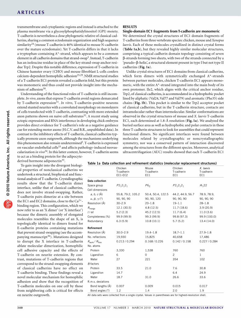
Ca2+-bound T-cadherin EC1-EC2 fragment is a nonswapped dimerTo investigate the potential role of other EC domains in T-cadherin homophilic interactions, we determined the 2.9-Å crystal structure of an EC1-EC2 ectodomain fragment of chicken T-cadherin. In the crystal structure (Fig. 2), both the EC1 and EC2 domains display the usual β-sandwich fold typical of cadherin domains, and three Ca2+ ions coordinated in the conserved interdomain calcium-binding site. The EC1 domain of this structure is virtually identical to those deter-mined in the single-domain structures presented above, with an r.m.s. deviation of less than 1 Å for main chain atoms.
The crystallographic asymmetric unit of the EC1-EC2 structure contains two molecules, which come together to form an X-shaped two-fold symmetric dimer that buries 2,173 Å2 of solvent-accessible surface. The contact area of the dimer is centered on the interdomain linker region (residues 99–104, which show 447.6 Å2 of buried surface area upon dimerization) but also involves both EC1 and EC2 domains (which show buried surface areas of 926.0 Å2 and 799.4 Å2, respectively). Contacts across EC1 domains are formed through two hydrophobic patches, mainly involving resi-dues from the N-terminal A-strand (Fig. 2e). The first of these includes side chain inter-actions between residues Leu3, Thr5 and Ile25 and is located near the ‘top’ of EC1. The sec-ond locus of EC1-EC1 interactions is found toward the ‘bottom’ of EC1 and involves side chains from Leu8, Pro10 and Ile98. The ‘core’ of the X dimer, around the juxtaposed calcium-binding sites, is characterized by hydrophobic contacts mediated by the side chains of residues that emanate from the EC2 domains: Met198, Val203 and Leu205 (Fig. 2c). These numerous hydrophobic con-tacts are likely to help stabilize the X dimer.
Additionally, polar and ionic interactions are detected within or flanking the core region of the X dimer. First, in the center of the core region, linker residues Asp99 and Gln100 of
each protomer, which also coordinate Ca2+, are hydrogen bonded to each other across the dimer interface. Flanking the core region, Arg14 from the EC1 domain of each protomer forms hydrogen bonds with the backbone carbonyl groups from Pro137 and Thr139 in the partner EC2 domain. Notably, the relative positioning of Arg14 and Asp140 of the opposite EC2 domain, even though not a canonical salt bridge, suggests that they form one at least transiently during regular molecu-lar dynamic motions. In addition, Arg104 is hydrogen bonded to the backbone of Asp202 from the partner EC2 domain (Fig. 2c).
The T-cadherin X-shaped dimer described here closely resembles previously observed EC1-EC2 dimers found in E-cadherin crystal structures derived from constructs with extra residues at their N termini (r.m.s. deviation ~1.9 Å for 363 core α-carbons) (Fig. 2b)14,19, which are unable to strand-swap14,18,37. The T-cadherin and these E-cadherin X-dimer interfaces are topologically identical and are of remarkably similar size (2,173 versus 2,170.7 Å2 buried surface area) and chemical character (Fig. 2c,d). In each case, a large hydrophobic area is buried upon dimerization and probably aids in dimer stabiliza-tion. In both cases, hydrophobic contacts established along the length of the EC1 domain involve residues from the N-terminal A-strand (Fig. 2e). Within the core region, both structures show interactions involving the side chains of hydrophobic residues emanating from
ChickenMouseXenopus
C F
DE
B
A
D E FB
G
N
Arg26
Asp88Val79
N
Phe35IIe2
Val91Val77
Val24Ala4
C
C
G
a b
Figure 1 Structures of T-cadherin EC1 domains. (a) Superposed ribbon diagrams from crystal structures of mouse, chicken and X. laevis T-cadherins. (b) Expanded view of A-strand region. The residue highlighted in yellow, Ile2, aligns with the strand-swap anchor tryptophan residue of classical cadherins. Side chains are shown for residues with contacts less than 4 Å from Ile2 side chain.
Table 1b Data collection, phasing and refinement statistics for the structure determination of mouse EC1-EC2 T-cadherin by SIR/AS
Mouse T-cadherin EC1-EC2 native
Mouse T-cadherin EC1-EC2 methyl mercury
Mouse T-cadherin EC1-EC2 bromine
Data collection
Space group H32 H32 H32
Cell dimensions
a, b, c (Å) 107.7, 107.7, 226.7 107.7, 107.7, 226.4 108.6, 108.6, 227.3
a, b, g (°) 90, 90, 120 90, 90, 120 90, 90, 120
Resolution (Å) 20–2.0 20–2.4 20–2.7
Rmerge 6.7 (27.3) 5.9 (27.3) 6.9 (27.4)
I / σI 31.4 (11.6) 38.5 (9.8) 47.3 (12.9)
Completeness (%) 100.0 (100.0) 100.0 (100.0) 100.0 (100.0)
Redundancy 11.1 (10.1) 11.2 (10.1) 22.2 (18.6)
Refinement
Resolution (Å) 20.0–2.0
No. reflections 32,616
Rwork / Rfree 0.213 / 0.259
No. atoms
Protein 3,356
Ligand/ion 0
Water 195
B-factors
Protein 29.6
Water 30.0
R.m.s deviations
Bond lengths (Å) 0.020
Bond angles (°) 2.00
All data sets were collected from a single crystal. Values in parentheses are for highest-resolution shell.
© 2
010
Nat
ure
Am
eric
a, In
c. A
ll ri
gh
ts r
eser
ved
.

342 VOLUME 17 NUMBER 3 MARCH 2010 nature structural & molecular biology
a r t i c l e s
the EC2 domain. The flanking edges of the interface are demarcated by similar polar interactions: in the T-cadherin structure, the Arg14-Asp140 and Arg104-Asp202 interactions correspond to well-formed salt bridges (Lys14-Asp138 and Arg105-Glu199) in the E-cadherin X-dimer structure.
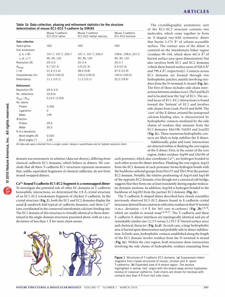
Structure of a Ca2+-free T-cadherin EC1-EC2 ectodomain fragmentBoth T-cadherin and classical cadherins require the presence of calcium to mediate adhesion25,29. However, structures of multidomain cadherins have not been determined in the absence of calcium. We therefore determined the 2.0-Å crystal structure of a mouse T-cadherin EC1-EC2 fragment (Fig. 3) crystallized in a Ca2+-free form by the inclusion of citrate in the crystallization experiment. Citrate behaves as a calcium chelator, and thus no Ca2+ ions are found in this structure. Although we observed no substantial differences between individual domains in the Ca2+-free structure and the equivalent ones in the Ca2+-bound EC1-EC2 structure presented above, we find that the relative orientation between domains and the quaternary struc-ture are markedly altered.
In the Ca2+-free T-cadherin structure, the EC1 and EC2 domains of the T-cadherin monomer fold onto each other, creating a clamshell-like structure. A relatively small (927 Å2) intramolecular interface is found between the EC1 and EC2 domains, with the interface formed mainly by van der Waals contacts. Few specific interactions, such as hydrogen bonds or salt bridges, are formed. It appears, therefore, that this intramolecular interface is due to crystal packing effects. Nevertheless, our observations imply that the absence of Ca2+ leaves six unbalanced negative charges on the calcium-coordinating residues in the interdomain region and permits much greater flexibility of the linker region. Ca2+-dependent rigidity of interdomain regions of classical cadherins has been shown previously by NMR analyses of
Ca2+-free E-cadherin38, and electron micro-scopic images have also shown the ‘collapse’ of rigid E-cadherin ectodomains upon Ca2+ removal15. The T-cadherin EC1-EC2 Ca2+-bound and Ca2+-free structures now provide an atomic-level view of the effects of Ca2+ in the stiffening of the cadherin ectodomain. Notably, T-cadherin EC1-EC2 fragments do not associate as dimers in the Ca2+-free structure or in solution (see below). Taken together, the Ca2+-bound and Ca2+-free T-cadherin EC1-EC2 structures show how
removal of Ca2+ can prevent formation of the dimer interface found in the Ca2+-bound form, which requires a rigid interdomain linker and the appropriate orientation of the EC1 and EC2 domains.
In summary, the crystallographic studies of T-cadherin ectodomain fragments presented here show that T-cadherin is similar in over-all structure to classical cadherins but differs markedly in the EC1 domain region. These differences prevent binding through strand-swapping, the primary adhesive binding mode for classical cadherins. Instead, T-cadherin EC1-EC2 forms a Ca2+-dependent X-shaped dimer that involves interaction between elements near the Ca2+-binding regions of partner T-cadherin molecules.
EC1
a c
db
e
Thr139
Leu205
Arg104
Asp102
Asn103
Gln100 Asn12 Arg14
Lys19
Glu13Asn12
Lys14
Leu196Leu201
Arg105
Asn140
Asp138Tyr142
Leu196
Glu199
Ser138
Thr139Asp99
Val203
Met198
Asp202
Leu143
Pro137Asp140
EC2
EC1
EC2
lle25 Val3 Trp2
Trp2lle4
Pro5Pro6
lle7Ser8Thr99
Pro10
Leu3
Thr5Leu8
lle98
Pro10
90°
90°
N N
N N
T-cadherin
T-cadherin E-cadherin
Mutant (1EDH) Wild type (1Q1P)
MutantE-cadherin
Figure 2 Structure of the X dimer from chicken T-cadherin, and comparison with the mutant E-cadherin X-dimer structure14 (PDB 1EDH). (a) Ribbon diagram of T-cadherin X dimer, with bound calcium ions shown as green spheres. (b) X dimer from E-cadherin N-terminal extension mutant. (c,d) Close-up view showing atomic-level interactions in the T-cadherin interface (c) and the E-cadherin interface (d). Interactions are described in the main text. Asn12, Asp102, Asp103, Ser138 and Leu143 are buried in the T-cadherin X-dimer interface but do not participate in identifiable specific interactions. Residues Arg14 and Asp140, underlined in c, were mutated to disrupt the T-cadherin interface (see text). (e) Comparison of EC1 domain interface regions of the T-cadherin X dimer with the X dimer and strand-swapped dimer of E-cadherin (from ref. 18).
Asp66
Glu68Asn101
Asn103
Asp102
Asp99Glu11
Asn12
EC1 EC2
N
C
Asp202
Asp197
Gln100
Figure 3 Calcium-free structure of mouse T-cadherin EC1-EC2. Side chains within the calcium-binding region are shown.
© 2
010
Nat
ure
Am
eric
a, In
c. A
ll ri
gh
ts r
eser
ved
.
nature structural & molecular biology VOLUME 17 NUMBER 3 MARCH 2010 343
a r t i c l e s
These structural observations raise the question of whether the crystallographically observed X-shaped dimer corresponds to the structure used by T-cadherin to mediate Ca2+-dependent cell adhesion. To address this question, we used biophysical and cellular assays with wild-type and site-directed mutant proteins to determine whether T-cadherin ectodomain dimerization in solution and in a cellular context depends on structural elements of the X dimer.
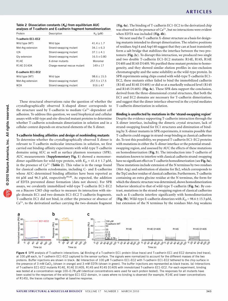
T-cadherin binding affinities and design of nonbinding mutantsTo determine whether the crystallographically observed X dimer is relevant to T-cadherin molecular interactions in solution, we first carried out binding-affinity experiments with wild-type T-cadherin domains using AUC as well as surface plasmon resonance (SPR). AUC measurements (Supplementary Fig. 1) showed a monomer-dimer equilibrium for wild-type protein, with KD = 41.4 ± 1.7 µM, in the presence of Ca2+ (Table 2). This value is in the range found for classical cadherin ectodomains, including C- and E-cadherins whose AUC-determined binding affinities have been reported as 64 µM and 96.5 µM, respectively39,40. As expected, the addition of EDTA prevented dimer formation (data not shown). For SPR assays, we covalently immobilized wild-type T-cadherin EC1-EC2 on a Biacore CM5 chip surface to measure its interaction with sin-gle-domain EC1 and two-domain EC1-EC2 T-cadherin fragments. T-cadherin EC1 did not bind, in either the presence or absence of Ca2+, to the derivatized surface carrying the two-domain fragment
(Fig. 4a). The binding of T-cadherin EC1-EC2 to the derivatized chip was observed in the presence of Ca2+, but no interactions were evident when EDTA was included (Fig. 4b).
We next used the T-cadherin X-dimer structure as a basis for design-ing mutants intended to disrupt dimerization. The relative positions of residues Arg14 and Asp140 suggest that they can at least transiently form a salt bridge that stabilizes the interface between the two pro-tomers (Fig. 2c). To disrupt this interaction, we produced two single and two double T-cadherin EC1-EC2 mutants: R14E, R14S, R14E D140S and R14S D140S. We purified these mutant proteins to homo-geneity, and they showed similar elution profiles in size-exclusion chromatography and the same solubility as the wild-type protein. In SPR experiments using chips coated with wild-type T-cadherin EC1-EC2, these mutants either failed to bind the immobilized cadherin (R14E and R14E D140S) or did so at a markedly reduced level (R14S and R14S D140S) (Fig. 4c). These SPR data support the conclusion, derived from the three-dimensional crystal structures, that both the EC1 and EC2 domains are necessary for T-cadherin dimerization, and suggest that the dimer interface observed in the crystal mediates T-cadherin dimerization in solution.
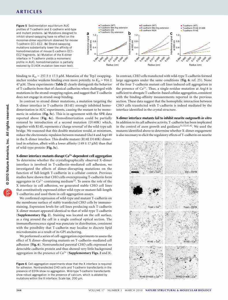
Binding is unaffected by mutations in the ‘strand-swapping region’Despite the evidence supporting T-cadherin interaction through the X-dimer interface, including the dimeric crystal structure, lack of strand-swapping found for EC1 structures and diminution of bind-ing by X-dimer mutants in SPR experiments, it remains possible that T-cadherin could engage in strand-swap binding as classical cadherins do. To test this possibility, we prepared T-cadherin EC1-EC2 proteins with mutations in either the X-dimer interface or the potential strand-swapping region, and assessed by AUC the effects of these mutations on homodimerization (Fig. 5). The introduction into T-cadherin of mutations known to interfere with classical cadherin strand-swapping have no significant effect on T-cadherin homodimerization (see Fig. 5a). These mutations include extension of the N terminus by two residues (Met-Arg) and substitution of alanine for Ile2, which corresponds to the Trp2 anchor residue of classical cadherins. Furthermore, T-cadherin containing an extra glycine residue at the N terminus, the form for which the dimeric structure was determined, shows homodimerization behavior identical to that of wild-type T-cadherin (Fig. 5a). By con-trast, mutations in the strand-swapping region of classical cadherins such as E-cadherin interfere significantly with homodimerization (Fig. 5b). Wild-type E-cadherin dimerizes with KD = 98.6 ± 15.5 µM, but extension of the N terminus by the residues Met-Arg weakens
80
a b cEC1-EC2
+CaCl2
+EDTAEC1
60
40
Nor
mal
ized
res
pons
e (R
U)
20
80 100
12060
Time (s)
40200
80
60
40
90
60R14E R14S R14S D140SR14E D140S
100 µM
50T-cadherin WT
2512.56.253.131.56, 0.780
30
0
Res
pons
e (R
U)
Res
pons
e (R
U)
20
buffer00 80 10
012
060
Time (s)
40200 80 100
12060
Time (s)
40200 80 100
1206040200 80 10
012
06040200 80 100
1206040200 80 10
012
06040200
Figure 4 SPR analysis of T-cadherin interactions. (a) Binding of a T-cadherin EC1 protein (blue trace) and T-cadherin EC1 and EC2 domains (red trace), at 100 µM each, to T cadherin EC1-EC2 captured to the sensor surface. The signals were normalized to account for the different masses of the two proteins. Buffer injections are shown in black. (b) Interaction of 100 µM T-cadherin EC1-EC2 with T-cadherin EC1-EC2 tethered to the chip surface in the presence of 3 mM CaCl2 (shown in orange) and 3 mM EDTA (shown in green). The buffer injections are represented as black traces. (c) Interactions of T-cadherin EC1-EC2 mutants R14E, R14E D140S, R14S and R14S D140S with immobilized T-cadherin EC1-EC2. For each experiment, binding was tested at a concentration range 100–0.78 µM (identical concentrations were used for each protein tested). The responses for all mutants have been scaled to the responses of the wild-type EC1-EC2 domain, in cases where no binding is observed (for example, R14E and lower concentrations of R14S), the traces collapse together at baseline response.
Table 2 Dissociation constants (KD) from equilibrium AUC analyses of T-cadherin and E-cadherin fragment homodimerizationProtein Description KD (µM)
T-cadherin EC1-EC2
Wild type (WT) Wild type 41.4 ± 1.7
Met-Arg extension Strand-swapping mutant 34.1 ± 6.3
I2A Strand-swapping mutant 37.1 ± 4.1
Gly extension Strand-swapping mutant 16.5 ± 0.80
R14E X-dimer mutants Monomer
R14E D140K Charge-reversal rescue mutant 149 ± 17
E-cadherin EC1-EC2
Wild type (WT) Wild type 98.6 ± 15.5
Met-Arg extension Strand-swapping mutant 257.5 ± 17.5
W2A Strand-swapping mutant 916 ± 47
© 2
010
Nat
ure
Am
eric
a, In
c. A
ll ri
gh
ts r
eser
ved
.
344 VOLUME 17 NUMBER 3 MARCH 2010 nature structural & molecular biology
a r t i c l e s
Ctrl + Ca2+
Tcad + Ca2+
Ctrl + EDTA
Tcad + EDTA
R14E + Ca2+
R14E D140S + Ca2+ R14S D140S + Ca2+
R14S + Ca2+
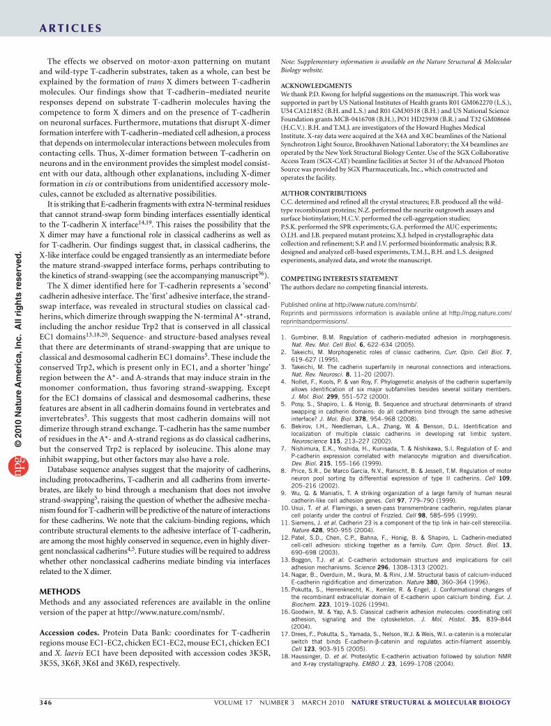
Figure 6 Cell-aggregation experiments show that the X interface is required for adhesion. Nontransfected CHO cells and T-cadherin transfectants in the presence of EDTA show no aggregation. Wild-type T-cadherin transfectants show robust aggregation in the presence of calcium, which is ablated by mutations within the X interface. Scale bar, 200 µm.
binding to KD = 257.5 ± 17.5 µM. Mutation of the Trp2 swapping-anchor residue weakens binding even more potently, to KD = 916 ± 47 µM. These experiments (Table 2) clearly distinguish the behavior of T-cadherin from that of classical cadherins when challenged with mutations in the strand-swapping region, and suggest that T-cadherin does not engage in strand-swap binding.
In contrast to strand dimer mutations, a mutation targeting the X-dimer interface in T-cadherin (R14E) strongly inhibited homo-dimerization in AUC experiments, causing the mutant to be mono-meric in solution (Fig. 5c). This is in agreement with the SPR data reported above (Fig. 4c). Homodimerization could be partially restored by introduction of a second mutation (D140K) which, together with R14E, represents a ‘charge reversal’ of the wild-type salt bridge. We reasoned that this double mutation would, at minimum, reduce the electrostatic repulsion between mutated Glu14 and Asp140 in the X-dimer interface. This double mutant (R14E D140K) dimer-ized in solution, albeit with a lower affinity (149 ± 17 µM) than that of wild-type protein (Fig. 5c).
X-dimer interface mutants disrupt Ca2+-dependent cell aggregationTo determine whether the crystallographically observed X-dimer interface is involved in T-cadherin–mediated cell adhesion, we investigated the effects of dimer-disrupting mutations on the function of full-length T-cadherin in a cellular context. Previous studies have shown that CHO cells overexpressing T-cadherin form aggregates in Ca2+-containing medium25. To assess the role of the X interface in cell adhesion, we generated stable CHO cell lines that constitutively expressed either wild-type or mutant full-length T-cadherins and used them in cell-aggregation assays.
We confirmed expression of wild-type and mutant T-cadherin on the membrane surface of stably transfected CHO cells by immuno-staining. Expression levels for cell lines producing each T-cadherin X-dimer mutant appeared identical to that of wild-type T-cadherin (Supplementary Fig. 2). Staining was located on the cell surface, as a ring around the cell in a single confocal optical section. The immunofluorescence signal was punctate in distribution, consistent with the possibility that T-cadherin may localize to discrete lipid microdomains as a result of its GPI anchoring.
We performed a series of cell-aggregation experiments to assess the effect of X dimer–disrupting mutants on T-cadherin–mediated cell adhesion (Fig. 6). Nontransfected parental CHO cells expressed no detectable cadherin protein and thus showed very little background aggregation in the presence of Ca2+ (Supplementary Figs. 2 and 3).
In contrast, CHO cells transfected with wild-type T-cadherin formed large aggregates under the same conditions (Fig. 6; ref. 25). None of the four T-cadherin mutant cell lines induced cell aggregation in the presence of Ca2+. Thus, a single-residue mutation at Arg14 is sufficient to abrogate T-cadherin–based cellular aggregation, consistent with the binding-affinity measurements reported in the previous section. These data suggest that the homophilic interaction between CHO cells transfected with T-cadherin is indeed mediated by the interface identified in the crystal structure.
X-dimer interface mutants fail to inhibit neurite outgrowth in vitroIn addition to its cell adhesive activity, T-cadherin has been implicated in the control of axon growth and guidance31,32,41,42. We used the mutants identified above to determine whether X-dimer engagement is also necessary to elicit the regulatory effects of T-cadherin on neurite
Figure 5 Sedimentation equilibrium AUC profiles of T-cadherin and E-cadherin wild-type and mutant proteins. (a) Mutations designed to inhibit strand-swapping have no effect on the monomer-dimer equilibrium observed for mouse T-cadherin EC1-EC2. (b) Strand-swapping mutations substantially lower the affinity of homodimerization of mouse E-cadherin EC1-EC2 fragments. (c) Mutation of the X-dimer interface in T-cadherin yields a monomeric profile in AUC; homodimerization is partially restored by D140K mutation (see main text).
1.2a b c
0.8
T-cadherin (WT)T-cadherin Met-Arg extensionT-cadherin I2AT-cadherin Gly extension
0.4
Inte
rfer
ence
sig
nal
at 6
00 n
m
5.98
45.
995
6.00
66.
017
6.02
86.
039
6.05
05.
984
5.99
56.
006
6.01
76.
028
6.03
96.
050
5.98
45.
995
6.00
66.
017
Radius (cm)Radius (cm)Radius (cm)
6.02
86.
039
6.05
0
0
1.2
0.8
0.4
Inte
rfer
ence
sig
nal
at 6
00 n
m
0
1.2
0.8
0.4
Inte
rfer
ence
sig
nal
at 6
00 n
m
0
T-cadherin (WT)T-cadherin R14E D140KT-cadherin R14E
E-cadherin (WT)E-cadherin Met-Arg extensionE-cadherin W2A
© 2
010
Nat
ure
Am
eric
a, In
c. A
ll ri
gh
ts r
eser
ved
.

nature structural & molecular biology VOLUME 17 NUMBER 3 MARCH 2010 345
a r t i c l e s
outgrowth. Neurite outgrowth from motor neurons is robust when the neurons are grown on a monolayer of naive CHO cells, but growth is stunted when they are grown on a monolayer of CHO cells that stably express T-cadherin32. To determine whether structural elements of the X dimer are necessary for T-cadherin–mediated inhibition of neurite outgrowth, we assessed the extension of motor axons when presented with an X dimer–incompetent R14S D140S substrate.
Neurite outgrowth assays were performed with mouse embry-onic stem (ES) cell–derived motor neurons43 grown on naive, T-cadherin–expressing and R14S D140S T-cadherin–expressing CHO cell monolayers. We verified surface expression of wild-type and mutant T-cadherins in stably transfected CHO cells by surface biotinylation (Supplementary Fig. 3a). The ES cell–derived motor neurons express T-cadherin, as shown by western blotting analysis with an anti– T-cadherin antibody (Supplementary Fig. 3b). Immunostaining with the same antibody showed that the majority of ES cell–derived motor neurons express T-cadherin on their surfaces (data not shown). ES cell–derived motor neurons cultured on naive CHO cell monolayers for ~18 h grew long axons (Fig. 7a,b). In agreement with previous reports32, we observed an ~50% reduction in axonal length when motor neurons were cultured on CHO cells expressing wild-type T-cadherin (Fig. 7a,b). In contrast, neurite lengths of motor neurons grown on CHO cells expressing the R14S D140S T-cadherin mutant were simi-lar to those observed on naive control cells (Fig. 7a,b). Thus, the T-cadherin–mediated inhibitory effect on neurite length is abolished by a mutation that disrupts the T-cadherin X-dimer interface.
Nevertheless, since ES cell–derived motor neurons express wild-type T-cadherin, the question remains whether homophilic inter-action between the neuron- and CHO cell–presented T-cadherin is required for effects on neurite outgrowth. To address this question, we performed similar experiments using spinal neurons dissected from wild-type and T-cadherin knockout mice34. We cultured dissoci-ated neurons on monolayers of CHO cells expressing either wild-type T-cadherin or one of the four dimer interface mutants, with naive CHO cells used as controls. After 21 h, we fixed the cultures and immuno-stained them using the neuron-specific antibody to βIII tubulin (TUJ-1). We measured and compared neurite lengths in each condition.
Spinal neurons isolated from wild-type mice showed an ~50% reduction of neurite length on wild-type T-cadherin substrates in comparison with parental nontransfected CHO control cells. However, neurons isolated from T-cadherin knockout mice extended axons to a
similar length as neurons presented with control substrates (Fig. 7c). Neurons from wild-type mice grown on substrates expressing each of the dimer-interface mutants also showed neurite lengths similar to neurons grown on control substrates (Fig. 7c). Together, these results provide evidence that T-cadherin–mediated neurite outgrowth inhibit-ion relies on the ability to form trans dimers using the nonswapped X interface identified in the crystal structure.
DISCUSSIONMechanisms of classical cadherin adhesion have been understood mainly through the strand-swap model of binding between cadherin EC1 domains13,18,22,44. However, the vast majority of cadherins lack the sequence determinants of strand-swap binding5. The structures of T-cadherin presented here define a binding mechanism for a nonclassical cadherin that lacks the signatures of strand-swapping. Our results show that T-cadherin engages in intercellular recogni-tion through a homophilic interface that is distinct from that of classical cadherins. Cell-based experiments in vitro show that bind-ing through this interface is required for cell adhesion and that T-cadherin–mediated inhibition of neurite outgrowth depends on the homophilic engagement of T-cadherin.
The novel adhesive interface of T-cadherin, designated here the X interface, joins two elongated EC1-EC2 molecules through a region near their Ca2+-binding sites to form a tetrahedral, X-like form. A number of the Ca2+-liganding residues participate in contacts that span this interface (Supplementary Fig. 4). The X interface buries approximately 2,200 Å2, substantially more than the interfaces of strand-swapped type I cadherins (between 1,600 and 1,800 Å2) and slightly less than that of type II cadherins (around 2,700 Å2). Yet these interfaces differ in character: whereas the X interface is formed exclu-sively by surface interactions with shallow features, the strand-swap interface is centered on the insertion of hydrophobic tryptophan side chains into the hydrophobic core of the cadherin binding mate. Despite a far less intimate association than in strand-swapped cadherin dimers, the ~40-µM KD binding affinity of the T-cadherin X dimer is somewhat tighter than has been reported for both E-18,40,45,46 and C-cadherins39. This apparent paradox probably has its basis, in part, in the idea that domain swap binding leads to lowering of interaction affinities23, but it may also be a result of the removal of charged groups, including the bound Ca2+ ions, from the vicinity of the solvent environment. In fact, both sets of binding affinities are relatively weak compared to protein-protein interfaces that bury comparable amounts of surface area23.
The similarity in dimerization affinities between T-cadherin and classical cadherins is consistent with the ability of T-cadherin to mediate cell adhesion. The cell-aggregation assays reported here confirm previous work25 showing that T-cadherin promotes cell-cell adhe-sion despite the absence of a cytoplasmic or transmembrane domain. Moreover, the inhibition of adhesion by mutants designed to disrupt dimerization suggests that the X interface represents the molecular interaction through which T-cadherin mediates adhesion.
120
Naivea
b c
T-cad T-cad R14S D140S
Naive
T-cad
T-cad R14S D140S
Mea
n ne
urite
leng
th(%
of c
ontr
ol, ±
SE
M)
100
80
60
40
20
0
120
140M
ean
neur
ite le
ngth
(% o
f con
trol
, ± S
EM
)
100
80 *60
40
T-cad
T-cad
R14
S
T-cad
R14E D
140S
T-cad
R14S D
140S
WT neurons
KO neurons
*P < 0.05 ANOVA
20
0
Figure 7 T-cadherin–mediated inhibition of neurite outgrowth depends on the X interface. (a) Fluorescence micrographs of HB9:GFP ES cell–derived motor neurons cultured on either naive CHO cells, T-cadherin–expressing CHO cells or X-interface mutant T-cadherin expressors. T-cad, T-cadherin. (b) Mean neurite outgrowth length of ES cell–derived motor neurons cultured on naive, wild-type T-cadherin–expressing or mutant T-cadherin–expressing CHO cells. T-cad, T-cadherin. (c) Mean neurite outgrowth length of cultured mouse spinal motor neurons from wild-type and T-cadherin knockout animals, cultured on naive, wild-type T-cadherin–expressing or mutant T-cadherin–expressing CHO cells. T-cad, T-cadherin.
© 2
010
Nat
ure
Am
eric
a, In
c. A
ll ri
gh
ts r
eser
ved
.
346 VOLUME 17 NUMBER 3 MARCH 2010 nature structural & molecular biology
a r t i c l e s
The effects we observed on motor-axon patterning on mutant and wild-type T-cadherin substrates, taken as a whole, can best be explained by the formation of trans X dimers between T-cadherin molecules. Our findings show that T-cadherin–mediated neurite responses depend on substrate T-cadherin molecules having the competence to form X dimers and on the presence of T-cadherin on neuronal surfaces. Furthermore, mutations that disrupt X-dimer formation interfere with T-cadherin–mediated cell adhesion, a process that depends on intermolecular interactions between molecules from contacting cells. Thus, X-dimer formation between T-cadherin on neurons and in the environment provides the simplest model consist-ent with our data, although other explanations, including X-dimer formation in cis or contributions from unidentified accessory mole-cules, cannot be excluded as alternative possibilities.
It is striking that E-cadherin fragments with extra N-terminal residues that cannot strand-swap form binding interfaces essentially identical to the T-cadherin X interface14,19. This raises the possibility that the X dimer may have a functional role in classical cadherins as well as for T-cadherin. Our findings suggest that, in classical cadherins, the X-like interface could be engaged transiently as an intermediate before the mature strand-swapped interface forms, perhaps contributing to the kinetics of strand-swapping (see the accompanying manuscript36).
The X dimer identified here for T-cadherin represents a ‘second’ cadherin adhesive interface. The ‘first’ adhesive interface, the strand-swap interface, was revealed in structural studies on classical cad-herins, which dimerize through swapping the N-terminal A*-strand, including the anchor residue Trp2 that is conserved in all classical EC1 domains13,18,20. Sequence- and structure-based analyses reveal that there are determinants of strand-swapping that are unique to classical and desmosomal cadherin EC1 domains5. These include the conserved Trp2, which is present only in EC1, and a shorter ‘hinge’ region between the A*- and A-strands that may induce strain in the monomer conformation, thus favoring strand-swapping. Except for the EC1 domains of classical and desmosomal cadherins, these features are absent in all cadherin domains found in vertebrates and invertebrates5. This suggests that most cadherin domains will not dimerize through strand exchange. T-cadherin has the same number of residues in the A*- and A-strand regions as do classical cadherins, but the conserved Trp2 is replaced by isoleucine. This alone may inhibit swapping, but other factors may also have a role.
Database sequence analyses suggest that the majority of cadherins, including protocadherins, T-cadherin and all cadherins from inverte-brates, are likely to bind through a mechanism that does not involve strand-swapping5, raising the question of whether the adhesive mecha-nism found for T-cadherin will be predictive of the nature of interactions for these cadherins. We note that the calcium-binding regions, which contribute structural elements to the adhesive interface of T-cadherin, are among the most highly conserved in sequence, even in highly diver-gent nonclassical cadherins4,5. Future studies will be required to address whether other nonclassical cadherins mediate binding via interfaces related to the X dimer.
METHODSMethods and any associated references are available in the online version of the paper at http://www.nature.com/nsmb/.
Accession codes. Protein Data Bank: coordinates for T-cadherin regions mouse EC1-EC2, chicken EC1-EC2, mouse EC1, chicken EC1 and X. laevis EC1 have been deposited with accession codes 3K5R, 3K5S, 3K6F, 3K6I and 3K6D, respectively.
Note: Supplementary information is available on the Nature Structural & Molecular Biology website.
ACKNOwLedGMeNTSWe thank P.D. Kwong for helpful suggestions on the manuscript. This work was supported in part by US National Institutes of Health grants R01 GM062270 (L.S.), U54 CA121852 (B.H. and L.S.) and R01 GM30518 (B.H.) and US National Science Foundation grants MCB-0416708 (B.H.), PO1 HD25938 (B.R.) and T32 GM08666 (H.C.V.). B.H. and T.M.J. are investigators of the Howard Hughes Medical Institute. X-ray data were acquired at the X4A and X4C beamlines of the National Synchrotron Light Source, Brookhaven National Laboratory; the X4 beamlines are operated by the New York Structural Biology Center. Use of the SGX Collaborative Access Team (SGX-CAT) beamline facilities at Sector 31 of the Advanced Photon Source was provided by SGX Pharmaceuticals, Inc., which constructed and operates the facility.
AUTHOR CONTRIBUTIONSC.C. determined and refined all the crystal structures; F.B. produced all the wild-type recombinant proteins; N.Z. performed the neurite outgrowth assays and surface biotinylation; H.C.V. performed the cell-aggregation studies; P.S.K. performed the SPR experiments; G.A. performed the AUC experiments; O.J.H. and J.B. prepared mutant proteins; X.J. helped in crystallographic data collection and refinement; S.P. and J.V. performed bioinformatic analysis; B.R. designed and analyzed cell-based experiments, T.M.J., B.H. and L.S. designed experiments, analyzed data, and wrote the manuscript.
COMPeTING INTeReSTS STATeMeNTThe authors declare no competing financial interests.
Published online at http://www.nature.com/nsmb/. Reprints and permissions information is available online at http://npg.nature.com/reprintsandpermissions/.
1. Gumbiner, B.M. Regulation of cadherin-mediated adhesion in morphogenesis. Nat. Rev. Mol. Cell Biol. 6, 622–634 (2005).
2. Takeichi, M. Morphogenetic roles of classic cadherins. Curr. Opin. Cell Biol. 7, 619–627 (1995).
3. Takeichi, M. The cadherin superfamily in neuronal connections and interactions. Nat. Rev. Neurosci. 8, 11–20 (2007).
4. Nollet, F., Kools, P. & van Roy, F. Phylogenetic analysis of the cadherin superfamily allows identification of six major subfamilies besides several solitary members. J. Mol. Biol. 299, 551–572 (2000).
5. Posy, S., Shapiro, L. & Honig, B. Sequence and structural determinants of strand swapping in cadherin domains: do all cadherins bind through the same adhesive interface? J. Mol. Biol. 378, 954–968 (2008).
6. Bekirov, I.H., Needleman, L.A., Zhang, W. & Benson, D.L. Identification and localization of multiple classic cadherins in developing rat limbic system. Neuroscience 115, 213–227 (2002).
7. Nishimura, E.K., Yoshida, H., Kunisada, T. & Nishikawa, S.I. Regulation of E- and P-cadherin expression correlated with melanocyte migration and diversification. Dev. Biol. 215, 155–166 (1999).
8. Price, S.R., De Marco Garcia, N.V., Ranscht, B. & Jessell, T.M. Regulation of motor neuron pool sorting by differential expression of type II cadherins. Cell 109, 205–216 (2002).
9. Wu, Q. & Maniatis, T. A striking organization of a large family of human neural cadherin-like cell adhesion genes. Cell 97, 779–790 (1999).
10. Usui, T. et al. Flamingo, a seven-pass transmembrane cadherin, regulates planar cell polarity under the control of Frizzled. Cell 98, 585–595 (1999).
11. Siemens, J. et al. Cadherin 23 is a component of the tip link in hair-cell stereocilia. Nature 428, 950–955 (2004).
12. Patel, S.D., Chen, C.P., Bahna, F., Honig, B. & Shapiro, L. Cadherin-mediated cell-cell adhesion: sticking together as a family. Curr. Opin. Struct. Biol. 13, 690–698 (2003).
13. Boggon, T.J. et al. C-cadherin ectodomain structure and implications for cell adhesion mechanisms. Science 296, 1308–1313 (2002).
14. Nagar, B., Overduin, M., Ikura, M. & Rini, J.M. Structural basis of calcium-induced E-cadherin rigidification and dimerization. Nature 380, 360–364 (1996).
15. Pokutta, S., Herrenknecht, K., Kemler, R. & Engel, J. Conformational changes of the recombinant extracellular domain of E-cadherin upon calcium binding. Eur. J. Biochem. 223, 1019–1026 (1994).
16. Goodwin, M. & Yap, A.S. Classical cadherin adhesion molecules: coordinating cell adhesion, signaling and the cytoskeleton. J. Mol. Histol. 35, 839–844 (2004).
17. Drees, F., Pokutta, S., Yamada, S., Nelson, W.J. & Weis, W.I. α-catenin is a molecular switch that binds E-cadherin-β-catenin and regulates actin-filament assembly. Cell 123, 903–915 (2005).
18. Haussinger, D. et al. Proteolytic E-cadherin activation followed by solution NMR and X-ray crystallography. EMBO J. 23, 1699–1708 (2004).
© 2
010
Nat
ure
Am
eric
a, In
c. A
ll ri
gh
ts r
eser
ved
.
nature structural & molecular biology VOLUME 17 NUMBER 3 MARCH 2010 347
a r t i c l e s
19. Pertz, O. et al. A new crystal structure, Ca2+ dependence and mutational analysis reveal molecular details of E-cadherin homoassociation. EMBO J. 18, 1738–1747 (1999).
20. Shapiro, L. et al. Structural basis of cell-cell adhesion by cadherins. Nature 374, 327–337 (1995).
21. Tamura, K., Shan, W.S., Hendrickson, W.A., Colman, D.R. & Shapiro, L. Structure-function analysis of cell adhesion by neural (N-) cadherin. Neuron 20, 1153–1163 (1998).
22. Patel, S.D. et al. Type II cadherin ectodomain structures: implications for classical cadherin specificity. Cell 124, 1255–1268 (2006).
23. Chen, C.P., Posy, S., Ben-Shaul, A., Shapiro, L. & Honig, B.H. Specificity of cell-cell adhesion by classical cadherins: critical role for low-affinity dimerization through β-strand swapping. Proc. Natl. Acad. Sci. USA 102, 8531–8536 (2005).
24. Ranscht, B. & Dours-Zimmermann, M.T. T-cadherin, a novel cadherin cell adhesion molecule in the nervous system lacks the conserved cytoplasmic region. Neuron 7, 391–402 (1991).
25. Vestal, D.J. & Ranscht, B. Glycosyl phosphatidylinositol–anchored T-cadherin mediates calcium-dependent, homophilic cell adhesion. J. Cell Biol. 119, 451–461 (1992).
26. Miskevich, F., Zhu, Y., Ranscht, B. & Sanes, J.R. Expression of multiple cadherins and catenins in the chick optic tectum. Mol. Cell. Neurosci. 12, 240–255 (1998).
27. Doyle, D.D. et al. T-cadherin is a major glycophosphoinositol-anchored protein associated with noncaveolar detergent-insoluble domains of the cardiac sarcolemma. J. Biol. Chem. 273, 6937–6943 (1998).
28. Koller, E. & Ranscht, B. Differential targeting of T- and N-cadherin in polarized epithelial cells. J. Biol. Chem. 271, 30061–30067 (1996).
29. Sacristan, M.P., Vestal, D.J., Dours-Zimmermann, M.T. & Ranscht, B. T-cadherin 2: molecular characterization, function in cell adhesion, and coexpression with T-cadherin and N-cadherin. J. Neurosci. Res. 34, 664–680 (1993).
30. Dames, S.A. et al. Insights into the low adhesive capacity of human T-cadherin from the NMR structure of its N-terminal extracellular domain. J. Biol. Chem. 283, 23485–23495 (2008).
31. Fredette, B.J. & Ranscht, B. T-cadherin expression delineates specific regions of the developing motor axon-hindlimb projection pathway. J. Neurosci. 14, 7331–7346 (1994).
32. Fredette, B.J., Miller, J. & Ranscht, B. Inhibition of motor axon growth by T-cadherin substrata. Development 122, 3163–3171 (1996).
33. Ivanov, D. et al. Expression of cell adhesion molecule T-cadherin in the human vasculature. Histochem. Cell Biol. 115, 231–242 (2001).
34. Hebbard, L.W. et al. T-cadherin supports angiogenesis and adiponectin association with the vasculature in a mouse mammary tumor model. Cancer Res. 68, 1407–1416 (2008).
35. Hug, C. et al. T-cadherin is a receptor for hexameric and high-molecular-weight forms of Acrp30/adiponectin. Proc. Natl. Acad. Sci. USA 101, 10308–10313 (2004).
36. Harrison, O. et al. Two-step adhesive binding by classical cadherins. Nat. Struct. Mol. Biol. advance online publication, doi:10:1038/nsmb.1784 (28 February 2010).
37. Harrison, O.J., Corps, E.M. & Kilshaw, P.J. Cadherin adhesion depends on a salt bridge at the N-terminus. J. Cell Sci. 118, 4123–4130 (2005).
38. Haussinger, D. et al. Calcium-dependent homoassociation of E-cadherin by NMR spectroscopy: changes in mobility, conformation and mapping of contact regions. J. Mol. Biol. 324, 823–839 (2002).
39. Chappuis-Flament, S., Wong, E., Hicks, L.D., Kay, C.M. & Gumbiner, B.M. Multiple cadherin extracellular repeats mediate homophilic binding and adhesion. J. Cell Biol. 154, 231–243 (2001).
40. Koch, A.W., Pokutta, S., Lustig, A. & Engel, J. Calcium binding and homoassociation of E-cadherin domains. Biochemistry 36, 7697–7705 (1997).
41. Bai, S., Datta, J., Jacob, S.T. & Ghoshal, K. Treatment of PC12 cells with nerve growth factor induces proteasomal degradation of T-cadherin that requires tyrosine phosphorylation of its cadherin domain. J. Biol. Chem. 282, 27171–27180 (2007).
42. Bai, S., Ghoshal, K. & Jacob, S.T. Identification of T-cadherin as a novel target of DNA methyltransferase 3B and its role in the suppression of nerve growth factor-mediated neurite outgrowth in PC12 cells. J. Biol. Chem. 281, 13604–13611 (2006).
43. Wichterle, H., Lieberam, I., Porter, J.A. & Jessell, T.M. Directed differentiation of embryonic stem cells into motor neurons. Cell 110, 385–397 (2002).
44. Parisini, E., Higgins, J.M., Liu, J.H., Brenner, M.B. & Wang, J.H. The crystal structure of human E-cadherin domains 1 and 2, and comparison with other cadherins in the context of adhesion mechanism. J. Mol. Biol. 373, 401–411 (2007).
45. Alattia, J.R. et al. Lateral self-assembly of E-cadherin directed by cooperative calcium binding. FEBS Lett. 417, 405–408 (1997).
46. Katsamba, P. et al. Linking molecular affinity and cellular specificity in cadherin-mediated adhesion. Proc. Natl. Acad. Sci. USA 106, 11594–11599 (2009).
© 2
010
Nat
ure
Am
eric
a, In
c. A
ll ri
gh
ts r
eser
ved
.

nature structural & molecular biology doi:10.1038/nsmb.1781
ONLINE METHODSCrystallographic methods. We expressed and purified proteins as described in Supplementary Methods. We grew crystals with the vapor diffusion method by mixing equal volumes of protein solution and crystallization buffer at 4 °C. The crystallization conditions were as follows (added cryoprotectants are indicated in parentheses): mouse T-cadherin EC1: 0.2 M ammonium sulfate, 20% (w/v) PEG 3350, pH 6.0 (30% (v/v) glycerol); chicken and X. laevis T-cadherin EC1 crystallized in the same conditions: 50 mM zinc acetate, 20% (w/v) PEG 3350 (35% (v/v) glycerol or 25% (v/v) ethylene glycol); mouse T-cadherin EC1-EC2: 50% saturated ammonium sulfate, 33 mM sodium citrate, pH 5.6 (15% (v/v) glycerol); chicken T-cadherin EC1-EC2: 20% (w/v) 2-methyl-2,4-pentanediol, 15% (w/v) PEG 400, 50 mM sodium acetate, pH 5.5 (35% (v/v) glycerol). We obtained two phasing derivatives of the mouse T-cadherin EC1-EC2 crys-tals by soaking either in mother liquor containing 1 mM methylmercury (II) chloride for 1 h or in cryoprotectant solution supplemented with 0.5 M NaBr for 30 s before flash freezing. Complete datasets from crystals frozen at 100 K were collected at the SGX-CAT beamline facilities (ID-31) at the Advanced Photon Source (mouse T-cadherin EC1-EC2) or at the National Synchrotron Light Source at Brookhaven National Laboratory, beamlines X4C (mouse T-cadherin EC1) and X29 (chicken T-cadherin EC1 and EC1-EC2, and X. laevis T-cadherin EC1). We processed and scaled the data with the HKL2000 suite47 (mouse T-cadherin EC1 and EC1-2), XDS (chicken T-cadherin EC1), or Mosflm48 and Scala49 (X. laevis T-cadherin EC1 and chicken T-cadherin EC1-EC2). We collected all datasets at a wavelength of 0.9791 Å, except in the bromine experiment, whose data we collected at 0.9202 Å.
We obtained initial low-resolution phases for the mouse T-cadherin EC1-EC2 crystal structure by SIR/AS analysis of the methyl mercury derivative using site positions determined with the program SOLVE50. These phases were then used to locate the positions of ordered bromine atoms in anomalous-difference Fourier maps. We performed phase calculations using both the mercury and bromine sites, using Sharp51 to generate experimental electron density maps. After solvent flattening using RESOLVE52, models of the individual mouse T-cadherin EC1 and EC2 domains were fit into the experimental maps and fitted manually using the program Coot53. We performed structure refinement with CNS54 and Refmac55. We solved all other crystal structures by molecular replacement with the program Phaser56 using the refined mouse T-cadherin EC1 or EC2 domain structures as independent search models. Rounds of manual model fitting in Coot were alternated with refinement using Refmac (all structures) and CNS (mouse and chicken T-cadherin EC1). Ramachandran angles were in the most favored regions for 87.3%, 79.0%, 92.6%, 93.8%, and 87.7% for the mouse EC1-EC2, chicken EC1-EC2, mouse EC1, chicken EC1 and X. laevis EC1 structures, respectively. Crystallographic statistics are summarized in Table 1.
Cell-aggregation and neurite-outgrowth assays. We performed short-term aggregation assays essentially as described previously, except that incubation time was 45 min (ref. 22). We prepared spinal neuron primary cultures from E14 wildtype (C57/BL6, Harlan–Sprague–Dawley) and T-cadherin knockout mice34. We removed spinal cords from 4–6 mouse embryos and dissociated cells in 30 units per ml papain (Worthington) for 45 min at 37°. We performed neurite outgrowth assays essentially as described57 by depositing ~7.5 × 10⁴ dissociated neurons onto confluent monolayers of CHO-FLP cells transfected with wild-type mouse T-cadherin or T-cadherin having single or double point mutations in 8-well chamber slides (Nunc). We incubated cultures for 21 h at 37 °C in 50% DMEM nutrient mixture F-12 and 50% Neurobasal media (Invitrogen)
supplemented with 2% (v/v) horse serum, 1× B27, 1× N2 (Invitrogen), 100 pg ml−1 glial cell–derived neurotrophic factor, 10 ng ml−1 ciliary neurotrophic factor, and 1 ng ml−1 brain-derived neurotrophic factor (Sigma). We then fixed cultures for 30 min with 2% (w/v) paraformaldehyde. We observed neurons and neurites using immunohistochemistry with antibodies against Tuj-1 (Covance Research Products). We measured neurites using ImageJ with identical contrast and bright-ness settings for each condition; n = 3 independent experiments. We measured 200–600 neurites for each condition for each experiment. Statistical analyses of each neuron genotype consisted of one-way ANOVA and Tukey post-test using Prism (http://www.graphpad.com).
Embryonic stem cell–derived motor neuron cultures. We derived ES cells from Hb9-GFP transgenic mice and differentiated into motor neurons as described previously42. Briefly, ES cells were grown for 2 d to form embryoid bodies and then treated with 1 µM retinoic acid (Sigma) and 1 µM sonic hedgehog agonist (Hh-Ag1.3, Curis Inc.) for 5 d to induce motor neuron differentiation. We then dissociated cells using papain and cultured for 24 h in 1:1 Advanced DMEM/F12: Neurobasal, supplemented with 1× B27, 2mM l-glutamine, and 5 ng ml−1 glial cell–derived neurotrophic factor. We fixed cultures after 21 h and immunostained them with anti-GFP antibody (Molecular Probes) for neurite-outgrowth assay as described above.
Cell-surface immunostaining. We incubated living cells 24 h after plating in primary antibody diluted in L-15 medium + 8 mM glucose for 30 min at 25 °C. We first washed cells two times with L-15 medium + 8 mM glucose and then incubated the cells with secondary antibody (Cy3-conjugated anti-rabbit) for 20 min at room temperature. Following fixation in 4% (v/v) paraformaldehyde for 10 min at 4 °C, we washed the cells three times in cold 1× PBS and then mounted them in Vectashield mounting medium and analyzed them using confo-cal microscopy. We confirmed surface localization of cadherin using a cell-surface biotinylation assay (Supplementary Methods).
47. Otwinowski, Z. & Minor, W. Processing of X-ray data collected in oscillation mode. Methods Enzymol. 276, 307–326 (1997).
48. Leslie, A.G.W. Recent changes to the MOSFLM package for processing film and image plate data. Joint CCP4 and ESF-EACBM Newsletters on Protein Crystallography 26 (1992).
49. Collaborative Computation Project, Number 4. The CCP4 suite: programs for protein crystallography. Acta Crystallogr. D Biol. Crystallogr. 50, 760–763 (1994).
50. Terwilliger, T.C. & Berendzen, J. Automated MAD and MIR structure solution. Acta Crystallogr. D Biol. Crystallogr. 55, 849–861 (1999).
51. Bricogne, G., Vonrhein, C., Flensburg, C., Schiltz, M. & Paciorek, W. Generation, representation and flow of phase information in structure determination: recent developments in and around SHARP 2.0. Acta Crystallogr. D Biol. Crystallogr. 59, 2023–2030 (2003).
52. Terwilliger, T. SOLVE and RESOLVE: automated structure solution, density modification and model building. J. Synchrotron Radiat. 11, 49–52 (2004).
53. Emsley, P. & Cowtan, K. Coot: model-building tools for molecular graphics. Acta Crystallogr. D Biol. Crystallogr. 60, 2126–2132 (2004).
54. Brunger, A.T. et al. Crystallography and NMR system (CNS): A new software system for macromolecular structure determination. Acta Crystallogr. D Biol. Crystallogr. 54, 905–921 (1998).
55. Murshudov, G.N., Vagin, A.A. & Dodson, E.J. Refinement of macromolecular structures by the maximum-likelihood method. Acta Crystallogr. D Biol. Crystallogr. 53, 240–255 (1997).
56. McCoy, A.J. et al. Phaser crystallographic software. J. Appl. Crystallogr. 40, 658–674 (2007).
57. Domeniconi, M. et al. MAG induces regulated intramembrane proteolysis of the p75 neurotrophin receptor to inhibit neurite outgrowth. Neuron 46, 849–855 (2005).
© 2
010
Nat
ure
Am
eric
a, In
c. A
ll ri
gh
ts r
eser
ved
.