Rapid Assembly of Functional Presynaptic Boutons Triggered by Adhesive Contacts
18
Cellular/Molecular Rapid Assembly of Functional Presynaptic Boutons Triggered by Adhesive Contacts Anna Lisa Lucido, 1,4 Fernando Suarez Sanchez, 2,4 Peter Thostrup, 2,4 Adam V. Kwiatkowski, 5 Sergio Leal-Ortiz, 6 Gopakumar Gopalakrishnan, 1,3,4 Dalinda Liazoghli, 1,4 Wiam Belkaid, 1,4 R. Bruce Lennox, 3,4 Peter Grutter, 2,4 Craig C. Garner, 6 and David R. Colman 1,4 1 Montreal Neurological Institute and Hospital, McGill University, Montreal, Quebec H3A 2B4, Canada, 2 Department of Physics, McGill University, Montreal, Quebec H3A 2T8, Canada, 3 Department of Chemistry, McGill University, Montreal, Quebec H3A 2K6, Canada 4 Program in NeuroEngineering, McGill University, Montreal, Quebec H3A 2B4, Canada, 5 Department of Biological Sciences, Stanford University, Palo Alto, California 94305, and 6 Department of Psychiatry and Behavioral Science, Nancy Pritzker Laboratory, Stanford University, Palo Alto, California 94304 CNS synapse assembly typically follows after stable contacts between “appropriate” axonal and dendritic membranes are made. We show that presynaptic boutons selectively form de novo following neuronal fiber adhesion to beads coated with poly-D-lysine (PDL), an artificial cationic polypeptide. As demonstrated by atomic force and live confocal microscopy, functional presynaptic boutons self-assemble as rapidly as 1 h after bead contact, and are found to contain a variety of proteins characteristic of presynaptic endings. Interestingly, presynaptic compartment assembly does not depend on the presence of a biological postsynaptic membrane surface. Rather, heparan sulfate proteoglycans, including syndecan-2, as well as others possibly adsorbed onto the bead matrix or expressed on the axon surface, are required for assembly to proceed by a mechanism dependent on the dynamic reorganization of F-actin. Our results indicate that certain (but not all) nonspecific cationic molecules like PDL, with presumably electrostatically mediated adhesive properties, can effec- tively bypass cognate and natural postsynaptic ligands to trigger presynaptic assembly in the absence of specific target recognition. In contrast, we find that postsynaptic compartment assembly depends on the prior presence of a mature presynaptic ending. Introduction Synapses are asymmetric sites of cell– cell contact that mediate the vectorial transfer of information between neuronal cells and their targets. In recent years, numerous proteins comprising the molecular architecture of the synaptic junction have been identified (Kim and Sheng, 2004; Okabe, 2007; Jin and Garner, 2008). Many of these proteins, particularly those of the pre- synaptic ending, can assemble rapidly following nascent axo- dendritic contact (Ahmari et al., 2000; Friedman et al., 2000), and this process can be driven artificially by transmembrane cleft-spanning molecular arrays such as neurexin–neuroligin pairs, NGL-2 and/or SynCAM (Scheiffele et al., 2000; Biederer et al., 2002; Graf et al., 2004; Kim et al., 2006). Eventually, the synaptic junctional complex becomes remarkably resistant to degradation such that only membrane-disruptive treatments will lead to dissolution of the complex (Phillips et al., 2001). Collectively, these data reveal that constituents of the synapse can integrate, dissociate and reassemble quite readily, and dis- close properties that would likely be essential features for syn- apse formation, pruning, and re-formation in situ. Of course, many molecular details of synaptogenesis are lacking, due to the rarity of observing these rapid events in vitro and in vivo and the difficulty in temporally controlling nascent synapse formation during neuronal development. In this study, we examined CNS synapse formation using an artificial matrix to induce the assembly of a presynaptic ending that appears strikingly similar to one produced in situ. The paradigm is based on earlier observations showing that cul- tured cerebellar neurons form presynaptic-like specializations when placed in contact with beads coated with positively charged proteins including poly-L-lysine (PLL) and arginine- rich histones, but not with beads coated with negative or neutral charge (Burry, 1980, 1982; Peng et al., 1987). These studies imply that target adhesion alone can trigger presynaptic endings to form in the absence of any specialized cues. However, it was never clear from these early studies whether these synaptic vesicle-filled varicosities were functional, whether the requirements for their formation were intrinsic to axons or dependent on dendritic con- tact, or whether the assembly process was similar to that observed in situ. Using atomic force microscopy (AFM), we first show that poly-D-lysine (PDL)-coated beads (PDL beads) induce adhesion to axonal processes such that the bead is rapidly resistant to de- Received March 19, 2009; revised July 24, 2009; accepted Aug. 4, 2009. This work was supported by a grant from the Canadian Institutes for Health Research and funds from Rio Tinto Alcan and the Molson Foundation (D.R.C.), and by the National Institutes of Health (NS39471, NS053862, and NS353862 to C.C.G.). A.L.L. was supported by a doctoral training award from the Fonds de la Recherche en Sante ´ de Que ´bec. F.S.S. was supported by a National Council for Science and Technology Fellowship. A.V.K. was supported by National Research Service Award Grant 5T32 CA09302. The monoclonal antibody anti--tubulin developed by M. Klymkowsky was obtained from the Developmental Studies Hybridoma Bank developed under the auspices of the National Institute of Child Health and Human Development and maintained by the Department of Biological Sci- ences, The University of Iowa (Iowa City, IA). We thank Mariette Lavalle ´e and Dr. Neil Cameron for technical support as well as Drs. Edward Ruthazer, Elise Stanley, and Alexander C. Jackson for helpful comments and discussions. Correspondence should be addressed to Dr. David R. Colman, Montreal Neurological Institute, McGill University, 3801 University Street, Room 636, Montreal, QC H3A 2B4, Canada. E-mail: [email protected]. DOI:10.1523/JNEUROSCI.1381-09.2009 Copyright © 2009 Society for Neuroscience 0270-6474/09/2912449-18$15.00/0 The Journal of Neuroscience, October 7, 2009 • 29(40):12449 –12466 • 12449
-
Upload
independent -
Category
Documents
-
view
6 -
download
0
Transcript of Rapid Assembly of Functional Presynaptic Boutons Triggered by Adhesive Contacts

Cellular/Molecular
Rapid Assembly of Functional Presynaptic BoutonsTriggered by Adhesive Contacts
Anna Lisa Lucido,1,4 Fernando Suarez Sanchez,2,4 Peter Thostrup,2,4 Adam V. Kwiatkowski,5 Sergio Leal-Ortiz,6
Gopakumar Gopalakrishnan,1,3,4 Dalinda Liazoghli,1,4 Wiam Belkaid,1,4 R. Bruce Lennox,3,4 Peter Grutter,2,4
Craig C. Garner,6 and David R. Colman1,4
1Montreal Neurological Institute and Hospital, McGill University, Montreal, Quebec H3A 2B4, Canada, 2Department of Physics, McGill University,Montreal, Quebec H3A 2T8, Canada, 3Department of Chemistry, McGill University, Montreal, Quebec H3A 2K6, Canada 4Program in NeuroEngineering,McGill University, Montreal, Quebec H3A 2B4, Canada, 5Department of Biological Sciences, Stanford University, Palo Alto, California 94305, and6Department of Psychiatry and Behavioral Science, Nancy Pritzker Laboratory, Stanford University, Palo Alto, California 94304
CNS synapse assembly typically follows after stable contacts between “appropriate” axonal and dendritic membranes are made. We showthat presynaptic boutons selectively form de novo following neuronal fiber adhesion to beads coated with poly-D-lysine (PDL), an artificialcationic polypeptide. As demonstrated by atomic force and live confocal microscopy, functional presynaptic boutons self-assemble asrapidly as 1 h after bead contact, and are found to contain a variety of proteins characteristic of presynaptic endings. Interestingly,presynaptic compartment assembly does not depend on the presence of a biological postsynaptic membrane surface. Rather, heparansulfate proteoglycans, including syndecan-2, as well as others possibly adsorbed onto the bead matrix or expressed on the axon surface,are required for assembly to proceed by a mechanism dependent on the dynamic reorganization of F-actin. Our results indicate thatcertain (but not all) nonspecific cationic molecules like PDL, with presumably electrostatically mediated adhesive properties, can effec-tively bypass cognate and natural postsynaptic ligands to trigger presynaptic assembly in the absence of specific target recognition. Incontrast, we find that postsynaptic compartment assembly depends on the prior presence of a mature presynaptic ending.
IntroductionSynapses are asymmetric sites of cell– cell contact that mediatethe vectorial transfer of information between neuronal cells andtheir targets. In recent years, numerous proteins comprising themolecular architecture of the synaptic junction have beenidentified (Kim and Sheng, 2004; Okabe, 2007; Jin and Garner,2008). Many of these proteins, particularly those of the pre-synaptic ending, can assemble rapidly following nascent axo-dendritic contact (Ahmari et al., 2000; Friedman et al., 2000),and this process can be driven artificially by transmembranecleft-spanning molecular arrays such as neurexin–neuroliginpairs, NGL-2 and/or SynCAM (Scheiffele et al., 2000; Biedereret al., 2002; Graf et al., 2004; Kim et al., 2006). Eventually, thesynaptic junctional complex becomes remarkably resistant todegradation such that only membrane-disruptive treatments
will lead to dissolution of the complex (Phillips et al., 2001).Collectively, these data reveal that constituents of the synapsecan integrate, dissociate and reassemble quite readily, and dis-close properties that would likely be essential features for syn-apse formation, pruning, and re-formation in situ. Of course,many molecular details of synaptogenesis are lacking, due tothe rarity of observing these rapid events in vitro and in vivoand the difficulty in temporally controlling nascent synapseformation during neuronal development.
In this study, we examined CNS synapse formation using anartificial matrix to induce the assembly of a presynaptic endingthat appears strikingly similar to one produced in situ. Theparadigm is based on earlier observations showing that cul-tured cerebellar neurons form presynaptic-like specializationswhen placed in contact with beads coated with positivelycharged proteins including poly-L-lysine (PLL) and arginine-rich histones, but not with beads coated with negative or neutralcharge (Burry, 1980, 1982; Peng et al., 1987). These studies implythat target adhesion alone can trigger presynaptic endings toform in the absence of any specialized cues. However, it was neverclear from these early studies whether these synaptic vesicle-filledvaricosities were functional, whether the requirements for theirformation were intrinsic to axons or dependent on dendritic con-tact, or whether the assembly process was similar to that observedin situ.
Using atomic force microscopy (AFM), we first show thatpoly-D-lysine (PDL)-coated beads (PDL beads) induce adhesionto axonal processes such that the bead is rapidly resistant to de-
Received March 19, 2009; revised July 24, 2009; accepted Aug. 4, 2009.This work was supported by a grant from the Canadian Institutes for Health Research and funds from Rio Tinto
Alcan and the Molson Foundation (D.R.C.), and by the National Institutes of Health (NS39471, NS053862, andNS353862 to C.C.G.). A.L.L. was supported by a doctoral training award from the Fonds de la Recherche en Sante deQuebec. F.S.S. was supported by a National Council for Science and Technology Fellowship. A.V.K. was supported byNational Research Service Award Grant 5T32 CA09302. The monoclonal antibody anti-!-tubulin developed by M.Klymkowsky was obtained from the Developmental Studies Hybridoma Bank developed under the auspices of theNational Institute of Child Health and Human Development and maintained by the Department of Biological Sci-ences, The University of Iowa (Iowa City, IA). We thank Mariette Lavallee and Dr. Neil Cameron for technical supportas well as Drs. Edward Ruthazer, Elise Stanley, and Alexander C. Jackson for helpful comments and discussions.
Correspondence should be addressed to Dr. David R. Colman, Montreal Neurological Institute, McGill University,3801 University Street, Room 636, Montreal, QC H3A 2B4, Canada. E-mail: [email protected].
DOI:10.1523/JNEUROSCI.1381-09.2009Copyright © 2009 Society for Neuroscience 0270-6474/09/2912449-18$15.00/0
The Journal of Neuroscience, October 7, 2009 • 29(40):12449 –12466 • 12449

tachment within minutes of contact. Our data reveal that PDLbeads can indeed induce the formation of presynaptic boutons bya mechanism that depends on F-actin reorganization and thepresence of heparan sulfate proteoglycans (HSPGs), and impli-cate the cell surface HSPG syndecan-2 as at least one importantcoreceptor. Functional presynaptic differentiation is fast, on atime scale of minutes, similar to native synapse formation and canoccur in the absence of postsynaptic partners, while postsynapticassembly is dependent on the presence of differentiated presynapticstructures. These findings suggest that early recognition events thatguide synaptic assembly need not be highly selective on axons, andreveal a readiness for axons to form noncognate presynaptic connec-tions at any site along their length. Together, these data strengthenprevious conclusions that the presynaptic complex is primed forself-assembly once presented with appropriate triggering signalsin a permissible environment (Burry, 1982; Phillips et al., 2001).
Materials and MethodsNeuronal culture. All animal experimentation was approved by the insti-tutional animal care committee and conformed to the guidelines of theCanadian Council of Animal Care. All culture media was purchased fromInvitrogen. Hippocampi were dissected from embryonic day 17–18(E17–E18) Sprague Dawley rat embryos (Charles River) as describedpreviously (Benson and Tanaka, 1998). For immunocytochemistry, cellswere plated at a density of 2.0 –2.5 ! 10 4 cm "1 on PDL (Sigma-Aldrich)-coated glass coverslips. For electron microscopy, cells were plated onPDL-treated four-well plates (Nunc, VWR) at a density of 5.0 ! 10 4 cellsper well. All cells were cultured in serum-free Neurobasal medium sup-plemented with L-glutamine and B-27. Transfection of plasmid DNA wasperformed using Lipofectamine (Invitrogen) according to the companyprotocol, and transfected cells were incubated for a minimum of 48 hbefore experimentation. Mouse synaptophysin-EGFP plasmid was a giftfrom Dr. Edward Ruthazer (McGill University), and pEGFP-C1 plasmidwas purchased from Clontech.
Lentiviral-infected hippocampal cultures were prepared using a mod-ified Banker-style protocol (Banker and Goslin, 1998) and infected asdescribed previously (Leal-Ortiz et al., 2008). Three constructs were usedfor these studies. These included EGFP-tagged versions of SV2, "-SAP97,and an mCherry-tagged version of !-actin. In the case of SV2 and "-SAP97,the EGFP-fusion protein was expressed under the ubiquitin promoter withthe FUGW lentiviral vector as previously described (Schluter et al., 2006;Leal-Ortiz et al., 2008; Waites et al., 2009). With regard to the mCherry-!-Actin construct, the CMV-enhancer/chicken !-actin/!-globin intron(CAG) promoter driving transgene expression in the lentiviral expressionvector pLL4.4 (Kwiatkowski et al., 2007; Kwiatkowski et al., 2009) wasreplaced with a ubiquitin promoter, creating pLL4.5. The EGFP ex-pression cassette in pLL4.5 was excised and replaced with a fusionbetween mCherry and human !-actin (gift from Frank Gertler, Mas-sachusetts Institute of Technology, Cambridge, MA), creating pLL4.5mCherry-!-actin.
Preparation of micropatterned glass substrates. Glass substrates werepatterned with polylysine using custom-made silicone elastomer stamps.To facilitate transfer of polylysine to the glass, coverslips were firstsurface-treated with 1% 3-glycidoxypropyl-trimethoxysilane (3-GPS,Gelest) in toluene for 1 h, transferred to a beaker containing fresh tolu-ene, and rinsed several times. Coverslips were then dried with nitrogensteam and autoclaved.
Polydimethylsiloxane (PDMS) was prepared from the Sylgard 184 Sil-icone elastomer kit (Dow Corning) according to company protocols andpoured onto a master silicone wafer etched with the following grid-likedimensions: 10-#m-wide lines spaced 420 #m apart with 20 #m circlescentered on line intersections. The PDMS was left standing on the waferfor 1 h at room temperature followed by curing at 60°C for 12 h, thencarefully peeled off of the wafer.
For stamping coverslips, the molded PDMS stamp was sterilized underUV light, soaked in 70% ethanol for 1 min, and then dried using nitrogengas. The stamp was placed in a solution containing poly-D-lysine (1
mg/ml in PBS, pH 7.4) for 3 h, dried with nitrogen gas, and then appliedto the treated coverslips with gentle pressure for several seconds. Thecoverslips were then rinsed several times with sterile water, placed intoculture dishes and used for plating neurons as described above. Culturesgrown on these substrates were incubated with poly-D-lysine-coatedbeads after 7 d in vitro (DIV).
Preparation of polylysine-coated beads. Neurons were cultured to vari-ous stages of development (between DIV7 and DIV21) before the addi-tion of beads. For coating with PDL or poly-L-lysine-FITC (PLL-FITC)(both Sigma), 7 #m polystyrene beads (Bangs Laboratories) were incu-bated with a solution of either polymer in sterile PBS (50 #g/ml unlessotherwise stated), overnight at 4°C with end-to-end mixing. Beads werethen washed three times in PBS by centrifugation, resuspended in Neu-robasal medium, and added dropwise to the neurons at a concentrationof 10 5 to 1.5 ! 10 5 beads/coverslip. Uncoated beads were washed in PBSalone and added to the neurons at a density three to four times that ofPDL-coated beads to account for the increased proportion of beads thatdid not adhere to the cultures.
Preparation of lipid bilayer-coated beads. Silica beads (5 #m; BangsLaboratories) were diluted to a concentration of 9,000,000 beads/ml inPBS, washed twice in PBS by centrifugation, and then resuspended andincubated in 1 ml of PBS containing 0.05 mg/ml avidin overnight at 4°C.The avidin-treated beads were then washed several times and resus-pended in a final volume of 500 #l of PBS before incubation with thelipids.
The following lipids purchased from Avanti Polar Lipids were usedfor the preparation of the bilayer membrane: 1,2-dioleoyl-sn-glycero-3-phosphatidylcholine (DOPC), 1,2-dioleoyl-3-trimethylammonium-propane(chloride salt) (DOTAP), and 1,2-distearoyl-sn-glycero-3-phosphatidyle-thanolamine-N-[biotinyl (polyethyleneglycol)2000] (ammonium salt)(DSPE-PEG2000-biotin). Chloroform solutions of DOPC (1.5 mM, 75#l), DOTAP (1.5 mM, 25 #l), and DSPE-PEG2000-biotin (0.6 mM, 5 #l)were mixed and dried in vacuum for 4 h under sterile conditions. Thefilm was then hydrated using 0.1 M sucrose through rapid mixing fol-lowed by sonication in a bath sonicator for 5 min, which results in theformation of small unilamellar vesicles.
A 500 #l quantity of the vesicle solution was then mixed with 500 #l ofthe avidin-coated silica beads, shaken gently, and incubated for 10 min.The bead–vesicle solution was then mixed vigorously and sonicated for 1min followed by centrifugation (12,000 rpm for 13 min), and the result-ing pellet was then resuspended in PBS. The bilayer-coated beads wereused within 1 week of preparation and added to cultures as describedabove.
Drug treatments. In cultures to which inhibitors of HSPGs were added,heparinase (III or II), heparan sulfate (all Sigma), or heparin (Organon)were added immediately before the addition of PDL-coated beads. He-parinase was diluted in Neurobasal medium and added to cells for a finalconcentration of 1–1.25 Sigma units/ml, while heparan sulfate or heparinwas diluted to concentration of either 1 #g/ml or 20 #g/ml. In cultureswhere inhibitors of actin were added, jasplakinolide (Invitrogen) andlatrunculin A (Sigma) were diluted from a 1000! stock in DMSO (In-vitrogen) to a final concentration of 5 #M in Neurobasal medium andadded to DIV13–DIV15 cultures immediately before the addition ofPDL-coated beads.
Immunocytochemistry. Cells were fixed with 4% paraformaldehyde(Sigma) in phosphate buffer (PB), pH 7.4, for 25 min, incubated inblocking solution [Tris-buffered saline, pH 7.4 (TBS), containing 4%normal donkey serum and 0.1% Triton X-100] for 30 min, followed byincubation in primary antibodies diluted in TBS containing 0.1% TritonX-100 and 0.5% normal donkey serum (NDS, Jackson ImmunoResearch),overnight at 4°C with gentle rotation. Cells were washed in TBS, incu-bated in fluorochrome-coupled secondary antibodies (in TBS-0.5%NDS), washed three times in TBS, and mounted on glass slides. Primaryantibodies used for our studies include rabbit polyclonal anti-Synapto-physin (Invitrogen), mouse monoclonal anti-Bassoon (Assay Designs),rabbit polyclonal anti-CaV2.2 (Ab571, gift from Dr. E. Stanley, Univer-sity of Toronto, Toronto, ON, Canada), mouse monoclonal anti-RIM(BD Biosciences), guinea pig polyclonal VGlut1 (Millipore BioscienceResearch Reagents), rabbit polyclonal anti-glutamate decarboxylase 65/67
12450 • J. Neurosci., October 7, 2009 • 29(40):12449 –12466 Lucido et al. • Timed Assembly of Functional Presynaptic Boutons

(GAD65/67) (Millipore Bioscience Research Reagents), mouse monoclonalanti-PSD95 (Affinity Bioreagents), rabbit polyclonal anti-N-cadherin(Fannon and Colman, 1996), mouse monoclonal anti-Tau1 (MilliporeBioscience Research Reagents), chicken polyclonal anti-MAP2 (Gene-Tex), goat polyclonal anti-syndecan-2 (Santa Cruz Biotechnology),mouse monoclonal anti-heparan sulfate 10E4 (Seikagaku), and mousemonoclonal anti-!-Tubulin (Developmental Studies Hybridoma Bank,see acknowledgments). All secondary antibodies (species-specific, highlycross-adsorbed IgG) were purchased from either Jackson ImmunoRe-search or Invitrogen and used at a dilution of 1:200 to 1:500. For culturesthat were probed for actin labeling, we added Alexa 488-phalloidin (In-vitrogen) to the secondary antibody solution at a concentration of 1:50.
Confocal microscopy of antibody-labeled cultures. Fixed coverslips wereimaged using an Olympus Fluoview FV1000 laser scanning confocal mi-croscope with a 60! PlanApo oil-immersion objective [1.4 numericalaperture (NA)] on an IX81 inverted microscope. Images of single opticalsections through the neuritic plane were acquired with 1! digital zoomand 4! Kalman averaging. At least 10 separate images were taken foreach condition. For double or triple immunostaining, images were ac-quired via sequential scanning of each individual channel along with thecorresponding brightfield (differential interference contrast, or DIC im-age). For each coverslip, optimal parameters were adjusted manually toavoid image saturation.
Electron microscopy. Unless otherwise stated, all electron microscopy(EM) reagents purchased from Cedarlane. Cells were grown directly ontissue culture plastic (PDL-coated four-well plates, Nunc) to DIV14 –DIV15, and following incubation with beads for 24 h were fixed with 2%glutaraldehyde-2% PFA (Sigma) for 30 min. For transmission electronmicroscopy (TEM), cells were then postfixed with 1–2% OsO4, dehy-drated in graded alcohols, embedded in epon, ultrathin-sectioned (80nm thick), counterstained with uranyl acetate and lead citrate, and ex-amined using a JEOL 100CX transmission electron microscope set to 80kV voltage. For scanning electron microscopy, cells were gently removedfrom their wells via gentle scraping and placed directly on copper grids,dried, and examined using a Hitachi S-4700 Field Emission scanningelectron microscope set between 1 and 3 kV voltage.
Combined fluorescence and atomic force microscopy. For these experi-ments, hippocampal neurons were cultured in 35 mm glass-bottomdishes (MatTek) coated with PDL. Cells were transfected with mousesynaptophysin-EGFP at DIV6 and incubated for 2– 4 d before experi-mentation. Polystyrene beads (7 #m) were attached to an AFM cantilevertip (Veeco, model: MSCT-AUHW) with a UV-curable adhesive (Electro-lite). The adhesive was applied to the cantilever tip using a pulled glasscapillary (World Precision Instruments) mounted on a micromanipula-tor stage (MX7600R, Syskiyou). Beads were then picked up with theglued cantilever (mounted in the AFM) from a microscope slide. For thePDL-coated bead experiments, the beaded cantilever was then incubatedin 50 #g/ml PDL solution overnight at 4°C.
Simultaneous adhesion and live imaging experiments were performedon a Bioscope AFM (Digital Instruments, Veeco Metrology Group), us-ing a 100! objective (1.45 NA) and Cascade:1k CCD Camera (Photo-metrics). Cells were mounted onto a heated stage (Warner Instruments)and kept at 37°C for the duration of the experiments (2 to 3 h). Here theAFM function was used to position and move the beaded cantilever withsubmicrometer precision.
Live imaging experiments using lentiviral-infected neurons. These exper-iments were performed on a custom-built imaging system comprised ofa spinning disc confocal head (Perkin-Elmer), a Zeiss Axiovert 200Mmicroscope, an argon/krypton laser (SpectraPhysics) driven by Meta-Morph software (Molecular Devices). Neuronal coverslips were mounted ina custom-built chamber designed for closed perfusion, heated to 37°C byforced-air blower. Images were collected with a Zeiss 63! PlanNeofluaroil-immersion objective (NA 1.4) and a Hamamatsu 512B CCD camerausing FITC and Texas Red filter sets (Chroma).
For the time-lapse imaging experiments, cells were mounted onto themicroscope stage and perfused with a specially prepared medium (Air me-dium) containing 25 mM HEPES, B-27, GlutaMAX, penicillin/streptomycin,5 mM D-glucose, and 25 #M !-mercaptoethanol in a 1:1 mixture of L-15 and
Hanks’ balanced solutions. D-Glucose and !-mercaptoethanol are fromSigma and all other reagents are from Invitrogen.
Frames were acquired sequentially with laser intensity kept low toavoid photobleaching and laser-induced toxicity. For each movie, severalframes were acquired before the addition of PDL beads, which wereadded thereafter by resuspension in Air medium and perfusion over thecells. The beads were allowed to settle and then time-lapse imaging wasquickly resumed within 3 min of their addition. The frames within animaged field were acquired as Z-stacks (0.5 #m width, 7 frames per field)at 30 s time intervals for the first hour, followed by variable intervals of1–3 min for longer movies.
Styryl FM4-64 dye imaging. For experiments using noninfected cul-tures, cells were plated on 42 mm glass coverslips (Hemogenix) at adensity of 400,000/coverslip and cultured for 12–15 d before the additionof PDL-coated beads for 24 h. HBSS (Invitrogen) containing Ca 2# (1.26mM), Mg 2# (0.9 mM), and D-glucose (5.6 mM) was used in all experi-ments. Hyperkalemic solutions were prepared by addition of KCl for afinal K # concentration of 45 mM. For fluorescence imaging, we used aZeiss LSM 510 META laser scanning confocal microscope with a 63!PlanNeofluar oil-immersion objective [1.4 numerical aperture] on aZeiss Axiovert 100M inverted microscope. Laser intensity was set to 25%of the maximum to avoid photobleaching and toxicity. Imaging param-eters were kept constant throughout all experimental sessions.
Coverslips were mounted onto a heated stage, rinsed 1! with warmedHBSS, and then depolarized in hyperkalemic HBSS containing 15 #M
FM4-64 (Invitrogen) for 90 s followed by regular HBSS containing 15 #M
FM4-64 for 2 min. Cells were then washed several times with regularHBSS containing 1 mg/ml Advasep (Cydex) and allowed to rest 5 min inHBSS-Advasep, during which the first series of images were acquired.The HBSS-Advasep was then removed and replaced with hyperkalemicHBSS for 90 s to facilitate destaining of hippocampal terminals. Cellswere washed with plain HBSS several times and imaged once again.
For experiments using EGFP-SV2-infected neurons, cells were firstincubated with beads for defined time points (1 and 24 h shown here) inglial-conditioned medium at 37°C, 5% CO2, then mounted onto themicroscope stage in Air medium for selection of EGFP-SV2-positivebead sites. We used a motorized stage to sample from several differentsites on a single coverslip.
EGFP-SV2-positive presynaptic boutons were labeled with the FM4-64dye by perfusion first with Tyrode’s saline solution (Leal-Ortiz et al.,2008), followed by incubation in high-K # Tyrode’s solution (Tyrode’s #90 mM KCl, 31.5 mM NaCl) containing $1 #g/ml FM dye for 60 s,followed by Tyrode’s # FM dye for 30 s. Neurons were then washed for 5min in normal Tyrode’s before imaging. Destaining was performed byperfusion with high-K # Tyrode’s for 60 s followed by a 2 min washout innormal Tyrode’s before acquisition of the destained images.
Image quantification and analysis. Quantification of immunocyto-chemistry and colocalization was performed using NIH ImageJ software.All quantifications were calculated for at least 30 beads per condition perexperiment and averaged across a minimum of three separate experi-ments per condition. All brackets beneath histograms show the totalnumber of beads analyzed. Values in histograms are always expressed asmean % SEM.
To quantify the recruitment of synaptic proteins under the beads, thebrightfield (DIC) image was used to locate and highlight each bead con-tacting a neurite, and for each bead, an adjacent equal-sized area washighlighted to act as a control. Great care was taken to ensure that theadjacent site contained a similar density of neurites with respect to thecorresponding bead site to be analyzed. Beads contacting cell bodies werenot included in the analysis. Fluorescence was quantified by measuringeither (1) the fluorescence intensity at each bead and correspondingadjacent contact or (2) the proportion of labeled pixels at each bead andadjacent contact following careful thresholding. Values were expressed asbead/adjacent fluorescence intensity ratios or fluorescence ratios, respec-tively. For the colocalization analysis (Fig. 1G), we used the IntensityCorrelation Analysis (ICA) plugin within the NIH ImageJ program (Li etal., 2004).
Quantification of the lentiviral-infected cultures imaged by live timelapse was performed using MetaMorph software (Molecular Devices).
Lucido et al. • Timed Assembly of Functional Presynaptic Boutons J. Neurosci., October 7, 2009 • 29(40):12449 –12466 • 12451

Figure 1. PDL-coated beads induce the formation of adherent synaptic vesicle complexes on axons. A, Bioscope-AFM images of axon– bead contacts (white circles). Top, PDL-coated bead incontact with a synaptophysin-EGFP-transfected axon for 2 h. Note how the axon follows the movement of the PDL bead via the cantilever to which the bead is attached, indicating a significantadhesion. Bottom, An uncoated bead manipulated in the same way results in the immediate detachment of bead from the axon, revealing that no significant adhesion had taken place. Scale bar,10 #m. B, DIV15 rat hippocampal neurons incubated with PDL-coated beads for 24 h (DIC, far left) labeled with antibodies to heavy chain neurofilament (green) and synaptophysin (red). Bottom,Zoom of the box shown in the larger DIC image. Scale bar, 20 #m. C, Scanning EM (left) and TEM (right) images of neurons cocultured for 24 h with PDL-coated (top) or uncoated (bottom) beads.By scanning EM, we observed that neurites extend dense and numerous processes onto PDL-coated beads (top left), but fail to extend processes onto uncoated beads (bottom left). By TEM, we findthat these dense processes contain bouton-like swellings and accumulate synaptic vesicles when contacting PDL beads (top right), whereas neurites contacting uncoated beads do not form thesevaricosities (bottom right). Scale bars: 1 #m (scanning EM), 250 nm (TEM). D, DIV15 hippocampal neurons incubated with PDL-coated beads for 24 h labeled with antibodies to VGlut1 (green) andGAD (red). The dashed box in the full DIC image (top left) corresponds to the location of the close-up panel (bottom). Scale bar, 20 #m. E, Proportion of bead contacts displaying enhancement(fluorescenceintensityratio&2)ofVGlut1orGAD.F,ColocalizationofVGlut1andGADpunctaatbeadsites.Comparedwiththedistributionsofsynaptophysinandbassoon(blackbar),whicharehighlycovariant,we observe that the VGlut1 and GAD staining distributions are segregated from one another even when on the same bead (blue bar), as would be expected if these puncta are derived from different axons.
12452 • J. Neurosci., October 7, 2009 • 29(40):12449 –12466 Lucido et al. • Timed Assembly of Functional Presynaptic Boutons

To measure changes in fluorescence intensity, we performed line scans ofaxonal/dendritic profiles with or without beads and measured their flu-orescence intensities for each time point acquired. All individual valueswere thereafter normalized to the average baseline fluorescence intensityvalues for each site (before the addition of the beads), plotted, and sta-tistically analyzed.
All images were processed and prepared for print using Photoshop(Adobe).
Statistics. All statistics were performed and data graphed using GraphPadPrism software. For comparisons of fluorescence changes between twogroups, we assessed significance using Student’s t test. For comparisonsbetween multiple groups we used one-way ANOVA followed by Bonfer-roni’s post hoc test. To assess changes in fluorescence intensity with time(live imaging data, see Fig. 5), we performed two-way ANOVA with timeas the repeated measure. All data shown are mean % SEM. In figures,statistical significance is indicated by #/n.s. for p & 0.05, * for 0.05 & p &0.01, ** for 0.01 & p & 0.001, and *** for p ' 0.001.
ResultsPDL-coated beads induce membrane adhesion followed bythe subplasmalemmal clustering of synaptic vesiclecomplexes under the adhesion site within axonsPrevious studies (Burry, 1980, 1982; Peng et al., 1987) revealedthat cultures of cerebellar granule cells were capable of formingcontact-induced clusters of vesicles within 24 h on PLL-coatedbeads. To further investigate this phenomenon, low-densityhippocampal cultures were grown (to DIV7, DIV15, or DIV21)and thereafter incubated with 7 #m polystyrene beads coatedwith PDL (50 #g/ml) (supplemental Fig. S1, available at www.jneurosci.org as supplemental material). PDL is an isomer ofPLL, which, like PLL, is typically used to promote in vitro adhe-sion of cells to a substrate. Incubation of these beads with cells foras long as 72 h appeared to have no adverse affect on neuronhealth or on their ability to arborize their axons or dendrites asassessed by either differential interference contrast (DIC) micros-copy (Fig. 1B; supplemental Fig. S1, available at www.jneurosci.org as supplemental material) or immunofluorescence withantibodies to neurofilaments (NF-H, see Fig. 1B).
When PDL-coated beads were added to cultured hippocam-pal neurons, we were first struck by how the beads adhered to cellsurface membrane domains within minutes, and were resistant tomechanical dislodgment throughout the incubation period. Incontrast, uncoated beads mostly remained free in the culturemedium even after several hours, and were readily washed awayafter fixation. To assess the “attachment phase” more directly, wecombined live imaging with AFM. In these experiments, neuronsexpressing synaptophysin-EGFP [to mark neuronal processes]were exposed to PDL-coated or uncoated beads immobilized ona cantilever attached to a Bioscope-AFM. When an immobilizedPDL-coated bead was gently placed against a synaptophysin-EGFP-expressing axonal membrane, the bead became resistant todetachment within just a few minutes of contact (data notshown). After a 2 h incubation period, moving the cantileverattached to a PDL-coated bead resulted in a corresponding dis-placement of the axon several micrometers from the site of theculture dish onto which the axon was attached, thus revealing astrong, grossly observable adhesion event (Fig. 1A, top; supple-mental Movie S1, available at www.jneurosci.org as supplementalmaterial). In contrast, moving the cantilever bound to an un-coated bead resulted in its immediate detachment from the axoneven after 30 min of contact, revealing no significant adhesiveinteraction between uncoated beads and axons (Fig. 1A, bottom;supplemental Movie S2, available at www.jneurosci.org as sup-plemental material).
The ability of the beads to induce clustering of synaptic vesi-cles (SVs) was assessed by determining the localization pattern ofthe synaptic vesicle protein synaptophysin. Following a 24 h beadincubation, we observed a marked enhancement of synaptophy-sin immunolabeling at sites of contact between neurites andPDL-coated beads in the vast majority of neurons visualized at allthree stages of maturity tested (Fig. 1B; supplemental Fig. S1,available at www.jneurosci.org as supplemental material). Prom-inent synaptophysin-positive puncta were also found in associa-tion with PDL beads following a 72 h incubation period,indicating that these bead-induced SV clusters are quite stable(supplemental Fig. S1, available at www.jneurosci.org as supple-mental material).
The morphological features of these presynaptic– bead com-plexes (following 24 h of incubation) were next assessed by elec-tron microscopy. By scanning EM, we observed dense andprofuse neurite extensions onto PDL-coated beads (Fig. 1C, topleft) but not on uncoated beads (Fig. 1C, bottom left). TEM re-vealed that processes extending onto PDL-coated beads con-tained bouton-like swellings filled with 50 nm vesicles (Fig. 1C,top right), while vesicle clusters were not observed in neuritescontacting uncoated beads (Fig. 1C, bottom right).
We next evaluated whether SV clusters were derived from theaxons of inhibitory and/or excitatory neurons. To this end, cultureswere incubated with beads for 24 h then immunostained with anti-bodies against the vesicular glutamate transporter 1 (VGlut1) orglutamic acid decarboxylase (GAD). These experiments revealedthat a majority of the SV clusters were VGlut1-positive ($90%),while a smaller proportion were immunopositive for GAD($40%) (Fig. 1D,E). Quantitative analysis revealed that in eachcase, GAD-positive puncta were not VGlut1 immunoreactive,and vice versa, indicating that each arises independently fromeither GAD- or VGlut1-positive axons, respectively (Fig. 1D,F).Although there were fewer GAD-positive boutons, wherepresent, they displayed enhanced clustering to the same degree asVGlut1 (fluorescence intensity ratios: VGlut1, 7.407 % 1.360 vsGAD, 8.766 % 0.337; p ( 0.39). This result suggests that PDL-coated beads robustly induce the clustering of SVs from bothexcitatory and inhibitory neurons.
PDL-coated beads induce the formation of functionalpresynaptic boutonsAlthough the SV clustering phenomenon had been describedpreviously (Burry, 1980, 1982; Peng et al., 1987), it was unclearwhether or not these bouton-like structures contained activezones indicative of functional bona fide presynaptic endings. Thiswas first explored by immunostaining cultures of hippocampalneurons, grown for 15 DIV, and incubated with beads for 24 h,with antibodies against a variety of SV and active zone markers(Fig. 2A, Table 1). In addition to synaptophysin (Fig. 2 Ai),we also observed a robust enhancement of immunoreactivityfor the active zone proteins bassoon (Fig. 2 Aii), rab3a-interacting molecule (RIM) (Fig. 2 Aiii), the N-type calciumchannel CaV2.2 (Fig. 2Aiv), N-cadherin (Fig. 2Av), and F-Actin(Alexa-phalloidin) (Fig. 2Avi) on beads coated with PDL but notuncoated beads. There was also a small but significant enhance-ment of tubulin at PDL-coated but not at uncoated bead con-tacts, suggesting that PDL-coated beads may induce changes inmicrotubular organization (Fig. 2Avii, Table 1) (cf. Dillon andGoda, 2005). Alternatively, the accumulation of tubulin at PDLbead sites may be accounted for by the small increase in axonalcontact area at PDL bead but not uncoated bead contacts, asdetermined from the expression of soluble EGFP along trans-
Lucido et al. • Timed Assembly of Functional Presynaptic Boutons J. Neurosci., October 7, 2009 • 29(40):12449 –12466 • 12453

Figure 2. PDL-coated beads induce the formation of functional presynaptic boutons. A, DIV15 neurons incubated for 24 h with either PDL-coated (left image) or uncoated (right image) beadswere fixed and immunostained for a variety of presynaptic and cytoskeletal markers (i–vii). For each fluorescence image, the corresponding DIC image is represented on the left to note the locationof the bead. Scale bars, 10 #m. Arrowheads denote the bead sites. B, Representative image of (DIV15) neurons cocultured with beads for 24 h (phase-contrast image, top) (Figure legend continues.)
12454 • J. Neurosci., October 7, 2009 • 29(40):12449 –12466 Lucido et al. • Timed Assembly of Functional Presynaptic Boutons

fected axons [ratios: 2.38 % 0.48 (PDL)** vs 1.06 % 0.08 (un-coated)] (supplemental Fig. S2, available at www.jneurosci.org assupplemental material). Although significant, this increase can-not account for the &10-fold increases in the clustering of syn-aptophysin, RIM, and bassoon (Table 1). Together, these datareveal that these axonal swellings contain more than just clustersof SVs, and in fact may be functional presynaptic boutons.
To assess whether these presynaptic-like endings could recycleSVs in an activity-dependent manner, we depolarized neuron-bead cultures (DIV15 # 24 h PDL-coated beads) with KCl (45mM) in the presence of the styryl dye FM4-64 (Ryan et al., 1993;Ryan and Smith, 1995). This treatment led to the appearance offluorescent puncta along dendritic profiles, confirming the pres-ence of functional presynaptic boutons at native axodendriticcontacts. We also observed the appearance of fluorescent punctaalong the perimeter of PDL-coated beads in contact with axons(Fig. 2B). Exposure to a second depolarizing concentration ofKCl, to cause exocytosis of dye-filled SVs, resulted in a significantdecrease in the intensity of fluorescent puncta along dendrites aswell as neurite–PDL bead contact sites (Fig. 2B,C). Together,these data demonstrate that PDL-coated beads induce the forma-tion of functional presynaptic boutons, closely resembling thepresynaptic compartments assembled as a result of induction bynatural substrates.
PDL induces the formation of presynaptic endings through amechanism involving HSPGsPDL is typically used to coat substrates to promote cell adhesionin vitro (Yavin and Yavin, 1974). In previous studies, it had beenreported that isolated axons grown on a PDL-coated substrateform presynaptic-like structures similar in composition to thebead–presynaptic complexes reported here, but these structureswere highly mobile and not thought to be triggered by the sub-strate itself (Krueger et al., 2003). Although our observationssuggest that PDL-coated beads induce highly stable presynapticcomplexes regardless of age or culture density (supplemental Fig.S1, available at www.jneurosci.org as supplemental material),both the dish and the beads were coated with PDL in our exper-iments; therefore, it remained possible that these two types ofstructures were somehow related.
To address this question, we devised the following experiment(Fig. 3A). Dissociated hippocampal cells were plated on sub-strates already containing PDL-coated beads and allowed to dif-ferentiate in their presence for several days (DIV0 to at leastDIV7). Then, we added a second population of beads, this timecoated with a FITC-conjugated version of polylysine (PLL-FITC)to allow us to distinguish between the two bead populations. Incontrol experiments, we found PLL-FITC to be as effective asPDL at inducing presynaptic complexes when either type of beadis added to cultures after DIV7 (data not shown). However in thisexperimental setup, where beads were added to cultures bothbefore and after the developmental stage in vitro in which theybecome competent to form synapses, only the PLL-FITC beadsinduced the formation of presynaptic clusters, while the PDLbeads added at DIV0 did not (Fig. 3B). This result suggests that
neurons must reach a stage of development at which they arecompetent to form synapses in order for polylysine to behave as asynaptogenic substrate, and before this, that PDL behaves pri-marily as a promoter of axon outgrowth and cell adhesion. Fur-thermore, we conclude that these presynaptic complexes inducedby PDL beads are unique, both functionally and morphologically,compared with the presynaptic-like structures reported previ-ously (Krueger et al., 2003).
PDL is a highly cationic artificial polymer. How might it in-duce the formation of functional presynaptic compartments? Weobserved that the accumulation of synaptophysin increases atbead contacts with increasing concentrations of PDL on the beadsurface (Fig. 3C), clearly indicating that this protein, and not thebead itself, is responsible for the observed effects. However, thisclustering effect is markedly reduced in cultures incubated withbeads coated with cationic lipids (Fig. 3D). To confirm that theconcentration of positive charge was the same, we performed a$-potential analysis to analyze overall surface charge and foundno difference between PDL- and lipid-coated beads (50 #g/mlPDL beads, 46 % 3 mV vs lipid beads, 45 % 3 mV; data notshown). These results indicate that positive charge alone is insuf-ficient to drive presynaptic assembly, suggesting that the config-uration of charge on the PDL surface might be instead thedefining characteristic that drives its inductive properties.
We then speculated that perhaps a direct interaction betweensome extracellular or transmembrane component and the PDL isresponsible for presynaptic compartment induction. Althoughcharge alone is not responsible for its effects, PDL is neverthelesspoised to interact with substances that are negatively charged inan electrostatic manner. HSPGs are a large and heterogeneousfamily of extracellular and transmembrane molecules which inpart comprise the architecture of the extracellular matrix, and agrowing body of literature points to a role for these molecules insynapse formation as well as other critical stages of neuronal andsynaptic development, including axon guidance and synapticplasticity (Rauvala and Peng, 1997; Lauri et al., 1999; Bandtlowand Zimmermann, 2000; Dityatev et al., 2004; Dityatev andSchachner, 2006; Johnson et al., 2006; Van Vactor et al., 2006;Matsumoto et al., 2007). HSPGs are composed of a proteoglycancore posttranslationally modified to provide anionic sites for anumber of extracellular binding partners (Gallagher et al., 1986).Given that these molecules are heavily negatively charged, theyhave been predicted to be good substrates for interaction withmolecules rich in positively charged residues such as lysinesand arginines (Cardin and Weintraub, 1989). Furthermore, ithas been shown in heterologous cell lines that short cationic
4
(Figure legend continued.) before depolarization-induced loading (1!KCl, middle) and unload-ing (2! KCl, bottom) of the synaptic dye FM4-64. Arrowheads denote the bead sites. Scale bar,20 #m. C, Histogram showing measurements of fluorescence intensity following dye loading(1! KCl) and unloading (2! KCl) at bead contacts (20 –30 beads per experiment, n ( 5experiments, ***p ' 0.001).
Table 1. Quantification and analysis of immunofluorescence using presynaptic andcytoskeletal markers
Fluorescence ratios24 h PDL beads 24 h Uncoated beads
Mean % SEMTotal # beads(n ( 3 expts.) Mean % SEM
Total # beads(n ( 3 expts.)
Synaptophysin 12.9 % 1.3*** 423 1.2 % 0.1 287Bassoon 24.4 % 2.9*** 200 1.6 % 0.1 132RIM 21.7 % 3.9*** 223 1.9 % 0.1 155CaV2.2 7.4 % 1.1*** 277 1.6 % 0.2 138N-cadherin 10.0 % 1.8** 299 1.5 % 0.1 177Actin 7.4 % 1.4* 319 1.2 % 0.1 99Tubulin 1.6 % 0.1* 214 1.2 % 0.1 148
Values represent the average fluorescence ratios at PDL-coated or uncoated bead sites for the presynaptic andcytoskeletal markers listed. The corresponding representative fluorescence images are shown in Figure 2Ai–vii.***p ' 0.001, **p ' 0.01, *p ' 0.05, PDL versus uncoated beads, Student’s t test.
Lucido et al. • Timed Assembly of Functional Presynaptic Boutons J. Neurosci., October 7, 2009 • 29(40):12449 –12466 • 12455

peptides and/or motifs bind to and can drive intracellularsignaling through interaction with cell-surface HSPGs (Mislickand Baldeschwieler, 1996; Chang et al., 1997; Nakase et al., 2007).We therefore hypothesized that HSPGs, which are known to beexpressed in hippocampal cultures in both transmembrane andsecreted form (Dow et al., 1988; Sugiura and Dow, 1994; Hsueh
and Sheng, 1999), were potential endogenous targets for interac-tion with PDL-coated beads.
We next evaluated whether HSPGs direct the formation ofpresynaptic endings onto PDL-coated beads. In the first of theseexperiments, neurons grown for 15 DIV were incubated withPDL beads for 24 h in the presence of heparinase II, an enzyme
Figure 3. PDL bead-induced presynaptic boutons are different from isolated axonal clusters, and form in a dose-dependent manner by a mechanism involving HSPGs. A, B, Addition of beads atdifferent developmental stages distinguishes inductive capacity of PDL on the substrate versus the bead. A, Experimental design. At the time of plating, dissociated hippocampal cells were addedto a PDL-coated substrate containing PDL-coated beads. After 7 DIV, PLL-FITC-coated beads were added and left for 24 h, fixed, and imaged as usual. B, DIC and fluorescence images of synaptophysin(red) and PLL-FITC beads (green). Only the PLL-FITC-coated bead (green circle) induces synaptophysin clusters, while the PDL-coated bead added at the time of plating (white circle) does not.C, Representative images of DIV7 axons contacting beads coated with increasing concentrations of PDL for 24 h. Histogram, Measurements of fluorescence at neurite– bead contacts with increasingPDL concentrations (**p ' 0.01). Scale bar, 2 #m. Dashed circles indicate the location of the beads. D, Beads coated with a synthetic lipid bilayer containing DOTAP, a cationic lipid, were incubatedwith neuronal cultures (DIV12–DIV14) for 24 h. Left, Representative images of cultures incubated with DOTAP beads (DIC, top left) and immunolabeled for synaptophysin (green, bottom left). Scalebar, 10 #m. Far right, DOTAP beads with rhodamine (red, bottom right) incorporated into the lipid bilayer confirm that the bilayer remains intact when incubated with neurons for 24 h. Scale bar,5 #m. E, Representative images of neurons (DIV15) incubated for 24 h with PDL or uncoated beads with or without heparinase or heparan sulfate. Histograms, Fluorescence ratio measurements inresponse to the different treatments (***p ' 0.001; n.s. between 20 #g/ml heparan sulfate and uncoated beads). Scale and circles are the same as in C.
12456 • J. Neurosci., October 7, 2009 • 29(40):12449 –12466 Lucido et al. • Timed Assembly of Functional Presynaptic Boutons

that degrades HSPGs. Here, we found a dramatic decrease in theintensity of synaptophysin puncta associated with PDL beads(Fig. 3E). Similarly, we observed a dose-dependent decrease inthe number and intensity of synaptophysin puncta when PDLbeads were added in the presence of 1 #g/ml or 20 #g/ml heparansulfate, a condition predicted to compete with the binding ofendogenous HSPGs to PDL (Fig. 3E, histogram). These treat-ments did not affect the binding of PDL beads to the axonalsurface. Furthermore, we did not find any overall changes inpresynaptic puncta size, number, or intensity at nonbead sites,suggesting that these treatments did not have any effect on estab-lished boutons (supplemental Fig. S3, available at www.jneurosci.org as supplemental material). Together, these data indicate that
PDL-coated beads depend on HSPGs to fa-cilitate the contact-mediated de novo as-sembly of presynaptic boutons.
Which HSPGs may be involved in thisphenomenon? To address this question,we first asked whether HSPGs themselvescould cluster directly onto the beads. Us-ing a pan-heparan sulfate antibody thatrecognizes an epitope present in a varietyof HSPGs (10E4 epitope), we found sig-nificantly enhanced HS immunofluores-cence at PDL bead sites compared withuncoated bead sites (supplemental Fig.S4A,C, available at www.jneurosci.org assupplemental material). This is consistentwith the increase in synaptophysin foundexclusively at PDL bead sites (supplemen-tal Fig. S4B,C, available at www.jneurosci.org as supplemental material). Next, wewished to assess the role of specific HSPGs.We focused our attention on syndecan-2(syn2), a cell-surface HSPG shown previ-ously to cluster at synapses both presyn-aptically and postsynaptically (Hsueh etal., 1998; Ethell and Yamaguchi, 1999;Hsueh and Sheng, 1999; Ethell et al.,2001). We found a significant enhance-ment of syn2 at PDL bead sites within 1 has well as after 24 h of contact (Fig. 4A,C).When we dually immunostained culturesfor syn2 along with synaptophysin, wefound that the vast majority of bead sitesdisplayed enhanced coclustering of bothproteins (Fig. 4B,C). It should be notedthat after 1 h, a sizeable proportion of beadsites expressed neither syn2 nor synapto-physin, while a second smaller proportionexpressed enhancement of synaptophysinonly. This latter population persisted atthe 24 h time point, suggesting that syn2does not become clustered in a small pro-portion of PDL bead sites (Fig. 4B). How-ever, after 24 h, the proportion of sites thatdisplayed no enhanced clustering wasmarkedly reduced, appearing to be re-placed by a larger proportion that was du-ally enhanced for both proteins. Thisanalysis reveals a correlated accumulationof the cell surface HSPG syndecan-2 andsynaptophysin at a majority of PDL bead
sites, suggesting that perhaps syn2 is involved in the triggering ofPDL bead-induced presynapse formation.
To better address this question, we incubated cultures withPDL beads in the presence of heparinase or 20 #g/ml heparin,which is more heavily sulfated than heparan sulfate but can sim-ilarly block interactions between HSPGs and their targets. Wefound that both treatments abolished the enhanced clustering ofboth syn2 and synaptophysin after 24 h (Fig. 4C,D). After 1 h ofPDL bead contact, we found that heparinase treatment signifi-cantly reduced, but did not entirely abolish, the clustering of syn2and synaptophysin (Fig. 4E). Since all the treatments were addedat the same time as PDL beads, this may reflect a delay in theenzymatic activity of heparinase relative to the inductive effects of
Figure 4. Syndecan-2 (syn2) is a cell-surface HSPG that mediates PDL bead-induced presynapse assembly. Hippocampal cul-tures were grown to DIV11–DIV13 before the addition of beads for these experiments. A, Histogram showing fluorescence inten-sity ratio measurements of syndecan-2 accumulation at PDL bead sites. Syn2 accumulation at PDL bead sites after both 1 and 24 his significantly higher than after 24 h of coculture with uncoated beads (***p'0.001). B, Diagram showing the proportion of beadsites displaying dually or singly enhanced syn2 and/or synaptophysin after 1 or 24 h of incubation. For this analysis, the fluores-cence intensity ratios for both syn2 and synaptophysin were quantified and binned according to whether a single bead site had amore than twofold enhancement of syn2, synaptophysin, or both. A large proportion of bead sites are dually enhanced after just 1 h(0.57), and this proportion is further increased after 24 h (0.77). C, Representative images of cultures incubated with PDL-coatedbeads and dually stained for syn2 (green) and synaptophysin (red). The cultures were incubated for either 1 h (top) or 24 h (lower3) in the presence or absence of the HSPG disruptors heparinase (III) and heparin (20 #g/ml). Circles in fluorescence images denotethe bead site shown in the corresponding brightfield (DIC) panel. Scale bar, 5 #m. D, E, Histograms showing quantification andanalysis of syn2 and synaptophysin coclustering at PDL bead sites after 24 h (D) or 1 h (E) of incubation [***p ' 0.001 comparedwith the No treat (PDL) condition; n.s., not significant compared with the No treat (Unc) bead condition].
Lucido et al. • Timed Assembly of Functional Presynaptic Boutons J. Neurosci., October 7, 2009 • 29(40):12449 –12466 • 12457

Figure 5. Coordinated recruitment of multiple presynaptic proteins to PDL bead sites proceeds by a mechanism dependent on F-actin reorganization. A, B, Time course study of the recruitmentof actin, synaptophysin, and bassoon to PDL bead contacts. Representative images and analysis are derived from three independent experiments (DIV11–DIV13 neurons), with a minimum of 50beads analyzed per time point per experiment. Circle, Location of the bead. A, Single neurite–PDL bead contacts labeled with antibodies to synaptophysin (red) and bassoon (blue) as well asAlexa-phalloidin to stain F-actin (green). Scale bar, 2 #m. B, Measurements of fluorescence intensity at neurite– bead contacts following incubation with PDL-coated beads (*p '0.05, **p '0.01,#p & 0.05 vs 3 h). C, Individual frames collected during live time-lapse imaging of a DIV8 hippocampal culture dually infected with SV2-EGFP (green) and mCherry-!-actin (red). (Figure legend continues.)
12458 • J. Neurosci., October 7, 2009 • 29(40):12449 –12466 Lucido et al. • Timed Assembly of Functional Presynaptic Boutons

the beads. However, treatment with heparin was highly effective,further reducing the PDL bead-induced clustering of both syn2and synaptophysin after 1 h down to levels not significantly dif-ferent from uncoated beads [syndecan-2: 1 h heparin, 1.39 % 0.09vs 24 h uncoated beads, 1.16 % 0.07(n.s.); synaptophysin: 1 hheparin, 1.35 % 0.07 vs 24 h uncoated, 1.14 % 0.11(n.s.), one-wayANOVA] (Fig. 4E). Together, these results for the first time im-plicate syn2 as a specific cell-surface HSPG important in trigger-ing associated with presynapse assembly induced by PDL beads.
Time-resolved determination of presynaptic proteinrecruitment and functionality at PDL bead sitesA variety of recent studies show that synapses can form within afew hours of axodendritic contact both in vitro and in vivo. There-fore, our next question was, do PDL-coated beads trigger synap-tic assembly in a time frame that is equivalent to that observed atnative axodendritic contacts? To investigate this, we performedfixed time course studies whereby neurons were incubated withPDL-coated beads for defined time periods and triple labeledwith antibodies against synaptophysin and bassoon along withAlexa-phalloidin to visualize F-actin. A rapid increase in the flu-orescence intensity of all three proteins was observed within thefirst hour of bead contact that continued to the third hour ofincubation (Fig. 5A,B). Beyond 3 h, there was no added enhance-ment of fluorescence intensity of actin, while the accumulation ofbassoon and synaptophysin appeared to continue, albeit mod-estly when compared with the 3 h time point (Fig. 5A,B).
Next, we used time-lapse imaging to observe the dynamics ofsynaptic protein recruitment to beads within the first severalhours of contact. To this aim, hippocampal neurons were in-fected with lentivirus expressing mCherry- or EGFP-tagged syn-aptic proteins. In our first experiments, we focused on the axonalresponse of neurons by dually infecting cells with viruses express-ing EGFP-tagged SV2 (EGFP-SV2) and mCherry-tagged !-actin(mCh-actin). Here, we observed a striking reorganization ofmCh-actin that occurs within the first several minutes of beadcontact (Fig. 5C; supplemental Fig. S6, Movie S3, available atwww.jneurosci.org as supplemental material). The mCh-actinappears very dynamic as if it is part of a filopodial-like processthat is exploring the surface of the bead (see t ( 30 min time pointin Fig. 5C). Intriguingly, we noticed that the accumulation of
EGFP-SV2 is similarly rapid and appears to coincide closely withthe observed reorganization of actin at bead sites (Fig. 5C, bot-tom) and once recruited, appeared remarkably stable throughoutthe remainder of the time course (Fig. 5D).
To quantify these data, we first analyzed the changes in fluo-rescence intensity at bead sites well as a separate, adjacent controlaxon for every bead site. We found a significant increase in thefluorescence intensity of both mCh-actin and EGFP-SV2 over the60 min imaging period (Fig. 5E,F). In contrast, there was nosignificant accumulation of either mCh-actin or EGFP-SV2 atcontrol nonbead sites (Fig. 5E,F).
Next, to determine the precise temporal pattern of actin andSV2 accumulation, we performed a two-way ANOVA with onerepeated measure (time). For mCh-actin, accumulation becamesignificant at t ( 28 min compared with the nonbead controls(Fig. 5E). This increase in mCh-actin fluorescence intensity, al-though remaining elevated throughout the remainder of the timelapse, was no longer significant after t ( 55 min (Fig. 5E), coin-ciding with the appearance of a stable but condensed focal site ofmCh-actin (see Fig. 5C, t ( 60 min).
When we applied this same analysis for EGFP-SV2, we foundthat accumulation became significant at t ( 30 min, just 2 minlater than mCh-actin (Fig. 5F). However, unlike mCh-actin, thisincrease in intensity remained significant relative to nonbeadcontrol sites for all time points after t ( 30 min (Fig. 5F). To-gether, these data suggest that there is a close temporal relation-ship between actin dynamics and synaptic vesicle accumulationduring presynapse assembly triggered by PDL-coated beads,whereby initial contact triggers a reorganization of the axonalactin cytoskeleton that is closely followed by the trapping of syn-aptic vesicles. To confirm that actin is not just temporally linkedbut is in fact critical for synaptic vesicle accumulation, we per-formed experiments whereby PDL-coated beads were added tohippocampal cultures (DIV13–DIV15) along with disruptors ofthe actin cytoskeleton and incubated for 3 h before fixation. Atthis developmental stage in vitro, it has been shown that preexist-ing clusters of synaptic vesicles were unaffected by actin depoly-merization (Zhang and Benson, 2001). However, in the presenceof (1) latrunculin A (LatA) or (2) Jasplakinolide (Jas), toxins that(1) sequester actin monomers, thereby inhibiting F-actin forma-tion, or (2) promote the stabilization of F-actin, respectively(Spector et al., 1999), we found that synaptophysin accumulationwas nearly abolished at PDL bead sites (Fig. 5G,H). These datareveal that actin reorganization is a critical step in nascent presyn-apse assembly, independent of neuronal age, adding to a numberof studies showing that dynamic actin reorganization is an im-portant property of axons during nascent presynapse assembly(Colicos et al., 2001; Sankaranarayanan et al., 2003; Dillon andGoda, 2005; Cingolani and Goda, 2008).
Finally, we wished to confirm that these nascent sites couldrecycle transmitter. To address this question, we performed ex-periments using cultures singly infected with EGFP-SV2, andincubated the cultures with PDL-coated beads for 1 or 24 h. Thisallowed us to visualize the formation of nascent presynaptic bou-tons at bead sites before stimulation, and to select for bead sitescontaining SV clusters. We then monitored the ability of theseboutons to recycle the vital dye FM4-64. After 1 h of incubationwith PDL-coated beads, we observed bright EGFP-SV2-positiveclusters outlining the bead perimeter at a subset of bead contacts,inbothyoung(supplementalFig.S5A, availableatwww.jneurosci.org as supplemental material) and more mature (supplementalFig. S5E, available at www.jneurosci.org as supplemental mate-rial) cultures. After 24 h, most bead contacts contained multiple
4
(Figure legend continued.) Scale bar, 2 #m. D, Kymograph of the complete set of time-lapse framesfrom C, showing the relationship of actin and SV2 accumulation at the bead site. We observed a rapidandhighlydynamicaccumulationofbothactinandSV2thatappearstostabilizeinthelatterhalfofthetime course. E, Quantification and analysis of mCherry-!-actin fluorescence intensity at PDL bead andadjacentcontrolsites(n(5).Top,Comparisonoffluorescenceintensityvaluestakenbefore(t("2min) and after (t(60 min) the addition of beads at bead (left) and adjacent (right) sites (*p'0.05,n.s., p & 0.05, paired t test). Bottom, Time course of fluorescence intensity at bead and adjacentcontrol sites (*p ' 0.05, n.s., p & 0.05, 2-way ANOVA). F, Quantification and analysis of SV2-EGFPfluorescence intensity at bead and adjacent control sites (n ( 7). Top, Comparison of fluorescenceintensity values taken before (t("2 min) and after (t(60 min) the addition of beads at bead (left)and adjacent (right) sites (*p ' 0.05; n.s., p & 0.05). Bottom, Time course of fluorescenceintensity at bead and adjacent control sites (**p ' 0.01, 2-way ANOVA). G, H, DIV13–DIV15hippocampal cultures were incubated with PDL-coated beads for 3 h, either alone (no treat-ment) or in the presence of jasplakinolide (Jas) or latrunculin A (LatA) (5 #M), which were addedimmediately before the addition of the beads. G, Representative image of cultures followingfixation and staining for actin and synaptophysin. Scale bar, 5 #M. Note the decrease in F-actinfluorescence intensity for both jasplakinolide and latrunculin A, the former due to competitionof the phalloidin agent for the same binding site as jasplakinolide, the latter due to a true loss ofF-actin due to the sequestration of actin monomers. H, Quantification of fluorescence intensityratios of synaptophysin in the presence or absence of these treatments. ***p ' 0.001 com-pared with PDL beads alone (No treatment), one-way ANOVA.
Lucido et al. • Timed Assembly of Functional Presynaptic Boutons J. Neurosci., October 7, 2009 • 29(40):12449 –12466 • 12459

EGFP-SV2-positive clusters (supplemental Fig. S5C,G, availableat www.jneurosci.org as supplemental material). When stimu-lated by a high-K# solution in the presence of the FM dye, weobserved robust uptake of the FM dye at these sites (supplementalFig. S5A,C,E,G, 1! KCl panel, available at www.jneurosci.org assupplemental material) that was unloaded following a secondstimulation (supplemental Fig. S5A,C,E,G, 2! KCl panel, avail-able at www.jneurosci.org as supplemental material). Followingquantification, we found that the unloading of the FM dye wassignificant at all time points tested (supplemental Fig. S5B,D,F,H,available at www.jneurosci.org as supplemental material). Theseresults confirm that presynaptic functionality can be achievedrapidly at nascent presynaptic endings formed at PDL bead contacts,as has been observed between native contacts in situ (Ahmari et al.,2000; Friedman et al., 2000).
PDL-coated beads facilitate postsynaptic differentiation thatis delayed and dependent on the presence of presynapticclustersIt is clear both from previous work and our data that substrate-bound polylysine can induce presynaptic assembly; however,whether these beads can induce postsynaptic assembly in centralneurons is unclear. To investigate the capacity of PDL beads todrive postsynaptic development, we first performed live imagingstudies using cultures dually infected with the postsynaptic pro-tein SAP97-EGFP along with mCh-actin. By visual inspection,the PDL beads adhered to dendrites with similar speed and resis-tance to dislodgment as was observed along axons. Furthermore,similar to our observations of PDL beads contacting axons, wealso observed an accumulation of mCh-actin at sites of dendriticcontact (Fig. 6A,B; supplemental Fig. S6, Movie S4, available at
Figure 6. PDL-coated beads can facilitate postsynaptic differentiation on beads where presynaptic clusters are also observed. A, Individual frames collected during live time-lapse imaging ofdendrites from DIV13 hippocampal neurons dually infected with SAP97-EGFP (green) and mCh-actin (red). Circle, Location of the bead. Scale bar, 2 #m. B, Kymograph of the complete set oftime-lapse frames from A, showing the relationship of mCh-actin and SAP97 accumulation at the bead site. We observe that actin accumulates at the bead site, while SAP97 fluorescence does notappear to change. C, Quantification and analysis of mCh-actin fluorescence intensity at bead and adjacent control sites (n ( 9). Top, Comparison of fluorescence intensity values taken before (t ("2 min) and after (t ( 150 min) the addition of beads at bead (left) and adjacent (right) sites (***p ' 0.001, n.s., p & 0.05). Bottom, Time course of fluorescence intensity at bead and adjacentcontrol sites (**p ' 0.01, 2-way ANOVA). D, Quantification and analysis of SAP97-EGFP fluorescence intensity at bead and adjacent control sites (n ( 9). Top, comparison of fluorescence intensityvalues taken before (t ("2 min) and after (t ( 150 min) addition of beads at bead (left) and adjacent (right) sites ( p & 0.05). Bottom, Time course of fluorescence intensity at bead and adjacentcontrol sites. No significant changes in fluorescence intensity were observed at any time point. E, Representative image of a DIV15 neuron cocultured with beads for 24 h (DIC) and immunostainedfor synaptophysin (red) and PSD95 (green). In the upper left DIC image, the beads are numbered to demarcate their proximity relative to the cell body (arrowhead). Bottom, Two neurite–PDL beadcontacts, one of which exhibits numerous synaptophysin as well as PSD95 clusters (bead 1) and the other showing synaptophysin clustering alone (bead 7). Scale bar, 20 #m.
12460 • J. Neurosci., October 7, 2009 • 29(40):12449 –12466 Lucido et al. • Timed Assembly of Functional Presynaptic Boutons

www.jneurosci.org as supplemental material). In contrast, therewas no net change in mCh-Actin fluorescence intensity at adja-cent control sites (Fig. 6C). When analyzed by two-way ANOVA,this dendritic accumulation of mCh-actin appeared to follow asimilar time course as axonal mCh-actin, as the accumulation ofactin became significant at t ( 30.5 min and remained sothroughout the remainder of the time course (Fig. 6C). However,we did not observe any accumulation of SAP97-EGFP-positivepuncta at any time during imaging, even after several hours (Fig.6A,B,D; supplemental Movie S4, available at www.jneurosci.orgas supplemental material). These latter results show that whilebeads can adhere to dendrites, and can induce actin reorganiza-tion that appears to be similar to that seen along axons, this doesnot lead to a postsynaptic differentiation on a time frame inwhich presynaptic differentiation may be observed along axons.
We hypothesized that perhaps postsynaptic protein accumu-lation proceeds in response to PDL beads, but is simply delayedrelative to presynaptic differentiation, as has been reported pre-viously (Friedman et al., 2000; Okabe et al., 2001). To address thisquestion, we next performed coimmunofluorescence experi-ments of synaptophysin with PSD95, a marker of the postsynap-tic density, following 24 h of coculture of DIV15 neurons withPDL beads. When we quantified this data we found that, likesynaptophysin, PSD95 immunofluorescence is also enhanced atPDL bead sites, albeit to a significantly lower degree (Fig. 6E,Table 2). Closer inspection of the data, however, revealed someinteresting trends.
Where beads contacted few thin-diameter processes locateddistally to the cell body, indicative of axons, it appeared that onlysynaptophysin clustering is induced (Fig. 6E, bead 7). Whenbeads contacted neurites closer to the cell body, presumablywhere there is a higher density of dendrites, PSD95 clusters werealso observed (Fig. 6E, bead 1). To investigate this further, weclassified the fluorescence intensity ratios into the following cat-egories: where both synaptophysin and PSD95 were increasedmore than twofold, where synaptophysin alone or PSD95 alonewere increased more than twofold, and where neither were in-creased. We found that $38% of the contacts displayed duallyenhanced synaptophysin and PSD95 labeling, but an even greaterproportion (45.4%) of the contacts displayed enhanced synapto-physin clustering in the absence of enhanced PSD95 clustering(Table 2). In contrast, the reverse (enhanced PSD95 without en-hanced synaptophysin) virtually never happened (3/207 PDL-coated beads) (Table 2). These results suggest that perhapspostsynaptic differentiation may indeed be induced at sites ofbead contact, but depends on the presence of differentiated pre-synaptic boutons.
Interdependence of presynaptic and postsynaptic elements oncognate synaptic developmentPrevious studies have shown that the induction of presynaptic orpostsynaptic differentiation can be achieved by the expression ofsubsets of synaptic adhesion molecules such as neuroligin, syn-
CAM, or neurexin on the surface of heterologous cells in the placeof native neuronal membranes (Scheiffele et al., 2000; Biederer etal., 2002; Graf et al., 2004; Sara et al., 2005; Kim et al., 2006).These molecules are part of a growing family of synaptic recog-nition molecules that are thought to behave as the earliest molec-ular triggers of synaptic development. However, the beads used inthe present study were not coated with any of these or othernative molecules shown in vitro to drive presynaptic or postsyn-aptic assembly, suggesting that PDL-coated beads bypass thesecognate recognition cues. To rule out the possibility that PDLbead-induced presynapse assembly relied on cues derived fromnearby or contacting postsynaptic membranes, we grew hip-pocampal neurons on a micropatterned substrate, which facili-tates the separation of axons from dendrites by encouraginggrowth along defined linear pathways. In these cultures, onlyisolated tau1-positive axons contacting PDL-coated beads dis-played robust synaptophysin clustering, while beads contactingisolated MAP2-positive dendrites did not (Fig. 7A,B). This resultconfirms that PDL-coated beads exclusively induce SV clusteringon axons and do not require the presence of dendritic mem-branes to do so.
The intriguing converse question to the above experiments iswhether isolated postsynaptic boutons could be induced to formby PDL-coated beads, or whether differentiation of a postsynapseis dependent on presynapse assembly. Although it is generallydifficult to separate axons from dendrites at stages of neuronaldifferentiation when postsynaptic protein clusters are forming(DIV9 and beyond), we were able to observe several examples ofbeads contacting isolated dendrites expressing SAP97-EGFP. Af-ter 1 h of incubation with PDL-coated beads, we did not observeappreciable clustering of SAP97-EGFP at sites of bead contact(Fig. 7C, circles), consistent with our time-lapse imaging data(Fig. 6A–D). After 24 h of bead contact, we did observe robustclustering of SAP97-EGFP only at sites where beads contactedseveral processes, presumably mixed tracts of axons and den-drites (Fig. 7D, arrowheads). These data are consistent with ourprevious PSD95 data (Fig. 6E). However, even at this 24 h timepoint, there was no enhanced clustering at bead sites contactingisolated SAP97-positive processes (Fig. 7D, circles). Together,these results clearly show that PDL-coated beads possess the ca-pacity to bypass native cognate interactions that normally triggerpresynaptic development, effectively substituting for the postsynap-tic ending. In contrast, this type of interaction between bead anddendritic membrane is insufficient to drive postsynaptic assem-bly, and lends strength to our assertion that postsynaptic devel-opment observed at bead sites is not driven by the bead but by thepresence of the presynaptic ending.
DiscussionThis work broadly extends some early observations revealing thataxons have the remarkable capacity to form presynaptic com-partments onto artificial substrates. By EM, prior studies charac-terized the SV clustering phenomenon in detail (Burry, 1980,
Table 2. Quantification and analysis of PSD95–synaptophysin dual labeling experiments
Total Both enhanced Just synaptophysin Just PSD95
NeitherSynapto PSD95 Synapto PSD95 Synapto PSD95 Synapto PSD95
Fluorescence intensity ratio (mean% SEM) 14.7 % 1.9 3.4 % 0.5 17.1 % 2.7 7.5 % 1.2 17.5 % 3.5 0.8 % 0.1 1.6 % 0.1 2.9 % 0.4Number of beads 207 79 94 3 31Proportion 100% 38.2% 45.4% 1.4% 15%
The fluorescence ratios for both PSD95 and synaptophysin were quantified and binned according to whether a single bead site had a more than twofold enhancement of PSD95, synaptophysin, or both. The pooled fluorescence ratios are alsoshown for each category.
Lucido et al. • Timed Assembly of Functional Presynaptic Boutons J. Neurosci., October 7, 2009 • 29(40):12449 –12466 • 12461

1982, 1983, 1985; Peng et al., 1987). Be-cause substrates were necessarily polyba-sic for the clustering effect to occur, it wasconcluded that electrostatic-based adhe-sion between an axon and its target repre-sents the first step that triggers synapseassembly (Burry, 1980). While intriguing,these studies did not sufficiently definethe early assembly steps to place adhesionwithin a logical sequence of events. Fur-thermore, it was never clear how these ar-tificial substrates interacted with neuronalmembranes to trigger SV clustering. Finally,these studies did not determine whether theobserved SV clusters represented true pre-synaptic boutons, nor did they define thepotential of these substrates to inducepostsynaptic development. In the presentwork, we sought to revive an old experimen-tal approach to reveal novel facets of the syn-aptogenic process. Our most significantfindings are as follows.
Adhesion as a first stepFirst, we confirm that adhesion ratherthan synaptic protein clustering is indeedthe first step toward building a functional,stable presynapse. Using a combination oflive imaging and atomic force micros-copy, we show that PDL-coated beads areresistant to detachment well in advance ofany synaptic protein accumulation. Wefind that SV clusters along axons arehighly mobile unless and until they en-counter a bead, shortly after which theystabilize and intensify with time. Further-more, SV clusters could be recruited to beadsites placed anywhere along the length of theaxon, suggesting that synapse assembly insitu does not necessarily take place at sites ofpreexisting presynaptic clusters, but ratherrelies on target adhesion to trigger synapticprotein recruitment.
Role of charge distribution andrecruitment of endogenous factorsOur observations distinguish not justcharge (Burry, 1980) but presentation ofcharge as critical for triggering presynapseassembly. We find that cationic polypep-tides immobilized on the bead surface se-lectively provide a platform that is bothadhesive and synaptically inductive,while positively charged lipids do not.In addition, we identify HSPGs, includ-ing the membrane spanning HSPGsyndecan-2, as an important class ofECM molecules driving PDL bead-induced synapse formation. HSPGs area family of molecules with enormousstructural diversity and thus are ratherintimidating to study. Yet, their ubiqui-tous expression, anionic structure, and
Figure 7. Dependency of presynaptic development on postsynaptic assembly and of postsynaptic development on presynapticassembly. A, B, Representative images of DIV7 neurons cultured on a micropatterned substrate and triple stained for Tau1 (green),MAP2 (blue), and synaptophysin (red) following incubation with PDL-coated beads for 24 h. Notice the separation of axons fromdendrites and the selective enhancement of synaptophysin exclusively on axons (arrowheads) and not on dendrites (arrows).Left-hand images are the merged fluorescence and DIC images to denote the location of the beads. Scale bars, 20 #m. C, D,Representative confocal images of DIV9, SAP97-EGFP-infected cultures (green) incubated with PDL-coated beads. Circles andarrowheads denote the location of beads shown in corresponding DIC (right) and/or merge (left) images. Scale bars, 10 #m.C, DIV9 cultures # 1 h PDL beads. Top, Singly infected SAP97-EGFP culture. Bottom, Dually infected SAP97-EGFP/mCh-actin (red)culture immunostained for MAP2 (blue). D, DIV9 cultures # 24 h PDL beads. Top, Singly infected SAP97-EGFP (green) cultureimmunostained for MAP2 (blue). Bottom, Dually infected SAP97-EGFP/mCh-actin (red) culture immunostained for MAP2 (blue).
12462 • J. Neurosci., October 7, 2009 • 29(40):12449 –12466 Lucido et al. • Timed Assembly of Functional Presynaptic Boutons

capacity to act as critical coreceptors for several growth factormolecules make them uniquely poised to participate in a num-ber of cell biological processes. In the CNS, this family ofmolecules is becoming increasingly linked to diverse neurode-velopmental processes including neuronal proliferation, differ-entiation, and neurite outgrowth (Bandtlow and Zimmermann,2000; Kleene and Schachner, 2004; de Wit and Verhaagen, 2007). Inthe present study, we further highlight their role in synaptogenicprocesses by identifying them as critical mediators in PDL bead-induced synapse assembly and identify a specific cell surface mole-cule, syndecan2, as at least one important molecular subtypeinvolved in these effects. Furthermore, the structure and localizationof syn2 at the cell surface suggests a mechanism whereby the beadbinds directly to axonal membranes and their receptors, therebyactivating signaling processes that lead to presynapse assembly (Fig.8A). Yet, at the moment we cannot rule out the possibility that othercell surface or secreted HSPG subtypes are also involved, possibly byadsorbing onto the bead surface and further recruiting other neces-sary soluble components (Fig. 8A). It will be of great interest in thefuture to identify other molecular mediators that can interact in aheterophilic manner with PDL beads. Nevertheless, these findingsunderscore an important role for the ECM as an adhesive supportand a scaffolding structure that can promote synaptogenesis.
Presynaptic complexes are true boutons, not mere synapticvesicle clustersThe ensuing presynaptic specializations formed between neuro-nal processes and PDL-coated beads are more than simple SV-filled varicosities (Burry, 1980, 1982; Peng et al., 1987), but
functional excitatory or inhibitory pr-esynapses. These bead–presynaptic com-plexes are highly stable, unlike the mobilepresynaptic-like clusters observed alongisolated axons (Krueger et al., 2003). Theycontain presynaptic proteins in their cor-rect dispositions, depend on F-actin reor-ganization for their assembly, and arecapable of the uptake and release of thestyryl dye FM4-64 as quickly as within 1 hfollowing PDL bead contact. When con-sidered in the context of other studies onpresynaptic development (Ahmari et al.,2000; Friedman et al., 2000; Jontes et al.,2000; Shapira et al., 2003; Tsuriel et al.,2006), these observations strongly suggestthat the assembly process triggered by PDL-coated beads is highly similar to that whichoccurs in situ.
Postsynaptic development only drivenby the nascent presynapseIn contrast to the readiness by which thepresynaptic ending forms to the bead, thebead does not act as a postsynaptic in-ducer (Fig. 8B). Where postsynaptic dif-ferentiation was observed, it was nearlyalways in relation to already formed pre-synaptic structures on the bead, suggest-ing that the nascent presynapse is criticalin directing postsynaptic differentiationin this system (Fig. 8B). The mechanismby which this takes place is unclear butcould involve the encouragement of selec-
tive interactions between cognate presynaptic and postsynapticmolecules beneath the bead, a fitting explanation given that inother studies of postsynapse formation in vitro, the presynapticmembrane could only be replaced by endogenous synaptogenicinducers (Graf et al., 2004; Kim et al., 2006; Ko et al., 2006).Although synaptogenic inducers derived from postsynapticmembranes can also drive presynaptic development (Scheiffele etal., 2000; Biederer et al., 2002; Dean et al., 2003; Kayser et al.,2006; Dalva et al., 2007), our system shows that presynapse for-mation can proceed in the absence of the postsynaptic membraneon which they are expressed.
Permissive versus inductive: what is necessary to trigger theformation of a synaptic ending?Following his groundbreaking observations that transected opticnerve axons can regrow to their target destinations in the tectum,Sperry (1963) hypothesized that the establishment of neuronalcircuitry is critically dependent on the expression of selectivecytochemical markers at the appropriate location and concentra-tion. In the context of synapse formation, a growing consensus inthe field is that cell adhesion molecules (CAMs), several classes ofwhich are enriched at central synapses (Gerrow et al., 2006; Dalvaet al., 2007; Shapiro et al., 2007), act as the molecular tags thatcorrectly identify synaptic partners to each other, and provide theadhesive substrate to lock in synaptic membranes. Other mole-cules also demonstrated to be synapse inducing fall into a varietyof categories, including soluble growth factors (Hall et al., 2000;Withers et al., 2000; Umemori et al., 2004; Ledda et al., 2007),extracellular matrix factors (Noakes et al., 1995; Gautam et al.,
Figure 8. A, Model to describe the function of the ECM in PDL bead-induced presynapse formation. The ECM is rich in avariety of heavily charged and complex molecules, including HSPGs, that forms a scaffold for the heterophilic binding ofsoluble and transmembrane proteins. The binding of a PDL bead to an axon (left) could therefore encourage (1) theheterophilic binding of transmembrane HSPGs, including syn2 and potentially other cell-surface receptors, (2) the presen-tation of soluble synaptogenic ligands to the axon, or both (middle). This results in the triggering of signaling mechanismsresulting in the assembly of a presynapse (right). B, Model for distinguishing the responsiveness of axons versus dendritesto form synaptic-like contacts to PDL beads. Top, Upon contact with a PDL-coated bead, an axon isolated from any nativetargets will rapidly form a functional presynaptic ending. In contrast, a bead contacting an isolated dendrite (middle) willadhere, but not trigger the formation of a postsynaptic density even after 24 h. Bottom, A PDL bead making simultaneouscontact with an axon and a dendrite can encourage the formation of a native synapse that, given the inability of the PSD toform on isolated dendrites, is likely driven by the presynaptic bouton induced to form by the bead.
Lucido et al. • Timed Assembly of Functional Presynaptic Boutons J. Neurosci., October 7, 2009 • 29(40):12449 –12466 • 12463
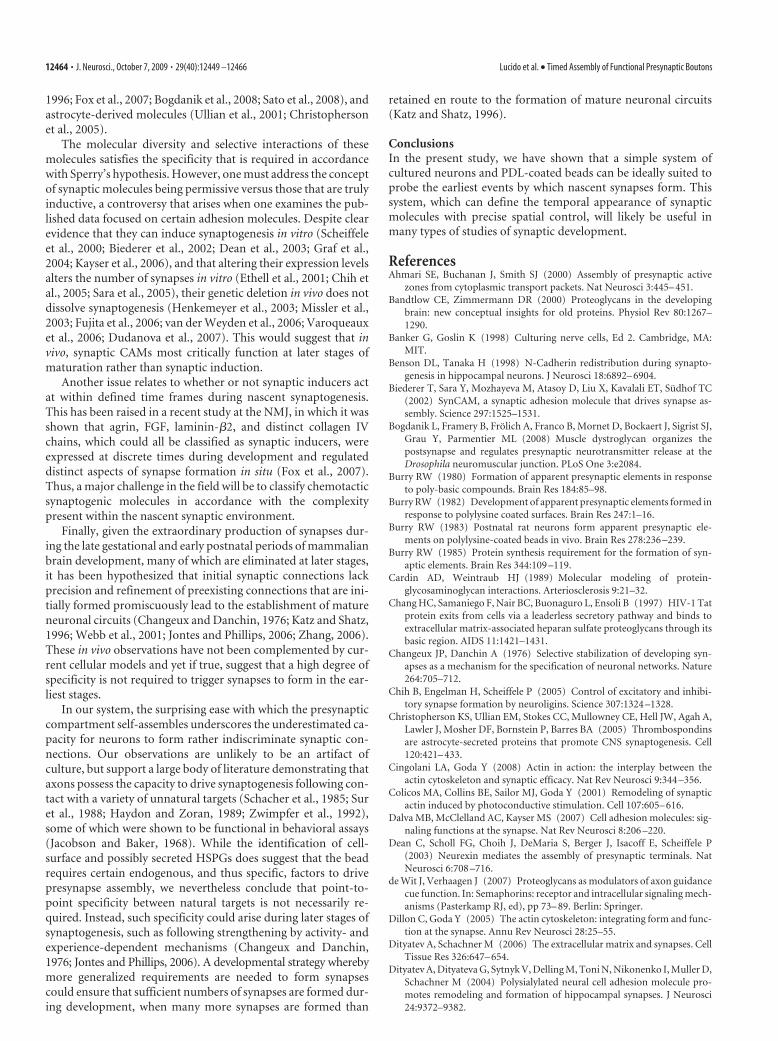
1996; Fox et al., 2007; Bogdanik et al., 2008; Sato et al., 2008), andastrocyte-derived molecules (Ullian et al., 2001; Christophersonet al., 2005).
The molecular diversity and selective interactions of thesemolecules satisfies the specificity that is required in accordancewith Sperry’s hypothesis. However, one must address the conceptof synaptic molecules being permissive versus those that are trulyinductive, a controversy that arises when one examines the pub-lished data focused on certain adhesion molecules. Despite clearevidence that they can induce synaptogenesis in vitro (Scheiffeleet al., 2000; Biederer et al., 2002; Dean et al., 2003; Graf et al.,2004; Kayser et al., 2006), and that altering their expression levelsalters the number of synapses in vitro (Ethell et al., 2001; Chih etal., 2005; Sara et al., 2005), their genetic deletion in vivo does notdissolve synaptogenesis (Henkemeyer et al., 2003; Missler et al.,2003; Fujita et al., 2006; van der Weyden et al., 2006; Varoqueauxet al., 2006; Dudanova et al., 2007). This would suggest that invivo, synaptic CAMs most critically function at later stages ofmaturation rather than synaptic induction.
Another issue relates to whether or not synaptic inducers actat within defined time frames during nascent synaptogenesis.This has been raised in a recent study at the NMJ, in which it wasshown that agrin, FGF, laminin-!2, and distinct collagen IVchains, which could all be classified as synaptic inducers, wereexpressed at discrete times during development and regulateddistinct aspects of synapse formation in situ (Fox et al., 2007).Thus, a major challenge in the field will be to classify chemotacticsynaptogenic molecules in accordance with the complexitypresent within the nascent synaptic environment.
Finally, given the extraordinary production of synapses dur-ing the late gestational and early postnatal periods of mammalianbrain development, many of which are eliminated at later stages,it has been hypothesized that initial synaptic connections lackprecision and refinement of preexisting connections that are ini-tially formed promiscuously lead to the establishment of matureneuronal circuits (Changeux and Danchin, 1976; Katz and Shatz,1996; Webb et al., 2001; Jontes and Phillips, 2006; Zhang, 2006).These in vivo observations have not been complemented by cur-rent cellular models and yet if true, suggest that a high degree ofspecificity is not required to trigger synapses to form in the ear-liest stages.
In our system, the surprising ease with which the presynapticcompartment self-assembles underscores the underestimated ca-pacity for neurons to form rather indiscriminate synaptic con-nections. Our observations are unlikely to be an artifact ofculture, but support a large body of literature demonstrating thataxons possess the capacity to drive synaptogenesis following con-tact with a variety of unnatural targets (Schacher et al., 1985; Suret al., 1988; Haydon and Zoran, 1989; Zwimpfer et al., 1992),some of which were shown to be functional in behavioral assays(Jacobson and Baker, 1968). While the identification of cell-surface and possibly secreted HSPGs does suggest that the beadrequires certain endogenous, and thus specific, factors to drivepresynapse assembly, we nevertheless conclude that point-to-point specificity between natural targets is not necessarily re-quired. Instead, such specificity could arise during later stages ofsynaptogenesis, such as following strengthening by activity- andexperience-dependent mechanisms (Changeux and Danchin,1976; Jontes and Phillips, 2006). A developmental strategy wherebymore generalized requirements are needed to form synapsescould ensure that sufficient numbers of synapses are formed dur-ing development, when many more synapses are formed than
retained en route to the formation of mature neuronal circuits(Katz and Shatz, 1996).
ConclusionsIn the present study, we have shown that a simple system ofcultured neurons and PDL-coated beads can be ideally suited toprobe the earliest events by which nascent synapses form. Thissystem, which can define the temporal appearance of synapticmolecules with precise spatial control, will likely be useful inmany types of studies of synaptic development.
ReferencesAhmari SE, Buchanan J, Smith SJ (2000) Assembly of presynaptic active
zones from cytoplasmic transport packets. Nat Neurosci 3:445– 451.Bandtlow CE, Zimmermann DR (2000) Proteoglycans in the developing
brain: new conceptual insights for old proteins. Physiol Rev 80:1267–1290.
Banker G, Goslin K (1998) Culturing nerve cells, Ed 2. Cambridge, MA:MIT.
Benson DL, Tanaka H (1998) N-Cadherin redistribution during synapto-genesis in hippocampal neurons. J Neurosci 18:6892– 6904.
Biederer T, Sara Y, Mozhayeva M, Atasoy D, Liu X, Kavalali ET, Sudhof TC(2002) SynCAM, a synaptic adhesion molecule that drives synapse as-sembly. Science 297:1525–1531.
Bogdanik L, Framery B, Frolich A, Franco B, Mornet D, Bockaert J, Sigrist SJ,Grau Y, Parmentier ML (2008) Muscle dystroglycan organizes thepostsynapse and regulates presynaptic neurotransmitter release at theDrosophila neuromuscular junction. PLoS One 3:e2084.
Burry RW (1980) Formation of apparent presynaptic elements in responseto poly-basic compounds. Brain Res 184:85–98.
Burry RW (1982) Development of apparent presynaptic elements formed inresponse to polylysine coated surfaces. Brain Res 247:1–16.
Burry RW (1983) Postnatal rat neurons form apparent presynaptic ele-ments on polylysine-coated beads in vivo. Brain Res 278:236 –239.
Burry RW (1985) Protein synthesis requirement for the formation of syn-aptic elements. Brain Res 344:109 –119.
Cardin AD, Weintraub HJ (1989) Molecular modeling of protein-glycosaminoglycan interactions. Arteriosclerosis 9:21–32.
Chang HC, Samaniego F, Nair BC, Buonaguro L, Ensoli B (1997) HIV-1 Tatprotein exits from cells via a leaderless secretory pathway and binds toextracellular matrix-associated heparan sulfate proteoglycans through itsbasic region. AIDS 11:1421–1431.
Changeux JP, Danchin A (1976) Selective stabilization of developing syn-apses as a mechanism for the specification of neuronal networks. Nature264:705–712.
Chih B, Engelman H, Scheiffele P (2005) Control of excitatory and inhibi-tory synapse formation by neuroligins. Science 307:1324 –1328.
Christopherson KS, Ullian EM, Stokes CC, Mullowney CE, Hell JW, Agah A,Lawler J, Mosher DF, Bornstein P, Barres BA (2005) Thrombospondinsare astrocyte-secreted proteins that promote CNS synaptogenesis. Cell120:421– 433.
Cingolani LA, Goda Y (2008) Actin in action: the interplay between theactin cytoskeleton and synaptic efficacy. Nat Rev Neurosci 9:344 –356.
Colicos MA, Collins BE, Sailor MJ, Goda Y (2001) Remodeling of synapticactin induced by photoconductive stimulation. Cell 107:605– 616.
Dalva MB, McClelland AC, Kayser MS (2007) Cell adhesion molecules: sig-naling functions at the synapse. Nat Rev Neurosci 8:206 –220.
Dean C, Scholl FG, Choih J, DeMaria S, Berger J, Isacoff E, Scheiffele P(2003) Neurexin mediates the assembly of presynaptic terminals. NatNeurosci 6:708 –716.
de Wit J, Verhaagen J (2007) Proteoglycans as modulators of axon guidancecue function. In: Semaphorins: receptor and intracellular signaling mech-anisms (Pasterkamp RJ, ed), pp 73– 89. Berlin: Springer.
Dillon C, Goda Y (2005) The actin cytoskeleton: integrating form and func-tion at the synapse. Annu Rev Neurosci 28:25–55.
Dityatev A, Schachner M (2006) The extracellular matrix and synapses. CellTissue Res 326:647– 654.
Dityatev A, Dityateva G, Sytnyk V, Delling M, Toni N, Nikonenko I, Muller D,Schachner M (2004) Polysialylated neural cell adhesion molecule pro-motes remodeling and formation of hippocampal synapses. J Neurosci24:9372–9382.
12464 • J. Neurosci., October 7, 2009 • 29(40):12449 –12466 Lucido et al. • Timed Assembly of Functional Presynaptic Boutons
Dow KE, Mirski SE, Roder JC, Riopelle RJ (1988) Neuronal proteoglycans:biosynthesis and functional interaction with neurons in vitro. J Neurosci8:3278 –3289.
Dudanova I, Tabuchi K, Rohlmann A, Sudhof TC, Missler M (2007) Dele-tion of alpha-neurexins does not cause a major impairment of axonalpathfinding or synapse formation. J Comp Neurol 502:261–274.
Ethell IM, Yamaguchi Y (1999) Cell surface heparan sulfate proteoglycansyndecan-2 induces the maturation of dendritic spines in rat hippocam-pal neurons. J Cell Biol 144:575–586.
Ethell IM, Irie F, Kalo MS, Couchman JR, Pasquale EB, Yamaguchi Y (2001)EphB/Syndecan-2 signaling in dendritic spine morphogenesis. Neuron31:1001–1013.
Fannon AM, Colman DR (1996) A model for central synaptic junctionalcomplex formation based on the differential adhesive specificities of thecadherins. Neuron 17:423– 434.
Fox MA, Sanes JR, Borza D-B, Eswarakumar VP, Fassler R, Hudson BG, JohnSWM, Ninomiya Y, Pedchenko V, Pfaff SL, Rheault MN, Sado Y, Segal Y,Werle MJ, Umemori H (2007) Distinct target-derived signals organizeformation, maturation, and maintenance of motor nerve terminals. Cell129:179 –193.
Friedman HV, Bresler T, Garner CC, Ziv NE (2000) Assembly of new indi-vidual excitatory synapses: time course and temporal order of synapticmolecule recruitment. Neuron 27:57– 69.
Fujita E, Kouroku Y, Ozeki S, Tanabe Y, Toyama Y, Maekawa M, Kojima N,Senoo H, Toshimori K, Momoi T (2006) Oligo-astheno-teratozoospermiain mice lacking RA175/TSLC1/SynCAM/IGSF4A, a cell adhesion molecule inthe immunoglobulin superfamily. Mol Cell Biol 26:718–726.
Gallagher JT, Lyon M, Steward WP (1986) Structure and function of hepa-ran sulfate proteoglycans. Biochem J 236:313–325.
Gautam M, Noakes PG, Moscoso L, Rupp F, Scheller RH, Merlie JP, Sanes JR(1996) Defective neuromuscular synaptogenesis in agrin-deficient mu-tant mice. Cell 85:525–535.
Gerrow K, Romorini S, Nabi SM, Colicos MA, Sala C, El-Husseini A (2006)A preformed complex of postsynaptic proteins is involved in excitatorysynapse development. Neuron 49:547–562.
Graf ER, Zhang X, Jin SX, Linhoff MW, Craig AM (2004) Neurexins inducedifferentiation of GABA and glutamate postsynaptic specializations vianeuroligins. Cell 119:1013–1026.
Hall AC, Lucas FR, Salinas PC (2000) Axonal remodeling and synaptic dif-ferentiation in the cerebellum is regulated by WNT-7a signaling. Cell100:525–535.
Haydon PG, Zoran MJ (1989) Formation and modulation of chemical con-nections: evoked acetylcholine release from growth cones and neurites ofspecific identified neurons. Neuron 2:1483–1490.
Henkemeyer M, Itkis OS, Ngo M, Hickmott PW, Ethell IM (2003) MultipleEphB receptor tyrosine kinases shape dendritic spines in the hippocam-pus. J Cell Biol 163:1313–1326.
Hsueh YP, Sheng M (1999) Regulated expression and subcellular localiza-tion of syndecan heparan sulfate proteoglycans and the syndecan-bindingprotein CASK/LIN-2 during rat brain development. J Neurosci19:7415–7425.
Hsueh YP, Yang FC, Kharazia V, Naisbitt S, Cohen AR, Weinberg RJ, ShengM (1998) Direct interaction of CASK/LIN-2 and syndecan heparan sul-fate proteoglycan and their overlapping distribution in neuronal syn-apses. J Cell Biol 142:139 –151.
Jacobson M, Baker RE (1968) Neuronal specification of cutaneous nervesthrough connections with skin grafts. Science 160:543–545.
Jin Y, Garner CC (2008) Molecular mechanisms of presynaptic differentia-tion. Annu Rev Cell Dev Biol 24:237–262.
Johnson KG, Tenney AP, Ghose A, Duckworth AM, Higashi ME, Parfitt K,Marcu O, Heslip TR, Marsh JL, Schwarz TL, Flanagan JG, Van Vactor D(2006) The HSPGs syndecan and dallylike bind the receptor phosphataseLAR and exert distinct effects on synaptic development. Neuron49:517–531.
Jontes JD, Phillips GR (2006) Selective stabilization and synaptic specificity:a new cell-biological model. Trends Neurosci 29:186 –191.
Jontes JD, Buchanan J, Smith SJ (2000) Growth cone and dendrite dynamicsin zebrafish embryos: early events in synaptogenesis imaged in vivo. NatNeurosci 3:231–237.
Katz LC, Shatz CJ (1996) Synaptic activity and the construction of corticalcircuits. Science 274:1133–1138.
Kayser MS, McClelland AC, Hughes EG, Dalva MB (2006) Intracellular and
trans-synaptic regulation of glutamatergic synaptogenesis by EphB recep-tors. J Neurosci 26:12152–12164.
Kim E, Sheng M (2004) PDZ domain proteins of synapses. Nat Rev Neuro-sci 5:771–781.
Kim S, Burette A, Chung HS, Kwon SK, Woo J, Lee HW, Kim K, Kim H,Weinberg RJ, Kim E (2006) NGL family PSD-95-interacting adhesionmolecules regulate excitatory synapse formation. Nat Neurosci 9:1294 –1301.
Kleene R, Schachner M (2004) Glycans and neural cell interactions. Nat RevNeurosci 5:195–208.
Ko J, Kim S, Chung HS, Kim K, Han K, Kim H, Jun H, Kaang BK, Kim E(2006) SALM Synaptic cell adhesion-like molecules regulate the differ-entiation of excitatory synapses. Neuron 50:233–245.
Krueger SR, Kolar A, Fitzsimonds RM (2003) The presynaptic release appa-ratus is functional in the absence of dendritic contact and highly mobilewithin isolated axons. Neuron 40:945–957.
Kwiatkowski AV, Rubinson DA, Dent EW, Edward van Veen J, Leslie JD,Zhang J, Mebane LM, Philippar U, Pinheiro EM, Burds AA, Bronson RT,Mori S, Fassler R, Gertler FB (2007) Ena/VASP is required for neurito-genesis in the developing cortex. Neuron 56:441– 455.
Kwiatkowski AV, Garner CC, Nelson WJ, Gertler FB (2009) Cell autono-mous defects in cortical development revealed by two-color chimera anal-ysis. Mol Cell Neurosci 41:44 –50.
Lauri SE, Kaukinen S, Kinnunen T, Ylinen A, Imai S, Kaila K, Taira T, RauvalaH (1999) Regulatory role and molecular interactions of a cell-surfaceheparan sulfate proteoglycan (N-syndecan) in hippocampal long-termpotentiation. J Neurosci 19:1226 –1235.
Leal-Ortiz S, Waites CL, Terry-Lorenzo R, Zamorano P, Gundelfinger ED,Garner CC (2008) Piccolo modulation of Synapsin1a dynamics regu-lates synaptic vesicle exocytosis. J Cell Biol 181:831– 846.
Ledda F, Paratcha G, Sandoval-Guzman T, Ibanez CF (2007) GDNF andGFR"1 promote formation of neuronal synapses by ligand-induced celladhesion. Nat Neurosci 10:293–300.
Li Q, Lau A, Morris TJ, Guo L, Fordyce CB, Stanley EF (2004) A syntaxin 1,G"o, and N-type calcium channel complex at a presynaptic nerve termi-nal: analysis by quantitative immunocolocalization. J Neurosci24:4070 – 4081.
Matsumoto Y, Irie F, Inatani M, Tessier-Lavigne M, Yamaguchi Y (2007)Netrin-1/DCC signaling in commissural axon guidance requires cell-autonomous expression of heparan sulfate. J Neurosci 27:4342– 4350.
Mislick KA, Baldeschwieler JD (1996) Evidence for a role of proteoglycansin cation-mediated gene transfer. Proc Natl Acad Sci U S A93:12349 –12354.
Missler M, Zhang W, Rohlmann A, Kattenstroth G, Hammer RE, GottmannK, Sudhof TC (2003) "-Neurexins couple Ca 2# channels to synapticvesicle exocytosis. Nature 423:939 –948.
Nakase I, Tadokoro A, Kawabata N, Takeuchi T, Katoh H, Hiramoto K,Negishi M, Nomizu M, Sugiura Y, Futaki S (2007) Interaction ofarginine-rich peptides with membrane-associated proteoglycans is cru-cial for induction of actin reorganization and macropinocytosis. Bio-chemistry 46:492–501.
Noakes PG, Gautam M, Mudd J, Sanes JR, Merlie JP (1995) Aberrant dif-ferentiation of neuromuscular junctions in mice lacking s-laminin/lami-nin [beta]2. Nature 374:258 –262.
Okabe S (2007) Molecular anatomy of the postsynaptic density. Mol CellNeurosci 34:503–518.
Okabe S, Miwa A, Okado H (2001) Spine formation and correlated assem-bly of presynaptic and postsynaptic molecules. J Neurosci 21:6105– 6114.
Peng HB, Markey DR, Muhlach WL, Pollack ED (1987) Development ofpresynaptic specializations induced by basic polypeptide-coated latexbeads in spinal cord cultures. Synapse 1:10 –19.
Phillips GR, Huang JK, Wang Y, Tanaka H, Shapiro L, Zhang W, Shan WS,Arndt K, Frank M, Gordon RE, Gawinowicz MA, Zhao Y, Colman DR(2001) The presynaptic particle web: ultrastructure, composition, disso-lution and reconstitution. Neuron 32:63–77.
Rauvala H, Peng HB (1997) HB-GAM (Heparin-binding growth-associatedmolecule) and heparin-type glycans in the development and plasticity ofneuron-target contacts. Prog Neurobiol 52:127–144.
Ryan TA, Reuter H, Wendland B, Schweizer FE, Tsien RW, Smith SJ (1993)The kinetics of synaptic vesicle recycling measured at single presynapticboutons. Neuron 11:713–724.
Sankaranarayanan S, Atluri PP, Ryan TA (2003) Actin has a molecular scaf-
Lucido et al. • Timed Assembly of Functional Presynaptic Boutons J. Neurosci., October 7, 2009 • 29(40):12449 –12466 • 12465
folding, not propulsive, role in presynaptic function. Nat Neurosci6:127–135.
Sara Y, Biederer T, Atasoy D, Chubykin A, Mozhayeva MG, Sudhof TC,Kavalali ET (2005) Selective capability of synCAM and neuroligin forfunctional synapse assembly. J Neurosci 25:260 –270.
Sato S, Omori Y, Katoh K, Kondo M, Kanagawa M, Miyata K, Funabiki K,Koyasu T, Kajimura N, Miyoshi T, Sawai H, Kobayashi K, Tani A, Toda T,Usukura J, Tano Y, Fujikado T, Furukawa T (2008) Pikachurin, a dys-troglycan ligand, is essential for photoreceptor ribbon synapse formation.Nat Neurosci 11:923–931.
Schacher S, Rayport SG, Ambron RT (1985) Giant Aplysia neuron R2 reli-ably forms strong chemical connections in vitro. J Neurosci 5:2851–2856.
Scheiffele P, Fan J, Choih J, Fetter R, Serafini T (2000) Neuroligin expressedin nonneuronal cells triggers presynaptic development in contacting ax-ons. Cell 101:657– 669.
Schluter OM, Xu W, Malenka RC (2006) Alternative N-terminal domainsof PSD-95 and SAP97 govern activity-dependent regulation of synapticAMPA receptor function. Neuron 51:99 –111.
Shapira M, Zhai RG, Dresbach T, Bresler T, Torres VI, Gundelfinger ED, ZivNE, Garner CC (2003) Unitary assembly of presynaptic active zonesfrom piccolo-bassoon transport vesicles. Neuron 38:237–252.
Shapiro L, Love J, Colman DR (2007) Adhesion molecules in the nervoussystem: Structural insights into function and diversity. Annu Rev Neuro-sci 30:451– 474.
Spector I, Braet F, Shochet NR, Bubb MR (1999) New anti-actin drugs in thestudy of the organization and function of the actin cytoskeleton. MicroscRes Tech 47:18 –37.
Sperry RW (1963) Chemoaffinity in the orderly growth of nerve fiber pat-terns and connections. Proc Natl Acad Sci U S A 50:703–710.
Sugiura M, Dow KE (1994) Activity-dependent regulation of neuronal syn-thesis and release of neurite-promoting heparan sulfate proteoglycans.Dev Biol 164:102–110.
Sur M, Garraghty PE, Roe AW (1988) Experimentally induced visual pro-jections into auditory thalamus and cortex. Science 242:1437–1441.
Tsuriel S, Geva R, Zamorano P, Dresbach T, Boeckers T, Gundelfinger ED,Garner CC, Ziv NE (2006) Local sharing as a predominant determinantof synaptic matrix molecular dynamics. PLoS Biol 4:e271.
Ullian EM, Sapperstein SK, Christopherson KS, Barres BA (2001) Controlof synapse number by glia. Science 291:657– 661.
Umemori H, Linhoff MW, Ornitz DM, Sanes JR (2004) FGF22 and its closerelatives are presynaptic organizing molecules in the mammalian brain.Cell 118:257–270.
van der Weyden L, Arends MJ, Chausiaux OE, Ellis PJ, Lange UC, Surani MA,Affara N, Murakami Y, Adams DJ, Bradley A (2006) Loss of TSLC1causes male infertility due to a defect at the spermatid stage of spermato-genesis. Mol Cell Biol 26:3595–3609.
Van Vactor D, Wall DP, Johnson KG (2006) Heparan sulfate proteogly-cans and the emergence of neuronal connectivity. Curr Opin Neuro-biol 16:40 –51.
Varoqueaux F, Aramuni G, Rawson RL, Mohrmann R, Missler M, GottmannK, Zhang W, Sudhof TC, Brose N (2006) Neuroligins determine synapsematuration and function. Neuron 51:741–754.
Waites CL, Specht CG, Hartel K, Leal-Ortiz S, Genoux D, Li D, Drisdel RC,Jeyifous O, Cheyne JE, Green WN, Montgomery JM, Garner CC (2009)Synaptic SAP97 isoforms regulate AMPA receptor dynamics and access topresynaptic glutamate. J Neurosci 29:4332– 4345.
Webb SJ, Monk CS, Nelson CA (2001) Mechanisms of postnatal neurobio-logical development: implications for human development. Dev Neuro-psychol 19:147–171.
Withers GS, Higgins D, Charette M, Banker G (2000) Bone morphogeneticprotein-7 enhances dendritic growth and receptivity to innervation incultured hippocampal neurons. Eur J Neurosci 12:106 –116.
Yavin E, Yavin Z (1974) Attachment and culture of dissociated cells from ratembryo cerebral hemispheres on polylysine-coated surface. J Cell Biol62:540 –546.
Zhang W, Benson DL (2001) Stages of synapse development defined bydependence on F-actin. J Neurosci 21:5169 –5181.
Zhang ZW (2006) Postnatal development of the mammalian neocortex:role of activity revisited. Can J Neurol Sci 33:158 –169.
Zwimpfer TJ, Aguayo AJ, Bray GM (1992) Synapse formation and prefer-ential distribution in the granule cell layer by regenerating retinal gan-glion cell axons guided to the cerebellum of adult hamsters. J Neurosci12:1144 –1159.
12466 • J. Neurosci., October 7, 2009 • 29(40):12449 –12466 Lucido et al. • Timed Assembly of Functional Presynaptic Boutons