Comparative Genomics Reveal That Host-Innate Immune
19
Comparative Genomics Reveal That Host-Innate Immune Responses Influence the Clinical Prevalence of Legionella pneumophila Serogroups Mohammad Adil Khan 1,2 , Natalie Knox 3 , Akriti Prashar 4 , David Alexander 1,2 , Mena Abdel-Nour 1,2 , Carla Duncan 1 , Patrick Tang 1 , Hajera Amatullah 5 , Claudia C. Dos Santos 5 , Nathalie Tijet 1 , Donald E. Low 1,2,6 , Christine Pourcel 7 , Gary Van Domselaar 4 , Mauricio Terebiznik 3 , Alexander W. Ensminger 1,8 , Cyril Guyard 1,2,6 * 1 Public Health Ontario, Toronto, Ontario, Canada, 2 Department of Laboratory Medicine and Pathobiology, Faculty of Medicine, University of Toronto, Toronto, Ontario, Canada, 3 National Microbiology Laboratory, Public Health Agency of Canada, Winnipeg, Manitoba, Canada, 4 Cell and Systems Biology and Biological Sciences, University of Toronto at Scarborough, Scarborough, Ontario, Canada, 5 The Keenan Research Centre of the Li Ka Shing Knowledge Institute of St. Michael’s Hospital, Toronto, Ontario, Canada, 6 Mount Sinai Hospital, Toronto, Ontario, Canada, 7 Institut de Ge ´ne ´ tique et Microbiologie, Universite ´ Paris-Sud, Paris, France, 8 Department of Molecular Genetics, Faculty of Medicine, University of Toronto, Toronto, Ontario, Canada Abstract Legionella pneumophila is the primary etiologic agent of legionellosis, a potentially fatal respiratory illness. Amongst the sixteen described L. pneumophila serogroups, a majority of the clinical infections diagnosed using standard methods are serogroup 1 (Sg1). This high clinical prevalence of Sg1 is hypothesized to be linked to environmental specific advantages and/or to increased virulence of strains belonging to Sg1. The genetic determinants for this prevalence remain unknown primarily due to the limited genomic information available for non-Sg1 clinical strains. Through a systematic attempt to culture Legionella from patient respiratory samples, we have previously reported that 34% of all culture confirmed legionellosis cases in Ontario (n = 351) are caused by non-Sg1 Legionella. Phylogenetic analysis combining multiple-locus variable number tandem repeat analysis and sequence based typing profiles of all non-Sg1 identified that L. pneumophila clinical strains (n = 73) belonging to the two most prevalent molecular types were Sg6. We conducted whole genome sequencing of two strains representative of these sequence types and one distant neighbour. Comparative genomics of the three L. pneumophila Sg6 genomes reported here with published L. pneumophila serogroup 1 genomes identified genetic differences in the O-antigen biosynthetic cluster. Comparative optical mapping analysis between Sg6 and Sg1 further corroborated this finding. We confirmed an altered O-antigen profile of Sg6, and tested its possible effects on growth and replication in in vitro biological models and experimental murine infections. Our data indicates that while clinical Sg1 might not be better suited than Sg6 in colonizing environmental niches, increased bloodstream dissemination through resistance to the alternative pathway of complement mediated killing in the human host may explain its higher prevalence. Citation: Khan MA, Knox N, Prashar A, Alexander D, Abdel-Nour M, et al. (2013) Comparative Genomics Reveal That Host-Innate Immune Responses Influence the Clinical Prevalence of Legionella pneumophila Serogroups. PLoS ONE 8(6): e67298. doi:10.1371/journal.pone.0067298 Editor: Yousef Abu Kwaik, University of Louisville, United States of America Received March 18, 2013; Accepted May 16, 2013; Published June 27, 2013 Copyright: ß 2013 Khan et al. This is an open-access article distributed under the terms of the Creative Commons Attribution License, which permits unrestricted use, distribution, and reproduction in any medium, provided the original author and source are credited. Funding: This work was supported by the Ontario Agency for Health Protection and Promotion (http://www.oahpp.ca/) and the Canadian Institutes of Health Research (MOP-102514) (http://www.cihr-irsc.gc.ca/). AP and MT are supported by the Natural Sciences and Engineering Research Council of Canada (http://www. nserc-crsng.gc.ca/) and the Ontario Association of Medical Laboratories grants (http://www.oaml.com/). The funders had no role in study design, data collection and analysis, decision to publish, or preparation of the manuscript. Competing Interests: The authors have declared that no competing interests exist. * E-mail: [email protected] Introduction Legionellosis is a potentially fatal infectious disease caused by Gram-negative, aerobic bacteria belonging to the genus Legionella [1,2]. Among 54 known Legionella species, L. pneumophila is the major cause of outbreaks (91.5%), and was the etiological agent of the first recognized outbreak in 1976 during a convention of the American Legion in Philadelphia [3,4,5,6]. The severity of this disease ranges from a mild respiratory illness to a rapidly fatal pneumonia [1,7]. The case fatality rate of legionellosis is between 40–80% in untreated immuno-suppressed patients but can be reduced to 5–30% with appropriate case management [7]. Legionellosis is a major public health concern in industrialized nations [8,9,10]. From 2000 to 2009, a 217% increase in legionellosis cases was reported in the United States [11,12]. Legionella spp. are found worldwide and can be detected in up to 80% of man-made freshwater sites [13,14,15,16,17,18,19]. More recently, a study showed that Legionella spp can persistently colonize aquifers over several years [19]. In natural and in man- made water systems, L. pneumophila may exist as planktonic cells or as biofilms [20,21]. The bacteria can also be isolated from different protozoa in the environment [22,23,24]. This is an essential step in L. pneumophila’s life cycle, since the bacteria are able to replicate in the environment within the protozoa host [23,25]. The human infection results from the inhalation of aerosols contaminated with either infected protozoa or free floating L. pneumophila [26,27]. The PLOS ONE | www.plosone.org 1 June 2013 | Volume 8 | Issue 6 | e67298
-
Upload
independent -
Category
Documents
-
view
3 -
download
0
Transcript of Comparative Genomics Reveal That Host-Innate Immune

Comparative Genomics Reveal That Host-Innate ImmuneResponses Influence the Clinical Prevalence of Legionellapneumophila SerogroupsMohammad Adil Khan1,2, Natalie Knox3, Akriti Prashar4, David Alexander1,2, Mena Abdel-Nour1,2,
Carla Duncan1, Patrick Tang1, Hajera Amatullah5, Claudia C. Dos Santos5, Nathalie Tijet1,
Donald E. Low1,2,6, Christine Pourcel7, Gary Van Domselaar4, Mauricio Terebiznik3,
Alexander W. Ensminger1,8, Cyril Guyard1,2,6*
1 Public Health Ontario, Toronto, Ontario, Canada, 2Department of Laboratory Medicine and Pathobiology, Faculty of Medicine, University of Toronto, Toronto, Ontario,
Canada, 3National Microbiology Laboratory, Public Health Agency of Canada, Winnipeg, Manitoba, Canada, 4Cell and Systems Biology and Biological Sciences, University
of Toronto at Scarborough, Scarborough, Ontario, Canada, 5 The Keenan Research Centre of the Li Ka Shing Knowledge Institute of St. Michael’s Hospital, Toronto,
Ontario, Canada, 6Mount Sinai Hospital, Toronto, Ontario, Canada, 7 Institut de Genetique et Microbiologie, Universite Paris-Sud, Paris, France, 8Department of Molecular
Genetics, Faculty of Medicine, University of Toronto, Toronto, Ontario, Canada
Abstract
Legionella pneumophila is the primary etiologic agent of legionellosis, a potentially fatal respiratory illness. Amongst thesixteen described L. pneumophila serogroups, a majority of the clinical infections diagnosed using standard methods areserogroup 1 (Sg1). This high clinical prevalence of Sg1 is hypothesized to be linked to environmental specific advantagesand/or to increased virulence of strains belonging to Sg1. The genetic determinants for this prevalence remain unknownprimarily due to the limited genomic information available for non-Sg1 clinical strains. Through a systematic attempt toculture Legionella from patient respiratory samples, we have previously reported that 34% of all culture confirmedlegionellosis cases in Ontario (n = 351) are caused by non-Sg1 Legionella. Phylogenetic analysis combining multiple-locusvariable number tandem repeat analysis and sequence based typing profiles of all non-Sg1 identified that L. pneumophilaclinical strains (n = 73) belonging to the two most prevalent molecular types were Sg6. We conducted whole genomesequencing of two strains representative of these sequence types and one distant neighbour. Comparative genomics of thethree L. pneumophila Sg6 genomes reported here with published L. pneumophila serogroup 1 genomes identified geneticdifferences in the O-antigen biosynthetic cluster. Comparative optical mapping analysis between Sg6 and Sg1 furthercorroborated this finding. We confirmed an altered O-antigen profile of Sg6, and tested its possible effects on growth andreplication in in vitro biological models and experimental murine infections. Our data indicates that while clinical Sg1 mightnot be better suited than Sg6 in colonizing environmental niches, increased bloodstream dissemination through resistanceto the alternative pathway of complement mediated killing in the human host may explain its higher prevalence.
Citation: Khan MA, Knox N, Prashar A, Alexander D, Abdel-Nour M, et al. (2013) Comparative Genomics Reveal That Host-Innate Immune Responses Influence theClinical Prevalence of Legionella pneumophila Serogroups. PLoS ONE 8(6): e67298. doi:10.1371/journal.pone.0067298
Editor: Yousef Abu Kwaik, University of Louisville, United States of America
Received March 18, 2013; Accepted May 16, 2013; Published June 27, 2013
Copyright: � 2013 Khan et al. This is an open-access article distributed under the terms of the Creative Commons Attribution License, which permitsunrestricted use, distribution, and reproduction in any medium, provided the original author and source are credited.
Funding: This work was supported by the Ontario Agency for Health Protection and Promotion (http://www.oahpp.ca/) and the Canadian Institutes of HealthResearch (MOP-102514) (http://www.cihr-irsc.gc.ca/). AP and MT are supported by the Natural Sciences and Engineering Research Council of Canada (http://www.nserc-crsng.gc.ca/) and the Ontario Association of Medical Laboratories grants (http://www.oaml.com/). The funders had no role in study design, data collectionand analysis, decision to publish, or preparation of the manuscript.
Competing Interests: The authors have declared that no competing interests exist.
* E-mail: [email protected]
Introduction
Legionellosis is a potentially fatal infectious disease caused by
Gram-negative, aerobic bacteria belonging to the genus Legionella
[1,2]. Among 54 known Legionella species, L. pneumophila is the
major cause of outbreaks (91.5%), and was the etiological agent of
the first recognized outbreak in 1976 during a convention of the
American Legion in Philadelphia [3,4,5,6]. The severity of this
disease ranges from a mild respiratory illness to a rapidly fatal
pneumonia [1,7]. The case fatality rate of legionellosis is between
40–80% in untreated immuno-suppressed patients but can be
reduced to 5–30% with appropriate case management [7].
Legionellosis is a major public health concern in industrialized
nations [8,9,10]. From 2000 to 2009, a 217% increase in
legionellosis cases was reported in the United States [11,12].
Legionella spp. are found worldwide and can be detected in up to
80% of man-made freshwater sites [13,14,15,16,17,18,19]. More
recently, a study showed that Legionella spp can persistently
colonize aquifers over several years [19]. In natural and in man-
made water systems, L. pneumophila may exist as planktonic cells or
as biofilms [20,21]. The bacteria can also be isolated from different
protozoa in the environment [22,23,24]. This is an essential step in
L. pneumophila’s life cycle, since the bacteria are able to replicate in
the environment within the protozoa host [23,25]. The human
infection results from the inhalation of aerosols contaminated with
either infected protozoa or free floating L. pneumophila [26,27]. The
PLOS ONE | www.plosone.org 1 June 2013 | Volume 8 | Issue 6 | e67298

general model predicts, that upon inhalation, the infection starts
with the invasion and replication in macrophage and epithelial
cells of the lung [28,29]. Once inside the cell, Legionella replicate
with in a non-acidified vacuolar environment [30,31]. While many
genes required for surviving in amoeba are yet to be described,
some like the dot/icm genes [32,33] have shown to be essential for
survival in both the amoeba and the human host.
L. pneumophila have been classified into sixteen serogroups based
on reactivity to specific monoclonal antibodies [34,35]. While
these serogroups have been reported worldwide, Sg1 has
historically been identified in most clinical cases (84.2%) [6].
The most widely used clinical diagnostic method for all suspected
legionellosis cases are rapid, non-invasive, urine detection tests that
recognize the presence of soluble antigen [36]. These clinical tests
provide high sensitivity for Sg1 infection diagnosis but they are
unable to detect most non-Sg1 strains [37,38]. Recently, we
reported that 34% of culture confirmed cases of legionellosis in
Ontario (n = 351) were caused by non-Sg1 L. pneumophila species in
the last 3 decades [39]. Interestingly such a high percentage of
non-Sg1 was also reported in some Scandinavian countries [40].
In a comparative clinical and environmental distribution analysis
study, Sg1 was shown to constitute 28.2% of the Legionella spp.
bacteria isolated from man-made water systems (n = 2,747) but
95.4% of clinical samples (n = 259) [41]. This suggests that the
clinical prevalence of Sg1 might be independent of predominance
in the environment, but rather to fitness or virulence advantages in
the human host.
The underlying reasons for the clinical prevalence of Sg1 strains
are a subject of active debate. A large, microarray multigenome
analysis of 217 L. pneumophila isolates previously suggested that the
clinical predominance of Sg1 may be linked to its specific
lipopolysaccharide [42]. In this study, thorough population-based
molecular epidemiology, we first identified the most clinically
prevalent Sg6 strain among non-Sg1 clinical isolates in Ontario.
Next, by combining next-generation sequencing with in vitro and
in vivo experimental models, we performed a comparative analysis
with the closest identified phylogenetic Sg1 (strain Philadelphia).
Our results support a model in which increased resistance to
serum complement, rather than variation in environmental fitness,
explains the relative clinical prevalence of Sg1.
Results
Identification of Phylogenetic Clusters within non-Sg1 L.pneumophila Clinical Isolates
To identify prevalent population based clusters of non-Sg1 L.
pneumophila clinical strains in Ontario, we conducted a phyloge-
netic analysis on 73 clinical isolates recovered between 1980 and
2009 using sequence based typing (SBT) and multiple-locus
variable number tandem repeat analysis (MLVA). SBT based
phylogenetic comparisons between different strains is accom-
plished by observing sequence variations of 7 allelic markers
including house-keeping genes and known virulence factors
[43,44]. MLVA takes advantage of the polymorphisms of 9
tandemly repeated DNA sequences [45]. The UPGMA analysis
based on a matrix of pairwise allelic differences between all the
sequence types (ST) of our dataset identified a large cluster (n = 40)
comprising most of the Sg6 isolates of our repository. Isolates of
this large cluster showed less phylogenetic diversity compared with
other clusters and it encompassed 2 two main subclusters (Figure
S1). The phylogenetic subcluster 1 included 17 isolates of Sg6 and
ST 187 (Figure S1), while the phylogenetic subcluster 2 included 9
isolates who were all identified as Sg6 ST 68 (Figure S1). MLVA
typing of this dataset, identified a large phylogenetic cluster as well,
comprising two subclusters, where cluster 1 grouped 20 isolates,
and included Sg6 ST 187 and Sg6 ST 68, along with Sg4, Sg8 and
Sg10 strains (Figure S2). Interestingly, cluster 2 had 10 isolates, out
of which 8 were Sg6 ST 187, 1 was a Sg6 ST242 and 1 was a Sg3
ST93 (Figure S2). To further increase the discriminatory power of
the molecular typing methods [46], we next constructed a
phylogenetic tree combining SBT and MLVA profiles (Figure 1).
This combined analysis confirmed that the largest phylogenetic
cluster of non-sg1 clinical isolates comprised mainly of Sg6 ST187
and Sg6 ST68. In contrast to several other molecular types such
Sg6 ST407 which had only been isolated over a period of 5 years,
Sg6 ST187 and Sg6 ST68 strains seemed clonally stable, since
isolates with identical sequence types have been obtained for 25 to
30 year periods of our repository (initiated in 1980). For the
purposes of further analysis we selected strain Thunder Bay,
Sudbury and Mississauga isolates as the representative ST187,
ST68 and ST407, respectively. The high-prevalence of Sg6 among
non-Sg1s was also confirmed through an inspection of the
European Working Group for Legionella Infections (EWGLI)
database (http://www.ewgli.org/) and has been reported inde-
pendently elsewhere [41].
The Optical Map of L. pneumophila Sg6 str. Thunder Bayand Sg12 str. 570-CO-H are Identical
Optical mapping is an emerging technology used for accurate
genome assemblies [47] and for conducting comparative chromo-
somal analysis [48]. It allows for rapid identification of divergent
regions, chromosomal rearrangement, and inversions [48,49,50].
We compared the relatedness of Sg6 str. Thunder Bay with other
non-Sg1 and Sg1 strains, whose genomic sequences are available.
First, we constructed an optical map of Sg6 str. Thunder Bay and
compared it to in silico maps of Sg1 str. Philadelphia and Sg12 str.
570-CO-H (Figure 2A). The optical maps of Sg6 str. Thunder Bay
and Sg1 str. Philadelphia only showed divergence in a 40 kB
segment. This segment corresponded to genes involved in O-
antigen biosynthesis. In contrast, Sg12 str. 570-CO-H showed a
similar restriction pattern with Sg6 str. Thunder Bay. Secondly,
we constructed an evolutionary tree based on optical maps of Sg6
str. Thunder Bay, Sg12 str. 570-CO-H and all sequenced and fully
assembled Sg1 strains (Figure 2B). This analysis confirmed that the
clinical non-Sg1 strains, Sg6 str. Thunder Bay and Sg12 str. 570-
CO-H share a high degree of homology, and are phylogenetic
neighbours of Sg1 str. Philadelphia.
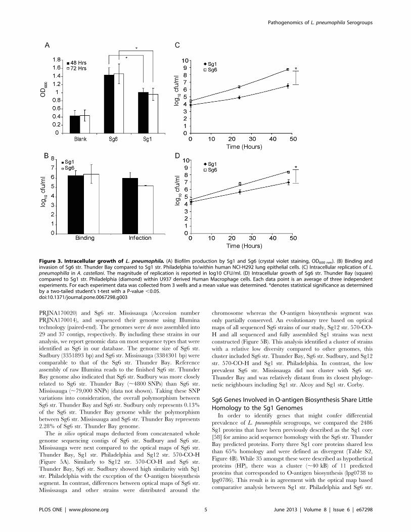
L. pneumophila Sg6 str. Thunder Bay Generates HigherAmounts of Biofilm Compared to Sg1 str. Philadelphia
The comparative optical map analyses identified possible
serogroup specific genomic parameters. In order to investigate if
these differences result in possible serogroup specific advantages
between Sg1 str. Philadelphia and Sg6 str. Thunder Bay, we
conducted comparative biological analysis at different stages of the
L. pneumophila life-cycle. A strategy used by L. pneumophila to survive
in anthropogenic aqueous environment is to generate biofilms
[51]. The ability to generate higher amounts of biofilm can be
directly correlated with persistence in the environment [52]. We
determined that Sg6 str. Thunder Bay produces significantly
higher levels of biofilm compared to Sg1 str. Philadelphia by 48
hours (P,0.05; Figure 3A). Furthermore, this difference further
increased by 72 hours (P,0.05). This suggests that while Sg1 are
able to generate significant amounts of biofilm, the Sg6 might
possess an advantage that could explain its higher reported
prevalence in some environmental surveys [53].
Pathogenomics of L. pneumophila Serogroups
PLOS ONE | www.plosone.org 2 June 2013 | Volume 8 | Issue 6 | e67298

Pathogenomics of L. pneumophila Serogroups
PLOS ONE | www.plosone.org 3 June 2013 | Volume 8 | Issue 6 | e67298

L. pneumophila Sg6 str. Thunder Bay Replicates moreEfficiently than Sg1 str. Philadelphia within A. castellaniAmoebae and U937 Derived Macrophages, but not inLung Epithelial Cells
In order to determine a possible advantage that either Sg1 or
Sg6 may possess in infecting humans and surviving in the
environment, we compared the replication rates of Sg1 str.
Philadelphia and Sg6 str. Thunder Bay over 48 hours in the
human lung epithelial cell line H292, U937 macrophages and
Acanthamoeba castellani amoeba models [54,55,56]. While no
differences were observed in the lung epithelial cells (Figure 3B),
significant increase of Sg6 str. Thunder Bay counts over Sg1 str.
Philadelphia was seen by 48 hours in the macrophage model
(Figure 3C). Similarly in amoeba, while no discernable difference
was observed between the Sg1 and Sg6 strains one hour after
uptake, by 24 hours, counts for the Sg6 strain were 28-fold higher
compared to Sg1 str. Philadelphia (Figure 3D). At 48 hours, this
difference had increased to 168-fold, suggesting that Sg6 str.
Thunder Bay is able to infect and replicate more effectively than
Sg1 str. Philadelphia in A. castellani model. Taken together, these
results suggest that the Sg6 str. Thunder Bay replicates more
efficiently than Sg1 str. Philadelphia within amoebae and
macrophages. Thus Sg6 str. Thunder Bay might possess a fitness
advantage in the environmental niche over the Sg1 str.
Philadelphia, which is an unlikely explanation for the observed
differences in clinical prevalence.
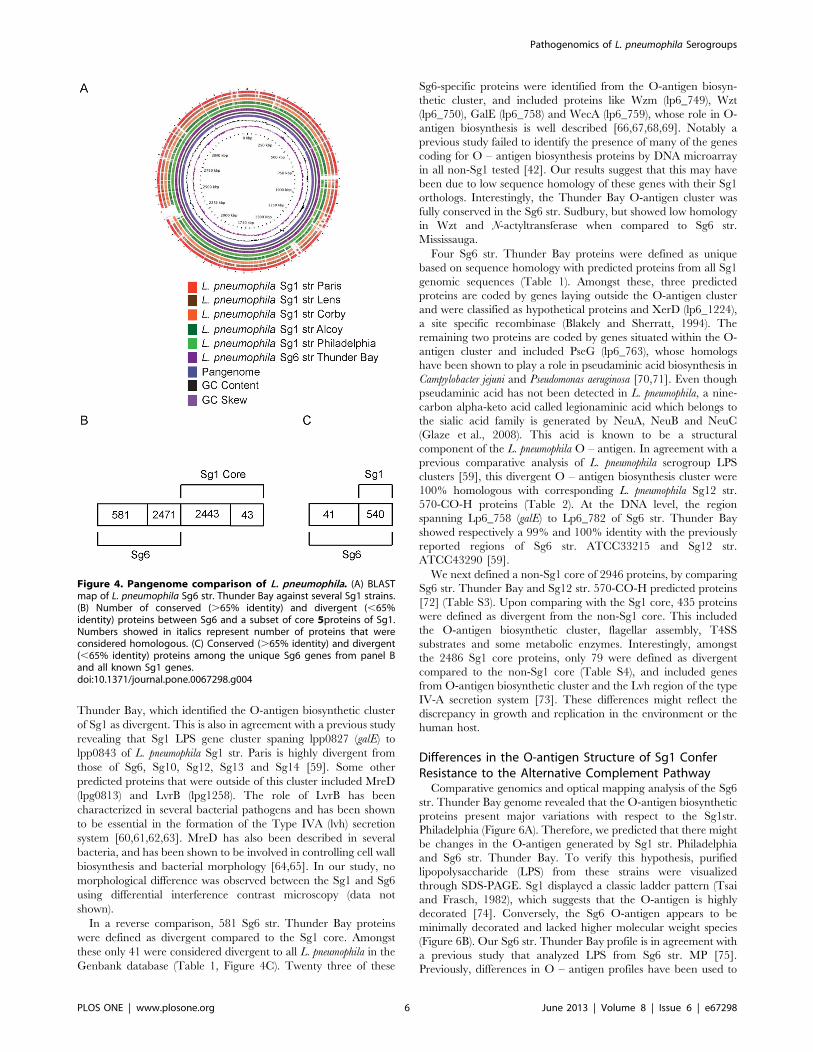
The most Prevalent Molecular Types of Sg6 Strains Sharea High Degree of Genomic Similarity
To identify the factors that may be contributing to the
population structure of L. pneumophila in clinical settings, we
performed whole genome sequencing of Sg6 str. Thunder Bay.
The L. pneumophila Sg6 str. Thunder Bay (accession number:
CP003730) genome consists of a 3455167-bp chromosome with an
average GC content of 38.24% (Table S1). A total of 3052 protein-
encoding genes are predicted, 2299 (75.3%) of which have been
assigned a putative function. A BLAST map analysis of Thunder
Bay against five reference Sg1 strains showed several conserved
regions (Figure 4A). Furthermore, high genomic strand asymmetry
identified through the GC skew suggested no abnormalities in the
assembly of the genome [57] (Figure 4A). To determine plasticity
of the Sg6 genomes, we selected two additional strains from our
clinical repository, Sg6 str. Sudbury (Accession number
Figure 1. Hybrid SBT-MLVA typing of a population based clinical repository of L. pneumophila. Phylogenetic clusters were formed byUPGMA analysis of the combined typing data. Cluster 1 is identified in blue, while cluster 2 and 3 are shown in green and red, respectively.doi:10.1371/journal.pone.0067298.g001
Figure 2. Optical Maps and genome based comparisons of L. pneumophila strains. (A) Optical map of L. pneumophilla Sg6 str. Thunder Bay(Middle) compared to Sg1 strains Philadelphia (Top) and Sg12 570-CO-H (Bottom). The regions in white indicate unique gene clusters, while areas inblue depict high similarity to Sg6. Regions in red are conserved between all three genome sequences. (B) UPGMA based cluster analysis of opticalmaps of sequenced L. pneumophila.doi:10.1371/journal.pone.0067298.g002
Pathogenomics of L. pneumophila Serogroups
PLOS ONE | www.plosone.org 4 June 2013 | Volume 8 | Issue 6 | e67298
PRJNA170020) and Sg6 str. Mississauga (Accession number
PRJNA170014), and sequenced their genome using Illumina
technology (paired-end). The genomes were de novo assembled into
29 and 37 contigs, respectively. By including these strains in our
analysis, we report genomic data on most sequence types that were
identified as Sg6 in our database. The genome size of Sg6 str.
Sudbury (3351893 bp) and Sg6 str. Mississauga (3384301 bp) were
comparable to that of the Sg6 str. Thunder Bay. Reference
assembly of raw Illumina reads to the finished Sg6 str. Thunder
Bay genome also indicated that Sg6 str. Sudbury was more closely
related to Sg6 str. Thunder Bay (,4800 SNPs) than Sg6 str.
Mississauga (,79,000 SNPs) (data not shown). Taking these SNP
variations into consideration, the overall polymorphism between
Sg6 str. Thunder Bay and Sg6 str. Sudbury only represents 0.13%
of the Sg6 str. Thunder Bay genome while the polymorphism
between Sg6 str. Mississauga and Sg6 str. Thunder Bay represents
2.28% of Sg6 str. Thunder Bay genome.
The in silico optical maps deducted from concatenated whole
genome sequencing contigs of Sg6 str. Sudbury and Sg6 str.
Mississauga were next compared to the optical maps of Sg6 str.
Thunder Bay, Sg1 str. Philadelphia and Sg12 str. 570-CO-H
(Figure 5A). Similarly to Sg12 str. 570-CO-H and Sg6 str.
Thunder Bay, Sg6 str. Sudbury showed high similarity with Sg1
str. Philadelphia with the exception of the O-antigen biosynthesis
segment. In contrast, differences between optical maps of Sg6 str.
Mississauga and other strains were distributed around the
chromosome whereas the O-antigen biosynthesis segment was
only partially conserved. An evolutionary tree based on optical
maps of all sequenced Sg6 strains of our study, Sg12 str. 570-CO-
H and all sequenced and fully assembled Sg1 strains was next
constructed (Figure 5B). This analysis identified a cluster of strains
with a relative low diversity compared to other genomes, this
cluster included Sg6 str. Thunder Bay, Sg6 str. Sudbury, and Sg12
str. 570-CO-H and Sg1 str. Philadelphia. In contrast, the low
prevalent Sg6 str. Mississauga did not cluster with Sg6 str.
Thunder Bay and was relatively distant from its closest phyloge-
netic neighbours including Sg1 str. Alcoy and Sg1 str. Corby.
Sg6 Genes Involved in O-antigen Biosynthesis Share LittleHomology to the Sg1 Genomes
In order to identify genes that might confer differential
prevalence of L. pneumophila serogroups, we compared the 2486
Sg1 proteins that have been previously described as the Sg1 core
[58] for amino acid sequence homology with the Sg6 str. Thunder
Bay predicted proteins. Forty three Sg1 core proteins shared less
than 65% homology and were defined as divergent (Table S2,
Figure 4B). While 35 amongst these were described as hypothetical
proteins (HP), there was a cluster (,40 kB) of 11 predicted
proteins that corresponded to O-antigen biosynthesis (lpg0738 to
lpg0786). This result is in agreement with the optical map based
comparative analysis between Sg1 str. Philadelphia and Sg6 str.
Figure 3. Intracellular growth of L. pneumophila. (A) Biofilm production by Sg1 and Sg6 (crystal violet staining, OD600 nm). (B) Binding andinvasion of Sg6 str. Thunder Bay compared to Sg1 str. Philadelphia to/within human NCI-H292 lung epithelial cells. (C) Intracellular replication of L.pneumophilla in A. castellani. The magnitude of replication is reported in log10 CFU/ml. (D) Intracellular growth of Sg6 str. Thunder Bay (square)compared to Sg1 str. Philadelphia (diamond) within U937 derived Human Macrophage cells. Each data point is an average of three independentexperiments. For each experiment data was collected from 3 wells and a mean value was determined. *denotes statistical significance as determinedby a two-tailed student’s t-test with a P-value ,0.05.doi:10.1371/journal.pone.0067298.g003
Pathogenomics of L. pneumophila Serogroups
PLOS ONE | www.plosone.org 5 June 2013 | Volume 8 | Issue 6 | e67298
Thunder Bay, which identified the O-antigen biosynthetic cluster
of Sg1 as divergent. This is also in agreement with a previous study
revealing that Sg1 LPS gene cluster spaning lpp0827 (galE) to
lpp0843 of L. pneumophila Sg1 str. Paris is highly divergent from
those of Sg6, Sg10, Sg12, Sg13 and Sg14 [59]. Some other
predicted proteins that were outside of this cluster included MreD
(lpg0813) and LvrB (lpg1258). The role of LvrB has been
characterized in several bacterial pathogens and has been shown
to be essential in the formation of the Type IVA (lvh) secretion
system [60,61,62,63]. MreD has also been described in several
bacteria, and has been shown to be involved in controlling cell wall
biosynthesis and bacterial morphology [64,65]. In our study, no
morphological difference was observed between the Sg1 and Sg6
using differential interference contrast microscopy (data not
shown).
In a reverse comparison, 581 Sg6 str. Thunder Bay proteins
were defined as divergent compared to the Sg1 core. Amongst
these only 41 were considered divergent to all L. pneumophila in the
Genbank database (Table 1, Figure 4C). Twenty three of these
Sg6-specific proteins were identified from the O-antigen biosyn-
thetic cluster, and included proteins like Wzm (lp6_749), Wzt
(lp6_750), GalE (lp6_758) and WecA (lp6_759), whose role in O-
antigen biosynthesis is well described [66,67,68,69]. Notably a
previous study failed to identify the presence of many of the genes
coding for O – antigen biosynthesis proteins by DNA microarray
in all non-Sg1 tested [42]. Our results suggest that this may have
been due to low sequence homology of these genes with their Sg1
orthologs. Interestingly, the Thunder Bay O-antigen cluster was
fully conserved in the Sg6 str. Sudbury, but showed low homology
in Wzt and N-actyltransferase when compared to Sg6 str.
Mississauga.
Four Sg6 str. Thunder Bay proteins were defined as unique
based on sequence homology with predicted proteins from all Sg1
genomic sequences (Table 1). Amongst these, three predicted
proteins are coded by genes laying outside the O-antigen cluster
and were classified as hypothetical proteins and XerD (lp6_1224),
a site specific recombinase (Blakely and Sherratt, 1994). The
remaining two proteins are coded by genes situated within the O-
antigen cluster and included PseG (lp6_763), whose homologs
have been shown to play a role in pseudaminic acid biosynthesis in
Campylobacter jejuni and Pseudomonas aeruginosa [70,71]. Even though
pseudaminic acid has not been detected in L. pneumophila, a nine-
carbon alpha-keto acid called legionaminic acid which belongs to
the sialic acid family is generated by NeuA, NeuB and NeuC
(Glaze et al., 2008). This acid is known to be a structural
component of the L. pneumophila O – antigen. In agreement with a
previous comparative analysis of L. pneumophila serogroup LPS
clusters [59], this divergent O – antigen biosynthesis cluster were
100% homologous with corresponding L. pneumophila Sg12 str.
570-CO-H proteins (Table 2). At the DNA level, the region
spanning Lp6_758 (galE) to Lp6_782 of Sg6 str. Thunder Bay
showed respectively a 99% and 100% identity with the previously
reported regions of Sg6 str. ATCC33215 and Sg12 str.
ATCC43290 [59].
We next defined a non-Sg1 core of 2946 proteins, by comparing
Sg6 str. Thunder Bay and Sg12 str. 570-CO-H predicted proteins
[72] (Table S3). Upon comparing with the Sg1 core, 435 proteins
were defined as divergent from the non-Sg1 core. This included
the O-antigen biosynthetic cluster, flagellar assembly, T4SS
substrates and some metabolic enzymes. Interestingly, amongst
the 2486 Sg1 core proteins, only 79 were defined as divergent
compared to the non-Sg1 core (Table S4), and included genes
from O-antigen biosynthetic cluster and the Lvh region of the type
IV-A secretion system [73]. These differences might reflect the
discrepancy in growth and replication in the environment or the
human host.
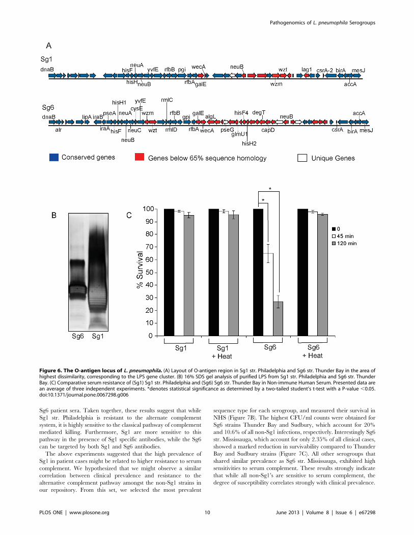
Differences in the O-antigen Structure of Sg1 ConferResistance to the Alternative Complement Pathway
Comparative genomics and optical mapping analysis of the Sg6
str. Thunder Bay genome revealed that the O-antigen biosynthetic
proteins present major variations with respect to the Sg1str.
Philadelphia (Figure 6A). Therefore, we predicted that there might
be changes in the O-antigen generated by Sg1 str. Philadelphia
and Sg6 str. Thunder Bay. To verify this hypothesis, purified
lipopolysaccharide (LPS) from these strains were visualized
through SDS-PAGE. Sg1 displayed a classic ladder pattern (Tsai
and Frasch, 1982), which suggests that the O-antigen is highly
decorated [74]. Conversely, the Sg6 O-antigen appears to be
minimally decorated and lacked higher molecular weight species
(Figure 6B). Our Sg6 str. Thunder Bay profile is in agreement with
a previous study that analyzed LPS from Sg6 str. MP [75].
Previously, differences in O – antigen profiles have been used to
Figure 4. Pangenome comparison of L. pneumophila. (A) BLASTmap of L. pneumophila Sg6 str. Thunder Bay against several Sg1 strains.(B) Number of conserved (.65% identity) and divergent (,65%identity) proteins between Sg6 and a subset of core 5proteins of Sg1.Numbers showed in italics represent number of proteins that wereconsidered homologous. (C) Conserved (.65% identity) and divergent(,65% identity) proteins among the unique Sg6 genes from panel Band all known Sg1 genes.doi:10.1371/journal.pone.0067298.g004
Pathogenomics of L. pneumophila Serogroups
PLOS ONE | www.plosone.org 6 June 2013 | Volume 8 | Issue 6 | e67298

define distinct serogroups [76,77], and are hypothesized to be
associated to differences in virulence of L. pneumophila serogroups in
humans [42].
Variations in the outer-membrane O – antigen segment of the
LPS can provide resistance to complement mediated serum killing
in pathogenic bacteria [78,79,80]. The differences observed in the
O – antigen structure of Sg1 str. Philadelphia and Sg6 str.
Thunder Bay indicated that there might be differential sensitivity
to serum complement. Indeed when the Sg6 was treated with
Non-immune Human Serum (NHS), a 35% and a 73% decrease
in colony forming units (CFUs) was seen by 45 and 120 min,
respectively (Figure 6C). However, no effect was observed for Sg1.
Sg6 and Sg1 viability was not affected by decomplemented (heat
inactivated) NHS, consistent with a Sg6-specific killing by heat-
sensitive complement. These results suggest that Sg6 O-antigen is
unable to provide any appreciable resistance to serum comple-
ment, whereas Sg1 O-antigen may allow the bacteria to escape
complement mediated killing by naive human serum.
We next measured the sensitivity of Sg1 str. Philadelphia and
Sg6 str. Thunder Bay to the classical complement pathway using
sera obtained from patients diagnosed with either Sg1 or Sg6
infections (Figure 7A). Presence of either Sg1 or Sg6 specific
antibodies was previously confirmed through IFA (data not
shown). The Sg1 str. Philadelphia isolate showed a 72.5%
decrease in CFU after a 60 min incubation in sera containing
Sg1 antibodies, an effect that was not observed in decomplemen-
ted patient sera. In presence of Sg6 specific antibodies, the Sg1 str.
Philadelphia CFUs were decreased by 31.5% at 60 min. The Sg6
Figure 5. Optical Maps and genome based comparisons of L. pneumophila strains. (A) Optical map of L. pneumophilla Sg6 str. Thunder Bay(Middle) compared to Sg1 strains Philadelphia (Top) and Sg12 570-CO-H (Bottom). The regions in white indicate unique gene clusters, while areas inblue depict high similarity to Sg6. Regions in red are conserved between all three genome sequences. (B) UPGMA based cluster analysis of opticalmaps of sequenced L. pneumophila. Percent difference at each node is indicated.doi:10.1371/journal.pone.0067298.g005
Pathogenomics of L. pneumophila Serogroups
PLOS ONE | www.plosone.org 7 June 2013 | Volume 8 | Issue 6 | e67298

str. Thunder Bay counts, when incubated with Sg1 antibodies were decreased by 98.8%, and similarly decreased by 99.3% in
Table 1. Divergent proteins present in Sg6 str. Thunder Bay compared to all sequenced Sg1 genomes in the Genbank database.
Sg6 Locus Gene Name Gene DescriptionIdentityto Sg1
Sg1Locus
GeneName Gene Description
Lp6_186 HP 43 lpg0112 HP
Lp6_749 Wzm LPS O-antigen ABC transporter 48 lpp0837 Wzm LPS O-antigen ABC transporter
Lp6_750 Wzt LPS O-antigen ABC transporter 63 lpg0773 Wzt LPS O-antigen ABC transporter
Lp6_755 HP 52 lpg0763 HP
Lp6_758 GalE UDP-glucose 4-epimerase 61 lpg0761 HP
Lp6_759 WecA a-N-acetylglucosaminyltransferase 57 lpg0762 WecA O-antigen initiating glycosyltransferase
Lp6_761 HP 48 lpg0788 HP
Lp6_762 GCN5-related N-acetyltransferase 29 lpp1089 Streptomycin 3’’-adenylyltransferase
Lp6_763 PseG Pseudaminic acid biosynthesis-associated protein
Lp6_764 Glutamate-1-semialdehyde 2,1-aminomutase 37 lpg0467 LasB Zinc metalloprotease
Lp6_765 GlmU1 Acylneuraminate cytidylyltransferase 31 lpg1919 KdsB 3-deoxy-manno-octulosonatecytidylyltransferase
Lp6_766 Acetyltransferase 50 lpg2848
Lp6_767 HisF4 Imidazoleglycerol-phosphate synthase 35 lpg0749 HisF Imidazole glycerol phosphatesynthase
Lp6_768 HisH2 Imidazole glycerol phosphate synthase 48 lpp2859 HisH Imidazole glycerol phosphatesynthase
Lp6_769 LPS biosynthesis protein 38 lpg0786 HP
Lp6_770 Aryl-alcohol dehydrogenase-like oxidoreductase 24 lpg2848 Ribonuclease
Lp6_771 DegT Aminotransferase 52 lpl0206 HP
Lp6_772 CapD Polysaccharide biosynthesis protein 59 lpg0561 PhaB Acetoacetyl-CoA reductase
Lp6_773 HP 38 lpg0635
Lp6_774 Capsule polysaccharide biosynthesis protein
Lp6_775 NeuB N-acetylneuraminic acid synthetase 38 lpp0818 NeuB N-acetylneuraminic acidsynthetase
Lp6_776 HP 47 lpa3427 HP
Lp6_777 HP
Lp6_780 HP 31 lpg0774 HP
Lp6_781 Dehydrogenase 29 lpg1888 HP
Lp6_782 Dehydrogenase 36 lpg2974 Psd Phosphatidylserine decarboxylase
Lp6_825 HP 47 lpg1344 DedE Colicin V
Lp6_828 UDP-N-acetyl-D-galactosamine dehydrogenase 23 lpg1942 3-hydroxyacyl CoA dehydrogenase
Lp6_829 UDP-glucose 4-epimerase 33 lpg2214 Nucleoside-diphosphate sugarepimerase
Lp6_830 Starch synthase 35 lpp3018 HP
Lp6_831 HP 22 lpg2485 HP
Lp6_832 HP 32 lpg2015 ProC Pyrroline-5-carboxylate reductase
Lp6_833 Glycosyl transferase family 2 35 lpg1183 HP
Lp6_976 HP 56 lpg0981 HP
Lp6_977 YwfO Phosphohydrolase 45 lpg1267 HP
Lp6_1186 HP
Lp6_1249 XerD Integrase/recombinase
Lp6_2002 integrase 59 lpc1833 HP
Lp6_2003 Protein of unknown function DUF1016 34 lpg1228 HP
Lp6_2041 HP 50 lpp0850 HP
Lp6_2164 HP 44 lpg1480 MutH DNA mismatch repair protein
Proteins were defined as divergent when they shared less than 65% homology and 75% coverage. HP identifies hypothetical proteins.doi:10.1371/journal.pone.0067298.t001
Pathogenomics of L. pneumophila Serogroups
PLOS ONE | www.plosone.org 8 June 2013 | Volume 8 | Issue 6 | e67298

Table 2. Comparative proteomics analysis between divergent proteins identified in Table 1 and L. pneumophila Sg12 str. 570-CO-H.
Sg6 LocusGeneName Gene Description
Identity toSg12
Sg12Locus Gene Name Gene Description
Lp6_186 HP 82 lp12_2703 Phenylalanyl tRNA synthetase
Lp6_749 Wzm LPS O-antigen ABC transporter 100 Wzm lp12_0766 Polysaccharide ABC transporter
Lp6_750 Wzt LPS O-antigen ABC transporter 100 Wzt Lp12_0767 LPS O-antigen ABC transporter
Lp6_755 HP 100 lp12_0772 HP
Lp6_758 GalE UDP-glucose 4-epimerase 100 lp12_0775 Putative NAD dependent epimerase
Lp6_759 WecA a-N-acetylglucosaminyltransferase 100 lp12_0776 a-N- acetylglucosaminyltransferase
Lp6_761 Hypothetical protein 100 lp12_0778 HP
Lp6_762 GCN5-related N-acetyltransferase 100 lp12_0779 N-Acyltransferase
Lp6_763 PseG Pseudaminic acid biosynthesis-associatedprotein
100 lp12_0780 putative polysaccharide biosynthesis protein
Lp6_764 Glutamate-1-semialdehyde 2,1-aminomutase100 lp12_0781 Putative aminotransferase class-III
Lp6_765 GlmU1 Acylneuraminate cytidylyltransferase 100 lp12_0782 Putative glycosyltransferase
Lp6_766 Acetyltransferase 100 lp12_0783 Putative N-acetyltransferase
Lp6_767 HisF4 Imidazoleglycerol-phosphate synthase 100 HisF lp12_0784 Putative imidazole glycerol phosphatesynthase
Lp6_768 HisH2 Imidazole glycerol phosphate synthase 100 HisH lp12_0785 Putative imidazole glycerol phosphatesynthase
Lp6_769 LPS biosynthesis protein 100 lp12_0786 LPS biosynthesisprotein
Lp6_770 Aryl-alcohol dehydrogenase-likeoxidoreductase
100 lp12_0787 Putative aldo/keto reductase
Lp6_771 DegT Aminotransferase 100 lp12_0788 AHBA synthase
Lp6_772 CapD Polysaccharide biosynthesis protein 100 CapD lp12_0789 Putative polysaccharide biosynthesis protein
Lp6_773 HP 100 lp12_0790 HP
Lp6_774 Capsule polysaccharide biosynthesis protein 100 lp12_0791 HP
Lp6_775 NeuB N-acetylneuraminic acid synthetase 100 NeuB lp12_0792 Putative N-acetylneuramic acid synthetase
Lp6_776 HP 100 lp12_0793 Putative methyltransferase
Lp6_777 HP 100 lp12_0794 Putative aminopeptidase
Lp6_780 HP 100 lp12_0797 HP
Lp6_781 Dehydrogenase 100 lp12_0798 Putative dehydrogenase
Lp6_782 Dehydrogenase 100 lp12_0799 Oxidoreductase domain-containing protein
Lp6_825 HP 48 FtsY lp12_2663 Cell division membrane protein
Lp6_828 UDP-N-acetyl-D-galactosaminedehydrogenase
100 lp12_0849 UDP-glucose/GDP-mannose dehydrogenase
Lp6_829 UDP-glucose 4-epimerase 100 lp12_0850 NAD dependent epimerase
Lp6_830 Starch synthase 100 lp12_0851 Putative Starch synthase
Lp6_831 HP 100 lp12_0852 TRP containing protein
Lp6_832 HP 100 lp12_0853 HP
Lp6_833 Glycosyl transferase family 2 100 lp12_0854 Glycosyl transferase
Lp6_976 HP 54 lp12_1008 Putative integrase
Lp6_977 YwfO Phosphohydrolase 63 lp12_1057 Deoxyguanosine triphosphatetriphosphohydrolase
Lp6_1186 HP 38 lp12_0204 Cytochrome D ubiquinol oxidase
Lp6_1249 XerD Integrase/recombinase 62 lp12_2416 HP
Lp6_2002 integrase 100 lp12_2057 integrase
Lp6_2003 Protein of unknown function DUF1016 100 lp12_2058 HP
Lp6_2041 HP 100 lp12_2105 HP
Lp6_2164 HP 100 lp12_0810 HP
HP identifies hypothetical proteins.doi:10.1371/journal.pone.0067298.t002
Pathogenomics of L. pneumophila Serogroups
PLOS ONE | www.plosone.org 9 June 2013 | Volume 8 | Issue 6 | e67298
Sg6 patient sera. Taken together, these results suggest that while
Sg1 str. Philadelphia is resistant to the alternate complement
system, it is highly sensitive to the classical pathway of complement
mediated killing. Furthermore, Sg1 are more sensitive to this
pathway in the presence of Sg1 specific antibodies, while the Sg6
can be targeted by both Sg1 and Sg6 antibodies.
The above experiments suggested that the high prevalence of
Sg1 in patient cases might be related to higher resistance to serum
complement. We hypothesized that we might observe a similar
correlation between clinical prevalence and resistance to the
alternative complement pathway amongst the non-Sg1 strains in
our repository. From this set, we selected the most prevalent
sequence type for each serogroup, and measured their survival in
NHS (Figure 7B). The highest CFU/ml counts were obtained for
Sg6 strains Thunder Bay and Sudbury, which account for 20%
and 10.6% of all non-Sg1 infections, respectively. Interestingly Sg6
str. Mississauga, which account for only 2.35% of all clinical cases,
showed a marked reduction in survivability compared to Thunder
Bay and Sudbury strains (Figure 7C). All other serogroups that
shared similar prevalence as Sg6 str. Mississauga, exhibited high
sensitivities to serum complement. These results strongly indicate
that while all non-Sg1’s are sensitive to serum complement, the
degree of susceptibility correlates strongly with clinical prevalence.
Figure 6. The O-antigen locus of L. pneumophila. (A) Layout of O-antigen region in Sg1 str. Philadelphia and Sg6 str. Thunder Bay in the area ofhighest dissimilarity, corresponding to the LPS gene cluster. (B) 16% SDS gel analysis of purified LPS from Sg1 str. Philadelphia and Sg6 str. ThunderBay. (C) Comparative serum resistance of (Sg1) Sg1 str. Philadelphia and (Sg6) Sg6 str. Thunder Bay in Non-immune Human Serum. Presented data arean average of three independent experiments. *denotes statistical significance as determined by a two-tailed student’s t-test with a P-value ,0.05.doi:10.1371/journal.pone.0067298.g006
Pathogenomics of L. pneumophila Serogroups
PLOS ONE | www.plosone.org 10 June 2013 | Volume 8 | Issue 6 | e67298

Serum Complement Proteins are able to Bind to theSurface of Sg6 but not Sg1
Serogroup specific differences in complement resistance may be
reflected in the efficiency to avoid the formation of membrane
attack complex (MAC) [81,82]. MAC forms transmembrane
channels in the bacterial membrane, leading to depolarization and
cell death [83]. We compared the binding of complement proteins
C3 and C9 to the surfaces of Sg1 str. Philadelphia and Sg6 str.
Figure 7. Serum Resistance of non-Sg1 L. pneumophila. (A) Percentage survival of Sg1 str. Philadelphia vs Sg6 str. Thunder Bay in the presenceof no Serum (NS), serum with Sg1 antibodies (S1), S1 heated for 30 min at 56uC (S1DH), serum with Sg6 antibodies (S6) and S6 heated for 30 min at56uC (S6DH). (B) Percentage survival of L. pneumophila serogroups after incubation in 90% non-immune human serum for 1 hour at 37uC. Both panelsA and B are an average of three independent experiments. (C) Relative prevalence of non-SG1 in the clinical isolates database maintained at PublicHealth Ontario Laboratories. (D) Immunodetection of complement proteins C3 and (E) C9 at the surface of NHS treated Sg1 str. Philadelphia vs Sg6 str.Thunder Bay. Panels on the left are fluorescent captures of the differential interference contrast images on the right. The bacteria are seen asindividual rods in these images. Images were acquired using Quorum Optigrid microscope with a 636oil immersion objective (Leica DMI6000B standwith a Hamamatsu EM-1K, EMCCD camera). Image acquisition and post-acquisition processing was performed using Volocity 4.3 software(Improvision).doi:10.1371/journal.pone.0067298.g007
Pathogenomics of L. pneumophila Serogroups
PLOS ONE | www.plosone.org 11 June 2013 | Volume 8 | Issue 6 | e67298

Thunder Bay by indirect fluorescent antibody (IFA). C3 protein is
able to bind to the LPS of bacteria and provides the scaffold for the
membrane attack complex [84,85]. The binding of the C9 protein
completes the membrane attack complex, which leads to bacterial
death [85,86,87]. Our experiments indicate that while only
residual amounts of C3 bind Sg1 (Figure 7D i), significant
quantities were detected on the Sg6 surface (Figure 7D iii). This
subsequently led to the detection of C9 on Sg6 (Figure 7E iii), but
absent from Sg1 surfaces (Figure 7E i). This result suggests that the
membrane attack complex can be assembled in Sg6 but not in
Sg1, highlighting Sg6’s sensitivity to the alternative complement
pathway.
Sg1 Disseminates more Efficiently than Sg6 in the A/JMice Infection Model
The different serum resistance profiles of Sg1 and Sg6 suggested
that these serogroups might differ in their ability to cause infection
in the lungs and/or dissemination to other organs. In order to test
this hypothesis, we infected A/J mice intratracheally with Sg1 str.
Philadelphia or Sg6 str. Thunder Bay and determined the severity
of the disease 48 hours post intra-tracheal inoculation [88,89].
Forty eight hours post infection, mice infected (n = 5) with Sg1
showed symptoms of distress as indicated by ruffled fur, loss of
appetite and lethargy. Conversely, mice infected with Sg6 (n = 5)
displayed no deviation from normal behaviour and appearance.
Furthermore, while no significant differences were seen in
bacterial counts in the lungs, a 100-fold increase was noted for
Sg1 in blood. Similarly, 100-fold and 10-fold increases were also
seen for Sg1 in kidneys and liver, respectively (Figure 8).
Surprisingly, no differences were observed in the spleens. This
data suggests that while both Sg1 and Sg6 cause similar level of
infections in the lung, they differ in their ability to induce
bacteraemia and disseminate to other organs. Severe complica-
tions associated with Legionnaire’s disease are well documented
and include septicemia and multi-organ failure [90,91]. Therefore,
our data are in agreement with the clinical manifestations of
Legionnaire’s disease, and suggests that bloodstream dissemination
of L. pneumophila is a key determining factor of the severity of
legionellosis.
Discussion
The human host provides a challenging environment for the
survival of pathogens. Given that no human to human transmis-
sion has been reported for L. pneumophila, the clinical prevalence of
L. pneumophila Sg1 over other serogroups poses a puzzling problem,
since any Sg1 specific advantage in the human host must be a
secondary consequence to an environmental benefit. Here, we
thoroughly investigated this question by reporting a comprehen-
sive study comparing the genomes, the clinical impact, the
environmental fitness and the pathogenicity of a clinically
prevalent Sg6 strain with a phylogenetically related Sg1 strain.
By combining MLVA and SBT analyses of a population based
repository of non-Sg1 clinical isolates, we could identify a large
cluster of prevalent molecular types presenting a low diversity. At
the phylogenetic level, isolates from this cluster seem clonally
stable as they have been collected for over 30 years. Based on this
finding, a member of this cluster, Sg6 str. Thunder Bay, was
selected for further analyses. Given the high plasticity of previously
reported Legionella genomes, the optical maps of Sg6 str. Thunder
Bay and Sg1 str. Philadelphia showed a high degree of similarity in
genomic structure and restriction pattern, except for one large
divergent region that includes several genes involved in O-antigen
biosynthesis [49]. Furthermore, a comparison of Sg6 str. Thunder
Bay and Sg12 str. 570-CO-H optical maps identified the O-
antigen region to be conserved between these two sequenced non-
Sg1 strains. Comparative genomics analysis was consistent with
our optical mapping experiments, where a majority of genetic
differences between Sg1 str. Philadelphia and Sg6 str. Thunder
Bay were identified in the O-antigen biosynthetic cluster. The
potential role of the LPS cluster in the predominance of Sg1 was
initially hypothesised in a large multigenome microarray analysis
of L. pneumophila strains [42]. More recently, the O-antigen regions
of several serogroups were sequenced and compared using a
PCR/Sanger sequencing approach. Using this approach, O-
antigen biosynthetic genes wzt, wzm and lag1 were only identified
in Sg1 and it was postulated that these genes might be absent in
other serogroups [59]. Here, while at the protein level they only
share low homology with the Sg1 and discrepancies with previous
reports may be explained by the use of different comparative
genomics cut-off values, next generation sequencing approaches
allowed us to identify distant homologues of wzm, wzt and galE
genes in Sg6. Together Wzm and Wzt form the inner membrane
O-antigen transporter [92,93] and GalE is a cytoplasmic
epimerase that converts UDP-Galactose to UDP-Glucose upon
which the remaining O-antigen sugars are assembled [94]. Lag1
was previously described as an O-acetyltransferase in some Sg1
strains [95], shared less than 50% sequence identity with a
predicted AlgL an alginate acetyltransferase in all three Sg6 strains
and Sg12 str. 570-CO-H. Although, mutations in lag-1 have been
associated with lack of the 8-O-acetyl groups in legionaminic acid
leading to polymorphic changes in the LPS, these modifications
were shown to have no effect on serum resistance or uptake in
amoeba [96]. Interestingly, it has been proposed that the 8-O-
acetylation of first three O-antigen sugars, which are located
closest to the core sugars, might occur via a Lag1 independent
pathway [95].
The chemical structure of Sg1 O-antigen is very unusual due to
its atypical hydrophobic nature, owing to the presence of a
homopolymer of legionaminic acid on its surface [97]. Previous
analysis of the LPS from non-Sg1 suggested that L. pneumophila O-
Figure 8. Sg1 and Sg6 infections in A/J mice. CFU counts of Sg1and Sg6 in different organs and blood 48 h post-intratrachealinoculation. Both Sg1 and Sg6 groups consisted of 5 mice each.doi:10.1371/journal.pone.0067298.g008
Pathogenomics of L. pneumophila Serogroups
PLOS ONE | www.plosone.org 12 June 2013 | Volume 8 | Issue 6 | e67298

antigens are structurally conserved, since legionaminic acid was
identified in all serogroups, although they lacked O-acetylation
[98]. This finding was further substantiated when O-antigens from
different L. pneumophila serogroups were visualized through SDS-
PAGE, and polymorphic changes compared to Sg1 were reported
[99]. Our analysis indicates that while Sg6 does generate an O-
antigen, it might lack high molecular weight species.
The structural differences in the O-antigen leading to the
increased clinical prevalence of Sg1 may be the result of a selective
pressure from the natural bacterial reservoir. In other pathogens,
O-antigen plays a role in biofilms architecture [100] and predation
by environmental amoebae [101]. Furthermore, GalE, which
showed serogroup specific differences in our study, has previously
been shown in Porphyromonas gingivalis to play a significant role both
in O-antigen biosynthesis and biofilm production [102]. In light of
these studies, we hypothesized that the O-antigen differences
between Sg1 and Sg6 might confer an advantage for Sg1 in
surviving in the environment. Comparative analysis showed that
Sg6 str. Thunder Bay generates increased levels of biofilm
compared to Sg1 str. Philadelphia, suggesting that the Sg6 might
possess an advantage when it comes to colonizing water systems. A
second strategy employed by L. pneumophila to survive within
environmental water systems is to replicate within the amoeba
host. Although our analysis was limited to one reference amoebae
species model [103], Sg1 str. Philadelphia and Sg6 str. Thunder
Bay were equally effective in infecting A. castellani, while Sg6 str.
Thunder Bay is able to replicate better within these amoebae.
These findings, in combination with the conclusions drawn from
our biofilm analyses, suggest that Sg1 may not possess environ-
mental advantage upon other serogroups. Furthermore, the
increased efficiency of Sg6 in replicating in amoebae should most
likely permit its transmission in larger doses. This is in agreement
with previous studies that have either detected similar levels of Sg1
and non-Sg1 in environmental surveys, or have reported the
environmental prevalence of Sg6 over all serogroups
[104,105,106,107]. More specifically, studies report that Sg6
strains are more abundant in urban water sources in Ontario [53],
and are also more prevalent than Sg1 strains in environmental
niches, like the Great Lakes [108]. More so, in absence of an
obvious demonstrated environmental advantage for Sg1 strains, it
remains unknown why certain LPS structures have been selected,
but we speculate it may protect Sg1 strains from being
compromised by exposure to environmental stress factors.
In several pathogens, highly decorated O-antigens structures
provide protection against serum complement [79,109,110].
Therefore, we hypothesized that different O-antigen profiles of
Sg1 and Sg6 might confer different levels of resistance to serum
complement. Our data indicates that Sg1 str. Philadelphia is
highly resistant to the alternative complement system, while both
Sg1 str. Philadelphia and Sg6 str. Thunder Bay are susceptible to
the classical complement pathway. Interestingly, differences in
serum resistance amongst different serogroups correlated to their
clinical prevalence, since Sg6 str. Thunder Bay and Sudbury, the
two most predominant non-Sg1 strains in Ontario exhibited the
least sensitivity to the alternative complement system. L.
pneumophila’s susceptibility to the classical complement pathway
has previously been reported [111]. Unlike the classical comple-
ment pathway where complement proteins bind the surface of
bacteria opsonised with IgG/IgM [112], in the alternative
pathway the spontaneous hydrolysis of C3 into C3a and C3b
fragments triggers the activation of the complement cascade [113].
C3 was previously shown to bind MOMP on L. pneumophila
surfaces in small quantities, which may lead to the activation of the
alternative complement system [114]. Perhaps, the highly deco-
rated LPS of Sg1 interferes with this interaction, and therefore,
would lead to a limited binding of C3, resulting in increased
resistance to serum complement [115]. This is in agreement with
previous evidence that suggests that certain strains of Sg1 might be
resistant to alternative complement pathway as well [116]. A
previous study also suggested that C3 cannot be detected on Sg1
surfaces using IFA in the absence of serum antibodies [117].
Similarly, we could not detect C3 on Sg1 str. Philadelphia surface,
but it was present on the surface of Sg6 str. Thunder Bay. We also
detected C9 on the surface Sg6 str. Thunder Bay indicating the
activation of the complement cascade.
We next compared the virulence of Sg1 str. Philadelphia and
Sg6 str. Thunder Bay using in vitro and in vivo experimental
models. Both strains showed equal ability to infect and replicate
within human lung epithelial cells. Unexpectedly, Sg6 str.
Thunder Bay showed a high propensity to replicate inside
macrophages compared to Sg1 str. Philadelphia. In intratracheally
infected naıve mice, our findings indicate that both strains have
comparable ability to colonize lungs, suggesting that complement
mediated killing via the alternative pathway does not play a
significant role in the immune response against L. pneumophila
within lungs. Previously, the alternative complement cascade was
shown to be limited within lungs [118]. From the lung alveoli, the
bacteria may disseminate to other organ systems by crossing the
lung endothelium as either free bacteria or inside macrophages
[119]. Strikingly, Sg1 str. Philadelphia and Sg6 str. Thunder Bay
differed in their potential to disseminate to other organs and to
induce bacteraemia. In blood, where complement mediated lysis is
most active, the Sg6 counts were 100-fold lower than Sg1.
Significantly lower Sg6 counts were also seen in the liver and
kidney. This is presumably due to the blood processing roles of
these organs. The lower counts of Sg6 str. Thunder Bay in the
blood suggests that after the first cycle of replication in circulating
macrophages, the bacteria lyse the cells for a second round of
infection, and in doing such exposes itself to the complement
system. Surprisingly, no serogroup specific difference was seen in
the spleen. Dissemination to kidneys and liver requires transit
through blood, but both lymphatic and the circulatory system are
directed to the spleen. Taking this into account, the data suggests
that Sg6 and possibly L. pneumophila use the lymphatic system for
dissemination beyond the lungs. All together our in vitro and in vivo
results support that resistance to the alternative complement
system may play a crucial role in the high clinical prevalence and
severity of L. pneumophila Sg 1 infections.
The analysis of the role of O – antigen would be better
examined by swapping the large O – antigen biosynthetic cluster
between L. pneumophila Sg1 and Sg6. However, genetic tools to
achieve this exchange have yet not been developed. Furthermore,
the region in question has previously been shown to be an essential
element in the genome of L. pneumophila [120], therefore limiting
the likelihood of obtaining knockout mutants. Thus, the data
presented in this comprehensive study combining long term
molecular epidemiology, comparative genomics and pathogenicity
analyses provide a unique perspective on the role of serogroups in
L. pneumophila infections. The ability of Sg1 strains to tolerate the
alternative complement cascade may allow it to disseminate to
other organs via transit through blood, which is in agreement with
the septicaemia and bacteraemia associated with the disease
[90,91]. Because there appears to be no immediate benefit of these
differences within biofilms or a natural amoebal host, future
investigations explaining why this specific trait is sequestered
amongst the Sg1 strains will provide critical insight into the
environmental factors and hosts that shape the evolution of this
important pathogen’s virulence.
Pathogenomics of L. pneumophila Serogroups
PLOS ONE | www.plosone.org 13 June 2013 | Volume 8 | Issue 6 | e67298

Materials and Methods
Ethics StatementThe collection of control human blood from healthy volunteers
used in this study was approved by the Research Ethics Board of
the Mount Sinai Hospital (Toronto, Canada). As approved by the
Research Ethics Board of the Mount Sinai Hospital, a written
informed consent to participate in this study was provided by all
participants. The Keenan Research Centre of the Li Ka Shing
Knowledge Institute, Animal Care and Use Committee reviewed
and approved our study protocol for infecting and sampling mice
with Legionella pneumophila. All work in our study was conducted
adhering to the institution’s guidelines for animal husbandry, and
followed the guidelines Canadian council on animal care for the
care and use of Laboratory Animals.
Strains used in this WorkLegionellosis is a notifiable disease in Ontario (population 13
million persons). Since 1978, the diagnosis of Legionella infections
has been centralized at the Ontario Public Health Laboratory.
This laboratory serves as the Legionella reference laboratory and
performs all testing for outbreak investigations and most testing of
clinical specimens. Therefore, isolates analyzed in this study are
representative of the non-Sg1 strains isolated in Ontario in the past
3 decades. The proportion of culture-confirmed case-patients with
L. pneumophila infection remained stable during the period of
analysis, and 34% of the isolates were non-SG1 [39]. Species and
serogroups were confirmed by direct immunofluorescent antibody
assay and slide-agglutination [121,122]. Isolates (n = 81) were
stored at –80uC in trypticase soy broth supplemented with 5%
horse blood. Eight isolates could not be resuscitated and were
omitted from our analysis. With the exception of 2 Sg6 isolates
collected at the same date in the same hospital, none of the isolates
were epidemiologically related. L. pneumophila ST 39 (Sg2–3
culture confirmed cases), ST 68 (str. Sudbury (CG346); Sg6–9
culture confirmed cases), ST 187 (str. Thunder Bay (CG315); Sg6–
17 culture confirmed cases), ST 378 (Sg4–2 culture confirmed
cases), ST 407 (str. Mississauga (CG331); Sg6–2 culture confirmed
cases), ST 465 (Sg3–5 culture confirmed cases), ST 466 (Sg8–1
culture confirmed case), ST 470 (Sg10–1 culture confirmed case),
ST 471 (Sg5–1 culture confirmed case) were selected among the
73 typable isolates for further analyses. Reference clinical strain L.
pneumophila subspecies pneumophila str. Philadelphia-1 (ST 36) was
obtained from ATCC (#33152).
Molecular Typing SchemesThe initial molecular typing of L. pneumophila strains was
conducted using Legionella Sequence based Typing (SBT) scheme
[43,44,123]. Molecular typing was also concurrently done through
the Multiple-Locus VNTR Analysis Typing Scheme (MLVA), as
formerly reported [45]. A combined MLST-MLVA dendrogram
was also prepared according to a recently described method [46].
The serogroups of patient isolated strains was determined using a
method previously described [39].
The Sg6 str. Thunder Bay optical map was generated according
to the manufacturer’s protocol (OpGen). In short, the strain was
grown for four days on buffered charcoal yeast extract (BCYE)
agar at 37uC with 5% CO2. Genomic DNA was extracted using
the OpGen DNA extraction kit from a single colony. DNA
purification via this method generated concentrated, non-sheared
DNA. The DNA was allowed to relax overnight and its quality was
checked via a Q-Card. The DNA was next applied to a special
cover slip using a wide-bore pipette, and as a result of capillary
action the DNA was stretched in channels. The DNA was stained
with a fluorescent dye and in the presence of an antifade solution,
and was visualized through a laser microscope. The DNA
concentration was adjusted to achieve 20–30 DNA fragments/
frame, with a minimum size of 150 kB. The diluted DNA was
applied to the M-card, and was treated with NheI for 30 min. The
digested pieces of DNA were visualized via the Optical mapping
array and fragment length was assigned by the onboard software
(MapManager). These fragments were aligned to generate a
circularized map with a minimum coverage of 65 X. Optical maps
for reference strains were constructed in silico by providing genome
sequences from public databases. The optical maps were used to
construct an evolutionary tree using Mapsolver (OpGen) based on
the UPGMA algorithm.
In vitro Biofilm AssaysBiofilm assays were conducted according to a previously
published protocol [124]. Briefly, single clones grown for 4 days
on BCYE agar at 37uC and 5% CO2 were used to inoculate broth
cultures, which were allowed to reach an OD of 2.0 and diluted in
fresh medium to give a final OD at 600 nm of 0.2. In a 96 well
plate, 200 ml of this suspension was dispensed, and the plates were
incubated at 37uC and 5% CO2 for 2 and 4 days. Biofilms were
stained with 40 ml 0.25% crystal violet in each well for 15 min and
washed 3 times in deionized water, and the crystal violet stain was
solubilized in 95% ethanol. Absorbance was read at 600 nm.
Three independent experiments were performed using eight
replicates.
Acanthamoeba castellani and U937 Derived MacrophageInoculationsA. castellani was obtained from ATCC (50739) and was cultured
in peptone yeast glucose (PYG) broth (2% Peptone, 0.1% Yeast
Extract, 0.1 M Glucose, 4 mM MgSO4, 0.4 M CaCl2, 0.1%
HOC(COONa)(CH2COONa)2 ? 2H2O, 0.05 mM
Fe(NH4)2(SO4)2-6H2O, 2.5 mM NaH2PO3, 2.5 mM K2HPO3,
pH 6.5) as previously described [125]. U937 monocytes were
obtained from ATCC (CRL-1593.2) and cultured in Roswell Park
Memorial Institute (RPMI) media, supplemented with Glutamine
and 10% heat inactivated fetal bovine serum. All cells were
passaged 3 times before inoculation assays were carried out. U937
cells were differentiated into adherent macrophages by treatment
with 1.061023 mg/ml 12-O-Tetradecanoylphorbol-13-Acetate
for 48 hours. L. pneumophila Sg1 and Sg6 patches were grown on
BCYE agar for 4 days at 37uC with 5% CO2 and suspended in
PBS. In a 24 well plate, 105 amoebae/macrophages were
incubated with 106 bacteria for a multiplicity of infection (MOI)
of 10. The incubation was carried out for 2 hours at 37uC with 5%
CO2, after which free bacterial cells were removed by washing the
wells 3 times with AC buffer (4 mM MgSO4, 0.4 M CaCl2, 0.1%
HOC(COONa)(CH2COONa)2 ? 2H2O, 0.05 mM
Fe(NH4)2(SO4)2-6H2O, 2.5 mM NaH2PO3, 2.5 mM K2HPO3,
pH 6.5) or PBS. Next the infected cells were treated with
gentamicin (100 mg/ml) for 1 hour to kill extracellular bacteria
and synchronize the infections. The amoebae/macrophages were
washed 3 times with AC buffer/PBS and incubated for 0, 24 and
48 hours in AC buffer/PBS at 37uC with 5% CO2. At the end of
the incubation period the cells were lysed by incubating them for
5 min in ice-cold filter sterilized tap water, followed by successive
passes through a 27.5 gauge needle. Serial dilutions of lysate and
supernatant mixture were plated on BCYE agar and incubated for
4 days at 37uC with 5% CO2 for CFU enumeration.
Pathogenomics of L. pneumophila Serogroups
PLOS ONE | www.plosone.org 14 June 2013 | Volume 8 | Issue 6 | e67298

NCI-H292 Lung Epithelial Cell InfectionsBinding and invasion assays of NCI-H292 (ATCC, CRL-1848)
cells from human lung mucoepidermoid carcinoma with L.
pneumophila str. Philadelphia and Thunder Bay were conducted
as previously described [124], but with the following modifications:
Bacteria were resuspended in PBS from 4 day old plates that had
been incubated at 37uC with 5% CO2, and lung epithelial cells
were inoculated at an MOI of 10 and were lysed by incubation in
filter sterilized tap water for 5 minutes and passed through a 27.5
gauge needle.
LPS Purification and O-antigen AnalysisLPS were purified from L. pneumophila str. Philadelphia and
Thunder Bay as previously described [99]. In brief, bacterial
suspensions (OD600 2.0) were centrifuged at 10,0006g for 5 min.
The pellets were washed twice in 5 ml PBS (pH 7.2) containing
0.15 mM CaCl2 and 0.5 mM MgCl2. Pellets were then resus-
pended in 10 ml PBS (pH 7.2) and sonicated at 4 W three times at
an output setting of 0.5 (Misonix S3000) for 10 min on ice. In
order to eliminate contaminating protein and nucleic acids,
samples were treated with proteinase K (100 mg/mL) at 65uCfor 2 hours, followed by DNaseI (20 mg/mL) and RNase (40 mg/
mL) treatments at 37uC overnight. The extraction of LPS was
initiated by adding an equal volume of hot (65–70uC) 90% phenol
with vigorous shaking at 65–70uC for 2 hours. Suspensions were
cooled on ice, transferred to 1.5 mL polypropylene tubes and
centrifuged at 85006g for 15 min. Supernatants were transferred
into 15 mL conical centrifuge tubes and phenol phases were re-
extracted using 300 mL dH2O. Sodium acetate at 0.5 M final
concentration followed by 10 volumes of 95% ethanol were added
to the extracts and samples were stored at 220uC overnight in
order to precipitate LPS. Tubes were centrifuged at 20006g, 4uCfor 10 min and the pellets were resuspended in 1 ml distilled
water. Extensive dialysis against distilled water at 4uC was carried
out to remove any residual phenol. Purified LPS was solubilized in
Laemmli buffer and boiled for 5 min to achieve a final
concentration of 1 mg/ml. 20 mL/well from each sample was
separated on 15% SDS gel with a 4% stacking gel under reducing
condition at 100 mA for 2 hrs, using a mini-PROTEAN
electrophoresis instrument (Bio-Rad Laboratories). Silver staining
of the gels was performed according to the manufacturer’s
protocol (Bio-Rad Laboratories).
DNA Extraction, Sequencing and Genome AssemblyThe genome of L. pneumophila str. Thunder Bay was sequenced
using a combination of Sanger, Roche-454 and Illumina
sequencing platforms. All general aspects of Roche-454 library
construction and sequencing were conducted according to the
manufacturer’s directions. Preparation of Illumina libraries and
sequencing were conducted as previously described [126].
Sequencing using the Roche-454 platform generated individual
reads at a minimum 196 coverage, while Illumina paired-end
reads were obtained at 996 coverage. Initial genome assembly of
454 reads via the Newbler software package (Roche) generated 36
large contigs. The order of the contigs was determined via optical
mapping and gaps between these contigs were closed via primer
walking to generate a single molecule. A total of 440 Sanger
finishing reads were produced to close gaps, while Illumina reads
were used to resolve repetitive regions, and raise the quality of the
finished sequence. Sequencing errors were corrected by mapping
Illumina reads to the assembled L. pneumophila str. Thunder Bay
genome using CLC Genomics Workbench (Ver 5.1.1). single 454
contig. The closed genome was annotated through the GenDB
platform. All gene annotations calls were checked manually and
corrected based on greater than 95% homology with other L.
pneumophila homologs, Pfam domains, TIGRFAM domains and
SignalP predictions. Raw reads of Sg6 str. Sudbury (ST 68) and
Mississauga (ST 407) were de novo assembled using CLC Genomics
Workbench (Ver 5.1.1), and were transitively annotated against L.
pneumophila str. Thunder Bay using the GenDB platform. The L.
pneumophila str. Sudbury and L. pneumophila str. Mississauga
genomes were compared with L. pneumophila str. Thunder Bay
using GenDB (ver 5.1.1) for Single Nucleotide Polymorphism
(SNP) analysis and gene content.
Genome Annotation and Comparative GenomicsThe single chromosome of L. pneumophila str. Thunder Bay was
annotated using a modified version of the GenDB platform as
previously described [127,128]. In short, ORF prediction was
done in GenDB using REGANOR [129], which uses CRITICA
[130] and Glimmer [131] in addition to RBSFinder [132] to make
ribosomal binding site and gene predictions. Ribosomal predic-
tions are done using tRNA-Scan-SE [133] and RNAmmer [134].
All intergenic regions were elongated with a 25 base pair overlap
on each end and run through a pipeline for identification of
potential frameshifts and short genes overlooked by GenDB
regional prediction pipeline. Regions found with significant
BLASTX [135] hits to EMBL database [136] were identified,
added as new CDS regions in GenDB. All coding regions were ran
through the functional annotation pipeline by running a BLAST
of each region against the following databases: PFAM [137],
TIGRFAM [138], KEGG [139], NCBI’s non-redundant protein
database, TMHMM [140], and SignalP [141].
Comparative proteomics was conducted by first comparing the
L. pneumophila str. Thunder Bay translated genes against Sg1 core,
which has previously been described [58]. Predicted proteins of L.
pneumophila str. Thunder Bay that exhibited greater than 65%
sequence identity were defined as the conserved group. These
were then compared against the entire public database of known
L. pneumophila proteins, in order to establish a list of unique proteins
of L. pneumophila str. Thunder Bay. Furthermore, a list of unique
proteins of the Sg1 core was compiled, by including proteins which
showed less than 65% homology to L. pneumophila str. Thunder Bay
predicted proteins. The pangenome of L. pneumophila was created
using GView ver 1.6 [142].
Serum Resistance AssaysHuman blood was collected from 4 healthy volunteers and
allowed to coagulate at room temperature for 30 min. The plasma
was collected by centrifuging the tubes for 10 min at 10006g and
aspirating the supernatant. The plasma was analyzed via
immunofluorescence assays (IFA) to confirm absence of L.
pneumophila antibodies against Sg1 and Sg6 [143], and then was
flash frozen in liquid nitrogen for storage at 280uC. Similarly, IFA
was used to confirm the presence of Sg1 and Sg6 specific
antibodies in patients diagnosed with Legionnaire’s disease.
L. pneumophila str. Philadelphia and Thunder Bay were grown
for 4 days on BCYE agar at 37uC with 5% CO2. The bacteria
were suspended in PBS to a final concentration of 106 CFU/ml.
The suspensions were diluted 10-fold in 90% non- immune human
serum (NHS) and incubated at 37uC for 45 min and 90 min. In
the experiments comparing serum resistance of several serogroups,
a single incubation time of 60 min was selected. Similarly,
experiments involving immune human serum (IHS) were carried
out for 60 min. As controls, bacteria were also incubated with heat
inactivated serum for the corresponding periods. Next bacteria
were pelleted and washed 3 X with PBS, and CFU were
determined by plating several serial dilutions on BCYE agar and
Pathogenomics of L. pneumophila Serogroups
PLOS ONE | www.plosone.org 15 June 2013 | Volume 8 | Issue 6 | e67298

incubated as before. These CFU values were compared to the
inoculums and the data is reported as % survival.
Immuno-fluorescence Asssays (IFA)L. pneumophila str. Philadelphia and Thunder Bay were grown
for 4 days on BCYE agar at 37uC with 5% CO2, were
resuspended in 16 PBS and adjusted to OD 2.0. Bacteria were
incubated in NHS for 25 min at 37uC and washed 26with PBS.
Bacterial suspensions were smeared onto glass slides and allowed
to air dry. The smears were then heat fixed, followed by fixation in
4% paraformaldehyde for 15 min. Cells were blocked in 5% fetal
calf serum in PBS for 1 hour at room temperature, followed by a 1
hour incubation with C3, C5 and C9 primary antibodies diluted
(1:10) in the blocking solution (Hycult Biotech). Images were
acquired using Quorum tech Canada Optigrid structure illumi-
nation system in a Leica DMI6000B stand with a with a
Hamamatsu EM-1K, EMCCD camera.
A/J Mice ModelSix to eight week old female A/J mice (Jackson Laboratories)
were infected with 50 ml of 106 bacterial suspensions of either Sg1
str. Philadelphia or Sg6 str. Thunder Bay as previously described
(Brieland et al., 1994). The bacterial suspensions were prepared in
PBS from bacteria grown on BCYE agar plates for 4 days at 37uCwith 5% CO2. The mice were infected via intra-tracheal
inoculations. Forty eight hours post infection, the mice were
sacrificed and organs were harvested. Homogenized organs and
blood were plated on BCYE agar plates and grown for 4 days at
37uC with 5% CO2. The counts are reported as log10 CFU. The
experiment was conducted in accordance with the approved
protocol at the research vivarium of St. Michael’s hospital
(Toronto, Ontario).
Supporting Information
Figure S1 SBT typing of a population based clinicalrepository of L. pneumophila. Phylogenetic clusters were
constructed based on UPGMA analysis of SBT distribution. The
large cluster and the subclusters 1 and 2 are identified in the
phylogenetic tree.
(PDF)
Figure S2 MLVA typing of a population based clinicalrepository of L. pneumophila. Phylogenetic clusters were
constructed based on UPGMA analysis of MLVA based
distribution. The large cluster and the subclusters 1 and 2 are
identified in the phylogenetic tree.
(PDF)
Table S1 General features of the sequenced Sg6 strains.(PDF)
Table S2 Comparative proteomics analysis betweenSg1 core genes and Sg6 str. Thunder Bay. Proteins that
share less than 65% sequence homology and 75% coverage were
defined as divergent. HP identifies hypothetical proteins.
(DOC)
Table S3 Genes that define the non-Sg1 core based oncomparisons of Sg6 str. Thunder Bay and Sg12 str. 570-CO-H.
(XLS)
Table S4 Comparison of non-Sg1 core with the Sg1 core.(XLS)
Author Contributions
Conceived and designed the experiments: MAK CG. Performed the
experiments: MAK AP DA MAN CD PT HA NT CP CG. Analyzed the
data: MAK NK AP DA CCDS DEL CP GVD MT AWE CG.
Contributed reagents/materials/analysis tools: CCDS DEL CP GVD
MT AWE CG. Wrote the paper: MAK NK AWE CG.
References
1. Fraser DW, Tsai TR, Orenstein W, Parkin WE, Beecham HJ, et al. (1977)
Legionnaires’ disease: description of an epidemic of pneumonia. N Engl J Med
297: 1189–1197.
2. Newton HJ, Ang DK, van Driel IR, Hartland EL (2010) Molecular
pathogenesis of infections caused by Legionella pneumophila. Clin Microbiol Rev
23: 274–298.
3. Winn WC Jr (1988) Legionnaires disease: historical perspective. Clin Microbiol
Rev 1: 60–81.
4. Sanford JP (1979) Legionnaires’ disease: one person’s perspective. Ann Intern
Med 90: 699–703.
5. McDade JE, Shepard CC, Fraser DW, Tsai TR, Redus MA, et al. (1977)
Legionnaires’ disease: isolation of a bacterium and demonstration of its role in
other respiratory disease. N Engl J Med 297: 1197–1203.
6. Yu VL, Plouffe JF, Pastoris MC, Stout JE, Schousboe M, et al. (2002)
Distribution of Legionella species and serogroups isolated by culture in patients
with sporadic community-acquired legionellosis: an international collaborative
survey. J Infect Dis 186: 127–128.
7. Carratala J, Garcia-Vidal C (2010) An update on Legionella. Curr Opin Infect
Dis 23: 152–157.
8. Skaza AT, Beskovnik L, Storman A, Kese D, Ursic S (2012) Epidemiological
investigation of a legionellosis outbreak in a Slovenian nursing home, August
2010. Scand J Infect Dis 44: 263–269.
9. Haupt TE, Heffernan RT, Kazmierczak JJ, Nehls-Lowe H, Rheineck B, et al.
(2012) An outbreak of legionnaires disease associated with a decorative water
wall fountain in a hospital. Infect Control Hosp Epidemiol 33: 185–191.
10. Silk BJ, Moore MR, Bergtholdt M, Gorwitz RJ, Kozak NA, et al. (2012) Eight
years of Legionnaires’ disease transmission in travellers to a condominium
complex in Las Vegas, Nevada. Epidemiol Infect: 1–10.
11. Legionellosis – United States, 2000–2009. MMWR Morb Mortal Wkly Rep
60: 1083–1086.
12. Hicks LA, Garrison LE, Nelson GE, Hampton LM (2012) Legionellosis–United
States, 2000–2009. Am J Transplant 12: 250–253.
13. Fields BS, Benson RF, Besser RE (2002) Legionella and Legionnaires’ disease: 25
years of investigation. Clin Microbiol Rev 15: 506–526.
14. Straus WL, Plouffe JF, File TM Jr, Lipman HB, Hackman BH, et al. (1996)
Risk factors for domestic acquisition of legionnaires disease. Ohio legionnaires
Disease Group. Arch Intern Med 156: 1685–1692.
15. Stout JE, Yu VL, Yee YC, Vaccarello S, Diven W, et al. (1992) Legionella
pneumophila in residential water supplies: environmental surveillance with
clinical assessment for Legionnaires’ disease. Epidemiol Infect 109: 49–57.
16. Stout JE, Yu VL, Muraca P, Joly J, Troup N, et al. (1992) Potable water as a
cause of sporadic cases of community-acquired legionnaires’ disease.
N Engl J Med 326: 151–155.
17. Leoni E, Legnani PP, Bucci Sabattini MA, Righi F (2001) Prevalence of
Legionella spp. in swimming pool environment. Water Res 35: 3749–3753.
18. Tobin JO, Bartlett CL, Waitkins SA, Barrow GI, Macrae AD, et al. (1981)
Legionnaires’ disease: further evidence to implicate water storage and
distribution systems as sources. Br Med J (Clin Res Ed) 282: 573.
19. Costa J, Tiago I, da Costa MS, Verissimo A (2005) Presence and persistence of
Legionella spp. in groundwater. Appl Environ Microbiol 71: 663–671.
20. Rogers J, Keevil CW (1992) Immunogold and fluorescein immunolabelling of
Legionella pneumophila within an aquatic biofilm visualized by using episcopic
differential interference contrast microscopy. Appl Environ Microbiol 58:
2326–2330.
21. Mampel J, Spirig T, Weber SS, Haagensen JA, Molin S, et al. (2006)
Planktonic replication is essential for biofilm formation by Legionella pneumophila
in a complex medium under static and dynamic flow conditions. Appl Environ
Microbiol 72: 2885–2895.
22. Berk SG, Faulkner G, Garduno E, Joy MC, Ortiz-Jimenez MA, et al. (2008)
Packaging of live Legionella pneumophila into pellets expelled by Tetrahymena spp.
does not require bacterial replication and depends on a Dot/Icm-mediated
survival mechanism. Appl Environ Microbiol 74: 2187–2199.
23. Molmeret M, Horn M, Wagner M, Santic M, Abu Kwaik Y (2005) Amoebae
as training grounds for intracellular bacterial pathogens. Appl Environ
Microbiol 71: 20–28.
Pathogenomics of L. pneumophila Serogroups
PLOS ONE | www.plosone.org 16 June 2013 | Volume 8 | Issue 6 | e67298

24. Rowbotham TJ (1980) Preliminary report on the pathogenicity of Legionella
pneumophila for freshwater and soil amoebae. J Clin Pathol 33: 1179–1183.
25. Abu Kwaik Y, Gao LY, Stone BJ, Venkataraman C, Harb OS (1998) Invasion
of protozoa by Legionella pneumophila and its role in bacterial ecology and
pathogenesis. Appl Environ Microbiol 64: 3127–3133.
26. Berk SG, Ting RS, Turner GW, Ashburn RJ (1998) Production of respirable
vesicles containing live Legionella pneumophila cells by two Acanthamoeba spp. Appl
Environ Microbiol 64: 279–286.
27. Brieland JK, Fantone JC, Remick DG, LeGendre M, McClain M, et al. (1997)
The role of Legionella pneumophila-infected Hartmannella vermiformis as an infectious
particle in a murine model of Legionnaire’s disease. Infect Immun 65: 5330–
5333.
28. Shi C, Pamer EG (2011) Monocyte recruitment during infection and
inflammation. Nat Rev Immunol 11: 762–774.
29. Horwitz MA, Silverstein SC (1980) Legionnaires’ disease bacterium (Legionella
pneumophila) multiples intracellularly in human monocytes. J Clin Invest 66:
441–450.
30. Horwitz MA (1983) The Legionnaires’ disease bacterium (Legionella pneumophila)
inhibits phagosome-lysosome fusion in human monocytes. J Exp Med 158:
2108–2126.
31. Kagan JC, Roy CR (2002) Legionella phagosomes intercept vesicular traffic from
endoplasmic reticulum exit sites. Nat Cell Biol 4: 945–954.
32. Vogel JP, Andrews HL, Wong SK, Isberg RR (1998) Conjugative transfer by
the virulence system of Legionella pneumophila. Science 279: 873–876.
33. Segal G, Purcell M, Shuman HA (1998) Host cell killing and bacterial
conjugation require overlapping sets of genes within a 22-kb region of the
Legionella pneumophila genome. Proc Natl Acad Sci U S A 95: 1669–1674.
34. Helbig JH, Luck PC, Witzleb W (1994) Serogroup-specific and serogroup-
cross-reactive epitopes of Legionella pneumophila. Zentralbl Bakteriol 281: 16–23.
35. Luck PC, Helbig JH, Ehret W, Ott M (1995) Isolation of a Legionella pneumophila
strain serologically distinguishable from all known serogroups. Zentralbl
Bakteriol 282: 35–39.
36. Olsen CW, Elverdal P, Jorgensen CS, Uldum SA (2009) Comparison of the
sensitivity of the Legionella urinary antigen EIA kits from Binax and Biotest with
urine from patients with infections caused by less common serogroups and
subgroups of Legionella. Eur J Clin Microbiol Infect Dis 28: 817–820.
37. Helbig JH, Uldum SA, Luck PC, Harrison TG (2001) Detection of Legionella
pneumophila antigen in urine samples by the BinaxNOW immunochromato-
graphic assay and comparison with both Binax Legionella Urinary Enzyme
Immunoassay (EIA) and Biotest Legionella Urin Antigen EIA. J Med Microbiol
50: 509–516.
38. Svarrer CW, Luck C, Elverdal PL, Uldum SA (2012) Immunochromatic kits
Xpect Legionella and BinaxNOW Legionella for detection of Legionella pneumophila
urinary antigen have low sensitivities for the diagnosis of Legionnaires’ disease.
J Med Microbiol 61: 213–217.
39. Ng V, Tang P, Jamieson F, Guyard C, Low DE, et al. (2009) Laboratory-based
evaluation of legionellosis epidemiology in Ontario, Canada, 1978 to 2006.
BMC Infect Dis 9: 68.
40. Helbig JH, Bernander S, Castellani Pastoris M, Etienne J, Gaia V, et al. (2002)
Pan-European study on culture-proven Legionnaires’ disease: distribution of
Legionella pneumophila serogroups and monoclonal subgroups. Eur J Clin
Microbiol Infect Dis 21: 710–716.
41. Doleans A, Aurell H, Reyrolle M, Lina G, Freney J, et al. (2004) Clinical and
environmental distributions of Legionella strains in France are different. J Clin
Microbiol 42: 458–460.
42. Cazalet C, Jarraud S, Ghavi-Helm Y, Kunst F, Glaser P, et al. (2008)
Multigenome analysis identifies a worldwide distributed epidemic Legionella
pneumophila clone that emerged within a highly diverse species. Genome Res 18:
431–441.
43. Gaia V, Fry NK, Afshar B, Luck PC, Meugnier H, et al. (2005) Consensus
sequence-based scheme for epidemiological typing of clinical and environmen-
tal isolates of Legionella pneumophila. J Clin Microbiol 43: 2047–2052.
44. Ratzow S, Gaia V, Helbig JH, Fry NK, Luck PC (2007) Addition of neuA, the
gene encoding N-acylneuraminate cytidylyl transferase, increases the discrim-
inatory ability of the consensus sequence-based scheme for typing Legionella
pneumophila serogroup 1 strains. J Clin Microbiol 45: 1965–1968.
45. Visca P, D’Arezzo S, Ramisse F, Gelfand Y, Benson G, et al. (2011)
Investigation of the population structure of Legionella pneumophila by analysis of
tandem repeat copy number and internal sequence variation. Microbiology
157: 2582–2594.
46. Sobral D, Le Cann P, Gerard A, Jarraud S, Lebeau B, et al. (2011) High-
throughput typing method to identify a non-outbreak-involved Legionella
pneumophila strain colonizing the entire water supply system in the town of
Rennes, France. Appl Environ Microbiol 77: 6899–6907.
47. Giongo A, Tyler HL, Zipperer UN, Triplett EW (2010) Two genome
sequences of the same bacterial strain, Gluconacetobacter diazotrophicus PAl 5,
suggest a new standard in genome sequence submission. Stand Genomic Sci 2:
309–317.
48. Latreille P, Norton S, Goldman BS, Henkhaus J, Miller N, et al. (2007) Optical
mapping as a routine tool for bacterial genome sequence finishing. BMC
Genomics 8: 321.
49. Ramirez MS, Adams MD, Bonomo RA, Centron D, Tolmasky ME (2011)
Genomic analysis of Acinetobacter baumannii A118 by comparison of optical maps:
identification of structures related to its susceptibility phenotype. Antimicrob
Agents Chemother 55: 1520–1526.
50. Shukla SK, Kislow J, Briska A, Henkhaus J, Dykes C (2009) Optical mapping
reveals a large genetic inversion between two methicillin-resistant Staphylococcus
aureus strains. J Bacteriol 191: 5717–5723.
51. Lau HY, Ashbolt NJ (2009) The role of biofilms and protozoa in Legionella
pathogenesis: implications for drinking water. J Appl Microbiol 107: 368–378.
52. Wingender J, Flemming HC (2011) Biofilms in drinking water and their role as
reservoir for pathogens. Int J Hyg Environ Health 214: 417–423.
53. Dutka BJ, Walsh K, Ewan P, El-Shaarawi A, Tobin RS (1984) Incidence of
Legionella organisms in selected Ontario (Canada) cities. Sci Total Environ 39:
237–249.
54. Barker J, Brown MR (1994) Trojan horses of the microbial world: protozoa and
the survival of bacterial pathogens in the environment. Microbiology 140 (Pt 6):
1253–1259.
55. Philippe C, Blech MF, Hartemann P (2006) Intra-amoebal development of
Legionella pneumophila and the potential role of amoebae in the transmission of
Legionnaires’ disease. Med Mal Infect 36: 196–200.
56. Jules M, Buchrieser C (2007) Legionella pneumophila adaptation to intracellular life
and the host response: clues from genomics and transcriptomics. FEBS Lett
581: 2829–2838.
57. Arakawa K, Tomita M (2007) The GC skew index: a measure of genomic
compositional asymmetry and the degree of replicational selection. Evol
Bioinform Online 3: 159–168.
58. D’Auria G, Jimenez-Hernandez N, Peris-Bondia F, Moya A, Latorre A (2010)
Legionella pneumophila pangenome reveals strain-specific virulence factors. BMC
Genomics 11: 181.
59. Merault N, Rusniok C, Jarraud S, Gomez-Valero L, Cazalet C, et al. (2011)
Specific real-time PCR for simultaneous detection and identification of
Legionella pneumophila serogroup 1 in water and clinical samples. Appl Environ
Microbiol 77: 1708–1717.
60. Gillespie JJ, Ammerman NC, Dreher-Lesnick SM, Rahman MS, Worley MJ,
et al. (2009) An anomalous type IV secretion system in Rickettsia is evolutionarily
conserved. PLoS One 4: e4833.
61. Sexton JA, Vogel JP (2002) Type IVB secretion by intracellular pathogens.
Traffic 3: 178–185.
62. Christie PJ, Vogel JP (2000) Bacterial type IV secretion: conjugation systems
adapted to deliver effector molecules to host cells. Trends Microbiol 8: 354–
360.
63. Segal G, Russo JJ, Shuman HA (1999) Relationships between a new type IV
secretion system and the icm/dot virulence system of Legionella pneumophila. Mol
Microbiol 34: 799–809.
64. Land AD, Winkler ME (2011) The requirement for pneumococcal MreC and
MreD is relieved by inactivation of the gene encoding PBP1a. J Bacteriol 193:
4166–4179.
65. Wachi M, Doi M, Okada Y, Matsuhashi M (1989) New mre genes mreC and
mreD, responsible for formation of the rod shape of Escherichia coli cells.
J Bacteriol 171: 6511–6516.
66. Luneberg E, Zetzmann N, Alber D, Knirel YA, Kooistra O, et al. (2000)
Cloning and functional characterization of a 30 kb gene locus required for
lipopolysaccharide biosynthesis in Legionella pneumophila. Int J Med Microbiol
290: 37–49.
67. Greenfield LK, Whitfield C (2012) Synthesis of lipopolysaccharide O-antigens
by ABC transporter-dependent pathways. Carbohydr Res.
68. Whitfield C (2006) Biosynthesis and assembly of capsular polysaccharides in
Escherichia coli. Annu Rev Biochem 75: 39–68.
69. Trent MS (2004) Biosynthesis, transport, and modification of lipid A. Biochem
Cell Biol 82: 71–86.
70. Rangarajan ES, Proteau A, Cui Q, Logan SM, Potetinova Z, et al. (2009)
Structural and functional analysis of Campylobacter jejuni PseG: a udp-sugar
hydrolase from the pseudaminic acid biosynthetic pathway. J Biol Chem 284:
20989–21000.
71. Raymond CK, Sims EH, Kas A, Spencer DH, Kutyavin TV, et al. (2002)
Genetic variation at the O-antigen biosynthetic locus in Pseudomonas aeruginosa.
J Bacteriol 184: 3614–3622.
72. Amaro F, Gilbert JA, Owens S, Trimble W, Shuman HA (2012) Whole-
genome sequence of the human pathogen Legionella pneumophila serogroup 12
strain 570-CO-H. J Bacteriol 194: 1613–1614.
73. Schroeder GN, Petty NK, Mousnier A, Harding CR, Vogrin AJ, et al. (2010)
Legionella pneumophila strain 130b possesses a unique combination of type IV
secretion systems and novel Dot/Icm secretion system effector proteins.
J Bacteriol 192: 6001–6016.
74. Tsai CM, Frasch CE (1982) A sensitive silver stain for detecting lipopolysac-
charides in polyacrylamide gels. Anal Biochem 119: 115–119.
75. Ciesielski CA, Blaser MJ, Wang WL (1986) Serogroup specificity of Legionella
pneumophila is related to lipopolysaccharide characteristics. Infect Immun 51:
397–404.
76. Thomason BM, Bibb WF (1984) Use of absorbed antisera for demonstration of
antigenic variation among strains of Legionella pneumophila serogroup 1. J Clin
Microbiol 19: 794–797.
77. Joly JR, McKinney RM, Tobin JO, Bibb WF, Watkins ID, et al. (1986)
Development of a standardized subgrouping scheme for Legionella pneumophila
serogroup 1 using monoclonal antibodies. J Clin Microbiol 23: 768–771.
Pathogenomics of L. pneumophila Serogroups
PLOS ONE | www.plosone.org 17 June 2013 | Volume 8 | Issue 6 | e67298

78. Pluschke G, Mayden J, Achtman M, Levine RP (1983) Role of the capsule andthe O antigen in resistance of O18:K1 Escherichia coli to complement-mediated
killing. Infect Immun 42: 907–913.
79. Grossman N, Schmetz MA, Foulds J, Klima EN, Jimenez-Lucho VE, et al.
(1987) Lipopolysaccharide size and distribution determine serum resistance inSalmonella montevideo. J Bacteriol 169: 856–863.
80. Goebel EM, Wolfe DN, Elder K, Stibitz S, Harvill ET (2008) O antigen
protects Bordetella parapertussis from complement. Infect Immun 76: 1774–1780.
81. Schneider MC, Exley RM, Ram S, Sim RB, Tang CM (2007) Interactionsbetween Neisseria meningitidis and the complement system. Trends Microbiol 15:
233–240.
82. Vogel U, Weinberger A, Frank R, Muller A, Kohl J, et al. (1997) Complementfactor C3 deposition and serum resistance in isogenic capsule and
lipooligosaccharide sialic acid mutants of serogroup B Neisseria meningitidis.Infect Immun 65: 4022–4029.
83. Peitsch MC, Tschopp J (1991) Assembly of macromolecular pores by immune
defense systems. Curr Opin Cell Biol 3: 710–716.
84. Marcus RL, Shin HS, Mayer MM (1971) An alternate complement pathway:C-3 cleaving activity, not due to C4,2a, on endotoxic lipopolysaccharide after
treatment with guinea pig serum; relation to properdin. Proc Natl AcadSci U S A 68: 1351–1354.
85. Daha MR (2010) Role of complement in innate immunity and infections. Crit
Rev Immunol 30: 47–52.
86. Lachmann PJ, Thompson RA (1970) Reactive lysis: the complement-mediatedlysis of unsensitized cells. II. The characterization of activated reactor as C56
and the participation of C8 and C9. J Exp Med 131: 643–657.
87. Rossi V, Wang Y, Esser AF (2010) Topology of the membrane-bound form ofcomplement protein C9 probed by glycosylation mapping, anti-peptide
antibody binding, and disulfide modification. Mol Immunol 47: 1553–1560.
88. Brieland J, Freeman P, Kunkel R, Chrisp C, Hurley M, et al. (1994)
Replicative Legionella pneumophila lung infection in intratracheally inoculated A/Jmice. A murine model of human Legionnaires’ disease. Am J Pathol 145:
1537–1546.
89. Ogawa M, Yoshida S, Mizuguchi Y (1994) 2-Deoxy-D-glucose inhibitsintracellular multiplication and promotes intracellular killing of Legionella
pneumophila in A/J mouse macrophages. Infect Immun 62: 266–270.
90. Takayanagi N, Ishiguro T, Matsushita A, Yoshii Y, Kagiyama N, et al. (2009)Severe complications and their outcomes in 65 patients with Legionella
pneumonia. Nihon Kokyuki Gakkai Zasshi 47: 558–568.
91. Demello D, Kierol-Andrews L, Scalise PJ (2007) Severe sepsis and acuterespiratory distress syndrome from community-acquired Legionella pneumonia:
case report. Am J Crit Care 16: 320, 317.
92. Pan YJ, Lin TL, Hsu CR, Wang JT (2011) Use of a Dictyostelium model forisolation of genetic loci associated with phagocytosis and virulence in Klebsiella
pneumoniae. Infect Immun 79: 997–1006.
93. Touchon M, Hoede C, Tenaillon O, Barbe V, Baeriswyl S, et al. (2009)Organised genome dynamics in the Escherichia coli species results in highly
diverse adaptive paths. PLoS Genet 5: e1000344.
94. Canals R, Jimenez N, Vilches S, Regue M, Merino S, et al. (2007) Role of Gne
and GalE in the virulence of Aeromonas hydrophila serotype O34. J Bacteriol 189:540–550.
95. Kooistra O, Luneberg E, Lindner B, Knirel YA, Frosch M, et al. (2001)
Complex O-acetylation in Legionella pneumophila serogroup 1 lipopolysaccharide.Evidence for two genes involved in 8-O-acetylation of legionaminic acid.
Biochemistry 40: 7630–7640.
96. Luck PC, Freier T, Steudel C, Knirel YA, Luneberg E, et al. (2001) A pointmutation in the active site of Legionella pneumophila O-acetyltransferase results in
modified lipopolysaccharide but does not influence virulence. Int J MedMicrobiol 291: 345–352.
97. Zahringer U, Knirel YA, Lindner B, Helbig JH, Sonesson A, et al. (1995) The
lipopolysaccharide of Legionella pneumophila serogroup 1 (strain Philadelphia 1):chemical structure and biological significance. Prog Clin Biol Res 392: 113–
139.
98. Knirel YA, Senchenkova SN, Kocharova NA, Shashkov AS, Helbig JH, et al.
(2001) Identification of a homopolymer of 5-acetamidino-7-acetamido-3,5,7,9-tetradeoxy-D-glycero-D-talo-nonulosonic acid in the lipopolysaccharides of
Legionella pneumophila Non-1 serogroups. Biochemistry (Mosc) 66: 1035–1041.
99. Otten S, Iyer S, Johnson W, Montgomery R (1986) Serospecific antigens ofLegionella pneumophila. J Bacteriol 167: 893–904.
100. Lau PC, Lindhout T, Beveridge TJ, Dutcher JR, Lam JS (2009) Differential
lipopolysaccharide core capping leads to quantitative and correlated modifi-cations of mechanical and structural properties in Pseudomonas aeruginosa
biofilms. J Bacteriol 191: 6618–6631.
101. Wildschutte H, Wolfe DM, Tamewitz A, Lawrence JG (2004) Protozoanpredation, diversifying selection, and the evolution of antigenic diversity in
Salmonella. Proc Natl Acad Sci U S A 101: 10644–10649.
102. Nakao R, Senpuku H, Watanabe H (2006) Porphyromonas gingivalis galE isinvolved in lipopolysaccharide O-antigen synthesis and biofilm formation.
Infect Immun 74: 6145–6153.
103. Holden EP, Winkler HH, Wood DO, Leinbach ED (1984) Intracellular growthof Legionella pneumophila within Acanthamoeba castellanii Neff. Infect Immun 45: 18–
24.
104. Napoli C, Fasano F, Iatta R, Barbuti G, Cuna T, et al. (2010) Legionella spp. and
legionellosis in southeastern Italy: disease epidemiology and environmental
surveillance in community and health care facilities. BMC Public Health 10:
660.
105. Boccia S, Laurenti P, Borella P, Moscato U, Capalbo G, et al. (2006)
Prospective 3-year surveillance for nosocomial and environmental Legionella
pneumophila: implications for infection control. Infect Control Hosp Epidemiol
27: 459–465.
106. Harrison TG, Afshar B, Doshi N, Fry NK, Lee JV (2009) Distribution of
Legionella pneumophila serogroups, monoclonal antibody subgroups and DNA
sequence types in recent clinical and environmental isolates from England and
Wales (2000–2008). Eur J Clin Microbiol Infect Dis 28: 781–791.
107. Mekkour M, Ben-Driss EK, Cohen N (2012) Prevalence of Legionella pneumophila
in production networks and distribution of domestic hot water in Morocco.
World Environment 2: 11–15.
108. Ewan BJDaP (1983) First Isolation of Legionella pneumophila from the Canadian
Great Lakes. Journal of Great Lakes Research 9: 430–432.
109. McCallum KL, Schoenhals G, Laakso D, Clarke B, Whitfield C (1989) A high-
molecular-weight fraction of smooth lipopolysaccharide in Klebsiella serotype
O1:K20 contains a unique O-antigen epitope and determines resistance to
nonspecific serum killing. Infect Immun 57: 3816–3822.
110. Williams P, Lambert PA, Brown MR, Jones RJ (1983) The role of the O and K
antigens in determining the resistance of Klebsiella aerogenes to serum killing and
phagocytosis. J Gen Microbiol 129: 2181–2191.
111. Mintz CS, Schultz DR, Arnold PI, Johnson W (1992) Legionella pneumophila
lipopolysaccharide activates the classical complement pathway. Infect Immun
60: 2769–2776.
112. Kojouharova M, Reid K, Gadjeva M (2010) New insights into the molecular
mechanisms of classical complement activation. Mol Immunol 47: 2154–2160.
113. Conrad DH, Carlo JR, Ruddy S (1978) Interaction of beta1H globulin with
cell-bound C3b: quantitative analysis of binding and influence of alternative
pathway components on binding. J Exp Med 147: 1792–1805.
114. Bellinger-Kawahara C, Horwitz MA (1990) Complement component C3 fixes
selectively to the major outer membrane protein (MOMP) of Legionella
pneumophila and mediates phagocytosis of liposome-MOMP complexes by
human monocytes. J Exp Med 172: 1201–1210.
115. Ohno A, Isii Y, Tateda K, Matumoto T, Miyazaki S, et al. (1995) Role of LPS
length in clearance rate of bacteria from the bloodstream in mice. Microbiology
141 (Pt 10): 2749–2756.
116. Plouffe JF, Para MF, Fuller KA (1985) Serum bactericidal activity against
Legionella pneumophila. J Clin Microbiol 22: 863–864.
117. Horwitz MA, Silverstein SC (1981) Interaction of the legionnaires’ disease
bacterium (Legionella pneumophila) with human phagocytes. II. Antibody
promotes binding of L. pneumophila to monocytes but does not inhibit
intracellular multiplication. J Exp Med 153: 398–406.
118. Ferguson JS, Weis JJ, Martin JL, Schlesinger LS (2004) Complement protein
C3 binding to Mycobacterium tuberculosis is initiated by the classical pathway in
human bronchoalveolar lavage fluid. Infect Immun 72: 2564–2573.
119. Chiaraviglio L, Brown DA, Kirby JE (2008) Infection of cultured human
endothelial cells by Legionella pneumophila. PLoS One 3: e2012.
120. O’Connor TJ, Adepoju Y, Boyd D, Isberg RR (2011) Minimization of the
Legionella pneumophila genome reveals chromosomal regions involved in host
range expansion. Proc Natl Acad Sci U S A 108: 14733–14740.
121. Cherry WB, Pittman B, Harris PP, Hebert GA, Thomason BM, et al. (1978)
Detection of Legionnaires disease bacteria by direct immunofluorescent
staining. J Clin Microbiol 8: 329–338.
122. Thacker WL, Wilkinson HW, Benson RF (1983) Comparison of slide
agglutination test and direct immunofluorescence assay for identification of
Legionella isolates. J Clin Microbiol 18: 1113–1118.
123. Tijet N, Tang P, Romilowych M, Duncan C, Ng V, et al. (2010) New endemic
Legionella pneumophila serogroup I clones, Ontario, Canada. Emerg Infect Dis 16:
447–454.
124. Duncan C, Prashar A, So J, Tang P, Low DE, et al. (2011) Lcl of Legionella
pneumophila is an immunogenic GAG binding adhesin that promotes
interactions with lung epithelial cells and plays a crucial role in biofilm
formation. Infect Immun 79: 2168–2181.
125. Lebeau I, Lammertyn E, De Buck E, Maes L, Geukens N, et al. (2004) Novel
transcriptional regulators of Legionella pneumophila that affect replication in
Acanthamoeba castellanii. Arch Microbiol 181: 362–370.
126. Ensminger AW, Yassin Y, Miron A, Isberg RR (2012) Experimental Evolution
of Legionella pneumophila in Mouse Macrophages Leads to Strains with Altered
Determinants of Environmental Survival. PLoS Pathog 8: e1002731.
127. Meyer F, Goesmann A, McHardy AC, Bartels D, Bekel T, et al. (2003)
GenDB–an open source genome annotation system for prokaryote genomes.
Nucleic Acids Res 31: 2187–2195.
128. Reimer AR, Van Domselaar G, Stroika S, Walker M, Kent H, et al. (2011)
Comparative genomics of Vibrio cholerae from Haiti, Asia, and Africa. Emerg
Infect Dis 17: 2113–2121.
129. Linke B, McHardy AC, Neuweger H, Krause L, Meyer F (2006) REGANOR:
a gene prediction server for prokaryotic genomes and a database of high quality
gene predictions for prokaryotes. Appl Bioinformatics 5: 193–198.
130. Badger JH, Olsen GJ (1999) CRITICA: coding region identification tool
invoking comparative analysis. Mol Biol Evol 16: 512–524.
131. Delcher AL, Bratke KA, Powers EC, Salzberg SL (2007) Identifying bacterial
genes and endosymbiont DNA with Glimmer. Bioinformatics 23: 673–679.
Pathogenomics of L. pneumophila Serogroups
PLOS ONE | www.plosone.org 18 June 2013 | Volume 8 | Issue 6 | e67298

132. Suzek BE, Ermolaeva MD, Schreiber M, Salzberg SL (2001) A probabilistic
method for identifying start codons in bacterial genomes. Bioinformatics 17:1123–1130.
133. Schattner P, Brooks AN, Lowe TM (2005) The tRNAscan-SE, snoscan and
snoGPS web servers for the detection of tRNAs and snoRNAs. Nucleic AcidsRes 33: W686–689.
134. Lagesen K, Hallin P, Rodland EA, Staerfeldt HH, Rognes T, et al. (2007)RNAmmer: consistent and rapid annotation of ribosomal RNA genes. Nucleic
Acids Res 35: 3100–3108.
135. Altschul SF, Madden TL, Schaffer AA, Zhang J, Zhang Z, et al. (1997) GappedBLAST and PSI-BLAST: a new generation of protein database search
programs. Nucleic Acids Res 25: 3389–3402.136. Rice P, Longden I, Bleasby A (2000) EMBOSS: the European Molecular
Biology Open Software Suite. Trends Genet 16: 276–277.137. Finn RD, Mistry J, Tate J, Coggill P, Heger A, et al. (2010) The Pfam protein
families database. Nucleic Acids Res 38: D211–222.
138. Haft DH, Selengut JD, White O (2003) The TIGRFAMs database of protein
families. Nucleic Acids Res 31: 371–373.139. Kanehisa M, Goto S, Sato Y, Furumichi M, Tanabe M (2012) KEGG for
integration and interpretation of large-scale molecular data sets. Nucleic Acids
Res 40: D109–114.140. Sonnhammer EL, von Heijne G, Krogh A (1998) A hidden Markov model for
predicting transmembrane helices in protein sequences. Proc Int Conf IntellSyst Mol Biol 6: 175–182.
141. Nielsen H, Brunak S, von Heijne G (1999) Machine learning approaches for
the prediction of signal peptides and other protein sorting signals. Protein Eng12: 3–9.
142. Petkau A, Stuart-Edwards M, Stothard P, Van Domselaar G (2010) Interactivemicrobial genome visualization with GView. Bioinformatics 26: 3125–3126.
143. McKinney RM, Thomason BM, Harris PP, Thacker L, Lewallen KR, et al.(1979) Recognition of a new serogroup of Legionnaires disease bacterium.
J Clin Microbiol 9: 103–107.
Pathogenomics of L. pneumophila Serogroups
PLOS ONE | www.plosone.org 19 June 2013 | Volume 8 | Issue 6 | e67298