Comparative Mitochondrial Genomics Toward Understanding ...
213
Université de Montréal Comparative Mitochondrial Genomics Toward Understanding Genetics and Evolution of Arbuscular Mycorrhizal Fungi By Maryam Nadimi Département de sciences biologiques Faculté des Arts et des sciences Thèse présentée à la Faculté des arts et des sciences en vue de l’obtention du grade de docteur en sciences biologiques November, 2014 © Maryam Nadimi, 2014
-
Upload
khangminh22 -
Category
Documents
-
view
1 -
download
0
Transcript of Comparative Mitochondrial Genomics Toward Understanding ...

Université de Montréal
Comparative Mitochondrial Genomics Toward Understanding
Genetics and Evolution of Arbuscular Mycorrhizal Fungi
By
Maryam Nadimi
Département de sciences biologiques Faculté des Arts et des sciences
Thèse présentée à la Faculté des arts et des sciences en vue de l’obtention du grade de docteur
en sciences biologiques
November, 2014
© Maryam Nadimi, 2014

i
Résumé Les champignons mycorhiziens arbusculaires (CMA) sont très répandus dans le sol où ils
forment des associations symbiotiques avec la majorité des plantes appelées mycorhizes
arbusculaires. Le développement des CMA dépend fortement de la plante hôte, de telle sorte
qu'ils ne peuvent vivre à l'état saprotrophique, par conséquent ils sont considérés comme des
biotrophes obligatoires. Les CMA forment une lignée évolutive basale des champignons et ils
appartiennent au phylum Glomeromycota. Leurs mycélia sont formés d’un réseau d’hyphes
cénocytiques dans lesquelles les noyaux et les organites cellulaires peuvent se déplacer librement
d’un compartiment à l’autre. Les CMA permettent à la plante hôte de bénéficier d'une meilleure
nutrition minérale, grâce au réseau d'hyphes extraradiculaires, qui s'étend au-delà de la zone du
sol explorée par les racines. Ces hyphes possèdent une grande capacité d'absorption d’éléments
nutritifs qui vont être transportés par ceux-ci jusqu’aux racines. De ce fait, les CMA améliorent la
croissance des plantes tout en les protégeant des stresses biotiques et abiotiques. Malgré
l’importance des CMA, leurs génétique et évolution demeurent peu connues. Leurs études sont
ardues à cause de leur mode de vie qui empêche leur culture en absence des plantes hôtes. En
plus leur diversité génétique intra-isolat des génomes nucléaires, complique d’avantage ces
études, en particulier le développement des marqueurs moléculaires pour des études biologiques,
écologiques ainsi que les fonctions des CMA. C’est pour ces raisons que les génomes
mitochondriaux offrent des opportunités et alternatives intéressantes pour étudier les CMA. En
effet, les génomes mitochondriaux (mt) publiés à date, ne montrent pas de polymorphismes
génétique intra-isolats. Cependant, des exceptions peuvent exister. Pour aller de l’avant avec la
génomique mitochondriale, nous avons besoin de générer beaucoup de données de séquençages
de l’ADN mitochondrial (ADNmt) afin d’étudier les méchanismes évolutifs, la génétique des

ii
population, l’écologie des communautés et la fonction des CMA. Dans ce contexte, l’objectif de
mon projet de doctorat consiste à: 1) étudier l’évolution des génomes mt en utilisant l’approche
de la génomique comparative au niveau des espèces proches, des isolats ainsi que des espèces
phylogénétiquement éloignées chez les CMA; 2) étudier l’hérédité génétique des génomes mt au
sein des isolats de l’espèce modèle Rhizophagus irregularis par le biais des anastomoses ; 3)
étudier l’organisation des ADNmt et les gènes mt pour le développement des marqueurs
moléculaires pour des études phylogénétiques.
Nous avons utilisé l’approche dite ‘whole genome shotgun’ en pyroséquençage 454 et
Illumina HiSeq pour séquencer plusieurs taxons de CMA sélectionnés selon leur importance et
leur disponibilité. Les assemblages de novo, le séquençage conventionnel Sanger, l’annotation et
la génomique comparative ont été réalisés pour caractériser des ADNmt complets. Nous avons
découvert plusieurs mécanismes évolutifs intéressant chez l’espèce Gigaspora rosea dans
laquelle le génome mt est complètement remanié en comparaison avec Rhizophagus irregularis
isolat DAOM 197198. En plus nous avons mis en évidence que deux gènes cox1 et rns sont
fragmentés en deux morceaux. Nous avons démontré que les ARN transcrits les deux fragments
de cox1 se relient entre eux par épissage en trans ‘Trans-splicing’ à l’aide de l’ARN du gene
nad5 I3 qui met ensemble les deux ARN cox1.1 et cox1.2 en formant un ARN complet et
fonctionnel. Nous avons aussi trouvé une organisation de l’ADNmt très particulière chez l’espèce
Rhizophagus sp. Isolat DAOM 213198 dont le génome mt est constitué par deux chromosomes
circulaires. En plus nous avons trouvé une quantité considérable des séquences apparentées aux
plasmides ‘plasmid-related sequences’ chez les Glomeraceae par rapport aux Gigasporaceae,
contribuant ainsi à une évolution rapide des ADNmt chez les Glomeromycota. Nous avons aussi
séquencé plusieurs isolats de l’espèces R. irregularis et Rhizophagus sp. pour décortiquer leur
position phylogénéque et inférer des relations évolutives entre celles-ci. La comparaison
iii
génomique mt nous montré l’existence de plusieurs éléments mobiles comme : des cadres de
lecture ‘open reading frames (mORFs)’, des séquences courtes inversées ‘short inverted repeats
(SIRs)’, et des séquences apparentées aux plasimdes ‘plasmid-related sequences (dpo)’ qui
impactent l’ordre des gènes mt et permettent le remaniement chromosomiques des ADNmt. Tous
ces divers mécanismes évolutifs observés au niveau des isolats, nous permettent de développer
des marqueurs moléculaires spécifiques à chaque isolat ou espèce de CMA.
Les données générées dans mon projet de doctorat ont permis d’avancer les connaissances
fondamentales des génomes mitochondriaux non seulement chez les Glomeromycètes, mais aussi
de chez le règne des Fungi et les eucaryotes en général. Les trousses moléculaires développées
dans ce projet peuvent servir à des études de la génétique des populations, des échanges
génétiques et l’écologie des CMA ce qui va contribuer à la compréhension du rôle primorial des
CMA en agriculture et environnement.
Mots clés : champignons mycorhiziens arbusculaires (CMA), génome mitochondrial,
mitogénomique comparative, marqueur moléculaire, homoplasmie, anastomose, analyses
phylogénétique, remaniement génomique, élément génétique mobile, recombinaison.

iv
Abstract Arbuscular mycorrhizal fungi (AMF) are the most widespread eukaryotic symbionts that
form mutualistic association with majority of plant-roots known as Arbuscular Mycorrhizae.
AMF are obligate biotrophs belonging to an ancient fungal lineage of phylum Glomeromycota.
Their mycelia are formed by a complex network made by coenocytic hyphae, where nuclei and
cell organelles can freely move from a compartment to another. AMF are commonly
acknowledged to improve plant growth by enhancing mineral nutrient uptake, in particular
phosphate and nitrate and they confer tolerance to abiotic and biotic stresses for plants. Despite
their significant roles in ecosystems, their genetics and evolution are not well understood.
Studying AMF is challenging due to their obligate biotrophy, their slow growth and limited
morphological treats. In addition, intra-isolate genetic polymorphism of nuclear DNA brings
another level of complexity for investigation biology, ecology and function of AMF. Genetic
polymorphism of nuclear DNA within a single isolate limits the development of efficient
molecular markers mainly at lower taxonomic level (i.e. inter-isolate level). Thus, mitochondrial
(mt) genomics have been used as an attractive alternative to study AMF. mt genomes have been
shown to be homogeneous or much less polymorphic than nuclear DNA in AMF. However, we
need to generate large mt sequence datasets in order to investigate its efficiency and usefulness in
developing molecular marker toolkits in order to study its dynamic and evolutionary mechanisms
as well as population genetics, community ecology and functions of Glomeromycota. In line with
these challenges, the objectives of my Ph.D. project were therefore: 1) To investigate
mitochondrial genome evolution using comparative mitogenomic analyses of closely related
species and isolates as well as phylogenetically distant taxa of AMF; 2) To explore mt genomes
inheritance among compatible isolates of the model AMF Rhizophagus irregularis through

v
anastomosis formation; and 3) To assess mtDNA and mt genes for marker development and
phylogenetic analyses.
We used whole genome shotgun, 454 pyrosequencing and HiSeq Illimina to sequence some
selected AMF taxa according to their importance and availability in our lab collections. De novo
assemblies, Sanger sequencing, annotation and comparative genomics were then performed to
characterize completed mtDNAs. We discovered interesting evolutionary mechanisms in
Gigaspora rosea in which we found that its mtDNA revealed the fully reshuffled genome
synteny compared to Rhizaphagus irregularis DAOM 197198 and presence of two fragmented
cox1 and rns genes. We demonstrated that two cox1 transcripts are joined by trans-splicing. We
also reported an unusual mtDNA organization in Rhizophagus sp. DAOM 213198 whose mt
genome consisted of two circular mtDNAs. In addition, we observed a considerable amount of mt
plasmid-related sequences in Glomeraceae compared with Gigasporaceae contributing to fast
evolution of mtDNA in Glomeromycota. We also sequenced other isolates of R. irregularis and
Rhizophagus sp. in order to unravel their evolutionary relationship and to develop molecular
toolkits for their discrimination. Comparative mitogenomic analyses of these mtDNAs revealed
the occurrence of many mobile elements such as mobile open reading frames (mORFs), short
inverted repeats (SIRs), plasmid-related sequences (dpo) that impact mt genome synteny and
mtDNA alteration. All together, these evolutionary mechanisms among closely related AMF
isolates give us clues to design reliable an efficient intra- and inter-specific markers that are to
discriminate closely related AMF taxa and isolates.
Data generated in my Ph.D. project advanced our knowledge on mitochondrial genomes
evolution not only in Glomeromycota, but also in Fungal kingdom and in Eukaryotes in general.
Molecular toolkits developed in this project will offer new opportunities to study population
genetics, genetic exchanges and ecology of AMF, which will contribute to understand the role of

vi
these fungi in nature with potential application in agriculture and environment protection.
Key words: arbuscular mycorrhizal fungi (AMF), mitochondrial genome, comparative
mitogenomics, molecular marker, homoplasmy, anastomosis, phylogenetic analyses, genome
rearrangement, mobile genetic elements, recombination.

vii
Table of Contents
Résumé ............................................................................................................................................. i
Abstract .......................................................................................................................................... iv
Table of Contents .........................................................................................................................vii
List of tables ..................................................................................................................................xii
List of figures ............................................................................................................................... xiv
List of abbreviations ...................................................................................................................xvii
This thesis is dedicated to my mother and father, who had the patience to be far from me to
complete this research in past five years. Thank you and I love you. ........................................... xix
Acknowledgments ......................................................................................................................... xx
Chapter 1 - General introduction ................................................................................................. 1
1.1 Mycorrhiza ............................................................................................................................. 1
1.2 Arbuscular mycorrhizal fungi ................................................................................................ 2
1.3 AMF biodiversity and interaction with plants ........................................................................ 4
1.4 AMF identification ................................................................................................................. 5
1.5 Anastomosis in AMF ............................................................................................................. 7
1.6 Mitochondrial DNA and its evolution .................................................................................... 9
1.7 Mitochondrial inheritance in AMF ...................................................................................... 10
1.7 Mitochondrial inheritance in AMF ...................................................................................... 11
1.9 Research objectives, hypotheses and thesis presentation ..................................................... 12
Presentation of article 1 ............................................................................................................... 16
Chapter 2 - Group I intron–mediated trans-splicing in mitochondria of Gigaspora rosea and a robust phylogenetic affiliation of arbuscular mycorrhizal fungi with Mortierellales . 17
2.1 Abstract ................................................................................................................................ 18
2.2 Key words ............................................................................................................................ 18
2.3 Introduction .......................................................................................................................... 18
2.4 Materials and Methods ......................................................................................................... 22
2.4.1 Fungal Material ............................................................................................................. 22

viii
2.4.2 DNA Purification .......................................................................................................... 23
2.4.3 RNA Purification ........................................................................................................... 23
2.4.4 Reverse Transcriptase–Polymerase Chain Reaction ..................................................... 24
2.4.5 Sequencing, Assembly, and Gene Annotation .............................................................. 25
2.4.6 Phylogenetic Analysis ................................................................................................... 27
2.5 Results and Discussion ......................................................................................................... 27
2.5.1 Comparison of G. irregulare and G. rosea Mitochondrial Genomes ........................... 27
2.5.2 Mitochondrial Plasmid Insertions ................................................................................. 31
2.5.3 Evolution of the Mitochondrial Genetic Code in Glomeromycota ............................... 32
2.5.4 Robust Phylogenetic Association of Glomeromycota with Mortierellales ................... 34
2.5.5 Group I Intron–Mediated Trans-splicing of G. rosea cox1 ........................................... 37
2.5.6 Gene for the Small Subunit rRNA in Two Pieces ......................................................... 41
2.5.7 How Many True Introns in rns and rnl? ....................................................................... 43
2.6 Conclusion ............................................................................................................................ 43
2.7 Supplementary Material ....................................................................................................... 44
2.8 Acknowledgments ................................................................................................................ 44
Presentation of article 2 ............................................................................................................... 45
Chapter 3 - Rapid Mitochondrial Genome Evolution through Invasion of Mobile Elements in Two Closely Related Species of Arbuscular Mycorrhizal Fungi ......................................... 46
3.1 Abstract ................................................................................................................................ 47
2.2 Keywords ............................................................................................................................. 47
3.2 Introduction .......................................................................................................................... 48
3.3 Materials and Methods ......................................................................................................... 51
3.3.2 DNA extraction ............................................................................................................. 52
3.3.3 RNA extraction ............................................................................................................. 52
3.3.4 cDNA synthesis ............................................................................................................. 52
3.3.5 Polymerase chain reaction (PCR) ................................................................................. 53
3.3.6 Reverse transcriptase – polymerase chain reaction (RT-PCR) ..................................... 53
3.3.7 Cloning .......................................................................................................................... 55
3.3.8 Sequencing, assembly and gene annotation .................................................................. 56
3.3.9 Phylogenetic analysis .................................................................................................... 56

ix
3.4 Results and Discussion ......................................................................................................... 57
3.4.1 Glomus sp. genome organization and structure ............................................................ 57
3.4.2 Comparative view of three Glomus mtDNAs ............................................................... 57
3.4.3 Rapid expansion of plasmid-like DNA polymerase sequences in Glomus ................... 64
3.4.4 Mobile ORF elements (mORFs) in Glomus .................................................................. 65
2.5.5 Evidences of horizontal gene transfer between Glomus spp. ........................................ 69
3.5 Conclusion ............................................................................................................................ 72
3.6 Acknowledgments ................................................................................................................ 73
Presentation of article 3 ............................................................................................................... 74
Chapter 4 - The mitochondrial genome of the glomeromycete Rhizophagus sp. DAOM 213198 reveals an unusual organization consisting of two circular chromosomes ................ 75
4.1 Abstract ................................................................................................................................ 76
4.3 Introduction .......................................................................................................................... 77
4.4 Materials and methods ......................................................................................................... 80
4.4.1 Fungal material and DNA extraction ............................................................................ 80
4.4.4 Quantitative real-time PCR ........................................................................................... 83
4.5 Results and Discussion ......................................................................................................... 84
4.5.1 Description of Rhizophagus sp. mtDNA ....................................................................... 84
4.5.3 Short inverted repeats (SIRs) mediate recombination in mtDNA ................................. 91
4.5.4 Relative quantification of mtDNA chromosomes ......................................................... 94
4.5.5 Mitochondrial inheritance ............................................................................................. 94
4.6 Conclusions .......................................................................................................................... 95
4.7 Acknowledgments ................................................................................................................ 96
Presentation of article 4 ............................................................................................................... 97
Chapter 5 - Mitochondrial comparative genomics and phylogenetic signal assessment of mtDNA among arbuscular mycorrhizal fungal taxa ................................................................ 98
5.1. Highlights ............................................................................................................................ 99
5.2. Abstract ............................................................................................................................... 99
5.3. Introduction ....................................................................................................................... 100
5.4. Material and methods ........................................................................................................ 103

x
5.4.1. Fungal material and DNA extraction ......................................................................... 103
5.4.2. Sequencing, assembly and gene annotation ............................................................... 104
5.4.3. Polymerase Chain reactions (PCR) ............................................................................ 104
5.4.4. Datasets and sequence alignments ............................................................................. 105
5.4.5. Phylogenetic analyses ................................................................................................ 105
5.4.6. Statistical analysis of phylogenies .............................................................................. 106
5.4.7. Distance matrix, principal coordinate analysis, and partitioning analysis ................. 106
5.5. Results and Discussion ...................................................................................................... 107
5.5.1. Overview of mtDNA diversity and evolution in the Glomeromycota ....................... 107
5.5.2. Assessment of the phylogenetic signal of single mt genes and subsets of genes ....... 111
5.5.3. Assessment of the relationship between isolates ........................................................ 115
5.5.4. Unraveling the Rhizophagus irregularis complex ..................................................... 118
5.6. Conclusion ......................................................................................................................... 121
5.7. Acknowledgment .............................................................................................................. 122
Presentation of article 5 ............................................................................................................. 123
Chapter 6 - Detection of a transient mitochondrial DNA heteroplasmy in the progeny of crossed genetically divergent isolates of arbuscular mycorrhizal fungi ................................ 124
6.1 Summary ............................................................................................................................ 125
6.2 Keywords ........................................................................................................................... 126
6.3 Introduction ........................................................................................................................ 126
6.4 Materials and Methods ....................................................................................................... 129
6.4.1 Growth conditions and maintenance of fungal cultures and roots .............................. 129
6.4.2 DNA extraction and sequencing ................................................................................. 129
6.4.3 de novo assembly and sequence analysis .................................................................... 130
6.4.4 Molecular marker development .................................................................................. 130
6.4.5 Experimental set up ..................................................................................................... 132
6.4.6 Pre-symbiotic experiment ........................................................................................... 133
6.4.7 Symbiotic experiment ................................................................................................. 134
6.4.8 Microscopy, data collection, harvest and statistical analysis ...................................... 136
6.4.9 PCR genotyping and sequencing of progeny spores ................................................... 137
6.5 Results ................................................................................................................................ 138

xi
6.5.1 Mitochondrial genome comparison and marker development .................................... 138
6.5.2 Pre-symbiotic interactions ........................................................................................... 139
6.5.3 Symbiotic interactions ................................................................................................. 140
6.5.4 mtDNA genotyping and sequencing in spore progeny ............................................... 144
6.5.5 Monosporal cultures with progeny from crossed experiments ................................... 145
6.5.6 Colonization detection ................................................................................................. 146
6.6 Discussion .......................................................................................................................... 146
6.7 Conclusions ........................................................................................................................ 150
6.8 Acknowledgements ............................................................................................................ 151
Chapter 7 - General discussion, Conclusion and Perspectives ............................................... 152
References ................................................................................................................................... 157
Annex 1 : Supplementary Information (Chapter 2) ............................................................... 171
Annex 2 : Supplementary Information (Chapter 3) ............................................................... 175
Annex 3 : Supplementary Information (Chapter 4) ............................................................... 180
Annex 4 : Supplementary Information (Chapter 5) ............................................................... 187

xii
List of tables Table 2.1. Gene and Intron Content in Selected Fungal mtDNAs. ................................................ 29
Table 2.2. Presence () or Absence (°) at Cognate Insertion Site of the Shared Introns between
Gigaspora and Glomus in Representatives of Other Fungi. .................................................. 38
Table 3.2. Gene and intron content in AMF and selected fungal mtDNAs. .................................. 60
Table 3.3. Description of the gene hybrids found in Glomus sp. 229456 mtDNA. ....................... 68
Table 4.1. Primers and probes designed for the real-time qPCR assays and long range PCR
primers used to validate the circularity of the two mtDNAs in Rhizophagus sp. DAOM
213198. Primer direction (F, forward and R, reverse), sequence, PCR product size in base
pair (bp) and melting temperature in degree Celsius (Tm) are indicated. .............................. 83
Table 4.2. Distribution of small inverted repeats (SIRs) found in R. irregularis DAOM 234179
and Rhizophagus sp. DAOM 213198 depending on categorized types and genome
localization. ............................................................................................................................ 93
Table 6.1. Isolate-specific primers used to discriminate the three Rhizophagus irregularis isolates
.............................................................................................................................................. 131
Table 6.2. Anastomosis1,2 frequency from the interaction of Rhizophagus irregularis isolates in
the symbiotic experiment ..................................................................................................... 141
Table S3.2. Sequence identity matrix of the atp9 native C-terminals along with the Glomus .... 176
sp. 229456 putative foreign inserted C*-terminal. ....................................................................... 176
Table S3.3. Sequence identity matrix of the cox2 native C-terminals along with the Glomus .... 177
Table S3.4. Sequence identity matrix of the nad3 native C-terminals along with the Glomus sp.
229456 putative foreign inserted C*-terminal. .................................................................... 178
Table S4.1. Absolute quantitative real-time PCR assays performed on DAOM 213198 and
DAOM 197198 using cox1 and rnl for the large and small mtDNAs, respectively. PCR
efficiencies were 98.33% and 99.86% for rnl and cox1, respectively. ................................ 186
Table S5.1. Number and percentage of anastomosis1 between germlings from spore clusters
either from the same or different isolates of R. irregularis. ................................................. 189
xiii
Table S5.2. Total number of spores1 of R. irregularis (full lipids, empty and aborted like-
structures) produced in the interaction zone and at both sides in the crossing experiments
and controls. ......................................................................................................................... 190
Table S5.3. Number and percentage of germinated and non-germinated spores of R. irregularis,
which were tested to produce monosporal culture lines. ......................................................... 0

xiv
List of figures Figure 2.1. Comparison of Gigaspora rosea and Glomus irregulare mitochondrial genomes. .... 30
Figure 2.2. Phylogenetic positioning of Glomeromycota with mitochondrial protein data. .......... 36
Figure 2.3. Model of group I intron–mediated trans-splicing in G. rosea and demonstration of
mRNA trans-splicing by RT-PCR. ......................................................................................... 40
Figure 2.4. Secondary structure model of the fragmented G. rosea small subunit rRNA. ............ 42
Figure 3.1. The Glomus sp. 229456 mitochondrial genome circular-map was opened upstream of
rnl. .......................................................................................................................................... 58
Figure 3.2. Comparative view of the three mitochondrial genomes linear map where the exons
(black), introns (white), rDNA (gray), dpo plasmid insertions (red), ORFs (blue) and mobile
endonuclease (yellow) are represented. ................................................................................. 59
Figure 3.3. Schematic alignment representation of two mitochondrial intergenic regions (rnl-cox2
and cox3-nad6) showing the presence of numerous insertions and deletions (indels) .......... 62
Figure 3.4. Comparison of gene hybrids found in atp6, atp9, cox2 and nad3. .............................. 67
Figure 3.5. Native and inserted C-terminals unrooted maximum likelihood phylogenetic trees. .. 71
Figure 4.1. A, Schematic representation of the two mtDNA contigs obtained after 454 reads
assembly of Rhizophagus sp. DAOM 213198. ...................................................................... 86
Figure 4.2. mtDNA circular maps of Rhizophagus sp. DAOM 213198. ....................................... 87
Figure 4.3. Hypothetical pathway of mtDNA conversion in Rhizophagus sp. DAOM 213198. ... 89
Figure 4.4. Linear representation of mtDNAs of R. irregularis DAOM 234179, DAOM 229456
and Rhizophagus sp. DAOM 213198 showing their genome synteny. .................................. 90
Figure 4.5. Multiple sequence alignment of the most common identified short inverted repeats
(SIRs). .................................................................................................................................... 92
Figure 5.1. Comparison of the mitogenomes of the Glomus aggregatum isolate (DAOM240163)
and the Rhizophagus irregularis isolate (DAOM240159). .................................................. 108
Table 5.1. Arbuscular mycorrhizal fungal isolates used in our mitogenomic analyses. .............. 109
Table 5.2. Sequence comparison of intergenic regions between R. irregularis 197198 and G
aggregatum. .......................................................................................................................... 110
xv
Figure 5.2. Difference in log-likelihood measured via SH test by comparing phylogenetic
topologies inferred from single mt genes relative to that of the ‘‘supergene’’ set. .............. 111
Figure 5.3. Topologies inferred from supergene set and concatenated set of rnl and cox1
revealing their high concordance in addition to topologies inferred from nad6 and cob as an
example of individual genes with significant inconcordance to supergene topology. ......... 113
Figure 5.4. PCoA plot based on the distance matrix obtained from mitochondrial genomic
alignments. ........................................................................................................................... 117
Figure 5.5. Complete mtDNA alignment of R. irreglaris isolates and G. aggregatum showing
sequence variations in intergenic regions, while coding genes are almost identical. .......... 119
Figure 5.6. Linear genome representations of some mt genes, harbouring the relative
recombinations within the Rhizophagus sp. complex to compare with R. irregularis. ....... 121
Figure 6.1. Isolate-specific mtDNA markers. .............................................................................. 131
Figure 6.2. Self-fusion between spore clusters belonging to Rhizophagus irregularis DAOM-
4240415. ............................................................................................................................... 134
Figure 6.3. Diagram presentation of the experimental set up for the study of anastomosis between
geographically distinct isolates of Rhizophagus irregularis in the symbiotic mycelium. ... 135
Figure 6.4. Variability in spore morphology of Rhizophagus irregularis at the interaction zone in
the combination DAOM197198/DAOM240415. ................................................................ 143
Figure 6.5. Aborted-like structures produced by the Rhizophagus irregularis isolates at the
interaction zone. ................................................................................................................... 144
Figure 6.6. Gel electrophoresis showing patterns of mtDNA genotyping of ten progeny spores
(S1–S10) of three combinations using three Rhizophagus irregularis isolates DAOM-
197198 (A) DAOM-240415 (B) and DAOM-234328 (C). .................................................. 145
Supplementary Figure S2.1. Comparison of the Helicosporidium sp. (Hs), Trichoplax adhaerens
(Ta), Gigaspora rosea (Gr), Isoetes engelmannii (Ie) and Selaginella moellendorffii (Sm)
cox1 group I intron trans-splicing positions represented on the Gigaspora rosea cox1 protein
sequence. .............................................................................................................................. 171
Supplementary Figure S2.2. Predicted UUG translation initiation in Gigaspora and Mortierella.
.............................................................................................................................................. 172
Figure S3.1. Multiple DNA sequence alignment of numerous AMF representatives of the ....... 175
atp6 native C-terminals along with the Glomus sp. 229456 putative foreign inserted C*- ......... 175

xvi
terminal. ........................................................................................................................................ 175
Figure S3.2. Multiple DNA sequence alignment of numerous AMF representatives of the atp9
native C-terminals along with the Glomus sp. 229456 putative foreign inserted C*- terminal.
.............................................................................................................................................. 176
Figure S3.3. Multiple DNA sequence alignment of numerous AMF representatives of the cox2
native C-terminals along with the Glomus sp. 229456 putative foreign inserted C*- terminal.
.............................................................................................................................................. 177
Figure S3.4. Multiple DNA sequence alignment of numerous AMF representatives of the nad3
native C-terminals along with the Glomus sp. 229456 putative foreign inserted C*- terminal
.............................................................................................................................................. 178
Fig. S4.1. Morphological description of spores of Rhizophagus sp. DAOM 213198 spores under
in vivo (a. b and c) and in vitro (e. f. g and h) culture conditions. ....................................... 180
Fig. S4.2. mtDNA comparative analysis between R. irregularis DAOM 234179 and Rhizophagus
sp. DAOM 213198 isolates. ................................................................................................. 182
Fig. S4.3. Alignment of potentially conserved dpo-like translated amino acid sequences from R.
irregularis DAOM 234179 and Rhizophagus sp. DAOM 213198. ..................................... 183
Fig. S4.4. Multiple sequence alignment of less frequent short inverted repeats. ......................... 184
Fig. S4.5. Hypothetical pathway of mtDNA inheritance and dynamics in Rhizophagus sp. DAOM
213198. ................................................................................................................................. 185
Figure S5.1. Spore germination of R. irregularis through the subtending hyphae. ..................... 187
Figure S5.2. Polymerase chain reaction banding patterns of the progenies coming from different
parental combinations (AB, AC, BC). ................................................................................. 188

xvii
List of abbreviations ~: approximately AM: arbuscular mycorrhiza AMF: arbuscular mycorrhizal fungi RNA: acide ribonucléique mRNA: ARN messager ATP: adenosine triphosphate atp: ATP synthetase BiP: binding protein BLAST: basic local alignmentBLAST: basic local alignment tool Bp: base pair(s) cDNA: complementary deoxyribonucleic acid cob: apocytochrome b cox: cytochrome oxidase C*-terminal: carboxy-terminal DAOM: agriculture and agri-Food Canada national mycological herbarium DGGE: denaturing gradient gel electrophoresis DNA: deoxyribonucleic acid dNTP: deoxyribonucleoside triphosphate dpo: DNA polymerase e.g.: Latin: exempli gratia (English: for example) HMM: Hidden Markov Model i.e.: Latin: id est (English: that is) in vitro: Latin: in glass in vivo: Latin: within the living organism ITS: internal transcribed spacer Kb: kilo base pair(s) LSU: large subunit μl: micro liter μM: micro molar mM: mili molar ML: Maximum likelihood mRNA: messenger RNA mt: mitochondrial MM: minimal medium MUCL: mycothèque de l'universite catholique de Louvain nad: NADH dehydrogenase NADH: nicotinamide adenine dinucleotide ng: nano gram

xviii
NGS: next generation sequencing NCBI: the national center for biotechnology information N*-terminal: amino-terminal NSERC: natural sciences and engineering research council (Canada) Numts: nuclear mitochondrial DNA Orf: open reading frames PCR: polymerase chain reaction PCG: protein coding gene RAPD: Randomly Amplified Polymorphic rDNA: ribosomal deoxyribonucleic acid RFLP: restriction fragment length polymorphism Ri T-DNA: transfer DNA transmission from Agrobacterium root-inducing plasmid into the genome of Dacus carota rnl: large ribosomal RNA rns: small ribosomal RNA rpm: revolutions per minute rpo: RNA polymerase gene rps: ribosomal protein subunit Rpo: RNA polymerase rRNA: ribosomal ribonucleic acid RNA: ribonucleic acid ROC: root organ culture RT-PCR: real-time polymerase chain reaction SSU: small subunit Spp.: species Taq: Thermus aquaticus TGGE: temperature gradient gel electrophoresis Tm: melting temperature tRNA: transfer ribonucleic acid TRFLP: terminal restriction fragment length polymorphism UV: ultraviolet v/v: volume/volume w/v: weight/volume WGA: whole genome amplification

xix
This thesis is dedicated to my mother and
father, who had the patience to be far
from me to complete this research in past
five years. Thank you and I love you.

xx
Acknowledgments “And still, after all this time, the Sun has never said to the Earth, "You owe me." Look what happens with love like that. It lights up the sky.” ― Hafez
It is my pleasure to thank the many people who made this research possible, first and
foremost my supervisor Dr. Mohamed Hijri. I wish to express my most sincere appreciation to his
nice attitude and kindness. His smiley face always full me up with energy for continueing my
research. I was not only learned scientific knowledge from him but also how patience and
calmness can be as effective as anything else. I would like to express very special gratitude to Dr.
Franz Lang for transferring his worthy knowledge to me in bioinformatic analyses. Wiothout his
assistance I would not be able to persue my research in this field. My appreciation also goes to
my husband, Amir Yadghar, my beloved parents, Hamid and Ensieh, my sister, Fatima and my
dear parents-in-law, Ali and Elaheh for their love, understanding, patience, and calmness that are
the most effective support.
I wish to express my sincere appreciation to knowledgeable postdoctoral fellows Dr. Franck
Stefani and Cristina Micali for their support and valuable advices. The work could also not have
been completed without the aid of my colleague Dr. Eva Boon, Dr. Denis Beaudet, Dr. Sebastien
Halary, Dr. Ivan de la Providencia, Laurence Daubois, Alice Roy-Bolduc, Guillaume Bourdel,
dear Stephanie Berthiaume, Iffis Bachir, and Rim Ben Haj Sassi. I would like to thank Dr. Firas
Bou Daher for his special supports. I would also acknowledge Dr. Anne Bruneau and Dr. Simon
Joly and Dr. Sebastien Renaut and Edeline Gagnon for their assistance in phylogenetic analyses
and Dr. Marc St. Arnaud and Dr. Mario Cappadocia for their beneficial advises in my predoctoral

xxi
exam. I wish to express my last, but not least, wholehearted thanks to my lovely friends
for their motivation, encouragement and support. Finally, I am the most grateful person in the
world by having the sweetest son, Ali, during my Ph.D. I am sorry that I did not have enough
time for him and I wish he would excuse me.

1
Chapter 1 - General introduction
_________________________________
1.1 Mycorrhiza Exploring the plants' rhizosphere reveals that it acts such as a factory and microorganisms
and plants activities produce the outcome of this natural factory which is essential to soil, plant
and ecosystem health. ‘Mycorrhiza' (Greek word means ‘fungus-root') used for the first time by
Franck (1885) to describe the symbiotic association between fungi and root of trees. This
association was dated back 400 million years ago based on the fossils records (Remy et al. 1994).
Mycorrhizas are the most important and widespread symbiosis between plants and fungi on earth.
This mutualistic association provides the fungus with carbohydrates, in a form that they can
easily translocate and absorb. In return, fungal mycelia extend out of the roots into the soil where
they uptake mineral nutrients and water that will be delivered to plants' root. As a consequence,
mycorrhizal fungi play a crucial role in plant nutrient uptake, water relations, ecosystem
establishment, plant productivity and diversity. Up to date, seven major important groups of
mycorrhizae have been characterized and described, (Peterson et al. 2004) as; (1) arbuscular
mycorrhiza, (2) ectomycorrhiza, (3) ectendomycorrhiza, (4) arbutoid mycorrhiza, (5)
monotropoid mycorrhiza, (6) ericoid mycorrhiza and (7) orchid mycorrhiza. Different groups of
mycorrhizas not only diverge in structural features such as intracellular structure (coil in
Arbuscular, ericoid and arbutoid mycorrhizas; peloton in orchid mycorrhiza, fungal peg in
monotropoid mycorrhiza and arbuscule in arbuscular mycorrhiza) but also in their preferred host
plants and ecosystems which outcome their distribution (Smith and Read 2008). Arbuscular

2
mycorrhiza is the most important group of mycorrhizae, which is widespread that is found in all
ecosystems and concerns more than 80% of plants. Arbuscular mycorrhizal fungi (AMF), is an
early divergent fungal lineage that can form mycorrhiza with most important crops in which they
improve growth and productivity. Therefore, they were largely used as an alternative to
hazardous mechanisms of agriculture and chemical fertilizers (biofertilizers), substituting the
development and maintenance of healthy ecosystems. Nowadays, many commercial inoculants of
AMF with a wide range of formulations are available in the market.
1.2 Arbuscular mycorrhizal fungi
Arbuscular mycorrhizal fungi (AMF) are a group of high potential microorganisms for
agriculture that belong to the ancient phylum Glomeromycota (Schüβler et al. 2001). They are
considered as living fossils since they are found to be 460 million years old (Redecker et al.
2000). AMF contributed directly to the evolution and survival of plant species, to the expansion
of biodiversity in the earth and consequently to the equilibrium of ecosystems. These
microorganisms are ubiquitous putative mutualistic fungi that can form symbiotic associations
with the majority of vascular plants (Parniske 2008). However, non-mutualistic or exploitive (i.e.
an association in which only the plant profits from the nutrient exchange, (Taylor and Bruns
1999) is a challenging term for mycorrhizal fungi in association with myco-heterotrophic plants
that are completely supported by a fungus (Imhof 1999 and Brundrett 2004). The mycelium of
these coenocytic (i.e. hyphae that are lacking septa) root symbionts acts as root extension for
plants improving the use of soil water and soil minerals from a much larger volume of soil.
Improvement in nutrient uptake enhances plant growth and health (Smith 1997) lead to resistance

3
and controlling the environmental stresses (e.g. drought, salinity and pollution) and plant
pathogens (Barea et al. 2002). Moreover, AMF stimulate the fitness of plants in polluted
environments (Hildebrandt et al. 1999) and affect soil structure by improving soil quality (Barea
et al. 2002). AMF also raises resistance to biotic and abiotic stresses, representing a relevant
alternative for sustainable agriculture (Subramanian and Charest 1999, Aliasgharzad et al. 2006).
These fungi are declared to be obligate biotrophs, which means they are not able to complete
their life cycle and cultivate without a host plant. However, Hildebrandt and colleagues
questioned this putative virtue of AMF by finding G. intraradices (G. irregulare) capable of
completing its life cycle in the presence of Paenibacillus validus in the absence of a plant root
(Hildebrandt et al. 2006). AMF grow a hyphal network both within the root cortex (linear and
coiling; (Smith 1997)) and the adjacent soil. The physical properties of the mycelium (a high
surface-to-volume ratio), enhance uptake of nutrients more than the plant's root system by
exploring a larger volume of soil (Bolan 1991, Tuomi et al. 2001). AMF were previously
considered as asexual (clonal) organisms (Gandolfi et al. 2003). However, increasing number of
evidences such as detection of genetic recombination, the presence of ‘core meiotic genes' and
retrotransposons (Halary et al. 2013), questioned this phenomenon. These fungi form spores
containing hundreds of nuclei that are most likely haploid (i.e., one set of chromosome in a single
nucleus) (Hijri and Sanders 2004). Analyses of Kuhn et al. (2001) by fluorescence in situ
hybridization (FISH) revealed two highly variable variants of internal transcribed spacer 2 (ITS2)
in different nuclei of Scutellospora castanea, suggesting AMF heterokaryosis. Pawlowska and
Taylor (2004) believed this fungus to be homokaryotic because of the analysis of POL-like
sequences from Glomus etunicatum, which show that all sequence variants were present in
offsprings. However, Sanders and Croll (2010) explained the results of Pawlowska and Taylor
(2004) in a review, and concluded that AMF are most likely heterokaryotic. Hijri & Sanders

4
(2005) and Boon et al. (2015) support the heterokaryosis hypothesis in AMF by reanalyzing
POL-like genetic sequence variations in G. etunicatum (and R. irregularis DAOM 197198). It is
important to note that AMF do not seem to go through a bottleneck of genetic variation (Marleau
et al. 2011); in other words, no observations of a single nucleus stage have been reported in AMF
life history. The lack of a genetic bottleneck is considered as a potential source for AMF extreme
intra-isolate genetic diversity (Kuhn et al. 2001). However, Lin et al. (2015) sequenced the
genomes of individual nuclei of R. irregularis DAOM 197198 and they found a low level of
polymorphism among nuclei consistent with homokaryotic hypothesis. Likewise, homokaryosis
has been again challenged by Beaudet et al. (2015) who demonstrated that Next Generation
Sequencing underestimates intra-isolate polymorphism that is a strong bias. Overall, some recent
publications support the hetrokaryosis hypothesis (Boon et al. 2015; Beaudet et al. 2015).
1.3 AMF biodiversity and interaction with plants
Primarily, observation of about 225,000 terrestrial plant species colonized by approximately
160 species of AMF (Smith 1997) suggested AMF low host specificity. In contrary, some
researchers have claimed a high rate of host specificity and local diversity of certain taxa (Yang
et al. 2012). If AMF possesses low host specificity, the researchers speculate, the fungi would
distribute widely although most AMF have a restricted geographical distribution, which suggests
host specificity (Yang et al. 2012). This speculation would indicate that the effects of ecosystems
(biogeographical territories and climatic zones) on AMF distribution might be mediated by plants
through host selectivity and preference. It has been reported by Alkan et al. (2006) that the
interface among several AMF and host plant roots would offer multiple benefits to the host plant

5
in comparison to a host plant colonized by a single AMF species. Concisely, different AMF
species might affect plant growth and biodiversity in different ways (Ravnskov and Jakobsen
1995) and possess a defined role in the community structure (Alkan et al. 2006). Thus,
appropriate management of mycorrhizae in agriculture should result in a considerable drop in
harmful chemical usage and also production expenses. The first step toward AMF application in
sustainable agriculture and industrial treatment is a development of precise identification and
quantification procedures.
1.4 AMF identification
Arbuscular mycorrhizal fungi (AMF) have traditionally been identified by various taxonomic
characters including spore size, colour, sporocarp structure (spore ontogeny, Franke and Morton
1994) and hyphal attachment morphology. Furthermore, the intraradical structures allow
identification to the family level the absence of spores (Clapp et al. 1995). In other words in the
absence of sexual reproduction, AMF were characterised mainly by morphological criteria.
However, there are several limitations for morphological identification and characterisation. For
example, AMF sporulation is influenced by ecological conditions and physiological parameters.
Several characteristics such as immaturity, degradation, or infection by parasites and chemical
reactions might influence spore shapes (Redecker 2002). Meanwhile, similarity of AMF
intraradical structures, obscuring usage of the light microscopy method for simultaneously
detecting of more than one AMF species in a common root fragment (Alkan et al. 2006). Thus,
neither morphology nor ultrastructure characters is sufficient to statistically support phylogenetic
inferences for identification and quantification of AMF. Therefore, molecular techniques are

6
powerful approaches that complement and overcome limitations of morphological-based
identification, detection and quantification of AMF providing information on the biodiversity,
community structure, and function of AMF in an ecosystem. Many investigations have attempted
to develop molecular markers based on nuclear genes in AMF. The high sequence variation
within the nuclear genome of AMF (heterogeneity) with high intra-isolate genetic diversity
(Kuhn et al. 2001) encountered researchers to some errors and complexities in identification.
Existence of multiple sequences within a single AMF isolates or even single spores (Clapp et al.
1995, Sanders et al. 1995), causes overestimation of the number of species making an ambiguity
in population analyses interpretation. An ideal molecular marker should be present in a broad
range of organisms from the target taxon and easily amplifiable by PCR. The marker should also
comprise highly conserved as well as variable regions so that it can be used for phylum to species
or even to isolate identification. In AMF, nuclear ribosomal DNA such as the internal transcribed
spacer (ITS) of rDNA region including the 5.8S rRNA gene (White et al. 1990, Sanders et al.
1995, Wubet et al. 2003, Hempel et al. 2007), small subunit (SSU) rRNA gene (Wubet et al.
2003, Helgason et al. 1999, Lee et al. 2008) and the large subunit (LSU) rRNA gene (Gollotte et
al. 2004, Pivato et al. 2007, Rosendahl et al. 2009, Stockinger et al. 2009) are the most commonly
regions used for marker development. Some attempts have been made to develop markers in
functional genes such as actin and elongation factor 1-α (Helgason et al. 2003, Sokolski et al.
2010), β-tubulin (Msiska and Morton 2009), phosphate transporter genes (Sokolski et al. 2011),
H+-ATPases (Requena et al. 2003). Although the molecular data have changed the systematic by
adding several new genera and families (Walker et al. 2004, Walker et al. 2007) there is still a
long way to make these approaches trustworthy. For example, although the SSU rDNA region
provides the largest taxon sampling for AMF, allowing phylogenetic resolution no more than
genus level (Walker et al. 2007). Stockinger et al. (2009) used a combination of SSU-ITS-LSU

7
rDNA amplicon and resolved AMF phylogenetic down to species' level. Single-copy genes are
another type of sequences that have been studied so far to elucidate evolutionary history of AMF
(Helgason et al. 2003, Stukenbrock and Rosendahl 2005, Msiska and Morton 2009, Sokolski et
al. 2010). Single-copy genes have the advantage that any sequence variation within a single spore
can be recognized as a variation among nuclei (Helgason et al. 2003). However, a weak point of
analyzing a single gene for evolutionary analyses is that a single-gene tree may not reflect the
entire organisms' evolutionary history. In fact, the evolutionary history and divergence time of
different segments of the genome may differ from each other and thus, their separate histories do
not reflect the entire organisms' evolutionary history (species tree) (Aguilera et al. 2008).
1.5 Anastomosis in AMF
Hyphal fusion (anastomosis) is a phenomenon that results in the cytoplasmic and genetic
connection between the same or genetically divergent individuals and is ubiquitous in many
filamentous fungi (reviewed in (Saupe 2000, Esser 2006). This phenomenon has crucial functions
specifically in the absence of the assumed sexual AMF mode of reproduction. Anastomosis has a
major effect in extending the sphere of the underground network among different host plants. It is
believed that fitness of AMF and consequently host plants can be enhanced via migration of
nutrient, cytoplasm, organelles and even nuclei especially under stressed conditions. Although
the host-plant communities influence the frequency of AMF hyphal fusion, their genome
similarity is another important prerequisite for anastomosis occurrence (Giovannetti et al. 2001).
It is still unknown how the process of hyphal fusion is regulated in AMF. Another important
issue is to comprehend the genetic distance required prohibiting the formation of an anastomosis

8
between two individual AMF, which might lead to a definition for AMF species. The self-
recognition and non-self recognition during hyphal fusion were first reported by Giovannetti et
al. (1999) in six different species of AMF. Hyphal fusion was observed within the same germline
(i.e. a single germinated spore) and between different germlines belonging to the same Glomus
species (Giovannetti et al. 1999). The authors also detected the exchange of nuclei in the middle
of hyphal bridges connecting mycelia (anastomosis) from either the same or different plant
species. They introduced anastomosis as a mechanism for maintaining the genetic diversity in the
absence of sexual recombination (Giovannetti et al. 2001). The authors performed another test for
vegetative compatibility to study the hyphal interactions between six isolates of G. mosseae
sampled from various worldwide ecosystems and they observed incompatibility responses among
them (Giovannetti et al. 2003). However, based on a new report from Croll et al. (2009a) hyphal
fusion (anastomosis) can occur even between genetically distinct isolates of Glomus irregulare
but with lower rates in comparison with genetically similar isolates. This event causes the
creation of more genetically diverse networks via nuclei exchange. The authors analyzed the
progenies by genetic markers and reported the evidence for biparental inheritance. Sets of
experiments have been also conducted by Angelard et al. (2010), which declared both genetic
exchange and random distribution (segregation) of nuclei in offspring affecting their phenotypes,
plant symbiosis and productivity. The most important consequence of non-self fusion is the
formation of heterokaryon, which is in agreement with the hypothesis of heterokaryosis vs.
homokaryosis. It seems that in contrast to what is prominent about the fitness of organisms in
favour of homokaryosis (Ballard and James 2004) (with a good match between nucleus and
mitochondria), mito-nuclear interaction in AMF is under different mechanisms that needs to be
explored.

9
1.6 Mitochondrial DNA and its evolution
Mitochondrial DNA (mtDNA) as it stands from the term, is the DNA located in organelle
called mitochondria. mtDNA contains set of genes essential for normal mitochondrial function
that is conversion of food chemical energy into a form that cells can consume (adenosine
triphosphate, ATP) via oxidative phosphorylation pathway. The current most popular theory
about the origin and evolution of mitochondria (endosymbiont theory) suggests that the origin of
nuclear genome of eukaryotic cells was occurred in parallel to the origin of mitochondrial
genome (Gray et al. 1999, Lang et al. 1999). This is one of the reasons why mtDNA has been
studied extensively in many diverse eukaryotic lineages and evolutionary analysis. Mitochondrial
genomes are present in multiple copies within a cell that results in better yield of amplification-
based methods (e.g., PCR) compared to single nuclear genes. mtDNA was thought to be fully
inherited from maternal lines (clonal), and so there is usually no or minor change in mtDNA from
parent to offspring with low chance of recombination (Birky 2001). Uniparental inheritance of
mitochondria is the main support of the homoplasmy (i.e. all the mitochondrial genomes in the
cell are essentially identical), which drastically decreases the chance of recombination (Lynch
1996). However, clonal and uniparental inheritance have been questioned and rejected in some
organisms such as mussel from bivalve family (Breton et al. 2007). Beside this phenomenon,
mtDNA higher rate of mutation to compare with nuclear DNA (because of biochemical processes
of mitochondrion itself, lack of histones in DNA structure, and low efficiency of the DNA repair
system (Rand 2001)), suggested this genomic pool as a potential candidate tool for evolutionary
analysis. mtDNA elevated mutation rate made it highly variable in natural population offering a
good tracer for population history over a short period of time. It has also been thought that the
10
mutation rate in mtDNA was constant, and mtDNA is almost neutral which both have been
questioned by collection of recent data (Galtier et al. 2009). Therefore, caution should be taken,
and mtDNA should be studied in a variety of organisms to upgrade the understanding of its
evolutionary constraints.
1.7 Mitochondrial inheritance in AMF
Mechanisms of mitochondrial inheritance in AMF remains to be demonstrated. So far, the
putative homoplasmy reported for the mitochondrial genome of Rhizophagus irregularis could be
explained by four hypotheses: (1) Vegetative incompatibility among genetically different
individuals prevents co-inheritance of divergent mtDNA haplotypes in the progeny. (2) The
exchange of both mitochondrial haplotypes occurs, but homoplasmy is reestablished in
subsequent generations. (3) Homoplasmy existed only in AMF in vitro cultures because of a
homogenized condition, and it does not exist in the nature. (4) Based on a hint by Barr et al.
(2005), heteroplasmy might rarely or only transiently occur, leading to recombination and
removal of some mutations. After all, in case of production of mitochondrion harboring
incomplete genome, mitochondrial autophagy (mitophagy), results in a selective degradation of
damaged mitochondria in cells. Therefore, studying the inheritance pattern, maintenance, and
stability of mtDNA in AMF help to address these important biological questions and facilitate the
development of mitochondrial markers as a promising choice for identification purposes.

11
1.7 Mitochondrial inheritance in AMF
In order to circumvent depletions and the probable complexity of nuclear genes for AMF
identification, studies of an independent genomic pool of mtDNA have noted several advantages
(Moritz et al. 1988, Raab et al. 2005, Borstler et al. 2008, Lang and Hijri 2009, Lee and Young
2009). Conversely, the use of such genomic information is not without pitfalls because we have
little understanding of selective forces that cause mitochondrial gene loss and reshuffling. Bruns
et al. (1989) found that the gene for the large subunit of the mitochondrial ribosomal RNA (rnl
gene) is useful for molecular identification of the ectomycorrhizal fungi. It has been reported that
the rnl gene has a very low degree of intra-isolate genetic variation in the AMF G. intraradices
(G. irregulare) and G. proliferum (Raab et al. 2005). However, sequences of this region were
found to be polymorphic among isolates of these species (Raab et al. 2005), and sequence
variation is more substantial (Borstler et al. 2008). It has also been reported that both exons and
introns of the rnl offered opportunities to develop markers for discriminating haplotypes of G.
intraradices (G. irregulare). Subsequently, investigating and mapping of several mitochondrial
genomes from R. irregularis species proposed homoplasmy in AMF offering mitochondrial
genome as a promising choice for species identification (Lee and Young 2009, Formey et al.
2012). Rapid genetic segregation (i.e. random distribution of mtDNA in the offsprings) (Lee and
Young 2009) and/or an active process of transmitting homogenous mtDNA to descendants (Ling
and Shibata 2004) are suggested to be the main reasons for lack of polymorphism in AMF
mtDNA. Thus, the mitochondrial genome of AMF has a yet-to-be-explored potential for
understanding the evolution of this important fungal group and designing molecular tools for
further characterization in natural populations.

12
1.9 Research objectives, hypotheses and thesis presentation
When I started my Ph.D. project on AMF, little was known about their mtDNA structure,
characteristics, evolutionary constraints and signals. There were only one published mitogenome
(Glomus intraradices isolate#494, Lee and Young, 2009) paper and few studies based on
ribosomal large subunit (mtLSU) (Raab et al. 2005; Borstler et al. 2008)! Considering the
difficulties and complexities of AMF identification required for their application, many questions
were raised regarding the sue of mtDNA for the development of molecular toolkits for
identification, quantification in order to investigate evolution, function, population genetics and
ecology of AMF. In this context, the general objective of my Ph.D. project was to investigate
mitochondrial genome diversity and their evolutionary mechanisms among representative
glomeromycotan taxa using next-generation sequencing, bioinformatics and molecular biology
approaches.
The specific objectives are:
1. To investigate mitochondrial genome evolution using comparative mitogenomic analyses
of closely related species and isolates as well as phylogenetically distant taxa of AMF
2. To assess mitogenomes and mt genes for marker development and phylogenetic analyses.
3. To explore mitogenomes, inheritance among compatible isolates of the model AMF
Rhizophagus irregularis through anastomosis formation
I have addressed the following scientific questions:
i. What is the mt genome structure, gene synteny in Gigaspora rosea, a phylogentetically
distant AMF taxon compared to R. irregularis DAOM 197198?
ii. What is the polymorphism status of mtDNAs among isolates of the same species?

13
iii. Is mt gene synteny conserved among taxa of the genus Rhizophagus?
iv. Can mtDNA be used for development of molecular toolkits and for phylogenetic analysis
in AMF?
v. Do the mt genes possess the same evolutionary signal?
vi. Are mtDNA haplotypes from two crossed isolates of R. irregularis inherited to the
progeny after anastomosis?
vii. Does heteroplasmy as an outcome of anastomosis make drawbacks in utilization of
mtDNA for phylogenetic and identification analyses?
I tested the following hypotheses in my Ph.D. project:
a. mtDNA evolves rapidly in AMF.
b. mtDNA is highly polymorphic among isolates of a given species.
c. mtDNA is suitable for developing isolate specific markers.
d. mt genes are not possessing the same evolutionary signal as it was thought and so some mt
genes should be targeted for facilitating phylogenetic analyses.
e. mtDNAs are exchanged via anastomosis but there are some unknown mechanisms (e.g.,
segregation and selection) which act in favor of homoplasmy.
Through the first investigation (2nd chapter), mtDNA of Gigaspora rosea DAOM 194757
has been sequenced revealing relatively large genome size (97,349 bp) comparing other
sequenced Glomus irregulare who's name has been changed to Rhizophagus irregularis, during
my Ph.D. project. We also annotated unorthodox fragmented genes (rns and cox1) in the genome
of Gigaspora rosea.
The 3rd chapter of my thesis (second article) consisted of sequencing, assembling and
analyzing mtDNA of Rhizophagus sp. DAOM 229456. Comparative mitogenomics on the

14
mitochondrial genome of Rhizophagus sp. DAOM 229456 revealed the first evidence of AMF
interspecific exchange of mitochondrial coding sequences resulting in formation of hybrid genes
in atp6, atp9 (coding for the subunit 6 and 9 of the ATP synthase complex), cox2 (cytochrome C
oxidase subunit 2) and nad3 (NADH dehydrogenase subunit 3) genes.
MtDNA sequencing and mapping of Rhizophagus sp. DAOM 213198 also reveal an a novel
mtDNA structure that has never been reported in fungi. This peculiar organization has been
characterized in the 4th chapter (third article). Further quantification approaches have been
conducted in order to measure mitochondrial genomes copy number. Comparative mitogenomics
analyses were also applied to trace the probable origins and causes of this novel mtDNA structure
formation in Rhizophagus sp. DAOM 213198.
Finally in 5th chapter (fourth article), I evaluated the power of individual mt genes in
phylogenetic analyses of AMF and revealing their evolutionary relationship. Mitochondrial genes
evolutionary signals have been compared to phylogenetic signal of "supergene" set signal in
order to explore the best-performing genes. Mitogenomics comparative analysis also has been
implemented to identify uncertain position isolate of AMF and to reveal the evolutionary history
among closely related isolates.
The 6th chapter (fifth article) of my thesis represents the investigation of heteroplasmy status
within genetically divergent isolates of R. irregularis in case of anastomosis occurrence. We
performed three crossing combinations both in pre-symbiotic and symbiotic phases. Progeny
spores per each crossing combination were genotyped using isolate-specific markers that have
been developed in the mitochondrial genome. Genotyping patterns of individual spores from the
progenies revealed the fate of the two parental mtDNA haplotypes. Further germination of some
progenies and the genotyping pattern of them also gave us a speculation about the fate of mtDNA
in next generation and persistence of our result.

15
My thesis is presented under the scientific article form for the Doctorate in Biological
Science Program at the Université de Montreal. Chapter 1 of the thesis (Introduction and
Literature review) introduces the current knowledge about the arbuscular mycorrhizal fungal
biology, genetics and genomics as well as mitochondrial genome evolution. The personal
experiments, methods and results are then introduced in the chapters 2, 3, 4, 5 and 6 in four
published articles and one submitted manuscript. Chapter 7, the last section of the thesis, serves
as a general discussion and conclusion of all the obtained results following by the perspectives of
this project.

16
Presentation of article 1 In 1990, arbuscular mycorrhizal fungi were organized in order "Glomales" containing three
families (Acaulosporaceae, Gigasporaceae, and Glomeraceae) and six genera (Acaulospora,
Entrophospora, Gigaspora, Glomus, Sclerocystis, and Scutellospora) within phylum Zygomycota
(Morton 1990). However, the authors have not implemented molecular aspects and evidences
such as their symbiotic habit, lack of zygospores that suggest AMF form a monophyletic group
distinct from other Zygomycotan lineages (Schüβler et al. 2001). Based on these evidences, the
phylum Glomeromycota has been proposed by Schüßler et al. (2001). Phylogenetic analyses
based on 18S rDNA revealed that Glomeromycota are the sister group to Ascomycota and
Basidiomycota (Schüßler et al. 2001). Advances made in fungal genome sequencing last decade,
allowed generating a huge datasets in fungal mtDNAs such as many mtDNA of the Dikarya
(Basidiomycota and Ascomycota) as well as basal fungal lineages Chitridiomycota, Zygomycota
and Glomeromycota. Publication of the first mitochondrial genome of AMF species by Lee and
young in 2009 opens the avenue through mitochondrial assessment of AMF (Lee and Young
2009). The increasing number of sequenced mtDNAs in fungal kingdom offers opportunities to
infer robust phylogenies using phylogenomics approaches. Sequencing of an AMF member
relatively distinct from the available sequenced mtDNAs (Rhizophagus and Glomus members)
would enhance our knowledge of AMF evolutionary history.
In this article, we report the complete mtDNA sequence of Gigaspora rosea, which encodes
two fragmented genes, transcripts of one of which undergo group I intron–mediated trans-
splicing. We further report the results of a phylogenetic analysis of mitochondrial proteins that
allows more confident positioning of Glomeromycota within Fungi.
This article was published in Molecular Biology and Evolution. I have contributed
significantly to data analyses using bioinformatics in particular analyses of detailed fragmentation
annotation (via generating secondary structure) for G. rosea. I have also contributed together with
D. Beaudet and L. Forget to RNA analyses using molecular biology methods. I have written
some sections of this article.

17
Chapter 2 - Group I intron–mediated trans-splicing in
mitochondria of Gigaspora rosea and a robust phylogenetic affiliation of arbuscular mycorrhizal fungi with Mortierellales
Maryam Nadimi*1, Denis Beaudet*1, Lise Forget2, Mohamed Hijri1, and B.
Franz Lang+2
1 Université de Montréal, Département de sciences biologiques, Institut de recherche en
biologie végétale (IRBV), 4101 rue Sherbrooke Est, Montréal, QC, H1X 2B2, Canada. 2 Département de Biochimie, Centre Robert-Cedergren, Université de Montréal, Québec, Canada
*These authors contributed equally to this work.
Published in : Molecular Biology and Evolution (MBE), March 12, 2012. 29(9): 2199-210, doi: 10.1093/molbev/mss088.
________________________

18
2.1 Abstract Gigaspora rosea is a member of the arbuscular mycorrhizal fungi (AMF; Glomeromycota)
and a distant relative of Glomus species that are beneficial to plant growth. To allow for a better
understanding of Glomeromycota, we have sequenced the mitochondrial DNA of G. rosea. A
comparison with Glomus mitochondrial genomes reveals that Glomeromycota undergo insertion
and loss of mitochondrial plasmid-related sequences and exhibit considerable variation in introns.
The gene order between the two species is almost completely reshuffled. Furthermore, Gigaspora
has fragmented cox1 and rns genes, and an unorthodox initiator tRNA that is tailored to decoding
frequent UUG initiation codons. For the fragmented cox1 gene, we provide evidence that its
RNA is joined via group I-mediated trans-splicing, whereas rns RNA remains in pieces.
According to our model, the two cox1 precursor RNA pieces are brought together by flanking
cox1 exon sequences that form a group I intron structure, potentially in conjunction with the nad5
intron 3 sequence. Finally, we present analyses that address the controversial phylogenetic
association of Glomeromycota within fungi. According to our results, Glomeromycota are not a
separate group of paraphyletic zygomycetes but branch together with Mortierellales, potentially
also Harpellales.
2.2 Key words Arbuscular mycorrhizal fungi (AMF), mitochondrial genome, intron evolution, phylogeny, tRNA
structure, genetic code.
2.3 Introduction Arbuscular mycorrhizal fungi (AMF) is a group of ubiquitous soil-borne fungi that form
symbiotic associations with the majority of vascular plants (Parniske 2008). AMF are obligate
19
biotrophs, that is, they are unable to grow without a host plant that provides them with
carbohydrates; in turn, AMF transfer nutrients such as phosphate to the plant (reviewed in (Strack
et al. 2003)). At the cellular level, AMF are characterized by the formation of large, multinucleate
hyphae, and asexual spores (e.g., (Marleau et al. 2011)). Apparently, the genetic segregation of
the hundreds of distinct nuclei that are present in these species does not follow canonical but
rather population rules, and recent analyses demonstrate substantial sequence variation in certain
nuclear genes (Hijri and Sanders 2005, Croll et al. 2009, Croll and Sanders 2009, Boon et al.
2010). It is therefore no surprise that the Glomus irregulare nuclear genome project has turned
into a sequence assembly nightmare (Martin et al. 2008). Yet in stark contrast, the first complete
Glomus mitochondrial DNAs (mtDNAs) that have been deciphered recently by 454 sequencing
are homogeneous in sequence (Lee and Young 2009) and GenBank ′FJ648425; Bullerwell CE,
Forget L, Lang BF, unpublished data), that is, genetic segregation of mtDNA is as effective in
Glomus as in other fungi. In these two cases, long homopolymer stretches that introduce
systematic pyrosequencing error are surprisingly absent. In other, more A+T-rich mtDNAs,
however, we have observed intolerable levels of 454 sequence error (close to one per 1 kbp
sequence on average in a heterolobosean amoeba; Bullerwell CE, Forget L, Lang BF,
unpublished data) causing frameshifts in several protein-coding genes. It therefore remains
advisable to carefully examine homopolymer-rich sequences for potential error, for instance by
resequencing with Sanger technology.
The taxonomic and phylogenetic identity of AMF have been, and continues to be,
controversial. Initially assigned to zygomycetes, a fungal taxon that is strongly suspected to be
paraphyletic (e.g., (Schwarzott et al. 2001, Seif et al. 2006, Hibbett et al. 2007, Liu et al. 2009),
AMF have been recently moved into a separate fungal phylum, Glomeromycota (Hibbett et al.
2007). Yet the underlying published phylogenies are controversial and often lack significant

20
statistical support, either due to a limited amount of sequence data (based on one or only few
gene sequences), poor taxon sampling, or a combination of both. In many instances, analyses
further suffer from potential phylogenetic artifacts such as long-branch attraction (e.g.,
Felsenstein 1978).
A phylogenetic data set of complete mtDNA sequences is currently restricted to two G.
irregulare isolates (Lee and Young 2009 and GenBank ′FJ648425) having identical coding
sequences. Likewise, a previous phylogenomic analysis with a large number of nuclear genes
(Liu et al. 2009) had only limited taxon sampling. Accordingly, phylogenetic analyses with both
mitochondrial and nuclear genome data have provided only a tentative answer to the question of
where AMF belong within Fungi. In some cases, they both show a weak affinity of Mortierellales
with Glomeromycota (Lee and Young 2009, Liu et al. 2009), unsupported however by strict
statistical analysis (such as the AU test, Shimodaira 2002). An updated, comprehensive fungal
phylogenomic analysis with nuclear sequence data published in 2011 (Ebersberger et al. 2012)
nicely summarizes the confusing state of the art, commenting that ‘‘at the moment, available data
do not allow to confidently attach glomeromycetes to the phylogenetic backbone of the fungi.’’
In the latter phylogenomic analysis, Mortierellales are shown separate from Mucorales, that is,
excluding Mortierellales from a monophyletic taxon Mucoromycotina favored by others and in
contradiction to conclusions reached in a previous phylogenomic analysis (Liu et al. 2009).
Evidently, better taxon sampling of genomic data sets is required to resolve these questions, in
particular by adding to both mitochondrial and nuclear gene data sets members of Mortierellales,
and AMF lineages that are distant from Glomeraceae.
Our rationale for sequencing the Gigaspora rosea mtDNA is that Gigasporaceae are at a
large evolutionary distance to Glomus species, with clearly distinct morphological characteristics.
Gigasporaceae form auxiliary cells in the extraradical mycelium, and giant spores that are usually

21
larger than 200 lm and visible to the naked eye. These atypical spores are formed individually
from hyphae and contain funnel-shaped hyphal attachments that extend from a specialized
bulbous sporogenous cell. Another motive for sequencing mtDNAs from additional
Glomeromycota relates to the origin and distribution of introns, other endonuclease-based mobile
elements, and mitochondrial plasmid-like inserts in mtDNAs that are frequent in Glomus species
((Lee and Young 2009); Lang BF, unpublished data). In fact, one of the group I introns of a
glomeromycotan cox1 gene might have been transmitted to plant mitochondria (Vaughn et al.
1995, Adams et al. 1998, Seif et al. 2005, Lang and Hijri 2009), potentially as part of symbiotic
plant–AMF interactions. Group I introns are mobile genetic elements that may insert into intron-
less gene copies (i.e., with respect to a given intron insertion point) by a process called intron
homing (Colleaux et al. 1986, Lambowitz and Zimmerly 2004). They are further characterized by
an RNA that folds into a distinct secondary structure, consisting of up to nine base-paired helical
regions (P1–P9) that are involved in bringing respective exons into close proximity and in the
splicing reaction itself (Anziano et al. 1982, Michel et al. 1982, Waring et al. 1982). In many but
not all cases (Lang et al. 2007) group I introns are self-splicing in vitro, that is, the RNA is a
ribozyme capable of catalyzing its own excision from precursor RNA (Cech et al. 1983, Jacquier
and Rosbash 1986, Schmelzer and Schweyen 1986, van der Veen et al. 1986).
Intron splicing usually occurs in cis, but a few cases of trans-splicing are known, and we
provide evidence in this paper that this also applies to a G. rosea mitochondrial gene. For
organelle genes, trans-splicing is usually mediated by group II introns (for a review, see (Bonen
2008)) but in a few recently discovered instances also by group I introns (Burger et al. 2009,
Grewe et al. 2009, Pombert and Keeling 2010, Hecht et al. 2011). Curiously, all known trans-
splicing group I introns are located in cox1, with identical insertion points in the quilwort Isoetes
and the spikemoss Selaginella but also in the evolutionarily distant Gigaspora and the metazoan

22
Trichoplax (supplementary fig. S5.2.1, Supplementary Material online). Trans-splicing entails the
joining and ligation of discontinuous coding regions that are transcribed separately, that is,
located on distinct RNA molecules. For group I and II introns, these separate transcripts may be
brought together by interaction of partial (i.e., fragmented) intron RNA sequences that fold into
the typical RNA structure that allows splicing to take place. In more complicated, rare instances,
the flanking sequences of precursor transcripts alone do not code for the full intron structure, but
additional helper RNAs are involved. In the two known instances, three distinct RNA molecules
in trans are required to form a complete intron RNA structure (Goldschmidt-Clermont et al. 1991,
Knoop et al. 1997). In any case, claims for intron-mediated trans-splicing should be based on
evidence for 1) the presence of a mature RNA that is ligated exactly at predicted exon–intron
junctions (e.g., based on multiple sequence alignments) and 2) an inferred intron RNA structure
that is complete, matching previously established conservation rules perfectly. Note that not all
discontinuous genes require trans-splicing. For instance, known fragmented rRNAs remain in
separate transcripts, folding into a functional ribosomal structure, but without the requirement for
trans-splicing (e.g., (Boer and Gray 1988, Schnare and Gray 1990).
In this article, we report the complete mtDNA sequence of G. rosea, which encodes two
fragmented genes, transcripts of one of which undergo group I intron–mediated trans-splicing.
We further report the results of a phylogenetic analysis of mitochondrial proteins that allows
more confident positioning of Glomeromycota within Fungi.
2.4 Materials and Methods
2.4.1 Fungal Material
Spores of G. rosea (DAOM194757) were extracted from soil of in vivo pot cultures with leek

23
as a host plant, using a wet-sieve cascade (400, 250, and 60 μM). Spores were further purified by
centrifugation through a water/50% sucrose step gradient (50-ml tube; 5 min at 5,000 rpm in an
Eppendorf 5804 centrifuge; (Esch et al. 1994). The resulting spore containing layer from the
upper interface of the 50% sucrose solution was collected, and clean spores were sorted manually
under a binocular.
2.4.2 DNA Purification
Spores were suspended in 400 μl of the DNeasy Plant Mini Kit AP1 buffer (Qiagen) and
crushed with a pestle in 1.5-ml microtubes. DNA was purified with the same kit according to the
manufacturer’s recommendations, with a final elution volume of 40 μl. Purified DNA was stored
at -20 °C until use.
2.4.3 RNA Purification
Freshly harvested G. rosea spores from in vivo pot cultures were vortexed with 1 ml TRIzol
reagent (Invitrogen) and glass beads for 10 min. Other RNA extraction steps followed the
manufacturer’s protocol. Total RNA was then purified with the RNeasy mini kit (Qiagen), treated
with DNase using the Turbo DNA free kit (Applied Biosystem), and stored at -20 °C. RNA
quality was checked by electrophoresis on a 1.5% agarose gel, stained with ethidium bromide,
and visualized under UV light.

24
2.4.4 Reverse Transcriptase–Polymerase Chain Reaction
We have used reverse transcriptase–polymerase chain reaction (RT-PCR) to test for intron
excision, trans-splicing, and the presence of (apparently nonintron) sequence inserts in rRNAs. In
all experiments, PCR amplifications were performed either with or without prior conversion of
RNA into cDNA (i.e., addition of RT). As an additional control, genomic DNA was PCR
amplified. To test for potential trans-splicing of cox1 and rns, primers were designed within the
coding regions that flank predicted break points. In case of cox1, primers were located in cox1
exon 4 (5′-CTGTATTGGTCACTGCCGT-3′) and exon 5 (5′-
AAAGGCTGAAATAACATGGCT-3′). For rns, the two respective primers were 5′-
ACCTTGATCCAGCCAACTAGA-3′ and 5′-CACACTATCGTATCTCAGCGTC-3′. To test for
functionality of the RT-PCR assay (positive control), an internal region of cox1 exon 4 was
amplified (primers 5′-CTCTAGCAGGGACCCAGTC3′ and 5′ -
CCGGATCATAGAAGCAGGT3′). To verify whether several inserts in rnl and rns remain at the
RNA level (suspected because of an apparent lack of conserved intron RNA secondary structure),
primers were designed within the flanking regions of these inserts. For rns, the primers were 5′-
GTGCATTGTCATCACAGGTG-3′, 5′-CGAGTTACAGAGCACAGTTCG-3′, and 5′-
GTCTCGTAACAAGCCTCCTTAAC-3′ (the latter is located inside the predicted insert). In the
case of rnl, five primers were designed to test inserts 3, 4, and 5 (5′-
AAGTAGAGGCTCCAGAAGCAG-3′, 5′-GAAGGGTTTCATGAGTAAGGTGA-3′, 5′-
CAGGTCTGCAGGTTCACG-3′, 5′-CGTTCAGTCTTAACACTTGGC-3′, and 5′-
CCCCTTTTTAGTGCCGC-3′). We further verified that the two predicted rnl introns are indeed
excised at the RNA level, using primer (5′-GGGCGCGTCTGTTTACTTA-3′) upstream of the
first intron (group II), in conjunction with a primer downstream of the second group IB intron
(5′CGAGTACCGGTACCAGAGTAGGT-3′). For samples undergoing cDNA synthesis, relevant

25
primers (0.1 μl of 5 μM stock solutions), 2 μl RNA solution (~1 to 10 ng), and 8 μl of RNase-
free water were mixed, denatured at 75 °C for 2 min, and immediately placed on ice for 2 min.
Then, 4 μl RT buffer, 2 μl of each dNTP (10 mM stocks), 4 μl water, and 0.1 μl (1.5 U) AMV
RT enzyme (Roche Applied Science) were added. After incubation at 45 °C for 45 min in the
reaction mix, the RNA was denatured again at 75 °C for 2 min, placed on ice, fresh AMV RT
enzyme was added, and the samples incubated for another 45 min at 45 °C. Reaction products
were stored at -20 °C until use. Subsequent PCR amplifications were carried out with the Expand
High Fidelity PCR system (Roche Applied Science) in a total volume of 40 μl, containing either
cDNAs (4 μl) or positive and negative controls (0.4 μl RNA or 0.2 μl genomic DNA), primers (4
μl each from 5 μM stocks), 4 μl PCR buffer (10X, without MgCl2), 3.2 μl MgCl2 (25 mM), 0.4
μl (1.5 U) DNA polymerase, 4 μl dNTPs (2 mM), and 1 μl RNase (5 μg/ml). Cycling parameters
were 4 min at 95°C, followed by 33 cycles of 15s at 95°C, 20 s at a 53 to 57 °C temperature
gradient, 20 s at 72 °C, and a final elongation step of 4 min at 72 °C. PCR products were
separated by electrophoresis in a 1.5% (w/v) agarose gel and visualized with ethidium bromide
under UV light.
2.4.5 Sequencing, Assembly, and Gene Annotation
Gigaspora rosea total DNA was sequenced by 454 shotgun technology, and the resulting
587,881 reads were assembled with Newbler (Genome Quebec Innovation Center, McGill
University, Montreal; Titanium Flex, 1⁄2 plate). The resulting 26 mitochondrial contigs were
combined into a single circular-mapping DNA by PCR amplification of total DNA and Sanger
sequencing of the PCR fragments. Regions with potential 454 sequence error (due to
homopolymer motifs) were resequenced in the same way. Gene annotation was performed with

26
MFannot (http://megasun.bch.umontreal.ca/cgi-bin/ mfannot/mfannotInterface.pl), followed by
manual inspection and addition of missing gene features. Mfannot predicts group I and II introns,
tRNAs, RNase P RNA, and 5S rRNA with Erpin (Gautheret and Lambert 2001) as a search
engine, based on RNA structural profiles developed in house. Exons of protein-coding genes are
inferred in a first round with Exonerate (Slater and Birney 2005) and then for less well-conserved
genes with HMM profile searches (Eddy 2008; based on models for all known mtDNA-encoded
proteins). Miniexons that are not recognized by Exonerate and that may be as short as 3
nucleotides are inferred by the presence of missing conserved protein regions (with reference to
multiple protein alignments including other species) and orphan introns. The precise placement of
small exons is based on the best fit of HMM protein profiles and on the fit with conserved
nucleotide sequence profiles of respective (group I or II) exon–intron boundaries. Genes
encoding the small and large rRNA subunits are predicted with HMM profiles that cover the most
highly conserved domains, allowing precise predictions of the small subunit rRNA termini but
only approximate positioning of large subunit rRNA ends. The latter termini, as well as the
precise exon–intron structure of rRNA genes, are predicted manually (using comparative
structure modeling principles). Furthermore, RT-PCR experiments are evaluated to distinguish
between true introns and sequence insertions that are not removed by splicing. In any case,
automated annotations are curated manually to account for MFannot warnings (e.g., potential
trans-spliced genes, gene fusions, frameshifts, alternative translation initiation sites, failure to
identify miniintrons, etc.), to correct potential errors, and to find features that are not recognized
by the automated procedures. The annotated sequences (mtDNA and plasmid) are available at
GenBank (JQ693395 and JQ693396).

27
2.4.6 Phylogenetic Analysis
The data set contains 13 mitochondrion-encoded proteins (cox1, 2, 3, cob, atp6, 9 and nad1,
2, 3, 4, 4L, 5, 6) and includes sequences from all zygomycetes for which complete mtDNA
sequences are available. Protein collections were managed and automatically aligned, trimmed,
and concatenated with Mams (developed in house; Lang BF, Rioux P, unpublished data). Mams
uses Muscle (Edgar 2004) for an initial alignment, followed by a refinement step with HMMalign
(S. Eddy; http:// hmmer.janelia.org). The final data set contains 37 taxa and 3,664 amino acid
positions and is available from the authors upon request.
Phylogenetic analyses were performed by Bayesian inference (PhyloBayes; (Lartillot and
Philippe 2004)) using the CAT+Gamma model, six discrete categories, four in- dependent chains,
10,000 cycles (corresponding to ~550,000 generations), and the -dc parameter to remove constant
sites. The site removal reduces the total number of amino acid positions to 3048. The first 1,700
cycles were removed as burn-in. The robustness of internal branches was evaluated based on 100
jackknife (60%) replicates, as modeling of duplicated sequence sites generated by bootstrap
analysis is problematic with the Bayesian approach. Maximum likelihood (ML) analysis was
performed with RAxML-HPC v7.2.2 (Stamatakis 2006), using the LG model
(PROTGAMMALGF), and the fast bootstrapping option (100 replicates).
2.5 Results and Discussion
2.5.1 Comparison of G. irregulare and G. rosea Mitochondrial Genomes
The mitochondrial genome of G. rosea has been completely sequenced by a combination of
454 sequencing from total DNA and filling of the remaining 26 gaps (due to low sequence
coverage and 454 sequence error) by PCR amplification and Sanger sequencing. The size of G.

28
rosea mtDNA is relatively large (97,349 bp) when compared with those of the two G. irregulare
isolates (70,606 and 70,799 bp) and contained several homopolymer stretches that required
correction of the 454 data by traditional Sanger sequencing. In addition, paired-end sequencing
was performed to avoid misassembly of the genome due to the presence of long sequence repeats.
Whenever sequence repeats occur in a genome, more than one genome conformation may exist
due to recombination events, but we have no evidence that this occurs in G. rosea. The large
genome size of G. rosea is essentially due to extended intergenic regions, not a variation in the
number or size of introns (table 2.1), or the presence of mitochondrial plasmid insertions that are
otherwise common in fungi.

29
Table 2.1. Gene and Intron Content in Selected Fungal mtDNAs.
Species Genes and Introns rnl,
rns
atp6,
8, 9
cob cox1,
2, 3
nad1
–6a
trn A-
W
rnpB rps3 ORFsb Intron
Ic
Intron
IIc
Gigaspora rosea 2 3 1 3 7 25 0 0 4 13 1
Glomus irregulare 494 2 3 1 3 7 25 0 0 8 24 0
G. irregulare 197198 2 3 1 3 7 25 0 0 8 26 0
Smittium culisetae 2 3 1 3 7 26 1 1 3 14 0
Mortierella verticillata 2 3 1 3 7 28 1 1 7 4 0
Rhizopus oryzae 2 3 1 3 7 23 1 0 4 9 0
Allomyces macrogynus 2 3 1 3 7 25 0 1 4 26 2
Saccharomyces
cerevisiaed
2 3 1 3 0 25 1 1 3 9 4
a Includes nad1, nad2, nad3, nad4, nad4L, nad5, and nad6. b Only ORFs greater than 100 amino acids in length are listed, not including intronic
ORFs and dpo and rpo fragments. c Intron I and Intron II denote introns of group I and group II, respectively. d S. cerevisiae strain FY 1679 (Foury, Roganti et al. 1998).
The high number of initial sequencing gaps was surprising as according to our experience,
the same amount of total 454 sequence information resulted in complete or almost complete
coverage of Glomus spp. mtDNA. Among possible explanations, a substantial contamination
with DNA from foreign species seems unlikely, as spores were carefully cleaned and sorted
individually prior to extraction of total DNA. It remains possible either that the copy number of
mtDNA is lower in G. rosea than in other AMF species, that its nuclear genome is much more
complex, or that the spores harbor endosymbionts accounting for a substantial increase in total
DNA relative to mtDNA content. Given the available sequence data, we have no reason to favor
any of these scenarios. As in most other fungi, the G. rosea mtDNA maps as a circular molecule

30
(fig. 2.1) but is likely organized as a linear multimeric concatamer in vivo (Bendich). To allow for
easier genome comparison, we have opened the circle at a previously introduced standard
position, upstream of rnl. Conservation of mitochondrial gene order between Gigaspora and
Glomus is virtually nonexistent and, in contrast to G. irregulare, genes are encoded on both
strands of the G. rosea mtDNA. These genes belong to the basic fungal set (table 2.1), including
all required tRNAs. Three tRNAs have a CAU anticodon, one each for initiator and methionine
elongator tRNAs and a third one in which a potential lysidine modification of the anticodon C
residue would allow decoding of AUA as isoleucine (e.g., (Muramatsu et al. 1988; Weber et al.
1990). Notably, the predicted initiator methionine tRNA exhibits structural features in the
anticodon loop that never occur in orthodox tRNAs, entailing a most unorthodox modification of
decoding preferences (for more details, see below).
Figure 2.1. Comparison of Gigaspora rosea and Glomus irregulare mitochondrial genomes.
The circular-mapping genomes of G. rosea and G. irregulare were opened upstream of rnl to
allow for easier comparisons. Genes on the outer and inner circumference are transcribed in
clockwise and counterclockwise direction, respectively. Arcs indicate coding regions interrupted
by introns. Gene fragments in G. rosea are numbered −1 and −2. Boxes of coding regions are

31
filled black, intron ORFs dark gray, and introns light gray. Regions with similarity to
mitochondrial plasmid-like DNA polymerase are marked light blue and insertions in rns and rnl
that are not excised from the rRNA (i.e., are not introns) in orange. Gene and corresponding
product names are atp6, ATP synthase subunit 6; cob, apocytochrome b; cox1–3, cytochrome c
oxidase subunits; nad1–4, 4L, 5–6, NADH dehydrogenase subunits; rnl, rns, large and small
subunit rRNAs; A-W, tRNAs, the letter corresponding to the amino acid specified by the
particular tRNA.
As in G. irregulare (Lee and Young 2009), genes for the ribosomal protein rps3 and the
RNA component of mitochondrial RNase P (rnpB) are absent, although they occur in most
zygomycetes (Seif et al. 2005). However, whereas rps3 may have been transferred to the nuclear
genome, this inference remains more contentious for rnpB as it would require targeting of a
relative large RNA molecule from the cytoplasm back into mitochondria. An alternative solution
would be a nucleus-encoded protein-only RNase P activity that replaces the ribozyme, as it
occurs in animal and plant mitochondria (Holzmann et al. 2008; Gobert et al. 2010).
2.5.2 Mitochondrial Plasmid Insertions
Mitochondrial autonomously replicating plasmids are widespread. They may carry their own
genes for DNA replication (dpo) and transcription (rpo), and in some cases, plasmids are known
to integrate into mtDNA. There is no biochemical or genetic evidence that complete or
fragmented versions of dpo and rpo are functional when inserted into mtDNA; in fact, plasmid
insertion into mtDNAs is followed by rapid genome reorganization and loss (e.g., Bertrand et al.
1986; Vierula and Bertrand 1992; Baidyaroy et al. 2000; Hausner 2011), confirming the view that
dpo and rpo are required for plasmid replication and transcription only.

32
We have used our shotgun data containing both mitochondrial and nuclear sequence reads to
search for plasmid-related sequences. G. irregulare mtDNA carries many more and larger
plasmid-related DNA polymerase (dpo) gene fragments than G. rosea (filled light blue boxes;
two tiny fragments in G. rosea are at map position ;50 kbp; fig. 2.1). In addition, G. rosea has an
apparently complete, plasmid-like dpo (but no rpo) in a 3,582-bp contig. Given its relatively high
sequence coverage with respect to nuclear genes, this element is likely a freestanding plasmid
that only encodes dpo. We have not succeeded in demonstrating a circular plasmid version by
PCR amplification (including the use of a dedicated long PCR kit; data not shown) and therefore
assume that the plasmid is linear. Its cellular location (cytoplasmic or mitochondrial) remains
unknown. The dpo gene is translated with the standard genetic code, but this would be
compatible with both a cytoplasmic and mitochondrial location (for a more detailed discussion of
the mitochondrial genetic code, see below). Whatever its cellular location, the G. rosea plasmid
dpo is most closely related to mitochondrial dpo fragments, in both Glomus and Gigaspora,
belonging to the family of plasmids that repeatedly invade mitochondrial genomes. A
freestanding form of a mitochondrial plasmid has so far not been described for glomeromycotan
species.
2.5.3 Evolution of the Mitochondrial Genetic Code in Glomeromycota
Some members of the paraphyletic zygomycete lineages, such as Rhizopus oryzae, but also
the rapidly evolving Smittium culisetae, have retained the standard genetic code for the regular
set of mitochondrial protein-coding genes, a trait inherited from their bacterial ancestors (Seif et
al. 2005). However, Mortierella verticillata reassigns UGA ‘‘stop’’ codons as tryptophan, one
each in nad3 and nad4. Likewise, UGA(Trp) codons are also present in the S. culisetae group I

33
intronic open reading frame (ORF) 283 and ORF 248, encoding homing endonucleases of the
LAGLI/DADG type (Seif et al. 2005). In Glomeromycota, deviations from the standard genetic
code also vary. A few clear-cut instances of UGA(Trp) codons are found in the two published G.
irregulare mtDNAs but not in G. rosea. In turn, translation initiation with either AUG(Met) or
GUG(Met) leads to seven proteins in G. rosea (i.e., a surprising 50% of standard protein-coding
genes) that are severely truncated at their amino-terminal. In these instances, sequence similarity
at the protein level extends further upstream to potential UUG initiation codons (supplementary
fig. S2.2, Supplementary Material online). UUG initiation is infrequent in fungal mitochondria
(e.g., an isolated instance in Mortierella; GenBank #AY863211; supplementary fig. S2.2,
Supplementary Material online). It does occur in bacteria where initiation of translation depends
on a specific initiator methionine tRNA that recognizes AUG, GUG, and UUG, in descending
order of efficiency. In the case of UUG initiation, only the second and third codon positions (U
and G) are able to interact directly with the tRNA anticodon (i.e., the C and A of the CAU
anticodon), accounting for the very low effectiveness of UUG as an initiation codon, despite
stabilization and precise positioning of translation initiation codons by Shine–Dalgarno sequence
motifs. These motifs occur at a defined distance upstream of initiation codons, yet in
mitochondria, they are only known from the jakobid flagellate Reclinomonas americana (Lang et
al. 1997). How then are UUG codons recognized in mitochondria of G. rosea?
A mechanism for more precise positioning of translation starts is known from mitochondria
of monoblepharidalian chytrid fungi and the sea anemone Metridium senile, in which almost
every protein-coding gene has a guanosine residue upstream of the predicted AUG or GUG start
codons (Bullerwell et al. 2003). This conserved G residue converts the triplet into a more stable
quadruplet initiation codon, correlating with the presence of an unorthodox cytosine residue at
position 37 in the anticodon loop of the initiator tRNAs. This feature permits a more stable 4-bp

34
interaction between CAUC anticodons and quartet GAUG/GGUG codons. Inspection of G. rosea
sequences does not confirm the presence of quadruplet initiation codons, yet its predicted initiator
tRNA is highly unorthodox, with a G residue at position 32 that is otherwise always a pyrimidine
(for a recent review on mitochondrial tRNA structure, see (Lang et al. 2011). In fact, in a 3D
structure, position G-32 of the anticodon loop is in close vicinity to both the U-36 of the
anticodon and the first U of UUG initiation codons. Interaction of G-32 with both U residues
would thus stabilize the U–U pair. In other words, all three nucleotides of the UUG codon are
capable of forming stable interactions with this unorthodox initiator tRNA, implying that
translation initiation with UUG becomes as effective as with AUG: in accord with the high
incidence of inferred UUG translation initiation codons in G. rosea mitochondria.
2.5.4 Robust Phylogenetic Association of Glomeromycota with Mortierellales
The Glomeromycota are part of zygomycetes, a phylogenetically heterogeneous
(paraphyletic) taxon (James et al. 2006, Seif et al. 2005, Liu et al. 2009). According to a current
proposal, Glomeromycota constitute an independent fungal phylum (Hibbett et al. 2007), yet
phylogenomic analyses with nuclear and mitochondrial genes (e.g., (Seif, Cadieux et al. 2006,
Liu, Leigh et al. 2009) do not support this view. When the complete mtDNA data of G.
irregulare are included in the analysis, this fungus associates with Mortierella (Lang and Hijri
2009, Lee and Young 2009): an affinity that was also observed with a nuclear data set (Liu et al.
2009), although in both cases based on poor taxon sampling and without significant statistical
support. Evidently, additional zygomycete nuclear or mitochondrial genome data are required to
address this question and demonstrate beyond reasonable doubt that Glomeromycota merit the
status of an independent phylum. When G. rosea data are included in a data set containing all
35
standard mtDNA-encoded proteins, Glomeromycota branch with Mortierella as noted above, but
with better statistical support (PP 0.99; 66% jackknife value [JV]; fig. 2.2). In addition, S.
culisetae, a rapidly evolving species belonging to Harpellales (grouping at a basal position with
ML and the applied global model of protein evolution; data not shown), occupies a sister group
position with Glomeromycota when PhyloBayes and the more realistic CAT model are used for
phylogenetic inference (modeling site-wise heterogeneity of amino acid positions, one of the
major sources of systematic phylogenetic error in global models) (Lartillot et al. 2004, Lartillot
and Philippe 2007). However, the inclusion of Smittium in a monophyletic group including
Mortierella and the two Glomeromycota draws only low statistical support (a posterior
probability value of 0.68% and 43% JV), which we attribute to a combination of low
phylogenetic signal (use of a single representative for Harpellales) and conflicting long-branch
attraction (because rapidly evolving) toward a basal position in the tree. When removing
Smittium from the data set, the support for Mortierella forming a monophyletic group with
Glomeromycota increases substantially (PP 1.0; 93% JV)
In summary, our results further support the view that Glomeromycota and Mortierellales are
sister taxa. This inference and the potential inclusion of Harpellales in the same monophyletic
group remain to be tested with extended species sampling across ‘‘zygomycetes’’. In particular,
representatives of Harpellales with more moderate evolutionary rates need to be identified and
data from Mortierellales and Glomeromycota added. Contrary to our previously more pessimistic
assessment (Lang and Hijri 2009), broad taxon sampling of mtDNAs alone may turn out to be
sufficient to resolve phylogenetic relationships of Glomeromycota with confidence. Whatever the
out-come, checking the consistency of mitochondrial phylogenies with phylogenomic analyses
based on nuclear gene sequences will be important as a means of informing us about hidden
phylogenetic artifacts.

36
Figure 2.2. Phylogenetic positioning of Glomeromycota with mitochondrial protein data.
The tree shown was inferred with PhyloBayes, the CAT+Gamma model and six discrete
categories (Lartillot and Philippe 2004) based on 13 concatenated proteins. The first number at
branches indicates posterior probability values of a PhyloBayes analysis with four independent
chains and the second number jackknife supports values. ML inference predicts a similar tree (not
shown), except for Smittium and Blastocladiales.

37
2.5.5 Group I Intron–Mediated Trans-splicing of G. rosea cox1
The G. rosea cox1 gene (encoding subunit 1 of respiratory chain complex IV) is broken up
into two fragments that are located at a distance of ~30 kbp, encoded on the same strand of the
mitochondrial genome. Formally, a cox1 gene with a 30 kbp intron could be postulated, but this
scenario is most unlikely for the following reasons. The two cox1 fragments are separated by a
total of 15 genes, including several tRNAs that are on the same strand. It is known that tRNAs
are rapidly and efficiently processed from RNA precursors, so that a 30-kb RNA precursor would
be rapidly fragmented within the large intervening region. In addition, the cox1 break points
correspond exactly to a position where G. irregulare carries a group I intron, favoring the
hypothesis of group I intron–mediated trans-splicing. Examples of group I intron–mediated trans-
splicing have been reported previously (Burger et al. 2009, Grewe et al. 2009, Pombert and
Keeling 2010, Hecht et al. 2011) which in all three cases occur in the most intron-rich
mitochondrial gene, cox1. The insertion points of trans-spliced group I introns vary
(supplementary fig. S2.1, Supplementary Material online; table 2.2), with exceptions in the
related species Isoetes and Selaginella and in G. rosea and Trichoplax where introns occupy the
same (otherwise frequent) insertion point.

38
Table 2.2. Presence () or Absence (°) at Cognate Insertion Site of the Shared Introns between
Gigaspora and Glomus in Representatives of Other Fungi.
Accession
Introns
Species
Glomeromycota
Glomus irregulare FJ648425 rnli1939 nad5i417 nad5i717 ° cobi429 cox1i386 cox1i731 cox1i867 cox1i1108
Gigaspora rosea nad5i954 ±
Basidiomycota Cryptococcus neoformans NC004336 ° ° ° ° ° ° ° ° °
Pleurotus ostreatus EF204913 ° ° ° ° ° ° °
Ascomycota Pneumocystis carinii GU133622 ° ° ° ° ° ° ° ° °
Podospora anserina NC001329 ° ° ° ° ° ° °
Zygomycota Mortierella verticillata AY863211 ° ° ° ° ° ° ° ° °
Rhizopus oryzae AY863212 ° ° ° ° ° ° ° °
Chytridiomycota Monoblepharella AY182007 ° ° ° ° ° ° °
A. macrogynus NC001715 ° ° ° °
NOTE.—The representation ± indicates a partial group I intron secondary structure. The gray
columns represent the cox1 intron that is trans-spliced in G. rosea, and the nad5 intron believed
to be involved in the trans-splicing. Introns were named according to Dombrovska and Qiu
(2004). Shared intron positions are based on the coding sequence of the gene in which they are
inserted in the first Glomeromycota mitochondrial genome published (Lee and Young 2009).
Unique introns to G. rosea are named according to their positions in the gene in which they were
identified. Of the 13 group I introns in G. rosea and the 24 in G. irregulare 494, only seven are

39
shared at cognate insertion site. Note that the G. rosea nad5i954 intron that may complement a
functional secondary structure of the trans-spliced cox1 intron is not present in G. irregulare and
in the other represented fungal species.
In the published examples, a complete canonical group I intron RNA secondary structure can
be inferred by pairing the flanking portion of the discontinuous exons and is confirmed by use of
group I intron prediction algorithms (Lang et al. 2007); Beck and Lang 2009). Yet in G. rosea,
certain structural core elements appear to be either missing or to deviate significantly from
established rules. As other group I introns in G. rosea, protein-coding genes do not display many
unusual features, and as a catalytically conserved intron RNA core is essential for intron excision,
we predict the use of an additional third helper RNA fragment in trans, similar to the situation of
fragmented introns in Chlamydomonas (Goldschmidt-Clermont et al.1991) and Oenothera
(Knoop et al. 1997). According to our assessment, the third intron in the nad5 gene matches
perfectly the two partial discontinuous intron sequences, completing a comprehensive intron
RNA secondary structure (fig. 2.3A and B). To provide evidence that the mitochondrial cox1 is
not a pseudogene in G. rosea but indeed trans-spliced in vivo, we have conducted RT-PCR
experiments, using primers located in the respective flanking exons. Sequencing of the resulting
PCR product confirms that the cox1 exons are accurately ligated in vivo (fig. 2.3C). Sequencing
of the PCR product confirmed sequence identity with the respective cox1 exons, without any
sequence modification. In the placozoan animal example as mentioned above, trans-splicing
combines with an additional RNA editing step (Burger et al. 2009). No PCR product was
obtained with total genomic DNA, ruling out the possibility that a contiguous cox1 gene exists in
the nuclear genome.

40
Figure 2.3. Model of group I intron–mediated trans-splicing in G. rosea and demonstration of
mRNA trans-splicing by RT-PCR.
(A) Schematic view of the trans-spliced cox1 group IB intron RNA secondary structure. The
pink-colored RNA corresponds to the predicted nad5 intron 3 helper sequence, and dashed loops
indicate its interaction with the two intron fragments flanking cox1 exons 4 and 5 (shown in blue).
P1–P10 illustrate the conserved stems of the intron secondary structure according to Burke et al.
(1987). Canonical Watson–Crick base pairing is shown by dashes. (B) Genomic view of RNAs
involved in cox1 trans-splicing. Blue and red arrows indicate PCR and RT-PCR primers (see
Materials and Methods). (C) Amplification products obtained with G. rosea cDNA and genomic
DNA. a, RT-PCR amplification in the presence of total RNA (demonstration of trans-splicing
and that mitochondrial cox1 is not a pseudogene); b, RT-PCR amplification of an internal section
of exon 4 (positive control); c, PCR amplification of total genomic DNA, with the same primers
as in “b” (positive control); and d, PCR amplification of total genomic DNA, with the primers
used in “a” (negative control, no presence of a contiguous gene in the nuclear genome). Further
negative controls (see Materials and Methods) not shown.
41
2.5.6 Gene for the Small Subunit rRNA in Two Pieces
The other fragmented Gigaspora gene is rns, coding for the small subunit rRNA. In similar
cases (including both the large and the small subunit rRNAs), trans-splicing has never been
observed, and break points usually occur in variable loops of the rRNA secondary structure rather
than in highly conserved regions as in protein-coding genes (Lang 1984). Secondary and tertiary
interactions between the unligated rRNA fragments are sufficient for folding into a complete
rRNA structure and formation of functional ribosomes. According to our rRNA secondary
structure model, the breakpoint of G. rosea is in a variable loop (fig. 2.4). RT-PCR experiments
did not produce DNA fragments of the predicted size and sequence (data not shown), and we
explain a few incongruent RT-PCR sequences that we obtained by a low level of template-
switching activity of RT. We have also been unsuccessful in folding the flanking regions into a
bona fide intron RNA structures (either by inference with intron models that are part of the
MFannot annotation or manually).

42
Figure 2.4. Secondary structure model of the fragmented G. rosea small subunit rRNA.
Schematic rns structure in which only sequences close to the breakpoint are indicated. The 5′ and
3′ termini at the breakpoint are precisely inferred based on (significant) sequence similarity with
the Glomus irregulare counterpart. Dashes indicate canonical Watson–Crick base pairings, and
dots denote guanine–uracil pairings. The arrowhead points to a 490-nt (nonintron) insertion in a
variable loop.

43
2.5.7 How Many True Introns in rns and rnl?
Modeling of rRNA structures and prediction of introns are problematic, without confirmation
at the RNA sequence level. It is well known that mitochondrial rRNAs may carry inserts in
variable regions of the structure that are not excised at the RNA level (e.g., (Lang et al. 1987)). In
G. rosea rns and rnl, we find numerous inserts of this kind (figs. 2.2 and 2.4) that do not carry
evidence for sound group I or group II intron structures. RT-PCR experiments confirm this view:
the insert in rns_2 is not excised at the RNA level nor are five intron-size inserts in rnl.
According to our results, RNA splicing occurs only for the two rnl introns that are predicted by
RNAweasel (Beck and Lang 2009; one group II followed by a group I intron; fig. 2.2). It is
noteworthy that one of the rnl inserts occurs precisely at a position where G. irregulare carries a
true intron, as verified in that case by cDNA sequencing (Lee and Young 2009). Evidently, the
nature and size of gene and genome insertions in glomeromycotan mtDNAs undergo the most
rapid change, similar to what we observe with mitochondrial plasmid insertions.
2.6 Conclusion The Gigaspora mitochondrial genome has turned out to be most unusual, with fragmented genes,
trans-splicing, a ribosomal RNA gene in pieces, and an inflated genome size. The presence of
substantial gene order differences between Glomus and Gigaspora is another intriguing
observation, suggesting the capacity for nonhomologous recombination repair. The repeated
insertion of plastid-like sequences into the mtDNA is in support of this view, as is a high but
variable number of (mostly group I) introns. Group I introns propagate by a homing mechanism
that relies on homologous recombination (Perrin et al. 1993, Chevalier et al. 2003) that is, we
hypothesize that the biochemical machineries for both homologous and nonhomologous

44
recombination are present in glomeromycotan mitochondria. The effectiveness and mechanisms
of recombination may be tested by combining, for instance, compatible Glomus species with
differences in sequence or intron content via anastomosis and analysis for recombination
products in the segregated offspring. Evidently, mtDNAs do segregate effectively in
Glomeromycota, as any mtDNA so far sequenced has had a perfectly homogeneous sequence: in
contrast to their nuclear genomes, which are multinucleate, most divergent in sequence, and
seemingly impossible to assemble (Martin et al. 2008). The large differences among
glomeromycotan mitochondrial genomes should motivate the future inclusion of other distant
lineages, such as Acaulospora and Scutellospora, for further comparisons. These new sequences
will at the same time serve in confirming the phylogenetic positioning proposed here of
Glomeromycota within Fungi.
2.7 Supplementary Material Supplementary figures S2.1 and S2.2 are available at Molecular Biology and Evolution
online (http://www.mbe. oxfordjournals.org/).
2.8 Acknowledgments We thank Dr Michael Gray (Halifax, NS) for critical reading of the manuscript, Natacha
Beck for continued development of automated organelle genome annotation software (MFannot),
and Dr Yves Piche ́ for kindly providing G. rosea. We thank NSERC-CRD and Genome
Quebec/Genome Canada for providing financial support to B.F.L. and M.H.

45
Presentation of article 2 During the past few years simultaneous with development of a low-cost 454 pyrosequencing
method, first sequenced AMF mtDNA has been published (Lee and Young 2009). This
publication was the pioneer of increasing number of sequenced AMF mtDNAs including more
Rhizophagus spp. opening a new avenue through comparative mitogenomics analyses of
representatives of AMF. Comparative mitogenomic analyses of Rhizophagus irregularis isolates
would not only reveal AMF evolutionary history but also reveal potential suitable region for
molecular marker development at low taxonomic level. MtDNA has been targeted in AMF to
overcome complexities of their nuclear genome for identification purposes.
In this article, we compared the mitochondrial genomes of the newly sequenced AMF
species Glomus sp. (Synonym: Rhizophagus sp.) with two isolates of the closely related R.
irregularis. Along with a highly divergent intron insertion pattern, we found insertions of
plasmid-related DNA polymerase and propagation of mobile open reading frame (mORFs)
encoding endonucleases in Rhizophagus mtDNAs. Our findings have brought to light the first
evidence of AMF interspecific exchange of mitochondrial coding sequences entailing formation
of gene hybrids in Rhizophagus sp. atp6, atp9 (coding for the subunit 6 and 9 of the ATP
synthase complex), cox2 (cytochrome C oxidase subunit 2) and nad3 (NADH dehydrogenase
subunit 3) genes.
This article was published in PLoS One. As a co-author, I contributed to mtDNA assembling
and annotation. I explored the presence of hybrid genes in G. diaphanum isolate. D. Beaudet and
B. Iffis continued these investigations and have done experiments on RNA. D. I have contributed
to write the first draft of article and the final version was achieved by D. Beaudet and M. Hijri
due to my maternity leave.

46
Chapter 3 - Rapid Mitochondrial Genome Evolution
through Invasion of Mobile Elements in Two Closely Related Species of Arbuscular Mycorrhizal Fungi
Denis Beaudet, Maryam Nadimi, Bachir Iffis, Mohamed Hijri
Institut de Recherche en Biologie Végétale, Département de Sciences Biologiques, Université de Montréal, 4101 Rue Sherbrooke Est, Montréal (Québec) H1X 2B2, Canada
Published in: PloS ONE, April 18, 2013. 8(4): e60768. doi: 10.1371/journal.pone.0060768
________________________

47
3.1 Abstract Arbuscular mycorrhizal fungi (AMF) are common and important plant symbionts. They have
coenocytic hyphae and form multinucleated spores. The nuclear genome of AMF is polymorphic
and its organization is not well understood, which makes the development of reliable molecular
markers challenging. In stark contrast, their mitochondrial genome (mtDNA) is homogeneous. To
assess the intra- and inter-specific mitochondrial variability in closely related Glomus species, we
performed 454 sequencing on total genomic DNA of Glomus sp. isolate DAOM-229456 and we
compared its mtDNA with two G. irregulare isolates. We found that the mtDNA of Glomus sp. is
homogeneous, identical in gene order and, with respect to the sequences of coding regions,
almost identical to G. irregulare. However, certain genomic regions vary substantially, due to
insertions/deletions of elements such as introns, mitochondrial plasmid-like DNA polymerase
genes and mobile open reading frames. We found no evidence of mitochondrial or cytoplasmic
plasmids in Glomus species, and mobile ORFs in Glomus are responsible for the formation of
four gene hybrids in atp6, atp9, cox2, and nad3, which are most probably the result of horizontal
gene transfer and are expressed at the mRNA level. We found evidence for substantial sequence
variation in defined regions of mtDNA, even among closely related isolates with otherwise
identical coding gene sequences. This variation makes it possible to design reliable intra- and
inter-specific markers.
2.2 Keywords Arbuscular mycorrhizal fungi; Mitochondrial Genome; Comparative Mitochondrial Genomics;
Plasmid related dpo; mobile endonuclease ORFs; Horizontal Gene Transfer.

48
3.2 Introduction Arbuscular mycorrhizal fungi (AMF) are plant root-inhabiting obligate symbionts that form
symbiotic associations with approximately 80% of plant species (Wang and Qiu 2006, Smith and
Read 2008). This symbiosis helps plants to acquire nutrients and protects them from soil-borne
pathogens (Azcón-Aguilar and Barea 1997, St-Arnaud and Vujanovik 2007) by inducing plant
resistance (Datnoff et al. 1995, Cordier et al. 1998, Pozo et al. 2002, Ismail and Hijri 2012) or
inhibiting pathogen growth (Ismail et al. 2011). In return, plants provide carbohydrates, which
AMF cannot acquire from extracellular sources. They are an important component of soil
microbial communities, as they are able to exchange their genetic material between compatible
isolates through a process called anastomosis (Croll et al. 2009). The latter have been
hypothesized to be an important factor in maintaining the genetic diversity found in
Glomeromycota and to attenuate the effect of genetic drift within a population (Bever and Wang
2005, Corradi et al. 2007, Croll et al. 2009, Angelard and Sanders 2011, Colard et al. 2011).
AMF are currently thought to reproduce clonally, based on the absence of a recognizable sexual
stage (or apparatus). However, this hypothesis has been challenged by the identification of many
orthologues of sexually-related genes (Halary et al. 2011, Sanders 2011, Tisserant et al. 2012),
which suggests at least the presence of cryptic recombination. AMF spores and hyphae are
multinucleated, but their true genetic organization is currently under debate (Kuhn et al. 2001,
Pawlowska and Taylor 2004, Bever and Wang 2005, Hijri and Sanders 2005, Pawlowska and
Taylor 2005). However, evidence strongly suggests that nuclei can be genetically divergent
within an AMF individual. Thus, AMF are characterized by considerable within-isolate nuclear
genetic diversity even at the expression level (Boon et al. 2010). The presence of such diversity
in AMF individuals/populations (Boon et al. 2010, VanKuren et al. 2013), combined with a lack

49
of molecular data, have hindered the use of nuclear markers to assess questions on community
structure, diversity and function. In contrast, AMF mitochondrial (mt) DNA is homogeneous
within single isolates (Raab et al. 2005, Lee and Young 2009), making it a good target for marker
development. Following this logic, the mitochondrial large subunit (LSU) rRNA gene has been
explored for its usefulness as a marker (Raab et al. 2005, Borstler et al. 2008, Thiéry et al. 2010),
although determining its specificity at the isolate level is still challenging for all AMF taxa aside
from the model species G. irregulare.
Comparative AMF mitochondrial genomics has been proposed as an approach to open up
new possibilities for development of strain-specific molecular markers (Lang and Hijri 2009, Lee
and Young 2009, Formey et al. 2012) given that the type of mitochondrial marker necessary to
establish specificity at different divergence levels may vary. This approach has been shown to be
a powerful tool for the study of evolutionary relationships among lower fungi (Seif et al. 2005).
Unfortunately, only three AMF mitochondrial genomes had been published until recently,
including that of Glomus intraradices (Lee and Young 2009, Formey et al. 2012) (renamed to G.
irregulare (Stockinger et al. 2009) and changed again recently to Rhizophagus irregularis based
on an exhaustive molecular phylogeny of rRNA genes (Krüger et al. 2012); in the present paper,
we will use the older nomenclature) as well as those of two distant AMF species, Gigaspora
rosea and Gigaspora margarita (Nadimi et al. 2012, Pelin et al. 2012). Compared to G.
irregulare, the Gigasporaceae genomes have an inflated mitochondrial genome size that is mainly
the result of extended intergenic regions. These regions are not syntenic and both genomes harbor
cox1 and rns genes with exons encoded on different strands, whose products are joined at the
RNA level through either trans-splicing events of group I introns, or basepairing. The
mitochondrial protein sequences in the dataset were sufficient to confirm the phylogenetic
relationship of AMF with Mortierellales as a sister group. This shows that a broader sampling of

50
AMF mtDNA can answer questions about the evolution of these ecologically important fungi.
Formey et al. (2012) have recently sequenced four isolates of G. irregulare (Formey et al. 2012)
and were able to develop isolate-specific markers using variable regions That were created by the
insertion of mobile elements.
Those elements, including linear or circular plasmids and mobile ORF encoding
endonucleases (mORFs), are present in a broad range of fungal mitochondrial genomes (For
review see (Hausner 2012)). Plasmids are autonomously-replicating circular or linear
extrachromosomal DNA molecules. They are found in three broad types: circular plasmids
encoding a DNA polymerase gene (dpo) (Griffiths and Yang 1995), linear plasmids with terminal
inverted repeats encoding either a dpo or rpo (RNA polymerase) gene or both (Klassen and
Meinhardt 2007) and retroplasmids, which usually encode a reverse transcriptase (Kennel and
Cohen 2004). Free linear or circular plasmids encoding dpo can be present in the mitochondria of
fungi (Griffiths and Yang 1995) and plants (Brown and Zhang 1995). Segments have been shown
to integrate within the mtDNA of fungi (Bertrand and Griffiths 1989, Ferandon et al. 2008), but
plasmid-related dpo insertions tend to fragment, shorten (since they are not selected for) and
eventually disappear from mitochondrial genomes. Plasmid-related dpo insertions have been
reported in the AMF Gigaspora rosea, but are virtually absent from the closely related
paraphyletic zygomycetes. The mobility of mORFs, elements that thrive in Glomus, is mediated
by the site-specific DNA endonuclease they encode. This endonuclease cleaves ORF-less alleles
by creating a double-strand break in DNA and initiates the insertion and fusion of the mobile
element. The same process, called intron homing, has been proposed for group I introns (Dujon
1980). Several lines of phylogenetic evidence support the hypothesis of the evolutionary-
independent ancestral origins of mORFs (Bell-Pedersen et al. 1990). These highly mobile
elements have the ability to carry group I introns (Dalgaard et al. 1993), intergenic sequences

51
(Sharma et al. 1992), and coding sequences (Eddy 1992). The first reported case of mitochondrial
gene transfer caused by those elements was a mORF-mediated insertion of a foreign atp6
carboxy-terminal in the blastocladiomycete Allomyces macrogynus (Paquin et al. 1994).
The present study compared the mitochondrial genomes of the newly sequenced AMF
species Glomus sp. DAOM226456 (a Glomus diaphanum like species based on spore
morphology) with two isolates of the closely related G. irregulare. Along with a highly divergent
intron insertion pattern, we found insertions of plasmid-related DNA polymerase and propagation
of mobile open reading frame (mORFs) encoding endonucleases in Glomus mtDNAs. Our
findings have brought to light the first evidence of AMF interspecific exchange of mitochondrial
coding sequences entailing formation of gene hybrids in Glomus sp. atp6, atp9 (coding for the
subunit 6 and 9 of the ATP synthetase complex), cox2 (cytochrome C oxidase subunit 2) and
nad3 (NADH dehydrogenase subunit 3) genes.
3.3 Materials and Methods
3.3.1 Fungal material
Spores and mycelium of Glomus sp. (DAOM 229456) and G. irregulare (DAOM 197198)
were cultivated in vitro on a minimal (M) medium with carrot roots transformed with
Agrobacterium rhizogenes, as described in the literature (Becard and Fortin 1988). The medium
was liquefied using a 0.82 mM sodium citrate and 0.18 mM citric acid extraction buffer solution.
The resulting fungal material was further purified by hand under a binocular microscope, to
remove root fragments.

52
3.3.2 DNA extraction
Spores and mycelium were suspended in 400 µL of the DNeasy Plant Mini Kit AP1 buffer
(Qiagen) and crushed with a pestle in 1.5 ml microtubes, and the DNA was purified according to
the manufacturer's recommendations. Purified DNA in a final elution volume of 40 µL was
stored at −20°C until use.
3.3.3 RNA extraction
Fresh Glomus sp. fungal material was harvested from in vitro cultures. RNA extraction was
performed using an E.Z.N.A. Fungal RNA Kit (Omega Biotek) according to manufacturer's
recommendations. Total RNA was treated with Turbo DNase (Applied Biosystems) for 30 min at
37°C to remove residual DNA fragments that could interfere with downstream applications. In
order to prevent chemical scission of the RNA during heat inactivation of the DNase at 75°C for
15 min, EDTA was added at a final concentration of 15 mM. In total, 40 µl of 100 ng/µl RNA
was collected and stored at −80°C until use. The RNA concentration was determined using a
Nanophotometer Pearl (Implen).
3.3.4 cDNA synthesis
From the total RNA previously extracted, 500 ng were used for cDNA synthesis with the
SuperScript III reverse transcriptase kit (Life Technologies, Canada) according to manufacturer's
recommendations, using oligo dT. The only change from these recommendations was the
addition of MgCl2 to a final concentration of 15 mM to compensate for the EDTA added in the
previous step. In order to remove RNA complementary to the cDNA, 1 µl of RNase ONE

53
ribonuclease (Promega, Canada) was added to the cDNA and incubated at 37°C for 20 min. The
resulting cDNA was stored at −20°C until use.
3.3.5 Polymerase chain reaction (PCR)
The proposed intergenic markers to discriminate between G. irregulare DAOM197198 and
Glomus sp. were tested by PCR using the KAPA2G Robust Hotstart ReadyMix PCR kit
(KapaBiosystems, Canada). The specific primers used were respectively rnl-
cox2_197198_spec_F (5′-AAAGGAATTACATCGATTTA-3′), rnl-cox2_197198_spec_R (5′-
ACAAGAAGGTTTGCATCGCTA-3′), nad6-cox3_dia_spec_F (5′-
CCACTAGTTAAGCTACCCTCTA-3′) and nad6-cox3_dia_spec_R (5′-
AATCATACCGTGTGAAAGCAAG-3′). The variable length primers were rnl-
cox2_197198_size_F (5′-TAGGGATCAGTACTTTAGCCAT -3′), rnl-cox2_197198_size_R (5′-
TCCTTACGGTATGAATGGTAAG-3′), rnl-cox2_dia_size_F (5′-
AGACTTCTTCAGTTCCACAATCA-3′) and rnl-cox2_dia_size_R (5′-
ATGGCTAAAGTACTGATCCCTAC-3′) (Table 3.1).. For 40 µl of PCR reaction volume, 12 µl
of water, 20 µl of 2× PCR buffer, 3.5 µl of (5 µM) forward and reverse primers, and 1 µl of DNA
were added. Cycling parameters were 94°C/3 min, followed by 38 cycles of: 94°C/30 sec,
54°C/25 sec, 72°C/45 sec and a final elongation at 72°C. PCR products were separated by
electrophoresis in a 1.5% (w/v) agarose gel and visualized with GelRed under UV light.
3.3.6 Reverse transcriptase – polymerase chain reaction (RT-PCR)
Our objective with regard to PCR reactions on cDNA was to assess which regions of the

54
gene hybrid reported in atp6, atp9, cox2 and nad3 were expressed at the mRNA level. For each
of the four hybrids, a forward primer designed in the conserved ‘core’ structure of the gene
(atp6_core_F: 5′-AGAGCAGTTTGAGATTGTTAAG-3′, atp9_core_F: 5′-
CTGGAGTAGGAGTAGGGATAGT-3′, cox2_core_F: 5′-CATGGCAATTAGGATTTCAAGA-
3′ and nad3_core_F: 5′-TCGTTCCTTTGTTCGTGCTA-3′) was used in combination with three
reverse primers designed respectively in the inserted C-terminal (atp6_insert_R1: 5′-
AGCCTGAATAAGTGCAACAC-3′, atp9_insert_R1: 5′- GTAAGAAAGCCATCATGAGACA-
3′, cox2_insert_R1: 5′-TGAGAAGAAAGCCATAACAAGT-3′ and nad3_insert_R1: 5′-
AGAAGTATGAAAACCATAGCAATC-3′), the mobile ORF (atp6_mORF_R2: 5′-
AGTCTTCGAATATACTGGCAG-3′, atp9_mORF_R2: 5′-TGTCGAGTCTCCAAAGTATGT-
3′, cox2_mORF_R2: 5′-ACTGAATTCCTGTGTTTCGATCT-3′ and nad3_mORF_R2: 5′-
TGACGAATGGTTAGACGATGT-3′) and the native C*-terminal portion of the corresponding
gene (atp6_native_R3: 5′-CGTACCGTCGTAACAAGTAGA-3′, atp9_native_R3: 5′-
CCATCATTAAGGCGAATAGA-3′, cox2_native_R3: 5′-
CTAACAAACTCCCGACTATTACCT-3′ and nad3_native_R3: 5′-
AGAATGAAGACCATTGCAAC-3′). To verify that there was no residual mitochondrial DNA
in the cDNA, the primers Ctrl_positive_nad5exon4_689F (5′-
ACCATTCTGTTATGTTCTAATGT-3′) and Ctrl_positive_nad5exon4_689R (5′-
GTCTGACTTAGCAGGTTAGTTAAG-3′) were designed in nad5 exon 4 and used as a positive
control on cDNA and negative control on RNA (Table 3.1). The RT-PCR reactions were carried
out using the KAPA2G Robust Hotstart ReadyMix PCR kit (KapaBiosystems, Canada) as
described above in the PCR section.
Table 3.1. List of primers for PCR and RT-PCR.

55
PCR
specific primers Primers rnl-cox2_197198_spec_F 5′-AAAGGAATTACATCGATTTA-3′ rnl-cox2_197198_spec_R 5′-ACAAGAAGGTTTGCATCGCTA-3′ nad6-cox3_dia_spec_F 5′-CCACTAGTTAAGCTACCCTCTA-3′ nad6-cox3_dia_spec_R 5′-AATCATACCGTGTGAAAGCAAG-3′
variable length primers Primers rnl-cox2_197198_size_F 5′-TAGGGATCAGTACTTTAGCCAT -3′ rnl-cox2_197198_size_R 5′-TCCTTACGGTATGAATGGTAAG-3′ rnl-cox2_dia_size_F 5′-AGACTTCTTCAGTTCCACAATCA-3′ rnl-cox2_dia_size_R 5′-ATGGCTAAAGTACTGATCCCTAC-3′
RT-P
CR
atp6_core_F 5′-AGAGCAGTTTGAGATTGTTAAG-3′ atp9_core_F 5′-CTGGAGTAGGAGTAGGGATAGT-3′ cox2_core_F 5′-CATGGCAATTAGGATTTCAAGA-3′ nad3_core_F 5′-TCGTTCCTTTGTTCGTGCTA-3′) atp6_insert_R1 5′-AGCCTGAATAAGTGCAACAC-3′ atp9_insert_R1 5′- GTAAGAAAGCCATCATGAGACA-3′ cox2_insert_R1 5′-TGAGAAGAAAGCCATAACAAGT-3′ nad3_insert_R1 5′-AGAAGTATGAAAACCATAGCAATC-3′ atp6_mORF_R2 5′-AGTCTTCGAATATACTGGCAG-3′ atp9_mORF_R2 5′-TGTCGAGTCTCCAAAGTATGT-3′ cox2_mORF_R2 5′-ACTGAATTCCTGTGTTTCGATCT-3′ nad3_mORF_R2 5′-TGACGAATGGTTAGACGATGT-3′ atp6_native_R3 5′-CGTACCGTCGTAACAAGTAGA-3′ atp9_native_R3 5′-CCATCATTAAGGCGAATAGA-3′ cox2_native_R3 5′-CTAACAAACTCCCGACTATTACCT-3′ nad3_native_R3 5′-AGAATGAAGACCATTGCAAC-3′ Ctrl_positive_nad5exon4_689F 5′-ACCATTCTGTTATGTTCTAATGT-3′ Ctrl_positive_nad5exon4_689R 5′-GTCTGACTTAGCAGGTTAGTTAAG-3′
3.3.7 Cloning
Cloning reactions were performed on each successful RT-PCR amplification. The ligation
reactions were done using the pGEM-T Easy Vector Systems kit (Promega, Canada) according to
manufacturer's recommendations. The transformation was carried out in E. coli DH5 alpha
competent cells. Bacterial colonies were screened via PCR using T7 and SP6 universal primers as
described in the PCR section.
56
3.3.8 Sequencing, assembly and gene annotation
Glomus sp. total DNA was sequenced using 454 Titanium Flex shotgun technology (one
plate) and the respective resulting 1,078,190 reads were assembled with Newbler (Genome
Quebec Innovation Center, McGill University, Montreal, Canada). Gene annotation was
performed with MFannot (http://megasun.bch.umontreal.ca/cgi-bin/
mfannot/mfannotInterface.pl), followed by manual inspection and introduction of missing gene
features as described in Nadimi et al., (2012). G. irregulare isolates 494 and DAOM-197198
mtDNAs (accession numbers FJ648425 and HQ189519 respectively) were used for comparison.
Sequencing of the cloned RT-PCR products was performed on the same sequencing platform,
using Sanger technology with T7 and SP6 universal primers.
3.3.9 Phylogenetic analysis
For each gene of interest (atp6, atp9, cox2 and nad3) in 12 AMF species, the dataset contains
the corresponding C*-terminal for: Glomus sp. DAOM-229456, G. irregulare isolate 494, G.
irregulare DAOM-197198, G. irregulare DAOM-240415, G. irregulare DAOM-234179, G.
irregulare DAOM-234328, G. irregulare DAOM-213198, Glomus sp. DAOM-240422, G.
fasciculatum DAOM-240159, G. aggregatum DAOM-240163, G. cerebriforme DAOM-227022,
Gigaspora rosea DAOM-194757 (accession number JQ693396) and 3 selected fungal
representatives: Mortierella verticillata (accession number AY863211), Smittium culisetae
(accession number AY863213) and Rhizopus oryzae (accession number AY863212). The
sequences were deposited in databases under the accession numbers: JX074786-JX074817. The
reference phylogeny was constructed using the concatenated ‘core’ sequence (without the C*-

57
terminal portion used previously) of the same four genes. The DNA sequence alignments and the
inference of maximum likelihood trees using GTR+G (with five distinct gamma categories) were
performed using the integrated program MEGA version 5 (Tamura et al. 2011). Bootstrap
resampling (1000 replicates) was carried out to quantify the relative support for each branch of
the trees. Bayesian analysis were done using MrBayes version 3.2 using the GTR+G model (with
five distinct gamma categories), four independant chains, one million cycles, tree sampling every
100 generations and a burn-in value of 25%.
3.4 Results and Discussion
3.4.1 Glomus sp. genome organization and structure
The complete sequence of the Glomus sp. 229456 mt genome was a double-stranded circular
DNA molecule, exempt of polymorphism, with a size of 87,763 bp. The annotated sequence of
Glomus sp. was deposited in GenBank under the accession number JX065416. Its mtDNA
harbors the typical set of 41 mitochondrial genes found in other AMF (two rRNAs, 14 protein
coding genes (PCGs) and 25 tRNAs). The PCGs include three ATP synthetase (atp), one
cytochrome b (cob), three cytochrome C oxydase (cox) and seven NADH dehydrogenase (nad)
genes. Also, 19 ORFs and 31 introns are inserted in this newly sequenced mt genome (Figure 3.1).
3.4.2 Comparative view of three Glomus mtDNAs
The gene content in Glomus sp. and G. irregulare mitochondrial genomes is similar to that
found in zygomycetes, except for rps3 and rnpB. The mtDNAs of both AMF species have the
same gene order, and all genes are transcribed from one strand with very similar coding regions

58
except for the insertion of mobile ORF elements (mORFs) in the atp6, atp9, cox2 and nad3 genes
of Glomus sp. (Figure 3.2).
Figure 3.1. The Glomus sp. 229456 mitochondrial genome circular-map was opened upstream of
rnl.
Genes on the outer and inner circumference are transcribed in a clockwise and counterclockwise
direction, respectively. Gene and corresponding product names are atp6, 8, 9, ATP synthase
subunit 6; cob, apocytochrome b; cox1–3, cytochrome c oxidase subunits; nad1–4, 4L, 5–6,
NADH dehydrogenase subunits; rnl, rns, large and small subunit rRNAs; A–W, tRNAs, the letter
corresponding to the amino acid specified by the particular tRNA followed by their anticodon.
Open reading frames smaller than 100 amino acids are not shown.

59
Figure 3.2. Comparative view of the three mitochondrial genomes linear map where the exons
(black), introns (white), rDNA (gray), dpo plasmid insertions (red), ORFs (blue) and mobile
endonuclease (yellow) are represented.
Divergence in intron insertion pattern is indicated by projections. A hyper-variable region in the
cox3-rnl intergene is boxed in grayscale.

60
However, there are many differences in the number of introns, some of which carry more
substantial sequence differences than do the coding sequences. The differences in the presence of
introns and mORFs explain the inflated genome size of 87,763 bp in Glomus sp., as compared to
70,800 bp in G. irregulare 197198 (Table 3.2). Glomus sp. cox1 intron 8 is the homolog of an
intron inserted at the same position in Rhizopus oryzae and angiosperms (with 76 and 79% of
sequence identity, respectively) (Lang and Hijri 2009). G. irregulare cox1 intron 7 is also
inserted at the same position, but has an eroded ORF encoding the homing endonuclease gene,
and thus also shares identity with the intron RNA secondary structure of R. oryzae and plants.
The plant cox1 intron was thought to have been acquired from a fungal donor, due to the
proximity of its clade to that of fungi rather than to the non-vascular plant Marchantia. Knowing
the extent to which the intron has spread in angiosperms (Cho et al. 1998, Sanchez-Puerta et al.
2008), it would be interesting to see whether such an invasion has also occurred within the
Glomeromycota phylum.
Table 3.2. Gene and intron content in AMF and selected fungal mtDNAs.

61
Further, intergenic regions differ substantially in sequence: some are identical while others
show signs of very fast, substantial changes including point mutations, insertions, deletions and
inversions (Figures 3.2 and 3.3). Most of these differences occur in the cox3-rnl intergene, a large
hyper-variable region that has been invaded by dpo fragments. The variations observed in
intergenic regions provide an opportunity to develop species- specific molecular markers as
shown in Figure 3.3, and even isolate-specific markers or methods allowing reliable identification
and/or quantification of these fungi.

62
Figure 3.3. Schematic alignment representation of two mitochondrial intergenic regions (rnl-cox2
and cox3-nad6) showing the presence of numerous insertions and deletions (indels)
A) The red arrows indicate the approximate position of the PCR primers that yield strain-specific
markers, while the green arrows indicate the position of PCR primers that produce a size-specific
marker. The yellow and orange arrows indicate potential regions to design, respectively, specific
and size-specific markers in G. irregulare 494. B) Agarose gel electrophoresis figure showing the
PCR results of the proposed markers on Glomus sp. 229456 (Gs) and G. irregulare
DAOM197198 (Gi) respectively for each marker. Marker 1 shows the Glomus sp. specific
amplification (156 bp), while marker 2 shows the G. irregulare 197198 specific marker (263 bp).
The size-specific marker 3 yield a length of 160 bp for Glomus sp. and 226 bp for G. irregulare
197198. Finally, the size-specific marker 4 yield a length of 159 bp for Glomus sp. and 131 bp for
G. irregulare 197198.

63
Lack of efficient and powerful molecular markers for AMF identification and quantification
constitutes a major problem that limits the analysis of population genetics and field studies in
AMF. Mitochondrial DNA is homogeneous within the AMF individuals studied to date, but
evidence of genetic polymorphism between G. irregulare isolates has been observed in intergenic
regions. They harbor highly conserved genes as well as highly variable regions, which promises
to facilitate AMF barcoding at different taxonomic levels, an analysis that is currently
challenging to carry out using nuclear genes. Hyper-variable intergenic regions with eroded dpo
insertions and indels in intergenic regions constitute useful mitochondrial areas on which to focus
attention in order to develop suitable markers for discriminating isolates of the same species.
Intron insertion pattern variations, genome reorganizations (such as gene shuffling) and coding
region divergences will make it possible to distinguish between different AMF species, genera
and families. Our 454 pyrosequencing data and direct PCR sequencing showed that G. irregulare
DAOM197198 and Glomus sp. mtDNAs are homogeneous, meaning that all the mitochondrial
genomes in a given isolate are essentially identical, in stark contrast to the nuclear genomes. Our
results confirm the previous report by Lee et al. (2009) suggesting homoplasmy in the first
completed Glomeromycota mitochondrial genome of the AMF G. intraradices (G. irregulare
isolate 494). A rapid and effective mitochondrial segregation mechanism was suggested to
explain those findings. It was previously demonstrated that isolates of the same species can
exchange nuclear material through anastomosis (Croll et al. 2009), but exchange of divergent
mitochondrial haplotypes has yet to be shown. This leads us to question whether polymorphism
does indeed occur through anastomosis, and for how many generations mitochondrial
heteroplasmy is maintained.

64
3.4.3 Rapid expansion of plasmid-like DNA polymerase sequences in Glomus
Plasmid-related DNA polymerase genes are found in mobile mitochondrial plasmids that
occur either as free linear or circular DNAs, and have been shown to also insert into mtDNA (for
review see (Hausner 2012). One striking feature in the comparison of the two closely related
Glomus species is the presence of numerous dpo insertions in the intergenic regions of their
mtDNA (Figure 3.2). All three Glomus mtDNAs contain a large number of dpo fragments, most
of which are substantially divergent in sequence and therefore are most likely the result of
independent plasmid insertion events. Even the two G. irregulare (isolates 494 and
DAOM197198), otherwise almost identical in sequence, differ in dpo sequence, which supports
the interpretation that dpo insertion occurs repeatedly and frequently through evolutionary time.
A bona fide and complete dpo gene is present in Glomus sp., and its sequence is different from
those in G. irregulare isolates. Because of its complete length, it most likely results from a recent
insertion event. There is no evidence that dpo is functional when inserted in mtDNA. As in
numerous other cases, dpo coding regions are fragmented in Glomus and occur on both strands,
representing a good indicator of a genomic region experiencing little if any selective evolutionary
constraints. The source of the dpo insertions in Glomus mtDNA remains elusive. We did not find
any free mitochondrial plasmids in our Glomus sp. and G. irregulare isolate DAOM197198
shotgun data (combining nuclear and mitochondrial DNAs), as we did for Gigaspora rosea,
where a 3582 bp contig with high sequence coverage was found (Nadimi et al. 2012). However,
since the Glomus strains used in this study come from aseptic in vitro cultures, and even though
the G. rosea fungal material was extracted from in vivo greenhouse pot cultures, we cannot rule
out the possibility that an environmental vector is the source of dpo plasmids and is responsible
for their propagation in G. rosea. Interestingly, dpo plasmids have been found to occur in
numerous plants, notably in Daucus carota (Robison and Wolyn 2005) which is used as a host

65
plant for AMF cultures in vitro. The obligate biotrophic dependence of AMF on plants could be
one of the reasons that dpo insertions are most abundant in Glomus mtDNAs yet virtually absent
in mitochondrial sequences of the Blastocladiomycota (except for a single 100 amino acid long
fragment occurrence in Smittium culisetae mtDNA), which is the closest phylogenetic group to
the Glomeromycota. Mobile element insertions have been shown to trigger genomic
rearrangements such as gene shuffling through homologous recombination (Brügger et al. 2004)
and even genome linearization (Biessmann et al. 1992, Fricova, Valach et al. 2010, Hausner
2012). Whenever sequence repeats occur, more than one genome conformation may exist, but we
have no evidence that this happens in Glomus mtDNA. It would be interesting to examine
whether numerous recent dpo insertions with high sequence similarity might act as genomic
repetitions and give rise to genome reorganization in closely related AMF species. Integrated
plasmid segments within mitochondrial genomes, even though they are neutral or cryptic, could
promote genomic rearrangements.
3.4.4 Mobile ORF elements (mORFs) in Glomus
Although most ORF-encoding endonuclease genes are inserted in introns where they have
been shown to play a role in propagation, they can also be present in genes in which their
evolutionary impact is less obvious. We identified numerous mORFs encoding endonuclease
genes unique to Glomus sp. isolate DAOM-229456 mtDNA. When we annotated the sequences
of the atp6, atp9, cox2 and nad3 genes, we observed that they all have a peculiar organization.
Indeed, these genes harbor a carboxy-terminal duplication (C*-terminal) that was found
downstream of a mORF insertion. For example, in the atp6 gene, the duplicated portion of the C-
terminal was found about 1000 bp downstream, following an inserted LAGLIDADG

66
endonuclease ORF. When we compared the DNA sequence of the C*-terminal portion with the
corresponding sequence of G. irregulare isolate 494, a close relative to Glomus sp., we found a
91.2% nucleotide identity. In contrast, the comparison between the Glomus sp. atp6 duplicated
carboxy-terminals (C-terminal and C*-terminal) showed a low sequence identity of 63.5%.
Interestingly, comparison of the Glomus sp. C*-terminal amino acid sequences with the
corresponding portion in G. irregulare showed 100% identity, indicating that the mutations
observed in DNA are all synonymous. However, the comparison of the amino acid sequences of
Glomus sp. atp6 carboxy-terminals showed 91% identity. Surprisingly, when we designed a
forward primer in the upstream sequence (5′ gene portion) and two reverse primers in the C-
terminal and C*-terminal respectively, we found that the C-terminal is transcribed with the
upstream sequence resulting in a putative hybrid transcript while the C*-terminal was not
expressed into mRNA. Thus we hypothesized that the C-terminal portion could have been
acquired from a donor through horizontal gene transfer (HGT). We also observed similar
organization in atp9, cox2 and nad3 genes of Glomus sp. where the carboxy-terminal portion
(C*-terminal) was replaced partially or completely by one carried by a mORF (C-terminal)
encoding a LAGLIDADG endonuclease (except in atp9, a GIY-YIG family endonuclease)
(Figure 3.4A: a, b, c and d). In atp6, the insert lacks a stop codon and the ORF is in phase with
the native gene. In atp9, the insert, along with the mORF, completely replaces the native 3′ end,
while in cox2 and nad3 only a portion of the carboxy-terminal is replaced (Table 3.3). The
resulting gene hybrids are expressed at the mRNA level in all four cases as shown in Figure 3.4B.
After sequencing of the cDNA bands, we found that the mORF and the inserted C-terminal are
integral parts of the transcript in all four genes. However, in atp6 and cox2, the native C*-
terminal was not expressed into mRNA.

67
Figure 3.4. Comparison of gene hybrids found in atp6, atp9, cox2 and nad3.
A) The atp6 gene hybrid reported for Allomyces macrogynus (grayscale, boxed) is used as a
reference in a comparison of the most similar atp6 (a), atp9 (b), cox2(c) and nad3 (d) genes in
Glomus sp. mtDNA. Each occurrence is put in perspective with the gene of a close relative
(either Allomyces arbusculus or G. irregulare) in order to show the insertion point of the foreign
element with the projections. Exons are in black, while the inserted foreign C-terminal is shaded
in gray. The mobile endonuclease element is in yellow. For each gene, the black arrow indicates

68
the position of the forward primer used in the downstream RT-PCR experiment in combination
with three different reverse primers. The green arrows indicate expression at the RNA level of the
corresponding portion of the gene, while the red arrows indicate a negative amplification. B)
Agarose gel electrophoresis figure showing the RT-PCR results. For each gene hybrid, the
expression at the RNA level was tested using a forward primer in the conserved gene core and a
reverse primer respectively in the inserted C-terminal (1), the mobile endonuclease (2) and the
native C*-terminal portion (3). Primers in nad5 exon 4 were used as a positive control on cDNA
(RT +) and negative control on RNA (RT −). The expected size of the amplified fragments was:
atp6 inserted C-terminal (684 bp), atp9 inserted C-terminal (149 bp), atp9 mORF (717 bp), atp9
native C*-terminal (1085 bp), cox2 inserted C-terminal (938 bp), cox2 mORF (1291 bp),
nad3inserted C-terminal (261 bp), nad3 mORF (597 bp), nad3 native C*-terminal (1183 bp) and
the positive control in nad5 exon 4 had an expected amplicon size of 689 bp. The red box
indicates a faint band that is present on the gel.
Table 3.3. Description of the gene hybrids found in Glomus sp. 229456 mtDNA.
These gene hybrid structures are similar to that of the atp6 gene previously described in the
Allomyces macrogynus (Figure 3.4, grayscale box), a species that belongs to the basal fungal
phylum Blastocladiomycota (Paquin et al. 1994). The same scenario has also been observed in

69
the Rhizopus oryzae atp9 and Mortierella verticillata cox2 genes (Seif et al. 2005). These hybrids
contain a carboxy-terminal duplication as well as a mORF encoding an endonuclease, which has
been biochemically demonstrated to be responsible for the element mobility. In Allomyces
macrogynus, the inserted C-terminal was shown to have been recently acquired by HGT based on
the divergence in sequence it had with the native C*-terminal, while the latter had a perfect
sequence identity with the corresponding gene portion of the closely related species Allomyces
arbusculus.
2.5.5 Evidences of horizontal gene transfer between Glomus spp.
The Glomus sp. atp6, atp9, cox2 and nad3 native C*-terminals showed higher nucleotide
sequence identity to those of G. irregulare 494 (91, 98, 93 and 98%, respectively) than their
duplicated C-terminal counterparts (64, 71 and 81 and 73%, respectively) (Figures S3.1, S3.2,
S3.3, and S3.4 and Tables S3.1, S3.2, S3.3, and S3.4). However at the protein level, the
comparison of the C*-terminal amino acid sequences of the atp6, atp9, cox2 and nad3 genes with
the corresponding portion in G. irregulare 494 was 100% for atp6, 94% for atp9, and 100% for
cox2 and nad3. The high sequence identity of the native Glomus sp. C*-terminals with G.
irregulare 494, is in stark contrast to the low similarity observed with the inserted C-terminal
portions and points to a recent HGT event, as was described in Allomyces spp. (Paquin et al.
1994). However, the HGT hypothesis could likely apply to the atp6 and cox2 genes, since their
native C*-terminal portion is no longer translated and could undergo rapid divergence. For the
atp9 and nad3 hybrids, even though it is less parsimonious, the observed sequence divergence
between the duplicated portions could have been caused by independent evolution following the
mobile element insertion, since both are expressed in the mRNA transcript. It would also be

70
interesting to see if some of the reported gene hybrids can still accomplish their functions at the
protein level, given that the mORF and both C*-terminals are expressed in some cases. They are
apparently expressed pseudogenes but post-translational modification mechanisms may be in
place to ensure that the resulting protein is functional. We did not find a mORF-less copy of
those genes that could have been transferred to the Glomus sp. nuclear genome that could explain
a pseudogenization in Glomus sp. mtDNA. In regards to the HGT hypothesis, and in order to
evaluate whether there is a plausible donor for the duplication, we compared the carboxy-
terminal sequence of these genes with those in 11 Glomus spp. (to avoid redundancy we didn't
add the G. margarita sequences since they are identical to G. rosea) and three phylogenetically
related fungal representatives (Figure 3.5). In all four Glomus sp. gene hybrids (atp6, atp9, cox2
and nad3), the native C*-terminal sequences cluster within the Glomus spp. group as expected
given the reference phylogeny (Figure 3.5, grayscale box), thereby supporting a recent insertion
of the foreign element. The atp6 gene carboxy-terminal comparison (Figure 3.5A) shows that the
mORF-derived C-terminal is related to a Glomus sp. isolate DAOM213198 with a moderate 60%
bootstrap value. Surprisingly, in atp9 (Figure 3.5B) the inserted C-terminal is even more distantly
related to Glomus spp. than to G. rosea. In cox2 (Figure 3.5C) the Glomus sp. inserted C-terminal
and the more divergent AMF species G. cerebriforme are in the same cluster. Finally, the nad3
C-terminal clustered with Glomus sp. 213198, as it was the case for atp6, with a 79% bootstrap
value (Figure 3.5D). Also, the nad3 gene shows high variability in length in Glomus spp., due to
the insertion of those elements.

71
Figure 3.5. Native and inserted C-terminals unrooted maximum likelihood phylogenetic trees.
The first number at branches indicates ML bootstrap values with 1000 bootstrap replicates and
the second number indicates posterior probability values of a MrBayes analysis with four
independant chains. Bayesian inference predicts similar trees (not shown). The concatenated tree
of the atp6, atp9, cox2 andnad3 ‘core’ genes (without the duplicated C*- terminal portion) (1489
alignment positions) of selected AMF representatives (grayscale boxed) are compared with those
of the atp6 (298 alignment positions), where the red box with the asterisk point out to the
reference Allomyces spp. HGT event (Figure 3.4) (A), atp9 (51 alignment positions) (B), cox2
(106 alignment positions) (C) and nad3 (120 alignment positions) (D) C*-terminals. The Glomus
sp. native C*-terminals are in blue, while the inserted C-terminals are in red.
In all four cases, the native Glomus sp. C*-terminal is nested within the Glomus spp. group

72
and the inserted C-terminal is in a different cluster. Although it is difficult to pinpoint the donor
of the sequence duplications, due to the possibly complex evolutionary history of those mobile
elements with numerous insertion/loss events and 3′ end reshufflings, our data suggest HGT from
a foreign AMF species, and thus the first reported occurrence in Glomeromycota. The presence
of foreign DNA elements could potentially hamper mitochondrial gene phylogeny analysis unless
the foreign C-terminals are carefully removed from the native portion of the gene.
3.5 Conclusion The inclusion of mitochondrial sequences from phylogenetically distant AMF species in the
database is essential for developing a better understanding and classification of AMF within fungi.
The mitochondrial genome comparison presented here for two closely related AMF species
reveals substantial changes in mitochondrial gene sequences, resulting from dpo plasmid
insertions and mobile ORFs invasions, along with intergenic sequence variation. This illustrates
the importance of adding closely related species to the numerous isolates of the same species in
the AMF mitochondrial genome collection. Comparative mitochondrial genomics, together with
a broader sequencing effort in AMF, opens new avenues for the development of molecular
markers at different evolutionary distances. It would be interesting to identify the source of
plasmid-related DNA polymerase in AMF mtDNA, which should provide an estimate of the
extent to which it is present within the Glomeromycota phylum and an assessment of the
consequences on mitochondrial genome organization. Also, the mORF-carried foreign C-terminal
described here represents the first reported evidence of HGT in AMF. The intimate relationship
between AMF, the roots of their plant symbiont and soil microorganisms might be a perfect
biological context to facilitate such transfers. To what extent the mobilome and HGT may have

73
contributed to AMF evolution is a topic that merits exploration in future studies.
3.6 Acknowledgments We thank Dr. B.F. Lang for bioinformatics assistance and access to an automated organelle
genome annotation software and to Dr. David Morse and Dr. Terrence Bell for comments on the
manuscript. We also like to thank Biopterre centre du développement des bioproduits and CRBM
for their help.

74
Presentation of article 3 Numerous mitochondrial genomes of AMF has been sequenced, mapped and deposited in
public databases during the past few years. Although genome structure (typically circular) and
gene content were similar in all the sequenced mt genomes so far, genome size and genome
arrangement were divergent among them. Variance in size of intronic and intergenic regions are
the major causes of polymorphism in mt genomes commonly mediated by indels of mobile
elements such as open reading frames (orfs), plasmid-related DNA polymerase sequences (dpo)
and short inverted repeats (SIRs) (Formey et al. 2012, Beaudet et al. 2013). Opposing to the
available AMF mtDNA, we sequenced mitogenome of an isolate of Rhizophagus sp., a close
relatives of Rhizophagus irregularis reveals the presence of fragmented mtDNA.
In this study, for the first time we report a novel mtDNA organization in the fungal kingdom.
Using bioinformatic analyses and experimental evidence, we characterize the unusual multi-
chromosomal architecture of mtDNA in Glomeormycota (Rhizophagus sp. isolate DAOM
213198, a close relative of the model AMF Rhizophagus irregularis). This peculiar mtDNA
architecture provides the opportunity to assess the evolutionary process on mtDNA in
Glomeromycota and the mechanism by which the fragmentation of mtDNA occurs. The mt
genome of Rhizophagus sp. DAOM 213198 provides insights into the role of dpo and SIRs in
mtDNA structure and evolution. The Article 3 is accepted for publication in Genome Biology and Evolution. I have taken the
leadership in this study by discovering the organization of this mtDNA using bioinformatics
followed by PCR and Sangers sequencing. I have done the major part of the sequence analyses,
and lab work. F.O. Stefani contributed in qPCR by designing primers, probes and performing the
quantifications. I have drafted the manuscript and the final version was completed by M. Hijri.

75
Chapter 4 - The mitochondrial genome of the
glomeromycete Rhizophagus sp. DAOM 213198 reveals an unusual organization consisting of two circular chromosomes
Maryam Nadimi, Franck O.P. Stefani, and Mohamed Hijri * 1
1 Université de Montréal, Département de sciences biologiques, Institut de recherche en
biologie végétale (IRBV), 4101 rue Sherbrooke Est, Montréal, QC, H1X 2B2, Canada.
Published in Genome Biology and Evolution, (2015) 7 (1): 96-105. doi: 10.1093/gbe/evu268
________________________

76
4.1 Abstract Mitochondrial (mt) genomes are intensively studied in Ascomycota and Basidiomycota, but
they are poorly documented in basal fungal lineages. In this study, we sequenced the complete
mtDNA of Rhizophagus sp. DAOM 213198, a close relative to Rhizophagus irregularis, a
widespread, ecologically and economical relevant species belonging to Glomeromycota. Unlike
all other known taxonomically close relatives harboring a full-length circular chromosome,
mtDNA of Rhizophagus sp. reveals an unusual organization with two circular chromosomes of
61,964 and 29,078 bp. The large chromosome contained nine protein-coding genes (atp9, nad5,
cob, nad4, nad1, nad4L, cox1, cox2, and atp8), small subunit rRNA gene (rns), and harbored 20
tRNA-coding genes and 10 orfs, while the small chromosome contained five protein-coding
genes (atp6, nad2, nad3, nad6, and cox3), large subunit rRNA gene (rnl) in addition to 5 tRNA-
coding genes, and 8 plasmid-related DNA polymerases (dpo). Although structural variation of
plant mt genomes is well documented, this study is the first report of the presence of two circular
mt genomes in arbuscular mycorrhizal fungi. Interestingly, the presence of dpo at the breakage
point in intergenes cox1-cox2 and rnl-atp6 for large and small mtDNAs, respectively, could be
responsible for the conversion of Rhizophagus sp. mtDNA into two chromosomes. Using
quantitative real-time polymerase chain reaction, we found that both mtDNAs have an equal
abundance. This study reports a novel mtDNA organization in Glomeromycota and highlights the
importance of studying early divergent fungal lineages to describe novel evolutionary pathways
in the fungal kingdom.
Key words: mitochondrial genome, genome sequencing, basal fungal lineages, fungi, plasmid-
like DNA polymerase genes (dpo), arbuscular mycorrhizal fungi, Glomeromycota, Rhizophagus.

77
4.3 Introduction Mitochondria are membrane-bound organelles that are involved in several cell processes
such as adenosine triphosphate (ATP) production via oxidative phosphorylation, respiration,
RNA maturation, and protein synthesis. Mitochondria are also involved in cell division, growth,
and death. They harbor their own genetic material that has evolved from an ancestral prokaryote
genome. The endosymbiotic theory (Margulis 1971) suggests that the origin of nuclear genome
of eukaryotic cells evolved in parallel to the origin of mitochondrial (mt) genome (Gray et al.
1999; Lang et al. 1999). Structure, size, and even function of mt genomes are variable among
eukaryotes (Fukuhara et al. 1993; Drissi et al. 1994; Nosek and Tomaska 2003; Alverson et al.
2011; Nadimi et al. 2012; Beaudet et al. 2013). Previous studies have shown that mitochondria
exhibit a large diversity of genome architectures. For example, linear, circular, and fragmented
mtDNAs have been described in Cucumis, Phythium, Ichthyosporean protists, Globodera pallida,
Pediculus humanus capitis, Candida labiduridarum, Candida frijolesensis, and Brachionus
plicatilis (Martin 1995; Armstrong et al. 2000; Burger et al. 2003; Suga et al. 2008; Alverson et
al. 2011; Valach et al. 2011; Shao et al. 2012). Genome reshuffling and evolution of mtDNA
structures have been observed in phylogenetically distant eukaryotic lineages as well as in closely
related species (Fukuhara et al. 1993; Wilson and Williamson 1997). Conversion of circular
genomes to monomeric linear genomes has been shown to occur by an insertion of linear
plasmids with inverted terminal repeats (Schnare et al. 1986; Heinonen et al. 1987), resulting in
the extension of mt genome size. Another feature of mtDNA is the size variability among
eukaryotic lineages, spanning from ∼6 kb in Plasmodium falciparum (Apicomplexa) to 11.3 Mb
in the angiosperm genus Silene (Conway et al. 2000; Sloan et al. 2012). Mitochondria without
any genes and organisms lacking mitochondria have been reported (reviewed in Keeling and

78
Slamovits 2004). The gene content of mtDNA also varies broadly from 5 genes in Plasmodium
(Conway et al. 2000) to 100 genes in jakobid flagellates (Burger et al. 2013), while 40–50 genes
are commonly observed in mtDNA of eukaryotes. Gene content, size of introns, intergenic
regions, and mobile elements such as open reading frames (orfs), plasmid-related DNA
polymerase sequences (dpo), and short inverted repeats (SIRs) are the major causes of
polymorphism in mt genomes of eukaryotes.
Arbuscular mycorrhizal fungi (AMF) are members of the phylum Glomeromycota (Schüssler
et al. 2001) and they represent an early-diverging fungal lineage dating back to the Early
Devonian (Remy et al. 1994; Redecker et al. 2000). AMF are plant root-inhabiting fungi, where
they form mutualistic symbiotic associations with ∼80% of vascular plants (Smith and Read
2008). They promote plant growth by enhancing mineral uptake, in particular phosphorus, and
protect plants against pathogens by controlling the growth of some soil fungal pathogens or by
inducing plant defense responses (Ismail et al. 2011, 2013; Ismail and Hijri 2012). Recently,
nuclear and mitochondrial genomics of AMF have been intensively studied (Tisserant et al. 2012,
2013; Halary et al. 2013). The first published mt genome of AMF was Rhizophagus irregularis
isolate 494, (previously named Glomus intraradices and then Glomus irregulare) followed by the
publication of the mt genomes of 11 taxa belonging to the genera Rhizophagus, Glomus, and
Gigaspora (Lee and Young 2009; Formey et al. 2012; Nadimi et al. 2012; Pelin et al. 2012;
Beaudet et al. 2013; Beaudet et al. 2013; de la Providencia et al. 2013). AMF identification using
the traditional ribosomal DNA markers of the nuclear genomes is uncertain due to high levels of
intraspecific variations (Stockinger et al. 2009; Kruger et al. 2012; Schoch and Seifert 2012).
Therefore, the publication of mt genomes provides useful data to identify AMF strains. For
instance, sequences from intergenic and intronic regions are very divergent, which allows
discrimination of closely related isolates (Formey et al. 2012; Beaudet et al. 2013; de la

79
Providencia et al. 2013).
Mitochondrial genome sequencing provides insights into the mtDNA evolution within
Glomeromycota. Indeed, mtDNA structure in Glomeromycota has been shown to undergo
different evolutionary mechanisms such as fragmented genes (Nadimi et al. 2012), lateral gene
transfer, insertion/excision of mobile elements (Beaudet et al. 2013; Beaudet et al. 2013), and
transmission of SIRs (Formey et al. 2012; Beaudet et al. 2013). AMF mt genomes have been
invaded by different types of selfish mobile genetic elements (MGEs) or mobilomes, such as
homing endonuclease, plasmid-related DNA polymerase (dpo), and SIRs (Formey et al. 2012;
Beaudet et al. 2013). However, their movement and recombination mechanisms are not clearly
understood. MGEs are typically known as DNA fragments encoding enzymes and other proteins
that mediate the movement of the related chromosomal segment within genomes (intracellular
mobility) or between different individuals (intercellular mobility). SIRs are thought to cause a
double-strand breakage (DSB) in close vicinity resulting in initiation of their replication (Rattray
et al. 2001). Pairing of two similar SIRs can lead to intrachromosomal homologous
recombination resulting in genome rearrangements (Smith 1988; Romero and Palacios 1997).
The plasmid-related DNA polymerase (dpo) genes typically found in mitochondria are
believed to be of a bacterial origin (Griffiths 1995; Cermakian et al. 1997). Mitochondrial
plasmids are small extragenomic mtDNA molecules that can be transmitted vertically and
horizontally, increasing the probability of gene transfer between genetically distinct
mitochondrial genomes (Yang and Griths 1993). dpo genes have been found in mtDNA of many
fungi such as Glomeromycota. Using comparative mt genomics, Beaudet et al. (2013) reported
evidence of dpo-mediated interhaplotype recombination leading to mt genome rearrangement in
Rhizophagus sp. DAOM 240422. This study revealed the presence of highly similar plasmids in
distantly related fungal lineages, supporting horizontal gene transmission of these elements.

80
However, it is not yet clear how dpo sequences are integrated into mtDNA and how they move
within mtDNAs.
In this study, we report a novel mtDNA organization in the Rhizophagus sp. isolate DAOM
213198, a close relative of the model AMF R. irregularis. Using bioinformatic analyses and
experimental evidence, we characterize the unusual multichromosomal architecture of mtDNA in
Glomeormycota. This peculiar mtDNA architecture provides the opportunity to assess the
evolutionary process on mtDNA in Glomeromycota and the mechanism by which the
fragmentation of mtDNA occurs. The mt genome of Rhizophagus sp. DAOM 213198 provides
insights into the role of dpo and SIRs in mtDNA structure and evolution.
4.4 Materials and methods
4.4.1 Fungal material and DNA extraction
Rhizophagus sp. DAOM 213198 (synonym MUCL 43203) was obtained from the National
Mycological Herbarium (DAOM), Ottawa, ON, Canada. This isolate was isolated from the
perennial grass Agrostis gigantean collected from Quetico Provincial Park, ON, Canada, in 1989.
Spore morphology showed some similarities with R. irregularis (supplementary fig. S4.1,
Supplementary Material online). The isolate DAOM 213198 is closely related to R. irregularis
(molecular virtual taxon VTX00114, accession number: AM849267) because the pairwise
alignment of their 18S rDNA sequences has 99% of similarity (13 different nucleotide
substitutions out of 1,716 bp).
The isolate DAOM 213198 was cultivated in vitro on a minimal (M) medium with carrot
roots transformed with Agrobacterium rhizogenes. The growing medium containing mycelium
and spores was dissolved in extraction buffer (0.82 mM sodium citrate and 0.18 mM citric acid).

81
DNA was extracted from spores and hyphae using the DNeasy Plant Mini Kit (Qiagen, Toronto,
ON) according to the manufacturer’s recommendations, except that spores and hyphae were
crushed using a micropestle in a 1.5-ml tube containing 400 µl of AP1 buffer.
4.4.2 Sequencing and Bioinformatic Analyses
Total DNA from Rhizophagus sp. DAOM 213198 was sequenced using Roche 454 GS FLX
Titanium. The full sequence run and subsequent Sanger sequencing were performed at the
Genome Quebec Innovation Center (McGill University, Montreal, QC). Reads were de novo
assembled with Newbler software (Roche, Version 2.9) at the Genome Quebec Innovation Center.
Gene annotation was performed automatically with MFannot
(http://megasun.bch.umontreal.ca/cgi-bin/mfannot/mfannotInterface.pl, last accessed December
16, 2014), followed by manual inspection and editing as described in Nadimi et al. (2012). The
mtDNA of R. irregularis DAOM 234179 (KC164354) was used as a reference for comparative
analyses. This isolate has been chosen as a reference because of its common gene synteny among
Glomeromycetes (Beaudet et al. 2013). Polymerase Chain reaction (PCR) and Sanger sequencing
were performed to fill the gaps and join mtDNA contigs. The annotated sequences of the mt
circular chromosomes of Rhizophagus sp. DAOM 213198 were deposited in GenBank under
accession numbers KF591215 and KF591216, respectively. mtDNA circular maps were built
using OGDRAW Version 1.2 software (Lohse et al. 2013). Sequence and comparative analyses
were performed using the National Center for Biotechnology Information (NCBI) genomic
database and multiple sequence alignment. Sequences were compared at the amino acid level due
to high mutation and substitution rates in intergenic regions and dpo-like sequences. A search
with TBLASTX was used to find the translated nucleotide database in NCBI using a minimum E-

82
value cutoff of 1 × 10⁻¹⁰ and 50% minimum identity.
4.4.3. Polymerase Chain Reactions
Conventional and long-range PCRs were used to sequence gaps between contigs, to validate
gene synteny, to demonstrate the circularity of the small mt chromosome (primers are listed in
table 4.1), and to correct potential pyrosequencing errors in homopolymer stretches.
Conventional PCRs were performed in a final volume of 50 µl containing 1X PCR buffer, 1.5
mM MgCl2, 0.2 mM of each deoxynucleotide triphosphate, 0.5 µM of each primer, and 1 unit of
Platinum Taq DNA Polymerase (Life Technologies, Burlington, ON). Cycling parameters were
as follows: 94 °C for 90 s, followed by 38 cycles of 94 °C for 1 min, annealing temperature (table
4.1) for 30 s, 72 °C for 90 s, and a final elongation at 72 °C for 5 min. Long-range PCR reactions
were also performed to attempt to assemble the last two mt contigs. Long-range PCR reactions
were done in a final volume of 50 µl using TaKaRa LA PCR Amplification Kit (Clontech,
Mountain View, CA) according to the manufacturer’s instructions or using the PCR protocol for
LongAmp Hot Start Taq DNA Polymerase (New England Biolabs). PCRs were run in an
Eppendorf Mastercycler ProS (Eppendorf, Mississauga, ON). PCR products were separated by
electrophoresis in a 1% or 0.8% (w/v) agarose gel, stained with GelRed Nucleic Acid Gel Stain
(Biotium Inc., Hayward, CA) and visualized with a Gel Doc System (BioRad, Mississauga, ON).

83
Table 4.1. Primers and probes designed for the real-time qPCR assays and long range PCR
primers used to validate the circularity of the two mtDNAs in Rhizophagus sp. DAOM 213198.
Primer direction (F, forward and R, reverse), sequence, PCR product size in base pair (bp) and
melting temperature in degree Celsius (Tm) are indicated.
4.4.4 Quantitative real-time PCR
The abundance of each mt genome in DAOM 213198 was assessed by quantitative real-time
PCR (qPCR). Four specific TaqMan assays (table 4.1) targeting intron 4 of rnl and cox3 genes
for the small mt chromosome and intron 7 of cox1 and nad4 genes for the large mt chromosome
were designed to quantify the abundance of each chromosome. TaqMan probes labeled with 5′
Region and primer
name
Primer
direction Sequence (5’-3’)
Size
(bp)
Tm
(°C)cox1 F CTTCGCAGGTTGGAATATG
5,690 60
cox2 R CATCCAGCCAACACCTAGTAG 59
rnl F ATAGGACCTGGGTGTAATAGC 3,309
58
atp6 R GCCTATATTAGTCAGCCCA 56
8.1 F GGACAAATACGCTACTCTTATAGATTCAGAAC 9,911
65.1
6.2 R GCTTCAAACATTCTGATGAGGCTAAAGA 65.6
7.1 F GGAGAGTAGGTTTTAAACAGTGTCTAATTTCTG 9,100
65.6
8.2 R GTTCTGAATCTATAAGAGTAGCGTATTTGTCC 65.1
6.1 F TCTTTAGCCTCATCAGAATGTTTGAAGC 10,160
65.6
7.2 R CAGAAATTAGACACTGTTTAAAACCTACTCTCC 65.6
nad4
F TCTTCGTGCTTCTGACTAC
125
55.5
R GTCTCTATCACAAGTAGCGA 54.4
Probe TCCGGTAATATTTCCGCTGTCAA 65
cox3
F ATGAAGTTTCAACCTCATCCTTATC
126
62.2
R TCCTCCATGACCATATCCATG 64.5
Probe AGTAGAACCTTCACCATGGCCTCTAGCA 71.7
rnl intron 4
F GACTTTACGTGGTTCTAGTTGTTAG
150
60.6
R CTACCCTAGTAAGTAAGGGTTTGG 60.9
Probe TGAAACAATTGGGTTCAAATCAAGGGTTGT 67.1

84
FAM fluorophore and 3′ BHQ-1 quencher (Alpha DNA, Montreal, QC) and primers were
designed using the plugin Primer3 (Untergrasser et al. 2012) implemented in Geneious Version
7.1.4 (BioMatters, Auckland, NZ). The cycling parameters for qPCR were 95 °C for 3 min,
followed by 40 cycles of 95 °C for 15 s and the final elongation at 60 °C for 1 min. qPCRs were
performed in a final volume of 20 μl containing 10 μl of iTaq Universal Probes Supermix (Bio-
Rad Laboratories, Mississauga, ON), 0.65 μM of each primer, 0.08 μM of TaqMan probe, and
2.7 μl of DNA template. Four and three 10-fold serial dilutions were used as DNA templates for
DAOM 213198 and 197198, respectively. Isolate DAOM 197198 was used for comparison
purposes. Absolute quantification was performed using six 10-fold serial dilutions of circular
plasmids. DNA was quantified using the Qubit dsDNA BR and HS Assay Kits (Life
Technologies) and the Qubit 2.0 Fluorometer (Life Technologies) according to the
manufacturer’s instructions. Each sample was amplified in triplicate in a ViiA 7 Real-Time PCR
System (Life Technologies). PCR efficiency was calculated by converting the slope produced by
the linear regression of the curves to percentage efficiency using the formula: Efficiency = −1 +
10 (−1/slope).
4.5 Results and Discussion
4.5.1 Description of Rhizophagus sp. mtDNA
A total of 223,988 reads were produced with an average size of 400 bp. De novo DNA
sequence assembly of Rhizophagus sp. produced 8 contigs ranging from 4,014 to 33,711 bp,
containing all the expected mtDNA genes previously recorded in isolates closely related to R.
irregularis. The sequence coverage of mtDNA contigs ranged between 10 × and 40 ×. The
mtDNA sequence of R. irregularis DAOM 234179 for which SIRs and plasmid-related DNA

85
polymerase (dpo) sequences were annotated, was used as a reference for gene synteny and for
designing primer sets at the termini of each contig to complete contig assemblies by Sanger
sequencing. The eight contigs were assembled in two contigs of 29 and 56 kb. Many attempts
were made to assemble the 29-kb contig flanked by atp6 and rnl and the 56-kb contig flanked by
cox2 and cox1 using the primers listed in table 4.1.
Using conventional and long PCRs, no amplification was observed for any of the four
possible primer combinations while positive controls produced fragments of the expected size
(data not shown). We therefore attempted to join the termini of each contig with the primers
designed at their termini (fig. 4.1A). The combination of the forward rnl primer with the reverse
atp6 primer produced an amplicon of 3,309 bp, while a PCR product of 5,690 bp was obtained
using the forward cox1 primer with the reverse cox2 primer (fig. 4.1 and table 4.1). Sanger
sequencing of these PCR products confirmed the circularity of the ∼29 and ∼56 kb contigs. To
double-check mtDNA circularity, the small mt chromosome was fully amplified using
overlapping long-range PCR. Three positions of the small mt chromosome were selected to
design three forward primers and their complementary sequences were used as reverse primers
(table 4.1 and fig. 4.2B). The three primer combinations successfully amplified three fragments
of the expected sizes covering the entire small chromosome (fig. 4.2C). Taken together, these
results confirmed that the mt genome of DAOM 213198 is made of two circular chromosomes
(fig. 4.2), an unusual structure because previously sequenced mt genomes within AMF exhibited
a single circular chromosome.

86
Figure 4.1. A, Schematic representation of the two mtDNA contigs obtained after 454 reads
assembly of Rhizophagus sp. DAOM 213198.
Red arrows represent the position and orientation of primers shown in Table 1. B, Agarose gel
electrophoresis showing long range PCR amplification patterns using primers designed in the
termini of each contig. The sizes of the amplicons were 5,690 and 3,309 for ‘1-2’ and ‘3-4’
primer combinations, respectively. M, molecular marker.

87
Figure 4.2. mtDNA circular maps of Rhizophagus sp. DAOM 213198.
The mt genome consists of two circular-mapping chromosomes (A, large mtDNA chromosome;
B, small mtDNA chromosome). The outer and inner boxes show genes that are transcribed in a
clockwise and counter clockwise direction, respectively. Gene color codes are indicated. C, PCR
amplifications of the entire small mtDNA using primers indicated by yellow arrows in panel B.
Three PCR bands of the expected sizes of 9,911 bp, 9,100 bp and 10,160 bp were recovered
respectively using primer combinations 8.1/6.2, 7.1/8.2 and 6.1/7.2 (Table 1). M, molecular
marker.
Prior to completing the whole mt maps, the presence of ∼6 kb fragments between atp8 and
atp9 was validated by PCR and Sanger sequencing approaches, finalizing the larger chromosome
size at 61,942 bp. Compared with closely related species, this region contained unexpected dpo-
like fragments and previously unidentified sequences based on querying GenBank database.

88
These fragments are the major cause of mt genome expansion in the size of Rhizophagus sp.
DAOM 213198 (figs. 4.1 and 4.2). Together, the two mtDNA chromosomes represented 91,042
bp, which is the largest mt genome sequenced so far in Glomerales order, while Gigaspora rosea
has the largest mt genome recorded in Glomeromycota (97.3 kb; Nadimi et al. 2012). Together,
the two mtDNAs of Rhizophagus sp. DAOM 213198 contained the 41 mt genes recorded in R.
irregularis DAOM 234179, consisting of two ribosomal ribonucleic acids (rRNAs), 14 protein-
coding genes, and 25 transcribed ribosomal ribonucleic acid (tRNA)-coding genes. The 29-kb
mtDNA chromosome contained rnl, atp6, nad2, nad3, nad6, and cox3 in addition to five tRNAs
and eight plasmid-related DNA polymerases (dpo). The 62-kb chromosome contained rns, atp9,
nad5, cob, nad4, nad1, nad4L, cox1, cox2, and atp8 and harbored 20 tRNAs and 3 dpo sequences.
Both mt chromosomes showed a low Guanine-Cytosine content of 36.5%.
4.5.2 Mitochondrial Genome Conversion
Although the gene synteny in Rhizophagus sp. DAOM 213198 is similar to R. irregularis
DAOM 234179 in nonrecombined regions, putative division of the ancestral circular mtDNA
created two novel intergenic regions between rnl-atp6 and cox1-cox2 (figs. 4.3 and 4.4). The
comparative analysis of AMF mt genomes suggests the presence of hot-spot regions subjected to
recombination mediated by the integration of dpo sequences, such as intergenic regions of cox3-
rnl, cox1-nad4L, and rns-atp9 (Beaudet et al. 2013).
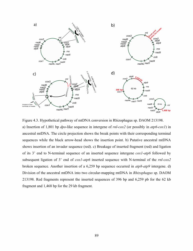
89
Figure 4.3. Hypothetical pathway of mtDNA conversion in Rhizophagus sp. DAOM 213198.
a) Insertion of 1,801 bp dpo-like sequence in intergene of rnl-cox2 (or possibly in atp6-cox1) in
ancestral mtDNA. The circle projection shows the break points with their corresponding terminal
sequences while the black arrow-head shows the insertion point. b) Putative ancestral mtDNA
shows insertion of an invader sequence (red). c) Breakage of inserted fragment (red) and ligation
of its 3’ end to N-terminal sequence of an inserted sequence intergene cox1-atp6 followed by
subsequent ligation of 3’ end of cox1-atp6 inserted sequence with N-terminal of the rnl-cox2
broken sequence. Another insertion of a 6,259 bp sequence occurred in atp8-atp9 intergene. d)
Division of the ancestral mtDNA into two circular-mapping mtDNA in Rhizophagus sp. DAOM
213198. Red fragments represent the inserted sequences of 396 bp and 6,259 pb for the 62 kb
fragment and 1,468 bp for the 29 kb fragment.

90
Figure 4.4. Linear representation of mtDNAs of R. irregularis DAOM 234179, DAOM 229456
and Rhizophagus sp. DAOM 213198 showing their genome synteny.
Yellow projections represent mtDNA regions with the same gene order.
The comparative sequence analyses of reshuffled intergenic regions of atp9-atp8, rnl-atp6,
and cox1-cox2 between the mitochondrial deoxyribonucleic acids of DAOM 213198 and the
close relative R. irregularis DAOM 234179 suggest that conversion and recombination could act
as mechanisms by which the fragmentation of mtDNA occurred in DAOM 213198 (fig. 4.3).
Interestingly, an intergenic region between cox1 and cox2 has never been found in any other
Glomeromycota sequenced so far. The nucleotide sequences of both termini of this unique
intergene matched with the putative ancestral intergenes of rnl-cox2 and cox1-atp6 in R.
irregularis DAOM 234179. This supports the idea that recombination could be initiated by a
DSB followed by translocation and reshuffling, resulting in fragmentation of the ancestral
mtDNA into two mtDNA circular molecules (fig. 4.3). Comparison between rnl-atp6 and cox1-
cox2 intergenes in DAOM 213198 and rnl-cox2 and cox1-atp6 intergenes in R. irregularis
DAOM 234179 suggests a dpo-like sequence insertion into their common ancestral mtDNA. The
inserted dpo-like sequence contains some conserved dpo domains (which are also found in other
mt genomes within Glomeromycetes) flanked by nonidentified sequences according to a

91
TBLASTX search (fig. 4.3 and supplementary fig. S4.2, Supplementary Mateiral online).
Conserved domains of dpo-like sequences at the amino acid level are shown in supplementary
figure S4.3, Supplementary Mateiral online. Figure 4.3 shows the hypothetical mechanism of the
mtDNA fragmentation in DAOM 213198: An insertion of a dpo sequence could have occurred in
the ancestral intergenic region of rnl-cox2 or cox1-atp6, followed by a breakdown and
disintegration of the inserted dpo sequence. Translocation and ligation of breaking sequence
termini of rnl to atp6 and cox1 to cox2 resulted in the formation of 29- and 62-kb mtDNA
circular chromosomes, respectively (fig. 4.3). The presence of dpo fragments in this region
strongly suggests a dpo-mediated recombination as reported previously (Beaudet et al. 2013). However, the origin of dpo domains flanking unknown sequences and their replication and
movement mechanisms remain unclear. Comparative genomics analysis between the several
AMF mtDNAs in Beaudet et al. (2013) and DAOM 213198 suggests that some intergene regions
can be considered as putative hot-spot regions for recombination. Supplementary figure S4.2,
Supplementary Material online summarizes putative recombination hot spots by dpo-like
sequences (containing the conserved domains) that could impact mt genes reshuffling in
Rhizophagus sp. DAOM 213198 and R. irregularis DAOM 234179. This comparative mtDNA
analysis provided some clues about the potential mechanisms of genome conversion occurring in
the isolate of Rhizophagus sp. DAOM 213198.
4.5.3 Short inverted repeats (SIRs) mediate recombination in mtDNA
Recombination in mtDNA can also be mediated by short inverted or palindromic repeats, as
previously demonstrated (Rattray et al. 2001, 2005). SIRs recorded in DAOM 213198 and in all
mtDNAs of Rhizophagus spp. publically available (Formey et al. 2012; Beaudet et al. 2013) were

92
analyzed to investigate their role in mtDNAs and their dispersal within and among mt genomes in
Rhizophagus. On the basis of their DNA secondary structure, a total of 13 types of SIRs were
recorded in the mtDNAs of DAOM 213198 (fig. 4.5 and supplementary fig. S4.4, Supplementary
Material online). The distribution patterns of SIR types, such as types 1 and 2 in R. irregularis
DAOM 234179, suggest potential repeat-mediated recombination (fig. 4.4 and table 4.2).
However, the decreased number of some SIRs’ types in mtDNAs of DAOM 213198 that were
frequently observed in DAOM 234179 (table 4.2 and fig. 4.5) supports their deletion through
repeat-mediated recombination as previously reported (Marechal and Brisson 2010; Davila et al.
2011). It remains unclear how such SIR replications and dispersals take place in mt genomes.
Presumably, SIRs tend to invade intergenic regions and introns that are under low selective
pressure.
Type%2%
Type%1%
Type%1%%R.#irregularis##234179%
Type%8%%Rhizophagus%sp.%%240422%%
Type%8%%R.#irregularis##234179%%
Type%2%%R.#irregularis##234179%%
Type%4%R.#irregularis#234179%Type%7%Rhizophagus%sp.%240422%%
Type%3%R.#irregularis%234179%Type%1%Rhizophagus%sp.%240422%%Type%2%Formey%et%al%%
SIR%6,%19%Rhizophagus#sp.#213198%
SIR%8%%Rhizophagus%sp.%213198%%
Type%9%R.#irregularis#234179%%Type%6%Rhizophagus#sp.%240422%%
Figure 4.5. Multiple sequence alignment of the most common identified short inverted repeats
(SIRs).
Multiple sequence alignment of 13 types of SIRs found in R. irregularis DAOM 234179,
Rhizophagus sp. DAOM 240422, DAOM 213198 and those reported by Formey et al. (2012).
SIRs types 1 and 2 are the most frequent SIRs found in all AMF mtDNA analyzed in this study.
Secondary structures were predicted based on the energy model of (Mathews, Disney et al. 2004)
and (Andronescu, Condon et al. 2007). The minimum free energy (MFE) structure of hairpins is

93
coloured according to the base-pairing probabilities (red: high; green: mid; blue: low). Blue and
red circles around nucleotides represent the beginning and the end of molecules, respectively.
Table 4.2. Distribution of small inverted repeats (SIRs) found in R. irregularis DAOM 234179
and Rhizophagus sp. DAOM 213198 depending on categorized types and genome localization.
R. irregularis DAOM 234179 Rhizophagus sp. DAOM 213198
SIR subtypes Genome localization SIR subtypes Genome localization
T1
cox1-atp6
T1
atp8-atp9(2)*
cox1-atp6 atp9-rns(2)
nad3-nad6 cox2-atp8(2)
cox3-rnl T2 rns-nad5(2)
rnl-cox2 T4
cox1 intron(2)
atp8-atp9 rnl-atp6(1)*
nad5 intron
T7
rns-nad5(2)
nad1-nad4l nad4l-cox1(2)
T2
cox1-atp6 cox3-rnl(1)
cox1-atp6
T8
nad4l-cox1(2)
nad6-cox3 atp8-atp9(2)
nad6-cox3 cox3 intron(1)
cox3-rnl
T9
atp9-rns(2)
cox2-atp8 cox1 intron(2)
atp9-rns cox1-cox2(2)
atp9-rns
T10
nad4l-cox1(2)
rns-nad5 cox1-cox2(2)
cob intron cox1-cox2(2)
T3 rns-nad5 T11 nad4l-cox1(2)
nad5-cob T12 cox3-rnl(1)
T6 nad4l-cox3
T7
rns-nad5
nad4l-cox3
T8 nad6-cox3
T11 atp9-rns

94
4.5.4 Relative quantification of mtDNA chromosomes
Real-time quantitative assays showed that the two mtDNAs had a similar abundance
(supplementary table S4.1, Supplementary Material online). This result is surprising because
smaller mt genomes usually replicate faster than larger mt genomes (Birky 2001; Schoch and
Seifert 2012). A putative mechanism controlling mtDNA replication might occur in the isolate
DAOM 213198, resulting in an equal abundance of both mt chromosomes. This has been shown
previously (Birky 2001; Schoch and Seifert 2012) where it was found that replication of mtDNAs
could be affected by not only the rate of initiation and the rate of completion of mtDNA synthesis
but also by the mechanisms that regulate the overall mass of each mtDNA.
4.5.5 Mitochondrial inheritance
Mt inheritance and dynamics have been intensively studied in many eukaryotes such as
mammals, plants, and yeasts (Birky 2001). However, these aspects have not been investigated in
Glomeromycota, probably because of their obligate biotrophic lifecycle and their slow growth.
Yet, such information about the mitochondrial dynamics and inheritance could give us insights
into the evolution of mtDNAs of Rhizophagus sp. DAOM 213198. Mt division could produce
mitochondria containing both mtDNAs or one of each mtDNA as shown in supplementary figure
S4.5, Supplementary Material online. Mitochondria and their mtDNAs could be randomly passed
to the progeny during cell division, as reported by Birky (2001). However, mitochondria are able
to undergo fusion followed by fission, thereby regulating their mtDNA segregation
(supplementary fig. S4.5, Supplementary Material online). If such a mechanism of inheritance
regulation did not exist, this could lead to forming nonfunctional mitochondria (Chan 2006; Chen
and Chan 2009).

95
Mt genes encoding essential enzymes and subunits for oxidative phosphorylation pathways
for energy (ATP) synthase are located in two mtDNAs in Rhizophagus sp. DAOM 213198. It is
likely that mitochondria lacking one of the two mtDNAs could not be functional. The isolate
DAOM 213198 could have a mechanism that controls mtDNA dynamics and inheritance.
Without this mechanism, mitochondria with incomplete mtDNA sets could lead to nonfunctional
cells (Benard and Karbowski 2009).
4.6 Conclusions We have documented an unusual mtDNA organization in the isolate Rhizophagus sp. DAOM
213198, a close relative to the model species R. irregularis. This novel mtDNA feature leads to a
new level of understanding of mt genome evolution in eukaryotes. The mtDNA comparative
analyses between close relatives of Rhizophagus spp. shows that dpo-like sequences and SIR-
mediated recombinations not only enhance mtDNA reshuffling but could also lead to
fragmentation, impacting mobile element dynamics in mt genomes. Mt genomes show a high
potential for developing molecular tool kits in order to discriminate isolates and closely related
taxa, and to monitor gene exchange and recombination among isolates. Rearrangement of genes
and intergenic regions has potential implications in studying population genetics, ecology, and
functions of Glomeromycetes in ecosystems. However, the mtDNA organization in two circular
chromosomes found in DAOM 213198 raises fundamental questions about their replication and
inheritance compared with other AMF harboring one single circular chromosome with a full set
of genes. Further investigations are needed to advance our understanding of the evolution of
mtDNA in eukaryotes, particularly in basal fungal lineages.

96
4.7 Acknowledgments This work was supported by The Natural Sciences and Engineering Research Council of
Canada (RDCPJ 395241-09 and discovery grant 328098-2012 to M.H.). Our thanks to Dr B.F.
Lang for bioinformatics assistance and access to an automated organelle genome annotation
software, to Dr Y. Dalpé for providing DAOM 213198 strain and associated images in
supplementary figure S4.1, Supplementary Material online, to Dr D. Beaudet and Dr F. Bou
Daher for their comments on the manuscript, and to Dr K. Fisher-Favret for English editing and
comments.

97
Presentation of article 4 The increasing number of sequenced AMF mitochondrial genes and mitogenomes shed light
on the importance of this genomic pool for evolutionary analyses of AMF. However,
evolutionary signal and phylogenetic power of mt genes have not been evaluated in AMF.
Therefore, nobody can truly and statistically claim which mt single gene or subset of genes could
be the most proper candidate for phylogenetic and evolutionary analyses. Moreover, it is
important to test whether mtDNA could complement other identification methods to identify and
characterize AMF at low taxonomic levels (isolate and species level).
In this study, we sequenced and mapped mtDNA of two more AMF species Rhizophagus
irregularis and Glomus aggregatum to implement the mtDNA datasets. The aim was to perform
mitogenomics comparative analyses and to assess mt genes phylogenetic signals and potential
use to infer phylogenies in AMF. We used two methods of phylogenetic analyses and in silico
mitogenomic comparative analyses.
The Article 4 is currently in preparation. I have designed the experiment and performed all
bioinformatics analyses as well as phylogenetic and statistical analyses. I have written the
complete manuscript which is corrected and organized by M. Hijri.

98
Chapter 5 - Mitochondrial comparative genomics and
phylogenetic signal assessment of mtDNA among arbuscular mycorrhizal fungal taxa
Maryam Nadimi, Laurence Daubois and Mohamed Hijri
Université de Montréal, Département de sciences biologiques, Institut de recherche en biologie
végétale (IRBV), 4101 rue Sherbrooke Est, Montréal, QC, H1X 2B2, Canada.
*Corresponding author
Submitted in Molecular Phylogenetics and Evolution.
_________________________

99
5.1. Highlights • Two mitochondrial genomes (mtDNA) of arbuscular mycorrhizal fungi were sequenced and
annotated.
• Mitochondrial comparative genomic analysis was performed on 16 Glomeromycotan complete
mtDNA.
• Phylogenetic signal of individual, subset and complete set of mt genes was assessed among
Glomeromycota taxa.
• mtDNA comparative genomics unraveled the intraspecific diversity of R. irregularis species
complex.
Keywords: Arbuscular mycorrhizal fungi, Fungi, Mitochondrial genome, Genome evolution,
Comparative mitogenomics, Phylogenetic analysis, SH-test, Supergene.
5.2. Abstract Mitochondrial (mt) genes, such as cytochrome C oxidase genes (cox), have been widely used
for barcoding in many groups of organisms, although this approach has been less powerful in the
fungal kingdom due to the rapid evolution of their mt genomes. The use of mt genes in
phylogenetic studies of Dikarya has been met with success, while basal fungal lineages remain
less studied, particularly the arbuscular mycorrhizal fungi (AMF). Considering the key roles
played by AMF in natural environments, it is critical to develop AMF-targeted molecular
toolkits, and to use these to describe the population genetics, community ecology, and phylogeny
of AMF. Advances in next-generation sequencing have substantially increased the number of
publically available mtDNA sequences for the Glomeromycota. As a result, comparison of

100
mtDNA across key AMF taxa can now be applied to assess the phylogenetic signal of individual
mt coding genes, as well as concatenated subsets of coding genes. Here we show comparative
analyses of publically available mt genomes of Glomeromycota, augmented with two mtDNA
genomes that were newly sequenced for this study (Rhizophagus irregularis DAOM240159 and
Glomus aggregatum DAOM240163), resulting in 16 complete mtDNA datasets. We assessed the
phylogenies inferred from single mt genes and complete sets of coding genes, which are referred
to as “supergenes” (16 concatenated coding genes), using Shimodaira-Hasegawa tests, in order to
identify genes that best described AMF phylogeny. We found that rnl, nad5, cox1, and nad2
genes, as well as concatenated subset of these genes, provided phylogenies that were similar to
the supergene set. This mitochondrial genomic analysis was also combined with principal
coordinate and partitioning analyses, which helped to unravel certain evolutionary relationships
in the Rhizophagus genus and for G. aggregatum within the Glomeromycota.
5.3. Introduction Elucidating the population structure and inferring the evolutionary histories of species are
critical towards understanding their role in ecosystems, and for microorganisms, this generally
requires a molecular approach. Developing effective methods, genetic markers, and
discriminating criteria are key to phylogenetic analysis and species diagnosis. Mitochondrial (mt)
genes and genomes have been used extensively in phylogenetic and population genetic studies of
a wide range of organisms. Typical mt genomes comprise a single molecule of DNA, which is
usually circular and contains all mt genes, while the number of mt genes varies among species. It
has often been assumed that the phylogenetic signal of each mt gene is identical, or highly similar,
due to the physical proximity of these genes which lie within the same mtDNA molecule (Avise

101
1994). However, mtDNA analyses in many organisms have suggested divergence in the
phylogenetic signal strength of mt genes among and within species (Duchene et al. 2011).
Several studies have indicated that sequencing of complete mtDNA molecules, referred to as
“supergenes” (i.e. complete set of mt protein coding genes), provides the highest phylogenetic
resolution among studied organisms (Duchene et al. 2011; Fenn et al. 2008; Havird and Santos,
2014). In addition, recent developments in next-generation sequencing technologies have
generated a huge number of mtDNA datasets across a broad range of organisms, in particular in
the kingdom Animalia (Duchene et al. 2011; Havird and Santos, 2014). These publically
available mtDNA datasets offer opportunities to perform large-scale comparative mt genomic
analyses, to infer phylogenetic relationships (for identification purposes) and to compare the
phylogenetic signals of complete sets of mt genes, or of concatenated subsets of individual mt
genes. Numerous phylogenetic studies have been performed on single mt genes such as
cytochrome C oxidase subunit 1 (cox1) (Borriello et al. 2014; Hebert et al. 2003; Ratnasingham
and Hebert 2007; Roe and Sperling 2007) and the mitochondrial large subunit gene (rnl) to infer
evolutionary relationships between organisms (Borstler 2008; Bruns 1998). However, the
phylogenetic signals produced by these extensively studied genes have not often been compared
with the complete set of mt genes or with other mt gene subsets. Such comparative analyses are
key to accurately assessing the evolutionary relationships of mt genomes within a given group of
organisms, and for identifying genes with strong phylogenetic signals that can be used to
discriminate between closely related taxa. For instance, the arbuscular mycorrhizal fungi (AMF),
a group of closely related symbionts of plant roots, are notoriously difficult to characterize and
distinguish through molecular methods. A more comprehensive mitochondrial-based approach
could provide a solution to this issue.

102
The arbuscular mycorrhizal fungi are among the most abundant eukaryotic symbionts of
plants, and not only improve plant mineral uptake and enhance plant productivity and diversity
(Smith and Read 2008), but also help in the control of plant pathogens (Azcón-Aguilar and Barea
1997; Garcia-Garrido and Ocampo 2002; Ismail and Hijri 2012; Ismail et al. 2011) and stimulate
the fitness of plants in polluted environments (Hildebrandt et al. 1999; Zhang et al. 2014). The
AMF constitute an early diverging fungal lineage, whose mycelia are typically formed by
coenocytic branching hyphae (i.e. hyphae that lack septae) in which nuclei and cell organelles
can freely move through the hyphal network (Marleau et al. 2011). Although, AMF have been
shown to harbour a complete and functional meiotic and pheromone-sensing pathway, as well as
sets of mating types (Halary et al. 2013; Halary et al. 2011; Pelin et al.,2012), sexual reproduction
has not yet been observed. Limitations in distinguishing morphological characters and molecular
traits are among the major factors limiting progress in AMF research. In addition, intra-isolate
genetic polymorphism and nuclear organization are still under debate. Furthermore, many lines of
evidence support the theory that AMF harbour genetically different nuclei in a common
cytoplasm, making these organisms highly unusual (Angelard et al. 2010; Beaudet et al., 2015;
Boon et al. 2015; Boon et al. 2010; Boon et al. 2013; Hijri and Sanders 2005; Kuhn et al. 2001).
In contrast to the nuclear genomes of AMF, mitochondrial coding-genes such as rnl and cox1, as
well as complete mtDNAs, have been reported to be homogeneous in many AMF taxa (Borstler
et al., 2008a; Lee and Young, 2009; Raab et al., 2005; Thiery et al., 2010), although this has been
challenged by Beaudet et al., (2015). Investigating large datasets of mtDNAs could offer
opportunities to advance our understanding of the population genomics and phylogeny of AMF.
To date, 16 fully sequenced Glomeromycota mt genomes have been published, representing
two major glomeromycotan families: the Glomeomeraceae (14 mtDNAs) and the Gigasporaceae
(two mtDNAs) (Beaudet et al., 2013a; Beaudet et al., 2013b; de la Providencia et al., 2013;

103
Formey et al., 2012; Nadimi et al., 2012; Pelin et al., 2012; Nadimi et al., 2014). These mtDNA
datasets offer opportunities to compare the intra- and inter-specific diversity of mt genomes and
genes among closely related AMF taxa, as well as the usefulness of various mt genes in
phylogenetic analyses. In this study, we sequenced and mapped the mtDNAs of two AMF species,
Rhizophagus irregularis DAOM240159 and Glomus aggregatum DAOM240163, to augment the
publically available set of AMF mtDNAs. We performed a mitogenomics comparative analysis,
as well as principal coordinate and partitioning analyses, in order to: (1) assess the phylogenetic
signals of mt coding genes; (2) evaluate their potential use in inferring AMF phylogenies; and
(3) unravel evolutionary relationships within the Rhizophagus genus.
5.4. Material and methods
5.4.1. Fungal material and DNA extraction
Spores and mycelium of a Glomus aggregatum isolate (DAOM240163) and of a
Rhizophagus irregularis isolate (DAOM240159) were cultivated in vitro on a minimal (M)
medium with carrot roots that had been transformed with Agrobacterium rhizogenes (Bécard and
Fortin 1988). The medium was dissolved using a solution of 0.82 mM sodium citrate and 0.18
mM citric acid, and fungal mycelia were collected by sieving the suspension. The collected
fungal material was checked for root contamination under a binocular microscope. DNA
extraction was performed using the DNeasy Plant kit (Qiagen, Mississauga, ON) according to the
manufacturer’s instructions.

104
5.4.2. Sequencing, assembly and gene annotation
Whole genome shotgun sequencing was performed using Roche 454 GS FLX Titanium
technology, with one full run per isolate. Sequencing was performed at the Genome Quebec
Innovation Center (McGill University, Montreal, QC). The reads were de novo assembled with
Newbler v2.9 at the Genome Quebec facility, and mitochondrial contigs were identified and
pooled for each isolate. Gene annotation was performed using an automated mt annotator
MFannot (http://megasun.bch.umontreal.ca/cgi-bin/mfannot/mfannotInterface.pl), followed by
manual inspection. The R. irregularis DAOM197198 mtDNA (accession number HQ189519)
was used as reference for sequence analyses and mtDNA comparison. Sequencing of the PCR
products (acquired for joining mtDNA contigs) was performed using Sanger technology at the
Genome Quebec Innovation Center (McGill University, Montreal, Canada). The annotated
mtDNAs of R. irregularis DAOM240159 and G. aggregatum DAOM240163 were deposited in
GenBank under the accession numbers KM586389 and KM586390, and circular maps were built
using OGDraw v. 1.2 software (Lohse et al. 2013). Comparative sequence analyses were
performed using the National Center for Biotechnology Information (NCBI) genomic database
and multiple sequence alignment. Nucleotide blast was used for similarity analyses, followed by
amino acid comparisons for more distinct sequences. A search with tBLASTx was also performed
using a minimum E-value cutoff of 1x10⁻¹⁰ and 50% minimum identity.
5.4.3. Polymerase Chain reactions (PCR)
As mentioned above, conventional PCRs were used to fill the gaps between mt contigs in
order to complete the mtDNA genome for each isolate. Conventional PCRs were performed in a
final volume of 50 µl containing 1X PCR buffer, 1.5 mM MgCl2, 0.2 mM of each

105
deoxynucleotide triphosphate (dNTP), 0.5 µM of each primer, 1 unit of Platinum Taq DNA
Polymerase (Life Technologies, Burlington, ON), and 5 to 20 ng of genomic DNA was used as a
template. Cycling parameters were: 94°C for 90 sec, followed by 38 cycles of 94°C for 1 min,
annealing temperature (variable based on the designed primers but generally ~60°C) for 30 sec,
72°C for 90 sec and a final elongation at 72°C for 5 min. PCR products were directly sequenced
as described above.
5.4.4. Datasets and sequence alignments
Sixteen completely sequenced mt-genomes whose length ranged between 59,633 bp (G.
cerebriforme) and 97,349 bp (Gigaspora rosea) were used in comparative analyses. The
complete mtDNA alignment was evaluated for phylogenetic and comparative mitogenomic
analyses. 16 genes containing 14 protein-coding genes (atp6, atp8, atp9, cox1, cox2, cox3, nad1,
nad2, nad3, nad4, nad4l, nad5, nad6 and cob) and two ribosomal coding subunits (rns and rnl)
were concatenated to construct the ‘‘supergene’’ set. Multiple sequence alignment was performed
using ClustalW as implemented in MUSCLE version 3.8.31 (Edgar 2004).
5.4.5. Phylogenetic analyses
Phylogenetic analyses were performed for all mt genes from fully sequenced mtDNAs of
AMF isolates (Table 5.1). Nucleotide alignments of all datasets were used to infer maximum
likelihood (ML) phylogenies as implemented in RAxML (Stamatakis et al. 2008), using the
RAxML-HPC2 on XSEDE web-server at CIPRES [http://www.phylo.org] (Miller et al.,2010).

106
Node support was determined using rapid bootstrapping with 1000 replicates. All datasets were
analyzed using the GTRGAMMA model. Phylogenies were also generated for the datasets using
concatenation of all mt genes (supergene set) and for a subset of genes, using concatenation of
the four best-performing mt genes (genes that showed similar tree topology to that of supergene).
5.4.6. Statistical analysis of phylogenies
Topologies of each dataset, composed of each of the 16 mt genes, or a subset of the four
best-performing mt genes with different combinations were compared to the supergene set.
Statistical analyses of the comparisons of phylogenetic signals for different datasets, as well as
comparisons of the topologies derived from mt genes and that of supergene, were performed
using Shimodaira-Hasegawa (SH) tests, implemented in RAxML using ‘-f h’ parameters locally.
The SH test provides log-likelihood values and the variation scores (Ln L) between the supergene
topology and each of the topologies inferred from single mt gene subsets, with respect to the
standard deviation (SD). This test was performed for phylogenies inferred using the ML approach
based on nucleotide datasets.
5.4.7. Distance matrix, principal coordinate analysis, and partitioning analysis
All analyses were conducted in R v.3.0.2 (R Foundation for Statistical Computing; available
at http://www.R-project.org). After using the ‘read.alignment’ function of the ‘seqinr’ packages
(Charif and Lobry 2007), distance matrices were computed using ‘dist.hamming’, ‘dist.dna’ and
‘dist.ml’ of the ‘phangorn’ package (Paradis et al. 2004; Schliep 2011). The similarity
relationship between isolates was assessed with a principal coordinate analysis (PCoA) plot,
using the ‘pcoa’ function of the ‘ape’ package (Paradis et al. 2004). In order to detect isolate

107
clusters within the plot, partitioning analysis were computed using a K-means clustering
approach, and analyses were computed using ‘kmeans’ of the ‘fpc’ package (Hennig 2010),
looking for a bend in the sum of squared roots (SSE). We also used partitioning around k-
medoids to estimate the number of clusters, using the ‘pamk’ function of the ‘fpc’ package
(Hennig 2010).
5.5. Results and Discussion
5.5.1. Overview of mtDNA diversity and evolution in the Glomeromycota
Fourteen complete mtDNAs belonging to two orders (Glomerales and Diversisporales) and
three genera (Glomus, Rhizophagus and Gigaspora) were retrieved from public databases, and
this dataset was augmented by two newly sequenced mtDNAs, resulting in a total of 16 mtDNAs
from the Glomeromycota phylum (Table 5.1). Although mt genome structure and genome
synteny varies somewhat among the studied isolates, they all contain 14 protein-coding genes and
2 ribosomal subunit genes. R. irregularis isolate DAOM240159 and G. aggregatum isolate
DAOM240163 are the two newly sequenced mt genomes, measuring 72,293 bp and 69,505 bp,
with G + C contents of 37.1% and 37.3%, respectively (Fig. 5.1). Both mitochondrial genomes
harbor the full set of typical fungal genes, such as three subunits of ATP synthase (atp6, atp8 and
atp9), three cytochrome oxidase subunits (cox1, cox2 and cox3), seven subunits of the NADH
dehydrogenase (nad1, nad2, nad3, nad4, nad5, nad6 and nad4L) and apocytochrome b (cob).
They encode 25 tRNAs and small and large ribosomal subunits (mtSSU and mtLSU) (Fig. 5.1).
In total, 41 genes encoded on the same strand in both genomes are found in each mtDNA (Fig.
5.1). The annotated sequences of R. irregularis DAOM240159 and G. aggregatum

108
DAOM240163 were deposited in GenBank under the accession numbers KM586389 and
KM586390, respectively. Our phylogenetic analysis, based on the concatenation of all individual
mt genes (supergene), clustered G. aggregatum within the R. irregularis group. However, using
sequence comparison of intergenic regions, we were able to discriminate G. aggregatum and R.
irregularis isolates (Table 5.2).
Figure 5.1. Comparison of the mitogenomes of the Glomus aggregatum isolate (DAOM240163)
and the Rhizophagus irregularis isolate (DAOM240159).
The circular mapping genomes were opened upstream of rnl. Genes on the outer and inner
circumference are transcribed in a clockwise and counterclockwise direction, respectively. Gene
and corresponding product names are cox1–3, cytochrome c oxidase subunits, atp6, 8, 9, ATP
synthase subunits; cob, apocytochrome b; nad1–4, 4L, 5–6, NADH dehydrogenase subunits; rnl,

109
rns, large and small subunit rRNAs; A-W, tRNAs, the letter corresponding to the amino acid
specified by the particular tRNA followed by their anticodon.
Table 5.1. Arbuscular mycorrhizal fungal isolates used in our mitogenomic analyses.

110
Table 5.2. Sequence comparison of intergenic regions between R. irregularis 197198 and G
aggregatum.
IGRs Variable regions position
rnl-cox2 41 bp insertion in position 1042 in G. aggregatum
nad3-nad6 37 bp insertion in position 88 in G. aggregatum
nad5-cob 40 bp insertion in position 927 in G. aggregatum
nad4L-cox1 Polymorphism from 748-787bp in DAOM197198 and 745-795 in G. aggregatum
atp9-rns 65 bp deletion from position 5550 in G. aggregatum
Polymorphism from 5410-5424 bp in DAOM197198 and 745-795 in G. aggregatum
nad6-cox3 36 bp insertion in position 557 in G. aggregatum
cox3-rnl 8628 bp insertion in position 1 in G. aggregatum
About 7500 bp polymorphism at position 404 of DAOM197198 (9034 in G. aggregatum)
G. aggregatum was described by Schenck and Smith in 1982 after being found in the roots of
citrus trees in Florida (Schenck and Smith 1982). Spores can be formed individually in the soil
and in roots or aggregates without a peridium. These spores are globose, subglobose, obovate,
and cylindrical to irregular. In fact, G. aggregatum and R. irregularis are very close according to
the morphological characters of their spores, with the only tangible difference being that G.
aggregatum spores possess an inner laminated wall that strongly reacts to Melzer’s reagent, while
the inside of spores are sometimes differentiated (Sokolski et al. 2011). A phosphate transporter
gene was able to slightly discriminate G. aggregatum from a dozen R. irregularis isolates
(Sokolski et al. 2011), while other genes such as ATPase H+, ATPaseF0F1, as well as ITS, 18R
and 25S rRNA genes were not able to discriminate these two species. In a recent reclassification
of the Glomeromycota, the position of G. aggregatum was classified as uncertain (Schussler et al.
2010).

111
5.5.2. Assessment of the phylogenetic signal of single mt genes and subsets of genes
Maximum likelihood phylogenies were inferred from nucleotide sequence alignments of the
complete set of mt genes (16 coding genes in total). We assessed the phylogenetic signal of single
mt genes, which was compared with the supergene signal via Shimodaira-Hasegawa (SH) test
(Fig. 5.2). These comparisons allowed us to determine the performance of single mt genes, and to
determine the best combination of gene subsets with phylogenies similar to the supergene
phylogeny. Twelve topologies inferred from single mt genes showed a significant reduction in
log-likelihood (Ln L) and statistically different topologies from that shown in the supergene
topology (Fig. 5.2).
Figure 5.2. Difference in log-likelihood measured via SH test by comparing phylogenetic
topologies inferred from single mt genes relative to that of the ‘‘supergene’’ set.

112
Difference in Ln L compared to the supergene phylogeny for all 16 mt genes (y-axis) has been
shown on the x-axis as ΔLn L. The four best-performing genes (i.e., cox1, rnl, nad5 and nad2)
are indicated by *. Phylogenetic analyses were performed under ML and SH test implemented
by RAxML locally.
However, the other four genes (i.e. rnl, nad5, cox1 and nad2), which were the longest mt
genes, reproduced similar topologies to the supergene tree, with much more subtle decreases in
Ln L relative to the supergene phylogeny (Fig. 5.2). Visual inspection, coupled with statistical
tests of single gene topologies, showed that the other mt genes provided poorly supported trees
for two main reasons: (i) genes such as atp9, cob, cox2 and rns could not resolve distant taxa
such as Gigaspora spp. and Glomus cerebriforme from Rhizophagus spp.; (ii) nad4L, nad6, and
atp8 genes provided low resolution for resolving Rhizophagus sp. isolates from other
Rhizophagus irregularis, which has been showed to be divergent (e.g. topologies inferred from
cob and nad6 are demonstrated in Figure 5.3). Furthermore, we evaluated the minimum number
of concatenated mt genes required to statistically reproduce a topology similar to that of the
supergene using ML. A SH test of the combination of ‘best-performing’ genes revealed low
decreases in log-likelihood scores (Fig. 5.2). Phylogenies inferred from combinations of genes
containing cox1 with nad2 or rnl, provided topologies that are statistically identical to the
supergene topology (Fig. 5.3), with the smallest decreases in Ln L (Fig. 5.2).

113
Figure 5.3. Topologies inferred from supergene set and concatenated set of rnl and cox1
revealing their high concordance in addition to topologies inferred from nad6 and cob as an
example of individual genes with significant inconcordance to supergene topology.

114
A) The two phylogenies are significantly congruent based on statistical analyses of Shimodaira-
Hasegawa (SH) with a difference of 7.6 in their Ln L values. Combination of cox1 and nad2 also
showed a statistically congruent topology, which is not repeated here. The supergene set consists
of a concatenation of all mt coding genes (i.e. 16 genes). rnl, cox1 are the two largest mt genes. G.
aggregatum is colored as red to highlight its position within the Rhizophagus clade. Numbers at
nodes represent bootstrap support values. Phylogenies were inferred using DNA sequences and
maximum likelihood (scale bars indicate replacements per site). B) Topologies inferred by nad6
and cob DAN sequences genes showing significant decreases in Ln L values compared with
supergene topology (ΔLn L of 294.6 and 228.1 respectively). Cob is not able to resolve distant
taxa such as Gigaspora spp. and Glomus cerebriforme from Rhizophagus spp. while nad6
possesses low resolution for resolving Rhizophagus sp. isolates from other Rhizophagus
irregularis.
Here we demonstrate that choosing a single mt gene in phylogenetic analysis may not reflect
the phylogeny of the complete set of mt coding genes. Some studies have shown that phylogenies
from combinations of all mt genes or from complete mitogenomes represent the highest
phylogenetic performance and more robust results than those produced by single genes and gene
subsets (Duchene et al. 2011; Havird and Santos 2014). These studies identified the most
informative mt regions (genes) and evaluated the minimum amount of data required to infer
phylogenies that were similar to supergene or complete mtDNA topologies. Therefore, the most
informative mt genes or regions might vary among taxonomic groups and organisms (Duchene et
al. 2011; Havird and Santos 2014).
Mt genes rnl and cox1 have previously been used independently to infer phylogenies among
taxa of the Glomeromycota (Borriello et al. 2014; Borstler et al. 2008b; Raab et al. 2005). Our
study demonstrates that phylogenetic analysis that combines cox1 and either rnl or nad2 performs
better than analysis on individual genes, not only considering the performed phylogenetic
analyses, but also the evolutionary features of each individual gene. There are certain

115
evolutionary aspects of rnl and cox1 that need to be considered prior to their use in phylogenetic
analyses of either organelles or organisms. For example, it is known that rnl is able to carry
inserts and introns in variable regions that are not excised at the RNA level (Raab et al. 2005).
Thus, it is often not easy to align mitochondrial rDNA sequences across species, due to introns,
insertions, and the loss of domains. There is still no reliable method for inferring precise ends of
the rnl gene, as the sequences of both terminal regions are highly variable. The only method for
precise annotation is RNA analysis (Lang et al. 1987; Nadimi et al. 2012). Therefore, rnl can be
useful for short distance species phylogenies, but would be problematic for phylogenies that
attempt to resolve among fungal clades or deeper levels. There are also several reports regarding
the evolutionary history and structure of cox1, which hampered the use of this gene for
phylogenetic analyses and barcoding. For example, Nadimi et al. (2012) reported the occurrence
of a group I intron-mediated trans-splicing in cox1 of Gigaspora rosea that has been previously
described in several other organisms (Burger et al. 2009; Grewe et al. 2009; Hecht et al. 2011;
Nadimi et al. 2012; Pombert and Keeling 2010). Frequent horizontal gene transfer within the
introns of this gene, which has been reported in the literature (Beaudet et al. 2013a; Lang and
Hijri 2009), may also complicate the sequencing and assembly of this region, as well as
downstream phylogenetic analyses. The above-mentioned findings limit the usefulness of
individual cox1 and rnl genes for phylogenetic analyses of AMF. Combinations of multiple genes
(e.g., different combinations of cox1, rnl and nad2) may help to overcome these drawbacks.
5.5.3. Assessment of the relationship between isolates
To further evaluate the relationships between all 16 isolates used in this study, we performed
a principal coordinate analysis (PCoA) based on the distance matrix obtained from mitochondrial

116
genomic alignments (Fig. 5.4). The PCoA plot shows that all Rhizophagus spp. isolates and G.
aggregatum group together. Of the other three isolates, G. cerebriforme is apart, while both
Gigaspora spp. cluster more closely together. Both partitioning methods used (k-means and k-
medoids) created two groups, corresponding to the two orders (Glomerales and Diversisporales)
used in the study. When inferring three groups instead of two on the plot, only G. cerebriforme
separates from the Glomerales group to form the third group. To further assess the cluster of
Rhizophagus isolates, another principal coordinate analysis (PCoA) was computed, excluding
both Gigaspora species and G. cerebriforme (Fig. 5.4). Both partitioning methods inferred three
groups, the first including solely Rhizophagus sp. DAOM213198, the second including R.
irregularis MUCL43204 and Rhizophagus sp. DAOM229456, and the third including the
remaining nine Rhizophagus isolates and G. aggregatum. One Rhizophagus sp. isolate,
DAOM240422, clustered with the other nine Rhizophagus isolates, but with a weaker similarity
(Fig. 5.4). These results support the G. cerebriforme isolate as genetically distant from all
Rhizophagus isolates and from the G. aggregatum isolate used in this study. As discussed above,
spore morphology and phosphate transporter phylogeny are able to marginally discriminate
between G. aggregatum from Rhizophagus isolates. However, our analysis based on the complete
mt genome shows that other Rhizophagus isolates, such as R. irregularis MUCL43204 and
Rhizophagus sp. DAOM213198/DAOM299456 are genetically more distant from the other nine
R. irregularis isolates than is G. aggregatum. This evidence supports the position of G.
aggregatum within the Rhizophagus irregularis ‘species complex’.

117
Figure 5.4. PCoA plot based on the distance matrix obtained from mitochondrial genomic
alignments.
(a) using all 16 mtDNAs examined in this study. Gigaspora spp. (G. rosea, and G. margarita)
and Glomus cerebriforme are distinguished from the Rhizophagus spp. and G. aggregatum

118
isolates. Two groups inferred by clustering analysis are shown in colors (b) Using 12 isolates of
Rhizophagus spp. and G. aggregatum. The three groups inferred by clustering analysis are shown
in different colors.
5.5.4. Unraveling the Rhizophagus irregularis complex
Comparative mitogenomics analysis of R. irregularis isolates (Fig. 5.5) and Rhizophagus spp.
reveals a high level of homogeneity within the coding sequences, while intronic and intergenic
regions showed substantial variable regions, which is consistent with the rate of evolutionary
constraint based on the functions of different mtDNA regions. However, comparative analyses
using ML and PCoA of coding regions suggest the occurrence of unorthodox recombinations (Fig.
5.6) among some Rhizophagus spp. isolates (DAOM229456, DAOM240422 and DAOM213198)
with uncertain classification, grouping them apart from other known R. irregularis isolates.
Genetic recombinations may have taken place at different points during the evolutionary history
of the different isolates or of a recent common ancestor, although horizontal gene transfer of
recombined regions is also a possibility. A shared common ancestor seems to be the most
parsimonious and plausible, due to similar signatures observed in recombined regions (Fig. 5.6).
Mt genes atp6, cox2, nad3, cob, nad4 and atp9 are examples of these recombined mt genes
within Rhizophagus spp. (Beaudet et al. 2013a; Beaudet et al. 2013b).

119
Figure 5.5. Complete mtDNA alignment of R. irreglaris isolates and G. aggregatum showing
sequence variations in intergenic regions, while coding genes are almost identical.
Comparative analyses of the intergenic regions of Rhizophagus spp. showed relatively higher
levels of similarity in up- and down-stream sequences of coding genes compared to other
intergenic regions. Up- and down-stream sequences of coding genes include 5′ and 3′ extensions
of genes (known as 5′ and 3′ untranslated regions), which are transcribed but not translated. The
conservation of such regions is due to their function, therefore resulting in a slower rate of
evolution relative to non-functional regions. Consequently, comparative analyses of multiple
sequence alignments of complete mtDNAs grouped Rhizophagus spp. isolates among the
Rhizophagus irregularis species, which we suggest to be considered as a species complex (e.g.
Rhizophagus irregularis complex).

120

121
Figure 5.6. Linear genome representations of some mt genes, harbouring the relative
recombinations within the Rhizophagus sp. complex to compare with R. irregularis.
Corresponding genes (atp6, cox2, nad3, cob, nad4 and atp9) among R. irregularis DAOM197198
and Rhizophagus sp. DAOM240422, DAOM213198 and DAOM229456 are linearized and
compared using projections. Projections in yellow represent nucleotide identity at more than
90%, while projections in orange illustrate the identity of more that 90% at the amino acid level.
Projections with no color were used to facilitate detection of related regions. The intron formed in
Rhizophagus sp. DAOM229456 is boxed in gray, which is transferred to the intergenic region
after cox2 in Rhizophagus sp. DAOM240422. Nucleotide and amino acid identity comparisons
were performed via blastn and tblastx among the studied AMF isolates.
5.6. Conclusion We implemented the existing mtDNA dataset with two newly completed and annotated mt
genomes. Our mitogenomic comparative analysis among several AMF species and isolates
allowed us to assess the phylogenetic signals of individual mt genes in comparison to the
supergene set. A statistical SH test revealed that the evolutionary signal differed significantly
between single mt genes, subsets of genes, and the supergene in the Glomeromycota. The
combination of cox1 with either rnl or nad2 genes is a promising subset of mt genes that could be
useful in inferring phylogenies within Glomeromycota. However, implementation of the mtDNA
dataset with members of other families and genera is needed to cover the diversity of the
Glomeromycota phylum and to infer robust phylogenies. Further studies on the comparison and
potential combination of mt gene subsets, supergenes, and nuclear ribosomal rRNA genes could
provide a higher phylogenetic resolution, and may helpt to discriminate between closely related
members of the Glomeromycota.

122
5.7. Acknowledgment This work was supported by the Natural Sciences and Engineering Research Council of
Canada (Discovery grant 328098-2012 to M.H.). Our thanks to Dr. B.F. Lang for bioinformatics
assistance and access to an automated organelle genome annotation software, to Dr. Y. Dalpé for
kindly providing fungal material, Dr. S. Joly for assistance in phylogenetic and statistical
analyses, and Dr. T. Bell for commenting on the manuscript and English editing.

123
Presentation of article 5 The presence of a mixture of more than one type of mt genome results in heteroplasmy,
which has never been observed in AMF. Typically in sexual organisms, biparental inheritance
(paternal leakage) and/or occurrence of mutation result in coexistence of different mt haplotypes
within a single cell. In case of AMF as asexual organisms, exchange of genomic material via a
process called anastomosis raises question about the presence of a heteroplasmic state in them.
Nonself recognition between members of the Rhizophagus genus has been documented (Croll et
al. 2009; Angelard et al. 2010; Colard et al. 2011, Purin and Morton 2012) and has resulted in
nuclear transmition to the progenies but exchange of divergent mitochondrial haplotypes has yet
to be shown. In yeast, heteroplasmic state has been reported and it has ben shown that this
condition is temporary due to different mechanisms such as segregation, leading to permanent
homoplasmy (White and Lilley 1997; MacAlpine et al. 1998; Birky 2001).
In this study, we employed three genetically well-characterized R. irregularis isolates
(DAOM197198, DAOM234328 and DAOM240415) to upgrade our knowledge about AMF
mitogenome and creditability of it for AMF identification and to address the question, whether
heteroplasmy occurs in AMF. These isolates were selected because they originate from distant
geographical locations. In addition, de novo genome sequencing, assembly and annotation were
performed for two other R. irregularis isolates, DAOM234328 (Finland) and DAOM240415
(Manitoba, Canada), and their results were compared with published mtDNAs (Lee and Young
2009; Formey et al. 2012; Nadimi et al. 2012). Subsequently, specific size-variable markers were
designed for each isolate to detect mtDNA mixture after anastomosis between the strains in
microcosm experiments.
The Article 5 was published in New Phytologist. I assembled and annotated both of
mtDNAs. It took 9 months to design and set up the pre-symbiotic and symbiotic experiments
examining different methods to apply the most appropriate ones. By taking maternity leave, Ivan
de La Providencia went through performing the experiments with assistant of Gabriela Rodriguez
and afterwards he completed writing of the article. I also contributed to writing the first draft of
the manuscript.

124
Chapter 6 - Detection of a transient mitochondrial DNA
heteroplasmy in the progeny of crossed genetically divergent isolates of arbuscular mycorrhizal fungi
Ivan Enrique de la Providencia, Maryam Nadimi, Denis Beaudet, Gabriela Rodriguez Morales and Mohamed Hijri
Université de Montréal, Département de sciences biologiques, Institut de recherche en biologie
végétale (IRBV), 4101 rue Sherbrooke Est, Montréal, QC, H1X 2B2, Canada.
Published in New Phytologist, 2013 Oct. 200(1): 211-21, doi: 10.1111/nph.12372.
__________________________

125
6.1 Summary • Nonself fusion and nuclear genetic exchange have been documented in arbuscular
mycorrhizal fungi (AMF), particularly in Rhizophagus irregularis. However,
mitochondrial transmission accompanying nonself fusion of genetically divergent isolates
remains unknown.
• Here, we tested the hypothesis that mitochondrial DNA (mtDNA) heteroplasmy occurs in
the progeny of spores, obtained by crossing genetically divergent mtDNAs in R.
irregularis isolates.
• Three isolates of geographically distant locations were used to investigate nonself fusions
and mtDNA transmission to the progeny. We sequenced two additional mtDNAs of two
R. irregularis isolates and developed isolate-specific size-variable markers in intergenic
regions of these isolates and those of DAOM-197198. We achieved three crossing
combinations in pre-symbiotic and symbiotic phases. Progeny spores per crossing
combination were genotyped using isolate-specific markers.
• We found evidence that nonself recognition occurs between isolates originating from
different continents both in pre-symbiotic and symbiotic phases. Genotyping patterns of
individual spores from the progeny clearly showed the presence of markers of the two
parental mtDNA haplotypes. Our results demonstrate that mtDNA heteroplasmy occurs
in the progeny of the crossed isolates. However, this heteroplasmy appears to be a
transient stage because all the live progeny spores that were able to germinate showed
only one mtDNA haplotype.

126
6.2 Keywords Anastomosis; arbuscular mycorrhizal fungi (AMF); heteroplasmy; mitochondrial genome;
nonself fusion; specific molecular marker
6.3 Introduction The Glomeromycota phylum encompasses an ancient group of obligate symbiotic fungi
colonizing c. 80% of plants to form arbuscular mycorrhizae (Smith and Read 2008). Long
considered asexual organisms, this paradigm has recently been challenged by the discovery of
intact meiotic machinery genes that are putatively functional (Halary et al. 2011), although
apparent reproductive structures have yet to be observed. Arbuscular mycorrhizal fungi (AMF)
reproduce clonally through large and multinucleated spores, and due to their coenocytic nature
(i.e. living hyphae lacking septa and thereby nuclei, mitochondria and other organelles sharing a
common cytoplasm), spores and mycelium can host communities of hundreds to thousands of
nuclei showing substantial genetic variability (Kuhn et al. 2001; Hijri and Sanders 2005; Jany and
Pawlowska 2010; Marleau et al. 2011) which persist at the transcriptional level (Boon et al. 2010).
Several studies have shown the impact of AMF on plant growth and health (Jeffries et al.
2003, Smith and Read 2008, van der Heijden et al. 2008, Ismail and Hijri 2012) soil quality and
structure (Rillig and Mummey 2006), and ecosystem functioning and biodiversity (van der
Heijden et al. 2006). When associated with plants, they often extend beyond roots into the
surrounding environment to form an intermingled coenocytic and multinucleated extraradical
mycelium network (Leake et al. 2004) that facilitates up to 90% of phosphorus (P) plant-uptake
and also enhances nitrogen (N)-acquisition under natural conditions (reviewed in Bolduc and
Hijri (2011)). AMF structures can also link plants from the same or different species and/or
families via the formation of hyphal fusions (i.e. anastomosis) (Giovannetti et al., 2004), creating
127
an extensive common mycorrhizal network. As previously demonstrated by Giovannetti et al.
(1999), anastomosis also plays a key role in genetic exchange, either within the same isolate or
between closely related isolates that are genetically divergent (Croll et al. 2009). This mechanism
is likely one of the most important factors contributing to the maintenance of genomic
composition among members of the Glomeromycota (Bever and Wang 2005, Corradi et al. 2007,
Croll et al. 2009; Angelard and Sanders 2011; Colard et al. 2011). However, such huge genetic
diversity within AMF populations (Corradi et al. 2007; Bonen 2008) or even in successive
monosporal subcultures (Ehinger et al. 2012) makes it difficult to use only nuclear markers to
address questions related to AMF community structure, diversity and function. In contrast to
nuclear genes, mitochondrial genomes appear to be homogeneous within AMF isolates.
This was clearly demonstrated by sequencing the first complete mitochondrial genome (Lee
and Young 2009) of the Rhizophagus irregularis isolate 494 (Błaszk., Wubet, Renker & Buscot)
C. Walker & A. Schussler – syn. Glomus irregulare Błaszk., Wubet, Renker & Buscot
(Blaszkowski et al. 2008; Stockinger et al. 2009; Schüßler and Walker 2010), followed by many
other genomes such as Gigaspora margarita, G. rosea, four isolates of R. irregularis and a
closely related Rhizophagus species (Formey et al. 2012; Nadimi et al. 2012; Pelin et al. 2012;
Beaudet et al. 2013). These genomes offer exceptional opportunities for designing specific
markers to identify AMF strains either in natural populations or in mesocosm/microscosm based-
experiments (Lang and Hijri 2009; Formey et al. 2012; Beaudet et al. 2013). It was previously
assumed that, based on incompatibility and nonself recognition mechanisms, anastomosis cannot
occur between genetically distinct isolates (Giovannetti et al. 2003). However, many studies have
challenged this point of view and have provided convincing contrary evidence (Croll and Sanders
2009; Angelard et al. 2010; Angelard and Sanders 2011). These studies used crossing
experiments and specific genetic markers to show that nuclear material from both parents is

128
passed on to their progeny, thus demonstrating genetic exchange and segregation between
genetically divergent isolates of the AMF R. irregularis. In addition, a recent study has clearly
demonstrated that genetically different isolates of Rhizophagus clarus (synonym, Glomus
clarum) originating from the same habitat and sampled close to each other underwent anatomosis
(Purin and Morton 2012). This once again raises the questions of whether mitochondria are also
exchanged via anastomosis between isolates of R. irregularis of different geographical origin,
that is, heteroplasmy (i.e. the mixture of genetically different mtDNAs in a common cytoplasm)
and whether this state is transient or is maintained into the progeny. Heteroplasmy is not rare in
the fungal kingdom (Lesemann 2006) and has been observed in several eukaryotes (Barr et al.
2005; White et al. 2008). As suggested by researchers working on natural populations of the plant
pathogen Armillaria sp. (Smith et al. 1990), possessing multiple types of mtDNA might confer an
advantage under changing environmental conditions. This hypothesis has already been
demonstrated for the model fungus Saccharomyces cerevisiae (Taylor et al. 2002; Hnatova et al.
2003) and the plant pathogen Podosphaera leucotricha (Lesemann 2006).
Heteroplasmy has never been studied in AMF, presumably due to the lack of reliable and
appropriate mitochondrial markers (Lang and Hijri 2009; Lee and Young 2009; Formey et al.
2012). Consequently, we employed three genetically well-characterized R. irregularis isolates
(DAOM-197198, DAOM-234328 and DAOM-240415) to address this question, which is an
important one for advancing our knowledge of genetic exchange between AMF. These isolates
were selected because they originate from distant geographical locations. In addition, de novo
genome sequencing, assembly and annotation were performed for two other R. irregularis
isolates, DAOM-234328 (Finland) and DAOM-240415 (Manitoba, Canada), and their results
were compared with published mtDNAs (Lee and Young 2009; Formey et al. 2012; Nadimi et al.
2012). Subsequently, specific size-variable markers were designed for each isolate to detect

129
mtDNA mixture after anastomosis between the strains in microcosm experiments.
6.4 Materials and Methods
6.4.1 Growth conditions and maintenance of fungal cultures and roots
Monoxenically produced spores of Rhizophagus irregularis; DAOM-197198 (Pont-Rouge,
Quebec, Canada) DAOM-234328 (Finland) and DAOM-240415 (Dufrost, Manitoba, Canada)
were provided by the DAOM collection (Ottawa, Ontario, Canada). These three isolates were
selected because they have different geographical origins and their mitochondrial genomes have
been fully sequenced. Spores were subcultured in association with Ri T-DNA transformed
chicory (Cichorium intybus) roots on a modified minimal (MM) medium (Becard and Fortin
1988) solidified with 0.4% (w/v) gellan gum (Sigma). Plates were incubated in the dark in an
inverted position at 25°C. Several thousand spores and extraradical mycelia were obtained in a
period of 12 wk. Ri T-DNA transformed chicory roots were routinely propagated by placing
actively growing root apexes on MM medium and subsequent incubation at 25°C in the dark.
6.4.2 DNA extraction and sequencing
For DNA extraction, the extracted fungal spores and mycelia from the MM medium were
dissolved in buffer citrate (pH 6.0) (Doner and Becard 1991) and washed with sterile water.
Extracted fungal material from the MM medium was observed under a binocular microscope in
order to detect and remove any root contaminants. DNA extraction was carried out using the
DNeasy Plant Mini Kit (Qiagen), following the manufacturer’s instructions. Whole genome
shotgun sequencing was performed at McGill University’s Genome Quebec Innovation Centre

130
using a Roche GS-FLX-Titanium sequencing platform (454 Life Sciences, Branford, CT, USA).
6.4.3 de novo assembly and sequence analysis
The total DNA of R. irregularis isolates DAOM-240415 and DAOM-234328 was sequenced
and the resulting reads (257 880 and 238826, respectively) were assembled using Newbler
(Genome Quebec Innovation Centre, McGill University, Montreal, PQ, Canada). Gene
annotation was performed with MFannot (http://megasun.bch.umontreal.ca/cgi-bin/mfan
not/mfannotInterface.pl), followed by manual inspection and notation of missing gene features.
More information on MFannot is described in Nadimi et al. (2012). The annotated mitochondrial
genomes of R. irregularis isolates DAOM-240415 and DAOM-234328 were deposited in
GenBank under the respective accession numbers JX993113 and JX993114.
6.4.4 Molecular marker development
In order to develop isolate size-specific molecular markers for the mtDNA of three different
R. irregularis isolates, the variable intergenic regions of their complete sequenced mitochondrial
genomes were used for multiple alignments. Consequently, we developed specific primers for
each isolate, which resulted in JX993114 three pairs of primers. In addition, these primers
produced PCR fragments of different lengths (Table 6.1). All primers were tested for hairpin and
dimer formation using AmplifX software and a SIGMA DNA calculator (http://www.sigma-
genosys.com/calc/ DNACalc.asp). The specificity of these markers was achieved using PCR on
amplified DNA of each isolate (Fig. 6.1). These markers made it possible to trace parental
mtDNA haplotypes in crossing experiments.

131
Table 6.1. Isolate-specific primers used to discriminate the three Rhizophagus irregularis isolates
Primers
Cod
es
Primer sequences (5′- 3′)
Size
(bp)
Position of primers in mtDNA
DAOM-197198 DAOM-240415 DAOM-234328
197198F A AGCAAATCTAAGTTCCTCAGAG 120 69523–69544
197198R TCCTGCCCCAGATACTCCAG 69624–69643
240415F B AGAAGAGTTAATAAGTTCAACTGT 147 68561–68584
240415R AGACTAATAATACGAGATGCAG 68687–68708
234328F C AAGAGGGGTGCATTCCGAAG 209 67641–67660
234328R AGCTACGCTTTGCCTTAGCA 67831–67850
Figure 6.1. Isolate-specific mtDNA markers.
(a) Comparative view of the cox3-rnl intergenic regions of the three Rhizophagus irregularis
isolates where primers were designed (arrows). Upper boxes (open reading frames, ORF) show
direct orientation while bottom boxes are opposite orientation. (b) Gel electrophoresis showing
the specificity of length-specific molecular markers developed for the R. irregularis isolates
DAOM-197198 (A), DAOM-240415 (B) and DAOM-234328 (C). L, ladder (molecular marker).

132
6.4.5 Experimental set up
We set up two experiments in pre-symbiotic and symbiotic phases, respectively, to study the
occurrence of anastomosis and subsequent exchange of mitochondria among the three isolates in
crossing experiments. We also performed a single symbiotic experiment to evaluate the
inheritance pattern of mitochondria from monosporal culture lines obtained from spores
harvested from the interaction zone. Hyphal interactions were classified into four categories as
described in Croll et al. (2009): (1) perfect fusion meant that hyphae entered into contact, fused
and protoplasmic flow was established following anastomosis. No sign of incompatibility
reaction was observed. (2) Pre-fusion incompatibility was characterized by profuse mycelium
ramification at the elongation zone of the hypha that could be followed by septa formation at the
hyphal apex, thus preventing contact between the approaching hyphae. (3) No interaction means
that two hyphae (regardless of the type of interaction) did not show signs of rejection (i.e. wall
thickening at the apex region, septa formation or change of growth direction) but intermingled
(i.e. hyphae from different isolates or the same isolate grew in different directions and crossed,
but did not touch each other) or overlapped (i.e. hyphae from different isolates or the same isolate
entered into contact and continued to grow over each other). (4) Post-fusion incompatibility
means that following homing, contact and fusion, the protoplasm of one hypha withdrew and a
septum was formed between the two fused hyphae. Percentage of anastomosis for the categories
perfect fusion and nonself fusion incompatibility was calculated by dividing the number of
fusions by the total number of observed hyphal contacts (Purin and Morton 2012).

133
6.4.6 Pre-symbiotic experiment
Spores of the three isolates were extracted from the gel by solubilisation in buffer citrate (pH
6) (Doner and Becard 1991). A mono-compartment plate (35 9 10 mm) filled with 5 ml of MM
medium was divided into four equal compartments. Before inoculation a colour-coded circle was
marked at the bottom of the plate on each compartment and used to identify clusters belonging to
the same or different isolates. On each compartment, ten clusters (a cluster was defined in this
study as a group of spores bearing the same mycelium (Fig. 6.2)) of up to five spores from each
isolate were gently placed over the medium in the following combinations: (DAOM-
197198/DAOM-234328; DAOM-197198/DAOM-240415; DAOM-234328/DAOM-240415).
Clusters belonging to different isolates were separated by a maximum distance of 2 mm. Plates
were then sealed and incubated at 25°C in the dark until germination. Following cluster
germination, the occurrence of hyphal fusion was checked weekly for a 45-d period, and results
were grouped into four categories (see above, ‘Experimental set up’ section) (Croll and Sanders
2009). Each experimental unit (i.e. one replicate) consisted of one plate with four repetitions of
the same pairing (combination) and ten replicates were performed for each combination. Ten
replicates constituted from pairings of the same isolate were used as controls.

134
Figure 6.2. Self-fusion between spore clusters belonging to Rhizophagus irregularis DAOM-
4240415.
(a) Emergence of several germ tubes through the cut extremities (black arrow) and anastomosis
formation. (b) Inset showing details of the anastomosis (white arrows); note the profuse growth
of thin hyphae at the interaction zone. Active bidirectional protoplasmic flow, is shown in Video
S1.
6.4.7 Symbiotic experiment
An autoclaved (121°C for 15 min) slide (25 9 75 9 1 mm) was placed in the middle of an
empty mono-compartment Petri dish (100 9 15 mm). Thirty millilitres of the MM medium was
then poured in to fill the plate and cover the slide. Following jellification, only the medium
covering the slide was gently removed in such a way that c. 1 mm of each of the slide edges was
still embedded in the medium. A flat surface where the development of symbiotic mycelia could
easily be traced, adjoined by two identical distal and proximal compartments, was created (Fig.
6.3). Subsequently, 2-cm long Ri T-DNA transformed chicory roots were placed in both
compartments and 100 spores of each isolate taken up from monoxenic cultures by solubilisation
were inoculated in one of the two compartments using the same combinations described above.
Plates were sealed and incubated upside down at 25°C in the dark. Plates were checked weekly
and chicory roots growing towards the slide were cut and gently removed using scalpel and
forceps. Because mycelia growing on both edges of the slide began to spread across it, plates
were checked weekly over 10 wk for hyphal interactions, and results were grouped into four
categories according to Croll et al. (2009). Each experimental unit (i.e. one replicate) consisted of
one plate inoculated with two isolates in association with a Ri T-DNA transformed chicory root.
Fourteen replicates were performed for each combination, and five replicates for each isolate

135
inoculated in both compartments with the same fungal isolate were used as control.
Figure 6.3. Diagram presentation of the experimental set up for the study of anastomosis between
geographically distinct isolates of Rhizophagus irregularis in the symbiotic mycelium.
(a, b) Different views of the experimental set up showing mycelium (black and gray) from both
sides growing towards the slide (i.e. zone of interaction) and (c) interacting with each other.
Monosporal cultures lines with progeny from crossed experiments. In order to test the
hypothesis of whether heteroplasmy is a transient state or is maintained into the progeny, twenty
full lipid spores (i.e. progenies) harvested from the interaction zone for each combination were

136
randomly chosen and individually cut out from the mycelium and extracted in 2 μl sterile ultra-
pure water (PureLab, Elga, Canada). Spores were placed in a new mono-compartment Petri dish
(90 mm) containing M medium in the close vicinity of a Ri-T transformed chicory root. Sixty
replicates, each consisting of one single spore associated with a chicory root were maintained as
above. Each plate was checked weekly for germination, root colonization and colony
development over 11 wk.
6.4.8 Microscopy, data collection, harvest and statistical analysis
For all experiments, the occurrence of anastomosis and/or spore germination was observed
under a Discovery V12 stereomicroscope (Carl Zeiss, Toronto, ON, Canada) at magnifications of
96.7–940. Bright-field microscopy (Axio Imager M1; Carl Zeiss) was also used to observe details
of hyphal interactions at higher magnifications. Images and movies of the hyphal interactions, as
well as protoplasmic streaming through hyphal fusions, were captured with an AxioCam HRC
using Axiovision software v4.8.1.0 (Carl Zeiss).
For the second experiment, only the cultures showing synchronized growth of crossed
isolates from both sides towards the slide were considered in order to trace the origin of hyphae
and to avoid self-fusions. This restriction reduced the experimental replicates to three for some
crossing combinations. The weekly growth of the mycelia over the slide was marked with
coloured dots according to the isolate, making it easy to trace development as well as interactions
between both mycelia. Hyphal interactions, number and phenotype of spores (e.g. shape, lipid
droplet content, aborted-like structures) formed at the zone of interaction (slide) at both sides of
the fungal cultures were evaluated in addition to measuring total hyphal length and hyphal
density. Thirty full lipid spores (20 spores out of 30 were used to initiate monosporal cultures

137
lines; see experiment 3) formed at the zone of interaction for each combination were randomly
chosen, individually cut out from the mycelium and extracted in 2 μl sterile ultra-pure water
(PureLab). Whole genome amplification was performed for all spores using the GenomiPhi V2
DNA Amplification Kit (GE Healthcare, Mississauga, ON, Canada) following the manufacturer’s
instructions. Monosporal culture lines initiated from progenies did not produce any further visible
extraradical structures, even though germination and root contact of several spores was observed.
Under these circumstances, all germinated and nongerminated spores were harvested and their
genome amplified following the same protocol described. The root segment in contact with the
germinative mycelium was removed and stained following the protocol described in (Phillips and
Hayman 1970) to observe hyphal attachment and also assess the development of intraradical
structures. Data analysis was performed using STATISTICA software. Data were subjected to an
ANOVA. The Tukey honestly significant difference test was used to identify significant
differences (P ≥ 0.05).
6.4.9 PCR genotyping and sequencing of progeny spores
As already described, isolate-specific markers were designed for each of the three R.
irregularis isolates, DAOM-240415, DAOM-234328 and DAOM-197198. Primer sequences
are summarized in Table 6.1. DNA from individual spore progeny for the second and third
experiments was subjected to PCR using the isolate-specific primers corresponding to their
parents. PCR conditions were as follows: PCR reactions in a 50-μl volume contained 0.2 mM
dNTPs (Fermentas, Canada), 0.5 μM of each primer, 0.5 U of Taq DNA polymerase (BioBasic,
Montreal, Quebec, Canada) and 2 μl of the whole genome amplification product of each spore.

138
PCRs were run on Mastercycler pro S (Eppendorf, Canada). PCR cycling conditions included an
initial denaturation at 95°C for 2 min followed by 30 cycles at 95°C for 30 s; 55°C or 60°C for 30
s and 72°C for 30 s. An elongation at 72°C for 10 min was performed at the end of PCR.
Hybridization temperature was 55°C for DAOM-240415 and 60°C for DAOM-97198 and
DAOM-234328. PCR products were revealed using GelRed dye (Invitrogen) and images were
recorded by GelDoc system (BioRad, Canada). PCR was performed according to the crossing
combinations summarized in Table 6.1. All the amplification products obtained from progeny
were sent for sequencing to McGill University and Genome Quebec Innovation Center facility
(Montreal, Canada). Nucleotide Blast searches were performed using the NCBI website to check
the presence of the target sequence.
6.5 Results
6.5.1 Mitochondrial genome comparison and marker development
The mtDNAs of the two newly sequenced R. irregularis isolates (DAOM-240415 and
DAOM-234328) are almost identical, both to each other and to those retrieved from GenBank, R.
irregularis isolates: 494 (Lee and Young 2009), DAOM-197198 (Nadimi et al. 2012), MUCL-
46239, MUCL-46240 and MUCL-46241 (Formey et al. 2012). Rhizophagus irregularis isolates
have a mitochondrial genome ranging in size from 68 995 bp to 74 799 bp, except for the isolate
MUCL-43204, which is outside of this range (87 754 bp). This variation in size is caused by a
divergence in the presence/absence and size of mobile elements such as plasmid related DNA
polymerase genes (dpo), open reading frames encoding homing endonuclease genes in defined
intergenic regions, as previously described in Formey et al. (2012).

139
In order to develop the mtDNA isolate-specific marker for DAOM-197198, DAOM-240415
and DAOM-234328, multiple alignments of their complete mtDNAs and those of five other R.
irregularis isolates were performed. We focused on the cox3-rnl intergene, which is a large
region comprising substantial sequence divergence, as a target to design size-variable isolate-
specific markers. The specific markers that were developed were used to distinguish between the
three R. irregularis isolates selected in our crossing experiments, although the presence of a large
number of dpo elements of different lengths and orientations in the cox3-rnl intergenic region
made it challenging to perform the multiple sequence alignments. Each specific marker amplified
a single band of an expected size for the corresponding isolate (Fig. 6.1). In addition to the
specificity, the three markers were designed to produce different PCR product sizes, in order to
facilitate tracing each marker in spore progeny. For example, marker A, specific for isolate
DAOM-197198, gave a PCR band of 120 bp only for DNA of its corresponding isolate and no
band was observed for isolates DAOM-240415 and DAOM-234328. Markers B and C, specific
for isolates DAOM-240415 and DAOM-234328, gave PCR bands of 147 bp and 209 bp,
respectively (Fig. 6.1, Table 6.1). Length polymorphism can also be used to trace each haplotype
in multiplex PCR reactions. However, primers of isolate DAOM-234328 can also amplify
mtDNA of the isolate 494, producing a 209-bp PCR product. Because isolate 494 was not
included in our crossing experiment, the primer pair developed for DAOM-234328 was valid.
6.5.2 Pre-symbiotic interactions
In this experiment, we recorded both self and nonself fusion between all the isolates tested
and characterized them by total fusion of hyphal membranes and cell wall followed by
bidirectional protoplasm streaming between the connected hyphae (Video S6.1). Cluster

140
germination for all isolates was observed during the first 24 h. They germinated as a syncytium,
which means that the germ tube never emerged through the subtending hypha when the spores
were sharing a common mycelium, but one or several germ tubes re-grew from the cut
extremities (Fig. 6.2). After 1 wk, several spores within the cluster lost their cytoplasmic content
and became translucent, while others within the same cluster seemed to be dormant, as no change
in color and lipid content was observed.
Self-fusion was a common event within the same isolate (Fig. 6.2), and ranged from 60% to
38% in DAOM-197198 and DAOM-2404515, respectively. Pre-fusion as well as post-fusion
incompatibility was never recorded in pairings within the same isolate. On the contrary, hyphal
interaction between pairings of different isolates (i.e. nonself interaction) showed heterogeneous
responses. Nonself hyphal fusion was rare between different isolates, and was recorded only two
times in the combination DAOM-234328/DAOM-240415 out of 157 contacts observed
(Supporting Information Table S6.1). Nonself post-fusion incompatibility was recorded for all
combinations and the highest value was 3 (2%) out of 148 contacts observed in DAOM-197198/
DAOM-240415. Microscopic observations of these hyphal fusions revealed slow protoplasm
movement alongside the connected hyphae followed by protoplasm withdrawal and septa
formation (Video S6.2).
6.5.3 Symbiotic interactions
Growth of the mycelium during the symbiotic phase showed the same developmental
patterns for Rhizophagus species (formerly Glomus genus) (Bago et al. 1998). After germination
and root colonization, runner hyphae (RH) rapidly colonized the medium, and extended towards
the interaction zone. They divided dichotomously into thinner hyphae of increasing branch order.
Hyphae subsequently branched profusely to form the so-called branching absorbing structures
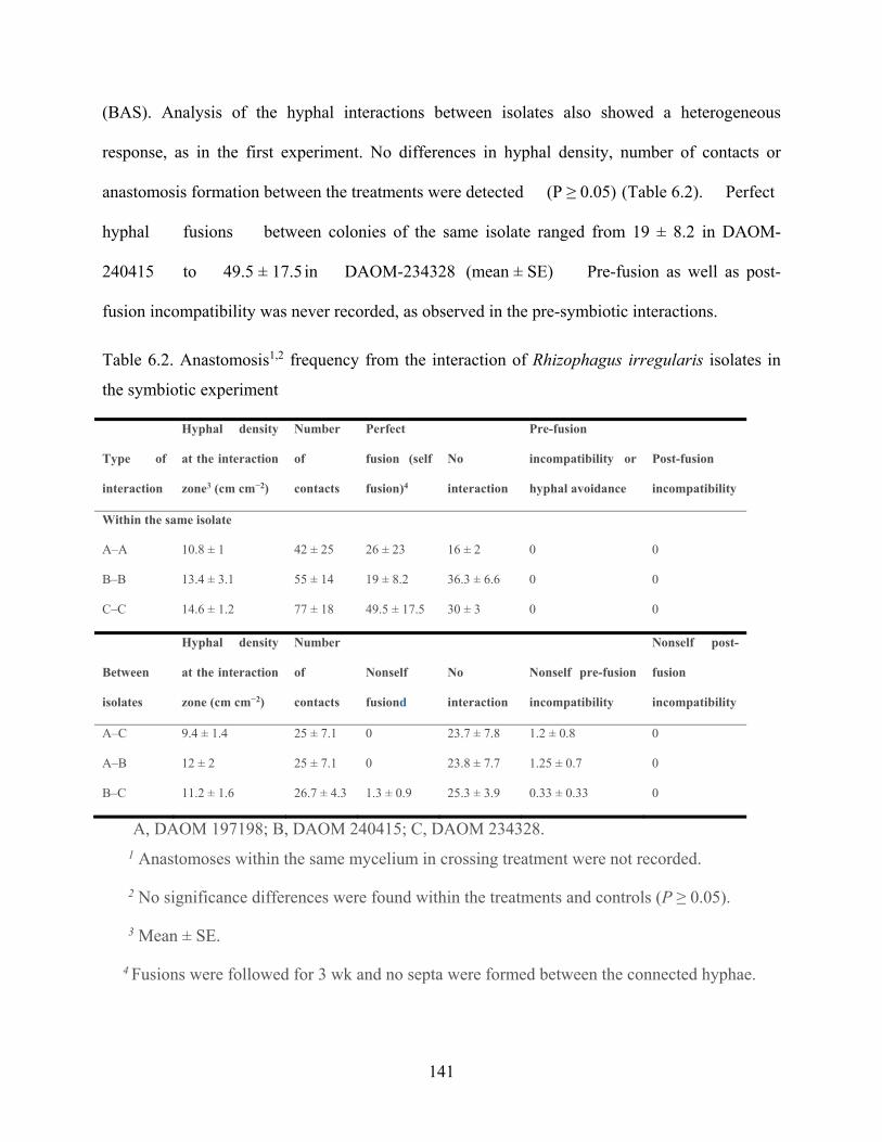
141
(BAS). Analysis of the hyphal interactions between isolates also showed a heterogeneous
response, as in the first experiment. No differences in hyphal density, number of contacts or
anastomosis formation between the treatments were detected (P ≥ 0.05) (Table 6.2). Perfect
hyphal fusions between colonies of the same isolate ranged from 19 ± 8.2 in DAOM-
240415 to 49.5 ± 17.5 in DAOM-234328 (mean ± SE) Pre-fusion as well as post-
fusion incompatibility was never recorded, as observed in the pre-symbiotic interactions.
Table 6.2. Anastomosis1,2 frequency from the interaction of Rhizophagus irregularis isolates in
the symbiotic experiment
Type of
interaction
Hyphal density
at the interaction
zone3 (cm cm−2)
Number
of
contacts
Perfect
fusion (self
fusion)4
No
interaction
Pre-fusion
incompatibility or
hyphal avoidance
Post-fusion
incompatibility
Within the same isolate
A–A 10.8 ± 1 42 ± 25 26 ± 23 16 ± 2 0 0
B–B 13.4 ± 3.1 55 ± 14 19 ± 8.2 36.3 ± 6.6 0 0
C–C 14.6 ± 1.2 77 ± 18 49.5 ± 17.5 30 ± 3 0 0
Between
isolates
Hyphal density
at the interaction
zone (cm cm−2)
Number
of
contacts
Nonself
fusiond
No
interaction
Nonself pre-fusion
incompatibility
Nonself post-
fusion
incompatibility
A–C 9.4 ± 1.4 25 ± 7.1 0 23.7 ± 7.8 1.2 ± 0.8 0
A–B 12 ± 2 25 ± 7.1 0 23.8 ± 7.7 1.25 ± 0.7 0
B–C 11.2 ± 1.6 26.7 ± 4.3 1.3 ± 0.9 25.3 ± 3.9 0.33 ± 0.33 0
A, DAOM 197198; B, DAOM 240415; C, DAOM 234328. 1 Anastomoses within the same mycelium in crossing treatment were not recorded.
2 No significance differences were found within the treatments and controls (P ≥ 0.05).
3 Mean ± SE.
4 Fusions were followed for 3 wk and no septa were formed between the connected hyphae.

142
Nonself fusions were only observed at the zone of interaction for the combination DAOM-
240415/DAOM-234328. Because anastomosis traceability was solely confined to the zone of the
interaction, the occurrence of nonself fusions far beyond the interaction zone might not be ruled
out. Pre-fusion incompatibility was observed for all combinations characterized by hyphal burst
at the hyphal apex or septa formation and cytoplasm retraction before contact. No signs of wall
thickening at the hyphal-apex region were observed when hyphae from different isolates came
into contact, but overlapped mycelium was the rule rather than the exception for all combinations.
Sporogenesis was observed during the first week after root colonization on the RH. Most spores
were formed into lower order hyphae after extensive growth of the fungal colony. Full lipids and
empty spores as well as aborted-like structures were observed and recorded in all the isolates
tested, regardless of the combination. The percentage of aborted-like structures in the interaction
zone ranged from absence to 2 ± 1 (mean ± SE) among crossed experiments and from 2 ± 2 to 5 ±
2 in controls. The same pattern was observed at each side, when the plate was inoculated with
both isolates in crossing (i.e. nonself interaction), or within controls (i.e. self interactions).
Statistical analysis did not show any significant difference in the number and percentage of
spores produced for any of the categories (P ≥ 0.05) mentioned above (Table S6.2). Although in
the combination DAOM-197198/DAOM-240415 (Fig. 6.4, Video S6.3), several aborted-like
structures (data not shown) often arrested their development and burst, after which several
hyphae grew out of the spore lumen (Fig. 6.5) Observations of these hyphae showed that
cytoplasm retracted and several septa were then formed.

143
Figure 6.4. Variability in spore morphology of Rhizophagus irregularis at the interaction zone in
the combination DAOM197198/DAOM240415.
(a) Spore clusters showing empty, full lipid droplets and crushed spores. (b) Extraradical
mycelium bearing both full lipid droplets (FL) and emptied spores (ES). (c, d), Irregular shaped
spores within the same mycelium. For a detailed view of the spore variability see Video S3.

144
Figure 6.5. Aborted-like structures produced by the Rhizophagus irregularis isolates at the
interaction zone.
(a, b) DAOM-197198/DAOM-240415; (c, d) DAOM-234328/DAOM-240415. (a) Several
hyphae exit from the aborted-like structures lumen and continue growing following cytoplasm
retraction and multiple septa formation. (b) Inset showing details of the lumen and septated
hyphae (arrows). (c) Burst and cytoplasm exit (black arrow). (d) Inset showing details of the
septated hypha inside the aborted-like structures.
6.5.4 mtDNA genotyping and sequencing in spore progeny
For each crossing combination in a symbiotic interaction, 10 full-lipid spores of the progeny
were individually genotyped using isolate-specific markers corresponding to their parents, as well
as the progeny of the monosporal cultures lines experiment. The progeny of the three crossing
combinations showed different PCR amplification patterns (Fig. 6.6). For example, the crossing
of isolates DAOM-197198 and DAOM-240415 showed that nine progeny spores (S) out of ten
contained both parental markers and one spore (S6) did not produce a PCR amplification product
using mtDNA markers, while 18S rDNA primers AML1 and AML2 showed a PCR band of an
expected size (790 bp; data not shown). Five spores of the crossing combination between
DAOM-197198 and DAOM-234328 showed both mtDNA haplotypes, while five contained only
197198-mtDNA haplotype. Three spores (S3, S6 and S9) of the crossing combination between
DAOM-240415 and DAOM-234328 showed both mtDNA haplotypes, six spores showed one
mtDNA haplotype whereas DAOM-240415 mtDNA haplotype was predominant (S1, S2, S5, S7
and S10) and spore (S8) did not produce a PCR amplification product using mtDNA markers,
while 18S rDNA primers did produce a PCR band (data not shown). Interestingly, the PCR bands
varied greatly in intensity among spores and independently of the length of the marker, probably
due to the frequency of each mtDNA haplotype.

145
Figure 6.6. Gel electrophoresis showing patterns of mtDNA genotyping of ten progeny spores
(S1–S10) of three combinations using three Rhizophagus irregularis isolates DAOM-197198 (A)
DAOM-240415 (B) and DAOM-234328 (C).
mtDNA marker sizes are 120, 147 and 209 bp corresponding to DAOM-197198, DAOM-240415
and DAOM-234328, respectively.
6.5.5 Monosporal cultures with progeny from crossed experiments
Even though attempts to obtain monosporal culture lines failed, spore germination was
observed for progenies from all crossed experiments and ranged from 40% in DAOM-
197198/DAOM-234328 to 60% in DAOM-240415/DAOM-234328 (Table S6.3). The
germination process was characterized by the emission of one to several germinative hyphae
through the subtending hyphae (Fig. S6.1). Some germinative hyphae showed a marked rhizo-
tropism (Fig. S6.1); however, a nonorientated growth was also observed. Germinative hyphae
also underwent contact with roots but after several weeks no progress of the fungal growth was
observed (i.e. runner hyphae bearing spores and branching absorbing structures).
Genotyping of the progeny showed that 14 spores gave positive PCR amplifications while
the rest of the progeny did not produce any amplification product. All PCR products were

146
sequenced and matched to the isolate-specific parental mtDNA markers. However, genotyping
results showed that all the progeny amplified only one parental mtDNA haplotype (Fig. S6.2).
For example, in the combination DAOM-197198/DAOM- 234328, three spores were genotyped
of which two contained the DAOM-197198-mtDNA haplotype and the other showed the DAOM-
234328 mtDNA haplotype. Five spores in the combination DAOM-240415/DAOM-234328 were
genotyped where two spores contained the DAOM-234328 mtDNA and three presented the
DAOM-240415 mtDNA haplotype. Finally, six spores were genotyped in combination DAOM-
197198/ DAOM-240415, where only one showed the DAOM-197198 genotype while the others
contained the DAOM-240415 mtDNA haplotype.
6.5.6 Colonization detection
Microscopic observation of stained root fragments, which were suspected for being
colonized by progenies, revealed the absence of hyphopodia formation. Consequently, no
intraradical structure was observed.
6.6 Discussion Nonself recognition between members of the Rhizophagus genus has been documented
(Croll and Sanders 2009;Angelard et al. 2010; Colard et al. 2011; Purin and Morton 2012) and
has resulted in nuclear segregation to new individuals affecting plant fitness (Angelard et al.
2010; Colard et al. 2011). Interestingly, combination B-C (isolates DAOM-240415 originating
from Manitoba, Canada, and DAOM-234328 originating from Finland) showed perfect nonself
fusion and the occurrence of heteroplasmy. Moreover, in the symbiotic crossing experiment of

147
combinations A-B and A-C, we did not observe perfect nonself fusion, but a few proportions
indicative of pre-fusion incompatibility; genotyping of mtDNA clearly showed the presence of
both parental haplotypes in the spore progeny. These results can likely be explained on the basis
of the coenocytic nature of the AMF mycelium allowing protoplasm mixture before hyphal
septation, suggesting the existence, as in higher fungi (Fu et al. 2011), of a putative molecular
mechanism regulating nonself recognition that has yet to be demonstrated.
The observed mtDNA heteroplasmy in the progeny following nonself recognition (i.e.
unrelated to apparent sexual reproduction) between genetically divergent isolates of the same
species originating from different continents is somewhat surprising. This assumption is made on
the basis that all mtDNA sequenced so far in AMF (Borstler et al. 2008; Lang and Hijri 2009;
Lee and Young 2009; Formey et al. 2012; Nadimi et al. 2012; Pelin et al. 2012; Beaudet et al.
2013) and those presented in this study, have been shown to be homogeneous. This
apparent contradiction is evidence of the putative transient heteroplasmic state in AMF, and is
consistent with our results as both germinated progeny and spores that do not break their
dormancy after long periods of sub-cultivation showed the presence of one parental mtDNA
marker. This evidence suggests the presence of an effective segregation mechanism, which might
regulate mtDNA inheritance in these organisms. Inheritance of mitochondria in fungi during the
mating process is mostly uniparental, with only one parental haplotype of the mitochondrial
genome inherited by the progeny (reviewed in (Ni et al. 2011)). However, biparental inheritance
of mtDNA has also been shown in some fungal species, such as Saccharomyces cerevisiae and
Schizosaccharomyces pombe, because the two mating cells contribute equally through their
cytoplasm organelles to form a mixed cytoplasm in the zygote. Interestingly, the coexistence of
different mitochondrial genome haplotypes has been shown to be transient in these organisms
(Birky 2001; Ni et al. 2011; Beaudet et al. 2013). Mechanisms contributing to homoplasmy are

148
often related to the recombination of both parental mtDNAs or by the selection of one of the
parental haplotypes due to the presence of segregation mechanisms, such as genetic bottleneck
followed by a stochastic drift (Birky 2001). As proposed by White et al. (2008), heteroplasmy
may either persist, become fixed, or be lost due to either selection forces or drift, or both. The
extent to which such mechanisms may occur in AMF and the resulting haplotype to be fixed in
the population will need to be assessed.
In arbuscular mycorrhizal fungi, no studies have been addressed to investigate patterns of
mtDNA inheritance and segregation to the progeny, however, the occurrence of distinct mtDNA
haplotypes within an AMF individual, with divergence in mobile elements and dpo insertions,
points to the possible occurrence of interhaplotype recombination and mtDNA segregation
mechanisms in these fungi (D. Beaudet et al. unpublished data). In budding yeasts mtDNA
segregation occurs via nucleoids (i.e. packages of identical mtDNAs maintained together by
Holliday junctions and proteins; (Lockshon et al. 1995; White and Lilley 1997; MacAlpine et al.
1998), creating a genetic bottleneck responsible for further rapid segregation. Due to this
mechanism, heteroplasmy can last over 20 generations in these organisms. It would be interesting
to investigate whether the formation of such mitochondrial nucleoids occurs in AMF and, if so,
how long this heteroplasmic state is maintained. The efficiency of the mitochondrial segregation
process is directly correlated to the initial population size and the relative proportion of one given
haplotype in a population (White et al. 2008). In this study, only two different mtDNA
haplotypes were mixed in vitro, giving rise to limited initial genetic variants. Also, we only have
evidence of the mtDNA haplotypes frequency from the intensity of the PCR bands we observed,
which make it difficult to predict the effectiveness of the segregation process.
Other studies have shown that coexistence of two mtDNAs within a common cytoplasm may
be disadvantageous, likely due to genetic conflicts or competition between divergent mtDNA

149
haplotypes (reviewed in (Ni et al. 2011), references therein). It has been proposed that uniparental
inheritance of mtDNA in fungi as well as other eukaryotes might be maintained by selection
forces to avoid such conflicts (White et al. 2008). Some authors have hypothesized that
uniparental inheritance of mtDNA and chloroplastic DNA may avoid dispersion of selfish and
deleterious mobile elements (Hoekstra 2000; Mamirova et al. 2007). Even if the divergence
between the mtDNA haplotypes used in this study appeared to be neutral, because they are
located in intergenic regions, it is difficult to predict with confidence the implied function in the
genome and its role in the segregation and inheritance process. In any case, selection of one
mtDNA haplotype due to potential mtDNA conflicts or stochastic segregation mechanisms might
explain the transient character of heteroplasmy in our experiment and also provides a rational
explanation for why homoplasmy is more often observed in spores from field (Borstler et al.
2008) and/or in vitro experiments than heteroplasmy (Lee and Young 2009; Formey et al. 2012;
Nadimi et al. 2012; Pelin et al. 2012; Beaudet et al. 2013). Previous studies on AMF mtDNA
have already demonstrated the great potential for developing isolate-specific molecular markers
(Borstler et al. 2008; Lang and Hijri 2009; Formey et al. 2012; Beaudet et al. 2013). For
example, Borstler et al. (2008) used the rnl gene encoding the mitochondrial ribosomal large
subunit (mtLSU) to discriminate different haplotypes among cultivated isolates of R. irregularis
(formerly G. intraradices) in soil and within root samples from the field. These authors found 12
rnl haplotypes among 16 isolates of R. irregularis originating from five continents. Formey et al.
(2012) recently compared the complete mtDNA of six R. irregularis isolates and were able to
develop mtDNA markers using the cox3-rnl intergene as well as other regions to discriminate
isolates using length polymorphism markers. These mtDNA markers can be useful for other
studies, both in population genetics as well as in tracing isolates in agriculture soil and plant roots
to test co-inoculation of different R. irregularis isolates. However, it will be important to have a

150
better understanding of the mitochondrial inheritance process and the extent of the mtDNA
variability present in a natural population in order to benefit from the full potential of such
markers.
In this particular study, we believe it was crucial to use size-variable markers combined with
a whole genome amplification step, to easily test the occurrence of heteroplasmy between the
crossed parental cultures. Some studies have suggested that during their formation, spores can
randomly inherit different nuclear haplotypes following hyphal fusion (Marleau et al. 2011). The
same pattern could be expected for mtDNA, however they could be critically underrepresented in
daughter spores, given the competition with the endogenous haplotypes and the putative process
of mtDNA segregation. If the isolate-specific markers were variable in single nucleotide
polymorphisms (SNPs), it would have required a large number of clones for each tested spore to
find even one different locus. PCR and cloning-based studies with such markers could fail to
identify occurrences of heteroplasmy. Despite its potential as a DNA barcode, mtDNA has never
been considered as a unique and reliable highly-sensitive target to address studies on fungal
individualism in AMF (Rayner 1991).
6.7 Conclusions We have demonstrated a potential use of mtDNA markers to address basic questions of
evolutionary biology in AMF using R. irregularis isolates as a model. Our study provided
additional information in the AMF mitochondrial genome collection with the complete mtDNA
of two R. irregularis isolates. Currently, there are eight R. irregularis isolates with annotated
mitochondrial genomes that differ substantially in some intergenic regions, making them useful
sites for developing isolate-specific markers. We also provided additional markers that were

151
specific to some isolates, and demonstrated their potential use in tracing mtDNA in a crossing
experiment.
We also showed that isolates of R. irregularis from different geographical locations, even
from different continents, were able to exchange their genetic material regarding mtDNA,
resulting in heteroplasmy in their spore progeny. The extent to which such a state may occur in
natural communities and the mechanisms responsible for the fixation of a homogeneous
haplotype in an AMF individual need to be assessed. Further studies on vegetative compatibility
and incompatibility as well as putative sex machinery in AMF, are needed to advance our
understanding of the evolution of AMF and to better understand their population dynamics.
6.8 Acknowledgements This study was conducted as part of a research project organized and coordinated by Premier
Tech. The authors are grateful for financial supports from NSERC Cooperative Research and
Development (grant number: RDCPJ 395241-09), NSERC Discovery (grant number: 328098-
2012), Premier Tech and CRI-BIQ. I.D.P. was supported by Genome Canada and Genome
Quebec funds. We would also like to thank Biopterre centre du developpement des bioproduits
and CRBM for assistance and Nathalie De Jaeger, Terrence Bell, Karen Fisher Favret and three
anonymous referees for their useful reading and comments on the manuscript.

152
Chapter 7 - General discussion, Conclusion and
Perspectives
_________________________________
My Ph.D. thesis contributes significantly to fundamental knowledge on the evolution of
mitochondrial genomes in fungi, in particular in the early diverging fungal lineage of
Glomeromycota. It clearly reports the presence of substantial intra- and inter-specific genetic
variation in glomeromycotan mtDNA. My thesis shows that these substantial variations are
generally caused by the invasion and integration of mobile elements such as plasmid-related
DNA polymerase gene (dpos), open reading frames (orfs) and small inverted repeats (SIRs). I
showed that mobilome variation and movement within mt DNA of AMF contributed to the
evolution of unorthodox genes, genome structure and variable intergenic and intronic regions.
The outcome of mobilome variation in mt genomes of AMF (e.g. genome rearrangement) can be
exploited as opportunities for developing efficient and accurate isolate- and species-specific
molecular marker. For example, formation of fragmented genes in Gigaspora rosea (chapter 2),
unorthodox C-terminal regions in some mt protein-coding genes (PCGs) of some Rhizophagus
spp. isolates (Chapter 3) and the mt genome fragmentation in an isolate of Rhizophagus sp.
(DAOM 213198) (Chapter 5), are mediated by mobile element activities. Consequently, I
hypothesize that the biochemical machineries for both homologous and non-homologous
recombination might be active in mitochondria of glomeromycotan fungi, although, this has not
been demonstrated yet.
The reported evolutionary mechanisms that are mediated by mobilome activities (dpos, orfs
and SIRs) also increase genetic polymorphism in intergenic regions giving rise to hypervariable

153
regions or “hotspots” for example cox3-rnl intergene. This intergene varies substantially among
closely related species and even among different isolates belonging to the same species as is
shown for the model AMF species R. irregularis, although conserved intergenic regions exist.
The genetic polymorphism observed in mtDNAs has been shown to be useful to isolate-specific
biomarkers development, as it has been applied and presented in chapters 3 and 4. Accurate
biomarkers and diagnosis toolkits are useful for identification, characterization and quantification
not only in AMF but also in other organisms. These studies have potential fundamental
implications in studying population genetics, ecology and functions of Glomeromycetes in
ecosystems, but also in applied research in agriculture and environmental protection.
In my Ph.D. study, I designed, tested and validated isolate-specific molecular markers for the
study of genetic exchange in three geographically distant isolates of R. irregularis. Using these
isolate-specific markers, I identified their efficiency in tracing and tracking mitochondria within
fused AMF isolates through anastomosis (chapter 5). My markers allowed discovering a transient
heteroplasmy in the AMF R. irregularis. I showed that isolates of R. irregularis from different
geographical locations can exchange their genetic material regarding mtDNA, resulting in
heteroplasmy in their spore progenies. However, this heteroplasmy state appeared to be transient
because of its loss in progeny spores that were able to germinate. This evidence suggests the
presence of an effective segregation mechanism, which might regulate mtDNA inheritance in
AMF and so support the reliability of mitogenome utilization for biomarker development.
However, since the divergence between the mtDNA haplotypes used in our study appeared to be
neutral (i.e. divergence mediated by dpo within hypervariable region of cox3-rnl), it is difficult to
predict the implied function in the genome and its role in the segregation and inheritance process.
Further experiments are needed to address anastomosis among isolates harboring mtDNA
haplotypes vary in the coding sequence. Such length specific markers and isolate-specific

154
markers could also be used to investigate population genetics and interaction of AMF with plant
roots and soil microorganisms.
In my Ph.D. thesis, I contribute significantly to the implementation of completed mtDNAs in
public databases and comparing mt genomes among different AMF taxa. My study provides
additional information in the AMF mitochondrial genome collection with the complete mtDNA
of ten R. irregularis isolates. I also performed phylogenetic analyses to compare and assess
phylogenetic signals between single mt genes, subsets of genes and complete set of concatenated
mt genes (16 mt coding genes). Additionally I documented an unusual mtDNA organization in a
close relative to the model species R. irregularis and this finding will lead to a new level of
understanding of mt genome evolution in eukaryotes. Moreover, I report that from a phylogenetic
perspective, not all mt genes possess the same phylogenetic signal and thus can not be equally
representative of the complete mt genes set.
Our phylogenetic analyses of mt protein-coding genes revealed that AMF are related to
Mucoromycotina rather than to the Dikarya phyla (Basidiomycota and Ascomycota), which was
in contrast to rDNA analyses that was suggesting the Zygomycota phylum is paraphyletic and
that Glomeromycota are sister to the Dikarya phyla (Schüßler et al. 2001; James et al. 2006).
However, recent findings revealed that the 45S rDNA locus of R. irregularis is highly
polymorphic (Stockinger et al. 2009; Lin et al. 2014) and so is not a proper candidate for such
analyses. Further analyses of a supermatrix of 35 highly conserved, putative single copy nuclear
genes (Capella-Gutie ́rez et al. 2012) support our result. Consequently, similar results in revealing
evolutionary relationship of AMF within Fungi support our hypothesis that mtDNA can be a
proper compliment for identification and phylogenetic analyses comprising relatively easier to
access database.

155
Sequencing of other mtDNAs in distant lineages of AMF in order to cover major AMF taxa
(families and genera) will advance our knowledge of this important fungal group. It will also
open new opportunities for phylogenetic and identification analyses in a wider taxonomic level,
which will help to improve AMF function in agriculture and forestry. However, sequencing
mtDNAs may be challenging in some AMF taxa, in particular, those that are not able to grow in
vitro, or the ones with low sporulation rate and slow growth. To overcome these limitations
development or optimization of cultivation both in vitro and in vivo conditions, could improve
sequencing procedure and bioinformatic analyses. Further studies need to reevaluate mt genes
phylogenetic signal at different taxonomic levels and within distant lineages of AMF. Taken all
together, comparison and complementing of mitochondrial- and nuclear-based phylogenetic
analyses and identification will help us better understand and apply AMF in nature. Moreover,
advanced investigation of mobilome (e.g., origin of mobilome, their replication and dispersal
mechanisms, their affinity to integrate specific regions such as hot-spots) that resides within mt
genomes of AMF will decipher their contributions in AMF mtDNA evolution.
Broad taxa sampling for mt genomic and phylogenetic analyses will strengthen anastomosis
and mtDNA exchange studies among AMF. This could lead to discovering mechanisms and
machineries that control vegetative compatibility and incompatibility. Implementation of mtDNA
datasets with morphological characters using advanced microscopy techniques and staining
procedures will aid taxonomists to better classify and study the evolutionary relationships in
Glomeromycata.
In conclusion, my thesis, along with other studies on mtDNA, report the usefulness of
mitochondrial genomics to address basic and applied research questions in glomeromycotan
biology. Accurate mt biomarkers and diagnostic toolkits have proven their efficiency and could
be used in applied aspects, such as the identification and quantification of AMF, their in vitro or

156
in vivo cultivation (e.g., in bioremediation applications or in agriculture investigations). These
tools could ultimately, improve the efficiency of phytoremediation and also sustain agriculture by
better managing AMF-based biofertilizers.

157
References Adams, K. L., M. J. Clements and J. C. Vaughn (1998). "The Peperomia mitochondrial coxI group I intron: timing of horizontal transfer and subsequent evolution of the intron." J Mol Evol 46(6): 689-696. Aguilera, P., T. Barry and J. Tovar (2008). "Entamoeba histolytica mitosomes: organelles in search of a function." Exp Parasitol 118(1): 10-16. Aliasgharzad, N., M. R. Neyshabouri and G. Salimi (2006). "Effects of arbuscular mycorrhizal fungi and Bradyrhizobium japonicum on drought stress of soybean." Biologia 61: S324-S328. Alkan, N., V. Gadkar, O. Yarden and Y. Kapulnik (2006). "Analysis of quantitative interactions between two species of arbuscular mycorrhizal fungi, Glomus mosseae and G. intraradices, by real-time PCR." Appl Environ Microbiol 72(6): 4192-4199. Alverson, A. J., D. W. Rice, S. Dickinson, K. Barry and J. D. Palmer (2011). "Origins and recombination of the bacterial-sized multichromosomal mitochondrial genome of cucumber." Plant Cell 23(7): 2499-2513. Andronescu, M., A. Condon, H. H. Hoos, D. H. Mathews and K. P. Murphy (2007). "Efficient parameter estimation for RNA secondary structure prediction." Bioinformatics 23(13): i19-28. Angelard, C., A. Colard, H. Niculita-Hirzel, D. Croll and I. R. Sanders (2010). "Segregation in a mycorrhizal fungus alters rice growth and symbiosis-specific gene transcription." Curr Biol 20(13): 1216-1221. Angelard, C. and I. R. Sanders (2011). "Effect of segregation and genetic exchange on arbuscular mycorrhizal fungi in colonization of roots." New Phytologist 189(3): 652-657. Angelard C, Tanner CJ, Fontanillas P, Niculita-Hirzel H, Masclaux F, Sanders IR. (2014). Rapid genotypic change and plasticity in arbuscular mycorrhizal fungi is caused by a host shift and enhanced by segregation. ISME J 8:284-294. Anziano, P. Q., D. K. Hanson, H. R. Mahler and P. S. Perlman (1982). "Functional domains in introns: trans-acting and cis-acting regions of intron 4 of the cob gene." Cell 30(3): 925-932. Armstrong, M. R., M. S. Blok Vc Fau - Phillips and M. S. Phillips "A multipartite mitochondrial genome in the potato cyst nematode Globodera pallida." (0016-6731 (Print)). Avise, J. C. (1994). Molecular markers, natural history and evolution. New York, Chapman & Hall. Azcón-Aguilar, C. and J. M. Barea (1997). "Arbuscular mycorrhizas and biological control of soil-borne plant pathogens – an overview of the mechanisms involved." Mycorrhiza 6(6): 457-464. Bago, B., C. Azcón-Aguilar and Y. Piché (1998). "Architecture and Developmental Dynamics of the External Mycelium of the Arbuscular Mycorrhizal Fungus Glomus intraradices Grown under Monoxenic Conditions." Mycologia 90(1): 52-62. Baidyaroy, D., J. M. Glynn and H. Bertrand (2000). "Dynamics of asexual transmission of a mitochondrial plasmid in Cryphonectria parasitica." Curr Genet 37(4): 257-267. Ballard, J. W. and A. C. James (2004). "Differential fitness of mitochondrial DNA in perturbation cage studies correlates with global abundance and population history in Drosophila simulans." Proc Biol Sci 271(1544): 1197-1201. Barea, J. M., R. Azcon and C. Azcon-Aguilar (2002). "Mycorrhizosphere interactions to improve plant fitness and soil quality." Antonie Van Leeuwenhoek 81(1-4): 343-351. Barr, C. M., M. Neiman and D. R. Taylor (2005). "Inheritance and recombination of mitochondrial genomes in plants, fungi and animals." New Phytol 168(1): 39-50. Beaudet, D., M. Nadimi, B. Iffis and M. Hijri (2013). "Rapid mitochondrial genome evolution through invasion of mobile elements in two closely related species of arbuscular mycorrhizal fungi." PLoS One 8(4): e60768.

158
Beaudet, D., Y. Terrat, S. Halary, I. E. de la Providencia and M. Hijri (2013). "Mitochondrial genome rearrangements in glomus species triggered by homologous recombination between distinct mtDNA haplotypes." Genome Biol Evol 5(9): 1628-1643. Becard, G. and J. A. Fortin (1988). "Early events of vesicular–arbuscular mycorrhiza formation on Ri T-DNA transformed roots." New Phytologist 108(2): 211-218. Bell-Pedersen, D., S. Quirk, J. Clyman and M. Belfort (1990). "Intron mobility in phage T4 is dependent upon a distinctive class of endonucleases and independent of DNA sequences encoding the intron core: mechanistic and evolutionary implications." Nucleic Acids Research 18(13): 3763-3770. Benard, G. and M. Karbowski (2009). "Mitochondrial fusion and division: Regulation and role in cell viability." Semin Cell Dev Biol 20(3): 365-374. Bendich, A. J. "Structural analysis of mitochondrial DNA molecules from fungi and plants using moving pictures and pulsed-field gel electrophoresis." (0022-2836 (Print)). Bertrand H Fau - Griffiths, A. J., D. A. Griffiths Aj Fau - Court, C. K. Court Da Fau - Cheng and C. K. Cheng "An extrachromosomal plasmid is the etiological precursor of kalDNA insertion sequences in the mitochondrial chromosome of senescent neurospora." (0092-8674 (Print)). Bever, J. D. and M. Wang (2005). "Arbuscular mycorrhizal fungi: Hyphal fusion and multigenomic structure." Nature 433(7022): E3-E4. Bever, J. D. and M. Wang (2005). "Arbuscular mycorrhizal fungi: hyphal fusion and multigenomic structure." Nature 433(7022): E3-4; discussion E4. Biessmann, H., K. Valgeirsdottir, A. Lofsky, C. Chin, B. Ginther, R. W. Levis and M. L. Pardue (1992). "HeT-A, a transposable element specifically involved in "healing" broken chromosome ends in Drosophila melanogaster." Molecular and Cellular Biology 12(9): 3910-3918. Birky, C. W., Jr. (2001). "The inheritance of genes in mitochondria and chloroplasts: laws, mechanisms, and models." Annu Rev Genet 35: 125-148. Blaszkowski J, C. B., Wubet T, Schuessler T, Buscot F, Renker C. (2008). "Glomus irregulare, a new arbuscular mycorrhizal fungus in the Glomeromycota." Mycotaxon 106: 247-267. Boer, P. H. and M. W. Gray (1988). "Scrambled ribosomal RNA gene pieces in Chlamydomonas reinhardtii mitochondrial DNA." Cell 55(3): 399-411. Bolan, N. S. (1991). "A critical review on the role of mycorrhizal fungi in the uptake of phosphorus by plants." Plant and Soil 134(2): 189-207. Bonen, L. (2008). "Cis- and trans-splicing of group II introns in plant mitochondria." Mitochondrion 8(1): 26-34. Boon, E., E. Zimmerman, B. F. Lang and M. Hijri (2010). "Intra-isolate genome variation in arbuscular mycorrhizal fungi persists in the transcriptome." Journal of Evolutionary Biology 23(7): 1519-1527. Boon, E., E. Zimmerman, M. St-Arnaud and M. Hijri (2013). "Allelic differences among sister spores suggest genetic drift in an arbuscular mycorrhizal fungus." PLoS ONE 8: e83301. Borriello, R., V. Bianciotto, A. Orgiazzi, E. Lumini and R. Bergero (2014). "Sequencing and comparison of the mitochondrial COI gene from isolates of Arbuscular Mycorrhizal Fungi belonging to Gigasporaceae and Glomeraceae families." Mol Phylogenet Evol 75: 1-10. Borstler, B., P. A. Raab, O. Thiery, J. B. Morton and D. Redecker (2008). "Genetic diversity of the arbuscular mycorrhizal fungus Glomus intraradices as determined by mitochondrial large subunit rRNA gene sequences is considerably higher than previously expected." New Phytol 180(2): 452-465. Borstler, B., P. A. Raab, O. Thiery, J. B. Morton and D. Redecker (2008). "Genetic diversity of the arbuscular mycorrhizal fungus Glomus intraradices as determined by mitochondrial large subunit rRNA gene sequences is considerably higher than previously expected." New Phytologist 180(2): 452-465. Brown, G. G. and M. Zhang (1995). Mitochondrial plasmids: DNA and RNA. The molecular biology of plant mitochondria. V. I. e. Levings CS III. Dordrecht, Kluwer: 61-91.

159
Brügger, K., E. Torarinsson, P. Redder, L. Chen and R. Garrett (2004). "Shuffling of Sulfolobus genomes by autonomous and non-autonomous mobile elements." Biochemical Society Transactions 32: 179-183. Brundrett, M. (2004). "Diversity and classification of mycorrhizal associations." Biological Reviews 79(3): 473-495. Bruns, T. D., D. S. Palmer Jd Fau - Shumard, L. I. Shumard Ds Fau - Grossman, M. E. Grossman Li Fau - Hudspeth and M. E. Hudspeth (1988). "Mitochondrial DNAs of Suillus: three fold size change in molecules that share a common gene order." (0172-8083 (Print)). Burger, G., L. Forget, Y. Zhu, M. W. Gray and B. F. Lang (2003). "Unique mitochondrial genome architecture in unicellular relatives of animals." Proc Natl Acad Sci U S A 100(3): 892-897. Burger, G., M. W. Gray, L. Forget and B. F. Lang (2013). "Strikingly bacteria-like and gene-rich mitochondrial genomes throughout jakobid protists." Genome Biol Evol 5(2): 418-438. Burger, G., Y. Yan, P. Javadi and B. F. Lang (2009). "Group I-intron trans-splicing and mRNA editing in the mitochondria of placozoan animals." Trends Genet 25(9): 381-386. Cahan, P. and J. Kennell (2005). "Identification and distribution of sequences having similarity to mitochondrial plasmids in mitochondrial genomes of filamentous fungi." Molecular Genetics and Genomics 273(6): 462-473. Capella-Gutie ́rrezS, Marcet-HoubenM,Gabaldo ́nT(2012). "Phylogenomics supports microsporidia as the earliest diverging clade of sequenced fungi". BMC Biol. 10:47. Cech, T. R., N. K. Tanner, I. Tinoco, Jr., B. R. Weir, M. Zuker and P. S. Perlman (1983). "Secondary structure of the Tetrahymena ribosomal RNA intervening sequence: structural homology with fungal mitochondrial intervening sequences." Proc Natl Acad Sci U S A 80(13): 3903-3907. Cermakian, N., T. M. Ikeda, P. Miramontes, B. F. Lang, M. W. Gray and R. Cedergren (1997). "On the evolution of the single-subunit RNA polymerases." J Mol Evol 45(6): 671-681. Chan, D. C. (2006). "Mitochondria: dynamic organelles in disease, aging, and development." Cell 125(7): 1241-1252. Chen, H. and D. C. Chan (2009) "Mitochondrial dynamics--fusion, fission, movement, and mitophagy--in neurodegenerative diseases." (1460-2083 (Electronic)). Chevalier, B., M. Turmel, C. Lemieux, R. J. Monnat, Jr. and B. L. Stoddard (2003). "Flexible DNA target site recognition by divergent homing endonuclease isoschizomers I-CreI and I-MsoI." J Mol Biol 329(2): 253-269. Cho, Y., Y.-L. Qiu, P. Kuhlman and J. D. Palmer (1998). "Explosive invasion of plant mitochondria by a group I intron." Proceedings of the National Academy of Sciences 95(24): 14244-14249. Clapp, J. P., J. P. W. Young, J. W. Merryweather and A. H. Fitter (1995). "Diversity of fungal symbionts in arbuscular mycorrhizas from a natural community." New Phytologist 130(2): 259-265. Colard, A., C. Angelard and I. R. Sanders (2011). "Genetic Exchange in an Arbuscular Mycorrhizal Fungus Results in Increased Rice Growth and Altered Mycorrhiza-Specific Gene Transcription." Applied and Environmental Microbiology 77(18): 6510-6515. Colleaux, L., L. d'Auriol, M. Betermier, G. Cottarel, A. Jacquier, F. Galibert and B. Dujon "Universal code equivalent of a yeast mitochondrial intron reading frame is expressed into E. coli as a specific double strand endonuclease." Cell 44(4): 521-533. Conway, D. J., J. M. Fanello C Fau - Lloyd, B. M. Lloyd Jm Fau - Al-Joubori, A. H. Al-Joubori Bm Fau - Baloch, S. D. Baloch Ah Fau - Somanath, C. Somanath Sd Fau - Roper, A. M. Roper C Fau - Oduola, B. Oduola Am Fau - Mulder, M. M. Mulder B Fau - Povoa, B. Povoa Mm Fau - Singh, A. W. Singh B Fau - Thomas and A. W. Thomas (2000). "Origin of Plasmodium falciparum malaria is traced by mitochondrial DNA." (0166-6851 (Print)). Cordier, C., M. J. Pozo, J. M. Barea, S. Gianinazzi and V. Gianinazzi-Pearson (1998). "Cell Defense Responses Associated with Localized and Systemic Resistance to Phytophthora parasitica Induced in Tomato by an Arbuscular Mycorrhizal Fungus." Molecular Plant-Microbe Interactions 11(10): 1017-1028.

160
Corradi, N., D. Croll, A. Colard, G. Kuhn, M. Ehinger and I. R. Sanders (2007). "Gene Copy Number Polymorphisms in an Arbuscular Mycorrhizal Fungal Population." Applied and Environmental Microbiology 73(1): 366-369. Croll, D., M. Giovannetti, A. M. Koch, C. Sbrana, M. Ehinger, P. J. Lammers and I. R. Sanders (2009). "Nonself vegetative fusion and genetic exchange in the arbuscular mycorrhizal fungus Glomus intraradices." New Phytologist 181(4): 924-937. Croll, D. and I. R. Sanders (2009). "Recombination in Glomus intraradices, a supposed ancient asexual arbuscular mycorrhizal fungus." BMC Evol Biol 9: 13. Dalgaard, J. Z., R. A. Garrett and M. Belfort (1993). "A site-specific endonuclease encoded by a typical archaeal intron." Proceedings of the National Academy of Sciences 90(12): 5414-5417. Datnoff, L. E., S. Nemec and K. Pernezny (1995). "Biological Control of Fusarium Crown and Root Rot of Tomato in Florida Using Trichoderma harzianum and Glomus intraradices." Biological Control 5(3): 427-431. Davila, J. I., M. P. Arrieta-Montiel, Y. Wamboldt, J. Cao, J. Hagmann, V. Shedge, Y. Z. Xu, D. Weigel and S. A. Mackenzie (2011). "Double-strand break repair processes drive evolution of the mitochondrial genome in Arabidopsis." BMC Biol 9: 64. de la Providencia, I. E., M. Nadimi, D. Beaudet, G. R. Morales and M. Hijri (2013). "Detection of a transient mitochondrial DNA heteroplasmy in the progeny of crossed genetically divergent isolates of arbuscular mycorrhizal fungi." New Phytologist 200(1): 211-221. Doner, L., x00E and G. card (1991). "Solubilization of gellan gels by chelation of cations." Biotechnology Techniques 5(1): 25-28. Drissi, R., F. Sor, J. Nosek and H. Fukuhara (1994). "Genes of the linear mitochondrial DNA of Williopsis mrakii: coding sequences for a maturase-like protein, a ribosomal protein VAR1 homologue, cytochrome oxidase subunit 2 and methionyl tRNA." Yeast 10(3): 391-398. Duchene, S., F. I. Archer, J. Vilstrup, S. Caballero and P. A. Morin (2011). "Mitogenome phylogenetics: the impact of using single regions and partitioning schemes on topology, substitution rate and divergence time estimation." PLoS One 6(11): e27138. Dujon, B. (1980). "Sequence of the intron and flanking exons of the mitochondrial 21S rRNA gene of yeast strains having different alleles at the co and rib-1 loci." Cell 20(1): 185-197. Ebersberger, I., R. de Matos Simoes, A. Kupczok, M. Gube, E. Kothe, K. Voigt and A. von Haeseler (2012). "A consistent phylogenetic backbone for the fungi." Mol Biol Evol 29(5): 1319-1334. Eddy, S. (1992). Introns in the T-seven bacteriophages. Ph. D. Dissertation, University of Colorado. Eddy S. (2008). HMMER website [Internet]. Ashburn (VA): HHMI Janelia Farm Research Campus. Available from: http://hmmer. janelia.org Edgar, R. C. (2004). "MUSCLE: multiple sequence alignment with high accuracy and high throughput." Nucleic Acids Res 32(5): 1792-1797. Ehinger, M. O., D. Croll, A. M. Koch and I. R. Sanders (2012). "Significant genetic and phenotypic changes arising from clonal growth of a single spore of an arbuscular mycorrhizal fungus over multiple generations." New Phytologist 196(3): 853-861. Esch, H., B. Hundeshagen, H. Schneider-Poetsch and H. Bothe (1994). "Demonstration of abscisic acid in spores and hyphae of the arbuscular-mycorrhizal fungus Glomus and in the N2-fixing cyanobacterium Anabaena variabilis." Plant Science 99(1): 9-16. Felsenstein, J. (1978). "Cases in which Parsimony or Compatibility Methods Will be Positively Misleading." Systematic Zoology 27(4): 401-410. Fenn, J. D., H. Song, S. L. Cameron and M. F. Whiting (2008). "A preliminary mitochondrial genome phylogeny of Orthoptera (Insecta) and approaches to maximizing phylogenetic signal found within mitochondrial genome data." Mol Phylogenet Evol 49(1): 59-68. Ferandon, C., S. E. K. Chatel, B. Castandet, M. Castroviejo and G. Barroso (2008). "The Agrocybe aegerita mitochondrial genome contains two inverted repeats of the nad4 gene arisen by

161
duplication on both sides of a linear plasmid integration site." Fungal Genetics and Biology 45(3): 292-301. Finn, R. D., J. Clements and S. R. Eddy (2011). "HMMER web server: interactive sequence similarity searching." Nucleic Acids Research 39(suppl 2): W29-W37. Formey, D., M. Moles, A. Haouy, B. Savelli, O. Bouchez, G. Becard and C. Roux (2012). "Comparative analysis of mitochondrial genomes of Rhizophagus irregularis - syn. Glomus irregulare - reveals a polymorphism induced by variability generating elements." New Phytologist 196(4): 1217-1227. Foury, F., T. Roganti, N. Lecrenier and B. Purnelle (1998). "The complete sequence of the mitochondrial genome of Saccharomyces cerevisiae." FEBS Lett 440(3): 325-331. Fricova, D., M. Valach, Z. Farkas, I. Pfeiffer, J. Kucsera, L. Tomaska and J. Nosek (2010). "The mitochondrial genome of the pathogenic yeast Candida subhashii: GC-rich linear DNA with a protein covalently attached to the 5′ termini." Microbiology 156(7): 2153-2163. Fu, C., P. Iyer, A. Herkal, J. Abdullah, A. Stout and S. J. Free (2011). "Identification and characterization of genes required for cell-to-cell fusion in Neurospora crassa." Eukaryot Cell 10(8): 1100-1109. Fukuhara, H., F. Sor, R. Drissi, N. Dinouel, I. Miyakawa, S. Rousset and A. M. Viola (1993). "Linear mitochondrial DNAs of yeasts: frequency of occurrence and general features." Mol Cell Biol 13(4): 2309-2314. G., H. (2011). Introns, mobile elements, and plasmids. Berlin (Germany), Springer Verlag. Gandolfi, A., I. R. Sanders, V. Rossi and P. Menozzi (2003). "Evidence of recombination in putative ancient asexuals." Mol Biol Evol 20(5): 754-761. Garcia-Garrido, J. M. and J. A. Ocampo (2002). "Regulation of the plant defence response in arbuscular mycorrhizal symbiosis." J Exp Bot 53(373): 1377-1386. Gautheret, D. and A. Lambert "Direct RNA motif definition and identification from multiple sequence alignments using secondary structure profiles." (0022-2836 (Print)). Giovannetti, M., D. Azzolini and A. S. Citernesi (1999). "Anastomosis formation and nuclear and protoplasmic exchange in arbuscular mycorrhizal fungi." Applied and Environmental Microbiology 65(12): 5571-5575. Giovannetti, M., P. Fortuna, A. S. Citernesi, S. Morini and M. P. Nuti (2001). "The occurrence of anastomosis formation and nuclear exchange in intact arbuscular mycorrhizal networks." New Phytologist 151(3): 717-724. Giovannetti, M., C. Sbrana, P. Strani, M. Agnolucci, V. Rinaudo and L. Avio (2003). "Genetic diversity of isolates of Glomus mosseae from different geographic areas detected by vegetative compatibility testing and biochemical and molecular analysis." Applied and Environmental Microbiology 69(1): 616-624. Gobert, A., A. Gutmann B Fau - Taschner, M. Taschner A Fau - Gossringer, J. Gossringer M Fau - Holzmann, R. K. Holzmann J Fau - Hartmann, W. Hartmann Rk Fau - Rossmanith, P. Rossmanith W Fau - Giege and P. Giege "A single Arabidopsis organellar protein has RNase P activity." (1545-9985 (Electronic)). Goldschmidt-Clermont, M., J. Choquet Y Fau - Girard-Bascou, F. Girard-Bascou J Fau - Michel, M. Michel F Fau - Schirmer-Rahire, J. D. Schirmer-Rahire M Fau - Rochaix and J. D. Rochaix "A small chloroplast RNA may be required for trans-splicing in Chlamydomonas reinhardtii." (0092-8674 (Print)). Gollotte, A., D. Van Tuinen and D. Atkinson (2004). "Diversity of arbuscular mycorrhizal fungi colonising roots of the grass species Agrostis capillaris and Lolium perenne in a field experiment." Mycorrhiza 14(2): 111-117. Gray, M. W., B. F. Burger G Fau - Lang and B. F. Lang (1999). "Mitochondrial evolution." (0036-8075 (Print)).

162
Grewe, F., P. Viehoever, B. Weisshaar and V. Knoop (2009). "A trans-splicing group I intron and tRNA-hyperediting in the mitochondrial genome of the lycophyte Isoetes engelmannii." Nucleic Acids Res 37(15): 5093-5104. Griffiths, A. J. F. and X. Yang (1995). "Recombination between heterologous linear and circular mitochondrial plasmids in the fungus Neurospora." Molecular and General Genetics 249(1): 25-36. Halary, S., L. Daubois, Y. Terrat, S. Ellenberger, J. Wostemeyer and M. Hijri (2013). "Mating type gene homologues and putative sex pheromone-sensing pathway in arbuscular mycorrhizal fungi, a presumably asexual plant root symbiont." PLoS One 8(11): e80729. Halary, S., S.-B. Malik, L. Lildhar, C. H. Slamovits, M. Hijri and N. Corradi (2011). "Conserved Meiotic Machinery in Glomus spp., a Putatively Ancient Asexual Fungal Lineage." Genome Biology and Evolution 3: 950-958. Hausner, G. (2012). Introns, Mobile Elements, and Plasmids Organelle Genetics. C. E. Bullerwell, Springer Berlin Heidelberg: 329-357. Havird, J. C. and S. R. Santos (2014). "Performance of single and concatenated sets of mitochondrial genes at inferring metazoan relationships relative to full mitogenome data." PLoS One 9(1): e84080. Hebert, P. D., S. Ratnasingham and J. R. deWaard (2003). "Barcoding animal life: cytochrome c oxidase subunit 1 divergences among closely related species." Proc Biol Sci 270 Suppl 1: S96-99. Hecht, J., F. Grewe and V. Knoop (2011). "Extreme RNA editing in coding islands and abundant microsatellites in repeat sequences of Selaginella moellendorffii mitochondria: the root of frequent plant mtDNA recombination in early tracheophytes." Genome Biology and Evolution 3: 344-358. Heinonen, T. Y., M. N. Schnare, P. G. Young and M. W. Gray (1987). "Rearranged coding segments, separated by a transfer RNA gene, specify the two parts of a discontinuous large subunit ribosomal RNA in Tetrahymena pyriformis mitochondria." J Biol Chem 262(6): 2879-2887. Helgason, T., A. H. Fitter and J. P. W. Young (1999). "Molecular diversity of arbuscular mycorrhizal fungi colonising Hyacinthoides non-scripta (bluebell) in a seminatural woodland." Molecular Ecology 8(4): 659-666. Helgason, T., I. J. Watson and J. P. Young (2003). "Phylogeny of the Glomerales and Diversisporales (fungi: Glomeromycota) from actin and elongation factor 1-alpha sequences." FEMS Microbiol Lett 229(1): 127-132. Hempel, S., C. Renker and F. Buscot (2007). "Differences in the species composition of arbuscular mycorrhizal fungi in spore, root and soil communities in a grassland ecosystem." Environtal Microbiology 9(8): 1930-1938. Hibbett, D. S., M. Binder, J. F. Bischoff, M. Blackwell, P. F. Cannon, O. E. Eriksson, S. Huhndorf, T. James, P. M. Kirk, R. Lucking, H. Thorsten Lumbsch, F. Lutzoni, P. B. Matheny, D. J. McLaughlin, M. J. Powell, S. Redhead, C. L. Schoch, J. W. Spatafora, J. A. Stalpers, R. Vilgalys, M. C. Aime, A. Aptroot, R. Bauer, D. Begerow, G. L. Benny, L. A. Castlebury, P. W. Crous, Y. C. Dai, W. Gams, D. M. Geiser, G. W. Griffith, C. Gueidan, D. L. Hawksworth, G. Hestmark, K. Hosaka, R. A. Humber, K. D. Hyde, J. E. Ironside, U. Koljalg, C. P. Kurtzman, K. H. Larsson, R. Lichtwardt, J. Longcore, J. Miadlikowska, A. Miller, J. M. Moncalvo, S. Mozley-Standridge, F. Oberwinkler, E. Parmasto, V. Reeb, J. D. Rogers, C. Roux, L. Ryvarden, J. P. Sampaio, A. Schussler, J. Sugiyama, R. G. Thorn, L. Tibell, W. A. Untereiner, C. Walker, Z. Wang, A. Weir, M. Weiss, M. M. White, K. Winka, Y. J. Yao and N. Zhang (2007). "A higher-level phylogenetic classification of the Fungi." Mycol Res 111(Pt 5): 509-547. Hijri, M. and I. R. Sanders (2005). "Low gene copy number shows that arbuscular mycorrhizal fungi inherit genetically different nuclei." Nature 433(7022): 160-163. Hildebrandt, U., M. Kaldorf and H. Bothe (1999). "The Zinc Violet and its Colonization by Arbuscular Mycorrhizal Fungi." Journal of Plant Physiology 154(5–6): 709-717. Hildebrandt, U., F. Ouziad, F. J. Marner and H. Bothe (2006). "The bacterium Paenibacillus validus stimulates growth of the arbuscular mycorrhizal fungus Glomus intraradices up to the formation of fertile spores." FEMS Microbiol Lett 254(2): 258-267.
163
Hnatova, M., Y. Gbelska, M. Obernauerova, V. Subikova and J. Subik (2003). "Cross-resistance to strobilurin fungicides in mitochondrial and nuclear mutants of Saccharomyces cerevisiae." Folia Microbiol (Praha) 48(4): 496-500. Hoekstra, R. F. (2000). "Evolutionary origin and consequences of uniparental mitochondrial inheritance." Hum Reprod 15 Suppl 2: 102-111. Holzmann, J., E. Frank P Fau - Loffler, K. L. Loffler E Fau - Bennett, C. Bennett Kl Fau - Gerner, W. Gerner C Fau - Rossmanith and W. Rossmanith "RNase P without RNA: identification and functional reconstitution of the human mitochondrial tRNA processing enzyme." (1097-4172 (Electronic)). Imhof, S. (1999). "Anatomy and mycotrophy of the achlorophyllous Afrothismia winkleri." New Phytologist 144(3): 533-540. Ismail, Y. and M. Hijri (2012). "Arbuscular mycorrhisation with Glomus irregulare induces expression of potato PR homologues genes in response to infection by Fusarium sambucinum." Functional Plant Biology 39(3): 236-245. Ismail, Y., S. McCormick and M. Hijri (2011). "A Fungal Symbiont of Plant-Roots Modulates Mycotoxin Gene Expression in the Pathogen Fusarium sambucinum." PLoS ONE 6(3): e17990. Ismail, Y., S. McCormick and M. Hijri (2013). "The arbuscular mycorrhizal fungus, Glomus irregulare, controls the mycotoxin production of Fusarium sambucinum in the pathogenesis of potato." FEMS Microbiol Lett 348(1): 46-51. Jacquier, A. and M. Rosbash (1986). "Efficient trans-splicing of a yeast mitochondrial RNA group II intron implicates a strong 5' exon-intron interaction." Science 234(4780): 1099-1104. James, T. Y., C. L. Kauff F Fau - Schoch, P. B. Schoch Cl Fau - Matheny, V. Matheny Pb Fau - Hofstetter, C. J. Hofstetter V Fau - Cox, G. Cox Cj Fau - Celio, C. Celio G Fau - Gueidan, E. Gueidan C Fau - Fraker, J. Fraker E Fau - Miadlikowska, H. T. Miadlikowska J Fau - Lumbsch, A. Lumbsch Ht Fau - Rauhut, V. Rauhut A Fau - Reeb, A. E. Reeb V Fau - Arnold, A. Arnold Ae Fau - Amtoft, J. E. Amtoft A Fau - Stajich, K. Stajich Je Fau - Hosaka, G.-H. Hosaka K Fau - Sung, D. Sung Gh Fau - Johnson, B. Johnson D Fau - O'Rourke, M. O'Rourke B Fau - Crockett, M. Crockett M Fau - Binder, J. M. Binder M Fau - Curtis, J. C. Curtis Jm Fau - Slot, Z. Slot Jc Fau - Wang, A. W. Wang Z Fau - Wilson, A. Wilson Aw Fau - Schussler, J. E. Schussler A Fau - Longcore, K. Longcore Je Fau - O'Donnell, S. O'Donnell K Fau - Mozley-Standridge, D. Mozley-Standridge S Fau - Porter, P. M. Porter D Fau - Letcher, M. J. Letcher Pm Fau - Powell, J. W. Powell Mj Fau - Taylor, M. M. Taylor Jw Fau - White, G. W. White Mm Fau - Griffith, D. R. Griffith Gw Fau - Davies, R. A. Davies Dr Fau - Humber, J. B. Humber Ra Fau - Morton, J. Morton Jb Fau - Sugiyama, A. Y. Sugiyama J Fau - Rossman, J. D. Rossman Ay Fau - Rogers, D. H. Rogers Jd Fau - Pfister, D. Pfister Dh Fau - Hewitt, K. Hewitt D Fau - Hansen, S. Hansen K Fau - Hambleton, R. A. Hambleton S Fau - Shoemaker, J. Shoemaker Ra Fau - Kohlmeyer, B. Kohlmeyer J Fau - Volkmann-Kohlmeyer, R. A. Volkmann-Kohlmeyer B Fau - Spotts, M. Spotts Ra Fau - Serdani, P. W. Serdani M Fau - Crous, K. W. Crous Pw Fau - Hughes, K. Hughes Kw Fau - Matsuura, E. Matsuura K Fau - Langer, G. Langer E Fau - Langer, W. A. Langer G Fau - Untereiner, R. Untereiner Wa Fau - Lucking, B. Lucking R Fau - Budel, D. M. Budel B Fau - Geiser, A. Geiser Dm Fau - Aptroot, P. Aptroot A Fau - Diederich, I. Diederich P Fau - Schmitt, M. Schmitt I Fau - Schultz, R. Schultz M Fau - Yahr, D. S. Yahr R Fau - Hibbett, F. Hibbett Ds Fau - Lutzoni, D. J. Lutzoni F Fau - McLaughlin, J. W. McLaughlin Dj Fau - Spatafora, R. Spatafora Jw Fau - Vilgalys and R. Vilgalys (2006). "Reconstructing the early evolution of Fungi using a six-gene phylogeny." (1476-4687 (Electronic)). Jany, J. L. and T. E. Pawlowska (2010). "Multinucleate spores contribute to evolutionary longevity of asexual glomeromycota." Am Nat 175(4): 424-435. Jeffries, P., S. Gianinazzi, S. Perotto, K. Turnau and J.-M. Barea (2003). "The contribution of arbuscular mycorrhizal fungi in sustainable maintenance of plant health and soil fertility." Biology and Fertility of Soils 37(1): 1-16. Keeling, P. J. and C. H. Slamovits (2004). "Simplicity and complexity of microsporidian genomes." (1535-9778 (Print)).

164
Klassen, R. and F. Meinhardt (2007). Linear protein primed replicating plasmids in eukaryotic microbes. Microbial linear plasmids. Berlin, Germany, Springer: 188-226. Knoop, V., M. Altwasser and A. Brennicke (1997). "A tripartite group II intron in mitochondria of an angiosperm plant." Mol Gen Genet 255(3): 269-276. Krüger, M., C. Krüger, C. Walker, H. Stockinger and A. Schüßler (2012). "Phylogenetic reference data for systematics and phylotaxonomy of arbuscular mycorrhizal fungi from phylum to species level." New Phytologist 193(4): 970-984. Kuhn, G., M. Hijri and I. R. Sanders (2001). "Evidence for the evolution of multiple genomes in arbuscular mycorrhizal fungi." Nature 414: 745-748. Kuhn, G., M. Hijri and I. R. Sanders (2001). "Evidence for the evolution of multiple genomes in arbuscular mycorrhizal fungi." Nature 414(6865): 745-748. Lambowitz, A. M. and S. Zimmerly (2004). "Mobile group II introns." Annu Rev Genet 38: 1-35. Lang, B. F. "The mitochondrial genome of the fission yeast Schizosaccharomyces pombe: highly homologous introns are inserted at the same position of the otherwise less conserved cox1 genes in Schizosaccharomyces pombe and Aspergillus nidulans." (0261-4189 (Print)). Lang, B. F., G. Burger, C. J. O'Kelly, R. Cedergren, G. B. Golding, C. Lemieux, D. Sankoff, M. Turmel and M. W. Gray (1997). "An ancestral mitochondrial DNA resembling a eubacterial genome in miniature." Nature 387(6632): 493-497. Lang, B. F., R. Cedergren and M. W. Gray (1987). "The mitochondrial genome of the fission yeast, Schizosaccharomyces pombe. Sequence of the large-subunit ribosomal RNA gene, comparison of potential secondary structure in fungal mitochondrial large-subunit rRNAs and evolutionary considerations." Eur J Biochem 169(3): 527-537. Lang, B. F., M. W. Gray and G. Burger (1999). "Mitochondrial genome evolution and the origin of eukaryotes." Annu Rev Genet 33: 351-397. Lang, B. F. and M. Hijri (2009). "The complete Glomus intraradices mitochondrial genome sequence--a milestone in mycorrhizal research." New Phytol 183(1): 3-6. Lang, B. F., M. J. Laforest and G. Burger (2007). "Mitochondrial introns: a critical view." Trends Genet 23(3): 119-125. Lang, B. F., D. Lavrov, N. Beck and S. Steinberg (2012). Mitochondrial tRNA Structure, Identity, and Evolution of the Genetic Code. Organelle Genetics. C. E. Bullerwell, Springer Berlin Heidelberg: 431-474. Lartillot, N., H. Brinkmann H Fau - Philippe and H. Philippe "Suppression of long-branch attraction artefacts in the animal phylogeny using a site-heterogeneous model." (1471-2148 (Electronic)). Lartillot, N. and H. Philippe (2004). "A Bayesian mixture model for across-site heterogeneities in the amino-acid replacement process." Mol Biol Evol 21(6): 1095-1109. Leake, J., D. Johnson, D. Donnelly, G. Muckle, L. Boddy and D. Read (2004). "Networks of power and influence: the role of mycorrhizal mycelium in controlling plant communities and agroecosystem functioning." Canadian Journal of Botany 82(8): 1016-1045. Lee, J., S. Lee and J. P. Young (2008). "Improved PCR primers for the detection and identification of arbuscular mycorrhizal fungi." FEMS Microbiol Ecol 65(2): 339-349. Lee, J. and J. P. Young (2009). "The mitochondrial genome sequence of the arbuscular mycorrhizal fungus Glomus intraradices isolate 494 and implications for the phylogenetic placement of Glomus." New Phytol 183(1): 200-211. Lesemann SS, S. S., Dunemann F, Deising HB (2006). "Mitochondrial heteroplasmy for the cytochrome b gene controls the level of strobilurin resistance in the apple powdery mildew fungus Podosphaera leucotricha (Ell. & Ev.) E.S. Salmon. ." Journal of Plant Diseases and Protection 113: 259–266. Lin, K., Limpens, E., Zhang, Z., Ivanov, S., Saunders, DGO., et al. (2014). "Single Nucleus Genome Sequencing Reveals High Similarity among Nuclei of an Endomycorrhizal Fungus." PLoS Genet 10(1).

165
Ling, F. and T. Shibata (2004). "Mhr1p-dependent concatemeric mitochondrial DNA formation for generating yeast mitochondrial homoplasmic cells." (1059-1524 (Print)). Liu, Y., J. W. Leigh, H. Brinkmann, M. T. Cushion, N. Rodriguez-Ezpeleta, H. Philippe and B. F. Lang (2009). "Phylogenomic analyses support the monophyly of Taphrinomycotina, including Schizosaccharomyces fission yeasts." Mol Biol Evol 26(1): 27-34. Liu, Y., E. T. Steenkamp, H. Brinkmann, L. Forget, H. Philippe and B. F. Lang (2009). "Phylogenomic analyses predict sistergroup relationship of nucleariids and fungi and paraphyly of zygomycetes with significant support." BMC Evol Biol 9: 272. Lockshon, D., S. G. Zweifel, L. L. Freeman-Cook, H. E. Lorimer, B. J. Brewer and W. L. Fangman (1995). "A role for recombination junctions in the segregation of mitochondrial DNA in yeast." Cell 81(6): 947-955. Lohse, M., O. Drechsel, S. Kahlau and R. Bock (2013). "OrganellarGenomeDRAW--a suite of tools for generating physical maps of plastid and mitochondrial genomes and visualizing expression data sets." Nucleic Acids Res 41(Web Server issue): W575-581. Lynch, M. (1996). "Mutation accumulation in transfer RNAs: molecular evidence for Muller's ratchet in mitochondrial genomes." Molecular Biology and Evolution 13(1): 209-220. MacAlpine, D. M., P. S. Perlman and R. A. Butow (1998). "The high mobility group protein Abf2p influences the level of yeast mitochondrial DNA recombination intermediates in vivo." Proc Natl Acad Sci U S A 95(12): 6739-6743. Mamirova, L., K. Popadin and M. S. Gelfand (2007). "Purifying selection in mitochondria, free-living and obligate intracellular proteobacteria." BMC Evol Biol 7: 17. Marechal, A. and N. Brisson (2010). "Recombination and the maintenance of plant organelle genome stability." New Phytol 186(2): 299-317. Margulis, L. (1971). Origin of Eukaryotic Cells, Yale University Press. Marleau, J., Y. Dalpe, M. St-Arnaud and M. Hijri (2011). "Spore development and nuclear inheritance in arbuscular mycorrhizal fungi." BMC Evol Biol 11: 51. Martin, F., V. Gianinazzi-Pearson, M. Hijri, P. Lammers, N. Requena, I. R. Sanders, Y. Shachar-Hill, H. Shapiro, G. A. Tuskan and J. P. Young (2008). "The long hard road to a completed Glomus intraradices genome." New Phytol 180(4): 747-750. Martin, F. N. (1995). "Linear mitochondrial genome organization in vivo in the genus Pythium." Curr Genet 28(3): 225-234. Mathews, D. H., M. D. Disney, J. L. Childs, S. J. Schroeder, M. Zuker and D. H. Turner (2004). "Incorporating chemical modification constraints into a dynamic programming algorithm for prediction of RNA secondary structure." Proc Natl Acad Sci U S A 101(19): 7287-7292. Michel, F., A. Jacquier and B. Dujon (1982). "Comparison of fungal mitochondrial introns reveals extensive homologies in RNA secondary structure." Biochimie 64(10): 867-881. Miller, M. A., W. Pfeiffer and T. Schwartz (2010). Creating the CIPRES Science Gateway for inference of large phylogenetic trees. Gateway Computing Environments Workshop (GCE), 2010. Moritz, C. and W. M. Brown "Tandem duplications in animal mitochondrial DNAs: variation in incidence and gene content among lizards." (0027-8424 (Print)). Morton JB, B. G. (1990). "Revised classification of arbuscular mycorrhizal fungi (Zygomycetes): a new order, Glomales, two new suborders, Glomineae and Gigasporineae, and two families, Acaulosporaceae and Gigasporaceae, with an emendation of Glomaceae." Mycotaxon 37: 471–491. Msiska, Z. and J. B. Morton (2009). "Phylogenetic analysis of the Glomeromycota by partial beta-tubulin gene sequences." Mycorrhiza 19(4): 247-254. Muramatsu, T., S. Yokoyama, N. Horie, A. Matsuda, T. Ueda, Z. Yamaizumi, Y. Kuchino, S. Nishimura and T. Miyazawa (1988). "A novel lysine-substituted nucleoside in the first position of the anticodon of minor isoleucine tRNA from Escherichia coli." J Biol Chem 263(19): 9261-9267.

166
Nadimi, M., D. Beaudet, L. Forget, M. Hijri and B. F. Lang (2012). "Group I intron-mediated trans-splicing in mitochondria of Gigaspora rosea, and a robust phylogenetic affiliation of arbuscular mycorrhizal fungi with Mortierellales." Molecular Biology and Evolution. Ni, M., M. Feretzaki, S. Sun, X. Wang and J. Heitman (2011). "Sex in fungi." Annu Rev Genet 45: 405-430. Nosek, J. and L. Tomaska (2003). "Mitochondrial genome diversity: evolution of the molecular architecture and replication strategy." Curr Genet 44(2): 73-84. Paquin, B., M. J. Laforest and B. F. Lang (1994). "Interspecific transfer of mitochondrial genes in fungi and creation of a homologous hybrid gene." Proceedings of the National Academy of Sciences 91(25): 11807-11810. Parniske, M. (2008). "Arbuscular mycorrhiza: the mother of plant root endosymbioses." Nat Rev Microbiol 6(10): 763-775. Pawlowska, T. E. and J. W. Taylor (2004). "Organization of genetic variation in individuals of arbuscular mycorrhizal fungi." Nature 427(6976): 733-737. Pawlowska, T. E. and J. W. Taylor (2005). "Arbuscular mycorrhizal fungi: Hyphal fusion and multigenomic structure (reply)." Nature 433(7022): E4-E4. Pelin, A., J. F. Pombert, A. Salvioli, L. Bonen, P. Bonfante and N. Corradi (2012). "The mitochondrial genome of the arbuscular mycorrhizal fungus Gigaspora margarita reveals two unsuspected trans-splicing events of group I introns." New Phytologist 194(3): 836-845. Perrin, A., M. Buckle and B. Dujon (1993). "Asymmetrical recognition and activity of the I-SceI endonuclease on its site and on intron-exon junctions." EMBO J 12(7): 2939-2947. Phillips, J. M. and D. S. Hayman (1970). "Improved procedures for clearing roots and staining parasitic and vesicular-arbuscular mycorrhizal fungi for rapid assessment of infection." Transactions of the British Mycological Society 55(1): 158-IN118. Pivato, B., S. Mazurier, P. Lemanceau, S. Siblot, G. Berta, C. Mougel and D. van Tuinen (2007). "Medicago species affect the community composition of arbuscular mycorrhizal fungi associated with roots." New Phytol 176(1): 197-210. Pombert, J. F. and P. J. Keeling (2010). "The mitochondrial genome of the entomoparasitic green alga helicosporidium." PLoS One 5(1): e8954. Pozo, M. J., C. Cordier, E. Dumas-Gaudot, S. Gianinazzi, J. M. Barea and C. Azcón-Aguilar (2002). "Localized versus systemic effect of arbuscular mycorrhizal fungi on defence responses to Phytophthora infection in tomato plants." Journal of Experimental Botany 53(368): 525-534. Purin, S. and J. B. Morton (2012). "Anastomosis behavior differs between asymbiotic and symbiotic hyphae of Rhizophagus clarus." (0027-5514 (Print)). Raab, P. A., A. Brennwald and D. Redecker (2005). "Mitochondrial large ribosomal subunit sequences are homogeneous within isolates of Glomus (arbuscular mycorrhizal fungi, Glomeromycota)." Mycol Res 109(Pt 12): 1315-1322. Rand, D. M. (2001). "THE UNITS OF SELECTION ON MITOCHONDRIAL DNA." Annual Review of Ecology and Systematics 32(1): 415-448. Ratnasingham, S. and P. D. Hebert (2007). "bold: The Barcode of Life Data System (http://www.barcodinglife.org)." Mol Ecol Notes 7(3): 355-364. Rattray, A. J., C. B. McGill, B. K. Shafer and J. N. Strathern (2001). "Fidelity of mitotic double-strand-break repair in Saccharomyces cerevisiae: a role for SAE2/COM1." Genetics 158(1): 109-122. Rattray, A. J., B. K. Shafer, B. Neelam and J. N. Strathern (2005). "A mechanism of palindromic gene amplification in Saccharomyces cerevisiae." Genes Dev 19(11): 1390-1399. Ravnskov, S. and I. Jakobsen (1995). "Functional compatibility in arbuscular mycorrhizas measured as hyphal P transport to the plant." New Phytologist 129(4): 611-618. Rayner, A. D. M. (1991). "The Challenge of the Individualistic Mycelium." Mycologia 83(1): 48-71. Redecker, D. (2002). "Molecular identification and phylogeny of arbuscular mycorrhizal fungi." Plant and Soil 244(1-2): 67-73.

167
Redecker, D., R. Kodner and L. E. Graham (2000). "Glomalean fungi from the Ordovician." Science 289(5486): 1920-1921. Redecker, D., J. B. Morton and T. D. Bruns (2000). "Ancestral lineages of arbuscular mycorrhizal fungi (Glomales)." Mol Phylogenet Evol 14(2): 276-284. Remy, W., T. N. Taylor, H. Hass and H. Kerp (1994). "Four hundred-million-year-old vesicular arbuscular mycorrhizae." Proc Natl Acad Sci U S A 91(25): 11841-11843. Requena, N., M. Breuninger, P. Franken and A. Ocon (2003). "Symbiotic status, phosphate, and sucrose regulate the expression of two plasma membrane H+-ATPase genes from the mycorrhizal fungus Glomus mosseae." Plant Physiol 132(3): 1540-1549. Rillig, M. C. and D. L. Mummey "Mycorrhizas and soil structure." (0028-646X (Print)). Robison, M. M. and D. J. Wolyn (2005). "A mitochondrial plasmid and plasmid-like RNA and DNA polymerases encoded within the mitochondrial genome of carrot Daucus carota." Current Genetics 47(1): 57-66. Roe, A. D. and F. A. Sperling (2007). "Patterns of evolution of mitochondrial cytochrome c oxidase I and II DNA and implications for DNA barcoding." Mol Phylogenet Evol 44(1): 325-345. Romero, D. and R. Palacios (1997). "Gene amplification and genomic plasticity in prokaryotes." Annu Rev Genet 31: 91-111. Rosendahl, S., P. McGee and J. B. Morton (2009). "Lack of global population genetic differentiation in the arbuscular mycorrhizal fungus Glomus mosseae suggests a recent range expansion which may have coincided with the spread of agriculture." Mol Ecol 18(20): 4316-4329. Sanchez-Puerta, M. V., Y. Cho, J. P. Mower, A. J. Alverson and J. D. Palmer (2008). "Frequent, Phylogenetically Local Horizontal Transfer of the cox1 Group I Intron in Flowering Plant Mitochondria." Molecular Biology and Evolution 25(8): 1762-1777. Sanders, Ian R. (2011). "Fungal Sex: Meiosis Machinery in Ancient Symbiotic Fungi." Current biology 21(21): R896-R897. Sanders, I. R., M. Alt, K. Groppe, T. Boller and A. Wiemken (1995). "Identification of ribosomal DNA polymorphisms among and within spores of the Glomales: application to studies on the genetic diversity of arbuscular mycorrhizal fungal communities." New Phytologist 130(3): 419-427. Saupe, S. J. (2000). "Molecular genetics of heterokaryon incompatibility in filamentous ascomycetes." Microbiol Mol Biol Rev 64(3): 489-502. Schmelzer, C. and R. J. Schweyen (1986). "Self-splicing of group II introns in vitro: mapping of the branch point and mutational inhibition of lariat formation." Cell 46(4): 557-565. Schnare, M. N. and M. W. Gray (1990). "Sixteen discrete RNA components in the cytoplasmic ribosome of Euglena gracilis." J Mol Biol 215(1): 73-83. Schnare, M. N., T. Y. Heinonen, P. G. Young and M. W. Gray (1986). "A discontinuous small subunit ribosomal RNA in Tetrahymena pyriformis mitochondria." J Biol Chem 261(11): 5187-5193. Schoch, C. L. and K. A. Seifert (2012). "Reply to Kiss: Internal transcribed spacer (ITS) remains the best candidate as a universal DNA barcode marker for Fungi despite imperfections." Proceedings of the National Academy of Sciences 109(27): E1812-E1812. Schüßler A, W. C. (2010). The Glomeromycota: a species list with new families and new genera. . Edinburgh & Kew, UK: The Royal Botanic Garden; Munich, Germany: Botanische Staatssammlung Munich; Oregon, USA: Oregon State University. URL: http://www.amf-phylogeny.com. . Schüβler, A., D. Schwarzott and C. Walker (2001). "A new fungal phylum, the Glomeromycota: phylogeny and evolution." Mycological Research 105(12): 1413-1421. Schwarzott, D., A. Walker C Fau - Schussler and A. Schussler "Glomus, the largest genus of the arbuscular mycorrhizal fungi (Glomales), is nonmonophyletic." (1055-7903 (Print)). Seif, E., A. Cadieux and B. F. Lang (2006). "Hybrid E. coli--Mitochondrial ribonuclease P RNAs are catalytically active." RNA 12(9): 1661-1670.

168
Seif, E., J. Leigh, Y. Liu, I. Roewer, L. Forget and B. F. Lang (2005). "Comparative mitochondrial genomics in zygomycetes: bacteria-like RNase P RNAs, mobile elements and a close source of the group I intron invasion in angiosperms." Nucleic Acids Res 33(2): 734-744. Shao, R., X. Q. Zhu, S. C. Barker and K. Herd (2012). "Evolution of extensively fragmented mitochondrial genomes in the lice of humans." Genome Biol Evol 4(11): 1088-1101. Slater, G. S. and E. Birney (2005). "Automated generation of heuristics for biological sequence comparison." BMC Bioinformatics 6: 31. Sloan, D. B., A. J. Alverson, J. P. Chuckalovcak, M. Wu, D. E. McCauley, J. D. Palmer and D. R. Taylor (2012). "Rapid evolution of enormous, multichromosomal genomes in flowering plant mitochondria with exceptionally high mutation rates." PLoS Biol 10(1): e1001241. Smith, G. R. (1988). "Homologous recombination in procaryotes." Microbiol Rev 52(1): 1-28. Smith, M. L., L. C. Duchesne, J. N. Bruhn and J. B. Anderson (1990). "Mitochondrial genetics in a natural population of the plant pathogen armillaria." Genetics 126(3): 575-582. Smith, S. and D. Read, Eds. (2008). Mycorrhizal Symbiosis. Cambridge, UK: Academic press. Smith SE, R. D. (1997). Mycorrhizal Symbiosis(Second Edition). London, Academic Press. Sokolski, S., Y. Dalpe and Y. Piche (2011). "Phosphate transporter genes as reliable gene markers for the identification and discrimination of arbuscular mycorrhizal fungi in the genus glomus." Appl Environ Microbiol 77(5): 1888-1891. Sokolski, S., Y. Dalpé, S. Séguin, D. Khasa, C. A. Lévesque and Y. Piché (2010). "Conspecificity of DAOM 197198, the model arbuscular mycorrhizal fungus, with Glomus irregulare: molecular evidence with three protein-encoding genes." Botany 88(9): 829-838. St-Arnaud, M. and V. Vujanovik (2007). "Effects of Arbuscular Mycorrhizal Fungi on Plant Diseases and Pests " Mycorrhizae in Crop Production: Applying knowledge, Haworth Press, Binghampton, NY. Stamatakis, A. (2006). "RAxML-VI-HPC: maximum likelihood-based phylogenetic analyses with thousands of taxa and mixed models." Bioinformatics 22(21): 2688-2690. Stamatakis, A., P. Hoover and J. Rougemont (2008). "A rapid bootstrap algorithm for the RAxML Web servers." Syst Biol 57(5): 758-771. Stockinger, H., M. Kruger and A. Schussler (2010). "DNA barcoding of arbuscular mycorrhizal fungi." New Phytol 187(2): 461-474. Stockinger, H., C. Walker and A. Schussler (2009). "'Glomus intraradices DAOM197198', a model fungus in arbuscular mycorrhiza research, is not Glomus intraradices." New Phytol 183(4): 1176-1187. Strack, D., T. Fester, B. Hause, W. Schliemann and M. H. Walter (2003). "Arbuscular mycorrhiza: biological, chemical, and molecular aspects." J Chem Ecol 29(9): 1955-1979. Stukenbrock, E. H. and S. Rosendahl (2005). "Development and amplification of multiple co-dominant genetic markers from single spores of arbuscular mycorrhizal fungi by nested multiplex PCR." Fungal Genet Biol 42(1): 73-80. Subramanian, K. S. and C. Charest (1999). "Acquisition of N by external hyphae of an arbuscular mycorrhizal fungus and its impact on physiological responses in maize under drought-stressed and well-watered conditions." Mycorrhiza 9(2): 69-75. Subramanian KS, Charest C 1999. Acquisition of N by external hyphae of an arbuscular mycorrhizal fungus and its impact on physiological responses in maize under drought-stressed and well-watered conditions. Mycorrhiza 9: 69-75. doi: Doi 10.1007/S005720050289 Suga, K., Y. Mark Welch Db Fau - Tanaka, Y. Tanaka Y Fau - Sakakura, A. Sakakura Y Fau - Hagiwara and A. Hagiwara "Two circular chromosomes of unequal copy number make up the mitochondrial genome of the rotifer Brachionus plicatilis." (1537-1719 (Electronic)). Taylor, D. L. and T. D. Bruns (1999). "Community structure of ectomycorrhizal fungi in a Pinus muricata forest: minimal overlap between the mature forest and resistant propagule communities." Molecular Ecology 8(11): 1837-1850.

169
Taylor, D. R., C. Zeyl and E. Cooke (2002). "Conflicting levels of selection in the accumulation of mitochondrial defects in Saccharomyces cerevisiae." Proc Natl Acad Sci U S A 99(6): 3690-3694. Thiery, O., B. Borstler, K. Ineichen and D. Redecker (2010). "Evolutionary dynamics of introns and homing endonuclease ORFs in a region of the large subunit of the mitochondrial rRNA in Glomus species (arbuscular mycorrhizal fungi, Glomeromycota)." Mol Phylogenet Evol 55(2): 599-610. Thiéry, O., B. Börstler, K. Ineichen and D. Redecker (2010). "Evolutionary dynamics of introns and homing endonuclease ORFs in a region of the large subunit of the mitochondrial rRNA in Glomus species (arbuscular mycorrhizal fungi, Glomeromycota)." Molecular Phylogenetics and Evolution 55(2): 599-610. Tisserant, E., A. Kohler, P. Dozolme-Seddas, R. Balestrini, K. Benabdellah, A. Colard, D. Croll, C. Da Silva, S. K. Gomez, R. Koul, N. Ferrol, V. Fiorilli, D. Formey, P. Franken, N. Helber, M. Hijri, L. Lanfranco, E. Lindquist, Y. Liu, M. Malbreil, E. Morin, J. Poulain, H. Shapiro, D. van Tuinen, A. Waschke, C. Azcón-Aguilar, G. Bécard, P. Bonfante, M. J. Harrison, H. Küster, P. Lammers, U. Paszkowski, N. Requena, S. A. Rensing, C. Roux, I. R. Sanders, Y. Shachar-Hill, G. Tuskan, J. P. W. Young, V. Gianinazzi-Pearson and F. Martin (2012). "The transcriptome of the arbuscular mycorrhizal fungus Glomus intraradices (DAOM 197198) reveals functional tradeoffs in an obligate symbiont." New Phytologist 193(3): 755-769. Tisserant, E., M. Malbreil, A. Kuo, A. Kohler, A. Symeonidi, R. Balestrini, P. Charron, N. Duensing, N. Frei dit Frey, V. Gianinazzi-Pearson, L. B. Gilbert, Y. Handa, J. R. Herr, M. Hijri, R. Koul, M. Kawaguchi, F. Krajinski, P. J. Lammers, F. G. Masclaux, C. Murat, E. Morin, S. Ndikumana, M. Pagni, D. Petitpierre, N. Requena, P. Rosikiewicz, R. Riley, K. Saito, H. San Clemente, H. Shapiro, D. van Tuinen, G. Becard, P. Bonfante, U. Paszkowski, Y. Y. Shachar-Hill, G. A. Tuskan, J. P. Young, I. R. Sanders, B. Henrissat, S. A. Rensing, I. V. Grigoriev, N. Corradi, C. Roux and F. Martin (2013). "Genome of an arbuscular mycorrhizal fungus provides insight into the oldest plant symbiosis." Proc Natl Acad Sci U S A 110(50): 20117-20122. Tuomi, J., M.-M. Kytöviita and R. Härdling (2001). "Cost efficiency of nutrient acquisition and the advantage of mycorrhizal symbiosis for the host plant." Oikos 92(1): 62-70. Untergasser, A., T. Cutcutache I Fau - Koressaar, J. Koressaar T Fau - Ye, B. C. Ye J Fau - Faircloth, M. Faircloth Bc Fau - Remm, S. G. Remm M Fau - Rozen and S. G. Rozen "Primer3--new capabilities and interfaces." (1362-4962 (Electronic)). Valach, M., Z. Farkas, D. Fricova, J. Kovac, B. Brejova, T. Vinar, I. Pfeiffer, J. Kucsera, L. Tomaska, B. F. Lang and J. Nosek (2011). "Evolution of linear chromosomes and multipartite genomes in yeast mitochondria." Nucleic Acids Res 39(10): 4202-4219. van der Heijden, M. G., N. M. Bardgett Rd Fau - van Straalen and N. M. van Straalen (2008). "The unseen majority: soil microbes as drivers of plant diversity and productivity in terrestrial ecosystems." (1461-0248 (Electronic)). van der Heijden, M. G., R. Streitwolf-Engel, R. Riedl, S. Siegrist, A. Neudecker, K. Ineichen, T. Boller, A. Wiemken and I. R. Sanders (2006). "The mycorrhizal contribution to plant productivity, plant nutrition and soil structure in experimental grassland." New Phytol 172(4): 739-752. van der Veen, R., A. C. Arnberg, G. van der Horst, L. Bonen, H. F. Tabak and L. A. Grivell (1986). "Excised group II introns in yeast mitochondria are lariats and can be formed by self-splicing in vitro." Cell 44(2): 225-234. VanKuren, N. W., H. C. den Bakker, J. B. Morton and T. E. Pawlowska (2013). "Ribosomal RNA gene diversity, effective population size, and evolutionary longevity in asexual Glomeromycota." Evolution 67(1): 207-224. Vaughn, J. C., M. T. Mason, G. L. Sper-Whitis, P. Kuhlman and J. D. Palmer (1995). "Fungal origin by horizontal transfer of a plant mitochondrial group I intron in the chimeric CoxI gene of Peperomia." J Mol Evol 41(5): 563-572.

170
Vierula, P. J. and H. Bertrand (1992). "A deletion derivative of the kalilo senescence plasmid forms hairpin and duplex DNA structures in the mitochondria of Neurospora." Mol Gen Genet 234(3): 361-368. Walker, C., Sch, uuml, szlig and A. ler (2004). "NOMENCLATURAL CLARIFICATIONS AND NEW TAXA IN THE GLOMEROMYCOTA." Mycological Research 108(09): 981-982. Walker, C., M. Vestberg, F. Demircik, H. Stockinger, M. Saito, H. Sawaki, I. Nishmura and A. Schussler (2007). "Molecular phylogeny and new taxa in the Archaeosporales (Glomeromycota): Ambispora fennica gen. sp. nov., Ambisporaceae fam. nov., and emendation of Archaeospora and Archaeosporaceae." Mycol Res 111(Pt 2): 137-153. Wang, B. and Y. L. Qiu (2006). "Phylogenetic distribution and evolution of mycorrhizas in land plants." Mycorrhiza 16(5): 299-363. Waring, R. B., R. W. Davies, C. Scazzocchio and T. A. Brown (1982). "Internal structure of a mitochondrial intron of Aspergillus nidulans." Proc Natl Acad Sci U S A 79(20): 6332-6336. Weber, F., J. H. Dietrich A Fau - Weil, L. Weil Jh Fau - Marechal-Drouard and L. Marechal-Drouard "A potato mitochondrial isoleucine tRNA is coded for by a mitochondrial gene possessing a methionine anticodon." (0305-1048 (Print)). White, D. J., J. N. Wolff, M. Pierson and N. J. Gemmell (2008). "Revealing the hidden complexities of mtDNA inheritance." Mol Ecol 17(23): 4925-4942. White, M. F. and D. M. Lilley (1997). "Characterization of a Holliday junction-resolving enzyme from Schizosaccharomyces pombe." Mol Cell Biol 17(11): 6465-6471. White, T., T. Bruns, S. Lee and J. Taylor (1990). Amplification and direct sequencing of fungal ribosomal RNA genes for phylogenetics. PCR Protocols: A Guide to Methods and Applications. M. Innis, D. Gelfand, J. Shinsky and T. White, Academic Press: 315-322. Wilson, R. J. and D. H. Williamson (1997). "Extrachromosomal DNA in the Apicomplexa." Microbiol Mol Biol Rev 61(1): 1-16. Wubet, T., I. Weiss M Fau - Kottke, D. Kottke I Fau - Teketay, F. Teketay D Fau - Oberwinkler and F. Oberwinkler "Phylogenetic analysis of nuclear small subunit rDNA sequences suggests that the endangered African Pencil Cedar, Juniperus procera, is associated with distinct members of Glomeraceae." (0953-7562 (Print)). Wubet, T., M. Weiß, I. Kottke and F. Oberwinkler (2003). "Morphology and molecular diversity of arbuscular mycorrhizal fungi in wild and cultivated yew (Taxus baccata)." Canadian Journal of Botany 81(3): 255-266. Yang, H., Y. Zang, Y. Yuan, J. Tang and X. Chen (2012). "Selectivity by host plants affects the distribution of arbuscular mycorrhizal fungi: evidence from ITS rDNA sequence metadata." BMC Evol Biol 12: 50. Yang, X. and A. J. Griffiths (1993). "Male transmission of linear plasmids and mitochondrial DNA in the fungus Neurospora." Genetics 134(4): 1055-1062. Zhang, X., B. H. Ren, S. L. Wu, Y. Q. Sun, G. Lin and B. D. Chen (2014). "Arbuscular mycorrhizal symbiosis influences arsenic accumulation and speciation in Medicago truncatula L. in arsenic-contaminated soil." Chemosphere 119C: 224-230.
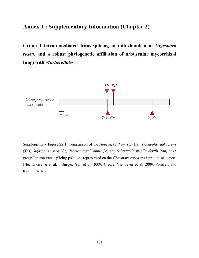
171
Annex 1 : Supplementary Information (Chapter 2)
Group I intron-mediated trans-splicing in mitochondria of Gigaspora
rosea, and a robust phylogenetic affiliation of arbuscular mycorrhizal fungi with Mortierellales
Supplementary Figure S2.1. Comparison of the Helicosporidium sp. (Hs), Trichoplax adhaerens
(Ta), Gigaspora rosea (Gr), Isoetes engelmannii (Ie) and Selaginella moellendorffii (Sm) cox1
group I intron trans-splicing positions represented on the Gigaspora rosea cox1 protein sequence.
(Hecht, Grewe et al. , Burger, Yan et al. 2009, Grewe, Viehoever et al. 2009, Pombert and
Keeling 2010)

172
Supplementary Figure S2.2. Predicted UUG translation initiation in Gigaspora and Mortierella.
Alignments of three mtDNA-encoded proteins are shown to demonstrate predictions of UUG
translation initiation (with M standing for methionine, labeled red). The first dozen amino acid
positions other than the predicted UUG methionine codons are color-coded according to amino
acid family (mauve, P,A,G,S,T; green, M,L,I,V; orange, K,R,H; and blue, F, Y), to facilitate
visual recognition of sequence similarity. Note that cob in Mortierella (the alleged phylogenetic
neighbor of Glomeromycota) is predicted to start with a UUG initiation codon as noted
previously (Seif, Leigh et al. 2005), yet in this species restricted to only one out of 14 protein
coding genes.

173

174

175
Annex 2 : Supplementary Information (Chapter 3)
Rapid mitochondrial genome evolution through invasion of mobile elements in two closely related species of arbuscular mycorrhizal fungi
Figure S3.1. Multiple DNA sequence alignment of numerous AMF representatives of the
atp6 native C-terminals along with the Glomus sp. 229456 putative foreign inserted C*-
terminal.
Table S3.1. Sequence identity matrix of the atp6 native C-terminals along with the Glomus
sp. 229456 putative foreign inserted C*-terminal.

176
Figure S3.2. Multiple DNA sequence alignment of numerous AMF representatives of the atp9
native C-terminals along with the Glomus sp. 229456 putative foreign inserted C*- terminal.
Table S3.2. Sequence identity matrix of the atp9 native C-terminals along with the Glomus
sp. 229456 putative foreign inserted C*-terminal.

177
Figure S3.3. Multiple DNA sequence alignment of numerous AMF representatives of the cox2
native C-terminals along with the Glomus sp. 229456 putative foreign inserted C*- terminal.
Table S3.3. Sequence identity matrix of the cox2 native C-terminals along with the Glomus
sp. 229456 putative foreign inserted C*-terminal.

178
Figure S3.4. Multiple DNA sequence alignment of numerous AMF representatives of the nad3
native C-terminals along with the Glomus sp. 229456 putative foreign inserted C*- terminal
Table S3.4. Sequence identity matrix of the nad3 native C-terminals along with the Glomus sp.
229456 putative foreign inserted C*-terminal.

179

180
Annex 3 : Supplementary Information (Chapter 4)
The mitochondrial genome of the glomeromycete Rhizophagus sp. DAOM
213198 reveals an unusual organization consisting of two circular
chromosomes
Fig. S4.1. Morphological description of spores of Rhizophagus sp. DAOM 213198 spores under
in vivo (a. b and c) and in vitro (e. f. g and h) culture conditions.
The spore wall (SW) is composed of three layers in both in vivo and in vitro cultures (a and e).
The outer layer SWL1 (b and c) was mucilaginous, evanescent, hyaline and approximately 1 µm
thick. The second layer (SWL2) was rigid, smooth, hyaline and approximately 1.5 µm thick. The
inner layer (SWL3) was laminate, smooth, pale yellow and approximately 1.5 µm thick. SWL1
and SWL2 were closely attached to each other forming a unique shell, easily detachable from the
third layer (SWL3). The subtending hyphae (8.5–9.5 µm) was hyaline and straight and pore open.

181
These images are courtesy of Dr Yolande Dalpé (AAFC, Ottawa. ON).

182
2
Fig. S4.2. mtDNA comparative analysis between R. irregularis DAOM 234179 and Rhizophagus
sp. DAOM 213198 isolates.
The linear-mapping of mtDNAs containing the newly formed and reshuffled intergenic regions of
rnl-atp6 and cox1-cox2 and atp9-atp8 in Rhizophagus sp. DAOM 213198 in comparison to its
relative R. irregularis DAOM 234179. Nucleotide identity comparison with tBLASTx, between
the homologous regions is indicated by the projections. Dark green projections represent
sequence identity higher than 90% while light green projections represent sequence identity
ranging between 50 and 90%.

183
3
Fig. S4.3. Alignment of potentially conserved dpo-like translated amino acid sequences from R.
irregularis DAOM 234179 and Rhizophagus sp. DAOM 213198.
Distance sequence analysis of dpo-like fragments of two closely related species of R. irregularis
DAOM 234179 and Rhizophagus sp. DAOM 213198 reveals that there are some conserved
domains in dpos positioned in different intergenic regions.
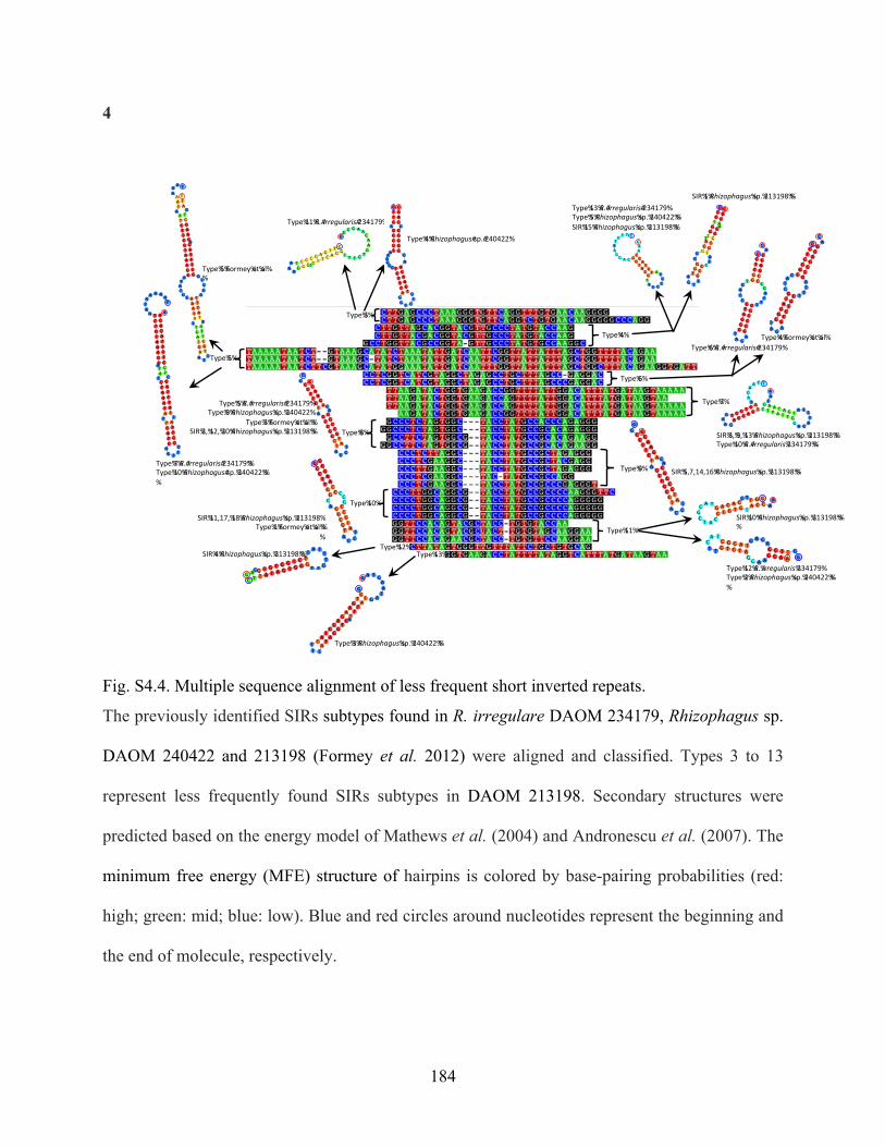
184
4
Type%11%R.#irregularis#234179%
Type%3%
Type%4%Rhizophagus#sp.#240422%
Type%4%
Type%13%R.#irregularis#234179%Type%5%Rhizophagus%sp.%240422%%SIR%15%Rhizophagus%sp.%213198%%
SIR%1%Rhizophagus%sp.%213198%%
Type%7%R.#irregularis#234179%%Type%10%Rhizophagus#sp.%240422%%%
Type%5%
Type%5%Formey%et%al%%
Type%6%
Type%6%R.#irregularis#234179%Type%4%Formey%et%al%
SIR%3,%9,%13%Rhizophagus%sp.%213198%%Type%10%R.#irregularis%234179%%
Type%7%
SIR%5,7,14,16%Rhizophagus%sp.%213198%%Type%9%
Type%8%
Type%10%
SIR%11,17,%18%Rhizophagus%sp.%213198%Type%1%Formey%et%al%%
%
Type%5%R.#irregularis#234179%Type%9%Rhizophagus%sp.%240422%
Type%3%Formey%et%al%%SIR%2,%12,%20%Rhizophagus%sp.%213198%%
Type%11%SIR%10%Rhizophagus%sp.%213198%%%
Type%12%R.%irregularis%234179%Type%2%Rhizophagus%sp.%240422%%%
Type%12%Type%13%SIR%4%Rhizophagus%sp.%213198%%
Type%3%Rhizophagus%sp.%240422%%
Fig. S4.4. Multiple sequence alignment of less frequent short inverted repeats.
The previously identified SIRs subtypes found in R. irregulare DAOM 234179, Rhizophagus sp.
DAOM 240422 and 213198 (Formey et al. 2012) were aligned and classified. Types 3 to 13
represent less frequently found SIRs subtypes in DAOM 213198. Secondary structures were
predicted based on the energy model of Mathews et al. (2004) and Andronescu et al. (2007). The
minimum free energy (MFE) structure of hairpins is colored by base-pairing probabilities (red:
high; green: mid; blue: low). Blue and red circles around nucleotides represent the beginning and
the end of molecule, respectively.

185
Fig. S4.5. Hypothetical pathway of mtDNA inheritance and dynamics in Rhizophagus sp. DAOM
213198.
Mitochondrial division could result in the formation of mitochondria containing either mtDNAs
or one of each mtDNAs. Fusion dynamic of mitochondria also provide the opportunity for the
ones harboring incomplete mtNA to fuse and complete set of mitochondrial genes.
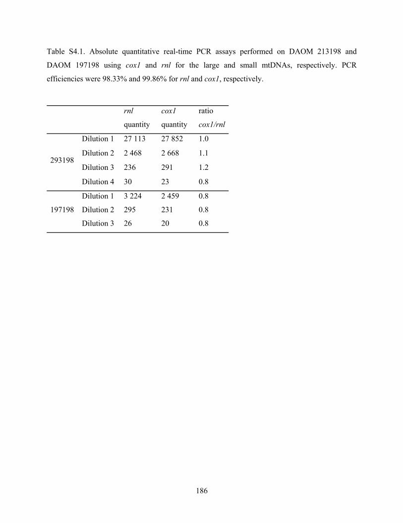
186
Table S4.1. Absolute quantitative real-time PCR assays performed on DAOM 213198 and
DAOM 197198 using cox1 and rnl for the large and small mtDNAs, respectively. PCR
efficiencies were 98.33% and 99.86% for rnl and cox1, respectively.
rnl
quantity
cox1
quantity
ratio
cox1/rnl
293198
Dilution 1 27 113 27 852 1.0
Dilution 2 2 468 2 668 1.1
Dilution 3 236 291 1.2
Dilution 4 30 23 0.8
197198
Dilution 1 3 224 2 459 0.8
Dilution 2 295 231 0.8
Dilution 3 26 20 0.8

187
Annex 4 : Supplementary Information (Chapter 5)
Detection of a transient mitochondrial DNA heteroplasmy in the progeny of
crossed genetically divergent isolates of arbuscular mycorrhizal fungi
Figures S5.1. And S5.2. monosporal culture lines of Rhizophagus irregularis
Figure S5.1. Spore germination of R. irregularis through the subtending hyphae.
Black arrowhead and rhizotropism (i.e. growth oriented towards the roots (R) (black arrow)
related to attempts to achieve monosporal culture lines from progenies originating from crossed
parents. Panels a and b: spore progenies originating from combination DAOM-197198/DAOM-
234328; c and d: spore progenies from combination DAOM-234328/DAOM-240415.

188
Figure S5.2. Polymerase chain reaction banding patterns of the progenies coming from different
parental combinations (AB, AC, BC).
Each progeny spore from the monosporal cultures lines are indicated by (PS), which has been
tested by both parental specific primers. R. irregularis isolates DAOM-197191 (A) DAOM-
240415 (B) and DAOM-234328 (C). mtDNA marker sizes are 120 bp, 147 bp and 209 bp
corresponding to DAOM-197198, DAOM-240415 and DAOM-234328, respectively.

189
Table S5.1. Number and percentage of anastomosis1 between germlings from spore clusters
either from the same or different isolates of R. irregularis.
A- DAOM 197198, B- DAOM 240415, C- DAOM 234328
* Fusions were followed for three weeks and no septa were formed between the connected
hyphae. 1Anastomoses within the same mycelium in crossing treatment were not recorded.
Type of interaction
Number of
contacts
Perfect
fusion (Self
fusion)*(%)
No interaction
Pre-fusion
incompatibility or
hyphal avoidance
(%)
Post-fusion
incompatibility
(%)
Within the same
isolate
A-A 205 123 (60) 82 0 0
B-B 175 68 (38) 107 0 0
C-C 243 135 (55) 108 0 0
Between isolates Number of
contacts
Non-self
fusion* (%) No interaction
Non-self pre-
fusion
incompatibility or
hyphal avoidance
(%)
Non-self post-
fusion
incompatibility
(%)
A-C 148 0 145 0 3 (2)
A-B 134 0 130 3 (2,3) 1 (0,7)
B-C 157 2* (1,2) 151 2 (1,3) 2 (1,3)

190
Table S5.2. Total number of spores1 of R. irregularis (full lipids, empty and aborted like-structures) produced in the interaction zone
and at both sides in the crossing experiments and controls.
A- DAOM 197198, B- DAOM 240415, C- DAOM 234328 1No significance differences were found within the treatments and controls (P≥0.05) 2 Mean ± Standard Error
Combination
A2 Int-Zone B B Int-Zone C A Int-Zone C
Full lipids 1329 ± 94 21± 8 1149 ± 89 1019±141 21±4 1638±66 1497±329 14±7 1317±267
Empty 27± 4 1± 0,5 17± 5 15±4 1±0,5 27±6 34±8 3±3 30±11
Aborted 20±4 2±1 13±6 19±12 0 15±8 14±3 2±1 23±3
Total 1376±90 25±10 1179±94 1054±152 22±4 1682±79 1546±322 19±11 1371±279
Control A Int-Zone A B Int-Zone B C Int-Zone C
Full lipids 1249±317 32±15 1582±125 1807±345 46±24 1899±212
1949 ± 84 45 ± 11 1584 ±
461
Empty 22±12 1±1 14±10 24±11 2±1 36±10 26 ± 4 0 7 ± 4
Aborted 16±9 5±1 9±4 25±9 5±2 28±8 17 ± 1 2 ± 2 23 ± 9
Total 1288±338 39±14 1604±139 1857±359 52 ± 27 1964±220
1988 ±
108 47 ± 12
1605 ±
473

0
Table S5.3. Number and percentage of germinated and non-germinated spores of R. irregularis,
which were tested to produce monosporal culture lines.
Combination Number of
offprings
tested
Germinated
offprings (%)
Progeny which
contacted the root
after germination
Offprings
showing
heteroplasmy
(%)
A-C 20 40 5 0
A-B 20 50 10 0
B-C 20 60 20 0
A- DAOM 197198, B- DAOM 240415, C- DAOM 234328