DENTAL PATHOLOGIES IN RURAL MEDIAEVAL POPULATIONS FROM CONTINENTAL CROATIA
Upload
independentCategory
view
3download
0
8
Oxidative Stress and Mitochondrial Dysfunction in Cardiovascular Diseases
Sauri Hernández-Reséndiz, Mabel Buelna-Chontal, Francisco Correa and Cecilia Zazueta
Departamento de Bioquímica, Instituto Nacional de Cardiología, Ignacio Chávez,
México
1. Introduction
Reactive oxygen species (ROS) include a wide variety of molecules and free radicals derived from molecular oxygen. O2 being highly electrophilic can be reduced by one electron at a time producing relatively stable intermediates, such as superoxide anion (O2•¯), precursor of most ROS and a relevant mediator in many biological reactions. Dismutation of O2•¯ produces hydrogen peroxide (H2O2), which, in turn, may be fully reduced to water or partially reduced to hydroxyl radical (•OH), one of the strongest oxidants in nature. The formation of •OH is catalyzed by reduced transition metals. On the other hand, superoxide anion is able to reduce transition metals and intensify in this way hydroxyl generation. In addition, O2•¯ may react with other radicals, in particular, with nitric oxide (•NO) in a reaction controlled by the rate of diffusion of both radicals. The product, peroxynitrite, is very powerful oxidant. The oxidants derived from •NO have been recently called reactive nitrogen species (RNS). Oxidative stress is an expression used to describe various deleterious processes resulting from an imbalance between the formation and elimination of ROS and/or RNS by antioxidant defenses. While small fluctuations in the steady-state concentration of these oxidants play an important role in intracellular signaling, uncontrolled increase in their concentration produces free radical-mediated chain reactions, which indiscriminately target proteins, lipids, polysaccharides, and DNA. Mitochondria are a major source of ROS, converting as much as 0.2-2% of molecular oxygen to superoxide as a by-product of the electron transfer activity (Wittenberg & Wittenberg, 1989; Alvárez et al., 2003). Other enzymatic sources of ROS include NADPH oxidases, located on the cell membrane of polymorphonuclear cells, macrophages, endothelial and other cells, and cytochrome P450-dependent oxygenases, along with the proteolytic conversion of xanthine dehydrogenase to xanthine oxidase, which produces both O2•¯ and H2O2.
Mitochondria’s critical role in cardiomyocyte survival and death has become an exciting finding in the field of cardiac biology. Indeed, it is accepted that mitochondrial dysfunction plays a crucial role in the pathogenesis of multiple cardiac diseases, mainly due to the imbalance of the fine interplay between aerobic metabolism, calcium homeostasis, and ROS production. Reactive oxygen species generated in the mitochondria, unless adequately
Oxidative Stress and Diseases
158
neutralized, cause mitochondrial oxidative stress and, through reactions with polyunsaturated fatty acids, form lipid hydroperoxides and unsaturated aldehydes that propagate among cellular compartments and react with proteins and nucleic acids. In the myocardium, the oxidative stress cascade impairs several functions, like mitochondrial biogenesis, fatty acid metabolism, ionic homeostasis, and antioxidant defense mechanisms, leading to diminished cardiac energetic efficiency, altered bioenergetics, apoptosis and degradation. Besides the obvious relevance of mitochondria in energy production, new processes like mitochondrial fusion and fission are reported to be linked to ROS generation and now are included in the cast of key players in cardiac disease. In this chapter, we explore the mechanisms of mitochondrial dysfunction driven by ROS generation associated with the pathophysiology of cardiovascular diseases.
2. Mitochondrial reactive oxygen species generation
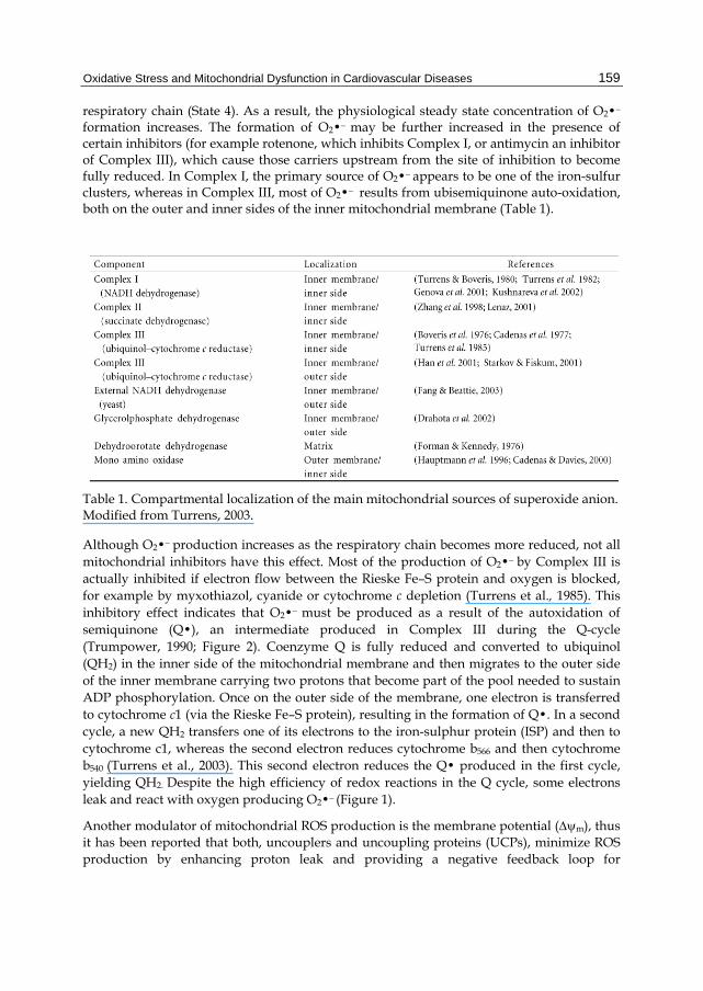
The standard reduction potential for the conversion of molecular oxygen to O2•¯ is -0.160 V (Wood, 1987). Given the highly reducing intramitochondrial environment, various respiratory components, including complexes I and III of the mitochondrial respiratory chain (Paradies et al., 2001; Tompkins et al., 2006), flavoproteins (Prosser et al., 2011), iron-sulfur clusters (Napoli et al., 2006), and ubisemiquinone (Wen & Garg, 2008) are thermodynamically capable to donate one electron to oxygen. Moreover, most steps in the respiratory chain involve single-electron reactions, further favoring the monovalent reduction of oxygen. On the other hand, the mitochondrion possesses various antioxidant defenses designed to eliminate both O2•¯ and H2O2. As a result, the steady state concentrations of O2•¯ and H2O2 have been estimated to be around 1 x 10⁻¹⁰ M and 5 x 10⁻⁹ M, respectively (Cadenas & Davies, 2000). Mitochondrial sources of O2•¯ include several respiratory complexes and individual enzymes. Superoxide formation occurs on the outer mitochondrial membrane, in the matrix, and on both sides of the inner mitochondrial membrane (Table 1, Figure 1). While superoxide anion generated in the matrix is mainly eliminated in that compartment, part of the O2•¯ produced in the intermembrane space may be carried to the cytoplasm via voltage-dependent anion channels (Han et al., 2003). The relative contribution of every site to the overall O2•¯ production varies from organ to organ and also depends on whether mitochondria are actively respiring (State 3) or if the respiratory chain is highly reduced (State 4) (Barja, 1999). Complex III appears to be responsible for most of the O2•¯ produced in heart mitochondria (Turrens & Boveris, 1980; Turrens et al., 1982), although Complex I is thought to be the primary source of ROS in a variety of pathological scenarios ranging from ageing to Parkinson’s disease (Betarbet et al., 2002; Sherer et al., 2003a). The rate of O2•¯ formation by the respiratory chain is controlled primarily by mass action law, increasing both when electron flow slows down (the concentration of electron donors, R• is higher) and when the concentration of oxygen increases1 (Turrens et al., 1982).
d[O2]/dt= k [O2] [R•] (1)
The electron flow through the respiratory chain establishes an H+ gradient across the inner mitochondrial membrane, used to drive ATP synthesis through the ATP synthase complex (Complex V). In the absence of ADP, the movement of H+ through ATP synthase is ceased and the H+ gradient builds up causing slowdown of electron flow and reduction of the
Oxidative Stress and Mitochondrial Dysfunction in Cardiovascular Diseases
159
respiratory chain (State 4). As a result, the physiological steady state concentration of O2•¯
formation increases. The formation of O2•¯ may be further increased in the presence of certain inhibitors (for example rotenone, which inhibits Complex I, or antimycin an inhibitor of Complex III), which cause those carriers upstream from the site of inhibition to become fully reduced. In Complex I, the primary source of O2•¯ appears to be one of the iron-sulfur clusters, whereas in Complex III, most of O2•¯ results from ubisemiquinone auto-oxidation, both on the outer and inner sides of the inner mitochondrial membrane (Table 1).
Table 1. Compartmental localization of the main mitochondrial sources of superoxide anion. Modified from Turrens, 2003.
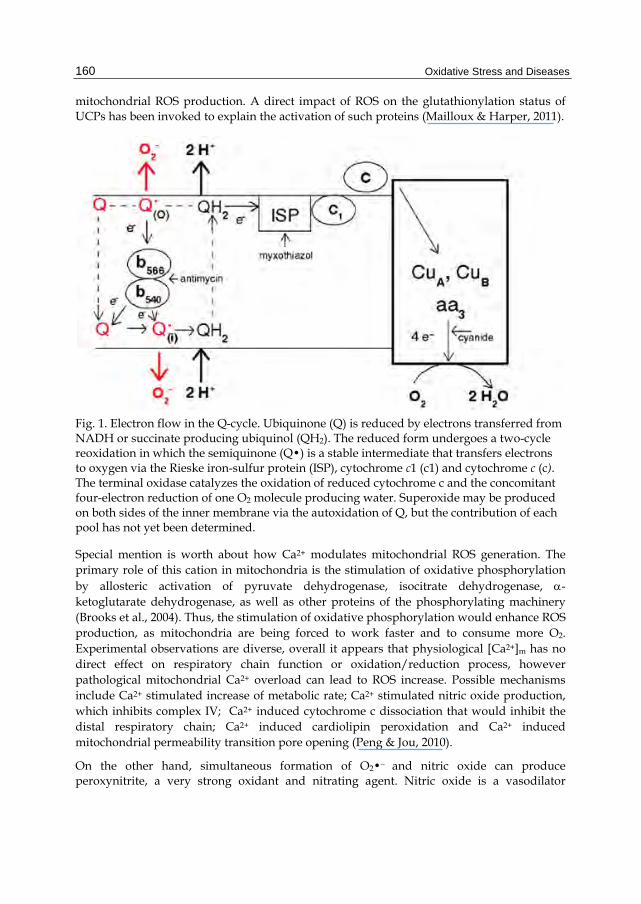
Although O2•¯ production increases as the respiratory chain becomes more reduced, not all mitochondrial inhibitors have this effect. Most of the production of O2•¯ by Complex III is actually inhibited if electron flow between the Rieske Fe–S protein and oxygen is blocked, for example by myxothiazol, cyanide or cytochrome c depletion (Turrens et al., 1985). This inhibitory effect indicates that O2•¯ must be produced as a result of the autoxidation of semiquinone (Q•), an intermediate produced in Complex III during the Q-cycle (Trumpower, 1990; Figure 2). Coenzyme Q is fully reduced and converted to ubiquinol (QH2) in the inner side of the mitochondrial membrane and then migrates to the outer side of the inner membrane carrying two protons that become part of the pool needed to sustain ADP phosphorylation. Once on the outer side of the membrane, one electron is transferred to cytochrome c1 (via the Rieske Fe–S protein), resulting in the formation of Q•. In a second cycle, a new QH2 transfers one of its electrons to the iron-sulphur protein (ISP) and then to cytochrome c1, whereas the second electron reduces cytochrome b566 and then cytochrome b540 (Turrens et al., 2003). This second electron reduces the Q• produced in the first cycle, yielding QH2. Despite the high efficiency of redox reactions in the Q cycle, some electrons leak and react with oxygen producing O2•¯ (Figure 1).
Another modulator of mitochondrial ROS production is the membrane potential (m), thus it has been reported that both, uncouplers and uncoupling proteins (UCPs), minimize ROS production by enhancing proton leak and providing a negative feedback loop for
Oxidative Stress and Diseases
160
mitochondrial ROS production. A direct impact of ROS on the glutathionylation status of UCPs has been invoked to explain the activation of such proteins (Mailloux & Harper, 2011).
Fig. 1. Electron flow in the Q-cycle. Ubiquinone (Q) is reduced by electrons transferred from NADH or succinate producing ubiquinol (QH2). The reduced form undergoes a two-cycle reoxidation in which the semiquinone (Q•) is a stable intermediate that transfers electrons to oxygen via the Rieske iron-sulfur protein (ISP), cytochrome c1 (c1) and cytochrome c (c). The terminal oxidase catalyzes the oxidation of reduced cytochrome c and the concomitant four-electron reduction of one O2 molecule producing water. Superoxide may be produced on both sides of the inner membrane via the autoxidation of Q, but the contribution of each pool has not yet been determined.
Special mention is worth about how Ca2+ modulates mitochondrial ROS generation. The primary role of this cation in mitochondria is the stimulation of oxidative phosphorylation by allosteric activation of pyruvate dehydrogenase, isocitrate dehydrogenase, -ketoglutarate dehydrogenase, as well as other proteins of the phosphorylating machinery (Brooks et al., 2004). Thus, the stimulation of oxidative phosphorylation would enhance ROS production, as mitochondria are being forced to work faster and to consume more O2. Experimental observations are diverse, overall it appears that physiological [Ca2+]m has no direct effect on respiratory chain function or oxidation/reduction process, however pathological mitochondrial Ca2+ overload can lead to ROS increase. Possible mechanisms include Ca2+ stimulated increase of metabolic rate; Ca2+ stimulated nitric oxide production, which inhibits complex IV; Ca2+ induced cytochrome c dissociation that would inhibit the distal respiratory chain; Ca2+ induced cardiolipin peroxidation and Ca2+ induced mitochondrial permeability transition pore opening (Peng & Jou, 2010).
On the other hand, simultaneous formation of O2•¯ and nitric oxide can produce peroxynitrite, a very strong oxidant and nitrating agent. Nitric oxide is a vasodilator
Oxidative Stress and Mitochondrial Dysfunction in Cardiovascular Diseases
161
resulting from the convertion of arginine to citrulline, in a reaction catalyzed by a family of NADPH-dependent enzymes called nitric oxide synthases. It has been reported that the mitochondrial matrix contains a unique form of nitric oxide synthase (Alvarez et al., 2003). Although its physiological role is still unclear, the formation of nitric oxide in mitochondria may have important pathological consequences, as it binds to the heme group of cytochrome oxidase, inhibiting respiration (Poderoso et al., 1996). ROS are also produced within mitochondria at sites other than the inner mitochondrial membrane (Dowrakowski et al., 2008), by proteins such as monoamine oxidase (MAO) and p66Shc (reviewed in Di Lisa et al., 2009).
3. Antioxidant systems in mitochondria
Mammalian mitochondria possess a multi-leveled ROS defense network of enzymes and non-enzymatic antioxidants. Thus, constantly generated ROS, essential for normal cellular physiology and signaling process, are maintained at specific levels by intrinsic antioxidant defenses, avoiding oxidative stress. In healthy mitochondria, ROS contention is driven by manganese-dependent superoxide dismutase (MnSOD), gluthatione peroxidase (GPx), thioredoxin (TrxSH2) and thioredoxin reductase (TrxR), peroxiredoxin (Prx), and glutaredoxin (Grx), as well as water- and lipid-soluble antioxidants, i.e. vitamins C, α-tocopherol (α-toc), reduced glutathione (GSH), and melatonin. It has been proposed that under certain circumstances, the mitochondrial respiratory chain can also contribute to mitochondrial antioxidant defense.
3.1 Non-enzymatic antioxidants
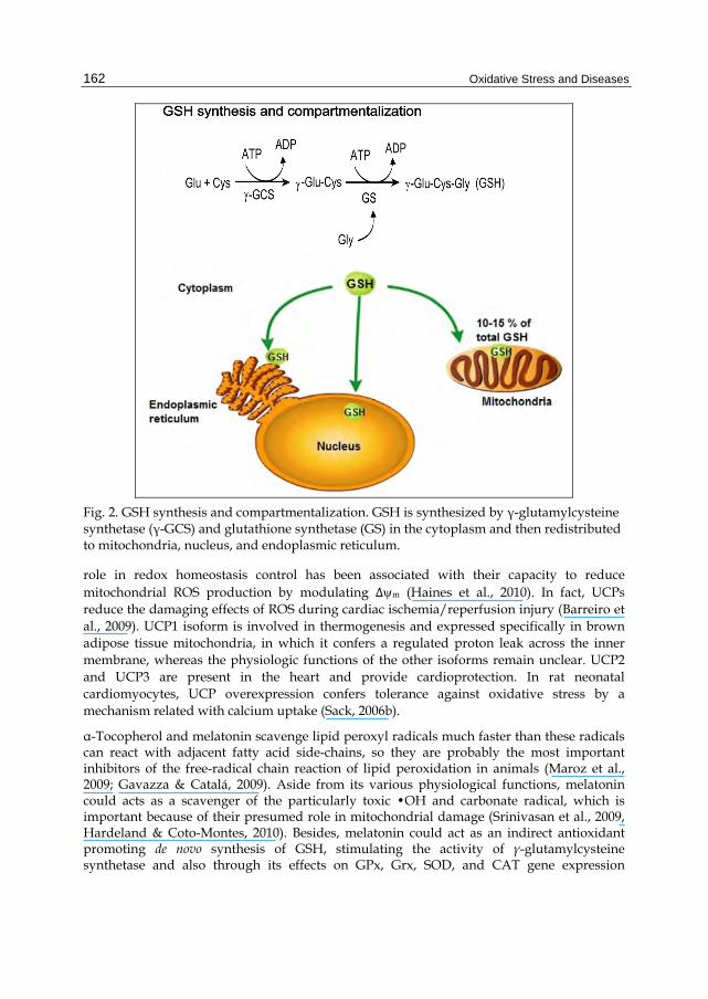
The tripeptide GSH is the main non-protein molecule, containing reactive thiol (-SH) groups, with scavenging properties, that provides an abundant source of reducing equivalents (Stowe & Camara, 2009). GSH reacts with hydroxyl radical (•OH), hypochlorous acid (HOCl), peroxyl radical (RO2●), carbon-centered radicals, and peroxynitrite anion (ONOO-) producing thiyl radical (GS●), which potentially generates O2•¯ among other ROS (Ježek & Hlavatá, 2005). Despite its exclusive synthesis in the cytosol, GSH is distributed in intracellular organelles, including the endoplasmic reticulum (RE), nucleus, and mitochondrion (Marí et al., 2009; Figure 2). GSH synthesis involves a two step reaction that requires ATP. Glutamate and cysteine are converted to γ-glutamyl-cysteine in a rate-limiting reaction driven by γ-glutamylcysteine synthetase. Then, γ-glutamylcysteine and glycine produce GSH by the action of the enzyme GSH synthetase (Figure 2). The first reaction is inhibited by GSH, a mechanism that regulates cellular GSH concentration (Marí et al., 2009). In the cytosol, GSH concentration is around 11 mM (Griffith & Meister, 1979; Mårtensson et al., 1990) and is transported into the mitochondrial matrix through a non-described high affinity carrier and a low affinity carrier, that could be the mitochondrial oxoglutarate carrier (Coll et al., 2003) or the dicarboxylate carrier (Lash et al., 2002). In mitochondria, GSH levels may fluctuate from 5 to 11 mM (Valko et al., 2007). Mitochondrial GSH plays a critical role in cell survival, as toxic cell death often correlates better with depletion of the mitochondrial GSH pool than with overall intracellular GSH depletion (Orrenius et al., 2007).
The importance of UCPs in the control of mitochondrial ROS generation remains unclear: it is known that UCPs are inner membrane carriers that transfer protons across the mitochondrial inner membrane (MIM), by-passing ATPase (Stuart et al., 2001). A putative
Oxidative Stress and Diseases
162
Fig. 2. GSH synthesis and compartmentalization. GSH is synthesized by γ-glutamylcysteine synthetase (γ-GCS) and glutathione synthetase (GS) in the cytoplasm and then redistributed to mitochondria, nucleus, and endoplasmic reticulum.
role in redox homeostasis control has been associated with their capacity to reduce mitochondrial ROS production by modulating Δm (Haines et al., 2010). In fact, UCPs reduce the damaging effects of ROS during cardiac ischemia/reperfusion injury (Barreiro et al., 2009). UCP1 isoform is involved in thermogenesis and expressed specifically in brown adipose tissue mitochondria, in which it confers a regulated proton leak across the inner membrane, whereas the physiologic functions of the other isoforms remain unclear. UCP2 and UCP3 are present in the heart and provide cardioprotection. In rat neonatal cardiomyocytes, UCP overexpression confers tolerance against oxidative stress by a mechanism related with calcium uptake (Sack, 2006b).
α-Tocopherol and melatonin scavenge lipid peroxyl radicals much faster than these radicals can react with adjacent fatty acid side-chains, so they are probably the most important inhibitors of the free-radical chain reaction of lipid peroxidation in animals (Maroz et al., 2009; Gavazza & Catalá, 2009). Aside from its various physiological functions, melatonin could acts as a scavenger of the particularly toxic •OH and carbonate radical, which is important because of their presumed role in mitochondrial damage (Srinivasan et al., 2009, Hardeland & Coto-Montes, 2010). Besides, melatonin could act as an indirect antioxidant promoting de novo synthesis of GSH, stimulating the activity of γ-glutamylcysteine synthetase and also through its effects on GPx, Grx, SOD, and CAT gene expression
Oxidative Stress and Mitochondrial Dysfunction in Cardiovascular Diseases
163
(Rodríguez et al., 2004), helping in GSH recycling and maintaining a high GSH/GSSG ratio. Melatonin is synthesized and released to the circulation by the pineal gland, and its amphiphilic properties lead to its free access to all compartments in the cell, being concentrated especially in the nucleus and mitochondria (Escames et al., 2010). A direct role of melatonin in regulation of Complex I and IV activity and in other mitochondrial functions has been suggested. This effect, not shared by other antioxidants, would reflect redox interactions with the electron transfer chain complexes, stimulating electron flow, limiting electron leakage, and ROS generation (Escames et al., 2010). Interestingly, it has also been reported that melatonin protects mitochondria from oxidative damage by preventing cardiolipin oxidation (Paradies et al., 2010).
3.2 Enzymatic antioxidants
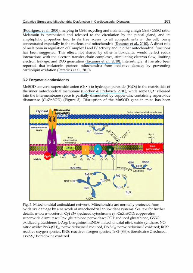
MnSOD converts superoxide anion (O2•¯) to hydrogen peroxide (H2O2) in the matrix side of the inner mitochondrial membrane (Liochev & Fridovich, 2010), while some O2•¯ released into the intermembrane space is partially dismutated by copper-zinc containing superoxide dismutase (CuZnSOD) (Figure 3). Disruption of the MnSOD gene in mice has been
Fig. 3. Mitochondrial antioxidant network. Mitochondria are normally protected from oxidative damage by a network of mitochondrial antioxidant systems. See text for further details. α-toc: α-tocoferol; Cyt c3+ (reduced cytochrome c) ; CuZnSOD: copper-zinc superoxide dismutase; Gpx: glutathione peroxidase; GSH: reduced glutathione, GSSG: oxidized glutathione; L-Arg: L-arginine; mtNOS: mitochondrial nitric oxide synthase, NO: nitric oxide; Prx3-(SH)2: peroxiredoxine 3 reduced, Prx3-S2: peroxiredoxine 3 oxidized; ROS: reactive oxygen species, RNS: reactive nitrogen species; Trx2-(SH)2: tioredoxine 2 reduced, Trx2-S2: tioredoxine oxidized.
Oxidative Stress and Diseases
164
associated with early postnatal lethality (Li et al., 1995), while MnSOD overexpression was shown to protect mitochondrial function and block apoptosis (Holley et al., 2010). The activity of MnSOD should be coordinated with H2O2-removing enzymes. Thus, H2O2 produced by MnSOD could be metabolized by Gpx, Prx, or by catalase that has been found in extremely small amounts in the mitochondrial matrix.
Besides of the importance of GSH as a direct antioxidant, it participates in multiple GSH-linked enzymatic defense systems. Among others, GSH acts as electron donor in the reduction of H2O2 and different hydroperoxides by GPx1 and GPx4 (Camara et al., 2011). Five different isoforms of GPx have been identified GPx1, GPx2, GPx3, GPx 4, and GPx 6. GPx1 is the major isoform and is localized predominantly in the cytosol, but a small proportion is also present within the mitochondrial matrix (Orrenius et al., 2007). GPx4 is a unique intracellular antioxidant enzyme that directly reduces peroxidized lipids produced in cell membranes (Nomura et al., 2000). Because GPx4 is membrane-associated, with a fraction localized in the intermembrane space of the mitochondria, possibly at the contact sites of the two membranes, and due to its small size and large hydrophobic surface it can interact with, and detoxify, membrane lipid hydroperoxides much more efficiently than the alternative pathway, phospholipase A2-GPx1 (PLA2) (Antunes et al., 1995). Hence, GPx4 is considered to be the primary enzymatic defense system against oxidative damage to cellular membranes. Accordingly, GPx4-null mice are embryonically lethal, while the heterozygotes are more sensitive to oxidants than wild type mice (Ran et al., 2003).
Other mitochondrial GSH-linked enzymes are glutaredoxins (Grx), which catalyze glutathione-dependent dithiol reaction, reducing protein disulfides, and monothiol reactions reducing mixed disulfides between proteins and GSH. An interesting member of this family is glutaredoxin 2 (Grx2), which was cloned and found to be present as both mitochondrial and nuclear isoforms (Lundberg et al., 2001). Modeling suggests that the GSH binding site and the hydrophobic surface of Grx2 are similar to those of Grx1 (Lundberg et al., 2001), although Grx2 lacks one of the conserved non-active site cysteine residues of Grx1 (Lundberg et al., 2001), hence it is more resistant to oxidants and oxidized glutathione (GSSG) action. Furthermore, Grx2 can be reactivated directly by thioredoxin reductase (TrxR) as well as by GSH (Johanson et al., 2004). GSSG is reduced by glutathione reductase (GR) with NADPH as a cofactor. In turn, mitochondrial NADPH can be regenerated by matrix dehydrogenases and by reaction of hydride ion transfer, which is proton motive force-dependent, utilizing intramitochondrial NADPH to reduce NADP+. Besides NADPH per se can serve directly as a non-enzymatic antioxidant, according to some authors (Kirsch & De Groot, 2001). Another potential source of disulfide reductase activity in mitochondria is the thioredoxin system, which includes thioredoxin 2 (Trx2) and thioredoxin reductase (TrxR2). Trx2 catalyzes the reduction of protein disulfides at much higher rates than Grx (Arner & Holmgren, 2000). This enzyme is important for life, given that disruption of the Trx2 gene in the homozygous mouse causes massive apoptosis and, finally, results in embryonic lethality (Nonn et al., 2003). Specific glutathione S-transferase (GST) isoforms: GSTα1-1, GSTα4-4, and GSTµ1-1, which neutralize reactive molecules such as 4-hydroxy-2-noneal (4-HNE), incorporating GSH to the radical molecule, have been found in mitochondria (Raza et al., 2002).
The intermembrane space of mitochondria contains ~0.7 mM cytochrome c (Hackenbrock et al., 1986) capable of superoxide removal. Cytochrome c can be alternatively reduced by the
Oxidative Stress and Mitochondrial Dysfunction in Cardiovascular Diseases
165
respiratory chain or superoxide. A diminution in its concentration may inhibit the distal respiratory chain and increase ROS production. Indeed, it has been reported that upon cytochrome c release during apoptosis initiation, mitochondrial ROS production increases (Cai & Jones, 1998).
As mentioned before, ubiquinone (Q) acts as a pro-oxidant in its semiquinone form, however when fully reduced it acts as an antioxidant. Ubiquinol (UQH2) contains phenolic hydrogen atoms that can be donated to a carbon- or oxygen centered radical, converting it to a non-radical molecule (Ježek & Hlavatá, 2005). Lipid peroxidation prevention by UQH2 has been reported (James et al., 2004). Preferential succinate oxidation can provide a tool against excessive accumulation of ROS by increasing the proportion of fully reduced ubiquinone. The antioxidant activity of UQH2 is independent from the effect of α-tocopherol (Ernster et al., 1992), which acts as a chain-breaking antioxidant, inhibiting the propagation of lipid peroxidation (Maroz et al., 2009). Even more, UQH2 can efficiently sustain the effect of α-tocopherol by regenerating it from the tocopheroxyl radical, which otherwise must rely on water-soluble agents such as ascorbate (vitamin C) (Ernster & Forsmark-Andree, 1993).
4. Oxidative stress and cardiovascular diseases
The pathological role of increased mitochondrial ROS in heart disease has been established from studies in gene-modified mice with altered mitochondrial antioxidant levels. Deletion of mitochondrial thioredoxin reductase 2 is embryonically lethal in mice because of impaired hematopoiesis and impaired cardiac function (Conrad et al., 2004). Fatal dilated cardiomyopathy in mice is developed after complete deletion of mitochondrial Mn superoxide dismutase (Li et al., 1995). In contrast, attenuated left ventricular remodeling after myocardial infarct was observed in transgenic mice overexpressing mitochondrial peroxiredoxin III (Matsushima et al., 2006) or glutathione peroxidase (Shiomi et al., 2004). Also, mice with a mitochondrial-targeted overexpression of catalase have a prolonged life span and improved cardiac function (Schriner et al., 2005).
A critical role of intracellular Ca2+ overload and oxidative stress in the genesis of myocyte dysfunction is well established in cardiovascular diseases like atherosclerosis, hypertension, ischemia/reperfusion damage, cardiac hypertrophy, and heart failure (HF). In general, Ca2+ overload can be induced by direct effect of ROS on Ca2+ handling proteins or indirectly, by inducing membrane lipid peroxidation (Santos et al., 2011). Recent evidence suggests that redox modification of ryanodine receptor (RyR2) may contribute to abnormal Ca2+ handling in disease states. RyR2 dysfunction with an increase in diastolic Ca2+ leak from the sarcoplasmic reticulum (SR) may reduce calcium transients and contribute to a reduced contractile force in the failing heart, as well as an increased likelihood of arrhythmia (González et al., 2010). In humans, increased cytosolic calcium ([Ca2+]c) has been related with augmented oxidative stress in atherosclerosis. A growing body of evidence indicates that the production of ROS is tightly linked with Angiotensin II-induced action. In this respect, a causative link between superoxide production and hypertension has been established in experiments in which SOD reduced blood pressure by 50 mm Hg in vascular smooth muscle cells (VSCM) from Angiotensin II-infused rats (Laursen et al., 1997). Much less attention has been paid to other reactions catalyzed by ROS. However, it is known that ATPase activity and inhibition of ATP-independent Ca2+ binding are severely depressed in
Oxidative Stress and Diseases
166
sarcolemmal membranes exposed to hydrogen peroxide and Fe2+ (Kukreja et al., 1992, Kaneko et al., 1989). In addition, augmented levels of iron pool in atherosclerotic lesions suggest that iron-catalyzed formation of free radicals may take place in the development of this pathology (Yuan & Li, 2003). High-fat diets stimulate stress response (heat shock protein 70) and signal transduction genes (Ras, MAPK1), inhibiting SOD and GPx gene expression. These effects could be prevented by scavengers of peroxides and antioxidant supplementation of the high-fat diet and caloric restriction (Rosier & Saes, 2006).
5. Mitochondrial dysfunction
Mitochondrial myopathies were described in the early 1960s, when systematic ultrastructural and histochemical studies revealed excessive proliferation of abnormally looking mitochondria in muscle of patients with weakness or exercise intolerance (Shy & Gonatas, 1964). Mitochondrial dysfunction, reflected in the structure, function and number of mitochondria within the cardiomyocyte, leads to diminished energy production, loss of myocyte contractility, altered electrical properties, and eventual cardiomyocyte cell death (Capetanaki, 2002). In addition, cardiotoxic stimuli often lead to excessive production of ROS and to Ca2+ overload in the mitochondrial matrix (Ragoni & Condolini, 2009). Evidences for a pathological role of mitochondrial ROS comes from studies in animal models of myocardial infarction, in which increased mitochondrial ROS production was observed, accompanied by decreases in mtDNA copy numbers, in mitochondrial-encoded gene transcripts, and in related enzymatic activities (complexes I, III, and IV), and from studies of genetically modified animals. Overexpression of Prx-3 (a mitochondrial antioxidant protein) improved post-myocardial infarction left ventricular function by restoring mitochondrial activity and DNA copy numbers (Matsushima et al., 2006). Other examples are studies in mice with complete deletion of mitochondrial MnSOD, which developed severe fatal dilated cardiomyopathy (Li et al., 1995). Decreased vascular SOD activities have also been associated with increased susceptibility to ischemia/reperfusion mediated damage; whereas overexpression of mitochondrial antioxidants increased cardiac tolerance to ischemia (Madamanchi, et al., 2005). Recently, the causal role of mitochondrial ROS in Angiotensin II-induced cardiomyopathy was shown by the observation that mice that overexpress catalase targeted to mitochondria, but not mice that overexpress wild-type peroxisomal catalase, are resistant to cardiac hypertrophy, fibrosis and mitochondrial damage induced by angiotensin II (Dai et al., 2011). Monoamine oxidase (MAO) has been shown to play a prominent role in myocardial injury caused by post-ischemic reperfusion (Bianchi et al., 2005a) and to contribute to the maladaptive evolution from myocardial hypertrophy to heart failure (Kaludercic et al., 2010). MAO-mediated ROS production has been related with serotonin-induced myocyte hypertrophy in vitro (Bianchi et al., 2005b) and in mitogenic signaling induction by a process that may involve the activation of the metalloproteinase MMP-2, in smooth muscle cells (Coatrieux et al., 2007).
Mitochondria isolated from hearts of rabbits exposed to hypercholesterolemic diet showed significantly reduced respiration rates (state 3 and state 4) (Kojik et al., 2011), whereas increased cholesterol is related with diminution of the mitochondrial membrane potential and mitochondrial pore opening (Chávez et al., 1998; Martínez Abundis et al., 2007) and activation of apoptosis (Martínez-Abundis et al., 2009).
Oxidative Stress and Mitochondrial Dysfunction in Cardiovascular Diseases
167
5.1 Metabolic adaptation
Complex mechanisms have evolved to maintain the balance between myocardial O2 supply and O2 consumption under pathological stresses such as hypoxia, ischemia, pressure, and volume overload. These mechanisms induce changes in cardiomyocyte structure and/or function through coordinated changes in gene and protein expression and/or the activities of various proteins. Redox mechanisms are involved in the signaling pathways underlying many of these mechanisms, both via the direct effects of O2 levels in the cardiomyocyte and through the effects of ROS (Santos et al., 2011). Metabolically, the adult mammalian heart normally uses lipids as the major fuel, and mitochondria supply over 90% of the total ATP through β-oxidation of plasma fatty acids (Opie & Sack, 2002). During hypoxia, under ischemia or settings of increased cardiac workload, there is a substantial increase in glycolytic ATP generation, which may be cardioprotective during ischemia/reperfusion by ensuring an adequate ATP supply for membrane and sarcoplasmic reticulum ion pumps (Opie & Sack, 2002; Correa et al., 2008a). Recent studies suggest that a metabolic shift to glycolysis is related with the redox status in the heart. NADPH has been recognized as a critical modulator of the antioxidant defense through the regeneration of reduced pools of glutathione, while G6PDH activity was shown to be of major importance for the maintenance of redox status, Ca2+ homeostasis, and contractile function in cardiomyocytes subjected to oxidative stress (Jain et al., 2003) .
5.2 Mitochondrial biogenesis
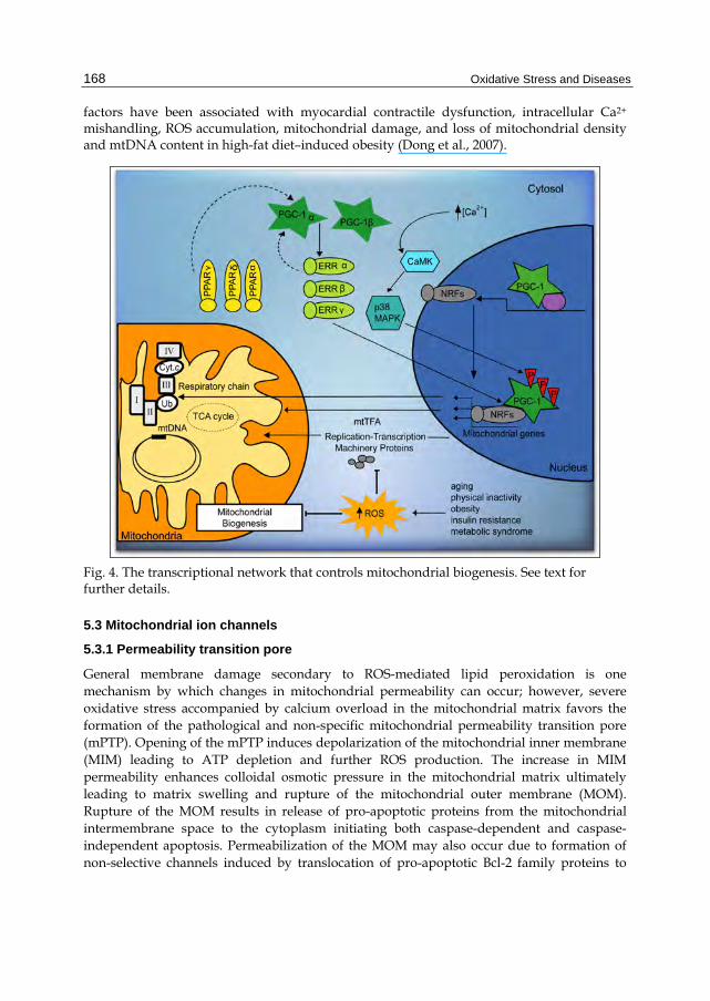
Mitochondrial biogenesis can be defined as a chain of events that promote growth and division of preexisting organelles. Mitochondrial biogenesis includes the synthesis, import and incorporation of proteins and lipids, as well as the replication of mitochondrial DNA (mtDNA). Replication and transcription of mtDNA are controlled by mtTFA (mitochondrial transcription factor A), and two specific transcription factors TFB1M and TFB2M (mitochondrial transcription factor B1/B2), an RNA polymerase (POLRMT), and a mitochondrial transcription termination factor (mTERF). The coordination between the expression of mitochondrial and nuclear genes is directed by nuclear respiratory factor (NRF-1 and/or NRF-2), the peroxisome proliferator-activated receptors (PPARs), estrogen receptor (ERR), and co-activators of peroxisome proliferator-activated receptor gamma (PGC-1α) (Scarpulla, 2008). PGC-1α is also involved in regulation of fatty acid oxidation (FAO) and in co-activation of ERRα (Figure 4).
Mitochondrial biogenesis decreases in aging, obesity, insulin resistance, dyslipidemia, and hypertension, co-morbidities associated with cardiovascular diseases. Impaired activation of the renin-angiotensin-aldosterone system (RAAS) has been associated with such pathologies (Cooper et al., 2007). In fact, elevated levels of angiotensin II (Ang II) and aldosterone promote alterations in insulin metabolism, endothelial dysfunction, and loss of myocardial function (Kim et al., 2008; Sowers et al., 2009). RAAS increases the activity of NADPH oxidase and stimulates ROS generation resulting in mitochondrial damage, decreased ATP production, diminished NO availability, and attenuated mitochondrial biogenesis. Clinical and experimental observations report the loss of expression of PGC-1α, and mtTFA NRFs in hypertension (Whaley-Connell et al., 2009). In addition, down-regulation of the mitochondrial biogenesis co-activator PGC-1α and its downstream nuclear
Oxidative Stress and Diseases
168
factors have been associated with myocardial contractile dysfunction, intracellular Ca2+ mishandling, ROS accumulation, mitochondrial damage, and loss of mitochondrial density and mtDNA content in high-fat diet–induced obesity (Dong et al., 2007).
Fig. 4. The transcriptional network that controls mitochondrial biogenesis. See text for further details.
5.3 Mitochondrial ion channels
5.3.1 Permeability transition pore
General membrane damage secondary to ROS-mediated lipid peroxidation is one mechanism by which changes in mitochondrial permeability can occur; however, severe oxidative stress accompanied by calcium overload in the mitochondrial matrix favors the formation of the pathological and non-specific mitochondrial permeability transition pore (mPTP). Opening of the mPTP induces depolarization of the mitochondrial inner membrane (MIM) leading to ATP depletion and further ROS production. The increase in MIM permeability enhances colloidal osmotic pressure in the mitochondrial matrix ultimately leading to matrix swelling and rupture of the mitochondrial outer membrane (MOM). Rupture of the MOM results in release of pro-apoptotic proteins from the mitochondrial intermembrane space to the cytoplasm initiating both caspase-dependent and caspase-independent apoptosis. Permeabilization of the MOM may also occur due to formation of non-selective channels induced by translocation of pro-apoptotic Bcl-2 family proteins to
Oxidative Stress and Mitochondrial Dysfunction in Cardiovascular Diseases
169
mitochondria. Due to its central role in cell death triggering, the mPTP represents a potential therapeutic target in some cardiovascular diseases. Studies performed over 20 years have demonstrated that acute cardiac ischemia followed by reperfusion damage is associated with mPTP opening (Arteaga et al., 1992; Griffiths & Halestrap, 1993; Chipuk et al., 2006; Lucken-Ardjomande et al., 2008; Halestrap and Pasdois, 2009). Furthermore, pharmacological and conditional inhibition of mPTP formation significantly improved cardiac function reducing ischemic injury and myocardial infarction size in animal models (Argaud et a., 2005; Hausenloy & Yellon, 2003; Correa et al., 2008b) and in patients (Shanmuganathan et al., 2005; Piot et al., 2008). mPTP opening also causes cell death in isolated endothelial and vascular smooth muscle cells. Indeed, atherosclerosis is exacerbated when mitochondrial antioxidant defenses are hampered and a decrease in mitochondrial ROS formation reduces atherogenesis.
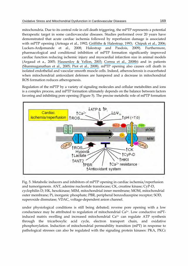
Regulation of the mPTP by a variety of signaling molecules and cellular metabolites and ions is a complex process, and mPTP formation ultimately depends on the balance between factors favoring and inhibiting pore opening (Figure 5). The precise metabolic role of mPTP formation
Fig. 5. Metabolic inducers and inhibitors of mPTP opening in cardiac ischemia/reperfusion and tumorigenesis. ANT, adenine nucleotide translocase; CK, creatine kinase; CyP-D, cyclophilin D; HK, hexokinase; MIM, mitochondrial inner membrane; MOM, mitochondrial outer membrane; Pi, inorganic phosphate; PBR, peripheral benzodiazepine receptor; SOD, superoxide dismutase; VDAC, voltage-dependent anion channel.
under physiological conditions is still being debated; reverse pore opening with a low conductance may be attributed to regulation of mitochondrial Ca2+. Low conductive mPT-induced matrix swelling and increased mitochondrial Ca2+ can regulate ATP synthesis through the tricarboxylic acid cycle, electron transport chain, and oxidative phosphorylation. Induction of mitochondrial permeability transition (mPT) in response to pathological stresses can also be regulated with the signaling protein kinases: PKA, PKCε
Oxidative Stress and Diseases
170
(protein kinase Cε) and GSK-3β (glycogen synthase kinase-3), which interact with the voltage-dependent anion channel (VDAC) (Bera et al., 1995; Baines et al, 2003; Javadov et al., 2009).
5.3.2 Structure of the mPTP complex
Although the crucial role of mPTPs in pathological conditions has been intensively studied in the heart, brain, and liver (Chipuk et al., 2006; Bernardi et al., 2006; Robertson et al., 2009), the actual molecular composition of the mPTP complex remains unclear. Until recently, three proteins had been accepted as key structural components of this megachannel: adenine nucleotide translocase (ANT), cyclophilin D (CyP-D), and the voltage-dependent anion channel (VDAC) located in the MIM, in the matrix and in the MOM, respectively. However, recent studies from different groups have questioned the molecular identity of the mPTP. A new model of the mPTP consisting of a phosphate carrier and ANT has been proposed where Ca2+ sensitivity of the pore is regulated by CyP-D binding to the phosphate carrier (Leung et al., 2008). Many studies have provided strong evidence that CyP-D plays a major regulatory role in mPTP formation (Baines et al., 2005; Nakagawa et al., 2005). Mitochondria isolated from CyP-D knockout mice were desensitized to the onset of the mPT, and required much higher concentrations of Ca2+ to induce pore opening compared to wild type animals, which is consistent with the role of CyP-D to regulate Ca2+-mPTP interactions (Nakagawa et al., 2005). Recent studies on transgenic mice questioned the role of ANT and VDAC as essential components of the mPTP suggesting a regulatory, rather than structural role in pore formation.
5.3.3 mPTP formation in cardiac Ischemia/Reperfusion (I/R)
mPTP opening has been examined extensively in cardiac pathological conditions, mostly in I/R (Chipuk et al., 2006; Correa et al., 2007; Halestrap & Pasdois, 2009). Acute I/R does not affect the expression of mPTP compounds due to its short duration, although it induces conformational changes in essential mPTP proteins, modifying their interactions with the pore effectors in the cytoplasm and mitochondrial matrix. Although many factors that induce pore opening are present during ischemia, including ATP depletion, Ca2+ overload, increased phosphate and ROS levels, it has been demonstrated that pore opening occurs during reperfusion rather than during ischemia (Griffiths and Halestrap, 1995). This is explained, in part, by the acidic conditions resulting from lactate and other acidic intermediates accumulation in the mitochondrial matrix. We and others demonstrated that delayed pHi recovery during reperfusion exerts beneficial effects on post-ischemic cardiac function, associated with improved mitochondrial function and inhibition of mPTP opening (Javadov et al., 2008; Correa et al., 2008a). In this regard, inhibition of the Na+/H+ exchanger 1(NHE-1) may be a promising therapeutic strategy against I/R damage (Linz & Busch, 2003; Karmazyn et al., 2001). mPTP opening causes mitochondrial uncoupling, thereby ATP is hydrolyzed rather than synthesized in the post-ischemic heart leading to myocardial death (Correa et al., 2005). mPTP opening also increases in Ca2+-induced cardiomyopathy (Nakayama et al., 2007), in diabetic cardiomyopathy (Oliveira et al., 2003), in heart failure following myocardial infarction (Javadov et al., 2005), and in intracoronary microembolization (Sharov et al., 2007).
Oxidative Stress and Mitochondrial Dysfunction in Cardiovascular Diseases
171
5.3.4 Mitochondrial KATP channels
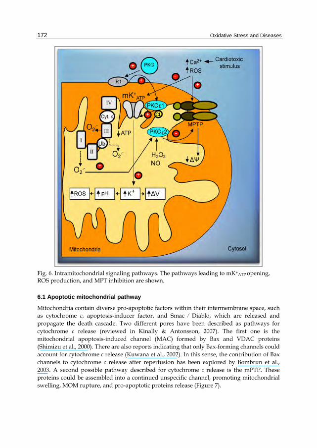
Mitochondrial ATP-sensitive potassium channels (mK+ATP) were described about 20 years ago in mitoplasts obtained from rat liver mitochondria. First advances in mK+ATP research demonstrated that different drugs stimulate the opening of mK+ATP channels decreasing mitochondrial potential (Δm) and that mK+ATP channels are involved in mechanisms regulating cell volume (Garlid, 1988). Latter, it was observed that bradykinin (Yang et al., 2004), opioids (Jang et al., 2008), and adenosine (Kin H, 2005), activate signaling cascades that induce the opening of mK+ATP and provide cardioprotection against ischemia/reperfusion injury. Recently a causative link between the opening of mK+ATP and cardioprotection conferred by post-conditioning has been proposed. Garlid et al. (2008) suggested that post-conditioning induces protection via an early redox-sensitive mechanism, followed by persistent mK+ATP activation. A complex signaling system involving mitochondrial PKCε 1 and 2 may prevent the formation of the mPTP. The putative signaling cascade includes the activation of Gi protein-coupled receptors and cGMP-dependent protein kinase (PKG). At mitochondrial level, this kinase binds to a hypothetic receptor, called "R1" in the mitochondrial outer membrane. This receptor may phosphorylate PKCε 1 (Jaburek et al., 2006), which, in turn, phosphorylates the mK+ATP channel favoring its opening. Once activated, the entry of K+ may produce alkalinization of the mitochondrial matrix and promote ROS production by Complex I. ROS could activate the mitochondrial matrix PKCε 2, and prevent the formation of mPTP, reducing cell death and infarction size (Costa et al., Garlid et al., 2009) (Figure 6).
Other proposals have been invoked to explain cardioprotection. One hypothesis is that K+ flow into the matrix may depolarize the inner membrane and reduce the driving force that sustains Ca2+ overload (Holmuhamedov et al., 1991; Murata et al., 2001). However, although Ca2+ reduction in the mitochondrial matrix may reduce mPTP opening in the post-ischemic heart, it is difficult to explain how a small depolarizing effect generated by activation of mK+ATP channels could avoid Ca2+-overload. Another assumption is that discrete mitochondrial swelling associated with K+ flow would change the architecture and respiratory control of mitochondria, creating a state of mitochondrial “super-efficiency” (Garlid, 2000). Whatever the mechanism involved in mK+ATP opening, its association with myocardial protection is clear.
6. Programmed cell death and autophagy
ROS/RNS can cause cell death by non-physiological (necrotic) or regulated pathways (apoptotic) in many cardiovascular diseases such as atherosclerosis, ischemic heart disease, heart failure, stroke, hypertension, and diabetes. The mechanisms by which ROS/RNS cause or regulate apoptosis typically are caspase-dependent and include the activation of membrane receptors, Bcl-2 family proteins, and mitochondrial dysfunction. Autophagy, a caspase-independent mechanism of cell death that protects cells against oxidative damage and is involved in the degradation and recycling of oxidized proteins and damaged organelles in cells, yields amino acids for de novo protein synthesis or energy provision (Nishida et al., 2009). While programmed cell death participation in cardiovascular diseases is well established, insights into caspase-independent mechanisms of cell death have emerged recently.
Oxidative Stress and Diseases
172
Fig. 6. Intramitochondrial signaling pathways. The pathways leading to mK+ATP opening, ROS production, and MPT inhibition are shown.
6.1 Apoptotic mitochondrial pathway
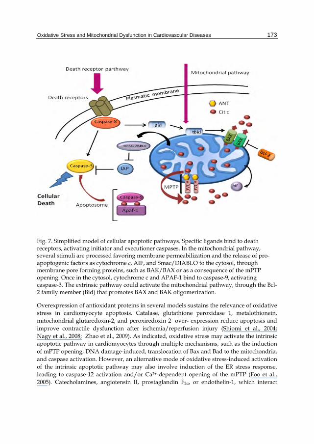
Mitochondria contain diverse pro-apoptotic factors within their intermembrane space, such as cytochrome c, apoptosis-inducer factor, and Smac ⁄ Diablo, which are released and propagate the death cascade. Two different pores have been described as pathways for cytochrome c release (reviewed in Kinally & Antonsson, 2007). The first one is the mitochondrial apoptosis-induced channel (MAC) formed by Bax and VDAC proteins (Shimizu et al., 2000). There are also reports indicating that only Bax-forming channels could account for cytochrome c release (Kuwana et al., 2002). In this sense, the contribution of Bax channels to cytochrome c release after reperfusion has been explored by Bombrun et al., 2003. A second possible pathway described for cytochrome c release is the mPTP. These proteins could be assembled into a continued unspecific channel, promoting mitochondrial swelling, MOM rupture, and pro-apoptotic proteins release (Figure 7).
Oxidative Stress and Mitochondrial Dysfunction in Cardiovascular Diseases
173
Fig. 7. Simplified model of cellular apoptotic pathways. Specific ligands bind to death receptors, activating initiator and executioner caspases. In the mitochondrial pathway, several stimuli are processed favoring membrane permeabilization and the release of pro-apoptogenic factors as cytochrome c, AIF, and Smac/DIABLO to the cytosol, through membrane pore forming proteins, such as BAK/BAX or as a consequence of the mPTP opening. Once in the cytosol, cytochrome c and APAF-1 bind to caspase-9, activating caspase-3. The extrinsic pathway could activate the mitochondrial pathway, through the Bcl-2 family member (Bid) that promotes BAX and BAK oligomerization.
Overexpression of antioxidant proteins in several models sustains the relevance of oxidative stress in cardiomyocyte apoptosis. Catalase, glutathione peroxidase 1, metalothionein, mitochondrial glutaredoxin-2, and peroxiredoxin 2 over- expression reduce apoptosis and improve contractile dysfunction after ischemia/reperfusion injury (Shiomi et al., 2004; Nagy et al., 2008; Zhao et al., 2009). As indicated, oxidative stress may activate the intrinsic apoptotic pathway in cardiomyocytes through multiple mechanisms, such as the induction of mPTP opening, DNA damage-induced, translocation of Bax and Bad to the mitochondria, and caspase activation. However, an alternative mode of oxidative stress-induced activation of the intrinsic apoptotic pathway may also involve induction of the ER stress response, leading to caspase-12 activation and/or Ca2+-dependent opening of the mPTP (Foo et al., 2005). Catecholamines, angiotensin II, prostaglandin F2α, or endothelin-1, which interact
Oxidative Stress and Diseases
174
with G-protein coupled receptors and induce cardiomyocyte hypertrophy, may also induce apoptosis. A well characterized mechanism is a Gαq-mediated PKC-dependent transcriptional upregulation of the Bcl-2 family member Nix, which activates the mitochondrial death pathway (Yussman et al., 2002)
6.2 Autophagy
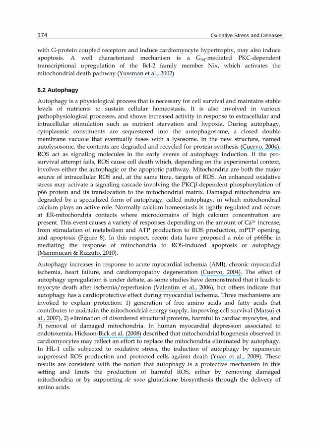
Autophagy is a physiological process that is necessary for cell survival and maintains stable levels of nutrients to sustain cellular homeostasis. It is also involved in various pathophysiological processes, and shows increased activity in response to extracellular and intracellular stimulation such as nutrient starvation and hypoxia. During autophagy, cytoplasmic constituents are sequestered into the autophagosome, a closed double membrane vacuole that eventually fuses with a lysosome. In the new structure, named autolysosome, the contents are degraded and recycled for protein synthesis (Cuervo, 2004). ROS act as signaling molecules in the early events of autophagy induction. If the pro-survival attempt fails, ROS cause cell death which, depending on the experimental context, involves either the autophagic or the apoptotic pathway. Mitochondria are both the major source of intracellular ROS and, at the same time, targets of ROS. An enhanced oxidative stress may activate a signaling cascade involving the PKCβ-dependent phosphorylation of p66 protein and its translocation to the mitochondrial matrix. Damaged mitochondria are degraded by a specialized form of autophagy, called mitophagy, in which mitochondrial calcium plays an active role. Normally calcium homeostasis is tightly regulated and occurs at ER-mitochondria contacts where microdomains of high calcium concentration are present. This event causes a variety of responses depending on the amount of Ca2+ increase, from stimulation of metabolism and ATP production to ROS production, mPTP opening, and apoptosis (Figure 8). In this respect, recent data have proposed a role of p66Shc in mediating the response of mitochondria to ROS-induced apoptosis or autophagy (Mammucari & Rizzuto, 2010).
Autophagy increases in response to acute myocardial ischemia (AMI), chronic myocardial ischemia, heart failure, and cardiomyopathy degeneration (Cuervo, 2004). The effect of autophagy upregulation is under debate, as some studies have demonstrated that it leads to myocyte death after ischemia/reperfusion (Valentim et al., 2006), but others indicate that autophagy has a cardioprotective effect during myocardial ischemia. Three mechanisms are invoked to explain protection: 1) generation of free amino acids and fatty acids that contributes to maintain the mitochondrial energy supply, improving cell survival (Matsui et al., 2007), 2) elimination of disordered structural proteins, harmful to cardiac myocytes, and 3) removal of damaged mitochondria. In human myocardial depression associated to endotoxemia, Hickson-Bick et al. (2008) described that mitochondrial biogenesis observed in cardiomyocytes may reflect an effort to replace the mitochondria eliminated by autophagy. In HL-1 cells subjected to oxidative stress, the induction of autophagy by rapamycin suppressed ROS production and protected cells against death (Yuan et al., 2009). These results are consistent with the notion that autophagy is a protective mechanism in this setting and limits the production of harmful ROS, either by removing damaged mitochondria or by supporting de novo glutathione biosynthesis through the delivery of amino acids.
Oxidative Stress and Mitochondrial Dysfunction in Cardiovascular Diseases
175
Fig. 8. Signaling pathways, regulating mitochondrial function. ROS production is induced by oxidative stress, which activates a signaling cascade involving the PKCβ-dependent phosphorylation of p66 its translocation to the mitochondrial matrix. Mitochondria are also targets of ROS damage. Damaged mitochondria are removed by mitophagy, a specialized form of autophagy, which is regulated by different pathways.
7. Mitochondrial fission and fusion
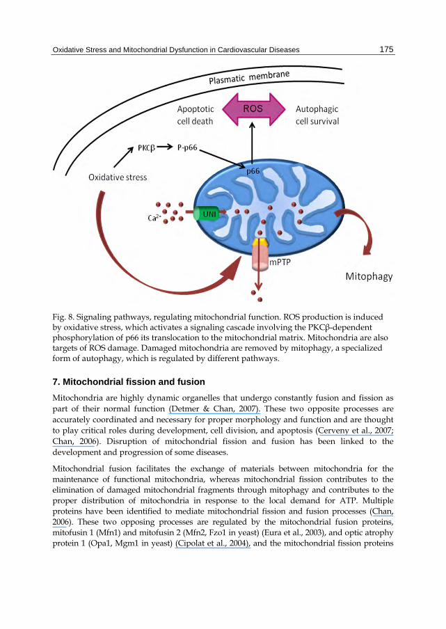
Mitochondria are highly dynamic organelles that undergo constantly fusion and fission as part of their normal function (Detmer & Chan, 2007). These two opposite processes are accurately coordinated and necessary for proper morphology and function and are thought to play critical roles during development, cell division, and apoptosis (Cerveny et al., 2007; Chan, 2006). Disruption of mitochondrial fission and fusion has been linked to the development and progression of some diseases.
Mitochondrial fusion facilitates the exchange of materials between mitochondria for the maintenance of functional mitochondria, whereas mitochondrial fission contributes to the elimination of damaged mitochondrial fragments through mitophagy and contributes to the proper distribution of mitochondria in response to the local demand for ATP. Multiple proteins have been identified to mediate mitochondrial fission and fusion processes (Chan, 2006). These two opposing processes are regulated by the mitochondrial fusion proteins, mitofusin 1 (Mfn1) and mitofusin 2 (Mfn2, Fzo1 in yeast) (Eura et al., 2003), and optic atrophy protein 1 (Opa1, Mgm1 in yeast) (Cipolat et al., 2004), and the mitochondrial fission proteins
Oxidative Stress and Diseases
176
dynamin-related protein 1 (Drp1, Dnm1 in yeast) (Frank et al., 2001; Smirnova et al., 2001; Ingerman et al., 2005), and human mitochondrial fission protein 1 (hFis1) (Yoon et al., 2003). The balance between mitochondrial fusion and fission within a cell can be disrupted by a myriad of factors, including oxidative stress (Frank et al., 2001) and simulated ischemia (Brady et al., 2006), and has also been linked to aging (Kowald & Kirkwood, 2011) (Figure 9).
Fig. 9. Mitochondrial fission and fusion. Mitochondrial fission involves the action of Drp1, which can self-assemble into polymeric spirals and is recruited into the mitochondrial membrane by hFis1 and Mdv1/Caf4. Drp1 polymers wrap around the mitochondrion and constrict the membrane until fission occurs. Mitochondrial fusion involves the interaction of Mfn1 and Mfn2 proteins located in the outer mitochondrial membrane of two mitochondria until outer membranes fuse, consequently, inner mitochondrial membrane fusion occurs through interaction of Opa1 proteins. See text for further details.
7.1 Mitochondrial fusion
Data on fission and fusion proteins’ role in heart diseases are scarce. Recently it has been reported that cardiac myocyte mitochondria, lacking the fusion protein Mfn2, are pleiomorphic and show enlarged morphopology. Consistent with an underlying mild mitochondrial dysfunction, Mfn2-deficient mice display modest cardiac hypertrophy accompanied by slight functional deterioration (Papanicolau et al., 2011). Expression of OPA1 is decreased in both human and rat failing hearts, which show small and fragmented mitochondria indicative of decreased fusion. OPA1 mRNA levels did not differ between failing and normal hearts, suggesting post-transcriptional control, possibly through degradation by proteases activated by ATP (Baricault et al., 2007).
Oxidative Stress and Mitochondrial Dysfunction in Cardiovascular Diseases
177
7.2 Mitochondrial fission
It has been suggested that defects in mitochondrial fusion and fission processes are responsible for abnormal mitochondrial morphologies observed in many cardiac diseases. In cultured neonatal ventricular myocytes, inhibition of mitochondrial fission, by over-expressing a dominant-negative mutant form of Drp1, prevents ROS production, mitochondrial permeability transition pore opening, and subsequent cell death after ceramide treatment (Parra et al., 2008). An increase in the level of cytosolic Ca2+ induced by thapsigargin (Tg) causes cardiac mitochondrial fission associated to ROS generation in a Drp1-dependent pathway (Hom et al., 2010). Because calcium overload is a common feature in heart failure (HF), this may increase mitochondrial fission and dysfunction, thus further contributing to the decrease in the metabolic demand of the heart and increasing its injury.
8. Conclusion
Mitochondrial redox signaling is paramount to the maintenance of cardiomyocyte homeostasis. Therefore, oxidative deregulation of mitochondrial key players, like mK+ATP, mPTP, ionic transporters, metabolism enzymes, and apoptotic machinery, has enormous impact on cardiovascular function. Intense research is devoted to obtain a better understanding of the complex regulatory mechanisms ruling these systems and to enable the development of more specific therapeutic strategies for heart diseases. In addition, fascinating links are beginning to be discovered between mitochondrial function and cardiac physiology and diseases in the context of diverse signaling mechanisms. Besides, proteins with previously known function, like those driving mitochondrial fusion and fission, are now reported to have emergent functions in intracellular calcium homeostasis, apoptosis, and vascular smooth muscle cell proliferation, all, key issues in cardiac disease. These processes broaden the traditional role in energy production undertaken by mitochondria and provide new directions for research in cardiovascular diseases.
9. Acknowledgment
This work was partially supported by grant 80791-M to C. Zazueta
10. References
Alvarez, S.; Valdez, L.; Zaobornyj, T. & Boveris A (2003). Oxygen dependence of mitochondrial nitric oxide synthase activity. Biochemical and Biophysical Research Community. Vol. 305, 771–775.
Antunes, F.; Salvador, A. & Pinto, R. (1995). PHGPx and phospholipase A2/GPx: comparative importance on the reduction of hydroperoxides in rat liver mitochondria. Free Radical Biology & Medicine. Vol. 19, 669-77.
Argaud, L.; Gateau-Roesch, O.; Muntean, D.; Chalabreysse, L.; Loufouat, J.; Robert, D. & Ovize, M. (2005). Specific inhibition of the mitochondrial permeability transition prevents lethal reperfusion injury. Journal of Molecular Cell Cardiology. Vol. 38, 367-374.
Arteaga, D.; Odor, O.; Lopez, R.; Contreras, G.; Aranda, A. & Chávez, E. (1992) Impairment by cyclosporine A of reperfusion-induced arrhythmias. Life Sciences, 1127-1134.
Oxidative Stress and Diseases
178
Arnér, E. & Holmgren, A. (2000). Physiological functions of thioredoxin and thioredoxin reductase. European Journal of Biochemistry. Vol. 267, 6102-9.
Baines, C.; Kaiser, R.; Purcell, N.; Blair, N.; Osinska, H.; Hambleton, M.; Brunskill, E.; Sayen, M.; Gottlieb, R.; Dorn, G.; Robbins, J. & Molkentin, J. (2005). Loss of cyclophilin D reveals a critical role for mitochondrial permeability transition in cell death. Nature. Vol. 434, 658-662.
Baines, C.; Song, C.; Zheng, Y.; Wang, G.; Zhang, J.; Wang, O.; Guo, Y.; Bolli, R.; Cardwell, E. & Ping, P. (2003). Protein kinase Cepsilon interacts with and inhibits the permeability transition pore in cardiac mitochondria. Circulation Research. Vol. 92, 873- 880.
Baricault, L.; Ségui, B.; Guégand, L.; Olichon, A.; Valette, A.; Larminat, F. & Lenaers, G. (2007). OPA1 cleavage depends on decreased mitochondrial ATP level and bivalent metals. Experimental Cell Research. Vol. 313, 3800-8.
Barja, G. (1999). Mitochondrial oxygen radical generation and leak: sites of production in states 4 and 3, organ specificity and relation to aging and longevity. Journal of Bioenergetics and Biomembranes. Vol. 31, 347–366.
Barreiro, E.; Garcia-Martinez, C.; Mas, S.; Ametller, E.; Gea, J.; Argiles, J.; Busquets, S. & Lopez-Soriano, F. J. (2009). UCP3 overexpression neutralizes oxidative stress rather than nitrosative stress in mouse myotubes. FEBS Letters. Vol. 583, 350–356.
Bera, A.; Ghosh, S. & Das, S. (1995). Mitochondrial VDAC can be phosphorylated by cyclic AMP-dependent protein kinase. Biochemical & Biophysical Research Community. Vol. 209, 213-217.
Bernardi, P.; Krauskopf, A.; Basso, E.; Petronilli, V.; Blachly-Dyson, E.; Di Lisa, F. & Forte, M. (2006). The mitochondrial permeability transition from in vitro artifact to disease target. FEBS Journal. Vol. 273, 2077- 2099.
Betarbet R, Sherer TB & Greenamyre JT (2002). Animal models of Parkinson’s disease. Bioessays 24, 308–318.
Bianchi, P.; Kunduzova, O.; Masini, E.; Cambon, C.; Bani, D.; Raimondi, L.; Seguelas, M.; Nistri, S.; Colucci, W.; Leducq, N. & Parini, A. (2005a). Oxidative stress by monoamine oxidase mediates receptor-independent cardiomyocyte apoptosis by serotonin and postischemic myocardial injury. Circulation. Vol. 112, 3297–3305.
Bianchi, P.; Pimentel, D.; Murphy, M.; Colucci, W. & Parini, A. (2005b). A new hypertrophic mechanism of serotonin in cardiac myocytes: receptor-independent ROS generation. FASEB Journal. Vol. 19, 641–643
Bombrun, A.; Gerber, P.; Casi, G.; Terradillos, O.; Antonsson, B. & Halazy, S. (2003). 3,6-Dibromocarbazole piperazine derivates of 2-propanol as first inhibitors of cytochrome c release via Bax channels modulation. Journal of Medical Chemistry. Vol. 46, 4365-68
Brady, N.; Hamacher-Brady, A.;Westerhoff, H. & Gottlieb, R. (2006). A wave of reactive oxygen species (ROS)-induced ROS release in a sea of excitable mitochondria. Antioxidants & Redox Signal. Vol. 8, 1651–1665.
Brigelius-Floé, R. (2006). Glutathione peroxidases and redox-regulated transcription factors. Biological Chemistry. Vol. 387, 1329-35.
Brookes, P.; Yoon, Y.; Robotham, J.; Anders, M. & Sheu, S. (2004). Calcium, ATP and ROS: a mitocondrial love-hate triangle. American Journal of Physiology & Cell Physiology. Vol. 287, C817-C833.
Oxidative Stress and Mitochondrial Dysfunction in Cardiovascular Diseases
179
Cadenas, E. & Davies, K. (2000). Mitochondrial free radical generation oxidative stress and aging. Free Radical Biological Medicine, Vol. 29, 222–230.
Cai, J. & Jones, D. (1988). Superoxide in apoptosis. Mitochondrial generation triggered by cytochrome c loss. Journal of Biological Chemistry Vol. 273, 11401-4.
Camara, A.; Bienengraeber, M. & Stowe, D. (2011). Mitochondrial approaches to protect against cardiac ischemia and reperfusion injury. Frontiers in Physiology. Vol. 12, 2-13.
Capetanaki, Y. (2002). Desmin cytoskeleton: a potential regulator of muscle mitochondrial behavior and function. Trends in Cardiovascular Medicine. Vol. 12, 339-348.
Cerveny, K.; Tamura, Y.; Zhang, Z.; Jensen, R. & Sesaki, H. (2007). Regulation of mitochondrial fusion and division. Trends in Cellular Biology. Vol. 17, 563-9.
Chan, D. (2006). Mitochondrial fusion and fission in mammals. Annual Reviews Cellular & Development Biology. Vol. 22, 79–99.
Chipuk, J.; Bouchier-Hayes, L. & Green, D. (2006). Mitochondrial outer membrane permeabilization during apoptosis: the innocent bystander scenario. Cell Death & Differentiation. Vol. 13, 1396-1402.
Cipolat, S.; Martins de Brito, O.; Dal Zilio, B. & Scorrano, L. (2004). OPA1 requires mitofusin 1 to promote mitochondrial fusion. Proceedings of the National Academic of Sciences U S A. Vol. 101, 15927–15932.
Coatrieux, C.; Sanson, M.; Negre-Salvayre, A.; Parini, A.; Hannun, Y.; Itohara, S.; Salvayre, R. & Auge, N. (2007). MAO-A induced mitogenic signaling is mediated by reactive oxygen species, MMP-2, and the sphingolipid pathway. Free Radical Biology & Medicine. Vol. 43, 80–89
Coll, O.; Colell, A.; García-Ruiz, C.; Kaplowitz, N. & Fernández-Checa, J. (2003). Sensitivity of the 2-oxoglutarate carrier to alcohol intake contributes to mitochondrial glutathione depletion. Hepatology. Vol. 38, 692-702.
Conrad, M.; Jakupoglu, C.; Moreno, S.; Lippl, S.; Banjac, A.; Schneider, M.; Beck, H.; Hatzopoulos, A.; Just, U.; Sinowatz, F.; Schmahl, W.; Chien, K.; Wurst, W.; Bornkamm, G. & Brielmeier, M. (2004). Essential role for mitochondrial thioredoxin reductase in hematopoiesis, heart development and heart function. Molecular & Cell Biology. Vol. 21, 9414-23.
Cooper, S.; Whaley-Connell, A. & Habibi, J. (2007). Renin-angiotensin-aldosterone system and oxidative stress in cardiovascular insulin resistance. American Journal of Physiology & Heart Circulation Physiology. Vol. 293, H2009-H2023.
Correa F.; García, N.; Robles, C.; Martínez-Abundis, E. & Zazueta, C. (2008b). Relationship between oxidative stress and mitochondrial function in the post-conditioned heart. Journal of Bioenergetics & Biomembranes. Vol. 40, 599-606.
Correa, F.; García, N.; Gallardo, J.; Carreño, L.; Rodríguez-Enríquez, S.; Marín, A. & Zazueta, C. (2008a). Post-conditioning preserves glycolytic ATP during early reperfusion: A survival mechanism for the reperfused heart. Cellular Physiology & Biochemistry. Vol. 22, 635-644.
Correa, F.; Soto, V. & Zazueta , C. (2007). Mitochondrial permeability transition relevance for apoptotic triggering in the post-ischemic heart. International Journal of Biochemistry & Cell Biology. Vol. 39, 787-798.
Costa, A. & Garlid, K. (2008). Intramitochondrial signaling: interactions among mitoKATP, PKCɛ , ROS, and MPT. American Journal of Physiology. Vol. 295, H874–82.
Cuervo A. (2004). Autophagy in sickness and in health. Trends in Cell Biology. Vol. 14, 70-77.
Oxidative Stress and Diseases
180
Chávez, E.; Franco, M.; Reyes-Vivas, H.; Zazueta, C.;Ramírez, J. & Carrillo, R. (1998) Hypothyroidism renders liver mitochondria to the opening of membrane permeability transition pore. Biochimica and Biophysica Acta. Vol. 1407, 243-248.
Dai, D.F.; Johnson, S.C.; Villarin, J.J.; Chin, M.T.; Nieves-Cintrón, M.; Chen, T.; Marcinek, D.J.; Dorn, G.W. 2nd, Kang, Y.J.; Prolla, T.A.; Santana, L.F. & Rabinovitch, P.S. (2011). Mitochondrial oxidative stress mediates angiotensin II-induced cardiac hypertrophy and Galphaq overexpression-induced heart failure. Circulation Research. Vol. 108, 837-46.
Detmer, S. & Chan, D. (2007). Functions and dysfunctions of mitochondrial dynamics. Nature Reviews Molecular Cell Biology. Vol. 8, 870–879.
Di Lisa, F.; Kaludercic, N.; Carpi, A.; Menabò, R. & Giorgio, M. (2009). Mitochondria and vascular pathology. Pharmacology Reports. Vol. 61, 123-30.
Dong, F.; Li, Q.; Sreejayan, N.; Nunn, J. & Ren, J. (2007). Metallothionein prevents high-fat diet induced cardiac contractile dysfunction: role of peroxisome proliferator activated receptor gamma coactivator 1-alpha and mitochondrial biogenesis. Diabetes. Vol. 56, 2201-12.
Dworakowski, R.; Alom-Ruiz, S. & Shah, A. (2008). NADPH oxidase-derived reactive oxygen species in the regulation of endothelial phenotype. Pharmacology Reports. Vol. 60, 21–28
Ernster, L. & Forsmark-Andrée, P. (1993). Ubiquinol: an endogenous antioxidant in aerobic organisms. Clinical Investigations. Vol. 71, S60-5.
Escames, G.; López, A.; García, J.; García, L.; Acuña-Castroviejo, D.; García, J. & López, L. (2010). The role of mitochondria in brain aging and the effects of melatonin. Current Neuropharmacology, Vol. 8, 182–193.
Eura Y, et al. (2003). Two mitofusin proteins, mammalian homologues of FZO, with distinct functions are both required for mitochondrial fusion. Journal of Biochemistry. Vol. 134, 333–344.
Foo, R.; Mani, K. & Kitsis, R. (2005). Death begets failure in the heart . Journal of Clinical Investigation. Vol. 115, 565-71.
Frank, S.; Gaume, B.; Bergmann-Leitner, E.; Leitner, W.; Robert, E.; Catez, F.; Smith, C.; & Youle, R. (2001). The role of dynamin-related protein 1, a mediator of mitochondrial fission, in apoptosis. Developmental Cell. Vol. 1, 515-25.
Garlid, K. (1988). Mitochondrial volume control. In: Integration of Mitochondrial Function. New York: Plenum, p.p. 257–276.
Garlid, K. (2000). Opening mitochondrial K(ATP) in the heart—what happens, and what does not happen. Basic Research in Cardiology. Vol. 95, 275–79.
Gavazza, M. & Catalá, A. (2009) Relative efficacies of alpha-tocopherol, N-acetyl-serotonin, and melatonin in reducing non-enzymatic lipid peroxidation of rat testicular microsomes and mitochondria. Molecular & Cellular Biochemistry. Vol. 321, 37-43.
Gonzalez, D.; Treuer, A.; Castellanos, J.; Dulce, R. & Hare, J. (2010). Impaired S-nitrosylation of the ryanodine receptor caused by xanthine oxidase activity contributes to calcium leak in heart failure. Journal of Biological Chemistry. Vol. 285, 28938–28945.
Griffith, O. & Meister, A. (1979). Potent and specific inhibition of glutathione synthesis by buthionine sulfoximine (S-n-butyl homocysteine sulfoximine). Journal of Biological Chemistry 254, 7558-7560.
Oxidative Stress and Mitochondrial Dysfunction in Cardiovascular Diseases
181
Griffiths, E. & Halestrap. A. (1993) Protection by Cyclosporin A of ischemia/reperfusion-induced damage in isolated rat hearts. J Mol Cell Cardiol. 25, 1461-9.
Griffiths, E. & Halestrap, A. (1995). Mitochondrial non-specific pores remain closed during cardiac ischaemia, but open upon reperfusion. Biochemical Journal. Vol. 307,93-98.
Hackenbrock, C.; Chazotte, B. & Gupte S. (1986). The random collision model and a critical assessment of diffusion and collision in mitochondrial electron transport. Journal of Bioenergetics & Biomembranes. Vol. 18, 331-68.
Haines, B.; Mehta, S.; Pratt, S.; Warden, C.; & Li, P. (2010). Deletion of mitochondrial uncoupling protein-2 increases ischemic brain damage after transient focal ischemia by altering gene expression patterns and enhancing inflammatory cytokines. Journal of Cerebral Blood Flow Metabolism. Vol. 30, 1825–1833.
Halestrap, A. & Pasdois, P. (2009). The role of the mitochondrial permeability transition pore in heart disease. Biochimical & Biophysical Acta. Vol. 1787,1402-1415.
Halestrap, A.P.; Connern, C.P.; Griffiths, E.J. & Kerr, P.M. (1997). Cyclosporin A binding to mitochondrial cyclophilin inhibits the permeability transition pore and protects heart from ischemia. Molecular & Cellular Biochemistry. Vol. 174, 167-72.
Han, D.; Antunes, F.; Canali, R.; Rettori, D. & Cadenas, E. (2003). Voltage-dependent anion channels control the release of the superoxide anion from mitochondria to cytosol. Journal of Biological Chemistry. Vol. 278, 5557-63.
Hardeland, R. & Coto-Montes, A. (2010). New vistas on oxidative damage and aging. The Open Biology Journal. Vol. 3, 39–52.
Hausenloy, D. & Yellon, D. (2003). The mitochondrial permeability transition pore: its fundamental role in mediating cell death during ischaemia and reperfusion. Journal of Molecular Cell Cardiology. Vol. 35, 339-341.
Hickson-Bick, D.; Jones, C. & Buja, L. (2008). Stimulation of mitochondrial biogenesis and autophagy by lipopolysaccharide in the neonatal rat cardiomyocyte protects against programmed cell death. Journal of Molecular & Cell Cardiology. Vol. 44, 411–418.
Holley, A.; Dhar, S.; Xu, Y. & St Clair, D. (2010). Manganese superoxide dismutase: beyond life and death. Amino Acids. Ë-pub ahead of print.
Holmuhamedov, E.; Wang, L. & Terzic, A. (1999). ATP-sensitive K+ channel openers prevent Ca2+ overload in rat cardiac mitochondria. Journal of Physiology. Vol. 519, 347–60.
Hom, J.; Yu, T.; Yoon, Y.; Porter, G. & Sheu, S. (2010). Regulation of mitochondrial fission by intracellular Ca(2+) in rat ventricular myocytes. Biochimical & Biophysical Acta. Vol. 1797, 913-21.
Ingerman, E.; Perkins, E.; Marino, M.; Mears, J.; McCaffery, J.; Hinshaw, J.; & Nunnari, J. (2005). Dnm1 forms spirals that are structurally tailored to fit mitochondria. Journal of Cell Biology. Vol. 170, 1021-1027.
Jaburek, M.; Costa, A.; Burton, J.; Costa, C. & Garlid, K. (2006). Mitochondrial PKCepsilon and mitoKATP co-purify and co-reconstitute to form a functioning signaling module in proteoliposomes. Circulation Research. Vol. 99, 878–83.
Jain, M.; Brenner, D.; Cui, L.; Lim, C.; Wang, B.; Pimentel, D.; Koh, S.; Sawyer, D.; Leopold, J.; Handy, D.; Loscalzo, J.; Apstein, C.; & Liao, R. (2003). Glucose-6-phosphate dehydrogenase modulates cytosolic redox status and contractile phenotype in adult cardiomyocytes. Circulation Research. Vol. 93, e9–e16.
James, A.; Smith, R. & Murphy, M. (2004). Antioxidant and prooxidant properties of mitochondrial Coenzyme Q. Archives of Biochemistry & Biophysics. Vol. 423, 47-56.
Oxidative Stress and Diseases
182
Jang, Y.; Xi, J.; Wang, H.; Mueller, R.A.; Norfleet, E.A & Xu, Z. (2008). Postconditioning prevents reperfusion injury by activating delta-opioid receptors. Anesthesiology. Vol. 108, 243-250.
Javadov, S.; Choi, A.; Rajapurohitam, V.; Zeidan, A.; Basnakian, A. & Karmazyn, M. (2008). NHE-1 inhibition-induced cardioprotection against ischaemia/reperfusion is associated with attenuation of the mitochondrial permeability transition. Cardiovascular Research. Vol. 77, 416-424.
Javadov, S.; Huang, C.; Kirshenbaum, L. & Karmazyn, M. (2005). NHE-1 inhibition improves impaired mitochondrial permeability transition and respiratory function during postinfarction remodelling in the rat. Journal of Molecular & Cell Cardiology. Vol. 38,135-143.
Javadov, S.; Rajapurohitam, V.; Kilic, A.; Zeidan, A.; Choi, A. & Karmazyn, M. (2009) Antihypertrophic effect of NHE-1 inhibition involves GSK-3beta-dependent attenuation of mitochondrial dysfunction. Journal of Molecular & Cell Cardiology. Vol. 46, 998-1007.
Jezek, P. & Hlavatá, L. (2005) Mitochondria in homeostasis of reactive oxygen species in cell, tissues, and organism. International Journal of Biochemistry and Cell Biology. Vol. 37, 2478-503.
Johansson, C.; Lillig, C. & Holmgren, A. (2004). Human mitochondrial glutaredoxin reduces S-glutathionylated proteins with high affinity accepting electrons from either glutathione or thioredoxin reductase. Journal of Biological Chemistry. Vol. 279, 7537-43.
Kaludercic, N.; Takimoto, E.; Nagayama, T.; Feng, N.; Lai, E.; Bedja, D.; Chen, K.; Gabrielson, K.; Blakely, R.; Shih, J.; Pacak, K.; Kass, D.; Di Lisa, F. & Paolocci, N. (2010). Monoamine oxidase A-mediated enhanced catabolism of norepinephrine contributes to adverse remodeling and pump failure in hearts with pressure overload. Circulation Research. Vol. 106,193-202.
Kaneko, M.; Elimban, V. & Dhalla, N. (1989). Mechanism for depression of heart sercolemmal Ca2+ pump bye oxygen free radicals. American Journal of Physiology. Vol. 257, H804-H811.
Karmazyn, M.; Sostaric, J. & Gan, X. (2001). The myocardial Na+/H+ exchanger: a potential therapeutic target for the prevention of myocardial ischaemic and reperfusion injury and attenuation of postinfarction heart failure. Drugs. Vol. 61, 375-389.
Kim, J.; Wei, Y. & Sowers, J. (2008). Role of mitochondrial dysfunction in insulin resistance. Circulation Research. Vol. 102, 401-414.
Kin, H.; Zatta, A.J.; Lofye, M.T.; Amerson, B.S.; Halkos, M.E.; Kerendi, F.; Zhao, Z.Q.; Guyton, R.A.; Headrick, J.P. & Vinten-Johansen J. (2005). Postconditioning reduces infarct size via adenosine receptor activation by endogenous adenosine. Cardiovascular Research. Vol.67, 124-133.
Kinnally, K. & Antonsson, B. (2007). A tale of two mitochondrial channels, MAC and PTP, in apoptosis. Apoptosis. Vol. 12, 857-68.
Kirsch, M. & De Groot, H. (2001). NAD(P)H, a directly operating antioxidant? FASEB Journal. Vol. 15, 1569-74.
Kojic, Z.; Gopcevic, K.; Marinkovic, D. & Tasic, G. (2011). Effect of captopril on serum lipid levels and cardiac mitochondrial oxygen consumption in experimentally-induced hypercholesterolemia in rabbits. Physiological Research. In press.
Oxidative Stress and Mitochondrial Dysfunction in Cardiovascular Diseases
183
Kowald, A. & Kirkwood, T. (2011). Evolution of the mitochondrial fusion-fission cycle and its role in aging. Proceedings of the National Academy of Sciences U S A. Vol. 108, 10237-42.
Kukreja, R. & Hess, M. (1992). The oxygen free-radical system- From equations through membrane-protein interactions to cardiovascular injury and protection. Cardiovascular Research. Vol. 26, 641-655.
Kuwana, T.; Mackey, M.R.; Perkins, G.; Ellisman, M.H.; Latterich, M.; Schneiter, R.; Green, D.R. & Newmeyer, D.D. (2002). Bid, Bax and lipids cooperate to form supramolecular openings in the outer mitochondrial membrane. Cell. Vol. 111, 331-42.
Laursen, J.; Rajagopalan, S.; Galis, Z.; Tarpey, M.; Freeman, B. & Harrison, D. (1997). Role of superoxide in angiotensin-II induced but not catecholamine-induced hypertension. Circulation, Vol. 95, 588–593.
Leung, A.; Varanyuwatana, P. & Halestrap, A. (2008). The mitochondrial phosphate carrier interacts with cyclophilin D and may play a key role in the permeability transition. Journal of Biological Chemistry. Vol. 283, 26312-26323.
Li, Y.; Huang, T.; Carlson, E.; Melov, S.; Ursell, P.; Olson. J.; Noble, L.; Yoshimura, M.; Berger, C.; Chan, P.; Wallace, D. & Epstein, C. (1995). Dilated cardiomyopathy and neonatal lethality in mutant mice lacking manganese superoxide dismutase. Nature Genetics. Vol. 11, 376-81.
Linz, W. & Busch, A. (2003). NHE-1 inhibition: from protection during acute ischaemia/ reperfusion to prevention/reversal of myocardial remodelling. Naunyn Schmiedebergs Archives of Pharmacology. Vol. 368, 239-246.
Liochev, S. & Fridovich, I. (2010). Mechanism of the peroxidase activity of Cu, Zn superoxide dismutase. Free Radical Biology and Medicine. Vol. 48, 1565–1569.
Lucken-Ardjomande, S.; Montessuit, S. & Martinou, J. (2008). Contributions to Bax insertion and oligomerization of lipids of the mitochondrial outer membrane. Cell Death & Differentiation. Vol. 15, 929-937.
Lundberg, M.; Johansson, C.; Chandra, J.; Enoksson, M.; Jacobsson, G.; Ljung, J.; Johansson, M. & Holmgren A. (2001). Cloning and expression of a novel human glutaredoxin (Grx2) with mitochondrial and nuclear isoforms. Journal of Biological Chemistry. Vol. 276, 26269-75.
Madamanchi, N.; Vendrov, A. & Runge, M. (2005). Oxidative stress and vascular disease. Arteriosclerosis & Thrombosis Vascular Biology. Vol. 25, 29–38.
Mailloux, R. & Harper, E. (2011). Uncoupling proteins and the control of mitochondrial reactive oxygen species production. Free Radical Biological Medicine. Jun 24. (Epub ahead of print).
Mammucari, C. & Rizzuto, R. (2010). Signaling pathways in mitochondrial dysfunction and aging. Mechanisms of ageing and development. Vol. 131, 536-43.
Marí, M.; Morales, A.; Colell, A.; García-Ruiz, C. & Fernández-Checa, J. (2009) Mitochondrial Glutathione, a Key Survival Antioxidant. Antioxidants & Redox Signals. Vol. 11, 2685-700.
Maroz, A.; Anderson, R.; Smith, R. & Murphy, M. (2009). Reactivity of ubiquinone and ubiquinol with superoxide and the hydroperoxyl radical: implications for in vivo antioxidant activity. Free Radical Biology & Medicine. Vol. 46, 105–109.
Oxidative Stress and Diseases
184
Mårtensson, J.; Lai, J. & Meister, A. (1990). High-affinity transport of glutathione is part of a multicomponent system essential for mitochondrial function. Proccedings of the National Academic Science U S A. Vol. 87, 7185-9.
Martínez-Abundis, E.; García, N.; Correa, F.; Franco, M. & Zazueta, C. (2007). Change in specific lipids regulates BAX-induced mitochondrial permeability transition. FEBS Journal. Vol. 274, 6500-6510.
Martínez-Abundis, E.; Correa, F.; Pavón, N. & Zazueta, C. (2009). Bax distribution into mitochondrial detergent-resistant microdomains is related to ceramide and cholesterol content in postischemic hearts. FEBS Journal. Vol. 276, 5579-5588.
Matsui, Y.; Takagi, H.; Qu, X.; Abdellatif, M.; Sakoda, H.; Asano, T.; & Sadoshima, J. (2007). Distinct roles of autophagy in the heart during ischemia and reperfusion: roles of AMP-activated protein kinase and Beclin 1 in mediating autophagy. Circulation Research. Vol. 100, 914-922.
Matsushima, S; Ide, T.; Yamato, M.; Matsusaka, H.; Hattori, F.; Ikeuchi, M.; Kubota, T.; Sunagawa, K.; Hasegawa, Y.; Kurihara, T.; Oikawa, S.; Kinugawa, S. & Tsutsui, H. (2006). Overexpression of mitochondrial peroxiredoxin-3 prevents left ventricular remodeling and failure after myocardial infarction in mice. Circulation. Vol. 113, 1779-86.
Murata, M.; Akao, M.; O’Rourke, B. & Marbán, E. (2001). Mitochondrial ATP-sensitive potassium channels attenuate matrix Ca2+ overload during simulated ischemia and reperfusion: possible mechanism of cardioprotection. Circulation Research. Res. Vol. 89, 891–98.
Nagy, N.; Malik, G.; Tosaki, A.; Ho, Y.; Maulik, N. & Das, D. (2008). Overexpression of glutaredoxin-2 reduces myocardial cell death by preventing both apoptosis and necrosis. Journal of Molecular & Cell Cardiology. Vol. 44, 252-60.
Nakagawa, T.; Shimizu, S.; Watanabe, T.; Yamaguchi, O.; Otsu, K.; Yamagata, H.; Inohara, H.; Kubo, T. & Tsujimoto, Y. (2005). Cyclophilin D-dependent mitochondrial permeability transition regulates some necrotic but not apoptotic cell death. Nature Vol. 434,652-658.
Nakayama, H.; Chen, X.; Baines, C.; Klevitsky, R.; Zhang, X.; Zhang, H.; Jalee, N.; Chua, B.; Hewett, T.; Robbins, J.; Houser, S. & Molkentin, J. (2007). Ca2+- and mitochondrial-dependent cardiomyocyte necrosis as a primary mediator of heart failure. Journal of Clinical Investigation. Vol. 117, 2431- 2444.
Napoli, E.; Taroni, F. & Cortopassi, G.A. (2006) Frataxin, iron sulfur clusters, heme, ROS and aging. Antioxidant & Redox Signaling. Vol. 8, 506-16.
Nishida, K.; Kyoi, S.; Yamaguchi, O.; Sadoshima, J. & Otsu, K. (2009). The role of autophagy in the heart. Cell Death & Differentiation. Vol. 16, 31–38.
Noma A. (1983). ATP-regulated K+ channels in cardiac muscle. Nature. Vol. 305, 147-148. Nomura, K.; Imai, H.; Koumura, T.; Kobayashi, T. & Nakagawa, Y. (2000) Mitochondrial
phospholipid hydroperoxide glutathione peroxidase inhibits the release of cytochrome c from mitochondria by suppressing the peroxidation of cardiolipin in hypoglycaemia-induced apoptosis. Biochemical Journal. Vol. 351, 183-93.
Nonn, L.; Williams, R,; Erickson, R. & Powis, G. (2003). The absence of mitochondrial thioredoxin 2 causes massive apoptosis, exencephaly, and early embryonic lethality in homozygous mice. Molecular & Cellular Biology. Vol. 1, 682-9.
Oxidative Stress and Mitochondrial Dysfunction in Cardiovascular Diseases
185
Oliveira, P.; Seica, R.; Coxito, P.; Rolo, A.; Palmeira, C.; Santos, M. & Moreno, A. (2005). Enhanced permeability transition explains the reduced calcium uptake in cardiac mitochondria from streptozotocin- induced diabetic rats. FEBS Letters. Vol. 554, 511-514.
Opie, L. & Sack, M. (2002). Metabolic plasticity and the promotion of cardiac protection in ischemia and ischemic preconditioning. Journal of Molecular & Cellular Cardiology. Vol. 34, 1077–1089.
Orrenius, S.; Gogvadze, V. & Zhivotovsky, B. (2007). Mitochondrial oxidative stress: implications for cell death. Annual Review of Pharmacology & Toxicology. Vol. 47,143-83.
Papanicolaou, K.; Khairallah, R.; Ngoh, G.; Chikando, A.; Luptak, I.; O'Shea, K.; Riley, D.; Lugus, J.; Colucci, W.; Lederer, W. & Walsh K. (2011). Mitofusin-2 maintains mitochondrial structure and contributes to stress-induced permeability transition in cardiac myocytes. Molecular & Cell Biology. Vol. 31, 1309-28.
Paradies, G.; Petrosillo, G.; Pistolese, M. & Ruggiero, F.M. (2001). Reactive oxygen species generated by the mitochondrial respiratory chain affect the complex III activity via cardiolipin peroxidation in beef-heart submitochondrial particles. Mitochondrion. Vol. 1, 151-9.
Paradies, G.; Petrosillo, G.; Paradies, V.; Reiter, R. & Ruggiero, F. Melatonin. (2010) Cardiolipin and mitochondrial bioenergetics in health and disease. Journal of Pineal Research. Vol. 48, 297-310.
Parra, V.; Eisner, V.; Chiong, M.; Criollo, A.; Moraga, F.; Garcia, A.; Härtel, S.; Jaimovich, E.; Zorzano, A.; Hidalgo, C. & Lavandero, S. (2008). Changes in mitochondrial dynamics during ceramide-induced cardiomyocyte early apoptosis. Cardiovascular Research. Vol. 77, 387-97.
Peng, T. & Jou, M. (2010). Oxidative stress caused by mitochondrial calcium overload. Annals of the New York Academy of Science. Vol. 1201,183-8.
Piot, C.; Croisille, P.; Staat, P.; Thibault, H.; Rioufol, G.; Mewton, N.; Elbelghiti, R.; Cung, T.; Bonnefoy, E.; Angoulvant, D.; Macia, C.; Raczka, F.; Sportouch, C.; Gahide, G.; Finet, G.; Andre-Fouet, X.; Revel, D.; Kirkorian, G.; Monassier, J.; Derumeaux, G. & Ovize, M. (2008). Effect of cyclosporine on reperfusion injury in acute myocardial infarction. New England Journal of Medicine. Vol. 359, 473-481.
Poderoso, J.; Carreras, M.; Lisdero, C.; Riobó, N.; Schöpfer, F. & Boveris, A (1996). Nitric oxide inhibits electron transfer and increases superoxide radical production in rat heart mitochondria and submitochondrial particles. Archives of Biochemistry and Biophysics. Vol. 328, 85–92.
Prosser, B.L.; Ward, C.W. & Lederer, W.J. (2011). X-ROS signaling: rapid mechano-chemo transduction in heart. Science. Vol. 333, 1440-5.
Ragone, M. & Consolini, A. (2009) Cardiac role of the mitochondrial Ca2+ transporters in the high-[K+](o) cardioprotection of rat hearts under ischemia and reperfusion: a mechano-energetic study. Journal of Cardiovascular Pharmacology. Vol..54, 213-22.
Ran, Q.; Van Remmen, H.; Gu, M.; Qi, W.; Roberts, L. 2nd; Prolla, T. & Richardson, A. (2003). Embryonic fibroblasts from Gpx4+/- mice: a novel model for studying the role of membrane peroxidation in biological processes. Free Radical Biology & Medicine. Vol. 35, 1101-9.
Oxidative Stress and Diseases
186
Raza, H.; Robin, M.; Fang, J. & Avadhani, N. (2002). Multiple isoforms of mitochondrial glutathione S-transferases and their differential induction under oxidative stress. Biochemical Journal. Vol. 366, 45-55.
Robertson, C.; Scafidi, S.; McKenna, M. & Fiskum, G. (2009). Mitochondrial mechanisms of cell death and neuroprotection in pediatric ischemic and traumatic brain injury. Experimental Neurology. Vol. 218, 371-380.
Rodríguez, C.; Mayo, J.; Sáinz, .R.; Antolín, I.; Herrera, F.; Martín, V. & Reiter, R. (2004). Regulation of antioxidant enzymes: a significant role for melatonin. Journal of Pineal Research, Vol. 36, 1–9.
Rosier, O. & Saes, P. (2006), Soy isoflavones reduce heat shock proteins in experimental atherosclerosis. European Journal of Nutriology. Vol. 45, 178-86.
Sack, M. N. (2006b). Mitochondrial depolarization and the role of uncoupling proteins in ischemia tolerance. Cardiovascular Research. Vol. 72, 210–219.
Santos, C.; Anilkumar, N.; Zhang, M.; Brewer, A . & Shah, A. (2011). Redox signaling in cardiac myocytes. Free Radical Biology & Medicine. Vol. 50, 777-93.
Scarpulla R. (2008). Transcriptional paradigms in mammalian mitochondrial biogenesis and function. Physiological Revisions. Vol. 88, 611-38.
Schriner, S.; Linford, N.; Martin, G.; Treuting, P.; Ogburn, C.; Emond, M.; Coskun, P.; Ladiges, W.; Wolf, N.; Van Remmen, H.; Wallace, D. & Rabinovitch, P. (2005). Extension of murine life span by overexpression of catalase targeted to mitochondria. Science. Vol.. 308, 5730.
Shanmuganathan, S.; Hausenloy, D.; Duchen, M. & Yellon, D. (2005). Mitochondrial permeability transition pore as a target for cardioprotection in the human heart. American Journal of Physiology & Heart Circulation Physiology. Vol. 289, H237-242.
Sharov, V.; Todor, A.; Khanal, S.; Imai, M. & Sabbah, H. (2007). Cyclosporine A attenuates mitochondrial permeability transition and improves mitochondrial respiratory function in cardiomyocytes isolated from dogs with heart failure. Journal of Molecular & Cell Cardiology. Vol. 42,150-158.
Sherer TB, Betarbet R, Kim JH & Greenamyre JT (2003a). Selective microglial activation in the rat rotenone model of Parkinson’s disease. Neuroscience Letters 341, 87–90.
Shimizu, S.; Ide, T.; Yanagida, T. & Tsujimoto, Y. (2000). Electrophysiological study of a novel large pore formed by Bax and the Voltage-dependent Anion Channel that is permeable to cytocrome c. Journal of Biological Chemistry. Vol.275, 12321-25.
Shiomi T,; Tsutsui, H.; Matsusaka, H.; Murakami, K.; Hayashidani, S.; Ikeuchi, M.; Wen, J.; Kubota, T.; Utsumi, H. & Takeshita, A. (2004). Overexpression of glutathione peroxidase prevents left ventricular remodeling and failure after myocardial infarction in mice. Circulation. Vol. 109, 544-9.
Shy, G. & Gonatas, N. (1964). Human Myopathy with giant abnormal mitochondrila. Science. Vol. 145: 493-6.
Smirnova, E.; Griparic, L.; Shurland, D. & van der Bliek, A. (2001) Dynamin-related protein Drp1 is required for mitochondrial division in mammalian cells. Molecular & Biology Cell. Vol. 12, 2245-2256.
Sowers, J.; Whaley Connell, A. & Epstein, M. (2009). The emerging clinical implications of the role of aldosterone in the metabolic syndrome and resistant hypertension. Annual International Medicine. Vol. 150, 776-785.
Oxidative Stress and Mitochondrial Dysfunction in Cardiovascular Diseases
187
Srinivasan, V.; Spence, W.; Pandi-Perumal, S.; Zakharia, R.; Bhatnagar, K. & Brzezinski, A. Melatonin and human reproduction: shedding light on the darkness hormone. Gynecological Endocrinology. Vol. 25, 779–785.
Stowe, D. & Camara, A. (2009). Mitochondrial reactive oxygen species production in excitable cells: modulators of mitochondrial and cell function. Antioxidant Redox Signaling. Vol. 6, 1373-414.
Stuart, J.; Cadenas, S.; Jekabsons, M.; Roussel, D. & Brand, M. D. (2001). Mitochondrial proton leak and the uncoupling protein 1 homologues. Biochimical & Biophysical Acta. Vol. 1504, 144–158.
Tompkins, A.J.; Burwell, L.S.; Digerness, S.B.; Zaragoza, C.; Holman, W.L. & Brookes, P.S. (2006). Mitochondrial dysfunction in cardiac ischemia-reperfusion injury: ROS from complex I, without inhibition. Biochimica & Biophysica Acta. Vol. 1762, 223-31.
Trumpower, B. (1990). The protonmotive Q cycle. Journal of Biological Chemistry. Vol. 265, 11409–11412.
Turrens, J. & Boveris, A. (1980). Generation of superoxide anion by the NADH dehydrogenase of bovine heart mitochondria. Biochemistry Journal. Vol. 191, 421–427.
Turrens, J.; Alexandre, A. & Lehninger, A. (1985). Ubisemiquinone is the electron donor for superoxide formation by complex III of heart mitochondria. Archives of Biochemistry and Biophysics. Vol. 237, 408–414.
Turrens, J.; Freeman, B.; Levitt, J. & Crapo, J. (1982). The effect of hyperoxia on superoxide production by lung submitochondrial particles. Archives of Biochemistry and Biophysics. Vol. 217, 401–410.
Turrens, J. (2003). Mitochondrial formation of reactive species. Journal of Physiology. Vol. 552, 335–344.
Valentim, L.; Laurence, K.; Townsend, P.; Carroll, C.; Soond, S.; Scarabelli, T.; Knight, R.; Latchman, D. & Stephanou, A. (2006). Urocortin inhibits Beclin1-mediated autophagic cell death in cardiac myocytes exposed to ischaemia/reperfusion injury. Journal of Molecular & Cell Cardiology. Vol. 40, 846-852.
Valko, M.; Leibfritz, D.; Moncol, J.; Cronin, M.; Mazur, M. & Telser, J. (2007). Free radicals and antioxidants in normal physiological functions and human disease. International Journal of Biochemistry & Cell Biology. Vol. 39, 44-84.
Wen, J.J. & Garg, N.J. (2008). Mitochondrial generation of reactive oxygen species is enhanced at the Q(o) site of the complex III in the myocardium of Trypanosoma cruzi-infected mice: beneficial effects of an antioxidant. Journal of Bioenergetics and Biomembranes. Vol. 40, 587-98.
Whaley- Connell, A. & Sowers, J. (2009). Hypertension and insulin resistance. Hypertension. Vol. 54, 462-464.
Wittenberg BA & Wittenberg JB (1989). Transport of oxygen in muscle. Annu Rev Physiol 51, 857–878.
Wood, P. (1987). The two redox potentials for oxygen reduction to superoxide.Trends Biochemical Science. Vol. 12, 250–251.
Yang, X.M.; Krieg, T.; Cui, L.; Downey, J.M. & Cohen, M.V. (2004). NECA and bradykinin at reperfusion reduce infarction in rabbit hearts by signaling through PI3K, ERK, and NO. Journal of Molecular & Cellular Cardiology. Vol. 36, 411-421.
Oxidative Stress and Diseases
188
Yoon, Y.; Krueger, W.; Oswald, B. & McNiven, M. (2003). The mitochondrial protein hFis1 regulates mitochondrial fission in mammalian cells through an interaction with the dynamin-like protein DLP1. Molecular & Cellular Biology. Vol. 23, 5409–5420.
Yuan, H.; Perry, C.; Huang, C.; Iwai-Kanai, E.; Carreira, R.; Glembotski, C. & Gottlieb, R. (2009). LPS-induced autophagy is mediated by oxidative signaling in cardiomyocytes and is associated with cytoprotection. American Journal of Physiology & Heart Circulation Physiology. Vol. 296, H470-9.
Yuan, X. M. & Li, W. (2003). The iron hypothesis of atherosclerosis and its clinical impact. Annals of Medicine.Vol. 35, 578–591.
Yussman, M.; Toyokawa, T.; Odley, A.; Lynch, R.; Wu, G.; Colbert, M.; Aronow, B.; Lorenz, J. & Dorn, G. 2nd. (2002). Mitochondrial death protein Nix is induced in cardiac hypertrophy and triggers apoptotic cardiomyopathy. Nature Medicine. Vol. 8, 725-30.
Zhao, W.; Fan, G.; Zhang, Z.; Bandyopadhyay, A.; Zhou, X. & Kranias, E.G. (2008). Protection of peroxiredoxin II on oxidative stress-induced cardiomyocyte death and apoptosis. Basic Research Cardiology. Vol. 104, 377-89.
Copyright © 2022 FDOKUMEN