Botulinum Neurotoxin E-Insensitive Mutants of SNAP25 Fail to Bind VAMP but Support Exocytosis

TH
EJ
OU
RN
AL
OF
CE
LL
BIO
LO
GY
JCB: ARTICLE
© 2008 Leal-Ortiz et al.The Rockefeller University Press $30.00J. Cell Biol. Vol. 181 No. 5 831–846www.jcb.org/cgi/doi/ JCB 831 10.1083/jcb.200711167
S.L. Ortiz and C.L. Waites contributed equally to this paper.
Correspondence to C.C. Garner: [email protected]
Abbreviations used in this paper: ANOVA, analysis of variance; AZ, active zone; CaMKII, CaM-dependent kinase II; CAZ, cytoskeletal matrix assembled at the active zone; DIV, days in vitro; MAP2, microtubule-associated protein 2; Pr, probability of release; PSD, postsynaptic density; RRP, readily releasable pool; shRNA, short hairpin RNA; SV, synaptic vesicle; TRP, total recycling pool; VAMP2, vesicle-associated membrane protein 2.
The online version of this paper contains supplemental material.
Introduction Presynaptic boutons are sophisticated compartments designed for
the rapid, regulated release of neurotransmitter via synaptic vesi-
cles (SVs). SVs release neurotransmitter at specialized sites called
active zones (AZs; Schoch and Gundelfi nger, 2006 ), which ap-
pear by electron microscopy as domains containing docked SVs
intermingled with tufts of electron-dense material. These electron-
dense tufts are thought to represent the cytoskeletal matrix assem-
bled at the AZ (CAZ), which is comprised of a collection of
multidomain scaffold proteins including ELKS (ERC/CAST),
Liprin1 � , RIMs, RIMBPs, Bassoon, and Piccolo. The CAZ has
hypothesized roles in structurally defi ning the AZ, maintaining it
in register with postsynaptic structures, regulating its size, and re-
cruiting key molecules involved in SV exocytosis such as Munc13
and voltage-gated calcium channels ( Garner et al., 2000 ; Schoch
and Gundelfi nger, 2006 ; Fejtova and Gundelfi nger, 2006 ).
With the notable exceptions of RIMs and Munc13, our
understanding of the roles played by individual CAZ proteins
is very limited. In particular, the functions of the two largest
CAZ proteins, Piccolo and Bassoon, have remained elusive
because of their enormous sizes ( > 400 kD). Both are expressed
very early during neuronal differentiation ( Cases-Langhoff et al.,
1996 ; Zhai et al., 2000 ) and are among the fi rst proteins to
arrive at newly forming synapses ( Friedman et al., 2000 ; Zhai
et al., 2000 ; Shapira et al., 2003 ), which implicates them in
nascent synapse formation ( Ziv and Garner, 2004 ; Waites
et al., 2005 ). These multidomain proteins are composed of
two N-terminal zinc fi nger motifs, three coiled-coiled regions
and, in the case of Piccolo, a PDZ and two C2 domains ( Fig. 1 A ;
tom Dieck et al., 1998 ; Wang et al., 1999 ; Fenster et al., 2000 ).
Interacting partners include other core components of the
CAZ (e.g., ELKs and Liprin) as well as proteins involved in
the regulation of actin and SV dynamics, such as GIT1, Abp1,
profi lin, and PRA-1 ( Wang et al., 1999 ; Fenster et al., 2000 ;
Fenster et al., 2003 ; Kim et al., 2003 ; tom Dieck et al., 2005 ;
Fejtova and Gundelfi nger, 2006 ).
Studies of Bassoon knockout mice, in which photorecep-
tor ribbon synapses are grossly malformed (e.g., ribbons de-
tached from the AZ), support a structural role for this protein
( tom Dieck et al., 1998, 2005 ; Altrock et al., 2003 ). The loss of
Active zones are specialized regions of the pre-
synaptic plasma membrane designed for the effi -
cient and repetitive release of neurotransmitter via
synaptic vesicle (SV) exocytosis. Piccolo is a high molecular
weight component of the active zone that is hypothesized
to participate both in active zone formation and the scaf-
folding of key molecules involved in SV recycling. In this
study, we use interference RNAs to eliminate Piccolo ex-
pression from cultured hippocampal neurons to assess its
involvement in synapse formation and function. Our data
show that Piccolo is not required for glutamatergic syn-
apse formation but does infl uence presynaptic function by
negatively regulating SV exocytosis. Mechanistically, this
regulation appears to be calmodulin kinase II – dependent
and mediated through the modulation of Synapsin1a dy-
namics. This function is not shared by the highly homolo-
gous protein Bassoon, which indicates that Piccolo has a
unique role in coupling the mobilization of SVs in the re-
serve pool to events within the active zone.
Piccolo modulation of Synapsin1a dynamics regulates synaptic vesicle exocytosis
Sergio Leal-Ortiz , 1 Clarissa L. Waites , 1 Ryan Terry-Lorenzo , 1 Pedro Zamorano , 2 Eckart D. Gundelfi nger , 3
and Craig C. Garner 1
1 Deptartment of Psychiatry and Behavioral Science, Nancy Pritzker Laboratory, Stanford University, Palo Alto, CA 94304 2 Deptartment of Biochemistry, University of Antofagasta, Antofagasta, Chile 3 Deptartment of Neurochemistry and Molecular Biology, Leibniz Institute for Neurobiology, Magdeburg D-39118, Germany
JCB • VOLUME 181 • NUMBER 5 • 2008 832
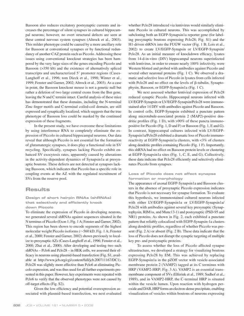
whether Pclo28 introduced via lentivirus would similarly elimi-
nate Piccolo in cultured neurons. This was accomplished by
subcloning both an EGFP-Synapsin1a reporter gene (for label-
ing presynaptic boutons expressing Pclo28; Fig. S1) and the
H1-driven shRNA into the FUGW vector ( Fig. 1 B ; Lois et al.,
2002 ) to create LV/EGFP-Synapsin or LV/EGFP-Synapsin/
Pclo28. As an initial measure of knockdown effi cacy, lysates
from 14-d-in-vitro (DIV) hippocampal neurons superinfected
with lentivirus, in order to ensure nearly 100% infectivity, were
Western blotted and probed with antibodies against Piccolo and
several other neuronal proteins ( Fig. 1 C ). We observed a dra-
matic and selective loss of Piccolo in lysates from cells infected
with Pclo28 and no effect on the levels of � -tubulin, Synapto-
physin, Bassoon, or EGFP-Synapsin1a ( Fig. 1 C ).
We next assessed whether lentiviral expression of Pclo28
reduced synaptic Piccolo. Hippocampal neurons infected with
LV/EGFP-Synapsin or LV/EGFP-Synapsin/Pclo28 were immuno-
stained after 14 DIV with antibodies against Piccolo and Bassoon.
In control cells, EGFP-Synapsin exhibited a punctate pattern
along microtubule-associated protein 2 (MAP2)-positive den-
dritic profi les ( Fig. 1 D ), with > 90% of these puncta immuno-
positive for Piccolo ( Fig. 1, D and F ) or Bassoon ( Fig. 1, E and G ).
In contrast, hippocampal cultures infected with LV/EGFP-
Synapsin1a/Pclo28 exhibited a dramatic loss of Piccolo immuno-
reactivity at EGFP-Synapsin1a clusters, with < 3% of clusters
along dendritic profi les containing Piccolo ( Fig. 1 F ). Importantly,
this shRNA had no effect on Bassoon protein levels or clustering
at EGFP-Synapsin1a sites ( Fig. 1, C, E, and G ). Collectively,
these data indicate that Pclo28 effi ciently and selectively elimi-
nates Piccolo from synapses.
Loss of Piccolo does not affect synapse formation or morphology The appearance of axonal EGFP-Synapsin1a and Bassoon clus-
ters in the absence of presynaptic Piccolo expression indicates
that Piccolo is not necessary for synapse formation. To evaluate
this hypothesis, we immunostained cultured neurons infected
with either LV/EGFP-Synapsin1a or LV/EGFP-Synapsin1a/
Pclo28 with antibodies against several key presynaptic (Synap-
tophysin, RIM1 � , and Munc13-1) and postsynaptic (PSD-95 and
NR1) proteins. As shown in Fig. 2 , each exhibited a punctate
pattern that reliably colocalized with EGFP-Synapsin-1a clusters
along dendritic profi les, regardless of whether Piccolo was pre-
sent ( Fig. 2 A ) or absent ( Fig. 2 B ). These data indicate that the
loss of Piccolo does not disrupt the synaptic targeting of multiple
key pre- and postsynaptic proteins.
To assess whether the loss of Piccolo affected synapse
ultrastructure, we developed a strategy for visualizing boutons
expressing Pclo28 by EM. This was achieved by replacing
EGFP-Synapsin1a in the pZOff vector with vesicle-associated
membrane protein 2 (VAMP2) tagged at its C terminus with
HRP (VAMP2-HRP; Fig. 3 A ). VAMP2 is an essential trans-
membrane component of SVs ( Elferink et al., 1989 ; Sudhof et al.,
1989 ), and in VAMP2-HRP, the C-terminal HRP is situated
within the vesicle lumen. Upon reaction with hydrogen per-
oxide and DAB, HRP forms an electron-dense precipitate, enabling
visualization of vesicles within boutons of neurons expressing
Bassoon also reduces excitatory postsynaptic currents and in-
creases the percentage of silent synapses in cultured hippocam-
pal neurons; however, no overt structural defects are seen at
these central nervous system synapses ( Altrock et al., 2003 ).
This milder phenotype could be caused by a more ancillary role
for Bassoon at conventional synapses or by functional redun-
dancy of another CAZ protein such as Piccolo. Addressing these
issues using conventional knockout strategies has been ham-
pered by the very large sizes of the genes encoding Piccolo and
Bassoon ( > 350 kb) and the existence of alternatively spliced
transcripts and uncharacterized 5 � promoter regions ( Cases-
Langhoff et al., 1996 ; tom Dieck et al., 1998 ; Winter et al.,
1999 ; Fenster and Garner, 2002 ; Altrock et al., 2003 ). As a case
in point, the Bassoon knockout mouse is not a genetic null but
rather a deletion of two large central exons from the Bsn gene,
leaving the N and C termini intact. Careful analysis of these mice
has demonstrated that these domains, including the N-terminal
Zinc fi nger motifs and C-terminal coiled-coil domain, are still
expressed and synaptically localized, which suggests that the true
phenotype of Bassoon loss could be masked by the continued
expression of these fragments.
In the present study, we have overcome these limitations
by using interference RNA to completely eliminate the ex-
pression of Piccolo in cultured hippocampal neurons. Our data
reveal that although Piccolo is not essential for the formation
of glutamatergic synapses, it does play a functional role in SV
recycling. Specifi cally, synapses lacking Piccolo exhibit en-
hanced SV exocytosis rates, apparently caused by alterations
in the activity-dependent dynamics of Synapsin1a at presyn-
aptic boutons. These defects are not detected at synapses lack-
ing Bassoon, which indicates that Piccolo has a specifi c role in
coupling events at the AZ with the regulated recruitment of
SVs from the reserve pool.
Results Design of short hairpin RNAs (shRNAs) that selectively and effi ciently knock down Piccolo To eliminate the expression of Piccolo in developing neurons,
we generated several shRNAs against sequences situated in the
N terminus of Piccolo (Exon 1; Fig. 1 A ; Fenster and Garner, 2002 ).
This region has been shown to encode segments of the highest
molecular weight Piccolo isoforms ( � 560 kD; Fig. 1 A ; Fenster
et al., 2000 ; Fenster and Garner, 2002 ) shown previously to local-
ize to presynaptic AZs ( Cases-Langhoff et al., 1996 ; Fenster et al.,
2000 ; Zhai et al., 2000 ). After developing and testing two such
shRNAs – Pclo6 and Pclo28 – in HEK cells, we assessed their ef-
fi cacy in neurons using plasmid-based transfection (Fig. S1, avail-
able at http://www.jcb.org/cgi/content/full/jcb.200711167/DC1).
Pclo28 was slightly more effective than Pclo6 at eliminating Pic-
colo expression, and was thus used for all further experiments pre-
sented in this paper. However, key experiments were repeated with
Pclo6 to verify that the observed phenotypes were not caused by
off-target effects (Fig. S2).
Given the low effi ciency and potential overexpression as-
sociated with plasmid-based transfection, we next evaluated

833PICCOLO REGULATES SYNAPTIC VESICLE EXOCYTOSIS • Leal-Ortiz et al.
gross morphological differences were detected between un-
labeled synapses, those expressing only VAMP2-HRP, and those
expressing VAMP2-HRP/Pclo28 ( Fig. 3, E and F ). Furthermore,
when synapses were carefully quantifi ed for bouton area, AZ/
postsynaptic density (PSD) length, docked SVs/AZ length, and
SV density (number of vesicles per bouton area), no morpho-
logical differences were found ( Table I ). These data strongly
suggest that Piccolo is not essential for the structural assembly
of excitatory asymmetrical synapses.
Synapses lacking Piccolo exhibit faster rates of SV exocytosis Although synapses still form in the absence of Piccolo, its
large size and multiple binding partners suggest that it may be
functionally important for SV recycling. We thus used the styryl
FM dyes ( Cochilla et al., 1999 ) to analyze presynaptic func-
tion. We fi rst examined whether boutons lacking Piccolo were
presynaptically active. This was accomplished by labeling the
total recycling pool (TRP) of vesicles with FM4-64 (90 mM
pZOff/VAMP2-HRP. To verify that VAMP2-HRP did not affect
Piccolo down-regulation, cultures expressing pZOff/VAMP2-
HRP or pZOff/VAMP2-HRP/Pclo28 were immunostained with
antibodies against Piccolo and HRP at 6 DIV ( Fig. 3 B ). Under
these conditions, Piccolo immunoreactivity was detected in the
axons of untransfected neurons or those expressing VAMP2-HRP
alone but not in axons of neurons expressing Pclo28. These data
indicate that Pclo28 shRNA was not hampered by coexpression
of VAMP2-HRP.
To visualize synapses, hippocampal neurons electropor-
ated at the time of plating with pZOff/VAMP2-HRP or pZOff/
VAMP2-HRP/Pclo28 were fi xed and processed for EM at 14 DIV.
In both cases, presynaptic boutons expressing VAMP2-HRP
were readily identifi ed based on the presence of electron-dense
SVs ( Fig. 3 D ). These labeled SVs were easily distinguishable
from both unlabeled SVs and the 80-nm dense core vesicles
hypothesized to carry Piccolo and Bassoon to nascent synapses
( Figs. 3 C and S3, available at http://www.jcb.org/cgi/content/
full/jcb.200711167/DC1; Zhai et al., 2001 ). Qualitatively, no
Figure 1. Lentivirus mediated knockdown of Piccolo with interference RNAs. (A) Schematic diagram of Piccolo, illustrating its multidomain structure (a poly-glutamate [pQ] region, two double zinc fi nger domains [Zn1/2], three coiled-coil domains [CC1-3], a PDZ domain, and two C2 domains [C2A/B]) and the location and sequence of the shRNA (Pclo28) used in this study. (B) Schematic diagram of the lentiviral vector (FUGW H1) used to express shRNAs under con-trol of the H1 promoter (H1 prom; yellow) and either EGFP or EGFP-Synapsin1a (Syn1a) under the ubiquitin-C promoter. (C) Western blots of cellular lysates from hippocampal neurons (uninfected or infected with LV/EGFP-Synapsin1a [EGFP-Syn] or LV/EGFP-Synapsin1a/Pclo28 [EGFPSynPclo28]) immunostained for Piccolo, Bassoon, tubulin, Synaptophysin (Syph), and GFP-Synapsin (GFP-Syn). Protein molecular weights are indicated on the right. (D and E) Hippocampal neurons infected with LV/EGFP-Synapsin1a (EGFPSyn) or LV/EGFP-Synapsin1a/Pclo28 (EGFPSynPclo28) and immunostained with antibodies against MAP2 (Merged, blue) and Piccolo (D, red) or Bassoon (E, red) after 14 DIV. Only a fraction of presynaptic boutons express EGFP-Synapsin1a/Pclo28; Piccolo immunoreactivity is from uninfected neurons. Bars, 10 μ m. (F and G) Bar graphs showing percent colocalization of EGFP-Synapsin1a (EGFPSyn) puncta with either Piccolo (F) or Bassoon (G) puncta along dendritic profi les ( n > 2,000 puncta per condition, fi ve experiments). Error bars indicate SEM.

JCB • VOLUME 181 • NUMBER 5 • 2008 834
relative sizes of the TRP. No difference was observed in the
mean intensity of FM4-64 fl uorescence ( Fig. 4 C ), which indi-
cates that TRP size was unaffected by the absence of Piccolo.
To evaluate whether the loss of Piccolo led to changes in SV
exocytosis, we compared the destaining kinetics of the TRP
using both 10- and 5-Hz electrical stimulation ( Fig. 4, D and E ).
Intriguingly, boutons lacking Piccolo destained more quickly
than those expressing only EGFP-Synapsin1a (two-way anal-
ysis of variance [ANOVA]; P < 0.0001 for both conditions;
KCl for 60 s; Pyle et al., 2000 ) in neuronal cultures infected
with either LV/EGFP-Synapsin1a or LV/EGFP-Synapsin1a/
Pclo28. At boutons with (EGFPSyn) or without (EGFPSyn-
Pclo28) Piccolo, > 80% of EGFP-Synapsin1a clusters co-
localized with FM4-64 puncta ( Fig. 4, A and B ). These data
indicated that synapses lacking Piccolo were indeed presynap-
tically functional and no more likely to be silent than control
boutons. We next compared the total FM fl uorescence inten-
sity at boutons containing or lacking Piccolo to determine the
Figure 2. Piccolo is not essential for the clustering of key pre- and postsynaptic proteins. Hippocampal neurons infected with LV/EGFP-Synapsin1a (EGFP-Syn; A) or LV/EGFP-Synap-sin1a/Pclo28 (EGFPSynPclo28; B) and immunostained with antibodies against Synaptophysin (Syph), RIM1, Munc13-1, PSD-95, or NR1 subunits of the NMDA receptor after 14 DIV. Shown are EGFP-Synapsin1a (green) puncta along dendrites (not depicted) that colocalize (Merged) with each pre- or post-synaptic protein (red). Bars, 10 μ m.

835PICCOLO REGULATES SYNAPTIC VESICLE EXOCYTOSIS • Leal-Ortiz et al.
Figure 3. Loss of Piccolo does not affect synapse ultrastructure. (A) Schematic diagram of the pZOff/VAMP2-HRP vector used to express VAMP2-HRP with or without Pclo28 shRNA. (B) Hippocampal neurons electroporated at the time of plating with pZOff/VAMP2-HRP or pZOff/VAMP2-HRP/Pclo28 and immunostained at 6 DIV with HRP (green) and Piccolo (red) antibodies. Note the lack of Piccolo immunoreactivity in Pclo28-expressing axons. (C) Transmission EM micrograph of a VAMP2-HRP – positive bouton containing both clear-centered (unlabeled SV) and dark-centered SVs (labeled SV). HRP-labeled vesicles are easily distinguished from 80-nm DCVs and other labeled structures. (D) Synaptic boutons from VAMP2-HRP – expressing (labeled bouton) and untrans-fected (unlabeled bouton) neurons. Arrowheads denote synaptic junctions, identifi ed based on electron-dense PSDs. (E) An excitatory synapse formed between a pZOff/VAMP2-HRP/Pclo28 (Pclo28)-transfected presynaptic bouton and a postsynaptic spine. (F) An excitatory synapse formed between a pZOff/VAMP2-HRP (control) transfected presynaptic bouton and a postsynaptic spine.

JCB • VOLUME 181 • NUMBER 5 • 2008 836
cytoskeleton ( Greengard et al., 1993 ; Ceccaldi et al., 1995 ).
Activity-dependent phosphorylation/dephosphorylation of Syn-
apsin by CaM-dependent kinase II (CaMKII), protein kinase A,
MAPK, and protein phosphatases 2A and 2B appears to regu-
late the exocytosis kinetics of SVs, presumably by regulating
Synapsin binding to SVs and/or the actin cytoskeleton ( Greengard
et al., 1993 ; Ryan et al., 1993 ; Jovanovic et al., 2000, 2001 ;
Pyle et al., 2000 ; Mozhayeva et al., 2002 ; Chi et al., 2003 ;
Sankaranarayanan et al., 2003 ). Importantly, Synapsin has also
been shown to undergo an activity-dependent dispersion away
from presynaptic boutons ( Chi et al., 2001 ), and the rate of dis-
persion is linked to its phosphorylation state ( Chi et al., 2001,
2003 ). However, the precise relationship between Synapsin
phosphorylation, dispersion, and SV exocytosis appears com-
plex and has not been fully characterized.
To explore whether the loss of Piccolo impacts SV trans-
location via alterations in the properties of Synapsin, we evalu-
ated whether the association and/or dispersion kinetics of
Synapsin1a within presynaptic boutons were altered. Here, we
took advantage of the fact that our shRNA expression vector
contained EGFP-Synapsin1a. Initially, we used antibodies
against Synapsin to verify that the lentiviral system did not
lead to a gross overexpression of Synapsin at individual bou-
tons. Surprisingly, we found no increase in Synapsin immuno-
staining at boutons expressing EGFP-Synapsin1a ( Fig. 5 A ),
which indicates that endogenous Synapsin expression is down-
regulated in the presence of EGFP-Synapsin to maintain simi-
lar total levels per bouton. Next, we quantifi ed and compared
the synaptic levels of EGFP-Synapsin1a in boutons of control
neurons and those expressing Pclo28. We found no discernable
differences in fl uorescence intensity when cultures were fi xed
with 4% paraformaldehyde ( Fig. 5 B ). However, when cultures
were fi xed with methanol, which extracts soluble proteins, we
observed a signifi cant decrease ( t test, P < 0.0001) in the inten-
sity of EGFP-Synapsin1a at boutons lacking Piccolo ( Fig. 5 B ).
This suggested that EGFP-Synapsin1a was not as tightly asso-
ciated with SVs and/or actin-related structures in the absence
of Piccolo but rather was shifted into a more soluble fraction.
This concept was supported by FRAP experiments designed to
monitor the steady-state exchange kinetics of EGFP-Synapsin1a
fl uorescence at individual boutons ( Fig. 5, C and D ). Again,
EGFP-Synapsin1a fl uorescence recovered more quickly at bou-
tons lacking Piccolo ( � fast = 1.2 min and � slow = 11.9 min vs.
� fast = 2.2 min and � slow = 33.7 min for control boutons; two-
way ANOVA, P < 0.0001; Fig. 5 D ), which indicates that
mechanisms regulating the association of Synapsin1a with SVs
and/or the actin/spectrin cytoskeleton were altered in the absence
of Piccolo.
To assess whether the faster exchange kinetics of EGFP-
Synapsin1a in the absence of Piccolo were caused by decreased
presynaptic stability and/or association of SVs with synapses, we
also examined the turnover rates of SV2, an SV integral mem-
brane protein, in the presence and absence of Piccolo. Here, an
N-terminally EGFP-tagged SV2 was subcloned into our lentiviral
vectors in place of EGFP-Synapsin1a. Like EGFP-Synapsin1a,
EGFP-SV2 reliably labeled presynaptic boutons, exhibiting a
high degree of colocalization with FM4-64 (Fig. S4, available at
Fig. 4, D and E ), which indicates that Piccolo is a negative
regulator of SV exocytosis.
Piccolo could regulate SV exocytosis by infl uencing
vesicle docking and/or fusion with the AZ plasma membrane
or by controlling SV translocation from the reserve to readily
releasable pool (RRP). To explore possible changes in SV dock-
ing and/or fusion, we evaluated whether the size of the RRP
of SVs or the release probability (Pr) were modifi ed in the
absence of Piccolo ( Fig. 4, F and G ). The former represents
the subpopulation of SVs that are docked at the AZ plasma
membrane and poised to undergo fusion with the arrival of an
action potential, whereas the latter is a measure of the proba-
bility that SVs in the RRP will undergo fusion. Two methods
were used to estimate RRP size: one using hypertonic sucrose
(500 mM, � 800 mOsm) and the other using a weak electrical
stimulus (2 Hz for 30 s; Pyle et al., 2000 ). Under both condi-
tions, we found no signifi cant difference in RRP size between
wild-type synapses and those lacking Piccolo ( t test, P > 0.5;
Fig. 4 F ).
To determine Pr, we measured the destaining kinetics
of boutons under conditions that stimulate RRP release, as
described previously ( Ryan et al., 1993 ; Pyle et al., 2000 ;
Mozhayeva et al., 2002 ; Sankaranarayanan et al., 2003 ). Again,
we found no signifi cant difference in release probability be-
tween wild-type boutons and those lacking Piccolo ( t test, P >
0.5; Fig. 4 G ). Collectively, these data indicated that the in-
creased SV exocytosis rates observed at 5 and 10 Hz were not
caused by increased RRP size or Pr.
Piccolo modulates the dispersion kinetics of Synapsin1a Based on the RRP experiments, we concluded that Piccolo ’ s
negative regulation of SV exocytosis was unlikely to occur at
the level of SV priming or fusion, as described for RIM1 � and
Munc13 ( Augustin et al., 1999 ; Schoch et al., 2002 ; Weimer
and Richmond, 2005 ; Gracheva et al., 2006 ; Weimer et al.,
2006 ), but rather at an earlier step, such as translocation of SVs
from the reserve pool to the RRP. To date, the only protein that
has been implicated in regulating SV translocation is the SV-
associated phosphoprotein Synapsin ( De Camilli et al., 1990 ;
Greengard et al., 1993 ; Ryan et al., 1996 ; Chi et al., 2003 ).
Mechanistically, Synapsin is hypothesized to mediate the clus-
tering and retention of SVs within boutons via its ability to
cross-bridge and tether them to the presynaptic actin/spectrin
Table I. Quantitative ultrastructural analysis of synapses with or without Pclo28
Parameter Control +Pclo28
AZ length (nm) 0.258 � 0.142 0.229 � 0.102
Docked SVs/AV length ( n / μ m) 13.87 � 6.26 14.29 � 5.30
SV density ( n / μ m 2 ) 163.8 � 58.36 169.0 � 49.02
Percentage of SVs labeled 20.4 � 12.5% 24.5 � 14.8%
All data are from boutons with at least two clearly labeled synaptic vesicles opposed to a distinct synaptic junction. All parameters were measured by an observer blind to phenotype. n = 64 boutons with 66 active zones for control and 53 boutons with 55 active zones for +Pclo28. Data are shown � standard deviation. t tests revealed no signifi cant differences, with no P < 0.05.

837PICCOLO REGULATES SYNAPTIC VESICLE EXOCYTOSIS • Leal-Ortiz et al.
Figure 4. Synapses lacking Piccolo have enhanced rates of SV exocytosis. (A) Images of 14 DIV hippocampal neurons infected with LV/EGFP-Synapsin1a (EGFPSyn, green) or LV/EGFP-Synapsin1a/Pclo28 (EGPFSynPclo28, green) and loaded with FM4-64 (FM, red) with 90 mM K+. Note the high degree of match between EGFP-Synapsin1a and FM4-64 in both merged images. Bar, 10 μ m. (B) Bar graphs quantifying the percent colocalization between EGFP-Synapsin and FM4-64 puncta in control cultures (EGFPSyn) or those lacking Piccolo (EGFPSynPclo28; n > 800 puncta per condition, two experiments). (C) Bar graph of FM4-64 fl uorescence intensity at EGFP-Synapsin1a puncta comparing the relative sizes of the TRP of SVs at control synapses (EGFPSyn) and those lacking Piccolo (EGFPSynPclo28). FM intensity values at EGFPSyn-expressing boutons were normalized against those from neighboring uninfected cells to enable cross-coverslip comparison. Mean normalized FM intensity from control boutons was set to 100; that from Pclo28 boutons was ratioed against this value ( n > 800 puncta, two experiments). (D and E) Destaining kinetics of the TRP at 10 Hz (D) and 5 Hz (E) comparing synapses with (EGFPSyn) and without (EGFPSynPclo28) Piccolo ( n = 5 experiments per condition). (F) Bar graph comparing the size of the RRP of SVs in boutons with (EGFPSyn) or without (EGFPSynPclo28) Piccolo, as determined by either the application of 500 mM sucrose (left) or 2-Hz, 30-s stimulation (right). No signifi cant differences were found ( t test, P > 0.5). Sucrose experiments were performed twice for each condition and stimulation experiments three times. (G) Destaining kinetics of the RRP during 2-Hz, 30-s stimulation for control (EGFPSyn) boutons and those lacking Piccolo (EGFPSynPclo28; n = 5 experiments per condition). Error bars indicate SEM.

JCB • VOLUME 181 • NUMBER 5 • 2008 838
Figure 5. Steady-state and activity-dependent dynamics of EGFP-Synapsin1a are altered at synapses lacking Piccolo. (A, left) Images of neurons infected with EGFP-Synapsin1a (green) and immunostained with Synapsin1a antibodies (red). (A, right) Bar graph comparing the intensity of Synapsin immuno-reactivity at boutons with or without EGFP-Synapsin1a. Total Synapsin levels are similar for infected and uninfected neurons. n > 100 puncta per condition. Bar, 10 μ m. (B) EGFP-Synapsin1a fl uorescence intensity at presynaptic boutons with (EGFPSyn) or without (EGFPSynPclo28) Piccolo after methanol versus 4% paraformaldehyde fi xation ( n > 800 puncta, seven fi elds of view per condition). (C) Time-lapse images of EGFP-Synapsin1a puncta. Single fl uorescent puncta (indicated by arrows) were photobleached and their recovery was monitored over time. (D) Fluorescence recovery curves for EGFP-Synapsin1a puncta at boutons with (EGFPSyn, n = 20) or without (EGFPSynPclo28, n = 17) Piccolo. (E) Images of EGFP-SV2 puncta at boutons with (EGFPSV2) or without (EGFPSV2Pclo28) Piccolo, photobleached as in C. Arrowheads indicate the positions of EGFP puncta that are bleached during FRAP experiments.

839PICCOLO REGULATES SYNAPTIC VESICLE EXOCYTOSIS • Leal-Ortiz et al.
http://www.jcb.org/cgi/content/full/jcb.200711167/DC1). However,
in contrast to Synapsin1a, the exchange kinetics of EGFP-SV2
were not altered in the absence of Piccolo ( Fig. 5, E and F ). These
data indicate that general presynaptic stability and SV retention
were not signifi cantly altered in the absence of Piccolo.
To assess whether the differences in steady-state exchange
kinetics translated into activity-dependent differences in the dis-
persion kinetics of EGFP-Synapsin1a, we used an electrical stim-
ulation protocol (10 Hz for 90 s) shown to promote the dispersion
of Synapsin1a ( Chi et al., 2001, 2003 ). Here, changes in the fl uo-
rescence intensity of EGFP-Synapsin1a at individual boutons
were monitored both during stimulation and for the subsequent
10-min recovery period. In contrast to wild-type boutons, we ob-
served both a more complete dispersion of EGFP-Synapsin1a
and a slower rate of recovery in boutons lacking Piccolo (two-
way ANOVA, P < 0.0001; Fig. 5, G and H ). Intriguingly, we also
observed remarkable, previously unreported heterogeneity in the
extent of EGFP-Synapsin1a dispersion at individual boutons
under both conditions. To evaluate whether the extent of EGFP-
Synapsin1a dispersion was coupled to the rate of SV exocytosis,
we simultaneously monitored the loss of EGFP-Synapsin1a and
FM5-95 fl uorescence from boutons during 5-Hz stimulation for
3 min. Our analysis revealed a tight correlation between the two
events. At boutons with minimal EGFP-Synapsin dispersion
( < 10% for all time points), very little FM5-95 destaining occurred
( Fig. 6, A and B ). In contrast, synapses with more complete
EGFP-Synapsin dispersion ( > 40% after 30 s of stimulation) ex-
hibited dramatic FM5-95 destaining ( Fig. 6, A and B ). This fea-
ture was present both at control synapses ( Fig. 6 A ) and those
lacking Piccolo ( Fig. 6 B ). A statistical analysis of the fi nal fl uo-
rescence intensity values for EGFP-Synapsin and FM after the
3-min stimulation revealed a strong correlation between the extent
of EGFP-Synapsin dispersion and that of FM5-95 destaining at
both control boutons and those lacking Piccolo (control: � = 0.58,
P < 0.0001; Pclo28: � = 0.59, P < 0.0001; Fig. 6, C and D ).
Although this tight correlation was observed for all synapses, the
mean degree of Synapsin dispersion and FM destaining was
greater at boutons lacking Piccolo, signifi cantly shifting these
correlation values to the lower right ( Fig. 6, C and D ). These data
strongly indicate that the accelerated SV exocytosis rates ob-
served at synapses lacking Piccolo are coupled to concomitant
increases in the dispersion kinetics of Synapsin1a.
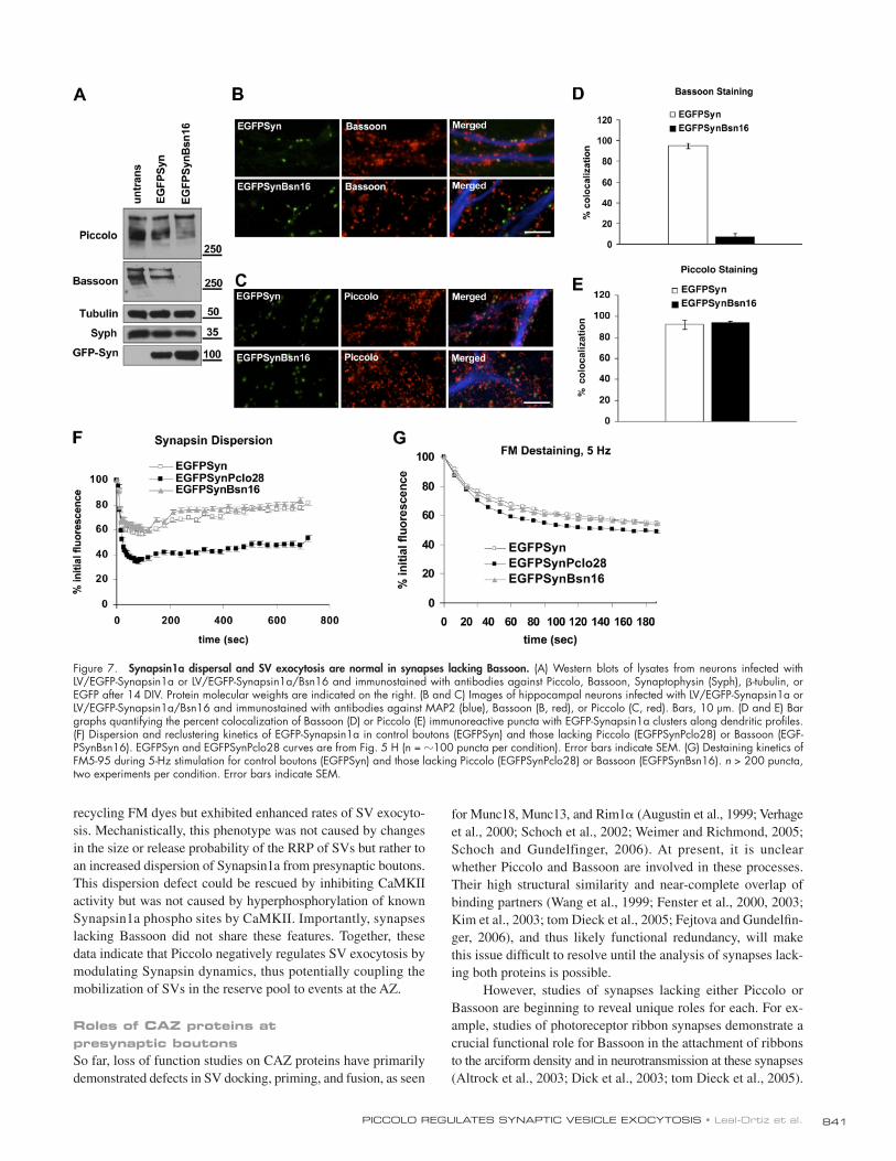
Changes in SV exocytosis and Synapsin dynamics are not observed at synapses lacking Bassoon Piccolo shares a high degree of structural similarity with Bas-
soon ( Fenster et al., 2000 ), another CAZ protein, and it is often
assumed that these two proteins are functionally redundant
( Fejtova and Gundelfi nger, 2006 ; Schoch and Gundelfi nger, 2006 ).
We were thus interested in exploring whether the phenotypes
seen at boutons lacking Piccolo were also observed in boutons
lacking Bassoon. To this end, we generated a set of shRNAs
against Bassoon and, after initial screening in the pZOff vector
(unpublished data), subcloned one (Bsn16) into the LV/EGFP-
Synapsin1a vector under the control of the H1 promoter. This
shRNA was found to effi ciently and specifi cally reduce the
expression of Bassoon in lysates of cultures infected with LV/
EGFP-Synapsin1a/Bsn16 on the day of plating and were har-
vested after 14 DIV ( Fig. 7 A ). Similarly, at the synaptic level,
we observed a dramatic decrease ( > 95%) in the percent co-
localization of Bassoon and EGFP-Synapsin1a in axons along
dendritic profi les ( Fig. 7, B and D ). This decrease was not ob-
served for Piccolo ( Fig. 7, C and E ), which indicates that the
Bsn16 shRNA is specifi c for Bassoon.
We next examined whether boutons lacking Bassoon also
exhibited changes in the dispersion and reclustering rates of
EGFP-Synapsin1a. As shown in Fig. 7 , synapses lacking Bas-
soon exhibited no detectable defects in EGFP-Synapsin disper-
sal or reclustering ( Fig. 7 F ) and no change in the destaining
kinetics of FM5-95 ( Fig. 7 G ). Together, these observations
strongly argue that the phenotypes observed for Piccolo are real
and not artifacts of long-term shRNA expression. Moreover, they
indicate that sequence elements unique to Piccolo play a funda-
mental role in negatively regulating SV exocytosis, apparently
by infl uencing the activity-dependent dispersion of Synapsin1a.
Loss of Piccolo enhances the CaMKII sensitivity of Synapsin 1a Because the association of Synapsin1a with SVs and the ac-
tin cytoskeleton in nerve terminals appears to be regulated
by CaMKII phosphorylation ( Schiebler et al., 1986 ; Benfenati
et al., 1992 ; Greengard et al., 1993 ; Stefani et al., 1997 ; Chi
et al., 2001, 2003 ), we next asked whether the altered dy-
namics of EGFP-Synapsin1a in the absence of Piccolo could
be caused by changes in its CaMKII-dependent phosphory-
lation. This was examined by blocking CaMKII activity with
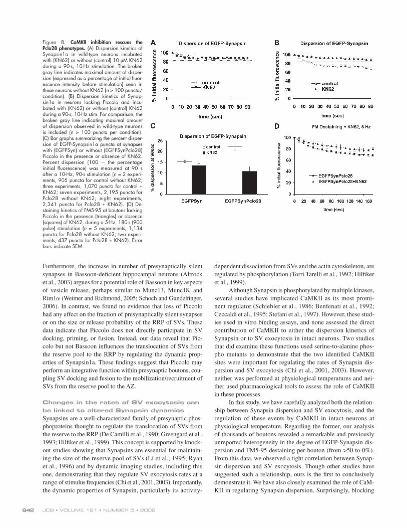
10 uM KN62 before and during a 10-Hz, 90-s stimulation.
Surprisingly, KN62 had no signifi cant effect on the disper-
sion kinetics or total amount of dispersion observed for
EGFP-Synapsin in wild-type boutons (two-way ANOVA, P =
0.15; Fig. 8, A and C ). However, it caused a dramatic reduc-
tion in EGFP-Synapsin dispersion at Pclo28 boutons, “ rescu-
ing ” dispersion to levels seen at wild-type boutons (two-way
ANOVA, P < 0.0001; Fig. 8, B and C ). To determine whether
this decrease in dispersion was accompanied by a decrease in
the SV exocytosis rate, we also assessed the impact of KN62
on the destaining kinetics of FM5-95 at Pclo28 boutons.
Again, KN62 attenuated the Pclo28 phenotype, slowing the
accelerated exocytosis of SVs seen in the absence of Piccolo
(two-way ANOVA, P < 0.0001; Fig. 8 D ). These results indi-
cate that the effects of Piccolo on presynaptic function are
(F) Fluorescence recovery curves for EGFP-SV2 puncta at synapses with (EGFPSV2, n = 8) or without (EGFP-SV2Pclo28, n = 12) Piccolo. (G) Time-lapse images of EGFP-Synapsin1a puncta. Images were acquired before, during, and after Synapsin dispersion was elicited with 10-Hz, 90-s stimulation. Shown here are sample images taken before and immediately after stimulation (10 Hz for 90 s) as well as after 10 min of recovery. (H) Graphical representation of data from G plotting the mean change in fl uorescence intensity over time at boutons with or without Piccolo ( n > 100 puncta per condition). The bar in the top left indicates time during which stimulation (10 Hz) was applied. Error bars indicate SEM.

JCB • VOLUME 181 • NUMBER 5 • 2008 840
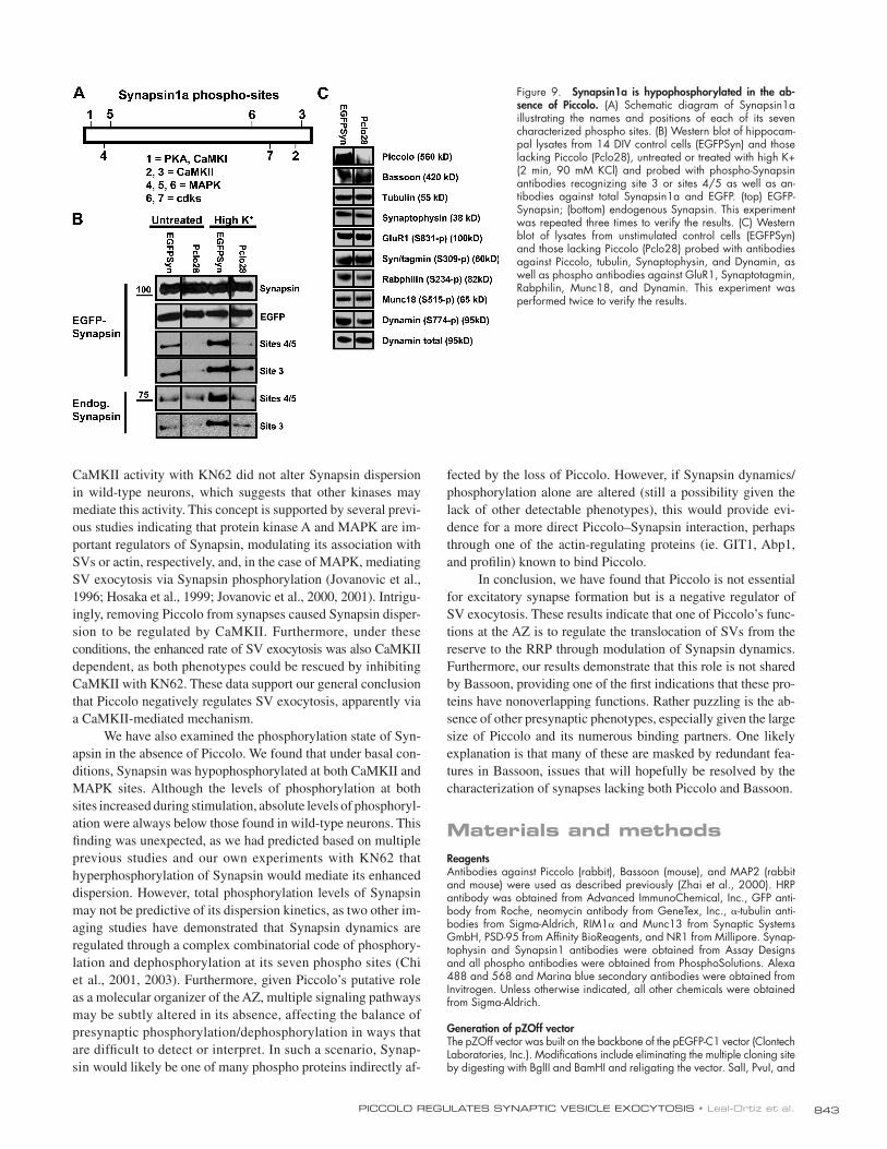
phosphatase activity. To test this hypothesis, we screened a
panel of phospho-specifi c antibodies against several pre- and
postsynaptic proteins, including GluR1, Synaptotagmin, Rab-
philin, Munc18, and Dynamin. Intriguingly, the phosphoryl-
ation levels of these proteins as assessed by Western blotting
were not altered in the absence of Piccolo ( Fig. 9 C ), which in-
dicates that the effect could be specifi c to Synapsin.
Discussion In the present study, we have used interference RNAs to disrupt
expression of the AZ protein Piccolo in developing hippocampal
neurons. This knockdown was specifi c for Piccolo and had no
overt effects on the expression or synaptic localization of other
pre- and postsynaptic proteins, including Bassoon, Synapsin,
Synaptophysin, RIM1 � , Munc13, PSD-95, and NR1, or on the
differentiation or life span ( > 4 wk) of cultured hippocampal neu-
rons. Furthermore, we detected no gross changes in the size or
morphology of excitatory glutamatergic synapses formed in the
absence of Piccolo, which indicates that Piccolo is not essential
for their assembly. Using live-imaging techniques, we also as-
sessed a possible role for Piccolo in presynaptic function. We
found that synapses lacking Piccolo were active and capable of
mediated by CaMKII. Moreover, they provide further evidence
that the Synapsin dispersion and SV exocytosis phenotypes
are mechanistically linked.
Synapsin1a is hypophosphorylated in the absence of Piccolo The ability of KN62 to rescue the Pclo28 phenotypes suggested
that Synapsin might be hyperphosphorylated by CaMKII in the
absence of Piccolo. To test this idea, we probed Western blots of
hippocampal lysates containing (EGFPSyn) or lacking (Pclo28)
Piccolo with two phospho-Synapsin1a antibodies that recog-
nize sites phosphorylated by CaMKII or MAPK ( Fig. 9 A ; Chi
et al., 2003 ). In these experiments, lysates from 14-DIV cul-
tures were analyzed either directly (untreated) or after high-K+
treatment (90 mM KCl for 2 min) to stimulate Synapsin
phosphorylation. As expected, the levels of both endogenous
and EGFP-Synapsin1a phosphorylation at sites 3, 4, and 5 in-
creased in all lysates after high-K+ stimulation ( Fig. 9 B ). How-
ever, in lysates from Pclo28 neurons, the levels of phosphorylation
at these sites under both basal and high-K+ conditions were dra-
matically reduced ( Fig. 9 B ). This unexpected fi nding suggested
that the kinase/phosphatase balance at boutons lacking Piccolo
was altered and that perhaps these boutons exhibited enhanced
Figure 6. Synapsin1a dispersion and FM destaining are tightly correlated. (A and B) FM5-95 destaining curves for individual EGFP-Synapsin1a puncta at boutons with (A) or without (B) Piccolo. Shown in black are examples of FM destaining curves for which there was either dramatic ( > 40%; triangles) or mini-mal ( < 10%; squares) dispersion of Synapsin. Shown in light gray are EGFP-Synapsin intensities for these boutons. (C and D) Correlation analysis of EGFP-Synapsin1a dispersion and FM5-95 destaining at synapses with (C) or without (D) Piccolo. Plotted are EGFP-Synapsin1a and FM intensity values (expressed as a percentage initial fl uorescence) at the fi nal time point (t =180 s) of 5-Hz stimulation (control = 709 puncta; Pclo28 = 855 puncta). Dashed lines and circles indicate mean intensity values for Synapsin and FM at control boutons (blue; 90.8 ± 1.0% and 75.6 ± 0.9%, respectively) and those lacking Piccolo (red; 79.2 ± 0.9% and 65.5 ± 0.8%, respectively). Note that values for boutons lacking Piccolo are signifi cantly shifted toward the bottom right.
841PICCOLO REGULATES SYNAPTIC VESICLE EXOCYTOSIS • Leal-Ortiz et al.
for Munc18, Munc13, and Rim1 � ( Augustin et al., 1999 ; Verhage
et al., 2000 ; Schoch et al., 2002 ; Weimer and Richmond, 2005 ;
Schoch and Gundelfinger, 2006 ). At present, it is unclear
whether Piccolo and Bassoon are involved in these processes.
Their high structural similarity and near-complete overlap of
binding partners ( Wang et al., 1999 ; Fenster et al., 2000, 2003 ;
Kim et al., 2003 ; tom Dieck et al., 2005 ; Fejtova and Gundelfi n-
ger, 2006 ), and thus likely functional redundancy, will make
this issue diffi cult to resolve until the analysis of synapses lack-
ing both proteins is possible.
However, studies of synapses lacking either Piccolo or
Bassoon are beginning to reveal unique roles for each. For ex-
ample, studies of photoreceptor ribbon synapses demonstrate a
crucial functional role for Bassoon in the attachment of ribbons
to the arciform density and in neurotransmission at these synapses
( Altrock et al., 2003 ; Dick et al., 2003 ; tom Dieck et al., 2005 ).
recycling FM dyes but exhibited enhanced rates of SV exocyto-
sis. Mechanistically, this phenotype was not caused by changes
in the size or release probability of the RRP of SVs but rather to
an increased dispersion of Synapsin1a from presynaptic boutons.
This dispersion defect could be rescued by inhibiting CaMKII
activity but was not caused by hyperphosphorylation of known
Synapsin1a phospho sites by CaMKII. Importantly, synapses
lacking Bassoon did not share these features. Together, these
data indicate that Piccolo negatively regulates SV exocytosis by
modulating Synapsin dynamics, thus potentially coupling the
mobilization of SVs in the reserve pool to events at the AZ.
Roles of CAZ proteins at presynaptic boutons So far, loss of function studies on CAZ proteins have primarily
demonstrated defects in SV docking, priming, and fusion, as seen
Figure 7. Synapsin1a dispersal and SV exocytosis are normal in synapses lacking Bassoon. (A) Western blots of lysates from neurons infected with LV/EGFP-Synapsin1a or LV/EGFP-Synapsin1a/Bsn16 and immunostained with antibodies against Piccolo, Bassoon, Synaptophysin (Syph), � -tubulin, or EGFP after 14 DIV. Protein molecular weights are indicated on the right. (B and C) Images of hippocampal neurons infected with LV/EGFP-Synapsin1a or LV/EGFP-Synapsin1a/Bsn16 and immunostained with antibodies against MAP2 (blue), Bassoon (B, red), or Piccolo (C, red). Bars, 10 μ m. (D and E) Bar graphs quantifying the percent colocalization of Bassoon (D) or Piccolo (E) immunoreactive puncta with EGFP-Synapsin1a clusters along dendritic profi les. (F) Dispersion and reclustering kinetics of EGFP-Synapsin1a in control boutons (EGFPSyn) and those lacking Piccolo (EGFPSynPclo28) or Bassoon (EGF-PSynBsn16). EGFPSyn and EGFPSynPclo28 curves are from Fig. 5 H (n = � 100 puncta per condition). Error bars indicate SEM. (G) Destaining kinetics of FM5-95 during 5-Hz stimulation for control boutons (EGFPSyn) and those lacking Piccolo (EGFPSynPclo28) or Bassoon (EGFPSynBsn16). n > 200 puncta, two experiments per condition. Error bars indicate SEM.
JCB • VOLUME 181 • NUMBER 5 • 2008 842
dependent dissociation from SVs and the actin cytoskeleton, are
regulated by phosphorylation ( Torri Tarelli et al., 1992 ; Hilfi ker
et al., 1999 ).
Although Synapsin is phosphorylated by multiple kinases,
several studies have implicated CaMKII as its most promi-
nent regulator ( Schiebler et al., 1986 ; Benfenati et al., 1992 ;
Ceccaldi et al., 1995 ; Stefani et al., 1997 ). However, these stud-
ies used in vitro binding assays, and none assessed the direct
contribution of CaMKII to either the dispersion kinetics of
Synapsin or to SV exocytosis in intact neurons. Two studies
that did examine these functions used serine-to-alanine phos-
pho mutants to demonstrate that the two identifi ed CaMKII
sites were important for regulating the rates of Synapsin dis-
persion and SV exocytosis ( Chi et al., 2001, 2003 ). However,
neither was performed at physiological temperatures and nei-
ther used pharmacological tools to assess the role of CaMKII
in these processes.
In this study, we have carefully analyzed both the relation-
ship between Synapsin dispersion and SV exocytosis, and the
regulation of these events by CaMKII in intact neurons at
physiological temperature. Regarding the former, our analysis
of thousands of boutons revealed a remarkable and previously
unreported heterogeneity in the degree of EGFP-Synapsin dis-
persion and FM5-95 destaining per bouton (from > 50 to 0%).
From this data, we observed a tight correlation between Synap-
sin dispersion and SV exocytosis. Though other studies have
suggested such a relationship, ours is the fi rst to conclusively
demonstrate it. We have also closely examined the role of CaM-
KII in regulating Synapsin dispersion. Surprisingly, blocking
Furthermore, the increase in number of presynaptically silent
synapses in Bassoon-defi cient hippocampal neurons ( Altrock
et al., 2003 ) argues for a potential role of Bassoon in key aspects
of vesicle release, perhaps similar to Munc13, Munc18, and
Rim1 � ( Weimer and Richmond, 2005 ; Schoch and Gundelfi nger,
2006 ). In contrast, we found no evidence that loss of Piccolo
had any affect on the fraction of presynaptically silent synapses
or on the size or release probability of the RRP of SVs. These
data indicate that Piccolo does not directly participate in SV
docking, priming, or fusion. Instead, our data reveal that Pic-
colo but not Bassoon infl uences the translocation of SVs from
the reserve pool to the RRP by regulating the dynamic prop-
erties of Synapsin1a. These fi ndings suggest that Piccolo may
perform an integrative function within presynaptic boutons, cou-
pling SV docking and fusion to the mobilization/recruitment of
SVs from the reserve pool to the AZ.
Changes in the rates of SV exocytosis can be linked to altered Synapsin dynamics Synapsins are a well-characterized family of presynaptic phos-
phoproteins thought to regulate the translocation of SVs from
the reserve to the RRP ( De Camilli et al., 1990 ; Greengard et al.,
1993 ; Hilfi ker et al., 1999 ). This concept is supported by knock-
out studies showing that Synapsins are essential for maintain-
ing the size of the reserve pool of SVs ( Li et al., 1995 ; Ryan
et al., 1996 ) and by dynamic imaging studies, including this
one, demonstrating that they regulate SV exocytosis rates at a
range of stimulus frequencies ( Chi et al., 2001, 2003 ). Importantly,
the dynamic properties of Synapsin, particularly its activity-
Figure 8. CaMKII inhibition rescues the Pclo28 phenotypes. (A) Dispersion kinetics of Synapsin1a in wild-type neurons incubated with (KN62) or without (control) 10 μ M KN62 during a 90-s, 10-Hz stimulation. The broken gray line indicates maximal amount of disper-sion (expressed as a percentage of initial fl uor-escence intensity before stimulation) seen in these neurons without KN62 ( n > 100 puncta/condition). (B) Dispersion kinetics of Synap-sin1a in neurons lacking Piccolo and incu-bated with (KN62) or without (control) KN62 during a 90-s, 10-Hz stim. For comparison, the broken gray line indicating maximal amount of dispersion observed in wild-type neurons is included ( n > 100 puncta per condition). (C) Bar graphs summarizing the percent disper-sion of EGFP-Synapsin1a puncta at synapses with (EGFPSyn) or without (EGFPSynPclo28) Piccolo in the presence or absence of KN62. Percent dispersion (100 � the percentage initial fl uorescence) was measured at 90 s after a 10-Hz, 90-s stimulation ( n = 2 experi-ments, 905 puncta for control without KN62; three experiments, 1,070 puncta for control + KN62; seven experiments, 2,195 puncta for Pclo28 without KN62; eight experiments, 2,341 puncta for Pclo28 + KN62). (D) De-staining kinetics of FM5-95 at boutons lacking Piccolo in the presence (triangles) or absence (squares) of KN62, during a 5-Hz, 180-s (900 pulse) stimulation ( n = 5 experiments, 1,134 puncta for Pclo28 without KN62; two experi-ments, 437 puncta for Pclo28 + KN62). Error bars indicate SEM.
843PICCOLO REGULATES SYNAPTIC VESICLE EXOCYTOSIS • Leal-Ortiz et al.
fected by the loss of Piccolo. However, if Synapsin dynamics/
phosphorylation alone are altered (still a possibility given the
lack of other detectable phenotypes), this would provide evi-
dence for a more direct Piccolo – Synapsin interaction, perhaps
through one of the actin-regulating proteins (ie. GIT1, Abp1,
and profi lin) known to bind Piccolo.
In conclusion, we have found that Piccolo is not essential
for excitatory synapse formation but is a negative regulator of
SV exocytosis. These results indicate that one of Piccolo ’ s func-
tions at the AZ is to regulate the translocation of SVs from the
reserve to the RRP through modulation of Synapsin dynamics.
Furthermore, our results demonstrate that this role is not shared
by Bassoon, providing one of the fi rst indications that these pro-
teins have nonoverlapping functions. Rather puzzling is the ab-
sence of other presynaptic phenotypes, especially given the large
size of Piccolo and its numerous binding partners. One likely
explanation is that many of these are masked by redundant fea-
tures in Bassoon, issues that will hopefully be resolved by the
characterization of synapses lacking both Piccolo and Bassoon.
Materials and methods Reagents Antibodies against Piccolo (rabbit), Bassoon (mouse), and MAP2 (rabbit and mouse) were used as described previously ( Zhai et al., 2000 ). HRP antibody was obtained from Advanced ImmunoChemical, Inc., GFP anti-body from Roche, neomycin antibody from GeneTex, Inc., � -tubulin anti-bodies from Sigma-Aldrich, RIM1 � and Munc13 from Synaptic Systems GmbH, PSD-95 from Affi nity BioReagents, and NR1 from Millipore. Synap-tophysin and Synapsin1 antibodies were obtained from Assay Designs and all phospho antibodies were obtained from PhosphoSolutions. Alexa 488 and 568 and Marina blue secondary antibodies were obtained from Invitrogen. Unless otherwise indicated, all other chemicals were obtained from Sigma-Aldrich.
Generation of pZOff vector The pZOff vector was built on the backbone of the pEGFP-C1 vector (Clontech Laboratories, Inc.). Modifi cations include eliminating the multiple cloning site by digesting with BglII and BamHI and religating the vector. SalI, PvuI, and
CaMKII activity with KN62 did not alter Synapsin dispersion
in wild-type neurons, which suggests that other kinases may
mediate this activity. This concept is supported by several previ-
ous studies indicating that protein kinase A and MAPK are im-
portant regulators of Synapsin, modulating its association with
SVs or actin, respectively, and, in the case of MAPK, mediating
SV exocytosis via Synapsin phosphorylation ( Jovanovic et al.,
1996 ; Hosaka et al., 1999 ; Jovanovic et al., 2000, 2001 ). Intrigu-
ingly, removing Piccolo from synapses caused Synapsin disper-
sion to be regulated by CaMKII. Furthermore, under these
conditions, the enhanced rate of SV exocytosis was also CaMKII
dependent, as both phenotypes could be rescued by inhibiting
CaMKII with KN62. These data support our general conclusion
that Piccolo negatively regulates SV exocytosis, apparently via
a CaMKII-mediated mechanism.
We have also examined the phosphorylation state of Syn-
apsin in the absence of Piccolo. We found that under basal con-
ditions, Synapsin was hypophosphorylated at both CaMKII and
MAPK sites. Although the levels of phosphorylation at both
sites increased during stimulation, absolute levels of phosphoryl-
ation were always below those found in wild-type neurons. This
fi nding was unexpected, as we had predicted based on multiple
previous studies and our own experiments with KN62 that
hyperphosphorylation of Synapsin would mediate its enhanced
dispersion. However, total phosphorylation levels of Synapsin
may not be predictive of its dispersion kinetics, as two other im-
aging studies have demonstrated that Synapsin dynamics are
regulated through a complex combinatorial code of phosphory-
lation and dephosphorylation at its seven phospho sites ( Chi
et al., 2001, 2003 ). Furthermore, given Piccolo ’ s putative role
as a molecular organizer of the AZ, multiple signaling pathways
may be subtly altered in its absence, affecting the balance of
presynaptic phosphorylation/dephosphorylation in ways that
are diffi cult to detect or interpret. In such a scenario, Synap-
sin would likely be one of many phospho proteins indirectly af-
Figure 9. Synapsin1a is hypophosphorylated in the ab-sence of Piccolo. (A) Schematic diagram of Synapsin1a illustrating the names and positions of each of its seven characterized phospho sites. (B) Western blot of hippocam-pal lysates from 14 DIV control cells (EGFPSyn) and those lacking Piccolo (Pclo28), untreated or treated with high K+ (2 min, 90 mM KCl) and probed with phospho-Synapsin antibodies recognizing site 3 or sites 4/5 as well as an-tibodies against total Synapsin1a and EGFP. (top) EGFP-Synapsin; (bottom) endogenous Synapsin. This experiment was repeated three times to verify the results. (C) Western blot of lysates from unstimulated control cells (EGFPSyn) and those lacking Piccolo (Pclo28) probed with antibodies against Piccolo, tubulin, Synaptophysin, and Dynamin, as well as phospho antibodies against GluR1, Synaptotagmin, Rabphilin, Munc18, and Dynamin. This experiment was performed twice to verify the results.

JCB • VOLUME 181 • NUMBER 5 • 2008 844
was introduced by electroporation of cells in suspension, conducted in 0.4-cm Gene Pulser Cuvettes (Bio Rad Laboratories) using 4 × 10 6 cells in 500 μ l of glia-conditioned media (10% FBS and 20 mM glucose in glutamine-free MEM) and 20 μ g of plasmid. Lentiviral infection was conducted after cov-erslips were transferred into 60-mm dishes using 10 μ l of virus per dish to infect 20 – 30% of cells (for imaging experiments) and 100 μ l/dish to super-infect � 100% of cells (for all biochemical experiments).
Immunohistochemistry Hippocampal neuronal cultures, grown for 5 – 21 DIV, were fi xed with ei-ther 4% formaldehyde in 1 × PBS for 10 min or 100% ice-cold methanol for 20 min. Cells were then permeabilized with 0.25% Triton X-100 in 1 × PBS for 5 min, washed in PBS, incubated in blocking solution (2% bovine serum albumin, 2% glycine, and 0.2% gelatin in 50 mM NH 4 Cl) for 15 min at room temperature, and incubated overnight at 4 ° C with primary antibod-ies in blocking solution. Afterward, cells were rinsed three to four times in PBS, incubated for 1 h at room temperature with secondary antibodies in blocking solution, rinsed again three to four times in PBS followed by a fi -nal rinse in deionized water, dried, and mounted in Vectashield mounting solution (Vector Laboratories).
Fixed images were acquired using a microscope (Axiovert 200M; Carl Zeiss, Inc.) with 100 × 1.3 NA or 40 × 1.3 NA Plan Neofl uar objec-tives (Carl Zeiss, Inc.). Fluorescence images were acquired with OpenLab software (at 1,344 × 1,022 resolution, 12 bits per pixel; PerkinElmer) with blue fl uorescent protein, FITC, and Texas red fi lter sets (Chroma Technology Corp.) using a digital camera (ORCA-ER; Hamamatsu). Quantifi cation of colocalization was performed manually, by determining the percentage of Piccolo or Bassoon puncta that colocalized along dendritic (MAP2 posi-tive) profi les with EGFP-Synapsin puncta.
Western blotting Immunoblots of cellular lysates were prepared from either transfected HEK293 or infected hippocampal neurons. Transient transfections of 60 – 70% confl uent HEK 293T cell cultures were performed using Lipofectamine 2000 (Invitrogen) with 10 μ g of DNA per 10-cm plate. Homogenates from cells expressing Pclo28 or Bsn16 shRNAs were prepared 48 h after transfection. In brief, cells were harvested into homogenization buffer (ice-cold 1 × PBS supplemented with protease inhibitors [complete protease inhibitor tablet, EDTA free; Roche]), and then placed directly into loading buffer. Protein lev-els were standardized empirically using a neomycin antibody and several loading concentrations. After separation by SDS-PAGE, proteins were trans-ferred to nitrocellulose membranes (GE Healthcare) and probed with pri-mary and secondary antibodies in Western blotting solution (5% nonfat dry milk and 0.05% NP-40 in Tris-buffered saline). Protein bands were visual-ized by HRP chemiluminescence (PerkinElmer). For Western blots of hippo-campal cultures, neurons from lentivirus-infected coverslips were harvested directly into loading buffer and the same procedure was followed. Here, protein levels were standardized using tubulin or GFP antibodies.
EM The ultrastructural analysis of glutamatergic asymmetrical synapses was per-formed on dissociated cultures of hippocampal neurons transfected by elec-troporation at the time of plating with pZOff-VAMP2-HRP ± Pclo28 shRNA. Samples were fi xed with 2.5% glutaraldehyde in 0.15 M cacodylate buffer for 1 h and processed as described previously ( Micheva and Smith, 2005 ) with a few modifi cations. In brief, after fi xation, neurons were incubated with 1 mg/ml DAB (Sigma-Aldrich) in 50 mM Tris, pH 7.5, for 10 min. 0.01% H 2 0 2 (Sigma-Aldrich) was then added to the DAB solution for 30 min at room temperature to stimulate HRP-mediated DAB precipitation. After ex-tensive washing, neurons were prepared for EM by a microwave irradiation protocol described in detail previously ( Micheva et al., 2003 ). After infi ltration in Embed 812 (Electron Microscopy Sciences), glass coverslips were re-moved by dissolution in hydrofl oric acid. Ultrathin 60-nm sections were cut with an ultramicrotome (Ultracut UCT; Leica) and placed on copper grids. Samples were poststained with 5% uranyl acetate dissolved in ultrapure wa-ter for 15 min followed by 4 min of staining in 0.2% lead citrate. Grids were extensively rinsed in water during and after poststaining. For control experi-ments (poststain No. 1 in Fig. S2), grids were more lightly poststained with 3.5% uranyl acetate in 50% acetone for 15 s followed by a 4-min 0.2% lead citrate stain. Samples were imaged with a transmission electron micro-scope (JEM-1230; JEOL Ltd.) at 80 kV accelerated voltage using a charge-coupled device camera (791; Gatan). All sample processing and EM was performed in the Cell Sciences Imaging Facility at Stanford University.
In a blinded fashion, control VAMP2-HRP and VAMP2-HRP-Pclo28 samples were imaged and synapses were quantitatively evaluated. Only
BamHI restriction sites were subsequently inserted between the MluI and DraIII sites using oligos (5 � -CGCGGTCGACGCGATCGCGGATCCTAC-3 � and 5 � -GGATCCGCGATCGCGTCGAC-3 � ) that destroy both of these sites. The H1 promoter taken from the pSuper plasmid (Oligoengine) was inserted into with SalI and BamHI sites of the modifi ed pEGFP vector, creating pZOff.
Design of shRNAs shRNAs were designed corresponding to the 21-mer target sites using Ambion criteria specifying oligo duplexes with 5 � -AA overhangs. The target sequence of Pclo28 is 5 � -AAGTGCTGTCTCCTCTGTTGT-3 � (nucleo-tides 640 – 660 of Rattus norvegicus Piccolo from GenBank/EMBL/DDBJ under accession no. NM_020098 ). The target sequence of Pclo6 is 5 � -AAGGGCGCAGGGGCTGCCCAA-3 � (nucleotides 334–354 of Piccolo from GenBank/EMBL/DDBJ under accession no. NM_020098 ) The target sequence of Bsn16 is 5 � -AACACCTGCACCCAGTGTCAC-3 � (nucleotides 652 – 673 of R. norvegicus Bassoon from GenBank/EMBL/DDBJ under accession no. NM_0191416 ). Sense and antisense oligodeoxynucleotides (Sigma-Aldrich) contained looped overhangs for BglII and Hind III. The fol-lowing oligos were used: Pclo28 sense, 5 � -GATCCCCGTGCTGTCTCCTC-TG TTGTTTCAAGAGAACAACAGAGGAGACAGCACTTTTTGGAAA-3 � ; Pclo28 antisense, 5 � -AGCTTTTCCAAAAAGTGCTGTCTCCTCTGTTGTT CTC -TTGAAACAACAGAGGAGACAGCACGGG-3 � ; Bsn16 sense, 5 � -GATCC-CCC ACCTGCACCCAGTGTCACTTCAAGAGAGTGACACTGGGTGCAGGTG-3 � ; and Bsn16 antisense, 5 � -AGCTTTTCCAAAAACACCTGCACCCAGTGT-CACTCTCTTGAAGTGACACTGGGTGCAGGTGGGG-3 � . Also used were: scrambled sense, 5 � -GATCCCC TTGAGCAGCTAGGCGACCA TTCAAGA-GATGGTCGCCTAGCTCAATTTTTGGAAA-3 � ; and scrambled antisense, 5 � -AGCTTTTCCAAAAA TTGAGCAGCTAGGCGACCA TCTCTTGAA TGGTCG-CCTAGCTGCTCAA GGG-3 � . 100 pmol of the respective strands were annealed in annealing buffer (100 mM potassium acetate, 30 mM Hepes-KOH, pH 7.4, and 2 mM magnesium acetate) using a Thermocycler (Bio- Rad Laboratories). The annealed cDNA duplexes were then phosphory-lated using phage T4 polynucleotide kinase (New England Biolabs, Inc.) and ligated into the pZOff vector between the BglII and HindIII sites.
Generation of FUGW H1vector Converting the FUGW vector ( Lois et al., 2002 ) into the shRNA silencing vector FUGW H1 required several steps. First, the EcoR1 site was elimi-nated from the FUGW vector. An oligonucleotide was then introduced into Pac1, creating a multicloning site with the following restriction enzyme sites: 5 � PvuI, BsiwI, EcoRI, BstBI, and PacI. Subsequently, the H1 promoter region and different shRNA sequences from the pZOff vector were inserted into the EcoRI and BstBI sites, creating FUGW H1. EGFP-Synapsin1a (a gift of T. Ryan, Weill Medical College of Cornell University, New York, NY) was incorporated into FUGW H1 by removing the EGFP cassette with XbaI and inserting the EGFP-Synapsin1a sequences into the Xba1 site (proteins were subcloned out of the pZOff vector using the NheI – XbaI sites; NheI is compatible with XbaI).
Lentivirus production Lentivirus was produced by transfecting a three-plasmid vector system comprising a shuttle plasmid (FUGW or FU[Syn-EGFP]W) and two pack-aging plasmids (pCMV R8.9 and pHCMV VSVg) into HEK293T cells (grown in DME + 10% fetal bovine serum and penicillin/streptomycin) as described previously ( Lois et al., 2002 ). In brief, transfections were con-ducted on confl uent (90 – 100%) cell cultures with Lipofectamine 2000 (In-vitrogen) using 22.5 μ g of total DNA and 60 μ l lipofectamine per 10 cm plate. 2 d after transfection, the virus-containing medium was collected, passed through a 0.45- μ m fi lter to remove cell debris, and frozen at � 80 ° C. The viral titer was determined by fl uorescence analysis of infected HEK293T cells (infective units).
Hippocampal cultures Hippocampal cultures were prepared using a modifi ed Banker culture pro-tocol ( Banker and Goslin, 1998 ). In brief, hippocampi from embryonic (embryonic day 18 or 19) Sprague-Dawley rats were dissected out and dissociated in 0.05% trypsin (Invitrogen), and cells were plated at a den-sity of 165/mm 2 on poly-L-lysine – coated coverslips (Carolina Biological Supply Company). 1 h after plating, coverslips were transferred in pairs to 60-mm dishes containing a glial feeder layer, where they were inverted (to maximize neuronal contact with secreted glial factors) and maintained in neurobasal medium containing B27 and GlutaMAX (all from Invitrogen).
Hippocampal transfection and lentivirus infection Hippocampal neurons were transfected or infected with the pZOff plasmid or lentivirus at the time of plating (0 DIV), respectively. The pZOff plasmid

845PICCOLO REGULATES SYNAPTIC VESICLE EXOCYTOSIS • Leal-Ortiz et al.
equation: [(intensity of bleached Synapsin punctum at time t )/(mean inten-sity of all unbleached puncta in fi eld of view at time t )] × 100. To control for variability in the extent of bleaching for each puncta, values were fur-ther normalized, making zero the default value for bleached puncta and enabling us to pool and average all recovery curves for a given condition (EGFPSyn and EGFPSynPclo28). � values were calculated using a custom macro written in Excel (N. Ziv; Tsuriel et al., 2006 ).
Synapsin dispersion Dispersion of EGFP-Synapsin was induced by electrical stimulation (10 Hz for 90 s) as described previously ( Chi et al., 2001 ). Puncta intensity values were expressed as a percentage of initial fl uorescence intensity before stimulation. Curves for all EGFP-Synapsin puncta were combined for a given condition (EGFPSyn and EGFPSynPclo28), averaged, and plotted. For this analysis, puncta that did not exhibit dispersion (percentage of ini-tial fl uorescence values > 90 for all time points) were excluded.
To compare extent of Synapsin dispersion to that of FM destaining, EGFP-Synapsin intensity was monitored during the 5-Hz destaining experi-ment (see FM destaining protocol).
To assess the role of CaMKII in Synapsin dispersion, coverslips were perfused with normal Tyrodes solution containing 10 uM KN62 (Tocris Bio-science) and incubated in the drug for 20 min before eliciting dispersion. EGFP-Synapsin intensity was monitored either before stimulation and every 5 s during stimulation (for dispersion curves), or before stimulation and at the last time point (t = 90 s; bar graphs).
FM5-95 destaining was also monitored after incubation with 10 μ M KN62. For this experiment, boutons were fi rst loaded with FM5-95 using high-K+ stimulation as described. After waiting 15 min to allow for some recovery of EGFP-Synapsin after stimulation, cells were incubated for an additional 20 min in 10 μ M KN62. SV exocytosis was then elicited by 5-Hz stimulation for 180 s, and both FM intensity and EGFP-Synapsin intensity were monitored over this time period (images taken every 10 s). Destain-ing curves were then plotted as described and compared with destaining curves from untreated coverslips of cultures infected with LV/EGFP-Synapsin or LV/EGFP-Synapsin-Pclo28.
Online supplemental material Fig. S1 shows initial testing of Piccolo shRNA (Pclo28) using pZOff plasmid-mediated knockdown in COS7 cells and neurons. Fig. S2 demonstrates that another shRNA against Piccolo (Pclo6) produces the same pheno-types as Pclo28. Fig. S3 shows additional EM micrographs showing that VAMP2-HRP unambiguously labels the synapses of transfected neurons and does not obscure synaptic junctions or other synaptic structures. Fig. S4 shows that EGFP-SV2, like EGFP-Synapsin, is a reliable presynaptic marker. Online supplemental material is available at http://www.jcb.org/cgi/content/full/jcb.200711167/DC1.
We are grateful to Noam Ziv, Nicole Calakos, and Rob Malenka for insightful suggestions and Tim Ryan for the EGFP-Synapsin clones. We also thank Kristina Micheva, John Perrino, and Yemane Gedde for their assistance with EM and hippocampal cultures.
This work was supported by grants from the National Institutes of Health (NS39471 and NS353862 to C.C. Garner), National Research Service Awards to C.L. Waites and R. Terry-Lorenzo, and the Bundesministerium f ü r Bil-dung und Forschung to E.D. Gundelfi nger.
Submitted: 30 November 2007 Accepted: 2 May 2008
References Altrock , W.D. , S. tom Dieck , M. Sokolov , A.C. Meyer , A. Sigler , C. Brakebusch ,
R. Fassler , K. Richter , T.M. Boeckers , H. Potschka , et al . 2003 . Functional inactivation of a fraction of excitatory synapses in mice defi cient for the active zone protein bassoon. Neuron . 37 : 787 – 800 .
Augustin , I. , C. Rosenmund , T.C. Sudhof , and N. Brose . 1999 . Munc13-1 is es-sential for fusion competence of glutamatergic synaptic vesicles. Nature . 400 : 457 – 461 .
Banker , G. , and K. Goslin . 1998 . Culturing Nerve Cells. Second edition. MIT Press, Cambridge, MA. 666 pp.
Benfenati , F. , F. Valtorta , J.L. Rubenstein , F.S. Gorelick , P. Greengard , and A.J. Czernik . 1992 . Synaptic vesicle-associated Ca2+/calmodulin-dependent protein kinase II is a binding protein for synapsin I. Nature . 359 : 417 – 420 .
Cases-Langhoff , C. , B. Voss , A.M. Garner , U. Appeltauer , K. Takei , S. Kindler , R.W. Veh , P. De Camilli , E.D. Gundelfi nger , and C.C. Garner . 1996 .
synapses with a clearly discernable PSD and at least two unambiguously HRP-labeled SVs were included in the analysis. The AZ was defi ned as the length of presynaptic membrane precisely opposed to the PSD. Docked SVs were defi ned as all SVs that had an edge within 25 nm of the AZ. All transmission EM quantitative analysis was performed using Image J.
Imaging experiments All live imaging experiments were performed on a custom-built (by S. Smith, Stanford University, Stanford, CA; and N. Ziv, Technion Faculty of Medicine, Haifa, Israel) scanning confocal microscope (Axiovert 100TV; Carl Zeiss, Inc.) equipped with a 40 × 1.3 NA Plan Neofl uar objective (Car Zeiss, Inc.) and 488 nm and 514 nm lasers (Sapphire 488-20CDRH and Compass 215M-20; Coherent), using OpenView software (written by N. Ziv). Neuronal coverslips were mounted in a custom-built chamber designed for perfusion and electrical stimulation, heated to 37 ° C by a forced-air blower, and perfused with Tyrode ’ s saline solution (25 mM Hepes, 119 mM NaCl, 2.5 mM KCl, 30 mM glucose, 2 mM CaCl, 2 mM MgCl 2 , 50 μ M CNQX, and 10 μ M APV, pH 7.4).
FM loading/destaining Functional presynaptic boutons were labeled with FM4-64 or FM5-95 dye (Invitrogen) by incubation in high-K+ Tyrodes solution (90 mM KCl and 31.5 mM NaCl) containing � 1 μ g/ml FM dye for 60 s followed by normal Tyrodes + FM dye for 30 s. Neurons were then washed for � 5 min before imaging. Destaining was performed by electrical stimulation at the fre-quency (10, 5, or 2 Hz) and time interval (180, 90, and 30 s) specifi ed for each experiment or by high-K+ Tyrodes solution for 60 s.
Image analysis and quantifi cation were performed with OpenView software and Excel (Microsoft). GraphPad Prism (GraphPad Software) was used for statistical analysis. To measure the percentage of FM colocalization with EGFP-Synapsin clusters, the number of FM-containing clusters was di-vided by the total number of EGFP-Synapsin clusters and multiplied by 100. To calculate relative FM intensities at boutons containing or lacking Piccolo, FM intensity values for infected neurons were normalized against those from neighboring uninfected neurons in the same fi eld of view, en-abling cross-coverslip comparisons.
For FM destaining experiments at 5 and 10 Hz, the total number of action potentials elicited and images acquired were kept constant. Thus, at 10-Hz stimulation, images were acquired every 5 s for a total of 90 s, whereas at 5 Hz they were acquired every 10 s for a total of 180 s. In all cases, intensity values for a given FM punctum at each time point were ex-pressed as a percentage of its initial fl uorescence intensity before destain-ing using the following equation: (current FM intensity at time point t /initial FM intensity) × 100. For each condition (EGFPSyn and EGFPSynPclo28), intensity curves that did not exhibit FM destaining (ie. values > 90% for all time points) were eliminated from the analysis. Remaining curves were pooled, averaged, and plotted using Excel.
Calculation of RRP size and Pr Boutons were loaded using high-K+ Tyrodes to label the TRP (TRP label). RRP release was induced with either 500 mM sucrose or low-frequency electrical stimulation (2 Hz at 30 s; RRP release). Total FM destaining was induced with high-K+ stimulation for 60 s (background). To calculated RRP size, puncta intensity values were put into the following equation to express the RRP as a percentage of the TRP: { [(TRP label – background) – (RRP release – background)]/(TRP label – background) } × 100. Similar values were ob-tained for both methods, indicating that they can be used interchangeably to measure RRP.
To calculate Pr, the TRP was loaded with FM5-95 using high-K+ Tyrodes, and release of the RRP was induced using 2-Hz, 30-s electrical stimulation. During stimulation, images were acquired every 5 s. Intensity values were expressed as a percentage of initial fl uorescence intensity, those exhibiting minimal destaining (values > 95% for all time points) were eliminated from the analysis, and remaining curves were averaged and plotted as described in the previous section.
FRAP analysis EGFP-Synapsin or SV2 puncta were bleached to � 20% of their initial fl uorescence by a high-intensity laser beam (488 nm wavelength) at high magnifi cation. Images were taken at a rate of 1 per minute to monitor fl uo-rescence recovery for Synapsin, and 1 per minute followed by 1 per 5 min-utes for SV2. For each time point, intensity values were expressed as a percentage of starting fl uorescence before bleaching. To control for non-specifi c photobleaching during image acquisition, the intensities of bleached puncta were normalized against those of unbleached puncta for each time point as described previously ( Tsuriel et al., 2006 ), with the following

JCB • VOLUME 181 • NUMBER 5 • 2008 846
Micheva , K.D. , J. Buchanan , R.W. Holz , and S.J. Smith . 2003 . Retrograde regulation of synaptic vesicle endocytosis and recycling. Nat. Neurosci. 6 : 925 – 932 .
Mozhayeva , M.G. , Y. Sara , X. Liu , and E.T. Kavalali . 2002 . Development of vesicle pools during maturation of hippocampal synapses. J. Neurosci. 22 : 654 – 665 .
Pyle , J.L. , E.T. Kavalali , E.S. Piedras-Renteria , and R.W. Tsien . 2000 . Rapid re-use of readily releasable pool vesicles at hippocampal synapses. Neuron . 28 : 221 – 231 .
Ryan , T.A. , H. Reuter , B. Wendland , F.E. Schweizer , R.W. Tsien , and S.J. Smith . 1993 . The kinetics of synaptic vesicle recycling measured at single pre-synaptic boutons. Neuron . 11 : 713 – 724 .
Ryan , T.A. , L. Li , L.S. Chin , P. Greengard , and S.J. Smith . 1996 . Synaptic vesicle recycling in synapsin I knock-out mice. J. Cell Biol. 134 : 1219 – 1227 .
Sankaranarayanan , S. , P.P. Atluri , and T.A. Ryan . 2003 . Actin has a molecular scaffolding, not propulsive, role in presynaptic function. Nat. Neurosci. 6 : 127 – 135 .
Schiebler , W. , R. Jahn , J.P. Doucet , J. Rothlein , and P. Greengard . 1986 . Characterization of synapsin I binding to small synaptic vesicles. J. Biol. Chem. 261 : 8383 – 8390 .
Schoch , S. , and E.D. Gundelfi nger . 2006 . Molecular organization of the pre-synaptic active zone. Cell Tissue Res. 326 : 379 – 391 .
Schoch , S. , P.E. Castillo , T. Jo , K. Mukherjee , M. Geppert , Y. Wang , F. Schmitz , R.C. Malenka , and T.C. Sudhof . 2002 . RIM1alpha forms a protein scaffold for regulating neurotransmitter release at the active zone. Nature . 415 : 321 – 326 .
Shapira , M. , R.G. Zhai , T. Dresbach , T. Bresler , V.I. Torres , E.D. Gundelfi nger , N.E. Ziv , and C.C. Garner . 2003 . Unitary assembly of presynaptic active zones from Piccolo-Bassoon transport vesicles. Neuron . 38 : 237 – 252 .
Stefani , G. , F. Onofri , F. Valtorta , P. Vaccaro , P. Greengard , and F. Benfenati . 1997 . Kinetic analysis of the phosphorylation-dependent interactions of synapsin I with rat brain synaptic vesicles. J. Physiol. 504 : 501 – 515 .
Sudhof , T.C. , M. Baumert , M.S. Perin , and R. Jahn . 1989 . A synaptic vesicle membrane protein is conserved from mammals to Drosophila . Neuron . 2 : 1475 – 1481 .
tom Dieck , S. , L. Sanmarti-Vila , K. Langnaese , K. Richter , S. Kindler , A. Soyke , H. Wex , K.H. Smalla , U. Kampf , J.T. Franzer , et al . 1998 . Bassoon, a novel zinc-fi nger CAG/glutamine-repeat protein selectively localized at the active zone of presynaptic nerve terminals. J. Cell Biol. 142 : 499 – 509 .
tom Dieck , S. , W.D. Altrock , M.M. Kessels , B. Qualmann , H. Regus , D. Brauner , A. Fejtova , O. Bracko , E.D. Gundelfi nger , and J.H. Brandstatter . 2005 . Molecular dissection of the photoreceptor ribbon synapse: physical inter-action of Bassoon and RIBEYE is essential for the assembly of the ribbon complex. J. Cell Biol. 168 : 825 – 836 .
Torri Tarelli , F. , M. Bossi , R. Fesce , P. Greengard , and F. Valtorta . 1992 . Synapsin I partially dissociates from synaptic vesicles during exocytosis induced by electrical stimulation. Neuron . 9 : 1143 – 1153 .
Tsuriel , S. , R. Geva , P. Zamorano , T. Dresbach , T. Boeckers , E.D. Gundelfi nger , C.C. Garner , and N.E. Ziv . 2006 . Local sharing as a predominant determi-nant of synaptic matrix molecular dynamics. PLoS Biol. 4 : e271 .
Verhage , M. , A.S. Maia , J.J. Plomp , A.B. Brussaard , J.H. Heeroma , H. Vermeer , R.F. Toonen , R.E. Hammer , T.K. van den Berg , M. Missler , et al . 2000 . Synaptic assembly of the brain in the absence of neurotransmitter secre-tion. Science . 287 : 864 – 869 .
Waites , C.L. , A.M. Craig , and C.C. Garner . 2005 . Mechanisms of vertebrate syn-aptogenesis. Annu. Rev. Neurosci. 28 : 251 – 274 .
Wang , X. , M. Kibschull , M.M. Laue , B. Lichte , E. Petrasch-Parwez , and M.W. Kilimann . 1999 . Aczonin, a 550-kD putative scaffolding protein of pre-synaptic active zones, shares homology regions with Rim and Bassoon and binds profi lin. J. Cell Biol. 147 : 151 – 162 .
Weimer , R.M. , and J.E. Richmond . 2005 . Synaptic vesicle docking: a putative role for the Munc18/Sec1 protein family. Curr. Top. Dev. Biol. 65 : 83 – 113 .
Weimer , R.M. , E.O. Gracheva , O. Meyrignac , K.G. Miller , J.E. Richmond , and J.L. Bessereau . 2006 . UNC-13 and UNC-10/rim localize synaptic vesi-cles to specifi c membrane domains. J. Neurosci. 26 : 8040 – 8047 .
Winter , C. , S. tom Dieck , T.M. Boeckers , J. Bockmann , U. Kampf , L. Sanmarti-Vila , K. Langnaese , W. Altrock , M. Stumm , A. Soyke , et al . 1999 . The presynaptic cytomatrix protein Bassoon: sequence and chromosomal lo-calization of the human BSN gene. Genomics . 57 : 389 – 397 .
Zhai , R. , G. Olias , W.J. Chung , R.A. Lester , S. tom Dieck , K. Langnaese , M.R. Kreutz , S. Kindler , E.D. Gundelfi nger , and C.C. Garner . 2000 . Temporal appearance of the presynaptic cytomatrix protein bassoon during synap-togenesis. Mol. Cell. Neurosci. 15 : 417 – 428 .
Zhai , R.G. , H. Vardinon-Friedman , C. Cases-Langhoff , B. Becker , E.D. Gundelfi nger , N.E. Ziv , and C.C. Garner . 2001 . Assembling the presyn-aptic active zone: a characterization of an active one precursor vesicle. Neuron . 29 : 131 – 143 .
Ziv , N.E. , and C.C. Garner . 2004 . Cellular and molecular mechanisms of presyn-aptic assembly. Nat. Rev. Neurosci. 5 : 385 – 399 .
Piccolo, a novel 420 kDa protein associated with the presynaptic cytoma-trix. Eur. J. Cell Biol. 69 : 214 – 223 .
Ceccaldi , P.E. , F. Grohovaz , F. Benfenati , E. Chieregatti , P. Greengard , and F. Valtorta . 1995 . Dephosphorylated synapsin I anchors synaptic vesicles to ac-tin cytoskeleton: an analysis by videomicroscopy. J. Cell Biol. 128 : 905 – 912 .
Chi , P. , P. Greengard , and T.A. Ryan . 2001 . Synapsin dispersion and reclustering during synaptic activity. Nat. Neurosci. 4 : 1187 – 1193 .
Chi , P. , P. Greengard , and T.A. Ryan . 2003 . Synaptic vesicle mobilization is regulated by distinct synapsin I phosphorylation pathways at different frequencies. Neuron . 38 : 69 – 78 .
Cochilla , A.J. , J.K. Angleson , and W.J. Betz . 1999 . Monitoring secretory mem-brane with FM1-43 fl uorescence. Annu. Rev. Neurosci. 22 : 1 – 10 .
De Camilli , P. , F. Benfenati , F. Valtorta , and P. Greengard . 1990 . The synapsins. Annu. Rev. Cell Biol. 6 : 433 – 460 .
Dick , O. , S. tom Dieck , W.D. Altrock , J. Ammermuller , R. Weiler , C.C. Garner , E.D. Gundelfi nger , and J.H. Brandstatter . 2003 . The presynaptic active zone protein bassoon is essential for photoreceptor ribbon synapse forma-tion in the retina. Neuron . 37 : 775 – 786 .
Elferink , L.A. , W.S. Trimble , and R.H. Scheller . 1989 . Two vesicle-associated membrane protein genes are differentially expressed in the rat central ner-vous system. J. Biol. Chem. 264 : 11061 – 11064 .
Fejtova , A. , and E.D. Gundelfi nger . 2006 . Molecular organization and assembly of the presynaptic active zone of neurotransmitter release. Results Probl. Cell Differ. 43 : 49 – 68 .
Fenster , S.D. , and C.C. Garner . 2002 . Gene structure and genetic localization of the PCLO gene encoding the presynaptic active zone protein Piccolo. Int. J. Dev. Neurosci. 20 : 161 – 171 .
Fenster , S.D. , W.J. Chung , R. Zhai , C. Cases-Langhoff , B. Voss , A.M. Garner , U. Kaempf , S. Kindler , E.D. Gundelfi nger , and C.C. Garner . 2000 . Piccolo, a presynaptic zinc fi nger protein structurally related to bassoon. Neuron . 25 : 203 – 214 .
Fenster , S.D. , M.M. Kessels , B. Qualmann , W.J. Chung , J. Nash , E.D. Gundelfi nger , and C.C. Garner . 2003 . Interactions between Piccolo and the actin/dynamin-binding protein Abp1 link vesicle endocytosis to pre-synaptic active zones. J. Biol. Chem. 278 : 20268 – 20277 .
Friedman , H.V. , T. Bresler , C.C. Garner , and N.E. Ziv . 2000 . Assembly of new individual excitatory synapses: time course and temporal order of synap-tic molecule recruitment. Neuron . 27 : 57 – 69 .
Garner , C.C. , S. Kindler , and E.D. Gundelfi nger . 2000 . Molecular determinants of presynaptic active zones. Curr. Opin. Neurobiol. 10 : 321 – 327 .
Gracheva , E.O. , A.O. Burdina , A.M. Holgado , M. Berthelot-Grosjean , B.D. Ackley , G. Hadwiger , M.L. Nonet , R.M. Weimer , and J.E. Richmond . 2006 . Tomosyn inhibits synaptic vesicle priming in Caenorhabditis elegans . PLoS Biol. 4 : e261 .
Greengard , P. , F. Valtorta , A.J. Czernik , and F. Benfenati . 1993 . Synaptic vesicle phosphoproteins and regulation of synaptic function. Science . 259 : 780 – 785 .
Hilfi ker , S. , V.A. Pieribone , A.J. Czernik , H.T. Kao , G.J. Augustine , and P. Greengard . 1999 . Synapsins as regulators of neurotransmitter release. Philos. Trans. R. Soc. Lond. B Biol. Sci. 354 : 269 – 279 .
Hosaka , M. , R.E. Hammer , and T.C. Sudhof . 1999 . A phospho-switch controls the dy-namic association of synapsins with synaptic vesicles. Neuron . 24 : 377 – 387 .
Jovanovic , J.N. , F. Benfenati , Y.L. Siow , T.S. Sihra , J.S. Sanghera , S.L. Pelech , P. Greengard , and A.J. Czernik . 1996 . Neurotrophins stimulate phosphoryl-ation of synapsin I by MAP kinase and regulate synapsin I-actin inter-actions. Proc. Natl. Acad. Sci. USA . 93 : 3679 – 3683 .
Jovanovic , J.N. , A.J. Czernik , A.A. Fienberg , P. Greengard , and T.S. Sihra . 2000 . Synapsins as mediators of BDNF-enhanced neurotransmitter release. Nat. Neurosci. 3 : 323 – 329 .
Jovanovic , J.N. , T.S. Sihra , A.C. Nairn , H.C. Hemmings Jr ., P. Greengard , and A.J. Czernik . 2001 . Opposing changes in phosphorylation of specifi c sites in synapsin I during Ca2+-dependent glutamate release in isolated nerve terminals. J. Neurosci. 21 : 7944 – 7953 .
Kim , S. , J. Ko , H. Shin , J.R. Lee , C. Lim , J.H. Han , W.D. Altrock , C.C. Garner , E.D. Gundelfi nger , R.T. Premont , et al . 2003 . The GIT family of proteins forms multimers and associates with the presynaptic cytomatrix protein Piccolo. J. Biol. Chem. 278 : 6291 – 6300 .
Li , L. , L.S. Chin , O. Shupliakov , L. Brodin , T.S. Sihra , O. Hvalby , V. Jensen , D. Zheng , J.O. McNamara , P. Greengard , et al . 1995 . Impairment of synaptic vesicle clustering and of synaptic transmission, and increased seizure propensity, in synapsin I-defi cient mice. Proc. Natl. Acad. Sci. USA . 92 : 9235 – 9239 .
Lois , C. , E.J. Hong , S. Pease , E.J. Brown , and D. Baltimore . 2002 . Germline transmission and tissue-specifi c expression of transgenes delivered by lentiviral vectors. Science . 295 : 868 – 872 .
Micheva , K.D. , and S.J. Smith . 2005 . Strong effects of subphysiological temper-ature on the function and plasticity of mammalian presynaptic terminals. J. Neurosci. 25 : 7481 – 7488 .
Copyright © 2022 FDOKUMEN