Astrocytes increase ATP exocytosis mediated calcium signaling in response to microgroove structures
Upload
independentCategory
view
1download
0
Astrocytes release a variety of chemical mediators by which theyrespond and signal to their environment (for review, see ref. 1). Theunderlying mechanisms for this release remain largely undefined. Oneway in which astrocytes initiate intercellular communication is by ele-vation of their internal Ca2+ concentration ([Ca2+]i). This event maydepend on either an intrinsic oscillatory activity2 or on the stimulationof astrocyte surface receptors, for example by neurotransmittersreleased during synaptic activity3–5 or by gliotransmitters involved inlong-range signal propagation6,7. The best-characterized consequenceof such [Ca2+]i elevations is the release of glutamate8,9, by which astro-cytes modulate neuronal excitability and synaptic functions (forreview, see ref. 10). Regulated exocytosis could be the process that sus-tains this form of glutamate release, in view of its (i) Ca2+ dependency,(ii) sensitivity to toxins that are highly specific for blocking exocytosisfrom neurosecretory cells9,11–13 and (iii) capacity to generate quantal-like current responses in glutamate-sniffing cells13. In further supportof this hypothesis, astrocytes express secretion-associated proteins andcontain secretion-associated organelles (for review, see ref. 14).Despite the above indications, the evidence for an exocytic process islargely indirect and stems almost exclusively from studies in culturedastrocytes. Indeed, no study to date has been able to show the presenceof a vesicular compartment that is competent for glutamate exocytosisin tissue astrocytes in situ. Lack of direct evidence has given rise todebate and brought some investigators to question the notion thatastrocytes release glutamate by exocytosis in situ15.
Here we have taken experimental approaches that permit us toaddress the above issue directly. The elusive glutamate-containingvesicles were sought in tissue astrocytes by postembedding immuno-gold electron microscopy using as specific markers the members ofthe vesicular glutamate transporter (VGLUT) family16 in combina-tion with markers of the vesicle fusion core complex, the v-SNAREs17.To visualize glutamate exocytosis directly and to generate informa-tion about its kinetics, glutamatergic vesicles were tagged in livingastrocytes with a VGLUT–enhanced green fluorescent protein(EGFP) construct, loaded with a fluorescent marker of exocytosis18
and studied dynamically19,20 with TIRF imaging21. By focusing oncellular events occurring within tens to hundreds of nanometers fromthe plasma membrane, TIRF imaging is ideally suited to study thedynamics of exocytosis with high temporal resolution at the level ofindividual vesicles22.
Our study shows the presence in astrocytes of a vesicular com-partment involved in the uptake and regulated exocytosis of gluta-mate and provides the first description of its ultrastructural andfunctional properties.
RESULTSHippocampal astrocytes contain VGLUT-positive microvesiclesIf, in astrocytes, Ca2+-regulated glutamate release8,9 takes place byexocytosis, these cells must contain a glutamate-storing and -releasing vesicular compartment. We initially investigated whether
1Department of Cell Biology and Morphology, University of Lausanne, and 2Cellular Imaging Facility UNIL-CHUV-Technological Development Unit, Rue du Bugnon 9,1005 Lausanne, Switzerland. 3Anomical Institute and Centre for Molecular Biology and Neuroscience, University of Oslo, POB 1105 Blindern, 0317 Oslo, Norway.4Department of Experimental Neurobiology, Neurosurgery, University of Bonn, Sigmund-Freud-Str. 25, D-53105 Bonn, Germany. 5Department of PharmacologicalSciences and Center of Excellence on Neurodegenerative Diseases, University of Milan, Via Balzaretti 9, Milan 20133, Italy. 6These authors contributed equally tothis work. Correspondence should be addressed to A.V. ([email protected]).
Published online 23 May 2004; doi:10.1038/nn1246
Astrocytes contain a vesicular compartment that iscompetent for regulated exocytosis of glutamatePaola Bezzi1,2,6, Vidar Gundersen1,3,6, José Luis Galbete1, Gerald Seifert4, Christian Steinhäuser4, Ethel Pilati1 &Andrea Volterra1,2,5
Astrocytes establish rapid cell-to-cell communication through the release of chemical transmitters. The underlying mechanismsand functional significance of this release are, however, not well understood. Here we identify an astrocytic vesicular compartmentthat is competent for glutamate exocytosis. Using postembedding immunogold labeling of the rat hippocampus, we show thatvesicular glutamate transporters (VGLUT1/2) and the vesicular SNARE protein, cellubrevin, are both expressed in small vesicularorganelles that resemble synaptic vesicles of glutamatergic terminals. Astrocytic vesicles, which are not as densely packed as theirneuronal counterparts, can be observed in small groups at sites adjacent to neuronal structures bearing glutamate receptors.Fluorescently tagged VGLUT-containing vesicles were studied dynamically in living astrocytes by total internal reflectionfluorescence (TIRF) microscopy. After activation of metabotropic glutamate receptors, astrocytic vesicles underwent rapid(milliseconds) Ca2+- and SNARE-dependent exocytic fusion that was accompanied by glutamate release. These data document theexistence of a Ca2+-dependent quantal glutamate release activity in glia that was previously considered to be specific to synapses.
A R T I C L E S
NATURE NEUROSCIENCE VOLUME 7 | NUMBER 6 | JUNE 2004 613
©20
04 N
atur
e P
ublis
hing
Gro
up
http
://w
ww
.nat
ure.
com
/nat
uren
euro
scie
nce

astrocytes in situ express VGLUTs, the proteins responsible for tak-ing up glutamate into synaptic vesicles in glutamatergic neurons, byfocusing on the VGLUT1 and VGLUT2 isoforms16. We whole-cellpatch clamped cells of the molecular layer of the dentate gyrus fromrat (postnatal day (P) 35–70) hippocampal slices. Those displayingelectrophysiological properties typical of ‘passive’ astrocytes23 werethen subjected to multiplex single-cell polymerase chain reaction
after reverse transcription of RNA (RT-PCR)24. Of the 37 cells ana-lyzed, 34 expressed the mRNA for S100β, a typical astrocyte marker,confirming their astrocytic nature. In almost 25% of such cells, wedetected VGLUT transcripts, notably VGLUT1 in seven cells andVGLUT2 in one cell (Fig. 1a). Similar proportions were observed byconfocal immunofluorescence microscopy of the dentate molecularlayers, using glial fibrillary acidic protein (GFAP) as an astrocytemarker (32% of GFAP-labeled cells were positive for VGLUT1 and8% for VGLUT2). To determine the ultrastructural localization ofVGLUT1 and VGLUT2 in astrocytes, we carried out immunogoldcytochemistry in astrocyte processes in the dentate gyrus. Theprocesses were recognized by the presence of filaments and/or bylabeling for the astrocyte plasma membrane glutamate transporters,GLT and GLAST25. Indeed, such processes showed immunogold
A R T I C L E S
614 VOLUME 7 | NUMBER 6 | JUNE 2004 NATURE NEUROSCIENCE
Figure 1 VGLUT-positive small vesicular organelles in astrocytic processesthat face neuronal structures in the hippocampus. (a) cDNA gels fromelectrophysiologically identified astrocytes of the outer molecular layer ofthe hippocampal dentate gyrus show the coexpression of the astrocytemarker S100β (175 bp) and VGLUT1 (165 bp, upper gel) or VGLUT2 (286 bp, lower gel). (b–f) Electron micrographs of (b–e) VGLUT1 and (f) VGLUT2 (small gold particles) in astrocyte processes in the molecularlayers of the dentate gyrus. The astrocyte processes are identified bylabeling for GLT/GLAST25 (large gold particles) and by the presence offilaments (filled arrowheads). In b, VGLUT1 is located in small groups overvesicular organelles in an astrocyte process (ast) close to the plasmamembrane facing a VGLUT1-positive nerve terminal (ter). Insets: Highermagnifications highlighting the similar appearance of VGLUT1-positivevesicles in astrocytes and in nerve terminals (open arrowheads). m,mitochondria. Other examples of VGLUT1-positive vesicles are shown in cand d, just beneath the plasma membrane that opposes VGLUT1-positivenerve terminals (ter). Insets: Higher magnifications showing the astrocytevesicles (open arrowheads) labeled for VGLUT1. In e, VGLUT1-positivevesicular organelles are close to a tubular structure resembling smoothendoplasmatic reticulum (ser). Inset: Higher magnification showing aVGLUT1 gold particle belonging to either of the vesicles (open arrowheads).den, dendrite. (f) VGLUT2 labeling in an astrocyte process. Inset: Avesicular organelle (open arrowhead) is positive for VGLUT2. den, dendrite.Scale bars, 100 nm in b–f and 50 nm in insets.
Figure 2 Colocalization of VGLUTs andcellubrevin in astrocytic vesicles: proximity toneuronal membranes carrying NMDA receptors.(a) VGLUT1 (small gold particles; short arrows)and cellubrevin (large gold particles; long arrows)localize to small vesicular organelles in anastrocyte process (ast) containing filaments(filled arrowheads). The astrocyte process isclose to a nerve terminal (ter) that expressesVGLUT1 and cellubrevin on synaptic vesicles.Inset: Higher magnifications showing associationof VGLUT1 and cellubrevin with the same vesicle(open arrowhead). (b) Analysis of the distancefrom the center of each VGLUT1 or VGLUT2 goldparticle to the center of the closest cellubrevingold particle in astrocyte processes (see a). Thedistribution of VGLUT1 or VGLUT2 versuscellubrevin intercenter distances, sorted intobins of 20 nm, indicates a close spatialrelationship and is significantly different from that of VGLUT1 or VGLUT2 versus random points (filled circles; P < 0.001, chi-squared test). (c) Doubleimmunogold labeling for VGLUT1 (small gold particles) and NMDA receptors (large gold particles). Note VGLUT1 vesicles in an astrocyte process (ast)containing filaments (filled arrowheads). The astrocyte plasma membrane is close to NMDA receptors located in the postsynaptic density of two synapsesmade by a VGLUT1-containing terminal (ter) and in the extrasynaptic dendritic membrane. Inset: Higher magnification showing the short distance betweenthe astrocyte VGLUT1 vesicles (open arrowheads) and the extrasynaptic NMDA receptors. Note the vesicle (large open arrowhead) nearly attached to theastrocyte plasma membrane. Scale bars, 100 nm in a and c and 50 nm in insets.
©20
04 N
atur
e P
ublis
hing
Gro
up
http
://w
ww
.nat
ure.
com
/nat
uren
euro
scie
nce
signals for both VGLUT1 (Fig. 1b–e) and VGLUT2 (Fig. 1f). In par-ticular, signals for VGLUT1 were observed in about 27% of theastrocytic processes, and signals for VGLUT2 were seen in about 8%.The densities of VGLUT1 and VGLUT2 immunogold particles in theastrocyte processes, although lower than those in nerve terminals(by about sevenfold and eightfold, respectively), were significantlyhigher than those in postsynaptic dendrites (by threefold and four-fold) as well as in mitochondria (by sevenfold and fourfold;P < 0.01, two-tailed Mann-Whitney U-test).
In the astrocyte processes, gold particles that indicated the presenceof VGLUT1 and VGLUT2 were associated with small vesicularorganelles, which were often present in small groups close to theplasma membrane that is in opposition to neuronal structures (Fig. 1b–f). The shape and size of the VGLUT-containing vesicles(mean diameter, 27.6 ± 12.3 nm; n = 49) weresimilar to those of the synaptic vesicles ofVGLUT-positive nerve terminals (meandiameter, 26.9 ± 8.6 nm; n = 45; Fig. 1b).
To investigate whether these astrocyticorganelles are homologous to synaptic vesi-cles, double-labeling immunogold experi-ments were carried out with antibodies to VGLUT1 or VGLUT2 and to vesicle-associated membrane protein 2 (VAMP2) orcellubrevin. The latter two proteins (whichare both v-SNAREs) are expressed in cul-tured astrocytes14. We could not detect sig-nificant VAMP2 labeling in the astrocyticprocesses, whereas nerve terminals wereclearly labeled (data not shown). In contrast,cellubrevin was expressed in small astrocyticvesicular organelles similar to those express-ing VGLUT1 and VGLUT2 (mean diameter,29.1 ± 12.8 nm; n = 20), as well as in synapticvesicles of nerve terminals (Fig. 2a).
To investigate whether the astrocytic vesi-cles that were positive for VGLUT1 orVGLUT2 coincide with those that are posi-tive for cellubrevin, we analyzed the distancebetween the center of VGLUT1- or VGLUT2-associated gold particles and the center of thenearest cellubrevin-associated gold particle.The distributions of the minimum distanceswere similar for VGLUT1 and VGLUT2, withthe highest relative numbers of VGLUT goldparticles being closer than 20 nm to the near-est cellubrevin gold particle (Fig. 2b).
Because of the diameter of the vesicles (∼ 30 nm) and the lateral reso-lution of the immunogold method (∼ 40 nm; ref. 25), VGLUT and cel-lubrevin gold particles that were separated by up to 110 nm could stilldenote epitopes in the same vesicle membrane. Most VGLUT and cel-lubrevin gold particles were located within this distance. Moreover,the VGLUT1– and VGLUT2–cellubrevin distributions were signifi-cantly different from the distribution of distances between VGLUT1
A R T I C L E S
NATURE NEUROSCIENCE VOLUME 7 | NUMBER 6 | JUNE 2004 615
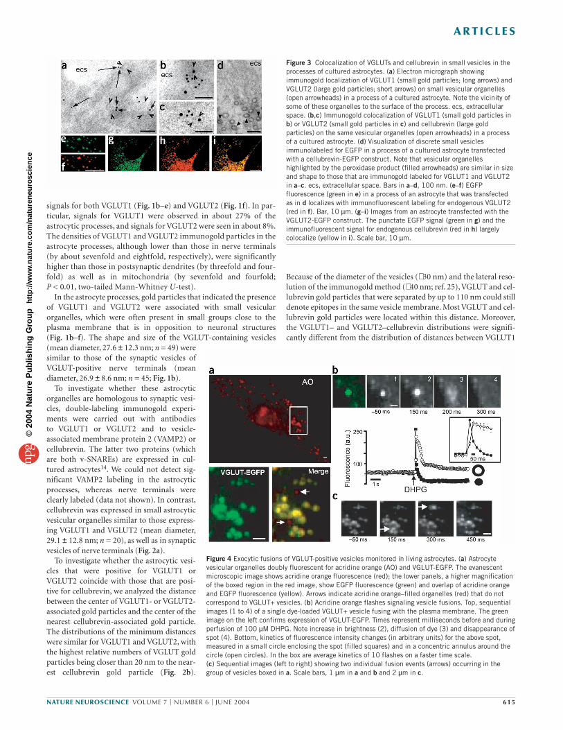
Figure 3 Colocalization of VGLUTs and cellubrevin in small vesicles in theprocesses of cultured astrocytes. (a) Electron micrograph showingimmunogold localization of VGLUT1 (small gold particles; long arrows) andVGLUT2 (large gold particles; short arrows) on small vesicular organelles(open arrowheads) in a process of a cultured astrocyte. Note the vicinity ofsome of these organelles to the surface of the process. ecs, extracellularspace. (b,c) Immunogold colocalization of VGLUT1 (small gold particles inb) or VGLUT2 (small gold particles in c) and cellubrevin (large goldparticles) on the same vesicular organelles (open arrowheads) in a processof a cultured astrocyte. (d) Visualization of discrete small vesiclesimmunolabeled for EGFP in a process of a cultured astrocyte transfectedwith a cellubrevin-EGFP construct. Note that vesicular organelleshighlighted by the peroxidase product (filled arrowheads) are similar in sizeand shape to those that are immunogold labeled for VGLUT1 and VGLUT2in a–c. ecs, extracellular space. Bars in a–d, 100 nm. (e–f) EGFPfluorescence (green in e) in a process of an astrocyte that was transfectedas in d localizes with immunofluorescent labeling for endogenous VGLUT2(red in f). Bar, 10 µm. (g–i) Images from an astrocyte transfected with theVGLUT2-EGFP construct. The punctate EGFP signal (green in g) and theimmunofluorescent signal for endogenous cellubrevin (red in h) largelycolocalize (yellow in i). Scale bar, 10 µm.
Figure 4 Exocytic fusions of VGLUT-positive vesicles monitored in living astrocytes. (a) Astrocytevesicular organelles doubly fluorescent for acridine orange (AO) and VGLUT-EGFP. The evanescentmicroscopic image shows acridine orange fluorescence (red); the lower panels, a higher magnificationof the boxed region in the red image, show EGFP fluorescence (green) and overlap of acridine orangeand EGFP fluorescence (yellow). Arrows indicate acridine orange–filled organelles (red) that do notcorrespond to VGLUT+ vesicles. (b) Acridine orange flashes signaling vesicle fusions. Top, sequentialimages (1 to 4) of a single dye-loaded VGLUT+ vesicle fusing with the plasma membrane. The greenimage on the left confirms expression of VGLUT-EGFP. Times represent milliseconds before and duringperfusion of 100 µM DHPG. Note increase in brightness (2), diffusion of dye (3) and disappearance ofspot (4). Bottom, kinetics of fluorescence intensity changes (in arbitrary units) for the above spot,measured in a small circle enclosing the spot (filled squares) and in a concentric annulus around thecircle (open circles). In the box are average kinetics of 10 flashes on a faster time scale. (c) Sequential images (left to right) showing two individual fusion events (arrows) occurring in thegroup of vesicles boxed in a. Scale bars, 1 µm in a and b and 2 µm in c.
©20
04 N
atur
e P
ublis
hing
Gro
up
http
://w
ww
.nat
ure.
com
/nat
uren
euro
scie
nce

or VGLUT2 immunogold particles and that of points randomly dis-tributed over the astrocyte (Fig. 2b). These data strongly support thelocalization of VGLUT1 and VGLUT2 in cellubrevin-containing vesi-cles (Fig. 2a).
To explore the possible functional relevance of astrocytic VGLUT-positive vesicles near neuronal terminals (Figs. 1b–d and 2a,c) ordendrites (Figs. 1e,f and 2c), we carried out double-labelingimmunogold experiments for VGLUT1 and NMDA (N-methyl-D-aspartate) receptors. We could find groups of astrocytic VGLUT-containing vesicles that were opposite extrasynaptic dendriticmembranes carrying NMDA receptors and were in close proximityto NMDA receptor–positive synapses (Fig. 2c). Astrocytic VGLUT-carrying vesicles facing presynaptically expressed NMDA receptorswere also observed.
Regulated exocytosis of astrocytic VGLUT-carrying vesiclesTo determine whether astrocytes release glutamate by regulated exo-cytosis of the VGLUT1-positive or VGLUT2-positive vesicularorganelles, we studied astrocytes in culture. We first verified whetherthe vesicular system expressing VGLUTs in cultured astrocytes hasproperties analogous to those of the astrocytes in the dentate gyrus in situ. We found that VGLUT1-positive and/or VGLUT2-positivevesicular organelles (i) were present throughout the cultured astro-cytes, including in the processes (Fig. 3a); (ii) had a diameter similarto vesicles in astrocytes in situ (Fig. 3a–c) and (iii) were immunoposi-tive for cellubrevin (Fig. 3b,c; and negative for VAMP2, despite thepresence of VAMP2 in the cultured cells), as indicated by a distribu-tion analysis that was done as for the astrocytes in situ. The vesicularorganelles could be distinctly visualized by transfecting astrocyteswith a cellubrevin-EGFP construct (in which the tag is in the intralu-minal portion of cellubrevin26) and by immunoperoxidase labeling ofthe astrocytes with antibodies to EGFP. This approach showed groups of discrete vesicles (Fig. 3d) that were similar in shape and size tothose immunolabeled for VGLUTs (Figs. 1b–f and 2a and 3a–c).Immunofluorescence microscopy analysis confirmed coexpression of
cellubrevin and VGLUTs (85 ± 7%, n = 222vesicles; Fig. 3e,f).
The dynamics of VGLUT-positive vesicleswere monitored in living astrocytes trans-fected with constructs of chimeric VGLUT1-EGFP or VGLUT2-EGFP fluorescent proteins.Either construct produced a punctate patternof EGFP fluorescence that was widespread in the astrocytic cell soma and processes (Fig. 3g). Cellubrevin largely localized withVGLUT-EGFP dots (91 ± 5%, n = 304 vesicles;Fig. 3h,i), indicating that the organelle popu-lation that was labeled by the tagged trans-porters essentially corresponds to thatcarrying the endogenous transporters.
To establish whether VGLUT-EGFP-posi-tive (VGLUT+) vesicles undergo regulatedexocytosis, vesicles were stained with acridineorange, a dye that accumulates in a self-quenched state inside acidic compart-ments18,19,27. When an acridine orange–filledorganelle fuses with the plasma membrane,the dye is released to the extracellularmedium with the production of a ‘flash’, alocalized and transient increase in fluores-cent light, followed by a lateral spread of flu-orescence (due to diffusion of the dye) andfinally by the disappearance of the labeledorganelle19,20. The dynamics of VGLUT+vesicles lying just beneath the plasma mem-
A R T I C L E S
616 VOLUME 7 | NUMBER 6 | JUNE 2004 NATURE NEUROSCIENCE
Figure 5 Properties of mGluR-evoked exocytosisof VGLUT+ vesicles. (a,b) DHPG (100 µM) elicitsan exocytic burst. In a, the cumulative numbersof acridine orange flashes (corresponding tofusion events) are plotted against time during a 3-min application of DHPG (n = 10). In b, thetime distribution of fusion events before, duringand after a 2-s pulse of DHPG are shown. Eachhistogram indicates the number of flashescounted in a 50-ms frame (n = 9). (c) Ca2+ andVAMP/cellubrevin dependency of DHPG-evokedvesicle fusions. Acridine orange flashes inresponse to 100 µM DHPG are reduced to control (Ctrl) levels if cells arepreincubated with either BAPTA-AM (50 µM, 30 min; n = 5) or TeNT (10µg/ml, 8 h; n = 5). TeNT-treated astrocytes respond to DHPG with [Ca2+]ielevations that are similar to those seen in untreated cells (data not shown).
Figure 6 Astrocyte vesicle fusions evoke glutamate-dependent [Ca2+]i elevations in INS-1 cells. (a) INS-1 cells as glutamate-sniffing cells. Fura-2-loaded INS-1 cells show [Ca2+]i elevation(R340/380) in response to glutamate (Glu, 100 µM; n = 25), which is inhibited by the NMDAreceptor antagonist AP5 (50 µM; n = 5). They show a similar increase in response to NMDA (50 µM; n = 4), but not to AMPA (50 µM; n = 7) or mGluR agonists t-ACPD or DHPG (both 100 µM; n = 6 and52, respectively). (b) On the left, an isolated cell pair formed by an astrocyte and an attached INS-1cell. Scale bar, 20 µm. On the right, acridine orange flashes (black histograms) in the astrocyte and[Ca2+]i changes (red trace) in the INS-1 cell were monitored in parallel (10 interlaced frames/s). Cellswere challenged with two applications of DHPG (50 µM, 2 s) separated by a 3-min washing period (inthe trace, 5 s of Wash). (c) [Ca2+]i elevation in the INS-1 cell is glutamate dependent. Experiment asin b but in the presence of the NMDA receptor antagonist AP5 (100 µM; n = 5). [Ca2+]i elevation inthe INS-1 cell is blocked, whereas acridine orange flashes in the astrocyte are not; the inhibitoryeffect of AP5 is reversible upon washout. (d) Blocking exocytosis with TeNT (10 µg/ml, 8 h; n = 4)abolishes both acridine orange flashes in the astrocyte and [Ca2+]i elevation in the INS-1 cell.
©20
04 N
atur
e P
ublis
hing
Gro
up
http
://w
ww
.nat
ure.
com
/nat
uren
euro
scie
nce

brane22 were followed in real time with TIRF imaging that was set todetect both acridine orange and EGFP fluorescence18. Typical TIRFimages are shown (Fig. 4a). Acridine orange–positive organelles,which were frequently arranged in small groups, generally localizedwith VGLUT+ dots (76.8 ± 14%, n = 40 cells; Fig. 4a). Under restingconditions, these double-positive organelles showed slow Brownianmotion (average displacement, 0.073 ± 0.011 µm/s; n = 20 cells). Toassess whether this population of doubly fluorescent vesicles under-goes regulated exocytosis, we challenged astrocytes with α-latrotoxin(LTx), a direct stimulant of exocytosis28; the Ca2+ ionophore iono-mycin (IONO) or dihydroxyphenylglicine (DHPG), an agonist ofgroup I metabotropic glutamate receptors (mGluRs), which is knownto evoke Ca2+-dependent glutamate release in astrocytes9,29. All threeagents induced acridine orange flashes, albeit with different efficacyand kinetics. (For results with LTx and IONO, see Supplementary Fig.1 and Supplementary Note online.) The time sequences of acridineorange fluorescence images (Fig. 4b,c) document fusion of individualVGLUT+ vesicles to the plasma membrane in response to DHPG. Byplotting fluorescence against time in a small circle around acridineorange spots and in a concentric annulus around the circle20, we con-sistently found that fluorescence initially increased in the circle, thenspread into the annulus and finally declined, which is in line withwhat is expected for an exocytic event20 (Fig. 4b). We counted thenumber of flashes per astrocyte (mean surface area, 1,165 ± 220 µm2)observed during 3-min-long applications of DHPG. With 10, 100 and1,000 µM DHPG, the number of flashes were 34 ± 5 (n = 4), 106 ± 12(n = 10) and 120 ± 18 (n = 6), respectively. In contrast, buffer induced10 ± 3 flashes/astrocyte (n = 6). Therefore, acridine orange flashes area specific response to DHPG. Two features of this phenomenon areworth noting. First, only a fraction of the VGLUT+ vesicles in theTIRF field underwent a flash in response to DHPG (34 ± 10% with1,000 µM DHPG). Second, the fusion events took place only at thevery beginning of DHPG perfusion (Fig. 5a).
To better define the kinetics of exocytosis, we next applied DHPGin a 2-s pulse by electrovalve-driven microperfusion while acquiringacridine orange images at 20 interlaced frames/s (Fig. 5b; n = 9).DHPG-evoked exocytosis occurred in a single burst: the fusion rate(flashes/s) was low in the 2 s preceding application of the agent (0.5 ±0.07) but abruptly increased by about 500-fold within 100–150 msafter the start of DHPG perfusion. The rate peaked in the next 50 ms(360 ± 22) and then declined to prestimulus values after a total of600 ms, remaining constant during the rest of the DHPG applicationand in the poststimulus period. Further insight into the mGluR-evoked exocytic process was obtained by examining vesicle fate inVGLUT-EGFP images. At 300 ms after undergoing an acridine orangeflash, vesicles monitored as EGFP spots showed two distinct fates:40 ± 11% had disappeared, whereas 60 ± 2% persisted in place, with-out any significant change of fluorescence intensity (n = 9 cells).
Finally, the acridine orange flashes in response to DHPG were abol-ished by (i) preloading the cells with BAPTA/AM, which buffers[Ca2+]i and prevents its increase after receptor activation (–88 ± 6%;Fig. 5c) or (ii) pre-exposing the cells to tetanus neurotoxin (TeNT),which cleaves both VAMP2 and cellubrevin, blocking vesicle fusion atthe level of SNARE complex formation30 (–95 ± 10%). The sametreatments also abolish mGluR-dependent glutamate release9,29,strongly indicating that flashes from VGLUT+ vesicles may representexocytic fusions of glutamate-containing vesicles.
Exocytosis is accompanied by glutamate releaseTo show directly that fusions of VGLUT-positive vesicles correspondto glutamate release events, we plated astrocytes together with ‘gluta-
mate-sniffing’ insulinoma-1 (INS-1) cells. Initial fura-2 experimentsshowed that, in the absence of astrocytes, INS-1 cells responded tolocal application of glutamate with rapid [Ca2+]i elevation, which wasinhibited by the NMDA receptor antagonist AP5 (Fig. 6a). Theseresponses were duplicated by NMDA but not by agents activatingother glutamate receptors, such as AMPA, t-ACPD and, notably,DHPG. Of 52 INS-1 cells challenged with DHPG, only 2 respondedwith rises in [Ca2+]i, and these were much slower and smaller thanthose triggered by glutamate or NMDA. Parallel experiments did notdetect release of glutamate9 from INS-1 cells stimulated with DHPG(100 µM for 2 s; n = 4).
We then used isolated cell pairs formed by an astrocyte with an INS-1 cell in direct lateral contact (Fig. 6b) and simultaneously monitoredchanges in acridine orange fluorescence from VGLUT+ vesicles (in theastrocyte) and fura-2 fluorescence changes (in the INS-1 cell) during 2-s pulses of DHPG, with intervening wash periods. In 17 cell pairs,DHPG always induced the same sequence of events: a burst of acridineorange flashes in the astrocyte followed by a [Ca2+]i elevation in theINS-1 cell (Fig. 6b). The two responses were correlated, with an averagedelay of 599 ± 171 ms between the flash peak and the calcium peak.Both returned to baseline during the wash and reappeared, with thesame sequential order and time interval, when DHPG was appliedagain. Moreover, if the second pulse of DHPG evoked fewer acridineorange flashes than the first pulse (Fig. 6b), the [Ca2+]i rise in the INS-1cell was smaller (n = 3 cell pairs). Therefore, the two responses must becorrelated and sustained by a defined sequence of signaling events. The[Ca2+]i elevation in the INS-1 cell is glutamate dependent, because inthe presence of the NMDA antagonist AP5, application of DHPGinduced almost no [Ca2+]i rise in the INS-1 cell (although it evoked theusual pattern of acridine orange flashes in the astrocyte; Fig. 6c). Uponwashout of AP5, however, the same INS-1 cell responded to DHPGwith a large [Ca2+]i rise that correlated with the acridine orange flashes(the Ca2+ response with AP5 was 22 ± 5% of that obtained after itswashout). When cells were pre-incubated with TeNT, DHPG elicitedneither acridine orange flashes in the astrocyte nor [Ca2+]i increase inthe INS-1 cell (Fig. 6d), although it enhanced [Ca2+]i in the astrocyte(∆R 340/380: 0.30 ± 0.04; n = 4). Taken together, these data confirm thatacridine orange flashes monitor exocytosis of glutamate-containingvesicles. Astrocytes do not acquire competence to exocytose glutamatebecause of the induced expression of VGLUT-EGFP. Thus, nontrans-fected cells also responded to DHPG stimulation with acridine orangeflashes accompanied by correlated Ca2+ responses in INS-1 cells (flashpeak to calcium peak = 680 ± 56 ms), and the Ca2+ responses wereblocked by AP5 (–75 ± 9%; n = 6 cell pairs).
DISCUSSIONProperties and distribution of the astrocytic microvesiclesThe small vesicles identified in hippocampal astrocytes share mor-phological and molecular properties with synaptic vesicles of gluta-matergic nerve terminals, notably their size (∼ 30 nm in diameter),rounded shape, clear appearance and expression of VGLUT and v-SNARE proteins. The astrocytic vesicles, however, expressed cellu-brevin rather than VAMP2, the main v-SNARE isoform of synapticvesicles. Whether this molecular difference imparts specific proper-ties to the fusion process in astrocytes remains to be established.Other aspects, particularly concerning distribution, distinguish astro-cytic vesicles from synaptic ones. Astrocytic vesicles often were insmaller and less-ordered groups than synaptic vesicles. In keepingwith this, lower levels of VGLUT1 and VGLUT2 were found in theastrocyte processes than were in the surrounding nerve terminals.This can explain why initial studies did not detect glial labeling31, as it
A R T I C L E S
NATURE NEUROSCIENCE VOLUME 7 | NUMBER 6 | JUNE 2004 617
©20
04 N
atur
e P
ublis
hing
Gro
up
http
://w
ww
.nat
ure.
com
/nat
uren
euro
scie
nce

is difficult to discern in the background of strong neuronal labeling.We succeeded by specifically marking the astrocytic processes and bycomparing VGLUT immunoreactivity in the processes with the levelof background labeling. The less-dense packing of the astrocyticVGLUT-containing vesicles as compared with synaptic vesicles couldbe another reason why the astrocytic vesicles were previously unrec-ognized on electron micrographs. This different arrangement mightreflect a different organization of the astrocytic vesicles in the cyto-plasm, for example, in their interactions with the cytoskeleton.
Single-cell PCR and immunolabeling results indicate that neitherVGLUT1 or 2 is present in all of the astrocytes in the dentate gyrus,and both provide a roughly similar estimate of the number of positivecells (25–40%). It is therefore possible that VGLUT-positive vesiclesare present only in subpopulations of astrocytes, which would iden-tify a new type of heterogeneity of these cells32. Alternatively, astro-cytes that are negative for VGLUT1 and 2 could express the VGLUT3isoform33, which we did not study. Diversity is found also at the ultra-structural level. VGLUT-carrying vesicles were not observed in all ofthe astrocytic processes, and they were not uniformly present in theprocesses that were positive, which indicates that they may be locatedat distinct sites. We often found these VGLUT-carrying vesicles justbeneath the astrocytic plasma membrane that was in opposition toneuronal structures, either nerve terminals or dendrites, thatexpressed glutamate receptors (notably of the NMDA type); it is ofnote that the distance between the astrocytic membranes and theneuronal structures was similar to that at synapses (Fig. 2c). Suchgroups of VGLUT-containing vesicles could, therefore, represent sitesfor point-to-point transmission from astrocytes to neurons.
Properties of glutamate exocytosis from astrocytesExocytosis of VGLUT-expressing vesicles was studied in culturedastrocytes, where we identified a vesicle population that is struc-turally and biochemically homologous to VGLUT-expressing vesiclesof tissue astrocytes. TIRF microscopy in combination with two fluo-rophores, acridine orange and VGLUT-EGFP, enabled us to monitorexocytosis of individual glutamate-containing vesicles directly and tocollect information on the secretory process evoked by activation ofgroup I mGluRs. This occurred as an exocytic burst involving aboutone-third of the VGLUT-positive vesicles present in the TIRF field,most likely the readily releasable pool34. The burst lasted 500–600 ms, but most fusions occurred within the first 200 ms. Thesekinetics provide a temporal frame to the stimulus–secretion cou-pling mechanism of astrocytes, which seems to be a most rapidprocess—perhaps the most rapid process—activated by G proteinreceptor–dependent Ca2+ release from internal stores35. Rapid fusionoccurs in pituitary gonadotrophs stimulated with eithergonadotropin-releasing hormone or inositol trisphosphate, its intra-cellular messenger36. The rapid coupling was ascribed to a localrelease of Ca2+ from internal stores near the exocytic sites36. Such alocal control could be operating also in the astrocytes, because as ingonadotrophs36, the Ca2+-dependent exocytic burst largely precedesthe cell-averaged [Ca2+]i elevation (the latter peaks 1.4 s after thestart of DHPG application; data not shown). Also, tubular structuresresembling smooth endoplasmic reticulum are present in the thinprocesses of tissue astrocytes. For example, the structure identified inFigure 1e is just 50–150 nm away from both VGLUT1-positive vesi-cles and the plasmalemma. Whether glutamate secretion is indeedcontrolled by localized Ca2+ elevations near the plasma membranethat are dependent on smooth endoplasmic reticulum remains to beestablished by future studies that will require techniques with highspatial and temporal resolution37.
Dual labeling of the vesicles with a marker of the lumen (acridineorange) and a marker of the membrane (VGLUT-EGFP) providedinformation on the modalities of vesicle fusion. Whereas acridineorange images marked the moment of fusion for a given vesicle, EGFPimages taken soon after the fusion event signaled changes in the vesi-cle shape as a result of fusion. Heterogeneous fusion modes38 wereobserved, as some EGFP spots rapidly disappeared after exocytosis,but others persisted, unchanged in shape and fluorescence intensity.Several explanations exist for loss of EGFP fluorescence18, but mostlikely it signals a complete fusion of the vesicle and plasma mem-branes. In contrast, persistence of the postexocytic vesicles indicates akiss-and-run type of fusion, with release of the vesicle contentthrough a transiently opened fusion pore39. Kiss-and-run fusion isassociated with a rapid recycling pathway that renders the vesiclesreadily reusable for exocytosis39. This could be of importance forrapid cross-talk between neurons and glia.
Significance of glutamate exocytosis from astrocytesOur study provides an initial framework for understanding the signif-icance of glutamate exocytosis from astrocytes in the context of signalexchanges between glia and neurons.
In terms of kinetics, the sharp exocytic burst triggered by mGluRstimulation, although not as rapid as the process in synapses, is in thetime scale of the most rapid responses of specialized secretory cells40.Obviously this newly identified property of astrocytes in cultureawaits confirmation from studies in the intact tissue. The close local-ization of astrocytic VGLUT-containing vesicles with neuronalNMDA receptors in the hippocampus indicates that astrocytes in situmay focally secrete glutamate onto neurons. Coupled to the evidenceof focal glutamate secretion from neurons to glia41, this observationhighlights an emerging specificity of neuron–glia communication.According to theoretical calculations, a single hippocampal astrocytecould oversee more than 100,000 synapses42. On the other hand, itscontacts could be highly compartmentalized. For example, in thecerebellum, a vast number of Bergmann glial processes enwrap smallgroups of Purkinje cell synapses and respond independently to paral-lel fiber stimulation43. Our observation that VGLUT-carrying vesiclesare distributed unevenly in the processes of hippocampal astrocytessupports the view that astrocytes exchange independent messages atdifferent subcellular locations. Focal glutamate signals from astro-cytes could serve to orchestrate subsets of synapses in functionalmicrodomains.
In conclusion, the presented evidence that many astrocytes containa vesicular compartment for regulated glutamate secretion stronglyindicates that this may be a candidate mechanism for informationprocessing by glia in concert with neurons and provides a concretebasis for redefinition of the roles these cells play in brain function.
METHODSElectrophysiology and single-cell RT-PCR. Hippocampal slices were preparedfrom postnatal day (P) 35–70 rats, and cells in the outer molecular layer of thedentate gyrus were selected for whole-cell recordings as reported23. Use of ani-mals and experimental procedures were approved by the Office VeterinaireCantonal (Lausanne), in accordance with the Swiss Federal Laws. After electro-physiological identification, cells were isolated by lifting them out of the tissuewhile harvesting the cytoplasm under microscopic control24. Only individualcells without any adhered tissue debris were selected for RT-PCR analysis.Single-strand cDNA synthesis and multiplex two-round single-cell PCR was asdescribed32, with primers for VGLUT1, VGLUT2 and S100β (for sequences,see Supplementary Table 1). Primer pairs were located on different exons toprevent amplification of genomic DNA. Omission of reverse transcriptase andaspiration of bath solution served as negative controls.
A R T I C L E S
618 VOLUME 7 | NUMBER 6 | JUNE 2004 NATURE NEUROSCIENCE
©20
04 N
atur
e P
ublis
hing
Gro
up
http
://w
ww
.nat
ure.
com
/nat
uren
euro
scie
nce

Antibodies. Rabbit VGLUT1 and VGLUT2 antibodies to peptide sequencescontaining the carboxy-terminal 68 (VGLUT1) and 64 amino acids (VGLUT2;both gifts from R. Edwards, University of California at San Francisco School ofMedicine), which are highly specific31,44, were diluted 1:100 and 1:2,000(respectively, for immunogold labeling) and 1:1,000 (for both, for immunoflu-orescence). A mixture of rabbit antibodies to GLT and GLAST25 (both giftsfrom N.C. Danbolt, Anatomical Institute, University of Oslo) was used at afinal dilution of 1 µg/ml. Rabbit antibodies to cellubrevin45 (gift from P. DeCamilli, Yale University School of Medicine) were diluted 1:1,500(immunoflourescence) and 1:100 (immunogold). A mixture of antibodies tothe NMDA receptor subunits 1 and 2A/B (final dilution 1:50) and a mono-clonal antibody to GFAP (diluted 1:1,000) were from Chemicon. A mono-clonal antibody to VAMP2 (Synaptic Systems) was diluted 1:500(immunofluorescence) and 1:100 (immunogold). Antibodies to EGFP (BDBiosciences) were diluted 1:100.
Immunocytochemistry. Immunogold cytochemistry was carried out asdescribed24,46 using hippocampal specimens from adult Wistar rats fixed byperfusion through the heart (4% formaldehyde and 0.1% glutaraldehyde) andrat primary astrocyte cultures (see below), with the same fixative. These tissueswere embedded at low temperature in Lowicryl HM20 or K4M. Ultrathin sec-tions were treated with the antibodies and were viewed in a Philips CM 100electron microscope. Electron micrographs were taken in the superficial gran-ule cell layer and in the molecular layer of the dentate gyrus. Astrocyticprocesses were identified by the presence of filaments and/or by labeling forGLT/GLAST25. The densities of gold particles signaling the presence ofVGLUT1 and VGLUT2 were quantified as before46. Only VGLUT1 or 2 signalsthat were twice the amount of background labeling seen over mitochondriawere included in the quantifications. The spatial relationship between gold par-ticles signaling VGLUT1 or VGLUT2 (10 nm) and cellubrevin or VAMP2 (15 nm) were determined by digitizing the positions of their centers with theprogram MicroTrace47 and then calculating the intercenter distances betweenthe 10- and 15-nm gold particles with custom software (mitochondria andareas apparently lacking electron-dense material were excluded). The distanceswere sorted into bins of 20 nm. The distributions were compared with those ofrandom points to gold particle centers47. With the exception of the excludedregions described above, 1,000 random points were spread over the astrocyte.
Pre-embedding electron microscopy was done on cultured astrocytes thatwere transfected with a construct to express cellubrevin-EGFP26 (gift from T.Coppola and R. Regazzi, Department of Cell Biology and Morphology,University of Lausanne), fixed as above and peroxidase-labeled according to abiotin–streptavidin method46 before embedding in Durcupan ACM.
Immunofluorescence was carried out as described11 on cultured astrocytestransfected with either cellubrevin-EGFP or VGLUT2-EGFP constructs or onintact hippocampal tissue. Primary antibodies were visualized with Cy3-con-jugated secondary antibodies (Jackson ImmunoResearch Laboratories) andviewed on a Zeiss Axioplan 2 microscope. Colocalization analysis in culturedcells was done on fluorescent spots representing individual vesicles. The aver-age diameter of a single vesicle was calculated indirectly from TIRF imageswhere a single flash represented a single fusion event (Fig. 3). Only spots over-lapping by ≥66% were considered to be colocalized.
Preparation and transfection of VGLUT-EGFP. To fuse rat genes encodingVGLUT1 and VGLUT2 in pcDNA3 mammalian expression plasmids (gifts ofR. Edwards) with a vector containing the EGFP coding sequence, restrictionsites EcoRI and XhoI were generated by PCR. The PCR products (1,698 basepairs for VGLUT1 and 1,764 bp for VGLUT2) were purified (Microcon-PCR,Millipore) and subcloned into the EGFP vector (Clontech) at the EcoRI–XhoIsite under the control of the CMV promoter. Restriction analysis, sequencing(Microsynth) and western blots indicated the successful construction of thefusion proteins. Either VGLUT1-EGFP or VGLUT2-EGFP plasmid (1 µg) wastransfected into primary cultures of rat cortical astrocytes with Fugene6 trans-fection reagent (Roche). In most experiments, we used VGLUT2-EGFPbecause of its higher transfection yield.
Astrocyte and INS-1 cell cultures for imaging experiments. Cultures ofpure astrocytes (>99% GFAP positive), prepared as described11, were plated
(2.5 × 10⁴ cells) on glass coverslips and used 2–3 d after transfection withthe VGLUT-EGFP construct. INS-1 insulinoma cells were plated eitheralone (1.5 × 10⁵ cells) or on the transfected astrocytes and used the next day.For acridine orange imaging experiments, cells were stained with 5 µM acri-dine orange (Molecular Probes) for 15 min at 34 °C in the dark. For single-cell [Ca2+]i measurements, cells were loaded with 5 µM fura-2 AM and0.02% pluronic acid (both from Molecular Probes) for 30 min. For com-bined acridine orange and fura-2 imaging, fura-2 loading was followed by15 min of staining with acridine orange. Coverslips were mounted in anopen perfusion micro-incubator at 37 °C (PDMI-2, Harvard Apparatus).Stimuli (DHPG, glutamate, NMDA, AMPA and t-ACPD from TocrisCookson Ltd.; ionomycin and α-latrotoxin from Fluka) were applied ontocells by a local microperfusion system driven by electrovalves (WarnerInstrument Corporation).
Optical imaging. A Zeiss Axiovert 200 inverted fluorescence microscope wasmodified to allow both epifluorescence and evanescent field (EF) illumination(Visitron Systems). For EF illumination, the expanded beam (488-nm argonion laser, 40 mW; Laserphysics) was passed through a shutter driver (Uniblitz,VMM-D1 model, Vincent Associates) that was synchronized with a SNAP-HQCCD camera (Roper Scientific) under Metafluor software (Universal Imaging)control and introduced from the high numerical aperture objective lens (Zeissα-plan FLUAR 100×, 1.45 n.a.). Light entered the coverslip at an angle of72.3–73.8° and underwent total internal reflection at the glass–cell interface.The refractive indices for glass (n = 1.52 at 488 nm) and cells (n = 1.38) predictan EF declining e-fold within 84 nm from the interface.
We imaged EGFP and acridine orange fluorescence simultaneously underEF illumination as described18. The images, projected side by side, wereacquired at either 10 or 20 interlaced frames/s. To image both fura-2 and acri-dine orange fluorescence, we combined EF (488 nm laser) and epifluorescenceillumination (340/380-nm xenon arc lamp, Visichrome polychromator sys-tem, Visitron). Lights were filtered with a beam splitter (Zeiss filter set 25).Images were further separated using a filter wheel optical system (Lambda-10A, Sutter) equipped with two Zeiss emission filters (bandpass 515–565 nmfor acridine orange and bandpass 505–530 nm for fura-2). They were thenacquired at 10 interlaced frames/s and projected side by side.
Image analysis. Video images, digitized with MetaFluor, were analyzed withMetaMorph software (Universal Imaging). Movements of VGLUT-positiveacridine orange spots in unstimulated astrocytes were measured by position-ing a circle (diameter, 895 nm) on the spot at the beginning of the experiment,taking the distance from the center of this circle at each frame and averaging itper second. Acridine orange flashes were measured (in red images) as fluores-cence intensity changes (arbitrary units, a.u.) in a circle as above, and in a con-centric annulus (895-nm inner and 1,195-nm outer diameter)20. Only whenfluorescence increased, spread and then declined, was the event counted as aflash20. The fate of VGLUT-EGFP spots (in green images) was analyzed bypositioning a circle as above on each spot and by comparing fluorescenceintensity just before stimulating the cell (with DHPG) and 300 ms after anacridine orange flash was observed in the red images. Changes in fura-2 fluo-rescence were measured with the 340/380-nm excitation wavelength ratiomethod (R340/380) in a circle centered on the cell soma.
Note: Supplementary information is available on the Nature Neuroscience website.
ACKNOWLEDGMENTSWe thank H. Stubbe, Y. Gomez, K. Hüttmann and Centre de MicroscopieElectronique, University of Lausanne for experimental support; J.-Y. Chatton, T.Coppola, P. Jourdain, G. Knott, T. Lang and R. Stoop for scientific discussions;R. Jahn, J. Storm-Mathisen for insights at various stages of this work and P. Clarke,J. Meldolesi, R. Regazzi and J. Storm-Mathisen for comments on the manuscript.This work was supported by grants OFES 00.0553 and FNRS 3100A0-100850/1 toA.V. and by Deutsche Forschungsgemeinschaft (SFB-TR3) and Fonds derChemischen Industrie to C.S. V.G. is a visiting fellow within the EuropeanCommunity grant QLG3-CT-2001–2004 and recipient of a fellowship from theNorwegian Research Council.
COMPETING INTERESTS STATEMENTThe authors declare that they have no competing financial interests.
A R T I C L E S
NATURE NEUROSCIENCE VOLUME 7 | NUMBER 6 | JUNE 2004 619
©20
04 N
atur
e P
ublis
hing
Gro
up
http
://w
ww
.nat
ure.
com
/nat
uren
euro
scie
nce

A R T I C L E S
620 VOLUME 7 | NUMBER 6 | JUNE 2004 NATURE NEUROSCIENCE
Received 5 February; accepted 5 April 2004Published online at http://www.nature.com/natureneuroscience/
1. Volterra, A. & Bezzi, P. Release of transmitters from glial cells. in The TripartiteSynapse: Glia in Synaptic Transmission (eds. Volterra, A., Magistretti, P.J. &Haydon, P.G.) 164–182 (Oxford Univ. Press, Oxford, UK, 2002).
2. Parri, R.H., Gould, T.M. & Crunelli, V. Spontaneous astrocytic Ca2+ oscillations in situdrive NMDAR-mediated neuronal excitation. Nat. Neurosci. 4, 803–812 (2001).
3. Pasti, L., Volterra, A., Pozzan, T. & Carmignoto, G. Intracellular calcium oscillationsin astrocytes: a highly plastic, bidirectional form of communication between neuronsand astrocytes in situ. J. Neurosci. 17, 7817–7830 (1997).
4. Kang, J., Jiang, L., Goldman, S.A. & Nedergaard, M. Astrocyte-mediated potentia-tion of inhibitory synaptic transmission. Nat. Neurosci. 1, 683–692 (1998).
5. Zhang, J.M. et al. ATP released by astrocytes mediates glutamatergic activity-dependent heterosynaptic suppression. Neuron 40, 971–982 (2003).
6. Newman, E.A. Propagation of intercellular calcium waves in retinal astrocytes andMüller cells. J. Neurosci. 21, 2215–2223 (2001).
7. Schipke, C.G., Boucsein, C., Ohlemeyer, C., Kirchhoff, F. & Kettenmann, H.Astrocyte Ca2+ waves trigger responses in microglial cells in brain slices. FASEB J.16, 255–257 (2002).
8. Parpura, V. et al. Glutamate-mediated astrocyte-neuron signalling. Nature 369,744–747 (1994).
9. Bezzi, P. et al. Prostaglandins stimulate calcium-dependent glutamate release inastrocytes. Nature 391, 281–285 (1998).
10. Newman, E.A. New roles for astrocytes: regulation of synaptic transmission. TrendsNeurosci. 26, 536–542 (2003).
11. Bezzi, P., et al. CXCR4-activated astrocyte glutamate release via TNFα: amplifica-tion by microglia triggers neurotoxicity. Nat. Neurosci. 4, 702–710 (2001).
12. Araque, A., Li, N., Doyle, R.T. & Haydon, P.G. SNARE protein-dependent glutamaterelease from astrocytes. J. Neurosci. 20, 666–673 (2000).
13. Pasti, L., Zonta, M., Pozzan, T., Vicini, S. & Carmignoto G. Cytosolic calcium oscil-lations in astrocytes may regulate exocytotic release of glutamate. J. Neurosci. 21,477–484 (2001).
14. Volterra, A. & Meldolesi, J. Quantal release of transmitter: not only from neurons butfrom astrocytes as well? in Neuroglia, Edn. 2 (eds. Kettenmann, H. & Ransom, B.)190–201 (Oxford Univ. Press, New York, 2004).
15. Nedergaard, M., Takano, T. & Hansen, A.J. Beyond the role of glutamate as a neuro-transmitter. Nat. Rev. Neurosci. 3, 748–754 (2002).
16. Fremeau, R.T. Jr., Voglmaier, S., Seal, R & Edwards, R.H. VGLUTs define subsets ofexcitatory neurons and suggest novel roles for glutamate. Trends Neurosci. 27,98–103 (2004).
17. Jahn, R., Lang, T. & Südhof, T.C. Membrane fusion. Cell 112, 519–533 (2003).18. Tsuboi, T., Zhao, C., Terakawa, S. & Rutter, G.A. Simultaneous evanescent wave
imaging of insulin vesicle membrane and cargo during a single exocytotic event.Current Biol. 10, 1307–1310 (2000).
19. Avery, J. et al. A cell-free system for regulated exocytosis in PC12 cells. J. Cell. Biol.148, 317–324 (2000).
20. Zenisek, D., Steyer, J.A. & Almers, W. Transport, capture and exocytosis of singlesynaptic vesicles at active zones. Nature 406, 849–854 (2000).
21. Stout, A.L. & Axelrod, D. Evanescent field excitation of fluorescence by epiillumina-tion microscopy. Appl. Optics 28, 5237–5242 (1989).
22. Steyer, J.A. & Almers, W. A real-time view of life within 100 nm of the plasma mem-brane. Nat. Rev. Mol. Cell. Biol. 2, 268–275 (2001).
23. Schröder, W. et al. Lesion-induced changes of electrophysiological properties inastrocytes of the rat dentate gyrus. Glia 28,166–174 (1999).
24. Seifert, G., Becker, A. & Steinhäuser, C. Combining patch-clamp techniques withRT-PCR in Neuromethods: Patch-clamp Analysis: Advanced Techniques Vol 35 (eds.
Walz, W., Boulton, A.A. & Baker, G.B.) 301–330 (Humana Press, Totowa, NJ,2002).
25. Chaudhry, F.A. et al. Glutamate transporters in glial plasma membranes: highly dif-ferentiated localizations revealed by quantitative ultrastructural immunocytochem-istry. Neuron 15, 711–720 (1995).
26. Randhawa, V.K. et al. VAMP2, but not VAMP3/cellubrevin, mediates insulin-dependent incorporation of GLUT4 into the plasma membrane of L6 myoblasts. Mol.Biol. Cell. 11, 2403–2417 (2000).
27. Steyer, J.A., Horstmann, H. & Almers, W. Transport, docking and exocytosis of singlesecretory granules in live chromaffin cells. Nature 388, 474–478 (1997).
28. Rosenthal, L. & Meldolesi, J. Alpha-latrotoxin and related toxins. Pharmacol. Ther.42, 115–134 (1989).
29. Muyderman, H. et al. α1-adrenergic modulation of metabotropic glutamate-receptorinduced calcium oscillations and glutamate release in astrocytes. J. Biol. Chem276, 46504–46514 (2001).
30. Humeau, Y., Doussau, F., Grant, N.J. & Poulain, B. How botulinum and tetanus neu-rotoxins block neurotransmitter release. Biochimie 82, 427–446 (2000).
31. Fremeau, R.T. Jr. et al. The expression of vesicular glutamate transporters definestwo classes of excitatory synapse. Neuron 31, 247–260 (2001).
32. Matthias, K. et al. Segregated expression of AMPA-type glutamate receptors and glu-tamate transporters defines distinct astrocyte populations in the mouse hippocam-pus. J. Neurosci. 23, 1750–1758 (2003).
33. Fremeau, R.T. Jr. et al. The identification of vesicular glutamate transporter 3 sug-gests novel modes of signaling by glutamate. Proc. Natl. Acad. Sci. USA 99,14488–14493 (2002).
34. Xu, T., Binz, T., Niemann, H. & Neher, E. Multiple kinetic components of exocytosisdistinguished by neurotoxin sensitivity. Nat. Neurosci. 1, 192–200 (1998).
35. Tse, F.W. & Tse, A. Regulation of exocytosis via release of Ca2+ from intracellularstores. Bioessays 21, 861–865 (1999).
36. Tse, F.W., Tse, A., Hille, B., Horstmann, H. & Almers, W. Local Ca2+ release frominternal stores controls exocytosis in pituitary gonadotrophs. Neuron 18, 121–132(1997).
37. Becherer, U., Moser, T., Stümer, W. & Oheim, M. Calcium regulates exocytosis at thelevel of single vesicles. Nat. Neurosci. 8, 846–853 (2003).
38. Alés, E. et al. High calcium concentrations shift the mode of exocytosis to the kiss-and-run mechanism. Nat. Cell. Biol. 1, 40–44 (1999).
39. Valtorta, F., Meldolesi, J. & Fesce, R. Synaptic vesicles: is kissing a matter of com-petence? Trends Cell. Biol. 11, 324–328 (2001).
40. Kasai, H. Comparative biology of Ca2+-dependent exocytosis: implications of kineticdiversity for secretory function. Trends Neurosci. 22, 88–93 (1999).
41. Matsui, K. & Jahr, C.E. Ectopic release of synaptic vesicles. Neuron 40, 1173–1183(2003).
42. Bushong, E.A., Martone, M.E., Jones, Y.Z. & Ellisman, M.H. Protoplasmic astrocytesin CA1 stratum radiatum occupy separate anatomical domains. J. Neurosci. 22,183–192 (2002).
43. Grosche, J. et al. Microdomains for neuron-glia interaction: parallel fiber signallingto Bergmann glial cells. Nat. Neurosci. 2, 139–143 (1999).
44. Bellocchio, E.E. et al. The localization of the brain-specific inorganic phosphatetransporter suggests a specific presynaptic role in glutamatergic transmission. J. Neurosci. 18, 8648–8659 (1998).
45. Chilcote, T.J. et al. Cellubrevin and synaptobrevins: similar subcellular localizationand biochemical properties in PC12 cells. J. Cell. Biol. 129, 219–231 (1995).
46. Gundersen, V. et al. Synaptic vesicular localization and exocytosis of L-aspartate inexcitatory nerve terminals: a quantitative immunogold analysis in rat hippocampus.J. Neurosci. 18, 6059–6070 (1998).
47. Bergersen, L., Ruiz, A., Bjaalie, J.G., Kullmann, D.M. & Gundersen, V. GABA andGABAA receptors at hippocampal mossy fibre synapses. Eur. J. Neurosci. 18,931–941 (2003).
©20
04 N
atur
e P
ublis
hing
Gro
up
http
://w
ww
.nat
ure.
com
/nat
uren
euro
scie
nce
Copyright © 2022 FDOKUMEN