The ins and outs of IgE-dependent mast-cell exocytosis
8
The ins and outs of IgE-dependent mast-cell exocytosis Ulrich Blank 1 and Juan Rivera 2 1 INSERM E 0225, Bichat Medical School, 16 rue Henri Huchard, BP 416, 75870 Cedex 18, France 2 Molecular Inflammation Section, Molecular Immunology and Inflammation Branch, National Institute of Arthritis and Musculoskeletal and Skin Diseases, National Institutes of Health, Building 10, Room 9N228, Bethesda, MD 20892-1820, USA Mast cells (MCs) are able to secrete the contents of preformed cytoplasmic secretory granules (SGs) on encountering certain stimulants. For MCs, this process is fundamental to their role in innate and acquired immunity. Immunological adaptation through the pro- duction of IgE antibodies, to normally innocuous sub- stances, has kidnapped the MC exocytotic response to cause allergic disease. Thus, understanding the molecu- lar events in IgE-dependent MC exocytosis holds prom- ise for therapeutic intervention. Recent advances, in deciphering the coupling of the high affinity IgE recep- tor (Fc1RI) to the secretory machinery, promote a new paradigm of the molecular coordination of MC exocyto- sis. A clearer picture of the link between Fc1RI stimu- lation and SG exocytosis is emerging. Mast cells (MCs) can mediate adaptive immune responses through the high affinity IgE receptor (Fc1RI) (reviewed in Ref. [1]). They store histamine and serotonin in preformed cytoplasmic secretory granules (SGs) as well as a variety of proteases that become active only on their release. Unlike many other secretory cells, they can release substantial amounts of vesicular content in a single stimulatory event through a process in which vesicle membranes fuse with the plasma membrane (PM) and with each other to form channels (compound exocytosis), which ensures a maximal biological effect. This massive exocytosis causes many of the symptoms associated with acute and chronic allergic disease. Chronic allergic disease is also coupled to secre- tion of SG-stored cytokines [e.g. tumor necrosis factor (TNF)] and to the MCs ability to generate a so-called late phase reaction, by producing prostaglandins, leukotrienes and inflammatory and immunoregulatory cytokines (reviewed in Ref. [2]). The function of MCs in immediate hypersensitivity is evolutionary conserved from amphibians and birds [3,4]. Similarly, MC exocytosis relies on the basic machinery of membrane trafficking conserved from yeast to humans [5], although differences in the regulation and execution of the process are apparent (Table 1). Although various triggers can elicit the MC exocytotic response [6], new information on how the IgE-triggered process is driven has emerged. Here, we attempt to integrate this information towards a more cohesive view of the requirements in IgE-dependent MC exocytosis. Regulation of SG exocytosis by Fc1RI The Fc1RI is a multisubunit receptor with a ligand- binding a subunit, a signal-amplifying membrane-tetra- spanning b subunit and a homodimeric disulfide-linked g subunit that provides the signaling ability of this recep- tor (reviewed in Ref. [7])(Figure 1). Initiation of signaling is mediated through the immunoreceptor tyrosine-based activation motif (ITAM) encoded in the cytoplasmic tails of the b and g subunits. Monomeric IgE binding to Fc1RI, in the absence of antigen, increases expression of the receptor and causes the generation of survival signals (reviewed in Ref. [8])(Figure 2). These signals result from autologous or cross-reactive aggregation of IgE once bound to MCs [9]. However, at the minimal concentration sufficient to occupy all receptors on the cell surface, monomeric IgE fails to elicit considerable MC exocytosis. By contrast, antigen-driven aggregation of IgE anti- body-occupied Fc1RI elicits intracellular signals that Table 1. Comparison of exocytosis in MCs a and neurons Parameter MC Neuron Granule type Secretory lysosome Specialized secretory granule Granule size 300–1000 nm 50 nm Granule number #1000 per cell 200–500 per synaptic terminal Calcium requirement ,1 mM . 20 mM PKC requirement PKCb and/or PKCd No Kinetics Minutes Milliseconds Recycling (granule) Long (hours to days) Very short (minutes) Release (type) One single release of #100% (compound or multigranular mode) Multiple release, one granule per fusion event a Abbreviations: MC, mast cell; PKC, protein kinase C. Corresponding author: Juan Rivera ( [email protected]). Review TRENDS in Immunology Vol.25 No.5 May 2004 www.sciencedirect.com 1471-4906/$ - see front matter. Published by Elsevier Ltd. doi:10.1016/j.it.2004.03.005
-
Upload
independent -
Category
Documents
-
view
1 -
download
0
Transcript of The ins and outs of IgE-dependent mast-cell exocytosis

The ins and outs of IgE-dependentmast-cell exocytosisUlrich Blank1 and Juan Rivera2
1INSERM E 0225, Bichat Medical School, 16 rue Henri Huchard, BP 416, 75870 Cedex 18, France2Molecular Inflammation Section, Molecular Immunology and Inflammation Branch, National Institute of Arthritis and
Musculoskeletal and Skin Diseases, National Institutes of Health, Building 10, Room 9N228, Bethesda, MD 20892-1820, USA
Mast cells (MCs) are able to secrete the contents of
preformed cytoplasmic secretory granules (SGs) on
encountering certain stimulants. For MCs, this process
is fundamental to their role in innate and acquired
immunity. Immunological adaptation through the pro-
duction of IgE antibodies, to normally innocuous sub-
stances, has kidnapped the MC exocytotic response to
cause allergic disease. Thus, understanding the molecu-
lar events in IgE-dependent MC exocytosis holds prom-
ise for therapeutic intervention. Recent advances, in
deciphering the coupling of the high affinity IgE recep-
tor (Fc1RI) to the secretory machinery, promote a new
paradigm of the molecular coordination of MC exocyto-
sis. A clearer picture of the link between Fc1RI stimu-
lation and SG exocytosis is emerging.
Mast cells (MCs) can mediate adaptive immune responsesthrough the high affinity IgE receptor (Fc1RI) (reviewed inRef. [1]). They store histamine and serotonin in preformedcytoplasmic secretory granules (SGs) as well as a variety ofproteases that become active only on their release. Unlikemany other secretory cells, they can release substantialamounts of vesicular content in a single stimulatory eventthrough a process in which vesicle membranes fuse withthe plasma membrane (PM) and with each other to formchannels (compound exocytosis), which ensures a maximalbiological effect. This massive exocytosis causes many ofthe symptoms associated with acute and chronic allergicdisease. Chronic allergic disease is also coupled to secre-tion of SG-stored cytokines [e.g. tumor necrosis factor(TNF)] and to the MCs ability to generate a so-called latephase reaction, by producing prostaglandins, leukotrienes
and inflammatory and immunoregulatory cytokines(reviewed in Ref. [2]).
The function of MCs in immediate hypersensitivity isevolutionary conserved from amphibians and birds [3,4].Similarly, MC exocytosis relies on the basic machinery ofmembrane trafficking conserved from yeast to humans [5],although differences in the regulation and execution of theprocess are apparent (Table 1). Although various triggerscan elicit the MC exocytotic response [6], new informationon how the IgE-triggered process is driven has emerged.Here, we attempt to integrate this information towards amore cohesive view of the requirements in IgE-dependentMC exocytosis.
Regulation of SG exocytosis by Fc1RI
The Fc1RI is a multisubunit receptor with a ligand-binding a subunit, a signal-amplifying membrane-tetra-spanning b subunit and a homodimeric disulfide-linkedg subunit that provides the signaling ability of this recep-tor (reviewed in Ref. [7]) (Figure 1). Initiation of signalingis mediated through the immunoreceptor tyrosine-basedactivation motif (ITAM) encoded in the cytoplasmic tails ofthe b and g subunits. Monomeric IgE binding to Fc1RI, inthe absence of antigen, increases expression of the receptorand causes the generation of survival signals (reviewed inRef. [8]) (Figure 2). These signals result from autologous orcross-reactive aggregation of IgE once bound to MCs [9].However, at the minimal concentration sufficient to occupyall receptors on the cell surface, monomeric IgE fails toelicit considerable MC exocytosis.
By contrast, antigen-driven aggregation of IgE anti-body-occupied Fc1RI elicits intracellular signals that
Table 1. Comparison of exocytosis in MCsa and neurons
Parameter MC Neuron
Granule type Secretory lysosome Specialized secretory granule
Granule size 300–1000 nm 50 nm
Granule number #1000 per cell 200–500 per synaptic terminal
Calcium requirement ,1 mM . 20 mM
PKC requirement PKCb and/or PKCd No
Kinetics Minutes Milliseconds
Recycling (granule) Long (hours to days) Very short (minutes)
Release (type) One single release of #100% (compound or
multigranular mode)
Multiple release, one granule per fusion event
aAbbreviations: MC, mast cell; PKC, protein kinase C.
Corresponding author: Juan Rivera ( [email protected]).
Review TRENDS in Immunology Vol.25 No.5 May 2004
www.sciencedirect.com 1471-4906/$ - see front matter. Published by Elsevier Ltd. doi:10.1016/j.it.2004.03.005

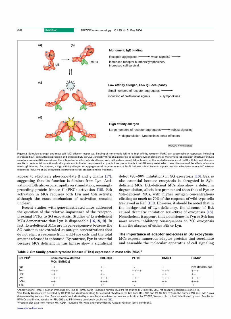
result in MC exocytosis. Signals generated by Fc1RIaggregation are subject to kinetic proofreading [10].Aggregation must persist for sufficient time to enablethe execution of the molecular events required for aproductive response (Figure 2). Because of its persistentinteraction with IgE (Figure 2), a high rather than alow affinity antigen is more likely to fulfill the kineticproofreading requirements. This might avoid spuriousexocytosis with a low affinity cross-reactive antigen.Nonetheless, some responses escape kinetic proofreadingbecause monomeric IgE, low affinity antigens and lowoccupancy of Fc1RI can all cause responses, such assurvival and lymphokine production, in the absence of SGexocytosis (reviewed in Ref. [8]) [11,12] (Figure 2). Thisuncoupling of a signal-generating Fc1RI from exocytosisis an apparent consequence of preferential engagementof certain intracellular signals [12]. The physiologicaladvantage is unclear, yet one might predict that a limitedresponse to a weak challenge might be advantageous by
priming the host immune system and initially avoidingpossible injurious effects of a full inflammatory response.
Protein tyrosine kinases and their relative importance to
SG exocytosis
Several Src family protein tyrosine kinases (PTKs) areexpressed in MCs (Table 2). These and other PTKs, such asSyk and Btk, rapidly respond to Fc1RI engagement bylocalizing to the appropriate site of function and/or byincreasing their activity (reviewed in Ref. [13]). The SrcPTK, Lyn, which is weakly associated with Fc1RI [14],phosphorylates the canonical ITAM-tyrosines of the b andg subunits (reviewed in Ref. [7]) (Figure 1). Although allthe steps required to initiate phosphorylation are still notcompletely defined, there is growing acceptance of a rolefor specialized lipid environments in receptor phosphoryl-ation [15]. It was recently recognized that the Src PTKFyn is also activated as a Fc1RI-proximal event that isindependent of Lyn activation [16] (Figure 1). Fyn does not
Figure 1. A model of structure and functional coupling of the high affinity receptor for immunoglobulin E (Fc1RI) to early signaling events. Fc1RI is comprised of an
IgE-binding a chain, a tretraspanning b chain and two disulfide linked g chains. Antigen-aggregation of IgE-occupied Fc1RI results in phosphorylation of the immuno-
receptor tyrosine-based activation motifs (ITAMs) by Lyn kinase (Lyn), activation of Syk kinase (Syk) through ITAM binding, and inclusion of the Fc1RI in lipid rafts. Aggre-
gation of Fc1RI also rapidly activates Fyn kinase (Fyn), whose activity is important for phosphorylation of the adaptor known as Grb-2 associated binder-like protein 2
(Gab2) and activation of phosphoinositide 3-kinase (PI3-K) activity in mast cells (MCs). Both Fyn and Lyn co-immunoprecipitate with Fc1RI but although Lyn is known to
directly interact with the tretraspanning b chain, it is not known whether Fyn directly or indirectly interacts with Fc1RI. Moreover, the requirements for Fc1RI-dependent Fyn
activation are unclear (þ?). Lyn kinase negatively regulates the activity of Fyn but positively regulates the activation of Syk and phosphorylation of another adaptor protein,
linker for activation of T cells (LAT), which scaffolds a complex required for normal MC calcium responses. Fyn kinase positively regulates Gab2 and its association with
PI3-K and is essential for initiation of the degranulation response. Signals generated by the two distinct adaptor complexes (LAT and Gab2) synergize to support novel PKC
activation (Fyn–Gab2–PI3-K) and calcium signals (Lyn–Syk–LAT–SLP-76–Vav1). This synergy provides competence for MC degranulation. Abbreviations: Ag, antigen;
PLCg, phospholipase Cg; PKC, protein kinase C; SHP-2, Src homology 2 domain-containing protein tyrosine phosphatase-2; SLP-76, Src homology 2 domain-containing
leukocyte phosphoprotein of 76 kDa.
PI3-K
TRENDS in Immunology
α
Syk
βγ
Lipid Raft
Grb2
+
+
+
+
Cbp
Csk–
+?
IgE
α
γ β
Ag
SHP-2
Gab2
Fyn–
PKC
Ras
Sos
Degranulationcytokine production
=(pY)ITAM
FcεRI
PI3-K Rac Vav
SLP-76
Gads
PLCγ2PLCγ1
Ca2+
LAT
Lyn
Review TRENDS in Immunology Vol.25 No.5 May 2004 267
www.sciencedirect.com
appear to effectively phosphorylate b and g chains [17],suggesting that its function is distinct from Lyn. Acti-vation of Btk also occurs rapidly on stimulation, seeminglypreceding protein kinase C (PKC) activation [18]. Btkactivation in MCs requires both Lyn and Syk activity,although the exact mechanism of activation remainsunclear.
Recent studies with gene-inactivated mice addressedthe question of the relative importance of the receptor-proximal PTKs to SG exocytosis. Studies of Lyn-deficientMCs demonstrate that Lyn is dispensable [16,18,19]. Infact, Lyn-deficient MCs are hyper-responsive because theSG contents are extruded at antigen concentrations thatdo not elicit a response from wild-type cells and the totalamount released is enhanced. By contrast, Fyn is essentialbecause MCs deficient in this kinase show a significant
defect (80–90% inhibition) in SG exocytosis [16]. Syk isalso essential because exocytosis is abrogated in Syk-deficient MCs. Btk-deficient MCs also show a defect indegranulation, albeit less pronounced than that of Fyn orSyk-deficient MCs, with higher antigen concentrationseliciting as much as 70% of the response of wild-type cells(reviewed in Ref. [13]). However, it should be noted that inthe background of Lyn-deficiency, the absence of Btkcaused dramatic inhibition (80–90%) of exocytosis [18].Nonetheless, it appears that a deficiency in Fyn or Syk hasmore severe inhibitory consequences on MC exocytosisthan the absence of either Btk or Lyn.
The importance of adaptor molecules in SG exocytosis
MCs express numerous adaptor proteins that coordinateand assemble the molecular apparatus of cell signaling
Figure 2. Stimulus strength and mast cell (MC) effector responses. Binding of monomeric IgE to its high affinity receptor (Fc1RI) can cause cellular responses, including
increased Fc1RI cell surface expression and enhanced MC survival, probably through a paracrine or autocrine lymphokine effect. Monomeric IgE does not effectively induce
secretory granule (SG) exocytosis. The interaction of a low affinity allergen with cell surface bound IgE antibody, or the limited occupancy of Fc1RI with IgE and allergen,
results in preferential induction of cell signals and in limited responses (i.e. lymphokine production but not SG exocytosis), which resemble some of the effects of mono-
meric IgE binding. By contrast, a high affinity allergen or aggregation of large numbers of Fc1RI induces robust cellular signals that can effectively induce MC effector
responses inclusive of SG exocytosis. Abbreviation: Fab, antigen-binding fragment.
lymphokines
Low affinity allergen, Low IgE occupancy
Small numbers of receptor aggregates
induction of preferential signals
(c)
Monomeric IgE binding
Receptor aggregates weak signals?
increased receptor numbers/lymphokines/increased cell survival.
(a) (b)
IgE
FabFab
FabFab
FcεRIα
TRENDS in Immunology
High affinity allergen
Large numbers of receptor aggregates robust signaling
degranulation, lymphokines, other effectors.
(d)
Table 2. Src family protein tyrosine kinases (PTKs) expressed in mast cells (MCs)a
Src PTKb Bone marrow-derived
MCs (BMMCs)
RBL-2H3 PT-18 HMC-1 HuMCc
Fgr þþ þþ þ /2 þ Not determined
Fyn þþþ þ þþþþ þþþ þþþ
Hck þþ þþ þ þþ þþ
Lyn þþþþ þþþþ þþþ þþþ þþþþ
c-Src þþ þþþ þþ þþ þþþ
Yes þ /2 þ /2 þ /2 þ þ
aAbbreviations: HMC-1, human immature MC line-1; HuMC, CD34þ cultured human MCs; PT-18, murine MC line; RBL-2H3, rat basophilic leukemia-clone 2H3.bSrc family kinases were detected by RT-PCR and Western blotting for cultured BMMCs or the MC lines RBL-2H3 and PT-18. Src PTKs in the human MC line HMC-1 was
determined by Western blot. Relative levels are indicated by þ , results where detection was variable either by RT-PCR, Western blot or both is indicated by þ /2 . Results for
BMMCs and limited results for RBL-2H3 and PT-18 were previously published [16].cWestern blot data from human MC (CD34þ cultured MC) was kindly provided by Alasdair Gilfillan (pers. commun.).
Review TRENDS in Immunology Vol.25 No.5 May 2004268
www.sciencedirect.com

(reviewed in Ref. [20]). However, only a few of theseadapters, such as Grb2-associated binder-like protein 2(Gab2), SLP-76 (Src homology 2 domain-containingleukocyte phosphoprotein of 76 kDa) and the linker foractivation of T cells (LAT), showed considerable involve-ment in SG exocytosis. Some (SLP-76 and LAT) appear toinfluence SG exocytosis through their essential role incalcium mobilization and potentially their role in theactivation of small GTPases, such as Rac and Cdc42. Some(SLP-76 and LAT) appear to influence SG exocytosisthrough their essential role in calcium mobilization andpotentially through their role in the activation of smallGTPases, such as Rac and Cdc42, which regulate IgE-dependent SG exocytosis (reviewed in Ref. [21]). Bycontrast, Gab2 appears to affect degranulation primarilythrough its effect on phosphoinositide 3-kinase (PI3-K)activity and PKC activation [16,22]. Mice deficient forGab2, LAT and SLP-76 were resistant to an anaphylacticchallenge.
Of particular interest is the ‘current hypothesis’ thatLAT and SLP-76 function together in a pathway distinctfrom that in which Gab2 functions (reviewed in Ref. [20])(Figure 1). Whereas LAT and SLP-76 require Lyn activity,Lyn and LAT seem dispensable for Gab2 phosphorylation,which instead requires Fyn activity leading to subsequentactivation of PI3-K [16]. In agreement with this, LAT andGab2 localize to different regions of the PM [23]. Theexistence of distinct early signaling pathways is alsosupported by studies on both murine and human MCsdemonstrating a PI3-K-independent activation of phos-pholipase Cg (PLCg) and calcium responses (Box 1).Whereas calcium responses are defective in LAT-deficientMCs (they fail to effectively activate PLCg), only a modestreduction in calcium was observed in Gab2-deficient MCs(in which PI3-K activity is dramatically reduced) [22,24].The perception that these pathways function indepen-dently of each other, however, must be regarded cautiouslybecause both pathways have crucial importance inexocytosis and it appears that LAT and Gab2 complementat the level of calcium mobilization and PKC activation,respectively [16,24].
MC apparatus for secretion
MCs contain the molecular machinery that drives mem-brane fusion during SG exocytosis. Essential to thisprocess are SNARE [soluble NSF (N-ethyl-maleimide-sensitive factor) attachment protein receptor] proteinsthat lie on opposing cellular membranes to form a stablemultimeric complex that catalyzes fusion (reviewed inRef. [25]). A typical SNARE complex includes a vesicularSNARE (R-SNARE), such as the vesicle-associated mem-brane protein (VAMP), which pairs with two PM-localizedSNAREs (Q-SNAREs), such as SNAP-23 (ubiquitous) orSNAP-25 (neuronal), and syntaxin (reviewed in Ref. [25]).MCs express several PM- and SG-localized SNAREs thatcould be part of the exocytotic machinery. They include, forexample, SNAP-23, syntaxin 2, 3 and 4 Q-SNAREs andseveral VAMP R-SNAREs (VAMP 2, 3, 7, 8) (reviewed inRef. [26]). Some, such as SNAP-23 [27] and syntaxin 4,have been implicated in IgE-dependent fusion [28](Figure 3). Yet, given the multiple fusion events during
compound exocytosis, it is likely that several otherSNAREs participate in this process.
SNAREs in MCs function with accessory proteins, suchas the NSF ATPase, which dissassembles SNARE com-plexes [29], and Rab3 GTPases (Rab3A, Rab3D), which aregenerally involved in regulating exocytotic traffickingsteps (reviewed in Refs [25,26]). Recently, the role ofRab3D in MC exocytosis has been challenged becauseperitonal MCs from Rab3D-deficient mice showed normalexocytosis in patch clamp studies [30]. However, whetherfunctional redundany by other Rab isoforms could bepresent or whether other compensatory mechanisms couldexist under these circumstances was not addressed by thisstudy. Accessory molecules also include syntaxin-bindingSec1 or Munc18 [Sec–Munc (SM)] family members, suchas Munc18–2 and Munc18–3, the former having beenimplicated in SG exocytosis [31] (Figure 3). SM proteinsare thought to have both essential and inhibitory functionsin exocytosis (reviewed in Ref. [32]). Although the essen-tial role remains unclear, inhibition results from Munc18-binding to syntaxin, which fixes the latter in a closedconformation unable to form SNARE complexes. Takentogether, the role of accessory proteins is to increase fusionefficiency by regulating the fusion-competent state ofSNAREs (reviewed in Ref. [25]). The fusion efficiency couldalso depend on the interaction of the fusion machinery
Box 1. Does phosphoinositide 3-kinase (PI3-K) regulate
secretory granule (SG) exocytosis independent of
phospholipase Cg (PLCg) and calcium?
There is growing evidence that mast-cell (MC) calcium responses,
but not SG exocytosis, are relatively insensitive to major reductions
in PI3-K activity. Thus, Grb2-associated binder-like protein 2 (Gab2)-
deficient MCs, which show a dramatic loss of total PI3-K activity, have
relatively normal calcium responses but defective SG exocytosis
[22]. This is mirrored by the Src protein tyrosine kinase (PTK) Fyn-
deficient MCs, which are also defective in activation of total PI3-K,
PI3-K-dependent kinase 1 (PDK1) and survival kinase Akt, as well as
SG exocytosis, yet show normal calcium responses [16]. Recent
studies demonstrate that PLCg activation is dependent on the
adaptor protein, linker for activation of T cells (LAT) because the
absence of LAT or expression of a LAT Y136 mutant (the binding site
for PLCg) inhibited its activation, whereas PI3-K-dependent Akt
phosphorylation appeared normal [24,65]. This evolving view of
PI3-K-independent activation of PLCg is strongly supported by
several recent studies. In human MCs, inhibitors of PI3-K activity
had little effect on PLCg activation and the calcium response [66].
These authors also showed that PLCg activation occurred in MCs
deficient in the regulatory p85a and b subunit of PI3-K. Others have
shown, by microinjection of inhibitory antibodies, that the PI3-K
catalytic subunits p110b and p110d are important in regulating
IgE-dependent calcium influx through the enhancement of inositol
1, 4, 5-trisphosphate [Ins(1,4,5)P3], which binds to receptors in the
endoplasmic reticulum eliciting calcium release [67]. However, the
p110a isoform showed no apparent role in the rise of intracellular
calcium. Nonetheless, inhibitory antibodies to the p110a isoform
were more recently demonstrated to ablate IgE-mediated MC exo-
cytosis [68]. This disconnection between calcium and SG exocytosis
is further shown in the SrcPTK Lyn-deficient MC, which has a
dramatic loss and prolonged delay of calcium responses, but an
enhanced SG exocytosis that is abrogated by PI3-K inhibitors
[16,18,19]. Regardless, although intracellular chelators of calcium
demonstrate the essential nature of calcium for SG exocytosis, the
collective studies point to a greater role for PI3-K in SG exocytosis
than was previously appreciated.
Review TRENDS in Immunology Vol.25 No.5 May 2004 269
www.sciencedirect.com

with the cytoskeleton, which has been recognized asanother important regulatory step [33]. MC exocytosis isindeed accompanied by the extensive reorganisation ofthe actino-myosin cortex [34] and the controlled movementof SGs along microtubular structures [35]. All of thesemolecular participants must coordinately function inresponse to Fc1RI stimulation.
Regulation of the secretory apparatus by calcium
MC exocytosis and SNARE complex formation in regu-lated-secretion competent cells requires calcium (reviewedin Refs [21,36]). During the last decade, a family of calciumsensors, the synaptotagmins (syt), emerged as moleculartargets of the calcium fusion machinery (Figure 3). Syt aremembrane-anchored proteins (vesicular- or PM-bound)that bind calcium by conserved tandem C2 domains (C2Aand C2B), albeit the affinity differs among the variousfamily members. Calcium binding promotes oligomeriz-ation of syt and interaction of their C2 domains withnegatively charged membrane phospholipids, similar to
what has been observed in other C2-containing proteins(such as PKC and phospholipase A2) (reviewed inRef. [37]). It also promotes the interaction of syt withmembrane-localized syntaxin and SNAP-25. It is thoughtthat the calcium-induced oligomerization of syt and theircombined binding to SNAREs and phospholipids functionto promote calcium-dependent SNARE assembly andmembrane fusion (reviewed in Ref. [37]) (Figure 3). MCsexpress several syt isoforms, such as II, III, V and IX [38].Some of them function to regulate intracellular traffickingsteps [39]. However, which syt might serve as the physio-logic sensor involved in the regulation of SG exocytosisremains at present unclear.
Another calcium sensor is calmodulin (CaM) (reviewedin Ref. [40]). In yeast, an organism that does not expresssyt, CaM regulates the calcium-dependent homotypicfusion of vacuoles through its association with proteinphosphatase 1 (PP1) (see later) and the vacuolar Hþ
ATPase [41,42]. In cells from higher organisms, the role ofCaM is more complex because it targets multiple proteins
Figure 3. The molecular events in secretory granule (SG) fusion and exocytosis. Mast cells (MCs) contain molecular machinery involved in the regulation of the final steps
of membrane fusion of SGs between themselves and with the plasma membrane (PM). This machinery includes soluble NSF (N-ethyl-maleimide-sensitive factor) attach-
ment protein receptor (SNARE) proteins, such as the vesicle-associated membrane protein (VAMP), and the plasma membrane-associated syntaxin 4 and SNAP-23 that are
able to form a stable multimolecular complex that energetically favors the merger of lipid membranes. SNARE complex formation is thought to be regulated by multiple
accessory proteins, such as the SNARE-priming NSF, small Rab3 GTPases with associated kinases, such as the Rab3D-associated kinase (Rak3D), Munc18 proteins and
phosphatases. For some of these proteins the precise mechanism of action remains unclear (?). In the resting state, some of the accessory proteins restrain primed and
docked SG from fusion. On stimulation, the cellular secondary messenger calcium and protein kinase C (PKC) activity act to reverse these inhibitory mechanisms but also
cause activation of proteins and cellular events that promote fusion. These include the recruitment of phosphatases to the membrane, the activation of a calcium sensor(s)
and cytoskeletal changes. Abbreviations: Ag, antigen; SNAK, SNARE kinase.
TRENDS in Immunology
PP1/2
L
SG
Munc18-2/18-3
SNAP-23
Resting cell
Syntaxin 3?
SG
Munc18-2
Ca2+ PKC
Ag
?
?
Munc18-3
PM PM
FcεRI
IgE
?
Ca2+ sensor
Rab3D
Rak3D
ADP
Actin
NSF
SNAK
Microtubules
VAMP?
Priming
ATP
ADP
Rak3D–Rab3D
ATP
Syntaxin 4
Activated cell
P
P
Review TRENDS in Immunology Vol.25 No.5 May 2004270
www.sciencedirect.com

with a potential role in exocytosis [40]. In MC exocytosis,sudies with CaM antagonists show conflicting resultsranging from no effect to an inhibitory effect [43,44].Potential CaM targets in MCs include: myosin light chainkinase (MLCK) [45]; CaM kinase II [46]; syntaxin 3 [47]and phosphlipase D (PLD) [48], a lipid-modifying enzymeimplicated in regulating SG exocytosis (Box 2). Newtargets also include VAMP proteins. VAMP-2 binds CaMand membrane phospholipids in a mutually exclusivemanner within a region adjacent to its transmembraneanchor. CaM binding activates VAMP-2 for fusion byliberating it from its lipid-bound state (reviewed inRef. [49]). In MCs, VAMP-2 incorporates into the PM ofdegranulating MCs (reviewed in Ref. [26]). Also, calciuminteracts directly with several other signaling molecules(see next section) and thus has a central role in coordinat-ing the molecular processes of cell signaling and SG fusion.Nonetheless, there is considerable evidence that calcium isbut one of many activators in SG exocytosis [21] (Box 1).
PKC and other Ser–Thr kinases in the regulation of
fusion and exocytosis
PKC activation has been established as a prerequisite forMC exocytosis (reviewed in Ref. [50]). PKC is partlycontrolled by the PI3-K-dependent kinase 1 (PDK1) [51],whose activity requires both Fyn and Gab2 [16,22]. Both acalcium-dependent, PKCb, and a calcium-independent,PKCd, PKC were implicated in degranulation usingdepletion–reconstitution or mutational studies thataddressed the calcium–PKC relationship or the conse-quences of PKC phosphorylation of cellular substrates(reviewed in Ref. [50]). Nonetheless, the recent findingthat PKCd-deficient MCs showed enhanced SG exocytosis[52] brings into question the essential nature of thisisozyme, although this apparent controversy might beexplained by direct or indirect crosstalk between Srcfamily kinases and PKCd [53].
Regardless, it is clear that PKC regulates late stepsin exocytosis. In vitro, PKC directly phosphorylates
recombinant SNAP-25 and syntaxin 4 fusion proteins,thereby inhibiting the interaction with other SNAREpartners. However, the functional role of this event in vivoremains to be established [54,55] (Figure 3). Patch clampstudies on eosinophils [56] and chromaffin cells [57]demonstrated that fusion pore expansion is sensitive toPKC inhibitors and is enhanced by the addition of PMA(phorbol 12-myristate 13-acetate). Whether this is due tothe phosphorylation of SNAREs, syt or other physiologicsubstrates is presently unclear. However, PKC phosphory-lates the neuronal SM family protein Munc18–1 [58]. Thisinhibits its ability to bind syntaxin 1 in vitro [58] and couldthus regulate the amount of syntaxin able to engage withother SNARE partners within the cell. In MCs, the PKCphosphorylation sites of Munc18–2 are conserved [31](Figure 3) and our preliminary evidence indicates thatMunc18–2 is phosphorylated in these cells.
Other Ser–Thr kinases, such as SNARE kinase(SNAK), might also regulate MC exocytosis. SNAK phos-phorylates SNAP-23 [59] (Figure 3), which renders itincapable of forming a SNARE complex but increases itscellular lifespan. Therefore, SNAK-dependent phosphoryl-ation could indirectly promote fusion by increasing theintracellular concentration of SNAP-23. Fusion could alsobe regulated by the putative kinase Rak3D, whose activityco-immunoprecipitates with granule-localized Rab3D [60].Rak3D phosphorylates syntaxin 4, but not syntaxin 2 and3, thereby inhibiting its binding to SNAP-23 in vitro(Figure 3). Rak3D activity decreases on Fc1RI stimulationin a calcium-dependent manner and thus this loss ofactivity could enable SNAP-23 to interact with syntaxin 4promoting fusion [60]. This provides another possiblemechanism for maintaining SNARE proteins in a fusion-incompetent state until intracellular calcium concen-trations increase.
Role of phosphatases in the regulation of fusion
Dephosphorylation of cellular proteins by phosphatasesalso has a central role in the activated state. For example,the protein tyrosine phosphatases, Src homology 2 domain-containing protein tyrosine phosphatase-1 (SHP-1) andSHP-2, and the lipid phosphatase Src homology 2-contain-ing 50-inositol phosphatase (SHIP) (reviewed in Ref. [61]),function in early Fc1RI-dependent events that initiate andregulate exocytosis. Recently, a role for Ser–Thr phospha-tase PP1 in the homotypic fusion of yeast vacuoles hasbeen demonstrated. PP1 was detected in a macromolecularcomplex with CaM, and was required at multiple intra-cellular trafficking steps in yeast [41]. In MCs, the phos-phatase PP2A is recruited to the PM in a manner thatcorrelates with the kinetics of exocytosis [62]. PP1 and PP2phosphatase inhibitors affect SG release to agents thatbypass early-receptor-mediated signals indicating thatthese phosphatases act at a late step in exocytosis [62].Immunofluorescent imaging demonstrated that both PP1and PP2A transiently associated with cortical myosin II,suggesting a role in the rearrangement of the actino-myosin cortex [63]. A crucial target for phosphataseactivity could be a PKC-dependent phosphorylation sitein myosin light and heavy chains [63]. However, given thedefined role in yeast vacuole membrane bilayer mixing, as
Box 2. Phospholipase D (PLD), an integrator of mast-cell
signaling and function?
Calcium has an important role in the activation of PLD family
members whose activities have been implicated in exocytosis [69]
and cytoskeletal rearrangement [70]. Interestingly, protein kinase C
(PKC) is also a modulator of PLD activity because both the secretory
granule (SG)-localized PLD1 and plasma membrane (PM)-localized
PLD2 are activated by PKCa in Sf9 cells [71]. Although the specifics
are still unclear, PLD activity might have multiple downstream
targets, including PKC and sphingosine kinase (reviewed in Ref. [72]),
and via the latter might modulate calcium [73]. Another recent study
in human A431 epidermoid carcinoma cells elucidates a possible link
between PM-localized PLD2 and early signals activated by tyrosine
phosphorylation [74]. It showed that PLD2 is a substrate of Src
protein tyrosine kinase (PTK), c-Src, and to a lesser extent of the
SrcPTK, Fyn, whereas PLD1 did not appear to be a substrate for Src
PTK activity. This tyrosine phosphorylation is promoted by protein–
protein interaction between the c-Src catalytic domain and the PH
domain of PLD2. Because both c-Src and Fyn are activated on
stimulation of the high affinity receptor for immunoglobulin E (Fc1RI)
[16], the findings suggest that the signaling axis of Src PTKs–PLD2–
PKC–sphingosine kinase–calcium could be important for mast-cell
exocytosis.
Review TRENDS in Immunology Vol.25 No.5 May 2004 271
www.sciencedirect.com
yet unidentified targets in the MC vesicular fusionmachinery might exist. Other phosphatases in the latesteps of MC exocytosis could include the recently dis-covered and SG-localized protein tyrosine phosphatase(PTP)-megakaryocyte cytosolic protein tyrosine phospha-tase 2 (MEG2) [64]. Overexpression of this phosphatase inrat basophilic leukemia (RBL) cells resulted in formationof large SGs, suggestive of a role in granule fusion.However, the relative importance of these and otherphosphatases on MC exocytosis, for the most part, remainsto be explored.
Concluding remarks
As becomes evident from this brief Review, MC exocytosisis a complex process that requires a signal input and asophisticated molecular machinery for output. A molecu-lar architecture is also required for coordination of bothearly and late signaling events. Not only must the exo-cytotic machinery be mobilized but signals must coordi-nately promote the activation of multiple enzymes,generate second messengers, activate specific moleculartargets, cause cytoskeletal changes and promote move-ment and fusion of SG. This coordination is provided byspecific protein–protein interactions regulated by theaction of kinases, phosphatases, calcium and lipidsignals. It is not surprising that this complex andenergy-demanding process requires fine-tuning by bothpositive and negative regulators that control the occur-rence and extent of the response. Future efforts are likelyto focus on a molecular understanding of how theexocytotic fusion machinery merges with the myriad ofearly and intermediate IgE-dependent signals. This willrequire identification of the key MC exocytotic moleculesthat respond to these signals and can initiate vesiclefusion. Nonetheless, the recent progress provides a betterunderstanding of the hierarchy of this exquisitely coordi-nated biological process.
Acknowledgements
We thank members of our respective laboratories, collaborators andcolleagues who contributed to the work described herein. We also wish toacknowledge the important contribution of colleagues whose work was notincluded due to space constraints. The work of J.R. was supported by theDepartment of Health and Human Services, National Institutes of Health,National Institute of Arthritis and Musculoskelelatal and Skin Diseases,and Grant No. 2000016 from the United States-Israel Binational ScienceFoundation. The work of U.B. was supported by a grant from theAssociation de la Recherche sur le Cancer (ARC N84389).
References
1 Wedemeyer, J. et al. (2000) Role of mast cells and basophils in innateand acquired immunity. Curr. Opin. Immunol. 12, 624–631
2 Galli, S. and Lantz, C.S. (1998) Allergy. In Fundamental Immunology(Paul, W., ed.), pp. 1127–1174, Raven Press
3 Vernersson, M. et al. (2002) Evidence for an early appearance ofmodern post-switch immunoglobulin isotypes in mammalian evolution(II); cloning of IgE, IgG1 and IgG2 from monotreme, the duck-billedplatypus, Ornithorhynchus anatinus. Eur. J. Immunol. 32, 2145–2155
4 Caldwell, D.J. et al. (2001) Intestinal anaphylaxis in chickens:epithelial ion secretion as a determinant and potential component offunctional immunity. Dev. Comp. Immunol. 25, 169–176
5 Bock, J.B. et al. (2001) A genomic perspective on membranecompartment organization. Nature 409, 839–841
6 Stassen, M. et al. (2002) Classical and alternative pathways of mastcell activation. Crit. Rev. Immunol. 22, 115–140
7 Kinet, J.P. (1999) The high-affinity IgE receptor (Fc(RI): fromphysiology to pathology. Annu. Rev. Immunol. 17, 931–972
8 Kawakami, T. and Galli, S.J. (2002) Regulation of mast-cell andbasophil function and survival by IgE. Nat. Rev. Immunol. 2, 773–786
9 Kitaura, J. et al. (2003) Evidence that IgE molecules mediate aspectrum of effects on mast cell survival and activation via aggregationof the Fc1RI. Proc. Natl. Acad. Sci. U. S. A. 100, 12911–12916
10 Torigoe, C. et al. (1998) An unusual mechanism for ligand antagonism.Science 281, 568–572
11 Liu, Z-J. et al. (2001) Unexpected signals in a system subject to kineticproofreading. Proc. Natl. Acad. Sci. U. S. A. 98, 7289–7294
12 Gonzalez-Espinosa, C. et al. (2003) Preferential signaling andinduction of allergy-promoting lymphokines upon weak stimulationof the high affinity IgE receptor on mast cells. J. Exp. Med. 197,1453–1465
13 Turner, H. and Kinet, J.P. (1999) Signaling through the high-affinityIgE receptor Fc1RI. Nature 402, B24–B30
14 Vonakis, B.M. et al. (2001) Interaction between the unphosphorylatedreceptor with high affinity for IgE and Lyn kinase. J. Biol. Chem. 276,1041–1050
15 Holowka, D. and Baird, B. (2001) Fc1RI as a paradigm for a lipidraft-dependent receptor in hematopoietic cells. Semin. Immunol. 13,99–105
16 Parravicini, V. et al. (2002) Fyn kinase initiates complementary signalsrequired for IgE-dependent mast cell degranulation. Nat. Immunol. 3,741–748
17 Kovarova, M. et al. (2001) Structure-function analysis of Lyn kinaseassociation with lipid rafts and initiation of early signaling events afterFc1 receptor I aggregation. Mol. Cell. Biol. 21, 8318–8328
18 Kawakami, Y. et al. (2000) Redundant and opposing functions of twotyrosine kinases, Btk and Lyn, in mast cell activation. J. Immunol.165, 1210–1219
19 Nishizumi, H. and Yamamoto, T. (1997) Impaired tyrosine phosphoryl-ation and Ca2þ mobilization, but not degranulation, in Lyn-deficientbone marrow-derived mast cells. J. Immunol. 158, 2350–2355
20 Rivera, J. (2002) Molecular adapters in Fc1RI signaling and theallergic response. Curr. Opin. Immunol. 14, 688–693
21 Pinxteren, J.A. et al. (2000) Thirty years of stimulus-secretioncoupling; from Ca2þ to GTP in the regulation of exocytosis. Biochimie82, 385–393
22 Gu, H. et al. (2001) Essential role for Gab2 in the allergic response.Nature 412, 186–190
23 Wilson, B.S. et al. (2001) High resolution mapping of mast cellmembranes reveals primary and secondary domains of Fc1RI and LAT.J. Cell Biol. 154, 645–658
24 Saitoh, S. et al. (2000) LAT is essential for Fc1RI-mediated mast cellactivation. Immunity 12, 525–535
25 Jahn, R. and Sudhof, T.C. (1999) Membrane fusion and exocytosis.Annu. Rev. Biochem. 68, 863–911
26 Blank, U. et al. (2002) SNAREs and associated regulators in the controlof exocytosis in the RBL-2H3 mast cell line. Mol. Immunol. 38,1341–1345
27 Guo, Z. et al. (1998) Relocation of the t-SNARE SNAP-23 fromlamellipodia-like cell surface projections regulates compound exo-cytosis in mast cells. Cell 94, 537–548
28 Paumet, F. et al. (2000) Soluble NSF attachment protein receptors(SNAREs) in RBL-2H3 mast cells: functional role of syntaxin 4 inexocytosis and identification of a vesicle-associated membrane protein8-containing secretory compartment. J. Immunol. 164, 5850–5857
29 Puri, N. et al. (2003) Mast cell degranulation requires N-ethylmale-imide-sensitive factor-mediated SNARE disassembly. J. Immunol.171, 5345–5352
30 Riedel, D. et al. (2002) Rab3D is not required for exocrine exocytosisbut for maintenance of normally sized secretory granules. Mol. Cell.Biol. 22, 6487–6497
31 Martin-Verdeaux, S. et al. (2003) Analysis of Munc18-2 compartmen-tation in mast cells reveals a role for microtubules in granuleexocytosis. J. Cell Sci. 116, 325–334
32 Rizo, J. and Sudhof, T.C. (2002) Snares and Munc18 in synaptic vesiclefusion. Nat. Rev. Neurosci. 3, 641–653
33 Hammer, J.A. 3rd and Wu, X.S. (2002) Rabs grab motors: defining theconnections between Rab GTPases and motor proteins. Curr. Opin.Cell Biol. 14, 69–75
Review TRENDS in Immunology Vol.25 No.5 May 2004272
www.sciencedirect.com
34 Pendleton, A. and Koffer, A. (2001) Effects of latrunculin revealrequirements for the actin cytoskeleton during secretion from mastcells. Cell Motil. Cytoskeleton 48, 37–51
35 Smith, A.J. et al. (2003) Microtubule-dependent transport of secretoryvesicles in RBL-2H3 cells. Traffic 4, 302–312
36 Rettig, J. and Neher, E. (2002) Emerging roles of presynaptic proteinsin Caþþ-triggered exocytosis. Science 298, 781–785
37 Chapman, E.R. (2002) Synaptotagmin: a Ca2þ sensor that triggersexocytosis? Nat. Rev. Mol. Cell Biol. 3, 498–508
38 Baram, D. et al. (2001) Synaptotagmin regulates mast cell functions.Immunol. Rev. 179, 25–34
39 Haberman, Y. et al. (2003) Synaptotagmin IX, a possible linkerbetween the perinuclear endocytic recycling compartment and themicrotubules. J. Cell Sci. 116, 4307–4318
40 Chin, D. and Means, A.R. (2000) Calmodulin: a prototypical calciumsensor. Trends Cell Biol. 10, 322–328
41 Peters, C. et al. (1999) Control of the terminal step of intracellularmembrane fusion by protein phosphatase 1. Science 285, 1084–1087
42 Peters, C. et al. (2001) Trans-complex formation by proteolipidchannels in the terminal phase of membrane fusion. Nature 409,581–588
43 Chakravarty, N. (1992) The roles of calmodulin and protein kinase Cin histamine secretion from mast cells. Agents Actions 36, 183–191
44 Sullivan, R. et al. (2000) Calmodulin regulates the disassembly ofcortical F-actin in mast cells but is not required for secretion. CellCalcium 28, 33–46
45 Choi, O.H. et al. (1994) Secretion from rat basophilic RBL-2H3 cells isassociated with diphosphorylation of myosin light chains by myosinlight chain kinase as well as phosphorylation by protein kinase C.J. Biol. Chem. 269, 536–541
46 Buxton, D.B. and Adelstein, R.S. (2000) Calcium-dependent threoninephosphorylation of nonmuscle myosin in stimulated RBL-2H3 mastcells. J. Biol. Chem. 275, 34772–34779
47 Risinger, C. and Bennett, M.K. (1999) Differential phosphorylation ofsyntaxin and synaptosome-associated protein of 25 kDa (SNAP-25)isoforms. J. Neurochem. 72, 614–624
48 Min, D.S. et al. (2000) Phospholipase C, protein kinase C, Ca2þ/calmodulin-dependent protein kinase II, and tyrosine phosphorylationare involved in carbachol-induced phospholipase D activation inChinese hamster ovary cells expressing muscarinic acetylcholinereceptor of Caenorhabditis elegans. J. Neurochem. 75, 274–281
49 De Haro, L. et al. (2003) Calmodulin-dependent regulation of a lipidbinding domain in the v-SNARE synaptobrevin and its role invesicular fusion. Biol. Cell. 95, 459–464
50 Rivera, J. and Beaven, M.A. (1997) Regulation of secretion fromsecretory cells by protein kinase C. In Protein Kinase C (Parker, P.J.and Dekker, L.V., eds), pp. 133–166, Landes Bioscience
51 Le Good, J.A. et al. (1998) Protein kinase C isotypes controlled byphosphoinositide 3-kinase through the protein kinase PDK1. Science281, 2042–2045
52 Leitges, M. et al. (2002) Protein kinase Cd is a negative regulatorof antigen-induced mast cell degranulation. Mol. Cell. Biol. 22,3970–3980
53 Brandt, D.T. et al. (2003) Protein kinase Cd induces Src kinase activityvia activation of the protein tyrosine phosphatase PTP alpha. J. Biol.Chem. 278, 34073–34078
54 Shimazaki, Y. et al. (1996) Phosphorylation of 25-kDa synaptosome-associated protein. Possible involvement in protein kinase C-mediatedregulation of neurotransmitter release. J. Biol. Chem. 271,14548–14553
55 Chung, S.H. et al. (2000) Protein kinase C phosphorylation ofsyntaxin 4 in thrombin-activated human platelets. J. Biol. Chem.
275, 25286–2529156 Scepek, S. et al. (1998) Fusion pore expansion in horse eosinophils is
modulated by Ca2þ and protein kinase C via distinct mechanisms.EMBO J. 17, 4340–4345
57 Graham, M.E. et al. (2000) Measurement of exocytosis by ampero-metry in adrenal chromaffin cells: effects of clostridial neurotoxins andactivation of protein kinase C on fusion pore kinetics. Biochimie 82,469–479
58 Fujita, Y. et al. (1996) Phosphorylation of munc 18/n-sec1/rbsec1 byprotein kinase C: its implication in regulating the interaction ofmunc-18/n-sec1/rbsec1 with syntaxin. J. Biol. Chem. 271, 7265–7268
59 Cabaniols, J.P. et al. (1999) Phosphorylation of SNAP-23 by the novelkinase SNAK regulates t-SNARE complex assembly. Mol. Biol. Cell 10,4033–4041
60 Pombo, I. et al. (2001) IgE receptor type I-dependent regulation of aRab3D-associated kinase: a possible link in the calcium-dependentassembly of SNARE complexes. J. Biol. Chem. 276, 42893–42900
61 Heneberg, P. and Draber, P. (2002) Nonreceptor protein tyrosine andlipid phosphatases in type I Fc1 receptor-mediated activation of mastcells and basophils. Int. Arch. Allergy Immunol. 128, 253–263
62 Ludowyke, R.I. et al. (2000) Transient translocation and activation ofprotein phosphatase 2A during mast cell secretion. J. Biol. Chem. 275,6144–6152
63 Holst, J. et al. (2002) Protein phosphatases 1 and 2A transientlyassociate with myosin during the peak rate of secretion from mastcells. Mol. Biol. Cell 13, 1083–1098
64 Wang, X. et al. (2002) Enlargement of secretory vesicles by proteintyrosine phosphatase PTP-MEG2 in rat basophilic leukemia mast cellsand Jurkat T cells. J. Immunol. 168, 4612–4619
65 Saitoh, S. et al. (2003) The four distal tyrosines are required forLAT-dependent signaling in Fc1RI-mediated mast cell activation.J. Exp. Med. 198, 831–843
66 Tkaczyk, C. et al. (2003) The PLCg1-dependent pathway of Fc1RI-mediated mast cell activation is regulated independently of PI-3kinase. J. Biol. Chem. 278, 48474–48484
67 Smith, A.J. et al. (2001) p110b and p110g phosphatidylinositol3-kinase up-regulate Fc1RI-activated Ca2þ influx by enhancinginositol 1, 4, 5-trisphosphate production. J. Biol. Chem. 276,17213–17220
68 Windmiller, D.A. and Backer, J.M. (2003) Distinct phosphoinositide3-kinases mediate mast cell degranulation in response to G-proteincoupled versus Fc(RI receptors. J. Biol. Chem. 278, 11874–11878
69 Choi, W.S. et al. (2002) Phospholipases D1 and D2 regulate differentphases of exocytosis in mast cells. J. Immunol. 168, 5682–5689
70 O’Luanaigh, N. et al. (2002) Continual production of phosphatidic acidby phospholipase D is essential for antigen-stimulated membraneruffling in cultured mast cells. Mol. Biol. Cell 13, 3730–3746
71 Siddiqi, A.R. et al. (2000) Regulation of human PLD1 and PLD2 bycalcium and protein kinase C. Biochim. Biophys. Acta 1497, 103–114
72 Melendez, A.J. and Allen, J.M. (2002) Phospholipase D and immunereceptor signaling. Semin. Immunol. 14, 49–55
73 Melendez, A.J. and Khaw, A.K. (2002) Dichotomy of Ca2þ signalstriggered by different phospholipid pathways in antigen stimulation ofhuman mast cells. J. Biol. Chem. 277, 17255–17262
74 Ahn, B-H. et al. (2003) Transmodulation between phospholipase D andc-Src enhances cell proliferation. Mol. Cell. Biol. 23, 3103–3115
Review TRENDS in Immunology Vol.25 No.5 May 2004 273
www.sciencedirect.com