DJ-1 regulates mast cell activation and IgE-mediated allergic responses
11
DJ-1 regulates mast cell activation and IgE-mediated allergic responses Do Kyun Kim, PhD, a * Hyuk Soon Kim, MS, a * A-Ram Kim, MS, a Ji Hyung Kim, BS, a Bokyung Kim, PhD, a Geunwoong Noh, MD, PhD, b Hyung Sik Kim, PhD, c Michael A. Beaven, PhD, d Young Mi Kim, PhD, e and Wahn Soo Choi, PhD a Chungju, Daejeon, Seoul, and Busan, Korea, and Bethesda, Md Background: DJ-1 is an antioxidant protein known to reduce levels of reactive oxygen species (ROS), but its presence or function in mast cells and allergic diseases is unknown. Objectives: We sought to determine the role and mechanism of DJ-1 in allergic responses in vitro and in vivo. Methods: ROS and DJ-1 levels in serum or culture medium were measured with ELISA kits. The role of DJ-1 was evaluated in mast cell cultures and passive cutaneous anaphylaxis in normal or DJ-1 knockout (KO) mice. The mechanism of DJ-1 action was examined by using immunoblotting, immunoprecipitation, RT-PCR, and other molecular biological approaches. Results: Patients with atopic dermatitis had increased levels of ROS and diminished levels of DJ-1. DJ-1 KO mice exhibited enhanced passive cutaneous anaphylaxis and augmented ROS levels in sera and bone marrow–derived mast cells (BMMCs). Furthermore, antigen-induced degranulation and production of TNF-a and IL-4 were significantly amplified in DJ-1 KO and anti–DJ-1 small interfering RNA–transfected BMMCs compared with that seen in wild-type (WT) BMMCs. Studies with these cells and BMMCs transfected with small interfering RNAs against the phosphatases Src homology domain 2- containing protein tyrosine phosphatase (SHP) 1 and SHP-2 revealed that the DJ-1 KO phenotype could be attributed to suppression of SHP-1 activity and enhancement of SHP-2 activity, leading to strengthened signaling through linker for activation of T cells, phospholipase Cg, and mitogen-activated protein kinases. Conclusions: A deficiency or constitutive activation of DJ-1 can have implications in mast cell–driven allergic diseases, such as asthma and anaphylaxis. (J Allergy Clin Immunol 2013;131:1653-62.) Key words: DJ-1, reactive oxygen species, mast cells, allergy, FcεRI-mediated signals There is evidence that allergic disorders, such as asthma, rhinitis, and atopic dermatitis (AD), are associated with oxidative stress. 1,2 One regulator of intracellular and extracellular reactive oxygen species (ROS) levels is DJ-1, for which no role has been reported in mast cells and allergy. However, this protein plays a critical role in antioxidative stress activity by reducing ROS levels and transcriptional regulation. Loss of function of DJ-1 is associ- ated with some types of neurodegenerative diseases and cancer. 3 The antioxidant activity of DJ-1 is achieved through several mech- anisms, namely self-oxidation at cysteine residues 46, 53, and 106 (C46, C53, and C106, respectively) to eliminate ROS 4-6 and inter- action with superoxide dismutase, glutathione peroxidase, and catalases to enhance their ability to remove ROS. 7 Mast cells are key mediators of IgE-mediated allergic diseases, including asthma, rhinitis, and AD. Activation of mast cells through cross-linking of FcεRI-bound IgE with antigen results in release of 3 classes of mediators: granule-associated mediators, cytokines, and inflammatory lipids. 8,9 The initiating signaling event is the activation of Src family tyrosine kinases, including Lyn. 10,11 Lyn then phosphorylates the immunoreceptor tyrosine- based activation motifs of the b and g subunits of adjacent FcεRI receptors. The subsequent recruitment and activation of spleen tyrosine kinase (Syk) by the phosphorylated immunoreceptor tyrosine-based activation motif results in activation of signaling molecules, such as linker for activation of T cells (LAT) and downstream signaling pathways that lead to release of various allergic mediators. 12,13 In addition, other Src family kinases, including Fyn, Hck, and Fgr, initiate signals required for optimal activation of mast cells. 14-16 Other, less well understood components of mast cell activation are ROS, such as superoxide anion and hydrogen peroxide (H 2 O 2 ). These diffusible molecules are produced by virtually all cells, including mast cells. 17 Earlier reports indicated that activated mast cells have increased intracellular ROS levels. 18,19 Also, ROS enhances histamine release from mast cells, 18,20 and H 2 O 2 in particular can regulate tyrosine phosphorylation of phos- pholipase C (PLC) g and LAT. 21 However, the mechanisms by which ROS influence mast cell signaling is largely unclear. The above considerations led us to study the allergic reaction in DJ-1–deficient mice, as well as the reactivity of mast cells from these mice. For the first time, we report that DJ-1 modulates ROS levels in mast cells and, as a consequence, regulates the activity of From a the Department of Immunology and Physiology, Functional Genomics Institute, College of Medicine, Konkuk University, Chungju; b the Subdivision of Allergy and Clinical Immunology, Department of Pediatrics, Chungnam National University Hos- pital, Daejeon; c the College of Pharmacy, Pusan National University, Busan; d the Lab- oratory of Molecular Immunology, National Heart, Lung, and Blood Institute, National Institutes of Health, Bethesda; and e the College of Pharmacy, Duksung Women’s University, Seoul. *These authors contributed equally to this work. Supported by a National Research Foundation of Korea (NRF) grant funded by the Korean government (MEST, no. 2011-0028873) and in part by the Regional Innova- tion Center Program of the Ministry of Knowledge Economy at Konkuk University, Korea. M.A.B. was supported by the Intramural Program of the National Heart, Lung, and Blood Institute, National Institutes of Health. Disclosure of potential conflict of interest: D. K. Kim, H. S. Kim, A.-R. Kim, J. H. Kim, and W. S. Choi have received research support from the National Research Foundation of Korea and in part from the Regional Innovation Center Program of the Ministry of Knowledge Economy, Korea. The rest of the authors declare that they have no relevant conflicts of interest. Received for publication March 15, 2012; revised September 6, 2012; accepted for pub- lication October 11, 2012. Available online November 22, 2012. Corresponding author: Wahn Soo Choi, PhD, Department of Immunology, College of Medicine, Konkuk University, Chungju 380-701, Korea. E-mail: [email protected] . 0091-6749 http://dx.doi.org/10.1016/j.jaci.2012.10.012 1653
-
Upload
independent -
Category
Documents
-
view
0 -
download
0
Transcript of DJ-1 regulates mast cell activation and IgE-mediated allergic responses
DJ-1 regulates mast cell activation and IgE-mediatedallergic responses
Do Kyun Kim, PhD,a* Hyuk Soon Kim, MS,a* A-Ram Kim, MS,a Ji Hyung Kim, BS,a Bokyung Kim, PhD,a
Geunwoong Noh, MD, PhD,b Hyung Sik Kim, PhD,c Michael A. Beaven, PhD,d Young Mi Kim, PhD,e and
Wahn Soo Choi, PhDa Chungju, Daejeon, Seoul, and Busan, Korea, and Bethesda, Md
Background: DJ-1 is an antioxidant protein known to reducelevels of reactive oxygen species (ROS), but its presence orfunction in mast cells and allergic diseases is unknown.Objectives: We sought to determine the role and mechanism ofDJ-1 in allergic responses in vitro and in vivo.Methods: ROS and DJ-1 levels in serum or culture mediumwere measured with ELISA kits. The role of DJ-1 was evaluatedin mast cell cultures and passive cutaneous anaphylaxis innormal or DJ-1 knockout (KO) mice. The mechanism of DJ-1action was examined by using immunoblotting,immunoprecipitation, RT-PCR, and other molecular biologicalapproaches.Results: Patients with atopic dermatitis had increased levels ofROS and diminished levels of DJ-1. DJ-1 KO mice exhibitedenhanced passive cutaneous anaphylaxis and augmented ROSlevels in sera and bone marrow–derived mast cells (BMMCs).Furthermore, antigen-induced degranulation and production ofTNF-a and IL-4 were significantly amplified in DJ-1 KO andanti–DJ-1 small interfering RNA–transfected BMMCscompared with that seen in wild-type (WT) BMMCs. Studieswith these cells and BMMCs transfected with small interferingRNAs against the phosphatases Src homology domain 2-containing protein tyrosine phosphatase (SHP) 1 and SHP-2revealed that the DJ-1 KO phenotype could be attributed tosuppression of SHP-1 activity and enhancement of SHP-2activity, leading to strengthened signaling through linker foractivation of T cells, phospholipase Cg, and mitogen-activatedprotein kinases.
From athe Department of Immunology and Physiology, Functional Genomics Institute,
College of Medicine, Konkuk University, Chungju; bthe Subdivision of Allergy and
Clinical Immunology, Department of Pediatrics, Chungnam National University Hos-
pital, Daejeon; cthe College of Pharmacy, Pusan National University, Busan; dthe Lab-
oratory of Molecular Immunology, National Heart, Lung, and Blood Institute,
National Institutes of Health, Bethesda; and ethe College of Pharmacy, Duksung
Women’s University, Seoul.
*These authors contributed equally to this work.
Supported by a National Research Foundation of Korea (NRF) grant funded by the
Korean government (MEST, no. 2011-0028873) and in part by the Regional Innova-
tion Center Program of the Ministry of Knowledge Economy at Konkuk University,
Korea. M.A.B. was supported by the Intramural Program of the National Heart,
Lung, and Blood Institute, National Institutes of Health.
Disclosure of potential conflict of interest: D. K. Kim, H. S. Kim, A.-R. Kim, J. H. Kim,
andW. S. Choi have received research support from the National Research Foundation
of Korea and in part from the Regional Innovation Center Program of the Ministry of
Knowledge Economy, Korea. The rest of the authors declare that they have no relevant
conflicts of interest.
Received for publication March 15, 2012; revised September 6, 2012; accepted for pub-
lication October 11, 2012.
Available online November 22, 2012.
Corresponding author: Wahn Soo Choi, PhD, Department of Immunology, College of
Medicine,KonkukUniversity, Chungju 380-701,Korea. E-mail:[email protected].
0091-6749
http://dx.doi.org/10.1016/j.jaci.2012.10.012
Conclusions: A deficiency or constitutive activation of DJ-1 canhave implications in mast cell–driven allergic diseases, such asasthma and anaphylaxis. (J Allergy Clin Immunol2013;131:1653-62.)
Key words: DJ-1, reactive oxygen species, mast cells, allergy,FcεRI-mediated signals
There is evidence that allergic disorders, such as asthma,rhinitis, and atopic dermatitis (AD), are associated with oxidativestress.1,2 One regulator of intracellular and extracellular reactiveoxygen species (ROS) levels is DJ-1, for which no role has beenreported in mast cells and allergy. However, this protein plays acritical role in antioxidative stress activity by reducing ROS levelsand transcriptional regulation. Loss of function of DJ-1 is associ-ated with some types of neurodegenerative diseases and cancer.3
The antioxidant activity ofDJ-1 is achieved through severalmech-anisms, namely self-oxidation at cysteine residues 46, 53, and 106(C46, C53, and C106, respectively) to eliminate ROS4-6 and inter-action with superoxide dismutase, glutathione peroxidase, andcatalases to enhance their ability to remove ROS.7
Mast cells are key mediators of IgE-mediated allergic diseases,including asthma, rhinitis, and AD. Activation of mast cellsthrough cross-linking of FcεRI-bound IgE with antigen results inrelease of 3 classes of mediators: granule-associated mediators,cytokines, and inflammatory lipids.8,9 The initiating signalingevent is the activation of Src family tyrosine kinases, includingLyn.10,11 Lyn then phosphorylates the immunoreceptor tyrosine-based activation motifs of the b and g subunits of adjacent FcεRIreceptors. The subsequent recruitment and activation of spleentyrosine kinase (Syk) by the phosphorylated immunoreceptortyrosine-based activation motif results in activation of signalingmolecules, such as linker for activation of T cells (LAT) anddownstream signaling pathways that lead to release of variousallergic mediators.12,13 In addition, other Src family kinases,including Fyn, Hck, and Fgr, initiate signals required for optimalactivation of mast cells.14-16
Other, less well understood components of mast cell activationare ROS, such as superoxide anion and hydrogen peroxide(H2O2). These diffusible molecules are produced by virtuallyall cells, including mast cells.17 Earlier reports indicated thatactivated mast cells have increased intracellular ROS levels.18,19
Also, ROS enhances histamine release from mast cells,18,20 andH2O2 in particular can regulate tyrosine phosphorylation of phos-pholipase C (PLC) g and LAT.21 However, the mechanisms bywhich ROS influence mast cell signaling is largely unclear.The above considerations led us to study the allergic reaction in
DJ-1–deficient mice, as well as the reactivity of mast cells fromthese mice. For the first time, we report that DJ-1 modulates ROSlevels in mast cells and, as a consequence, regulates the activity of
1653
J ALLERGY CLIN IMMUNOL
JUNE 2013
1654 KIM ET AL
Abbreviations used
AD: A
topic dermatitisBMMC: B
one marrow–derived mast cellDNP: 2
,4-DinitrophenylH2O2: H
ydrogen peroxideKO: K
nockoutLAT: L
inker for activation of T cellsMAP: M
itogen-activated proteinPCA: P
assive cutaneous anaphylaxisPI3K: P
hosphoinositide 3-kinasePLC: P
hospholipase CROS: R
eactive oxygen speciesSHP: S
rc homology domain 2-containing protein tyrosinephosphatase
siRNA: S
mall interfering RNASyk: S
pleen tyrosine kinaseTEMPO: 2
,2,6,6-Tetramethylpiperidin-1-oxylWT: W
ild-typeFyn, Syk, and LAT, as well as responses to antigen, in vitro andin vivo.
METHODS
ReagentsReagents were from the following sources: antibodies detecting activating
phosphorylations of Syk (Y352 or Y525/526), LAT (Y191), Gab2 (Y452),
PLCg1 (Y783), p85 phosphoinositide 3-kinase (PI3K; Y458), and Src homol-
ogy domain 2-containing protein tyrosine phosphatase (SHP) 2 (Y542) were
fromUpstate Biotechnology (Lake Placid, NY); antibodies detecting activated
phosphorylated forms of Akt (T308), Erk1/2 (T202/Y204), p38 (T180/Y182),
and c-Jun N-terminal kinase (T183/Y185) were from Cell Signaling Technol-
ogy (Danvers,Mass); and antibodies against Lyn, Fyn, Fgr, SHP-1, SHP-2, and
DJ-1 were from Santa Cruz Biotechnology (Santa Cruz, Calif). Unless
otherwise stated, other reagents were from Sigma (St Louis, Mo).
Generation of passive cutaneous anaphylaxis in
mice and sera from patients with ADPassive cutaneous anaphylaxis (PCA) was induced by injection of antigen
(2,4-dinitrophenyl [DNP]–BSA) into mice previously primed with DNP-specific
IgE in one ear.22 Evans blue dye in ear tissues was extracted in formamide for as-
say at 620 nm. Mast cells in the ear tissues were histologically examined, as de-
scribed elswhere.16 The animal study was approved by the Institutional Animal
Care and Use Committee at Konkuk University. Patients with AD (n 5 56)
who visited the Seoul Allergy Clinic, Seoul, Korea, participated in this study.
They fulfilled the criteria ofHanifinandRajka.23Thepatients hadbeenexperienc-
ing a wide range of AD severity with a SCORAD index score of 35.06 19.8 (22
withmild, 17 withmoderate, and 17with severe disease) at the time of study (see
Table E1 in this article’s Online Repository at www.jacionline.org).24 All medi-
cations were discontinued for at least 2 weeks before the study, although topical
application of 1%hydrocortisonewas allowed. Healthy control subjects (n5 15)
had no history of specific diseases, including allergic disease. The study was ap-
proved by the Institutional Review Board of Eulji University, Daejeon, Korea.
Mice, cell culture, and stimulation of bone marrow–
derived mast cellsDJ-1 knockout (KO) and wild-type (WT) C57BL/6 mice were obtained
from the Jackson Laboratory (Bar Harbor, Me). Bone marrow–derived mast
cells (BMMCs) were from mouse bone marrow and prepared by means of
culture for 4 weeks in a 50% enriched medium (RPMI 1640 containing
2 mmol/L L-glutamine, 0.1 mmol/L nonessential amino acids, antibiotics, and
10% FBS) containing 10 ng/mL IL-3.25 For individual experiments, cells
(2 3 105 cells/1.5-mL tube) were primed overnight with DNP-specific IgE
(50 ng/mL) and then stimulated with 25 ng/mL antigen in Tyrode-BSA buffer
(20 mmol/L HEPES [pH 7.4], 135 mmol/L NaCl, 5 mmol/L KCl, 1.8 mmol/L
CaCl2, 1 mmol/L MgCl2, 5.6 mmol/L glucose, and 0.05% BSA) or complete
growth medium, as indicated.
Measurement of ROS levels in sera and cellsROS levels were measured with the OxiSelect In vitroROS/RNSAssay Kit
(Cell Biolabs, San Diego, Calif). BMMCs (2 3 105 cells/1.5 mL) were incu-
bated with dichlorofluorescein diacetate (20 mmol/L) for 10 minutes at 378Cand washed before stimulation with antigen for 10 minutes in Tyrode-BSA
buffer. Dichlorofluorescein fluorescence (excitation, 492 nm; emission, 535
nm) of 100 mL of lysed cells (0.5% Triton-X 100) was monitored in a GENios
fluorescent plate reader (ReTiSoft, San Diego, Calif).26 Cells were fixed in 4%
formaldehyde for 10 minutes for measurement of intracellular ROS levels in
dichlorofluorescein-loaded cells by using confocal microscopy. Confocal
images were obtained in an Olympus FV-1000 confocal laser scanning micro-
scope with an Apochromat 603 objective (Olympus, Center Valley, Pa).
Measurement of degranulation and TNF-a, IL-4, andDJ-1 levels
IgE-primed BMMCs were stimulated with antigen in Tyrode-BSA buffer
for 10 minutes or as indicated. Degranulation was determined based on
measurement of release of the granule marker b-hexosaminidase.27 Other-
wise, cells were stimulated with antigen for 8 hours in complete media for
measurement of TNF-a and IL-4 levels with ELISA kits from Invitrogen–Bio-
source Cytokine & Signaling (Camarillo, Calif) or measurement of DJ-1 by
using ELISA kits from R&D Systems (Minneapolis, Minn).
Immunoprecipitation and immunoblot analysisIgE-primed BMMCs stimulated with antigen in Tyrode-BSA buffer for
7 minutes or as indicated were lysed with ice-cold lysis buffer (20 mmol/L
HEPES [pH7.5], 150mmol/LNaCl, 1%Nonidet p-40, 10%glycerol, 60mmol/
L octyl-b-glucoside, 10mmol/LNaF, 1mmol/LNa3VO4, 1mmol/L phenylme-
thylsulfonyl fluoride, 2.5 mmol/L nitrophenylphosphate, 0.7 mg/mL pepstatin,
and a protease inhibitor cocktail tablet). Lysates were kept on ice for 30minutes
and then centrifuged at 13,000g for 10minutes at 48C. Equal aliquots of proteinwere subjected to immunoprecipitation and immunoblotting analysis.16
Transfection of DJ-1 DNA plasmid and small
interfering RNAs against DJ-1, SHP-1, SHP-2 or SykBMMCs were transfected with DNA plasmids (10 mg/53 106 cells, unless
stated otherwise) by using the Amaxa Nucleofector (Lonza Cologne AG,
Cologne, Germany) and used within 48 hours of transfection. To knock
down target proteins, BMMCs (5 3 106 cells) were transfected with
100 nmol/L siGENOME ON-TARGETplus SMARTpool against target
proteins or ON-TARGETplus siCONTROLpool as controls (Dharmacon,
Chicago, Ill) and used 48 hours after transfection.
Flow cytometric analysisIgE-primed BMMCs from DJ-1–deficient or WT mice were incubated with
fluorescein isothiocyanate–conjugated anti-IgE or anti-CD117 (c-Kit)–phyco-
erythrin (BD Biosciences, San Jose, Calif). Multicolor analysis was performed
in a FACSCalibur flow cytometer (Becton-Dickinson, Franklin Lakes, NJ).
Measurement of activities of SHP-1, SHP-2, and
tyrosine kinases in vitroSHP-1 and SHP-2 were immunoprecipitated from lysates of BMMCs
stimulated or not with antigen in Tyrode-BSA buffer for 7 minutes.
J ALLERGY CLIN IMMUNOL
VOLUME 131, NUMBER 6
KIM ET AL 1655
Immunoprecipitates were assayed for phosphatase activities by using the
malachite green SHP-1 and SHP-2 activity kit (Duoset IC ELISA, R&D
Systems). Similarly, Lyn, Fyn, Fgr, and Syk immunoprecipitates prepared
from 1 mg of protein of whole-cell lysates were assayed for kinase activity by
using the ELISA-based Universal Tyrosine Kinase Assay Kit (Gen Way, San
Diego, Calif). One unit of tyrosine kinase represents incorporation of 1 pmol
of phosphate into the substrate (KVEKIGEGTYGV VYK: 6-20 residues of
p34cdc2) per minute.
Presentation of resultsAll data are means 6 SEMs from 3 or more independent experiments and
were analyzed statistically by using 1-way ANOVA and the Dunnett test.
Significance (P < .05 and P < .01) was determined with SigmaStat software
(Systat Software, Point Richmond, Calif).
RESULTS
Diminished DJ-1 serum levels in patients with ADOn the basis of the evidence that allergic disorders, such as AD,
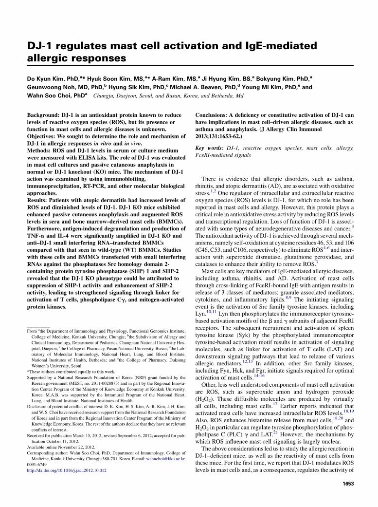
asthma, and rhinitis, could be mediated by oxidative stress,1,2 wefirst measured ROS levels in sera from healthy and allergicpatients with AD. Significantly increased ROS levels wereobserved in sera from these patients (59.96 1.6 mmol/L for thosewith mild, 57.8 6 1.7 mmol/L for those with moderate, and57.8 6 1.6 mmol/L for those with severe disease) comparedwith levels seen in healthy subjects (24.2 6 1.4 mmol/L,P <_.00019; Fig 1, A). Conversely, serum levels of DJ-1 were sub-stantially diminished in patients (2.86 0.3 ng/mL for those withmild, 2.16 0.2 ng/mL for those with moderate, and 2.26 0.2 ng/mL for those with severe disease) versus those seen in healthysubjects (8.06 0.4 ng/mL, P <_ .00114; Fig 1, B), which suggeststhat DJ-1 could also be associated with AD. However, we ob-served no significant differences in ROS and DJ-1 levels betweenpatients with severe or moderate and mild AD (Fig 1).
FIG 1. DJ-1 levels are decreased in sera from allergic patients, as opposed
to increased ROS levels, compared with those seen in healthy volunteers.
ROS (A) and DJ-1 (B) levels in sera were measured by using the ELISA kit,
according to the manufacturer’s instructions. Values are means 6 SEMs of
values from healthy volunteers (n5 15) and patients with AD (n5 56).Opencircles, Twenty-two patients with mild AD; gray circles, 17 patients with
moderate AD; solid circles, 17 patients with severe AD. n.s., Not significant.
DJ-1–deficient mice show significantly enhanced
PCA reactions and mast cell degranulationNext we investigated whether DJ-1 plays a role in the mast
cell–mediated allergic response by using the mouse PCA modelin DJ-1 KO mice with no detectable DJ-1 (Fig 2, A). The antigen-induced PCA reaction was increased in these mice compared withthat seen in WT mice (Fig 2, B and C). Of note, histologic exam-ination of ear tissue16 revealed that the degranulation of mast cellswas significantly increased in DJ-1 KO mice (Fig 2, D and E). Asobserved previously,16,28 the total number of mast cells also in-creased during the PCA reaction (Fig 2, D and E), presumablythrough infiltration and differentiation of progenitor cells.8,29
DJ-1 deficiency leads to enhancement of ROS
generation, degranulation, and cytokine production
in antigen-stimulated BMMCsIn view of the above results and the fact that antigen-stimulated
ROS production has been functionally linked to mast celldegranulation,18,30,31 we examined ROS levels and release ofbioactive mediators in DJ-1 KO and WT BMMC cultures. Thegeneration of ROS by antigen was significantly enhanced inDJ-1 KO BMMCs (8.8 6 0.87 mmol/L) compared with thatseen in WT BMMCs (5.9 6 1.38 mmol/L; Fig 3, A and B).A similar enhancement was noted in vivo in the PCA model,
with no significant difference in serum ROS levels before antigenchallenge but substantial differences between WT (24.1 6 1.92mmol/L) and DJ-1 KO (37.36 1.03mmol/L) mice after challenge(Fig 3, C).Both types of BMMCs exhibited equivalent expression of the
mature mast cell markers, c-Kit and FcεRI by means of flowcytometric and immunoblotting analysis (Fig 3, D and E). Ofnote, antigen-induced degranulation and the secretion of IL-4and TNF-a were markedly increased in DJ-1 KO BMMCs (Fig3, F and G).
DJ-1 regulates early FcεRI-mediated signals,
including phosphorylation of Syk and LATTo determine whether DJ-1 regulated responses to stimulants
other than antigen, we next examined degranulation by thecalcium-mobilizing agents thapsigargin and ionomycin. In con-trast to antigen, degranulation induced by either agent was thesame inWTandDJ-1K/OBMMCs (Fig 4,A). This suggested that
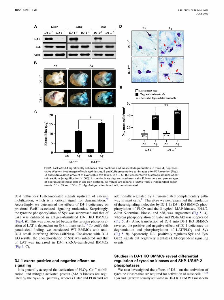
FIG 2. Lack of DJ-1 significantly enhances PCA reactions and mast cell degranulation in mice. A, Represen-
tativeWestern blot images of indicated tissues. B and C, Representative ear images after PCA reaction (Fig 2,
B) and extravasated amount of Evans blue dye (Fig 2, C; n 5 5). D, Representative histologic images of ear
skin sections (magnification31000). Arrows indicate degranulated mast cells. E, Numbers and percentages
of degranulated mast cells in ear skin sections. All values are means 6 SEMs from 3 independent experi-
ments. *P < .05 and **P < .01. Ag, Antigen stimulated; NS, nonstimulated.
J ALLERGY CLIN IMMUNOL
JUNE 2013
1656 KIM ET AL
DJ-1 influences FcεRI-mediated signals upstream of calciummobilization, which is a critical signal for degranulation.32
Accordingly, we determined the effects of DJ-1 deficiency onproximal FcεRI-associated signaling molecules. Surprisingly,the tyrosine phosphorylation of Syk was suppressed and that ofLAT was enhanced in antigen-stimulated DJ-1 KO BMMCs(Fig 4, B). This was unexpected because the tyrosine phosphoryl-ation of LAT is dependent on Syk in mast cells.33 To verify thisparadoxical finding, we transfected WT BMMCs with anti–DJ-1 small interfering RNAs (siRNAs). Consistent with DJ-1KO results, the phosphorylation of Syk was inhibited and thatof LAT was increased in DJ-1 siRNA–transfected BMMCs(Fig 4, C).
DJ-1 exerts positive and negative effects on
signalingIt is generally accepted that activation of PLCg, Ca21 mobili-
zation, and mitogen-activated protein (MAP) kinases are regu-lated by the Syk/LAT pathway, whereas Gab2 and PI3K/Akt are
additionally regulated by a Fyn-mediated complementary path-way in mast cells.11 Therefore we next examined the regulationof these signaling molecules by DJ-1. In DJ-1 KO BMMCs phos-phorylation of PLCg and the 3 typical MAP kinases, Erk1/2,c-Jun N-terminal kinase, and p38, was augmented (Fig 5, A),whereas phosphorylation of Gab2 and PI3K/Akt was suppressed(Fig 5, A). Also, transfection of DJ-1 into DJ-1 KO BMMCsreversed the positive and negative effects of DJ-1 deficiency ondegranulation and phosphorylation of LAT/PLCg and Syk(Fig 5, B). Apparently, DJ-1 positively regulates Syk and Fyn/Gab2 signals but negatively regulates LAT-dependent signalingevents.
Studies in DJ-1 KO BMMCs reveal differential
regulation of tyrosine kinases and SHP-1/SHP-2
phosphatasesWe next investigated the effects of DJ-1 on the activation of
tyrosine kinases that are required for activation of mast cells.11,16
Lyn and Fgr were equally activated in DJ-1 KO andWTmast cells

FIG 3. ROS levels, degranulation, and cytokine production in BMMCs. A, Representative images obtained
by means of confocal microscopy. B and C, ROS levels determined by using ELISA analysis in BMMCs
(Fig 3, B) or in sera after PCA reactions (Fig 3, C) were measured, as described in the Methods section. D,
Representative fluorescence-activated cell sorting images for c-Kit and FcεRI expression in BMMCs. E, Rep-
resentative Western blot images of the a, b, and g subunits of the FcεRI receptor. F, Percentage release of
granules in BMMCs. G, TNF-a and IL-4 levels in the culture medium determined by using ELISA. All values
are means6 SEMs from 3 independent experiments (Fig 3, B, C, E, and F). *P < .05. Ag, Antigen stimulated;
NS, nonstimulated.
J ALLERGY CLIN IMMUNOL
VOLUME 131, NUMBER 6
KIM ET AL 1657
(Fig 6, A and B). However, in DJ-1 KO BMMCs the activation ofFyn was totally blocked (Fig 6, C) and that of Syk was signifi-cantly, although partially, reduced (Fig 6, D).
The apparent differential regulation of signaling molecules byDJ-1 was examined further by investigating the activation statusof SHP-1 and SHP-2. SHP-1 is reported to regulate phosphoryl-ation of LATandMAP kinases without affecting phosphorylationof Syk.34,35 SHP-1 phosphorylation and activation were muchdiminished in stimulated DJ-1 KO BMMCs (Fig 7, A, upperpanel) but were restored by treatment with 30 mmol/L 2,2,6,6-tet-ramethylpiperidin-1-oxyl (TEMPO), a general ROS scavenger(Fig 7, A, lower panel), indicating that the SHP-1 activation waspositively regulated by increases in intracellular ROS levels.Another SHP isoform, SHP-2, has been linked to negative
regulation of Syk.36 We found that SHP-2 phosphorylation and
activity, in contrast to SHP-1, was augmented in DJ-1 KO mastcells and that this augmentation was reversed by treating cellswith 30 mmol/L TEMPO (Fig 7, A).
Syk and LAT are differentially dephosphorylated by
SHP-2 and SHP-1, respectivelyThe roles of both SHPs were examined further by knock down of
the individual SHPs with siRNAs in WT and DJ-1 KO BMMCs.Antigen-induced phosphorylation of LAT was substantially in-creased in anti–SHP-1 siRNA–transfectedWTorDJ-1KOBMMCsbut was not enhanced in anti–SHP-2 siRNA–transfected BMMCs(Fig 7,B). Conversely, antigen-induced phosphorylation of Sykwasincreased in anti–SHP-2 siRNA–transfected WT and DJ-1 KOBMMCsbutwasnot altered in anti–SHP-1 siRNA–transfectedcells,
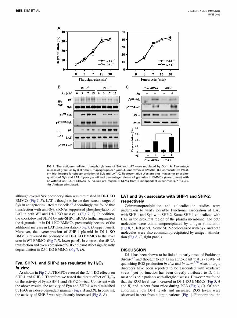
FIG 4. The antigen-mediated phosphorylations of Syk and LAT were regulated by DJ-1. A, Percentage
release of granules by 300 nmol/L thapsigargin or 1 mmol/L ionomycin in BMMCs. B, Representative West-
ern blot images for phosphorylation of Syk and LAT. C, Representative Western blot images for phospho-
rylation of Syk and LAT (upper panel) and percentage release of granules in BMMCs (lower panel) with
or without anti–DJ-1 siRNAs. All values are means 6 SEMs from 3 independent experiments. *P < .05.
Ag, Antigen stimulated.
J ALLERGY CLIN IMMUNOL
JUNE 2013
1658 KIM ET AL
although overall Syk phosphorylation was diminished in DJ-1 KOBMMCs (Fig 7, B). LAT is thought to be the downstream target ofSyk in antigen-stimulated mast cells.32 Accordingly, we found thattransfection with anti-Syk siRNAs suppressed phosphorylation ofLAT in both WT and DJ-1 KO mast cells (Fig 7, C). In addition,the knockdownofSHP-1byanti–SHP-1 siRNAsfurther augmentedthe degranulation in DJ-1 KO BMMCs, presumably because of theadditional increase in LAT phosphorylation (Fig 7,D, upper panel).Moreover, the overexpression of SHP-1 plasmid in DJ-1 KOBMMCs reversed the phenotype in DJ-1 KO BMMCs to the levelseen inWTBMMCs (Fig 7,D, lower panel). In contrast, the siRNAtransfection andoverexpression of SHP-2 didnot affect significantlydegranulation in DJ-1 KO BMMCs (Fig 7, D).
Fyn, SHP-1, and SHP-2 are regulated by H2O2
in vitroAs shown in Fig 7, A, TEMPO reversed the DJ-1 KO effects on
SHP-1 and SHP-2. Therefore we tested the direct effect of H2O2
on the activity of Fyn, SHP-1, and SHP-2 in vitro. Consistent withthe above results, the activity of Fyn and SHP-1 was diminishedby H2O2 in a dose-dependent manner (Fig 8, A and B). In contrast,the activity of SHP-2 was significantly increased (Fig 8, B).
LAT and Syk associate with SHP-1 and SHP-2,
respectivelyCoimmunoprecipitation and colocalization studies were
undertaken to verify possible functional association of LATwith SHP-1 and Syk with SHP-2. Some SHP-1 colocalized withLAT in the proximal region of the plasma membrane, and bothmolecules were coimmunoprecipitated by antigen stimulation(Fig 8,C, left panel). Some SHP-2 colocalized with Syk, and bothmolecules were also coimmunoprecipitated by antigen stimula-tion (Fig 8, C, right panel).
DISCUSSIONDJ-1 has been shown to be linked to early onset of Parkinson
disease37 and thought to act as an antioxidant that is capable ofquenching ROS production in vivo and in vitro.3,38 Also, allergicdisorders have been reported to be associated with oxidativestress,1 yet no function has been directly attributed to DJ-1 inmast cells or in patients with allergic diseases. However, we foundthat the ROS level was increased in DJ-1 KO BMMCs (Fig 3, Aand B) and in sera from mice during PCA (Fig 3, C). Of note,abnormally low DJ-1 levels and increased ROS levels wereobserved in sera from allergic patients (Fig 1). Furthermore, the
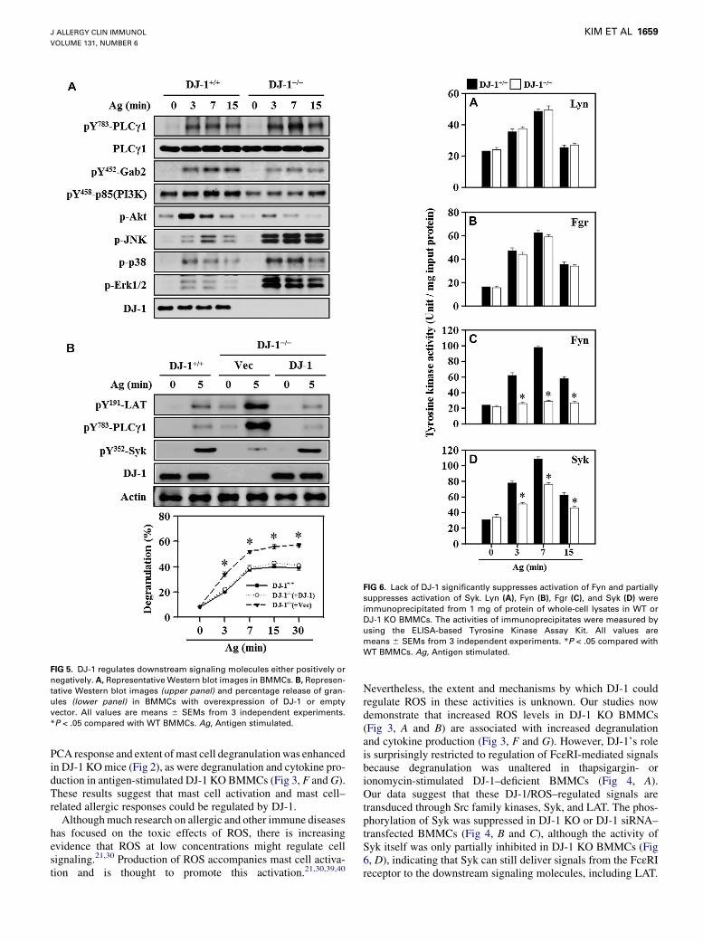
FIG 5. DJ-1 regulates downstream signaling molecules either positively or
negatively. A, Representative Western blot images in BMMCs. B, Represen-
tative Western blot images (upper panel) and percentage release of gran-
ules (lower panel) in BMMCs with overexpression of DJ-1 or empty
vector. All values are means 6 SEMs from 3 independent experiments.
*P < .05 compared with WT BMMCs. Ag, Antigen stimulated.
FIG 6. Lack of DJ-1 significantly suppresses activation of Fyn and partially
suppresses activation of Syk. Lyn (A), Fyn (B), Fgr (C), and Syk (D) were
immunoprecipitated from 1 mg of protein of whole-cell lysates in WT or
DJ-1 KO BMMCs. The activities of immunoprecipitates were measured by
using the ELISA-based Tyrosine Kinase Assay Kit. All values are
means 6 SEMs from 3 independent experiments. *P < .05 compared with
WT BMMCs. Ag, Antigen stimulated.
J ALLERGY CLIN IMMUNOL
VOLUME 131, NUMBER 6
KIM ET AL 1659
PCA response and extent ofmast cell degranulationwas enhancedin DJ-1 KOmice (Fig 2), as were degranulation and cytokine pro-duction in antigen-stimulated DJ-1 KO BMMCs (Fig 3, F andG).These results suggest that mast cell activation and mast cell–related allergic responses could be regulated by DJ-1.Althoughmuch research on allergic and other immune diseases
has focused on the toxic effects of ROS, there is increasingevidence that ROS at low concentrations might regulate cellsignaling.21,30 Production of ROS accompanies mast cell activa-tion and is thought to promote this activation.21,30,39,40
Nevertheless, the extent and mechanisms by which DJ-1 couldregulate ROS in these activities is unknown. Our studies nowdemonstrate that increased ROS levels in DJ-1 KO BMMCs(Fig 3, A and B) are associated with increased degranulationand cytokine production (Fig 3, F and G). However, DJ-1’s roleis surprisingly restricted to regulation of FcεRI-mediated signalsbecause degranulation was unaltered in thapsigargin- orionomycin-stimulated DJ-1–deficient BMMCs (Fig 4, A).Our data suggest that these DJ-1/ROS–regulated signals aretransduced through Src family kinases, Syk, and LAT. The phos-phorylation of Syk was suppressed in DJ-1 KO or DJ-1 siRNA–transfected BMMCs (Fig 4, B and C), although the activity ofSyk itself was only partially inhibited in DJ-1 KO BMMCs (Fig6, D), indicating that Syk can still deliver signals from the FcεRIreceptor to the downstream signaling molecules, including LAT.
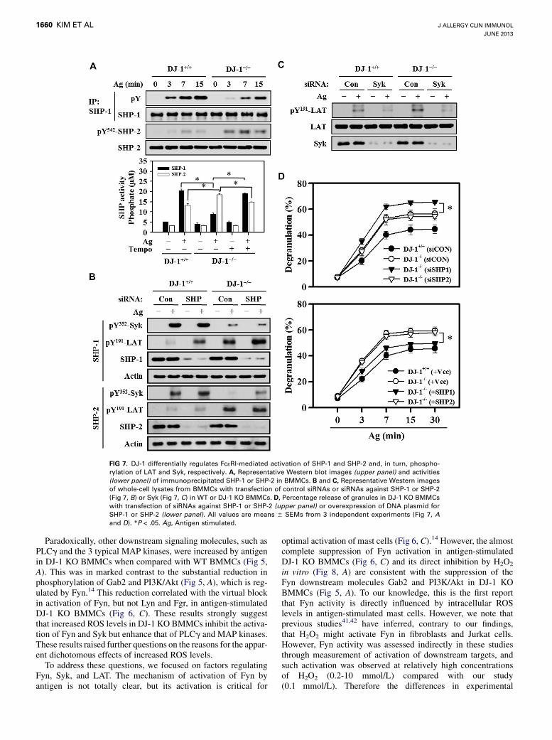
FIG 7. DJ-1 differentially regulates FcεRI-mediated activation of SHP-1 and SHP-2 and, in turn, phospho-
rylation of LAT and Syk, respectively. A, Representative Western blot images (upper panel) and activities
(lower panel) of immunoprecipitated SHP-1 or SHP-2 in BMMCs. B and C, Representative Western images
of whole-cell lysates from BMMCs with transfection of control siRNAs or siRNAs against SHP-1 or SHP-2
(Fig 7, B) or Syk (Fig 7, C) in WT or DJ-1 KO BMMCs. D, Percentage release of granules in DJ-1 KO BMMCs
with transfection of siRNAs against SHP-1 or SHP-2 (upper panel) or overexpression of DNA plasmid for
SHP-1 or SHP-2 (lower panel). All values are means 6 SEMs from 3 independent experiments (Fig 7, Aand D). *P < .05. Ag, Antigen stimulated.
J ALLERGY CLIN IMMUNOL
JUNE 2013
1660 KIM ET AL
Paradoxically, other downstream signaling molecules, such asPLCg and the 3 typical MAP kinases, were increased by antigenin DJ-1 KO BMMCs when compared with WT BMMCs (Fig 5,A). This was in marked contrast to the substantial reduction inphosphorylation of Gab2 and PI3K/Akt (Fig 5, A), which is reg-ulated by Fyn.14 This reduction correlated with the virtual blockin activation of Fyn, but not Lyn and Fgr, in antigen-stimulatedDJ-1 KO BMMCs (Fig 6, C). These results strongly suggestthat increased ROS levels in DJ-1 KO BMMCs inhibit the activa-tion of Fyn and Syk but enhance that of PLCg and MAP kinases.These results raised further questions on the reasons for the appar-ent dichotomous effects of increased ROS levels.To address these questions, we focused on factors regulating
Fyn, Syk, and LAT. The mechanism of activation of Fyn byantigen is not totally clear, but its activation is critical for
optimal activation of mast cells (Fig 6, C).14 However, the almostcomplete suppression of Fyn activation in antigen-stimulatedDJ-1 KO BMMCs (Fig 6, C) and its direct inhibition by H2O2
in vitro (Fig 8, A) are consistent with the suppression of theFyn downstream molecules Gab2 and PI3K/Akt in DJ-1 KOBMMCs (Fig 5, A). To our knowledge, this is the first reportthat Fyn activity is directly influenced by intracellular ROSlevels in antigen-stimulated mast cells. However, we note thatprevious studies41,42 have inferred, contrary to our findings,that H2O2 might activate Fyn in fibroblasts and Jurkat cells.However, Fyn activity was assessed indirectly in these studiesthrough measurement of activation of downstream targets, andsuch activation was observed at relatively high concentrationsof H2O2 (0.2-10 mmol/L) compared with our study(0.1 mmol/L). Therefore the differences in experimental

FIG 8. Fyn, SHP-1, and SHP-2 activities are regulated directly by H2O2 and a proposed scheme for DJ-1 reg-
ulation of ROS and downstream signals. A and B, Activities of immunoprecipitated Fyn, SHP-1, and SHP-2
from nonstimulated (NS) and antigen-stimulated (Ag) BMMCs were assayed in vitro with different concen-
trations of H2O2 for 15 minutes. Values (means 6 SEMs) are from 3 independent experiments. *P < .05.
C, Representative confocal and Western blot images for coimmunoprecipitation of LAT and SHP-1 or Syk
and SHP-2. D, Proposed scheme. Absence of DJ-1 leads to excess ROS levels, which inhibit SHP-1 and
enhance phosphorylation of LAT and LAT-dependent signals while inhibiting Fyn and, through activation
of SHP-2, Syk. Nevertheless, the enhancement of LAT phosphorylation is sufficient to ultimately augment
mediator release in DJ-1–deficient BMMCs and PCA in DJ-1–deficient mice.
J ALLERGY CLIN IMMUNOL
VOLUME 131, NUMBER 6
KIM ET AL 1661
conditions and cell types could account for the apparent discrep-ancy in the different studies.With respect to LAT, its phosphorylation is increased in SHP-
1–deficient BMMCs34 and T cells,39 as well as DJ-1 KO BMMCs(Fig 4, B). Therefore we hypothesized that the increase in phos-phorylation of LAT in DJ-1 KO and DJ-1 siRNA–transfectedBMMCs (Fig 4, B and C) was attributable to inhibition of SHP-1 by the increased ROS levels in these cells. This was confirmedby the suppressed activation of SHP-1, which was inhibited inDJ-1 KO BMMCs (Fig 7, A), and restoration of activation witha low concentration of TEMPO (Fig 7, A, lower panel). Further-more, SHP-1 activity was significantly inhibited by H2O2 in vitro(Fig 8, B), indicating that the intracellular ROS level could inhibitSHP-1 activity and thus modulate the phosphorylation of LAT.In regard to the diminished phosphorylation of Syk in DJ-1 KO
BMMCs, we considered the role of SHP-2 because this phospha-tase is known to dephosphorylate Syk.36 Indeed, the activation ofSHP-2 was increased (Fig 7, A) and that of Syk was decreased(Figs 4, B, and 6, D) in DJ-1 KO BMMCs. In addition, the phos-phorylation of Syk was increased in the SHP-2 siRNA–trans-fected WT and DJ-1 KO BMMCs (Fig 7, B). These results
suggest that the activation of Syk is negatively regulated byDJ-1 through SHP-2.In conclusion, FcεRI signaling events, including activation of
Src family kinases, protein tyrosine phosphatases, and othercritical molecules,11 are highly affected by excess ROS levels, assummarized schematically in Fig 8, D. These include the inhibi-tion of activation of Fyn and Syk and the unexpected enhance-ment of phosphorylation of LAT in DJ-1 KO BMMCs. Sucheffects are associated with the inhibitory actions of ROS on Fynand SHP-1 and the positive actions on SHP-2. These multipleeffects of ROS are associated with enhanced degranulation andcytokine production, and thus deficiency or constitutive activationof DJ-1 could have implications in mast cell–driven inflammatoryallergic and autoimmune diseases (Fig 8, D). As reported previ-ously, a distinguishing feature of patients with AD was the mark-edly diminished levels of DJ-1 with corresponding increases inROS levels when compared with those seen in healthy subjects(Fig 1). However, we note that although no discernible differenceswere noted with severity of disease in the patients studied, theonset of AD occurred early in life (newborn, childhood, or earlyteens) and before this study in almost all of our patients. Therefore
J ALLERGY CLIN IMMUNOL
JUNE 2013
1662 KIM ET AL
we suggest only that abnormal DJ-1/ROS levels might be associ-ated with the onset of AD that relapses through adulthood.Whether the extent of DJ-1/ROS abnormality correlates withthe onset of disease will require further study of samples exam-ined retrospectively from pediatric subjects before the onset ofdisease. It would also be of interest to determine whether DJ-1/ROS dysfunction underlies other mast cell–driven inflammatorydiseases or is unique to AD.
Key messages
d Serum levels of DJ-1 were significantly decreased in pa-tients with AD (P <_ .00114) and were associated with in-creased ROS levels.
d DJ-1 regulates antigen-induced mast cell activation andallergic responses in vitro and in vivo.
d DJ-1 differentially regulates activation of Fyn, Syk, andLAT in mast cells.
d Dysregulation of DJ-1 has implications in patients withmast cell–driven allergic diseases.
REFERENCES
1. Bowler RP, Crapo JD. Oxidative stress in allergic respiratory diseases. J Allergy
Clin Immunol 2002;110:349-56.
2. Barnes PJ. Biochemical basis of asthma therapy. J Biol Chem 2011;286:
32899-905.
3. Kahle PJ, Waak J, Gasser T. DJ-1 and prevention of oxidative stress in Parkinson’s
disease and other age-related disorders. Free Radic Biol Med 2009;47:1354-61.
4. Taira T, Saito Y, Niki T, Iguchi-Ariga SM, Takahashi K, Ariga H. DJ-1 has a role in
antioxidative stress to prevent cell death. EMBO Rep 2004;5:213-8.
5. Kinumi T, Kimata J, Taira T, Ariga H, Niki E. Cysteine-106 of DJ-1 is the most
sensitive cysteine residue to hydrogen peroxide-mediated oxidation in vivo in
human umbilical vein endothelial cells. Biochem Biophys Res Commun 2004;
317:722-8.
6. Martinat C, Shendelman S, Jonason A, Leete T, Beal MF, Yang L, et al. Sensitivity
to oxidative stress in DJ-1-deficient dopamine neurons: an ES-derived cell model
of primary parkinsonism. PLoS Biol 2004;2:327.
7. Xu XM, Møller SG. ROS removal by DJ-1: arabidopsis as a new model to under-
stand Parkinson’s disease. Plant Signal Behav 2010;5:1034-6.
8. Gilfillan AM, Beaven MA. Regulation of mast cell responses in health and disease.
Crit Rev Immunol 2011;31:475-530.
9. Kalesnikoff J, Galli S. New developments in mast cell biology. Nat Immunol 2008;
9:1215-23.
10. Kinet JP. The high-affinity IgE receptor (Fc epsilon RI): from physiology to pathol-
ogy. Annu Rev Immunol 1999;17:931-72.
11. Gilfillan AM, Rivera J. The tyrosine kinase network regulating mast cell activation.
Immunol Rev 2009;228:149-69.
12. Bischoff SC. Role of mast cells in allergic and non-allergic immune responses:
comparison of human and murine data. Nat Rev Immunol 2007;7:93-104.
13. Rivera J, Gilfillan AM. Molecular regulation of mast cell activation. J Allergy Clin
Immunol 2006;117:1214-25.
14. Parravicini V, Gadina M, Kovarova M, Odom S, Gonzalez-Espinosa C, Furumoto
Y, et al. Fyn kinase initiates complementary signals required for IgE-dependent
mast cell degranulation. Nat Immunol 2002;3:741-8.
15. Hong H, Kitaura J, Xiao W, Horejsi V, Ra C, Lowell CA, et al. The Src family
kinase Hck regulates mast cell activation by suppressing an inhibitory Src family
kinase Lyn. Blood 2008;110:2511-9.
16. Lee JH, Kim JW, Kim do K, Kim HS, Park HJ, Park DK, et al. The Src family
kinase Fgr is critical for activation of mast cells and IgE-mediated anaphylaxis
in mice. J Immunol 2011;187:1807-15.
17. Halliwell B, Gutteridge JM. Role of free radicals and catalytic metal ions in human
disease: an overview. Methods Enzymol 1990;186:1-5.
18. Wolfreys K, Oliveira DB. Alterations in intracellular reactive oxygen species genera-
tionand redoxpotentialmodulatemast cell function.Eur J Immunol 1997;27:297-306.
19. Brooks AC, Whelan CJ, Purcell WM. Reactive oxygen species generation and his-
tamine release by activated mast cells: modulation by nitric oxide synthase inhibi-
tion. Br J Pharmacol 1999;128:585.
20. Niu XF, Ibbotson G, Kubes P. A balance between nitric oxide and oxidants regulates
mast cell-dependent neutrophil-endothelial cell interactions. CircRes 1996;79:992-9.
21. Suzuki Y, Yoshimaru T, Matsui T, Inoue T, Niide O, Nunomura S, et al. Fc epsilon
RI signaling of mast cells activates intracellular production of hydrogen peroxide:
role in the regulation of calcium signals. J Immunol 2003;171:6119-27.
22. Kim JW, Lee JH, Hwang BY, Mun SH, Ko NY, Kim do K, et al. Morin inhibits Fyn
kinase in mast cells and IgE-mediated type I hypersensitivity response in vivo. Bio-
chem Pharmacol 2009;77:1506-12.
23. Rudzki E, Samochocki Z, Rebandel P, Saciuk E, Ga1ecki W, Raczka A, et al. Fre-
quency and significance of the major and minor features of Hanifin and Rajka
among patients with atopic dermatitis. Dermatology 1994;189:41-6.
24. Oranje AP, Glazenburg EJ, Wolkerstorfer A, de Waard-van der Spek FB. Practical
issues on interpretation of scoring atopic dermatitis: the SCORAD index, objective
SCORAD and the three-item severity score. Br J Dermatol 2007;157:645-8.
25. Jensen BM, Swindle EJ, Iwaki S, Gilfillan AM.Generation, isolation, and maintenance
of rodentmast cells andmast cell lines.Curr Protoc Immunol 2006;Chapter 3:Unit 3.23.
26. Swindle EJ, Coleman JW, DeLeo FR, Metcalfe DD. FcepsilonRI- and Fcgamma
receptor-mediated production of reactive oxygen species by mast cells is
lipoxygenase- and cyclooxygenase-dependent and NADPH oxidase-independent.
J Immunol 2007;179:7059-71.
27. Ozawa K, Szallasi Z, Kazanietz MG, Blumberg PM, Mischak H, Mushinski JF,
et al. Ca21-Dependent and Ca21-independent isozymes of protein kinase C medi-
ate exocytosis in antigen-stimulated rat basophilic RBL-2H3 cells: reconstitution
of secretory responses with Ca21 and purified isozymes in washed permeabilized
cells. J Biol Chem 1993;268:1749-56.
28. Pushparaj PN, Manikandan J, Tay HK, H’ng SC, Kumar SD, Pfeilschifter J, et al.
Sphingosine kinase 1 is pivotal for Fc epsilon RI-mediated mast cell signaling and
functional responses in vitro and in vivo. J Immunol 2009;183:221-7.
29. Ekoff M, Nilsson G. Mast cell apoptosis and survival. Adv Exp Med Biol 2011;
716:47-60.
30. Swindle EJ, Metcalfe DD, Coleman JW. Rodent and human mast cells produce
functionally significant intracellular reactive oxygen species but not nitric oxide.
J Biol Chem 2004;279:48751-9.
31. Kuehn HS, Swindle EJ, Kim MS, Beaven MA, Metcalfe DD, Gilfillan AM. The
phosphoinositide 3-kinase-dependent activation of Btk is required for optimal
eicosanoid production and generation of reactive oxygen species in antigen-
stimulated mast cells. J Immunol 2008;181:7706-12.
32. Ma HT, Beaven MA. Regulation of Ca21 signaling with particular focus on mast
cells. Crit Rev Immunol 2009;29:155-86.
33. Zhang J, Berenstein E, Siraganian RP. Phosphorylation of Tyr342 in the linker
region of Syk is critical for Fc epsilon RI signaling in mast cells. Mol Cell Biol
2002;22:8144-54.
34. Nakata K, Yoshimaru T, Suzuki Y, Inoue T, Ra C, Yakura H, et al. Positive and
negative regulation of high affinity IgE receptor signaling by Src homology region
2 domain-containing phosphatase 1. J Immunol 2008;181:5414-24.
35. Zhang L, Oh SY, Wu X, Oh MH, Wu F, Schroeder JT, et al. SHP-1 deficient mast
cells are hyperresponsive to stimulation and critical in initiating allergic inflamma-
tion in the lung. J Immunol 2010;184:1180-90.
36. Xu R, Pecht I. The protein tyrosine kinase syk activity is reduced by clustering the
mast cell function-associated antigen. Eur J Immunol 2001;31:1571-81.
37. Bonifati V, Rizzu P, van Baren MJ, Schaap O, Breedveld GJ, Krieger E, et al.
Mutations in the DJ-1 gene associated with autosomal recessive early-onset parkin-
sonism. Science 2003;299:256-9.
38. Wilson MA. The role of cysteine oxidation in DJ-1 function and dysfunction. Anti-
oxid Redox Signal 2011;15:111-22.
39. Sly LM, Kalesnikoff J, Lam V, Wong D, Song C, Omeis S, et al. IgE-induced mast
cell survival requires the prolonged generation of reactive oxygen species.
J Immunol 2008;181:3850-60.
40. Suzuki Y, Yoshimaru T, Inoue T, Niide O, Ra C. Role of oxidants in mast cell
activation. Chem Immunol Allergy 2005;87:32-42.
41. Abe J, Okuda M, Huang Q, Yoshizumi M, Berk BC. Reactive oxygen species ac-
tivate p90 ribosomal S6 kinase via Fyn and Ras. J Biol Chem 2000;275:1739-48.
42. Jin YJ, Friedman J, Burakoff SJ. Regulation of tyrosine phosphorylation in isolated
T cell membrane by inhibition of protein tyrosine phosphatases. J Immunol 1998;
161:1743-50.

TABLE E1. Characteristics of patients with AD and healthy
subjects
Recruitment period (month/year)
Healthy subjects:
April 2010 to
November 2011
Patients with AD:
April 2010 to
August 2012
Age (y [range]) 25.5 6 3.3 (21-31) 25.3 6 10.4 (5-61)
No. (male/female) 15 (7/8) 56 (28/28)
Eosinophil (% in WBC) 3.1 6 5.4 7.1 6 4.2
Serum total IgE (IU/mL) 235 6 137 1592 6 1415
Scoring 0.0 6 0.0 35.0 6 19.8*
All values for this table are presented as means 6 SDs.
*Clinical severity was evaluated by using the SCORAD index, a system used
worldwide to assess the severity of atopic eczema.E1
REFERENCE
E1. Oranje AP, Glazenburg EJ, Wolkerstorfer A, de Waard-van der Spek FB. Practi-
cal issues on interpretation of scoring atopic dermatitis: the SCORAD index,
objective SCORAD and the three-item severity score. Br J Dermatol 2007;157:
645-8.
J ALLERGY CLIN IMMUNOL
VOLUME 131, NUMBER 6
KIM ET AL 1662.e1