Cholesterol and hematopoietic stem cells: inflammatory mediators of atherosclerosis
THE JOURNAL OF
AND
VOLUME 90 NUMBER 4. PART 2
IWediators of allergic rhinitis
Martha V. White, MD, and Michael A. Kaliner, MD Berhesh, Md.
Although histaminc~ is the principal tnediator aj the immediutr ullergic reuction. other ittjfumtnuto~ tnediatars us well us neurapeptides also contribute to rhinorrheu and nasa/ congestion. Within minutes of exposure to ullergen. mast cells produce hi.stumine, leukotrirtw C,, . and prostu~lat~din 17,. A cotzcotnitunt increuse occurs in twuropeptidrs und hrudykinin. In \,itro mast cell uctivution ulso lends to the release oJ’ tutnor necrosis ,fuctor-a, seiwul interleukins, and grunulocyte-macr[~phnge colony-.stimulrttitt~ j&tar. Because trll these variorr.~ mediutors und neurapeptides tnq play u role in producing rhinorrhea and congestion, antihistamines ulone cannot control ull of the .symptotns of ullergic rhinitis. However. thr comhinution a/‘ antihistamines with topical corticosteroids can inhibit the generation, rr4ea.w. card trcti~*it~ of‘ mast if not all of the mediutors potentiall? itz\wlwd in the> nllergic response?, I J AI.I.tx<;Y C1.t.v ~MMlA’OL 1992 ;90:699-704 .)
Key words: Allergic rhinitis I must cells, mediutor release, nusal mutxsa. neuropeptides
Allergic rhinitis is the most common immunologic disease and the most common chronic disease expe- rienced by humans. It affects approximately 10% to 20% of the American population. More than 1 billion dollars are spent yearly on over-the-counter and pre- scription allergy preparations. In addition, allergic rhi- nitis accounts for 2.5% of all physician visits for all diseases. and another 0.5% of all visits are for re- ceiving allergy immunotherapy. ’
From the Allergic Diseases Section, Laboratory of Clincial Inves- tigation. National Institute of Allergy and Infectious Diseases. National Institutes of Health, Bethesda, MD.
Reprint requests: Michael A. Kaliner, MD, National Institutes of Health, Bldg. IO. I iC2OS. 9ooO Rwkville Pike. Bethesda. MD ‘0892
110140673
To understand the pathophysiology of allergic rhi- nitis, it is first necessary to be knowledgeablr about nasal anatomy. (See the box “Anatomy of the nasal mucosa” that appears later in this article.)
MEDIATORS OF ALLERGIC RHfNfFlS
To assess which mediators are important in inducing the symptoms of allergic rhinitis, it is important to know which mediators are released during an allergic reaction. Mast cell activation after antigen challenge is well described. In electron micrographic studies of resting mast cells, the ubiquitous, densely stained,
secretory granules are roughly spherical, and most are 0.25 to 0.5 km in diameter. Most granules comprise a dense, amorphous matrix with embedded or inter- spersed crystalline constituents in the form of scrolls. gratings, or lattices. Scrolls are found most commonly in cells undergoing degranulation. It should be noted
639

700 White and Kaliner
Abbreviations used LTC,, LTD,, LTE,: Leukotrienes C,, D,, and E,
PGD,: Prostaglandin D2 CGRP: Calcitonin gene-related peptide
SP: Substance P VIP: Vasoactive intestinal peptide
TNF-a: Tumor necrosis factor-o IL: Interleukin
GM-CSF: Granulocyte-macrophage col- ony-stimulating factor
PAF: Platelet-activating factor MC-,: Mast cells that contain only
tryptase NKA: Neurokinin A GRP: Gastrin-releasing peptide
that even in experimentally unstimulated nasal tissue, variable numbers of degranulating mast cells are pres- ent. In unstimulated nasal epithelium, one third of mast cells may be degranulated, and the percentage progressively increases from the lamina propria to the mucosal surface.
Early events after IgE-mediated stimulation of hu- man lung mast cells include granule swelling with loss of stainable matrix, an increase in the proportion of granules demonstrating the scroll pattern, and the appearance of electron-dense clumps in the granules. Granules progressively become more ropelike, and granule contents eventually become soluble. In nasal and lung mast cells, extruded granules are never ob- served intact in the external environment of the de- granulated nasal mast cell. Over a few-minute period, the degranulating mast cell may discharge the solu- bilized contents from 1000 secretory granules into the surrounding interstitial fluid.2
Within minutes of exposure to allergen, the mast cell products histamine, LTQ, and PGD, can be mea- sured in nasal washings.3 Mast cells are found near superficial postcapillary venules (which respond with increased vascular permeability), near sensory nerves (which respond by initiating the itch sensation and so eliciting the sneeze reflex), and near glands that re- spond by secretion. Many of the effects associated with acute allergic rhinitis can be reproduced by his- tamine challenge,4 and antihistamines prevent many of the symptoms of allergic rhinitis.’ Thus histamine challenge to the nose leads to pruritus, sneezing, nasal congestion that is induced by vasodilation, in- creased vascular permeability, and reflex cholinergic
J ALLERGY CLIN IMMUNOL OCTOBER 1992
stimulation of glandular secretion in both the ipsilat- era1 and contralateral nares. Activation of mast cells also leads to the generation of the 5lipoxygenase products, LTC,, LTD4, and LTE4, which can induce vascular permeability, vasodilation, and mucous se- cretion.
Concomitant with the increase in mast cell media- tors, an increase in the neuropeptides CGRP, SP, and VIPh and in bradykinin’ can also be observed. In ad- dition, mast cell activation in vitro leads to the tran- scription and/or translation and release of a number of cytokines including TNF-a, IL- 1, -3, -4, -5, and -6, and GM-CSF.8-” During an allergic response, the nose also contains many inflammatory cells that may contribute cytokines to the allergic response. Lym- phocytes comprise most of the cells in the nasal sub- mucosa. After allergen challenge, lymphocytes in the mucosa of allergic individuals transcribe message for the TH-2 products, IL-4 and IL-5, as well as for GM- CSF” and IL-3. Only scant data are available on cy- tokines recovered in lavages after antigen challenge; however, IL-l, -2, -3. -4, and -5 and GM-CSF have been recovered,‘3-‘7 IL-l and IL-2 during the first 30 minutes, and all of them during the ensuing 10 hours.
SYMPTOMS OF ALLERGIC RHINITIS AND RESPONSIBLE MEDIATORS
The cardinal symptoms of allergic rhinitis include pruritus, sneezing, rhinorrhea, and nasal congestion. Pruritus and sneezing are each induced by sensory nerve stimulation, whereas congestion results from vasodilation with resultant engorgement of cavernous sinusoids. Rhinorrhea can be induced by increased vascular permeability as well as direct glandular se- cretion. During the first 30 minutes after allergen chal- lenge, concomitant with an increase in mediators, an enormous increase occurs in the plasma proteins al- bumin and IgG and a smaller increase in the glandular proteins lysozyme, lactoferrin, and secretory IgA in nasal lavage fluids. ”
In the nares contralateral to antigen challenge, rhi- norrhea also occurs and is comprised entirely of glan- dular proteins. This contralateral rhinorrhea appears to be cholinergically mediated because it can be in- hibited by pretreatment with atropine. Thus antigen- induced rhinorrhea is caused mostly by vascular per- meability on the ipsilateral side only and by reflex- mediated glandular secretion on both the ipsilateral and contralateral sides.” This pattern is identical to that of histamine-induced rhinorrhea. In addition, an- tigen challenge leads to a late inflammatory response,
VOLUME 90 NUMBER 4. PART 2
Mediators of allergic riilr:rtis 701
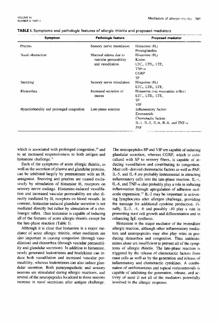
TABLE I. Symptoms and pathologic features of allergic rhinitis and proposed mediators
Symptom Pathologic feature Proposed mediator
Pruritus
Nasal obstruction
Sneezing
Rhinorrhea
Hyperirritability and prolonged congestion
Sensory nerve stimulation
Mucosal edema due to vascular permeability and vasodilation
Sensory nerve stimulation
Increased secretion of mucus
Late-phase reaction
Histamine (H,) Prostaglandins Histamine (H,) Kinins LTC,, LTD,. ILTE, TNP-U CGRP SP Histamine (H,) LTC,. LTD,, LTE, Histamine (via muscarinic reflex) LTC,, LTD,. LTE, SP VIP Inflammatory factors Eicosanoids Chemotactic factors IL-l, IL-5, [L-6, IL-8, and TN&u P.4E‘
which is associated with prolonged congestion,20 and to an increased responsiveness to both antigen and histamine challenge.”
Each of the symptoms of acute allergic rhinitis, as well as the secretion of plasma and glandular proteins, can be inhibited largely by pretreatment with an H, antagonist. Sneezing and pruritus are caused exclu- sively by stimulation of histamine H, receptors on sensory nerve endings. Histamine-induced vasodila- tion and increased vascular permeability are also di- rectly mediated by H, receptors on blood vessels. In contrast, histamine-induced glandular secretion is not mediated directly but rather by stimulation of a cho- linergic reflex. Thus histamine is capable of inducing all of the features of acute allergic rhinitis except for the late-phase reaction (Table I).
Although it is clear that histamine is a major me- diator of acute allergic rhinitis, other mediators are also important in causing congestion (through vaso- dilation) and rhinorrhea (through vascular permeabil- ity and glandular secretion). In addition to histamine, newly generated leukotrienes and bradykinin can in- duce both vasodilation and increased vascular per- meability, whereas leukotrienes can also induce glan- dular secretion. Both parasympathetic and sensory neurons are stimulated during allergic reactions, and several of the neuropeptides localized to these neurons increase in nasal secretions after antigen challenge.
The neuropeptides SP and VIP are capable of inducing glandular secretion, whereas CGRP, which is colo- calized with SP to sensory fibers, is capable of in- ducing vasodilation and contributing to congestion. Mast cell-derived chemotactic factors as weii as PAF, IL-5, and IL-8 are probably instrumental in attracting inflammatory cells into the late-phase reaction, EL-l, IL-& and TNF-ok also probably play a role in inducing inflammation through upregulation of adhesion mol- ecule expression.” IL-2 may be important in activat- ing lymphocytes after allergen challenge, providing the message for additional cytokine production. Fi- nally, IL-3, -4, -6 and possibly -10 play a role in promoting mast cell growth and differentiation and in enhancing IgE synthesis.
Histamine is the major mediator of the immediate allergic reaction, although other inflammatory media- tors and neuropeptides may also play roles in pro- ducing rhinorrhea and congestion. Thus antihista- mines alone are insufficient to prevent all of the symp- toms of allergic rhinitis. The late-phase reaction is triggered by the release of chemotactic factors from mast cells as well as by the generation and release of inflammatory and chemotactic cytokines. A combi- nation of antihistamines and topical corticosteroids is capable of inhibiting the generation, release, and ac- tivity of most if not all of the mediators potentially involved in the allergic response.
702 White and Kaliner J ALLERGY CLIN IMMUNOL OCTOBER 1992
ANATOMY OF THE NASAL MUCOSA
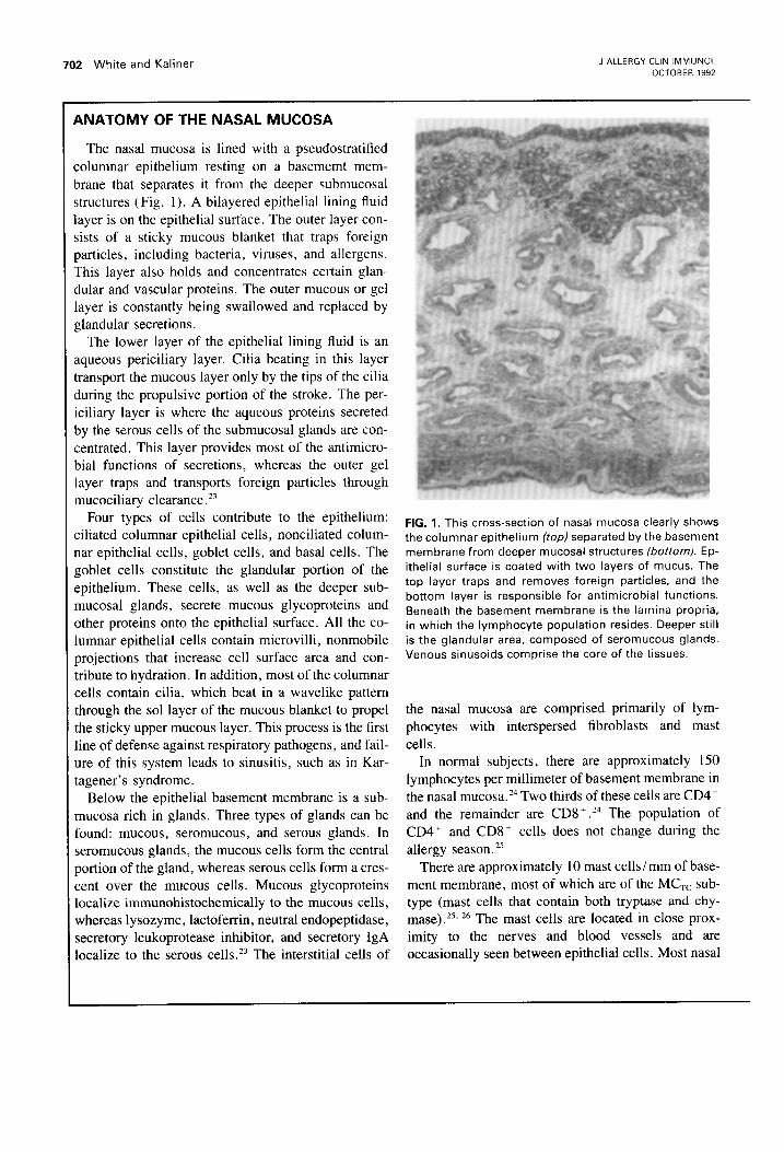
The nasal mucosa is lined with a pseudostratified :olumnar epithelium resting on a basememt mem- brane that separates it from the deeper submucosal structures (Fig. 1). A bilayered epithelial lining fluid layer is on the epithelial surface. The outer layer con- sists of a sticky mucous blanket that traps foreign particles, including bacteria, viruses, and allergens. This layer also holds and concentrates certain glan- dular and vascular proteins. The outer mucous or gel layer is constantly being swallowed and replaced by glandular secretions.
The lower layer of the epithelial lining fluid is an aqueous periciliary layer. Cilia beating in this layer transport the mucous layer only by the tips of the cilia during the propulsive portion of the stroke. The per- iciliary layer is where the aqueous proteins secreted by the serous cells of the submucosal glands are con- centrated. This layer provides most of the antimicro- bial functions of secretions, whereas the outer gel layer traps and transports foreign particles through mucociliary clearance.”
Four types of cells contribute to the epithelium: ciliated columnar epithelial cells, nonciliated colum- nar epithelial cells, goblet cells, and basal cells. The goblet cells constitute the glandular portion of the epithelium. These cells, as well as the deeper sub- mucosal glands, secrete mucous glycoproteins and other proteins onto the epithelial surface. All the co- lumnar epithelial cells contain microvilli, nonmobile projections that increase cell surface area and con- tribute to hydration. In addition, most of the columnar cells contain cilia, which beat in a wavelike pattern through the sol layer of the mucous blanket to propel the sticky upper mucous layer. This process is the first line of defense against respiratory pathogens, and fail- ure of this system leads to sinusitis, such as in Kar- tagener’s syndrome.
Below the epithelial basement membrane is a sub- mucosa rich in glands. Three types of glands can be found: mucous, seromucous, and serous glands. In seromucous glands, the mucous cells form the central portion of the gland, whereas serous cells form a cres- cent over the mucous cells. Mucous glycoproteins localize immunohistochemically to the mucous cells, whereas lysozyme, lactoferrin, neutral endopeptidase, secretory leukoprotease inhibitor, and secretory IgA localize to the serous cells.23 The interstitial cells of
FIG. 1. This cross-section of nasal mucosa clearly shows the columnar epithelium (top) separated by the basement membrane from deeper mucosal structures /bottom). Ep- ithelial surface is coated with two layers of mucus. The top layer traps and removes foreign particles, and the bottom layer is responsible for antimicrobial functions. Beneath the basement membrane is the lamina propria, in which the lymphocyte population resides. Deeper still is the glandular area, composed of seromucous glands. Venous sinusoids comprise the core of the tissues.
the nasal mucosa are comprised primarily of lym- phocytes with interspersed fibroblasts and mast cells.
In normal subjects, there are approximately 150 lymphocytes per millimeter of basement membrane in the nasal mucosa.24 Two thirds of these cells are CD4’ and the remainder are CD8’ .“’ The population of CD4’ and CD8 + cells does not change during the allergy season. 25
There are approximately 10 mast cells/ mm of base- ment membrane, most of which are of the MC,, sub- type (mast cells that contain both tryptase and chy- mase).25, 26 The mast cells are located in close prox- imity to the nerves and blood vessels and are occasionally seen between epithelial cells. Most nasal

VOLUME 90 NUMBER 4, PART 2
Mediators of allergic rhsnitls 703
mast cells are found in the superficial 200 p,m of mucosa, generally clustered beneath the basement membrane and in the epithelium. The number of mast cells in normal subjects is approximately 7000/mm3 in the tissue and only 50/mm3 in the epithelium. Ep- ithelial mast cells are of the MC, subtype,Z6 and the number of these epithelial mast cells increases during the allergy season.” The total number of mast cells and lymphocytes does not increase during the allergy season.‘5 Interestingly, the only cells bearing IgE in the human nasal mucosa are mast cells.24
The nose is one of the most vascular organs of the body, with a total blood flow per cubic centimeter of tissue exceeding that in muscle, brain, and liver.‘* Blood flows in the superficial mucosa are at a rate of 42 ml/ 100 gm of tissue per minute, which is excep- tionally high.29 Blood vessels feeding the epithelium and glands contain fenestrations in their basement membranes, similar to those of the renal glomeruli. This anatomic arrangement facilitates rapid extrava- sation of fIuid through the vascular walls, presumably to assist in the hydration of inspired air.
Although blood in the nose flows typically from arteries to capillaries and into veins, numerous arte- riovenous anastomoses are also found in the nasal mucosa. In addition, cavernous sinusoids lie between the capillaries and venules. These vessels, which are most dense in the inferior and middle turbinates, con- tain smooth muscle cells that are under sympathetic control.“’ Normally, the cavernous sinuses are con- tracted; however, under conditions of reduced sym- pathetic tone or cholinergic stimulation, this erectile tissue becomes engorged and causes considerable na- sal obstruction.
Nasal neuropeptides
Although the veins and venules in the nasal mucosa are predominately innervated by sympathetic nerves, the arterial vessels that supply the glands, as well as the glands themselves, are innervated by both cholin- ergic and sympathetic fibers.”
Cholinergic stimulation causes arteriolar dilation, theoretically enhancing passive diffusion of plasma protein into the glands. In addition, the same cholin- ergic stimulation simultaneously induces active se- cretion from serous and mucous cells.19 Most of the cholinergic receptors on nasal glands and blood ves- sels are of the M3 type.“‘. 33 In addition to containing the classic neurotransmitters, the three types of nerve
fibers innervating the nose (the sensory, cbolinergic. and sympathetic nerves) also contain neuropeptides. Sensory fibers from the trigeminal nerve are capable of responding to noxious stimuli such as mechanical or thermal injury, mediators such as bradykinin or histamine, and acute injury.‘” These nerves i‘~mn the afferent limb of several central reflexes and also par- ticipate in local vascular responses that are similar to the cutaneous wheal-and-flare response.
Several neurotransmitters including CGRP, the tachykinins SP and NKA. and GRP have been fi,und in sensory nerves? Parasympathetic neurons con- tain acetylcholine as well as VIP and a neuropep- tide called peptide histidine methionine. Sympathetic neurons contain norepinephrine and neuropeptide Y. a peptide that has many of the same effects as nor- epinephrine but produces very long-lasting vasocon- striction.
The exact role of these neuropeptides in allergic diseases is still being determined. Immunohistochem- ical studies have demonstrated weak staining for CGRP, SP, and NKA fibers in the nasal muci)sal ep- ithelium. The submucosal glands, arterioles, and venous sinusoids are innervated by fibers staming for GRP, CGRP, SP, NKA, and VIP. On the assumption that these neuropeptides can affect only structures that contain specific receptors, the distribution ot neuro- peptide binding sites has also been studied. The only neuropeptide for which both fibers and receptors have been localized to the epithelium is SP.”
Submucosal glands are rich in both receptors and fibers for (in descending order) GRP. VIP, and SP.j5”’ The venous sinusoids, however, contain only a few neuropeptide receptors for CGRP, SP, and VIP. In contrast, arterioles contain receptors for a11 of the neuropeptides studied except GRP; however, most oi
the receptors are for CGRP. which causes v,isodila- tion,38 or for neuropeptide Y. which induces c’:.tsocon- striction. ”
To assess the effect of these neuropeptides <,n gian- dular secretion, nasal turbinate explants were incu- bated ex vivo with control fluids or with various neu-
ropeptides, and the secretion of glycocon,jugates from
mucous cells and of lactoferrin from serous celis was measured. ” Both GRP and VIP were potent secre- tagogues for serous cells and somewhat les% so for mucous cells. SP and NKA were weak mucous cell secretagogues, whereas the latter was also .I weak serous cell secretagogue. iv

704 White and Kaliner J ALLERGY CLIN IMMUNOL OCTOBER 1992
REFERENCES
1. Kaliner MA, Lcmanske R. Rhinitis and asthma. JAMA (in press).
2. Friedman MM, Kaliner M. In situ degranulation of human nasal mucosal mast cells: ultrastructural features and cell-cell interactions. J ALLERGY CLIN IMMUNOL 1985;76:70-82.
3. Naclerio RM, Meier HL, Kagey-Sobotka A, et al. Mediator release after nasal airway challenge with allergen. Am Rev Respir Dis 1983;128:597-602.
4. Raphael GD, Meredith SD, Baraniuk JN, Druce HM, Banks SM, Kaliner MA. The pathophysiology of rhinitis. II. As- sessment of the sources of protein in histamine-induced nasal secretions. Am Rev Respir Dis 1989;139:791-800.
5. Simons FER. H,-receptor antagonists: clinical pharmacology and therapeutics. J ALLERGY CLIN IMMUNOL 1989;84:845-61.
6. Mosimann B, White MV, Kaulbach HK, Goldrich M, Kaliner MA. Substance P in nasal secretions after allergen challenge [Abstract]. J ALLERGY CLIN IMMUNOL 1992;89:210.
7. Proud D, Baumgarten CR, Naclerio RM, Ward PE. Kinin metabolism in human nasal secretions during experimentally induced allergic rhinitis. J Immunol 1987;138:428-34.
8. Burd PR, Rogers HW, Gordon JR, Martin CA, Jayaraman S, Wilson SD, Dvorak AM, Galli SF, Dorf ME. Interleukin-3- dependent and -independent mast cells stimulated with IgE and antigen express multiple cytokines. J Exp Med 1989;170;245- 57.
9. Gordon JR, Galli SJ. Mast cells as a source of both preformed and immunologically inducible TNF-a cachectin. Nature 1990;346:274-6.
10. Plaut M, Pierce JH, Watson CJ, Hanley-Hyde J, Nordan RP, White WE. Mast cell lines produce lymphokines in response to cross-linkage of FC.RI or to calcium ionophores. Nature 1989;339:64-7.
11. Gurish MF, Ghildyal N, Arm J, et al. Cytokine mRNA are preferentially increased relative to secretory granule protein mRNA in mouse bone marrow-derived mast cells that have undergone IgE-mediated activation and degranulation. J Im- munol 1991;146:1527-33.
12. Durham SR, Ying S, Vamey VAS, et al. Cytokine messenger RNA expression for IL-3, IL-4, IL-5, and granulocyte/mac- rophage-colony stimulating factor in the nasal mucosa after local allergen provocation: relationship to tissue eosinophilia. J Immunol 1992;148:2390-4.
13. Moss RB, Ohkubo K, Paciotti G, et al. Interleukin 2 (IL-2) immunoreactivity in nasal mucosa [Abstract]. J ALLERGY CLIN IMMUNOL 1992;89: 179.
14. Bochner BS, Charlesworth EN. Lichtenstein LM, et al. Inter- leukin-1 is released at sites of human cutaneous allergic re- actions. J ALLERGY CLIN IMMUNOL 1990;86:830-9.
15. Sim TC, Alam R, Hilsmeier KA, Grant JA. Detection of in- flammatory cytokines in nasal secretions (NS) of allergic sub- jects following antigen challenge [Abstract]. J ALLERGY CLIN IMMLJNOL 1992;89:216.
16. Steinberg DG, Rosenwasser LJ. Interleukin-1B production af- ter nasal challenge with FEL d 1 in allergic and nonallergic subjects [Abstract]. J ALLERGY CLIN IMMUNOL 1992;89:217.
17. Sur S, Hunt LW, Weiler DA, Ohnishi T, Gleich GJ. In vitro release of eosinophil active cytokines from airway during re- flexes. Am J Rhino1 1988;2:109-16.
18. Raphael GD, Igarashi Y, White MV, Kaliner MA. The patho- physiology of rhinitis. V: sources of protein in allergen-induced nasal secretions. J ALLERGY CLIN IMMUNOL 1991;88:33-42.
19. Raphael GD, Baraniuk JN, Kaliner MA. How the nose runs and why. J ALLERGY CLIN IMMUNOL 1991;87:457-67.
20. Naclerio RM, Proud D, Togias AG, et al. Inflammatory me- diators in late antigen-induced rhinitis. N Engl J Med 1985;313:65-70.
21. White M, Goldrich M, Daniel A, Kaliner M. Antigen challenge causes increased nasal histamine-induced vascular permeability [Abstract]. J ALLERGY CLIN IMMUNOL 1992;89:206.
22. Gundel RH, Wegner CD, Torcelbni CA. Endothelial leukocyte adhesion molecule- 1 mediates antigen-induced acute airway inflammation and late-phase airway obstruction in monkeys. 3 Clin Invest 1991;88:1407-11.
23. Kaliner MA. Human respiratory secretions and host defense. Am Rev Respir Dis 1991; 144:S52-S66.
24. Igarashi Y, Scott TA, Hausfeld JH, Kaliner MA, White MV. Inflammatory cell infiltrates in human nasal mucosa [Abstract]. J ALLERGY CLIN IMMUNOL 1992;89:204.
25. Bentley AM, Jacobson MR, Cumberworth V, et al. Immu- nohistology of the nasal mucosa in seasonal allergic rhinitis: increases in activated eosinophils and epithelial mast cells. J ALLERGY CLIN IMMUNOL 1992;89:877-83.
26. Igarashi Y, White M, Hausfeld J, Irani A, Schwartz L, Kaliner M. IgE-positive cells in nasal mucosa are mostly mast cells. FASEB J 1992;6:A2005.
27. Lozewicz S, Gomez E, Clague J, Gatland D, Davies RJ. Al- lergen-induced changes in the nasal mucous membrane in sea- sonal allergic rhinitis: effect of nedocromil sodium. J ALLERGY CLIN IMMUNOL 1990;85: 125-31,
28. Drettner B, Aust R. Plethysmographic studies of the blood flow in the mucosa of the human maxillary sinus, Acta Oto- laryngol 1974;74:259-63.
29. Druce HM, Bonner RF, Patow C, Choo P, Kaliner MA. Re- sponse of nasal blood flow to neurohotmones as measured by laser-Doppler velocimetry. J Appl Physiol 1984;57: 1276-83.
30. Atkinson TP, Kaliner MA. Vascular mechanisms in rhinitis. In: Busse W, Holgate S, eds. Asthma and rhinitis. Cambridge, Mass.: Blackwell Scientific (in press).
31. Raphael GD, Meredith SD, Baraniuk JN, Kaliner MA. Nasal reflexes. Am J Rhino1 1988;2:109-16.
32. Okayama M, Mull01 J, Baraniuk JN, et al. Muscarinic receptor subtypes in human nasal mucosa: characterization, autoradi- ographic localization and function in vitro. J ALLERGY CLIN IMMUNOL (in press).
33. Baraniuk JN, Kaliner MA, Barnes PJ. Localization of M3 muscarinic receptor mRNA in human nasal mucosa. Am J Rhino1 (in press).
34. Baraniuk JN, Kaliner MA. Functional activity of upper airway nerves. In: Busse W, Holgate S, eds. Asthma and rhinitis. Cambridge, Mass.: Blackwell Scientific (in press).
35. Baraniuk JN, Lundgren JD, Okayama M, et al. Substance P and neurokinin A in human nasal mucosa. Am J Resp Cell Molec Biol 1991;4:228-36.
36. Baraniuk JN, Lundgren JD, Goff J, et al. Gastrin releasing peptide (GRP) in human nasal mucosa. J Clin Invest 1990;85:998-1005.
37. Baraniuk JN, Lundgren JD, Okayama M, et al. Vasoactive intestinal peptide (VIP) in human nasal mucosa. J Clin Invest 1990;86:825-31.
38. Baraniuk JN, Lundgren JD, Goff J, et al. Calcitonin gene related peptide in human nasal mucosa. J Appl Physiol 1990;258:81-8.
39. Baraniuk JN, Castellino S, Lundgren JD, et al. Neuropeptide Y (NPY) in human nasal mucosa. Am J Resp Cell Molec Biol 1990:3:165-74.
Copyright © 2022 FDOKUMEN


![[ARIA (Allergic Rhinitis and its Impact on Asthma). Achievements in 10 years and future needs in Latin America]](https://static.fdokumen.com/doc/165x107/6333c8977a687b71aa08605d/aria-allergic-rhinitis-and-its-impact-on-asthma-achievements-in-10-years-and.jpg)