Differential oxidation of apolipoprotein E isoforms and interaction with phospholipids
Upload
independentCategory
view
0download
0
Rhinitis, sinusitis, and upper airway disease
Apolipoprotein A-IV is a candidate target molecule for thetreatment of seasonal allergic rhinitis
Yuka Makino, MS,a Emiko Noguchi, MD, PhD,a Noboru Takahashi, MD, PhD,c Yuri Matsumoto, MS,a Seita Kubo, MD,c
Takechiyo Yamada, MD, PhD,c Yoshimasa Imoto, MD,c Yumi Ito, MD,c Yoko Osawa, MD,c Masanao Shibasaki, MD,d
Kazuhiko Uchida, PhD,b Kohji Meno, PhD,b Hideaki Suzuki, BA,b Kimihiro Okubo, MD, PhD,e Tadao Arinami, MD, PhD,a
and Shigeharu Fujieda, MD, PhDc Tsukuba, Fukui, and Tokyo, Japan
Background: Allergic rhinitis is a global health problem thatcauses major illnesses and disability worldwide. Allergen-specificimmunotherapy (SIT) is the only available treatment that canalter the natural course of allergic disease. However, the precisemechanism underlying allergen-SIT is not well understood.Objective: The aim of the current study was to identify proteinexpression signatures reflective of allergen-SIT—morespecifically, sublingual immunotherapy (SLIT).Methods: Serum was taken twice from patients with seasonalallergic rhinitis caused by Japanese cedar: once before thepollen season and once during the season. A total of 25 patientswas randomly categorized into a placebo-treated group and anactive-treatment group. Their serum protein profiles wereanalyzed by 2-dimensional electrophoresis.Results: Sixteen proteins were found to be differentiallyexpressed during the pollen season. Among the differentiallyexpressed proteins, the serum levels of complement C4A,apolipoprotein A-IV (apoA-IV), and transthyretin weresignificantly increased in SLIT-treated patients but not inplacebo-treated patients. Among these proteins, the serumlevels of apoA-IV correlated with the clinical symptom-medication scores (r 5 –0.635; P < .05) and with quality of lifescores (r 5 –0.516; P < .05) in the case of SLIT-treated patients.The amount of histamine released from the basophils in vitrowas greatly reduced after the addition of recombinant apoA-IVin the medium (P < .01).Conclusion: Our data will increase the understanding of themechanism of SLIT and may provide novel insights into the
From the Departments of aMedical Genetics and bMolecular Biological Oncology,
Graduate School of Comprehensive Human Sciences, University of Tsukuba, Ibaraki,
Japan; cthe Department of Otorhinolaryngology Head and Neck Surgery, Faculty of
Medical Sciences, University of Fukui, Fukui, Japan; dthe Department of Pediatrics,
Tsukuba University of Technology; University of Tsukuba, Ibaraki, Japan; and ethe
Department of Otorhinolaryngology, Nippon Medical School, Tokyo, Japan.
Supported by a Grant-in-Aid for Scientific Research from the Ministry of Health and
Welfare, Japan (H17-Genome-001, H17-Immunology-001, H20-Immunology-001,
-004) and from the Ministry of Education, Science and Culture of Japan (17390458,
18591097, 20390441).
Disclosure of potential conflict of interest: The authors have declared that they have no
conflict of interest.
Received for publication March 2, 2010; revised June 18, 2010; accepted for publication
June 28, 2010
Available online August 31, 2010.
Reprint requests: Emiko Noguchi, MD, PhD, Department of Medical Genetics, Graduate
School of Comprehensive Human Sciences, University of Tsukuba, 1-1-1 Tennodai,
Tsukuba-city, Ibaraki, Japan. E-mail: [email protected].
0091-6749/$36.00
� 2010 American Academy of Allergy, Asthma & Immunology
doi:10.1016/j.jaci.2010.06.031
treatment of allergic rhinitis. (J Allergy Clin Immunol2010;126:1163-9.)
Key words: Sublingual immunotherapy, apoA-IV, HNF4A, proteome
Allergic rhinitis is a global health problem that causes majorillnesses and disability worldwide. A conservative estimaterevealed that allergic rhinitis occurs in over 500 million peoplearound the world.1 Seasonal allergic rhinitis (SAR) caused byJapanese cedar (Cryptomeria japonica; JC) is the most commonallergic disease in Japan. According to a national survey, the prev-alence of allergic rhinitis in Japan was 0.16 in 1992 and 0.21 in2002.2 The results of our recent study showed that over 35% ofJapanese individuals in the age group 20 to 50 years develop aller-gic symptoms during JC pollination season.3
Allergen-specific immunotherapy (SIT) is the only availabletreatment that can alter the natural course of allergic disease bypreventing new sensitization/onset and providing long-termremission after discontinuation of treatment.4 Many clinical trialshave proven the efficacy of SIT in controlling allergic diseases. 5
Conventional SIT, subcutaneous injection, however, requiresfrequent hospital visits and is painful, resulting in a low patientcompliance. Further, it may cause some adverse events such asanaphylaxis. To overcome these disadvantages, sublingual immu-notherapy (SLIT), oral administration of the allergen, was intro-duced as an alternative method,6 and to date, SLIT has beenwidely used to treat patients with asthma and rhinitis.4 The clin-ical efficacy of SLIT has been widely proven by many studies,6,7
and several studies have revealed that there is increased produc-tion of blocking antibody, IgG4,8 as well as induction of regula-tory T cells9 as a result of SLIT. However, the precisemechanism underlying SLIT is not well understood.
The word proteome describes the entire collection of proteinsin a cell, tissue, or body fluid at a given time. With a proteomicapproach, all the proteins present in a biological sample can bevisualized simultaneously and identified. This approach is notbased on any experimental hypothesis but on correlation-associated network analyses of proteomic profiles, leading tohypotheses regarding relations between structurally and biolog-ically related proteins/peptides. Therefore, this approach can beused to identify proteins associated with SLIT, which could im-prove our understanding of the mechanism underlying this ther-apy. In this study, we performed proteomic analysis to identifyprotein expression signatures that reflect the responsiveness toSLIT and to determine novel therapeutic targets for the treatmentof SAR.
1163

J ALLERGY CLIN IMMUNOL
DECEMBER 2010
1164 MAKINO ET AL
Abbreviations used
ApoA-IV: A
polipoprotein A-IV2-DE: T
wo-dimensional polyacrylamide gelelectrophoresis
HNF4A: H
epatocyte nuclear factor 4aHRP: H
orseradish peroxidaseJAU: Ja
panese allergy unitJC: C
ryptomeria japonica JRQLQ: Ju niper Rhinoconjunctivitis Quality-of-LifeQuestionnaire
MALDI-TOF/TOF: M
atrix laser desorption/ionization 2-stagetime-of-flight
QOL: Q
uality of lifeSAR: S
easonal allergic rhinitisSIT: S
pecific immunotherapySLIT: S
ublingual immunotherapySMS: S
ymptom-medication scoreMETHODS
SubjectsPatients with SAR caused by JC pollens were enrolled in this study. All the
patients exhibited the following characteristics: (1) JC pollen-specific RAST
score >_2, (2) positive allergic reaction after nasal challenge with JC allergens,
(3) JC pollen–induced symptoms of allergic rhinoconjunctivitis (from Feb-
ruary to April) in the last 2 years and medication for the treatment of the
symptoms, (4) no history of asthma, and (5) no allergen-specific immuno-
therapy in the past. Total and specific IgE (JC, Dermatophagoides, Dactylis
glomerata, Ambrosia artemisiifolia, Candida albicans, and Aspergillus)
were measured by using the CAP-RAST method (Pharmacia Diagnostics
AB, Uppsala, Sweden) in all patients, and positive allergic sensitization was
defined if the levels of 1 or more specific IgE molecules were greater than
or equal to 0.70 IU/mL (class 2). A total of 25 patients were randomly catego-
rized into a placebo-treated group and an actively treated group; the patients in
the latter group received JC pollen extract. One patient in the placebo group
withdrew from the study for personal reasons. The characteristics of the pa-
tients are presented in Table I and this article’s Table E1 in the Online Repos-
itory at www.jacionline.org.
Before enrollment, written informed consent was obtained from each
patient, and the trial was performed in compliance with the Declaration of
Helsinki and Good Clinical Practice. The trial was approved by the ethics
committees of the University of Fukui and University of Tsukuba. The
members of the ethical committee suggested that the number of samples
collected for the active group should be 150% of that collected for the placebo
group; thus, we collected samples according to their suggestion. JC pollen
extracts were prepared by Torii Pharmaceutical Co, Ltd (Tokyo, Japan). The
extracts (2000 Japanese allergy units [JAU]/mL) contained 15 mg Cry j 1 and 2
to 5 mg Cry j 2. Administration of the allergen extract was initiated at a dose of
2 JAU/mL with 50% glycerin as a diluent in November 2004; thereafter, the
dose was gradually increased to 1 mL 2000 JAU/mL (final maintenance
concentration) and maintained at this concentration until April 2005. The
placebo-treated group was administered only the diluent, 50% glycerin. The
details of the JC pollen sublingual immunotherapy have been described
previously.10
Symptom-medication score and quality of life scoreThe number of paroxysmal sneezes and occasions when the patients blew
their noses were recorded daily on forms that were used to record nasal
symptoms. On the basis of these numbers, the patients were graded on a scale
of 0 to 4 (0, none; 1, 1-5 times; 2, 6-10 times; 3, 11-20 times; and 4, over 20
times). Nasal congestion was also graded on a scale of 0 to 3 (0, no symptoms;
1, mild; 2, moderate; and 3, severe symptoms).10,11 During SLIT/placebo
treatment, the use of other medications, including oral antihistamine and top-
ical steroids, was also recorded daily. The total symptom-medication score
(SMS) was daily calculated on the basis of the abovementioned grades; fur-
ther, we analyzed the correlation between the scores obtained over 2 weeks,
during the peak JC pollination season, and the level of serum proteins. Quality
of life (QOL) was assessed by using the modified Japanese version of Juniper
Rhinoconjunctivitis Quality-of-Life Questionnaire (JRQLQ).10-12 This ques-
tionnaire includes 17 questions in 6 domains designed to measure the effects
of rhinoconjunctivitis symptoms on disease-specific QOL.
Sample collectionSerum was collected from each patient twice: before the pollen season and
the initiation of SLIT (November 2004) and during the pollen season (May to
April 2005). Serum samples were centrifuged at 3000g for 10 minutes and
stored at –808C until use.
Two-dimensional polyacrylamide gel
electrophoresis and protein identificationTwo-dimensional polyacrylamide gel electrophoresis (2-DE) was per-
formed with the IPGphor IEF System (GE Healthcare, Piscataway, NJ) and
Ettan DALT six (GE Healthcare) as described previously.13 Labeled proteins
were visualized with a Typhoon 9400 Imager (GE Healthcare), and 24 images
of paired samples were analyzed with the DeCyder Software Platform version
4.0 (GE Healthcare). The detailed Methods are available in this article’s
Online Repository at www.jacionline.org.
The data concerning the changes in the amounts of proteins in each spot
were combined, and the spots, which indicated significant changes in the
amount of proteins before and after the treatment, were analyzed.
Protein spots that satisfied both the following criteria were subjected to
protein identification: (1) those in which at least a 1.1-fold increase or decrease
in protein expression was observed, and (2) those that indicated significant
differences in the protein expression levels before and after SLIT (paired t test,
P < .05). Differentially expressed protein spots were subjected to nano-HPLC,
and samples were separated on a Paradigm MS4 LC system (Michrom Biore-
sources, Auburn, Calif).14 The purified samples were analyzed by using
matrix-assisted laser desorption/ionization 2-stage time-of-flight (MALDI-
TOF/TOF) mass spectrometry; all the imaging analysis was performed on
an Ultraflex II (Bruker Daltonics, Billerica, Mass). Detailed Methods can be
accessed in the Online Repository.
Western blot analysis of apolipoprotein A-IVThe protocol of the Western blot analysis to validate the apolipoprotein
A-IV (apoA-IV) spot is described in the Methods in the Online Repository. For
the quantification of apoA-IV, the protein concentration in the serum was mea-
sured by a protein assay (Bio-Rad, Hercules, Calif). Then the protein concen-
trations in the serum samples were adjusted to 4 mg/mL, and these prepared
samples were separated on 10% SDS-polyacrylamide gels. The separated
proteins were transferred onto nylon membranes (PVDF; GE Healthcare) by
a semidry electrical transfer (Bio-Rad). Nonspecific binding sites were
blocked for 1 hour at room temperature with 1% blocking reagent in PBS-
Tween 20 (0.1%; Roche, Indianapolis, Ind). The membranes were incubated
with mAb (dilution 1:2000; antihuman apoA-IV mouse IgG antibody;
BML, Saitama, Japan) for 1 hour at room temperature. After washing, the
membranes were incubated with antimouse IgG (H 1 L-chain)–horseradish
peroxidase (HRP) goat IgG antibody (MBL, Nagoya, Japan) for 1 hour at
room temperature. After washing 3 times, the membranes were incubated
with Immobilon western chemiluminescent HRP substrate (Millipore, Biller-
ica, Mass) for 1 minute. The chemiluminescent images were then analyzed
with a LAS-4000UVmini and Multi Gauge Version 3.0 (Fujifilm Life Science,
Tokyo, Japan).
Test for histamine release from basophils
containing various apolipoproteinsRecombinant apolipoprotein A-I and apolipoprotein E were purchased
from BioVision (Mountain View, Calif), and recombinant apolipoprotein C-III

FIG 1. Upregulated and downregulated proteins by 2-DE analysis. Upregu-
lated or downregulated protein spots that were altered in the actively
treated and placebo-treated groups are marked with circles. The spot
numbers correspond to the numbers in Table II.
TABLE I. Characteristics of patients with SAR
Placebo-treated
(n 5 9)
Actively treated
(n 5 15)
P
value
Age (y), mean 6 SD 47.5 6 13.1 48.1 6 14.7 .926
Male/female ratio 11/4 6/3 .742
IgE (IU/mL), mean
(range)
124.7 (7-370) 136.5 (11-470) .823
JC pollen–specific IgE
(UA/mL), mean (range)
17.8 (1.25-73.5) 10.6 (1.18-27.1) .308
J ALLERGY CLIN IMMUNOL
VOLUME 126, NUMBER 6
MAKINO ET AL 1165
was purchased from Abnova Co (Taipei, Taiwan). Recombinant apoA-IV was
expressed in COS-7 cells and purified by using the QIAexpress Ni-NTA Fast
Start Kit (Qiagen, Valencia, Calif). Detailed Methods for the production of re-
combinant apoA-IV can be accessed in the Online Repository. Anticoagulated
blood obtained from 6 patients with JC pollinosis was subjected to gradient
centrifugation to obtain peripheral mononuclear cells by using Lymphoprep
(Axis-Shield, Oslo, Norway). The basophils were enriched by using the Baso-
phil Isolation Kit II (Miltenyi Biotec, Gladbach, Germany) and autoMACS
(Miltenyi Biotec) according to the manufacturer’s instructions. The enriched
basophils (>98%) were seeded on a 96-well plate at a density of 5 3 104 cells/
well and incubated in RPMI 1640 medium (Nissui Pharmaceutical, Tokyo, Ja-
pan) containing 10% FCS (Gibco, Grand Island, NY), 100 U/mL penicillin-G
potassium salt (Sigma, St Louis, Mo), and 100 mg/mL streptomycin sulfate
salt (Sigma) at 378C under 5% CO2 for 30 minutes. The basophils were further
incubated with or without the apolipoproteins (apolipoprotein A-I, apoA-IV,
apolipoprotein C-III, and apolipoprotein E; final concentration of apolipopro-
teins, 1 mg/mL) for 30 minutes; subsequently, Cry j 1 (Hayashibara Biochem-
ical Laboratories, Okayama, Japan) was added to each well at a final
concentration of 0.1 mg/mL. After 30 minutes of incubation, histamine con-
centrations in the cells and supernatants were determined by using a Histamine
ELISA kit (Oxford Biomedical Research, Oxford, Mich) according to the
manufacturer’s instructions. Histamine release (%) was calculated as follows:
histamine in supernatant
=ðhistamine in supernatant
1 histamine in basophilsÞ3 100:
The percent inhibition of histamine release by apolipoproteins was
calculated as follows:
�½histamine release without apolipoproteins
� histamine release with apolipoproteins�
=histamine release without apolipoproteins�3 100:
Pathway analysisTo investigate whether the differentially expressed proteins belong to
specific pathways or networks, we used the IPA version 7.1 software
(Ingenuity Systems, Mountain View, Calif). This web-based software enables
the identification of biologic networks relevant to each researcher’s experi-
ment. A data set containing protein identifiers and the corresponding
expression values was uploaded onto the Ingenuity pathways knowledge
base. These uploaded proteins (referred to as focus genes) were then used as
starting points for generating biological networks, and a network was con-
structed such that it was enriched with the proteins of interest.
Statistical analysisThe differences in the protein levels before and after SLIT were analyzed
for statistical significance by the paired t test. The statistical significance of the
difference in apoA-IV levels observed by Western blot analysis was calculated
by the Wilcoxon signed-rank sum test. Pairwise correlations between SMS
and serum levels of apoA-IV were calculated by using the Pearson correlation.
Significance was defined as P < .05.
RESULTSA representative 2-DE image of the serum samples from a
SLIT-treated patient is shown in Fig 1. Sixteen spots were differ-entially expressed in the samples obtained before and after thepollen seasons (Fig 1; Table II). Before 2-DE analysis of the sam-ples of patients with SAR, we evaluated the reproducibility anddeviation of the spot intensity by repeated experiments withsame samples labeled with 2 different dyes (Cy5 and Cy3). Thevariation in the 2-DE analysis was less than 1.1-fold. Thus, a pro-tein spot showing a greater than 1.1-fold change in expressionwith a statistically significant difference of P <. 05 in the samplesobtained before and after the pollen season was considered to bedifferentially expressed spot. Fig 2 shows the output of the De-Cyder differential analysis software in the case of spot 10 in ac-tively treated patients. The graphical data showed that theamount of apoA-IV protein increased after SLIT. These alteredspots were excised and analyzed by MALDI-TOF/TOF analysis.Fifteen spots were successfully identified by MALDI-TOF/TOFanalysis. These identified spots corresponded to 7 proteins(Table II). We classified these 7 proteins on the basis of their func-tion into 4 categories: lipid transporters, complement factors, pro-tease inhibitors, and transporters. An example of MALDI-TOF/TOF analysis is shown in this article’s Fig E1 in the Online Repos-itory at www.jacionline.org (spot 10, ApoA-IV). Among theseidentified proteins, the serum levels of complement C4A, apoA-IV, and transthyretin were significantly increased in the patientsbelonging to the actively treated group (P < .05), but this trendwas not observed in the patients belonging to the placebo-treated group. The results of pathway analysis with the 3 SLIT-related proteins are shown in Fig 3. The IPA software generatesa large global molecular network on the basis of hundreds of thou-sands of curated direct and indirect physical and functional inter-actions between orthologous mammalian genes/proteins frompublished, peer-reviewed content in the Ingenuity knowledgebase. As shown in Fig 3, hepatocyte nuclear factor 4a
(HNF4A) was identified as the hub protein in the network, indi-cating that these 3 proteins were regulated by the commontranscription factor HNF4A.
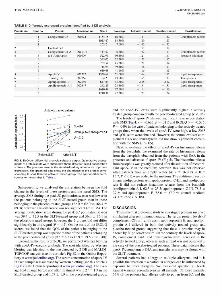
TABLE II. Differently expressed proteins identified by 2-DE analysis
Protein no Spot no Protein Accession no Score Coverage Actively treated Placebo-treated Classification
1 1 Complement C3 P01024 1154.15 16.60% 1.4 1.42 Complement factors
2 1015.47 14.50% 1.29 1.25
11 252.2 7.00% 21.45 21.32
2 3 Unidentified 1.17 1.12
3 4 Complement C4-A P0C0L4 414.57 6.30% 1.32 1.13 Complement factors
4 5 a-1-Antitrypsin P01009 522.93 30.40% 1.2 1.17 Protease inhibitors
6 395.05 32.50% 1.21 1.17
7 752.54 45.20% 1.21 1.14
8 715.94 39.50% 1.16 1.17
9 619.68 41.10% 1.16 1.15
5 10 ApoA-IV P06727 1339.68 51.00% 1.64 1.12 Lipid transporters
6 12 Transthyretin P02766 184.23 43.50% 1.65 1.31 Transporters
7 13 Apolipoprotein E P02649 647.84 43.80% 2.88 2.82 Lipid transporters
8 14 Apolipoprotein A-I P02647 262.23 28.80% 21.3 21.51 Lipid transporters
15 1610.49 77.50% 21.1 21.16
16 1210.31 77.20% 21.27 21.24
FIG 2. DeCyder differential analysis software output. Quantitative assess-
ments of protein spots were obtained with the DeCyder-based quantization
software. The y-axis represents the standardized log abundance of protein
expression. The graphical data show the abundance of the protein corre-
sponding to spot 10 in the actively treated group. The spot number corre-
sponds to the number in Table II.
J ALLERGY CLIN IMMUNOL
DECEMBER 2010
1166 MAKINO ET AL
Subsequently, we analyzed the correlation between the foldchange in the levels of these proteins and the nasal SMS. Theaverage SMS during the peak JC pollination season was lower inthe patients belonging to the SLIT-treated group than in thosebelonging to the placebo-treated group (122.0 6 62.0 vs 166.4 6
89.0); however, this difference was not significant (P 5 .36). Theaverage medication score during the peak JC pollination seasonwas 39.4 6 12.5 in the SLIT-treated group and 56.0 6 16.1 inthe placebo-treated group; however, the 2 groups did not differsignificantly in this regard (P 5 .42). On the basis of the JRQLQscores, we found that the QOL of the patients belonging to theSLIT-treated group was superior to that of the patients belongingto the placebo-treated group (9.5 6 8.3 vs 15.9 6 19.6; P 5 .048).
To confirm the results of 2-DE, we performed Western blottingwith apoA-IV–specific antibody. The spot identified by Westernblotting was identical to the one detected by 2-DE and MALDI-TOF/TOF analysis (see this article’s Fig E2 in the Online Repos-itory at www.jacionline.org). The serum concentration of apoA-IVin each sample was assessed by Western blotting (see this article’sFig E3 in the Online Repository at www.jacionline.org). The aver-age fold change before and after treatment was 3.27 6 1.3 in theSLIT-treated group and 1.57 6 1.0 in the placebo-treated group,
and the apoA-IV levels were significantly higher in activelytreated group compared with the placebo-treated group (P < .05).
The levels of apoA-IV showed significant reverse correlationwith SMS (Fig 4; r 5 –0.635; P 5 .011) and JRQLQ (r 5 –0.516;P 5 .049) in the case of patients belonging to the actively treatedgroup; thus, when the levels of apoA-IV were high, a low SMSand QOL score were obtained. However, the serum levels of com-plement C4A and transthyretin did not show significant correla-tion with the SMS (P > .05).
Next, to evaluate the effect of apoA-IV on histamine releasefrom the basophils, we examined the rate of histamine releasefrom the basophils obtained from the patients with SAR in thepresence and absence of apoA-IV (Fig 5). The histamine releasefrom basophils was greatly reduced after the addition of recombi-nant apoA-IV in the medium; however, this was not observedwhen extracts from an empty vector (41.7 6 16.0 vs 70.0 6
13.7; P < .01) were added to the medium. The addition of recom-binant apolipoprotein A-I, apolipoprotein C-III, and apolipopro-tein E did not reduce histamine release from the basophils(apolipoprotein A-I, 62.5 6 25.3; apolipoprotein C-III, 78.3 6
29.2; and apolipoprotein E, 85.0 6 37.0 vs control medium,74.3 6 26.9; P > .05).
DISCUSSIONThis is the first proteomic study to investigate proteins involved
in inhalant allergen immunotherapy. The serum protein levels ofcomplement C3, a-1-antitrypsin, apolipoprotein E, and apolipo-protein A-I differed in both the actively treated group andplacebo-treated group, suggesting that these 4 proteins may bealtered by JC pollen exposure. On the contrary, the levels of apoA-IV, complement C4A, and transthyretin were increased in theactively treated group, whereas such a trend was not observed inthe case of the placebo-treated patients. These data indicate thatapoA-IV, complement C4A, and transthyretin may be altered withthe SLIT treatment.
Several patients had allergy to multiple allergens, and it ispossible that reaction to a particular allergen can be influenced byexposure to other allergens. We measured specific IgE levelsagainst 6 major aeroallergens in all patients. Of these patients,63% of the patients had allergy only to pollen from JC, and the

FIG 3. Results of the network analysis by Ingenuity pathways analysis. The complement factor C4A,
apoA-IV, and transthyretin are shown in red.
FIG 4. Correlation of the fold change in apoA-IV with the SMS.
A statistically significant correlation was observed between the levels of
apoA-IV and the SMS (r 5 –0.635; P 5 .011 in the actively treated group.
FIG 5. Histamine release from basophils with or without apoA-IV. The
graph shows the histamine release rate from the basophils of SAR patients
with or without apoA-IV.
J ALLERGY CLIN IMMUNOL
VOLUME 126, NUMBER 6
MAKINO ET AL 1167
rest had allergy to mites and other aeroallergens. Among theallergens tested, the pollens of D glomerata and A artemisiifoliaare not dispersed by air currents between February and April inJapan. One patient had allergy to C albicans, and none of the pa-tients had allergy to Aspergillus. Five patients had allergy tomites, but they did not exhibit any symptoms related to perennial
allergic rhinitis or asthma. The ratio of patients with allergy toaeroallergens other than JC in the placebo-treated group (0.33)did not differ from that in the actively treated group (0.40; P 5
1, Fisher exact test); therefore, the allergic reaction obtainedagainst the other allergens did not have a significant impact onthe results of our proteomic study.

J ALLERGY CLIN IMMUNOL
DECEMBER 2010
1168 MAKINO ET AL
Among 3 proteins altered during SLIT, the apoA-IV serumlevels correlated only with the clinical symptom scores, andidentified apoA-IV was detected on the same molecular weightand isoelectric point as native apoA-IV, which was theoreticallycalculated from the database. Therefore, we think apoA-IV weidentified by 2-DE was similar to native apoA-IV. ApoA-IV is a46-kd glycoprotein that is produced mainly in the small intestineand liver.15 Although the precise function of apoA-IV has notbeen completely elucidated, several functions have been pro-posed, such as lipid transport and metabolism,16,17 satiety,18 andantiatherogenic effects.19,20 Several lines of evidence also sug-gested that apoA-IV has anti-inflammatory effects. Vowinkelet al21 showed in their experimental colitis model that apoA-IVknockout mice exhibited a significantly greater inflammatory re-sponse than their wild-type littermates, and this inflammation wasreversed by exogenous administration of apoA-IV to knockoutmice. It has also been shown that expression of human apoA-IVin apolipoprotein E knockout mice significantly reduced the de-velopment of atherosclerosis and release of cytokines such asIL-4, IFN-g, and TNF-a induced by repeated injections ofLPS.22 In the current study, increased levels of apoA-IV wereobserved in the actively treated group, and apoA-IV inhibitedthe release of histamine from basophils. These results combinedwith the results of previous studies indicate that the anti-inflammatory effects of apoA-IV and inhibition of histaminerelease by apoA-IV may contribute to the effect of SLIT for thetreatment of allergic rhinitis.
The complement component system provides innate defenseagainst microbial pathogens and acts in conjunction withantibody-mediated immunity. It has been reported thatcomplement-activation products such as C3a and C5a, known asanaphylatoxins, contribute to inflammation in allergic rhinitis.Andersson et al23 showed that an allergen challenge test adminis-tered to subjects with allergy induced nasal symptoms and con-comitantly increased their C3a and C5a levels. We observedthat the acidic complement component C4A was increased inthe actively treated group compared with the control placebogroup, and the levels of 2 C3 isoforms were upregulated and1 was downregulated in the actively treated group. The humanC4 complement components are encoded by 2 genes, acidicC4A and basic C4B, located on chromosome 6p21.3. C4 defi-ciencies are reported to be associated with Mycobacterium lepraeinfection and autoimmune diseases24; further, increased levels ofC4a, the C4 fragment formed by the cleavage reaction, were ob-served in the case of patients with aspirin-induced asthma com-pared with those with aspirin-tolerant asthma.25 To date, themechanisms underlying the control of C4A gene transcriptionare poorly understood; however, C4A, apoA-IV, and transthyretinwere reported to be regulated by a common transcription factor,HNF4A (Fig 3).26-28 Furthermore, apoA-IVand transthyretin pos-sess HNF4A-binding sites in their regulatory regions.28-30
HNF4A is a liver-enriched transcription factor, and many acutephase proteins have HNF4A-binding sites in their regulatory ele-ments, and changes in the expression of these acute phase proteinsalter the serum protein composition, which facilitates recoveryfrom insult or stress. A recent study showed that HNF4A is re-sponsible for the transcriptional regulatory changes in a cell injurymodel in which IL-1b, IL-6, and TNF-a are induced.30 Therefore,we speculated that HNF4A may be upregulated by SLIT, and as aresult, the SAR symptoms may be relieved, but the precise mech-anism underlying HNF4A upregulation is unknown.
In conclusion, we identified proteins associated with SLIT by2-DE analysis. Our data will increase the understanding of themechanism of SLIT and may provide novel insights into thetreatment of allergic rhinitis.
We thank Ms Y. Ishikawa of the University of Fukui for the excellent
technical assistance. We also thank all of the participants in this study.
Key messages
d This is the first proteomic study to investigate proteins as-sociated with SLIT for SAR, and it led to the identifica-tion of apoA-IV, complement C4A, and transthyretin asSLIT-related proteins.
d The serum levels of apoA-IV were inversely correlatedwith the clinical symptom scores, and apoA-IV was shownto have an inhibitory effect on basophil histamine release,indicating that apoA-IV was a possible therapeutic targetfor allergic diseases.
REFERENCES
1. Bousquet J, Khaltaev N, Cruz AA, Denburg J, Fokkens WJ, Togias A, et al. Aller-
gic Rhinitis and its Impact on Asthma (ARIA) 2008 update (in collaboration with
the World Health Organization, GA(2)LEN and AllerGen). Allergy 2008;
63(suppl 86):8-160.
2. Nishima S, Chisaka H, Fujiwara T, Hayashi S, Hiraba K, Hisada N, et al. Surveys
on the prevalence of pediatric bronchial asthma in Japan: a comparison between
the 1982, 1992, and 2002 surveys conducted in the same region using the same
methodology. Allergol Int 2008;58:37-53.
3. Sakashita M, Hirota T, Harada M, Nakamichi R, Tsunoda T, Osawa Y, et al. Prev-
alence of allergic rhinitis and sensitization to common aeroallergens in a Japanese
population. Int Arch Allergy Immunol 2010;151:255-61.
4. Passalacqua G, Durham SR. Allergic rhinitis and its impact on asthma update:
allergen immunotherapy. J Allergy Clin Immunol 2007;119:881-91.
5. Calderon MA, Alves B, Jacobson M, Hurwitz B, Sheikh A, Durham S. Allergen
injection immunotherapy for seasonal allergic rhinitis. Cochrane Database Syst
Rev 2007:CD001936;7.
6. Frew AJ. Sublingual immunotherapy. N Engl J Med 2008;358:2259-64.
7. Compalati E, Penagos M, Tarantini F, Passalacqua G, Canonica GW. Specific
immunotherapy for respiratory allergy: state of the art according to current
meta-analyses. Ann Allergy Asthma Immunol 2009;102:22-8.
8. de Blay F, Barnig C, Kanny G, Purohit A, Leynadier F, Tunon de Lara JM, et al. Sub-
lingual-swallow immunotherapy with standardized 3-grass pollen extract: a double-
blind, placebo-controlled study. Ann Allergy Asthma Immunol 2007;99:453-61.
9. Bohle B, Kinaciyan T, Gerstmayr M, Radakovics A, Jahn-Schmid B, Ebner C.
Sublingual immunotherapy induces IL-10-producing T regulatory cells, allergen-
specific T-cell tolerance, and immune deviation. J Allergy Clin Immunol 2007;
120:707-13.
10. Okubo K, Gotoh M, Fujieda S, Okano M, Yoshida H, Morikawa H, et al. A ran-
domized double-blind comparative study of sublingual immunotherapy for cedar
pollinosis. Allergol Int 2008;57:265-75.
11. Hyo S, Fujieda S, Kawada R, Kitazawa S, Takenaka H. The efficacy of short-term
administration of 3 antihistamines vs placebo under natural exposure to Japanese
cedar pollen. Ann Allergy Asthma Immunol 2005;94:457-64.
12. Juniper EF, Thompson AK, Ferrie PJ, Roberts JN. Validation of the standardized
version of the Rhinoconjunctivitis Quality of Life Questionnaire. J Allergy Clin
Immunol 1999;104:364-9.
13. Nishioka T, Uchida K, Meno K, Ishii T, Aoki T, Imada Y, et al. Alpha-1-antitrypsin
and complement component C7 are involved in asthma exacerbation. Proteomics
Clin Appl 2008;2:46-54.
14. Wang Y, Rosen H, Madtes DK, Shao B, Martin TR, Heinecke JW, et al. Myeloper-
oxidase inactivates TIMP-1 by oxidizing its N-terminal cysteine residue: an oxida-
tive mechanism for regulating proteolysis during inflammation. J Biol Chem 2007;
282:31826-34.
15. Elshourbagy NA, Walker DW, Paik YK, Boguski MS, Freeman M, Gordon JI, et al.
Structure and expression of the human apolipoprotein A-IV gene. J Biol Chem
1987;262:7973-81.

J ALLERGY CLIN IMMUNOL
VOLUME 126, NUMBER 6
MAKINO ET AL 1169
16. Dvorin E, Gorder NL, Benson DM, Gotto AM Jr. Apolipoprotein A-IV.
A determinant for binding and uptake of high density lipoproteins by rat hepato-
cytes. J Biol Chem 1986;261:15714-8.
17. Goldberg IJ, Scheraldi CA, Yacoub LK, Saxena U, Bisgaier CL. Lipoprotein
ApoC-II activation of lipoprotein lipase: modulation by apolipoprotein A-IV.
J Biol Chem 1990;265:4266-72.
18. Tso P, Liu M. Apolipoprotein A-IV, food intake, and obesity. Physiol Behav 2004;
83:631-43.
19. Cohen RD, Castellani LW, Qiao JH, Van Lenten BJ, Lusis AJ, Reue K. Reduced
aortic lesions and elevated high density lipoprotein levels in transgenic mice over-
expressing mouse apolipoprotein A-IV. J Clin Invest 1997;99:1906-16.
20. Duverger N, Tremp G, Caillaud JM, Emmanuel F, Castro G, Fruchart JC, et al.
Protection against atherogenesis in mice mediated by human apolipoprotein
A-IV. Science 1996;273:966-8.
21. Vowinkel T, Mori M, Krieglstein CF, Russell J, Saijo F, Bharwani S, et al. Apolip-
oprotein A-IV inhibits experimental colitis. J Clin Invest 2004;114:260-9.
22. Recalde D, Ostos MA, Badell E, Garcia-Otin AL, Pidoux J, Castro G, et al. Human
apolipoprotein A-IV reduces secretion of proinflammatory cytokines and athero-
sclerotic effects of a chronic infection mimicked by lipopolysaccharide. Arterios-
cler Thromb Vasc Biol 2004;24:756-61.
23. Andersson M, Michel L, Llull JB, Pipkorn U. Complement activation on the nasal
mucosal surface—a feature of the immediate allergic reaction in the nose. Allergy
1994;49:242-5.
Correction
With regard to the March 2010 article entitled ‘‘Evidence for dimrhinosinusitis’’ (J Allergy Clin Immunol 2010;125:667-675), the nlisted in the article.
24. Samano ES, Ribeiro Lde M, Gorescu RG, Rocha KC, Grumach AS. Involvement
of C4 allotypes in the pathogenesis of human diseases. Rev Hosp Clin Fac Med Sao
Paulo 2004;59:138-44.
25. Lee SH, Rhim T, Choi YS, Min JW, Kim SH, Cho SY, et al. Complement C3a and
C4a increased in plasma of patients with aspirin-induced asthma. Am J Respir Crit
Care Med 2006;173:370-8.
26. Naiki T, Nagaki M, Shidoji Y, Kojima H, Imose M, Kato T, et al. Analysis of
gene expression profile induced by hepatocyte nuclear factor 4alpha in hepa-
toma cells using an oligonucleotide microarray. J Biol Chem 2002;277:
14011-9.
27. Rhee J, Ge H, Yang W, Fan M, Handschin C, Cooper M, et al. Partnership of PGC-
1alpha and HNF4alpha in the regulation of lipoprotein metabolism. J Biol Chem
2006;281:14683-90.
28. Spath GF, Weiss MC. Hepatocyte nuclear factor 4 expression overcomes repression
of the hepatic phenotype in dedifferentiated hepatoma cells. Mol Cell Biol 1997;
17:1913-22.
29. Ktistaki E, Lacorte JM, Katrakili N, Zannis VI, Talianidis I. Transcriptional regu-
lation of the apolipoprotein A-IV gene involves synergism between a proximal or-
phan receptor response element and a distant enhancer located in the upstream
promoter region of the apolipoprotein C-III gene. Nucleic Acids Res 1994;22:
4689-96.
30. Wang Z, Burke PA. Effects of hepatocyte nuclear factor-4alpha on the regulation of
the hepatic acute phase response. J Mol Biol 2007;371:323-35.
inished levels of epithelial psoriasin and calprotectin in chronicame of one of the authors, Roderick G. Carter, was incorrect as

J ALLERGY CLIN IMMUNOL
DECEMBER 2010
1169.e1 MAKINO ET AL
METHODS
2-DEThe protein concentrations in the serum were measured by a protein assay
(Bio-Rad, Hercules, Calif). Serum was diluted in lysis buffer, and samples
containing 30 mg solubilized proteins were labeled with 240 pmol fluorescent
dyes (Cy2, Cy3 or Cy5; GE Healthcare), which have the same molecular
weight and isoelectric point but different excitation and emission wavelengths.
The internal standard, which was a mixture of equal volumes of all the
samples, was labeled with Cy2. Serum samples obtained before the initiation
of SLITwere labeled with Cy3, and those obtained during SLIT from the same
patients were labeled with Cy5. These labeled samples were mixed and
solubilized in 450 mL of rehydration buffer and loaded onto 24-cm
immobilized pH gradient gel strips (GE Healthcare). Isoelectric focusing
was conducted at 8000 V for a total of 65 kV/h at 208C, and 2-DE was run
at 2.5 W for 30 minutes and then at 30 W for 3 hours.
Gel image pairs were processed by the Differential In-gel Analysis
(DeCyderTM-DIA) software module to codetect and quantify protein spots
in the images, considering the internal standard sample as a reference to
normalize the data so the rest of the normalized spot maps could be compared.
DeCyder biological variation analysis (DeCyderTM-BVA) was used for gel-to-
gel matching of the internal standard spot maps from each gel. In the BVA, we
initially analyzed 24 images of the paired samples obtained from the patients
belonging to the 2 groups—the placebo-treated and actively treated groups.
Protein identificationProtein spots that satisfied both of the following criteria were subjected to
protein identification: (1) protein spots showing at least a 1.1-fold change in
expression, and (2) proteins spots showing statistically significant differences
in expression before and after SLIT (paired t test; P values < .05). The 2-DE
was performed with internal control samples; gels were stained with Dodeca
Silver Stain Kits (Bio-Rad Laboratories). Differentially expressed protein
spots were excised from the gels with Ettan Spot Picker version 1.10 (GE
Healthcare). These excised gels were destained with destaining solution con-
taining 15 mmol/L potassium hexacyanoferrate (III) (Wako, Osaka, Japan)
and 50 mmol/L sodium thiosulfate (Sigma, St Louis, Mo) and digested with
sequencing-grade modified trypsin (Promega, Madison, Wis), and using these
peptide extracts, we performed nano-HPLC sample separation with a Para-
digm MS4 LC system (Michrom BioResources, Auburn, Calif)14 and
MALDI-TOF/TOF mass spectrometry with ultraflex II (BRUKER Daltonics,
Billerica, Mass).13 Molecular mass information obtained by MALDI-TOF/
TOF mass spectrometry was searched against the Swiss-Prot protein database
(version 56.5, http://www.expasy.org/sprot/) with the MASCOT search pro-
gram (version 2.2; MatrixScience, Boston, Mass) automatically using the
Warp-LC software (BURUKER Daltnics), which attached to the ultraflex II.
The following search criteria were used: (1) the taxonomy was Homo sapiens
(human being); (2) the specified enzyme was trypsin, with up to 1 missed
cleavage permitted; (3) the fixed modifications were carbamidomethylation
of cysteine residues and variable modifications were oxidation of methionine
residues; and (4) the peptide tolerance and MS/MS tolerance were set at 100
ppm and 6 0.5 d, respectively.
Western blot analysis to validate the apoA-IV spotThe serum protein concentration was measured by a protein assay (Bio-
Rad). 2-DE was performed with the IPGphor IEF System (GE Healthcare) and
Hoefer SE 600 Ruby standard vertical electrophoresis (GE Healthcare). The
protein concentration was adjusted to 300 mg in 250 mL rehydration buffer and
loaded onto 13-cm immobilized pH gradient gel strips (GE Healthcare).
Isoelectric focusing was performed at 8000 V for a total of 65 kV/h at 208C,
and 2-DE was run at 2.5 W for 30 minutes and then at 30 W for 3 hours.
Separated proteins were transferred onto nylon membranes (PVDF; GE
Healthcare) by a semidry electrical transfer (Bio-Rad). Then the total protein
separated by 2-DE was also stained with the Deep Purple Total Protein Stain
(GE Healthcare) and visualized with a Typhoon 9400 Imager (GE Healthcare).
Next, nonspecific binding sites on the membranes were blocked for 1 hour at
room temperature in 5% skim milk (Morinaga Nyugyou, Tokyo, Japan) in
PBS-Tween 20 (0.05%). Membranes were incubated with mAb (dilution
1:2000; antihuman apoA-IV mouse IgG antibody; BML, Saitama, Japan)
overnight at room temperature. After washing, the membranes were incubated
with antimouse IgG HRP-linked sheep antibody (GE Healthcare) overnight at
room temperature. After washing 3 times, the membranes were incubated with
the Western blotting luminol reagent (Santa Cruz Biotechnology, Inc, Santa
Cruz, Calif) for 1 minute; then the chemiluminescent images were analyzed
with the Typhoon 9400 Imager (GE Healthcare).
ApoA-IV protein purificationThe vector expressing apoA-IV in mammalian cells was developed on the
basis of pBudCE4.1 (Invitrogen, San Diego, Calif). PCR was performed by
using the primer pair 59-CAG TCG ACG ATG TTC CTG AAG GCC GTG
GTC and 59-GGG ATC CCA GCT CTC CAA AGG GGC CAwith human liver
cDNA as a PCR template. The PCR product was digested with SalI and BamHI
(TOYOBO, Tokyo, Japan), then subcloned into pBudCE4.1 (Invitrogen). The
accuracy of the sequence was confirmed by the direct sequencing.
COS-7 cells were cultured in RPMI 1640 medium (Nissui Pharmaceutical,
Tokyo, Japan) containing 10% FCS (Gibco, Grand Island, NY), 100 U/mL
penicillin G potassium salt (Sigma), and 100 mg/mL streptomycin sulfate salt
(Sigma), and these cells were transfected with apoA-IVexpression constructs with
the Effectene Transfection Reagent (Qiagen, Chatsworth, Calif) according to the
manufacturer’s protocol. After a 48-hour transfection, purification of the His-
tagged protein from the cultured cells was performed by using the QIAexpress
Ni-NTA Fast Start Kit (Qiagen) according to the manufacturer’s instructions.
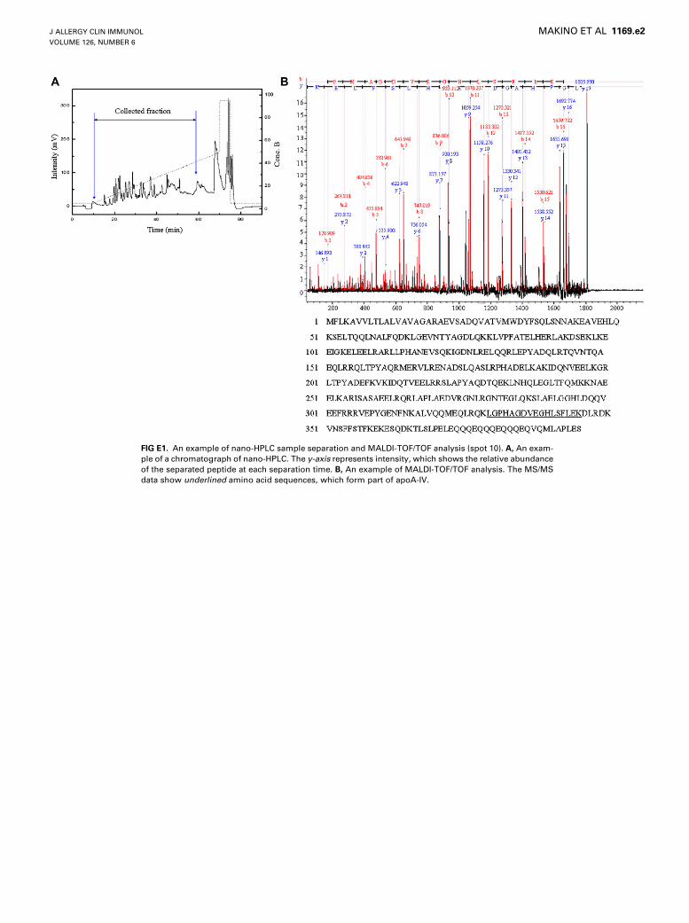
FIG E1. An example of nano-HPLC sample separation and MALDI-TOF/TOF analysis (spot 10). A, An exam-
ple of a chromatograph of nano-HPLC. The y-axis represents intensity, which shows the relative abundance
of the separated peptide at each separation time. B, An example of MALDI-TOF/TOF analysis. The MS/MS
data show underlined amino acid sequences, which form part of apoA-IV.
J ALLERGY CLIN IMMUNOL
VOLUME 126, NUMBER 6
MAKINO ET AL 1169.e2

FIG E2. Western blotting with apoA-IV–specific antibody. A, 2-DE image of the total protein stained with
Deep Purple total protein stain. B, 2-DE image of the total protein merged with the image detected with
the apoA-IV–specific antibody. The arrow indicates the spot corresponding to apoA-IV.
J ALLERGY CLIN IMMUNOL
DECEMBER 2010
1169.e3 MAKINO ET AL

FIG E3. Representative Western blot image of apoA-IV in actively treated
and placebo-treated groups.
J ALLERGY CLIN IMMUNOL
VOLUME 126, NUMBER 6
MAKINO ET AL 1169.e4

TABLE E1. Characteristics of patients
IgE RAST (UA/mL)
Patient
no.
IgE RIST
(IU/mL) JC
Dermatophagoides
pteronyssinus
Dermatophagoides
farinae
D
glomerata
A
artemisiifolia
C
albicans Aspergillus
Placebo 1 7 3.69 <0.34 <0.34 <0.34 <0.34 <0.34 <0.34
2 290 6.57 19.10 18.20 2.88 <0.34 <0.34 <0.34
3 68 11.30 <0.34 <0.34 <0.34 <0.34 <0.34 <0.34
4 190 44.90 1.51 1.51 0.63 <0.34 <0.34 <0.34
5 82 12.00 <0.34 <0.34 <0.34 <0.34 0.52 <0.34
6 24 5.00 <0.34 <0.34 <0.34 <0.34 <0.34 <0.34
7 70 1.95 0.54 0.53 <0.34 <0.34 <0.34 <0.34
8 21 1.25 <0.34 <0.34 <0.34 <0.34 <0.34 <0.34
9 370 73.50 2.89 0.49 1.60 <0.34 <0.34 <0.34
Actively treated 1 210 9.65 <0.34 <0.34 0.79 3.85 <0.34 <0.34
2 180 11.60 <0.34 <0.34 <0.34 <0.34 <0.34 <0.34
3 99 14.50 <0.34 <0.34 0.53 <0.34 <0.34 <0.34
4 29 19.90 <0.34 <0.34 <0.34 <0.34 <0.34 <0.34
5 120 13.40 0.40 <0.34 <0.34 <0.34 <0.34 <0.34
6 48 1.92 <0.34 <0.34 <0.34 <0.34 <0.34 <0.34
7 60 5.18 <0.34 <0.34 5.91 <0.34 <0.34 <0.34
8 240 25.10 0.45 <0.34 0.60 <0.34 0.72 <0.34
9 72 3.52 <0.34 <0.34 <0.34 <0.34 <0.34 <0.34
10 25 5.39 <0.34 <0.34 <0.34 <0.34 <0.34 <0.34
11 11 1.18 <0.34 <0.34 <0.34 <0.34 <0.34 <0.34
12 150 8.09 12.40 18.10 <0.34 0.38 <0.34 <0.34
13 84 5.23 <0.34 <0.34 <0.34 <0.34 <0.34 <0.34
14 470 27.10 0.46 <0.34 <0.34 0.70 <0.34 <0.34
15 250 7.68 18.60 24.80 3.14 0.58 <0.34 <0.34
Specific IgE Class
<0.34 0
0.35-0.69 1
0.70-3.49 2
3.50-17.49 3
17.50-49.99 4
50.00-99.99 5
>100 6
J ALLERGY CLIN IMMUNOL
DECEMBER 2010
1169.e5 MAKINO ET AL
Copyright © 2022 FDOKUMEN