ALLERGEN-INDUCED RHINITIS - Bibliothèque et Archives ...
104
MONOCYTE CHEMOTACTIC PROTEINS IN ALLERGEN-INDUCED RHINITIS Pota Christodoulopoulos Department of Pathology McGU University, Montreal September 1998 A thesis submitted to the Faculty of Graduate Studies and Research in partial fulfiliment of the requirements of the degree of Master of Science O Pota Christodoulopoulos 1998
-
Upload
khangminh22 -
Category
Documents
-
view
2 -
download
0
Transcript of ALLERGEN-INDUCED RHINITIS - Bibliothèque et Archives ...

MONOCYTE CHEMOTACTIC PROTEINS IN
ALLERGEN-INDUCED RHINITIS
Pota Christodoulopoulos
Department of Pathology
McGU University, Montreal
September 1998
A thesis submitted to the Faculty of Graduate Studies and Research in partial fulfiliment of the requirements of the degree of Master of Science
O Pota Christodoulopoulos 1998

National Library BiMiothèque nationale du Canada
Acquisitions and Acquisitions et Bibliographie Services services bibliographiques 395 Wellington Street 395. nie W e W i OttawaON K l A W ûtîawaON K1AON4 canada CaMda
The author has granted a non- exclusive licence dowing the National Library of Canada to reproduce, loan, distribute or sell copies of this thesis in microforni, paper or electronic formats.
L'auteur a accordé une licence non exclusive permettant à la Bibliothèque nationale du Canada de reproduire, prêter, distribuer ou vendre des copies de cette thèse sous la forme de microfiche/nIm. de reproduction sur papier ou sur format électronique.
The author retains ownership of the L'auteur conserve la propriété du copyright in ths thesis. Neither the droit d'auteur qui protège cette thèse. thesis nor substantiai extracts fiom it Ni la thèse ni des extraits substantiels may be printed or othenvise de celle-ci ne doivent être imprimés reproduced without the author' s ou autrement reproduits sans son permission. autorisation.

I would like to express my deepest gratituùe to Dr. Qutayba Hamid for his supervision, guidance, and support throughout my Msc. training.
I also wish to thank my CO-advisors, Dr. A. Giaid and Dr. M.F. Chen for their advice and their help during these îwo years of my training.
1 would iike CO express my appreciation to Dr. Zorychta for her encouragement, support, and U ~ O S ~ concem for al1 the graàuate students in the department.
I thankjùlly acknowledge Dr. Erin Wright and Dr. Saul Frenkiel ut the SMBD-Jewish General Hospital for their assistance in recnriting the patients in this s&, and for perfonning the challenge and the biopsies.
A note of thanks to Dr. Andrew Lusterat Harvard University for his generous gifr of the MCP-4 antibody.
I grarefully acknowledge Dr. Eleanor Minshall for her critical review and insigh@l contributions to this thesis.
I greatly appreciate Elsa Schotman 's invaluable technical assisiunce and advice.
I am indebted to all my colleagues in Dr. Hamid's lab for their help, advice and support ar all rimes.
me work for this thesis was supported by MRC Canada and Network Centers of Excellence.

APAAP
APC
CCRl-8
CXCR1-5
ECP
EDN
EPO
GM-CSF
ICAM-1
ICC
IgE
IL-
ISH
LTC.
L m 4
. L m
MCrc
MCT
MBP
RANTES
Antigen presenting ceU
CC chemokine receptors 1-8
CXC chemokine receptors 1-5
Eosinophil derived neurotoxin
Eosinophil peroxidase
Granulocyte-macrophage colony stimulating factor
Intercellular adhesion molecule- l
In sim hybridization
Leukotriene C d
Leukotriene D4
Leukotriene &
Connective tissue mast ceii
Mucosal mast ceii
Major basic protein
Regulated upon activation, normal T ceii expresseci and secreted

Tumor necrosis factor*
Vascular celi adhesion molecule- 1
Very late antigen-4

Table of Contents
1. Introduction
1.1.2. Epidemiology
1.2. Patbophysiology of rhiaitis
1.3. The Rok of Idammatory Celis in AUergic Rhioitif
1.3.1. Mast ceils
1.3.2. Eosinophïls
1 -3 -3. T lymphocytes
1.3 -4. Macrophages
L .3 S. Basophils
1.4. Leukocyte Migration
1.5. Chemokines
1 S. 1. Chemokine receptors
1 -5.2. Chemokines in allergic inflammation
1.6. Monocyte Chernotactic Reteins
1.6.1. Structure and function
1 -6.2. Regulaîion and celiular sources
1.6.3. Receptors

1 A.4. Cellular Activation
1.6.5. Regulation of ceii growth and tissue accumulation
1 -6.6. Chernoattraction
1 -6.7. Role in infiammatory diseases
1 -7.1. Topical nasal corticosteroids
1 -7 -2. Mode of action
1.7.3. Effects of steroids
2. Materials and Metbods
2.1. Study Design
2.2. Allergen chalienge d e l
2.3. Tissue preparation
2.4. Immunocytochemistry
2.4.1. Immunocytochemis~ry methodology
2.4.2. Double irnmunocytochemistry
2.5. In süu Hybridizrition
2.5.1. Preparation of complementary RNA probes
2.5.2. In vitro transcription
2.5.3. In situ hybridization methodology
2.5.4. Controls

3.1. CWcal observations
3.2. MCP-1 immunareactive exptession
3.3. MCP-3 immunoreactive expression
3.4. MCP-4 immunoreactive expression
3.5. MCP4 mRNA expression
3.6. Assessrnent of infhmtory cell ia2lltrate
3.7. Phenotype of celis expressing MCP-3 and MCP4
4. Discussion
5. Future studies

Allergen-induced rhinitis is associated with the recruitment and activation of
inflammatory cek, particularly eosinophiis and CD4 + T ce& into the nasal mucosa
Monocyte chemotactic proteins (MCPs) have been shown to induce chemotactic
activity in these particular ceii types under in vitro assay conditions. To assess the
contribution of MCPs in the recruitment of inflamnratory ce& in vivo, we investigated
the aliergm-induced late respoase m subjects with allergie Mtis. Using
immunocytochemistry and in siru hybridization, we demonstrated a constitutive
expression of MCP-1, -3 and -4, of whicb MCP-3 and 4 were simiificantly increased
in the nasal mucosa following allergen provocation. This upregulation of MCP-3 and -
4 imrnunoreactivity in response to ailergen, was reduced in patients pretreated with
topical corticosteroids. Colocaiization experiments revealed that the majority of MCP-
positive ceiis were macrophages. The results of this study suggest that dergen-
induced rhinitis is associated with an increased expression of MCP-3 and -4, which
may be closely re1ated to the infiux of inflammaîory œiis and may thus contribute to
the pathogenesis of aiiergic rhinitis.
La rhinite allergique est associée au recrutement et à l'activation de cellules
inflammatoires (principalement éosinophiles et cellules T CD4+) dans la muqueuse
nasale. In vitro, des protéines ont été décrites comme étant chémoattractantes pour les
monocytes (MCPs). La contribution des MCPs dans le recrutement des cellules
inflammatoires in vivo a été évaluée lors de ta phase allergique tardive chez des sujets
atteints de rhinite allergique. Nous avons démontré que les MCP-1, -3 et 4
s'expriment constitutivement dans la muqueuse nasale, toutefois l'expression des
MCP-3 et -4 augmente significativement après provocation dergènique. Cette
augmentation est moindre chez les sujets ayant reçu préalablement des corticostéroides
locaux. La plupart des cellules positives pour les MCPs sont des macrophages. En
conclusion, nos résultats suggèrent que la rhinite allergique est associée à une
augmentation d'expression des MCP-3 et -4 qui, relativement à l'infdtrat de cellules
inflammatoires, pourrait contribuer h la pathogenèse de la rhinite allergique.

1. introduction
1.1. Aüergk Rhinitis
Ailergic rhinitis describes an infiammatory condition of the aasal mucosa
chracterized by the anterior nasal symptoms of pruritus, sneeze, discharge and
stuffiness. There may also be an associatecl loss of sense of smeil and inability to taste.
Although allergic rhinitis may have its onset at any age, the incidence of onset is
greatest in children and at adolescence, with a decrease in incidence seen in advancing
ages The medical history of the patient is fundamental to the diagnosis of aliergic
rhinitis. important historical data include a f d y history of allergy and of personal
atopy, age at onset of symptoms, medication use, and history of any nasal trauma
(Noble 1995, Naclerio 199 1, Mullins et al., 1989).
Aliergic rhinitis is frequently divided into two types, seasonai and perennial,
based on the type of allergen the patient is exposed to, and the time of symptom onset
and duration. With seasonal rhinitis, the symptoms are periodic occuring in temporal
relationship to the presence of seasonal allergens in hdividuals who are appropriately
sensitized. Pollens causing seasonal allergic M t i s are tree polien present in the
springtime, grass pollen present in May through M y , and weed pollen and mould
spores which may produce symptoms in late summer and autumn. Perennial disease,
which is present all year round, relates to the presence of a non-seasonai ailergen. The
dergens causing perennial rhinitis are frequently indoor aeroaiiergens, the most
common king the house dust mite (Dennutophagoides) (Platts-Milis et al., 1987,
Noble 1995, Howarth & Holgate 1990).

Because dergic rhinicis is so common and the symptoms are variable, and
often chronic, it is essential that the treatment is fast-acting, well tolerated, and above
ail, safe. Treatment of nasal allergies should be individualized, with therapeutic
measures aimed at the underlying etiology (Howarth 1989).
1.12. Epidemiology of Allergic Rhinitis
Rhinitis is a very common disease yet littie is h o w n about its epidemioiogy.
Allergic rhinitis is now recognized as a major cause of morbidity that signrficantly
impairs hiaction and quality of Life (Bousquet et al. 1994). Moreover, it is now widely
held that the pathophysiologicai mechanisms causing nasal allergy, contribute, o r
predispose many individuais, to the development of other ainivay diseases, including
asthma Allergic rhinitis may weii be a factor in 24% of children with otis media with
effusion, and perhaps 28% of cases of chronic sinusitis (Mygind & Dahl 1996). A large
number of cross-sectional studies have demonstrated that thinitis and asthnra
cornmonly occur together . Long -term epidemiological studies suggest that rhinitis
frequently precedes asthma and that upper airway dysfunction may be a predictive
factor for subsequent development of lower airway disease (Settipane 1986, Van
Arsdel 1959, Settipane et al., 1994). Nasai syrnptoms have been reported among 28%
to 78% of patients with asthma, compared with approximately 20% of the general
population (Blair et al., 1977, Smith 1988, Pederson & Weeke 1983). SUaiiariy, as
rnany as 19% to 38% of patients with allergic rtiinitis may have asthma, much more
than 3 to 5% prevalence among the general population (Evans et al., 1987).

Considerable evidence now suggests that early and appropriate intervention
can improve the quality of life aud productivity of patients with allergic rtiinitis, and
reduce the prevelance of airway complications. The goal of treatment has shifted h m
mere symptom alleviation to blocking the pathophysiologic meçhanisms that cause
chronic dergic inflammation. The earlier in a patients life that this can be
accomplished, the better the anticipated consequences (Corren 1997).
1.2. Pathophysiology of rbinitis
AUergic rhinitis is an immunologidy rneâiated disease initiated by an IgE-
dependent reaction. Allergens, which generally enter the body through inhalation,
interact with T and B lymphocytes to produce IgE antibodies, which attach to the
surfaces of mast cells and basophils. Reexposure to the same allergen on a mucosal
surface, results in a coupling or cross-linking of the IgE molecule that leads to cellular
degranulation and tbe release of infiammatory mediators, a process resulting in both an
early- and a late- phase response (Naclerio 1991, Dvoracek et al., 1984).
1.2.1. Early- phase response
Cross-linking of surface bound IgE, leads to mast degranulation, the critical
initiating event of acute aiiergic symptoms. This explosive degranulation of mast cells
induced by aiiergen, leads to the release of a complex cascade of mediators, which may
have synergistic effects on resident cells in tissues. The mediators of the immediate-
phase response, such as, histamine, bndykinh, Ieuicotrienes. and platelet-activating
factor, generate the acute symptoms of itfh, rhinorrbea, congestion, and sneezing of
allergic rhinitis. Histamine is one of the most important d a t o r s of the early phase
allergic response in the nasai mucosa as its release stimulates seasory nerves Ieading to
vasodilation and mucus hypersecretion (Kawabori er al., 1995, Galli et al., 1993). The
release of leuLotrienes and chymase aiso stimulates gîandular exocytosis and mucus
secretion. (Sornmerhof et al., 1989, Holgate et al., 1996). The mediators of this early
phase generate the acute symptoms, and as these mediators are metabolized and
cleared from the mucosa, these symptoms wane. However, the release of cytokines
and mediators, activates endothdial œUs to express adhesion markers that bind
circulating leukocytes, leading to a latent recmitment phase that is thought to usher in
the inflammatory late-phase response (Sedgwick et al., 199 1, Butcher 199 1).
1.2.2. Late Phase R e s p o e
The late phase response occurs 4 to 6 hours after the irnmediate phase. It is
noted clùiically by an increase in nasal mucosal thickness that can be detected as an
increased nasal airflow resistance. During the LNR, inflafnmatory gmulocytes,
including eosinophils, basophils. and, less dramaticaily, neutrophils, are found within
the mucosa The numbers of mononuclear celis and metachromatic cells, such as mast
cells, also increase. Once there, by interacting with additionai stimuli. they release their
own mediators. This perpetuates the innammatory response and augments aspects of
the immediate hyprsensitivity reaction, such as mucosai congestion and mucus

secretion. The increase in eosinophiIs is reflected by large increases in eosinophil
products in the nasal secretions, such as MBP. Mediators of the late-phase response
include leukotrienes, histamine, and Th2 cytokines (Terada et al., 1994, Togias et al.,
1988, Gosset et al., 1993, Nacleno et al., 1994). Hisramine increases without a change
in cryptase, suggesting the activation of basophils rather than a secondary
degranulation of mast cells. In addition, the release of Th2 type cytokines from the
leukocytes present, and fiom the epithelium is increased maraniuk 1997). It appears
that the factors released fkorn these inflammatory cells promote the expression of a
combination of adhesion markers and the production of a combination of
c hemoattractants that promote m e r Ieukoc yte infiltration, perpetuating and
maintainhg the infiammatory response.
1.3. The Role of Inflammatory CeUs in Aiiergic Rhinitis
One of the ballmarks of aiiergic diseases is the intense accumulation of
infiammatory cells in tissue locations at specific mucosaI surfaces. The presence of an
increased nwnber of mast cells, basophils, T cek, macrophages, and particularly
eosinophils, have been detected in nasal smears and biopsies from patients with ailergic
rhinitis. It has also been shown that in response to certain mediators, these
infiarnrnatov cells undergo local activation, releasing their own mediators, and thus
contributing to the pathological features of this disease. These cellular changes exist to
varying degrees, but have been demonstrated to occur in temporal relationship to
seasonal allergen exposure. Mediator release fiom the primary effector cells such as

mast cells, basophls and eosinophils, can explain the symptom development in this
disease through interactions with the structural elements within the nose (Howarth &
Holgate 1990).
1.3.1. Mast C e k
Mast œlls are constitutive cells within the no& nasal mucosa and are the
recognized key ceils of type 1 hypesensitivity reactions. These ceils c m be subdivided
into connective and mucosal phenotypes. Comective tissue mast ceils (MCx) express
chymase, rryptase and TNF-a (Bradding et al., 1995). This cell population represents
85% of the IL4positive mast cells in the nasal lamina propria. hiMg aliergen
exposure, there is an increase in the proportion of mast cells in the epitheiial œ U layer
(luluisson et al., 1995). These ceils produce predoxninantly tryptase, without chymase,
and are called mucosal mast cells (MCt). MCTS express IL-4, IL-5 and IL-6, md
represent 15% of al1 of the IL4positive mast ceils in the mucosa (Bradding et al.,
1995). These cells proliferate in aüergic rhinitis, perhaps under the influence of Th2
cytokines (Kawabori et al., 1995). Pmliferation of MCTs appears to occur in the
epithelium and most superficial layers of the lamina propria Epithelial mast ceils have
a higher rate of c d division in diergic rhinitis compared with nasal mucosa taken h m
non-atopic individuais. Both phenotypes, MCT and MCw, express k&RI and may
therefore participate fully in IgE-dependent allergic reactions (Church & Levi-Schaffer
1997). Cross-iinking of IgE on the surface of mast ceils by allergen leads to a series of
intracellular events culminating in the release of preforrned mediators from mast ceils
including histamine. tryptase, and heparin, and the generation of lipid mediators,

including PG& and the sulphidopeptide leukotriene, LTC4, and its metabolites L m
and LTL (Howarth & Holgate 1990). These released mediators induce nasal
symptoms of itch, sneeze, discharge and blockage, through interaction with receptors
present in both neural and vascular elements within the nasal mucosa with histamine
king prominent in this respect (Mygind 1993).
In addition to the effects of acute mast cell degranulation on imrnediate
symptom generation, mast ceii degranulation will contribute to the eosinophilic
mucosal inflammation wbich is evident in rhinitis. Mast cells within the nasal mucosa
have k e n demonstrated to contain preformed cytokines, in particular IL-4, IL-5, IL-6,
IL43 and TNF-a (Bradding 1993a, 1995). Both IL-5 and TNF-a have actions
relevant to eosinophil activation and recruitrnent (Howarth et al., 1994). IL-4
potentiate the effects of TNF-a on the expression of VCAM-i on the vascular
endotheliurn which leads to tissue eosinophil recruitment through its interaction with
eosinophii ligand VLA4 (Jl~onihiU & Haskard 1990, Montefort. et al., 1993). In
addition, I L 4 and IL-13 are involved in switching the B lymphocyte to IgE
production. The abiiity of every sensitized mast ce11 to respond to stimulation with any
individual allergen, as opposed to T ceils, in which only a srnall percentage of ceils
specific to that ailergen respond, makes mast celis potentially important cytokine-
generating ceUs in allergy. Furthemore, the presence of preformed cytokines within
mast cells, which is not the case with T cells, suggests that they are available for rapid
secretion on ce11 stimulation (Chwch & Levi-Schaffer 1997).

1.3.2. Eahophiis
Eosinophils are bone marrow-denved granulocytes that are not normally
prominent in either the peripheral b l d or the tissues. (Weller 1997).
Immunohistochemicai stainuig of nasal mucosa biopsies has s h o w that eosinophils are
evident. within the submucosa and epithelium, in symptornatic rhinitis (Bentley et al.,
1992, Bradding et al., 1993a,b). The steps that lead to eosinophii recruiûnent into
certain sites of idammation are multiple, and require a combination of factors
including enhanced expression andor function of integrins on intravascular eosinophils
and their counterligands on the vascuiar endotheliwn, as well as the actions of
eosinophil chemoattractant factors (Weller et al., 1996).
The influx of eosinophils in atopic aiiergic dis- are believed to effect tissue
damage through the release of their mediators (Bousquet et ai., 1990). The
armamentarium of eosinophiiic granules consists of a number of very toxic and potent
mediators including major basic protein (MBP), eosinophü cationic protein (ECP),
eosinophil peroxidase (EPO), and eosinophil derived neurotoxin (EDN) (Peters et al.,
1986, Bainton et al., 1970). At the site of inaammatim, these proteins can cause
cellular disaggregation and epithelial desquamation. Eosinophil activation is ais0
associated with the de novo generation of arafhidonic acid products such as LTCd,
which contribute to nasal obstruction and to rhinorrhoea through their potent smooth
muscle contractile, vasoactive, and mucous secretory activities (Volovitz, 1988).
In addition, it has k e n shown that human eosinophils express and synthesize a
number of cytokines, including GMCSF, IL-6, IL-1% IL-2, IL-3, IL-4, ILS, IL-8,

MIP- l a, RANTES, and TNF-a (Moqbel et al., 199 1, Hamid et al., 1992, WeUer et
al., 1993, Kita et al., 199 1, Desreumaux et al ., 1993, Braun et al., 3993, Costa et al .,
1993). Since IL,-3, IL-5 and GM-CSF influence eosinophil differentiation, activation,
and survival, many of these functions may be regulated by eosinophifs at least partly in
an autocrine fashion (Lopez et al., 1992).
It is abundantly clear that eosinophds are major participants in the
imrnunopaîhogenesis of aüergic i n f l d o n as tbey are characteristicaüy recruitcd to
such sites, releasing their cationic proteins and lipid mediators, and thus contributhg
to damage and dysfunction of other resident ceii types.
1.33. T Lymphocytes
As key cells in the adaptive immune response, T lymphocytes have evolved to
coordinate and ampiify the effector functions of antigen specific and non-specific
infiammatory ceils such as B cells and eosïnophils. T lymphocytes have been divided
into two distinct subgroups based on their effector functioas. CD4+ T cells represent
the T helper celis, which are important in the regdation of antigen-driven
inflanmatory processes. Via antigen-specific T ceil receptors, CD4 T ceils are capable
of recognizing processed foreign antigen in association with MHC class 11 on
specialized antigen-presenting cells. On the other band, CDS+ T ceiis which represent
T suppressor cells, drive the cell-mediated response and respond to APC presenting
antigen in conjunction with MHC class II molecule. The T lymphocyte represents a
significant non-structural cell within the nasal mucosa An increase in this cell
population has been described in nasai biopsies spccimens in rhinitic patients. These
are generally CD4' T ceils displayhg the activaîeâ phenotype (CD253 (Calderon et
al., 1994, Varney et al., 1992). CD4' T lymphocytes have been shown to play a
crucial role in the induction and maintenance of chronic aiiergic inflammation.
The presence of T lymphocytes in allergic i n f i d o n fias been weil
demonstrated, however, the major reason for their importance lies on the cytokine
profile which they express upon activation. Although individual T ceiis have the
capacity to produce a wide range of cytokines, restricted cytokine profiles exist in
chronic infiammatory diseases. This may occur as a result of the microenvironment
favoring the development of specific T ceIl populations at the tirne of induction. Cross-
regulation of Th1 and Th2 cytokines occws and in certain cases, this may lead to the
polarkation of the cytokine milieu towards Th1 or Th2 (Kelso 1995). A major feature
of allergic diseases is the high expression of Th2-type cytokines. T lymphocytes of the
T-helper 2 (TU) subpopulation c m generate IL-3, IL-4, IL-5, GM-CSF and TNF-a
(Mossman & Co- 1989). Foilowing nasal Aergen challenge, an increase in IL-4,
IL-5 and GM-CSF rnRNA-positive cells has been described in association with a
mucosal eosinophilia (Durham et al., 1992), and T cell clones derived from nasal
mucosa chdenged with dergen in vivo have a cytokine profile comparable to that of
a ThZiike population (Ilel-Prete 1994). The Th2 phenotype is thought to influence
subsequent T cell activation and IgE production by B ceiis in addition to promoting
the attraction, activation, growth, and differentiation of specific leukocytes suc h as
eosinophils. in this way, activated T cells can initiate and propagate aliergic
inflammation and participate directly in the events responsible for aüergic diseases.
Steroids have been shown to improve the symptoms of aliergic rhinitics by reduchg
the expression of Thz cytokines (Bentley et al., 1996, Masuyama et al., 1994),
therefore their success in suppressing inflammation may be partly attributed to their
activity in limiting Th2 lymphocyte activation.
13.4. Macrophages
Macrophages are widely distributed in virnially ai l tissues, and their activation
is a prominent feature at sites of inflammation, in particular, granuloma formation.
Macrophages play an important role in the immune response as immunornodulators,
APC, and efiector cells. Once b l d monocytes leave the circulation and enter the
extravascular tissues, they differentiate into macrophages. As macrophages mature
fiom the monocyte through to the resident mature tissue macrophage stage, they go
through a senes of fûnctional changes which affect their interaction with T cells.
Macrophages can also act as APCs. They take up antigen via non-specific receptors or
as Unmune complexes, process it, and retum fragments to the celi surface in
association with ciass II molecules. It has been shown that the number of macrophages
on the epithelial surface is i n c d 4-8 hours after allergen challenge, suggesting that
macrophages rnay play a role in the allergic nasal late phase reaction (Bachert 199 1).
Macrophages are also an important source of cytokines that can manipulate the
emerging T celi response. Macrophage products such as IL-1, IL-6, and TNF-a have
b e n implicated in many idammatory and immunologie responses elicited during

tissue injury. Th2 type cytokines, in particular IL- 13, IL4 and K- 10, may decrease
the abiiity of activated macrophages to participate in inflanvnatory responses, and by
inhi'biting the ability of macrophages to produce IL-12, may thereby antagonize the
development of Th1 ceiis, which can augment inflammation, @oherty et al., 1993).
1.3.5. Basophiis
Basophils are only present in very low numbers in peripheral blood, and are not
found in nonnal non-inflarned tissues, indicating they are recruited to sites of
inflammation by mediators fiom other ceil types. Basophils are evident in nasal smear
samples in aliergic rhinitis (Okuda et al., 1985, Otsuka et al., 1985) and can be
demonstrated to increase in rhinitic patients foilowing nasal aiiergen challenge
(Bascom et al., 1988). Eventhough there are no specific surface markers for its precise
identification in biological fluids and tissue, evidence of basophil infdtration into the
nasal mucosa during aiiergen challenge, is based on the mediator profde in nasal
secretions (Naderio et al., 1985, Bascom et al., 1988). Basophils were initially
thought to function in a similar fashion to mast cells, however it is now apparent that
these two œil types differ significantly fiom each other. Basophils, like mast cens,
possess high affinity IgE receptors, are also derived fiom CD34-positive progenitor
cells in the bone rnarrow (Knapp 1990). However, unlike mast celi precursors which
require stem celi factor to mature in tissue, basophils are thought to be released h m

bone rnarrow as mature cells, with their development king dependent on the effects of
IL-3 and GM-CSF as seen with eosinophils. When activated, basophils are prominent
sources of inflammaîory mediators found in dergic late-phase reactions, such as
histamine and LTCa. Basophils possess fewer, larger granules and differ Erom the mast
ceil in that they contain l e s histamine. Following IgE-dependent activation. the
basophil only releases 20-30% of the histamine released from a comparable number of
mast cells (Canteiis et al., 1987). Human basophils have also been shown to secrete
cytokines, particuiarly I L 4 and IL-13, when activated by IgE-dependent stimuli,
modulating their response and the immune responses of other ce11 types, that
participate in allergic rhinitis (Mac Glashan et al., 1994, Schroeder et al., 1994,
Sc hroeder et al., 1996). T 'us , basophil-derived cytokines could potentiaiiy influence
the invoivement of eosinophils and lymphocytes in allergic responses by upregulating
adhesion molecules, such as vascular ceii adhesion molecules, and by promoting the
development of Th2-type cells, both of which are processess that constitute the
hallmark of aliergic disease.
1.4. Leukocyte Migration
Leukocyte extravasation from the blood into the tissues is a regulated muitistep
process involving a series of cwrdinated interactions between leukocytes and
endothelial ceUs (Butcher 199 1, Springer 1994). Several families of molecular
regdators, such as selectins, integrins, and chemokïnes, are thought to control

different aspects of this process. Initially, circulatiag leukocytes uadergo margnation,
whereby they move from the centre to the periphery of the b l d vesse1 and begin to
bind reversibly to the endotheiium, a process referred to as rolling. This process is
mediated by the interaction of L-selectin on the leukocytes with diverse, c h h y d r a t e
containing structures such as P-selectin or E-selectin on the endothelial celis (Varici
1994). This initial weak b i i g may be followed, due to activation of the Ieukocytes
by chemoattractants or bùiding to E-selectin (Kuijpers et al.. 1991), by the induction
of fum adhesion. This second step is mediated by leukocyte integrins interacting with
endothelial ceU adhesion molecules, such as ICAM- 1 and VCAM- 1. (Rice et al., 1990,
Bochner et al.. 1991, Lawrence & Springer 1991). Once a leukocyte has becorne
M y attached to the endothelium, it must be guideci to move across the endothehum
to reach the site of injury. Chemokines are thought to provide the signals that convert
the low-affinity, selectin-mediated interaction into the higher affinity, integrin mediated
interaction that leads to extravasation of leukocytes (Luster et al.. 1998). In
chemotaxis, ceils move in the ditection of increasing concentration of a
chemoattractant, which t y p i d y is a soluble rnolecule that c m diffuse away h m the
site of its production, where its concentration is highest (Devreotes & Zigmond 1988).
Many molecular regulators have been shown to play a role in leukocyte trafficking,
however, recent studies have brought our attention to the importance of chemokine
involvement in this event ( refer to schema on following page).

Endothelium
Tksue
Inflammatory Ceii Extravasation
Chemotactic Gradient
Lumen
Representative example of inflammatory cell extravasation. Extravasation of Ieukocytes during inflammation involves multiple sequential steps including (i) rolling of Ieukocytes along the vesse1 walt, which is mcdiated by the selectin family of adhesion molecules, (ii) tight binding of leukocytes to the endothelium resulting from activation of integrin adhesion molecules, and (iii) migration of leukocytes through the endothelid junctions and underlying tissues in response to gradients of chernotactic agents generated at the site of inflammation.
MCPs EOTAXIN RANTES PAF IL-8 CSa

1 .S. Chemokines
The discovery of IL-8, in 1987, revealed the existence of a novel class of small
chemoattractant cytokines, now caiied chemobes, that are seen as the stimuli that
largely control leukocyte migration (Baggiolini et al., 1989, Baggiolini & Dahinden
1994). While the pathologists of last century knew that leukocytes ernigrated fiom the
blood across the waU of microvessels,. the purpose of this migration remaineci
unknown. This remained so until Elias Metchnikoff showed that leukocytes engutf and
kill bactena and recognized diapedesis as a fundamental mechanism of host defence
(Metchnikoff 1901). Today, eleven years after the discovery of the fmt chemokine, as
rnany as 40 to 50 human chemokines have k e n identifieci. Chemokines constitute a
superfamily of small, inducible, secreted, proinflarnmatory cytokines that regdate
diverse properties of specific leukocytes, including chernoattraction, cellular regulation
and ceil growth. These srnail proteins have four conserved cysteines forming two
essen tid disulphide bonds (Cys 1-Cys3 and Cys2-Cysrl), and have thus been classified
depending upon the spacing of their conserved cysteine residues. The CXC (a)
chemokines have one amino acid residue separating the the two fmt conserved
cysteine residues. In the CC ($) chemokines, the fmt two cysteine residues are
adjacent to each other. The C (y) chemokine, lyrnphotactin, has two instead of four
conserved cysteines in the mature protein, and lacks one cysteine near the N terminus.
CX,C (6) chemokines have three intewening amho acid residues between the fïrst two
cysteine residues at the N-terminal end of a mucin structure (Clark-Lewis 1995,
Loetscher et al., 1996, Kennedy et al-, 1995, Bazan et ab, 1997, Pan et al., 1997).

Chromoromal Family Structure Locus
CXC C C CXC 14q12-21
The four chemokine subfamilies subdivided based on the relative position of the cysteine residues found in the mature protein.
The a and B chemokines are the two major subfamiles that have been
extensively characterized. The a chemokines are subdivided into two groups. Those
containing the sequence glutamic acid-leucine-arginïne near the N tenninal (preceding
the CXC sequence) and those that do not. Those a chemokines containing this
sequence are chernotactic for neutrophils, whereas those not containing the sequence
act on lymphocytes. The chemokines, in general, attract monocytes, eosinophüs,
basophils, and lymphocytes with variable selectivity. Smicturally the B chernokines can
be subdivided into two families, the mnocytechemoattractant-protein (MCP)eotaxin
family, containing the monocyte chemoattractant proteins and eotaxin, which are
approximately 65 percent identical to each other, and ali other B chemokines (Proost
1996, Taub 1995). As with the a chemokines, the N-terminal acids preceding the
conserved cysteine residues of pchemokines are critical components of the biological
activity and leukocyte selectivity of these chemokines (Gong et al., 1995).
Activation of leukocytes leads to shape changes which are observed within
seconds after the addition of a chemoattfactant to a leukocyte suspension.
Polymerïzation and breakdown of actin Narnents leads to the formation and retraction
of lamellipodia, which function like arms and legs of the migrating ceUs. Stimulation
dso induces the upregulation and activation of integrins, which enable the leukocytes
to adhere to the endothelid cells of the vesse1 wali before migrating into the tissues
(Springer et al., 1994). Several other rapid and transient responses are characteristic of
the activation of leukocytes by chemokines, such as the rise in the intraceiiuiar fke
calcium concentration, the production of microbicidal oxygen radicals and bioactive

lipids, and the release of the contents of ibe cytoplasmk storage granules, such as
proteases h m neutrophils and monocytes, histamine h m basophils and cytotoxic
proteins h m eosinophils (Baggiolini & Dahinden 1994, Baggioiini & Moser 1997).
Most chemokines are produceci under pathological conditions by tissue ceiis
and infïdtrating leukocytes (Baggiolini & Dahin&n 1994, Furie Br Randolph 1995).
Some chemokines on the other band, seem to fulnll housckeeping functions. They may
be involved in feukocyte maniration in the bone marrow, the aaffc and homùig of
lymphocytes, and the mechanisms that ensure the renewal of circulating leukocytes
(Baggioiini et al., 1998). In addition, chemokines may play a broad role in cellular
physiology. They have been show to be involved in diverse processes such as
hematopoiesis, angiogenesis, and inhibition of HIV pdiferation (Broxmeyer et ai.,
1993, Strieter et al., 1995, Cocchi et al., 1995).

Chemokine Target ceUs
Eotaxin
1309
HCC- 1
TARC
CXC chemokines
IL-8
m i 0
M G
NAP-2
ENA-78
GRO-a
G R e p
GRO-y
C chemobes
Lymphotactin
CXaC chemokines
Fractalkine
monocytes, T cells, basophils
monocytes, T cells, eosinophils, basophils,
monocytes, T cells, eosinophils, basophils, NK
cclls
monocytes, T cells, cosinophils
T cells. cosinophils. basophils, NK cclls,
monocytes, T cells, NK cells, basophils,
eosinophils
monocytes, T cells, clendritic cells,
eosinophi b
monw'tes
monocytes, hernatopoictic progenitors
T cells
neutrophils, T cells, basophils,
NK cells, rnernory T cells
T -11s
neutrophils, basophils
neutrophils
endothelial cells
neutrophils, endothelid cells
neutrophils, endothelial cells
T cells
monocytes, lymphocyt=

1.5.1. Chemokiw Receptors
Chemokines induce ceil migration and activation via seven triansmembrane
domain receptors (7TM) which form a distinct group of smicturally related proteins
within the superfamiiy of receptors that signal through heterotrimeric GTP-binding
proteins (Baggiolini & Dahinden 1994, Murphy 1994). Eight receptors for CC
chernokines (CCRl through CCR8) and five receptors for CXC chemokines (CXCRI
through CXCRS) have been characterized (Murphy 1996). Most receptors recognize
more than one chemokine, and several chemokines bind to more than one receptor,
indicating that redundancy and versatility are characteristic for the chemokine system.
In addition, chemokuie receptors are constitutively expressed in some cells, whereas
they are inducible in others. For example, CCRl and CCR2 are constitutiveIy
expressed on monocytes but are expressed on lymphocytes only after stimulation by
IL-2 (Loetscher et al., 1996). Chemokines have two main sites of interaction with their
receptors, one in the N-terminai region and the other withïn an exposed loop of the
backbone that extends between the second and the third cysteine residue. The N-
teminal binding site is essentid for triggering of the receptor. The receptor recognizes
the loop region fmt, and this interaction is necessary for the correct presentation of
the triggering dornain (Clark-kwis 1995).

RECEPTOR LIGANDS
CXC Receptors
CC Receptors
IL-8
IL-8, GRO-a, GRO-p, GRO-7, NAP-2
IP- IO, MIG
SDF- la
MIP- 1 a, MIP- 1 b, RANTES, MCP-3
MCP- 1, MCP-3, MCP-4
Eotaxin, RGNTES, MCP-2, MCP-3, MCP-4
MIP-1 a, RANTES, MCP- 1, TARC
MIP-la, MIP-1p, RANTES
MIP-3a/LARC
MIP-3B/ELC

1.5.2. Chemokihes in AUergic Intlnmmntion
CC c hemokines at tract and activate eosinophils, basophils, and lymphocytes.
Because these celis are the hallmark of aiiergic inilammation, and CC chemokines act
on these particular ceiis, it is believed chat CC chemokines may play a role in the
pathogenesis of ailergic disease. The obsewation that RANTES and MCP-3 activate
eosinophii and basophil leukocytes, induchg chernotaxis and the release of histamine
and Ieukotrienes, was the fmt hint that chemokines are involved in aliergy (Baggioliai
& Dahinden 1994). Then eotaxin was discovered and it was show to be a potent
eosinophil chernoattractant. Eotaxin was initiaUy describeci as the predominant and
selective eosinophil chemoattractant in the bronchoalveolar lavage fluid from ailergen-
chailenged guinea pigs (Gnffsths-Johnson et al., 1993, Jose et al., 1994). It was later
shown that, eotaxin is increased in tissue and BAL fluid of atopic asthmatics when
compared with no& controls (Lamichioued et ai., 1997). Recently, it was shown
that ailergen challenge of the nasal mucosa in rhinitics results in the a local
upregulaiion of eotaxin expression (Minshali et al., 1997). In addition MCP-1. -2, -3
and 4, also part of the CC chemokine subfamily, have been implicated in allergic
diseases based maùily on in vitro fmdings, however their actual role in human allergic
diseases remains to be M e r clarified.

1.6. Monocyte Chemoattractant Proteins
Four human MCP proteins (-1, -2, -3, -4) have been identifieci to date. MCPs
share many structural and bctional features forming a distinct subgroup within the
CC chemokine family. MCPs have diverse functions, including the regdation of
leukocyte chernoattraction, cellular activation, including histamine release from
basophils, and the regulation of the homeostatic growth and tissue level of immune
cells involved in aliergy.
MCP-1 was the fkst CC chemokine to be characterized biologidy MCP-1
was fmt purified h m conditioned media of baboon aortic smmth muscle cells in
culture on the basis of its ability to attract monocytes, but not neutrophils, in vitro
(Vaiente et al., 1988, Baggiolini & Dahinden 1994). Subsequently, MCP-2 and MCP-
3 were idenad They were isolateci as novel monocyte chemoattractants from the
conditionned medium of MG-63 osteosarcorna =Us (Van Damme et al., 1992). One
of the newest members, M e - 4 was described only recently. MCP-4 was i&ntXd
fiom human fetal RNA, cloned iato a baculovirus vector, and expressed in S B insect
celis. (Uguccioni et al., 1996, Van Damme et al., 1992). These chemokines are
strikingly related in terms of chromosomal location, gene structure, primary protein
sequence, biological activity, and receptor usage. MCPs have emerged as chemokines
involved in the recruitment and acitivation of cells seen in ailergy, and proposed to
play a fundamental role in the development of such responses.

1.6.1. Stiucture and Function
The MCPs constitute a subfamiiy of CC chemokines that share structural and
functional feature sharing approximately 65% amino acid identity (Proost et al., 1996,
Garcia-Zepeda et al., 1996). The primary structure of the chemokules can, in general,
predict function. MCPs are inactive on neutrophils and instead chemoattract
monocytes, eosinophils, basophils, and lymphocytes with variable selectivity. Human
MCP- 1, -2, -3 and 4 have a remarkably similar intron-exon structure each gene is
organized into three exons and two introns. In addition, the positions of the splice sites
within the codons is conserved, suggesting that these genes arose h m a common
ancestral gene. In support of a recent gene duplication, the MCPs were found to be
situated on the long arm of chromosome 17 (17q11.2). The MCP cDNAs encode
typical secreted proteins with signal peptides that are, surprisingly, more conserved
than the mature proteins. After signal peptide cleavage, the mature proteins are -8.6
kDa. Although they do not contain potential N-linked glycosylation sites, the MCP
proteins probably contain O-linked carbohydrates and sialic residues (Coillie et al.,
1997, Naruse et al., 1996).
The structure of the mature amino-terminal acid is thought to be important for
the biological activity and leukocyte selectivity of the MCP proteins. For example, the
addition of a single amino acid residue before the N- terminal glutamine reduces the
biological activity of MCP- 1 on monocytes by 100-to 1000-fold. Fmthermore,
deletion of this amino terminal glutamine residue converts MCP-1 fiom an activator of
basophils to an eosinopbd chemoattractant. However if the amino terminal glutamine

of MCP-1 is replaced by an other small amino =id, such as asparagine or alanine, it
still retains activity on monocytes. During the process of of generating biologically
active recombinant MCP-4 m E-coli, it was also found that an amino-tenninal
extension completely inactivated these molecules.
1.6-2. Reguiation and Cellular Sources
The MCPs are produced by numerous ceil types, including fibroblasts,
endothelial cells, and mononuclear leukocytes, in response to pro-idammatory stimuli
such as cytokines, lipopolysaccharide, or infectious agents. The main stimuli however,
for secretion of the MCPs appear to be early pro-inflammatory cytokines such as I L 1
and TNF-a (Proost et al.. 1996, Garcia-Zepeda et al., 1996). In addition, products of
both Th1 and Th2 ceiis, IFN-y and IL-4, respectively, can induœ the production of
these chemokines and also synergize with IL-1 and TNF-a to induce their secretion.
IL- 1, TNF-a, and IFNq are potent endogenous inducers of MCP-1 in mononuclear
ceils, fibroblasts, endothelial and whereas other cytokines such as IL-6 generally have
no stimulatory effect (Van Darnrne et al., 1989, Wang et al., 199 1, Sica et al., 1990).
Recently, it has dm been shown that human eosinophils represent an important source
of MCP-1 (Lin et aL, 1998). In addition, it is weii known that epithelial ceiis produce
these chemokines in response to cytokine stimulation. Stellato et al. have shown that
MCP-1 and MCP-4 expression can be induced in airway epithelial cells upon
stimulation with either TNF-a or IFN-y.

1.6.3. Receptors
Tbere have been eight human CC chemokine rceptor g e n s cloned to date,
and these are referred to as CCR-1 through CCR-8. CC chemokuie receptors ail
appear to exhibit overlapping specificities, aibeit with variable affinity for particular
chemokines. Moreover, several CC chemokhes can bind more than one receptor.
Thus each chemokine can recruit multiple types of cells even if they express different
types of receptors, whereas as each celi may respond to multiple types of cbemokines
even by expressing a single type of receptor (Murphy 1994, Neote 1993, Gao et al.,
1993, Charo et al., 1994, Power et al., 1995). Monocytes express the CC chemokine
receptors CCR- 1, CCR-2, CCR-4, and CCR-5. Eosinophils express CCR- 1 and CCR-
3 and basophils express CCR-3 and CCR-4. The fmt receptors for CC chemokines,
CCRl and CC=, were originaily designated MIP-la / RANTES and MCP-1
receptor, respectively based on the ligands they bound with high afnnity. Subsequent
studies demonstrated that CCRl and CCR2 also recognize MCP-2, MCP-3, and
possibly MCP4 (Franci et al.. 1995, Ben-Baruch et al., 1995, Combadiere et al.,
1995, Gong et al., 1996). CCR2 on the other han4 recognizes ali of the human MCP
proteins characterized to date, and is selective for the MCP proteins. CCR3, the so-
caUed eotaxin receptor, is prominently expressed on eosinophils and is believed to be
the principle chemokine receptor for the recruitment of these ceiis in allergy. MCP-2, -
3 and4 are also ligands for CCR3. MCP4 compleiely desensitizes eosinophiis toward
eotaxin, suggesting that it binds with comparibly high affinity to CCR3 (Uguccioni

1996). C a - 4 , another CC chemokine receptor, only recognizes MCP-1 among ai l the
MCPs.
1.6.4. Cellular activation
In addition to promoting cellular accumulation, MCPs are potent c e U
activators. After binding to appropriate G-protein linked seven -&ansmembrane-
spanning receptor, MCPs eiicit a transient intracelluiar calcium flux, actin
polymerization, oxiciative burst with release of superoxide free radicals, and exocytosis
of secondary granule constituents (Bischoff et a l ., 1992, Dahinden et al., 1994, Elsner
et al., 1996). Ceiiular activation is also accompanied by increased avidity of integrins
for their adhesioa molecules. In eosinophils, MCP-3 has been show to differrntiaily
regulate $1 and $2 integrin avidity (Weber et al., 1996). In basophils, chemokine
induced cellular activation results in degranulation with the release of histamine and
the de novo generation of leukotriene Cd (Alam et al., 1992, Nam et al., 1994).
Basophil activation requires celluiar priming with IL-3, IL-5, or GM-CSF for maximal
effect of each chemokine. Although aU MCPs can induce histamine release in cytokine
primed basophils, there is marked varïability between individual basophil donors. The
intraceilular signaling pathway triggered by chemokines in eosinophiis is poorly
understood, but recent studies have suggested the involvement of leukotriene
metabolites because chernotaxis is inhibited by leukotriene inhibitors in vitro and in
vivo (Stafford & Alam 1997).
1.6.5. Chemoatîraction
Although CC chemokines have diverse fhctions in allergic reactions, tbey
have mainly been recognized and become the focus of interest because they are
thought to provide the directional cues for the movement of leukocytes. Leukocyte
extravasation fiom the blood into the tissues is a regdated proçess which consists a
senes of events involving a marked induction of specific chemokines. The CC
chemokines, inçiuding the MCPs, have emerged as mediators involved in the
recruitment of inflammatoq ceiis seen in allergic reactions.
As their narne impies, ali MCPs have strong chernoattractive activity for
monocytes. Although MCP-1 has generally been observed to be the most potent and
effîcacious chemoattractant for monocytes CUguccioni et al., 1995, Van Damme et al.,
1 992). Ln con trast, they display partial overlapping chernoattractive activity on other
cell types. MCP- 1, -2, -3 and -4 all have chernotactic activity on basophiis and T celis
(Alam 1994). MCP-3 has also b e n shown to attract dendritic celis (Sozzani 1995).
One important distinguishing feature among the MCPs, is the ability of MCP-2, -3,
and -4 to attract eosinophils, whereas MCP-1 displays no activity for this particular
cell type. In terms of relative chemoattractant potency, MCP-4 is very potent in
attracting eosinophils, but less potent than MCP-1 in attracting monocytes or T cells
(Uguccioni et al., 1996). For each chemokine, cellular specificity has been
demonstrated for recombinant proteins by the use of in vitro chernotaxis assays with
different populations of leukocytes.

Although othet CC chemokines such as RANTES and eotaxin have been
investigated in human allergen-induced responses, MCPs and their role as
chemoattractants in allergen-induced rhinitis has yet to be demonstrated.
1.6.6. Role of MCPs in inflammatory diseases
The secretion of MCPs has been detected in a wide variety of intiarnmatory
diseases, and it is likely that in these diseases, chemokines cause the accumulatioa and
activation of leukocytes in tissues (Baggioiinï & Dahindenl994). The type of
inûammatory infiirate that characterizes a specific disease is thought to be controlied
in part, by the subgroup of chemokines expressed in the diseased tissue. Virai
meningitis is characterized by monocyte and lymphocyte infdtration of the meninges.
This disease is associated with an increase in MCP-1 expression in the cerebrospinal
fluid, and this increase is correlated with the extent of mononuclear-ceil infiltration of
the meninges (Lahrtz et al., 1997). MCP-1 expression can also be detected in human
atheromatous plaques, consistent with a mode1 of atherogenesis in which MCP-1 in
the vesse1 w a k attracts monocytes that evenhiaily become foam celis (Neken et al.,
1 99 1, Yla-Herttuala et al., 199 1 ). In experimental autoimmune encephalomyelitis
which closely rniraics many of the manifstations of multiple sclerosis, the expression
of MCP- 1 and MCP-3 occurs immdately before the appearance of infiltrating celis in
the centrai nervous system (Ransohoff et al., 1993, 1994, Godiska et al., 1995).
The in vitro activity of MCPs on inflammatory celi such as eosinophils and
basophils, suggests that these molecules play a role in human ailergic diseases also.

MCPs are potent eosinophil chemoattractants and histamine releasing factors, making
them particularly important in ailergic inflammation (Luster et al., 1997). MCP-1, -3
and -4 have a l l been detected in the ainivays of patients with asthma (Sousa 1994,
Humbert et al., 1997, Lamichioued et al., 1997). The expression of MCP-3 and4 has
also been detected in tissue isolated from patients with chronic sinusitis (Wright et al.,
1998). Although MCPs have been studied in aliergic diseases such as asthma and
chronic sinusitis, the role of MCPs in the pathogenesis of aiiergic rhuiitis has not been
investigated.
1.7. Treatrnent of aiiergic rhinitis
As far as we have yet to completely understand the pathogenesis of aiiergic
rhinitis, the current therapeutic goals are purely to relieve symptoms. Whether by the
use of ailergen avoidance intranasal anti-histamines, or steroids. Topicai steroids,
however, have becorne the d m g of choice in the management of allergic rhinitis, as
they are quite effective at relieving the symptoms in this disease, and permit direct
delivery to the nasal mucosa minimizing systemic effects.
1.7.1. Nasal Topical Corticosternids
Topicai corticosteroids have been extensively used therapeuticaüy and shown
to be effective dmgs in the treatment of allergic rhinitis, possessing sigxuficant topical
anti-infiammatory efficacy with low systemic effects (Check et al., 1990, b u s e
1992b). The systemic absorption of most intranasal corticosteroids is minimal at

recornmended doses and their effects appear to be due to local activity on the nasal
mucosa (Toogood 199 1). It has been shown that intranasal corticosteroid therapy can
inhibit both the early and the late inflammatory responses after allergen challenge
(Welch et al., 1992). Topical application of corticosteroids can achieve favorable
therapeutic effects locaiiy, without the risks of the systemic side effects nomially
associated with oral corticosteroids (Van As et al., 1991). Long-tenn intranasal
treatment of addts with 200-400 pg / day of these streroids, has shown that the nsk of
adverse effects with the use of topical corticosteroids is low (Mygind 1993).
Budesonide, the topical nasal corticosteroid used by the patients in this study,
has very high topical potency in the treatment of seasonal and perennial allergic rhinitis
(Vanzieleghem et al.. 1987, Siegel 1988, Bunnag et al., 1992). Budesonide is more
effective on the late-phase response than on the early phase response after allergen
challenge (Gronborg et al., 1993). Following intranasal administration, peak plasma
concentrations are reached witbin 15-45 minutes. Budesonide is completely inactivated
in the liver, therefore it has a low systemic potency. The haif-life is 2-3 hours in adults
and 1.5 hours in children after inhalation, and plasma elimination time is approximately
2 hours (Check & Kaliner 1990, Mygind 1993). Recommended dosage is 400 pg / &y
(50 pg twice each nostril twice a &y) (Siegel 1988, Wight et al., 1992).
1.7.2. Mode of action
The mechanisrn of action of corticosteroids involves the passive diffusion of
free or unbound glucocorticoid molecules through the œil membrane in the cytoplasm,
..

where they bind to hormone receptors. This steroid-rezeptor complex migrates into
the ceIl nucleus, where it is anchored to specific nuclear biading sites on the DNA
molecules, leading to the inhibition of mRNA synthesis and protein production
Pauwels 1986, Siegel 1988, Mabry et al., 1992). In aiiergic rhinitis, giucocorticoids
reduce inflammaiory cell idiitration of mast ceil, basophils, T celis and eosinophils in
the nasal mucosa and nasal secretions and reduce the generaîion of their mediators,
particuiarly Th2 type cytokines (Gomez et al., 1988, Krause 1992, Lozewicz et al.,
1992, Rak et al., 1994). In addition, steroids have been shown to reduce capillary
penneability and mucus secretion in the nasal mucosa (Pauwels 1986). These
beneficial changes appear to be reversible and discontinuation of topical nasal steroids
ailows the retum of seasonal ailergic inflammation.
1.73. Effects of Steroids
The beneficial effects of topical application of glucocorticoids includes
inhibition of both the early and late hypersensitivity response to allergens with an
observed decrease in the numbers of epithelial Langerhans ceus, mast celis, Th2 ceiis
and eosinophiis, infiitrating the nasal mucosa during an dergic response. This is
related to the suppression of Th2 type cytokines such as IL4. Prolonged steroid
treatment has k e n s h o w to inhibit the release of other mediators such as histamine,
leukotrienes, eosinophil cationic protein, and chemokines. Thus there is a substantial
inhibition of the nasal secretory response (Bascom et al., 1988, Pipkorn et al., 1987,
Sim et al., 1992, Miyamasu et al., 1998, Rak et al., 1994). Steroids have also been
s h o w to inhibit ckmokine expression in vitro. A study done by SteUato et al.
demonstrated that budesonide was an effmtive inhibitor of the expression of MCP4,
MCP-1, and RANTES, in a culnired epithelial ce11 line (BEAS-2B), activateci with
cytokines in vitro (Steiiato et al., 1997, Schwiebert et al,, 19%). In addition, it has
recently been demonstrated that corticosteroids reduce the expression of IL-16, a
potent chemoattractant factor for CD4+ celis, in ailergen-induced rhinitis. Therefore,
one of their mechanism of action rnay be to reduce ckxnokine expression with
concorni tant reduction in inflammatory cell infiltration.
1.8. Hypothesis
In this study we tested the hypothesis that MCPs play a role in the recmitment
of infïammatory ceiis into the nasal mucosa following allergen challenge. Furthemore,
we hypothesized that the inhibition of the iaflammatory response seen in allergen-
induced rhinitis as a result of steroid therapy, may be partly attributed to the inhibition
of MCPs expression.
1.9. Aims
To investigate the potential role of MCPs in allergic infiammation, we set out
to characterize the expression of MCPs in aiiergen-induced rhinitis, and to correlate

this with the inflammaîory celi infiitrate. In addition, in the context of a double-blind
trial, we aimed to investigate the effects of topical corticosteroids on the cellular
infiltrate and MCPs expression in the nasal mucosa after allergen provocation.
2.1. MaterialsandMethods
2.1.1. Study design
A total of 12 patients with aiiergic rhinitis were recruited fkom the Nasal Clinic
at the Sir. Mortimer B. Davis-Jewish General Hospital. Hospital ethics cornmittee
approval was obtained prior to to patient recruitment and aii participating subjects
gave infomed consent prior to commencement of the study. Prior to consideration for
the study, patients underwent skin testing using a panel of cornmon seasonal and
perennial aeroailergens. A positive histamine control is a standard part of the study.
Inclusion criteria for the subjects included: (i) history of seasonal rfiùiitis, (ii) a positive
skin-pnck test (>5rnm) to ragweed extract. Patients were excluded if they had
perennial allergies (e-g. dust, pets), had previously received irnmunotherapy or were
taking oral anti-inflammatory medication. Baseline iderior n a d turbinate biopsies
were taken out of season at a time when the subjects were asymptornatic. AU patients
were treated in a double-blind fashion with either topical nasal corticoisteroids
(budesonide 200pg twice daily) or a matched placebo for 6 weeks. Afier the 6 weeks
of treatment ali subjects underwent a nasal allergen challenge using ragweed extract
(1000 PNU aerosol per nostril) foilowed by a second nasal turbinate biopsy which was
obtained 24 hours after challenge. In order to c o n f i clinicaiiy the adequacy of the
challenge, patient symptoms after challenge were noted, specifidy the number of
sneezes and the degree of nasal obstruction (rated on the scale of 4). Nasal biopsies
(2.5 mm) were taken h m the inferior tuhinate just beyond the anterior tip.
Anasthesia was obtained using a mixture of oxyrnetazoline and qlocaine applied
topically.
2.1.2. Aiiergen Challenge nmdel
The use of the nasal antigen challenge has been successfuily used to monitor
dergic idammatory responses during both the early- and late- phase reactions.
Importantly, observations obtained during allergen provocation have been comparable
to those during natutal exposure to the allergen. The nasal aliergen challenge mode1
provides evidence for a steplike progression of ailergic inflammation that begins when
allergen binds to IgE on mast ceiis and the binding l a d s to release of mast œil
mediators. These mediators activate endothelid cells to express adhesion markers that
bind circulating leukocytes that respond to chemoattractants and other activators,
entering the tissue and releasing theh own mediators during the late-phase response.
Thus, as described, this mode1 mimics the events occuring during natural exposure to
an allergen.
2.13. T i e Reparation
In preparation for in sihr hybridization, nasal biopsies were fixed immediately
in freshly prepared 4% piuaformaidehyde for 2 hr, followed by three washes with 15%
sucrose in DEPC treated 0.1 M phosphate- buffered saline (PBS), pH 7.4 (fmt two
washes for 1 hr at room temperature and then overnight at 4* C). Biopsies were then
placed in OCï (optimal cutting temperature) embedding medium, snap fiozen in
isopentane precooled in iiquid nitrogen, and stored at -80" C until further use.
Cryostat sections, 6 p thick, were cut and mounted on poly-L-Lysine (PLL)-coated
microscope slides, in order to maximize tissue retention on the slides throughout the
various rigorous treatments involved in in situ hybridization. They were then air dried
for 1 hr, and left to incubate at 37" C overnight. Fixation in freshly prepared
paraforrnaldehyde maintains tissue morphology wMe aliowing cellular penetration of
the probe and thus efficient hybridization.
In preparation for immunocytochemisüy, the nasal biopsy tissue was irnrnersed
in 15% PBS on ice for 15-20 minutes. M e r the tissue was blocked in OCT medium,
cryostat sections were cut at a thickness of 5 p, mounted on microscope slides, air
dried for 1 hour. The sections were subsequently fixed by immersion in a mixture of
acetone-methanol (60:40) for 7 minutes at room temperature, air dried for 1 hr, and
stored at -20°C until further use.

2.1.4. Immunocytochemistry
The technique of immunocytochemisûy allows us to identify a protein by
means of antigen-antibody imteraction. This concept of using antibodies raised in the
Iaboratory to localize antigen within tissue has been used since the 1940's (Coons et
al., 194 1 ). Many rnethods are available for immunocytochernistry, but the choice of
technique is usually based on the sensitivity of the method. In general, three prînciple
methods are available: the direct methoci, indirect method and the indirect antibody-
enzyme method. In this study, we used an indirect-enzyme method, the alkaiine
phosphatase-anti-aikaline phosphatase (APAAP) technique which utilizes an enzyme-
an ti-enzyrne complex appraoc h. This method consists of a primary unlabelled antibody ,
an unconjugated bndging secondary antibody. raised ta the immunoglobulin of the
species providing the primary antibody, and the label detection reagent, in the form of
an enzyme-amienzyme complex, which is a labelled antibody raised against the
species in which the second antiserum has been raised. Althougb a complex procedure,
this indirect enzyrnatic method aiiows a higher sensitivity to be obtained (Bullock &
Petnisz, 1982). Thus, this method is extremely important when trying to detect
secreted proteins which act locally, and are often only produced in smali quantities.
2.1.4.1. lmmunocyrochemistry Methodology
The APAAP technique was used to phenotype the cells present within the nasal
biopsies, by use of specific monoclonal mouse anti-human antibodies directed against
human T helper lymphocytes (CM; Becton Dickinson, Cowley, U.K.), hwaan

eosinophils (anti-MBP, BMK-13, a gif t from Dr. Reùwan Moqbel, University of
Alberta), and macrophages (anti-CD68, Dako Diagnostics). MCP- 1, -3 and -4
immunoreactivity was detected by the use of specific monoclonal antibodies, anti-
MCP-1 and anti-MCP-3 (Cedar Lane, Canada) and anti-MCP-4 (supplied by Dr.
Andrew Luster, Harvard University, Boston). For negative control preparations, the
primary antibody was replaced by a nonspecifk inappropriate mouse immunoglobulin, -
at the same dilution. An antibody to MCP-2 was unable to be procured either through
commercial or non-commercial sources.
Slides were retrieved from -20" C and allowed to reach room temperature
before beginning the experiment. Subsequently, a water resistant marker
(dimethylpolysiloxane 20% in propan- 1-01 with 1 % concentrated sulphuric acid) was
used to encircle, and thus isolate each individual section on each slide. Slides were
immersed in T8S (15ûm.M NaCl, 50 mM Tris-HCI pH7.4) for 5 min, and incubated
with a Universal Blocking Solution for 10 min in order to reduce non-specific staining-
The slides were then incubated with 60 p i of the primary monoclonal antibody
(specified above) in a humid chamber at 4" C overnight. The next day, slides were
washed in tris-buffered saline (TBS), three tirnes for 5 mins, and then the biopsy
samples incubated with 60 pl of the secondary antibody for 30 mins at room
temperature. An uncongujated rabbit-anti-mouse Ig (Dako) in antibody diluting buffer
(ADB) at 1:60 dilution was used. After the 30 min incubation, the slides were washed
in TBS as before. The slides were then incubated with 60 pi of murine monoclonal
antibody to allcaline phosphatase, an APAAP conjugate (Dako), for 30 min. Foliowing

a further washing of the slides with TBS, to develop the speci6c immunosîaining, each
biopsy specimen was sanirated with Fast Red (Sigma), the chromogen, dissolved in
the alkaline phosphatase substrate. Rior to use, this mixture was fdtered through a
0 . 2 ~ filtre. After t 5 - 20 mins, the slides were immersed in TBS for 5 mins and then
in water for 2 mins, in order to stop the rcaction. The intensity of the immmostaining
reaction which depends on the duration of the fast red incubation, and was adjusted by
examining the slides fkquently under a light microscope until an optimal signai was
observed. Positive immunoreactivity was observed as a red staining The slides were
then counterstahed in Mayer's haematoxylin for 40-60 seconds to mveal the histology
of the biopsy section. After rinsing the slides in tap water, they were immersed in
lithium carbonate for 20 secs to enhance the contrast of the blue nuclear staining.
Again, the slides were weil rinsed in water and fïnaiiy, a thin layer of crystal mount
(Dako) was appiied on each di&. The slides were then dried in the oven at 37°C
overnight, and the following day coverslipped for purposes of light microxopy
examination.
2.1.4.2. Double Immunocytochemistry
To confjrxn the phenotype of MCP Unmunoteactive positive ceiis, we
undertook double sequential immunocytochernistry. Specific monoclonal antibodies
(anti-MBP, anti--8, anti-CD3) were used to phenotype eosinophils, macrophages
and T cells, respectively. Each of these monoclonal antibodies was used in
combination with a polyclonal rabbit anti-human MCP-3 antibody or an anti-MCP-4

antibody. Endogenous peroxidase was blocked using 1% H2G (plus 0.02% sodium
azide in TBS) for 30 min. Afier a brief wash in TBS, sections were incubated in 1 %
BSA (Sigma Chernical Co.) in TBS for 30 min to prevent any possible background
reaction. Each of the monoclonal antibodïes used to phenotype the inflammattory ceils
was combined with the polyclonal antibody and left to incubaîe ovemight in a humid
chamber at 4°C. After the ovemight incubation, slides were washed in TBS and then
incubated with a secondary layer of a mixture of rabbit anti-mouse IgG and a
peroxidase conjugated swine anti-rabbit IgG. After a 30 min incubation, slides were
washed in TBS, and then incubated with a tertiary layer of a mixture of streptavidin
peroxidase (Amersharn) and murine APAAP conjugate. Sections were developed
sequentially in Fast Red foUowed by diarninobenzidine @AB) (Sigma Chernical Co).
The slides then were washed in TBS and counterstained Lightly with Meyer's
haematoxylin. MCP-3 and MCP-4 producing celIs stained brown, eosinophils,
macrophages and T ceiis stained red and eosinophiis, macrophages, and T ceus
producing MCP-3 stained reddish brown.
2.1.5. In Situ Hybridization (EH)
In situ hybridization, which is the cellular localization of specific nucleic acid
sequences @NA or RNA) using a labeiied complementary strand, was fmt introduced
in 1969 by Pardue & Gall. More recently, ISH has been appiied to localize mRNA
(Hamid et al., 1987). The demonstration of mRNA within a celi provides valuable
information about gene expression and indicates possible synthesis of the

corresponding protein. The general principle of ISH is based on the fact that labelled
single-stranded RNA or DNA containing sequences (probes) are hybridized
intracellularly to mRNA under appropriate conditions, there by fonning stable hybrids
(Harnid 1993). Different types of probes are available to detect mRNA, including
double- and single-stranded DNA probes, oiigonucleotides, and single-stranded RNA
probes (Singer et al., 1986, Hemngton & McGee 1990% Denny et al., 1989, Cox et
al., 1984). Singie-stranded RNA probes, also known as complementaq RNA (CRNA)
probes produced by the process of in vitro transcription using an mRNA expression
vector, have been used extensively in recent years for the detection of mRNA. The use
of RNA probes has a nwnber of advantages over other types of probes. These include
the ability to synthesize probes of relatively constant size and with no vector
sequences, the high thermal stability and affinity of RNA-RNA hybrids. AU these favor
high specificity and sensitivity for RNA probes (Herringhton er al., 199ûb, Hamid
1993). Two types of labelling can be used for RNA probes: isotopic and non-isotopic
labelling. Although highiy sensitive, certain problems are encountered with
radiolabelled probes such as the hazards of radioactivity and the slow speed of
visualization. These problems have prompted the development of non-isotopic labels
to be used for RNA hybridization. Biotin was one of the fmt non-isotopic labels to be
used for RNA hybridization (Giaid et al., 1989). More recently, digoxigenin- 1 1-UTP,
a very sensitive and efficient label has been employed in labelling RNA probes (Yap et
al., 1992, Ying et al., 1993, 1994). The RNA hybrids obtained by using non-
radiolabelled probes are usuaiiy detected by immunocytochemical methods. The
cellular resolution obtained with non-isotopically labeiled probes is usually excellent
and the signals are developed in a very short time when compareci with radiolabelled
probes. Thus, in this study, in situ hybridization using digoxigenin labelled nboprobes
was used to localize mRNA for MCP-4. Ody MCP-4 mRNA was examined in this
study, given that riboprobes for MCP-1, -2 and -3 were not available to us.
2.1.5.1. Preparation of Compkntentary RNA (CRNA) probes
Cox et al- were the h t to describe an in situ hybridization technique which
used antisense RNA probes generated fiom specially constmcted plasrnid vectors. The
desired cDNA fiagrnent is inserted in reverse orientation downstream fiom a
bacteriophage promoter. In the presence of a specific RNA polymerase, 1;ibeUed
antisense RNA (CRNA) can be syntbesized and used to generate RNA-RNA hybnds
(CRNA-mRNA) in the individual ceils (Cox et al., 1984).
The production of cRNA probes begins with the insertion of template DNA,
which encodes the CRNA sequence in question, into an RNA expression vector (e.g.
pGEM, pBiuescript). These special transcription vectors contain two promoters that
are recognized by specific polymerases which, depending on the specific polymerase
used, will transcribe labeled antisense (compiementary RNA strand to target mRNA)
or sense (identical RNA strand to target mRNA) probes from the included DNA
template. In o w study, MCP-4 DNA template was inserted into a pcDNA l.l/Amp
(Invitrogen) transcription vector, 4.8 kb pairs in length, with SP6 and Ti polymerase
promoter si tes allowing transcription of the insert in altemate directions. To obtain

transcripts of the right size the vector must be iinearized immediately downstream of
the insert or within the insert with the use of enzymes. The linearization of the
pcDNA 1.1 vector was performed using the restriction enzymes EcoRI (sense) and
Hind ïII (antisense).
2.1 -5.2. In vitro rrtuiscnption
To synthesize a single-stranded, digoxïgenin RNA probe, hurnan MCP-4
cDNA was used as a template. We incubated 1 pg of this linearized plasmid template
in a 20 pi reaction mixture containing S X transcription bufTer (4 pl ), 0Spl of RN=
inhibitor (Rnasin), 8 pl of the foilowing nucleotide triphosphate mixture (3.5 pl of Dig
UTP, 6.5 pi of normal UTP, and 10 pi of each ATP, GTP, and CTP), 4.5 pl of DEPC
H20, 2 pl of RNA polymerase making up a total volume of a 20 pl mixture. The
mixture was then incubated for 2 hours at 37 OC. Transcription was terrninated by the
addition of 1 p1 RNase-free DNase and incubated for 15 minutes at 37 OC to destroy
the template. 2.5 pl of 4M NaCl and 70 pl of 100% ethanol was added to this mixture
before ailowing to precipitate ovemight at -20°C. The next &y, the mixture was spun
down at 12 000 rpm at 4°C for 10 rnins. After carefdly dixarding the supernatant,
approximately 1 0 pi of ice cold 70% ethanol was added to the mixture inorder to
precipitate the RNA probe, spun at 12 000 rpm at 4°C for 15 mins, and again the
supernatant was discarded. The RNA pellet was then dried in a speed vacuum for 5

mins. Once dry, the peuet was resuspended in 10 pi of DEPC H D and stored at -20°C
und further use.
2.1 S.3. In Situ Hybri'dization Methoàology
In order to facilitate the hybridization of the digonigenin-labelled probe to the
mRNA within the celis, the tissues must fmt undergo a stage of prehybridization.
Once removed from the -80" C freezer, the slides were brought to room temperature
before beginning the experiment. Initially, the slides were immersed in 4%
parafomaldehydeRBS for 5 mins at room temperature in order to further frx the
biopsy specimens. Subsequently, the sections were treated with 0.3% Triton X- 100 in
PBS for 10 minutes. After a bnef wash in PBS, sections were exposed to proteinase K
solution (1 pg/ml in 20 mmoi/L Tris-HC1 and 1 mmoUL EDTA, pH 7.2) for 15
minutes at 37°C. M e r a brief rime in PBS, slides were immersed in 4%
paraformaldehyde/PBS for 5 minutes, transferred to PBS for a further wash and then
allowed to air-dry for 1 hr. Once dry, the MCP4 digoxigenin-labeiled probe was
appiied to each section. The concentration of MCP-4 digoxigenin-labeiled riboprobe,
both antisense and sense control, were adjusted to approximately 100 to 500ng per
section in hybridization buffer composed of 50% formamide, 5X Denhards solution,
5X standard saline citrate buffer (SSC), and 500 pg/rnl denatured Salmon sperm DNA
(Sigma) denatured at 100" C whkh is added right before use. A volume of 25 pi of
this hybridization mixture was added to each section. The sections were covered with
a coverslip and incubated ovemight at 40 C in a humid chamber. The next day, slides

were washed sequentially in 4xSSC at 42 C for 10 mins twice, incubated with
20pg/mi RNase A in 2xSSC for 30 mins at 37 C to remove any unhybridized RNA,
and washed in 2xSSC at 42" C for 10 mins followed by O. lxSSC for 10 min at room
temperature. After a brief wash in Tris-buffered saline (TBS), sections were incubated
with 3% bovine senun albumWïBS for 10 minutes to reduce nonspecific background,
then incubated with 1:500 to 1:5000 dilutions of <Dig>-AP congugate (sheep
polyclonai antidigoxigenin antiserum conjugated with alkaline phosphotase) in TBS
containing 0.1% Triton X-100 in a humid box, for 4 hm, at room temperature. After
the 4 hr incubation, the slides were washed three times in TBS, each for 15 mins,
followed by an incubation with equdization buffer (O. 1 m o n Tris-HCL, O. 1 mol/L
NaCL, 50 mmoVL MgCl*, pH 9.5) for 5 mins at room temperature. Color
development was achieved by immersing the slides in a freshiy prepared substrate
solution consisting of BCP ~-phosphotase-5-bromO-Q-Chlom-3-indolyl phosphate)
and NBT (nitroblue tetrazoîiurn salt) and equaiization buffer for approximately 50
mins at room temperature. The reaction is usually monitored by by examiming the
intensity of the staining under Light rnicroscopy. Once the reaction is cornpiete, slides
were transferred to TBS and washed in tap water, counterstained with hematoxylin for
5 seconds, and dipped briefly in lithium carbonate. The slides were then mounted with
crystal mount and left to dry overnight in the incubator at 37°C- and coversiipped the
following day.
2.1 -5.4. Con trols
Some sets of tissue sections were hybridized with the sense probes identicai to
the coding strand of the mRNA of humaa MCP-4 following the same procedure as
above. A further conml procedure was performed by treaîing a separate set of slides
with an RNase A solution (20 at 37 OC for 60 min) before the prehybridization
step. Hybrickaîion was then performed with the labeled antisense cRNA probes as
previously described. The absence of specific hybridization in botb cases, confimied
the specificity of the signals obtained using the antisense probes.
2.1.6. Quanatation
For inununocytochemicai anaiysis and in situ hybridization, sections were
coded and counted in a blindeci, random fashion with an Olympus microscope with an
eyepiece graticule at x 200 magnification. At least two sections were immunostained
or hybridized fiom which six to eight fields were evaluated. Epithelial staining was
assessed using morphometry, and the results were expressed as the percentage of
epithelium that expressed mRNA or iromunoreactivity on the ba is of a
semiquantitative score fiom O - 4 ( O, no staining; 0.5,<12.5%; 1, 12.5% to 25%; 1.5,
25% to 37.5%; 2, 37.5% to 50%; 2.5, 50% to 62.5%; 3, 62.5% to 75%; 3.5, 75% to
87.5%; 4, 87.5% to 100%). Subepithelial immunostaining or hybridkîion was
presented as the mean counts of ceils expressing protein or rnRNA per high-power
field (0.202 mn2 ). For CO-locaihation sttdies, the results were expressed as a
percentage of the MCP-3 and 4 immunoreaiive ceils which coexpressed either CD3,
CD68 or MBP.
2.1.7. Sîaîisücs
Data analysis was performed using a statisticai package (Systat v6.0, Evanston,
IL). To compare the number of celis expressing MCP-1, -3 and -4 mRNA and
immunoreactivity, and the number of MBP, CD68 and CD4 +ve œlis at baseline and
after aiiergen challenge, we used a paired student's t test. The mean A values for the
steroid and placebo treated patients were compared with the use of an unpaired
student's t test. Correlation coeffkien and significance values were obtained by the
use of Pearson's moment correlation with a subsequent Bonferroni correction factor.
Results were considered statisticaiiy signif~cant for values of p<0.05.
3. Resufts
3.1. Clinid Observations
Patients were requested to note the nurnber of sneezes and the degree of nasal
blockage during the 24 hour p e n d foiiowing nasal ragweed challenge. While steroids
were not effective in suppressing the early response as assessed by the number of
sneezes, there was a significant reduction in the degree of nasal obstruction between
the t h e points of 1 hour and 24 hours. Thus there was a clearly observable dif5erence
between the placebo and steroid treated groups, which validateci the adequacy of
challenge and the efficacy of our topicai corticosteroid (Fig. 1).
3.2. MCP-1 Imm~~~)reactivity
At baseline, MCP-1 immunoreactive ceiis were identified at tiigh leveb in both
epithelial and subepithelial areas. Within the epithelium, MCP- 1 immunoreactivity was
localized predorninaatly in the apical layer. In the placebo treated subjects, there was
no signif~cant increase either in the epithelial or subepithelial MCP- 1 irnmunoreactivity
24 hours after antigen provocation compared with baseline values. Within the
budesonide-treated group, there was a significant decrease in the MCP-1
immunoreactive positive ceiis after challenge (p<0.005), both in the epithelium and
subepithelium of the nasal mucosa as compared to badine levels (Fig. Sa and 2b).
3.3. MCP-3 Immunoreactivi ty
MCP-3 immunoreactivity was identifieci at baseline in aU rhinitic subjects and
statistically increased 24 hours foiiowing ailergen challenge both in the e p i t h e h
( p 4 . 0 0 1 ) and subepithelium ( ~ 4 . 0 5 ) of the nasal mucosa in subjects given placebo.
At baseline, there was no ~ i ~ c a n t difference in the number of MCP-3 positive cek
between patients treated either with steroid or placebo. However, within the steroid
treated group, an increase in ceUs expressing MCP-3 irnrnunoreactivity after ailergen
challenge was not observed. In contrast, within this group, there was a signifiant
decrease in epithelial MCP-3 immunoreactivity ( ~ 4 . 0 5 ) and in the number of MCP-3
imrnunoreactive ceiis in the subepithelium ( ~ 4 . 0 5 ) after challenge, when compared to
baseline values (Fig. 3a and 3b).

3.4. MCP-4 Immuborecictivity
At baseline, the MCP4 irnmunoreactivity was i&ntified predominantly within
the epithelium, with some staining detected in the subepithelium of the nasal mucosa
In subjects treated with placebo, immunohistologic examination of nasai biopsy
specirnens tbat were obtained 24 hours after antigen challenge revealed a substantial
augmentation in MCP4 immunoteactivity focaiiy distributed dong the epithelial ce1
layer (p4.001)- in addition, there was a statistically significant increase in the number
of celis expressing MCP-4 after challenge in the submucosal layer. At baseiine, there
was no si@cant difference in the number of MCP-4 positive ceUs between patients
treated either with corticosteroid or placebo. In patients treated with corticosteroid,
MCP-4 imrnunoreactivity was decreased following challenge both in the epitheliurn
and subepithelium (@.OS), aithough epithelial counts faiied to reach statistical
significance (Fig. 4a and 4b).
3.5. MCP-4 mRNA expression
MCP-4 mRNA expression was identifieci at baseliw in ali rhinitic subjects,
predominantly within the epitheliurn of the nasai mucosa. In subjects treated with
placebo, MCP-4 mRNA expression was significantly increased 24 hours following
allergen challenge both in the epithelium (p<0.05) and subepithelium (p<0.001) of the
nasal mucosa when compared with baselule values. In tbe budesonide-treated group,
MCP-4 mRNA was decreased following challenge both in the epitheliurn and
subepithelium, however this failed to achieve statistical significance (Fig 5a and Sb).

3.6. Assessrnent of Inflammatory CeU mtraîe
In patients treated with placebo, there was a statistically significant increase in
the number of UBP +ve =ils in the nasal submucosa following nasal aiiergen
chalienge as compared with baselirae biopsies (@.OS). There was no signiflcant
difference at b a s e h in the nwnber of MBP-positive celis between patients
subsequently treated with eitber steroid or placebo. However, the increase in ceUs
expressing MBP after ailergen challenge in the placebo-treated group was not
observed when patients were pretreated with topical corticosteroids., Actudy, there
was a significant difference between the delta values (difference between post and pre
challenge) for MBP immunoreactivity between the placebo and steroid treated groups
(p4.05) (Fig. 6).
The number of CD4 +ve cells also increased signiIlcantly foilowing ailergen
challenge in the placebo-treated group (p4.05). There was no difference in the
baseline expression of CD4+ T ceils between the placebo and steroid treated groups,
however, the increase in CD4 immunoreactivity observed 24 hours after challenge was
reversed by pretreatment with topical corticosteroids (p4.05). Cornparison of the
mean diierences between the two groups indic& that the placebo-treated patients
had significantly more CD4+ œiis after aiiergen challenge when compared witb the
steroid-treated group (p4.05) (Fig. 7).
Finally, the expression of CD68 +ve ceiis in the subepithelium after aüergen
challenge showed no significant merence wben compared to baseline biopsies in the
placebo treated group, however, the number of positive œils was significanly
decreased after ailergen provocation in the steroid treated group (p<0.05) and
cornparison of the mean dincrences for the two groups indicated that the p l a c e b
treated group had signifïcantiy more CD68 +ve cells when compared with the steroid
treated groups (pc0.05) (Fig. 9).
To determint if the expression of the MCPs in the epitbelial or subepithelial
ceii layers was associated with tbe infiltration of infiammatory ceils foiiowing
challenge, we perfonned correlation analyses. There were no significant correlation
ktween MCP-1 and any of the ianarnmaîory cells and although both MCP-3 and -4
moderately correlated with MBP, CD4 and CD68 +ve cek, this failed to achieve
statistical signincance.
3.7. Phenotype of Ceils Expressing MCP-3 Pnd MCP4
Colocalization studies were performed in nasal biopsies fiom three ttiinitic
subjects given placebo after challenge using double inim~tlocytochemistry . The
majority of MCP-3 and MCP-4 within the nasal mucosa was associated with CD68-
positive macrophages (mean f SD; n=3, 64.8 f 10.2% ; 67.3 f 6.4% respectively).
Significant numbers of œlis expressing MCP-3 and MCP-4 also expressed eosinophil
and T ceii markers ( mean SD; n=3, MBP-positive eosinophils, 14.2 f 3.05%
expressing MCP-3 and 12 f 2.8% expressing MCP-4; CDZpositive T lymphocytes,
15.83 f 6.6% expressing MCP4 and 15.5 I 3.9% expressing MCP4) (Table 1 and

2). The remainder of the MCP-3 and -4 positive ceiis within the submucosa
(approximately 5%) did not colocalize to any of the inflammatory markers used.

Clinical Response to Nasal Ragweed Challenge
O Placebo
WSteroid
Allergic Response
Figure 1: Cliaicai responses to d ragwctd challenge. Early rcsponse estimateci by rexording the number of sneezes during the first 60 minutes postchallenge. The late response was estïmated by the degree of Msal blockage (subjective score out of 4) h m 1-24 hours post challenge. Tbe iate response was sigillficantly suppressed by steroid pre-treatment.

Epithelial MCP-1 Immunoreactivity
placebo steroid
Figure 20: Epitheiial MCP-1 immunoreactivity in nasal biopsy specimens obtained kfore (B) and 24 hours d e r (A) alîergen challenge in patients with allergic rhinitis receiving either placebo or steroid treatment. Results are expressed as percentage of epitheiium showing positive signalhotal epitheiiun

Piacebo Steroid
Figure 2b: Number of MCP- 1 immunoreactive c e k in the subepithelium of nasal biopsy specimens obtained before (B) and 24 hours after (A) aliergen challenge in patients with ailergic rhinitis recciving either placebo or steroid treatment. Resdts are expressed as the rnean number of MCP- 1 positive ceiis per field.
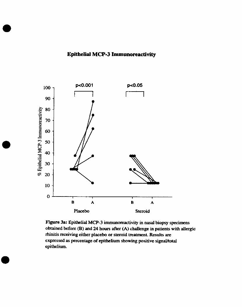
Placebo Steroid
Figure 3a: Epitheiial MCP-3 immunoreactivity in nasal biopsy specimens obtained before (B) and 24 houn aRer (A) challenge in patients with allergic rhinitis receiving either placebo or steroid treatment. Results are expressed as percentage of epithelium showing positive signaihotal epiklium.

Placebo Steroid
Figum 3b: Number of MCP-3 inimuioreactive ceh in the su bepitheliurn of nasal biopsy specimeas obtained be fore (B) and after (A) akrgen chalienge in patients with aiiergic rhinitis receiving either piacebo or steroid treatnient. Results are
expressed as the mean nurnber of MCP-3 positive cells m r k ld .

Placebo Steroid
Figure 4.: Epitheiial MCP-4 immunoreactivity in nasal biopsy specimens obtained before (B) and 24 hours after (A) challenge in patients with aiiergic rhinitis rcceiving either placebo or steroid treatment. results are expressed as percentage of epithelium showing positive signdtotal epithelium.
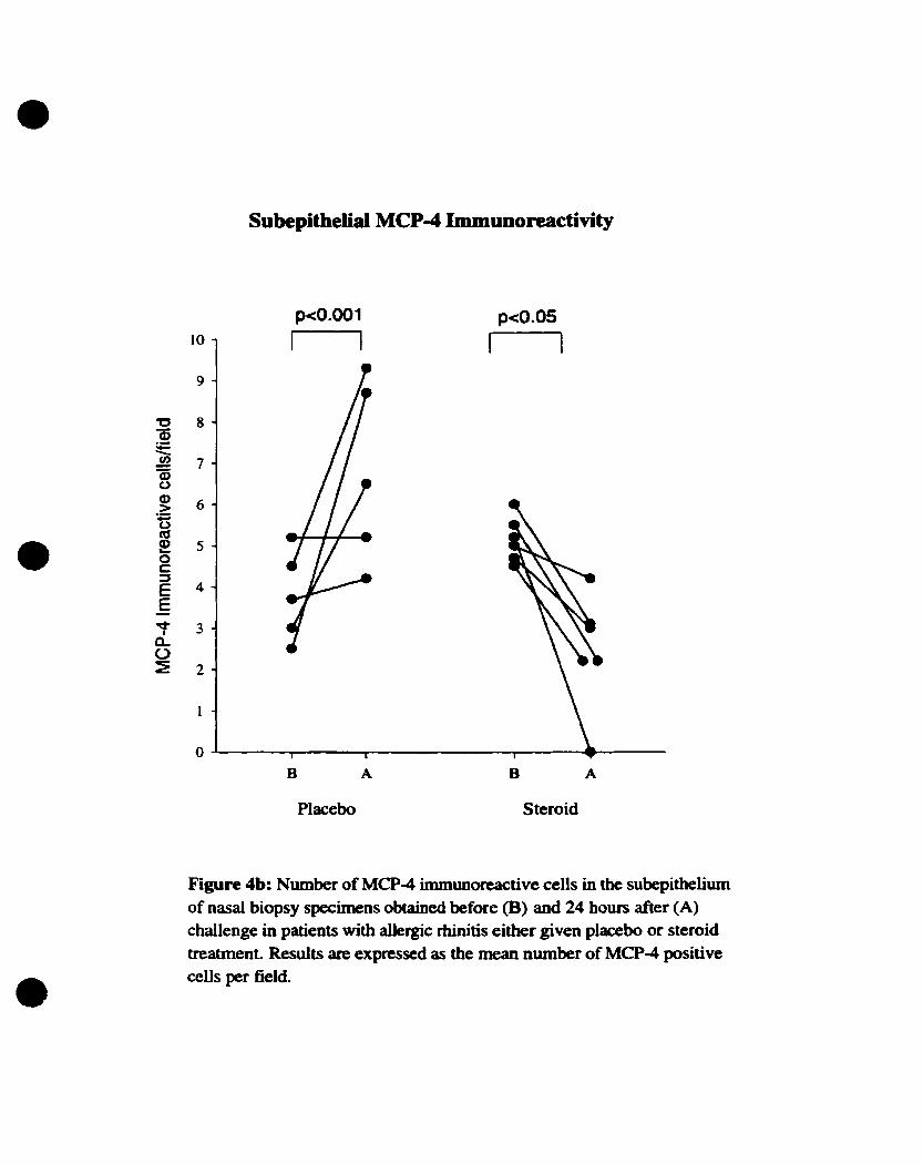
Subepitheiiai MCP4 Immunoreactivity
B A
Placebo
B A
S teroid
Figure 4b: Number of MCP-4 immunoreactive cells in the subepitheiiurn of nasal biopsy specimens obtained before (B) and 24 hours after (A) challenge in patients with alkrgic rhinitis either given placebo or steroid treatment. Results are expresseci as the mean number of MCP-4 positive cells per field

Epithelial MCP-4 mRNA Expression
NS
n
1 I 1 1 I 1
6 A B A
Placebo Steroid
Figure Sa: Epitheiial expression of MCP-4 mRNA in nasal biopsy specimens obtained kfore (B) and 24 hours &ter (A) allergen challenge in patients with allergic rhiaitis receiving either placebo or steroid treatment. Results are expressed as percentage of epitheiium showing positive signaihotai epithelium.

Subepitheiiai MCP4 mRNA Expression
p<O.OOl NS
n n
Figure Sb: Subepithelial expression of MCP-4 mRNA in nasal biopsy specimens obtained before (B) and 24 hours after (A) allergen challenge in patients with aiiergic rhinitis receiving either placebo or steroid treatment. Results are expresscd as number of positive cells per field.
O 1 I I I I 1
6 A 6 A Placebo Steroid

MBP Lmmunorcpctivity
Placebo S teroid
Figure 6: Number of MBP+ cells (eosinophils) within the subrnucosa of nasal biopsy specimens obtained before (B) and 24 hours d e r (A) challenge in patients with allergic rhinitis either given placebo or treated with steroids. Results are expresseci as the mean nurnber of positive ceiîs per field.

PIacebo S teroid
Figure 7: Number of CD4+ ceiis (T helper cells) within the submucosa of nasal biopsy specimens obtained before (B) and 24 hours afler (A) challenge in patients with allergic thinitis either given placebo or treated with steroids. Results are expressed as the mean number of CD4+ ceiis per field.

5 1 1 I I 1
8 A B A
Placebo Steroid
Figure 8: The number of CD68+ cells (macrophages) within the submucosa of nasal biopsy specimens obtained before (B) and 24 hours after (A) challenge in patients with ailergic rtiinitis either given placebo or treated with steroids. Results are expressed as the m a n number of CD68+ ceils per field.
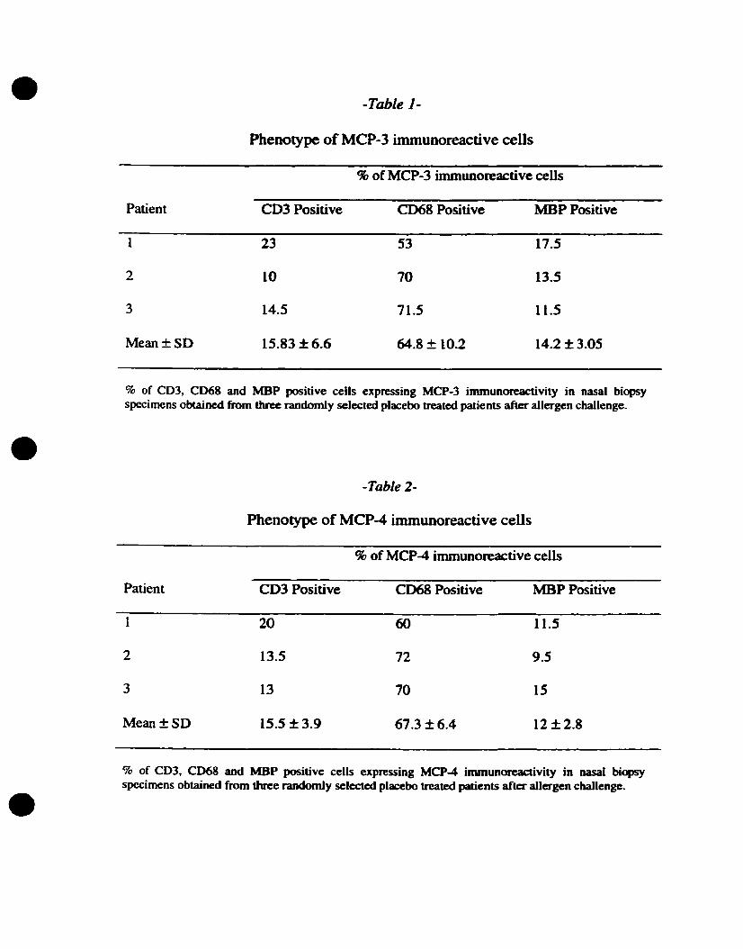
-Table 1-
Phenotype of MCP-3 immunoreactive ceiis
96 of MCP-3 immunoreactive cells
Patient CD3 Positive CD68 Positive MBP Positive
% of CD3, CD68 and MBP positive cells expressing MCP-3 imrnunoreactivity in nasal biopsy specimens obtained frorn three randomly selected placebo treated patients &ter allergen challenge.
-Table 2-
Phenotype of MCP4 immunoreactive ceîïs
% of MCP4 immunoreactive cells
Patient CD3 Positive CD68 Positive MBP Positive
% of CD3, CD68 and MBP positive cells expressing MCP-4 irnmunoreactivity in nasal bicpsy specimens obtained h m three randomly selected placebo treated patients after allergen challengc.

Representative examples of immunostaining for MCP-4 in nasal biopsy specimens from placebo treated aliergic rhinitic patients at baseline (a) and foiiowing aiiergen challenge (b). Note a constitutive expression of MCP-4 in the nasal epithelium (a) and an upregulation of MCP4 expression foiiowing aiiergen challenge both in the epitheiium and subepitheliurn of the nasal rnucosa (b).

Representative examples of immunostainhg for MCP-4 in nasal biopsy specimens from placebo (a) and steroid treated (b) allergic rhinitic patients following allergen challenge. Note a high expression of MCP4 foilowing dergen challenge in the nasal biopsy section from a subject given placebo (a). In cornparison, the expression of MCP-4 in the nasal mucosa following chaiienge was inhibited in subjects pretreated with steroids (b).

4. Discussion
The aims of this study were to examine the expression of monocyte
chemotactic proteins in allergen-induced rhinitis and to hvestigate their possible
contribution to the late nasal response with the use of the nasal allergen challenge
model. Furthermore, we assessed whether the Iate nasal response could be inhibited by
pretreatment with topical glucocorticoid steroids. Our results demonstrate that
exposure to diergen increases the expression of MCP-3 and 4 within the nasal
mucosa of patients with ailergic rtiinitis and that these effects are abrogated by topical
steroid treatment.
We observed that ailergen provocation in the nasal mucosa of rhinitic patients
resulted in early (0-60 min) and late (1-24 h) nasal symptoms. These responses were
assessed by the number of sneezes and the degree of nasal blockage respectively,
experienced by the patients during the 24 hour period following nasai ragweed
challenge. These clinical observations validated the adequacy of challenge and the
effectiveness of the steroid.
Budesonide was shown clinicaily to inhibit the late nasai response as it
significantly reduced the degree of nasai obstruction as expected. On the contrary,
steroid treatment didn't seem to inhibit the syrnptoms of the early nasal response
eventhough steroids are normally clinicadly effective at blocking the nasal symptoms
characteristic of the early nasal response. It is possible that the assessrnent made for
the early phase response may not be very accurate given the limited number of patients

in this study. In addition, intersubject variability may also be a factor responsible for
this observation.
It has previwsly been shown that the ailergen-induced late nasal responses are
accompanied by an inaux of iu.fhnmatory ceus particdarly eosinophüs and =+ T
ceus and basopbils (Vamey et al., 1992, MinsbaU et al., 1997. Ying et al., 1994). The
presence of these œlls and the local release of their mediators at the ailergic site have
been associated witb the pathological features of aüergic rhinitis (Vamey et al., 1992,
Durham et al., 1992). The mechanisms responsible for the recruitment of these celis to
the site of inflammation, are not clearly understood. however, it has been proposed
that one possibility may k the local generation of chemotactic agents that regulate this
influx of leukocytes. (Baggiolini & Dahinden 1994). A number of chemotactic factors
such as bacterial peptides, phospholipid metabolites, and cytokines, have been
identifieci and found to be associated with ailergic inflammation. These
chernoattractants however, are weak and have little target selectivity. Chemokines, on
the other hand, are potent chemoattractants, specific for a variety of leukocytes. In
particular, CC chemokines have been impiicated in allergic diseases, since they have
k e n s h o w to act on the inflatnmatory ceils specificaiiy involveci in allergic
inflammation.
Although the expression of MCPs have previously been detected in ailergic
tissues isolated fiom patients witb chronic allergic sinusitis, asthma and atopic
dermatitis ( Garcia-Zepeda et al., 1996b, Lamkhioued et al., 1997, Wright et al.,
1998), there is no previous evidewe in vivo, for a role of MCPs in human allergic
rhinitis. Therefore, our findings of an increased expression of MCP-4 mRNA and
MCP-3 and 4 immunoreactive celis in the nasal mucosa, as a result of allergen
challenge is novel, and this increased expression may partly account for the
inBammatory ce11 accumulation seen in ailergen-induced late nasal respomes.
We demonstrated a constitutive expression of MCP-1, -3 and -4 in the d
mucosa, particularly in the epithelial œU layer. This observation suggests a role for
MCPs in baseline leukocyte trafncking through epithelial tissue, as part of the natural
host defenses against pathogens, as previously suggested by Garcia-Zepeda et al.
While chemokines often have similar in vitro functions, their pattern of expression in a
given inflammatory disease determines which chemokines may play a pathobiological
role. The constitutive expression of MCP-4 has previously been detected within
normal tissues, particularly with large epitbelial surfaces in contact with the
environment such as the intestines and the lung. This pattern of expression is sunilar to
what has been seen for eotaxin, except that MCP-4 is expressed at higher baseline
leveIs in the lung (Garcia-Zepeda et al., 1996a, b). In addition, MCP-1 has been
detected predominantly in salivary glands, and MCP-2, in the intestines and o v q
(Saraf~ et al., 1997, Coiliie et al., 1997). Since MCPs have been shown to act on
infiammatory cells that are not n o d y fowid in high numbers withia nonnal tissues,
the constitutive expression of MCPs and their action within these tissues needs to be
further investigated (Collins et al., 1995, Garcia Zepeda et al., 1996, Luster et al.,
1997).
Although constitutively expressed at high levels within the nasal mucosa at
baseline, MCP-1 imrnunoreactivity was not increased as a result of ailergen
provocation as seen with MCP-3 and 4. This hding supports those h m previous
shidies demonstrating that MCP- 1 is abundant in mononuclear ceil-nch inflarnmatory
conditions such as grandomatous inflammation, and in macrophage-rich regions of
atherosclerotic lesions (Antony et al., 1993, na-Hertuala et al., 199 1). Interestingly,
few eosinophils are present in these conditions uolike in allergic diseases. supporting
the observations that MCP-1 may not have any chernotactic activity for eosinophils. It
was recently demonstrated, that MCP-1 production is enhanced in the blood and tissue
of patients with tuberculosis, suggesting that MCP-1 may contribute to the
antimycobactenal inflanmatory response by attracting monocytes and T lymphocytes
which are characteristic of these disease (Lin et al., 1998).-Nevertheless, the role of
MCP- 1 in the pathogenesis of aliergic disease can not be d e d out since MCP- 1 has
been detected in human ailergic tissue, and is well known for its abiity to chemoattract
and activate basophils, which represent a major source of mediators, and an essential
element of IgE-mediated hypersensitivîty reactions (Schoeder et al., 1995, Alam et
al., 1992).
While the expression of several C-C chemokines such as eotaxin and RANTES
have previously been showa to be upregulated in the nasal mucosa in allergen-induced
rhinitis (Bazan et al., 1997, Minshali et al., 1997), and to be associateci with the
infiltrating inflammato~y cells, the factors responsible for the infiltration of leukocytes
in disease States have not been clearly defmed. To deteninine whetber the local
upregulation of MCP-3 and 4, which we observed in the nasal mucosa after allergen
challenge, is associated with the infiltrating cells, we examined the numbers and
phenotype of cells within the nasal mucosa after allergen challenge. Our data
demonstrated a significant eosinophil, and CW' T ceU infiltration foiiowing ailergen
challenge, consistent with previous hndings ( Vamey et al., 1992, Ying et a1, 1993,
Minshall et al., 1997). This increase may be associated with the upregulation of MCP-
3 and4 within the nasai mucosa The presence of these cells and the release of their
mediators such as cytotoxic proteins or Th2 type ctyokines, has been associated with
the pathological feanires of aüergic rhinitis (Varney et al., 1992, Durham et al., 1992).
In contrast, no change in C068'ceUs were observeci in the nasal mucosa following
challenge when compared to baseline biopsies, consistent with o w previous flndings
(Minshail et al., 1997, Rajakulasingam et al., 1997). Monocytes generally infiltrate
Th1 type granulomatous diseases (Lin et al., 1998). The fact that MCP-1
immunoreactivity was not increased in the nasal mucosa after challenge may also
account for this hding, given that macrophages are one of the main sources of MCP-
1, and MCP-1 levels seem to be enhanced particularly in macrophage nch conditions
(Yla-Hertuala et al., 199 1).
Furthemore, to determine if the expression of MCP-3 and -4 in the epithelial
and subepithelial cell layers was quantitatively associated with the infiltration of
inflammatory ceiis, we performed correlation analyses. The fact that there were no
significant correlations between the infiltration of inflammatory celis and MCPs
expression may suggest that other chemokines, such as eotaxin and RANTES, play a

substantial role in the acute infiammatory ceii influx foliowing allergen provocation. In
addition, the CC chemokines MIP-la, MIP-lp, have also been shown to be
chemotactic for monocytes, Merentially chnotactic for lymphocyte subsets, and
chemotactic for eosinophils in vitro and presumably in ailergic responses (Taub et al.,
1993, Schaii et al., 1993, Rot et al., 1992). The lack of correlation observed rnay also
be due to the time course of chemokine production and its influence on the kinetics of
leukocyte recruitment. A disadvantage of the nasal biopsy technique is that it is not
amenable to the performance of tirnecourse studies for ethical reasons. We chose the
24 hour time point based on previous studies showing identifiable changes m
inflarnrnatory cells particularly eosinophils and T lymphocytes at this particular tune
point (Varney et al., 1992).
In our tissue specimens, we obsemed MCP-3 and -4 expression within the
nasal epithelium and resident infiammatory celis in the nasal submucosa. It is weii
known that chemokines are often secreted Erom activated leukocytes preswnably as
part of amplification and desensitization mectianisms. Phenotyping the celis expressing
MCP-3 and MCP-4, demonstrated that macrophages, eosinophiis and T celis were
significant cellular sources of these chemokines, with macrophages k ing a prominent
source within the submucosa. However, the expression of MCP-3 and 4 was most
strongly locaiized to the ciliated pseudostratified epithelium in the nasal mucosa in Our
clinical specimens. This is consistent with previous findings showing that MCPs are
strongly expressed in branchial epithelial cell iines after cytokine stimulation (Stellato
et al., 1997, Garcia-Zepeda et al., 1996). This however, is not only the case for

MCPs, but for other CC chemokines as weU, such as eotaxin and RANTES (Minshall
et al., 1997, Ponath et al., 1996, Rajakulasingam et al., 1997)
In this study we also examined the effets of nasal topical corticosteroids on
the expression of MCPs and on the cellular innltraîe in allergen-induced rtiuiitis.
AUergic rhinitis has been demonstrated to be amenable to treatment with topical nasal
corticosteroids (PipKom et al., 1987). Numerous studies have demonstrated the
efficacy of steroids in the suppression the clinical symptoms in this disease. Studies
have demonstrated that prolonged treatment with topical corticosteroids inhibits both
early, and late nasal responses and the associated tissue eosinophilia following allergen
provocation, by inhibiting the expression of Th2 cells and the release of their cytokines
(Rak et al., 1994, Masuyama et al., 1994). We demonstrated that pretreatment with
budesonide abrogates the increase in MCP-1, -3 and -4 protein expression both in the
epithelium and subepithelium as a result of allergen provocation in the pIacebo treated
subjects. These data are consistent with previous studies showing that topical
corticosteroid treatment leads to the inhibition of the LNR (Rak et al., 1994) and with
previous in vitro studies showing that budesonide may be an effective inhibitor of
MCP-4 mRNA in cultured BEAS-2B epithehal cells activated with cytokines (Stellato
et al., 1992). We postulate that the suppression of ceilular infdtraie after allergen
challenge in subjects undergoing glucocorticoid therapy occurs partly through the
inhibition of MCPs, effectively attenuating leukocyte transendothelial migration.
Epithelial cells are the fmt ce11 type to corne in contact with inhaled g1ucocorticoids
and since we and others have shown that epithelial celis are the main source of
chemokines (Stellato et al., 1992), this ceil type may be an important target of the
anti-infiammatory effects of inhaled giucocorticoids. Furthermore since application of
topical corticosteroids represent in many
treatment of seasonal allergic rbinitis, these
to the therapeutic management of rhinitis.
cases the dmg of first choice for the
hdings must be considered of relevance
In summary, we have demonstrated a constitutive expression of MCP- 1, -3 and
-4 in the nasal mucosa of dergic patients with a substantiai increase in MCP-3 and -4
immunoreactivity aswell as MCP-4 rnRNA expression following allergen challenge.
The hcreases in these chemokines were accompanied by an upregulation of
inflammatory ceUs, particulariy eosinophils, within the nasal submucosa Furthermore,
prolonged treatment with topical corticosteroids was associated with a sigrilficant
attenuation of aiiergen-induced increases in MCP- 1, -3 and -4 immunoreactivity and in
MBP, CD68, and CD4 positive cells. These data suggest that the increased expression
of MCP-3 and -4 may be relevant to the pathophysiology of latephase reactions in
ailergen-induced rhinitis through their abiiity to recruit leukocytes into the nasal
mucosa in response to antigen.
We beiieve that CC chemokines, in particular MCPs are likely to play a critical
role in regulating the inflammation characteristic of ailergic diseases and inhibition of
their production may thus serve as a favorable approach for the treatment of allergic
diseases. Chemokine antagonists are of therapeutical interest as an approach for anti-
ailergic therapy. It has been shown that antagonists are effective, and some low-
molecular-weight compounds that block chemokine receptors are aiready at hand

(Baggiolini & Moser 1997). However, whether strategies directed agaînst MCP-3 and
-4 , such as synthesis inhibitors or receptor antagonists might modifL the response to
allergen challenge and the course of allergic rhinitis, rernains to be investigated.

S. Future Studies
In order to hinher investigate the role of MCPs in the pathogenesis of allergif
rhinitis, additional stuclies m u t be conducted whkh fan hclp t o answer many of the
questions raised in this study. In vitro chernotaxis assays, which examine cbemotactic
activity more precisely. will allow us to assess the actual p tency of MCPs as
chemoattractants. Since the processes governing the recruitment of infiammatory ceils
are Nely to be the resuit of a combination of chemokims, with the use of neuaalitiag
antibodies we rnay be able to identify the chernoattractive contribution and potency of
each individual chemokïne. Furthemore, with the use of an in vitro assay we çan
examine whether the chemoattractant effect of MCPs is enhanced by the addition of
other mediators or cytokines. T o detennine whether there is a local upregulation of
MCPs in the nasal mucosa after challenge, ex vivo studies isolating the nasal mucosa
fiom systemic inaltration would provide us with such insight. Given the Limitations of
the in vivo allergen challenge model. the nasal explant model cm also serve as a mans
of perfonning thne course studies to examine the kuietics of MCPs expression. At the
sarne thne, the regulation of MCPs expression can be investigated more precisely by
hcubating the explanted tissue with various mediators, in particular Th2-type
cytokines.
These in vitro studies may thus provide valuable iasight in determinhg more
accurately the replation and function of monocyte chernotactic proteins in alkrgic
inflammation.

Alam R., Lett B., Forsythe PA., Anderson WD., Kenamore C., Konnos C., Grant JA. 1992. Monocyte chemotactic and activating factor is a potent histamine-releasing factor for basophils. J Clin Invest. 89: 723-728.
Alam RP., Forsythe PA., Stafford S., Heinrich J., Bravo R., Proost P., Van damme J. 1994. Monocyte chemotactic protein -2, Monocyte chemotactic protein-3, and fibroblast-induced cytokine. J Immunol. 153: 3 155-3 159.
Allavena P., B i s h i G a , Zhou D., Van Damme J., Jiiek P., Sozzani S., Mantovani A 1994. Induction of naturai killer ce1 migration by monocyte chemotactic protein- 1, -2 and -3. Eur J Imunol. 24:3233.
Bachert 199 1. Possible role of macrophages in allergic rtiinitis. Infernutio~l Archives A Llergy Applied Immunology. 94: 1-4: 244-5.
Baggiolini, M., Dewald, B., Moser, B. 1994. Interleukin-8 and related chemotactic cytokines-CXC and CC-chemokines. Adv.lrnmunol.55:97- 179.
Baggiolini, M., Dahinden, C.A. 1994. CC chemokines in allergic inflammation. Immun01 Today. 1 5 : 127.
Baggiolini M., Dewald B., Moser B. 1997. Hurnan Chemokines: An Update. Annu Rev Immunof. 15:675-705.
Baggiolini M., Moser B. 1997. Blocking chemokine receptors.. 186: 1 189- 1 19 1.
Bainton, DF, Farquhar MG. 1970. Segregation and packaging of granule enzymes m eosinophilic leukocytes. J Ce11 Biol. 45: 54-73.
Bascom R. Wachs M., Nacleno R., Pipkorn U., Galli SJ., Lichtenstein LM. 1988. Basophil influx occurs after n a d antigen challenge: effects of topical corticosteroid pretreatment. J Allergy Clin Irnmunol. 8 1 :580-9.
Baraniuk J. 1997. Pathogenesis of Aiiergic Rhinitis. I Allergy Clin Immunol. 99: S763- 72.
Bazan JF., Bacon KB., Hardiman G., Wang W., Soo K., Rossi D., Greaves DR., Zlotnik A., Schaii TJ. 1997. A new class of membrane bound chemokine with a CX3C motif. Nature. 385:640.

Ben-Baruch A., Xu U., Young PR., Bengali K., Oppenheim JJ., Wang JM.1995. Monocyte chemoîactic pmtein-3 (MCP-3) interacts with muitiple leukocyte receptors- CC CKRI, a receptor for macrophage infiammatory protein-l-tes, is also a functional receptor for MCP-3. J Biol Chem. 27q22): 123-28.
Bentley AM., Jacobson MR., Cumbenworth B. 1992. Immunohistology of the nasal mucosa in seasonal ailergic rhinitis: increaeses in activated eosinophiis and epitheiial mast ceils. J Allergy Clin Immunol. 89:82 1-9.
Bentley AM., Hamid Q., Robinson DS., Schotman E., Meng Q., 1996. Rednisilone treatment in asthma Reduction in the numbers of eosinophils, T ce&, uyptase-only positive mast cells, and modulation of IL-4, IL-5 and IFN-y cytokine gene expression within the brouchial mucosa Am J Respir Crir Cure Meh 153: 55 1-556.
Bischoff SC., Krieger M., Bninner T., Dahinden CA., 1992. Monocyte chexnotactic protein 1 is a potent activator of human basophils. J Exp Med. 127 1-1275.
Blair H. 1977. Natural history of childhood asthma: 20-year foilow-up. Arch Dis Child. S2:6 13-9.
Bochner BS., Luscinskas FW., Gimbrone MA Jr. 199 1. Adhesion of human basophils, eosinophils and neutrophils to interleukin l-activated human vascular endothelid ceils: contributions of endothelid celi adhesion molecules. 173: 1553- 1557.
Bousquet J., Chanez P., Lacoste N., Barneon G., Ghavanian N., Enander I., Venge P., Ahlstedt S., Simony-Latontain J., Godard P., Micbel FB. 1990. iV Eng J Med. 323: 1033.
Bousquet J., Bullinger M., Fayol C., Marquis P., Valentin B., Burtin B. assessrnent of quality of Me in patients with perenniai aüergic rhinitis with the French version of the SF-36 Health Status Questionnaire. 1994. JAllergy Clin Immunol. 94: 182-8.
Bradding P., Feather IH., Wilson S., Bardin P., Holgate ST., Howarth PH. 1993. Immunolocalisation of cytokinesin the nasal mucosa of nomal and perennial rhinitis subjects: the mast ceU as a source of IL-4, IL-5 and IL-6 in human allergic inflammation. 3 Immunol. 15 1 : 3853-65.
Bradding P., Mediwake R., Feather IH. 1995. TNF-a is localized to nasal mucosal mast ceils and is released in acute allergic rhinitis. Clin Erp Allergy. 25: 406- 15.
Braun RK., Hansel TT.,de Vries UM. 1995. Human peripheral blood eosinophils have the capacity to produce IL-8. Eur J Immunol. 23:956-60.
Broxmeyer HE., Sherry B., Cooper S., Lu L., Maze R., Beckmann MP., Cerami A., Ralph P. 1993. Comparative analysis of the human macrophage inflammatory protein
farnily of cytokines (chemokuies) on proMeration of hwnan myeloid progenitor cells. Interacting effects involving suppression, and blocking of suppression. J Immunol. 150:3448-3458.
Bdlock GR., Petrusz P. (eds) 1982. Techniques in Immunocytochemistry. Vol. 1 . Acad Press, New York.
Bunnag C., Jareoncharsi P., Wong EC. 1992. A double-blind comparison of nasai budesonide and oral astemizole for the treatment of perennial rhinitis. Allergy. 47 (4, part 1 ) : 3 13- 17.
Butcher EC. 199 1. Leukocyte-endothelid celi recognition: three (or more) steps to specificity and diversity . Cell. 67: 1033- 1036.
Calderon MA., Lozewicz S., Prior A., Jordan S., Trigg CJ., Davies RI. 1994. Lymphocyte inf'iitration and thickness of the nasal mucous membrane in perennial and seasonai aliergic rhinitis. J Allergy Clin Immunol. 93: 635-43.
CanteLls MC., Irani AM-, Schwartz LB. 1987. Evaluaîion of human peripheral blood leukocytes for mast ceU tryptase, J Immunol. l38:2 18.4-8 1 .
Charo IF., Myers SJ-, Herman A., Franci C., Comolly CU., Coughlin SR. 1994. Molecular cloning and functional expression of two monocyte chemoattractant protein 1 receptors reveals alternative splicing of the carboxyl-tenninal taiis. Proc Nat1 Acad S C ~ USA. 9 1 : 2752-2756.
Check WA., Kaliner MA. 1990. Phanaacology and Phamiacokinetics of topical corticosteroids derivates used for asthma therapy. Am Rev Resp Dis. 141 (2, part 2): S44-S5 1.
Church MK., Okayama Y., Bradding P. 1995. functional mast celi heterogeneity. in: Busse WW, Holgate ST, editors. Athma and rhinitis. Boston: Blackweil Scientific, 1995: 209-20.
Church MK., Levi-Schaffer F. 1997. Updates on cells and cytokines. The human mast cell. J allergy Clinical Immunol. 1997.99: 155-60.
Clark-Lewis 1. Et al. 1995. Structure-activity relationships of chemokines. J Leukocyte Bio1.57:703-7 1 1.
Cocchi F., DeVico AL., Garzino-Demo A., Arya S., GaiIo RC., Lusso P.1995. Identification of RANTES. - - l a and M[P- 1 as the major HIV-suppressive factors produced by CDS+ T celis. Science. 270: 1 8 1 1 - 1 8 15.

Coillie EV., Fiften P., Nomiyama H., Sakaki Y., Miura R.. Yosbie O., Van Damme J., Opdenakker G., 1997. The human MCP-2 gene (SCYAS): cloning, sequence analysis tissue expression. and assignment to the cc chemokine gene contig on chromosome l7q 1 1.2. Genomics. 40: 323-33 1.
Coons AH.. Cmxb HJ., Jones RN. 1941. Immunological properties of an antibody containing a fluorescent group. Proc Soc Exp Bi01 Med. 47:200-2.
Corren J. 1997. AUergic rhinitis and asthma: How important is the Linlr? J Allergy Clin Imrnunol. 99:S78 1-6.
Costa J., Mattossian K., Resnick NB., Beil W., Wong DTW., Gordon IR.. Dorak AM., WeUer PF., G d i S JI 1993. J Clin Invest. 9 1 : 2673.
Cox KH., Deleon D., Angerer LM., Angerer RC. 1984. Detection of mRNA in sea urchin embryos by in situ hybridization using RNA probes. Dev Biol. 101:485-502.
Dahinden CA., Geiser T., B r u ~ e r T., Von Tscharner V., Caput D., Ferrara P.. Minty A., Baggioluù M. 1994. Monocyte chernotactic protein 3 is a most effective basophil- and eosinophil-activating rhemokine. J fip Meà. 179:75 1.
Del-Prete G., Maggie E., Rornagnani S. Human Thl and Th2 celis: fwictional properties, rneachanisms of regulation , and role in disease. ïuboratory Investigation. 70(3): 299-306. 1994.
Demy P., Hamid QA., Krause J.. Polak J., Leagon S. 1989. Oiigoriboprobes. tools for in situ hybridization. Histochernistry. 89:48 1 -3.
Desreumaux P.. Janin A., Dubuequoi S.. Copin MC., Torpier G., Capron A., Capron M.. Prin L. 1993. Synthesis of interleukin-5 by activated eosinophils in patients with eosinophilic k a r t diseases. Blood. 82(5): 1553-60.
Devreotes PN..Zgmond SH. 1988. Chernotaxis in euliaryotic ceiis: a f aus on leukocytes and Dictyostelium. Annu Rev Ce11 Biol. 4: 649-686.
Doherty TM., Kastelein S., Menon S., Andrade S., Coffman RL. 1993. Modulation of murine macrophage function b y IL- 13. I Immunol. 15 1 : 7 15 1.
Durham, S.R.. Ying S., Vamey, V.A., Jacobson, M.R., Suddenck, Mackay. I.S., Kay, A.B., Harnid, Q. 1992. Cytokine messenger RNA expression for IL-3, IL-4, IL-5 and granulocyte/macrophage colony-stimulating factor in the nasal mucosa after Iocal allergen provocation: relationship to tissue eosinophiiia J. Immunol. 148: 2390-2394.
Del Prete G., Romagnani S. 1994. The role of Th1 and Th2 subsets in human infectious diseases. Trends in Microbiology. 2( 1 ): 4-6.

Dvoracek JE., Junginger JW., Kem EB., Hyatt RE., Gleich GJ. 1984. Induction of nasal late-phase reactions by insufflation of ragweed-pollen extract. J dergy Clin Imniunoi. 73 :363-8.
Elsner J., Hochstetter R., Kunmig D., Kapp A. 19%. Human eotaxin represents a potent activator of the respiratory burst of human eosinopbils. Eur J Imnrzuwi. 26: 1919-1925.
Evans R m., Mullally DI., W h RW., et al. 1987. National trends in the morbidity of asthma in the U.S.: prevalance, hospitakation and death from asthma over two decades, 1965- 1984. Chest. 9 l:65S-74S.
F'idlay S., Huber Fm, Garcia J.. Huang L. 1992. Efficacy of once-a-day intranasal admuristartion of triamcinolone acetonide in patients with seasonal alïergic rhinitis. Ann Allergy. 68: 228-32.
Franci C., Wong LM., Van Damme i., Proost P., Charo IF., 1995. Monocyte chemoattractant protein-3, but not monocyte chemoattractant protein-2, is a functional ligand of the human monocyte chemoattractant protein- 1 receptor. J Imrnunol. 154:6511-17.
Furie MB., Randolph GJ. 1995. Chemokines and tissue injury. Am J Pathol. 146: 1287-1301.
Galli SJ. 1993. New concepts about the mast ceil. N Engl J Med. 328:257-65.
Garcia-Zepeda, E.A., Rothenberg, M.E., Owmbey, R.T., Celstin, J., Leder, P.. Luster, A.D. 1996a. Human eotaxin is a specific chernoattractant for eosinophil cells and provides a new mechanism to explain tissue eosinophilia Nat. Med. 2:449.
Garcia-Zepeda, E.A., Combardiere. C., Rothenberg, M.E., Sasafi, M.N., Lavigne, F., Hamid,Q., Murphy, P.M., Luster, A.D. 1996b. Human Monocyte Chemoattractant Protein (MCP-4) 1s a Novel CC Chemokine with Activities on Monocytes, Eosinophils, and Basophils Induced in AUergic and Nonallergic Inûammarion That Signals Through the CC Chemokine Receptors (CCR)-2 and -3. 1 h m o l . l57:56 13-5626.
Gao K., Kuhns DB-, Tiffany HL., McDermott D., Li X.. Francke U.. Murphy PM. 1993. Structure and huictional expression of the human macrophage infiammatory protein l -a /RANTES receptor. J Exp Med. 177 (5): 142 1-7.
Giad A., Hamid Q., Adams C. Terrenghi G., Polak M. 1989. Non-isotopic RNA probes: cornparison between different labels and detec tion systems. Histochemistry. 93: 19 1-6.

Godiska R-, Chantry D., Dietsch GN., Gray PW. 1995. Chemokine expression m murine experimental allergic encephalomyelitiïs. I Neuroimrnunol. 58: 167.
Gomez E., Clague JE., GatJand D., Davies RJ. 1988. Effect of topical corticosteroids on seasonaUy induced increase in nasal mast cek. Brtit Med J Clin Res. 29q6636). 1572-3.
Gong JH., Uguccioni M., Dewald B., Baggiolini M., Clark-Lewis 1. 1996. RANTES and MCP-3 antagonists bind multiple chemokk receptors. J Biol Chem. 27 1 (10): 52 1-27.
Gosset P., Malaquin F., Delnest Y. 1993. Interleukin 6 and interleukin- la production is associated with antigen induced late nasal response. J Allergy Clin Immunol. 92: 878-90.
Griffith-Johnson DA., Collins PD., Rossi AG. Jose PJ. Williams TJ. 1993. The chemokine, eotaxin, activates guinea-pig eosinophils in vitro and causes their accumuiation into the lung in vivo. Biochem Biophys Res Commun. 197(3):1167- 1172.
Gronborg H., Bisgaard H., Romeling F., Mygind N. 1993. Early and late nasal response to allergen challenge. Allergy. 48: 87-93.
Hamid Q., Wharton J., Terenghi G.1987. halizat ion of atrial natniretic peptide mRNA and immunoreactivity in rat heart and human airial appendage. Proc Nat Acad sci USA. 84: 6760-4.
Hamid Q., Barkans J., Meng Q., Ying S., Abrams JS., Kay AB., Moqbel R. 1992. Human eosinophils synthesize and secrete iL-6 in vitro. Blood. 80: 1 496.
Hamid Q. 1993. Isotopic Localkation of ceiiular mRNA in clinical matenal using RNA probes. In: Diagnostic Molecular Pathology.= a Pratical Approach. Vol. 1 (eds C.S. Herrington & OD. McGee) pp. 169-85. IRL Ress, New York.
Hemngton CS., McGee J.1990a. Non-radiolabelied in situ hybridization. In: in situ hybridization: Application to developmental Biology and medecine (eds H. Harris & D. WiMnson). P24 1. Cambridge University Ress, Cambridge.
Hemngton CS., Burns J., Graham AIC, McGee J. 1990b. Discrimination of closely homologous HPV types by nonisotopic in situ hybridization: definition and denvation of tissue melting temperature. Histochem J. 22545-50.
Holgate ST., Bradding BM., Sampson AP.1996. Leukotriene antagonists and synthesis inhibitors: new directions in asthma therapy. J Allergy Clin Immunol. 98: 1- 13.

Howarth PH. 1989. Aiiergic Rhinitis: a rational choice of treatment. Resp. Med. 83: 179-88.
Howarth PH., Holgate ST. 1990. Basic aspects of aiiergic reactions. In : Childhouû Rhinitis and Sinmiris: Pathophysiology and Treatment (eds C . NaspitzsL D.G. Tinkelman). pp. 1-35. Marcel Dekker. New York.
Howarth PH., Bradding P., Quint D., Redington AE., Holgate SC. 1994. Cytokines and a h a y infiammation. In: cells und cytokines in Lung Inflammation 1993 (eds M . Chignard, M. Pretolani, P. Prenesto & B. Vargaftig). Ann NYAcad. Sci. 72569-82.
Humbert M., Ying S., Comgan C. 1997. Branchial mucosal expression of the genes encoding chemokines RANTES and MCP-3 in symptomatic atopic and nonatopic asthmatics: relationship to the eosinophiî cytokines interleukin (IL-S), granulocyte macrophage-colony-stimulating factor, and L 13. Am J Respir Ce11 Mol Biol. 16: 1-8.
Jose PJ., Griffitbs-Johnson DA., Coilins PD., Walsh DT., Moqbel R., Totty NF., Truong O., Hsuan JJ., WiIliams TJ. 1994. Eotaxin: a potential eosinophil chemoattractant cytokine detected in a guinea-pig mode1 of ailergic airways inflammation. J Erp Med. 179: 88 1-887.
Juluisson S., Aldenborg F., Enerback L. 1995. Protease content of mast ceiis of nasal mucosa: effects of naturai ailergen exposure and of local corticosteroid treatment. Allergy. 1995. 50: 15-22.
Kawabori Y., Kami N., Tosho T.1995. ProMerative activity of mast cells in allergic nasal mucosa Clin Exp Allergy. 25: 173-8.
Kennedy J., Kelner GS., Kleyensteuber S., Schaii TJ., Weiss MC., Yssel H., Schneider PV., Cocks BG., Bacon KB., Zotnik A. 1995. Molecdar cloning and functional characterizaion of human ly mphotactin. J i m u n o l . 1 55: 203-209.
Kelso A. 1995. Th1 and Thî type subsets: paradigms lost? immunol Today. 16(8): 374-79.
Kita H., Ohnishi T., Okubo Y., Weiler D., Abram JS., Gleich GJ. 1991. Granulocyte/macrophage colony-stimdating factor and interleukin 3 release h m human peripheral blood eosinophils and neutrophils. J Ep Med. 174:745.
Knapp HR. 1990. Reduced allergen-induced nasal congestion and leukotriene synthesis with an o d y active 5-lipoxygenase inhibitor. New Engl J Med. 327: 1745-8.
Krause HF. 1992. Pharmacology of upper respiratory allergy (review). Otolaryngol. Clin. North Am., 25(1), 134-50.
Kuijpers TW., Hakkert BC., Hoogerwerf M., Leeuwenberg JFM. Rws D. Role of endothelial leukocyte adhesion molecde-1 and platelet-activating factor in neutrophil adherence to I L 1 prestimuiated endothelia1 ceiïs: endothelial leukocyte adhesion molecule- 1 -mediated CD 1 8 activation. J Immunol. 147: 1369- 1376.
Lamkhioued, B., Rcnzi, P., Abi-Younes, S., Garcia-Zepeda, E.A., Allakhverdi, Z., Ghaffar, O., Rothenberg, MD., Luster, A.D., Hamid, Q. 1997. Increased Expression of Eotaxin in Branchial Lavage and Airways of Asthmatics Contributes to the chernotaxis of Eosinophils to the Site of inflammation. J Immunof. 159: 4593-.160l.
Lahrtz F., Piali L., Naâai D. 1997. Chemokines in viral meningitis: chemotactic cerebrospinal fluid factors include MCP-1 and IP-10 for monocytes and activate T lymphocytes. Eur f Immunol. 27:2484-9. -.
Lawrence M.., Springer TA. 1991. Leukocytes roll on a selectin at physiologicai flow rates: distinction h m and prerequisite for adhesion through integrins. Cell. 65859- 873.
Lin Y., Gong J., Zhang M., Xue W., Barnes P. 1998. Production of Monocyte C hernoattrac tant Protein 1 in Tuberculosis Patients. Infection and Immuniîy. 66 (5): 23 19-22.
Loetscher P., Seitz M., Clark-Lewis I., Baggiolini M., Moser B. 1994. Monocyte chemotactic proteins MCP-1, MCP-2, and MCP-3 are major attractants for hwnan CD4+ and CDS+ T lymphocytes. FASEB J. 8: 1055.
Loetscher M., Gerber B., Loetscher P., et al 1996. Chemokine receptor specific for IP 1 O and Mig: structure, function, and expression in activated T-lymphocytes. J Ejcp Med. 184: 963-969.
Lopez AF., Elliot MJ., Woodcock J., Vadas MA. 1992. Immunol Today. 13:495.
Lozewicz S., Gomez E., Clague J., Gatland D., Davies RJ. 1990. Allergen-induced changes in the nasal mucous membrane in seasonal allergic rhinitis: effect of nedocromil sodium. J AZiergy Clin Immunof. 8S(l, part 1): 125-3 1.
Macglashan DW. Jr., White M., Huang SIC, Ono SJ., Schroeder J., Lichtenstein LM., MacDonald S. 1994. Secretion of I L 4 h m human basopbils: the relationship of IL4 mRNA and protein in resting and stimuiated basophils. f Inunmol. 152:3006-16.
Mabry RL. 199 1. Uses and misuses of intranasal corticosteroids and cromolyn. Am J Rhinol. 5: 121-4.

MacGlashan DW,, White JM., Huang SIC, Ono SJ., Schroeder J., Lichtenstein LM. 1994. Secretion of JI4 h m human basophils: the relationship between IL-4 mRNA and aprotein in stimuiated basophiis. J Immunol. 152: 3OO6- 16.
Masuyama, K., Jacobson, M.R., Rak, S., Meng, Q., Sudderick, R.M., Kay, B.A., Lowhagen, O., Hamid, Q., Durham, S.R. 1994. Topical glucocorticosteroid (fluticasone propionate) inhibits celis expressing cytokine mRNA for interleukin-4 in the nasal mucosa in allergen-induced rhinitis. 1994. Immunology. 82: 192- 199.
Matemowski U., Mathews KP. 1962. The prevalence of ragweed poUinosis in foreign and native students at a midwestem university and its implications conceming metbods for determining inheritance of atopy . J Atlergy . 33: 1 3 0 .
Metchnikoff E. L 'Immunite dans les maladies Infectieuses. (Masson & Cie, Paris, 1901).
MinshaU. E.M., Cameron, L., Lavigne, F., Leung, D. Y. M., Hamifos, D., Garcia- Zepeda, A., Rothenberg, M., Luster, A.D., Hamid, Q.1997.Eotaxin mRNA and Protein Expression in Chronic Sinusitis and Alergen-induced Niasai Responses in Seasonal Nergic Rhuzitis. Am J Respir Cet1 Mol Biol. 17~683-690.
Miyamasu M., Misaki Y., Izumi S., Takaishi T., Morita Y., Nakamura H., Matsushima K., Kasahara T., Hirai K. 1998. Glucocorticoids inhibit chemokine generation by human eosinophils. J Allergy Clin Immunol .10 1 :75-83.
Monte fort S ., Holgate ST., Howarth PH. 1993. Leukocyte endothelid adhesion rnoLecules and their role in branchial asthma and allergic rhinitis. Eur Resp J. 6: 1044- 54.
Moqbel R., Hamid Q., Ying S., Barkans J., Hartnell A., Tsicopoulos A., Wardlaw A., Kay AB.1991. Expression of mRNA and immunoreactivity for the granulocyte/macrophage colony stimulahg factor in activated human eosinophils. J Exp Med. 174:749.
Mossman TR., Coffman RL.1989. TH1 and TH2 cells: different patterns of lymphokine secre tion lead to different bctional properties . Ann. Rev. Immunol. 7 : 145-73.
Mullins RJ., Olson LG., Sutherland DC. 1989. Nasal histamine chaiienges m symptomatic aiiergic rhinitis. J Allergy Clin Inununol. 83: 955-9.
Murp h y PM. 1994. The molecular bioiogy of leukocyte chemoattractant receptors. Annu Rev Immunol. 12: 593-633.
Murphy PM. 1996. Chemokiw receptors: srnicture, fùnction and role in miCrobial pathogenesis. Cytokine Grow:h Facr Rev.7: 47-64.
Mygind N. Glucocorticosteroids and rhinitis (review). 1 993. Allergy .48:476-90.
Mygind N., Dahl R. Epidemiology of aliergic rhinitis. 1996. Pedhtr Allergy h u n o l . 7 (suppl9): 57-62.
Naclerio RM., Proud D., Togias AG. 1985. Inflammatory in Iate antigen-induced rhinitis. New E n g l d J Med. 3 13: 65-70.
Naclerio RM. 199 1. Allergic rhinitis. N Engl J Meà. 325:86û-9.
Naruse K. Ueno M., Stoh T., Nomiyama H., Tei H., Takeda M., Ledbetter DH., Coillie EV., Opàenakker G., Gunge N., Sakaki Y., Lio M., Miura R. 1996. A YAC contig of the human CC chemokine genes ciusterd on chromosome 17qq11.2. Genomics. 34:
/
Nelken NA., Coughlin SR., Gordon D., Wilcox JN. 1991. Monocyte chemoattractant protein- 1 in human atheromatous plaques. J Clin Invest. 88: 1 12 1-7.
Neote K., DiGregorio D., Mak JY., Horuk R., Scball TJ. 1993. Molecular cloning, functional expression, and signailhg characteristics of a CC chemokine receptor. Cell. 72:4 15-425.
Noble SL. 1995. Practical Therapeutics: Ailergic Rhinitis. American F m i l y Physician. 5 1 (4): 837-846.
Okuda M., Otsuka H., Kawabori S. 1985. Studies on nasal surface basophilie celis. Ann Allergy. 54: 69.
Otsuka H., Denburg JA., Dolovitvh J. 1985. Heterogeneity of instachromatic celis in human nose: signifieance of mucosal mast celis. J Allergy Clin Imrnunol. 76:695.
Pan Y., Lloyd C., Zhou H., Doiich S., Deeds J., Gonzalo JA., Vath J., Gosselin M., Ma J., Dussault B., Woolf E., aiperin G., Culpepper J., Gutierrez-Ramos JC, Gearing D. 1997. Neurotactin, a membrane-anc hored chemokine upregulated in brain inflammation. Naare. 387:6 1 1 .
Pauwels R. 1986. Mode of action of corticosteroids in asthma and rhinitis. Clin Allergy. 16(4): 28 1-8.
Pedersen PA,Weeke ER. 1983. Asthma and allergic rbinitis in the same patients. Allergy 38: 59-9.

Peters MS., Rodriguez M., Gleich GJ. 1986. Localisation of human eosiaophil grandes major basic protein, eosinophil cationic protein and eosiirophil derived neurotoxin by immunœlectron microscopy . Lab Invest. 656-62.
Pipkorn W. 1993. AUergic rhinitis-diagnosis and treatrnent. In: Alfergy (eds S . Holgate & MK Church). Pp. 18.1- 18.10. Gower Medical Publishing, Londonn.
Platts-Mills T., Hayden ML., Chapman MD., WiUcens SR. 1987. Seasonal variation m dust mite and grass-polien allergens in dust from the houses of patients with asthma. J Allergy Clin Irnmunol. 79: 78 1-9 1.
Ponath PD., Qin S., Ringer DL, Clark-Lewis I., Wang J., Kassarn N., Smith H., Shi X., Gonzalo JA., Newman W., Gutierrez-Ramos K., Mackay CR. 1996. Cloning of the hurnan eosinophii chenmattractant, eotaxin. J Clin Invest. 97: 604-6 1 2.
Power CA., Meyer A., Nemeth K., Bacon KB., Hoogewerf AL, Proudfoot AH., WeUs TNC. 1995. Molecular cloning and functional expression of a novel CC chemokine receptor cDNA from a human basophilie cell line. J Biol Chem. 270: 19495- 19500.
Proost PA., Wuyts A., Vvan Damme J. 1996. Human monocyte chernotactic pmteins- 2 and -3: structural and fimctional cornparison with MCP-1. J Leuk Biol. 5957.
Rajakulasingam, K., Hamid, Q., O'Brien, F., Shotrnan, E., Jose, P.J., Williams, T.J., Jacobson, M., Barkans, J., Durham, S.R. 1997. Rantes in Ailergen-induced Rhinitis. Ceilular Source and Relation to Tissue Eosinophilia Am J Respir C n t Care Med 155:696-703.
Rak, S., Jacobson, M.R., Sudderick, R.M., Masuyama, K, Juliusson, S., Kay, B.A., Harnid, Q., Lowhagen, O., Durham, S.R. 1994. Influence of prolonged treatment with topical corticosteroid (fluticasone propionate) on early and late phase nasal responses and cellular infiltration in the nasal mucosa after allergen challenge. Cfinical and E'xperimental Allergy.24:930-939.
Ransohoff RM., Hamilton TA., Tani M., Stoler MH., Shick HE., Major JA., Estes ML., Thomas DM., Tuohy VK. 1993. Astrocyte expression of mRNA encoding cytokines IP- 10 and JE/MCP- 1 in experirnental autoimmune encephalomyelitis. FASEB 3. 7592,
Ransohoff R., Lehman P., Tuohy V. 1994. Irnmunolgy of multiple sdemsis: new intricacies and new insights. Curr Opin Neurol. 7: 242.
Rice GE., Munro M., Bevilacqua MP. 1990. Inducible celi adhesion molecule 1 10(INCAM- 1 10) is an endothelid receptor for lymphocytes: a CD 1 l/CD 18- independent adhesion mechanism. J fip Med. 17 1 : 1369- 1374.

S M 1 MN., Garcia-Zepeda EA., M a c h JA., Charo IF., Luster AD. 1997. Murine monocyte chemoattractant protein-5 (MCP-5): A novel CC chemokiae that is a structurai and functional homologue of human MCP- 1. J Ekp Med. 185: 99- 109.
Schall TJ., Bacon K., Camp RDR., Kaspari W., Goeddel DV. 1993. Human macrophage inflammatory protein a (MIP- la) and MIP- l$. Science. 260:355.
Schroeder m., MacGlashan DW. 1997. Updates on celis and cytokines. New concepts: The basopbil. J Allergy and Clhical Immunol. 99(4): 429-433.
Shroeder JT., Lichtenstein L., Macdonald S. 19%. An IgE-dependent recombinant histamine-releasing factor induces IL4 secretion h m human basophils, J Erp M e d
Schroeder JT., MacGlashan DW. Jr. Kagey-Sobotka A., White M., Lichtenstein LM. 1994. The IgE-deoendent IL4 secretion of human basophils: the relationsbip between cytokine production andhistarnine release in mixed leukocyte cultures. J Immunol. 153: 1808-18.
Schroeder m., MacGlashan DW Jr., Kagey-Sobotka A., White M., Lichtenstein LM. 1994. The IgE-dependent IL4 secretion by human basophils: the relatiomhip between cytokine production and histamine release in mixed leukocyte cultures. J Immunol. 153: 1808-18.
Schroeder JT., Lichtenstein LM., MacDonald S. 1996. An IgE-dependent recombinant histamine-releasing factor induces IL-4 secretion from human basophils. J Exp Med. 1 83: 1265-70.
Sedgwick JB., Calhoun WJ., Gleich GJ. 199 1. Immediate and late airway responses of allergic rhinitis patients to segmental antigen challenge. Am Rev Respir Dis. 144: 1274- 1281.
Settipane GA. 1986. Aliergic rhinitis: update. Otolaryngol Head Neck surg. 94: 470-5.
Settipane RJ., Hagy GW., Settipne GA. 1994. Long-tem nsk factors for developping asthma and ailergic rtiinitis : a 23-year follow-up study of coilege students. Allergy Proc. 15: 21-5.
Sica A., Wang M., Colotta F., Dejana E.,Mantovani A, Oppenheim JJ., Larsen CG., Zachariae COC., Matsushima K. 1990. Monocyte chernotactic and activating factor gene expression induced in endothelia1 ceiis by IL-1 and tumor necrosis factor. J Immunol. 144: 3034-3038.
Siegel SC. 1988. Topical intranasal corticosteroid therapy in rbinitis. J AIlergy Clin Immunol. 8 1 (5 , part 2): 984-91.

Sim TC., Hilsmeier KA., Alam R, Ailen RKI, Lett-Brown A., Grant JA. 1992. Effect of topicai corticosteroids on the recovery of histamine releasing factors in nasal washings of patients with allergic rhinitis. Am Rev Respir Dis. 145: 13 16- 1320.
Sim, T-C., Reece, L.M., Hilsmeier, KA., Grant, J.A., Aiam, R. 1995. Secretion of chemokines and other cytokines in allergen-induceû responses: inhibition by topical steroid treatment. Am I Respir Crit Cure Med. 152: 927-33.
Smith JM. 1988. Epidemiology and natural history of asthma, ailergic rhinitis and atopic dermatitis (eczema). In: Middleton E, editor. Allergy: principles and practice. 89 1 -929.
Sommerhof CP., Caughey GH., F ' i i n e r WE., Lazanis SC.. Basbaum CB., Nadel JA. 1989. Mast ceU chymase: a potent secretagogue for airway gland serous celis. J Irnmunol. 142:2450-6.
Sousa AR., Lane SJ., Nakhosteen JA., Yoshimura T., LRe TH., Poston RN. 1994. Increased expression of the monocyte chemoattractant protein- 1 in branchial tissue fiorn asthmatic subjects. Am J Respir Cell Mol Biol. 10: 142-7.
Sozzani S., Sallusto F., Luini W., Zhou D., Piemonti L., AUavena P., Van Damme f ., Valitutti S., Lanzavecchia A., Mantovani A. 1995. Migration of dendritic celis in response to fomyl peptides, CSa, and a distinct set of chemokines. J Immunol. 155: 3292-95.
Springer TA. 1994. Trafic signals for lymphocyte recirculation and leukocyte emigration: the multistep paradigm. Cell. 76: 301-14.
Stafford S., Alam R. 1997. Lipoxygenase metabolites are essential for the chernotactic activi ty of RANTES. J Allergy Clin Immunol. 99: S 12 1.
Stellato, C., Collins, P., Ponath, P-D., Soler, D., Newman, W., La Rosa, G., Haodong, Li., White, J., Schwiekrt, L.M., Bickel, liu, M., Bochner, B.S., Williams, Te, Schleimer, R.P. 1997. Production of the novel C-C Chemokine MCP-4 in M a y Ceiis and cornparison of its Biological Activity to other C-C Chemokines.
Storms W., Bronsky E., F'idlay S et ai. 199 1. Once daily triamcinolone acetonide nasal spray is effective for the treatment of perennial ailergic rhinitis. Ann Allergy.66: 329-33.
Strieter R., Polverini M., Kunkel SL., Arenberg DA., Burdick MD., Kasper J., Dzuiba J., Van Damme S., W& A., Marriott D., Chan SY., Roczniak S., Shanafelt AB.1995. The hnctionai role of the ERL motif in CXC chemokine-mediated angiogenesis. J Bi01 Chem. 270:27348-27357.

Taub D., Proost P., Murphy WJ., Anver M., Longo DL., Van Damme J. 1995. Mocyte chemotactic protein-1 (MCP-l), -2 and -3 are chemotactic for human ZT lymphocytes. J Clin Invest. 95: 1370.
Terada N., Kono a.. Togawa K 1994. Biochemical properties of eosinophils and tbeir preferential &umulation mechanism in nasal aiiergy. 3 Allergy Clin Immunol. W:629- 42.
Thomhill MH., Haskard DO., 1990. I L 4 regulates endothefial ceil activation by IL-1. tumor necrosis factor, or IFN-y. J Immunol. 145: 865-72.
Togias A., Nacleno RM., Proud D. 1988.Studies on aiiergic and nonaiiergic nasal inflammation. J Allergy Clin Immunol. 8 1 :782-90.
Toogood W. 1991. Efficacy of topical steroids in the treatment of nasal and pulmonary diseases. Immunol. Alleric Roc., 13: 4- 10.
Ugucciono M., DTApuzzo M., Loetscher M., Dewald B., Baggiolinu M. 1995. Actions of the chemotactic cytokines MCP-1. MCP-2, MCP-3, RANTES, MLP-la and MIP- 1 on human monocytes. Eur J Immunol. 25:-68.
Uguccioni M., Loetscher P., Forssmann U., Dewald B., Li H., Lima SH., Li Y., Kreider B., Garotta G., Thelen M., Baggiolini M. 19%. Monocyte chemotactic protein 4 (MCP-4), a novel stnictural and functional analogue of MCP-3 and eotaxin. J E'xp Med. 183: 2379.
Valente AJ., Graves DT., Vialle-Valentin CE., Delgado Re, Schwartz CJ. 1988. Purification of a monocyte chemotactic factor secreted by nonhuman primate vascular cells in culture. Biochernistry 27: 4162.
Van Arsdel PP Jr., Motuisky AG. 1959. Frequency and bereditability of asthma and allergic rhinitis in coiiege students. Acta Genet. 9: 101- 14.
Van As A., Bronsky E.9 Grossman J., Meltzer E-, Ramer P., Reed C. 1991. Dose tolerance study of fluticasone propionate aqueous nasal spray in patients with seasonal allergic rhinitis. Ann Allergy. 67: 156-62.
Van Damme JeT Decock B., Lenaerts B., Conings JP, Bertini R e , Mantovani A., Billiau A. 1989. Identification by sequence analysis of chemotactic factors for monocytes produced by nonnal and transformed ceUs stimuiated with virus, double-stranded RNA or cytokine. Eur J Immunol. 19:2367-2373.

Van Damme, J., Roost., Lenaerts, JP., Opdenakker G. 1992. Stnictural and functional identification of two human, tumor-deriveci monocyte chemotactic proteins (MCP-2 and MCP-3) belonging to the chemokine farnily. J Exp Med. 17659.
Van Damme, J., Proost, P., Put, W., Arens, S., Lenaerts, JP., Conings, R., Opdenakker, G., Heremans, H., Billiau, A. 1995. Induction of monocyte chemotactic proteins MCP-1 and MCP-2 in human fibmblasts and leukocytes by cytokines and cytokine inducers. J lrnmunol. lS2:549S-S50S.
Vanzieleghem MA., Junipex EF.1987. A conparison of budesonide and becIomethasone dipropionate nasal aerosols in ragweed-induced rhinitis. J Allergy Clin Immunol. 79(6): 887-92.
Varki A. 1994. Selectin ligands. froc Natl Acad USA. 91 : 7390-7397.
Varney, V.A., Jacobsen, M.R., Sudderick, RM., Robinson, D.S., Irani, A.M.A., Schwartz, L.B., Kay, A.B. & Dwham, S.R. 1992. immunohistology of the nasal mucosa foliowing aliergen-induced rhinitis. Identification of activated T lymphocytes, eosinophils and neuîmphils. Am RevXespir. Dis 146,170.
Volovitz B., Oscur SL., Berstein M., Ogra PL. 1988. Leukotriene G release in the upper respiratory mucosa during natural exposure to ragweed in ragweed sensitive children. JAllergy Clin Immunol. 82: 414-18.
Wang M., Sica A., Pen G., Walter S., Martin Padura I., Libby P., Ceska M., Lindley I., Colona F., Mantovani A. 199 1. Expression of monocyte protein and interleukin-8 by cytokine-activated human vascular smooth muscle cells. Arteriosclerosis Thrombosis. 1 1 : 1 1 66- 1 1 74.
Weber M., Uguccioni M., Ochemsberger B., Baggiolini M., Clark-Lewis I., Dahinden CA., 1995. Monocyte chemotactic protein MCP-2 activates human basophil and eosinophil leukocytes sirnilar to MCP-3. J Immunol. 154:4 166.
Weber M., Uguccioni M., Baggiolini M., Clark-Lewis I., Dahinden CA.1996. deletion of the Na-terrninal residue converts monocyte chemotactic protein 1 from an activator of basophil mediator release to an eosinophil chemoattractant. J Exp Med. 183: 68 1-685.
Welch MJ.1993. Topical nasal steroids for allergic rfiinitis. Western J Med. 158(6):616-17.
Weller PE., Rand TH., Barrett T., Elovic A., Wong DTW., Finberg W. 1993. J Immunol. 1 5O:2554.
Weiler PF., Lim K, Wan HC., Dvorak AM. Wong DTW., Cruikshank WW. 1996. Role of the eosinophil in aliergic reactions. Eur Respir L 22: 10%- 1 15s.
Weiier PF. 1997. Updates on cells and cytokines: Human eoainopbiis. J Allergy Clin Immunol- 100(3):283-287.
Wight RG., Jones AS., Beckingham E., Andersson B., Ek L. 1992. A double blind cornparison of intranasal budesonide 400 microgram and 800 micmgrams in perennial rhinitis. Clin Otolaryngol. 17(4): 354-8.
Wright, E., Frenkiel, S., Ghaffar, O., Al-Ghamdi, K., Small, P., Hamid, Q. 1998. Monocyte Chernotactic Rotein Expression in AUergy and Non-aUergy-Associated Chronic Sinusilis. 3 of Otolaryngology (in press).
Ying, S., Durham, S.R., Barkaas J. 1993. T cells, mast celis and eosinophils are principal sources of interleukin-5 mRNA in aiiergen-induced aiîergic rhinittis. Am J Respir Cell Biol. 9335b.
Ying, S., Durham, S.R., Jacobsen, M.R., Rak, S., Masuyama, K., Lowhagen, 0. 1994. T lymphocytes and mast ceils express messager RNA for interleulin-4 in the nasal mucosa in aiiergen-induced rhinitis. Irnmunol.: 82: 200-206.
na- Hertuala , S., Lipton, B.A., Rosenfeld, M.E.. Sibkioja., T., Yoshimura, T.. Leonard, E.J., Wiimim. J.L., Steinberg, D. 1991. Expression of monocyte chernotactic protein 1 in macrophage-rich areas of human rabbit atherosclerotic lesions. Proc Natl acad Sci USA. 88: 5252-5256,