Apoptosis in the Homeostasis of the Immune System and in Human Immune Mediated Diseases
Upload
khangminh22Category
view
1download
0
Immunomodulation of the IgE dependent immune response
by docosahexaenoic acid Dissertat ion
zur Erlangung des akademischen Grades
d o c t o r r e r u m n a t u r a l i u m
(Dr. rer. nat.)
im Fach Biologie
eingereicht an der
Mathematisch-Naturwissenschaftlichen Fakultät I
der Humboldt-Universität zu Berlin
von
Dipl. troph. Christin Koch
(geboren am 01.11.1976 in Neuhaus am Rennweg)
Präsident der Humboldt-Universität zu Berlin
Prof. Dr. Dr. h.c. Christoph Markschies
Dekan der Mathematisch-Naturwissenschaftlichen Fakultät I
Prof. Dr. Lutz-Helmut Schön
Gutachter/innen: 1. Prof. Dr. Richard Lucius
2. Prof. Dr. Andreas Radbruch
3. Prof. Dr. Margitta Worm
Tag der mündlichen Prüfung: 12.03.2009
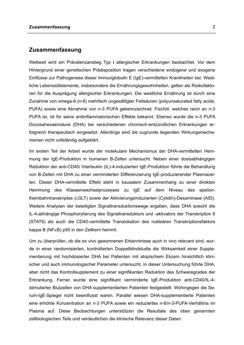
Zusammenfassung
2
Zusammenfassung
Weltweit wird ein Prävalenzanstieg Typ I allergischer Erkrankungen beobachtet. Vor dem
Hintergrund einer genetischen Prädisposition tragen verschiedene endogene und exogene
Einflüsse zur Pathogenese dieser Immunglobulin E (IgE)-vermittelten Krankheiten bei. West-
liche Lebensstilelemente, insbesondere die Ernährungsgewohnheiten, gelten als Risikofakto-
ren für die Ausprägung allergischer Erkrankungen. Die westliche Ernährung ist durch eine
Zunahme von omega-6 (n-6) mehrfach ungesättigter Fettsäuren (polyunsaturated fatty acids,
PUFA) sowie eine Abnahme von n-3 PUFA gekennzeichnet. Fischöl, welches reich an n-3
PUFA ist, ist für seine antiinflammatorischen Effekte bekannt. Ebenso wurde die n-3 PUFA
Docosahexaensäure (DHA) bei verschiedenen chronisch-entzündlichen Erkrankungen er-
folgreich therapeutisch eingesetzt. Allerdings sind die zugrunde liegenden Wirkungsmecha-
nismen nicht vollständig aufgeklärt.
Im ersten Teil der Arbeit wurde der molekulare Mechanismus der DHA-vermittelten Hem-
mung der IgE-Produktion in humanen B-Zellen untersucht. Neben einer dosisabhängigen
Reduktion der anti-CD40/ Interleukin (IL)-4-induzierten IgE-Produktion führte die Behandlung
von B-Zellen mit DHA zu einer verminderten Differenzierung IgE-produzierender Plasmazel-
len. Dieser DHA-vermittelte Effekt steht in kausalem Zusammenhang zu einer direkten
Hemmung des Klassenwechselprozesses zu IgE auf dem Niveau des epsilon-
Keimbahntranskriptes (εGLT) sowie der Aktivierungsinduzierten (Cytidin)-Desaminase (AID).
Weitere Analysen der beteiligten Signaltransduktionswege ergaben, dass DHA sowohl die
IL-4-abhängige Phosphorylierung des Signaltransduktors und -aktivators der Transkription 6
(STAT6) als auch die CD40-vermittelte Translokation des nukleären Transkriptionsfaktors
kappa B (NFκB) p50 in den Zellkern hemmt.
Um zu überprüfen, ob die ex vivo gewonnenen Erkenntnisse auch in vivo relevant sind, wur-
de in einer randomisierten, kontrollierten Doppelblindstudie die Wirksamkeit einer Supple-
mentierung mit hochdosierter DHA bei Patienten mit atopischem Ekzem hinsichtlich klini-
scher und auch immunologischer Parameter untersucht. In dieser Untersuchung führte DHA,
aber nicht das Kontrollsupplement zu einer signifikanten Reduktion des Schweregrades der
Erkrankung. Ferner wurde eine signifikant verminderte IgE-Produktion anti-CD40/IL-4-
stimulierter Blutzellen von DHA supplementierten Patienten festgestellt. Wohingegen die Se-
rum-IgE-Spiegel nicht beeinflusst waren. Parallel wiesen DHA-supplementierte Patienten
eine erhöhte Konzentration an n-3 PUFA sowie ein reduziertes n-6/n-3-PUFA-Verhältnis im
Plasma auf. Diese Beobachtungen unterstützen die Resultate des oben genannten
zellbiologischen Teils und verdeutlichen die klinische Relevanz dieser Daten.
Zusammenfassung
3
Im dritten Teil der Arbeit wurden die lokalen Prozesse in der Haut nach DHA-
Supplementierung untersucht. Hierzu wurde ein Mausmodell eingesetzt, da es aus ethischen
Gründen nur begrenzt möglich ist, Hautproben von Patienten zu analysieren. In Überein-
stimmung mit den Ergebnissen der klinischen Untersuchung führte die orale DHA-
Supplementierung auch zu einem verbesserten klinischen Bild der proteininduzierten Derma-
titis. Die Reduktion der Symptomstärke und der damit einhergehende verminderte klinische
Schweregrad des Ekzems waren mit der verringerten Zahl dermaler CD8+ T-Zellen verbun-
den, indessen waren andere Parameter wie Mastzell- und CD4+ T-Zellzahlen sowie
Epidermisdicke nicht beeinflusst. Da auch im Mausmodell die Serum-IgE-Konzentration nicht
durch die orale DHA-Verabreichung beeinflusst wurde, wirkt DHA hier vermutlich primär lokal
als systemisch immunmodulierend.
Durch die Fähigkeit von DHA in den IgE-Klassenwechsel in B-Zellen einzugreifen, stellt die
orale Supplementierung mit DHA eine mögliche präventive Maßnahme gegenüber Typ I al-
lergischen Erkrankungen dar. Weiterhin verdeutlichen diese Daten, dass DHA den Schwere-
grad des atopischen Ekzems durch eine positive Beeinflussung lokaler inflammatorischer
Prozesse signifikant verbessern kann. Um diätetische DHA bestmöglich als therapeutisches
Instrument im Zusammenhang mit allergischen Erkrankungen einsetzen zu können, muss
allerdings die Supplementierung hinsichtlich des Zeitpunktes sowie der Dosierung optimiert
werden.
Abstract
4
Abstract
The prevalence of type I allergic diseases have increased worldwide. Based on genetic sus-
ceptibility diverse endogenous and exogenous factors contribute to the pathogenesis of
these immunoglobulin (Ig) E mediated disorders. The modern life style is hypothesised to be
one risk factor for allergic diseases. Thereby the Westernised diet is characterised by an in-
creasing consumption of omega-6 (n-6) polyunsaturated fatty acids (PUFA) and a decreasing
intake of n-3 PUFA. Fish oil, rich in n-3 PUFA, exerts a wide range of antiinflammatory ef-
fects. Docosahexaenoic acid (DHA) is one major n-3 PUFA of fish oil and has been reported
to be beneficial in different chronic and inflammatory diseases as well. However, the underly-
ing mechanisms of its action are not completely understood.
The molecular mechanisms of DHA on IgE production in human B cells were examined in the
first part of this thesis. DHA inhibited IgE production and the differentiation of IgE secreting
cells in a dose-dependent manner. This inhibition was mediated through direct inhibition of
the immunoglobulin isotype switching process, which was detected by decreased epsilon
germline transcript (εGLT) and activation induced (cytidin) desaminase (AID) transcription.
Analysis of involved signalling pathways revealed that DHA caused both an inhibition of inter-
leukin (IL)-4 driven signal transducer and activator of transcription 6 (STAT6) phosphorylation
but also a reduced nuclear factor kappa B (NFκB) p50 translocation into the nucleus upon
anti-CD40 stimulation.
Next it was verified whether the ex vivo findings are relevant in vivo as well. In a randomised,
double bind, controlled clinical study the efficacy of high-dose DHA supplementation in atopic
eczema was determined and thereby the impact on clinical as well as immunological pa-
rameters investigated. In the DHA treated, but not in control group a significant clinical im-
provement of atopic eczema in terms of severity was observed. Additionally, in DHA group a
significant reduction of anti-CD40/IL-4 mediated IgE synthesis of peripheral blood cells was
detected whereas serum IgE concentrations remained unchanged. Furthermore, the DHA
group showed an increase of plasma n-3 PUFA and a decrease of n-6/n-3 PUFA ratio. These
observations support the findings from the first part of this work and point to their potential
biological relevance.
The third part of this work deals with the clinical impact of oral DHA administration on aller-
gen induced dermatitis. Due to ethical considerations analysis of skin biopsies from patients
is limited. Therefore a mouse model was used. The obtained data correspond to the other
results. Analysis of the local mechanisms of clinical improvement implicated a favourable
Abstract
5
modulation of the cellular immune response by DHA. The reduced clinical skin score was
associated with a decreased number of dermal CD8+ T cells, whereas other parameters like
epidermis thickening, CD4+ and mast cell numbers were not affected. However, the serum
IgE values remained unchanged indicating that DHA might rather act locally than systemi-
cally on an established allergic response.
Taken together the results of this thesis indicate that dietary DHA may be effective in preven-
tion of type Ι allergic diseases by interference with the IgE switching process. Additionally,
DHA has been shown to improve the clinical outcome of atopic eczema by having a positive
impact on local inflammatory processes. Dietary DHA might be a potential therapeutical tool
for dietary management of IgE mediated diseases, like atopic eczema. However, further re-
search in order to reveal the best time-point and optimal dose of DHA application is still nec-
essary.
Schlagwörter:
Docosahexaensäure, Mehrfach ungesättigte Fettsäuren, Allergie, IgE
Keywords:
Docosahexaenoic acid, Polyunsaturated fatty acids, Allergy, IgE

List of content
6
List of content
ZUSAMMENFASSUNG ...........................................................................................................2
ABSTRACT..............................................................................................................................4
LIST OF CONTENT .................................................................................................................6
1 INTRODUCTION...............................................................................................................8
1.1 TYPE I ALLERGIC REACTIONS........................................................................................8
1.1.1 Mechanisms and molecular regulation of IgE production.......................................8
1.2 ATOPIC ECZEMA.........................................................................................................15
1.2.1 Pathophysiology of atopic eczema.......................................................................15
1.2.2 Atopic eczema and fatty acids..............................................................................18
1.2.3 Murine models of atopic eczema..........................................................................19
1.3 POLYUNSATURATED FATTY ACIDS (PUFA) ...................................................................21
1.3.1 Nomenclature and molecular structure of PUFA ..................................................21
1.3.2 Dietary Sources of PUFA......................................................................................21
1.3.3 Physiological function of PUFA ............................................................................22
1.3.4 Metabolism of PUFA.............................................................................................22
1.3.5 PUFA and immune system...................................................................................23
1.3.6 Docosahexaenoic acid (DHA) ..............................................................................27
1.4 OBJECTIVE ................................................................................................................28
2 MATERIALS AND METHODS........................................................................................29
2.1 MATERIALS ................................................................................................................29
2.2 METHODS..................................................................................................................29
2.2.1 Donors and cells...................................................................................................29
2.2.2 Participants and clinical study ..............................................................................29
2.2.3 Mouse model of allergen induced eczema...........................................................31
2.2.4 Cell culture methods.............................................................................................34
2.2.5 Immunological methods .......................................................................................35
2.2.6 Molecular biological methods ...............................................................................41
2.3 STATISTICAL ANALYSIS................................................................................................45
3 RESULTS........................................................................................................................46
3.1 DHA INHIBITS IGE PRODUCTION IN HUMAN B CELLS.....................................................46
List of content
7
3.1.1 Modulation of immunoglobulin synthesis..............................................................46
3.1.2 DHA modulates IL-4 and anti-CD40 signalling .....................................................51
3.2 DHA SUPPLEMENTATION IN ATOPIC ECZEMA – A RANDOMISED, DOUBLE BLIND,
CONTROLLED STUDY.................................................................................................................
.................................................................................................................................55
3.2.1 The SCORAD is significantly reduced by DHA supplementation .........................55
3.2.2 DHA inhibits IgE synthesis ex vivo .......................................................................56
3.2.3 Fatty acid supplementation modulates activation of monocytes and B cells........57
3.2.4 Systemic IFNγ and IL-4 response remains unaffected by fatty acid
supplementation..............................................................................................................57
3.3 ORAL ADMINISTRATION OF DHA INHIBITS THE DEVELOPMENT OF ECZEMA IN A MURINE
MODEL OF PROTEIN INDUCED DERMATITIS ..............................................................................58
3.3.1 Dietary DHA reduces the clinical symptoms of protein induced dermatitis ..........58
3.3.2 Dietary DHA reduces the number of CD8+ T cells in eczematous skin ................59
3.3.3 Oral DHA administration did not alter the systemic immune response ................61
4 DISCUSSION..................................................................................................................62
4.1 DHA INHIBITS IGE PRODUCTION IN HUMAN B CELLS.....................................................62
4.2 DHA SUPPLEMENTATION IN ATOPIC ECZEMA – A RANDOMISED, DOUBLE BLIND,
CONTROLLED STUDY.................................................................................................................
.................................................................................................................................66
4.3 ORAL ADMINISTRATION OF DHA INHIBITS THE DEVELOPMENT OF ECZEMA IN A MURINE
MODEL OF PROTEIN INDUCED DERMATITIS ..............................................................................69
4.4 CONCLUSION.............................................................................................................73
LITERATURE.........................................................................................................................76
APPENDIX .............................................................................................................................96
LIST OF ABBREVIATIONS .................................................................................................108
ACKNOWLEDGEMENT ...................................................................................................... 111
STATEMENT OF AUTHORSHIP .........................................................................................113
1 Introduction
8
1 Introduction
1.1 Type I allergic reactions
Whereas the Western world was experiencing a dramatic increase in the prevalence of aller-
gic diseases during the last 30 to 40 years of the previous century, a similar tendency can be
currently seen in fast developing countries such as India and China. Even in Arctic region the
same trend is detected with a doubling of sensitisation rates in only 11 years [1].
Allergies are inappropriate or exaggerated reactions of the immune system to substances
that are usually harmless. Allergy inducing substances are called allergens. Common indica-
tions of allergy may include sneezing, itching and skin rashes [2].
Immediate allergic reactions are manifested as anaphylaxis, rhinoconjunctivitis allergica and
allergic asthma [3]. These immunoglobulin (Ig) E mediated processes are initiated by the first
contact between immune system and allergen, the sensitisation phase. Thereby, the allergen
is internalised and processed by antigen presenting cells (APC; like macrophages, dendritic
cells [DC], Langerhans cells [LC], B cells). Peptides derived from allergens are presented via
major histocompatibility complex II (MHC II) to naïve CD4+ cells (TH0) leading to T cell acti-
vation and differentiation into TH2 cells. Signalling molecules of TH2 cells, like CD40 ligand
(CD40L, CD154) and interleukin (IL)-4, activate B cells to proliferate and differentiate to IgE
secreting plasma cells [4,5]. The subsequently secreted allergen specific IgE binds to its high
affinity receptor (FcεRI) on granule containing cells, like mast cells (MC) and basophils. Re-
peated exposure leads to cross linking of receptor bound IgE by polyvalent allergens, what
triggers the degranulation of MC and basophils. The release of mediators, like histamine and
prostaglandins (PG), elicits the symptoms of the immediate allergic reaction characterised by
increased vascular permeability and smooth muscle contraction. The late phase is caused by
leukotrienes (LT), chemokines and cytokines. The second phase of smooth muscle contrac-
tion and tissue remodelling takes place. Chemokines and local cytokine pattern recruit
macrophages and neutrophils, but also eosinophils and TH2 cells to sites of inflammation.
Eosinophilic inflammation is supported by TH2 cell derived IL-5. A sustained allergen expo-
sure with a persisting immune reaction can lead to a chronic inflammation, hallmarked by
hyperreactivity and damage of the affected tissue [6].
1.1.1 Mechanisms and molecular regulation of IgE production
IgE is the major player of type I hypersensitivities. The heavy chain constant (CH) region do-
mains of IgE are encoded by a single gene, the strictly regulated Cε gene [7]. In serum of
1 Introduction
9
non-atopic individuals, IgE is found in very low concentrations ranging between
1 and 400 ng/mL [7]. This is mainly adjusted by a tight regulation of gene expression and
protein stability [7]. The rapid turnover and the short half-life of 2.5 days contribute to the low
serum IgE levels [1]. It has been considered that a high level of serum IgE comes along with
high IgE production. However, increasing IgE levels lead to diminished IgE catabolism. Thus,
half-life arises threefold when IgE levels proceed from 0.07 to 35 µg/mL [8]. Furthermore,
when bound to its high affinity receptor on MC, half-life of IgE is increased up to 8 to 14 days
[8]. Interestingly, in healthy individuals about 67 % of IgE is found extravascularly compared
to 48 % in patients with severe atopic diseases [8].
According to their CH regions immunoglobulins can be categorised into nine isotypes: IgA1,
IgA2, IgD, IgE, IgG1, IgG2, IgG3, IgG4 and IgM. Except IgM and IgD, all other isotypes result
from class switch recombination (CSR). Thereby, the specificity defining variable region is
molecularly linked with the constant (C) regions of different isotypes and distinct effector
functions [6].
Switching to all isotypes is commonly regulated at the germline transcription of CH genes and
the induction of activation induced (cytidine) desaminase (AID) expression. Switching from
naïve B cell expressing IgM and IgD to IgE can occur directly or sequentially via IgG. This
process is induced by two signals: antigen specific T cell interaction with B cell via
CD40L/CD40 and IL-4 secreted by activated T cells [5].
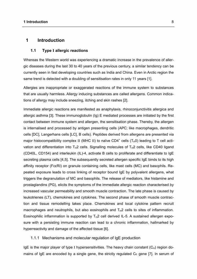
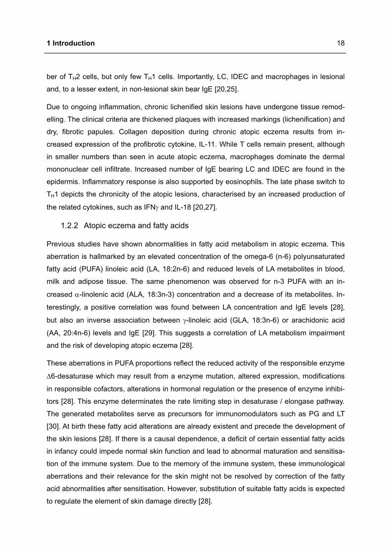
IL-4 and IL-13 exert their biological effects by binding to the IL-4 receptor (IL-4R) complex,
which consists of two subunits (Figure 1). Type I IL-4R complexes, composed of IL-4Rα
chain and γc chain, bind IL-4 but not IL-13. IL-4 and IL-13 can bind to Type II IL-4R com-
plexes, which are composed of IL-4Rα and IL-13Rα1. Both receptor complexes amplify their
signals through IL-4Rα. A second IL-13 receptor, IL-13Rα2, binds to IL-13 exclusively and
serves as a decoy receptor. IL-4 binding mediates receptor heterodimerisation and activation
of cytoplasmic protein tyrosine kinases called Janus activated kinases (JAK) which are con-
stitutively associated with IL-4Rα (JAK1) and γc chain (JAK3) [9,10].
Tyrosine phosphorylation of the IL-4Rα (Y575, Y603, Y633) enables the recruitment of signal
transducer and activator of transcription 6 (STAT6) through the SH2 domain. Further STAT6
phosphorylation by JAK and dimerisation result in translocation to the nucleus, where it acti-
vates the transcription of IL-4 and IL-13 responsive genes. STAT6 dephosphorylation stimu-
lates its export into cytosol, where it can be recruited into another cycle of tyrosine phos-
phorylation and nuclear entry [10].
1 Introduction
10
JAK3 JAK1
STAT6
P STAT6
STAT6P
P STAT6
STAT6P
Shp-1
Shc
IRS-1/2PI3K
MAPK
Gene expression
Proliferation
Differentiation
Tyk2 JAK1
Y
Y
YYYY
Y
Y
YYYY
STAT3
γc
Type I IL-4R
IL-4 IL-4 IL-13IL-13
IL-4Rα IL-4RαIL-13Rα1 IL-13Rα2
Type II IL-4R
IL-4- and IL-13- responsive genes
Iε, AID, IL-4, MHC-II etc.
Nucleus
Plasma membrane
JAK3 JAK1
STAT6
P STAT6
STAT6P
P STAT6
STAT6P
Shp-1
Shc
IRS-1/2PI3K
MAPK
Gene expression
Proliferation
Differentiation
Tyk2 JAK1
Y
Y
YYYY
Y
Y
YYYY
STAT3
γc
Type I IL-4R
IL-4 IL-4 IL-13IL-13
IL-4Rα IL-4RαIL-13Rα1 IL-13Rα2
Type II IL-4R
IL-4- and IL-13- responsive genes
Iε, AID, IL-4, MHC-II etc.
JAK3 JAK1JAK3 JAK1
STAT6
STAT6
P STAT6
STAT6P
P STAT6
STAT6P
Shp-1Shp-1
Shc
IRS-1/2PI3K
MAPKMAPK
Gene expression
Proliferation
Differentiation
Tyk2 JAK1JAK1
Y
Y
YYYY
Y
Y
YYYY
STAT3
STAT3
γc
Type I IL-4R
IL-4 IL-4 IL-13IL-13
IL-4Rα IL-4RαIL-13Rα1 IL-13Rα2
Type II IL-4R
IL-4- and IL-13- responsive genes
Iε, AID, IL-4, MHC-II etc.
Nucleus
Plasma membrane
Figure 1: Illustration of IL-4R and IL-13R complexes focussed on IL-4Rα signalling
[9,10]. Type I IL-4R complexes are composed of IL-4Rα and γc and bind exclusively to IL-4.
Type ΙΙ IL-4R complexes consist of IL-4Rα and IL-13Rα1 and bind to both IL-4 and IL-13.
Signalling via IL-13Rα1 include additional intermediates like STAT3. The second IL-13 recep-
tor IL-13Rα2 exclusively binds to IL-13. Signalling pathways activated via IL-4Rα are de-
picted in more detail. Receptor binding of IL-4 and IL-13 promotes activation of members of
the JAK family that constitutively associate with component subunits of the receptor com-
plexes. Thereby, several intracellular signalling cascades are initiated by phosphorylating
specific tyrosine residues in the cytoplasmic domain of IL-4Rα. These phosphorylated resi-
dues act as connecting sites for signalling molecules. The first residue represents the insulin/
interleukin-4 receptor (I4R) motif which facilitates the interaction with Shc and insulin receptor
substrate (IRS) proteins. Phosphorylated IRS binds to phosphatidylinositol 3-kinase (PI3-K)
which controls several protein kinases regulating proliferation. Together with PI3-K connected
pathways, Shc mediates the regulatory functions of I4R motif and activates components of
mitogen-activated protein kinase (MAPK) cascade. Phosphorylation of tyrosine residue cas-
sette recruits transcription factor STAT6. JAK phosphorylate STAT6 which lead to its dimeri-
sation and translocation to the nucleus, where it activates the transcription of IL-4 and IL-13
responsive genes like AID. The last tyrosine residue binds to the phosphatase Src homology
domain 2 (SH2) containing inositol phosphatase (Shp-1), which is a negative regulator of
receptor signalling. Thus, the multiple effector functions of IL-4Rα are transmitted by the
STAT6 and the I4R pathways separately and in combination.

1 Introduction
11
In addition to the JAK-STAT pathway, IL-4 also induces tyrosine phosphorylation of IRS-1
and/or IRS-2, which mediate the activation of PI3-K. Thereby I4R motif, a conserved tyrosine
residue of IL-4Rα (Y497, Y500), engages phosphotyrosine binding domain adaptor proteins,
like IRS and Shc, which may be also involved in IgE regulation [9,11].
IL-4Rα also bears an immuno-tyrosine inhibitory motif (ITIM) site at Y713 and its adjoining
residues, which binds to several regulatory phosphatases, including Shp-1. It is discussed as
a negative regulator in IL-4Rα signalling in STAT6 activation [9].
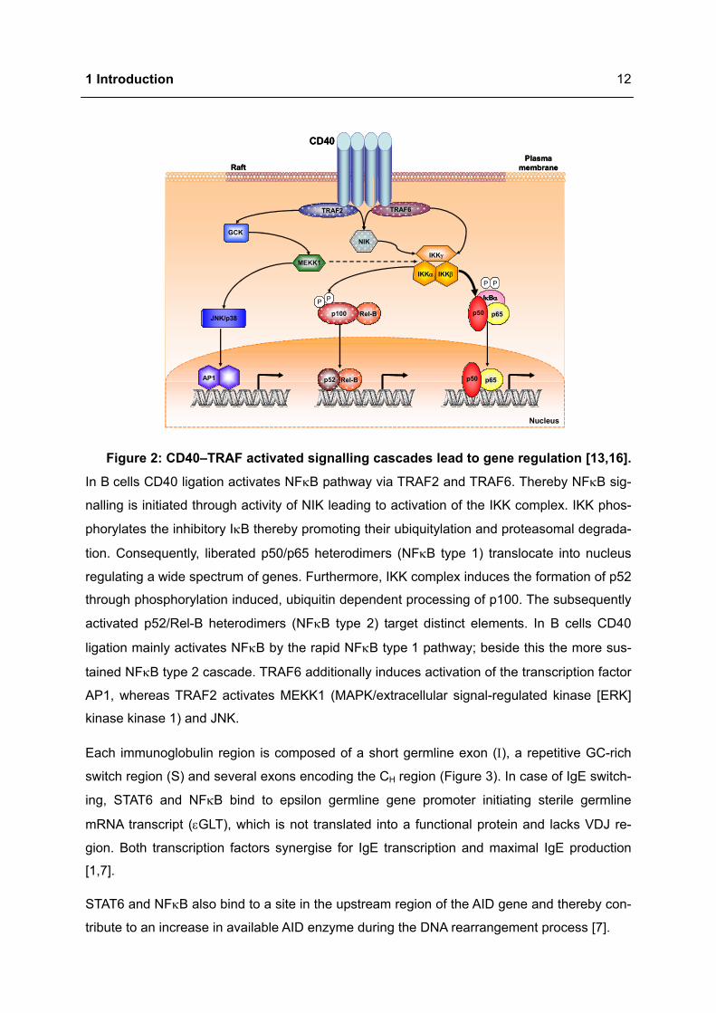
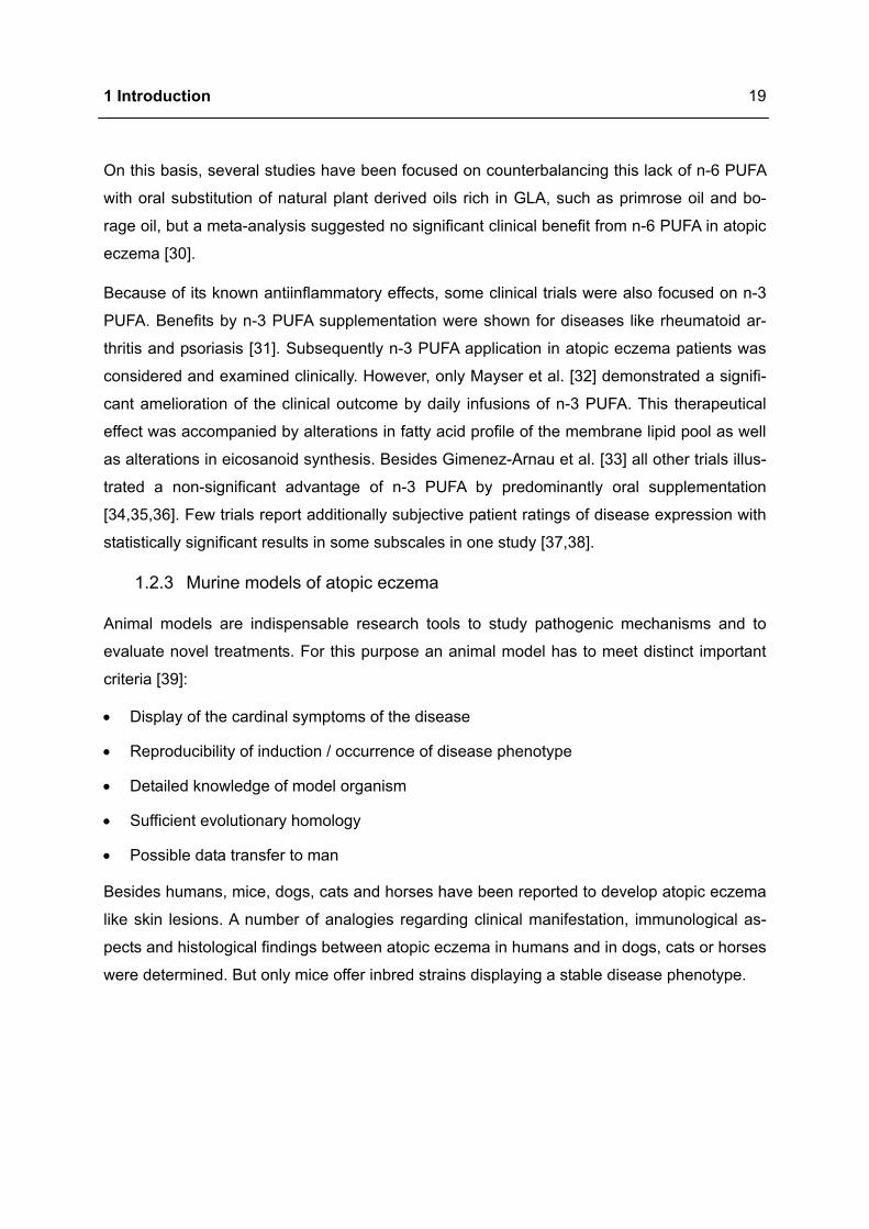
CD40 is a member of the tumor necrosis factor receptors (TNFR) family (Figure 2). Since its
cytoplasmic tail does not have an intrinsic kinase activity, downstream signalling is transmit-
ted by the adapter molecules TNFR associated factors (TRAF) [12]. CD40 ligation results in
rapid recruitment of TRAF1, TRAF2, TRAF3 and TRAF6 to the CD40 cytoplasmic domain
[13]. TRAF2 and TRAF3 are drafted to the cell membrane after CD40 engagement, which
allows the contact to membrane cholesterol-rich microdomains or lipid rafts. Subsequently
TRAF2 induces its own as well as TRAF3 ubiquitylation. Degradation of these TRAF occurs
in cytoplasmic proteasome [13].
TRAF1 is implicated in regulation of other TRAF and enhances the TRAF2 degradation.
TRAF2 and TRAF6 are essential for c-jun kinase (JNK) and p38 activation, but also nuclear
factor kappa B (NFκB) signal. TRAF3 negatively regulates JNK and NFκB activation by com-
petitive binding of TRAF2 to CD40. Additionally, TRAF3 inhibits the interaction between B cell
receptor and CD40 [13]. Besides TRAF numerous kinases, like germinal centre kinase
(GCK) contribute to CD40 signal transduction [14]. The transcription factors activator pro-
tein 1 (AP1) is also induced and its signalling pathway involves JNK and p38 [13].
The activation of NFκB by TRAF2 and TRAF6 starts with NFκB inducing kinase (NIK). This
MAPK phosphorylates and thus activates the inhibitor of NFκB (IκB) kinase complex (IKKα,
IKKβ and IKKγ) which leads to phosphorylation of IκB proteins. Following IκB degradation,
liberated NFκB translocates to the nucleus where it regulates the expression of a wide spec-
trum of genes [13,14,15].
1 Introduction
12
B cell
Nucleus
MEKK1
GCK
Raft
NIK
JNK/p38
CD40
TRAF6TRAF2
IKKγ
IKKα IKKβPP
IκBα
p65p50
AP1 Rel-Bp52 p65p50
Plasma membrane
Rel-Bp100
PP
Nucleus
B cell
Nucleus
MEKK1MEKK1
GCKGCK
Raft
NIKNIK
JNK/p38JNK/p38
CD40
TRAF6TRAF2
IKKγ
IKKα IKKβ
IKKγ
IKKα IKKβPP
IκBα
p65p50
PP
IκBα
p65p50 p65p50
AP1 Rel-Bp52 Rel-Bp52p52 p65p50 p65p50
Plasma membrane
Rel-Bp100
PP
Rel-Bp100 Rel-Bp100
PP
Nucleus
Figure 2: CD40–TRAF activated signalling cascades lead to gene regulation [13,16].
In B cells CD40 ligation activates NFκB pathway via TRAF2 and TRAF6. Thereby NFκB sig-
nalling is initiated through activity of NIK leading to activation of the IKK complex. IKK phos-
phorylates the inhibitory IκB thereby promoting their ubiquitylation and proteasomal degrada-
tion. Consequently, liberated p50/p65 heterodimers (NFκB type 1) translocate into nucleus
regulating a wide spectrum of genes. Furthermore, IKK complex induces the formation of p52
through phosphorylation induced, ubiquitin dependent processing of p100. The subsequently
activated p52/Rel-B heterodimers (NFκB type 2) target distinct elements. In B cells CD40
ligation mainly activates NFκB by the rapid NFκB type 1 pathway; beside this the more sus-
tained NFκB type 2 cascade. TRAF6 additionally induces activation of the transcription factor
AP1, whereas TRAF2 activates MEKK1 (MAPK/extracellular signal-regulated kinase [ERK]
kinase kinase 1) and JNK.
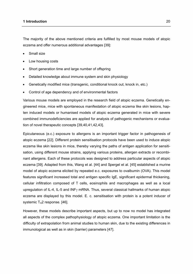
Each immunoglobulin region is composed of a short germline exon (Ι), a repetitive GC-rich
switch region (S) and several exons encoding the CH region (Figure 3). In case of IgE switch-
ing, STAT6 and NFκB bind to epsilon germline gene promoter initiating sterile germline
mRNA transcript (εGLT), which is not translated into a functional protein and lacks VDJ re-
gion. Both transcription factors synergise for IgE transcription and maximal IgE production
[1,7].
STAT6 and NFκB also bind to a site in the upstream region of the AID gene and thereby con-
tribute to an increase in available AID enzyme during the DNA rearrangement process [7].
1 Introduction
13
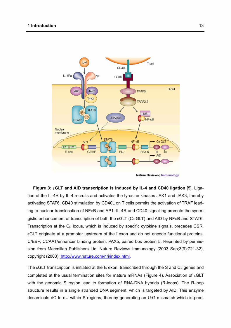
Figure 3: εGLT and AID transcription is induced by IL-4 and CD40 ligation [5]. Liga-
tion of the IL-4R by IL-4 recruits and activates the tyrosine kinases JAK1 and JAK3, thereby
activating STAT6. CD40 stimulation by CD40L on T cells permits the activation of TRAF lead-
ing to nuclear translocation of NFκB and AP1. IL-4R and CD40 signalling promote the syner-
gistic enhancement of transcription of both the εGLT (Cε GLT) and AID by NFκB and STAT6.
Transcription at the CH locus, which is induced by specific cytokine signals, precedes CSR.
εGLT originate at a promoter upstream of the Ι exon and do not encode functional proteins.
C/EBP, CCAAT/enhancer binding protein; PAX5, paired box protein 5. Reprinted by permis-
sion from Macmillan Publishers Ltd: Nature Reviews Immunology (2003 Sep;3(9):721-32),
copyright (2003); http://www.nature.com/nri/index.html.
The εGLT transcription is initiated at the Iε exon, transcribed through the S and CH genes and
completed at the usual termination sites for mature mRNAs (Figure 4). Association of εGLT
with the genomic S region lead to formation of RNA-DNA hybrids (R-loops). The R-loop
structure results in a single stranded DNA segment, which is targeted by AID. This enzyme
desaminats dC to dU within S regions, thereby generating an U:G mismatch which is proc-
1 Introduction
14
essed through uracil removal, base-excision or mismatch repair. In order to achieve CSR by
an intrachromosomal deletion, double strand DNA breaks are brought into both the upstream
Sµ/γ and the downstream Sε region [1,7,17]. Since there is an almost linear correlation be-
tween S region size and CSR frequency, the low IgE expression levels are supported by the
small size of the Sε region [7].
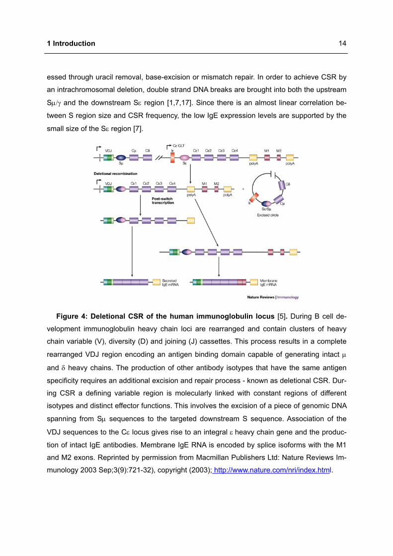
Figure 4: Deletional CSR of the human immunoglobulin locus [5]. During B cell de-
velopment immunoglobulin heavy chain loci are rearranged and contain clusters of heavy
chain variable (V), diversity (D) and joining (J) cassettes. This process results in a complete
rearranged VDJ region encoding an antigen binding domain capable of generating intact µ
and δ heavy chains. The production of other antibody isotypes that have the same antigen
specificity requires an additional excision and repair process - known as deletional CSR. Dur-
ing CSR a defining variable region is molecularly linked with constant regions of different
isotypes and distinct effector functions. This involves the excision of a piece of genomic DNA
spanning from Sµ sequences to the targeted downstream S sequence. Association of the
VDJ sequences to the Cε locus gives rise to an integral ε heavy chain gene and the produc-
tion of intact IgE antibodies. Membrane IgE RNA is encoded by splice isoforms with the M1
and M2 exons. Reprinted by permission from Macmillan Publishers Ltd: Nature Reviews Im-
munology 2003 Sep;3(9):721-32), copyright (2003); http://www.nature.com/nri/index.html.
1 Introduction
15
1.2 Atopic eczema
Atopic eczema is a chronically relapsing, inflammatory skin disease associated with ery-
thema, severe pruritus, crusty and oozing plaques on forehead, face, neck and hands [18].
The pathogenesis is driven by a complex combination of environmental, genetic, immu-
nologic factors and skin barrier dysfunctions [19,20].
The incidence of atopic eczema is increasing worldwide and varies in different populations.
10 to 20 % of infants and children and 1 to 3 % of adults are affected by this disease,
whereas the prevalence of atopic eczema varies between 2 and 5 % in the whole population.
In most cases, about 70 %, manifestation occurs within the first five years of life. At later age
30 to 60 % of children suffering from this disease develop respiratory diseases such as
asthma or hay fever (atopic march) [20,21]. Thus, atopic eczema causes high costs and is a
serious socioeconomic burden besides its negative impact on the quality of life of suffering
patients [18,20,22,23].
Two types of atopic eczema have been delineated. Most patients (70 to 80 %) suffer from the
extrinsic type of atopic eczema which is associated with an IgE mediated sensitisation. By
contrast, the intrinsic type does not seem to be linked to IgE related mechanisms and affects
20 to 30 % of atopic eczema patients. Cutaneous lymphocyte associated antigen (CLA) ex-
pressing memory T cells are recruited to the skin where they produce higher amounts of TH2
cytokines. IL-4 and IL-13 induce the isotype switching towards IgE, whereas IL-5 leads to
eosinophil expansion and survival. Inheritance and other internal processes are associated
here. Intrinsic atopic eczema is characterised by a lower IL-4 and IL-13 production compared
to extrinsic form. However, few patients suffer from both extrinsic and intrinsic form [20,22].
1.2.1 Pathophysiology of atopic eczema
The pathophysiology of atopic eczema involves genetic predisposition, disturbed skin barrier
function, defects in the antimicrobial immune defence and frequent allergic responses
against allergens [19,20].
A high familial association has been described for atopic eczema. Genetic research was fo-
cussed on specific atopic eczema alleles, as well as identifying overlapping genes associ-
ated with other allergic disorders. Genetic polymorphisms have been assigned for a cluster
of TH2 cytokine genes located on chromosome 5q22-23, the IL-13 coding region and the
IL-4Rα subunit (16q12) [20,21,24]. Heritable epidermal barrier defect has been shown to be
connected to the filaggrin (filament-aggregating protein) gene (FLG). FLG mutations are a
major risk factor for eczema associated asthma [21,24].

1 Introduction
16
Atopic eczema is characterised by dry skin with increased transepidermal water loss. The
disturbed barrier function results from a reduced activity of acid ceramidase and decreased
ceramides content, the most important water retaining molecules in the extracellular space.
Additionally, this dysfunction causes a reduction of sphingosine levels in the stratum
corneum. Since sphingosines are potent antimicrobials, the colonisation with Staphylococcus
aureus (S. aureus) is supported. Diminished levels of additional antimicrobial peptides, e.g.
β-defensine 2 and cathelicidin, promote the ongoing colonisation of inflamed skin by various
microorganisms [20,21].
The TH2 predominance in atopic eczema patients, resulting from an increased frequency of
allergen specific TH2 cells and a decrease of interferon (IFN) γ producing cells, favours IgE
production and peripheral eosinophilia. Disturbed cellular immunity, humoral factors like in-
creased IgE synthesis and high production of related cytokines contribute to development of
skin lesions [18,20].
Clinically unaffected skin in atopic eczema is characterised by sparse perivascular T cell infil-
trate, increased number of TH2 cells expressing IL-4 and IL-13, but not IFNγ [25] (Figure 5).
Initially, allergens penetrate into damaged skin and activate DC by cross linking FcεRI bound
IgE molecules leading to an enhanced antigen presentation capacity. In this phase naive
T cells are polarised into TH2 cells producing associated cytokines. Additionally, MC are acti-
vated via antigen specific IgE and contribute to induction of the inflammatory response [22].
Very early events initiating atopic skin inflammation are not completely elucidated. Environ-
mental allergens, scratching or microbial toxins cause skin injury thereby activating keratino-
cytes, MC and DC to release proinflammatory cytokines and chemokines, like IL-1, thymic
stromal lymphopoietin (TSLP) and TNFα. Local proinflammatory mediator expression or-
chestrates adhesion to the endothelium and subsequent extravasation of cells and thus de-
fining the nature of the inflammatory infiltrate. These mediators enhance the expression of
adhesion molecules on vascular endothelium and facilitate the extravasation of inflammatory
cells into the skin. In the tissue these cells respond to chemotactic gradients established by
cytokines and chemokines originated from sites of injury or infection [20,26]. Cutaneous T
cell attracting chemokine (CCL27) is pivotal in mediating the migration of CLA+ T cells. Addi-
tionally, macrophage derived chemokine and activation regulated cytokine are increased in
patients with atopic eczema. These molecules selectively recruit CCR4 expressing TH2 cells.
Dimension of thymus and activation regulated cytokine levels have been linked to severity of
atopic eczema [20].
1 Introduction
17
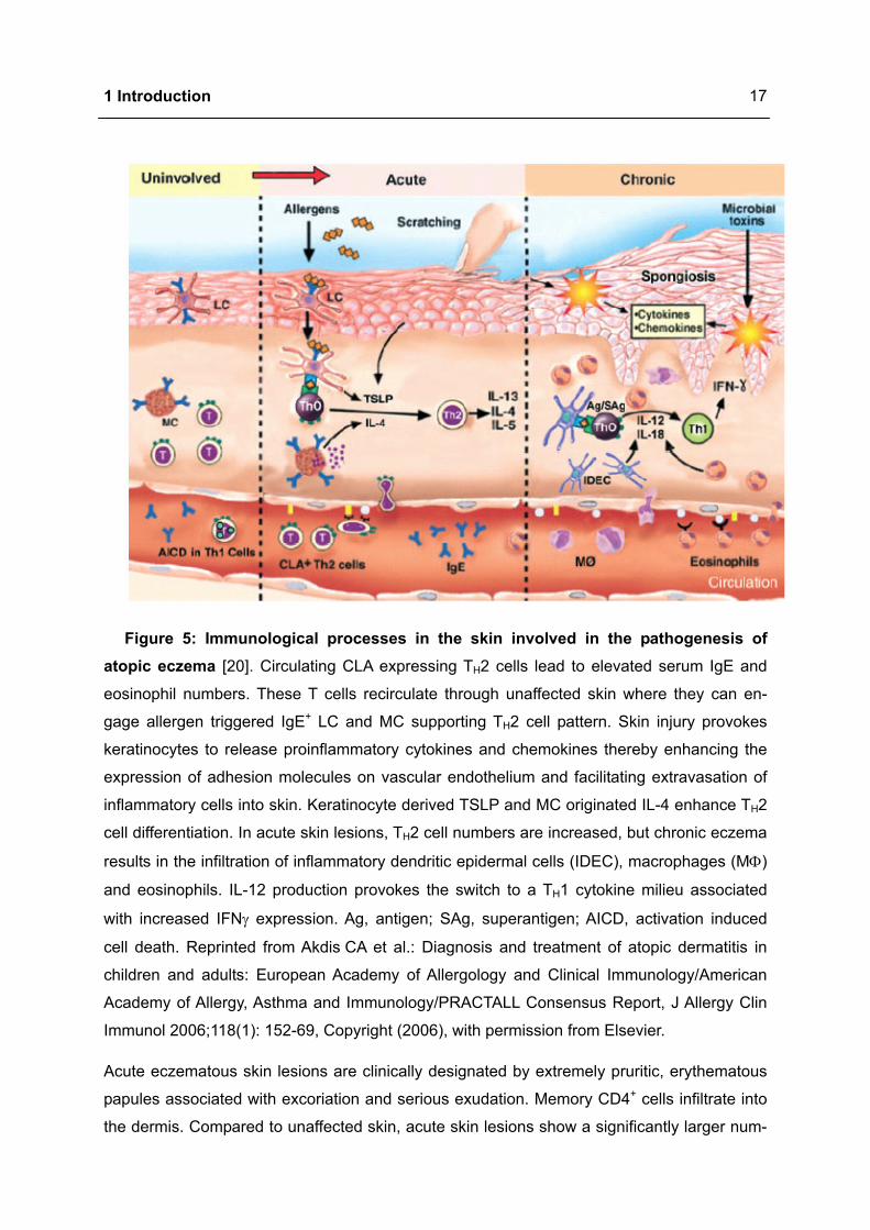
Figure 5: Immunological processes in the skin involved in the pathogenesis of atopic eczema [20]. Circulating CLA expressing TH2 cells lead to elevated serum IgE and
eosinophil numbers. These T cells recirculate through unaffected skin where they can en-
gage allergen triggered IgE+ LC and MC supporting TH2 cell pattern. Skin injury provokes
keratinocytes to release proinflammatory cytokines and chemokines thereby enhancing the
expression of adhesion molecules on vascular endothelium and facilitating extravasation of
inflammatory cells into skin. Keratinocyte derived TSLP and MC originated IL-4 enhance TH2
cell differentiation. In acute skin lesions, TH2 cell numbers are increased, but chronic eczema
results in the infiltration of inflammatory dendritic epidermal cells (IDEC), macrophages (MΦ)
and eosinophils. IL-12 production provokes the switch to a TH1 cytokine milieu associated
with increased IFNγ expression. Ag, antigen; SAg, superantigen; AICD, activation induced
cell death. Reprinted from Akdis CA et al.: Diagnosis and treatment of atopic dermatitis in
children and adults: European Academy of Allergology and Clinical Immunology/American
Academy of Allergy, Asthma and Immunology/PRACTALL Consensus Report, J Allergy Clin
Immunol 2006;118(1): 152-69, Copyright (2006), with permission from Elsevier.
Acute eczematous skin lesions are clinically designated by extremely pruritic, erythematous
papules associated with excoriation and serious exudation. Memory CD4+ cells infiltrate into
the dermis. Compared to unaffected skin, acute skin lesions show a significantly larger num-
1 Introduction
18
ber of TH2 cells, but only few TH1 cells. Importantly, LC, IDEC and macrophages in lesional
and, to a lesser extent, in non-lesional skin bear IgE [20,25].
Due to ongoing inflammation, chronic lichenified skin lesions have undergone tissue remod-
elling. The clinical criteria are thickened plaques with increased markings (lichenification) and
dry, fibrotic papules. Collagen deposition during chronic atopic eczema results from in-
creased expression of the profibrotic cytokine, IL-11. While T cells remain present, although
in smaller numbers than seen in acute atopic eczema, macrophages dominate the dermal
mononuclear cell infiltrate. Increased number of IgE bearing LC and IDEC are found in the
epidermis. Inflammatory response is also supported by eosinophils. The late phase switch to
TH1 depicts the chronicity of the atopic lesions, characterised by an increased production of
the related cytokines, such as IFNγ and IL-18 [20,27].
1.2.2 Atopic eczema and fatty acids
Previous studies have shown abnormalities in fatty acid metabolism in atopic eczema. This
aberration is hallmarked by an elevated concentration of the omega-6 (n-6) polyunsaturated
fatty acid (PUFA) linoleic acid (LA, 18:2n-6) and reduced levels of LA metabolites in blood,
milk and adipose tissue. The same phenomenon was observed for n-3 PUFA with an in-
creased α-linolenic acid (ALA, 18:3n-3) concentration and a decrease of its metabolites. In-
terestingly, a positive correlation was found between LA concentration and IgE levels [28],
but also an inverse association between γ-linoleic acid (GLA, 18:3n-6) or arachidonic acid
(AA, 20:4n-6) levels and IgE [29]. This suggests a correlation of LA metabolism impairment
and the risk of developing atopic eczema [28].
These aberrations in PUFA proportions reflect the reduced activity of the responsible enzyme
∆6-desaturase which may result from a enzyme mutation, altered expression, modifications
in responsible cofactors, alterations in hormonal regulation or the presence of enzyme inhibi-
tors [28]. This enzyme determinates the rate limiting step in desaturase / elongase pathway.
The generated metabolites serve as precursors for immunomodulators such as PG and LT
[30]. At birth these fatty acid alterations are already existent and precede the development of
the skin lesions [28]. If there is a causal dependence, a deficit of certain essential fatty acids
in infancy could impede normal skin function and lead to abnormal maturation and sensitisa-
tion of the immune system. Due to the memory of the immune system, these immunological
aberrations and their relevance for the skin might not be resolved by correction of the fatty
acid abnormalities after sensitisation. However, substitution of suitable fatty acids is expected
to regulate the element of skin damage directly [28].
1 Introduction
19
On this basis, several studies have been focused on counterbalancing this lack of n-6 PUFA
with oral substitution of natural plant derived oils rich in GLA, such as primrose oil and bo-
rage oil, but a meta-analysis suggested no significant clinical benefit from n-6 PUFA in atopic
eczema [30].
Because of its known antiinflammatory effects, some clinical trials were also focused on n-3
PUFA. Benefits by n-3 PUFA supplementation were shown for diseases like rheumatoid ar-
thritis and psoriasis [31]. Subsequently n-3 PUFA application in atopic eczema patients was
considered and examined clinically. However, only Mayser et al. [32] demonstrated a signifi-
cant amelioration of the clinical outcome by daily infusions of n-3 PUFA. This therapeutical
effect was accompanied by alterations in fatty acid profile of the membrane lipid pool as well
as alterations in eicosanoid synthesis. Besides Gimenez-Arnau et al. [33] all other trials illus-
trated a non-significant advantage of n-3 PUFA by predominantly oral supplementation
[34,35,36]. Few trials report additionally subjective patient ratings of disease expression with
statistically significant results in some subscales in one study [37,38].
1.2.3 Murine models of atopic eczema
Animal models are indispensable research tools to study pathogenic mechanisms and to
evaluate novel treatments. For this purpose an animal model has to meet distinct important
criteria [39]:
• Display of the cardinal symptoms of the disease
• Reproducibility of induction / occurrence of disease phenotype
• Detailed knowledge of model organism
• Sufficient evolutionary homology
• Possible data transfer to man
Besides humans, mice, dogs, cats and horses have been reported to develop atopic eczema
like skin lesions. A number of analogies regarding clinical manifestation, immunological as-
pects and histological findings between atopic eczema in humans and in dogs, cats or horses
were determined. But only mice offer inbred strains displaying a stable disease phenotype.
1 Introduction
20
The majority of the above mentioned criteria are fulfilled by most mouse models of atopic
eczema and offer numerous additional advantages [39]:
• Small size
• Low housing costs
• Short generation time and large number of offspring
• Detailed knowledge about immune system and skin physiology
• Genetically modified mice (transgenic, conditional knock out, knock in, etc.)
• Control of age dependency and of environmental factors
Various mouse models are employed in the research field of atopic eczema. Genetically en-
gineered mice, mice with spontaneous manifestation of atopic eczema like skin lesions, hap-
ten induced models or humanised models of atopic eczema generated in mice with severe
combined immunodeficiencies are applied for analysis of pathogenic mechanisms or evalua-
tion of novel therapeutic concepts [39,40,41,42,43].
Epicutaneous (e.c.) exposure to allergens is an important trigger factor in pathogenesis of
atopic eczema [22]. Different protein sensitisation protocols have been used to induce atopic
eczema like skin lesions in mice, thereby varying the paths of antigen application for sensiti-
sation, using different mouse strains, applying various proteins, allergen extracts or recombi-
nant allergens. Each of these protocols was designed to address particular aspects of atopic
eczema [39]. Adapted from this, Wang et al. [44] and Spergel et al. [45] established a murine
model of atopic eczema elicited by repeated e.c. exposures to ovalbumin (OVA). This model
features significant increased total and antigen specific IgE, significant epidermal thickening,
cellular infiltration composed of T cells, eosinophils and macrophages as well as a local
upregulation of IL-4, IL-5 and INFγ mRNA. Thus, several classical hallmarks of human atopic
eczema are displayed by this model. E. c. sensitisation with protein is a potent inducer of
systemic TH2 response. [46].
However, these models describe important aspects, but up to now no model has integrated
all aspects of the complex pathophysiology of atopic eczema. One important limitation is the
difficulty of extrapolation from animal studies to human skin, due to the existing differences in
immunological as well as in skin (barrier) parameters [47].
1 Introduction
21
1.3 Polyunsaturated fatty acids (PUFA)
Historically “fat” has been considered important as a high calorific nutrient and a source of
essential fatty acids. Today, fat and in particular fatty acids have been accepted as important
biological regulators. Modifications in dietary fatty acids change cell membrane structure and
function. Furthermore, fatty acids differentially influence the production of cytokines, chemo-
taxis and other immunologic factors. They are known to modulate signal transduction, en-
zyme activities, receptor expression, cell proliferation and differentiation [48].
1.3.1 Nomenclature and molecular structure of PUFA
One criterion for the classification of fatty acids is the number of carbon atoms present in
hydrophobic hydrocarbon chain (chain length). Thus it is distinguished between short
chained (< 4), middle chained (6 - 10) and long chained fatty acids (> 10). Besides saturated
fatty acids, without any double bond and monounsaturated fatty acids, containing a single
double bond, there are PUFA which comprise at least two double bonds in their hydrocarbon
chain. In line with the nomenclature, PUFA are designated by their chain length (C), the
number of double bonds (D) and the position of the first double bond proximal to the terminal
methyl group (n-). Therefore, the structure of fatty acids is described by the commonly used
short hand nomenclature as following C:Dn-x.
According to their fatty acid precursor, four independent PUFA families are classified: The
omega-3 (n-3) series are derived from ALA (18:3n-3), the n-6 series descended from LA
(18:2n-6); the n-9 from oleic acid (OA, 18:1n-9) and the n-7 series from palmitoleic acid
(16:1n-7). It is important to mention that beyond PUFA only LA and ALA are essential, i.e.
they cannot be synthesised in the human body and thus have to be obtained from the diet
[49,50,51].
1.3.2 Dietary Sources of PUFA
LA is mainly found in cereals, eggs, vegetable oils, like sunflower and corn oils, margarine
and whole grain breads. The main dietary sources of ALA are vegetable oils like canola oil,
flaxseed oil, linseed and rapeseed oils, but also walnuts.
The longer fatty acids of n-3 and n-6 PUFA are either biosynthesised after intake of their pre-
cursors or are obtained directly from animal and marine sources. AA is found in modest
amounts in human milk and in small amounts in cow’s milk. Egg yolk, meat, but also liver and
brain contain large amounts of AA.
1 Introduction
22
The major dietary source of eicosapentaenoic acid (EPA, 20:5n-3) and docosahexaenoic
acid (DHA, 22:6n-3) is marine oily fish and some species of algae and other seafood.
Thereby the content of EPA and DHA varies from species to species according to diet, water
temperature and season. Fresh water fish is unlikely to contain significant amounts of EPA
and DHA [52].
1.3.3 Physiological function of PUFA
PUFA and fatty acids in general exert a variety of physiological functions within immune cells.
They operate as [53]:
• Energy sources
• Elements of cell phospholipids contributing to the physical and functional membrane
characteristics
• Covalent modulators of protein structure modifying their location and function
• Regulators of gene expression either through effects on receptor activity, on intracellular
signalling processes or on activation of transcription factors
• Precursors for bioactive lipid mediators like PG, LT, lipoxins and resolvins
1.3.4 Metabolism of PUFA
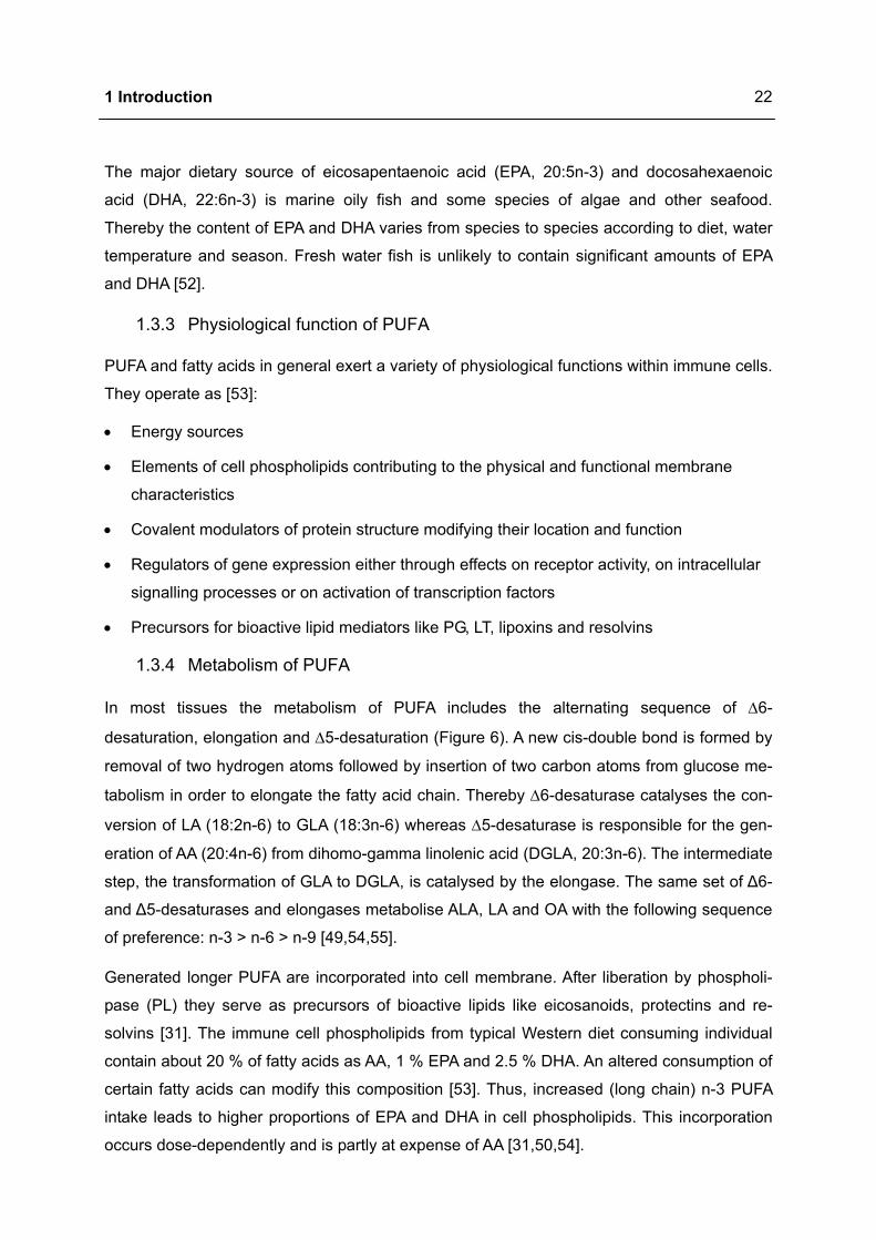
In most tissues the metabolism of PUFA includes the alternating sequence of ∆6-
desaturation, elongation and ∆5-desaturation (Figure 6). A new cis-double bond is formed by
removal of two hydrogen atoms followed by insertion of two carbon atoms from glucose me-
tabolism in order to elongate the fatty acid chain. Thereby ∆6-desaturase catalyses the con-
version of LA (18:2n-6) to GLA (18:3n-6) whereas ∆5-desaturase is responsible for the gen-
eration of AA (20:4n-6) from dihomo-gamma linolenic acid (DGLA, 20:3n-6). The intermediate
step, the transformation of GLA to DGLA, is catalysed by the elongase. The same set of ∆6-
and ∆5-desaturases and elongases metabolise ALA, LA and OA with the following sequence
of preference: n-3 > n-6 > n-9 [49,54,55].
Generated longer PUFA are incorporated into cell membrane. After liberation by phospholi-
pase (PL) they serve as precursors of bioactive lipids like eicosanoids, protectins and re-
solvins [31]. The immune cell phospholipids from typical Western diet consuming individual
contain about 20 % of fatty acids as AA, 1 % EPA and 2.5 % DHA. An altered consumption of
certain fatty acids can modify this composition [53]. Thus, increased (long chain) n-3 PUFA
intake leads to higher proportions of EPA and DHA in cell phospholipids. This incorporation
occurs dose-dependently and is partly at expense of AA [31,50,54].
1 Introduction
23
Figure 6: Metabolic pathways of n-6 and n-3 long chain PUFA, according to [54,56].
Broken arrows represent retroconversion cascades.
1.3.5 PUFA and immune system
On the basis of their physiological functions, fatty acids can mediate their biological action at
different cellular levels [53]:
• By alterations in the pattern of lipid mediators
• Through changes in the physical membrane characteristics
• By modulation of membrane receptor expression, activity or avidity
• By alteration of intracellular signal transduction
One key link between PUFA action and function of the immune system is the generation of
eicosanoids and resolvins from 20-carbon PUFA. Cell membrane phospholipids contain large
1 Introduction
24
amounts of AA. This n-6 PUFA is the major precursor for eicosanoids, a family of hydroxy-
lated PUFA with a wide range of functions on inflammatory and immune responses [31,53].
Cyclooxygenase (COX), lipoxygenase (LOX) and cytochrome P 450 pathways competitively
metabolise AA and EPA to eicosanoids such as PG, thromboxanes (TX), LT, lipoxins and ep-
oxy-compounds. Incorporation of increased amounts of EPA and DHA into cell membranes
results in a higher formation of EPA derived products at expense of AA derived mediators.
This is caused by decreased availability of AA as enzyme substrate and inhibition of AA me-
tabolism [53,57]. EPA and DHA are poor COX and LOX substrates compared to AA. Due to a
mislocalisation of carbon 13, EPA is only inadequately metabolised by COX-1. Therefore,
EPA competitively inhibits AA metabolism to its eicosanoids. Equally, DHA inhibits AA derived
mediator synthesis either itself or by retroconversion to EPA [58].
After liberation from membrane phospholipids the typically highly abundant AA is metabolised
through COX to PG and TX of the 2-series, e.g. PGE2 and PGF2a. Metabolism of AA via
5-LOX provides 5-hydroxyeicosatetraenoic acid and the 4-series of LT, e.g. LTA4 and LTB4
[58]. These mediators have been shown to exert proinflammatory but also antiinflammatory
properties [54].
Since EPA also operates as a substrate for COX and LOX, an increased availability of EPA
potentially results in increased production of EPA derived eicosanoids such as PGE3 and
5-series LT [53]. The functional significance of these mediators originated from EPA is the
proven lower biological activity compared to those from AA. For example, LTB5 is 10- to
100-fold less potent neutrophil chemotactic agent than LTB4 and PGE3 [31]. Long chained
n-3 PUFA are also metabolised to resolvins (from EPA and DHA) and docosanoids (from
DHA) through pathways involving COX and LOX. These mediators are antiinflammatory, im-
munomodulatory and resolve inflammation [53,54,59]. However, the physiological or patho-
physiological outcome is designated by cells present, the nature of stimulus, timing of eico-
sanoid generation, concentrations of different bioactive lipids as well as sensitivity of target
cells and tissues to generated mediators [53].
N-3 PUFA are mainly found at position sn-2 of membrane phospholipids, thereby replacing
AA. PUFA incorporation into membrane lipids decreases the membrane microviscosity influ-
encing the mobility, expression and function of membrane proteins [54]. Importantly,
n-3 PUFA containing phospholipids are also found in lipid rafts, thereby modulating cellular
signalling processes [49,58]. “Membrane rafts are small (10 to 200 nm), heterogeneous,
highly dynamic, sterol and sphingolipid enriched domains that compartmentalise cellular
processes. Small rafts can sometimes be stabilised to form larger platforms through protein–
1 Introduction
25
protein and protein–lipid interactions” [60]. Proteins are anchored in the membrane rafts
exoplasmically by glycosyl phosphatidylinositol or cytoplasmically by acyl moieties. Many
proteins involved in signal transduction are modified in this way and / or concentrated in lipid
rafts [58].
PUFA treatment was shown to modify the cytoplasmic leaflet of lipid rafts, thereby selectively
displacing acylated proteins and integrating n-3 PUFA. Moreover, dietary n-3 PUFA supple-
mentation reduces significantly the sphingomyelin content of lipid rafts in vivo. This causes
an altered exoplasmic membrane leaflet and folds acyl chains in the cytoplasmic leaflet. In-
corporation of PUFA as a replacement for saturated fatty acids or changes in raft lipid com-
position could lead to modified protein acylation and thus protein displacement from lipid rafts
[58]. Additionally, changing the fatty acid composition of immune cells affects phagocytosis,
T cell signalling and antigen presentation at the membrane level suggesting important roles
of fatty acids in membrane order, lipid raft structure and function as well as membrane traf-
ficking [53].
Cells are responsive to extracellular signals by an up or downregulated expression of specific
genes which leads to altered metabolism, proliferation, differentiation or apoptosis. Receptor
mediated signal transduction pathways transmit these extracellular signals to their intracellu-
lar targets. In addition to their roles as structural components of membrane lipids and as pre-
cursors of eicosanoids, fatty acids can act as second messengers or regulators of signal
transducing molecules [61]. Signalling molecules that may be modulated by different fatty
acids are divided into three groups:
1.3.5.1 Signalling molecules that require fatty acid acylation for membrane translocation
and functional activation
Translocation and protein function can be dramatically influenced by covalent attachment of
long chain fatty acids to a wide range of proteins. Many molecules involved in transmitting
extracellular signals are acylated for their membrane translocation. Cotranslational myristoy-
lation and posttranslational palmitoylation are described in this manner [61].
1.3.5.2 Lipid mediators containing different fatty acids or free fatty acids
PI3-K phosphorylate various phosphoinositides and regulate multiple cell functions such as
chemotaxis and apoptosis. The activity of the newly generated phosphatidylinositol
3,4,5-trisphosphate varies with types of fatty acids in the sn-1 and sn-2 positions of phospho-
inositides which can be altered by dietary fatty acids [61].
1 Introduction
26
Hydrolysis of sphingomyelin creates ceramide containing one fatty acyl moiety linked to the
sphingosine backbone by an amide bond. Ceramide acts as an intracellular signal effector
molecule with several downstream targets onto various extracellular signals. Thus, control-
ling ceramide activity by dietary fatty acids would exert profound nutritional implications [61].
Since the composition of membrane phospholipids is influenced by diverse dietary fatty ac-
ids, fatty acyl residues of diacylglycerol (DAG) may also be altered by dietary fatty acids. It
has been shown that the capacity to activate the protein kinase C (PKC) is regulated by fatty
acid esterification at the sn-1 or sn-2 position of DAG [61]. The activation of PKC is enhanced
via DAG by cis-unsaturated fatty acids like DHA, whereas other PUFA, especially AA in-
creases the activation of PLCγ. Additionally, AA and other PUFA control the activities of multi-
ple cellular proteins, including ion channels and protein kinases [61].
1.3.5.3 Signalling molecules that can be modulated by different fatty acids
Nuclear receptors, a family of ligand activated transcription factors, are able to control sev-
eral genes involved in lipid metabolism and inflammatory signalling directly and indirectly.
Ligand binding leads to conformational changes which enable the nuclear receptors to disso-
ciate their corepressors and recruit coactivator proteins to allow transcriptional activation
[54]. Various PUFA and prostanoids are ligands for peroxisome proliferator activated receptor
(PPAR), a nuclear hormone receptor regulating the transcription of genes involved in lipid
metabolism but also in diverse cellular responses [61]. Longer chain PUFA, but not shorter
chain monounsaturated or saturated fatty acids, are able to repress the liver X receptor
(LXR) enhancer complex in the sterol regulatory element binding protein (SREBP) 1c pro-
moter region [48]. The discussed “cross-talk” between PPAR signalling, retinoic acid X recep-
tor (RXR), SREBP expression and LXR may influence basically cell lipid homeostasis in a
highly complex but coordinated manner. Due to the various characteristics and the exhibited
transcriptional regulatory properties of each n-3 PUFA, the subsequent effects of these inter-
actions are also highly complex [48,54].
Furthermore, it has been shown that n-3 PUFA inhibit NFκB activity directly. EPA diminishes
degradation of the IκB thereby blocking NFκB. AA derived prostanoids inhibit NFκB translo-
cation and activation by potently blocking the IKK complex and thus retaining NFκB in the
cytoplasm. Consequently, n-3 PUFA and fatty acid derived mediators modulate manifold the
NFκB pathway [54].
1 Introduction
27
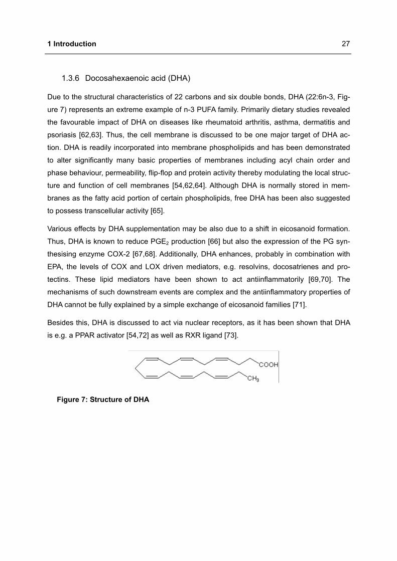
1.3.6 Docosahexaenoic acid (DHA)
Due to the structural characteristics of 22 carbons and six double bonds, DHA (22:6n-3, Fig-
ure 7) represents an extreme example of n-3 PUFA family. Primarily dietary studies revealed
the favourable impact of DHA on diseases like rheumatoid arthritis, asthma, dermatitis and
psoriasis [62,63]. Thus, the cell membrane is discussed to be one major target of DHA ac-
tion. DHA is readily incorporated into membrane phospholipids and has been demonstrated
to alter significantly many basic properties of membranes including acyl chain order and
phase behaviour, permeability, flip-flop and protein activity thereby modulating the local struc-
ture and function of cell membranes [54,62,64]. Although DHA is normally stored in mem-
branes as the fatty acid portion of certain phospholipids, free DHA has been also suggested
to possess transcellular activity [65].
Various effects by DHA supplementation may be also due to a shift in eicosanoid formation.
Thus, DHA is known to reduce PGE2 production [66] but also the expression of the PG syn-
thesising enzyme COX-2 [67,68]. Additionally, DHA enhances, probably in combination with
EPA, the levels of COX and LOX driven mediators, e.g. resolvins, docosatrienes and pro-
tectins. These lipid mediators have been shown to act antiinflammatorily [69,70]. The
mechanisms of such downstream events are complex and the antiinflammatory properties of
DHA cannot be fully explained by a simple exchange of eicosanoid families [71].
Besides this, DHA is discussed to act via nuclear receptors, as it has been shown that DHA
is e.g. a PPAR activator [54,72] as well as RXR ligand [73].
Figure 7: Structure of DHA
1 Introduction
28
1.4 Objective
Due to the increased incidence and limited causal therapeutics allergic diseases have be-
come a major medical issue in the Western world [1]. The reasons of this development are
not completely understood. In recent years research has pointed to the possible role of envi-
ronmental factors like changing Western dietary habits. The increased intake of n-6 PUFA
(margarine, vegetable oil) and decreased n-3 PUFA (oily fish) consumption have been hy-
pothesised to contribute to this development. However, the associations with n-6 and n-3
PUFA are complex and may differ between different allergic diseases [74,75].
Although many immunomodulatory properties of DHA are described, the underlying mecha-
nisms of DHA action are not exactly understood. Therefore, the following objectives were
investigated:
• First, the target cells of antiallergic DHA action were identified and subsequently its mo-
lecular mechanisms investigated.
• Within a randomised clinical study the therapeutical efficacy of high-dose DHA supple-
mentation was examined in patients with extrinsic atopic eczema. Beside the clinical ef-
fects, immunological parameters were analysed.
• Finally, the clinical efficiency of oral DHA administration was verified in a mouse model of
protein mediated eczema and the underlying local processes in eczematous skin exam-
ined in more detail.
2 Materials and methods
29
2 Materials and methods
2.1 Materials
Equipment, software, commodities, chemicals, antibodies and buffer and solutions used are
listed in detail in the appendix (page 94 - 105).
2.2 Methods
2.2.1 Donors and cells
The in vitro experiments were approved by the ethical committee of the Charité–
Universitätsmedizin Berlin. Therefore, blood was obtained from allergic patients and healthy
persons.
Atopic donors were patients suffering from rhinoconjunctivitis allergica, asthma allergica and
from atopic eczema defined according the criteria of Hanifin and Rajka [76] with serum IgE
concentrations below 2500 ng/mL. Donors with allergic conjunctivitis, allergic rhinitis and
asthma were recruited based on their history and positive prick testing as well as patients
with atopic eczema from the Department of Dermatology and Allergy, Charité.
None of the enrolled donors received a systemic immunosuppressive medication during the
past three months before the blood sample was taken. Usage of topical steroids (class II-III)
or antihistamines was not excluded.
Blood samples from age and sex matched healthy volunteers (no type I allergies or positive
family history of atopic diseases) and blood filters from blood donation of the Charité with IgE
levels up to 100 ng/mL were chosen as control.
2.2.2 Participants and clinical study
The clinical study was approved by the ethical committee of the Charité–Universitätsmedizin
Berlin and was performed in the Department of Dermatology and Allergy, Charité from Janu-
ary 2005 to June 2005. Written informed consent was obtained from each participant and
eligibility was confirmed by a dermatologist.
Patients suffering from atopic eczema aged between 18 and 40 years were enrolled into the
study. Atopic eczema was defined according the criteria of Hanifin and Rajka [76]. Exclusion
criteria were pregnancy, lactation, seafood allergies and consumption of dietary supplements,
systemic immunomodulatory or immunosuppressive therapy in the last three months, pres-
ence of other systemic and chronic disorders than allergic diseases. Participants were al-
2 Materials and methods
30
lowed to use standard therapy for atopic eczema, including emollients, topical corticosteroids
and oral antihistamines. Subjects were asked to maintain their habitual diet and activities
throughout the intervention. Randomisation was stratified according to gender, age (cut off
29 years) and body mass index (BMI; cut off 25 kg/m2) in a ratio of 1:1 using a randomised
allocation schedule (based on block randomisation). Allocation concealment was assured by
a sealed envelope up to finishing the data analysis. Recruited participants were randomly
allocated to receive a treatment with either DHA capsules (n = 21) or control supplement
(n = 23) by an independent clinician.
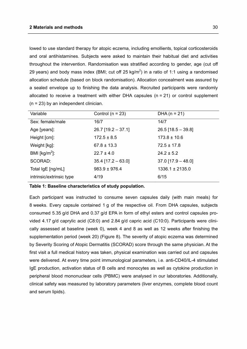
Variable Control (n = 23) DHA (n = 21)
Sex: female/male 16/7 14/7
Age [years]: 26.7 [19.2 – 37.1] 26.5 [18.5 – 39.8]
Height [cm]: 172.5 ± 8.5 173.8 ± 10.6
Weight [kg]: 67.8 ± 13.3 72.5 ± 17.8
BMI [kg/m2]: 22.7 ± 4.0 24.2 ± 5.2
SCORAD: 35.4 [17.2 – 63.0] 37.0 [17.9 – 48.0]
Total IgE [ng/mL] 983.9 ± 976.4 1336.1 ± 2135.0
intrinsic/extrinsic type 4/19 6/15
Table 1: Baseline characteristics of study population.
Each participant was instructed to consume seven capsules daily (with main meals) for
8 weeks. Every capsule contained 1 g of the respective oil. From DHA capsules, subjects
consumed 5.35 g/d DHA and 0.37 g/d EPA in form of ethyl esters and control capsules pro-
vided 4.17 g/d caprylic acid (C8:0) and 2.84 g/d capric acid (C10:0). Participants were clini-
cally assessed at baseline (week 0), week 4 and 8 as well as 12 weeks after finishing the
supplementation period (week 20) (Figure 8). The severity of atopic eczema was determined
by Severity Scoring of Atopic Dermatitis (SCORAD) score through the same physician. At the
first visit a full medical history was taken, physical examination was carried out and capsules
were delivered. At every time point immunological parameters, i.e. anti-CD40/IL-4 stimulated
IgE production, activation status of B cells and monocytes as well as cytokine production in
peripheral blood mononuclear cells (PBMC) were analysed in our laboratories. Additionally,
clinical safety was measured by laboratory parameters (liver enzymes, complete blood count
and serum lipids).
2 Materials and methods
31
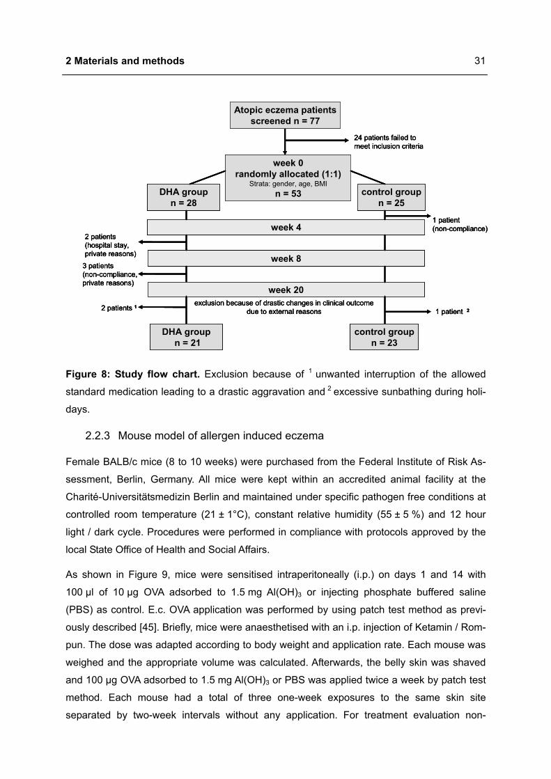
Atopic eczema patients screened n = 77
1 patient(non-compliance)
2 patients (hospital stay,private reasons)
3 patients (non-compliance,private reasons)
2 patients 1
week 0randomly allocated (1:1)
Strata: gender, age, BMIn = 53DHA group
n = 28control group
n = 25
week 4
week 8
week 20exclusion because of drastic changes in clinical outcome
due to external reasons 1 patient 2
control groupn = 23
DHA groupn = 21
24 patients failed to meet inclusion criteria
Figure 8: Study flow chart. Exclusion because of 1 unwanted interruption of the allowed
standard medication leading to a drastic aggravation and 2 excessive sunbathing during holi-
days.
2.2.3 Mouse model of allergen induced eczema
Female BALB/c mice (8 to 10 weeks) were purchased from the Federal Institute of Risk As-
sessment, Berlin, Germany. All mice were kept within an accredited animal facility at the
Charité-Universitätsmedizin Berlin and maintained under specific pathogen free conditions at
controlled room temperature (21 ± 1°C), constant relative humidity (55 ± 5 %) and 12 hour
light / dark cycle. Procedures were performed in compliance with protocols approved by the
local State Office of Health and Social Affairs.
As shown in Figure 9, mice were sensitised intraperitoneally (i.p.) on days 1 and 14 with
100 µl of 10 µg OVA adsorbed to 1.5 mg Al(OH)3 or injecting phosphate buffered saline
(PBS) as control. E.c. OVA application was performed by using patch test method as previ-
ously described [45]. Briefly, mice were anaesthetised with an i.p. injection of Ketamin / Rom-
pun. The dose was adapted according to body weight and application rate. Each mouse was
weighed and the appropriate volume was calculated. Afterwards, the belly skin was shaved
and 100 µg OVA adsorbed to 1.5 mg Al(OH)3 or PBS was applied twice a week by patch test
method. Each mouse had a total of three one-week exposures to the same skin site
separated by two-week intervals without any application. For treatment evaluation non-
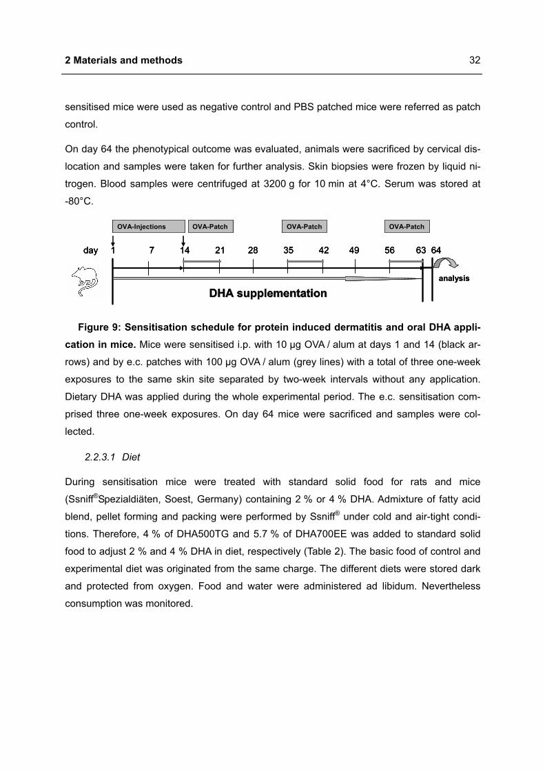
2 Materials and methods
32
sensitised mice were used as negative control and PBS patched mice were referred as patch
control.
On day 64 the phenotypical outcome was evaluated, animals were sacrificed by cervical dis-
location and samples were taken for further analysis. Skin biopsies were frozen by liquid ni-
trogen. Blood samples were centrifuged at 3200 g for 10 min at 4°C. Serum was stored at
-80°C.
analysis
OVA-Patch
day 1 7 14 21 28 35 42 49 56 63 64
OVA-PatchOVA-PatchOVA-Injections
DHA supplementationDHA supplementationanalysis
OVA-Patch
day 1 7 14 21 28 35 42 49 56 63 64
OVA-PatchOVA-PatchOVA-Injections
DHA supplementationDHA supplementation
OVA-Patch
day 1 7 14 21 28 35 42 49 56 63 64
OVA-PatchOVA-PatchOVA-Injections
DHA supplementationDHA supplementation
Figure 9: Sensitisation schedule for protein induced dermatitis and oral DHA appli-cation in mice. Mice were sensitised i.p. with 10 µg OVA / alum at days 1 and 14 (black ar-
rows) and by e.c. patches with 100 µg OVA / alum (grey lines) with a total of three one-week
exposures to the same skin site separated by two-week intervals without any application.
Dietary DHA was applied during the whole experimental period. The e.c. sensitisation com-
prised three one-week exposures. On day 64 mice were sacrificed and samples were col-
lected.
2.2.3.1 Diet
During sensitisation mice were treated with standard solid food for rats and mice
(Ssniff®Spezialdiäten, Soest, Germany) containing 2 % or 4 % DHA. Admixture of fatty acid
blend, pellet forming and packing were performed by Ssniff® under cold and air-tight condi-
tions. Therefore, 4 % of DHA500TG and 5.7 % of DHA700EE was added to standard solid
food to adjust 2 % and 4 % DHA in diet, respectively (Table 2). The basic food of control and
experimental diet was originated from the same charge. The different diets were stored dark
and protected from oxygen. Food and water were administered ad libidum. Nevertheless
consumption was monitored.
2 Materials and methods
33
Treatment group Control 2 % DHA 4 % DHA
Fatty acid Composition [%] Composition [%] Composition [%]
C14:0 0.3 n.d. n.d.
C16:0 1.5 n.d. n.d.
C16:1 0.3 n.d. n.d.
C18:0 2.5 3.7 0.1
C18:1 19.1 6.9 1.4
C18:2 5.5 1.6 0.2
C18:3 n-3 7.1 0.2 0.1
C18:3 n-6 0.3 0.5 n.d.
C18:4 n-3 0.6 0.3 0.1
C20:4 n-6 n.d. 2.8 1.8
C20:4 n-3 n.d. 0.7 0.5
C20:5 n-3 (EPA) n.d. 8.3 5.6
C22:5 n-3 n.d. 3.1 3.5
C22:6 n-3 (DHA) n.d. 57.5 76.4
Table 2: Fatty acid composition of lipids in experimental diet; n.d. - not detectable.
2.2.3.2 Clinical Evaluation
Evaluation of severity of OVA induced eczematous skin lesions was performed by a skin
scoring system, which considers typical skin features of human atopic eczema, erythema,
edema / papulation, excoriation / crusting, dryness and extension [76,77]. The total clinical
skin score (CSS) was initially described in a murine model with spontaneous manifestation of
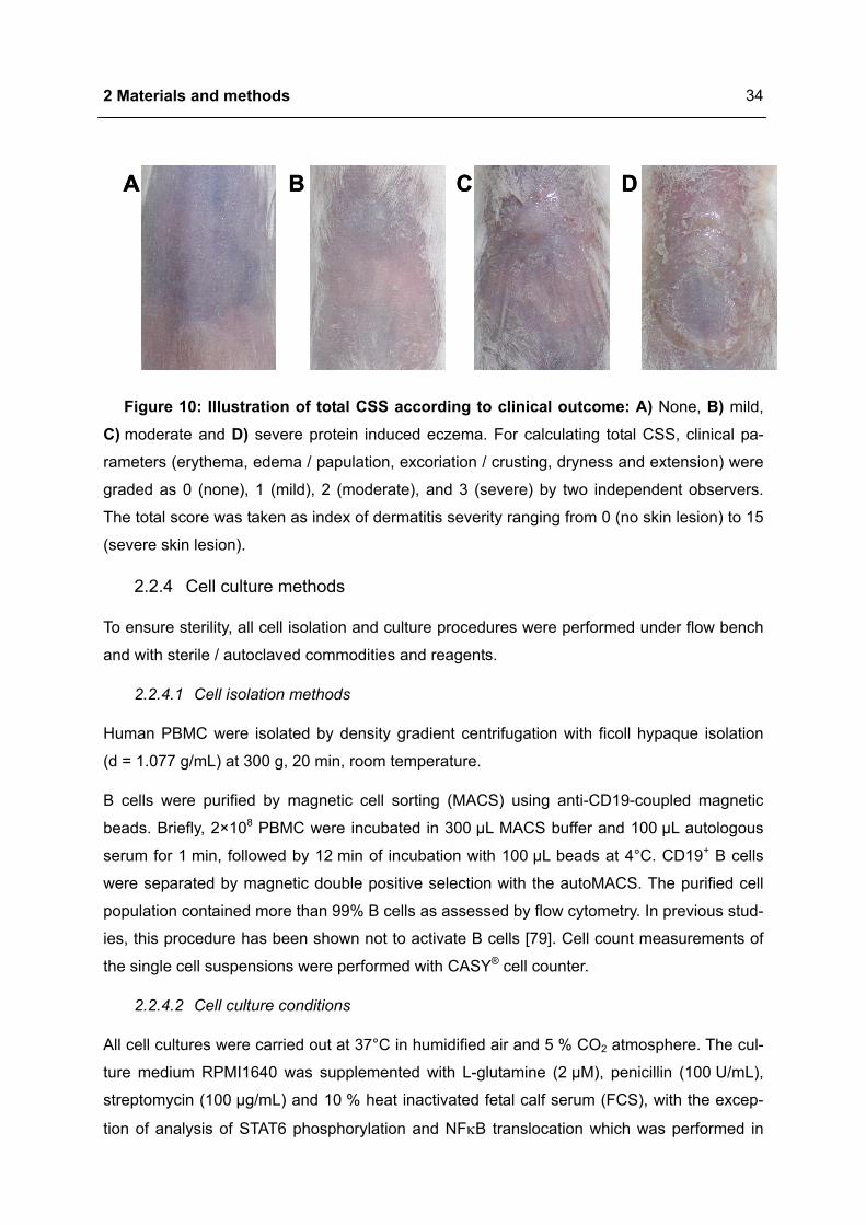
atopic eczema like skin lesions [78]. For calculating total CSS, parameters were graded as
0 (none), 1 (mild), 2 (moderate), and 3 (severe) by two independent observers. The total
score was taken as index of dermatitis severity ranging from 0 (no skin lesion) to 15 (severe
skin lesion) (Figure 10).
2 Materials and methods
34
A DCBA DCB
Figure 10: Illustration of total CSS according to clinical outcome: A) None, B) mild, C) moderate and D) severe protein induced eczema. For calculating total CSS, clinical pa-
rameters (erythema, edema / papulation, excoriation / crusting, dryness and extension) were
graded as 0 (none), 1 (mild), 2 (moderate), and 3 (severe) by two independent observers.
The total score was taken as index of dermatitis severity ranging from 0 (no skin lesion) to 15
(severe skin lesion).
2.2.4 Cell culture methods
To ensure sterility, all cell isolation and culture procedures were performed under flow bench
and with sterile / autoclaved commodities and reagents.
2.2.4.1 Cell isolation methods
Human PBMC were isolated by density gradient centrifugation with ficoll hypaque isolation
(d = 1.077 g/mL) at 300 g, 20 min, room temperature.
B cells were purified by magnetic cell sorting (MACS) using anti-CD19-coupled magnetic
beads. Briefly, 2×10⁸ PBMC were incubated in 300 µL MACS buffer and 100 µL autologous
serum for 1 min, followed by 12 min of incubation with 100 µL beads at 4°C. CD19+ B cells
were separated by magnetic double positive selection with the autoMACS. The purified cell
population contained more than 99% B cells as assessed by flow cytometry. In previous stud-
ies, this procedure has been shown not to activate B cells [79]. Cell count measurements of
the single cell suspensions were performed with CASY® cell counter.
2.2.4.2 Cell culture conditions
All cell cultures were carried out at 37°C in humidified air and 5 % CO2 atmosphere. The cul-
ture medium RPMI1640 was supplemented with L-glutamine (2 µM), penicillin (100 U/mL),
streptomycin (100 µg/mL) and 10 % heat inactivated fetal calf serum (FCS), with the excep-
tion of analysis of STAT6 phosphorylation and NFκB translocation which was performed in
2 Materials and methods
35
pure RPMI1640. DHA, dissolved in 100 % ethanol, was portioned and stored at -20°C for a
maximum of six months. The vehicle control was performed in culture media containing the
same volume of ethanol (0.001 %). Toxic effects of DHA were excluded by propidium iodide
and trypan blue staining.
2.2.4.3 Carboxy-fluorescein diacetate, succinimidyl ester (CFSE) labelling
B cells were suspended in PBS containing 5 µM CFSE for 5 min at room temperature and
subsequently washed. Cells were stimulated with anti-CD40 (1 µg/mL; 82111) and IL-4
(5 ng/mL) [80] and were treated with 0.1 µM, 1 µM and 10 µM DHA. If not otherwise men-
tioned, CD40 ligation was mediated by clone 82111 at 1 µg/mL. Analysis by means of flow
cytometry was performed on day 6.
2.2.4.4 WST-1 assay
For studies of cell mitochondrial activity WST-1 assay was performed. Briefly, 105 PBMC
were cultured for 4 days and stimulated with anti-CD40/IL-4. 10 µL WST-1 reagent was
added during the last 3 hours of culture to each well. Cellular enzymatic activity was deter-
mined by photometrical measurement at 450 nm. Cells treated with 0.1 µM, 1 µM and 10 µM
DHA were compared with DHA untreated samples. Phorbol-12-myristate-13-acetate (PMA,
10 ng/mL) was used as positive control. All experiments were performed in triplets.
2.2.5 Immunological methods
2.2.5.1 Enzyme-Linked Immuno Sorbent Assay (ELISA)
ELISA was used to quantify immunoglobulins. Thereby, the unknown amount of antigen is
bound to a surface by a primary antibody. The secondary antibody is incubated over this sur-
face so that it can bind specifically to the antigen. This detection antibody is linked to an en-
zyme, which converts a chromogenic substrate to some detectable signal. In case of antigen
specific immunoglobulin determination primary or secondary antibody is replaced by the spe-
cific antigen. The amount of protein in the sample was calculated by means of standard
curve. If not otherwise mentioned all steps were performed at room temperature.
2.2.5.1.1 Human immunoglobulin ELISA
For immunoglobulin assays cells (106/mL) were cultured for 10 days. Cells were stimulated
with anti-CD40/IL-4 [80] and were treated with 0.1 µM, 1 µM and 10 µM DHA. After 10 days
immunoglobulins were measured in cell free supernatants by ELISA. For IgE detection
clones HP6061 and HP6029 were applied. The matched antibody pairs for IgA, IgG and IgM
ELISA (anti-IgA, anti-IgG and anti-IgM) were purchased from Dianova. Immunoplates (maxi
2 Materials and methods
36
Sorb) were coated overnight at 4°C with the primary antibodies for IgE clone HP6061
(2.5 µg/mL) or for the other immunoglobulins (0.4 µg/mL) diluted in sodium carbonat buffer.
Unspecific binding was prevented by adding 2 % bovine serum albumine (BSA) / Tris-
buffered saline (TBS) for 1 hour. Supernatants and standards were then incubated for 2
hours. After washing, the second alkaline phosphatase conjugated anti-immunoglobulin anti-
body (0.15 µg/mL diluted in 0.2 % BSA/TBS) was added for 1.5 hours. Since the secondary
mouse anti-human IgE antibody was biotinylated (HP6029, 0.5 µg/mL diluted in 0.2 %
BSA/TBS) another hour of incubation with alkaline phosphatase conjugated streptavidin
(1.5 µg/mL) was performed. Following the final reaction with phosphatase substrate, plates
were read in a microplate ELISA reader at 405 nm and the amount of immunoglobulin was
calculated according to the standard curve. Diluted human serum of an atopic individual
served as standard curve. The sensitivity of the ELISA was 100 pg/mL.
2.2.5.1.2 Murine immunoglobulin ELISA
Plates were coated overnight with anti-mouse IgE (EM95.3, 5 µg/mL) diluted in sodium car-
bonate buffer. After blocking with 3 % milk powder/PBS, sera (diluted in 1 % milk pow-
der/PBS) were incubated overnight and were detected with biotin conjugated anti-mouse IgE
(84 1-C, 1 µg/mL diluted in 1 % milk powder/PBS). The reaction was developed with strepta-
vidin peroxidase and tetra methyl benzidine and was stopped with 2 N sulphuric acid. The
plates were measured at 450/490 nm and total IgE was calculated according to the standard
curve. The monoclonal antibodies EM95.3 and 84 1-C were kindly provided by Dr. Elke Lu-
ger, DRFZ, Berlin, Germany / Dr. Lamer, MPI, Freiburg, Gemany.
For measurement of OVA specific immunoglobulins plates were coated overnight either with
rat anti-mouse IgE (R35-72, 2 µg/mL) or 5 µg/mL OVA for IgG1 and IgG2a in sodium carbon-
ate buffer. After blocking with 3 % milk powder/PBS, serial dilution of sera in 1 % milk pow-
der/PBS were incubated overnight, detected with biotin conjugated OVA (1.25 mg/mL, cou-
pling rate 1:50) for IgE, with rat anti-mouse IgG1 (A85-1, 0.5 µg/mL) or rat anti-mouse IgG2a
(R19-15, 0.5 µg/mL) in 1 % milk powder/PBS and developed as mentioned above. Plates
were read in a microplate ELISA reader at 450/490 nm. OVA specific immunoglobulin con-
centrations were calculated according to the standard serum pool.
The validity of ELISAs was verified for total IgE by using serial sera dilutions and a standard
of known concentration and for OVA specific immunoglobulins by using serial sera dilutions
as well as by the determination of the semimaximal saturation.
2 Materials and methods
37
2.2.5.2 Enzyme Linked Immuno Spot Technique (ELISpot)
The number of antibody secreting cells was determined by ELISpot technique. Therefore,
B cells were stimulated with anti-CD40/IL-4 in the presence of 0.1 µM, 1 µM and 10 µM DHA
for 7 days. 106 B cells were cultured for IgA and IgG, whereas 3x10⁶ cells were used for IgE
ELISpot. MultiScreen-High Protein Binding Immibilion-P Membrane plates were coated over-
night with mouse anti-human IgE (HP6061, 2.5 µg/mL) and with goat anti-human IgA or IgG
(0.4 µg/mL, Dianova) in sodium carbonate buffer. Unspecific binding was blocked by adding
2 % BSA/PBS for 1 hour. After several washings, serial dilutions of B cells from 7 day cul-
tures were incubated at 37°C for adequate period of time. After washing, detection was per-
formed with biotin conjugated antibodies: mouse anti-human IgE (HP6029, 1.5 µg/mL),
mouse anti-human IgA (G20-359, 5 µg/mL) or mouse anti-human IgG (G18-145, 0.1 µg/mL)
in 0.2 % BSA/PBS. The reaction was developed with streptavidin-horseradish peroxidase
(0.75 µg/mL) and peroxidase substrate 33-Amino-9-ethyl-carbazole/ n,n-Dimethyl formamide.
Antibody secreting cells appeared as red spots and were counted with CTL ImmunoSpot®
S4.
2.2.5.3 Flow cytometry
Fluorescence activated cell sorting (FACS) enables the analysis of single cells due to their
characteristical light scattering and their emitted fluorescence. By the mean of forward light
scatter (FSC) / side light scatter (SSC) characteristics, i.e. size and granularity, it is possible
to distinguish between different cell populations. Analysis of specific intracellular and ex-
tracellular antigens can be performed by staining with fluorescence dye (e.g. FITC [Fluo-
resceine isothiocyanat], PE [Phycoerythrine], APC [Allophycocyanine]) coupled antibodies. In
flow cytometer these dyes are excited by appropriate laser and subsequently emit light of
different wave lengths. This emission is measured by defined detectors and is depicted in a
graph as intensity per cell (Figure 11).

2 Materials and methods
38
CD27 Pe
010 110 210 310 410
Cou
nt
4024
3018
2012
1006
0
IgE FITC
010 110 210 310 410
CD
38 P
erC
P C
y5.5
010
110
210
310
410
CD38 PerCP Cy5.5
010 110 210 310 410
CD
27 P
e
010
110
210
310
410
R3
R2
R4
FSC-H0 256 512 768 1024
SSC
-H
0
256
512
768
1024
R1A B
C D
CD27 Pe
010 110 210 310 410
Cou
nt
4024
3018
2012
1006
0
IgE FITC
010 110 210 310 410
CD
38 P
erC
P C
y5.5
010
110
210
310
410
CD38 PerCP Cy5.5
010 110 210 310 410
CD
27 P
e
010
110
210
310
410
R3
R2
R4
FSC-H0 256 512 768 1024
SSC
-H
0
256
512
768
1024
R1A B
C D
Figure 11: Exemplary flow cytometry images. A) FSC / SSC characteristics of B cells
after 7 days of stimulation with anti-CD40/IL-4/IL-21, B) dot blot of anti-CD27 plus anti-CD38
stained, C) histoblot of anti-CD27 stained and D) dot blot of anti-CD38 plus anti-IgE stained
B cells.
2.2.5.3.1 Flow cytometric analysis of PBMC activation
Within the clinical trial the effect of DHA supplementation on activation status of PBMC iso-
lated cells was investigated. Therefore, isolated PBMC were stained with a combination of
fluorescence dye labelled monoclonal antibodies. Antibodies used were anti-CD19 (4G7),
anti-CD14 (MφP9), anti-CD23 (EBVCS-5) and anti-HLA-DR (Human Leukocyte Antigene-DR,
L243). Staining was performed for 15 min at 4°C in the dark.
2.2.5.3.2 Flow cytometric analysis of cytokines in PBMC
Further on, the interference of DHA supplementation with systemic cytokine response was
investigated in PBMC of treated patients. Therefore, 2x10⁶ PBMC were cultured in the pres-
ence of Staphylococcus Enterotoxine B (SEB; 1 µg/mL) and anti-CD28 (L293, L25;
0.2 µg/mL) or without any stimulation for 24 hours. To accumulate synthesised proteins in-

2 Materials and methods
39
tracellularly, 1 µg/mL Brefeldin A was added 4 hours before expiration of the incubation. After
harvesting, cells were stained with fluorescence dye labelled monoclonal anti-human CD4
(SK3) and anti-CD69 (L78) for 15 min at 4°C in the dark. After the following washing step,
PBMC were fixed with 2 % paraformaldehyde (PFA) for 15 min at room temperature. Staining
of intracellular cytokines was performed with anti-human IFNγ (4S·B3) and anti-IL-4 (8D4-8)
in 0.5 % saponin / FACS buffer for 35 min at 4°C. Cells were immediately analysed at the
flow cytometer. At least 100000 gated cells were collected for each sample.
2.2.5.3.3 Flow cytometric analysis of STAT6 phosphorylation
Within the scope of in vitro investigations, the impact of DHA on the IL-4 signalling pathway
STAT6 phosphorylation was investigated by flow cytometry. After starving in the presence of
0.1 µM, 1 µM and 10 µM DHA overnight, isolated B cells were stimulated with 10 ng/mL IL-4
for 1 hour and harvested into BD Phosflow Fix Buffer I according to manufacturer’s instruc-
tions. Unstimulated cells were harvested in a similar fashion. After fixing the cells for 10 min
at 37°C, B cells were permeabilised with Phosflow Perm Buffer III for 30 min on ice. After
washing, cells were stained with anti-pSTAT6 (pY641; clone 18), anti-STAT6 (clone 23),
anti-CD27 (LG.7F9) and anti-CD38 (HIT2) for 1 hour at room temperature. Cells were imme-
diately analysed at the flow cytometer. At least 30000 gated B cells were collected for each
sample and geometric mean fluorescence was analysed.
2.2.5.3.4 Flow cytometric analysis of IκBα degradation
IkBα degradation was measured by flow cytometry to disclose the mechanisms of DHA me-
diated inhibition of p50 translocation. CD19+ B cells were preincubated with 0.1 µM, 1 µM
and 10 µM DHA for 2 hours followed by a stimulation with 1 µg/mL anti-CD40 (G28.5) for
1 hour. Subsequently, stimulated and unstimulated cells were harvested directly into 4 %
PFA. After fixing the cells for 10 min at 37°C, B cells were incubated for 15 min with
anti-CD20 (2H7), anti-CD27 (LG.7F9) and anti-CD38 (HIT2) on ice. After an additional wash-
ing step, cells were stained with anti-IκBα (L35A5) in 1 % saponin / FACS buffer for 30 min at
4°C. Cells were immediately analysed at the flow cytometer. At least 30000 gated B cells
were collected for each sample and geometric mean fluorescence was analysed.
2.2.5.4 Immunohistochemistry
Using biopsy punches, 5 mm diameter sections were obtained from patch areas of murine
bellies, embedded in freezing medium and carefully frozen into liquid nitrogen. The tissue
was cut into 5 µm specimens by a microtome at about -30°C and directly transferred on mi-

2 Materials and methods
40
croscope slide. After drying on a hot plate, samples were stored at -80°C until further prepa-
ration.
2.2.5.4.1 Staining of CD4+ and CD8+ T cells
Initially, unspecific binding was blocked by using 5 % goat serum in TBS for 20 min and ap-
plication of the Avidin / Biotin Blocking Kit according to manufacturer’s instructions. After
1 hour of incubation with rat anti-mouse CD4 (L3T4, 15 µg/mL, Figure 12A) or CD8 (53-6.7,
15 µg/mL, Figure 12B), sections were exposed to biotinylated goat anti-rat IgG over 30 min.
Staining was developed using Dako REAL™ Detection System Alkaline Phosphatase / RED.
Thereby, alkaline phosphatase is covalently bound to biotin of secondary antibody by a strep-
tavidine bridge. The subsequently converted substrate designates the positive cells. Haema-
toxylin was used for nuclei counterstaining. This staining is based on the building of chelate
rings of haematin plus salts with phosphate groups of nucleic acid in nucleus. The whole
specimen was afterwards mounted on glass slides with preheated Kaiser's glycerin jelly. All
steps were performed at room temperature.
2.2.5.4.2 Mast cell staining
To visualise MC granula filled with heparin and histamine (metachromatic), samples were
stained with 0.1 % toluidine blue in 0.5 N HCl for 1 hour followed by short washing step with
tap water (Figure 12C).
2.2.5.4.3 Measurement of epidermal thickening
Thickness of epidermis was determined by Axiovision measuring tools on the Axioplan light
microscope at x100 magnification. The measurement included the stratum granulosum, stra-
tum spinosum and stratum basale. Since stratum corneum was partly damaged due to the
procedure of fixing and slicing of the skin, its thickness could not be determined in all cases
and was therefore excluded. For each mouse, eight measurements were obtained (Figure
12D).

2 Materials and methods
41
A B
DC
A B
DC
Figure 12: Illustration of histological analysis of protein induced dermatitis in BALB/c mice. Cell infiltrates of A) CD4+, B) CD8+ and C) MC as well as D) skin thickness
were determined in 5 µm section of frozen skin samples. Positive immunohistochemical
stained cells were counted in 100 µm x 200 µm areas at x100 magnification using the meas-
uring tools of Axiovision software. Skin thickness (µm) was determined in haematoxylin
stained skin site by digital measurement tools.
2.2.5.4.4 Quantification of cellular infiltrates
To record dermal infiltration of CD4+, CD8+ cells and MC, positive stained cells were counted
in 100 µm x 200 µm areas at x100 magnification using measuring tools of Axiovision soft-
ware. Only cells in the dermis were counted by positioning the square lengthwise on the bor-
der of epidermis and dermis. The mean of eight sections per mouse was calculated.
2.2.6 Molecular biological methods
For gene expression analysis real-time reverse-transcriptase (RT) polymerase chain reaction
(PCR) was performed. Therefore RNA was isolated, transcribed into complementary DNA
(cDNA) and finally analysed by quantitative PCR.
2 Materials and methods
42
2.2.6.1 RNA isolation
For analysis of εGLT and AID mRNA expression 106 B cells were stimulated with anti-CD40
and / or IL-4 in the presence of 0.1 µM, 1 µM and 10 µM DHA for 4 days.
RNA isolation of single cell suspensions was performed using RNA isolation kit Nucleo-
spin®RNA II according to manufacturer’s guidelines. This technique bases on a two column
system. The first shredder column removes firm components from nucleic acids and the sec-
ond column binds nucleic acids. Salts, metabolites and macromolecular cellular components
are removed by several washing steps with different buffers.
Briefly, 106 cells were directly lysed by adding 350 µL RA1 buffer with 1 % beta mercapto
ethanol. This lysis buffer immediately inactivates RNases and generates optimal conditions
for adsorption of RNA to the silica membrane. Contaminating DNA was removed by a 15 min
rDNase digestion. After numerous washing steps, RNA is eluted in RNase free water and its
concentration is photometric measured at 260 nm in ND-1000 Spectrophotometer. To
achieve higher RNA concentrations, the visible dye labeled carrier Pellet Paint™ Co-
Precipitant was used in combination with alcohol precipitation method. Pellets were resus-
pended in RNase free water for cDNA synthesis.
2.2.6.2 cDNA synthesis
Synthesis of cDNA was performed with Taq Man Reverse Transcription Reagent according to
manufacturer’s instructions. This kit contains MultiScribe® Reverse Transcriptase, a recom-
binant Moloney Murine Leukemia Virus Reverse Transcriptase, random hexamers and oligo
(dT). Finally, cDNA was stored at -20°C.
2.2.6.3 Quantitative PCR
For gene expression analysis real-time RT-PCR was performed. After thermal denaturation of
double stranded DNA into single stranded DNA (95°C), oligonucleotides (primer) can com-
plementarily hybridise to a specific sequence on each strand of the target DNA at a primer
dependent “annealing” temperature (60 to 65°C). Afterwards, a thermo stable DNA poly-
merase prolongs the oligonucleotides (72°C). The newly generated DNA sequence matches
the sequence following the primer. After synthesising new DNA, the same primers will be
released and used again. This gives DNA a logarithmic amplification.
By the mean of double stranded DNA binding dye chemistry the amplicon production was
quantified by using a non-sequence specific fluorescent intercalating agent. SYBR Green is a
fluorogenic minor groove binding dye that does not bind to single stranded DNA. This dye
2 Materials and methods
43
emits a strong fluorescent signal upon binding to double stranded DNA whereas it exhibits
only little fluorescence in solution. Thus, as PCR products accumulate, fluorescence in-
creases. Real-time PCR monitors the fluorescence emitted during the reaction as an indica-
tor of amplicon production during each PCR cycle, i.e. in real time. Melting curve analysis
was used to identify the specific amplicon and to exclude non-specific amplifications. All
samples were measured at least in duplicates. Water was used as negative control.
The used oligonucleotides (primer) were designed by means of the internet databases UCSC
and NCBI and created by online software Primer3 (http://frodo.wi.mit.edu/cgi-
bin/primer3/primer3_www.cgi). The required parameters for well designed primers and sam-
ples have been documented and are built into the program. These parameters include a
melting temperature for the sample that is 10°C higher than the primers, primer melting tem-
perature about 60°C, amplicon size between 80 and 150 bases, absence of 5' guanosines
and primer length between 20 and 25 nucleotides. The best design for primers for the quanti-
fication of mRNA expression requires intron spanning positioning. To achieve most optimal
and specific results, PCR conditions were optimised regarding primer concentration, anneal-
ing temperature and time, magnesium chloride concentration as well as elongation time. The
depicted primers are applied at a hybridisation temperature of 65°C and at concentration of
500 nM (Table 3).
Gene 5’ - 3’ Sequence amplicon
[bp]
MgCL2
[mM] Efficiency E
HPRT for TggCTTATATCCAACACTTCgTg 187 4 2.00
HPRT rev ATCAgACTgAAgAgCTATTgTAATgACCA
εGLT for gACgggCCACACCATCCACAggCAC-
CAAATggACgAC 409 5 1.91
εGLT rev CAggACgACTgTAAgATCTTCACg
AID for AgAggCgTgACAgTgCTACA 93 4 1.88
AID rev ATgTAgCggAggAAgAgCAA
Table 3: Used oligonucleotides (primer) for quantitative real-time PCR.
For quantitative real-time PCR LightCycler-Fast Start Master SYBR Green I was used ac-
cording to manufacturers’ guidelines. The reaction volume was reduced to 5 µL per sample.
While reaction mass is denaturating for 10 min at 95°C, hot start polymerase is activated in
the LightCycler unit. The measurement of the fluorescent signal is performed directly after
elongation by polymerase. Thus, amplification progress can be detected simultaneously with
2 Materials and methods
44
intercalation of SYBR Green. Amplification of a given cDNA over time follows a curve, with an
initial flat phase, followed by an exponential phase. As the experiment reagents are used up,
DNA synthesis slows and exponential curve flattens into a plateau. For the RT-PCR principle,
the more cDNA is in a sample, the earlier it will be detected during repeated cycles of ampli-
fication.
Target gene levels were normalised to a housekeeping gene as an endogenous control, to
correct potential variation in RNA loading, cDNA synthesis or efficiency of the amplification.
Expression levels of reference genes should remain roughly constant between cells of differ-
ent tissues and under different experimental conditions. Here hypoxanthine guanine phos-
phoribosyltransferase (HPRT) was used to evaluate expression of target genes.
The comparative CP method for analysis involves comparing the threshold cycle or crossing
point (CP) values of samples of interest with a control. The relative expression ratio (R) of a
target gene is based on the real-time efficiency (E) of the primer pair, the CP deviation of an
unknown sample versus a control and is expressed in comparison to a reference gene (ref)
(Formula 1). Either DHA untreated sample in in vitro experiments or control diet fed mice
were set as control. Efficiency corrected relative quantification was performed by 2-∆∆CP
method [81].
sample)-lref(controCref
sample)-troltarget(conCtarget
P
P
)(E)(E
∆
∆
=R
Formula 1: ∆∆CP method for mRNA quantification.
2.2.6.4 Western blot
The western blot technique enables the detection of a specific protein in a given sample ho-
mogenate or extract. Primarily, gel electrophoresis is used to separate native or denatured
proteins by the length of the polypeptides. The proteins are transferred afterwards to a mem-
brane (typically nitrocellulose or polyvinylidene difluoride [PVDF]), where they are detected
using antibodies specific for target protein. Here, western blot is used to investigate the im-
pact of DHA on the NFκB signalling by the means of p50 translocation into nucleus.
After 2 hours preincubation with 10 µM DHA, isolated B cells were stimulated with 1 µg/mL
anti-CD40 (G28.5) for 1 hour. Protein extracts of 5x10⁶ B cells were prepared by NE-PER
extraction reagent containing a protease inhibitor cocktail and were subsequently quantified
by standard coomassie plus protein assay according to manufacturer’s instructions. Nuclear
2 Materials and methods
45
extracts (3 to 5 µg) were separated in a 12 % tris glycine sodium dodecyl sulfate (SDS) poly-
acrylamide gel. Proteins were transferred to PVDF membrane and blocked for 1 hour with
5 % milk powder/PBS. Overnight membrane was incubated with 0.2 µg/mL anti-human p50
(H119) in 1 % milk powder/PBS-Tween (PBST) at 4°C. After washing the membrane with
PBST, horse radish peroxidase (HRP) conjugated goat anti-rabbit IgG (0.1 µg/mL in 1 % milk
powder/PBST) was added for 1 hour at room temperature. The antigen detection was per-
formed with the chemiluminescent detection system ECL Plus.
2.3 Statistical analysis
Wilcoxon test for non-parametrical, paired data using SPSS (Statistical Package for the So-
cial Sciences) software package for Windows, Release 12.0 and 14.0 was carried out to ana-
lyse the statistical significance of within-group changes. Non-parametrical, unpaired data was
analysed by Mann-Whitney U-test to evaluate the significance of between group changes. A
p-value < 0.05 was considered to be statistically significant.
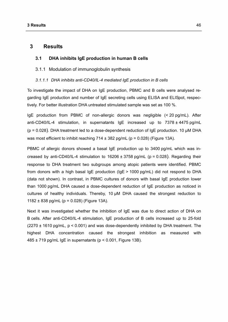
3 Results
46
3 Results
3.1 DHA inhibits IgE production in human B cells
3.1.1 Modulation of immunoglobulin synthesis
3.1.1.1 DHA inhibits anti-CD40/IL-4 mediated IgE production in B cells
To investigate the impact of DHA on IgE production, PBMC and B cells were analysed re-
garding IgE production and number of IgE secreting cells using ELISA and ELISpot, respec-
tively. For better illustration DHA untreated stimulated sample was set as 100 %.
IgE production from PBMC of non-allergic donors was negligible (< 20 pg/mL). After
anti-CD40/IL-4 stimulation, in supernatants IgE increased up to 7378 ± 4475 pg/mL
(p = 0.028). DHA treatment led to a dose-dependent reduction of IgE production. 10 µM DHA
was most efficient to inhibit reaching 714 ± 382 pg/mL (p = 0.028) (Figure 13A).
PBMC of allergic donors showed a basal IgE production up to 3400 pg/mL which was in-
creased by anti-CD40/IL-4 stimulation to 16206 ± 3758 pg/mL (p = 0.028). Regarding their
response to DHA treatment two subgroups among atopic patients were identified. PBMC
from donors with a high basal IgE production (IgE > 1000 pg/mL) did not respond to DHA
(data not shown). In contrast, in PBMC cultures of donors with basal IgE production lower
than 1000 pg/mL DHA caused a dose-dependent reduction of IgE production as noticed in
cultures of healthy individuals. Thereby, 10 µM DHA caused the strongest reduction to
1182 ± 838 pg/mL (p = 0.028) (Figure 13A).
Next it was investigated whether the inhibition of IgE was due to direct action of DHA on
B cells. After anti-CD40/IL-4 stimulation, IgE production of B cells increased up to 25-fold
(2270 ± 1610 pg/mL, p < 0.001) and was dose-dependently inhibited by DHA treatment. The
highest DHA concentration caused the strongest inhibition as measured with
485 ± 719 pg/mL IgE in supernatants (p < 0.001, Figure 13B).
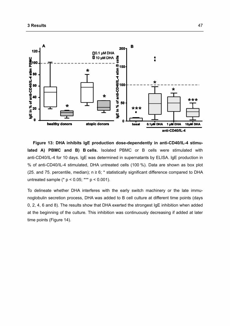
3 Results
47
healthy donors atopic donors0
20
40
60
80
100
120 0.1 µM DHA10 µM DHA
IgE
in %
of a
nti-C
D40
/IL-4
stim
PBM
C
**
****
basal 0.1µM DHA 1 µM DHA 10µM DHA0
50
100
150
200
IgE
in %
of a
nti-C
D40
/IL-4
stim
B c
ells
A B
anti-CD40/IL-4
**
******
healthy donors atopic donors0
20
40
60
80
100
120 0.1 µM DHA10 µM DHA
IgE
in %
of a
nti-C
D40
/IL-4
stim
PBM
C
**
****
basal 0.1µM DHA 1 µM DHA 10µM DHA0
50
100
150
200
IgE
in %
of a
nti-C
D40
/IL-4
stim
B c
ells
A B
anti-CD40/IL-4
healthy donors atopic donors0
20
40
60
80
100
120 0.1 µM DHA10 µM DHA
IgE
in %
of a
nti-C
D40
/IL-4
stim
PBM
C
**
****
basal 0.1µM DHA 1 µM DHA 10µM DHA0
50
100
150
200
IgE
in %
of a
nti-C
D40
/IL-4
stim
B c
ells
A B
healthy donors atopic donors0
20
40
60
80
100
120 0.1 µM DHA10 µM DHA
IgE
in %
of a
nti-C
D40
/IL-4
stim
PBM
C
**
****
healthy donors atopic donors0
20
40
60
80
100
120 0.1 µM DHA10 µM DHA0.1 µM DHA10 µM DHA
IgE
in %
of a
nti-C
D40
/IL-4
stim
PBM
C
**
****
basal 0.1µM DHA 1 µM DHA 10µM DHA0
50
100
150
200
IgE
in %
of a
nti-C
D40
/IL-4
stim
B c
ells
basal 0.1µM DHA 1 µM DHA 10µM DHA0
50
100
150
200
IgE
in %
of a
nti-C
D40
/IL-4
stim
B c
ells
A B
anti-CD40/IL-4
**
******
Figure 13: DHA inhibits IgE production dose-dependently in anti-CD40/IL-4 stimu-lated A) PBMC and B) B cells. Isolated PBMC or B cells were stimulated with
anti-CD40/IL-4 for 10 days. IgE was determined in supernatants by ELISA. IgE production in
% of anti-CD40/IL-4 stimulated, DHA untreated cells (100 %). Data are shown as box plot
(25. and 75. percentile, median); n ≥ 6; * statistically significant difference compared to DHA
untreated sample (* p < 0.05; *** p < 0.001).
To delineate whether DHA interferes with the early switch machinery or the late immu-
noglobulin secretion process, DHA was added to B cell culture at different time points (days
0, 2, 4, 6 and 8). The results show that DHA exerted the strongest IgE inhibition when added
at the beginning of the culture. This inhibition was continuously decreasing if added at later
time points (Figure 14).
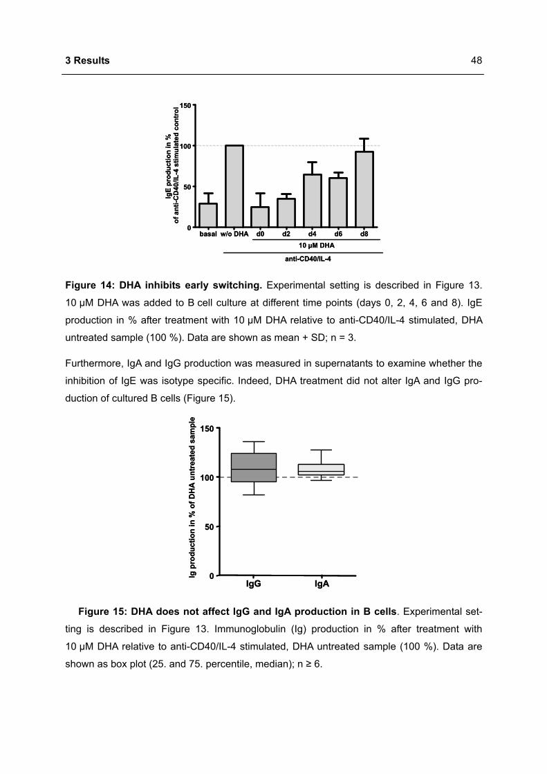
3 Results
48
basal w/o DHA d0 d2 d4 d6 d80
50
100
150
IgE
prod
uctio
n in
%of
ant
i-CD
40/IL
-4 s
timul
ated
con
trol
10 µM DHA
anti-CD40/IL-4
basal w/o DHA d0 d2 d4 d6 d80
50
100
150
IgE
prod
uctio
n in
%of
ant
i-CD
40/IL
-4 s
timul
ated
con
trol
10 µM DHA
anti-CD40/IL-4
Figure 14: DHA inhibits early switching. Experimental setting is described in Figure 13.
10 µM DHA was added to B cell culture at different time points (days 0, 2, 4, 6 and 8). IgE
production in % after treatment with 10 µM DHA relative to anti-CD40/IL-4 stimulated, DHA
untreated sample (100 %). Data are shown as mean + SD; n = 3.
Furthermore, IgA and IgG production was measured in supernatants to examine whether the
inhibition of IgE was isotype specific. Indeed, DHA treatment did not alter IgA and IgG pro-
duction of cultured B cells (Figure 15).
IgG IgA0
50
100
150
Igpr
oduc
tion
in %
of D
HA
unt
reat
ed s
ampl
e
IgG IgA0
50
100
150
Igpr
oduc
tion
in %
of D
HA
unt
reat
ed s
ampl
e
Figure 15: DHA does not affect IgG and IgA production in B cells. Experimental set-
ting is described in Figure 13. Immunoglobulin (Ig) production in % after treatment with
10 µM DHA relative to anti-CD40/IL-4 stimulated, DHA untreated sample (100 %). Data are
shown as box plot (25. and 75. percentile, median); n ≥ 6.
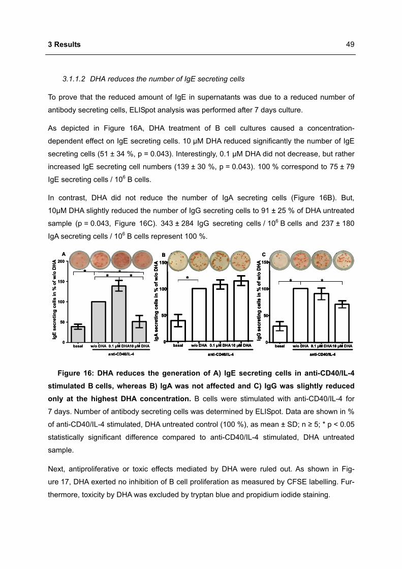
3 Results
49
3.1.1.2 DHA reduces the number of IgE secreting cells
To prove that the reduced amount of IgE in supernatants was due to a reduced number of
antibody secreting cells, ELISpot analysis was performed after 7 days culture.
As depicted in Figure 16A, DHA treatment of B cell cultures caused a concentration-
dependent effect on IgE secreting cells. 10 µM DHA reduced significantly the number of IgE
secreting cells (51 ± 34 %, p = 0.043). Interestingly, 0.1 µM DHA did not decrease, but rather
increased IgE secreting cell numbers (139 ± 30 %, p = 0.043). 100 % correspond to 75 ± 79
IgE secreting cells / 106 B cells.
In contrast, DHA did not reduce the number of IgA secreting cells (Figure 16B). But,
10µM DHA slightly reduced the number of IgG secreting cells to 91 ± 25 % of DHA untreated
sample (p = 0.043, Figure 16C). 343 ± 284 IgG secreting cells / 106 B cells and 237 ± 180
IgA secreting cells / 106 B cells represent 100 %.
C
anti-CD40/IL-4
* *
basal w/o DHA 0.1 µM DHA10 µM DHA0
50
100
150Ig
G s
ecre
ting
cells
in %
of w
/o D
HA
anti-CD40/IL-4
*
basal w/o DHA 0.1 µM DHA10 µM DHA0
50
100
150
IgA
secr
etin
g ce
lls in
% o
f w/o
DH
A
BA
anti-CD40/IL-4
* ***
basal w/o DHA 0.1 µM DHA10 µM DHA0
50
100
150
200
IgE
secr
etin
g ce
lls in
% o
f w/o
DH
A
C
anti-CD40/IL-4
* *
basal w/o DHA 0.1 µM DHA10 µM DHA0
50
100
150Ig
G s
ecre
ting
cells
in %
of w
/o D
HA
C
anti-CD40/IL-4anti-CD40/IL-4
** **
basal w/o DHA 0.1 µM DHA10 µM DHA0
50
100
150Ig
G s
ecre
ting
cells
in %
of w
/o D
HA
anti-CD40/IL-4
*
basal w/o DHA 0.1 µM DHA10 µM DHA0
50
100
150
IgA
secr
etin
g ce
lls in
% o
f w/o
DH
A
B
anti-CD40/IL-4anti-CD40/IL-4
**
basal w/o DHA 0.1 µM DHA10 µM DHA0
50
100
150
IgA
secr
etin
g ce
lls in
% o
f w/o
DH
A
BA
anti-CD40/IL-4anti-CD40/IL-4
* *** * ****
basal w/o DHA 0.1 µM DHA10 µM DHA0
50
100
150
200
IgE
secr
etin
g ce
lls in
% o
f w/o
DH
A
Figure 16: DHA reduces the generation of A) IgE secreting cells in anti-CD40/IL-4 stimulated B cells, whereas B) IgA was not affected and C) IgG was slightly reduced only at the highest DHA concentration. B cells were stimulated with anti-CD40/IL-4 for
7 days. Number of antibody secreting cells was determined by ELISpot. Data are shown in %
of anti-CD40/IL-4 stimulated, DHA untreated control (100 %), as mean ± SD; n ≥ 5; * p < 0.05
statistically significant difference compared to anti-CD40/IL-4 stimulated, DHA untreated
sample.
Next, antiproliferative or toxic effects mediated by DHA were ruled out. As shown in Fig-
ure 17, DHA exerted no inhibition of B cell proliferation as measured by CFSE labelling. Fur-
thermore, toxicity by DHA was excluded by tryptan blue and propidium iodide staining.
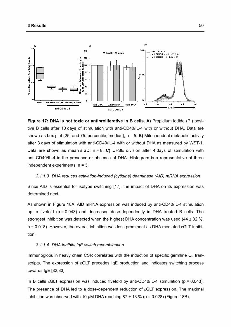
3 Results
50
Figure 17: DHA is not toxic or antiproliferative in B cells. A) Propidium iodide (PI) posi-
tive B cells after 10 days of stimulation with anti-CD40/IL-4 with or without DHA. Data are
shown as box plot (25. and 75. percentile, median); n = 5. B) Mitochondrial metabolic activity
after 3 days of stimulation with anti-CD40/IL-4 with or without DHA as measured by WST-1.
Data are shown as mean ± SD; n = 8. C) CFSE division after 4 days of stimulation with
anti-CD40/IL-4 in the presence or absence of DHA. Histogram is a representative of three
independent experiments; n = 3.
3.1.1.3 DHA reduces activation-induced (cytidine) deaminase (AID) mRNA expression
Since AID is essential for isotype switching [17], the impact of DHA on its expression was
determined next.
As shown in Figure 18A, AID mRNA expression was induced by anti-CD40/IL-4 stimulation
up to fivefold (p = 0.043) and decreased dose-dependently in DHA treated B cells. The
strongest inhibition was detected when the highest DHA concentration was used (44 ± 32 %,
p = 0.018). However, the overall inhibition was less prominent as DHA mediated εGLT inhibi-
tion.
3.1.1.4 DHA inhibits IgE switch recombination
Immunoglobulin heavy chain CSR correlates with the induction of specific germline CH tran-
scripts. The expression of εGLT precedes IgE production and indicates switching process
towards IgE [82,83].
In B cells εGLT expression was induced fivefold by anti-CD40/IL-4 stimulation (p = 0.043).
The presence of DHA led to a dose-dependent reduction of εGLT expression. The maximal
inhibition was observed with 10 µM DHA reaching 87 ± 13 % (p = 0.028) (Figure 18B).

3 Results
51
*
anti-CD40/IL-4
**
*
T0 w/o DHA 0.1µM DHA1µM DHA 10µM DHA0.0
0.5
1.0
1.5
2.0
Rel
ativ
e ex
pres
sion
of ε
GLT
T0 w/o DHA 0.1µM DHA1 µM DHA10 µM DHA0.0
0.5
1.0
1.5
2.0
Rel
ativ
e ex
pres
sion
of A
ID m
RN
A
anti-CD40/IL-4
* **
A B *
anti-CD40/IL-4
**
*
T0 w/o DHA 0.1µM DHA1µM DHA 10µM DHA0.0
0.5
1.0
1.5
2.0
Rel
ativ
e ex
pres
sion
of ε
GLT
*
anti-CD40/IL-4
**
*
T0 w/o DHA 0.1µM DHA1µM DHA 10µM DHA0.0
0.5
1.0
1.5
2.0
Rel
ativ
e ex
pres
sion
of ε
GLT
anti-CD40/IL-4anti-CD40/IL-4
**
*
T0 w/o DHA 0.1µM DHA1µM DHA 10µM DHA0.0
0.5
1.0
1.5
2.0
Rel
ativ
e ex
pres
sion
of ε
GLT
T0 w/o DHA 0.1µM DHA1 µM DHA10 µM DHA0.0
0.5
1.0
1.5
2.0
Rel
ativ
e ex
pres
sion
of A
ID m
RN
A
anti-CD40/IL-4
* **
T0 w/o DHA 0.1µM DHA1 µM DHA10 µM DHA0.0
0.5
1.0
1.5
2.0
Rel
ativ
e ex
pres
sion
of A
ID m
RN
A
anti-CD40/IL-4
* **
A B
Figure 18: DHA inhibits A) AID and B) εGLT expression in anti-CD40/IL-4 stimulated
B cells in a dose-dependent manner. B cells were stimulated with anti-CD40/IL-4 and
treated with DHA. After 4 days of incubation cells were harvested and AID and εGLT mRNA
expression was determined using quantitative RT-PCR. Fold expression of anti-CD40/IL-4
stimulated, DHA untreated control. Data are shown as box plot (25. and 75. percentile, me-
dian); n = 8; * p < 0.05 statistically significant difference compared to anti-CD40/IL-4 stimu-
lated DHA untreated sample.
To delineate whether the IL-4 and / or the CD40 pathway was disrupted by DHA, B cells were
stimulated with anti-CD40 or IL-4 alone. Thereby 10 µM DHA inhibited εGLT expression in-
duced by IL-4 (51 ± 31 %, p = 0.018) as well as by CD40 ligation (52 ± 1 %, p = 0.001) dose-
dependently (data not shown).
3.1.2 DHA modulates IL-4 and anti-CD40 signalling
3.1.2.1 DHA reduces STAT6 phosphorylation
Since εGLT and AID were inhibited by DHA and their promoter regions contain IL-4 depend-
ent STAT6 and CD40 dependent p50 binding sites [5], DHA mediated IgE inhibition might be
attributed to the modulation of CD40 and / or IL-4 signalling.
IL-4 signalling regulates transcription of IL-4 responsible genes through STAT6 phosphoryla-
tion and dimerisation [9]. Phosphorylation of STAT6 (pSTAT6) in the presence of DHA was
measured by flow cytometry.
The basal phosphorylation level of STAT6 was low (mean fluorescence intensity [MFI]
3.0 ± 0.6). Whereas IL-4 stimulation increased the number of pSTAT6 positive cells 50-fold
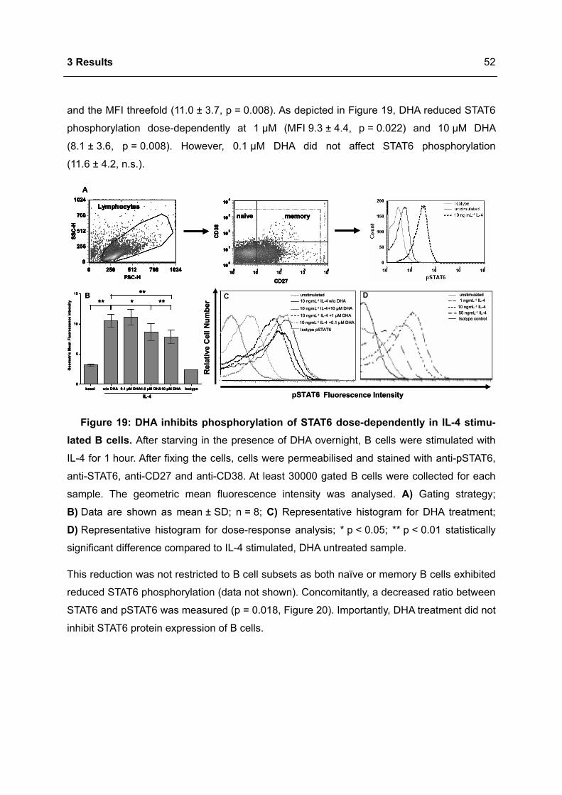
3 Results
52
and the MFI threefold (11.0 ± 3.7, p = 0.008). As depicted in Figure 19, DHA reduced STAT6
phosphorylation dose-dependently at 1 µM (MFI 9.3 ± 4.4, p = 0.022) and 10 µM DHA
(8.1 ± 3.6, p = 0.008). However, 0.1 µM DHA did not affect STAT6 phosphorylation
(11.6 ± 4.2, n.s.).
B
0
5
10
15
Geo
met
ric M
ean
Fluo
resc
ence
Inte
nsity
*******
10 ngmL-1 IL-4 +0.1 µM DHA
unstimulated
10 ngmL-1 IL-4+10 µM DHA
10 ngmL-1 IL-4 +1 µM DHA
10 ngmL-1 IL-4 w/o DHA
Isotype pSTAT6
unstimulated1 ngmL-1 IL-4
10 ngmL-1 IL-450 ngmL-1 IL-4Isotype control
DC
pSTAT6
Rel
ativ
e C
ell N
umbe
r
Fluorescence Intensitybasal w/o DHA 0.1 µM DHA1.0 µM DHA10 µM DHA Isotype
IL-4
A
FSC-H0 256 512 768 1024
SSC
-H
0
256
512
768
1024Lymphocytes
CD27
010 110 210 310 410
CD
38
010
110
210
310
410
naive memory
FSC-H0 256 512 768 1024
SSC
-H
0
256
512
768
1024Lymphocytes
FSC-H0 256 512 768 1024
SSC
-H
0
256
512
768
1024LymphocytesLymphocytes
CD27
010 110 210 310 410
CD
38
010
110
210
310
410
naive memory
B
0
5
10
15
Geo
met
ric M
ean
Fluo
resc
ence
Inte
nsity
*******
0
5
10
15
Geo
met
ric M
ean
Fluo
resc
ence
Inte
nsity
*******
*******
*******
10 ngmL-1 IL-4 +0.1 µM DHA
unstimulated
10 ngmL-1 IL-4+10 µM DHA
10 ngmL-1 IL-4 +1 µM DHA
10 ngmL-1 IL-4 w/o DHA
Isotype pSTAT6
10 ngmL-1 IL-4 +0.1 µM DHA
unstimulated
10 ngmL-1 IL-4+10 µM DHA
10 ngmL-1 IL-4 +1 µM DHA
10 ngmL-1 IL-4 w/o DHA
Isotype pSTAT6
unstimulated1 ngmL-1 IL-4
10 ngmL-1 IL-450 ngmL-1 IL-4Isotype control
unstimulated1 ngmL-1 IL-4
10 ngmL-1 IL-450 ngmL-1 IL-4Isotype control
DC
pSTAT6
Rel
ativ
e C
ell N
umbe
r
Fluorescence Intensitybasal w/o DHA 0.1 µM DHA1.0 µM DHA10 µM DHA Isotype
IL-4basal w/o DHA 0.1 µM DHA1.0 µM DHA10 µM DHA Isotype
IL-4
A
FSC-H0 256 512 768 1024
SSC
-H
0
256
512
768
1024Lymphocytes
CD27
010 110 210 310 410
CD
38
010
110
210
310
410
naive memory
FSC-H0 256 512 768 1024
SSC
-H
0
256
512
768
1024Lymphocytes
FSC-H0 256 512 768 1024
SSC
-H
0
256
512
768
1024LymphocytesLymphocytes
CD27
010 110 210 310 410
CD
38
010
110
210
310
410
naive memory
Figure 19: DHA inhibits phosphorylation of STAT6 dose-dependently in IL-4 stimu-lated B cells. After starving in the presence of DHA overnight, B cells were stimulated with
IL-4 for 1 hour. After fixing the cells, cells were permeabilised and stained with anti-pSTAT6,
anti-STAT6, anti-CD27 and anti-CD38. At least 30000 gated B cells were collected for each
sample. The geometric mean fluorescence intensity was analysed. A) Gating strategy;
B) Data are shown as mean ± SD; n = 8; C) Representative histogram for DHA treatment;
D) Representative histogram for dose-response analysis; * p < 0.05; ** p < 0.01 statistically
significant difference compared to IL-4 stimulated, DHA untreated sample.
This reduction was not restricted to B cell subsets as both naïve or memory B cells exhibited
reduced STAT6 phosphorylation (data not shown). Concomitantly, a decreased ratio between
STAT6 and pSTAT6 was measured (p = 0.018, Figure 20). Importantly, DHA treatment did not
inhibit STAT6 protein expression of B cells.
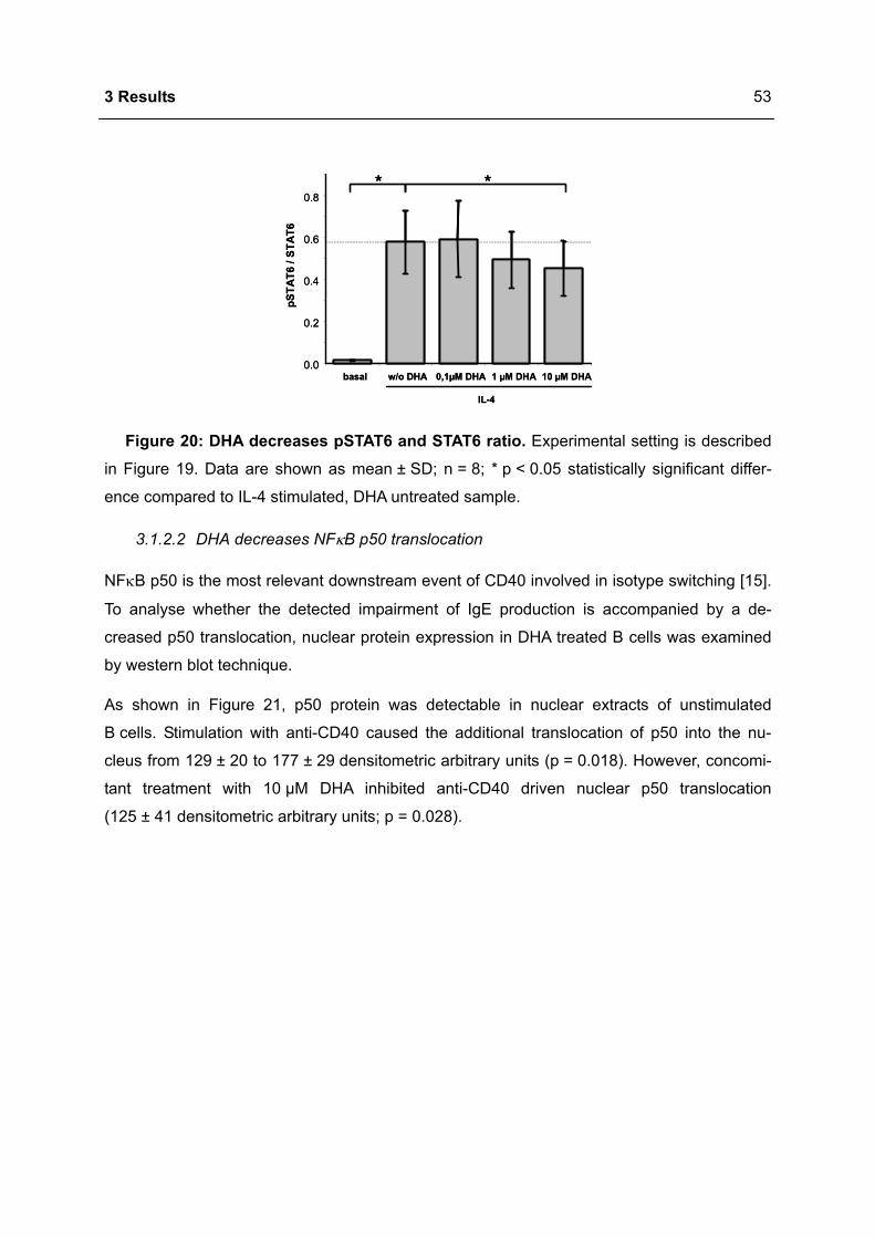
3 Results
53
0.0
0.2
0.4
0.6
0.8
basal w/o DHA 0,1µM DHA 1 µM DHA 10 µM DHA
pSTA
T6 /
STA
T6
* *
IL-4
0.0
0.2
0.4
0.6
0.8
basal w/o DHA 0,1µM DHA 1 µM DHA 10 µM DHA
pSTA
T6 /
STA
T6
** **
IL-4
Figure 20: DHA decreases pSTAT6 and STAT6 ratio. Experimental setting is described
in Figure 19. Data are shown as mean ± SD; n = 8; * p < 0.05 statistically significant differ-
ence compared to IL-4 stimulated, DHA untreated sample.
3.1.2.2 DHA decreases NFκB p50 translocation
NFκB p50 is the most relevant downstream event of CD40 involved in isotype switching [15].
To analyse whether the detected impairment of IgE production is accompanied by a de-
creased p50 translocation, nuclear protein expression in DHA treated B cells was examined
by western blot technique.
As shown in Figure 21, p50 protein was detectable in nuclear extracts of unstimulated
B cells. Stimulation with anti-CD40 caused the additional translocation of p50 into the nu-
cleus from 129 ± 20 to 177 ± 29 densitometric arbitrary units (p = 0.018). However, concomi-
tant treatment with 10 µM DHA inhibited anti-CD40 driven nuclear p50 translocation
(125 ± 41 densitometric arbitrary units; p = 0.028).
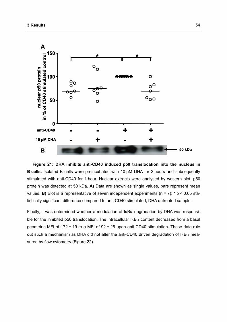
3 Results
54
--
+-
-+
++
10 µM DHA
anti-CD40
50 kDa
**
0
50
100
150
nucl
ear p
50 p
rote
inin
% o
f CD
40 s
timul
ated
con
trol
A
B
--
+-
-+
++
10 µM DHA
anti-CD40
50 kDa
** **
0
50
100
150
nucl
ear p
50 p
rote
inin
% o
f CD
40 s
timul
ated
con
trol
A
B
Figure 21: DHA inhibits anti-CD40 induced p50 translocation into the nucleus in B cells. Isolated B cells were preincubated with 10 µM DHA for 2 hours and subsequently
stimulated with anti-CD40 for 1 hour. Nuclear extracts were analysed by western blot. p50
protein was detected at 50 kDa. A) Data are shown as single values, bars represent mean
values. B) Blot is a representative of seven independent experiments (n = 7); * p < 0.05 sta-
tistically significant difference compared to anti-CD40 stimulated, DHA untreated sample.
Finally, it was determined whether a modulation of IκBα degradation by DHA was responsi-
ble for the inhibited p50 translocation. The intracellular IκBα content decreased from a basal
geometric MFI of 172 ± 19 to a MFI of 92 ± 26 upon anti-CD40 stimulation. These data rule
out such a mechanism as DHA did not alter the anti-CD40 driven degradation of IκBα mea-
sured by flow cytometry (Figure 22).
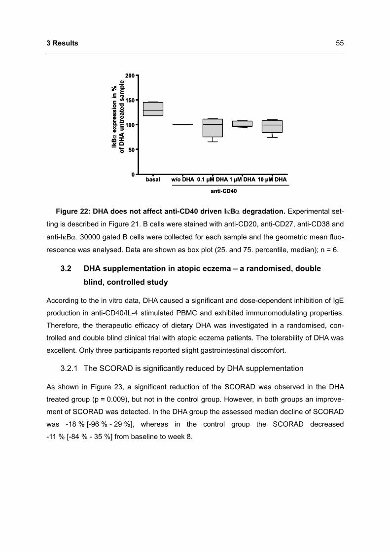
3 Results
55
basal w/o DHA 0.1 µM DHA 1 µM DHA 10 µM DHA0
50
100
150
200
IkB
αex
pres
sion
in %
of D
HA
unt
reat
ed s
ampl
e
anti-CD40
basal w/o DHA 0.1 µM DHA 1 µM DHA 10 µM DHA0
50
100
150
200
IkB
αex
pres
sion
in %
of D
HA
unt
reat
ed s
ampl
e
anti-CD40
Figure 22: DHA does not affect anti-CD40 driven IκBα degradation. Experimental set-
ting is described in Figure 21. B cells were stained with anti-CD20, anti-CD27, anti-CD38 and
anti-IκBα. 30000 gated B cells were collected for each sample and the geometric mean fluo-
rescence was analysed. Data are shown as box plot (25. and 75. percentile, median); n = 6.
3.2 DHA supplementation in atopic eczema – a randomised, double blind, controlled study
According to the in vitro data, DHA caused a significant and dose-dependent inhibition of IgE
production in anti-CD40/IL-4 stimulated PBMC and exhibited immunomodulating properties.
Therefore, the therapeutic efficacy of dietary DHA was investigated in a randomised, con-
trolled and double blind clinical trial with atopic eczema patients. The tolerability of DHA was
excellent. Only three participants reported slight gastrointestinal discomfort.
3.2.1 The SCORAD is significantly reduced by DHA supplementation
As shown in Figure 23, a significant reduction of the SCORAD was observed in the DHA
treated group (p = 0.009), but not in the control group. However, in both groups an improve-
ment of SCORAD was detected. In the DHA group the assessed median decline of SCORAD
was -18 % [-96 % - 29 %], whereas in the control group the SCORAD decreased
-11 % [-84 % - 35 %] from baseline to week 8.
3 Results
56
SCO
RA
D
n.s.
control
p = 0.009
DHA0
25
50
75
week 0 week 8
SCO
RA
D
n.s.
control
p = 0.009
DHA0
25
50
75
week 0 week 8
Figure 23: SCORAD is significantly reduced by dietary DHA. SCORAD was deter-
mined in control (n = 23) and DHA (n = 21) group during the course of supplementation by
the same physician. Data are shown as box plot (25. and 75. percentile, median) [84].
The decreased SCORAD in the DHA group was mainly attributed to the significant reduction
of affected areas from 12 [2 - 41] SCORAD points at baseline to 7 [2 - 25] at week 8
(p = 0.02; data not shown). No change of individuals’ medication usage occurred during sup-
plementation.
3.2.2 DHA inhibits IgE synthesis ex vivo
Before and after supplementation the IgE production of stimulated PBMC was measured in
order to determine the impact of fatty acid supplementation on IgE synthesis. At baseline IgE
production in anti-CD40/IL-4 stimulated PBMC was comparable in both groups and de-
creased only during DHA treatment significantly (week 0: 10.62 ± 9.2 ng/mL; week 8:
3.6 ± 3.1 ng/mL; p = 0.013; Figure 24). By contrast, no impact on ex vivo IgE production was
detected in the control group (week 0: 7.0 ± 5.8 ng/mL; week 8: 5.1 ± 5.6 ng/mL; n.s.). By
checking the proportions of CD14+, CD19+, CD4+, and CD8+ changes of anti-CD40/IL-4 me-
diated IgE synthesis due to an alteration of PBMC composition was excluded. However, the
total IgE concentration measured in sera was not altered in both groups (data not shown).
3 Results
57
week 0 week 8 week 0 week 80
10
20
30
40
DHAControl
IgE
in n
g/m
L
*n.s.
week 0 week 8 week 0 week 80
10
20
30
40
DHAControl
IgE
in n
g/m
L
**n.s.
Figure 24: Ex vivo IgE production is inhibited by dietary DHA. IgE production of
anti-CD40/IL-4 stimulated PBMC before and after supplementation in the control (n = 23) and
DHA group (n = 21). Therefore, isolated PBMC were stimulated with anti-CD40/IL-4 for
10 days. IgE was determined in supernatants by ELISA. Data are shown as box plot (25. and
75. percentile, median); * p < 0.05 statistically significant difference compared to baseline
values.
3.2.3 Fatty acid supplementation modulates activation of monocytes and B cells
In both groups supplementation led to a significant modulation of the activation status of
monocytes (CD14+), but also of B cells (CD19+), as determined by HLA-DR and CD23 ex-
pression, respectively. Thereby, proportion of CD14+HLA-DR+ and CD19+CD23+ decreased
slightly in relation to the relevant cell population up to week 8. After termination of the sup-
plementation period, proportions increased up to initial values (data not shown).
3.2.4 Systemic IFNγ and IL-4 response remains unaffected by fatty acid sup-
plementation
Additionally, the impact of fatty acid supplementation on systemic cytokine production was
investigated. Therefore, untreated or SEB/anti-CD28 stimulated PBMC were analysed re-
garding IFNγ and IL-4 production of activated CD69+ TH cells. In unstimulated PBMC a sig-
nificantly increased percentage of CD4+CD69+IL-4+ was determined whereas the proportion
of CD4+CD69+IFNγ+ decreased at the basal level in both groups during intervention. In con-
trast, the cytokine production was not altered in SEB/anti-CD28 stimulated PBMC during
supplementation. Solely a significant decline of CD4+CD69+IL-4+ was measured in control
group (week 0: 0.31 ± 0.14 %; week 8: 0.21 ± 0.12 %; p = 0.016). However, no significant
differences were detected between both groups (data not shown).
3 Results
58
3.3 Oral administration of DHA inhibits the development of eczema in a murine model of protein induced dermatitis
Immunological impact of oral administration of high dose DHA was investigated in a murine
model of protein induced eczema. DHA containing diet was well consumed and no side ef-
fects were observed. All feeding groups showed a comparable development of body mass,
liver and spleen weight.
3.3.1 Dietary DHA reduces the clinical symptoms of protein induced dermatitis
As shown in Figure 25, OVA patched mice developed local dermatitis with macroscopic clini-
cal symptoms, like dryness, erythema, edema, papules, oozing and crusts. Thereby initial
systemic sensitisation was essential to elicit visible skin lesions in a significant response rate
by e.c. OVA applications [85].
A B C DA B C D
Figure 25: Improved severity of eczema in DHA fed mice. Representative illustration of
protein induced dermatitis in mice fed with A) control diet, B) 2 % DHA and C) 4 % DHA in
the diet. Picture D) illustrates the respective skin area of a non-sensitised control mouse.
As graded by total CSS, e.c. OVA treatment strengthened clinical symptoms from a median
of 4 [2 – 5] points in control patched mice (PBS/alum) up to 12 [5 – 12] points in OVA patched
mice (p = 0.017) (Figure 26). In contrast non-sensitised mice revealed no skin lesions. Fol-
lowing DHA supplementation a significant reduction of CSS was determined compared to
control fed diet. Interestingly, 2 % DHA supplemented diet was most effective to reduce the
CSS to 7 [5 – 8] (p = 0.008), while in 4 % DHA group the outcome was ameliorated to
8 [7 - 10] (p = 0.032).
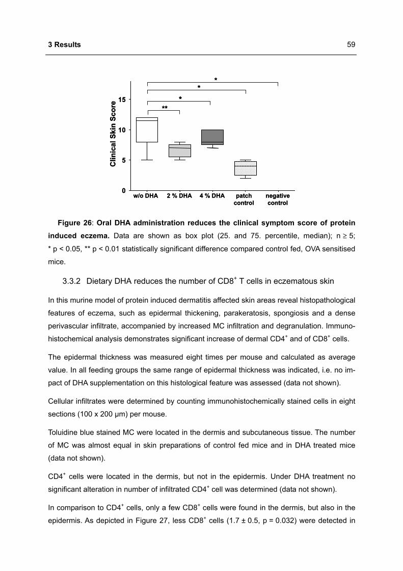
3 Results
59
*
**
w/o DHA 2 % DHA 4 % DHA patch control
negative control
0
5
10
15
Clin
ical
Ski
n Sc
ore
****
****
w/o DHA 2 % DHA 4 % DHA patch control
negative control
0
5
10
15
Clin
ical
Ski
n Sc
ore
****
Figure 26: Oral DHA administration reduces the clinical symptom score of protein induced eczema. Data are shown as box plot (25. and 75. percentile, median); n ≥ 5;
* p < 0.05, ** p < 0.01 statistically significant difference compared control fed, OVA sensitised
mice.
3.3.2 Dietary DHA reduces the number of CD8+ T cells in eczematous skin
In this murine model of protein induced dermatitis affected skin areas reveal histopathological
features of eczema, such as epidermal thickening, parakeratosis, spongiosis and a dense
perivascular infiltrate, accompanied by increased MC infiltration and degranulation. Immuno-
histochemical analysis demonstrates significant increase of dermal CD4+ and of CD8+ cells.
The epidermal thickness was measured eight times per mouse and calculated as average
value. In all feeding groups the same range of epidermal thickness was indicated, i.e. no im-
pact of DHA supplementation on this histological feature was assessed (data not shown).
Cellular infiltrates were determined by counting immunohistochemically stained cells in eight
sections (100 x 200 µm) per mouse.
Toluidine blue stained MC were located in the dermis and subcutaneous tissue. The number
of MC was almost equal in skin preparations of control fed mice and in DHA treated mice
(data not shown).
CD4+ cells were located in the dermis, but not in the epidermis. Under DHA treatment no
significant alteration in number of infiltrated CD4+ cell was determined (data not shown).
In comparison to CD4+ cells, only a few CD8+ cells were found in the dermis, but also in the
epidermis. As depicted in Figure 27, less CD8+ cells (1.7 ± 0.5, p = 0.032) were detected in

3 Results
60
dermis of 2 % DHA supplemented mice compared to sections of control fed mice (3.9 ± 2.1).
Additionally, 4 % DHA supplementation tended to result in a reduced dermal CD8+ number
(1.5 ± 1.0, n.s.), but failed to reach significance.
B
D
A
C
*
w/o DHA 2 % DHA 4 % DHA patch control
negative control
0
2
4
6
8
CD
8+ce
lls [c
ells
/100
x 2
00 µ
m] **
**E
B
D
A
C
*
w/o DHA 2 % DHA 4 % DHA patch control
negative control
0
2
4
6
8
CD
8+ce
lls [c
ells
/100
x 2
00 µ
m] **
**E
**
w/o DHA 2 % DHA 4 % DHA patch control
negative control
0
2
4
6
8
CD
8+ce
lls [c
ells
/100
x 2
00 µ
m] ****
****E
Figure 27: Oral DHA administration reduces the number of CD8+ T cells in eczema-tous skin. Representative histological image of anti-CD8+ stained 5 µm sections of frozen
skin from A) a PBS patched BALB/c mouse, from mice with protein induced dermatitis fed
with B) control diet, C) 2 % DHA and D) 4 % DHA in the diet. E) Positive immunohistochemi-
cal stained cells were counted in 100 µm x 200 µm areas at x100 magnification using digital
measuring tools. Data are depicted as box plot (25. and 75. percentile, median); n ≥ 5;
* p < 0.05, ** p < 0.01 statistically significant difference compared control fed, OVA sensitised
mice.
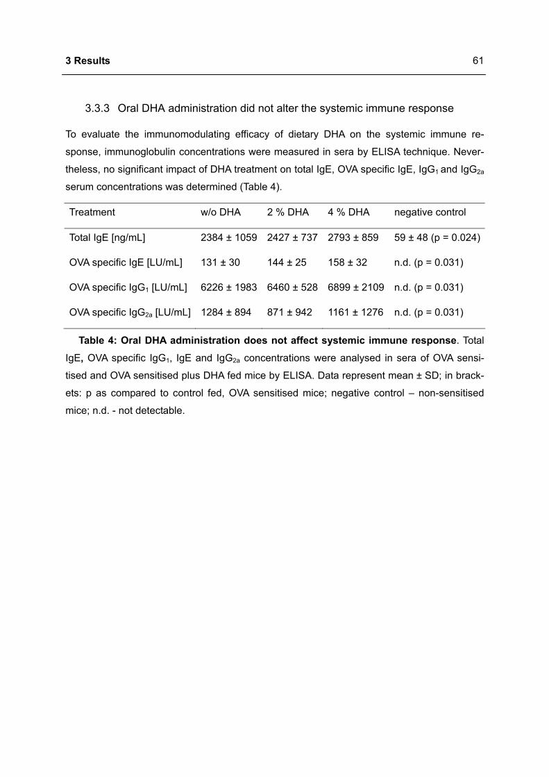
3 Results
61
3.3.3 Oral DHA administration did not alter the systemic immune response
To evaluate the immunomodulating efficacy of dietary DHA on the systemic immune re-
sponse, immunoglobulin concentrations were measured in sera by ELISA technique. Never-
theless, no significant impact of DHA treatment on total IgE, OVA specific IgE, IgG1 and IgG2a
serum concentrations was determined (Table 4).
Treatment w/o DHA 2 % DHA 4 % DHA negative control
Total IgE [ng/mL] 2384 ± 1059 2427 ± 737 2793 ± 859 59 ± 48 (p = 0.024)
OVA specific IgE [LU/mL] 131 ± 30 144 ± 25 158 ± 32 n.d. (p = 0.031)
OVA specific IgG1 [LU/mL] 6226 ± 1983 6460 ± 528 6899 ± 2109 n.d. (p = 0.031)
OVA specific IgG2a [LU/mL] 1284 ± 894 871 ± 942 1161 ± 1276 n.d. (p = 0.031)
Table 4: Oral DHA administration does not affect systemic immune response. Total
IgE, OVA specific IgG1, IgE and IgG2a concentrations were analysed in sera of OVA sensi-
tised and OVA sensitised plus DHA fed mice by ELISA. Data represent mean ± SD; in brack-
ets: p as compared to control fed, OVA sensitised mice; negative control – non-sensitised
mice; n.d. - not detectable.
4 Discussion
62
4 Discussion
The incidence of common allergic diseases has increased quite dramatically during the last
decades and in many countries the prevalence has reached almost epidemic proportions [1].
At this time a cure for these diseases does not really exist, although a wide range of treat-
ments are used to control the symptoms. The costs are manifold including both, direct and
indirect components [23]. Thus, allergies have become a major medical issue of the Western
world. Since there is an indication that the nutritional factors, especially the changing dietary
fatty acids may contribute to this alarming development, many investigations have been done
in this field [74,75].
The first evidence for relevance of dietary n-3 PUFA in inflammation was derived from epi-
demiological observations displaying a low incidence of autoimmune and inflammatory disor-
ders in a population of Greenland Inuit compared with gender and age matched groups living
in Denmark [86]. Most of these diseases are designated by inappropriate activation of T cells
and destruction of tissues. Interestingly, native Greenland Inuit [87] and Japanese [88] con-
sume high amounts of long chain n-3 PUFA from seafood and have a low incidence of
chronic inflammatory or autoimmune disorders, even compared to their Westernised ethnic
counterparts.
It is very likely that DHA and EPA are responsible for the beneficial effects of fish oils on the
immune system [53,89]. Until now the majority of studies dealing with the immunomodulatory
effects of long chain n-3 PUFA have been dealing with fish oils, what limits the understanding
of the particular effects of EPA and DHA and prohibits their comparison. There are still con-
troversial results regarding which n-3 PUFA is accountable for the effects of fish oil
[90,91,92]. Thereby, DHA exerts a lot of favourable effects in many diseases and is known to
be antiinflammatory. However, some researchers assume that DHA is the biologically active
component of fish oil [93].
4.1 DHA inhibits IgE production in human B cells
Allergic individuals exposed to minute amounts of allergen experience an immediate immune
response with possibly a fatal outcome. The spreading epidemic of allergies and atopic di-
seases has generated a great interest in IgE, the central molecule of type I allergic reactions.
IgE is usually found at low concentrations in the plasma and mainly secreted by plasma cells
in mucosa associated lymphoid tissue. Patients suffering from atopic disorders, like asthma,
allergic rhinitis and extrinsic atopic eczema have elevated serum levels of both, total IgE and
allergen specific IgE. The differentiation of a B cell to an IgE producing cell is tightly adjusted
4 Discussion
63
via CSR [5,94]. Consequently, a potential target for prevention and treatment of atopic
diseases by DHA is the regulation of IgE production, in particullar the switch towards IgE.
To induce IgE synthesis in vitro, cells were stimulated with anti-CD40 and IL-4 [80]. Applica-
tion of DHA resulted in a dose-dependent inhibition of IgE production in both, PBMC and
B cells. This effect was isotype specific since both, IgG and IgA remained unchanged. Toxic
or antiproliferative effects were excluded using B cells labelled with CFSE and WST-1 assay
as well as by staining with tryptan blue and propidium iodide for identification of dead cells.
Furthermore, it was investigated if the reduced amount of IgE in supernatants was due to
inhibition of immunoglobulin secretion, altered differentiation into IgE secreting plasma cells
or a result of impaired switching towards IgE.
DHA dose-dependently inhibited the anti-CD40/IL-4 driven differentiation to IgE secreting
cells. However, the number of IgA secreting cells was not altered, excluding the possibility of
a general inhibited immunoglobulin secretion and indicating IgE specific alteration of plasma
cell differentiation.
It was determined that DHA specifically interfered with the IgE switching process at the tran-
scriptional level of AID and εGLT. The importance of AID in CSR got obvious after rigorous
impairment in AID-/- mice [95] as well as in patients with autosomal recessive hyper-IgM syn-
drome (HIGM2), who carry mutations in the AID gene [96]. However, DHA inhibited AID
mRNA expression dose-dependently suggesting interference in AID dependent switch re-
combination. Interestingly, IgA and IgG remained almost unchanged regarding production
and number of antibody secreting cells indicating that the dimension of DHA mediated AID
inhibition was not sufficient to affect switch recombination in general.
CSR towards IgE in B cells stimulated with anti-CD40/IL-4 involves a number of molecular
events [97]. The transcription of εGLT precedes B cell IgE synthesis and the dependency of
switching to IgE on its expression has been reported [83]. Consequently, the dose-dependent
inhibition of εGLT points to the IgE specific action of DHA. Thereby, DHA caused this obvious
inhibition of IgE by reducing εGLT transcription together with decreasing expression of AID.
B cell IgE production is regulated by cytokines like IL-4 [98] as well as direct contact between
B and T cells [99], especially by CD40/CD154 interaction [80]. Importantly, AID [100] and
εGLT [101] are dependent on IL-4 and CD40, whose downstream signalling is mediated
through STAT6 and NFκB. Since DHA downregulated εGLT expression in B cells stimulated
4 Discussion
64
with anti-CD40 or IL-4 alone, the involved signalling pathways were examined by analysis of
STAT6 phosphorylation and NFκB p50 translocation into nucleus.
Preincubation of B cells with DHA caused a dose-dependent inhibition of STAT6 phosphory-
lation. The analysis of the total STAT6 protein revealed no impact of DHA, but a decreased
pSTAT6/STAT6 ratio. These data exclude the decrease of STAT6 protein expression by over-
night incubation with DHA and point to its exclusive direct interference in IL-4 signal transduc-
tion. These data are in line with previous reports by Gorjao et al. [102]. They detected a de-
creased IL-2 mediated JAK1 and JAK3 phosphorylation and consequently diminished STAT5
phosphorylation through DHA. This was associated with a reduced surface expression of the
IL-2 receptor resulting from an altered lipid raft organisation.
Other studies confirmed that DHA has properties to interfere with lipid raft and membrane
domains [62,68]. Stillwell et al. [103] recently described that dietary consumption of DHA cre-
ates DHA-rich membrane domains which lead to dislocation of sterol into sphingomyelin-rich
lipid raft domains. In these environments the acyl chain order is extremely different. Thereby,
protein movement between the domains is associated with a change of conformation and
consequently has the potential to modulate cellular signalling. It has also been reported that
DHA treatment causes stimulation of PLD1 activity in human PBMC by partial disorganisation
of membrane microdomains, which results in activating the protein and disrupting signal
transduction events [104]. Such mechanisms may apply for DHA action on STAT6 phos-
phorylation. This assumption is supported by the fact that DHA preincubation was necessary
to reduce the IL-4 driven STAT6 phosphorylation possibly via downregulation of IL-4R ex-
pression.
Even though, IL-4 plays a key role in the production of IgE by B cells [105] and provokes
εGLT, IL-4 alone is not sufficient to induce IgE production. However, additionally to IL-4,
CD40 signalling is indispensable for induction of the mature ε transcripts and consequent IgE
synthesis [80,106,107].
CD40 ligation, along with the other B cell stimuli, results in activation of transcription factors
belonging to the Rel/NFκB family [13,14]. It is reported that genes involved in isotype swit-
ching like AID are regulated by type 1 but not type 2 NFκB activity [15]. Transcriptional regu-
lation by type 1 NFκB involves activation of IKK complex leading to degradation of IκBα and
IκBβ and as a result release NFκB p50:p65 and p50:cRel dimers [108]. Additionally, mice
lacking p50 show a clear reduction of IgE indicating the central role of p50 in IgE production
[109]. Therefore, the impact of DHA on degradation of IκBα and nuclear translocation of p50
4 Discussion
65
was investigated thoroughly. DHA reduced the anti-CD40 driven translocation of p50 into the
nucleus, thereby affecting the second essential signal for IgE switch, IgE plasma cell devel-
opment and IgE production. These data are in line with previous reports showing that DHA
inhibits NFκB activation [110,111]. Interestingly, DHA did not affect IκBα degradation which is
an important upstream event for p50 release. However, NFκB can dissociate from phos-
phorylated IκBα and move into the nucleus without IκBα degradation, indicating that a re-
duced p50 translocation is not necessarily related to reduced IκB degradation [112].
Another essential step for p50 translocation is its generation by processing from the precur-
sor p105. This sequence is initiated by ubiquitinylation, followed by gradual proteasomal deg-
radation of the p105 from the C-terminal end [15]. DHA acts probably via modulation of ubiq-
uitinylation [15] or alteration of proteasomal activity [113], consequently resulting in altered
p50 translocation and thus modulated transcriptional activity.
DHA inhibited the anti-CD40/IL-4 driven IgE production in PBMC of healthy individuals and
allergic donors with lower serum IgE, but had no impact on PBMC of atopic patients with high
serum IgE. Previous reports show that IgE hyperproduction in patients with atopic eczema is
determined by constitutive and enhanced JAK3 phosphorylation in B cells [114]. This could
explain the missing effect of DHA in patients with high serum IgE. The increased inherited
JAK3 activation prevents the modulation of the IL-4 signalling pathway by DHA.
DHA leads to a profound repression of the IgE switching process and thus IgE plasma cell
development as well as IgE production in human B cells. This is caused by an early inhibition
of CD40 and IL-4 signalling pathways resulting from a simultaneously decreased STAT6 and
NFκB activation [115]. Therefore, the slight reduction of IgG secreting cells after treatment
with 10 µM DHA may result from this concomitant inhibition, since CD40 ligation and IL-4
induce not only IgE, but also IgG4 [107,116]. Hereby, DHA acted selectively on IgE isotype by
means of immune modulation rather than suppression. These results comprise a strong bio-
logical relevance for the development of allergies, since it has been shown that the DHA con-
centrations used in this study correspond to serum levels found in humans [117]. More
importantly, upon high dose DHA supplementation in patients with extrinsic atopic eczema
the inhibition the anti-CD40/IL-4 driven IgE production in allergic PBMC can be also seen ex
vivo [84].
In terms of IgE production and its related signal transduction, these data reveal the early in-
terference of DHA in a possibly longer chain of events initiated by DHA. It cannot be ex-
cluded that DHA causes additional effects on IgE producing B cells later on. Thereby, modu-
4 Discussion
66
lation of eicosanoid synthesis, nuclear receptor binding and activation / repression, regulation
of gene expression, alteration of cytokine production etc. may be potential effects provoked
by DHA. Certainly, these observations might be important for dietary management of IgE
mediated diseases and may offer an opportunity for a successful prevention of allergic dis-
eases.
4.2 DHA supplementation in atopic eczema – a randomised, double blind, controlled study
Several clinical trials have been conducted to ameliorate symptoms of atopic eczema by
supplementation with n-6 PUFA. Based on the data generated so far, there is a lack of evi-
dence supporting the use of n-6 fatty acid supplementation on improvement the clinical out-
come of atopic eczema [30]. Due to its known antiinflammatory effects, some clinical trials
have been also focused on fish oil supplementation [118,119]. Particularly, the beneficial effi-
ciency of DHA supplementation in vivo has been previously suggested by several studies
[63].
The assumption that DHA may be therapeutically effective in atopic eczema is based on the
prerequisites that atopic eczema is an inflammatory disease which is amplified by an exces-
sive production of proinflammatory eicosanoids [120,121,122] and is associated with a ab-
normal fatty acid metabolism [28], that DHA supplementation acts antiinflammatorily (among
others) by modulating eicosanoid generation [53] and there is a correlation between intake
and a decreased risk of atopic eczema [123,124]. However, there were no accessible reports
on clinical trial with dietary highly purified DHA in atopic eczema. Accordingly, a randomised,
double blind, controlled trial was performed to evaluate the therapeutical impact of 8 week
supplementation with 5.4 g of highly purified DHA daily in this atopic skin disorder. Thereby,
DHA, but not the control treatment resulted in a significant clinical improvement of atopic ec-
zema in terms of a decreased SCORAD. A significant reduction of anti-CD40/IL-4 mediated
IgE synthesis of PBMC was detected in DHA group only. Concomitantly, DHA treated patients
showed an increase of plasma n-3 PUFA and a decrease of n-6/n-3 PUFA ratio. Finally, sup-
plementation led to a modulated activation status of PBMC in both groups [84]. However,
there was no significant difference between DHA and control group. Thus, the possibility of a
placebo effect cannot be excluded, despite the fact that DHA and control capsules were in-
distinguishable. Furthermore, the small study size limited the statistical power of these com-
parisons. Due to the lack of reported data showing the clinical efficacy of dietary DHA in
atopic eczema the exact calculation of the necessary number of patients prior to this pilot
study was not possible. Therefore, this exploratory trial with an arbitrary sample size was
4 Discussion
67
performed. This limitation might have led to the possibility that randomisation was not likely to
ensure the desired prognostic comparability between DHA and control group. However, both
treated groups were well matched for gender, age and BMI and the study was double
blinded. To determine whether the observed clinical effect of dietary DHA is of therapeutical
significance needs further clarification and confirmation.
As already demonstrated in former studies [125], an increased plasma n-3 PUFA proportion
was detected in all patients consuming DHA [84]. Due to DHA treatment plasma fraction of
DHA and EPA increased two- to threefold indicating an excellent compliance. The elevated
EPA plasma level may be explained either by retroconversion of DHA to EPA when used in
high doses [126] or the amount of EPA (0.4 g/d) contained in the used capsules or both. The
detected clinical efficacy in therapy of atopic eczema points to antiinflammatory properties of
DHA. Metabolism via COX and LOX pathways is highly dependent on the availability of lipid
precursors [127,128]. Indeed, DHA does not directly act as a substrate for eicosanoid syn-
thesising enzymes, but inhibits AA metabolism [66] and serves as a precursor of n-3 eico-
sanoids via EPA [126]. DHA is known to reduce PGE2 production [66] and expression of PG
synthesising enzyme COX-2 [68]. Accordingly, a favourable shift in eicosanoid formation may
be responsible for several effects of DHA supplementation. In addition, DHA, possibly in
combination with EPA, can act as precursor for COX and LOX driven bioactive lipids such as
lipoxins, protectins and resolvins which are known to be antiinflammatory [129]. Importantly,
an alteration of PUFA level following allergic sensitisation [130] as well as an involvement of
n-6 PUFA derived mediators in the pathophysiology of atopic eczema is suggested since
patients have an enhanced synthesis of these metabolites and thus increased concentrations
of AA derived LT and PG in skin lesions [120,121]. Mayser et al. [32] demonstrated that daily
infusions of n-3 PUFA effectively ameliorate the clinical outcome of atopic eczema. This
therapeutical effect was accompanied by alterations in fatty acid profile of membrane lipid
pool as well as changes in eicosanoid synthesis. Following the rise of free EPA an increase
of n-3 PUFA derived metabolites was observed, which may also be associated with the clini-
cal improvement. Furthermore, it is speculated that a combination with dietary supplements
is capable to enhance this therapeutical effect. However, patients of the DHA group showed
a strong reduction of plasma n-6/n-3 PUFA ratio and a significant clinical improvement. It is
likely that the fatty acid pattern in the skin was also altered in a similar way, since it has been
shown that dietary n-3 PUFA supplementation is associated with their increased incorpora-
tion into skin [131]. EPA and DHA play an important role for stratum corneum and are there-
fore essential for maintaining a normal function and structure of skin barrier [132]. Addition-
ally, it is discussed that components of the lipid metabolism pathway are involved in

4 Discussion
68
pathogenesis of atopic eczema and represent an opportunity to manage and perhaps even
prevent subsequent atopic disorders through restorement of the barrier [133,134]. Willemsen
et al. have recently shown that DHA effectively supports skin barrier integrity by improving
resistance and permeability [135]. Lipid infusions may result in sufficient availability of fatty
acids in skin. Whether this can also be achieved by oral supplementation needs to be inves-
tigated in further investigations.
Extrinsic atopic eczema is associated with an unbalanced cytokine pattern promoting an IgE
response to common environmental allergens [24]. In this study a reduced ex vivo
anti-CD40/IL-4 mediated IgE synthesis by DHA supplementation was determined [84]. These
data are in line with the B cell in vitro results showing the DHA mediated inhibition of
anti-CD40/IL-4 driven IgE production comprising a strong biological relevance regarding the
development of allergies.
Since a change of total fat intake influences several immunological parameters [118] the con-
trol supplement contained the same amount of fat as the DHA capsules. Short to middle
chained saturated fatty acids exhibit a low biological efficacy [136]. Hence, the control cap-
sules supplied the middle chained fatty acids, caprylic and capric acid. Therefore, the effects
caused by an increased amount of dietary fat were intended to be eliminated.
Analysis of the systemic IL-4 and IFNγ response revealed no impact by fatty acid supplemen-
tation suggesting that DHA acts rather locally than on systemic level. The determined non-
specific increased IL-4 with a concomitant decreased IFNγ may be attributed to seasonal
influences resulting from natural exposure to allergens in spring [137]. However, the reduced
IL-4 levels in control group suggest a potential immunological activity of the control supple-
ment. This is supported by the finding, that both, DHA and control supplementation led to
significantly lower HLA-DR surface expression on monocytes and CD23 expression on B
cells. This fact points to a requirement of a three-armed, placebo controlled study in order to
compare DHA, an isoenergetic fatty acid control supplement and a placebo without any fat.
Nevertheless, the unchanged anti-CD40/IL-4 stimulated IgE production within the control
group suggests DHA specific pathways.
In conclusion, although the sample size may be underpowered to detect small but relevant
effects, the clinical results imply that dietary DHA may be beneficial for the standard treat-
ment of atopic eczema. It may not be excluded that different doses of DHA and long term
treatment are more effective in atopic eczema. To deal with this subject, an extensive ran-
domised, placebo controlled, three-armed follow up trial should be performed.
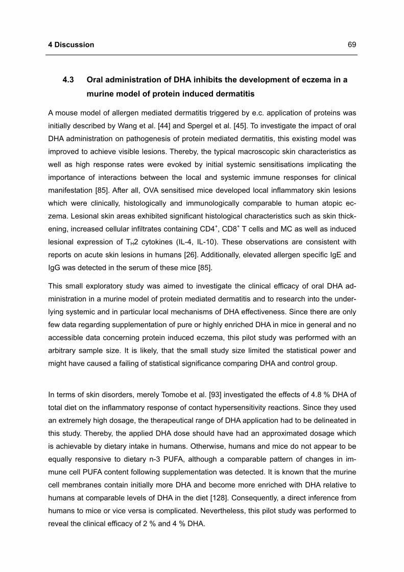
4 Discussion
69
4.3 Oral administration of DHA inhibits the development of eczema in a murine model of protein induced dermatitis
A mouse model of allergen mediated dermatitis triggered by e.c. application of proteins was
initially described by Wang et al. [44] and Spergel et al. [45]. To investigate the impact of oral
DHA administration on pathogenesis of protein mediated dermatitis, this existing model was
improved to achieve visible lesions. Thereby, the typical macroscopic skin characteristics as
well as high response rates were evoked by initial systemic sensitisations implicating the
importance of interactions between the local and systemic immune responses for clinical
manifestation [85]. After all, OVA sensitised mice developed local inflammatory skin lesions
which were clinically, histologically and immunologically comparable to human atopic ec-
zema. Lesional skin areas exhibited significant histological characteristics such as skin thick-
ening, increased cellular infiltrates containing CD4+, CD8+ T cells and MC as well as induced
lesional expression of TH2 cytokines (IL-4, IL-10). These observations are consistent with
reports on acute skin lesions in humans [26]. Additionally, elevated allergen specific IgE and
IgG was detected in the serum of these mice [85].
This small exploratory study was aimed to investigate the clinical efficacy of oral DHA ad-
ministration in a murine model of protein mediated dermatitis and to research into the under-
lying systemic and in particular local mechanisms of DHA effectiveness. Since there are only
few data regarding supplementation of pure or highly enriched DHA in mice in general and no
accessible data concerning protein induced eczema, this pilot study was performed with an
arbitrary sample size. It is likely, that the small study size limited the statistical power and
might have caused a failing of statistical significance comparing DHA and control group.
In terms of skin disorders, merely Tomobe et al. [93] investigated the effects of 4.8 % DHA of
total diet on the inflammatory response of contact hypersensitivity reactions. Since they used
an extremely high dosage, the therapeutical range of DHA application had to be delineated in
this study. Thereby, the applied DHA dose should have had an approximated dosage which
is achievable by dietary intake in humans. Otherwise, humans and mice do not appear to be
equally responsive to dietary n-3 PUFA, although a comparable pattern of changes in im-
mune cell PUFA content following supplementation was detected. It is known that the murine
cell membranes contain initially more DHA and become more enriched with DHA relative to
humans at comparable levels of DHA in the diet [128]. Consequently, a direct inference from
humans to mice or vice versa is complicated. Nevertheless, this pilot study was performed to
reveal the clinical efficacy of 2 % and 4 % DHA.
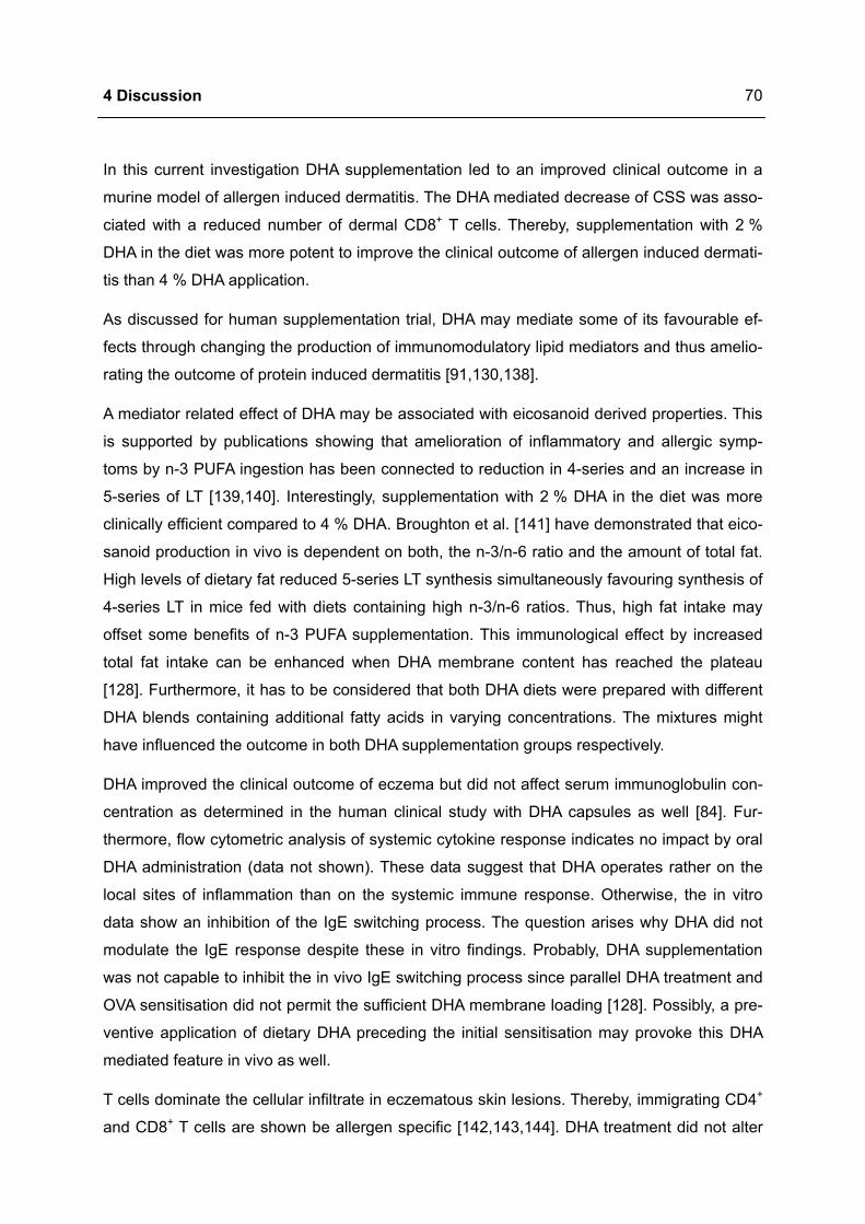
4 Discussion
70
In this current investigation DHA supplementation led to an improved clinical outcome in a
murine model of allergen induced dermatitis. The DHA mediated decrease of CSS was asso-
ciated with a reduced number of dermal CD8+ T cells. Thereby, supplementation with 2 %
DHA in the diet was more potent to improve the clinical outcome of allergen induced dermati-
tis than 4 % DHA application.
As discussed for human supplementation trial, DHA may mediate some of its favourable ef-
fects through changing the production of immunomodulatory lipid mediators and thus amelio-
rating the outcome of protein induced dermatitis [91,130,138].
A mediator related effect of DHA may be associated with eicosanoid derived properties. This
is supported by publications showing that amelioration of inflammatory and allergic symp-
toms by n-3 PUFA ingestion has been connected to reduction in 4-series and an increase in
5-series of LT [139,140]. Interestingly, supplementation with 2 % DHA in the diet was more
clinically efficient compared to 4 % DHA. Broughton et al. [141] have demonstrated that eico-
sanoid production in vivo is dependent on both, the n-3/n-6 ratio and the amount of total fat.
High levels of dietary fat reduced 5-series LT synthesis simultaneously favouring synthesis of
4-series LT in mice fed with diets containing high n-3/n-6 ratios. Thus, high fat intake may
offset some benefits of n-3 PUFA supplementation. This immunological effect by increased
total fat intake can be enhanced when DHA membrane content has reached the plateau
[128]. Furthermore, it has to be considered that both DHA diets were prepared with different
DHA blends containing additional fatty acids in varying concentrations. The mixtures might
have influenced the outcome in both DHA supplementation groups respectively.
DHA improved the clinical outcome of eczema but did not affect serum immunoglobulin con-
centration as determined in the human clinical study with DHA capsules as well [84]. Fur-
thermore, flow cytometric analysis of systemic cytokine response indicates no impact by oral
DHA administration (data not shown). These data suggest that DHA operates rather on the
local sites of inflammation than on the systemic immune response. Otherwise, the in vitro
data show an inhibition of the IgE switching process. The question arises why DHA did not
modulate the IgE response despite these in vitro findings. Probably, DHA supplementation
was not capable to inhibit the in vivo IgE switching process since parallel DHA treatment and
OVA sensitisation did not permit the sufficient DHA membrane loading [128]. Possibly, a pre-
ventive application of dietary DHA preceding the initial sensitisation may provoke this DHA
mediated feature in vivo as well.
T cells dominate the cellular infiltrate in eczematous skin lesions. Thereby, immigrating CD4+
and CD8+ T cells are shown be allergen specific [142,143,144]. DHA treatment did not alter
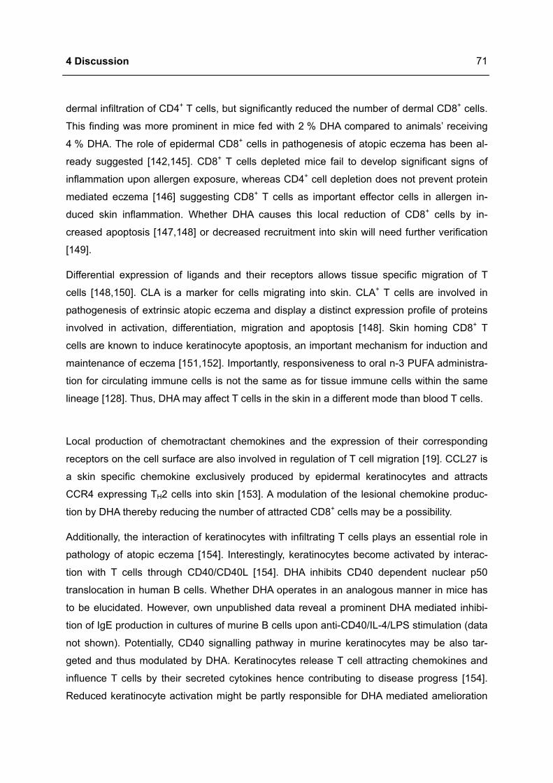
4 Discussion
71
dermal infiltration of CD4+ T cells, but significantly reduced the number of dermal CD8+ cells.
This finding was more prominent in mice fed with 2 % DHA compared to animals’ receiving
4 % DHA. The role of epidermal CD8+ cells in pathogenesis of atopic eczema has been al-
ready suggested [142,145]. CD8+ T cells depleted mice fail to develop significant signs of
inflammation upon allergen exposure, whereas CD4+ cell depletion does not prevent protein
mediated eczema [146] suggesting CD8+ T cells as important effector cells in allergen in-
duced skin inflammation. Whether DHA causes this local reduction of CD8+ cells by in-
creased apoptosis [147,148] or decreased recruitment into skin will need further verification
[149].
Differential expression of ligands and their receptors allows tissue specific migration of T
cells [148,150]. CLA is a marker for cells migrating into skin. CLA+ T cells are involved in
pathogenesis of extrinsic atopic eczema and display a distinct expression profile of proteins
involved in activation, differentiation, migration and apoptosis [148]. Skin homing CD8+ T
cells are known to induce keratinocyte apoptosis, an important mechanism for induction and
maintenance of eczema [151,152]. Importantly, responsiveness to oral n-3 PUFA administra-
tion for circulating immune cells is not the same as for tissue immune cells within the same
lineage [128]. Thus, DHA may affect T cells in the skin in a different mode than blood T cells.
Local production of chemotractant chemokines and the expression of their corresponding
receptors on the cell surface are also involved in regulation of T cell migration [19]. CCL27 is
a skin specific chemokine exclusively produced by epidermal keratinocytes and attracts
CCR4 expressing TH2 cells into skin [153]. A modulation of the lesional chemokine produc-
tion by DHA thereby reducing the number of attracted CD8+ cells may be a possibility.
Additionally, the interaction of keratinocytes with infiltrating T cells plays an essential role in
pathology of atopic eczema [154]. Interestingly, keratinocytes become activated by interac-
tion with T cells through CD40/CD40L [154]. DHA inhibits CD40 dependent nuclear p50
translocation in human B cells. Whether DHA operates in an analogous manner in mice has
to be elucidated. However, own unpublished data reveal a prominent DHA mediated inhibi-
tion of IgE production in cultures of murine B cells upon anti-CD40/IL-4/LPS stimulation (data
not shown). Potentially, CD40 signalling pathway in murine keratinocytes may be also tar-
geted and thus modulated by DHA. Keratinocytes release T cell attracting chemokines and
influence T cells by their secreted cytokines hence contributing to disease progress [154].
Reduced keratinocyte activation might be partly responsible for DHA mediated amelioration
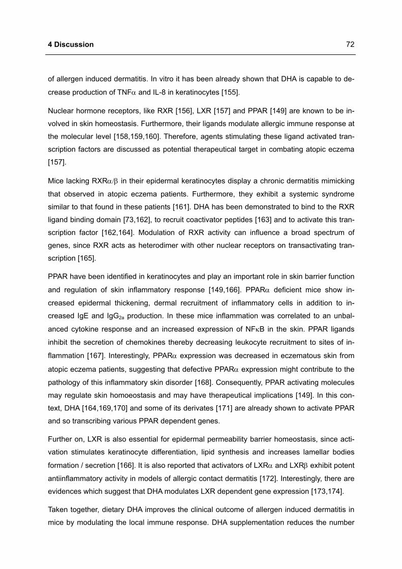
4 Discussion
72
of allergen induced dermatitis. In vitro it has been already shown that DHA is capable to de-
crease production of TNFα and IL-8 in keratinocytes [155].
Nuclear hormone receptors, like RXR [156], LXR [157] and PPAR [149] are known to be in-
volved in skin homeostasis. Furthermore, their ligands modulate allergic immune response at
the molecular level [158,159,160]. Therefore, agents stimulating these ligand activated tran-
scription factors are discussed as potential therapeutical target in combating atopic eczema
[157].
Mice lacking RXRα/β in their epidermal keratinocytes display a chronic dermatitis mimicking
that observed in atopic eczema patients. Furthermore, they exhibit a systemic syndrome
similar to that found in these patients [161]. DHA has been demonstrated to bind to the RXR
ligand binding domain [73,162], to recruit coactivator peptides [163] and to activate this tran-
scription factor [162,164]. Modulation of RXR activity can influence a broad spectrum of
genes, since RXR acts as heterodimer with other nuclear receptors on transactivating tran-
scription [165].
PPAR have been identified in keratinocytes and play an important role in skin barrier function
and regulation of skin inflammatory response [149,166]. PPARα deficient mice show in-
creased epidermal thickening, dermal recruitment of inflammatory cells in addition to in-
creased IgE and IgG2a production. In these mice inflammation was correlated to an unbal-
anced cytokine response and an increased expression of NFκB in the skin. PPAR ligands
inhibit the secretion of chemokines thereby decreasing leukocyte recruitment to sites of in-
flammation [167]. Interestingly, PPARα expression was decreased in eczematous skin from
atopic eczema patients, suggesting that defective PPARα expression might contribute to the
pathology of this inflammatory skin disorder [168]. Consequently, PPAR activating molecules
may regulate skin homoeostasis and may have therapeutical implications [149]. In this con-
text, DHA [164,169,170] and some of its derivates [171] are already shown to activate PPAR
and so transcribing various PPAR dependent genes.
Further on, LXR is also essential for epidermal permeability barrier homeostasis, since acti-
vation stimulates keratinocyte differentiation, lipid synthesis and increases lamellar bodies
formation / secretion [166]. It is also reported that activators of LXRα and LXRβ exhibit potent
antiinflammatory activity in models of allergic contact dermatitis [172]. Interestingly, there are
evidences which suggest that DHA modulates LXR dependent gene expression [173,174].
Taken together, dietary DHA improves the clinical outcome of allergen induced dermatitis in
mice by modulating the local immune response. DHA supplementation reduces the number
4 Discussion
73
of dermal CD8+ T cells which are essentially involved in pathogenesis of murine dermatitis.
The related mechanisms, like local prostanoid production, cytokine and chemokine expres-
sion as well as activation status of involved cell types have to be investigated. Additionally,
further examinations are necessary for determination of optimal time point and dosage of
DHA application for gaining maximum of information about the local processes in skin under
DHA treatment.
4.4 Conclusion
Within the scope of a changing life style as well as altering dietary habits epidemiological and
experimental data suggest a causal relation between the decreasing n-3 PUFA intake and
the increasing prevalence of atopic diseases. Although complete prevention of atopic ec-
zema and other atopic disorders seems to be not feasible, interventions which modify dis-
ease expression may be possible. In order to improve allergy therapy the potential benefits of
dietary DHA as well as its underlying mechanisms were investigated in vitro, in a mouse
model of protein induced dermatitis and in a randomised clinical trial with atopic eczema pa-
tients.
The in vitro investigation revealed that DHA acts directly on B cells leading to a profound re-
pression of the IgE switching process. Consequently, IgE plasma cell development and IgE
production in human B cells is inhibited. This is caused by an early inhibition of CD40 and
IL-4 signalling pathways resulting in a simultaneously decreased STAT6 and NFκB activation.
Unanswered is still the question whether DHA is simply incorporated into phospholipids and
exerts its effects through modulation of membrane properties or does DHA penetrates into
cell and potentially binds to a specific receptor. Subcellular membrane fractionation of cells
after incubation with radionucleotide labelled DHA may give information about the localisation
of DHA in the cell.
DHA inhibited the expression of εGLT in anti-CD40/IL-4 stimulated B cells. Does DHA also
act on transcription of other germline transcripts like γGLT and αGLT?
Furthermore, the data show that DHA application results in a reduced STAT6 phosphoryla-
tion. Whether this DHA mediated diminishment of IL-4 signalling is associated with a reduced
JAK1 and JAK3 activation and / or an impairment of surface IL-4R complex expression has
to be elucidated.
DHA reduces the anti-CD40 driven nuclear NFκB p50 translocation without affecting IκBα
degradation. The question arises whether DHA targets the liberation of p50 or does DHA
4 Discussion
74
prevent the entry into nucleus. Are other members of the Rel/NFκB family affected as well?
Analysis of IKK phosphorylation and investigation of cytosolic p105 may help to understand
the basic mechanisms.
Supplementation with 5.4 g/d highly purified DHA over 8 weeks resulted in a significant clini-
cal improvement of atopic eczema in this randomised, double blind, controlled trial. Addition-
ally, a significant reduction of anti-CD40/IL-4 mediated IgE synthesis of PBMC was detected
in the DHA group only. These effects were associated with an increase of plasma n-3 PUFA
and a decrease of n-6/n-3 PUFA ratio. However, supplementation did not affect the systemic
cytokine production, but led to a modulated activation status of PBMC in both groups.
Dietary DHA administration has directed to an impressive modulation of the plasma fatty acid
spectrum. Whether this modification reflects the PUFA incorporation into the phospholipids of
immune cells or particularly into membranes in skin lesions has to be elucidated. Further-
more, it has to be investigated whether the altered fatty acid profile leads to a favourable
manipulation of eicosanoid production both locally and systemically.
In the DHA group ex vivo anti-CD40/IL-4 mediated IgE synthesis was significantly inhibited.
These findings illustrate the strong biological relevance of the in vitro data. Whether this inhi-
bition is caused by a diminished STAT6 and NFκB signalling in B cell can be discovered by
analysis of B cells from DHA supplemented donors.
The reduced activation status of PBMC in both groups implies a biological active control sup-
plement. Not only the quality, but also the quantity of lipids influences immunological parame-
ters. This fact points to a requirement of a three-armed, placebo controlled study in order to
compare DHA, an isoenergetic fatty acid control supplement and a placebo without any fat.
Thereby, the basic diet of participants might be defined and standardised to ensure compa-
rable premises. Additionally, study of local processes in lesional skin after DHA supplementa-
tion should be performed. This includes histological characterisation of cellular infiltrates
(CD4+, CD8+, MC, DC), transcriptional analysis of local chemokines expression pattern (e.g.
CCL27) and investigation of local PUFA phospholipid content as well as fatty acid mediator
profile.
In the last part of this thesis, the clinical efficiency of oral DHA administration was verified in a
mouse model of allergen induced eczema thereby investigating local processes in eczema-
tous skin. DHA supplementation caused an improved clinical outcome of protein induced
dermatitis. The DHA mediated amelioration was associated with a reduced number of dermal
4 Discussion
75
CD8+ T cells. Interestingly, supplementation with 2 % DHA was more potent to improve the
clinical outcome of allergen induced dermatitis than 4 % DHA application. However, DHA
administration did not affect serum immunoglobulin concentrations.
It is known that DHA administration alters the plasma fatty acid in mice [130]. In order to de-
lineate the local DHA mechanisms leading to an improved CSS, analysis of PUFA membrane
content as well as local DHA metabolite generation has to be carried out.
The clinical amelioration by oral DHA was associated with a reduced number of dermal
CD8+. Further investigations to reveal the underlying trigger factors may imply the histological
examinations of cell specific apoptosis and proliferation as well as keratinocyte activation.
Since chemokines are involved in orchestrating cellular skin infiltration, expression analysis
may be of importance. Moreover, DHA is a potential ligand of some nuclear receptors, which
are described to be aphysiologically expressed in lesional skin of atopic eczema patients.
Therefore, histological investigations of e.g. PPARα and NFκB in eczematous skin of DHA
treated versus control fed mice might be of interest.
Keratinocytes are involved in pathogenesis of atopic eczema and are known to be activated
by T cell interaction through CD40/CD40L. This communication may be affected by DHA. In
vitro investigations probably will reveal the direct impact of DHA on keratinocytes. Analysis of
CD40 and NFκB expression may be thereby of importance.
Finally, in order to achieve the full spectrum of DHA affects different DHA treatment protocols
might be performed. Thereby, a preventive application of dietary DHA preceding the initial
sensitisation may help to reach the sufficient DHA membrane loading for inhibition of IgE
production in vivo. Potentially, DHA administration to the mothers before delivery or directly to
offspring will be an opportunity.
In summary these data show that dietary DHA has the capacity to influence the allergic im-
mune response. These current observations support previous reports suggesting the immu-
nomodulatory impact of this dietary component. Referring to this for achieving a selective
manipulation of allergy it is necessary to promote the understanding of the underlying
mechanisms of action of both DHA as well as its involved metabolites.
Literature
76
Literature
[1] Poulsen, L. K. and Hummelshoj, L. (2007): Triggers of IgE class switching and al-lergy development, Ann Med (vol. 39), No. 6, pp. 440-56. URL: http://www.ncbi.nlm.nih.gov/entrez/query.fcgi?cmd=Retrieve&db=PubMed&dopt=Citation&list_uids=17852040
[2] Hildebrandt, Helmut (1994): Pschyrembel: Klinisches Wörterbuch (vol. 257), Walter de Gruyter, Berlin, New York, ISBN: 3-11-012692-3.
[3] Averbeck, M.; Gebhardt, C.; Emmrich, F.; Treudler, R. and Simon, J. C. (2007): Im-munologic principles of allergic disease, J Dtsch Dermatol Ges (vol. 5), No. 11, pp. 1015-28. URL: http://www.ncbi.nlm.nih.gov/entrez/query.fcgi?cmd=Retrieve&db=PubMed&dopt=Citation&list_uids=17976144
[4] Worm, M. and Henz, B. M. (1997): Molecular regulation of human IgE synthesis, J Mol Med (vol. 75), No. 6, pp. 440-7. URL: http://www.ncbi.nlm.nih.gov/entrez/query.fcgi?cmd=Retrieve&db=PubMed&dopt=Citation&list_uids=9231884
[5] Geha, R. S.; Jabara, H. H. and Brodeur, S. R. (2003): The regulation of immunoglobu-lin E class-switch recombination, Nat Rev Immunol (vol. 3), No. 9, pp. 721-32. URL: http://www.ncbi.nlm.nih.gov/entrez/query.fcgi?cmd=Retrieve&db=PubMed&dopt=Citation&list_uids=12949496
[6] Murphy, Kenneth M.; Travers, Paul and Walport, Mark (2008): Janeway's Immunol-ogy, 7. ed., Taylor & Francis Inc, London, ISBN: 9780815341239.
[7] Hellman, L. (2007): Regulation of IgE homeostasis, and the identification of potential targets for therapeutic intervention, Biomed Pharmacother (vol. 61), No. 1, pp. 34-49. URL: http://www.ncbi.nlm.nih.gov/entrez/query.fcgi?cmd=Retrieve&db=PubMed&dopt=Citation&list_uids=17145160
[8] Dreskin, S. C.; Goldsmith, P. K.; Strober, W.; Zech, L. A. and Gallin, J. I. (1987): Me-tabolism of immunoglobulin E in patients with markedly elevated serum immu-noglobulin E levels, J Clin Invest (vol. 79), No. 6, pp. 1764-72. URL: http://www.ncbi.nlm.nih.gov/entrez/query.fcgi?cmd=Retrieve&db=PubMed&dopt=Citation&list_uids=3584468
[9] Chatila, T. A. (2004): Interleukin-4 receptor signaling pathways in asthma pathogene-sis, Trends Mol Med (vol. 10), No. 10, pp. 493-9. URL: http://www.ncbi.nlm.nih.gov/entrez/query.fcgi?cmd=Retrieve&db=PubMed&dopt=Citation&list_uids=15464449
[10] Hebenstreit, D.; Wirnsberger, G.; Horejs-Hoeck, J. and Duschl, A. (2006): Signaling mechanisms, interaction partners, and target genes of STAT6, Cytokine Growth Factor Rev (vol. 17), No. 3, pp. 173-88. URL: http://www.ncbi.nlm.nih.gov/entrez/query.fcgi?cmd=Retrieve&db=PubMed&dopt=Citation&list_uids=16540365
[11] Ikizawa, K. and Yanagihara, Y. (2000): Possible involvement of Shc in IL-4-induced germline epsilon transcription in a human B cell line, Biochem Biophys Res Commun (vol. 268), No. 1, pp. 54-9. URL:
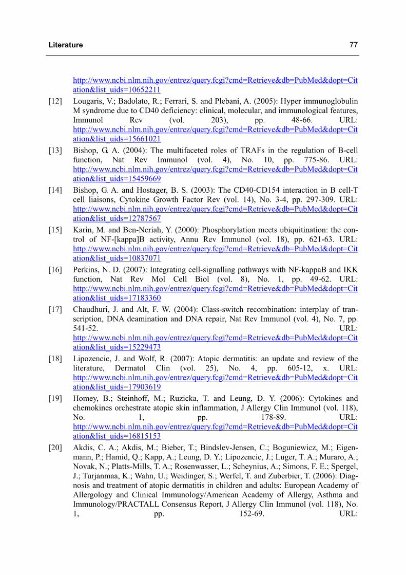
Literature
77
http://www.ncbi.nlm.nih.gov/entrez/query.fcgi?cmd=Retrieve&db=PubMed&dopt=Citation&list_uids=10652211
[12] Lougaris, V.; Badolato, R.; Ferrari, S. and Plebani, A. (2005): Hyper immunoglobulin M syndrome due to CD40 deficiency: clinical, molecular, and immunological features, Immunol Rev (vol. 203), pp. 48-66. URL: http://www.ncbi.nlm.nih.gov/entrez/query.fcgi?cmd=Retrieve&db=PubMed&dopt=Citation&list_uids=15661021
[13] Bishop, G. A. (2004): The multifaceted roles of TRAFs in the regulation of B-cell function, Nat Rev Immunol (vol. 4), No. 10, pp. 775-86. URL: http://www.ncbi.nlm.nih.gov/entrez/query.fcgi?cmd=Retrieve&db=PubMed&dopt=Citation&list_uids=15459669
[14] Bishop, G. A. and Hostager, B. S. (2003): The CD40-CD154 interaction in B cell-T cell liaisons, Cytokine Growth Factor Rev (vol. 14), No. 3-4, pp. 297-309. URL: http://www.ncbi.nlm.nih.gov/entrez/query.fcgi?cmd=Retrieve&db=PubMed&dopt=Citation&list_uids=12787567
[15] Karin, M. and Ben-Neriah, Y. (2000): Phosphorylation meets ubiquitination: the con-trol of NF-[kappa]B activity, Annu Rev Immunol (vol. 18), pp. 621-63. URL: http://www.ncbi.nlm.nih.gov/entrez/query.fcgi?cmd=Retrieve&db=PubMed&dopt=Citation&list_uids=10837071
[16] Perkins, N. D. (2007): Integrating cell-signalling pathways with NF-kappaB and IKK function, Nat Rev Mol Cell Biol (vol. 8), No. 1, pp. 49-62. URL: http://www.ncbi.nlm.nih.gov/entrez/query.fcgi?cmd=Retrieve&db=PubMed&dopt=Citation&list_uids=17183360
[17] Chaudhuri, J. and Alt, F. W. (2004): Class-switch recombination: interplay of tran-scription, DNA deamination and DNA repair, Nat Rev Immunol (vol. 4), No. 7, pp. 541-52. URL: http://www.ncbi.nlm.nih.gov/entrez/query.fcgi?cmd=Retrieve&db=PubMed&dopt=Citation&list_uids=15229473
[18] Lipozencic, J. and Wolf, R. (2007): Atopic dermatitis: an update and review of the literature, Dermatol Clin (vol. 25), No. 4, pp. 605-12, x. URL: http://www.ncbi.nlm.nih.gov/entrez/query.fcgi?cmd=Retrieve&db=PubMed&dopt=Citation&list_uids=17903619
[19] Homey, B.; Steinhoff, M.; Ruzicka, T. and Leung, D. Y. (2006): Cytokines and chemokines orchestrate atopic skin inflammation, J Allergy Clin Immunol (vol. 118), No. 1, pp. 178-89. URL: http://www.ncbi.nlm.nih.gov/entrez/query.fcgi?cmd=Retrieve&db=PubMed&dopt=Citation&list_uids=16815153
[20] Akdis, C. A.; Akdis, M.; Bieber, T.; Bindslev-Jensen, C.; Boguniewicz, M.; Eigen-mann, P.; Hamid, Q.; Kapp, A.; Leung, D. Y.; Lipozencic, J.; Luger, T. A.; Muraro, A.; Novak, N.; Platts-Mills, T. A.; Rosenwasser, L.; Scheynius, A.; Simons, F. E.; Spergel, J.; Turjanmaa, K.; Wahn, U.; Weidinger, S.; Werfel, T. and Zuberbier, T. (2006): Diag-nosis and treatment of atopic dermatitis in children and adults: European Academy of Allergology and Clinical Immunology/American Academy of Allergy, Asthma and Immunology/PRACTALL Consensus Report, J Allergy Clin Immunol (vol. 118), No. 1, pp. 152-69. URL:
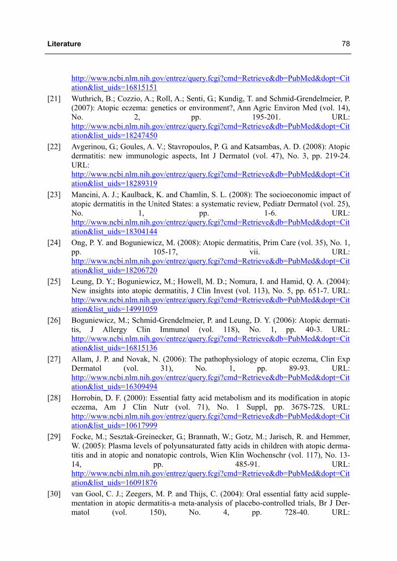
Literature
78
http://www.ncbi.nlm.nih.gov/entrez/query.fcgi?cmd=Retrieve&db=PubMed&dopt=Citation&list_uids=16815151
[21] Wuthrich, B.; Cozzio, A.; Roll, A.; Senti, G.; Kundig, T. and Schmid-Grendelmeier, P. (2007): Atopic eczema: genetics or environment?, Ann Agric Environ Med (vol. 14), No. 2, pp. 195-201. URL: http://www.ncbi.nlm.nih.gov/entrez/query.fcgi?cmd=Retrieve&db=PubMed&dopt=Citation&list_uids=18247450
[22] Avgerinou, G.; Goules, A. V.; Stavropoulos, P. G. and Katsambas, A. D. (2008): Atopic dermatitis: new immunologic aspects, Int J Dermatol (vol. 47), No. 3, pp. 219-24. URL: http://www.ncbi.nlm.nih.gov/entrez/query.fcgi?cmd=Retrieve&db=PubMed&dopt=Citation&list_uids=18289319
[23] Mancini, A. J.; Kaulback, K. and Chamlin, S. L. (2008): The socioeconomic impact of atopic dermatitis in the United States: a systematic review, Pediatr Dermatol (vol. 25), No. 1, pp. 1-6. URL: http://www.ncbi.nlm.nih.gov/entrez/query.fcgi?cmd=Retrieve&db=PubMed&dopt=Citation&list_uids=18304144
[24] Ong, P. Y. and Boguniewicz, M. (2008): Atopic dermatitis, Prim Care (vol. 35), No. 1, pp. 105-17, vii. URL: http://www.ncbi.nlm.nih.gov/entrez/query.fcgi?cmd=Retrieve&db=PubMed&dopt=Citation&list_uids=18206720
[25] Leung, D. Y.; Boguniewicz, M.; Howell, M. D.; Nomura, I. and Hamid, Q. A. (2004): New insights into atopic dermatitis, J Clin Invest (vol. 113), No. 5, pp. 651-7. URL: http://www.ncbi.nlm.nih.gov/entrez/query.fcgi?cmd=Retrieve&db=PubMed&dopt=Citation&list_uids=14991059
[26] Boguniewicz, M.; Schmid-Grendelmeier, P. and Leung, D. Y. (2006): Atopic dermati-tis, J Allergy Clin Immunol (vol. 118), No. 1, pp. 40-3. URL: http://www.ncbi.nlm.nih.gov/entrez/query.fcgi?cmd=Retrieve&db=PubMed&dopt=Citation&list_uids=16815136
[27] Allam, J. P. and Novak, N. (2006): The pathophysiology of atopic eczema, Clin Exp Dermatol (vol. 31), No. 1, pp. 89-93. URL: http://www.ncbi.nlm.nih.gov/entrez/query.fcgi?cmd=Retrieve&db=PubMed&dopt=Citation&list_uids=16309494
[28] Horrobin, D. F. (2000): Essential fatty acid metabolism and its modification in atopic eczema, Am J Clin Nutr (vol. 71), No. 1 Suppl, pp. 367S-72S. URL: http://www.ncbi.nlm.nih.gov/entrez/query.fcgi?cmd=Retrieve&db=PubMed&dopt=Citation&list_uids=10617999
[29] Focke, M.; Sesztak-Greinecker, G.; Brannath, W.; Gotz, M.; Jarisch, R. and Hemmer, W. (2005): Plasma levels of polyunsaturated fatty acids in children with atopic derma-titis and in atopic and nonatopic controls, Wien Klin Wochenschr (vol. 117), No. 13-14, pp. 485-91. URL: http://www.ncbi.nlm.nih.gov/entrez/query.fcgi?cmd=Retrieve&db=PubMed&dopt=Citation&list_uids=16091876
[30] van Gool, C. J.; Zeegers, M. P. and Thijs, C. (2004): Oral essential fatty acid supple-mentation in atopic dermatitis-a meta-analysis of placebo-controlled trials, Br J Der-matol (vol. 150), No. 4, pp. 728-40. URL:
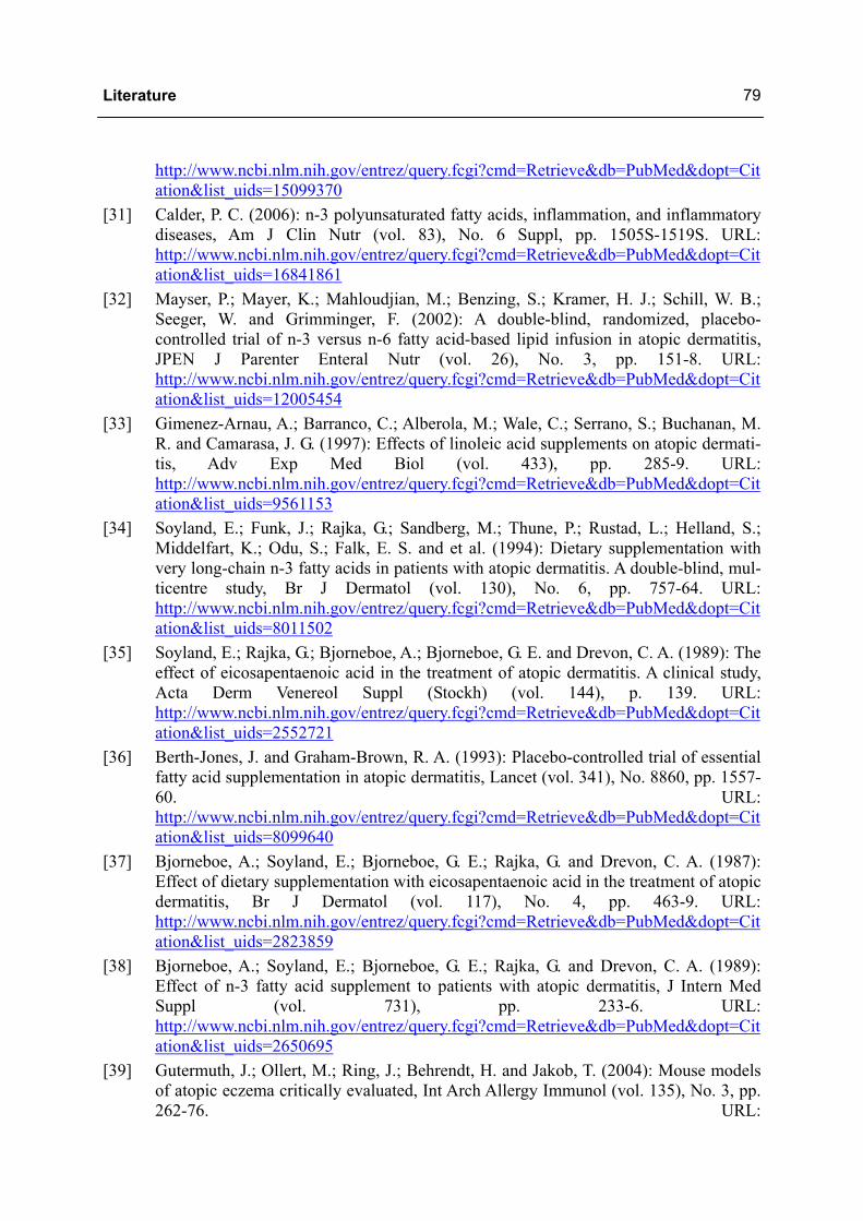
Literature
79
http://www.ncbi.nlm.nih.gov/entrez/query.fcgi?cmd=Retrieve&db=PubMed&dopt=Citation&list_uids=15099370
[31] Calder, P. C. (2006): n-3 polyunsaturated fatty acids, inflammation, and inflammatory diseases, Am J Clin Nutr (vol. 83), No. 6 Suppl, pp. 1505S-1519S. URL: http://www.ncbi.nlm.nih.gov/entrez/query.fcgi?cmd=Retrieve&db=PubMed&dopt=Citation&list_uids=16841861
[32] Mayser, P.; Mayer, K.; Mahloudjian, M.; Benzing, S.; Kramer, H. J.; Schill, W. B.; Seeger, W. and Grimminger, F. (2002): A double-blind, randomized, placebo-controlled trial of n-3 versus n-6 fatty acid-based lipid infusion in atopic dermatitis, JPEN J Parenter Enteral Nutr (vol. 26), No. 3, pp. 151-8. URL: http://www.ncbi.nlm.nih.gov/entrez/query.fcgi?cmd=Retrieve&db=PubMed&dopt=Citation&list_uids=12005454
[33] Gimenez-Arnau, A.; Barranco, C.; Alberola, M.; Wale, C.; Serrano, S.; Buchanan, M. R. and Camarasa, J. G. (1997): Effects of linoleic acid supplements on atopic dermati-tis, Adv Exp Med Biol (vol. 433), pp. 285-9. URL: http://www.ncbi.nlm.nih.gov/entrez/query.fcgi?cmd=Retrieve&db=PubMed&dopt=Citation&list_uids=9561153
[34] Soyland, E.; Funk, J.; Rajka, G.; Sandberg, M.; Thune, P.; Rustad, L.; Helland, S.; Middelfart, K.; Odu, S.; Falk, E. S. and et al. (1994): Dietary supplementation with very long-chain n-3 fatty acids in patients with atopic dermatitis. A double-blind, mul-ticentre study, Br J Dermatol (vol. 130), No. 6, pp. 757-64. URL: http://www.ncbi.nlm.nih.gov/entrez/query.fcgi?cmd=Retrieve&db=PubMed&dopt=Citation&list_uids=8011502
[35] Soyland, E.; Rajka, G.; Bjorneboe, A.; Bjorneboe, G. E. and Drevon, C. A. (1989): The effect of eicosapentaenoic acid in the treatment of atopic dermatitis. A clinical study, Acta Derm Venereol Suppl (Stockh) (vol. 144), p. 139. URL: http://www.ncbi.nlm.nih.gov/entrez/query.fcgi?cmd=Retrieve&db=PubMed&dopt=Citation&list_uids=2552721
[36] Berth-Jones, J. and Graham-Brown, R. A. (1993): Placebo-controlled trial of essential fatty acid supplementation in atopic dermatitis, Lancet (vol. 341), No. 8860, pp. 1557-60. URL: http://www.ncbi.nlm.nih.gov/entrez/query.fcgi?cmd=Retrieve&db=PubMed&dopt=Citation&list_uids=8099640
[37] Bjorneboe, A.; Soyland, E.; Bjorneboe, G. E.; Rajka, G. and Drevon, C. A. (1987): Effect of dietary supplementation with eicosapentaenoic acid in the treatment of atopic dermatitis, Br J Dermatol (vol. 117), No. 4, pp. 463-9. URL: http://www.ncbi.nlm.nih.gov/entrez/query.fcgi?cmd=Retrieve&db=PubMed&dopt=Citation&list_uids=2823859
[38] Bjorneboe, A.; Soyland, E.; Bjorneboe, G. E.; Rajka, G. and Drevon, C. A. (1989): Effect of n-3 fatty acid supplement to patients with atopic dermatitis, J Intern Med Suppl (vol. 731), pp. 233-6. URL: http://www.ncbi.nlm.nih.gov/entrez/query.fcgi?cmd=Retrieve&db=PubMed&dopt=Citation&list_uids=2650695
[39] Gutermuth, J.; Ollert, M.; Ring, J.; Behrendt, H. and Jakob, T. (2004): Mouse models of atopic eczema critically evaluated, Int Arch Allergy Immunol (vol. 135), No. 3, pp. 262-76. URL:

Literature
80
http://www.ncbi.nlm.nih.gov/entrez/query.fcgi?cmd=Retrieve&db=PubMed&dopt=Citation&list_uids=15542938
[40] Man, M. Q.; Hatano, Y.; Lee, S. H.; Man, M.; Chang, S.; Feingold, K. R.; Leung, D. Y.; Holleran, W.; Uchida, Y. and Elias, P. M. (2008): Characterization of a hapten-induced, murine model with multiple features of atopic dermatitis: structural, immu-nologic, and biochemical changes following single versus multiple oxazolone chal-lenges, J Invest Dermatol (vol. 128), No. 1, pp. 79-86. URL: http://www.ncbi.nlm.nih.gov/entrez/query.fcgi?cmd=Retrieve&db=PubMed&dopt=Citation&list_uids=17671515
[41] Shiohara, T.; Hayakawa, J. and Mizukawa, Y. (2004): Animal models for atopic der-matitis: are they relevant to human disease?, J Dermatol Sci (vol. 36), No. 1, pp. 1-9. URL: http://www.ncbi.nlm.nih.gov/entrez/query.fcgi?cmd=Retrieve&db=PubMed&dopt=Citation&list_uids=15488700
[42] Igney, F. H.; Asadullah, K. and Zollner, T. M. (2004): Techniques: species' finest blend--humanized mouse models in inflammatory skin disease research, Trends Phar-macol Sci (vol. 25), No. 10, pp. 543-9. URL: http://www.ncbi.nlm.nih.gov/entrez/query.fcgi?cmd=Retrieve&db=PubMed&dopt=Citation&list_uids=15380939
[43] Zollner, T. M.; Renz, H.; Igney, F. H. and Asadullah, K. (2004): Animal models of T-cell-mediated skin diseases, Bioessays (vol. 26), No. 6, pp. 693-6. URL: http://www.ncbi.nlm.nih.gov/entrez/query.fcgi?cmd=Retrieve&db=PubMed&dopt=Citation&list_uids=15170867
[44] Wang, L. F.; Lin, J. Y.; Hsieh, K. H. and Lin, R. H. (1996): Epicutaneous exposure of protein antigen induces a predominant Th2-like response with high IgE production in mice, J Immunol (vol. 156), No. 11, pp. 4077-82. URL: http://www.ncbi.nlm.nih.gov/entrez/query.fcgi?cmd=Retrieve&db=PubMed&dopt=Citation&list_uids=8666772
[45] Spergel, J. M.; Mizoguchi, E.; Brewer, J. P.; Martin, T. R.; Bhan, A. K. and Geha, R. S. (1998): Epicutaneous sensitization with protein antigen induces localized allergic dermatitis and hyperresponsiveness to methacholine after single exposure to aerosol-ized antigen in mice, J Clin Invest (vol. 101), No. 8, pp. 1614-22. URL: http://www.ncbi.nlm.nih.gov/entrez/query.fcgi?cmd=Retrieve&db=PubMed&dopt=Citation&list_uids=9541491
[46] Scharschmidt, T. C. and Segre, J. A. (2008): Modeling atopic dermatitis with increas-ingly complex mouse models, J Invest Dermatol (vol. 128), No. 5, pp. 1061-4. URL: http://www.ncbi.nlm.nih.gov/entrez/query.fcgi?cmd=Retrieve&db=PubMed&dopt=Citation&list_uids=18408744
[47] Jungersted, J. M.; Hellgren, L. I.; Jemec, G. B. and Agner, T. (2008): Lipids and skin barrier function--a clinical perspective, Contact Dermatitis (vol. 58), No. 5, pp. 255-62. URL: http://www.ncbi.nlm.nih.gov/entrez/query.fcgi?cmd=Retrieve&db=PubMed&dopt=Citation&list_uids=18416754
[48] Deckelbaum, R. J.; Worgall, T. S. and Seo, T. (2006): n-3 fatty acids and gene expres-sion, Am J Clin Nutr (vol. 83), No. 6 Suppl, pp. 1520S-1525S. URL:
Literature
81
http://www.ncbi.nlm.nih.gov/entrez/query.fcgi?cmd=Retrieve&db=PubMed&dopt=Citation&list_uids=16841862
[49] Das, U. N. (2006): Essential fatty acids: biochemistry, physiology and pathology, Bio-technol J (vol. 1), No. 4, pp. 420-39. URL: http://www.ncbi.nlm.nih.gov/entrez/query.fcgi?cmd=Retrieve&db=PubMed&dopt=Citation&list_uids=16892270
[50] Calder, P. C. and Deckelbaum, R. J. (2008): Omega-3 fatty acids: time to get the mes-sages right!, Curr Opin Clin Nutr Metab Care (vol. 11), No. 2, pp. 91-3. URL: http://www.ncbi.nlm.nih.gov/entrez/query.fcgi?cmd=Retrieve&db=PubMed&dopt=Citation&list_uids=18301081
[51] Covington, M. B. (2004): Omega-3 fatty acids, Am Fam Physician (vol. 70), No. 1, pp. 133-40. URL: http://www.ncbi.nlm.nih.gov/entrez/query.fcgi?cmd=Retrieve&db=PubMed&dopt=Citation&list_uids=15259529
[52] Racine, R. A. and Deckelbaum, R. J. (2007): Sources of the very-long-chain unsatu-rated omega-3 fatty acids: eicosapentaenoic acid and docosahexaenoic acid, Curr Opin Clin Nutr Metab Care (vol. 10), No. 2, pp. 123-8. URL: http://www.ncbi.nlm.nih.gov/entrez/query.fcgi?cmd=Retrieve&db=PubMed&dopt=Citation&list_uids=17284998
[53] Calder, P. C. (2007): Immunomodulation by omega-3 fatty acids, Prostaglandins Leu-kot Essent Fatty Acids (vol. 77), No. 5-6, pp. 327-35. URL: http://www.ncbi.nlm.nih.gov/entrez/query.fcgi?cmd=Retrieve&db=PubMed&dopt=Citation&list_uids=18032006
[54] Schmitz, G. and Ecker, J. (2008): The opposing effects of n-3 and n-6 fatty acids, Prog Lipid Res (vol. 47), No. 2, pp. 147-55. URL: http://www.ncbi.nlm.nih.gov/entrez/query.fcgi?cmd=Retrieve&db=PubMed&dopt=Citation&list_uids=18198131
[55] Ziboh, V. A.; Cho, Y.; Mani, I. and Xi, S. (2002): Biological significance of essential fatty acids/prostanoids/lipoxygenase-derived monohydroxy fatty acids in the skin, Arch Pharm Res (vol. 25), No. 6, pp. 747-58. URL: http://www.ncbi.nlm.nih.gov/entrez/query.fcgi?cmd=Retrieve&db=PubMed&dopt=Citation&list_uids=12510822
[56] Leonard, A. E.; Bobik, E. G.; Dorado, J.; Kroeger, P. E.; Chuang, L. T.; Thurmond, J. M.; Parker-Barnes, J. M.; Das, T.; Huang, Y. S. and Mukerji, P. (2000): Cloning of a human cDNA encoding a novel enzyme involved in the elongation of long-chain polyunsaturated fatty acids, Biochem J (vol. 350 Pt 3), pp. 765-70. URL: http://www.ncbi.nlm.nih.gov/entrez/query.fcgi?cmd=Retrieve&db=PubMed&dopt=Citation&list_uids=10970790
[57] Grimm, H.; Mayer, K.; Mayser, P. and Eigenbrodt, E. (2002): Regulatory potential of n-3 fatty acids in immunological and inflammatory processes, Br J Nutr (vol. 87 Suppl 1), pp. S59-67. URL: http://www.ncbi.nlm.nih.gov/entrez/query.fcgi?cmd=Retrieve&db=PubMed&dopt=Citation&list_uids=11895155
[58] Stulnig, T. M. (2003): Immunomodulation by polyunsaturated fatty acids: mechanisms and effects, Int Arch Allergy Immunol (vol. 132), No. 4, pp. 310-21. URL:
Literature
82
http://www.ncbi.nlm.nih.gov/entrez/query.fcgi?cmd=Retrieve&db=PubMed&dopt=Citation&list_uids=14707462
[59] Serhan, C. N. and Savill, J. (2005): Resolution of inflammation: the beginning pro-grams the end, Nat Immunol (vol. 6), No. 12, pp. 1191-7. URL: http://www.ncbi.nlm.nih.gov/entrez/query.fcgi?cmd=Retrieve&db=PubMed&dopt=Citation&list_uids=16369558
[60] Pike, L. J. (2006): Rafts defined: a report on the Keystone Symposium on Lipid Rafts and Cell Function, J Lipid Res (vol. 47), No. 7, pp. 1597-8. URL: http://www.ncbi.nlm.nih.gov/entrez/query.fcgi?cmd=Retrieve&db=PubMed&dopt=Citation&list_uids=16645198
[61] Hwang, D. and Rhee, S. H. (1999): Receptor-mediated signaling pathways: potential targets of modulation by dietary fatty acids, Am J Clin Nutr (vol. 70), No. 4, pp. 545-56. URL: http://www.ncbi.nlm.nih.gov/entrez/query.fcgi?cmd=Retrieve&db=PubMed&dopt=Citation&list_uids=10500025
[62] Stillwell, W. and Wassall, S. R. (2003): Docosahexaenoic acid: membrane properties of a unique fatty acid, Chem Phys Lipids (vol. 126), No. 1, pp. 1-27. URL: http://www.ncbi.nlm.nih.gov/entrez/query.fcgi?cmd=Retrieve&db=PubMed&dopt=Citation&list_uids=14580707
[63] Horrocks, L. A. and Yeo, Y. K. (1999): Health benefits of docosahexaenoic acid (DHA), Pharmacol Res (vol. 40), No. 3, pp. 211-25. URL: http://www.ncbi.nlm.nih.gov/entrez/query.fcgi?cmd=Retrieve&db=PubMed&dopt=Citation&list_uids=10479465
[64] Seo, J.; Barhoumi, R.; Johnson, A. E.; Lupton, J. R. and Chapkin, R. S. (2006): Doco-sahexaenoic acid selectively inhibits plasma membrane targeting of lipidated proteins, Faseb J (vol. 20), No. 6, pp. 770-2. URL: http://www.ncbi.nlm.nih.gov/entrez/query.fcgi?cmd=Retrieve&db=PubMed&dopt=Citation&list_uids=16469846
[65] Onuki, Y.; Morishita, M.; Chiba, Y.; Tokiwa, S. and Takayama, K. (2006): Docosahex-aenoic acid and eicosapentaenoic acid induce changes in the physical properties of a lipid bilayer model membrane, Chem Pharm Bull (Tokyo) (vol. 54), No. 1, pp. 68-71. URL: http://www.ncbi.nlm.nih.gov/entrez/query.fcgi?cmd=Retrieve&db=PubMed&dopt=Citation&list_uids=16394552
[66] Bousserouel, S.; Brouillet, A.; Bereziat, G.; Raymondjean, M. and Andreani, M. (2003): Different effects of n-6 and n-3 polyunsaturated fatty acids on the activation of rat smooth muscle cells by interleukin-1 beta, J Lipid Res (vol. 44), No. 3, pp. 601-11. URL: http://www.ncbi.nlm.nih.gov/entrez/query.fcgi?cmd=Retrieve&db=PubMed&dopt=Citation&list_uids=12562859
[67] Jia, Q.; Zhou, H. R.; Bennink, M. and Pestka, J. J. (2004): Docosahexaenoic acid at-tenuates mycotoxin-induced immunoglobulin a nephropathy, interleukin-6 transcrip-tion, and mitogen-activated protein kinase phosphorylation in mice, J Nutr (vol. 134), No. 12, pp. 3343-9. URL: http://www.ncbi.nlm.nih.gov/entrez/query.fcgi?cmd=Retrieve&db=PubMed&dopt=Citation&list_uids=15570035
Literature
83
[68] Fan, Y. Y.; Ly, L. H.; Barhoumi, R.; McMurray, D. N. and Chapkin, R. S. (2004): Die-tary docosahexaenoic acid suppresses T cell protein kinase C theta lipid raft recruit-ment and IL-2 production, J Immunol (vol. 173), No. 10, pp. 6151-60. URL: http://www.ncbi.nlm.nih.gov/entrez/query.fcgi?cmd=Retrieve&db=PubMed&dopt=Citation&list_uids=15528352
[69] Serhan, C. N. (2005): Novel omega -- 3-derived local mediators in anti-inflammation and resolution, Pharmacol Ther (vol. 105), No. 1, pp. 7-21. URL: http://www.ncbi.nlm.nih.gov/entrez/query.fcgi?cmd=Retrieve&db=PubMed&dopt=Citation&list_uids=15626453
[70] Serhan, C. N. (2005): Novel eicosanoid and docosanoid mediators: resolvins, docosa-trienes, and neuroprotectins, Curr Opin Clin Nutr Metab Care (vol. 8), No. 2, pp. 115-21. URL: http://www.ncbi.nlm.nih.gov/entrez/query.fcgi?cmd=Retrieve&db=PubMed&dopt=Citation&list_uids=15716788
[71] Miles, E. A.; Allen, E. and Calder, P. C. (2002): In vitro effects of eicosanoids derived from different 20-carbon Fatty acids on production of monocyte-derived cytokines in human whole blood cultures, Cytokine (vol. 20), No. 5, pp. 215-23. URL: http://www.ncbi.nlm.nih.gov/entrez/query.fcgi?cmd=Retrieve&db=PubMed&dopt=Citation&list_uids=12550106
[72] Zhao, G.; Etherton, T. D.; Martin, K. R.; Vanden Heuvel, J. P.; Gillies, P. J.; West, S. G. and Kris-Etherton, P. M. (2005): Anti-inflammatory effects of polyunsaturated fatty acids in THP-1 cells, Biochem Biophys Res Commun (vol. 336), No. 3, pp. 909-17. URL: http://www.ncbi.nlm.nih.gov/entrez/query.fcgi?cmd=Retrieve&db=PubMed&dopt=Citation&list_uids=16169525
[73] Lengqvist, J.; Mata De Urquiza, A.; Bergman, A. C.; Willson, T. M.; Sjovall, J.; Perlmann, T. and Griffiths, W. J. (2004): Polyunsaturated fatty acids including docosa-hexaenoic and arachidonic acid bind to the retinoid X receptor alpha ligand-binding domain, Mol Cell Proteomics (vol. 3), No. 7, pp. 692-703. URL: http://www.ncbi.nlm.nih.gov/entrez/query.fcgi?cmd=Retrieve&db=PubMed&dopt=Citation&list_uids=15073272
[74] Devereux, G. and Seaton, A. (2005): Diet as a risk factor for atopy and asthma, J Al-lergy Clin Immunol (vol. 115), No. 6, pp. 1109-17; quiz 1118. URL: http://www.ncbi.nlm.nih.gov/entrez/query.fcgi?cmd=Retrieve&db=PubMed&dopt=Citation&list_uids=15940119
[75] Prescott, S. L. and Calder, P. C. (2004): N-3 polyunsaturated fatty acids and allergic disease, Curr Opin Clin Nutr Metab Care (vol. 7), No. 2, pp. 123-9. URL: http://www.ncbi.nlm.nih.gov/entrez/query.fcgi?cmd=Retrieve&db=PubMed&dopt=Citation&list_uids=15075701
[76] Hanifin, J. M. and Rajka, G. (1980): Diagnostic Features of Atopic-Dermatitis, Acta Dermato-Venereologica, pp. 44-47. URL: <Go to ISI>://A1980KS87600013
[77] Leung, D. Y.; Hirsch, R. L.; Schneider, L.; Moody, C.; Takaoka, R.; Li, S. H.; Meyer-son, L. A.; Mariam, S. G.; Goldstein, G. and Hanifin, J. M. (1990): Thymopentin ther-apy reduces the clinical severity of atopic dermatitis, J Allergy Clin Immunol (vol. 85), No. 5, pp. 927-33. URL:
Literature
84
http://www.ncbi.nlm.nih.gov/entrez/query.fcgi?cmd=Retrieve&db=PubMed&dopt=Citation&list_uids=2185294
[78] Matsuda, H.; Watanabe, N.; Geba, G. P.; Sperl, J.; Tsudzuki, M.; Hiroi, J.; Matsumoto, M.; Ushio, H.; Saito, S.; Askenase, P. W. and Ra, C. (1997): Development of atopic dermatitis-like skin lesion with IgE hyperproduction in NC/Nga mice, Int Immunol (vol. 9), No. 3, pp. 461-6. URL: http://www.ncbi.nlm.nih.gov/entrez/query.fcgi?cmd=Retrieve&db=PubMed&dopt=Citation&list_uids=9088984
[79] Funderud, S.; Erikstein, B.; Asheim, H. C.; Nustad, K.; Stokke, T.; Blomhoff, H. K.; Holte, H. and Smeland, E. B. (1990): Functional properties of CD19+ B lymphocytes positively selected from buffy coats by immunomagnetic separation, Eur J Immunol (vol. 20), No. 1, pp. 201-6. URL: http://www.ncbi.nlm.nih.gov/entrez/query.fcgi?cmd=Retrieve&db=PubMed&dopt=Citation&list_uids=1689662
[80] Jabara, H. H.; Fu, S. M.; Geha, R. S. and Vercelli, D. (1990): CD40 and IgE: syner-gism between anti-CD40 monoclonal antibody and interleukin 4 in the induction of IgE synthesis by highly purified human B cells, J Exp Med (vol. 172), No. 6, pp. 1861-4. URL: http://www.ncbi.nlm.nih.gov/entrez/query.fcgi?cmd=Retrieve&db=PubMed&dopt=Citation&list_uids=1701824
[81] Livak, K. J. and Schmittgen, T. D. (2001): Analysis of relative gene expression data using real-time quantitative PCR and the 2(-Delta Delta C(T)) Method, Methods (vol. 25), No. 4, pp. 402-8. URL: http://www.ncbi.nlm.nih.gov/entrez/query.fcgi?cmd=Retrieve&db=PubMed&dopt=Citation&list_uids=11846609
[82] Zhang, K.; Clark, E. A. and Saxon, A. (1991): CD40 stimulation provides an IFN-gamma-independent and IL-4-dependent differentiation signal directly to human B cells for IgE production, J Immunol (vol. 146), No. 6, pp. 1836-42. URL: http://www.ncbi.nlm.nih.gov/entrez/query.fcgi?cmd=Retrieve&db=PubMed&dopt=Citation&list_uids=1706382
[83] Shapira, S. K.; Vercelli, D.; Jabara, H. H.; Fu, S. M. and Geha, R. S. (1992): Molecu-lar analysis of the induction of immunoglobulin E synthesis in human B cells by inter-leukin 4 and engagement of CD40 antigen, J Exp Med (vol. 175), No. 1, pp. 289-92. URL: http://www.ncbi.nlm.nih.gov/entrez/query.fcgi?cmd=Retrieve&db=PubMed&dopt=Citation&list_uids=1370532
[84] Koch, C.; Dolle, S.; Metzger, M.; Rasche, C.; Jungclas, H.; Rühl, R.; Renz, H. and Worm, M. (2008): Docosahexaenoic acid (DHA) supplementation in atopic eczema: a randomized, double-blind, controlled trial, Br J Dermatol (vol. 158), No. 4, pp. 786-92. URL: http://www.ncbi.nlm.nih.gov/entrez/query.fcgi?cmd=Retrieve&db=PubMed&dopt=Citation&list_uids=18241260
[85] Dahten, A.; Koch, C.; Ernst, D.; Schnoller, C.; Hartmann, S. and Worm, M. (2008): Systemic PPARgamma Ligation Inhibits Allergic Immune Response in the Skin, J In-vest Dermatol (vol. 128), No. 9, pp. 2211-8. URL: http://www.ncbi.nlm.nih.gov/entrez/query.fcgi?cmd=Retrieve&db=PubMed&dopt=Citation&list_uids=18401424
Literature
85
[86] Kromann, N. and Green, A. (1980): Epidemiological studies in the Upernavik district, Greenland. Incidence of some chronic diseases 1950-1974, Acta Med Scand (vol. 208), No. 5, pp. 401-6. URL: http://www.ncbi.nlm.nih.gov/entrez/query.fcgi?cmd=Retrieve&db=PubMed&dopt=Citation&list_uids=7457208
[87] Dyerberg, J. and Bang, H. O. (1979): Haemostatic function and platelet polyunsatu-rated fatty acids in Eskimos, Lancet (vol. 2), No. 8140, pp. 433-5. URL: http://www.ncbi.nlm.nih.gov/entrez/query.fcgi?cmd=Retrieve&db=PubMed&dopt=Citation&list_uids=89498
[88] Hirai, A.; Hamazaki, T.; Terano, T.; Nishikawa, T.; Tamura, Y.; Kamugai, A. and Jajiki, J. (1980): Eicosapentaenoic acid and platelet function in Japanese, Lancet (vol. 2), No. 8204, pp. 1132-3. URL: http://www.ncbi.nlm.nih.gov/entrez/query.fcgi?cmd=Retrieve&db=PubMed&dopt=Citation&list_uids=6107739
[89] Grimble, R. F. (2001): Nutritional modulation of immune function, Proc Nutr Soc (vol. 60), No. 3, pp. 389-97. URL: http://www.ncbi.nlm.nih.gov/entrez/query.fcgi?cmd=Retrieve&db=PubMed&dopt=Citation&list_uids=11681814
[90] Brown, E. R. and Subbaiah, P. V. (1994): Differential effects of eicosapentaenoic acid and docosahexaenoic acid on human skin fibroblasts, Lipids (vol. 29), No. 12, pp. 825-9. URL: http://www.ncbi.nlm.nih.gov/entrez/query.fcgi?cmd=Retrieve&db=PubMed&dopt=Citation&list_uids=7854007
[91] Sierra, S.; Lara-Villoslada, F.; Comalada, M.; Olivares, M. and Xaus, J. (2008): Die-tary eicosapentaenoic acid and docosahexaenoic acid equally incorporate as decosa-hexaenoic acid but differ in inflammatory effects, Nutrition (vol. 24), No. 3, pp. 245-54. URL: http://www.ncbi.nlm.nih.gov/entrez/query.fcgi?cmd=Retrieve&db=PubMed&dopt=Citation&list_uids=18312787
[92] Thies, F.; Miles, E. A.; Nebe-von-Caron, G.; Powell, J. R.; Hurst, T. L.; Newsholme, E. A. and Calder, P. C. (2001): Influence of dietary supplementation with long-chain n-3 or n-6 polyunsaturated fatty acids on blood inflammatory cell populations and func-tions and on plasma soluble adhesion molecules in healthy adults, Lipids (vol. 36), No. 11, pp. 1183-93. URL: http://www.ncbi.nlm.nih.gov/entrez/query.fcgi?cmd=Retrieve&db=PubMed&dopt=Citation&list_uids=11795850
[93] Tomobe, Y. I.; Morizawa, K.; Tsuchida, M.; Hibino, H.; Nakano, Y. and Tanaka, Y. (2000): Dietary docosahexaenoic acid suppresses inflammation and immunoresponses in contact hypersensitivity reaction in mice, Lipids (vol. 35), No. 1, pp. 61-9. URL: http://www.ncbi.nlm.nih.gov/entrez/query.fcgi?cmd=Retrieve&db=PubMed&dopt=Citation&list_uids=10695925
[94] Gould, H. J. and Sutton, B. J. (2008): IgE in allergy and asthma today, Nat Rev Immu-nol (vol. 8), No. 3, pp. 205-17. URL: http://www.ncbi.nlm.nih.gov/entrez/query.fcgi?cmd=Retrieve&db=PubMed&dopt=Citation&list_uids=18301424

Literature
86
[95] Muramatsu, M.; Sankaranand, V. S.; Anant, S.; Sugai, M.; Kinoshita, K.; Davidson, N. O. and Honjo, T. (1999): Specific expression of activation-induced cytidine deaminase (AID), a novel member of the RNA-editing deaminase family in germinal center B cells, J Biol Chem (vol. 274), No. 26, pp. 18470-6. URL: http://www.ncbi.nlm.nih.gov/entrez/query.fcgi?cmd=Retrieve&db=PubMed&dopt=Citation&list_uids=10373455
[96] Okazaki, I. M.; Kinoshita, K.; Muramatsu, M.; Yoshikawa, K. and Honjo, T. (2002): The AID enzyme induces class switch recombination in fibroblasts, Nature (vol. 416), No. 6878, pp. 340-5. URL: http://www.ncbi.nlm.nih.gov/entrez/query.fcgi?cmd=Retrieve&db=PubMed&dopt=Citation&list_uids=11875397
[97] Manis, J. P.; Tian, M. and Alt, F. W. (2002): Mechanism and control of class-switch recombination, Trends Immunol (vol. 23), No. 1, pp. 31-9. URL: http://www.ncbi.nlm.nih.gov/entrez/query.fcgi?cmd=Retrieve&db=PubMed&dopt=Citation&list_uids=11801452
[98] Claassen, J. L.; Levine, A. D. and Buckley, R. H. (1990): Recombinant human IL-4 induces IgE and IgG synthesis by normal and atopic donor mononuclear cells. Similar dose response, time course, requirement for T cells, and effect of pokeweed mitogen, J Immunol (vol. 144), No. 6, pp. 2123-30. URL: http://www.ncbi.nlm.nih.gov/entrez/query.fcgi?cmd=Retrieve&db=PubMed&dopt=Citation&list_uids=2313090
[99] Vercelli, D.; Jabara, H. H.; Arai, K. and Geha, R. S. (1989): Induction of human IgE synthesis requires interleukin 4 and T/B cell interactions involving the T cell recep-tor/CD3 complex and MHC class II antigens, J Exp Med (vol. 169), No. 4, pp. 1295-307. URL: http://www.ncbi.nlm.nih.gov/entrez/query.fcgi?cmd=Retrieve&db=PubMed&dopt=Citation&list_uids=2522501
[100] Dedeoglu, F.; Horwitz, B.; Chaudhuri, J.; Alt, F. W. and Geha, R. S. (2004): Induction of activation-induced cytidine deaminase gene expression by IL-4 and CD40 ligation is dependent on STAT6 and NFkappaB, Int Immunol (vol. 16), No. 3, pp. 395-404. URL: http://www.ncbi.nlm.nih.gov/entrez/query.fcgi?cmd=Retrieve&db=PubMed&dopt=Citation&list_uids=14978013
[101] Iciek, L. A.; Delphin, S. A. and Stavnezer, J. (1997): CD40 cross-linking induces Ig epsilon germline transcripts in B cells via activation of NF-kappaB: synergy with IL-4 induction, J Immunol (vol. 158), No. 10, pp. 4769-79. URL: http://www.ncbi.nlm.nih.gov/entrez/query.fcgi?cmd=Retrieve&db=PubMed&dopt=Citation&list_uids=9144491
[102] Gorjao, R.; Hirabara, S. M.; de Lima, T. M.; Cury-Boaventura, M. F. and Curi, R. (2007): Regulation of interleukin-2 signaling by fatty acids in human lymphocytes, J Lipid Res (vol. 48), No. 9, pp. 2009-19. URL: http://www.ncbi.nlm.nih.gov/entrez/query.fcgi?cmd=Retrieve&db=PubMed&dopt=Citation&list_uids=17592174
[103] Wassall, S. R. and Stillwell, W. (2008): Docosahexaenoic acid domains: the ultimate non-raft membrane domain, Chem Phys Lipids (vol. 153), No. 1, pp. 57-63. URL: http://www.ncbi.nlm.nih.gov/entrez/query.fcgi?cmd=Retrieve&db=PubMed&dopt=Citation&list_uids=18343224
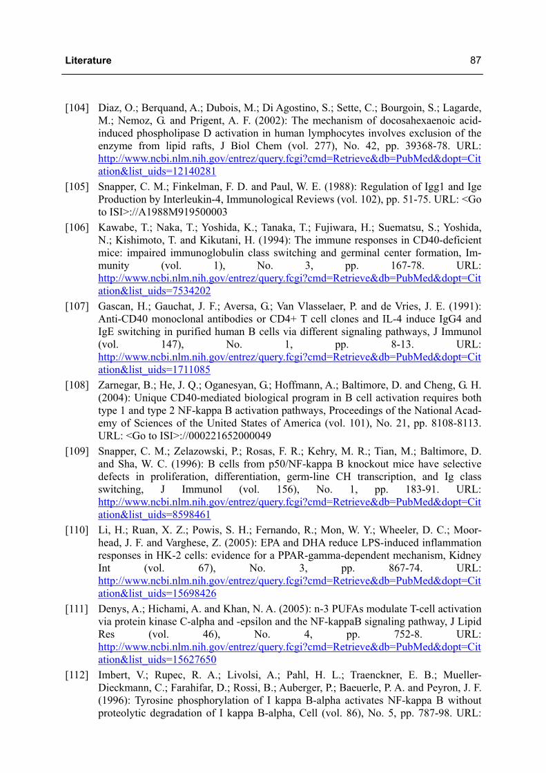
Literature
87
[104] Diaz, O.; Berquand, A.; Dubois, M.; Di Agostino, S.; Sette, C.; Bourgoin, S.; Lagarde, M.; Nemoz, G. and Prigent, A. F. (2002): The mechanism of docosahexaenoic acid-induced phospholipase D activation in human lymphocytes involves exclusion of the enzyme from lipid rafts, J Biol Chem (vol. 277), No. 42, pp. 39368-78. URL: http://www.ncbi.nlm.nih.gov/entrez/query.fcgi?cmd=Retrieve&db=PubMed&dopt=Citation&list_uids=12140281
[105] Snapper, C. M.; Finkelman, F. D. and Paul, W. E. (1988): Regulation of Igg1 and Ige Production by Interleukin-4, Immunological Reviews (vol. 102), pp. 51-75. URL: <Go to ISI>://A1988M919500003
[106] Kawabe, T.; Naka, T.; Yoshida, K.; Tanaka, T.; Fujiwara, H.; Suematsu, S.; Yoshida, N.; Kishimoto, T. and Kikutani, H. (1994): The immune responses in CD40-deficient mice: impaired immunoglobulin class switching and germinal center formation, Im-munity (vol. 1), No. 3, pp. 167-78. URL: http://www.ncbi.nlm.nih.gov/entrez/query.fcgi?cmd=Retrieve&db=PubMed&dopt=Citation&list_uids=7534202
[107] Gascan, H.; Gauchat, J. F.; Aversa, G.; Van Vlasselaer, P. and de Vries, J. E. (1991): Anti-CD40 monoclonal antibodies or CD4+ T cell clones and IL-4 induce IgG4 and IgE switching in purified human B cells via different signaling pathways, J Immunol (vol. 147), No. 1, pp. 8-13. URL: http://www.ncbi.nlm.nih.gov/entrez/query.fcgi?cmd=Retrieve&db=PubMed&dopt=Citation&list_uids=1711085
[108] Zarnegar, B.; He, J. Q.; Oganesyan, G.; Hoffmann, A.; Baltimore, D. and Cheng, G. H. (2004): Unique CD40-mediated biological program in B cell activation requires both type 1 and type 2 NF-kappa B activation pathways, Proceedings of the National Acad-emy of Sciences of the United States of America (vol. 101), No. 21, pp. 8108-8113. URL: <Go to ISI>://000221652000049
[109] Snapper, C. M.; Zelazowski, P.; Rosas, F. R.; Kehry, M. R.; Tian, M.; Baltimore, D. and Sha, W. C. (1996): B cells from p50/NF-kappa B knockout mice have selective defects in proliferation, differentiation, germ-line CH transcription, and Ig class switching, J Immunol (vol. 156), No. 1, pp. 183-91. URL: http://www.ncbi.nlm.nih.gov/entrez/query.fcgi?cmd=Retrieve&db=PubMed&dopt=Citation&list_uids=8598461
[110] Li, H.; Ruan, X. Z.; Powis, S. H.; Fernando, R.; Mon, W. Y.; Wheeler, D. C.; Moor-head, J. F. and Varghese, Z. (2005): EPA and DHA reduce LPS-induced inflammation responses in HK-2 cells: evidence for a PPAR-gamma-dependent mechanism, Kidney Int (vol. 67), No. 3, pp. 867-74. URL: http://www.ncbi.nlm.nih.gov/entrez/query.fcgi?cmd=Retrieve&db=PubMed&dopt=Citation&list_uids=15698426
[111] Denys, A.; Hichami, A. and Khan, N. A. (2005): n-3 PUFAs modulate T-cell activation via protein kinase C-alpha and -epsilon and the NF-kappaB signaling pathway, J Lipid Res (vol. 46), No. 4, pp. 752-8. URL: http://www.ncbi.nlm.nih.gov/entrez/query.fcgi?cmd=Retrieve&db=PubMed&dopt=Citation&list_uids=15627650
[112] Imbert, V.; Rupec, R. A.; Livolsi, A.; Pahl, H. L.; Traenckner, E. B.; Mueller-Dieckmann, C.; Farahifar, D.; Rossi, B.; Auberger, P.; Baeuerle, P. A. and Peyron, J. F. (1996): Tyrosine phosphorylation of I kappa B-alpha activates NF-kappa B without proteolytic degradation of I kappa B-alpha, Cell (vol. 86), No. 5, pp. 787-98. URL:
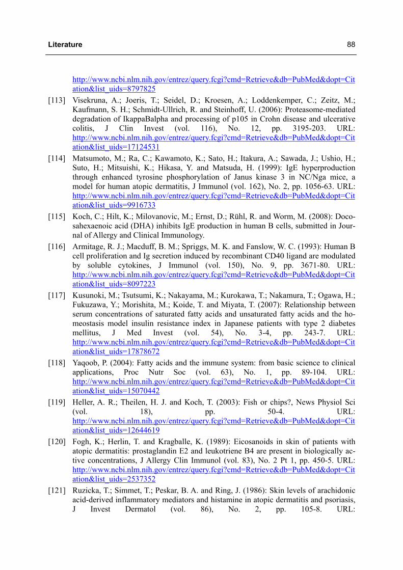
Literature
88
http://www.ncbi.nlm.nih.gov/entrez/query.fcgi?cmd=Retrieve&db=PubMed&dopt=Citation&list_uids=8797825
[113] Visekruna, A.; Joeris, T.; Seidel, D.; Kroesen, A.; Loddenkemper, C.; Zeitz, M.; Kaufmann, S. H.; Schmidt-Ullrich, R. and Steinhoff, U. (2006): Proteasome-mediated degradation of IkappaBalpha and processing of p105 in Crohn disease and ulcerative colitis, J Clin Invest (vol. 116), No. 12, pp. 3195-203. URL: http://www.ncbi.nlm.nih.gov/entrez/query.fcgi?cmd=Retrieve&db=PubMed&dopt=Citation&list_uids=17124531
[114] Matsumoto, M.; Ra, C.; Kawamoto, K.; Sato, H.; Itakura, A.; Sawada, J.; Ushio, H.; Suto, H.; Mitsuishi, K.; Hikasa, Y. and Matsuda, H. (1999): IgE hyperproduction through enhanced tyrosine phosphorylation of Janus kinase 3 in NC/Nga mice, a model for human atopic dermatitis, J Immunol (vol. 162), No. 2, pp. 1056-63. URL: http://www.ncbi.nlm.nih.gov/entrez/query.fcgi?cmd=Retrieve&db=PubMed&dopt=Citation&list_uids=9916733
[115] Koch, C.; Hilt, K.; Milovanovic, M.; Ernst, D.; Rühl, R. and Worm, M. (2008): Doco-sahexaenoic acid (DHA) inhibits IgE production in human B cells, submitted in Jour-nal of Allergy and Clinical Immunology.
[116] Armitage, R. J.; Macduff, B. M.; Spriggs, M. K. and Fanslow, W. C. (1993): Human B cell proliferation and Ig secretion induced by recombinant CD40 ligand are modulated by soluble cytokines, J Immunol (vol. 150), No. 9, pp. 3671-80. URL: http://www.ncbi.nlm.nih.gov/entrez/query.fcgi?cmd=Retrieve&db=PubMed&dopt=Citation&list_uids=8097223
[117] Kusunoki, M.; Tsutsumi, K.; Nakayama, M.; Kurokawa, T.; Nakamura, T.; Ogawa, H.; Fukuzawa, Y.; Morishita, M.; Koide, T. and Miyata, T. (2007): Relationship between serum concentrations of saturated fatty acids and unsaturated fatty acids and the ho-meostasis model insulin resistance index in Japanese patients with type 2 diabetes mellitus, J Med Invest (vol. 54), No. 3-4, pp. 243-7. URL: http://www.ncbi.nlm.nih.gov/entrez/query.fcgi?cmd=Retrieve&db=PubMed&dopt=Citation&list_uids=17878672
[118] Yaqoob, P. (2004): Fatty acids and the immune system: from basic science to clinical applications, Proc Nutr Soc (vol. 63), No. 1, pp. 89-104. URL: http://www.ncbi.nlm.nih.gov/entrez/query.fcgi?cmd=Retrieve&db=PubMed&dopt=Citation&list_uids=15070442
[119] Heller, A. R.; Theilen, H. J. and Koch, T. (2003): Fish or chips?, News Physiol Sci (vol. 18), pp. 50-4. URL: http://www.ncbi.nlm.nih.gov/entrez/query.fcgi?cmd=Retrieve&db=PubMed&dopt=Citation&list_uids=12644619
[120] Fogh, K.; Herlin, T. and Kragballe, K. (1989): Eicosanoids in skin of patients with atopic dermatitis: prostaglandin E2 and leukotriene B4 are present in biologically ac-tive concentrations, J Allergy Clin Immunol (vol. 83), No. 2 Pt 1, pp. 450-5. URL: http://www.ncbi.nlm.nih.gov/entrez/query.fcgi?cmd=Retrieve&db=PubMed&dopt=Citation&list_uids=2537352
[121] Ruzicka, T.; Simmet, T.; Peskar, B. A. and Ring, J. (1986): Skin levels of arachidonic acid-derived inflammatory mediators and histamine in atopic dermatitis and psoriasis, J Invest Dermatol (vol. 86), No. 2, pp. 105-8. URL:

Literature
89
http://www.ncbi.nlm.nih.gov/entrez/query.fcgi?cmd=Retrieve&db=PubMed&dopt=Citation&list_uids=3018086
[122] Worm, M.; Vieth, W.; Ehlers, I.; Sterry, W. and Zuberbier, T. (2001): Increased leukot-riene production by food additives in patients with atopic dermatitis and proven food intolerance, Clin Exp Allergy (vol. 31), No. 2, pp. 265-73. URL: http://www.ncbi.nlm.nih.gov/entrez/query.fcgi?cmd=Retrieve&db=PubMed&dopt=Citation&list_uids=11251628
[123] Manku, M. S.; Horrobin, D. F.; Morse, N. L.; Wright, S. and Burton, J. L. (1984): Es-sential fatty acids in the plasma phospholipids of patients with atopic eczema, Br J Dermatol (vol. 110), No. 6, pp. 643-8. URL: http://www.ncbi.nlm.nih.gov/entrez/query.fcgi?cmd=Retrieve&db=PubMed&dopt=Citation&list_uids=6329254
[124] Biagi, P. L.; Hrelia, S.; Celadon, M.; Turchetto, E.; Masi, M.; Ricci, G.; Specchia, F.; Cannella, M. V.; Horrobin, D. F. and Bordoni, A. (1993): Erythrocyte membrane fatty acid composition in children with atopic dermatitis compared to age-matched controls, Acta Paediatr (vol. 82), No. 9, pp. 789-90. URL: http://www.ncbi.nlm.nih.gov/entrez/query.fcgi?cmd=Retrieve&db=PubMed&dopt=Citation&list_uids=8241678
[125] Kew, S.; Mesa, M. D.; Tricon, S.; Buckley, R.; Minihane, A. M. and Yaqoob, P. (2004): Effects of oils rich in eicosapentaenoic and docosahexaenoic acids on immune cell composition and function in healthy humans, Am J Clin Nutr (vol. 79), No. 4, pp. 674-81. URL: http://www.ncbi.nlm.nih.gov/entrez/query.fcgi?cmd=Retrieve&db=PubMed&dopt=Citation&list_uids=15051614
[126] Miles, E. A.; Aston, L. and Calder, P. C. (2003): In vitro effects of eicosanoids derived from different 20-carbon fatty acids on T helper type 1 and T helper type 2 cytokine production in human whole-blood cultures, Clin Exp Allergy (vol. 33), No. 5, pp. 624-32. URL: http://www.ncbi.nlm.nih.gov/entrez/query.fcgi?cmd=Retrieve&db=PubMed&dopt=Citation&list_uids=12752591
[127] Caughey, G. E.; Mantzioris, E.; Gibson, R. A.; Cleland, L. G. and James, M. J. (1996): The effect on human tumor necrosis factor alpha and interleukin 1 beta production of diets enriched in n-3 fatty acids from vegetable oil or fish oil, Am J Clin Nutr (vol. 63), No. 1, pp. 116-22. URL: http://www.ncbi.nlm.nih.gov/entrez/query.fcgi?cmd=Retrieve&db=PubMed&dopt=Citation&list_uids=8604658
[128] Fritsche, K. (2007): Important differences exist in the dose-response relationship be-tween diet and immune cell fatty acids in humans and rodents, Lipids (vol. 42), No. 11, pp. 961-79. URL: http://www.ncbi.nlm.nih.gov/entrez/query.fcgi?cmd=Retrieve&db=PubMed&dopt=Citation&list_uids=17713802
[129] Serhan, C. N.; Yacoubian, S. and Yang, R. (2008): Anti-inflammatory and proresolving lipid mediators, Annu Rev Pathol (vol. 3), pp. 279-312. URL: http://www.ncbi.nlm.nih.gov/entrez/query.fcgi?cmd=Retrieve&db=PubMed&dopt=Citation&list_uids=18233953
Literature
90
[130] Rühl, R.; Koch, C.; Marosvolgyi, T.; Mihaly, J.; Schweigert, F. J.; Worm, M. and Decsi, T. (2008): Fatty acid composition of serum lipid classes in mice following al-lergic sensitisation with or without dietary docosahexaenoic acid-enriched fish oil sub-stitution, Br J Nutr (vol. 99), No. 6, pp. 1239-46. URL: http://www.ncbi.nlm.nih.gov/entrez/query.fcgi?cmd=Retrieve&db=PubMed&dopt=Citation&list_uids=18005485
[131] Ziboh, V. A. (1989): Implications of dietary oils and polyunsaturated fatty acids in the management of cutaneous disorders, Arch Dermatol (vol. 125), No. 2, pp. 241-5. URL: http://www.ncbi.nlm.nih.gov/entrez/query.fcgi?cmd=Retrieve&db=PubMed&dopt=Citation&list_uids=2913961
[132] Eriksen, B. B. and Kare, D. L. (2006): Open trial of supplements of omega 3 and 6 fatty acids, vitamins and minerals in atopic dermatitis, J Dermatolog Treat (vol. 17), No. 2, pp. 82-5. URL: http://www.ncbi.nlm.nih.gov/entrez/query.fcgi?cmd=Retrieve&db=PubMed&dopt=Citation&list_uids=16766331
[133] Nagelkerken, L.; Verzaal, P.; Lagerweij, T.; Persoon-Deen, C.; Berbee, J. F.; Prens, E. P.; Havekes, L. M. and Oranje, A. P. (2008): Development of atopic dermatitis in mice transgenic for human apolipoprotein C1, J Invest Dermatol (vol. 128), No. 5, pp. 1165-72. URL: http://www.ncbi.nlm.nih.gov/entrez/query.fcgi?cmd=Retrieve&db=PubMed&dopt=Citation&list_uids=18049452
[134] Epp, N.; Furstenberger, G.; Muller, K.; de Juanes, S.; Leitges, M.; Hausser, I.; Thieme, F.; Liebisch, G.; Schmitz, G. and Krieg, P. (2007): 12R-lipoxygenase deficiency dis-rupts epidermal barrier function, J Cell Biol (vol. 177), No. 1, pp. 173-82. URL: http://www.ncbi.nlm.nih.gov/entrez/query.fcgi?cmd=Retrieve&db=PubMed&dopt=Citation&list_uids=17403930
[135] Willemsen, L. E.; Koetsier, M. A.; Balvers, M.; Beermann, C.; Stahl, B. and van Tol, E. A. (2008): Polyunsaturated fatty acids support epithelial barrier integrity and reduce IL-4 mediated permeability in vitro, Eur J Nutr (vol. 47), No. 4, pp. 183-91. URL: http://www.ncbi.nlm.nih.gov/entrez/query.fcgi?cmd=Retrieve&db=PubMed&dopt=Citation&list_uids=18497998
[136] Wanten, G. J.; Janssen, F. P. and Naber, A. H. (2002): Saturated triglycerides and fatty acids activate neutrophils depending on carbon chain-length, Eur J Clin Invest (vol. 32), No. 4, pp. 285-9. URL: http://www.ncbi.nlm.nih.gov/entrez/query.fcgi?cmd=Retrieve&db=PubMed&dopt=Citation&list_uids=11952815
[137] Munoz-Bellido, F. J.; Monteseirin, F. J.; Escribano, M. M.; Delgado, J.; Velazquez, E. and Conde, J. (1998): Effect of seasonal exposure to pollen on nonspecific interleukin-4, interleukin-5, and interferon-gamma in vitro release by peripheral blood mononu-clear cells from subjects with pollinosis, Allergy (vol. 53), No. 4, pp. 420-5. URL: http://www.ncbi.nlm.nih.gov/entrez/query.fcgi?cmd=Retrieve&db=PubMed&dopt=Citation&list_uids=9580416
[138] Little, S. J.; Lynch, M. A.; Manku, M. and Nicolaou, A. (2007): Docosahexaenoic acid-induced changes in phospholipids in cortex of young and aged rats: a lipidomic analysis, Prostaglandins Leukot Essent Fatty Acids (vol. 77), No. 3-4, pp. 155-62.
Literature
91
URL: http://www.ncbi.nlm.nih.gov/entrez/query.fcgi?cmd=Retrieve&db=PubMed&dopt=Citation&list_uids=17928211
[139] Broughton, K. S.; Johnson, C. S.; Pace, B. K.; Liebman, M. and Kleppinger, K. M. (1997): Reduced asthma symptoms with n-3 fatty acid ingestion are related to 5-series leukotriene production, Am J Clin Nutr (vol. 65), No. 4, pp. 1011-7. URL: http://www.ncbi.nlm.nih.gov/entrez/query.fcgi?cmd=Retrieve&db=PubMed&dopt=Citation&list_uids=9094887
[140] Doshi, M.; Watanabe, S.; Niimoto, T.; Kawashima, H.; Ishikura, Y.; Kiso, Y. and Hamazaki, T. (2004): Effect of dietary enrichment with n-3 polyunsaturated fatty acids (PUFA) or n-9 PUFA on arachidonate metabolism in vivo and experimentally induced inflammation in mice, Biol Pharm Bull (vol. 27), No. 3, pp. 319-23. URL: http://www.ncbi.nlm.nih.gov/entrez/query.fcgi?cmd=Retrieve&db=PubMed&dopt=Citation&list_uids=14993795
[141] Broughton, K. S. and Wade, J. W. (2002): Total fat and (n-3):(n-6) fat ratios influence eicosanoid production in mice, J Nutr (vol. 132), No. 1, pp. 88-94. URL: http://www.ncbi.nlm.nih.gov/entrez/query.fcgi?cmd=Retrieve&db=PubMed&dopt=Citation&list_uids=11773513
[142] Akdis, M.; Simon, H. U.; Weigl, L.; Kreyden, O.; Blaser, K. and Akdis, C. A. (1999): Skin homing (cutaneous lymphocyte-associated antigen-positive) CD8+ T cells re-spond to superantigen and contribute to eosinophilia and IgE production in atopic dermatitis, J Immunol (vol. 163), No. 1, pp. 466-75. URL: http://www.ncbi.nlm.nih.gov/entrez/query.fcgi?cmd=Retrieve&db=PubMed&dopt=Citation&list_uids=10384150
[143] Sager, N.; Feldmann, A.; Schilling, G.; Kreitsch, P. and Neumann, C. (1992): House dust mite-specific T cells in the skin of subjects with atopic dermatitis: frequency and lymphokine profile in the allergen patch test, J Allergy Clin Immunol (vol. 89), No. 4, pp. 801-10. URL: http://www.ncbi.nlm.nih.gov/entrez/query.fcgi?cmd=Retrieve&db=PubMed&dopt=Citation&list_uids=1373161
[144] Seneviratne, S. L.; Jones, L.; King, A. S.; Black, A.; Powell, S.; McMichael, A. J. and Ogg, G. S. (2002): Allergen-specific CD8(+) T cells and atopic disease, J Clin Invest (vol. 110), No. 9, pp. 1283-91. URL: http://www.ncbi.nlm.nih.gov/entrez/query.fcgi?cmd=Retrieve&db=PubMed&dopt=Citation&list_uids=12417567
[145] Savinko, T.; Lauerma, A.; Lehtimaki, S.; Gombert, M.; Majuri, M. L.; Fyhrquist-Vanni, N.; Dieu-Nosjean, M. C.; Kemeny, L.; Wolff, H.; Homey, B. and Alenius, H. (2005): Topical superantigen exposure induces epidermal accumulation of CD8+ T cells, a mixed Th1/Th2-type dermatitis and vigorous production of IgE antibodies in the murine model of atopic dermatitis, J Immunol (vol. 175), No. 12, pp. 8320-6. URL: http://www.ncbi.nlm.nih.gov/entrez/query.fcgi?cmd=Retrieve&db=PubMed&dopt=Citation&list_uids=16339573
[146] Hennino, A.; Vocanson, M.; Toussaint, Y.; Rodet, K.; Benetiere, J.; Schmitt, A. M.; Aries, M. F.; Berard, F.; Rozieres, A. and Nicolas, J. F. (2007): Skin-infiltrating CD8+ T cells initiate atopic dermatitis lesions, J Immunol (vol. 178), No. 9, pp. 5571-7. URL:
Literature
92
http://www.ncbi.nlm.nih.gov/entrez/query.fcgi?cmd=Retrieve&db=PubMed&dopt=Citation&list_uids=17442939
[147] Clark, R. A.; Chong, B.; Mirchandani, N.; Brinster, N. K.; Yamanaka, K.; Dowgiert, R. K. and Kupper, T. S. (2006): The vast majority of CLA+ T cells are resident in normal skin, J Immunol (vol. 176), No. 7, pp. 4431-9. URL: http://www.ncbi.nlm.nih.gov/entrez/query.fcgi?cmd=Retrieve&db=PubMed&dopt=Citation&list_uids=16547281
[148] Seneviratne, S. L.; Black, A. P.; Jones, L.; Bailey, A. S. and Ogg, G. S. (2007): The role of skin-homing T cells in extrinsic atopic dermatitis, QJM (vol. 100), No. 1, pp. 19-27. URL: http://www.ncbi.nlm.nih.gov/entrez/query.fcgi?cmd=Retrieve&db=PubMed&dopt=Citation&list_uids=17175560
[149] Kuenzli, S. and Saurat, J. H. (2003): Peroxisome proliferator-activated receptors in cutaneous biology, Br J Dermatol (vol. 149), No. 2, pp. 229-36. URL: http://www.ncbi.nlm.nih.gov/entrez/query.fcgi?cmd=Retrieve&db=PubMed&dopt=Citation&list_uids=12932225
[150] Weninger, W.; Manjunath, N. and von Andrian, U. H. (2002): Migration and differen-tiation of CD8+ T cells, Immunol Rev (vol. 186), pp. 221-33. URL: http://www.ncbi.nlm.nih.gov/entrez/query.fcgi?cmd=Retrieve&db=PubMed&dopt=Citation&list_uids=12234374
[151] Trautmann, A.; Akdis, M.; Blaser, K. and Akdis, C. A. (2000): Role of dysregulated apoptosis in atopic dermatitis, Apoptosis (vol. 5), No. 5, pp. 425-9. URL: http://www.ncbi.nlm.nih.gov/entrez/query.fcgi?cmd=Retrieve&db=PubMed&dopt=Citation&list_uids=11256884
[152] Klimczak, A. and Lange, A. (2000): Apoptosis of keratinocytes is associated with in-filtration of CD8+ lymphocytes and accumulation of Ki67 antigen, Bone Marrow Transplant (vol. 26), No. 10, pp. 1077-82. URL: http://www.ncbi.nlm.nih.gov/entrez/query.fcgi?cmd=Retrieve&db=PubMed&dopt=Citation&list_uids=11108306
[153] Humphreys, T. L.; Baldridge, L. A.; Billings, S. D.; Campbell, J. J. and Spinola, S. M. (2005): Trafficking pathways and characterization of CD4 and CD8 cells recruited to the skin of humans experimentally infected with Haemophilus ducreyi, Infect Immun (vol. 73), No. 7, pp. 3896-902. URL: http://www.ncbi.nlm.nih.gov/entrez/query.fcgi?cmd=Retrieve&db=PubMed&dopt=Citation&list_uids=15972475
[154] Wittmann, M. and Werfel, T. (2006): Interaction of keratinocytes with infiltrating lym-phocytes in allergic eczematous skin diseases, Curr Opin Allergy Clin Immunol (vol. 6), No. 5, pp. 329-34. URL: http://www.ncbi.nlm.nih.gov/entrez/query.fcgi?cmd=Retrieve&db=PubMed&dopt=Citation&list_uids=16954785
[155] Storey, A.; McArdle, F.; Friedmann, P. S.; Jackson, M. J. and Rhodes, L. E. (2005): Eicosapentaenoic acid and docosahexaenoic acid reduce UVB- and TNF-alpha-induced IL-8 secretion in keratinocytes and UVB-induced IL-8 in fibroblasts, J Invest Dermatol (vol. 124), No. 1, pp. 248-55. URL: http://www.ncbi.nlm.nih.gov/entrez/query.fcgi?cmd=Retrieve&db=PubMed&dopt=Citation&list_uids=15654981
Literature
93
[156] Li, M.; Indra, A. K.; Warot, X.; Brocard, J.; Messaddeq, N.; Kato, S.; Metzger, D. and Chambon, P. (2000): Skin abnormalities generated by temporally controlled RXRal-pha mutations in mouse epidermis, Nature (vol. 407), No. 6804, pp. 633-6. URL: http://www.ncbi.nlm.nih.gov/entrez/query.fcgi?cmd=Retrieve&db=PubMed&dopt=Citation&list_uids=11034212
[157] Schmuth, M.; Jiang, Y. J.; Dubrac, S.; Elias, P. M. and Feingold, K. R. (2008): The-matic review series: skin lipids. Peroxisome proliferator-activated receptors and liver X receptors in epidermal biology, J Lipid Res (vol. 49), No. 3, pp. 499-509. URL: http://www.ncbi.nlm.nih.gov/entrez/query.fcgi?cmd=Retrieve&db=PubMed&dopt=Citation&list_uids=18182682
[158] Worm, M.; Krah, J. M.; Manz, R. A. and Henz, B. M. (1998): Retinoic acid inhibits CD40 + interleukin-4-mediated IgE production in vitro, Blood (vol. 92), No. 5, pp. 1713-20. URL: http://www.ncbi.nlm.nih.gov/entrez/query.fcgi?cmd=Retrieve&db=PubMed&dopt=Citation&list_uids=9716600
[159] Heine, G; Anton, K; Henz, BM and Worm, M (2002): 1alpha,25-dihydroxyvitamin D3 inhibits anti-CD40 plus IL-4-mediated IgE production in vitro, Eur J Immunol (vol. 32), No. 12, pp. 3395-404. URL: http://www.ncbi.nlm.nih.gov/entrez/query.fcgi?cmd=Retrieve&db=PubMed&dopt=Citation&list_uids=12432570
[160] Ruhl, R.; Dahten, A.; Schweigert, F. J.; Herz, U. and Worm, M. (2003): Inhibition of IgE-production by peroxisome proliferator-activated receptor ligands, J Invest Derma-tol (vol. 121), No. 4, pp. 757-64. URL: http://www.ncbi.nlm.nih.gov/entrez/query.fcgi?cmd=Retrieve&db=PubMed&dopt=Citation&list_uids=14632193
[161] Li, M.; Messaddeq, N.; Teletin, M.; Pasquali, J. L.; Metzger, D. and Chambon, P. (2005): Retinoid X receptor ablation in adult mouse keratinocytes generates an atopic dermatitis triggered by thymic stromal lymphopoietin, Proc Natl Acad Sci U S A (vol. 102), No. 41, pp. 14795-800. URL: http://www.ncbi.nlm.nih.gov/entrez/query.fcgi?cmd=Retrieve&db=PubMed&dopt=Citation&list_uids=16199515
[162] de Urquiza, A. M.; Liu, S.; Sjoberg, M.; Zetterstrom, R. H.; Griffiths, W.; Sjovall, J. and Perlmann, T. (2000): Docosahexaenoic acid, a ligand for the retinoid X receptor in mouse brain, Science (vol. 290), No. 5499, pp. 2140-4. URL: http://www.ncbi.nlm.nih.gov/entrez/query.fcgi?cmd=Retrieve&db=PubMed&dopt=Citation&list_uids=11118147
[163] Stafslien, D. K.; Vedvik, K. L.; De Rosier, T. and Ozers, M. S. (2007): Analysis of ligand-dependent recruitment of coactivator peptides to RXRbeta in a time-resolved fluorescence resonance energy transfer assay, Mol Cell Endocrinol (vol. 264), No. 1-2, pp. 82-9. URL: http://www.ncbi.nlm.nih.gov/entrez/query.fcgi?cmd=Retrieve&db=PubMed&dopt=Citation&list_uids=17184907
[164] Gani, O. A. and Sylte, I. (2008): Molecular recognition of Docosahexaenoic acid by peroxisome proliferator-activated receptors and retinoid-X receptor alpha, J Mol Graph Model. URL:
Literature
94
http://www.ncbi.nlm.nih.gov/entrez/query.fcgi?cmd=Retrieve&db=PubMed&dopt=Citation&list_uids=18547851
[165] Khan, S. A. and Vanden Heuvel, J. P. (2003): Role of nuclear receptors in the regula-tion of gene expression by dietary fatty acids (review), J Nutr Biochem (vol. 14), No. 10, pp. 554-67. URL: http://www.ncbi.nlm.nih.gov/entrez/query.fcgi?cmd=Retrieve&db=PubMed&dopt=Citation&list_uids=14559106
[166] Jiang, Y. J.; Lu, B.; Kim, P.; Paragh, G.; Schmitz, G.; Elias, P. M. and Feingold, K. R. (2008): PPAR and LXR activators regulate ABCA12 expression in human keratino-cytes, J Invest Dermatol (vol. 128), No. 1, pp. 104-9. URL: http://www.ncbi.nlm.nih.gov/entrez/query.fcgi?cmd=Retrieve&db=PubMed&dopt=Citation&list_uids=17611579
[167] Straus, D. S. and Glass, C. K. (2007): Anti-inflammatory actions of PPAR ligands: new insights on cellular and molecular mechanisms, Trends Immunol (vol. 28), No. 12, pp. 551-8. URL: http://www.ncbi.nlm.nih.gov/entrez/query.fcgi?cmd=Retrieve&db=PubMed&dopt=Citation&list_uids=17981503
[168] Staumont-Salle, D.; Abboud, G.; Brenuchon, C.; Kanda, A.; Roumier, T.; Lavogiez, C.; Fleury, S.; Remy, P.; Papin, J. P.; Bertrand-Michel, J.; Terce, F.; Staels, B.; Delaporte, E.; Capron, M. and Dombrowicz, D. (2008): Peroxisome proliferator-activated recep-tor alpha regulates skin inflammation and humoral response in atopic dermatitis, J Al-lergy Clin Immunol (vol. 121), No. 4, pp. 962-8 e6. URL: http://www.ncbi.nlm.nih.gov/entrez/query.fcgi?cmd=Retrieve&db=PubMed&dopt=Citation&list_uids=18249437
[169] Abedin, M.; Lim, J.; Tang, T. B.; Park, D.; Demer, L. L. and Tintut, Y. (2006): N-3 fatty acids inhibit vascular calcification via the p38-mitogen-activated protein kinase and peroxisome proliferator-activated receptor-gamma pathways, Circ Res (vol. 98), No. 6, pp. 727-9. URL: http://www.ncbi.nlm.nih.gov/entrez/query.fcgi?cmd=Retrieve&db=PubMed&dopt=Citation&list_uids=16514067
[170] Andersson, C.; Zaman, M. M.; Jones, A. B. and Freedman, S. D. (2008): Alterations in immune response and PPAR/LXR regulation in cystic fibrosis macrophages, J Cyst Fibros (vol. 7), No. 1, pp. 68-78. URL: http://www.ncbi.nlm.nih.gov/entrez/query.fcgi?cmd=Retrieve&db=PubMed&dopt=Citation&list_uids=17889625
[171] Itoh, T.; Murota, I.; Yoshikai, K.; Yamada, S. and Yamamoto, K. (2006): Synthesis of docosahexaenoic acid derivatives designed as novel PPARgamma agonists and antidiabetic agents, Bioorg Med Chem (vol. 14), No. 1, pp. 98-108. URL: http://www.ncbi.nlm.nih.gov/entrez/query.fcgi?cmd=Retrieve&db=PubMed&dopt=Citation&list_uids=16198578
[172] Fowler, A. J.; Sheu, M. Y.; Schmuth, M.; Kao, J.; Fluhr, J. W.; Rhein, L.; Collins, J. L.; Willson, T. M.; Mangelsdorf, D. J.; Elias, P. M. and Feingold, K. R. (2003): Liver X receptor activators display anti-inflammatory activity in irritant and allergic contact dermatitis models: liver-X-receptor-specific inhibition of inflammation and primary cytokine production, J Invest Dermatol (vol. 120), No. 2, pp. 246-55. URL: http://www.ncbi.nlm.nih.gov/entrez/query.fcgi?cmd=Retrieve&db=PubMed&dopt=Citation&list_uids=12542530
Literature
95
[173] Murthy, S.; Born, E.; Mathur, S. N. and Field, F. J. (2004): Liver-X-receptor-mediated increase in ATP-binding cassette transporter A1 expression is attenuated by fatty acids in CaCo-2 cells: effect on cholesterol efflux to high-density lipoprotein, Biochem J (vol. 377), No. Pt 3, pp. 545-52. URL: http://www.ncbi.nlm.nih.gov/entrez/query.fcgi?cmd=Retrieve&db=PubMed&dopt=Citation&list_uids=14604434
[174] Yoshikawa, T.; Shimano, H.; Yahagi, N.; Ide, T.; Amemiya-Kudo, M.; Matsuzaka, T.; Nakakuki, M.; Tomita, S.; Okazaki, H.; Tamura, Y.; Iizuka, Y.; Ohashi, K.; Takahashi, A.; Sone, H.; Osuga Ji, J.; Gotoda, T.; Ishibashi, S. and Yamada, N. (2002): Polyun-saturated fatty acids suppress sterol regulatory element-binding protein 1c promoter activity by inhibition of liver X receptor (LXR) binding to LXR response elements, J Biol Chem (vol. 277), No. 3, pp. 1705-11. URL: http://www.ncbi.nlm.nih.gov/entrez/query.fcgi?cmd=Retrieve&db=PubMed&dopt=Citation&list_uids=11694526
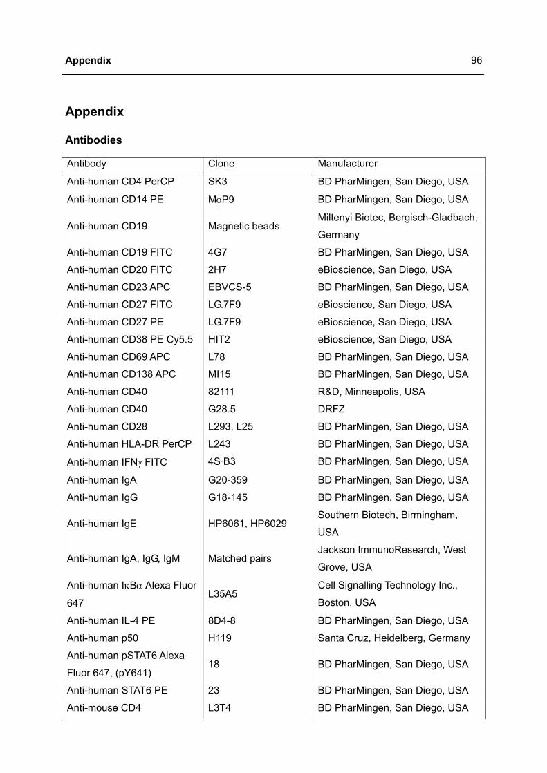
Appendix
96
Appendix
Antibodies
Antibody Clone Manufacturer
Anti-human CD4 PerCP SK3 BD PharMingen, San Diego, USA
Anti-human CD14 PE MφP9 BD PharMingen, San Diego, USA
Anti-human CD19 Magnetic beads Miltenyi Biotec, Bergisch-Gladbach,
Germany
Anti-human CD19 FITC 4G7 BD PharMingen, San Diego, USA
Anti-human CD20 FITC 2H7 eBioscience, San Diego, USA
Anti-human CD23 APC EBVCS-5 BD PharMingen, San Diego, USA
Anti-human CD27 FITC LG.7F9 eBioscience, San Diego, USA
Anti-human CD27 PE LG.7F9 eBioscience, San Diego, USA
Anti-human CD38 PE Cy5.5 HIT2 eBioscience, San Diego, USA
Anti-human CD69 APC L78 BD PharMingen, San Diego, USA
Anti-human CD138 APC MI15 BD PharMingen, San Diego, USA
Anti-human CD40 82111 R&D, Minneapolis, USA
Anti-human CD40 G28.5 DRFZ
Anti-human CD28 L293, L25 BD PharMingen, San Diego, USA
Anti-human HLA-DR PerCP L243 BD PharMingen, San Diego, USA
Anti-human IFNγ FITC 4S·B3 BD PharMingen, San Diego, USA
Anti-human IgA G20-359 BD PharMingen, San Diego, USA
Anti-human IgG G18-145 BD PharMingen, San Diego, USA
Anti-human IgE HP6061, HP6029 Southern Biotech, Birmingham,
USA
Anti-human IgA, IgG, IgM Matched pairs Jackson ImmunoResearch, West
Grove, USA
Anti-human IκBα Alexa Fluor
647 L35A5
Cell Signalling Technology Inc.,
Boston, USA
Anti-human IL-4 PE 8D4-8 BD PharMingen, San Diego, USA
Anti-human p50 H119 Santa Cruz, Heidelberg, Germany
Anti-human pSTAT6 Alexa
Fluor 647, (pY641) 18 BD PharMingen, San Diego, USA
Anti-human STAT6 PE 23 BD PharMingen, San Diego, USA
Anti-mouse CD4 L3T4 BD PharMingen, San Diego, USA

Appendix
97
Anti-mouse CD8 RM4-15 BD PharMingen, San Diego, USA
Anti-mouse IgG1, biotin A85-1 BD PharMingen, San Diego, USA
Anti-mouse IgG2a, biotin R19-15 BD PharMingen, San Diego, USA
Anti-rabbit HRP Goat, Sc-2004 Santa Cruz, Heidelberg, Germany
Anti-rat IgG, biotin Goat BD PharMingen, San Diego, USA
Purified mouse IgE, κ C38-2 BD PharMingen, San Diego, USA
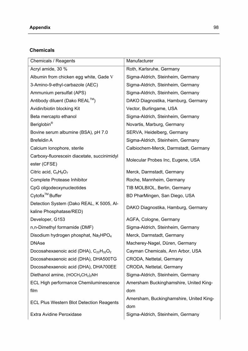
Appendix
98
Chemicals
Chemicals / Reagents Manufacturer
Acryl amide, 30 % Roth, Karlsruhe, Germany
Albumin from chicken egg white, Gade V Sigma-Aldrich, Steinheim, Germany
3-Amino-9-ethyl-carbazole (AEC) Sigma-Aldrich, Steinheim, Germany
Ammunium persulfat (APS) Sigma-Aldrich, Steinheim, Germany
Antibody diluent (Dako REALTM) DAKO Diagnostika, Hamburg, Germany
Avidin/biotin blocking Kit Vector, Burlingame, USA
Beta mercapto ethanol Sigma-Aldrich, Steinheim, Germany
Beriglobin® Novartis, Marburg, Germany
Bovine serum albumine (BSA), pH 7.0 SERVA, Heidelberg, Germany
Brefeldin A Sigma-Aldrich, Steinheim, Germany
Calcium Ionophore, sterile Calbiochem-Merck, Darmstadt, Germany
Carboxy-fluorescein diacetate, succinimidyl
ester (CFSE) Molecular Probes Inc, Eugene, USA
Citric acid, C6H8O7 Merck, Darmstadt, Germany
Complete Protease Inhibitor Roche, Mannheim, Germany
CpG oligodeoxynucleotides TIB MOLBIOL, Berlin, Germany
CytofixTM Buffer BD PharMingen, San Diego, USA
Detection System (Dako REAL, K 5005, Al-
kaline Phosphatase/RED) DAKO Diagnostika, Hamburg, Germany
Developer, G153 AGFA, Cologne, Germany
n,n-Dimethyl formamide (DMF) Sigma-Aldrich, Steinheim, Germany
Disodium hydrogen phosphat, Na2HPO4 Merck, Darmstadt, Germany
DNAse Macherey-Nagel, Düren, Germany
Docosahexaenoic acid (DHA), C22H32O2 Cayman Chemicals, Ann Arbor, USA
Docosahexaenoic acid (DHA), DHA500TG CRODA, Nettetal, Germany
Docosahexaenoic acid (DHA), DHA700EE CRODA, Nettetal, Germany
Diethanol amine, (HOCH2CH2)2NH Sigma-Aldrich, Steinheim, Germany
ECL High performance Chemiluminescence
film
Amersham Buckinghamshire, United King-
dom
ECL Plus Western Blot Detection Reagents Amersham, Buckinghamshire, United King-
dom
Extra Avidine Peroxidase Sigma-Aldrich, Steinheim, Germany
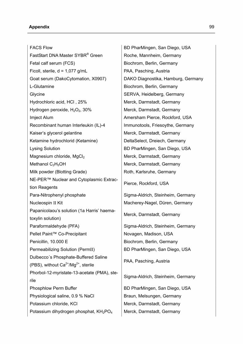
Appendix
99
FACS Flow BD PharMingen, San Diego, USA
FastStart DNA Master SYBR® Green Roche, Mannheim, Germany
Fetal calf serum (FCS) Biochrom, Berlin, Germany
Ficoll, sterile, d = 1,077 g/mL PAA, Pasching, Austria
Goat serum (DakoCytomation, X0907) DAKO Diagnostika, Hamburg, Germany
L-Glutamine Biochrom, Berlin, Germany
Glycine SERVA, Heidelberg, Germany
Hydrochloric acid, HCl , 25% Merck, Darmstadt, Germany
Hydrogen peroxide, H2O2, 30% Merck, Darmstadt, Germany
Imject Alum Amersham Pierce, Rockford, USA
Recombinant human Interleukin (IL)-4 Immunotools, Friesoythe, Germany
Kaiser’s glycerol gelantine Merck, Darmstadt, Germany
Ketamine hydrochlorid (Ketamine) DeltaSelect, Dreiech, Germany
Lysing Solution BD PharMingen, San Diego, USA
Magnesium chloride, MgCl2 Merck, Darmstadt, Germany
Methanol C2H5OH Merck, Darmstadt, Germany
Milk powder (Blotting Grade) Roth, Karlsruhe, Germany
NE-PER™ Nuclear and Cytoplasmic Extrac-
tion Reagents Pierce, Rockford, USA
Para-Nitrophenyl phosphate Sigma-Aldrich, Steinheim, Germany
Nucleospin II Kit Macherey-Nagel, Düren, Germany
Papanicolaou’s solution (1a Harris’ haema-
toxylin solution) Merck, Darmstadt, Germany
Paraformaldehyde (PFA) Sigma-Aldrich, Steinheim, Germany
Pellet Paint™ Co-Precipitant Novagen, Madison, USA
Penicillin, 10.000 E Biochrom, Berlin, Germany
Permeabilizing Solution (PermII) BD PharMingen, San Diego, USA
Dulbecco´s Phosphate-Buffered Saline
(PBS), without Ca2+/Mg2+, sterile PAA, Pasching, Austria
Phorbol-12-myristate-13-acetate (PMA), ste-
rile Sigma-Aldrich, Steinheim, Germany
Phosphlow Perm Buffer BD PharMingen, San Diego, USA
Physiological saline, 0.9 % NaCl Braun, Melsungen, Germany
Potassium chloride, KCl Merck, Darmstadt, Germany
Potassium dihydrogen phosphat, KH2PO4 Merck, Darmstadt, Germany
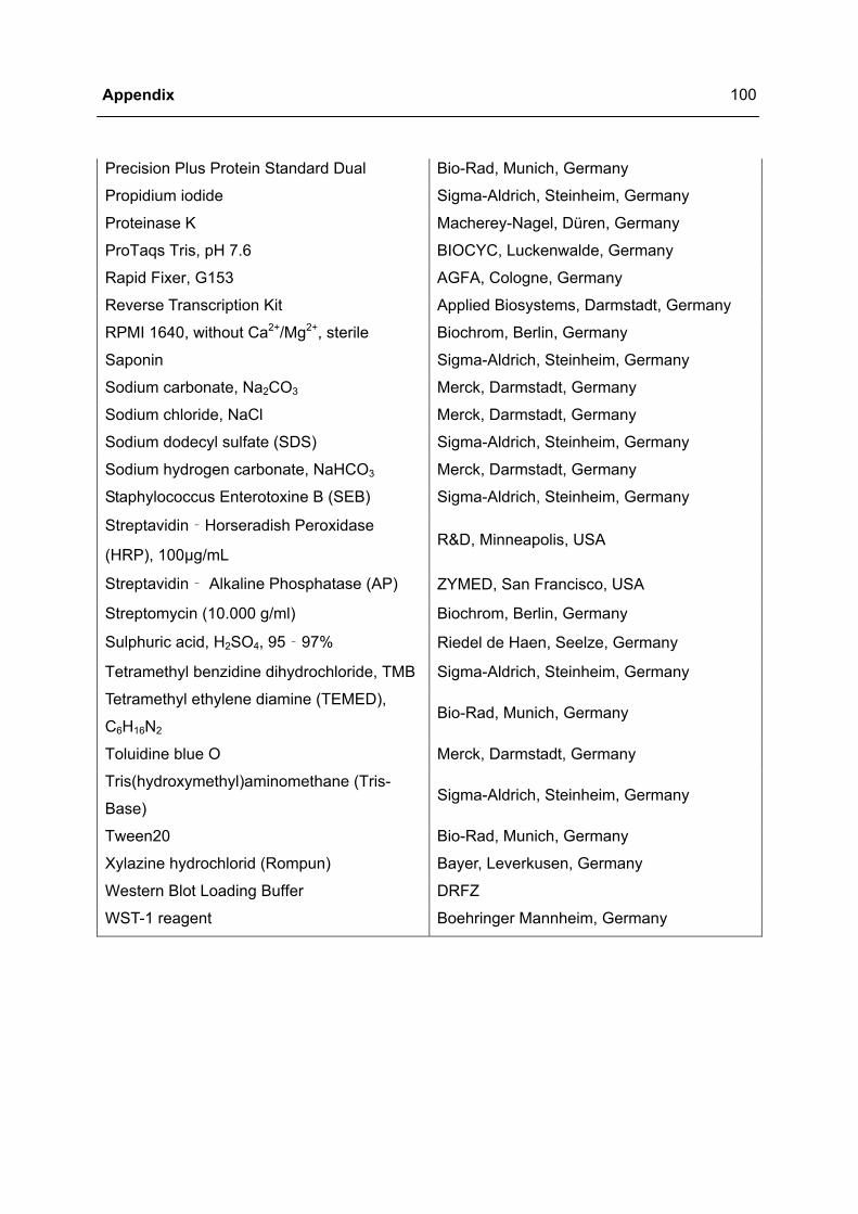
Appendix
100
Precision Plus Protein Standard Dual Bio-Rad, Munich, Germany
Propidium iodide Sigma-Aldrich, Steinheim, Germany
Proteinase K Macherey-Nagel, Düren, Germany
ProTaqs Tris, pH 7.6 BIOCYC, Luckenwalde, Germany
Rapid Fixer, G153 AGFA, Cologne, Germany
Reverse Transcription Kit Applied Biosystems, Darmstadt, Germany
RPMI 1640, without Ca2+/Mg2+, sterile Biochrom, Berlin, Germany
Saponin Sigma-Aldrich, Steinheim, Germany
Sodium carbonate, Na2CO3 Merck, Darmstadt, Germany
Sodium chloride, NaCl Merck, Darmstadt, Germany
Sodium dodecyl sulfate (SDS) Sigma-Aldrich, Steinheim, Germany
Sodium hydrogen carbonate, NaHCO3 Merck, Darmstadt, Germany
Staphylococcus Enterotoxine B (SEB) Sigma-Aldrich, Steinheim, Germany
Streptavidin‐Horseradish Peroxidase
(HRP), 100µg/mL R&D, Minneapolis, USA
Streptavidin‐ Alkaline Phosphatase (AP) ZYMED, San Francisco, USA
Streptomycin (10.000 g/ml) Biochrom, Berlin, Germany
Sulphuric acid, H2SO4, 95‐97% Riedel de Haen, Seelze, Germany
Tetramethyl benzidine dihydrochloride, TMB Sigma-Aldrich, Steinheim, Germany
Tetramethyl ethylene diamine (TEMED),
C6H16N2 Bio-Rad, Munich, Germany
Toluidine blue O Merck, Darmstadt, Germany
Tris(hydroxymethyl)aminomethane (Tris-
Base) Sigma-Aldrich, Steinheim, Germany
Tween20 Bio-Rad, Munich, Germany
Xylazine hydrochlorid (Rompun) Bayer, Leverkusen, Germany
Western Blot Loading Buffer DRFZ
WST-1 reagent Boehringer Mannheim, Germany
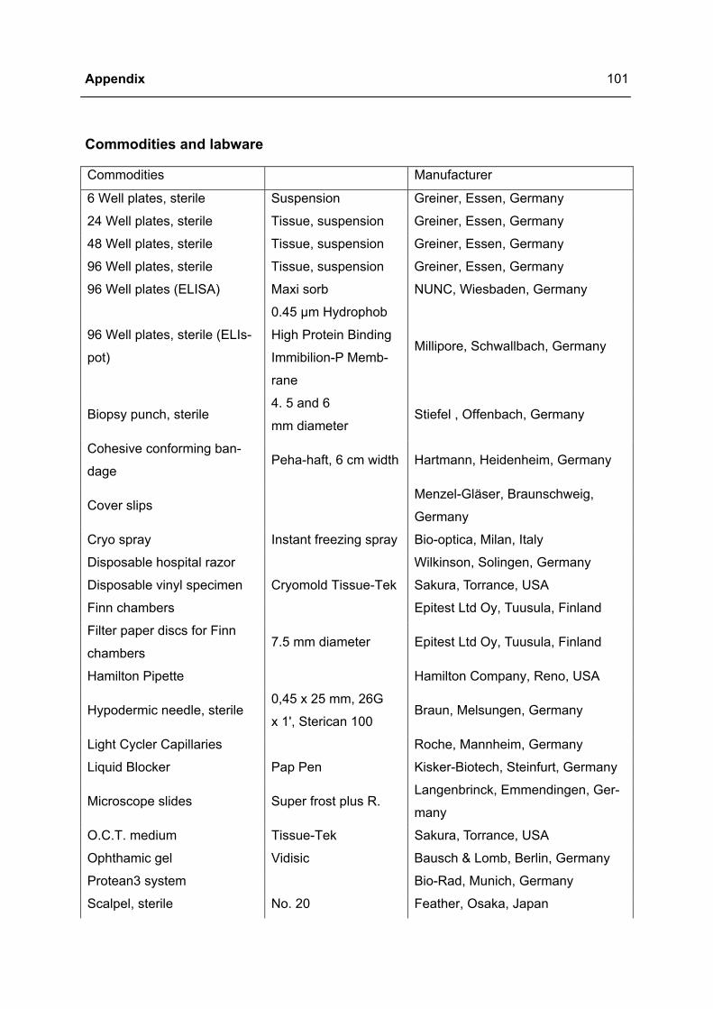
Appendix
101
Commodities and labware
Commodities Manufacturer
6 Well plates, sterile Suspension Greiner, Essen, Germany
24 Well plates, sterile Tissue, suspension Greiner, Essen, Germany
48 Well plates, sterile Tissue, suspension Greiner, Essen, Germany
96 Well plates, sterile Tissue, suspension Greiner, Essen, Germany
96 Well plates (ELISA) Maxi sorb NUNC, Wiesbaden, Germany
96 Well plates, sterile (ELIs-
pot)
0.45 µm Hydrophob
High Protein Binding
Immibilion-P Memb-
rane
Millipore, Schwallbach, Germany
Biopsy punch, sterile 4. 5 and 6
mm diameter Stiefel , Offenbach, Germany
Cohesive conforming ban-
dage Peha-haft, 6 cm width Hartmann, Heidenheim, Germany
Cover slips Menzel-Gläser, Braunschweig,
Germany
Cryo spray Instant freezing spray Bio-optica, Milan, Italy
Disposable hospital razor Wilkinson, Solingen, Germany
Disposable vinyl specimen Cryomold Tissue-Tek Sakura, Torrance, USA
Finn chambers Epitest Ltd Oy, Tuusula, Finland
Filter paper discs for Finn
chambers 7.5 mm diameter Epitest Ltd Oy, Tuusula, Finland
Hamilton Pipette Hamilton Company, Reno, USA
Hypodermic needle, sterile 0,45 x 25 mm, 26G
x 1', Sterican 100 Braun, Melsungen, Germany
Light Cycler Capillaries Roche, Mannheim, Germany
Liquid Blocker Pap Pen Kisker-Biotech, Steinfurt, Germany
Microscope slides Super frost plus R. Langenbrinck, Emmendingen, Ger-
many
O.C.T. medium Tissue-Tek Sakura, Torrance, USA
Ophthamic gel Vidisic Bausch & Lomb, Berlin, Germany
Protean3 system Bio-Rad, Munich, Germany
Scalpel, sterile No. 20 Feather, Osaka, Japan

Appendix
102
Syringes, sterile 10ml, 20 ml, 50ml Braun, Melsungen, Germany
Wattman Paper Schleicher & Schull, Dassel, Ger-
many
Western Blot Cassette ECL Hypercassette Amersham Buckinghamshire, Uni-
ted Kingdom
Western Blot Membrane Polyvinylidenfluorid
(PVDF)
Amersham Buckinghamshire, Uni-
ted Kingdom
Reaction tubes, pipette tips and other commodities were used from Eppendorf, Falcon, and
Sarstedt.

Appendix
103
Equipment
Equipment Model Manufacturer
Autoclave MELAG, Berlin, Germany
Camera EOS 20D Canon, Krefeldt, Germany
Centrifuge Megafuge 1.OR Heraeus, Hanau, Germany
Multifuge 4KR Heraeus, Hanau, Germany
5417R; 5417R Eppendorf, Hamburg, Germany
Minifuge RF Heraeus, Hanau, Germany
Cryostat Jung frigocut 2800N Leica, Wetzlar, Germany
Electronic cell counter CASY 1, Modell TT Innovatis, Reutlingen, Germany
ELISA reader Dynex MRX version
1.33 DYNATECH, Chantilly, USA
ELISPOT reader CTL ImmunoSpot®
S4 Analyzer
C.T.L Cellular Technology, Cleve-
land, USA
Flow bench HERA safe Heraeus, Hanau, Germany
Flow cytometre FACS Calibur Becton Dickinson, Heidelberg, Ger-
many
Freezer (-20°C)/Fridge (4°C) TKF380 EUREKA, Emsdetten, Germany
Freezer (-80°C) Hera Freeze Heraeus, Hanau, Germany
Gel chamber 40 1214 peqLab PEQLAB Biotechnologie, Erlangen,
Germany
Gradient cycler Px2 Thermal Cycler Thermo, Hamburg, Germany
Hot plate nuova II Thermolyne, Dubuque, USA
Incubator HERA cell Heraeus, Hanau, Germany
Lab balance Sartorius, Göttingen, Germany
Light Cycler LightCycler 1.5 Roche, Mannheim, Germany
Magnetic cell sorter autoMACS Separator Miltenyi Biotec, Bergisch Gladbach,
Germany
Magnetic stirrer Magnetmix 2070 Hecht-Assistent, Sondenheim, Ger-
many
Microscope Axioskop and Axi-
oplan2 imaging Carl Zeiss, Jena, Germany
Microscope camera Axio Cam HRc Carl Zeiss, Jena, Germany
Pipets 10µl, 100µl, 200µl; Eppendorf, Hamburg, Germany
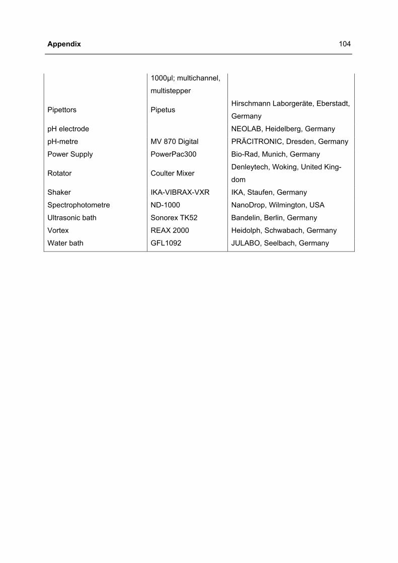
Appendix
104
1000µl; multichannel,
multistepper
Pipettors Pipetus Hirschmann Laborgeräte, Eberstadt,
Germany
pH electrode NEOLAB, Heidelberg, Germany
pH-metre MV 870 Digital PRÄCITRONIC, Dresden, Germany
Power Supply PowerPac300 Bio-Rad, Munich, Germany
Rotator Coulter Mixer Denleytech, Woking, United King-
dom
Shaker IKA-VIBRAX-VXR IKA, Staufen, Germany
Spectrophotometre ND-1000 NanoDrop, Wilmington, USA
Ultrasonic bath Sonorex TK52 Bandelin, Berlin, Germany
Vortex REAX 2000 Heidolph, Schwabach, Germany
Water bath GFL1092 JULABO, Seelbach, Germany
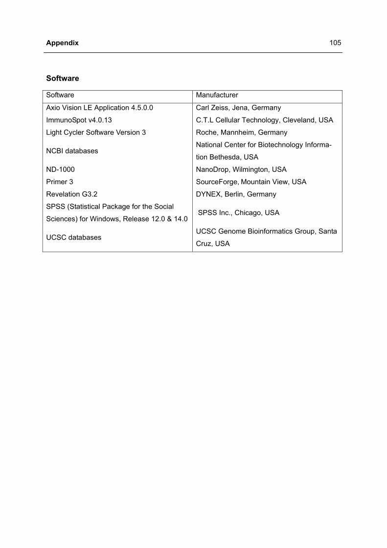
Appendix
105
Software
Software Manufacturer
Axio Vision LE Application 4.5.0.0 Carl Zeiss, Jena, Germany
ImmunoSpot v4.0.13 C.T.L Cellular Technology, Cleveland, USA
Light Cycler Software Version 3 Roche, Mannheim, Germany
NCBI databases National Center for Biotechnology Informa-
tion Bethesda, USA
ND-1000 NanoDrop, Wilmington, USA
Primer 3 SourceForge, Mountain View, USA
Revelation G3.2 DYNEX, Berlin, Germany
SPSS (Statistical Package for the Social
Sciences) for Windows, Release 12.0 & 14.0 SPSS Inc., Chicago, USA
UCSC databases UCSC Genome Bioinformatics Group, Santa
Cruz, USA

Appendix
106
Buffers and solutions
ACE-DMF buffer (pH 5.0) 64 mM Sodium acetate
45 mM Acetate
Blotting buffer 25 mM Tris-base
190 mM glycine
20 % v/v Methanol
Diethanol amine buffer (pH 9.9) 840 mM Diethanol amine
1.1 mM MgCl2
FACS buffer PBS
1 % BSA
MACS buffer PBS
20 mM EDTA
0.2 % BSA
Medium complete 500 mL RPMI1640, with 2.0 g/L NaHCO3,
with stabile glutamine
50 mL FCS
100 U/mL Penecillin
100 µg/mL Streptomycin
Ketamine / Rompun (8 : 1 : 1) 0.9 % NaCl :
50 mg/mL Ketamine
2 % Rompun
Running buffer 25 mM Tris-base
190 mM Glycine
7 mM SDS
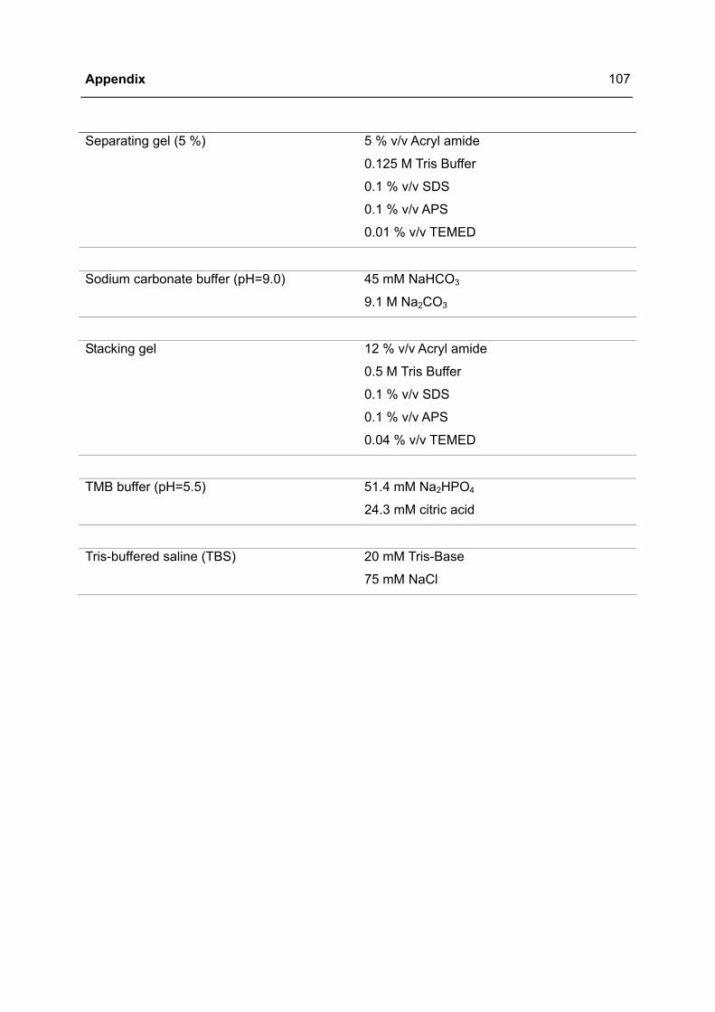
Appendix
107
Separating gel (5 %) 5 % v/v Acryl amide
0.125 M Tris Buffer
0.1 % v/v SDS
0.1 % v/v APS
0.01 % v/v TEMED
Sodium carbonate buffer (pH=9.0) 45 mM NaHCO3
9.1 M Na2CO3
Stacking gel 12 % v/v Acryl amide
0.5 M Tris Buffer
0.1 % v/v SDS
0.1 % v/v APS
0.04 % v/v TEMED
TMB buffer (pH=5.5) 51.4 mM Na2HPO4
24.3 mM citric acid
Tris-buffered saline (TBS) 20 mM Tris-Base
75 mM NaCl
List of abbreviations
108
List of abbreviations
AA Arachidonic acid, 20:4n-6
AICD Activation induced cell death
AID Activation induced (cytidine) desaminase
ALA α-linolenic acid, 18:3n-3
AP-1 Activator protein 1 APC Antigen presenting cell
BMI Body mass index
CH Heavy chain constant regions
CLA Cutaneous lymphocyte-associated antigen
COX Cyclooxygenase
CSS Clinical skin score
CSR Class switch recombination
DAG Diacylglycerol
DC Dendritic cells
DGLA Dihomo-gamma linolenic acid, 20:3n-6
DHA Docosahexaenoic acid, 22:6n-3
e.c. Epicutaneous
εGLT Epsilon germline transcript
ELISA Enzyme Linked Immuno Assay
ELIspot Enzyme Linked Immuno Spot Technique
EPA Eicosapentaenoic acid, 20:5n-3
ERK Extracellular signal-regulated kinase
FACS Fluorescence activated cell sorter
FcεRI High affinity receptor for IgE
FLG Filament-aggregating protein
FSC Forward light scatter
g Acceleration of gravity
GCK Germinal center kinase
GLA γ-linoleic acid, 20:3n-6
HDL High density lipoprotein
HLA human leukocyte antigen
HPRT Hypoxanthine-Guanine Phosphoribosyltransferase
Ig Immunoglobulin

List of abbreviations
109
IDEC Inflammatory dendritic epidermal cells
IFN Interferon
IκB inhibitor of NFκB
IKK inhibitor of NFκB (IκB) kinase
IL- Interleukin
IL-4R Interleukin-4 Receptor
i.p. Intraperitoneal
IRS- Insulin receptor substrate
ITIM Immuno-tyrosine inhibitory motif
JAK Janus activated kinase JNK c-jun kinase
LA Linoleic acid, 18:2n-6
LC Langerhans cells
LOX Lipoxygenase
LT Leukotriene
LXR Liver X receptor
MACS Magnetic cell sorting
MAPK Mitogen-activated protein kinase
MEKK1 MAPK/ ERK kinase kinase 1
MFI Mean fluorescence intensity
MHC II Major histocompatibility complex II
min Minute
MO Monocyte
n.d. Not detectable
NFκB Nuclear factor kappa B
NIK NFκB inducing kinase
n.s. Not significant
OA Oleic acid, 18:1n-9
OVA Ovalbumin
PBMC Peripheral blood mononuclear cells
PG Prostaglandin
PI3-K Phosphatidylinositol 3-kinase
PKC Protein kinase C
PL Phospholipase
PPAR Peroxisome proliferator-activated receptor
List of abbreviations
110
pSTAT6 Phosphorylated signal transducer and activator of transcription 6
PUFA Polyunsaturated fatty acids
RT-PCR Reverse-transcriptase polymerase chain reaction
RXR Retinoic acid X receptor
S. aureus Staphylococcus aureus
SCORAD Severity Scoring of Atopic Dermatitis
SH2 Src homology domain 2
Shp-1 SH2-containing tyrosine phosphatase--1
SREBP Sterol regulatory-element binding protein
SSC Side light scatter
STAT6 Signal transducer and activator of transcription 6
TH1/2 T Helper cell type 1/2
TNF Tumor necrosis factor
TRAF TNF-receptor-associated factor
Treg Regulatory T cells
TSLP Thymic stromal lymphopoietin
TX Thromboxane

Acknowledgement
111
Acknowledgement
Many people made their contribution to the realisation of my thesis and I would like to thank
them all for their help and support.
First of all I would like to express my deepest gratitude to Prof. Dr. Margitta Worm for giving
me the opportunity to do my PhD thesis and to make this interesting research at the Depart-
ment of Dermatology. Many thanks for her support and supervision and of course for kind
advices and help during these years. Moreover, I am grateful for giving me the opportunities
to attend these relevant national and international congresses.
Many thanks to Prof. Dr. Andreas Radbruch for his helpful scientific discussions in the “B cell
club” and for supporting my PhD thesis.
I wish to thank Dr. Elke Luger and Dr. Magda Babina for their scientific discussions and kind-
ness during the course of my thesis.
My deepest gratitude to my family for being there for me, especially to Oliver for sharing my
ups and downs, for support and for beeing patient during this time.
I am grateful to Milena Milovanovic, for helping me in my hardest situations and for encour-
aging me. Thank you for your optimistic discussions, for proofreading and supporting me with
my manuscript(s). Many thanks for your nice friendship, and for your kind supports.
Many thanks to Dennis Ernst for his great help in the lab and for his wonderful support during
the mouse studies.
I am very thankful to Sabine Dölle for our nice time and collaboration during the clinical trial.
We complemented one another.
Many thanks to the people from the working group who supported me with constructive ad-
vices, assistance, encouraging words and the nice working atmosphere. Above all, I would
like to thank Kerstin Hilt, Björn Hartman, Carolin Heunemann, Dana Hoser and Miriam Ger-
stenberg.
Special thanks goes to Sven Guhl for giving me useful technical and scientific advices during
the whole course of my PhD thesis, especially for western blot help.
My gratitude to Dr. Claudia Rasche and Manuel Metzger as well as the participants who were
essential for realisation of the clinical study. Many thanks to Dr. Henrik Jungclass and Prof.
Dr. Harald Renz who supported the success of the clinical study by performing fatty acid

Acknowledgement
112
analysis. Additionally, I would like to thank the staff of Clinic of Dermatology for their kind
support and patience.
Furthermore, I would like to thank to all the self-forgetful blood donors and Charité blood
donation. Without them the study would not have been performed.
Statement of the authorship
113
Statement of authorship
I hereby certify that I performed and wrote the current thesis independently and did not use
other than the listed support. This thesis does not exist neither in the same or similar form
nor is it submitted to another examination procedure. I did not gain an academic degree be-
fore nor did I try to do this. I claim full responsibility for the contents of this thesis.
Hiermit erkläre ich, die vorliegende Arbeit selbstständig angefertigt und keine anderen als die
angegebenen Hilfsmittel verwendet zu haben. Diese Arbeit hat weder in gleicher noch in
ähnlicher Form in einem anderen Prüfungsverfahren vorgelegen. Ich habe in keinem frühe-
ren Verfahren einem akademischen Grad erworben oder zu erweben versucht.
Christin Koch
Mathematisch-Naturwissenschaftlichen Fakultät I
Dekan: Prof. Dr. Lutz-Helmut Schön
Gutachter/innen: 1. Prof. Dr. Richard Lucius
2. Prof. Dr. Andreas Radbruch
3. Prof. Dr. Margitta Worm
Tag der mündlichen Prüfung: 12.03.2009
Copyright © 2022 FDOKUMEN