Improvements in Mucopolysaccharidosis I Mice After Adult Retroviral Vector–mediated Gene Therapy...
14
© The American Society of Gene Therapy original article Molecular Therapy vol. 15 no. 5, 889–902 may 2007 889 Improvements in Mucopolysaccharidosis I Mice After Adult Retroviral Vector–mediated Gene Therapy with Immunomodulation Xiucui Ma 1 , Yuli Liu 1 , Mindy Tittiger 1 , Anne Hennig 1 , Attila Kovacs 1 , Sarah Popelka 1 , Baomei Wang 2 , Ramin Herati 1 , Mark Bigg 1 and Katherine P Ponder 1,3 1 Department of Internal Medicine, Washington University School of Medicine, St. Louis, Missouri, USA; 2 Department of Pathology, Washington University School of Medicine, St. Louis, Missouri, USA; 3 Department of Biochemistry and Molecular Biophysics, Washington University School of Medicine, St. Louis, Missouri, USA Mucopolysaccharidosis I (MPS I) is caused by deficient α-L-iduronidase (IDUA) activity and results in the accumu- lation of glycosaminoglycans and multisystemic disease. Gene therapy could program cells to secrete mannose 6-phosphate–modified IDUA, and enzyme in blood could be taken up by other cells. Neonatal retroviral vector (RV)–mediated gene therapy has been shown to reduce the manifestations of murine MPS I; however, intrave- nous injection of RV into adults was ineffective owing to a cytotoxic T lymphocyte (CTL) response against trans- duced cells. In this study, prolonged inhibition of CD28 signaling with CTLA4-Ig, or transient administration of CTLA4-Ig with an anti-CD40 ligand antibody or with an anti-CD4 antibody, resulted in stable expression in most mice that received RV as adults. Mice with stable expression had 81 ± 41 U/ml IDUA activity in serum. This resulted in reductions in bone disease, improve- ments in hearing and vision, and reductions in biochem- ical and pathological evidence of lysosomal storage in most organs. Improvements in brain were likely due to diffusion of enzyme from blood. However, aortic disease was refractory to treatment. This demonstrates that most manifestations of MPS I can be prevented using adult gene therapy if an immune response is blocked. Received 26 September 2006; accepted 3 January 2007; published online 20 February 2007. doi:10.1038/mt.sj.6300112 INTRODUCTION Mucopolysaccharidosis I (MPS I) is an autosomal recessive disease caused by a deficiency of α-l-iduronidase (IDUA; EC 3.2.1.76). 1 It results in the accumulation of the glycosaminoglycans (GAGs) heparan and dermatan sulfate, and has an incidence of 1:100,000 live births. 2 Patients with severe disease (Hurler syndrome; OMIM #607014) have skeletal deformities, hearing and vision abnor- malities, cardiac disease, and mental retardation. Patients with intermediate disease (Hurler–Scheie syndrome; OMIM #607015) have similar somatic manifestations without neurological disease. Patients with mild disease (Scheie syndrome; OMIM #607016) have somatic manifestations of reduced severity. MPS I is currently treated with hematopoietic stem cell (HSC) transplantation or enzyme replacement therapy (ERT). HSC transplantation has reduced the manifestations of disease in mice, 3 dogs, 4 and humans 5 with MPS I. e likely mechanism is migra- tion of transplant-derived blood cells into organs, where the cells secrete enzyme modified with mannose 6-phosphate (M6P) that can be taken up by adjacent cells via the M6P receptor (M6PR). HSC transplantation is limited by the need for a compatible donor and the risks and costs of the procedure. ERT involves the intra- venous injection of M6P-modified enzyme, which can diffuse into organs and be internalized via the M6PR. 6 ERT has been thera- peutic in cats, 7 dogs, 8 and humans 9 with MPS I. Difficulties with ERT include the need for infusion once a week and a cost of more than $10,000 per kg body weight per year. Gene therapy has been successfully used to treat MPS in animal models. 10 HSC-directed gene therapy to adults reduces the manifestations of MPS I in mice 3 in a similar way to HSC transplantation. As an alternative treatment modality, other cells could secrete enzyme with M6P into blood, and enzyme in blood could be taken up by cells in other organs via the M6PR. Indeed, neonatal administration of an adeno-associated virus vector, 11 γ-retroviral vector (RV 12,13 ), or lenti-retroviral (lentiviral) vector 14 resulted in stable enzyme expression and prevented disease mani- festations in many organs. As most patients are not diagnosed at birth, it is important to achieve gene transfer in older animals. In contrast to the suc- cess with gene therapy in newborns, transduction of liver with γ-RV 12 or lentiviral vectors 14,15 in adults had no therapeutic effect. is failure was probably due to the response of cytotoxic T lym- phocytes (CTLs) to IDUA-expressing cells, as liver IDUA activity and vector DNA levels were low in two of these studies. 12,14 e long-term expression in some mice that received HSC-directed gene therapy as adults 3 may be due to the ability of this proce- dure to induce tolerance to a transgene, although this approach Correspondence: Katherine P Ponder, Department of Internal Medicine, Washington University School of Medicine, 660 South Euclid Avenue, St. Louis, Missouri 63110, USA. E-mail: [email protected]
-
Upload
independent -
Category
Documents
-
view
4 -
download
0
Transcript of Improvements in Mucopolysaccharidosis I Mice After Adult Retroviral Vector–mediated Gene Therapy...

© The American Society of Gene Therapy original article
Molecular Therapy vol. 15 no. 5, 889–902 may 2007 889
Improvements in Mucopolysaccharidosis I Mice After Adult Retroviral Vector–mediated Gene Therapy with ImmunomodulationXiucui Ma1, Yuli Liu1, Mindy Tittiger1, Anne Hennig1, Attila Kovacs1, Sarah Popelka1, Baomei Wang2, Ramin Herati1, Mark Bigg1 and Katherine P Ponder1,3
1Department of Internal Medicine, Washington University School of Medicine, St. Louis, Missouri, USA; 2Department of Pathology, Washington University School of Medicine, St. Louis, Missouri, USA; 3Department of Biochemistry and Molecular Biophysics, Washington University School of Medicine, St. Louis, Missouri, USA
Mucopolysaccharidosis I (MPS I) is caused by deficient α-l-iduronidase (IDUA) activity and results in the accumu-lation of glycosaminoglycans and multisystemic disease. Gene therapy could program cells to secrete mannose 6-phosphate–modified IDUA, and enzyme in blood could be taken up by other cells. Neonatal retroviral vector (RV)–mediated gene therapy has been shown to reduce the manifestations of murine MPS I; however, intrave-nous injection of RV into adults was ineffective owing to a cytotoxic T lymphocyte (CTL) response against trans-duced cells. In this study, prolonged inhibition of CD28 signaling with CTLA4-Ig, or transient administration of CTLA4-Ig with an anti-CD40 ligand antibody or with an anti-CD4 antibody, resulted in stable expression in most mice that received RV as adults. Mice with stable expression had 81 ± 41 U/ml IDUA activity in serum. This resulted in reductions in bone disease, improve-ments in hearing and vision, and reductions in biochem-ical and pathological evidence of lysosomal storage in most organs. Improvements in brain were likely due to diffusion of enzyme from blood. However, aortic disease was refractory to treatment. This demonstrates that most manifestations of MPS I can be prevented using adult gene therapy if an immune response is blocked.
Received 26 September 2006; accepted 3 January 2007; published online 20 February 2007. doi:10.1038/mt.sj.6300112
IntroductIonMucopolysaccharidosis I (MPS I) is an autosomal recessive disease caused by a deficiency of α-l-iduronidase (IDUA; EC 3.2.1.76).1 It results in the accumulation of the glycosaminoglycans (GAGs) heparan and dermatan sulfate, and has an incidence of 1:100,000 live births.2 Patients with severe disease (Hurler syndrome; OMIM #607014) have skeletal deformities, hearing and vision abnor-malities, cardiac disease, and mental retardation. Patients with intermediate disease (Hurler–Scheie syndrome; OMIM #607015)
have similar somatic manifestations without neurological disease. Patients with mild disease (Scheie syndrome; OMIM #607016) have somatic manifestations of reduced severity.
MPS I is currently treated with hematopoietic stem cell (HSC) transplantation or enzyme replacement therapy (ERT). HSC transplantation has reduced the manifestations of disease in mice,3 dogs,4 and humans5 with MPS I. The likely mechanism is migra-tion of transplant-derived blood cells into organs, where the cells secrete enzyme modified with mannose 6-phosphate (M6P) that can be taken up by adjacent cells via the M6P receptor (M6PR). HSC transplantation is limited by the need for a compatible donor and the risks and costs of the procedure. ERT involves the intra-venous injection of M6P-modified enzyme, which can diffuse into organs and be internalized via the M6PR.6 ERT has been thera-peutic in cats,7 dogs,8 and humans9 with MPS I. Difficulties with ERT include the need for infusion once a week and a cost of more than $10,000 per kg body weight per year.
Gene therapy has been successfully used to treat MPS in animal models.10 HSC-directed gene therapy to adults reduces the manifestations of MPS I in mice3 in a similar way to HSC transplantation. As an alternative treatment modality, other cells could secrete enzyme with M6P into blood, and enzyme in blood could be taken up by cells in other organs via the M6PR. Indeed, neonatal administration of an adeno-associated virus vector,11 γ-retroviral vector (RV12,13), or lenti-retroviral (lentiviral) vector14 resulted in stable enzyme expression and prevented disease mani-festations in many organs.
As most patients are not diagnosed at birth, it is important to achieve gene transfer in older animals. In contrast to the suc-cess with gene therapy in newborns, transduction of liver with γ-RV12 or lentiviral vectors14,15 in adults had no therapeutic effect. This failure was probably due to the response of cytotoxic T lym-phocytes (CTLs) to IDUA-expressing cells, as liver IDUA activity and vector DNA levels were low in two of these studies.12,14 The long-term expression in some mice that received HSC-directed gene therapy as adults3 may be due to the ability of this proce-dure to induce tolerance to a transgene, although this approach
Correspondence: Katherine P Ponder, Department of Internal Medicine, Washington University School of Medicine, 660 South Euclid Avenue, St. Louis, Missouri 63110, USA. E-mail: [email protected]

890 www.moleculartherapy.org vol. 15 no. 5 may 2007
© The American Society of Gene TherapyAdult Gene Therapy for MPS I Mice
did not block an immune response to IDUA in MPS I dogs.16 A recent report suggested that use of a liver-restricted promoter could prevent an immune response to human IDUA in MPS I mice.17 However, the expression achieved was low and reduction in lysosomal storage was incomplete. We therefore investigated alter-native ways to induce tolerance to IDUA after systemic delivery.
CTLA4-Ig is a fusion protein between CTLA4 and IgG. CTLA4-Ig can bind to CD80 and CD86 present on antigen- presenting cells, which blocks CD80/86 from binding to CD28 on lymphocytes and activating their response.18 Indeed, continu-ous expression of CTLA4-Ig from an adenoviral vector allowed β-glucuronidase (GUSB) expression from a second adenoviral vector to be maintained for up to 200 days in MPS VII mice,19 and transient administration of CTLA4-Ig protein to cats resulted in long-standing tolerance to canine IDUA after neonatal gene ther-apy.20 An anti (α)-CD40-ligand (CD40L, also known as CD154) antibody can interfere with a second co-stimulatory pathway in which CD40 present on antigen-presenting cells binds to CD40L on T lymphocytes.18 Administration of an α-CD40L antibody for 2 weeks initiated at the time of injection of an adenoviral vec-tor expressing GUSB prolonged expression, although levels still fell at 3 months.21 The combination of CTLA4-Ig and α-CD40L acted synergistically to prevent immune responses.18,22 α-CD4 antibodies can reduce levels of CD4+ cells, which play an impor-tant role in providing T cell help.23 An α-CD4 antibody blocked an antibody response to GUSB after implantation of microcap-sules expressing GUSB into MPS VII mice.24 Finally, a 2-month course of cyclosporine and azathioprine induced tolerance to human IDUA in MPS I dogs.25 In this study, we evaluated the ability of immunosuppressive agents to prolong gene expres-sion after adult gene therapy to MPS I mice. Animals with stable expression were then tested for correction of the manifestations of the disease.
resultsserum IduA activity after adult gene therapyAn RV expressing canine IDUA (hAAT-cIDUA-WPRE) was injected intravenously into 6-week-old MPS I mice after induc-tion of hepatocyte replication with hepatocyte growth factor (HGF). Although animals achieved 73 ± 54 (SD; N = 12) U/ml serum IDUA activity at 1 week, enzyme activity fell to low levels at 2–3 weeks (Figure 1a). In an attempt to prolong the expres-sion, three separate groups of mice were treated transiently with a single immunosuppressive agent. The Transient CTLA4-Ig group received four intraperitoneal doses of human CTLA4-Ig over 2 weeks, as indicated by the short bars at the top of Figure 1b. Serum IDUA activity was stable for 1–2 months but fell there-after, although some mice maintained low expression that was clearly above the background activity. The Transient α-CD40L (Figure 1c) and Transient α-CD4 (Figure 1d) groups received two doses of an α-mouse CD40L antibody or an α-mouse CD4 antibody, respectively, around the time of transduction. Serum IDUA activity was high at 1 week but fell later. The Prolonged CTLA4-Ig (Figure 1e) group, which received adult gene trans-fer in conjunction with long-term administration of CTLA4-Ig, maintained IDUA activity at stable levels for up to 6.5 months,
the duration of evaluation. The Transient CTLA4-Ig + α-CD40L (Figure 1f) and Transient CTLA4-Ig + α-CD4 (Figure 1g) groups maintained stable IDUA activity for 6.5 months in 80 and 100% of mice, respectively.
evaluation of immune response genes in spleens of adult and newborn miceTo understand why mice produce a potent immune response to IDUA after adult, but not neonatal, gene therapy, RNA isolated from spleens collected at 3 days or at 6 weeks after birth was tested for levels of genes that are important for an immune response (Figure 2a). Some mice received RV injection 24 hours before they were killed, whereas others received no treatment. As admin-istration of the RV had very little effect upon RNA expression, values for all adults were pooled and compared with values for all newborns. The levels of RNA in adult mice relative to those in newborns were increased 7-fold for CD28 RNA (P = 0.007), 46-fold for CTLA4 (P = 0.004), 18-fold for CD40L (P = 0.002), 3-fold for CD4 (P = 0.03), 21-fold for CD8 (P = 0.03), 13-fold for IL-2 (P = 0.007), and 4-fold for IL-10 (P = 0.01). In contrast, the levels of RNA for IL-4 in adults were reduced to 18% of the levels in newborns (P = 0.03). Fluorescence-activated cell sorting demonstrated that 41 ± 6% of total splenocytes from 6-week-old mice were positive for CD3 (present on all T lymphocytes), 24 ± 6% were positive for CD4 (present on helper T cells, mono-cytes, and macrophages), and 12 ± 2% were positive for CD8 (present on cytotoxic or suppressor T cells and natural killer cells). The percentage of lymphocytes positive for CD3, CD4, and CD8 was reduced for newborn mice, at 6 ± 2, 3 ± 1, and 2 ± 1%, respectively. Thus, adults had sevenfold, sevenfold, and fivefold the percentage of CD3-, CD4-, and CD8-positive cells in total splenocytes compared with newborns (P < 0.001 for all comparisons).
Immunosuppression is transientReduction in responses to other antigens is a potential adverse effect of immunosuppressive agents. Mice that received tran-sient immunosuppression were challenged with two doses of human factor IX (hFIX) with adjuvant starting at 6 months of age (4.5 months after gene transfer). They produced similar levels of anti-hFIX antibodies to age-matched normal or untreated MPS I mice (Figure 2b). The mice that received prolonged CTLA4-Ig were not challenged in a similar fashion.
evaluation of the effect of gene therapy in adult MPs I miceThe second aim of this project was to determine whether adult gene therapy could prevent biochemical, pathological, and func-tional manifestations of disease. A total of 14 RV-treated mice showed stable expression, which included mice from the Pro-longed CTLA4-Ig (N = 5), the Transient CTLA4-Ig + α-CD40L (N = 4), and the Transient CTLA4-Ig + α-CD4 (N = 5) groups. Their average serum IDUA activity was 81 ± 41 U/ml, with values ranging from 25 to 156 U/ml. These mice were evaluated biochemically, pathologically, and with specialized testing to determine whether disease was corrected at 8 months after birth, which was 6.5 months after transduction. As some mice were not
© The American Society of Gene Therapy

Molecular Therapy vol. 15 no. 5 may 2007 891
© The American Society of Gene Therapy Adult Gene Therapy for MPS I Mice
evaluated with all tests because of accidents in the animal room, deaths from anesthesia, or scheduling problems, the number and the average serum activity for the group that was evaluated are reported.
IduA activity in organsWe previously demonstrated that the DNA levels in livers were more than tenfold those in other organs at 11 months after HGF-potentiated gene transfer of a similar γ-RV to adult mice.26
© The American Society of Gene Therapy
Figure 1 serum α-l-iduronidase (IduA) activity after transduction of adult mucopolysaccharidosis I (MPs I) mice. Six-week-old MPS I mice were injected with hepatocyte growth factor (HGF). During the period of peak hepatocyte replication, mice were injected with 5 × 10⁹ transducing units per kilogram of hAAT-cIDUA-WPRE. Each line represents IDUA activity for an individual mouse. (a) No immunomodulation. Serum IDUA activity was determined for retroviral vector (RV)–treated mice that did not receive immunosuppression. The mean serum IDUA activity ±1 SD in homozygous normal mice (0.9 ± 0.3 U/ml) is indicated by the gray-shaded region. Untreated MPS I mice had undetectable serum IDUA activity, as indicated by the level of 0.05 U/ml at 0 days. (b) Transient CTLA4-Ig. RV-treated MPS I mice also received four doses of human CTLA4-Ig intraperitoneally (IP) over 2 weeks, as indicated by the short bars at the top left. (c) Transient α-CD40L. RV-treated MPS I mice also received two doses of 12.5 mg/kg of an α-CD40L antibody IP over 3 days, as indicated by the long bars at the top left. (d) Transient α-CD4. RV-treated MPS I mice also received two doses of 12.5 mg/kg of an α-CD4 antibody over 2 days, as indicated by the long bars at the top left. (e) Prolonged CTLA4-Ig. RV-treated MPS I mice also received prolonged administration of human CTLA4-Ig, as indicated by the short bars at the top. (f) Transient CTLA4-Ig + α-CD40L. RV-treated MPS I mice also received transient CTLA4-Ig and transient α-CD40L antibody as indicated by the short and long bars, respectively, at the top. (g) Transient CTLA4-Ig + α-CD4. RV-treated MPS I mice also received transient CTLA4-Ig and transient α-CD4 antibody as indicated by the short and long bars, respectively, at the top.

892 www.moleculartherapy.org vol. 15 no. 5 may 2007
© The American Society of Gene TherapyAdult Gene Therapy for MPS I Mice
Therefore, the hypothesis was that transduced hepatocytes would secrete IDUA and that M6P-modified IDUA in blood would diffuse into other organs and correct disease. For RV-treated mice with stable expression, IDUA activity was highest in liver, at 4.2 ± 2.2 U/mg (2.5-fold normal), whereas untreated MPS I mice had IDUA activity that was <1% of that in homozygous normal mice (Figure 3a). Activity in other organs is organized from left (highest) to right (lowest) according to the relative levels of IDUA (U/mg) in RV-treated mice, and was >4% of nor-mal activity in all organs except brain. These levels are predicted to have at least a partial therapeutic effect on lysosomal storage, as the enzyme activity in fibroblasts from patients with Scheie syndrome is usually ≤7% of normal.27 IDUA activity in brain was 0.02 ± 0.02 U/mg, which was 3% of the value in normal mice (P < 0.01 for RV-treated versus normal mice) and fourfold the value in untreated MPS I mice (P is not significant for RV-treated versus MPS I mice).
β-Hexosaminidase ( β-hex) activityMPS I results in an elevation in the activity of other lysosomal enzymes, and normalization of this elevation by effective treat-ment correlates with pathological improvements in lysosomal storage. In general, the total β-hex levels in untreated MPS I mice in this study were lower than in our previous study.13 This
may have been due to the use of a kinetic fluorometer here, which eliminates the need to dilute samples with high activity to achieve an accurate reading as was done in our previous study. β-Hex activity was significantly increased in samples from MPS I mice to 3- to 23-fold the levels found in normal mice in liver, testis, kidney, heart, small intestine, muscle, aorta, and ovary (Figure 3b; P < 0.05 versus normal). β-Hex activity was sig-nificantly reduced in RV-treated mice with stable expression as compared with MPS I mice in all of these organs (P < 0.05). β-Hex activity was not statistically elevated in untreated MPS I mice as compared with normal mice in spleen, thymus, large intestine, lung, or brain.
GAG levelsDeficient IDUA enzyme activity leads to accumulation of der-matan and heparan sulfate, and effective treatments can reduce GAG levels. As shown in Figure 3c, GAG levels in most organs were significantly increased in untreated MPS I mice to val-ues that were 2- to 100-fold those in normal mice (P < 0.01) and were significantly reduced in most organs, to normal or near-normal values, in RV-treated mice with stable expression (P < 0.01 for RV-treated versus MPS I mice; P is not significant for RV-treated versus normal mice). However, the GAG levels of 24 ± 12 µg GAG/mg protein in aorta of RV-treated mice remained
Figure 2 expression of immune response genes in spleen, and the immune response to human factor IX after adult gene therapy. (a) Expres-sion of genes involved in immune responses. RNA from the spleens of C57BL/6 mice that were killed at 6 weeks (N = 6) or at 3 days after birth (N = 8) was analyzed for levels of expression of genes using real-time polymerase chain reaction (PCR). Taqman technology with normalization to Mac1 was used for samples on the left and SYBRgreen technology with normalization to β-actin was used for samples on the right. Levels in 6-week-old mice were defined as 1, and the mean relative level ± SD is shown. Statistical comparisons used the Student’s t-test. * and ** indicate P values of 0.01–0.05 and <0.01, respectively. (b) Immune response to an irrelevant protein after immunomodulation. Heterozygous normal mice (normal; N = 5), untreated mucopolysaccharidosis I mice (MPS I, N = 6), and some of the retroviral vector (RV)–treated mice that received transient immuno-suppression (transient α-CD40L, N = 2; transient CTLA4Ig + α-CD40L, N = 3; transient α-CD4, N = 2; and transient CTLA4Ig + α-CD4, N = 3) were challenged with human factor IX (hFIX) with adjuvant at 4.5 and 5.25 months after transduction, as indicated by the black bars at the top of the graph. Serum was tested for anti-hFIX antibodies at the indicated days after the first dose of hFIX. Each line represents the average for a group.
Molecular Therapy vol. 15 no. 5 may 2007 893
© The American Society of Gene Therapy Adult Gene Therapy for MPS I Mice
markedly elevated at 48-fold the value in normal mice (P < 0.05) and were 58% of those in untreated MPS I mice (not significant). GAG levels were not elevated in brain of untreated MPS I mice and could, therefore, not be used to evaluate lysosomal storage in RV-treated mice.
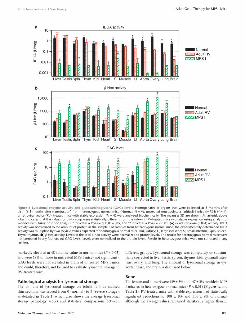
Pathological analysis for lysosomal storageThe amount of lysosomal storage on toluidine blue–stained thin sections was scored from 0 (normal) to 3 (severe storage), as detailed in Table 1, which also shows the average lysosomal storage pathology scores and statistical comparisons between
different groups. Lysosomal storage was completely or substan-tially corrected in liver, testis, spleen, thymus, kidney, small intes-tine, ovary, and lung. The amount of lysosomal storage in eye, aorta, heart, and brain is discussed below.
BoneThe femurs and humeri were 139 ± 3% and 147 ± 3% as wide in MPS I mice as in heterozygous normal mice (P < 0.01) (Figure 4a and Table 2). RV-treated mice with stable expression had statistically significant reductions to 108 ± 8% and 114 ± 9% of normal, although the average values remained statistically higher than in
Figure 3 lysosomal enzyme activity and glycosaminoglycans (GAG) levels. Homogenates of organs that were collected at 8 months after birth (6.5 months after transduction) from heterozygous normal mice (Normal; N = 4), untreated mucopolysaccharidosis I mice (MPS I; N = 4), or retroviral vector (RV)–treated mice with stable expression (N = 4) were analyzed biochemically. The means ± SD are shown. An asterisk above a bar indicates that the values for that group were statistically different from the values in RV-treated mice with stable expression using analysis of variance with Tukey post hoc analysis. * indicates a P value of 0.01–0.05, and ** indicates a P value < 0.01. (a) α-l-iduronidase (IDUA) activity. IDUA activity was normalized to the amount of protein in the sample. For samples from heterozygous normal mice, the experimentally determined IDUA activity was multiplied by two to yield values expected for homozygous normal mice. Kid, kidney; LI, large intestine; SI, small intestine; Spln, spleen; Thym, thymus. (b) β-Hex activity. Levels of the total β-hex activity were normalized to protein levels. The results for heterozygous normal mice were not corrected in any fashion. (c) GAG levels. Levels were normalized to the protein levels. Results in heterozygous mice were not corrected in any fashion.
894 www.moleculartherapy.org vol. 15 no. 5 may 2007
© The American Society of Gene TherapyAdult Gene Therapy for MPS I Mice
normal mice. Figure 4b and c demonstrates that the width of the femur and humerus of RV-treated mice with stable expression was strongly inversely proportional to the serum IDUA activity, with R2 values of 0.39 and 0.38, respectively (P = 0.02 for a statistically significant correlation between the serum IDUA activity and bone widths). Untreated MPS I mice had increased bone mineral density (BMD) at 0.069 ± 0.005 gm/cm2, whereas RV-treated mice with sta-ble expression had normal BMD at 0.053 ± 0.004 gm/cm2 (Table 2). The BMD in RV-treated mice was moderately inversely proportional to the serum IDUA activity, with an R2 value of 0.24 (Figure 4d; P = 0.05).
HearingThe threshold to elicit a neurological signal in an auditory-evoked brainstem response at a sound frequency of 5 kHz was 84 ± 6 dB for MPS I mice, which was significantly higher than in normal mice (40 ± 11 dB; P < 0.01; Figure 4e and Table 2). RV-treated mice with stable expression had a statistically significant improvement in hearing, as their threshold was 65 ± 21 dB (P < 0.05 versus MPS I), although this remained higher than in normal mice (P < 0.01). There was a very strong inverse correlation between the serum IDUA activity in individual RV-treated mice and the hearing threshold at 5 and 10 kHz,
table 1 summary of pathological evaluation for lysosomal storage in organsa
organ
Area or cell type
normal P valueb versus MPs I
untreated MPs I P valuec versus adult rV
Adult rV stable P valued versus normal
Liver Hepatocytes 0 ± 0 (N = 2); P < 0.01 3 ± 0 (N = 2); P < 0.01 0 ± 0 (N = 4); NS
Kupffer cells 0 ± 0 (N = 2); P < 0.01 3 ± 0 (N = 2); P < 0.01 0 ± 0 (N = 4); NS
Testis Interstitial region 0 ± 0 (N = 2); P < 0.01 3 ± 0 (N = 2); P < 0.01 0.3 ± 0.4 (N = 4); NS
Spleen Red pulp 0 ± 0 (N = 2); P < 0.01 2.5 ± 0.5 (N = 2); P < 0.01 0 ± 0 (N = 4); NS
Thymus Cortex 0 ± 0 (N = 3); P < 0.01 3 ± 0 (N = 3); P < 0.01 0 ± 0 (N = 9); NS
Medulla 0 ± 0 (N = 3); P < 0.01 3 ± 0 (N = 3); P < 0.01 0.3 ± 0.5 (N = 9); NS
Kidney Tubules 0 ± 0 (N = 2); P < 0.01 3 ± 0 (N = 2); P < 0.01 0 ± 0 (N = 6); NS
Interstitial cells 0 ± 0 (N = 2); P < 0.01 3 ± 0 (N = 2); P < 0.01 0 ± 0 (N = 6); NS
Glomeruli 0 ± 0 (N = 2); P < 0.01 3 ± 0 (N = 2); P < 0.01 0 ± 0 (N = 6); NS
Small intestine Lamina propria 0 ± 0 (N = 2); P < 0.01 2.5 ± 0.5 (N = 2); P < 0.01 0 ± 0 (N = 3); NS
Submucosa 0 ± 0 (N = 2); NS 2 ± 1 (N = 2); NS 0 ± 0 (N = 3); NS
Ovary Stroma 0 ± 0 (N = 2); P < 0.01 3 ± 0 (N = 5); P < 0.01 0.7 ± 0.5 (N = 3); NS
Lung Parenchyma 0 ± 0 (N = 2); P < 0.01 3 ± 0 (N = 2); P < 0.01 0.8 ± 0.5 (N = 4); NS
Eye Number of nuclei in outer nuclear layere
10.3 ± 0.6 (N = 5); P < 0.01 6.3 ± 0.8 (N = 5); NS 7.5 ± 1.2 (N = 11); P < 0.01
Cornea 0 ± 0 (N = 3); P < 0.01 3 ± 0 (N = 3); NS 2.1 ± 0.9 (N = 10); P < 0.01
Aorta Media 0 ± 0 (N = 2); P < 0.01 3 ± 0 (N = 2); NS 2.6 ± 0.9 (N = 9); P < 0.01
Heart Valve 0 ± 0 (N = 2); P < 0.01 3 ± 0 (N = 2); P < 0.01 0.3 ± 0.03 (N = 4); NS
Parenchyma 0 ± 0 (N = 2); P < 0.01 3 ± 0 (N = 3); P < 0.01 0 ± 0 (N = 4); NS
Brain Perivascular cells 0 ± 0 (N = 2); P < 0.01 3 ± 0 (N = 3); P < 0.05 1.7 ± 0.4 (N = 8); P < 0.05
Meninges 0 ± 0 (N = 2); P < 0.01 3 ± 0 (N = 3); NS 2.7 ± 0.5 (N = 8); P < 0.01
Choroid plexus 0 ± 0 (N = 1); NS 2.5 ± 0.5 (N = 2); NS 0 ± 0 (N = 2); NS
Cortex neurons 0 ± 0 (N = 3) P < 0.01 3 ± 0 (N = 3) P < 0.01 1.0 ± 0.9 (N = 8); NS
Percentage of cortex neurons with storagef
6 ± 3 (N = 3); P < 0.01 63 ± 27% (N = 3); P < 0.01 21 ± 16% (N = 8); NS
Hippocampus neurons 0 ± 0 (N = 3); P < 0.01 3 ± 0 (N = 3); P < 0.05 1.0 ± 1.1 (N = 8); NS
Percentage of hippocampus neurons with storagef
8 ± 7% (N = 3); P < 0.01 77 ± 25% (N = 3); NS 32 ± 32% (N = 8); NS
Purkinje cells 0 ± 0 (N = 2); P < 0.01 2.7 ± 0.5 (N = 3); NS 2.6 ± 0.7 (N = 8); P < 0.01
Abbreviations: IDUA, α-l-iduronidase; MPS I, mucopolysaccharidosis I; NS, not significant; RV, retroviral vector.Shaded rows indicate regions where pathology in RV-treated mice with stable expression was significantly better than in untreated MPS I mice.aHeterozygous normal mice, untreated MPS I mice, or MPS I mice that received gene therapy as adults and had stable expression owing to immunosuppression (Adult RV stable) were killed at 8 months. Pathology was evaluated for the indicated number (N) of animals, as shown for representative examples in Figures 5 and 6. Lysosomal storage was scored as 0 (none detected), 1 (low amounts of storage in some cells), 2 (moderate storage in many cells), or 3 (severe storage in most cells), and the mean ± SD was determined.bP value when scores from normal mice were compared with those in MPS I mice using analysis of variance with Tukey post hoc analysis.cP value for MPS I versus RV-treated MPS I mice with stable expression.dP value for RV-treated MPS I mice with stable expression versus normal mice.eThe number of nuclei in a thickness of the outer nuclear layer of the retina was determined.fThe percentage of cells of a particular phenotype with lysosomal storage was determined after evaluation of >100 cells.

Molecular Therapy vol. 15 no. 5 may 2007 895
© The American Society of Gene Therapy Adult Gene Therapy for MPS I Mice
with R2 values of 0.89 and 0.86,respectively (Figure 4f and g; P < 0.0005).
VisionThe initial hyperpolarization (a-wave) in response to light in a dark-adapted flash electroretinogram reflects photoreceptor function, and the depolarization that follows (b-wave) indicates function of photoreceptors and secondary neurons.28 The a- and b-wave amplitudes in MPS I mice (Figure 5b) were 125 ± 51 and 452 ± 155 µV, respectively, which were significantly lower than the amplitudes of 346 ± 38 and 735 ± 88 µV in normal mice (Figure 5a and Table 2). RV-treated mice with stable expres-sion had a- and b-wave amplitudes of 220 ± 63 and 620 ± 69 µV, which was statistically better than in untreated MPS I mice but statistically worse than in normal mice. There was a poor cor-relation that failed to show statistical significance between the
serum IDUA activity and the amplitudes of the a- and b-waves in RV-treated mice with stable expression: R2 values of 0.11 and 0.18, respectively (Figure 5e and f). The outer nuclear layer of the retina from MPS I mice had only 6.3 ± 0.8 nuclei per thickness, which was fewer than the 10.3 ± 0.6 in normal mice (Figure 5i and j, and Table 1). Although the average number of nuclei in the outer nuclear layer for RV-treated mice with stable expression was still reduced, at 7.5 ± 1.2, and was not statistically improved from the values in untreated MPS I mice (Table 1), there was a strong direct correlation between the serum IDUA activity and number of nuclei in RV-treated mice, with an R2 value of 0.44 (Figure 5g; P = 0.02). Figure 5h and n and Table 1 demonstrate that the cornea from an MPS I mouse had substantial amounts of lysosomal storage in the stroma (scored as 3), which was absent in a normal mouse (scored as 0). Although the average cornea pathology score of 2.1 ± 0.9 in RV-treated mice with stable expression was not statistically better
Figure 4 evaluation of bone and hearing. (a) Radiographs of the femur. Radiographs of femurs were obtained at 8 months after birth and include an example from a retroviral vector (RV)–treated mouse with stable serum α-l-iduronidase (IDUA) activity <100 U/ml and an example from an animal with stable serum IDUA activity >100 U/ml (see Figure 1). Arrows indicate the position where the width of the femur was measured. (b) Relationship between serum IDUA activity and femur width. For individual RV-treated mice with stable expression, the serum IDUA activity was plotted against the ratio of the femur width to that in normal mice. The linear regression line, the R2 value, and the statistical likelihood that there is a correlation between the two variables are shown. The shaded area indicates the average ±1 SD for normal mice, and the stippled area indicates the average ±1 SD for untreated mucopolysaccharidosis I (MPS I) mice. (c) Relationship between the serum IDUA activity and the humerus diameter. Humerus width was plotted as described for the femur width in panel b. (d) Relationship between the serum IDUA activity and the bone mineral density (BMD). The BMD was determined and plotted in a similar fashion to that in panels b and c. (e) Auditory-evoked brainstem response (ABR). The average auditory threshold in dB ± SD was determined at sound frequencies of 5–20 kHz. Statistical comparisons were between other groups and untreated MPS I mice using analysis of variance with Tukey post hoc analysis. (f) Relationship between the serum IDUA activity and the ABR at 5 kHz. (g) Relationship between the serum IDUA activity and the ABR at 10 kHz.

896 www.moleculartherapy.org vol. 15 no. 5 may 2007
© The American Society of Gene TherapyAdult Gene Therapy for MPS I Mice
than in untreated MPS I mice, there was a moderate inverse cor-relation between the cornea pathology score and the serum IDUA activity (Figure 5h), with an R2 value of 0.34 (P = 0.03).
HeartLysosomal storage was mild or absent in the aortic valves and the cardiac parenchyma of RV-treated mice with stable expres-sion (Figure 5s and t and Table 1). However, the aortas of most RV-treated mice with stable expression had large amounts of lysosomal storage material (Figure 5w), although one of the nine mice had a marked reduction (Figure 5x). Indeed, the average lysosomal storage pathology score of 2.6 ± 0.9 in the aorta was not significantly better than the value in untreated MPS I mice (Table 1). Furthermore, most RV-treated mice had elastic fiber fragmentation, and the average aortic diameter of 2.4 ± 0.5 mm (Table 2) was not statistically different from that in untreated MPS I mice (2.8 ± 0.7 mm) and was wider than in normal mice (1.8 ± 0.3 mm; P < 0.01). Finally, 56% of RV-treated MPS I mice had aortic insufficiency, which was not statistically lower than the value of 73% in untreated MPS I mice (Table 2). We conclude that stor-age is reduced in the aortic valves and parenchyma of RV-treated mice with stable expression but remains severe in the aorta.
BrainUntreated MPS I mice had substantial amounts of lysosomal storage in perivascular cells, the meninges, and the choroid plexus, with lysosomal storage pathology scores of 3.0 ± 0, 3.0 ± 0, and 2.5 ± 0.5, respectively (Table 1). Lysosomal storage was partially improved in the perivascular cells of RV-treated mice with stable expression, although it was not significantly reduced in the meninges. Lysosomal storage was completely absent in the choroid plexus of two RV-treated mice with stable expression, although the small number of samples with this region limited statistical evaluation.
Large pyramidal neurons in the cortex and the hippocampus of untreated MPS I mice had substantial amounts of lysosomal storage (Figure 6b and f), resulting in an average pathology score of 3.0 ± 0 in both regions (Table 1). Furthermore, 63 ± 27 and 77 ± 25% of the neurons in the cortex and hippocampus of MPS I mice contained at least three small white vacuoles that were consistent with lysosomal storage (Figure 6m and n, respec-tively, and Table 1). RV-treated mice with stable expression had partial or marked reductions in storage in neurons of the cortex and hippocampus for animals with serum activity of <100 U/ml (Figure 6c and g) or >100 U/ml (Figure 6d and h), respectively,
table 2 summary of results of specialized testinga
organ
test performed
Average score Average serum
IduA in rV-treated mice (u/ml)
normal P valueb versus MPs I
untreated MPs I P valuec versus adult rV
Adult rV stable P valued versus normal
Bone Femur diametere (% normal)
100 ± 9% (N = 11); P < 0.01 139 ± 3% (N = 13); P < 0.01
108 ± 8% (N = 11); P < 0.05
84 ± 43
Humerus diametere (% normal)
100 ± 9% (N = 11); P < 0.01 147 ± 3% (N = 13); P < 0.01
114 ± 10% (N = 11); P < 0.01
84 ± 43
Bone mineral density (gm/cm2)
0.054 ± 0.002 (N = 11); P < 0.01
0.069 ± 0.005 (N = 14); P < 0.01
0.053 ± 0.004 (N = 12); NS
81 ± 42
Ear ABR at 5 kHz (dB) 40 ± 11 (N = 11); P < 0.01 84 ± 6 (N = 7); P < 0.05 65 ± 21 (N = 8); P < 0.01 87 ± 46
ABR at 10 kHz (dB) 30 ± 13 (N = 11); P < 0.01 76 ± 18 (N = 7); NS 63 ± 28 (N = 8); P < 0.01 87 ± 46
ABR at 20 kHz (dB) 52 ± 26 (N = 11); P < 0.05 77 ± 10 (N = 7); NS 91 ± 8 (N = 8); P < 0.01 87 ± 46
ABR at 40 kHz (dB) 99 ± 8 (N = 11); NS 102 ± 15 (N = 7); NS 102 ± 3 (N = 8); NS 87 ± 46
Eye Electroretinogram a-wave(µV)
346 ± 38 (N = 12); P < 0.01 125 ± 51 (N = 7); P < 0.01 220 ± 63 (N = 9); P < 0.01 86 ± 44
Electroretinogram b-wave (µV)
735 ± 88 (N = 12); P < 0.01 452 ± 155 (N = 7); P < 0.01
620 ± 69 (N = 9); P < 0.05 86 ± 44
Aorta Diameter of aorta at 2 mm from the aortic valve (mm)
1.8 ± 0.3 (N = 6); P < 0.01 2.8 ± 0.7 (N = 11); NS 2.4 ± 0.5 (N = 9); NS 86 ± 44
Heart Frequency of aortic insufficiencyf
0 out of 6 (0%); P = 0.009 8 out of 11 (73%); NS 5 out of 9 (56%); P = 0.04 86 ± 43
Abbreviations: ABR, auditory-evoked brainstem response; IDUA, α-l-iduronidase; MPS I, mucopolysaccharidosis; NS, not significant; RV, retroviral vector.Shaded rows indicate tests where RV-treated mice with stable expression were significantly better than untreated MPS I mice.aMice were treated as described in Figure 1, and means ± SD were determined at 8 months. The number of animals in each group (N) and the average serum IDUA activity for RV-treated mice with stable expression that were evaluated for each test are shown.bP value for normal versus MPS I mice.cP value for MPS I versus RV-treated MPS I mice with stable expression.dP value for RV-treated MPS I versus normal mice.eThe diameter of long bones relative to the average value for normal mice.fFisher’s exact test was used to determine whether the frequency of aortic insufficiency was significantly different.

Molecular Therapy vol. 15 no. 5 may 2007 897
© The American Society of Gene Therapy Adult Gene Therapy for MPS I Mice
Figure 5 evaluation of eye, heart, and aorta. (a–d) Representative electroretinograms (ERGs). ERGs were evaluated at 8 months of age for normal, mucopolysaccharidosis I (MPS I), or retroviral vector (RV)–treated mice with stable expression (see Figure 1 for details of treatment). The a- and b-waves are indicated. (e) Relationship between the serum α-l-iduronidase (IDUA) activity and the ERG a-wave. The relationship between the serum IDUA activity and the ERG a-wave is shown for individual RV-treated mice with stable expression. Stippled and shaded regions indicate the average ±1 SD for MPS I and normal mice, respectively. (f) Relationship between the serum IDUA activity and the ERG b-wave. (g) Relationship between the serum IDUA activity and the number of cells in a thickness of the outer nuclear layer (ONL). Retinas were evaluated pathologically as shown in panels i–l. The number of cells in a thickness of the outer nuclear layer versus the serum IDUA activity in individual RV-treated mice is shown. (h) Relation-ship between the serum IDUA activity and pathology score in the cornea. Corneas were evaluated pathologically for evidence of lysosomal storage, as shown in panels m–p. The amount of lysosomal storage was scored from 0 (normal) to 3 (severe storage) and is plotted against the serum IDUA activity in individual RV-treated mice. (i–l) Pathology of the retina. Thin sections of the retina were stained with toluidine blue. The pigmented retinal epithelium (RPE) is indicated. The length of the outer segments (OS) and the inner segments (IS) and the number of cells in the ONL are markedly reduced in the untreated MPS I mouse. Dotted lines indicate the boundaries between different regions. The original magnification is identified at the left. (m–p) Pathology of the cornea. The endothelium is at the top right. White arrows indicate lysosomal storage in the stroma, and the black arrow indicates storage in the endothelium. (q–t) Aortic valve pathology. White arrows indicate lysosomal storage material in the aortic valve. (u–x) Aorta pathology. White arrows indicate lysosomal storage in the media of the aorta.

898 www.moleculartherapy.org vol. 15 no. 5 may 2007
© The American Society of Gene TherapyAdult Gene Therapy for MPS I Mice
resulting in reduced lysosomal storage pathology scores and fewer neurons with storage (Table 1). The percentage of neu-rons with lysosomal storage in the cortex and the hippocampus of RV-treated mice was strongly inversely correlated with the serum IDUA activity, with R2 values of 0.76 (P = 0.005) and 0.63 (P = 0.02), respectively (Figure 6m and n). Most RV-treated mice with stable expression had no improvement in stor-age in the Purkinje cells (Table 1) as shown for a representa-tive example (Figure 6k), although the mouse with the highest IDUA activity had reduced lysosomal storage (pathology score of 1; Figure 6l).
Analysis of rV dnA and rnA in the brain and other organsThe liver contained 13.8 ± 7.4 copies of RV per 100 cells and RV RNA levels were 8.5 ± 2.1% of β-actin RNA levels at 1 week after transduction (Figure 6o and p). Spleen DNA and RNA levels were 6 and 11% of those in liver, respectively, demonstrating that the vector was not liver specific and that antigen-presenting cells may have been transduced. At 6.5 months after transduction, RV DNA and RNA copies remained high in liver, and spleen contained 5 and 6% as much DNA and RNA, respectively, as liver. The RV was also expressed at low levels in bone marrow at 0.01 ± 0.01%
Figure 6 evaluation of brain. Mice were treated as described in Figure 1 and killed at 8 months. (a–l) Pathology of brain. Sections were stained with toluidine blue. The region evaluated and the original magnification are identified at the left. (a–h) Neurons of the brain cortex (panels a–d) and hippocampus (panels e–h). White arrows identify neurons with lysosomal storage. (i–l) Cerebellum. White arrows identify Purkinje cells with storage. (m–n) Relationship between the serum α-l-iduronidase (IDUA) activity and the percentage of cells with lysosomal storage in large pyramidal neurons of the brain cortex (panel m) or neurons of the hippocampus (panel n). The relationship between the serum IDUA activity and the percentage of cells with three or more white vacuoles consistent with lysosomal storage is shown for individual retroviral vector (RV)–treated mice with stable expres-sion. Stippled and shaded regions indicate the average ±1 SD for mucopolysaccharidosis I (MPS I) and normal mice, respectively. (o) RV DNA levels in organs. RV-treated MPS I mice were killed at 1 week (left) or at 6.5 months (right) after transduction at 6 weeks of age, and RV DNA copies per 100 cells were determined with real-time polymerase chain reaction (PCR) for 2–3 samples in each group. (p) RV RNA levels in organs. The percentage of RV RNA relative to that of β-actin was determined with real-time reverse transcription PCR.

Molecular Therapy vol. 15 no. 5 may 2007 899
© The American Society of Gene Therapy Adult Gene Therapy for MPS I Mice
β-actin levels (0.4% of liver) and in blood cells at 0.002 ± 0.002% β-actin levels (0.1% of liver). The identification of RV RNA in bone marrow and blood cells, which did not contain detectable RV DNA copies, was likely due to the greater sensitivity of the RNA assay. RV RNA and DNA were undetectable in brain.
dIscussIonstable expression of cIduA in MPs I mice required immunosuppressionThe reduction in IDUA activity in serum and organs that we and others have observed after gene transfer to adult MPS I mice is likely due to a CTL response, as anti-canine IDUA antibodies were not detected and the RV DNA levels were very low in the liver.12 Furthermore, serum levels of the liver enzyme alanine amino-transferase, a marker of liver damage, were elevated at 14 days after transduction in RV-treated mice that did not receive immunosup-pression at 73 ± 31 U/L (N = 6). This was 2.1-fold the level in RV-treated mice that were effectively treated with immunosuppression (34 ± 4, N = 4; P < 0.01; data not shown) and 3-fold the level in untreated MPS I mice of 24 ± 8 U/L (N = 5). In addition, the livers of RV-treated mice that did not receive immunosuppression con-tained lymphocytic infiltrates at 14 days (data not shown).
The approach that was taken here to prevent an immune response after gene therapy to adults was to block co-stimulation and/or deplete CD4 cells. Blockade of co-stimulation is intellectu-ally appealing, as cells that encounter antigen in the absence of co-stimulation can undergo apoptosis,18 resulting in clonal dele-tion and long-standing tolerance after a short period of drug administration. Adult spleens had 7- and 18-fold as much CD28 and CD40L RNA, respectively, as newborns, suggesting that both of these co-stimulatory pathways may potentiate an immune response in adults. The combination of CTLA4-Ig with α-CD40L antibody prevented an immune response to IDUA in most adult animals, which is consistent with the results of others in organ transplantation (reviewed in ref. 18) or gene therapy for hemo-philia A.22 Adult spleens also had threefold more CD4 RNA and a sevenfold higher percentage of CD4+ cells in the spleen than newborns; the discrepancy in the magnitude of the difference may be because adult splenocytes had only 65% as much cell- surface CD4 as newborn splenocytes (data not shown). The combination of CTLA4-Ig with the α-CD4 antibody was effec-tive in preventing an immune response to IDUA after adult gene therapy in this study. Mice that received transient immunosup-pression were not immunocompromised 4.5 months later, as they mounted a normal antibody response to hFIX. Although admin-istration of CTLA4-Ig alone for 6.5 months was effective, it would likely result in substantial immunosuppression. It remains pos-sible that administration of CTLA4-Ig for an intermediate period might be effective long term.
An alternative approach to reduce an immune response after gene transfer to adult MPS I mice was to avoid expression from a lentiviral vector in antigen-presenting cells using the liver-restricted albumin promoter.17 However, there was only ~1% of normal IDUA activity in liver and spleen, and a recent report suggested that a similar construct was leaky and evoked an immune response to green fluorescent protein.29 In this study, some animals that received a single immunosuppressive agent
and had a decrease in serum IDUA activity to <1% of the initial activity still had detectable serum IDUA activity. Furthermore, all mice with unstable expression had liver IDUA activity that was >1% of normal and liver RV RNA that was above the background, albeit low at 0.15% of the level found in mice with stable expres-sion (data not shown). We propose that some cells with very low expression may escape a CTL response and that detect-able enzyme activity and/or vector RNA levels do not exclude the possibility of an immune response. Mice with unstable expression had partial improvements in bone widths and BMD but no improvements in auditory-evoked brainstem response, electroretinograms, or echocardiograms (data not shown).
lysosomal storage is reduced in organsThe main goal of this study was to determine whether gene ther-apy to adult mice could correct the biochemical and clinical mani-festations of MPS I. Serum IDUA activity in RV-treated mice of 25–156 U/ml was associated with reductions in biochemical and/or pathological evidence of lysosomal storage in all mice in liver, testis, spleen, thymus, kidney, small intestine, ovary, and lung, which should reduce hepatosplenomegaly and improve fertility, and may reduce gastrointestinal and pulmonary symptoms.
Improvements in bone, ear, and eye, but not in aortaIn this study, RV-treated mice with stable expression had statis-tically significant improvements in long bone widths, hearing, BMD, and vision, with serum IDUA activity >100 U/ml confer-ring greater benefits than activity <100 U/ml. Reduced bone widths may contribute to improved hearing, as bone thickening probably contributes to the conductive defect.30 In contrast, lyso-somal storage and dilatation of the aorta remained severe. The dense structure of the aorta may make it difficult for IDUA to diffuse to the interior, or aortic smooth muscle cells may have low levels of the M6PR.
The failure to correct manifestations completely could be due to initiation of treatment after disease had developed or to the relatively low average serum IDUA activity of 81 ± 41 U/ml. We favor the latter hypothesis, as evaluation of bones, hearing, vision, eye, and aorta were normal or near-normal in MPS I mice at 6 weeks of age (X.M., J.A. Metcalf, A.H., R. Knutsen, R. Mecham, M.T., and K.P.P., unpublished data). In addition, in our previous study on gene therapy in newborns, mice that achieved only 30 U/ml had some defects in all of these parameters, whereas those that achieved 1,000 U/ml were almost completely corrected.12 We estimate that the target serum IDUA activity for improvement of young adult mice is >100 U/ml in bone and hearing, >200 U/ml for vision and eye, and >500 U/ml for aorta.
BrainEvaluation of brain is important, as patients with Hurler syndrome have neurological impairment. RV-treated mice with stable expres-sion had reductions in lysosomal storage pathology in perivascular cells, pyramidal neurons of the cortex and hippocampus, and the choroid plexus. Improvements were greater in RV-treated mice with >100 U/ml of IDUA activity in serum than in those with <100 U/ml. However, most RV-treated mice still had severe storage in the

900 www.moleculartherapy.org vol. 15 no. 5 may 2007
© The American Society of Gene TherapyAdult Gene Therapy for MPS I Mice
meninges and Purkinje cells. As the brain was almost completely corrected after neonatal gene therapy in mice with 1,000 U/ml of serum IDUA activity, but was only partially improved in mice with 30 U/ml,12 the failure to correct disease completely in brain in this study may be due to insufficient IDUA activity in blood rather than the late age at therapy. A reasonable target for serum IDUA activity for correction of brain disease after adult gene therapy is >500 U/ml.
It was surprising that lysosomal storage was reduced in brain, as it has been a long-standing belief that enzyme in blood can-not cross the blood–brain barrier in adults. However, a recent study demonstrated that ERT to adults reduced lysosomal storage in neurons in MPS VII mice,31 and neonatal gene ther-apy approaches in which nucleic acid levels were undetectable11 or were very low13 in brain reduced lysosomal storage in MPS I mice. Indeed, in this study, RV RNA levels were undetectable in the brain using a very sensitive real-time reverse transcription– polymerase chain reaction assay. We therefore favor the hypo-thesis that improvements were due to enzyme diffusion from blood into the brain. It is unclear why Purkinje cells and menin-ges were relatively refractory to treatment. It is possible that they contain low levels of the M6PR. A future study will evaluate whether RV-treated MPS I mice have improved neurological function in an open field test of habituation.11
Implications for patients with MPs IThese results hold out the promise that gene therapy may be feasible and efficacious in older MPS I patients with null muta-tions if transient immunosuppression is given. CTLA4-Ig was recently approved for use in humans with rheumatoid arthri-tis as the drug called Orencia (abatacept; Bristol-Myers-Squibb, Princeton, NJ). Although α-CD40L antibodies have resulted in thromboembolic events in human trials and will probably not be pursued further,32 an α-CD40 antibody, which blocks the same pathway, was effective at prolonging graft survival in non-human primates in conjunction with CTLA4-Ig.33 In a similar way, an α-CD4 antibody blocked immune responses in non-human pri-mates.34 It is also possible that immunosuppressive agents that are routinely used in patients such as cyclosporine and azathio-prine could be effective, as a 2-month course prevented an anti-body response to IDUA protein in MPS I dogs.25 The risks of gene therapy also need to be considered, as liver cancers developed in some mice that received neonatal or fetal injection of some, but not other, lentiviral vectors.35 It is reassuring that we have not seen tumors with our vector in any of more than 100 mice that have been followed long term (data not shown). In addition, leuke-mia or lymphoma developed in some human patients and in a non-human primate that received HSC-directed gene therapy.36 As the latter malignant transformation was associated with trans-duction of hematopoietic cells, the low transduction efficiency of bone marrow and blood cells in our study should reduce this risk. Nevertheless, it will be important to continue to evaluate animals for evidence of toxicity before implementing this gene therapy approach in humans.
MAterIAls And MetHodsAll reagents were purchased from Sigma Chemical (St. Louis, MO) unless otherwise stated. HGF was purified as described previously.37
Human CTLA4-Ig was generously provided by Robert Peach of Bristol-Myers Squibb. The α-mouse CD4 antibody GK1.5 and the α-mouse CD40L antibody MR1 were obtained from BD Biosciences Pharmingen (San Diego, CA 92121).
Animals. All animal studies have been approved by the authors’ institu-tional review board. Six week-old MPS I mice38 in a C57BL/6 background were injected intraperitoneally with five doses of 5 mg/kg HGF with 15 mg/kg dextran sulfate per dose at 0, 3, 6, 9, and 12 hours. Mice were injected via the caudal vein with two or three doses of ~300 µl hAAT-cIDUA-WPRE at 30–48 hours after the first dose of HGF for a cumulative dose of 5 × 10⁹ transducing units per kg. Human CTLA4-Ig was injected intraperitoneally at 25 mg/kg per dose in phosphate-buffered saline, which is approximately twofold the dose used by Miao et al.22 after correction for the fact that human CTLA4-Ig is only ~50% as active as murine CTLA4-Ig in mice (Robert Peach, Bristol-Myers Squibb, personal communication, 1 March 2003). For transient administration of CTLA4-Ig, injections were given at 6 hours before and at 4, 7, and 11 days after the first dose of RV. For prolonged administration of CTLA4-Ig, injections were given at the same time as for transient administration of CTLA4-Ig and were also given twice a week for the remainder of the first month, once a week for the sec-ond month, and once a month thereafter. α-Mouse CD40L antibody and α-mouse CD4 antibody were injected intraperitoneally at 12.5 mg/kg in phosphate-buffered saline at 1 and 3 days (α-CD40L) or at 1 and 2 days (α-CD4) after the first dose of HGF, which is slightly less than the dose used by others.39,40 Serum was obtained from the right retro-orbital plexus or the caudal vein. The diameter of the femur was measured from radiographs, and the ratio to that in normal mice was determined. BMD, echocardiogra-phy, auditory-evoked brainstem response, and electroretinograms were per-formed as described elsewhere.12,28,30,41 Before mice were killed, serum was collected, and the animals were transcardially perfused with 20 ml saline.
Analysis of spleen RNA. Three normal C57BL/6 mice were killed at 6 weeks after birth without any prior treatment, and three were treated with HGF and 5 × 10⁹ transducing units per kg hAAT-cIDUA-WPRE and killed 24 hours later. Four newborn normal mice were killed at 3 days after birth without any prior treatment, and four received 1010 transducing units per kg hAAT-cGUSB-WPRE26 at 2 days after birth and were killed 24 hours later. Spleen was homogenized in TRIzol Reagent (Invitrogen, Carlsbad, CA) and reverse transcription was performed with oligo-dT. Taqman technology was performed using assays from Applied Biosystems (Foster City, CA) for the following genes, with normalization to the level of Mac 1 (CT value for 6-week-old mice in parentheses): Mac 1 (CT = 26), CD80 (CT = 30), CD86 (CT = 26), CD28 (CT = 30), CTLA4 (CT = 31), CD40 (CT = 27), CD40L (CT = 32), CD4 (CT = 28), CD8 (CT = 26), CD11c (CT = 28), and B220 (CT = 25). SYBRgreen technology was performed for the following genes42,43 (CT for normal adult spleen samples in parentheses): β-actin (CT = 19), IL-2 (CT = 36), IL-4 (CT = 35), IL-10 (CT = 33), and IFNγ (CT = 29). As RNA levels were similar in RV-treated and untreated mice of the same age, results were pooled for statistical analysis.
Challenge with hFIX. Mice were injected with 30 IU/kg BeneFix (hFIX) with 200 µl of the adjuvant RIBI (MPL+TDM Emulsion R-700, Corixa, Hamilton, MT) at 6 and 6.75 months of age. Serum was tested for anti-hFIX antibodies using an immunoassay as described elsewhere.44
IDUA activity, β-hex activity, and GAG levels. Organs were homogenized in lysis buffer as described elsewhere12 and the same homogenate was used for enzyme and GAG assays. The total protein concentration was deter-mined with the Bradford assay (BioRad Laboratories, Hercules, CA). For the IDUA assay, 10 µl organ extract was mixed with 90 µl 55 µM 4-methy-lumbelliferyl-β-l-idopyranosiduronic acid (Toronto Research Chemicals, North York, Ontario, Canada) in 0.4 M formate buffer (pH 3.5) at 37 °C and the fluorescence was determined every 10 minutes for 2 hours with
Molecular Therapy vol. 15 no. 5 may 2007 901
© The American Society of Gene Therapy Adult Gene Therapy for MPS I Mice
a Fluoroskan Ascent microplate fluorometer from Thermo Electron (Mil-ford, MA) with excitation at 355 nm and emission at 460 nm. The plate was protected from light and incubated at 37 °C for an additional 2–4 hours, as which time 100 µl 2.9 M glycine with 1.8 M sodium carbonate (pH 9.5) was added to stop the reaction and increase the fluorescence of the product, and the plate was read again. Standards of 4-methylumbel-liferone in water were protected from light and stored at 4 °C, and 10 µl of each was mixed with 90 µl reaction buffer for the standard curve. Kinetic readings were used for samples with high activity, and the stop read-ing was used for samples with low activity. One unit of enzyme releases 1 nmol 4-methylumbelliferone per hour. IDUA activity in organs from homozygous normal mice was assumed to be twice the level determined for heterozygous normal mice. The β-hex substrate contained 5.5 mM 4-methylumbelliferyl-acetyl-β-d-glucosaminide in 0.1 M sodium acetate (pH 4.8). The reaction was similar, but no stop reaction reading was per-formed. GAG levels were determined using a sulfated glycosaminoglycan kit from Blyscan (Newtownabbey, Northern Ireland) with chondroitin 4-sulfate as the standard.45
Pathological evaluation. Pieces of organs were fixed and embedded in Epon, and 1 µm sections were stained with toluidine blue as previously described.12 Brains were placed in an RBM-2000C rodent brain matrix mold (Protech International, San Antonio, TX). Slices made at positions 7, 8, and 9 (counting started at the anterior brain) contained the parietal cortex and the hippocampus, and both pieces were submitted for pathol-ogy. Pathology was evaluated without knowledge of the genotype and treatment status and was scored as described in Table 1. For quantitation of the number of cells with storage in the parietal cortex, at least 100 cells from different regions of the slide were scored as positive (at least three small white vacuoles consistent with lysosomal storage in the cytoplasm) or negative. For the hippocampus, at least 100 cells from the CA3 region were evaluated.
Nucleic acid analysis. DNA and RNA were isolated and real-time poly-merase chain reaction was performed as described for the WPRE with normalization to the β-actin signal after reverse transcription of RNA samples.13 No signal was observed using samples that did not receive reverse transcription. Samples with no detectable DNA copies are depicted as having 0.02 copies of RV DNA per cell, which is below the sensitivity of the assay. Samples with no detectable RNA copies are depicted as having 0.0005% as much RV RNA as β-actin RNA.
Fluorescence-activated cell sorting. Splenocytes from newborn and adult mice were treated with 0.74 M ammonium chloride to remove erythro-cytes and then washed twice with phosphate-buffered saline. The cells were incubated with fluorescein isothiocyanate–conjugated CD3 or with PE-conjugated CD4 or CD8 monoclonal antibodies (BD Pharmingen, San Diego, CA) for 30 minutes on ice in staining buffer (phosphate- buffered saline containing 1% BSA and 0.1% NaN3). Cells were washed and analyzed on a FACS Calibur (BD Biosciences, San Jose, CA) and analyzed using CellQuest software (BD Biosciences).
Statistics. Analysis of variance with Tukey post hoc analysis or the Student’s t-test was used to compare values in different groups using Sigma Stat software (Systat Software, Point Richmond, CA). The statistical significance of the correlation coefficient (R2) was determined by calculation of the t statistic using the formula t = r divided by the square root of [(1 − r2)/ (n − 2)] and comparison with a table to determine the P value.46
AcknowledGMentsWe thank Elizabeth Neufeld (University of California at Los Angeles) for the canine IDUA cDNA and the MPS I mice, Clay Semenkovich and Trey Coleman (Washington University) for assistance with BMD and real-time polymerase chain reaction, and Daniel Schuster (Washington University) for help with statistical analysis. This work was
supported by the Ryan Foundation, the National MPS Society, and the National Institutes of Health (DK66448 awarded to K.P.P.). Histology was supported by P30 DK52574, EY02687, DC04665, and Research to Prevent Blindness. BMD equipment was supported by the Clinical Nutrition Research Unit (DK56341) and the Diabetes Research Training Core (DK20579). Electroretinogram and auditory-evoked brainstem response equipment was supported by the James S. McDonnell Foun-dation. Echocardiography equipment was supported by the Mouse Cardiovascular Phenotyping Core Facility at the Center for Cardiovas-cular Research. Real-time polymerase chain reaction was supported by the Phenotyping Core of the Diabetes Research and Training Center (DK20579 awarded to Clay Semenkovich).
reFerences1. Neufeld, EF and Muenzer, J (2001). The mucopolysaccharidoses. In: Scriver, CR,
Beaudet, AL, Sly, WS and Valle, D (eds). Metabolic and Molecular Basis of Inherited Disease. McGraw-Hill: New York, pp 3421–3452.
2. Lowry, RB, Applegarth, DA, Toone, JR, MacDonald, E and Thunem, NY (1990). An update on the frequency of mucopolysaccharide syndromes in British Columbia. Hum Genet 85: 389–390.
3. Zheng, Y, Rozengurt, N, Ryazantsev, S, Kohn, DB, Satake, N and Neufeld, EF (2003). Treatment of the mouse model of mucopolysaccharidosis I with retrovirally transduced bone marrow. Mol Genet Metab 79: 233–244.
4. Breider, MA, Shull, RM and Constantopoulos, G (1989). Long-term effects of bone marrow transplantation in dogs with mucopolysaccharidosis I. Am J Pathol 134: 677–692.
5. Staba, SL, Escolar, ML, Poe, M, Kim, Y, Martin, PL, Szabolcs, P et al. (2004). Cord-blood transplants from unrelated donors in patients with Hurler’s syndrome. N Engl J Med 350: 1960–1969.
6. Desnick, RJ (2004). Enzyme replacement and enhancement therapies for lysosomal diseases. J Inherit Metab Dis 27: 385–410.
7. Kakkis, ED, Schuchman, E, He, X, Wan, Q, Kania, S, Wiemelt, S et al. (2001). Enzyme replacement therapy in feline mucopolysaccharidosis I. Mol Genet Metab 72: 199–208.
8. Kakkis, ED, McEntee, MF, Schmidtchen, A, Neufeld, EF, Ward, DA, Gompf, RE et al. (1996). Long-term and high-dose trials of enzyme replacement therapy in the canine model of mucopolysaccharidosis I. Biochem Mol Med 58: 156–167.
9. Kakkis, ED, Muenzer, J, Tiller, GE, Waber, L, Belmont, J, Passage, M et al. (2001). Enzyme-replacement therapy in mucopolysaccharidosis I. N Engl J Med 344: 182–188.
10. Sands, MS and Davidson, BL (2006). Gene therapy for lysosomal storage diseases. Mol Ther 13: 839–849.
11. Hartung, SD, Frandsen, JL, Pan, D, Koniar, BL, Graupman, P, Gunther, R et al. (2004). Correction of metabolic, craniofacial, and neurologic abnormalities in MPS I mice treated at birth with adeno-associated virus vector transducing the human alpha-l-iduronidase gene. Mol Ther 9: 866–875.
12. Liu, Y, Xu, L, Hennig, AK, Kovacs, A, Fu, A, Chung, S et al. (2005). Liver-directed neonatal gene therapy prevents cardiac, bone, ear, and eye disease in mucopolysaccharidosis I mice. Mol Ther 11: 35–47.
13. Chung, S, Ma, X, Liu, Y, Lee, D, Tittiger, ME and Ponder, KP (2007). Effect of neonatal administration of a retroviral vector expressing α-l iduronidase upon lysosomal storage in brain and other organs in mucopolysaccharidosis I mice. Mol Genet Metab 90: 181–192
14. Kobayashi, H, Carbonaro, D, Pepper, K, Petersen, D, Ge, S, Jackson, H et al. (2005). Neonatal gene therapy of MPS I mice by intravenous injection of a lentiviral vector. Mol Ther 11: 776–789.
15. Di Domenico, C, Villani, GR, Di Napoli, D, Reyero, EG, Lombardo, A, Naldini, L et al. (2005). Gene therapy for a mucopolysaccharidosis type I murine model with lentiviral-IDUA vector. Hum Gene Ther 16: 81–90.
16. Lutzko, C, Kruth, S, Abrams-Ogg, AC, Lau, K, Li, L, Clark, BR et al. (1999). Genetically corrected autologous stem cells engraft, but host immune responses limit their utility in canine alpha-l-iduronidase deficiency. Blood 93: 1895–1905.
17. Domenico, CD, Napoli, DD, Reyero, EG, Lombardo, A, Naldini, L and Natale, PD (2006). Limited transgene immune response and long-term expression of human alpha-l-iduronidase in young adult mice with mucopolysaccharidosis type I by liver-directed gene therapy. Hum Gene Ther 17: 1112–1121.
18. Snanoudj, R, de Preneuf, H, Creput, C, Arzouk, N, Deroure, B, Beaudreuil, S et al. (2006). Costimulation blockade and its possible future use in clinical transplantation. Transpl Int 19: 693–704.
19. Kosuga, M, Takahashi, S, Sasaki, K, Li, XK, Fujino, M, Hamada, H et al. (2000). Adenovirus-mediated gene therapy for mucopolysaccharidosis VII: involvement of cross-correction in wide-spread distribution of the gene products and long-term effects of CTLA-4Ig coexpression. Mol Ther 1: 406–413.
20. Ponder, KP, Wang, B, Wang, P, Ma, X, Herati, R, Wang, B et al. (2006). Mucopolysaccharidosis I cats mount a cytotoxic T lymphocyte response after neonatal gene therapy that can be blocked with CTLA4-Ig. Mol Ther 14: 5–13.
21. Stein, CS, Ghodsi, A, Derksen, T and Davidson, BL (1999). Systemic and central nervous system correction of lysosomal storage in mucopolysaccharidosis type VII mice. J Virol 73: 3424–3429.
22. Miao, CH, Ye, P, Thompson, AR, Rawlings, DJ and Ochs, HD (2006). Immunomodulation of transgene responses following naked DNA transfer of human factor VIII into hemophilia A mice. Blood 108: 19–27.
23. Zhan, Y, Brown, LE, Deliyannis, G, Seah, S, Wijburg, OL, Price, J et al. (2004). Responses against complex antigens in various models of CD4 T-cell deficiency: surprises from an anti-CD4 antibody transgenic mouse. Immunol Res 30: 1–14.

902 www.moleculartherapy.org vol. 15 no. 5 may 2007
© The American Society of Gene TherapyAdult Gene Therapy for MPS I Mice
24. Ross, CJ, Bastedo, L, Maier, SA, Sands, MS and Chang, PL (2000). Treatment of a lysosomal storage disease, mucopolysaccharidosis VII, with microencapsulated recombinant cells. Hum Gene Ther 11: 2117–2127.
25. Kakkis, E, Lester, T, Yang, R, Tanaka, C, Anand, V, Lemontt, J et al. (2004). Successful induction of immune tolerance to enzyme replacement therapy in canine mucopolysaccharidosis I. Proc Natl Acad Sci USA 101: 829–834.
26. Xu, L, Mango, RL, Sands, MS, Haskins, ME, Ellinwood, NM and Ponder, KP (2002). Evaluation of pathological manifestations of disease in mucopolysaccharidosis VII mice after neonatal hepatic gene therapy. Mol Ther 6: 745–758.
27. Bunge, S, Clements, PR, Byers, S, Kleijer, WJ, Brooks, DA and Hopwood, JJ (1998). Genotype-phenotype correlations in mucopolysaccharidosis type I using enzyme kinetics, immunoquantification and in vitro turnover studies. Biochim Biophys Acta 1407: 249–56.
28. Ohlemiller, KK, Vogler, CA, Roberts, M, Galvin, N and Sands, MS (2000). Retinal function is improved in a murine model of a lysosomal storage disease following bone marrow transplantation. Exp Eye Res 71: 469–481.
29. Brown, BD, Venneri, MA, Zingale, A, Sergi, L and Naldini, L (2006). Endogenous microRNA regulation suppresses transgene expression in hematopoietic lineages and enables stable gene transfer. Nat Med 12: 585–591.
30. Ohlemiller, KK, Hennig, AK, Lett, JM, Heidbreder, AF and Sands, MS (2002). Inner ear pathology in the mucopolysaccharidosis VII mouse. Hear Res 169: 69–84.
31. Vogler, C, Levy, B, Grubb, JH, Galvin, N, Tan, Y, Kakkis, E et al. (2005). Overcoming the blood-brain barrier with high-dose enzyme replacement therapy in murine mucopolysaccharidosis VII. Proc Natl Acad Sci USA 102: 14777–14782.
32. Kawai, T, Andrews, D, Colvin, RB, Sachs, DH and Cosimi, AB (2000). Thromboembolic complications after treatment with monoclonal antibody against CD40 ligand. Nat Med 6: 114.
33. Adams, AB, Shirasugi, N, Jones, TR, Durham, MM, Strobert, EA, Cowan, S et al. (2005). Development of a chimeric anti-CD40 monoclonal antibody that synergizes with LEA29Y to prolong islet allograft survival. J Immunol 174: 542–550.
34. Winsor-Hines, D, Merrill, C, O’Mahony, M, Rao, PE, Cobbold, SP, Waldmann, H et al. (2004). Induction of immunological tolerance/hyporesponsiveness in baboons with a nondepleting CD4 antibody. J Immunol 173: 4715–4723.
35. Themis, M, Waddington, SN, Schmidt, M, von Kalle, C, Wang, Y, Al-Allaf, F et al. (2005). Oncogenesis following delivery of a nonprimate lentiviral gene therapy vector to fetal and neonatal mice. Mol Ther 12: 763–771.
36. Nienhuis, AW, Dunbar, CE and Sorrentino, BP (2006). Genotoxicity of retroviral integration in hematopoietic cells. Mol Ther 13: 1031–1049.
37. Gao, C, Jokerst, R, Gondipalli, P, Cai, SR, Kennedy, S, Flye, MW et al. (1999). Lipopolysaccharide potentiates the effect of hepatocyte growth factor on hepatocyte replication in rats by augmenting AP-1 activity. Hepatology 30: 1405–1416.
38. Ohmi, K, Greenberg, DS, Rajavel, KS, Ryazantsev, S, Li, HH and Neufeld, EF (2003). Activated microglia in cortex of mouse models of mucopolysaccharidoses I and IIIB. Proc Natl Acad Sci USA 100: 1902–1907.
39. Martin, P, del Hoyo, GM, Anjuere, F, Ruiz, SR, Arias, CF, Marin, AR et al. (2000). Concept of lymphoid versus myeloid dendritic cell lineages revisited: both CD8alpha (-) and CD8alpha(+) dendritic cells are generated from CD4(low) lymphoid-committed precursors. Blood 96: 2511–2519.
40. Miga, AJ, Masters, SR, Durell, BG, Gonzalez, M, Jenkins, MK, Maliszewski, C et al. (2001). Dendritic cell longevity and T cell persistence is controlled by CD154-CD40 interactions. Eur J Immunol 31: 959–965.
41. Hennig, AK, Ogilvie, JM, Ohlemiller, KK, Timmers, AM, Hauswirth, WW and Sands, MS (2004). AAV-mediated intravitreal gene therapy reduces lysosomal storage in the retinal pigmented epithelium and improves retinal function in adult MPS VII mice. Mol Ther 10: 106–116.
42. Liao, YH, Yuan, J, Wang, ZH, Cheng, X, Zhang, JH, Tian, Y et al. (2005). Infectious tolerance to ADP/ATP carrier peptides induced by anti-L3T4 monoclonal antibody in dilated cardiomyopathy mice. J Clin Immunol 25: 376–384.
43. Overbergh, L, Giulietti, A, Valckx, D, Decallonne, B, Bouillon, R and Mathieu, C (2003). The use of real-time reverse transcriptase PCR for the quantification of cytokine gene expression. J Biomol Tech 14: 33–43.
44. Zhang, J, Xu, L, Haskins, ME and Ponder, KP (2004). Neonatal gene transfer with a retroviral vector results in tolerance to human factor IX in mice and dogs. Blood 103: 143–151.
45. Wang, B, O’Malley, TM, Xu, L, Vite, C, Wang, P, O’Donnell, PA et al. (2006). Expression in blood cells may contribute to biochemical and pathological improvements after neonatal intravenous gene therapy for mucopolysaccharidosis VII in dogs. Mol Genet Metab 87: 8–21.
46. Donnelly, RA (2004). Correlation and simple regression. In: The Complete Idiot’s Guide to Statistics. Penguin Group: New York. pp 293–314.