Evaluation of Pathological Manifestations of Disease in Mucopolysaccharidosis VII Mice after...
14
Evaluation of Pathological Manifestations of Disease in Mucopolysaccharidosis VII Mice after Neonatal Hepatic Gene Therapy Lingfei Xu, 1,2 Robert L. Mango, 1 Mark S. Sands, 1,3 Mark E. Haskins, 4 N. Matthew Ellinwood, 4 and Katherine Parker Ponder 1,2, * 1 Department of Internal Medicine, 2 Department of Biochemistry and Molecular Biophysics, and 3 Department of Genetics, Washington University School of Medicine, St. Louis, Missouri 63110 4 Department of Pathobiology, University of Pennsylvania School of Veterinary Medicine, Philadelphia, Pennsylvania 19104 *To whom correspondence and reprint requests should be addressed at the Department of Internal Medicine, Washington University, Box 8125, 660 S. Euclid Avenue, St. Louis, MO 63110. Fax: (314) 362-8813. E-mail: [email protected]. Mucopolysaccharidosis VII (MPS VII) is a lysosomal storage disease caused by -glucuronidase (GUSB) deficiency. Intravenous injection of a retroviral vector expressing canine GUSB into neonatal MPS VII mice resulted in transduction of 6 to 35% of hepatocytes, which secreted GUSB into blood. Serum GUSB activity was stable for 6 months at 600 (low expression) to 10,000 (high expression) U/ml, and enzyme was modified appropriately with mannose 6-phosphate. The average serum GUSB activity (3531 U/ml) is the highest long-term expression reported for MPS VII mice after gene therapy. Secreted enzyme was taken up by other tissues, as the average enzyme activity was >13% of normal in somatic organs and 2% of normal in brain. Low expression markedly reduced histopathological evidence of lysosomal storage in liver, spleen, kidney, small intestine, neurons, and glial cells. High expression appeared to be more effective than low expression at reducing lysosomal storage in aorta, heart valves, thymus, bronchial epithelium, cornea, and retinal pigmented epithelium. Future experiments will determine if greater pathological improvements will consistently be observed in retrovirus-treated MPS VII mice with higher serum GUSB activity relative to animals with lower activity and if these result in clinical benefits. Key Words: gene therapy, retroviral vector, -glucuronidase, mucopolysaccharidosis, lysosomal storage disease, glycosaminoglycans, neonatal, liver INTRODUCTION Lysosomal storage diseases (LSD) have an overall inci- dence of 1:7700 live births [1] and are caused by defi- cient activity in enzymes that degrade various intracellu- lar compounds. The mucopolysaccharidoses (MPS) are a subset of LSD that involve the inability to degrade glycos- aminoglycans (GAGs). MPS VII, which is caused by defi- cient -glucuronidase (GUSB; EC 3.2.1.31) activity, results in growth retardation, poor mobility, dysostosis multi- plex, facial dysmorphia, hepatosplenomegaly, corneal clouding, cardiac valvular abnormalities, and mental re- tardation in humans [1– 4]. Although it is one of the rarest of the MPS syndromes, the availability of MPS VII mice [5], dogs [6], and cats [7] has made it an important model system for testing gene therapy approaches for this class of disorders. The MPS VII mouse has a single base pair deletion, resulting in a frameshift at codon 490 of the 648-amino-acid precursor protein, and hence does not express the C-terminal 159 amino acids [8]. GUSB is modified posttranslationally within the Golgi to contain mannose 6-phosphate (M6P), which allows most of the GUSB produced by a cell to be transported directly to the lysosome via the M6P receptor (M6PR) [9]. However, some mannose-6-phosphorylated enzyme is se- creted and can be taken up by adjacent cells since the M6PR is also present on the plasma membrane of most cells. This pathway allows cells to take up enzyme from blood after intravenous (iv) injection, as occurs during enzyme replacement therapy (ERT), which is therapeutic for MPS VII mice [10–14]. The M6P is necessary for effi- cient uptake by most organs, although cells of the reticu- loendothelial system can take up enzyme without M6P via other mechanisms [15]. The rationale for using hepatic gene therapy to treat ARTICLE doi:10.1006/mthe.2002.0809 745 MOLECULAR THERAPY Vol. 6, No. 6, December 2002 Copyright © The American Society of Gene Therapy 1525-0016/02 $35.00
-
Upload
washington -
Category
Documents
-
view
3 -
download
0
Transcript of Evaluation of Pathological Manifestations of Disease in Mucopolysaccharidosis VII Mice after...

ARTICLEdoi:10.1006/mthe.2002.0809
Evaluation of Pathological Manifestations of Diseasein Mucopolysaccharidosis VII Mice after Neonatal
Hepatic Gene Therapy
Lingfei Xu,1,2 Robert L. Mango,1 Mark S. Sands,1,3 Mark E. Haskins,4
N. Matthew Ellinwood,4 and Katherine Parker Ponder1,2,*
1Department of Internal Medicine, 2Department of Biochemistry and Molecular Biophysics, and 3Department of Genetics, Washington University School ofMedicine, St. Louis, Missouri 63110
4Department of Pathobiology, University of Pennsylvania School of Veterinary Medicine, Philadelphia, Pennsylvania 19104
*To whom correspondence and reprint requests should be addressed at the Department of Internal Medicine, Washington University, Box 8125,660 S. Euclid Avenue, St. Louis, MO 63110. Fax: (314) 362-8813. E-mail: [email protected].
Mucopolysaccharidosis VII (MPS VII) is a lysosomal storage disease caused by �-glucuronidase(GUSB) deficiency. Intravenous injection of a retroviral vector expressing canine GUSB intoneonatal MPS VII mice resulted in transduction of 6 to 35% of hepatocytes, which secreted GUSBinto blood. Serum GUSB activity was stable for 6 months at 600 (low expression) to 10,000 (highexpression) U/ml, and enzyme was modified appropriately with mannose 6-phosphate. Theaverage serum GUSB activity (3531 U/ml) is the highest long-term expression reported for MPSVII mice after gene therapy. Secreted enzyme was taken up by other tissues, as the averageenzyme activity was >13% of normal in somatic organs and 2% of normal in brain. Lowexpression markedly reduced histopathological evidence of lysosomal storage in liver, spleen,kidney, small intestine, neurons, and glial cells. High expression appeared to be more effectivethan low expression at reducing lysosomal storage in aorta, heart valves, thymus, bronchialepithelium, cornea, and retinal pigmented epithelium. Future experiments will determine ifgreater pathological improvements will consistently be observed in retrovirus-treated MPS VIImice with higher serum GUSB activity relative to animals with lower activity and if these result inclinical benefits.
Key Words: gene therapy, retroviral vector, �-glucuronidase, mucopolysaccharidosis, lysosomalstorage disease, glycosaminoglycans, neonatal, liver
INTRODUCTION
Lysosomal storage diseases (LSD) have an overall inci-dence of �1:7700 live births [1] and are caused by defi-cient activity in enzymes that degrade various intracellu-lar compounds. The mucopolysaccharidoses (MPS) are asubset of LSD that involve the inability to degrade glycos-aminoglycans (GAGs). MPS VII, which is caused by defi-cient �-glucuronidase (GUSB; EC 3.2.1.31) activity, resultsin growth retardation, poor mobility, dysostosis multi-plex, facial dysmorphia, hepatosplenomegaly, cornealclouding, cardiac valvular abnormalities, and mental re-tardation in humans [1–4]. Although it is one of the rarestof the MPS syndromes, the availability of MPS VII mice[5], dogs [6], and cats [7] has made it an important modelsystem for testing gene therapy approaches for this classof disorders. The MPS VII mouse has a single base pairdeletion, resulting in a frameshift at codon 490 of the
MOLECULAR THERAPY Vol. 6, No. 6, December 2002Copyright © The American Society of Gene Therapy1525-0016/02 $35.00
648-amino-acid precursor protein, and hence does notexpress the C-terminal 159 amino acids [8].
GUSB is modified posttranslationally within the Golgito contain mannose 6-phosphate (M6P), which allowsmost of the GUSB produced by a cell to be transporteddirectly to the lysosome via the M6P receptor (M6PR) [9].However, some mannose-6-phosphorylated enzyme is se-creted and can be taken up by adjacent cells since theM6PR is also present on the plasma membrane of mostcells. This pathway allows cells to take up enzyme fromblood after intravenous (iv) injection, as occurs duringenzyme replacement therapy (ERT), which is therapeuticfor MPS VII mice [10–14]. The M6P is necessary for effi-cient uptake by most organs, although cells of the reticu-loendothelial system can take up enzyme without M6Pvia other mechanisms [15].
The rationale for using hepatic gene therapy to treat
745

MPS VII is that transduced liver cells will secrete enzymewith M6P into blood, and enzyme that is taken up byother organs will prevent or ameliorate disease manifes-tations [reviewed in 16]. Transduction of hepatocyteswith retroviral vectors (RV) [17,18], adenovirus-associatedvirus (AAV) vectors [19–22], or adenoviral vectors [23–27]resulted in the appearance of GUSB in the blood andreduction in lysosomal storage in MPS VII mice. In addi-tion, we recently reported that neonatal hepatic genetherapy with an RV resulted in stable secretion of GUSBwith M6P into the blood in MPS VII dogs [28] and pre-vented the development of many of the clinical manifes-tations [29]. This work was performed to determine theeffect of different serum GUSB levels upon pathologicalabnormalities in MPS VII mice. These results will be fol-lowed by a future study to determine if these pathologicaldifferences are clinically significant. Together, these datamay lead to predictions regarding the serum GUSB activ-ity necessary to prevent different manifestations of dis-ease in MPS VII.
RESULTS
Neonatal Delivery of hAAT-cGUSB-WPRE to MPS VIIMiceRV-treated mice were MPS VII mice that were injected ivwith 1 � 1010 red-forming units (rfu)/kg of hAAT-cGUSB-WPRE at 3 days after birth. This is an amphotropic Molo-ney murine leukemia virus (MLV)-based RV in which theliver-specific human �1-anti-trypsin (hAAT) promoter andthe woodchuck hepatitis virus posttranscriptional regula-tory element (WPRE) are present upstream and down-stream, respectively, of the canine GUSB (cGUSB) cDNA[28]. This represents a multiplicity of infection of 1.2 rfuper hepatocyte, based upon the assumptions that the liveris 5% of the body weight, there are 1.7 � 108 hepatocytesper gram of liver [30], and all RV particles reach the liverafter an iv injection. Animals were sacrificed at 6 monthsafter treatment to allow pathology in RV-treated animalsto be compared with that of age-matched untreated MPSVII controls, which have a shortened life span [5].
Serum GUSB ActivityHepatocytes of RV-treated mice secreted GUSB into blood,as serum GUSB levels were 3531 � 1258 [standard error ofthe mean (SEM)] U/ml (127-fold that of homozygous nor-mal mice) for 6 months after gene transfer (Fig. 1A).Animals referred to as having low serum GUSB activityinclude D13 (average of 581 U/ml GUSB activity in serumover the 6-month period of analysis) and D14 (947 U/ml).Animals with medium serum GUSB activity include D11(1844 U/ml), D12 (2733 U/ml), and D10 (2890 U/ml).Animals with high GUSB activity include D9 (5649 U/ml)and D8 (10,070 U/ml). In normal mice, 22.9 � 1.6%(SEM) of the serum GUSB contained M6P. In RV-treatedmice, 34.2 � 2.9% of the serum GUSB contained M6P (not
significant vs values in normal mice). Serum GUSB wasmannose 6-phosphorylated at a normal percentage evenfor RV-treated mice with the highest serum GUSB activity(Fig. 1B).
Analysis of Transduction Efficiency in OrgansWe analyzed DNA isolated from organs at 6 months aftertreatment using real-time PCR for the WPRE sequence ofthe RV and normalized it to the �-actin sequence of themouse genome. The average for the seven RV-treatedmice was 0.40 � 0.13 (SEM) copies of the RV per cell inthe liver. RV-treated mice with higher serum GUSB activ-ity had more copies of RV DNA per cell than did thosewith lower GUSB activity (Fig. 1C). We determined the RVDNA copy number in other organs for the three RV-treated mice with the highest serum GUSB activity. TheRV DNA sequences were undetectable (�0.002 copies percell) in large intestine, brain, and gonads and were 14% orless of the value in liver for other organs (Fig. 1D). Sincethe RV contains the LTR in addition to the hAAT pro-moter, the low levels of DNA present in some organscould result in expression. Therefore we performed real-time RT-PCR on RNA isolated from liver and other organs(Fig. 1D). This demonstrated that all organs except formuscle contained very low amounts of RV RNA, whichvaried from 0.1 to 0.6% of the value in liver. These datasuggest that the liver was the major organ that expressedcGUSB.
We determined the percentage of hepatocytes that ex-pressed the cGUSB cDNA from the RV by histochemicalstaining for GUSB activity. Hepatocytes from untreatedMPS VII mice have no detectable GUSB activity after anovernight stain (Fig. 2A). Hepatocytes from normal ani-mals have low levels of activity (Fig. 2B), although all cellsare positive after a longer stain (not shown). At 6 monthsafter injection of RV, 6% of the hepatocytes had very highlevels of GUSB activity for an animal with low serumGUSB activity (Fig. 2C), which is likely due to transduc-tion. These cells were present in large clusters, whichprobably represent clonal expansion of transduced hepa-tocytes during postnatal growth. After a longer GUSBstain, numerous bright red clusters were still identified atlow power (Fig. 2D). In addition, the entire liver had faintactivity, which likely represents uptake of enzyme fromblood or adjacent transduced cells. The highest percent-age of hepatocytes with high GUSB activity was 35% forD8, and the average for all RV-treated mice was 19 �3.8%. The fraction of hepatocytes that expressed thecGUSB cDNA of the RV at high levels was approximatelyhalf the number of copies per cell in individual animals(Fig. 1C).
GUSB and �-Hexosaminodase Activity in OrgansWe determined GUSB activity in liver homogenates at 6months after treatment. Prior to sacrifice, mice were per-fused with saline to reduce the possibility that some ac-
ARTICLE doi:10.1006/mthe.2002.0809
746 MOLECULAR THERAPY Vol. 6, No. 6, December 2002Copyright © The American Society of Gene Therapy

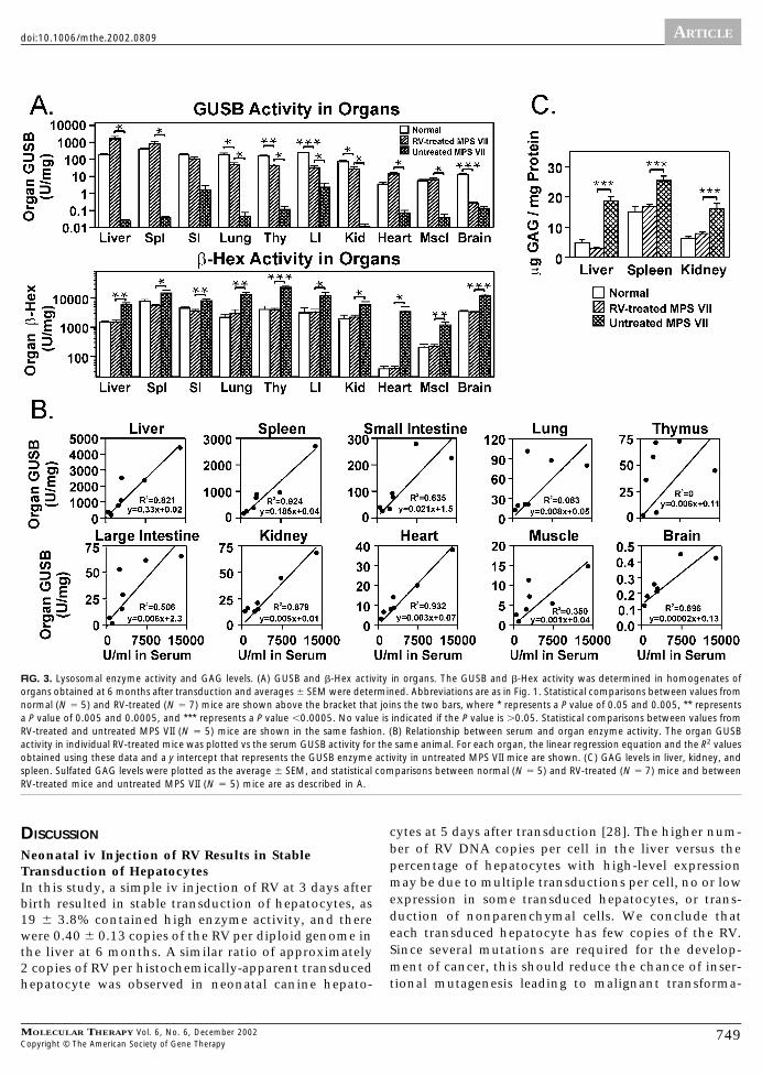
tivity was due to contaminating blood. The average liverGUSB activity was 1664 � 570 U/mg (8.7-fold normal)(Fig. 3A). In addition, total �-hexosaminodase (�-Hex)activity was determined, as activity for these enzymes iselevated in MPS VII, and normalization indicates effectivetreatment. For the RV-treated mice, liver �-Hex levelswere significantly lower than in untreated MPS VII mice(P � 0.0007) and were indistinguishable from those innormal mice (P � 0.98). This suggests that the GUSBlevels were sufficient to reduce this secondary effect oflysosomal storage in the liver.
GUSB activity in other organs from RV-treated mice ispresented in Fig. 3A in order from highest to lowest spe-cific activity. In spleen, lung, thymus, large intestine,
kidney, heart, and muscle, GUSB activity was significantlyhigher in the RV-treated than in the untreated MPS VIImice (P � 0.05) and resulted in a statistically significantreduction in �-Hex activity (P � 0.02 vs values in un-treated MPS VII mice). In the small intestines and brain,GUSB activity was not statistically higher in the RV-treated than in the untreated MPS VII mice. Nevertheless,�-Hex activity was normalized for the RV-treated animalsin both of these organs (P � 0.004 for RV-treated vsuntreated MPS VII mice), suggesting that the GUSB activ-ity that was present was sufficient to reduce lysosomalstorage.
The serum GUSB levels were plotted vs the organ ac-tivity for individual animals to determine if there was a
FIG. 1. Serum GUSB activity and transduction efficiency in RV-treated mice. (A) Serum GUSB activity. MPS VII mice were injected with 1 � 1010 rfu/kg ofhAAT-cGUSB-WPRE at 3 days after birth. Serum was tested for GUSB activity at the indicated times after transduction. The values at 0 months (0.04 U/ml)represent those present in adult untreated MPS VII mice. The average values in homozygous normal mice (28 U/ml � 2 SD) are indicated by the boxed region.D8, D9, D11, and D12 were male, and D10, D13, and D14 were female. (B) Percentage serum GUSB with M6P. The percentage of GUSB that contained M6Pwas determined for serum samples obtained at 6 months after birth and are plotted vs the average serum GUSB activity in the same animal. (C) Transductionefficiency in liver. DNA was isolated from liver at 6 months after transduction and analyzed for RV DNA sequences using real-time PCR. The copies of RV perdiploid genome were plotted vs the serum GUSB level for the same animal. The fraction of hepatocytes that expressed GUSB was determined by analysis of liversections after histochemical staining for GUSB activity. Two nontransduced MPS VII mice had no detectable RV DNA sequences (�0.002 copies per cell) and notransduced hepatocytes. (D) DNA and relative RNA levels in organs. The average RV DNA copy number per diploid genome � SEM in organs was determinedusing real-time PCR for the three RV-treated mice with the highest serum GUSB activity. Spl, spleen; Kid, kidney; Thy, thymus; SI, small intestines; LI, largeintestines; and Mscl, muscle. Only two testes were evaluated, as one animal was female. Note that the average copy number in the liver for these mice is higherthan the average obtained using all seven RV-treated mice. The average relative amount of RNA � SEM was determined in organs for three animals with relativelylow levels of expression using real-time PCR after reverse transcription (RT) of DNase I-treated RNA. The average level in the liver was defined as 100%. Samplesfrom the liver of RV-treated mice had no signal when real-time PCR was performed without preceding RT, and samples from nontransduced mice had no signalafter RT and PCR (not shown). Testis was not evaluated for RNA levels as all animals were female.
ARTICLEdoi:10.1006/mthe.2002.0809
747MOLECULAR THERAPY Vol. 6, No. 6, December 2002Copyright © The American Society of Gene Therapy

linear relationship (Fig. 3B). Serum GUSB activity wasdirectly proportional to the liver GUSB (R2 � 0.82), whichsuggests that secretion of enzyme from the liver does notbecome saturated even with very high levels of expres-sion. There was also a good linear relationship (R2 be-tween 0.93 and 0.88) between serum GUSB and organactivity in spleen, kidney, and heart. A moderate linearcorrelation was seen (R2 between 0.70 and 0.51) in smalland large intestine and in brain. Little or no correlation(R2 �0.35) was seen in lung, muscle, and thymus.
GAG Levels in Liver, Spleen, and KidneyCells that are deficient in GUSB activity cannot degradedermatan, heparan, and chondroitin sulfates, which re-sults in an overall elevation in sulfated GAG levels. Liver,spleen, and kidney sulfated GAG levels in the RV-treatedmice were statistically lower than in untreated MPS VIImice (P � 0.0004) and were indistinguishable from valuesin normal mice (Fig. 3C). All RV-treated mice had similarnormalization of sulfated GAG levels in these organs (notshown).
Histochemical and Pathological Analysis of EnzymeActivity and Lysosomal Storage in OrgansWe analyzed organs from seven RV-treated MPS VII mice,three age-matched untreated MPS VII mice, and two age-matched homozygous normal mice by histochemistry forGUSB enzyme activity and by toluidine blue staining of
1-�m sections for evidence of lysosomal storage. Repre-sentative examples of GUSB staining and histopatholog-ical analyses are shown for kidney (Figs. 4A to 4D), smallintestines (Figs. 4E and 4F), heart (Figs. 5A and 5B), aorta(Figs. 5C and 5D), lung (Figs. 5E and 5F), thymus (Figs. 6Ato 6C), eye (Figs. 6D to 6F), and brain (Fig. 7).
The severity of lysosomal storage was evaluated foreach organ in these animals, as summarized in Table 1. AllRV-treated mice had a marked reduction in histopatho-logical evidence of lysosomal storage in liver, spleen,small intestine, kidney glomeruli and cortical tubules,cortical and hippocampal neurons, glial cells, meninges,and perivascular cells of the brain. Most or all RV-treatedmice had at least a partial reduction in lysosomal storagein the bronchial epithelium, cortex and medulla of thethymus, medullary tubules of the kidney, heart valve,aorta, corneal stroma, and retinal pigmented epithelium.In most cases, RV-treated mice with high serum GUSBactivity had more pathological improvement in these or-gans than did those with low serum GUSB activity, al-though the small number of animals analyzed and thevariation within groups make it difficult to definitely saythat higher serum GUSB activity is better. Six of the sevenRV-treated mice analyzed had no improvement in storagematerial in Purkinje cells. However, D8, the RV-treatedmouse with the highest serum GUSB activity, had amarked reduction in lysosomal storage in this cell type.
FIG. 2. Histochemical analysis of liverfor GUSB activity. Animals were sacri-ficed at 6 months of age. Frozen sec-tions were stained for GUSB activity(red in the cytoplasm) and counter-stained with hematoxylin (blue for nu-clei). (A) MPS VII. After 16 h of GUSBstaining, no enzyme activity waspresent. Original magnification 60�.(B) Normal. After 1 h of GUSB stainingthere was little activity in the hepato-cytes. Original magnification 60�. (Cand D) RV-treated mouse with low se-rum GUSB activity. For an RV-treatedMPS VII mouse with low serum GUSBactivity (D14), many hepatocytes hadhigh levels of GUSB activity (arrow)after a 1-h stain (C original magnifica-tion 60�). A 16-h stain demonstratedthat there were numerous clusters ofhepatocytes with high GUSB activity,and the entire liver had some activity(D original magnification 10�).
ARTICLE doi:10.1006/mthe.2002.0809
748 MOLECULAR THERAPY Vol. 6, No. 6, December 2002Copyright © The American Society of Gene Therapy
DISCUSSION
Neonatal iv Injection of RV Results in StableTransduction of HepatocytesIn this study, a simple iv injection of RV at 3 days afterbirth resulted in stable transduction of hepatocytes, as19 � 3.8% contained high enzyme activity, and therewere 0.40 � 0.13 copies of the RV per diploid genome inthe liver at 6 months. A similar ratio of approximately2 copies of RV per histochemically-apparent transducedhepatocyte was observed in neonatal canine hepato-
cytes at 5 days after transduction [28]. The higher num-ber of RV DNA copies per cell in the liver versus thepercentage of hepatocytes with high-level expressionmay be due to multiple transductions per cell, no or lowexpression in some transduced hepatocytes, or trans-duction of nonparenchymal cells. We conclude thateach transduced hepatocyte has few copies of the RV.Since several mutations are required for the develop-ment of cancer, this should reduce the chance of inser-tional mutagenesis leading to malignant transforma-
FIG. 3. Lysosomal enzyme activity and GAG levels. (A) GUSB and �-Hex activity in organs. The GUSB and �-Hex activity was determined in homogenates oforgans obtained at 6 months after transduction and averages � SEM were determined. Abbreviations are as in Fig. 1. Statistical comparisons between values fromnormal (N � 5) and RV-treated (N � 7) mice are shown above the bracket that joins the two bars, where * represents a P value of 0.05 and 0.005, ** representsa P value of 0.005 and 0.0005, and *** represents a P value �0.0005. No value is indicated if the P value is 0.05. Statistical comparisons between values fromRV-treated and untreated MPS VII (N � 5) mice are shown in the same fashion. (B) Relationship between serum and organ enzyme activity. The organ GUSBactivity in individual RV-treated mice was plotted vs the serum GUSB activity for the same animal. For each organ, the linear regression equation and the R2 valuesobtained using these data and a y intercept that represents the GUSB enzyme activity in untreated MPS VII mice are shown. (C) GAG levels in liver, kidney, andspleen. Sulfated GAG levels were plotted as the average � SEM, and statistical comparisons between normal (N � 5) and RV-treated (N � 7) mice and betweenRV-treated mice and untreated MPS VII (N � 5) mice are as described in A.
ARTICLEdoi:10.1006/mthe.2002.0809
749MOLECULAR THERAPY Vol. 6, No. 6, December 2002Copyright © The American Society of Gene Therapy

FIG. 4. Evaluation of kidney and small intestine. GUSB staining was performed for 16 h followed by counterstain with hematoxylin. Histopathological analysisinvolved staining of 1-�m sections with toluidine blue (TB), and arrows in these images identify lysosomal storage material. Normal mice (images labeled with1), untreated MPS VII mice (labeled with 2), RV-treated mice with low serum GUSB activity (labeled with 3), and RV-treated mice with high serum GUSB activity(labeled with 4) were sacrificed at 6 months of age. The original magnification is shown at the left of each row. (A) GUSB stain of kidney cortex. Black arrowsidentify glomeruli, and open arrows identify tubules. (B) GUSB stain of the kidney medulla. Black arrows identify cells with GUSB activity. (C) Histopathology of kidneycortical tubules. The pathological scores for lysosomal storage were 0, , , and , for 1, 2, 3, and 4, respectively. (D) Histopathology of kidney medulla.The pathological scores for lysosomal storage were 0, , , and , for 1, 2, 3, and 4, respectively. (E) GUSB stain of small intestines. Black arrows identifyenzyme activity in the lamina propria of the small intestines of RV-treated MPS VII mice, while the open arrow identifies enzyme activity in the submucosa. (F)Histopathology of the lamina propria of the small intestines. The pathological scores for lysosomal storage were 0, , 0, and 0, for 1, 2, 3, and 4, respectively.
ARTICLE doi:10.1006/mthe.2002.0809
750 MOLECULAR THERAPY Vol. 6, No. 6, December 2002Copyright © The American Society of Gene Therapy

FIG. 5. Evaluation of heart, aorta, and lung. Groups are identified and GUSB and toluidine blue (TB) staining were done as described for Fig. 4. For all imagesthat involve histopathological analysis with TB staining, arrows identify lysosomal storage material. (A) Histopathology of the aortic valve leaflet. The pathologicalscores for lysosomal storage were 0, , , and 0, for 1, 2, 3, and 4, respectively. (B) Histopathology of the aortic valve attachment region. Arrows identifylysosomal storage material in the region where the aortic valve leaflet attaches to the heart. The pathological scores for lysosomal storage were 0, , ,and 0, for 1, 2, 3, and 4, respectively. (C and D) Histopathology of aorta. The pathological scores for lysosomal storage were 0, , , and 0, for 1, 2,3, and 4, respectively. (E) GUSB staining of lung bronchi. Arrows identify the bronchi. (F) Histopathology of the bronchial epithelium of the lung. The pathologicalscores for lysosomal storage were 0, , , and 0, for 1, 2, 3, and 4, respectively.
ARTICLEdoi:10.1006/mthe.2002.0809
751MOLECULAR THERAPY Vol. 6, No. 6, December 2002Copyright © The American Society of Gene Therapy

FIG. 6. Evaluation of thymus and eye. Groups are identified, and GUSB and toluidine blue (TB) staining were done as described for Fig. 4. For all histopathologywith toluidine blue staining, arrows identify lysosomal storage material. (A) GUSB stain of thymus. Arrows identify GUSB activity in the cortex of the thymus ofRV-treated mice. (B) Histopathology of the thymus cortex. The pathological scores for lysosomal storage were 0, , , and 0, for 1, 2, 3, and 4, respectively.(C) Histopathology of the thymus medulla. The pathological scores for lysosomal storage were 0, , , and , for 1, 2, 3, and 4, respectively. (D) GUSBstain of the cornea. The arrow identifies GUSB activity in the cornea of a RV-treated mouse. (E) Histopathology of the cornea. Black arrows identify lysosomalstorage material in the stroma of the cornea, and open arrows identify storage in the corneal endothelium. The pathological scores for lysosomal storage in thestroma were 0, , , and 0, for 1, 2, 3, and 4, respectively. (F) Histopathology of retina. For all images, open arrows identify the right edge of thepigmented retinal epithelium, while black arrows identify lysosomal storage. The pathological scores for lysosomal storage were 0, , , and 0, for 1,2, 3, and 4, respectively.
ARTICLE doi:10.1006/mthe.2002.0809
752 MOLECULAR THERAPY Vol. 6, No. 6, December 2002Copyright © The American Society of Gene Therapy

FIG. 7. Evaluation of brain. Groups are identified, and GUSB and toluidine blue (TB) staining were done as described for Fig. 4. (A) GUSB stain of brain cortex.Arrows identify cells in the cortex of RV-treated mice that contain GUSB activity. (B) Histopathology of brain cortex. Black and open arrows identify storagematerial within a neuron and a glial cell, respectively. The pathological scores for lysosomal storage were 0, , 0, and 0, for 1, 2, 3, and 4, respectively.(C) GUSB stain of hippocampus. The black arrows identify cells with enzyme activity, while open arrows identify the hippocampus. (D) Histopathology ofhippocampus. Black and open arrows identify lysosomal storage in neurons and a glial cell, respectively. The pathological scores for lysosomal storage were 0,, 0, and 0, for 1, 2, 3, and 4, respectively. (E) GUSB stain of cerebellum. Arrows identify cells in the granular layer with GUSB activity, while the open arrowidentifies enzyme activity in the meninges. (F) Histopathology of cerebellum. Arrows identify lysosomal storage in Purkinje cells. The pathological scores forlysosomal storage were 0, , , and 0, for 1, 2, 3, and 4, respectively.
ARTICLEdoi:10.1006/mthe.2002.0809
753MOLECULAR THERAPY Vol. 6, No. 6, December 2002Copyright © The American Society of Gene Therapy

tion over that observed if each transduced cell hadmany integrations.
The percentage of hepatocytes that expressed thecGUSB cDNA at high levels in the RV-treated MPS VIImice (19%) was ninefold higher than after iv injection ofthe same vector into neonatal MPS VII dogs (2.1%) [28].This may be due to the threefold higher dose of RV perkilogram used in mice compared with the canine study.Alternatively, there may be a higher percentage of repli-cating hepatocytes in neonatal mice than in dogs, al-though this has not yet been tested experimentally. Therewas an eightfold variation in the copies of RV per cell inthe liver among individual animals. This may reflect vari-ation in the number of replicating hepatocytes in individ-ual animals, since cell division is necessary for transduc-tion with an MLV-based RV.
The liver was the major target organ for long-termtransduction, as the DNA copy number was greater thansevenfold higher than in any other organ at 6 months.The maintenance of a high copy number despite a sub-stantial increase in size is likely the result of clonal expan-sion of hepatocytes during normal postnatal growth [31].Since the cluster size observed at 6 months after transduc-tion was similar to what was observed in transgenic micethat expressed �-galactosidase in a subset of hepatocytesbeginning shortly after birth [32], we do not believe thattransduction conferred abnormal growth properties uponthe hepatocytes. However, further studies will be per-formed to determine if some transduced cells proliferateabnormally. Although splenocytes were transduced as ef-ficiently as liver cells when DNA analysis was performed 5days after injection of an RV into neonatal dogs [28], the
TABLE 1: Summary of effect of RV-mediated gene therapy upon pathology in MPS VII mice
Organ Cell type or regionNormal mice
(N � 2)Untreated MPS VII
mice (N � 3)
RV-treated MPS VII mice
Low serumGUSB
Medium serumGUSB
High serumGUSB
Liver Hepatocytes 0 0 0 0
Sinus lining cells 0 0 0 0
Spleen Red pulp 0 0a 0 0
Small intestine Lamina propria 0 0 0 0a
Submucosa 0 , , 0 0 0a
Lung Bronchial cells 0 , 0 0a
Thymus Cortex 0 a , a 0a
Medulla 0 a , , a
Kidney Glomeruli 0 0 0 0
Cortical tubules 0
Medullary tubules 0 , , , , 0 a
Heart Valve 0 , , 0 0a
Valve attachment area 0 a , , , 0a
Aorta Media 0 , , 0
Eye Corneal stroma 0 a , , 0 0
Retinal pigmented epithelium 0 a , , 0 , 0
Brain Cortical neurons 0 0 0 0
Hippocampal neurons 0 0 0 0
Purkinje cells 0 , 0
Glial cells 0 0 0 0
Meninges 0 0 0 0
Perivascular cells 0 0 0 0
The severity of pathologic changes consistent with lysosomal storage disease was determined for organs or cell types from normal, untreated MPS VII, or RV-treated MPS VII mice with low(581 or 947 U/ml), medium (1844, 2733, or 2890 U/ml), or high (5649 or 10070 U/ml) serum GUSB activity. 0 represents histology that is indistinguishable from normal; representsanimals in which storage was absent in some fields and present in others; represents animals in which storage was present in all fields, but at low levels; represents animals inwhich storage was present in all fields at moderate levels; represents animals with large amounts of storage material in all fields. In some organs, the untreated MPS VII mice hadonly low amounts of storage material. Unless otherwise indicated, results from all of the samples of a particular organ and group were concordant.aOnly one of two animals, or only two of three animals, of the group were evaluated, and values were concordant if more than one animal was evaluated. For results that were discordantwithin the group, the results for each animal that was evaluated are shown.
ARTICLE doi:10.1006/mthe.2002.0809
754 MOLECULAR THERAPY Vol. 6, No. 6, December 2002Copyright © The American Society of Gene Therapy

copy number was only 8% of that in liver at 6 monthsafter transduction in mice. This decline in spleen may bedue to the limited life span of splenocytes. Since otherorgans had low RV DNA and RNA levels relative to thosein liver, most of the enzyme activity in other organs wasprobably taken up from blood, although it remains pos-sible that some enzyme was derived from expressing cells.
The Liver Secretes GUSB with M6PSerum GUSB activity for RV-treated mice was stable for 6months at 3531 U/ml (127-fold normal), which exceedsthe highest long-term expression of GUSB from an AAVvector (�300 U/ml) [21]. Although the peak serum GUSBactivity was higher (�9000 U/ml) after administration ofan adenoviral vector, levels fell over time [24]. The serumGUSB contained normal amounts of M6P, demonstratingthat the addition of M6P was not saturated by the over-expression of GUSB in hepatocytes. The level of mannose-6-phosphorylated GUSB in serum varied from 269 U/mlfor D13 to 3291 U/ml for D8. If one assumes that thehalf-life of mannose-6-phosphorylated canine GUSB is 10min based upon data with murine GUSB in mice [22], andthat the serum volume of mice in liters is 4% of the bodyweight in kilograms, it can be calculated that D13 and D8would secrete at least 1.8 million and 22 million units ofphosphorylated enzyme per kilogram per week, respec-tively. For comparison, mice that started ERT shortly afterbirth received 28 million units/kg for their first dose,followed by lower doses on a per kilogram basis (�1.4million units/kg at 6 weeks) as they grew in size, but stillreceived a constant amount of enzyme [10–14].
Serum levels of 269 and 3291 U/ml phosphorylatedGUSB would correspond to 0.22 and 2.8 nM, respectively,if one assumes a specific activity for phosphorylated en-zyme of 4 � 106 U/mg [12] and a molecular weight of thetetramer of 300 kDa. Since the affinity of the cationic-independent M6PR for uptake of diphosphorylated oligo-saccharides is 2 nM [9] and the enzyme concentration islikely lower in tissues than in blood, uptake should not besaturated at these levels. Indeed, organ activity correlatedreasonably well with serum GUSB activity in spleen, smallintestine, large intestine, kidney, heart, and brain. It isunclear why there was a poor correlation (R2 less than 0.5)between serum and organ GUSB activity in lung, thymus,and muscle.
No Immunological Response to cGUSB Was DetectedGene therapy might induce antibody or cytotoxic T lym-phocyte (CTL) responses that would prevent secreted pro-tein from reaching other organs or destroy transducedcells, respectively. In this study, none of the sera fromRV-treated mice contained anti-cGUSB IgG antibodiesthat could be captured by an ELISA plate that was coatedwith 5 �g/ml purified cGUSB (data not shown). Similarly,no anti-human GUSB (hGUSB) antibodies were detectedafter neonatal injection of an AAV vector into muscle
[33]. In contrast, anti-hGUSB antibodies developed afterinjection of hGUSB protein [34] or gene therapy to adultmice with hGUSB-expressing retroviral [17] or adenoviral[24] vectors. In addition, anti-murine GUSB (mGUSB) an-tibodies developed in MPS VII mice that received trans-duced fibroblasts [35], although they did not develop inmice that received injections of mGUSB protein begin-ning shortly after birth or as young adults [13]. A signif-icant CTL response did not occur here since transducedcells clearly survived and maintained high expression lev-els for 6 months. Thus, neonatal gene therapy approachesmay induce tolerance to GUSB.
Effect of Gene Therapy upon Organ PathologyThis gene therapy approach resulted in a marked reduc-tion in lysosomal storage in liver, spleen, and kidneyglomeruli of RV-treated MPS VII compared with untreatedMPS mice, which is consistent with the results of othersdemonstrating that it is relatively easy to correct patho-logical storage in these sites even with low levels of en-zyme activity [16]. In contrast, histopathological correc-tion of tubular storage material in the kidney has beenvariable and has required either high serum levels ofGUSB or bone marrow transplant (BMT) [17,24,36,37].The fact that animals with higher serum activity hadmore histochemical staining for GUSB activity in the tu-bules than did animals with lower expression in this studyis consistent with the hypothesis that high serum levelsare required for delivery of a lysosomal enzyme to thetubules, which may have implications for Fabry disease.The presence of GUSB and reduction in lysosomal storagematerial in the small intestines may have implications forWolman disease, which is a lysosomal storage disease thatis associated with gastrointestinal symptoms. The pres-ence of GUSB and reduction in lysosomal storage in thebronchi may have implications for lung disease in MPSsyndromes, although lung disease has generally been at-tributed to upper airway disease [2,3]. The presence ofGUSB and reduction in lysosomal storage in the thymusmay have implications for the immune defect that occursin MPS VII [38]. The reduction in storage material in theheart valves in this study is important, as heart disease isa major cause of death in MPS syndromes. Our results areconsistent with the effect of AAV-mediated gene therapyin mice [20] and with echocardiographic improvementsin RV-treated MPS VII dogs [29]. In addition, BMT to adultMPS VII mice improved pathology in the heart valves[37]. However, this approach may have advantages overother treatments for arterial disease, as substantial correc-tion of pathological abnormalities was achieved in theaortic media here, but did not occur with ERT beginningshortly after birth or BMT to adult mice [11,37].
Eye and neurological symptoms are important mani-festations of MPS VII. The reduction in the amount ofstorage material in the cornea here is consistent with thereduction in cornea clouding observed on clinical exam-
ARTICLEdoi:10.1006/mthe.2002.0809
755MOLECULAR THERAPY Vol. 6, No. 6, December 2002Copyright © The American Society of Gene Therapy

ination in MPS VII dogs that received RV-mediated neo-natal gene therapy [29]. This result is similar to what wasobserved after BMT [37], although surprisingly ERT wasineffective in the cornea [11–13]. The pathological im-provements in the pigmented retinal epithelium are sim-ilar to what occurred after ERT [13], BMT [39], and sys-temic AAV vector-mediated gene therapy [20] and willlikely correlate with improvements in retinal function[21,40]. The relatively small amounts of GUSB activity inthe brain of RV-treated mice was sufficient to normalize�-Hex activity and prevent accumulation of lysosomalstorage material in cortical and hippocampal neurons,glial cells, perivascular cells, and the meninges for allRV-treated mice. Similar improvements were observed inMPS VII mice after systemic neonatal AAV-mediated genetherapy [20]. ERT alone or in combination with BMTresulted in improvements in all of these regions except forglial cells [11–13], while BMT alone to adults failed toimprove pathology in neurons or glial cells [37] in MPSVII mice. For the RV-treated mouse with the highest se-rum GUSB activity, storage was also reduced in Purkinjecells of the cerebellum, a cell type that has been resistantto correction with other treatments, although none of theother mice had reduced storage in Purkinje cells. Thepathological improvements observed here will likely re-sult in neurological improvements based upon resultswith other treatments [16,41–43]. However, since theGUSB activity present in brain likely derived from enzymethat entered from blood prior to the development of theblood–brain barrier, systemic gene therapy might need tobe combined with a brain-directed approach for long-term benefit.
Implications for Gene TherapyThe goal of this study was to identify a target level ofserum GUSB for preventing the development of clinicalsymptoms in MPS VII without adverse effects. Since ani-mals with low serum GUSB activity had at least partialimprovements in most pathological abnormalities,achieving long-term expression of �500 U/ml GUSB inserum is a reasonable target. This is consistent with resultsobtained after AAV-mediated gene therapy in mice, inwhich serum activity that was �300 U/ml had a markedeffect upon many pathological and clinical parameters ofdisease [21]. Our results are also consistent with genetherapy experiments in MPS VII dogs, in which achieving195 U/ml resulted in marked improvements in mobility,cardiac disease, and eye disease for up to 14 months [29].In this study, higher GUSB expression (5000 to 10,000U/ml in serum) appeared to result in less lysosomal stor-age in heart valves, aorta, thymus, bronchial epithelium,and eye compared with RV-treated animals with low se-rum GUSB activity. In addition, the animal with the high-est level of serum GUSB activity had reduced storage inPurkinje cells, which has not been improved in previousexperiments. However, animals with higher serum GUSB
activity have a greater percentage of transduced hepato-cytes and may be at increased risk for the development ofcancer over those with lower expression. Future experi-ments will determine if these additional pathological im-provements in animals with higher serum GUSB activitywill be consistently observed when more animals are an-alyzed and if they will result in a more profound clinicaleffect compared with that which occurs in RV-treatedmice with low serum GUSB activity. In addition, studiesare in progress to determine if RV-treated MPS VII micedevelop tumors. These studies should help to define thetarget serum GUSB activity for treating patients with sys-temic gene therapy.
MATERIALS AND METHODSAnimal procedures. MPS VII mice and homozygous normal controls in aB6.C-H-2bm1 background were maintained in a pathogen-free environ-ment on a diet with 11% fat (PicoLab Mouse Chow 20 5058). Three-day-old MPS VII mice were injected via the temporal vein with 100 �l ofhAAT-cGUSB-WPRE containing 1 � 107 rfu, for a dose of 1 � 1010 rfu/kg.hAAT-cGUSB-WPRE has been described previously [28] and 8 �g/ml Poly-brene (final concentration) was added just prior to injection. Serum wasobtained by retro-orbital bleeding.
Analysis of GUSB activity and GAG levels. Eight-micrometer frozen sec-tions were fixed and stained for GUSB activity with 0.25 mM naphtholAS-BI-�-D-glucuronide as described [44] except for the following modifi-cation. Liver sections were fixed at 25°C for 20 min to reduce the enzymeactivity and stained for 1 to 16 h. Sections from other organs were fixed at4°C for 20 min and stained for 16 h. To obtain the percentage of trans-duced hepatocytes, the number of large intensely red cells per high-powerfield after a short GUSB stain was divided by the total number of hepato-cytes, as detailed previously [28]. For quantification of GUSB activity, 5 �lof serum or �25 �g of organ homogenate was incubated with 5 mM4-methylumbelliferyl �-D-glucuronide (Sigma Chemical, St. Louis, MO) for1 to 16 h, and the fluorescence was measured [44]. One unit of enzymeactivity releases 1 nmol of 4-methylumbelliferone per hour. Samples withhigh activity were diluted. GUSB activity was normalized to total proteinas determined with the Bradford assay (Bio-Rad Laboratories, Hercules,CA). The percentage of enzyme that contained M6P was determined usinga M6PR column as described previously [29]. Total �-Hex activity wasdetermined using 4 mM 4-methylumbelliferyl-2-acetamido-2-deoxy-�-glucopyranoside as the substrate. GAGs were measured by the alcian bluemethod [45] and normalized to milligrams of protein present in thesample. Statistical comparisons between two groups used the Student t testwithin the program Quatto Pro (Corel Corp., San Diego, CA).
Pathological analysis of organs. Samples were fixed as detailed previouslyand 1-�m sections were stained with toluidine blue [5]. Organs wereevaluated for lysosomal storage by M.S.S., who was blinded as to thegenotype and treatment status of animals. Uncertainties were referred to apathologist. For most organs, all regions of 10 sections were examined at40� magnification and at higher power if necessary. For the heart valveand bronchial epithelium, fewer fields were evaluated in some animals dueto insufficient amounts of tissue with the cells of interest. A failure toanalyze an organ from an animal was due to the absence of the region ofinterest on the slide or to loss of the organ by the pathology department.
PCR for detection of RV DNA and RNA sequences. DNA was isolated fromorgans in a room distant from the main laboratory and real-time PCR wasperformed on 100 ng of DNA using Taqman technology (Applied Biosys-tems, Rockville, MD) [28]. DNA from nontransduced MPS VII mice wasisolated and amplified at the same time to demonstrate a lack of contam-ination. Samples were heated to 95°C for 10 min, and then 40 cycles ofPCR were performed with 15 s at 95°C and 1 min at 60°C. The WPRE
ARTICLE doi:10.1006/mthe.2002.0809
756 MOLECULAR THERAPY Vol. 6, No. 6, December 2002Copyright © The American Society of Gene Therapy

reaction used primers from nt 1448 to1467 of the top (5�-GGCTGTT-GGGCACTGACAAT-3�) and nt 1546 to 1530 of the bottom (5�-ACGTC-CCGCGCAGAATC-3�) strand, and the Taqman probe was from nt 1498 to1520 (5�-TTTCCATGGCTGCTCGCCTGTGT-3�) [46]. Real-time PCR wasperformed for the mouse �-actin gene for normalization purposes withprimers from nt 195 to 217 of the top (5�-GACCCTGAAGTACCCCATT-GAAC-3�) and nt 286 to 265 of the bottom (5�-CACGCAGCTCATTGTA-GAAGGT-3�) strand and a Taqman probe from nt 224 to 252 (5�-TTGT-TACCAACTGGGACGACATGGAGAAG-3�) [47]. Standards were created byinfecting the murine GUSB-deficient fibroblasts (3521 cells [44]) withhAAT-cGUSB-WPRE at a multiplicity of infection of 0.05 and isolating asingle colony with enzyme activity. DNA from these cells was mixed withDNA from normal mice to create standards with 0.5 copy or fewer of theRV DNA per diploid genome. For analysis of other organs, DNA wasisolated from three mice with relatively high levels of expression (D8, D9,and D10). To determine the relative amount of RV RNA, real-time RT-PCRwas used. RNA was isolated from organs of animals with relatively lowlevels of expression (D11, D12, and D14) as described [17], and 5 to 20 �gwas treated with 10 units of RQ1 RNase-free DNase (Fisher Scientific)followed by phenol:chloroform extraction and ethanol precipitation. Foreach sample, 1.5 �g of RNA was used for reverse transcription with theSuperScript First-Strand Synthesis System (Invitrogen, Carlsbad, CA) andthe bottom-strand primers of the WPRE and mouse �-actin sequencesnoted above. Real-time PCR was performed with 1 �l of the 20-�l RTasereaction using the SYBR green dye kit (Applied Biosystems) and the prim-ers noted above, and the difference in the cycle at which the threshold wasreached (CT) was used to determine the relative amounts of RNA. The limitof detection was a level of RNA that was 0.04% of that in liver fromRV-treated mice.
ACKNOWLEDGMENTS
We thank Marie Roberts for breeding of MPS VII mice and Carole Vogler (St.Louis University School of Medicine) for assistance with pathological analyses.This work was supported by the National Institutes of Health (R01 DK54061and K02 DK02575 awarded to K.P.P., DK57586 and HD35671 awarded toM.S.S., DK54481 awarded to M.E.H.) and a Washington University DigestiveDiseases Research Core Center grant (P30 DK 52574). Robert Mango wassupported by an HHMI fellowship and N. Matthew Ellinwood by training grantRR T32-07063.
RECEIVED FOR PUBLICATION AUGUST 12, 2002; ACCEPTED OCTOBER 16,2002.
REFERENCES1. Meikle, P. J., Hopwood, J. J., Clague, A. E., and Carey, W. F. (1999). Prevalence of
lysosomal storage disorders. JAMA 281: 249–254.2. Neufeld, E. F, and Meunzer, J. (2001). The mucopolysaccharidoses. In Metabolic and
Molecular Basis of Inherited Disease (C. R. Scriver, A. L. Beaudet, W. S. Sly, and D. Valle,Eds.), pp. 3421–3452. McGraw–Hill, New York.
3. Sly, W. S., Quinton, B. A., McAlister, W. H., and Rimoin, D. L. (1973). Beta glucuron-idase deficiency: Report of clinical, radiologic, and biochemical features of a newmucopolysaccharidosis. J. Pediatr. 82: 249–257.
4. Yamada, Y., et al. (1998). Treatment of MPS VII (Sly disease) by allogeneic BMT in afemale with homozygous A619V mutation. Bone Marrow Transplant. 21: 629–634.
5. Birkenmeier, E. H., et al. (1989). Murine mucopolysaccharidosis type VII: Characteriza-tion of a mouse with �-glucuronidase deficiency. J. Clin. Invest. 83: 1258–1266.
6. Ray, J., Scarpinom, V., Laing, C., and Haskins, M. E. (1999). Biochemical basis of thebeta-glucuronidase gene defect causing canine mucopolysaccharidosis VII. J. Hered. 90:119–123.
7. Fyfe, J. C., et al. (1999). Molecular basis of feline beta-glucuronidase deficiency: Ananimal model of mucopolysaccharidosis VII. Genomics 58: 121–128.
8. Sands, M. S., and Birkenmeier, E. H. (1993). A single-base-pair deletion in the beta-glucuronidase gene accounts for the phenotype of murine mucopolysaccharidosis typeVII. Proc. Natl. Acad. Sci. USA. 90: 6567–6571.
9. Kornfeld, S. (1992). Structure and function of the mannose 6-phosphate/insulinlikegrowth factor II receptors. Annu. Rev. Biochem. 61: 307–330.
10. Vogler, C., et al. (1993). Enzyme replacement with recombinant beta-glucuronidase inthe newborn mucopolysaccharidosis type VII mouse. Pediatr. Res. 34: 837–840.
11. Sands, M. S., et al. (1994). Enzyme replacement therapy for murine mucopolysaccha-ridosis type VII. J. Clin. Invest. 93: 2324–2331.
12. Vogler, C., et al. (1996). Enzyme replacement with recombinant beta-glucuronidase inmurine mucopolysaccharidosis type VII: Impact of therapy during the first six weeks oflife on subsequent lysosomal storage, growth, and survival. Pediatr. Res. 39: 1050–1054.
13. Sands, M. S., et al. (1997). Murine mucopolysaccharidosis type VII: Long term thera-peutic effects of enzyme replacement and enzyme replacement followed by bonemarrow transplantation.. J. Clin. Invest. 99: 1596–1605.
14. Soper, B. W., et al. (1999). Enzyme replacement therapy improves reproductive per-formance in mucopolysaccharidosis type VII mice but does not prevent postnatal losses.Pediatr. Res. 45: 180–186.
15. Sands, M. S., et al. (2001). Biodistribution, kinetics, and efficacy of highly phosphory-lated and non-phosphorylated beta-glucuronidase in the murine model of muco-polysaccharidosis VII. J. Biol. Chem. 276: 43160–43165.
16. Vogler, C., et al. (2001). Murine mucopolysaccharidosis VII: Impact of therapies on thephenotype, clinical course, and pathology in a model of a lysosomal storage disease.Pediatr. Dev. Pathol. 4: 421–433.
17. Gao, C., Sands, M. S., Haskins, M. E., and Ponder, K. P. (2000). Delivery of a retroviralvector expressing human beta-glucuronidase to the liver and spleen decreases lysoso-mal storage in mucopolysaccharidosis VII mice. Mol. Ther. 2: 233–244.
18. Stein, C. S., et al. (2001). In vivo treatment of hemophilia A and mucopolysaccharidosistype VII using nonprimate lentiviral vectors. Mol. Ther. 3: 850–856.
19. Watson, G. L., et al. (1998). Treatment of lysosomal storage disease in MPS VII miceusing a recombinant adeno-associated virus. Gene Ther. 5: 1642–1649.
20. Daly, T. M., Vogler, C., Levy, B., Haskins, M. E., and Sands, M. S. (1999). Neonatal genetransfer leads to widespread correction of pathology in a murine model of lysosomalstorage disease. Proc. Natl. Acad. Sci. USA 96: 2296–2300.
21. Daly, T. M., Ohlemiller, K. K., Roberts, M. S., Vogler, C. A., and Sands, M. S. (2001).Prevention of systemic clinical disease in MPS VII mice following AAV-mediated neo-natal gene transfer. Gene Ther. 8, 1291–1298.
22. Elliger, S. S., Elliger, C. A., Lang, C., and Watson, G. L. (2002). Enhanced secretion anduptake of beta-glucuronidase improves adeno-associated viral-mediated gene therapyof mucopolysaccharidosis type VII mice. Mol. Ther. 5: 617–626.
23. Ohashi, T., et al. (1997). Adenovirus-mediated gene transfer and expression of humanbeta-glucuronidase gene in the liver, spleen, and central nervous system in muco-polysaccharidosis type VII mice. Proc. Natl. Acad. Sci. USA. 94: 1287–1292.
24. Stein, C. S., Ghodsi, A., Derksen, T., and Davidson, B. L. (1999). Systemic and centralnervous system correction of lysosomal storage in mucopolysaccharidosis type VII mice.J. Virol. 73: 3424–3429.
25. Kosuga, M., et al. (2000). Adenovirus-mediated gene therapy for mucopolysacchari-dosis VII: Involvement of cross-correction in wide-spread distribution of the geneproducts and long-term effects of CTLA-4Ig coexpression. Mol. Ther. 1: 406–413.
26. Xia, H., Mao, Q., and Davidson, B. L. (2001). The HIV Tat protein transduction domainimproves the biodistribution of beta-glucuronidase expressed from recombinant viralvectors. Nat. Biotechnol. 19: 640–644.
27. Li, X. K., et al. (2002). Prolongation of transgene expression by coexpression of cytokineresponse modifier a in rodent liver after adenoviral gene transfer. Mol. Ther. 5: 262–268.
28. Xu, L., et al. (2002). Transduction of hepatocytes after neonatal delivery of a Moloneymurine leukemia virus-based retroviral vector results in long-term expression of �-glu-curonidase in mucopolysaccharidosis VII dogs. Mol. Ther. 5: 141–153.
29. Ponder, K. P., et al. (2002). Therapeutic neonatal hepatic gene therapy in muco-polysaccharidosis VII dogs. Proc. Natl. Acad. Sci. USA 99: 13102–13107.
30. Gates, G. A., Henley, K. S., Pollard, H. M., Schmidt, E., and Schmidt, F. W. (1961). Thecell populations of human liver. J. Lab. Clin. Med. 57: 182–184.
31. Ponder, K. P. (1996). Analysis of liver development, regeneration and carcinogenesis bygenetic marking studies. FASEB J. 10: 673–682.
32. Kennedy, S. C., Rettinger, S. D., Flye, M. W., and Ponder, K. P. (1995). Experiments intransgenic mice demonstrate that hepatocytes are the source for postnatal liver growthand do not stream. Hepatology 22: 160–168.
33. Daly, T. M., et al. (1999). Neonatal intramuscular injection with recombinant adeno-associated virus results in prolonged beta-glucuronidase expression in situ and correc-tion of liver pathology in mucopolysaccharidosis type VII mice. Hum. Gene Ther. 10:85–94.
34. Sly, W. S., et al. (2001). Active site mutant transgene confers tolerance to humanbeta-glucuronidase without affecting the phenotype of MPS VII mice. Proc. Natl. Acad.Sci. USA 98: 2205–2210.
35. Ross, C. J., Bastedo, L., Maier, S. A., Sands, M. S., and Chang, P. L. (2000). Treatmentof a lysosomal storage disease, mucopolysaccharidosis VII, with microencapsulatedrecombinant cells. Hum. Gene Ther. 11: 2117–2127.
36. Barker, J. E., et al. (2001). In utero fetal liver cell transplantation without toxic irradiationalleviates lysosomal storage in mice with mucopolysaccharidosis type VII. Blood CellsMol. Dis. 27: 861–873.
37. Birkenmeier, E. H., et al. (1991). Increased life span and correction of metabolic defectsin murine mucopolysaccharidosis type VII after syngeneic bone marrow transplanta-tion. Blood 78: 3081–3092.
ARTICLEdoi:10.1006/mthe.2002.0809
757MOLECULAR THERAPY Vol. 6, No. 6, December 2002Copyright © The American Society of Gene Therapy

38. Daly, T. M., Lorenz, R. G., and Sands, M. S. (2000). Abnormal immune function in vivoin a murine model of lysosomal storage disease. Pediatr. Res. 47: 757–762.
39. Sands, M. S., et al. (1993). Treatment of murine mucopolysaccharidosis type VII bysyngeneic bone marrow transplantation in neonates. Lab. Invest. 68: 676–686.
40. Ohlemiller, K. K., Vogler, C. A., Roberts, M., Galvin, N., and Sands. M. S. (2000). Retinalfunction is improved in a murine model of a lysosomal storage disease following bonemarrow transplantation. Exp. Eye Res. 71: 469–481.
41. O’Connor, L. H., et al. (1998). Enzyme replacement therapy for murine mucopolysac-charidosis type VII leads to improvements in behavior and auditory function. J. Clin.Invest. 101: 1394–1400.
42. Brooks, A. I., et al. (2002). Functional correction of established central nervous systemdeficits in an animal model of lysosomal storage disease with feline immunodeficiencyvirus-based vectors. Proc. Natl. Acad. Sci. USA 99: 6216–6221.
43. Frisella, W. A., et al. (2001). Intracranial injection of recombinant adeno-associated virus
improves cognitive function in a murine model of mucopolysaccharidosis type VII. Mol.Ther. 3: 351–358.
44. Wolfe, J. H., and Sands, M. S. (1996). Murine mucopolysaccharidosis type VII: A modelsystem for somatic gene therapy of the central nervous system. In Protocols for GeneTransfer in Neuroscience: Towards Gene Therapy of Neurological Disorders (P. R. Lowen-stein and L. W. Enquist, Eds.), pp. 264–274. Wiley. New York.
45. Bjornsson, S. (1993). Simultaneous preparation and quantitation of proteoglycans byprecipitation with alcian blue. Anal. Biochem. 210: 282–291.
46. Zufferey, R., Donello, J. E., Trono, D., and Hope, T. J. (1999). Woodchuck hepatitis virusposttranscriptional regulatory element enhances expression of transgenes delivered byretroviral vectors. J. Virol. 73: 2886–2892.
47. Tokunaga, K., Taniguchi, H., Yoda, K., Shimizu, M., and Sakiyama, S. (1986). Nucleo-tide sequence of a full-length cDNA for mouse cytoskeletal �-actin mRNA. Nucleic AcidsRes. 14: 2829.
ARTICLE doi:10.1006/mthe.2002.0809
758 MOLECULAR THERAPY Vol. 6, No. 6, December 2002Copyright © The American Society of Gene Therapy