Edge-competing Pathological Liver Vessel Segmentation with ...
9
Edge-competing Pathological Liver Vessel Segmentation with Limited Labels Zunlei Feng 1,5# , Zhonghua Wang 1# , Xinchao Wang 2 , Xiuming Zhang 1 , Lechao Cheng 3 , Jie Lei 4 , Yuexuan Wang 1* , Mingli Song 1,5 1 Zhejiang University 2 Stevens Institute of Technology 3 Zhejiang Lab 4 Zhejiang University of Technology 5 Alibaba-Zhejiang University Joint Research Institute of Frontier Technologies {zunleifeng,wang97zh,1508056,amywang,brooksong}@zju.edu.cn, [email protected], [email protected], [email protected] Abstract The microvascular invasion (MVI) is a major prognostic fac- tor in hepatocellular carcinoma, which is one of the malig- nant tumors with the highest mortality rate. The diagnosis of MVI needs discovering the vessels that contain hepatocellu- lar carcinoma cells and counting their number in each ves- sel, which depends heavily on experiences of the doctor, is largely subjective and time-consuming. However, there is no algorithm as yet tailored for the MVI detection from patho- logical images. This paper collects the first pathological liver image dataset containing 522 whole slide images with label- s of vessels, MVI, and hepatocellular carcinoma grades. The first and essential step for the automatic diagnosis of MVI is the accurate segmentation of vessels. The unique character- istics of pathological liver images, such as super-large size, multi-scale vessel, and blurred vessel edges, make the accu- rate vessel segmentation challenging. Based on the collected dataset, we propose an Edge-competing Vessel Segmentation Network (EVS-Net), which contains a segmentation network and two edge segmentation discriminators. The segmenta- tion network, combined with an edge-aware self-supervision mechanism, is devised to conduct vessel segmentation with limited labeled patches. Meanwhile, two discriminators are introduced to distinguish whether the segmented vessel and background contain residual features in an adversarial man- ner. In the training stage, two discriminators are devised to compete for the predicted position of edges. Exhaustive ex- periments demonstrate that, with only limited labeled patch- es, EVS-Net achieves a close performance of fully supervised methods, which provides a convenient tool for the patholog- ical liver vessel segmentation. Code is publicly available at https://github.com/zju-vipa/EVS-Net. Introduction Liver cancer is one of the malignant tumors with the highest mortality rate, which witnesses an increasing number of cas- es in recent years (D, Suna, and Boyacioglu 2017). Hepato- cellular carcinoma represents the most frequent primary liv- er tumor, taking up about 90% of all liver cancer cases (Pe- * Corresponding author. # Both the authors have equal contribu- tion to this work. Copyright c 2021, Association for the Advancement of Artificial Intelligence (www.aaai.org). All rights reserved. Figure 1: The overview of the edge-competing mechanism. The Over-Segmentation Discriminator (OS-D) and Under- Segmentation Discriminator (US-D) are devised for distin- guishing whether the segmented background and segment- ed vessel contain residual features. The over-segmented and under-segmented patches are generated with different ero- sion and dilation operations following binarization process- ing. Only when the predicted mask depicts the precise seg- mentation will the true criterion of the two discriminators be satisfied simultaneously for an input patch. rumpail et al. 2017). The microvascular invasion (MVI) is a major prognostic factor in hepatocellular carcinoma, which significantly affects the choice of treatment and the over- all survival of patients (Sumie et al. 2008; Rodriguezper- alvarez et al. 2013). However, the current MVI detection heavily depends on the experiences of doctors, which is a largely subjective and time-consuming to diagnose. Existing machine learning algorithms for predicting MVI mainly fo- cused on computed tomography, magnetic resonance imag- ing, and positron emission tomography (Cuccurullo et al. 2018), yet ignore pathological images. Even to data, there is no learning-based method yet for automatically detecting MVI in pathological liver images. As deep learning approaches have achieved promising re- sults in the medical image analysis area (Ker et al. 2018; The Thirty-Fifth AAAI Conference on Artificial Intelligence (AAAI-21) 1325
-
Upload
khangminh22 -
Category
Documents
-
view
0 -
download
0
Transcript of Edge-competing Pathological Liver Vessel Segmentation with ...

Edge-competing Pathological Liver Vessel Segmentation with Limited Labels
Zunlei Feng1,5#, Zhonghua Wang1#, Xinchao Wang2, Xiuming Zhang1, Lechao Cheng3, Jie Lei4,Yuexuan Wang1∗, Mingli Song1,5
1Zhejiang University2Stevens Institute of Technology
3Zhejiang Lab4Zhejiang University of Technology
5Alibaba-Zhejiang University Joint Research Institute of Frontier Technologies{zunleifeng,wang97zh,1508056,amywang,brooksong}@zju.edu.cn, [email protected], [email protected],
AbstractThe microvascular invasion (MVI) is a major prognostic fac-tor in hepatocellular carcinoma, which is one of the malig-nant tumors with the highest mortality rate. The diagnosis ofMVI needs discovering the vessels that contain hepatocellu-lar carcinoma cells and counting their number in each ves-sel, which depends heavily on experiences of the doctor, islargely subjective and time-consuming. However, there is noalgorithm as yet tailored for the MVI detection from patho-logical images. This paper collects the first pathological liverimage dataset containing 522 whole slide images with label-s of vessels, MVI, and hepatocellular carcinoma grades. Thefirst and essential step for the automatic diagnosis of MVI isthe accurate segmentation of vessels. The unique character-istics of pathological liver images, such as super-large size,multi-scale vessel, and blurred vessel edges, make the accu-rate vessel segmentation challenging. Based on the collecteddataset, we propose an Edge-competing Vessel SegmentationNetwork (EVS-Net), which contains a segmentation networkand two edge segmentation discriminators. The segmenta-tion network, combined with an edge-aware self-supervisionmechanism, is devised to conduct vessel segmentation withlimited labeled patches. Meanwhile, two discriminators areintroduced to distinguish whether the segmented vessel andbackground contain residual features in an adversarial man-ner. In the training stage, two discriminators are devised tocompete for the predicted position of edges. Exhaustive ex-periments demonstrate that, with only limited labeled patch-es, EVS-Net achieves a close performance of fully supervisedmethods, which provides a convenient tool for the patholog-ical liver vessel segmentation. Code is publicly available athttps://github.com/zju-vipa/EVS-Net.
IntroductionLiver cancer is one of the malignant tumors with the highestmortality rate, which witnesses an increasing number of cas-es in recent years (D, Suna, and Boyacioglu 2017). Hepato-cellular carcinoma represents the most frequent primary liv-er tumor, taking up about 90% of all liver cancer cases (Pe-
∗Corresponding author. #Both the authors have equal contribu-tion to this work.Copyright c© 2021, Association for the Advancement of ArtificialIntelligence (www.aaai.org). All rights reserved.
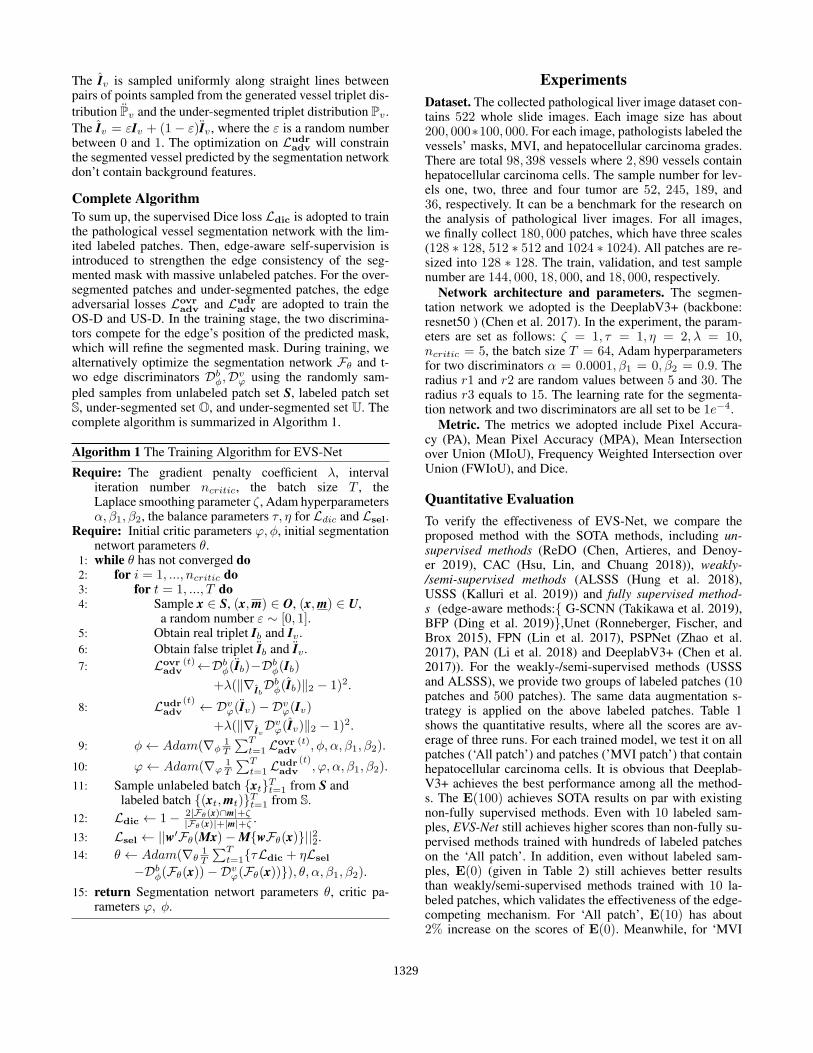
Figure 1: The overview of the edge-competing mechanism.The Over-Segmentation Discriminator (OS-D) and Under-Segmentation Discriminator (US-D) are devised for distin-guishing whether the segmented background and segment-ed vessel contain residual features. The over-segmented andunder-segmented patches are generated with different ero-sion and dilation operations following binarization process-ing. Only when the predicted mask depicts the precise seg-mentation will the true criterion of the two discriminators besatisfied simultaneously for an input patch.
rumpail et al. 2017). The microvascular invasion (MVI) is amajor prognostic factor in hepatocellular carcinoma, whichsignificantly affects the choice of treatment and the over-all survival of patients (Sumie et al. 2008; Rodriguezper-alvarez et al. 2013). However, the current MVI detectionheavily depends on the experiences of doctors, which is alargely subjective and time-consuming to diagnose. Existingmachine learning algorithms for predicting MVI mainly fo-cused on computed tomography, magnetic resonance imag-ing, and positron emission tomography (Cuccurullo et al.2018), yet ignore pathological images. Even to data, thereis no learning-based method yet for automatically detectingMVI in pathological liver images.
As deep learning approaches have achieved promising re-sults in the medical image analysis area (Ker et al. 2018;
The Thirty-Fifth AAAI Conference on Artificial Intelligence (AAAI-21)
1325

Altaf et al. 2019), we expect they have the potential to be de-ployed in the pathological liver vessel segmentation. Thereare many deep learning based segmentation methods forgeneral natural images (Minaee et al. 2020) and medical im-ages (Ronneberger, Fischer, and Brox 2015; Xue et al. 2018;Dai et al. 2019). Due to the specific attributes of different im-ages, however, existing methods cannot be directly appliedto the pathological liver vessel segmentation. Moreover, thesuccess of deep learning are highly based on a large numberof annotated samples; yet for pathological liver vessel seg-mentation, there was not a dedicated dataset available, as itis extremely tedious, time-consuming, and expensive to an-notate those vessels accurately.
In this paper, we first collect a pathological liver imagedataset containing 522 whole slide images with labels ofvessels, MVI, and hepatocellular carcinoma grades. It canreadily serve as a benchmark for the research on the analysisof pathological liver images. Fig. 2 shows some cut patchesand the whole slide pathological image, which has a size of200, 000∗100, 000. For the patches, the coarse edges of mostvessels can be obtained by binarization processing. For thevessels containing hepatocellular carcinoma cells, binariza-tion processing will fail to segment those patches. However,combining with the erosion and dilation operation, binariza-tion processing may easily produce some over-segmented orunder-segmented vessels.
Based on such findings, we devise an edge-competingmechanism to assist the vessel segmentation, for whichthe overview is shown in Fig. 1. With the easily obtainedover-segmented patches as the positive training samples, theover-segmentation discriminator is devised for distinguish-ing whether the segmented background contains residualfeatures of vessels. Similarly, the under-segmentation dis-criminator is devised for distinguishing whether the seg-mented vessels contain residual features of the background.For an input patch, only when the predicted mask depict-s the perfect segmentation will the true criterion of the t-wo discriminators be satisfied simultaneously. It will effec-tively relieve the dependence of annotation by incorporatingthe edge-competing mechanism into the vessel segmentationframework.
Besides, from the whole slide image in Fig. 2, we cansee that the pathological liver images have some unique at-tributes, such as super-large sample size, multi-scale vessel,blurred vessel edge, which is challenging for the accuratevessel segmentation. Given such specific characteristics ofpathological liver images, we propose the Edge-competingVessel Segmentation Network (EVS-Net), which is shownin Fig. 2. For the massive unlabeled patches, the doctor on-ly needs to label limited patches as guidance. For the restof the unlabeled patches, the binarization processing com-bined with erosion and dilation will generate many over-segmented and under-segmented patches.
As shown in Fig. 2, the proposed EVS-Net is composedof a Pathological Vessel Segmentation Network (PVSN), anOver-Segmentation Discriminator (OS-D), and an Under-Segmentation Discriminator (US-D). In the training stage,the limited labeled patches will supervise the PVSN to gaininitial segmentation ability for the pathological vessel patch-
es. The edge-competing mechanism is then incorporated in-to EVS-Net for assisting the training of the segmentationnetwork in an adversarial manner.
To further strengthen consistency of the segmentation onmassive unlabeled patches, an edge-aware self-supervisionmodule is introduced to generate the self-supervision in-formation for training PVSN. The core idea of edge-awareself-supervision is that, for the same image and segmenta-tion network, the predicted mask of the transformed imageshould be equal to the transformed mask predicted by thenetwork with the original image as input.
Our contribution can be summarized as follows. First-ly, we collect a pathological liver image dataset that con-tains 522 whole slide images with labels of vessels, MVI,and hepatocellular carcinoma grades. It can readily serveas a benchmark for the analysis of hepatocellular carci-noma. Next, the proposed segmentation network combinesan edge-aware self-supervision module and two segmenta-tion discriminators to efficiently solve the challenges in thepathological vessel segmentation. Furthermore, the edge-competing mechanism between two discriminators is first-ly proposed for learning accurate segmentation with over-segmented and under-segmented patches, which effectivelyrelieves the dependence on a large number of annotations.Finally, with limited labels, the EVS-Net achieves results onpar with fully supervised ones, providing a handy tool forthe pathological liver vessel segmentation.
Related WorksTo our knowledge, there are no vessel segmentation meth-ods for pathological images until now. Existing methods,which are related to pathological images, mainly focus onthe pathological grading of various carcinomas (Yang et al.2019; Jiao et al. 2020; Liao et al. 2020). Here, we give therelated segmentation works for general natural images andmedical images from two perspectives.
Edge-aware Segmentation. For edge-aware semanticsegmentation, the commonly adopted framework is a two-branch network that simultaneously predicts segmentationmaps and edges, where different constraints (Bertasius, Shi,and Torresani 2016; Chen et al. 2016; Cheng et al. 2017;Yu et al. 2018; Yin et al. 2018; Ye et al. 2019; Takikawaet al. 2019) are devised for strengthening the segmenta-tion results with the predicted edges. Unlike predicting theedge directly, Hayder, He, and Salzmann (2017) proposedpredicting pixels’ distance to the edge of object and post-processed the distance map into the final segmented result-s. Khoreva et al. (2017) proposed edge-aware filtering toimprove object delineation. Zhang et al. (2017) proposed alocal edge refinement network to learn the position-adaptivepropagation coefficients so that local contextual informationfrom neighbors can be optimally captured for refining ob-ject edges. Qin et al. (2019) adopted the patch-level SSIMloss (Wang, Simoncelli, and Bovik 2003) to assign high-er weights to the edges. Peng et al. (2017) proposed anedge refinement block to improve the localization perfor-mance near the object edges. Unlike the above methods, twoedge-competing discriminators are devised for distinguish-ing whether the segmented vessels and background contain
1326

size:200,000 *100,000
…
massiveunlabeled patches
edge-competing
OS-D
US-D
less erosion& more dilation
Pathological Vessel Segmentation Network
binarizationprocessing
…
…
…
over-seg.
under-seg.
False
True
False
True
labeling limitedpatches
more erosion& less dilation
Figure 2: The flow diagram of pathological liver vessel segmentation. For massive unlabeled patches cut from the whole slidepathological image, the doctor only needs to label limited patches. Meanwhile, some under-segmented (over-seg.) patches andunder-segmented (under-seg.) patches are generated through different erosion and dilation operations following binarizationprocessing. Then, the EVS-Net is proposed for segmenting the vessels in the patches based on the above limited labels, over-segmented and under-segmented patches. The EVS-Net contains a Pathological Vessel Segmentation Network (PVSN), anover-segmentation discriminator (OS-D), and an under-segmentation discriminator (US-D). The PVSN is first initialized bytraining with the limited labels. With the over-segmented and under-segmented patches, the two discriminators are trained fordistinguishing whether the segmented vessels and background contain residual features. In the training stage, for an unlabeledpatch, only when the predicted mask is the perfect segmentation will the true criterion of the two discriminators be satisfiedsimultaneously, which facilitates the PVSN segmenting more accurately.
residual features. The over-segmented and under-segmentedpatches are adopted to train the two discriminators, focusingon the outer and inner area of edges.
GAN based Segmentation contains two categories: com-position fidelity based methods (Ostyakov et al. 2018; Re-mez, Huang, and Brown 2018; Chen, Artieres, and Denoyer2019; Qiu et al. 2020) and mask distribution-based method-s (Luc et al. 2016; Han and Yin 2017; Arbelle and Raviv2018; Hung et al. 2018; Ye et al. 2020). The composition fi-delity based methods (Ostyakov et al. 2018; Remez, Huang,and Brown 2018; Chen, Artieres, and Denoyer 2019) adopt-ed the discriminator to distinguish the fidelity of natural im-ages and the composited images, which is composition ofthe segmented objects and some background images. For themask distribution-based methods, Luc et al. (Luc et al. 2016)adopted the adversarial optimization between segmented re-sults and GT mask to train the segmentation network, whichis the first GAN based semantic segmentation network. Nex-t, some researchers (Han and Yin 2017; Arbelle and Ra-viv 2018; Xue et al. 2018) applied the same adversarialframework on the medical image segmentation task. Fur-thermore, Souly et al. (Souly, Spampinato, and Shah 2017)and Hung et al. (Hung et al. 2018) adopted the adversari-al strategy to train the discriminator output class confidencemaps. Unlike the above existing GAN-based methods, wedevised two-discriminator architecture, where the two dis-criminators will compete the vessel edge’s position of theunlabeled patch in the adversarial training stage. In addition,the true inputs of the two discriminators are over-segmentedand under-segmented patches.
Proposed MethodIn the real diagnostic scenario, the assisting diagnosis tool ofMVI should be objective, accurate, and convenient. The less
time and labor cost, the better. Based on the real requirementof diagnosis, we introduce a pathological liver vessel seg-mentation method that can achieve applicable performancewith limited labels. The proposed method contains two part-s: the pathological patches pre-processing and pathologi-cal vessel segmentation. In the former part, the doctor onlyneeds to label limited patches. Some over/under-segmentedpatches are generated with different erosion and dilation op-erations following binarization processing. In the latter part,the EVS-Net is devised for segmenting the pathological liv-er vessels based on the above limited labeled patches, over-segmented and under-segmented patches.
Pathological Patch PreprocessingFrom Fig. 2, we can see that the pathological liver imagehas a super-large size and multi-scale vessel. Meanwhile, theareas of vessels and background are unbalanced. To get theuseful training samples, we first locate the vessels’ generalpositions according to the white color. Then, three sizes areadopted to cut patches from the whole slide image aroundthe located positions.
For the massive cut patch set S = {x1, x2, x3, ..., xN},the doctor only needs to label limited patches S ={(x1,m1), (x2,m2), (x3,m3), ..., (xK ,mK)}. Next, for apatch x ∈ S, the under-segmented mask m of patch x canbe calculated as follows:
m = [1]− Tostu(x)Sr1 ⊕Sr2, r1 > r2, (1)
where, [1] denotes the unit matrix that has the same sizeof the mask, Tostu denotes the OTSU binarization algo-rithm (Otsu 1979), S denotes the disk structuring elemen-t. denotes the erosion operation by structure Sr1 withradius r1, ⊕ denotes the dilation operation by structureSr2 with radius r2. The r1 and r2 are integers. The ob-tained m is the mask of background. The over-segmented
1327

patches constitute the over-segmented image set O ={(x1,m1), (x2,m2), (x3,m3), ..., (xN ,mN )}.
Similarly, the under-segmented image set U ={(x1,m1), (x2,m2), (x3,m3), ..., (xN ,mN )} is calculatedas follows:
m = Tostu(x)Sr1 ⊕Sr2, r1 < r2. (2)
Pathological Vessel SegmentationAs shown in Fig. 2, the proposed EVS-Net is composed of asegmentation network and two discriminators. The segmen-tation network is trained with supervision loss on the labeledlimited patches and an edge-aware self-supervision loss onunlabeled patches. The two discriminators are trained onover-segmented and under-segmented patches for distin-guishing whether the segmented background and vessel con-tain residual features, respectively. In the adversarial trainingstage, the edge competition between two discriminators willfacilitate the vessels’ accurate segmentation.
Segmentation Network. In the EVS-Net, the segmenta-tion network Fθ is designed to be an encoder-decoder archi-tecture. With a labeled patch x ∈ S, the segmented resultm = Fθ(x) is expected to approximate the GT mask m,which can be achieved by minimizing the pixel-wise two-class Dice loss Ldic:
Ldic = 1− 2|m ∩m|+ ζ
|m|+ |m|+ ζ, (3)
where, ζ is the Laplace smoothing parameter for preventingzero error and reducing overfitting. For the labeled samples,we augment samples’ diversity through compositing back-ground. The composited background is generated by addingother pure background and normalizing the new values intothe normal range.
Edge-aware Self-supervision. To reduce the dependenceon massive labeled samples, inspired by Wang et al. (2019),we introduce an edge-aware self-supervision strategy, whichcan strengthen the edge consistency of vessels. The core ideais that the predicted mask of a transformed image shouldbe equal to the transformed mask predicted by the networkwith the original image as input. Formally, for the robustsegmentation network, given an affine transformation ma-trix M, segmented result Fθ(Mx) of the affine transformedimage should be consistent with the affine transformed maskMFθ(x) in the following way:Fθ(Mx) = MFθ(x). Further-more, we obtain the edge neighborhood weight map w asfollows:
w = m⊕Sr3 − mSr3, (4)where, r3 denotes the radius of the structure S. With theweight map w, the edge-aware self-supervision loss Lsel isdefined as follows:
Lsel = ||w′Fθ(Mx)−M{wFθ(x)}||22, x ∈ S, (5)
where, w′ and w are the weight maps of the predictedmasks Fθ(Mx) and Fθ(x), respectively. The edge-awareself-supervision mechanism not only strengthens the edgeconsistency of the segmented vessels but also can eliminatethe unreasonable holes in the predict masks. The root reasonis that unreasonable holes will be assigned high weights.
Edge-competing Discriminator. Based on the obtainedunder-segmented set O and under-segmented set U, we de-vised two discriminators for distinguishing whether the seg-mented patch is a pure vessel or a pure background.
Over-segmentation Discriminator. For the over-segmented patch pairs (x,m) ∈ O, the background xbis computed using the following equation: xb = m ∗ x,where ‘∗’ denotes pixel-wise multiplication. Then the con-catenated triplet Ib = [x,m, xb] is set as the positive sampleof OS-D. The true condition of OS-D is that the segmentedbackground doesn’t contain any residual vessels’ features.For the unlabeled patch x ∈ S, the segmentation networkpredicts the mask of vessel m = Fθ(x). The reversed maskis calculated as follows: m′ = [1]−m, where the [1] denotesthe unit matrix that has the same size of the mask. Next, thesegmented background xb is computed using the followingequation: xb = m′ ∗ x, Then the concatenated tripletIb = [x, m′, xb] is fed to the OS-D Dbφ, which discriminateswhether the segmented background xb contains the vessels’residual features. The Ib is regarded as a false triplet. Theadversarial optimization between the segmentation networkand OS-D will constrain the segmented background pre-dicted by the segmentation network don’t contain vessels’features. The over-segmentation adversarial loss Lovr
adv isgiven as follows:
Lovradv = E
Ib∼Pb[Dbφ(Ib)]− E
Ib∼Pb[Dbφ(Ib)]
+ λ EIb∼PIb
[(‖∇IbDbφ(Ib)‖2 − 1)2],
(6)
where, the Pb, Pb are the generated background triplet distri-bution and under-segmented triplet distribution, respective-ly. The Ib is sampled uniformly along straight lines betweenpairs of points sampled from the generated backgroundtriplet distribution Pb and the under-segmented triplet dis-tribution Pb. The Ib = εIb + (1 − ε)Ib, where the ε is arandom number between 0 and 1. The second term (gradientpenalty) is firstly proposed in WGAN-GP (Gulrajani et al.2017). The λ is the gradient penalty coefficient.
Under-segmentation Discriminator. For the under-segmented patch pairs (x,m) ∈ U, the segmented vessel xvis calculated with the following equation: xv = m ∗ x. Thenthe concatenated triplet Iv = [x,m, xv] satisfies the truecondition of the US-D that the segmented vessel doesn’tcontain any residual background features. For the unlabeledpatch x ∈ S, the concatenated triplet Iv = [x, m, xv] isfed to the US-D Dvϕ, which distinguishes whether thesegmented vessel xv contains the background residualfeatures. The Iv is regarded as the false triplet for US-D.The under-segmentation adversarial loss Ludr
adv is given asfollows:
Ludradv = E
Iv∼Pv[Dvϕ(Iv)]− E
Iv∼Pv[Dvϕ(Iv)]
+ λ EIv∼PIv
[(‖∇IvDvϕ(Iv)‖2 − 1)2],
(7)
where, the Pv , Pv are the generated vessel triplet distribu-tion and under-segmented triplet distribution, respectively.
1328
The Iv is sampled uniformly along straight lines betweenpairs of points sampled from the generated vessel triplet dis-tribution Pv and the under-segmented triplet distribution Pv .The Iv = εIv + (1 − ε)Iv , where the ε is a random numberbetween 0 and 1. The optimization on Ludr
adv will constrainthe segmented vessel predicted by the segmentation networkdon’t contain background features.
Complete AlgorithmTo sum up, the supervised Dice loss Ldic is adopted to trainthe pathological vessel segmentation network with the lim-ited labeled patches. Then, edge-aware self-supervision isintroduced to strengthen the edge consistency of the seg-mented mask with massive unlabeled patches. For the over-segmented patches and under-segmented patches, the edgeadversarial losses Lovr
adv and Ludradv are adopted to train the
OS-D and US-D. In the training stage, the two discrimina-tors compete for the edge’s position of the predicted mask,which will refine the segmented mask. During training, wealternatively optimize the segmentation network Fθ and t-wo edge discriminators Dbφ,Dvϕ using the randomly sam-pled samples from unlabeled patch set S, labeled patch setS, under-segmented set O, and under-segmented set U. Thecomplete algorithm is summarized in Algorithm 1.
Algorithm 1 The Training Algorithm for EVS-Net
Require: The gradient penalty coefficient λ, intervaliteration number ncritic, the batch size T , theLaplace smoothing parameter ζ, Adam hyperparametersα, β1, β2, the balance parameters τ, η for Ldic and Lsel.
Require: Initial critic parameters ϕ, φ, initial segmentationnetwort parameters θ.
1: while θ has not converged do2: for i = 1, ..., ncritic do3: for t = 1, ..., T do4: Sample x ∈ S, (x,m) ∈ O, (x,m) ∈ U,
a random number ε ∼ [0, 1].5: Obtain real triplet Ib and Iv .6: Obtain false triplet Ib and Iv .7: Lovr
adv(t)←Dbφ(Ib)−Dbφ(Ib)
+λ(‖∇IbDbφ(Ib)‖2 − 1)2.
8: Ludradv
(t) ← Dvϕ(Iv)−Dvϕ(Iv)+λ(‖∇IvD
vϕ(Iv)‖2 − 1)2.
9: φ← Adam(∇φ 1T
∑Tt=1 Lovr
adv(t), φ, α, β1, β2).
10: ϕ← Adam(∇ϕ 1T
∑Tt=1 Ludr
adv
(t), ϕ, α, β1, β2).
11: Sample unlabeled batch {xt}Tt=1 from S andlabeled batch {(xt,mt)}Tt=1 from S.
12: Ldic ← 1− 2|Fθ(x)∩m|+ζ|Fθ(x)|+|m|+ζ .
13: Lsel ← ||w′Fθ(Mx)−M{wFθ(x)}||22.14: θ ← Adam(∇θ 1
T
∑Tt=1{τLdic + ηLsel
−Dbφ(Fθ(x))−Dvϕ(Fθ(x))}), θ, α, β1, β2).15: return Segmentation networt parameters θ, critic pa-
rameters ϕ, φ.
ExperimentsDataset. The collected pathological liver image dataset con-tains 522 whole slide images. Each image size has about200, 000∗100, 000. For each image, pathologists labeled thevessels’ masks, MVI, and hepatocellular carcinoma grades.There are total 98, 398 vessels where 2, 890 vessels containhepatocellular carcinoma cells. The sample number for lev-els one, two, three and four tumor are 52, 245, 189, and36, respectively. It can be a benchmark for the research onthe analysis of pathological liver images. For all images,we finally collect 180, 000 patches, which have three scales(128 ∗ 128, 512 ∗ 512 and 1024 ∗ 1024). All patches are re-sized into 128 ∗ 128. The train, validation, and test samplenumber are 144, 000, 18, 000, and 18, 000, respectively.
Network architecture and parameters. The segmen-tation network we adopted is the DeeplabV3+ (backbone:resnet50 ) (Chen et al. 2017). In the experiment, the param-eters are set as follows: ζ = 1, τ = 1, η = 2, λ = 10,ncritic = 5, the batch size T = 64, Adam hyperparametersfor two discriminators α = 0.0001, β1 = 0, β2 = 0.9. Theradius r1 and r2 are random values between 5 and 30. Theradius r3 equals to 15. The learning rate for the segmenta-tion network and two discriminators are all set to be 1e−4.
Metric. The metrics we adopted include Pixel Accura-cy (PA), Mean Pixel Accuracy (MPA), Mean Intersectionover Union (MIoU), Frequency Weighted Intersection overUnion (FWIoU), and Dice.
Quantitative EvaluationTo verify the effectiveness of EVS-Net, we compare theproposed method with the SOTA methods, including un-supervised methods (ReDO (Chen, Artieres, and Denoy-er 2019), CAC (Hsu, Lin, and Chuang 2018)), weakly-/semi-supervised methods (ALSSS (Hung et al. 2018),USSS (Kalluri et al. 2019)) and fully supervised method-s (edge-aware methods:{ G-SCNN (Takikawa et al. 2019),BFP (Ding et al. 2019)},Unet (Ronneberger, Fischer, andBrox 2015), FPN (Lin et al. 2017), PSPNet (Zhao et al.2017), PAN (Li et al. 2018) and DeeplabV3+ (Chen et al.2017)). For the weakly-/semi-supervised methods (USSSand ALSSS), we provide two groups of labeled patches (10patches and 500 patches). The same data augmentation s-trategy is applied on the above labeled patches. Table 1shows the quantitative results, where all the scores are av-erage of three runs. For each trained model, we test it on allpatches (‘All patch’) and patches (’MVI patch’) that containhepatocellular carcinoma cells. It is obvious that Deeplab-V3+ achieves the best performance among all the method-s. The E(100) achieves SOTA results on par with existingnon-fully supervised methods. Even with 10 labeled sam-ples, EVS-Net still achieves higher scores than non-fully su-pervised methods trained with hundreds of labeled patcheson the ‘All patch’. In addition, even without labeled sam-ples, E(0) (given in Table 2) still achieves better resultsthan weakly/semi-supervised methods trained with 10 la-beled patches, which validates the effectiveness of the edge-competing mechanism. For ‘All patch’, E(10) has about2% increase on the scores of E(0). Meanwhile, for ‘MVI
1329

Type Unsupervised Weakly Supervised Edge-aware Fully Supervised Ours
Method ReDO CAC ALSSS/ 500 USSS/ 500 G-SCNN BFP Unet FPN PSPNet PAN Deeplab E(100)A
llpa
tch MPA 87.89 84.32 62.92/ 91.84 89.88/ 94.93 97.29 96.83 96.45 98.56 98.29 98.44 98.82 96.59
MIOU 82.20 78.35 55.41/ 89.40 84.57/ 92.35 97.28 96.04 92.76 97.40 96.86 97.11 97.79 94.38FWIoU 89.34 80.57 74.45/ 93.77 90.75/ 95.46 95.89 96.32 95.64 98.46 98.13 98.29 98.69 96.67DICE 96.56 88.32 91.99/ 98.08 97.02/ 98.58 98.76 98.21 98.62 99.52 99.42 99.47 99.60 98.96
MV
Ipat
ch MPA 81.85 78.32 56.09/ 77.71 80.61/ 89.34 95.87 95.43 89.14 97.55 96.61 97.17 97.97 90.85MIOU 74.06 69.87 48.21/ 72.43 73.82/ 84.83 92.85 94.72 81.77 95.60 94.45 94.82 95.84 86.19FWIoU 84.75 79.10 71.55/ 84.44 84.81/ 91.24 95.90 94.53 89.16 97.48 96.80 97.00 97.60 91.98DICE 94.95 85.32 91.28/ 95.19 95.09/ 97.25 94.83 97.53 96.42 99.23 99.01 99.07 99.29 97.46
Table 1: The quantitative results of different methods. Underline indicates the best performance among all methods. Boldindicates the best performance among all non-fully supervised methods. ‘E(K)’ denotes EVS-Net with ‘K’ labeled patches.The ‘ALSSS/ 500’ and ‘USSS/ 500’ denote methods with 10 or 500 labeled patches (All scores are the average of three runs).
Figure 3: The visual result of different methods. The ‘E(K)’ denotes the method trained with K labeled patches.
patch’, E(10) has about 4% performance increase comparedwith E(0), which verifies the necessity of the limited la-beled samples. More results of EVS-Net with different la-beled patches are given in Table 2 and Fig. 4.
Qualitative EvaluationThe visual results of different methods are shown in Fig. 3,where we can see that most of the fully supervised methodsachieve similar results. Deep learning-based methods indeedhave the potential to be applied to the pathological liver ves-sel segmentation. For the unsupervised methods (ReDO andCAC), the segmented background and vessels contain manysmall holes, which indicates the instability of the unsuper-vised methods. From the column 5, we can see that ALSSSwith 10 labeled patches fails to segment most cases, demon-strating that ten labeled patches cannot support enough su-pervision information for the semi-supervised method. Evenwith 500 labeled patches, USSS still fails to segment the ves-sel with a large hepatocellular carcinoma area (row 4, col-umn 8). For the blurry edge vessels (row 1 and row 5), EVS-Net with larger than 10 labeled patches can achieve moreaccurate edge segmentation than non-fully supervised meth-ods. For MVI patches, E(0) achieves inaccurate segmenta-tion on those hepatocellular carcinoma areas, which indi-cates that limited labeled patches are necessity for guidingthe segmentation of hepatocellular carcinoma areas. In sum-
mary, with limited labeled patches, EVS-Net can achieveclose results on par with fully supervised methods.
Ablation StudyTo verify the effectiveness of each component, we conductan ablation study on the edge-aware self-supervision, over-segmentation discriminator, under-segmentation discrimina-tor, two discriminators, and the different numbers of la-beled patches. The E−self , E−over, E
−under and E−disc denote
the EVS-Net without edge-aware self-supervision, over-segmentation discriminator, under-segmentation discrimina-tor, and two discriminators, which are trained in the samesetting of 10 labeled patches. Table 2 shows the quantita-tive results of the above methods. The E(K) denotes theEVS-Net with K labeled patches. We can see that E(10)achieves the best performance among E−self , E−over, E
−under
and E−disc, which demonstrates that all components of EVS-Net are helpful for the segmentation performance. The per-formance of E−disc that doesn’t contain two discriminatorsdegrades about 10% compared with the E(10). Method-s with only one discriminator (E−over and E−under) reduceabout 2% compared with the E(10). From Fig. 4, we cansee that predicted masks of E(10) have accurate edges thanE−over and E−under, which verifies the effectiveness and ne-cessity of edge-competing. In addition, Fig. 4 shows that the
1330

Index\Ablation E−self E−over E−under E−disc E(0) E(5) E(10) E(20) E(50) E(100) E(fully)A
llpa
tch MPA 95.73 94.28 93.59 90.90 94.77 95.47 96.24 96.38 96.72 96.59 98.48
MIoU 92.38 90.69 90.02 78.83 91.93 92.40 93.34 93.55 93.68 94.38 97.25FWIoU 95.44 94.02 93.63 86.38 95.20 95.46 96.02 96.21 96.22 96.67 98.37DICE 98.56 97.42 97.30 95.08 98.50 98.58 98.75 98.76 98.81 98.96 99.50
MV
I pat
ch MPA 87.80 88.42 87.26 80.47 85.12 87.89 89.81 89.63 89.25 90.85 97.16MIoU 78.97 78.73 78.05 62.22 77.57 81.32 82.03 82.91 83.14 86.19 95.05
FWIoU 87.33 86.64 86.32 74.16 86.74 89.01 89.25 89.83 90.10 91.98 97.14DICE 95.71 94.75 94.67 89.19 94.66 96.43 96.42 96.35 96.80 97.46 99.12
Table 2: The ablation study result on EVS-Net.E−self ,E−cond,E−over,E−under andE−disc denote the EVS-Net without edge-aware
self-supervision, over-segmentation discriminator, under-segmentation discriminator, and two discriminators.E(K) denotes theEVS-Net with K labeled patches. Italic indicates the best performance among methods trained with 10 labeled patches. Boldand underline indicate the best and second-best performance among all methods (All scores in %).
Figure 4: The visual result of ablation study. E denotes segmented vessels of method E (Zooming in to compare edges).
result of E−self has some holes. By contrast, the result ofE(10) is accurate, which indicates that the edge-aware self-supervision is also beneficial for eliminating the incorrectholes. For the different numbers of labeled patches, we findthat the critical cut-off point is 10-labeled-samples, whichcan supply relatively sufficient guidance. For the patchesthat have MVI, vessels’ edges are usually blurry, and ves-sels’ inner usually contain the large area of the hepatocellu-lar carcinoma cells. Without any guidance of labeled sam-ples, the pathological vessel segmentation network will failto obtain the vessels’ desired edges. Therefore, the E(0)achieves the worst performance among all E(K) method-s, which also verifies the necessity of the limited labeledpatches. In addition, the comparisons between the segment-ed vessels of E−over, E
−under, E(10) are shown in the last
three columns on the right of Fig. 4, where we can see thatsegmented vessels’ contours of E−over and E−under contrac-t inward and expand outward, respectively. With the edge-competing between OS-D and US-D, E(10) have accurateedges’ contours, which effectively clarifies the effectivenessof the edge-competing mechanism.
ConclusionIn this paper, we propose the first computer-aided diag-nosis method for the pathological liver vessel segmenta-tion, which is an essential and critical step for the diag-
nosis of MVI. For massive unlabeled pathological patch-es, the doctor only needs to label limited patches. Next, bi-narization processing combining with erosion and dilationis adopted to generate some under-segmented and under-segmented masks. Then, the EVS-Net is proposed for seg-menting the pathological liver vessel with limited labels. TheEVS-Net contains a pathological vessel segmentation net-work and two discriminators. With the limited labeled patch-es, the segmentation network is initialized with the super-vision training. Meanwhile, an edge-aware self-supervisionmodule is proposed for enhancing the edge consistency ofmassive unlabeled patches. With the over-segmented andunder-segmented patches, two discriminators are devisedfor distinguishing whether the segmented vessels and back-ground contain residual features. The segmentation networkand two discriminators are trained in an adversarial manner.The edge-competing mechanism is verified to be very ef-fective for facilitating the segmentation network segmentingmore accurately. Exhaustive experiments demonstrate thatthe proposed EVS-Net achieves a close performance of fullysupervised methods with limited labeled patches. It providesa convenient tool for the pathological liver vessel segmenta-tion. In future work, we will focus on detecting and countingthe hepatocellular carcinoma cells in the segmented vessel-s, which will provide a complete assisting diagnosis methodfor the diagnosis of MVI.
1331
AcknowledgmentsThis work was supported by National Natural Science Foun-dation of China (No.62002318), Zhejiang Provincial Nat-ural Science Foundation of China (LQ21F020003), Zhe-jiang Provincial Science and Technology Project for PublicWelfare (LGF21F020020), Programs Supported by NingboNatural Science Foundation (202003N4318), and Alibaba-Zhejiang University Joint Research Institute of FrontierTechnologies.
ReferencesAltaf, F.; Islam, S.; Akhtar, N.; and Janjua, N. K. 2019. Go-ing Deep in Medical Image Analysis: Concepts, Methods,Challenges, and Future Directions. IEEE Access 7: 99540–99572.
Arbelle, A.; and Raviv, T. R. 2018. Microscopy Cell Seg-mentation via Adversarial Neural Networks. IEEE Interna-tional Symposium on Biomedical Imaging 645–648.
Bertasius, G.; Shi, J.; and Torresani, L. 2016. Semantic Seg-mentation with Boundary Neural Fields. IEEE Conferenceon Computer Vision and Pattern Recognition 3602–3610.
Chaurasia, A.; and Culurciello, E. 2017. LinkNet: Exploit-ing encoder representations for efficient semantic segmenta-tion. Visual Communications and Image Processing 1–4.
Chen, L.; Barron, J. T.; Papandreou, G.; Murphy, K.; andYuille, A. L. 2016. Semantic Image Segmentation withTask-Specific Edge Detection Using CNNs and a Discrim-inatively Trained Domain Transform. IEEE Conference onComputer Vision and Pattern Recognition 4545–4554.
Chen, L.; Papandreou, G.; Schroff, F.; and Adam, H. 2017.Rethinking Atrous Convolution for Semantic Image Seg-mentation. arXiv: Computer Vision and Pattern Recognition.
Chen, M.; Artieres, T.; and Denoyer, L. 2019. UnsupervisedObject Segmentation by Redrawing. Neural InformationProcessing Systems 12726–12737.
Cheng, D.; Meng, G.; Xiang, S.; and Pan, C. 2017. Fusion-Net: Edge Aware Deep Convolutional Networks for Seman-tic Segmentation of Remote Sensing Harbor Images. IEEEJournal of Selected Topics in Applied Earth Observationsand Remote Sensing 10(12): 5769–5783.
Cuccurullo, V.; Stasio, G. D. D.; Mazzarella, G.; and Casci-ni, G. L. 2018. Microvascular Invasion in HCC: The Molec-ular Imaging Perspective. Contrast Media & MolecularImaging 2018: 9487938.
D, O. E.; Suna, N.; and Boyacioglu, A. S. 2017. Man-agement of Hepatocellular Carcinoma: Prevention, Surveil-lance, Diagnosis, and Staging. Experimental and ClinicalTransplantation 15: 31.
Dai, C.; Mo, Y.; Angelini, E.; Guo, Y.; and Bai, W. 2019.Transfer Learning from Partial Annotations for Whole BrainSegmentation. Medical Image Computing and ComputerAssisted Intervention Society Workshop 199–206.
Ding, H.; Jiang, X.; Liu, A. Q.; Thalmann, N. M.; and Wang,G. 2019. Boundary-Aware Feature Propagation for SceneSegmentation. IEEE International Conference on ComputerVision 6819–6829.
Gulrajani, I.; Ahmed, F.; Arjovsky, M.; Dumoulin, V.; andCourville, A. 2017. Improved training of wasserstein GANs.Neural Information Processing Systems 5769–5779.
Han, L.; and Yin, Z. 2017. Transferring Microscopy ImageModalities with Conditional Generative Adversarial Net-works. IEEE Conference on Computer Vision and PatternRecognition 2017: 851–859.
Hayder, Z.; He, X.; and Salzmann, M. 2017. Boundary-Aware Instance Segmentation. IEEE Conference on Com-puter Vision and Pattern Recognition 587–595.
Hsu, K.; Lin, Y.; and Chuang, Y. 2018. Co-attention C-NNs for Unsupervised Object Co-segmentation. Interna-tional Joint Conference on Artificial Intelligence 748–756.
Hung, W.; Tsai, Y.; Liou, Y.; Lin, Y.; and Yang, M. 2018.Adversarial Learning for Semi-Supervised Semantic Seg-mentation. British Machine Vision Conference 65.
Jiao, W.; Atwal, G.; Polak, P.; Karlic, R.; Cuppen, E.; Dany-i, A.; De Ridder, J.; Van Herpen, C. M. L.; Lolkema, M. P.;Steeghs, N.; et al. 2020. A deep learning system accurate-ly classifies primary and metastatic cancers using passengermutation patterns. Nature Communications 11(1): 728.
Kalluri, T.; Varma, G.; Chandraker, M.; and Jawahar, C. V.2019. Universal Semi-Supervised Semantic Segmentation.IEEE International Conference on Computer Vision 5259–5270.
Ker, J.; Wang, L.; Rao, J.; and Lim, T. 2018. Deep LearningApplications in Medical Image Analysis. IEEE Access 6:9375–9389.
Khoreva, A.; Benenson, R.; Hosang, J.; Hein, M.; andSchiele, B. 2017. Simple Does It: Weakly Supervised In-stance and Semantic Segmentation. IEEE Conference onComputer Vision and Pattern Recognition 1665–1674.
Li, H.; Xiong, P.; An, J.; and Wang, L. 2018. Pyramid Atten-tion Network for Semantic Segmentation. British MachineVision Conference 285.
Liao, H.; Long, Y.; Han, R.; Wang, W.; Xu, L.; Liao, M.;Zhang, Z.; Wu, Z.; Shang, X.; Li, X.; Peng, J.; Yuan, K.;and Zeng, Y. 2020. Deep learning-based classification andmutation prediction from histopathological images of hepa-tocellular carcinoma. Clinical and Translational Medicine10(2): e102.
Lin, T.; Dollar, P.; Girshick, R.; He, K.; Hariharan, B.; andBelongie, S. 2017. Feature Pyramid Networks for ObjectDetection. IEEE Conference on Computer Vision and Pat-tern Recognition 936–944.
Luc, P.; Couprie, C.; Chintala, S.; and Verbeek, J. 2016. Se-mantic Segmentation using Adversarial Networks. NeuralInformation Processing Systems .
Minaee, S.; Boykov, Y.; Porikli, F.; Plaza, A.; Kehtarnavaz,N.; and Terzopoulos, D. 2020. Image Segmentation Using
1332
Deep Learning: A Survey. arXiv: Computer Vision and Pat-tern Recognition .
Ostyakov, P.; Suvorov, R.; Logacheva, E.; Khomenko, O.;and Nikolenko, S. I. 2018. SEIGAN: Towards Composition-al Image Generation by Simultaneously Learning to Seg-ment, Enhance, and Inpaint. arXiv: Computer Vision andPattern Recognition .
Otsu, N. 1979. A Threshold Selection Method from Gray-Level Histograms. IEEE Transactions on Systems, Man, andCybernetics 9(1): 62–66.
Peng, C.; Zhang, X.; Yu, G.; Luo, G.; and Sun, J. 2017.Large Kernel Matters ł Improve Semantic Segmentation byGlobal Convolutional Network. IEEE Conference on Com-puter Vision and Pattern Recognition 1743–1751.
Perumpail, B. J.; Khan, M. A.; Yoo, E. R.; Cholankeril, G.;Kim, D.; and Ahmed, A. 2017. Clinical epidemiology anddisease burden of nonalcoholic fatty liver disease. WorldJournal of Gastroenterology 23(47): 8263–8276.
Qin, X.; Zhang, Z.; Huang, C.; Gao, C.; Dehghan, M.; andJagersand, M. 2019. BASNet: Boundary-Aware Salient Ob-ject Detection. IEEE Conference on Computer Vision andPattern Recognition 7479–7489.
Qiu, J.; Yang, Y.; Wang, X.; and Tao, D. 2020. HallucinatingVisual Instances in Total Absentia. In European Conferenceon Computer Vision.
Remez, T.; Huang, J.; and Brown, M. 2018. Learning to Seg-ment via Cut-and-Paste. European Conference on ComputerVision 39–54.
Rodriguezperalvarez, M.; Luong, T. V.; Andreana, L.; Mey-er, T.; Dhillon, A. P.; and Burroughs, A. K. 2013. A sys-tematic review of microvascular invasion in hepatocellularcarcinoma: diagnostic and prognostic variability. Annals ofSurgical Oncology 20(1): 325–339.
Ronneberger, O.; Fischer, P.; and Brox, T. 2015. U-Net:Convolutional Networks for Biomedical Image Segmenta-tion. Medical Image Computing and Computer Assisted In-tervention Society 234–241.
Souly, N.; Spampinato, C.; and Shah, M. 2017. Semi Super-vised Semantic Segmentation Using Generative AdversarialNetwork. IEEE International Conference on Computer Vi-sion 5689–5697.
Sumie, S.; Kuromatsu, R.; Okuda, K.; Ando, E.; Takata,A.; Fukushima, N.; Watanabe, Y.; Kojiro, M.; and Sata, M.2008. Microvascular Invasion in Patients with Hepatocellu-lar Carcinoma and Its Predictable Clinicopathological Fac-tors. Annals of Surgical Oncology 15(5): 1375–1382.
Takikawa, T.; Acuna, D.; Jampani, V.; and Fidler, S. 2019.Gated-SCNN: Gated Shape CNNs for Semantic Segmenta-tion. arXiv: Computer Vision and Pattern Recognition .
Wang, Y.; Zhang, J.; Kan, M.; Shan, S.; and Chen, X. 2019.Self-supervised Scale Equivariant Network for Weakly Su-pervised Semantic Segmentation. arXiv: Computer Visionand Pattern Recognition .
Wang, Z.; Simoncelli, E. P.; and Bovik, A. C. 2003. Mul-tiscale Structural Similarity for Image Quality Assessment.Asilomar Conference on Signals, Systems and Computers 2:1398–1402.Xue, Y.; Xu, T.; Zhang, H.; Long, L. R.; and Huang, X.2018. SegAN: Adversarial Network with Multi-scale L 1Loss for Medical Image Segmentation. Neuroinformatics16(3): 383–392.Yang, D.; Jia, X.; Xiao, Y.; Wang, X.; Wang, Z.; and Yang,Z. 2019. Noninvasive Evaluation of the Pathologic Gradeof Hepatocellular Carcinoma Using MCF-3DCNN: A PilotStudy. BioMed Research International 2019: 9783106.Ye, J.; Ji, Y.; Wang, X.; Gao, X.; and Song, M. 2020. Data-Free Knowledge Amalgamation via Group-Stack Dual-GAN. In IEEE Conference on Computer Vision and PatternRecognition.Ye, J.; Ji, Y.; Wang, X.; Ou, K.; Tao, D.; and Song, M. 2019.Student Becoming the Master: Knowledge Amalgamationfor Joint Scene Parsing, Depth Estimation, and More. InIEEE Conference on Computer Vision and Pattern Recogni-tion.Yin, X.; Wang, X.; Yu, J.; Zhang, M.; Fua, P.; and Tao, D.2018. FishEyeRecNet: A Multi-Context Collaborative DeepNetwork for Fisheye Image Rectification. In European Con-ference on Computer Vision.Yu, C.; Wang, J.; Peng, C.; Gao, C.; Yu, G.; and Sang, N.2018. Learning a Discriminative Feature Network for Se-mantic Segmentation. IEEE Conference on Computer Visionand Pattern Recognition 1857–1866.Zhang, R.; Tang, S.; Lin, M.; Li, J.; and Yan, S. 2017.Global-residual and local-boundary refinement networks forrectifying scene parsing predictions. International Join-t Conference on Artificial Intelligence 3427–3433.Zhao, H.; Shi, J.; Qi, X.; Wang, X.; and Jia, J. 2017. Pyra-mid Scene Parsing Network. IEEE Conference on ComputerVision and Pattern Recognition 6230–6239.
1333